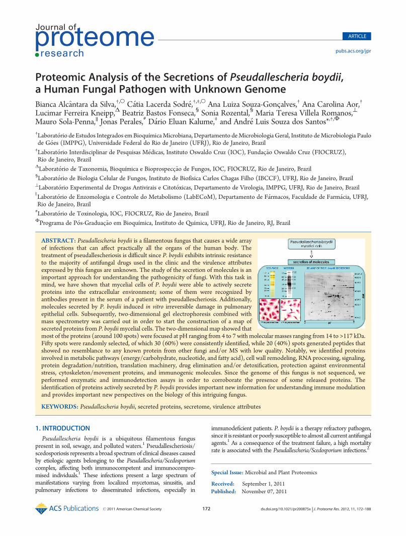
Published: November 07, 2011 r2011 American Chemical Society 172 dx.doi.org/10.1021/pr200875x | J. Proteome Res. 2012, 11, 172–188 ARTICLE pubs.acs.org/jpr Proteomic Analysis of the Secretions of Pseudallescheria boydii, a Human Fungal Pathogen with Unknown Genome Bianca Alc ^ antara da Silva, †,O C atia Lacerda Sodr e, †,‡,O Ana Luiza Souza-Gonc -alves, † Ana Carolina Aor, † Lucimar Ferreira Kneipp, Δ Beatriz Bastos Fonseca, § Sonia Rozental, § Maria Teresa Villela Romanos, ^ Mauro Sola-Penna, || Jonas Perales, # D ario Eluan Kalume, ‡ and Andr e Luis Souza dos Santos* ,†,Φ † Laborat orio de Estudos Integrados em Bioquímica Microbiana, Departamento de Microbiologia Geral, Instituto de Microbiologia Paulo de G oes (IMPPG), Universidade Federal do Rio de Janeiro (UFRJ), Rio de Janeiro, Brazil ‡ Laborat orio Interdisciplinar de Pesquisas M edicas, Instituto Oswaldo Cruz (IOC), Fundac - ~ ao Oswaldo Cruz (FIOCRUZ), Rio de Janeiro, Brazil Δ Laborat orio de Taxonomia, Bioquímica e Bioprospecc - ~ ao de Fungos, IOC, FIOCRUZ, Rio de Janeiro, Brazil § Laborat orio de Biologia Celular de Fungos, Instituto de Biofísica Carlos Chagas Filho (IBCCF), UFRJ, Rio de Janeiro, Brazil ^ Laborat orio Experimental de Drogas Antivirais e Citot oxicas, Departamento de Virologia, IMPPG, UFRJ, Rio de Janeiro, Brazil ) Laborat orio de Enzomologia e Controle do Metabolismo (LabECoM), Departamento de F armacos, Faculdade de Farm acia, UFRJ, Rio de Janeiro, Brazil # Laborat orio de Toxinologia, IOC, FIOCRUZ, Rio de Janeiro, Brazil Φ Programa de P os-Graduac - ~ ao em Bioquímica, Instituto de Química, UFRJ, Rio de Janeiro, RJ, Brazil 1. INTRODUCTION Pseudallescheria boydii is a ubiquitous filamentous fungus present in soil, sewage, and polluted waters. 1 Pseudallescheriosis/ scedosporiosis represents a broad spectrum of clinical diseases caused by etiologic agents belonging to the Pseudallescheria/Scedosporium complex, affecting both immunocompetent and immunocompro- mised individuals. 1 These infections present a large spectrum of manifestations varying from localized mycetomas, sinusitis, and pulmonary infections to disseminated infections, especially in immunodeficient patients. P. boydii is a therapy refractory pathogen, since it is resistant or poorly susceptible to almost all current antifungal agents. 1 As a consequence of the treatment failure, a high mortality rate is associated with the Pseudallescheria/Scedosporium infections. 2 Special Issue: Microbial and Plant Proteomics Received: September 1, 2011 ABSTRACT: Pseudallescheria boydii is a filamentous fungus that causes a wide array of infections that can affect practically all the organs of the human body. The treatment of pseudallescheriosis is difficult since P. boydii exhibits intrinsic resistance to the majority of antifungal drugs used in the clinic and the virulence attributes expressed by this fungus are unknown. The study of the secretion of molecules is an important approach for understanding the pathogenicity of fungi. With this task in mind, we have shown that mycelial cells of P. boydii were able to actively secrete proteins into the extracellular environment; some of them were recognized by antibodies present in the serum of a patient with pseudallescheriosis. Additionally, molecules secreted by P. boydii induced in vitro irreversible damage in pulmonary epithelial cells. Subsequently, two-dimensional gel electrophoresis combined with mass spectrometry was carried out in order to start the construction of a map of secreted proteins from P. boydii mycelial cells. The two-dimensional map showed that most of the proteins (around 100 spots) were focused at pH ranging from 4 to 7 with molecular masses ranging from 14 to >117 kDa. Fifty spots were randomly selected, of which 30 (60%) were consistently identified, while 20 (40%) spots generated peptides that showed no resemblance to any known protein from other fungi and/or MS with low quality. Notably, we identified proteins involved in metabolic pathways (energy/carbohydrate, nucleotide, and fatty acid), cell wall remodeling, RNA processing, signaling, protein degradation/nutrition, translation machinery, drug elimination and/or detoxification, protection against environmental stress, cytoskeleton/movement proteins, and immunogenic molecules. Since the genome of this fungus is not sequenced, we performed enzymatic and immunodetection assays in order to corroborate the presence of some released proteins. The identification of proteins actively secreted by P. boydii provides important new information for understanding immune modulation and provides important new perspectives on the biology of this intriguing fungus. KEYWORDS: Pseudallescheria boydii, secreted proteins, secretome, virulence attributes

Welcome message from author
This document is posted to help you gain knowledge. Please leave a comment to let me know what you think about it! Share it to your friends and learn new things together.
Transcript
Published: November 07, 2011
r 2011 American Chemical Society 172 dx.doi.org/10.1021/pr200875x | J. Proteome Res. 2012, 11, 172–188
ARTICLE
pubs.acs.org/jpr
Proteomic Analysis of the Secretions of Pseudallescheria boydii,a Human Fungal Pathogen with Unknown GenomeBianca Alcantara da Silva,†,O C�atia Lacerda Sodr�e,†,‡,O Ana Luiza Souza-Gonc-alves,† Ana Carolina Aor,†
Lucimar Ferreira Kneipp,Δ Beatriz Bastos Fonseca,§ Sonia Rozental,§ Maria Teresa Villela Romanos,^
Mauro Sola-Penna,|| Jonas Perales,# D�ario Eluan Kalume,‡ and Andr�e Luis Souza dos Santos*,†,Φ
†Laborat�orio de Estudos Integrados emBioquímicaMicrobiana, Departamento deMicrobiologia Geral, Instituto deMicrobiologia Paulode G�oes (IMPPG), Universidade Federal do Rio de Janeiro (UFRJ), Rio de Janeiro, Brazil‡Laborat�orio Interdisciplinar de Pesquisas M�edicas, Instituto Oswaldo Cruz (IOC), Fundac-~ao Oswaldo Cruz (FIOCRUZ),Rio de Janeiro, BrazilΔLaborat�orio de Taxonomia, Bioquímica e Bioprospecc-~ao de Fungos, IOC, FIOCRUZ, Rio de Janeiro, Brazil§Laborat�orio de Biologia Celular de Fungos, Instituto de Biofísica Carlos Chagas Filho (IBCCF), UFRJ, Rio de Janeiro, Brazil^Laborat�orio Experimental de Drogas Antivirais e Citot�oxicas, Departamento de Virologia, IMPPG, UFRJ, Rio de Janeiro, Brazil
)Laborat�orio de Enzomologia e Controle do Metabolismo (LabECoM), Departamento de F�armacos, Faculdade de Farm�acia, UFRJ,Rio de Janeiro, Brazil#Laborat�orio de Toxinologia, IOC, FIOCRUZ, Rio de Janeiro, BrazilΦPrograma de P�os-Graduac-~ao em Bioquímica, Instituto de Química, UFRJ, Rio de Janeiro, RJ, Brazil
1. INTRODUCTION
Pseudallescheria boydii is a ubiquitous filamentous funguspresent in soil, sewage, and polluted waters.1 Pseudallescheriosis/scedosporiosis represents a broad spectrum of clinical diseases causedby etiologic agents belonging to the Pseudallescheria/Scedosporiumcomplex, affecting both immunocompetent and immunocompro-mised individuals.1 These infections present a large spectrum ofmanifestations varying from localized mycetomas, sinusitis, andpulmonary infections to disseminated infections, especially in
immunodeficient patients. P. boydii is a therapy refractory pathogen,since it is resistant or poorly susceptible to almost all current antifungalagents.1 As a consequence of the treatment failure, a high mortalityrate is associated with the Pseudallescheria/Scedosporium infections.2
Special Issue: Microbial and Plant Proteomics
Received: September 1, 2011
ABSTRACT: Pseudallescheria boydii is a filamentous fungus that causes a wide arrayof infections that can affect practically all the organs of the human body. Thetreatment of pseudallescheriosis is difficult since P. boydii exhibits intrinsic resistanceto the majority of antifungal drugs used in the clinic and the virulence attributesexpressed by this fungus are unknown. The study of the secretion of molecules is animportant approach for understanding the pathogenicity of fungi. With this task inmind, we have shown that mycelial cells of P. boydii were able to actively secreteproteins into the extracellular environment; some of them were recognized byantibodies present in the serum of a patient with pseudallescheriosis. Additionally,molecules secreted by P. boydii induced in vitro irreversible damage in pulmonaryepithelial cells. Subsequently, two-dimensional gel electrophoresis combined withmass spectrometry was carried out in order to start the construction of a map ofsecreted proteins from P. boydiimycelial cells. The two-dimensional map showed thatmost of the proteins (around 100 spots) were focused at pH ranging from 4 to 7 with molecular masses ranging from 14 to >117 kDa.Fifty spots were randomly selected, of which 30 (60%) were consistently identified, while 20 (40%) spots generated peptides thatshowed no resemblance to any known protein from other fungi and/or MS with low quality. Notably, we identified proteinsinvolved in metabolic pathways (energy/carbohydrate, nucleotide, and fatty acid), cell wall remodeling, RNA processing, signaling,protein degradation/nutrition, translation machinery, drug elimination and/or detoxification, protection against environmentalstress, cytoskeleton/movement proteins, and immunogenic molecules. Since the genome of this fungus is not sequenced, weperformed enzymatic and immunodetection assays in order to corroborate the presence of some released proteins. Theidentification of proteins actively secreted by P. boydii provides important new information for understanding immune modulationand provides important new perspectives on the biology of this intriguing fungus.
KEYWORDS: Pseudallescheria boydii, secreted proteins, secretome, virulence attributes
173 dx.doi.org/10.1021/pr200875x |J. Proteome Res. 2012, 11, 172–188
Journal of Proteome Research ARTICLE
Despite the growing importance of P. boydii infections in thelast decades, very little is known about the physiology, biochem-istry, and genetics of this human fungal pathogen.3 Furtherhindering the evolution of knowledge about the cell biology ofthis fungus is the absence of the sequencing of its genome.Therefore, proteomic approaches should help in elucidatingsome of the fungal cell life processes, revealing the presence ofproteins associated with events such as nutrition, proliferation,growth, differentiation, and interaction with host structures.Secretome-related studies are particularly relevant in under-standing the physiology of filamentous fungi because many ofthem secrete a vast number of proteins to accommodate theirsaprophytic lifestyle. Many of these proteins are of specialinterest in the study of pathogens.4 For instance, pathogenicfilamentous fungi could use extracellular enzymes to degrade thestructural barriers of the host to facilitate this tissue invasion, andthey could also participate in the infection by eliminating somemechanisms of the immune defense and/or helping in theobtaining of nutrients.3
Experimental evidence about the expression of secreted mol-ecules by fungi belonging to the Pseudallescheria/Scedosporiumcomplex was first reported by Larcher and co-workers, who hadpurified an extracellular serine protease able to degrade humanfibrinogen, suggesting an action as a mediator of the severechronic bronchopulmonary inflammation from which cysticfibrosis patients suffer.5 Our group described the secretion oftwo acidic metallo-type proteases of 28 and 35 kDa capable ofhydrolyzing various substrates, including serum human proteins(e.g., albumin, hemoglobin, and immunoglobulin G), extracel-lular matrix components (e.g., laminin and fibronectin),and sialylated proteins (e.g., fetuin and mucin). Those resultsdemonstrated that extracellular metalloproteases produced byP. boydii could help the fungus to escape from natural humanbarriers and defenses.6 Bertrand and co-workers reported thesecretion of two siderophores of hydroxamates class, dimerumicacid and Nα-methyl coprogen B, by S. apiospermum, and com-parison of various strains suggested a higher production ofNα-methyl coprogen B by clinical isolates of respiratory origin.7,8
Furthermore, different species of Pseudallescheria are able torelease antibiotic substances, including the following: (i) tyro-scherin, an antitumor antibiotic that selectively inhibited thegrowth of MCF-7 human breast cancer cells;9 (ii) YM-193221(2-dimethylamino-1-(4-hydroxyphenyl)-8,10-dimethyl-6-dode-cene-3-one), which exhibited potent antifungal activity againstCandida albicans;10 (iii) a new antibacterial dioxopiperazine(dehydroxybisdethiobis(methylthio)gliotoxin) and the pre-viously described bisdethiobis(methylthio)gliotoxin and glio-toxin, which exhibited potent antibacterial activity against themethicillin-resistant andmultidrug-resistant Staphylococcus aureus;11
and (iv) a fungistatic substance capable of reducing the dis-ease incidence of black leaf spot of spoon cabbage causedby Alternaria brassicicola and inhibiting the germination ofA. brassicicola conidia.12 P. boydii spores also produce cyclicnonribosomal peptides with a unique structure, named pseuda-cyclins, which can be used as early indicators of fungal infectioncaused by Pseudallescheria species.13 Therefore, the identificationof proteins actively secreted by P. boydii provides importantnew information for understanding immune modulation andprovides important new perspectives on the biology of this fungalpathogen.
In the present work, we have initially reported that mycelialcells of P. boydiiwere able to release proteins to the extracellular
environment, some of them recognized by antibodies pre-sented in the serum obtained from a patient with pseudal-lescheriosis, showing the antigenic nature of these extracellularproteins. Also, molecules secreted by P. boydii induced injuryin pulmonary epithelial cells. Two-dimensional gel electro-phoresis combined with mass spectrometry was carried outin order to start the construction of a map of secretedproteins from P. boydii mycelia. Proteomics analysis revealeda variety of proteins associated with fungal survival and hostpathogenicity.
2. MATERIALS AND METHODS
Microorganism and Growth ConditionPseudallescheria boydii (RKI07_0416) belongs to the clade 4
to the Pseudallescheria boydii/Scedosporium apiospermum com-plex, as previously proposed by Gilgado and co-workers.14 Thefungus was grown on Sabouraud-dextrose broth (containing 2%glucose, 1% peptone, and 0.5% yeast extract) at 25 �C for 7 dayswith constant shaking (200 rpm). Then, mycelial cells werefiltered with filter paper and washed twice with sterile phosphate-buffered saline (PBS; 150 mM NaCl, 20 mM phosphate buffer,pH 7.2).6
Secretion AssayThe P. boydiimycelia (approximately 10 g) were resuspended
in 10 mL of sterile isotonic PBS supplemented with 2% glucoseand incubated for 20 h at 25 �C with constant agitation(200 rpm).6 After this interval, the cells were removed bycentrifugation (2500g/20 min/4 �C) and the supernatant waspassed over a 0.22-μmmembrane (Millipore, S~ao Paulo, Brazil).The cell-free PBS�glucose supernatant was concentrated in a10,000 molecular weight cutoff AMICONmicropartition system(AMICON, Beverly, MA, USA), obtaining a final concentrationof 100-fold, and kept at �20 �C until use. Proteins in theseextracts were then quantified by the method of Lowry and co-workers,15 using bovine serum albumin (BSA) as the proteinstandard.
Fungal ViabilityThe viability of the mycelia during the incubation period in the
isotonic phosphate buffer was assessed by fluorescence micro-scopy observation. Mycelial cells were incubated for 30 min atroom temperature with calcofluor white (Sigma). Subsequently,the systems were washed with PBS and incubated with propi-dium iodide at 10 μg/mL (Sigma) for 10 min. After, the systemswere washed with PBS and taken immediately for viewing on aZeiss epifluorescence microscope (Axioplan). Cells killed byautoclaving (121 �C/30 min) were used as positive controlsfor staining with propidium iodide.
Transmission Electron Microscopy (TEM)After secretion experiments, mycelial cells were washed in PBS
and then fixed with 2.5% glutaraldehyde and 4% paraformalde-hyde with 10 mM calcium chloride added in 100 mM cacodylatebuffer, pH 7.4, for 1 h at 25 �C, washed in cacodylate buffer,and then postfixed with 1%OsO4 and 0.8% K4Fe(CN)6 3 3H2Oin 100mM cacodylate buffer for 2 h. Samples were then washedin cacodylate buffer, dehydrated in graded series of acetone,and embedded in Spurr resin. Ultrathin sections were routinelystained with aqueous uranyl acetate and alkaline lead citrateand examined in a TEM type JEOL 1200 EX, operating at80 kV.
174 dx.doi.org/10.1021/pr200875x |J. Proteome Res. 2012, 11, 172–188
Journal of Proteome Research ARTICLE
Secretory Protein ProfilePBS-glucose supernatant of P. boydiimycelial form (equivalent
to 5 μg of protein) was treated with an equal volume of sodiumdodecyl sulfate�polyacrylamide gel electrophoresis (SDS-PAGE)sample buffer (125 mM Tris, pH 6.8; 4% SDS; 20% glycerol;0.002% bromophenol blue) supplemented with 10% β-mercap-toethanol, followed by heating at 100 �C for 5 min. Polypeptideswere analyzed on 10% SDS-PAGE by the method described byLaemmli.16 Electrophoresis was carried out at 100 V for 90 minat 4 �C, and the gel was silver stained. Prior to electrophoresis,Gibco BRL (Grand Island, NY, USA) molecular mass standardswere boiled in SDS-PAGE sample buffer and then applied to thesame gel.
Detection of Antigenic Proteins Secreted by Mycelial CellsExtracellular protein extracts from P. boydiimycelial cells were
separated by SDS-PAGE and silver-stained or, alternatively,transferred at 4 �C at 100 V/300 mA for 2 h to a nitrocellulosemembrane. Blotted proteins were blocked in PBS containing0.1% Tween 20 (PBS-T) and 5% low fat dried milk. Afterblocking, the membrane was incubated for 1 h at 37 �C in thepresence of a serum from a human patient with pseudallescher-iosis (kindly provided by Dr. Bodo Wanke, Fundac-~ao OswaldoCruz, Rio de Janeiro, Brazil) or with a healthy serum both at1:1000 dilution and washed three times in PBS-T, followed byincubation for 1 h at room temperature with a peroxidase-labeledanti-human antibody at 1:2500 dilution. Immunoblot was ex-posed to an X-ray film after chemiluminescence reaction usingthe ECL system (Pierce). Alternatively, membranes containingthe secreted proteins of P. boydii were individually incubated for1 h at 37 �C with the anti-actin (Sigma) and anti-tubulin mono-clonal antibody (Sigma) and then revealed as described above.
Cytotoxycity AssayIn order to determine the cytotoxicity of the molecules
released by P. boydii mycelial cells, different supernatant con-centrations (ranging from 0.1 to 5 μg/μL of protein) were placedin contact with monolayers of A549 cells (that is an establishedlineage from human Caucasian lung carcinoma) in RPMImedium and then incubated at 37 �C in a 5% CO2 atmospherefor 24 h. In the control system, the cell lineages were incubated inRPMI medium supplemented only with PBS. After that, cellularviability was evaluated by the neutral red dye-uptake method.17
Briefly, mammalian cells were incubated in the presence of 0.01%neutral red solution for 3 h at 37 �C in a 5% CO2 atmosphere.Then, the medium was removed and the cells were fixed with 4%formalin in PBS. The dye incorporated by the viable cells waseluted using a mixture of methanol/acetic acid/water (50:1:49),and the dye uptake was determined by measuring the opticaldensity of the eluate at 490 nm in an automatic spectrophot-ometer (ELx800TM, Bio-TeK Instruments, Inc.). The 50%cytotoxic concentration (CC50) was defined as the compoundconcentration which caused a 50% reduction in the number ofviable cells.
Two Dimensional Gel ElectrophoresisProteins from the supernatant containing a protease inhibitor
cocktail were precipitated by 17% tricholoracetic acid (TCA) andthen solubilized in rehydration solution (2% CHAPS, 20 mMdithiothreitol (DTT), and 8 M urea) and stored at �70 �C.Protein concentration was determined by the RC DC method(Bio-Rad, Hercules, CA, USA), using BSA as standard protein.This assay is based on the Lowry assay but has been modified to
be reducing agent compatible (RC) as well as detergent compa-tible (DC).15 For the first dimension, aliquots of 700 μg ofprotein were applied to 17 cm immobilized pH gradient (IPG)gel strips (Bio-Rad, Hercules, CA, USA) with a linear separationpH range by in-gel sample rehydration. After 11 h of activerehydration at 50 V at 20 �C using a Protean isoelectric focusingsystem (IEF) (Bio-Rad, Hercules, CA, USA), proteins wereseparated using the following protocol: 250 V, 20 min; 10,000 V,2 h 30 min; 10,000 V, 90,000 VH. Before the second dimen-sion, proteins were reduced (10 mg/mL DTT) and alkylated(25 mg/mL iodoacetamide) in equilibration buffer (6 M urea,2% SDS, 300 mM Tris-HCl, pH 8.8, and 20% glycerol) and thenseparated on 12% SDS-PAGE gels and stained with CoomassieBrilliant Blue G-250. Gel images were captured with a densit-ometer (GS-800, Bio-Rad, Hercules, CA, USA), digitalized,and analyzed by PDQuestTM software (Bio-Rad, Hercules,CA, USA).
Protein Digestion and Sample PreparationFollowing 2D SDS-PAGE, several spots were manually ex-
cised from the gel in order to perform trypsin digestion asdescribed previously.18 The selected spots were destained with100 μL of 25 mM NH4HCO3, pH 8.0, in 50% acetonitrile for15 min. After three washings, gel pieces were dehydrated with100% acetonitrile and then dried using a Speed Vac evaporator(Savant, Farmingdale, NY, USA). Proteolytic in-gel digestionwas performed using 10 μL of ice-cold sequence grade modifiedtrypsin (Promega, Madison, WI, USA) solution (20 ng/μL in25 mM NH4HCO3, pH 8.0) at 37 �C, overnight, followed bypeptide extraction from the gel with 50% acetonitrile in 5%trifluoroacetic acid (TFA) in a sonicator for 30 min. The extractswere dried under vacuum and then solubilized in 50% acetoni-trile in 0.1% TFA. Contaminants from the peptide mixture wereremoved using C18 ZipTip micropipet tips (Millipore, Bedford,MA). Before loading the samples, the C18 media was firstactivated with 10 μL of wetting solution (100% acetonitrile),followed by equilibration in 10 μL of 0.1% TFA. The sampleswere loaded onto the ZipTip by 8 to10 repeated aspiration anddispensing cycles, followed by a washing step with 0.1% TFA.The bound peptides were eluted by carefully aspirating anddispensing 8�10 times 5 μL of 70% acetonitrile in 0.1% TFA.Afterward, approximately 0.3 μL of the peptide samples werecocrystallized with an equal volume ofmatrix solution (10mg/mLα-cyano-4-hydroxycinnamic acid solution (Sigma) in 0.1% TFAin 50% acetonitrile) directly onto a MALDI target plate.
In a few samples, 4-sulfophenyl-isothiocyanate (SPITC) deri-vatization was carried out according to Wang and co-workers, inorder to improve the de novo sequencing of the peptides.19
Approximately 5�10 μL of the peptide mixture from the in-geldigestion was dried down in a vacuum centrifuge and concen-trated to 1 μL. This peptide solution was derivatized by mixingwith 10 μL of SPITC (10 mg/mL in 20 mM NaHCO3, pH 9.0)and incubating for 30 min at 55 �C. The reaction was terminatedby adding 1 μL of 1% TFA, and the samples were loaded onto aC18 ZipTip micropipet tip to remove contaminants prior toanalysis by mass spectrometry.
MALDI-TOF/TOF AnalysisThe tryptic peptides were analyzed on a 4700-Proteomics
Analyzer MALDI-TOF/TOF (Applied Biosystems, Foster City,CA). All mass spectra were acquired in positive ion reflectormode with 2000 shots per spot and externally mass calibratedwith a peptidemixture. Themass spectra were recorded in a mass

175 dx.doi.org/10.1021/pr200875x |J. Proteome Res. 2012, 11, 172–188
Journal of Proteome Research ARTICLE
range from 900 to 3,000 or 3,500 Da. The 10 or 15 most intenseion peaks from the peptide mass fingerprinting (orMS run) werefurther submitted to fragmentation using PSD mode with CIDgas off and 1 keV collision energy. Subsequent toMS acquisition,the peak list files ppw (Protein-Peptide by Well), pps (Protein-Peptide by Spot Set), and pmf (Peptide Mass Fingerprinting)were generated from the raw mass spectrum data using the “peakto mascot” script of the 4000 Series Explorer Software accordingto the settings: for pmf, mass range from 500 to 4000 Da, peakdensity of 15 per 200 Da, signal-to-noise of 10, minimal areavalue of 100, and maximal 60 peaks per spot. For pps andppw files, mass range from 60 to precursor�20 Da, peak densityof 55 peaks per 200 Da, signal-to-noise of 2, minimal area value of10, and maximal 200 peaks per precursor.
Bioinformatics Analysis and Identification of ProteinsFollowing MS acquisition, each mass spectrum was submitted
to a search using Mascot version 2.2 online (Matrix Science,http://www.matrixscience.com/cgi/search_form.pl?FORMVER=2&SEARCH=MIS). For the identification of proteins, the searchwas performed against the NCBInr nonredundant database(National Center for Biotechnology Information, http://www.ncbi.nlm.nih.gov/) with no taxonomy restriction. When neces-sary, further searches were performed against the Fungi database.The parameters used were trypsin as the enzyme of choice andtwo missed cleavage,(1 Da for the precursor mass, and(0.5 Dafor the fragment ion mass. Oxidation of methionine and trypto-phan along with N-terminal acetylation of proteins, cyclization ofglutamine and glutamate (pyro-glutamate), and alkylation ofcysteine (carbamidomethylcysteine) were allowed as variablemodifications. In order to validate a protein identification, theMascot ion score was considered greater than 40 and thesignificance threshold P < 0.05. If a protein hit was identifiedby only one peptide, the MS/MS data should exhibit a clearspectrum with sequence tags that matched at least 3 consecutivey, b, or even a fragment ion series. The MS/MS spectra that didnot automatically match a peptide sequence were manuallyinterpreted and blast searched against the NCBI protein ornucleotide Fungi database (taxid 4751). The proteins identifiedwith a blast E value lower than 1e�1 were considered as positivematches. One should bear in mind that the P. boydii genome isnot sequenced and identification hits were especially based onorthologous proteins found in other fungus species whosegenome has already been sequenced.
Measurement of Enzymatic ActivityMalate Dehydrogenase. Assays were conducted as de-
scribed by Steffan and McAlister-Henn in a reaction mediumcontaining 50 mM Tris-HCl (pH 7.4), 5 mM MgCl2, 2 mMsodium malate, and 2 mM NAD+.20 Reactions were initiated byaddition of PBS-conditioned supernatant to a final concentrationof 20 μg/mL, and the time course of NADH formation wasevaluated spectrophotometrically at 340 nm. One unit of malatedehydrogenase was attributed to the formation of 1 μmol ofNADH per minute of reaction.Triosephosphate Isomerase. Triosephosphate isomerase
activity was evaluated from conversion of glyceraldehyde-3-phosphate into dihydroxyacetone phosphate followed by re-duction of this product into α-glycerophosphate catalyzed byα-glycerophosphate dehydrogenase. Assays were conducted asdescribed previously by Sola-Penna and co-workers in a reactionmedium containing 50 mM Tris-HCl (pH 7.4), 5 mM MgCl2,2 mM glyceraldehyde-3-phosphate, 0.2 mMNADH, and 3 U/mL
α-glycerophosphate dehydrogenase.21 Reaction was initiated byaddition of PBS-conditioned supernatant to a final concentrationof 20 μg/mL. Oxidation of NADH was evaluated spectrophoto-metrically at 340 nm, and one unit of triosephosphate isomerasewas attributed to the oxidation of 1 μmol of NADHperminute ofreaction.Aldolase. Aldolase activity was evaluated as described pre-
viously by Meira and co-workers.22 Reaction medium contained50 mM Tris-HCl (pH 7.4), 5 mM MgCl2, 1 mM fructose-1,6-bisphosphate, 0.2 mM NADH, 3 U/mL triosephostate isomer-ase, and 3 U/mL β-glycerophosphate dehydrogenase. Reactionwas initiated by addition of PBS-conditioned supernatant to afinal concentration of 20 μg/mL. Oxidation of NADH wasevaluated spectrophotometrically at 340 nm, and one unit ofaldolase was attributed to the oxidation of 1 μmol NADH perminute of reaction.Mannitol-1-phosphate-5-dehydrogenase. Assays were
conducted as described by Iwamoto and co-workers in a reactionmedium containing 50 mM Tris-HCl (pH 7.4), 5 mM MgCl2,1 mM fructose-6-phosphate, and 0.2 mM NADH. Reaction wasinitiated by addition of PBS-conditioned supernatant to a finalconcentration of 20 μg/mL, and the time course of NADHoxidation was evaluated spectrophotometrically at 340 nm.23
One unit of mannitol-1-phosphate-5-dehydrogenase was attrib-uted to the oxidation of 1 μmol of NADH per minute of reaction.Aspartyl Proteinase. The enzymatic activity over the cathep-
sin D substrate (7-methoxycoumarin-4-acetyl-Gly-Lys-Pro-Ile-Leu-Phe-Phe-Arg-Leu-Lys(DNP)-D-Arg-amide) was determinedin the absence or in the presence of 1 μMpepstatin A, a powerfulaspartic protease inhibitor. Cleavage of cathepsin D substrate wasmonitored continuously in a spectrofluorometer (SpectraMaxGemini XPS, Molecular Devices, CA, USA) using an excitationwavelength of 340 nm and an emission wavelength of 490 nm. A500 μM stock solution of the fluorogenic substrate sample wasprepared in dimethylsulfoxide (DMSO). The reaction wasstarted by the addition of the substrate at 2 μM to the PBS-conditioned supernatant (10 μg protein) in a total volume of60 μL of 100 mM sodium acetate, pH 4.0. The reaction mixturewas incubated at 37 �C for 20min. The assays were controlled forself-liberation of the fluorophore over the same time interval.Phosphatase. The phosphatase activity was determined
using a microtiter assay plate by measuring the rate ofp-nitrophenol (p-NP) production from the hydrolysis ofp-nitrophenyl phosphate (p-NPP). PBS-conditioned superna-tant (equivalent to 10 μg/mL of protein) was mixed with 20 mMTris-HCl buffer at pH 7.0 and 5 mM p-NPP as substrate. Thepreparation was incubated for 60 min at 26 �C, in the absence orpresence of 15 mM sodium tartrate, a secreted phosphataseinhibitor. Reactions were terminated by adding 50 μL of 2 MNaOH to each well, and the mixture (100 μL) obtained wasmeasured with an ELISA microplate reader (415 nm), usingp-NP as standard.24 The phosphatase activity was calculated bysubtracting the nonspecific p-NPP hydrolysis measured in theabsence of PBS-conditioned supernatant and expressed permilligram of protein.Superoxide Dismutase. Superoxide dismutase activity was
determined using a method that involves generation of super-oxide and reduction of the tetrazolium dye (3-(4,5-dimethylthia-zol-2-yl)-2,5-diphenyltetrazolium bromide (MTT) to its forma-zan, which is measured at 570 nm, as described by Madesh andBalasubramanian.25

176 dx.doi.org/10.1021/pr200875x |J. Proteome Res. 2012, 11, 172–188
Journal of Proteome Research ARTICLE
3. RESULTS
The survivability of the mycelia along the incubation period(20 h) in PBS-glucose medium was assessed by light andfluorescence microscopy observations after double labeling ofcells with calcofluor white (a nonspecific fluorochrome stain thatbinds to chitin in the cell wall of fungi) and propidium iodide(a DNA intercalate agent that is excluded by viable cells but canpenetrate cell membranes of dying or dead cells), in order toensure noncontamination of the secretome-containing medium
by components released by dying or damaged fungal cells. Bymeans of optical microscopic inspection, mycelial cells main-tained their morphological architecture with a preserved cell wall,as evidenced by calcofluor white staining, and no propidiumiodide staining was observed (Figure 1A). Conversely, myceliakilled by heat (autoclaving) showed intense incorporation ofpropidium iodide (Figure 1A). In parallel, TEM images evi-denced the normal ultrastructural architecture of mycelia aftersecreting in PBS-glucose, presenting typical electron-densecytoplasm, a distinct cell wall, a well-delineated cytoplasmicmembrane, and an intact septum (Figure 1B). Taken together,these results discharged the possibility of cell membrane and/orcell wall injuries, testifying that extracellular proteins were notartifacts and were in fact present due to bona fide secretion ofliving mycelial cells.
The proteins secreted by P. boydii mycelia were concentratedand then separated by 1D SDS-PAGE, revealing the presence ofat least 20 polypeptides with molecular masses ranging from 14to >117 kDa (Figure 1C). In order to assess whether thesecretions of mycelial cells have a biological effect on the host,we tested the immunoreactivity of these proteins with a serumobtained from a patient with pseudallescheriosis. The recognitionof diverse proteins by hyperimmune patient serum indicates thatthese secreted proteins could be important in the pathogenesis of
Figure 1. Secretion of proteins by Pseudallescheria boydii. After growthof mycelial cells for 7 days in Sabouraud medium, the culture washarvested by centrifugation and mycelia were washed in PBS andincubated for 20 h in PBS-glucose. Then, fungal cells were separatedfrom the supernatant by centrifugation. (A) The fungal viability wasassessed by incubation of mycelial cells sequentially with calcofluorwhite (blue staining) and propidium iodide (red staining) and thenobserved by both optical (upper panels) and fluorescence (lower panels)microscopy. In this set of experiments, mycelia submitted to autoclava-tion were used as nonviable cells. (B) Transmission electron microscopyshowing the preserved ultrastructural architecture of mycelial cells afterincubation for 20 h in PBS-glucose. The white asterisks show the cell wall,black arrows indicate the cytoplasmic membrane, and the white arrowevidences the septum. (C) SDS-PAGE revealing the protein profile of theconcentrated PBS-conditioned supernatant. The gel was silver-stained.(D) Secreted proteins were separated by SDS-PAGE, blotted to anitrocellulose membrane, and then incubated with the serum of apseudallescheriosis patient. The numbers on the right indicate the majorreactive bands. In parts C and D, the numbers on the left indicate therelative molecular mass markers expressed in kilodaltons. (E) Differentconcentrations of secreted proteins were added to a confluent monolayerof A549 epithelial cells, and the viability was evaluated by the neutral reddye-uptake method. The inset represents the monolayer of A549 afterincubation in the absence (� supernatant) or in the presence (+ super-natant) of PBS-conditioned supernatant at the CC50 value.
Figure 2. Secretome map of Pseudallescheria boydii. (A) An aliquot of700 μg of proteins was applied to 17 cm immobilized pH range 4�7 IPGstrips. The 2-DE gel was stained with Coomassie Brilliant Blue G-250.Protein spots indicated by numberswere randomly selected for subsequentidentification by mass spectrometry (Table 1). Molecular mass markers(MM) expressed in kilodalton. (B) Rational sequence used for the analysisof the spots obtained from the secretome map of P. boydii mycelial cells.

177 dx.doi.org/10.1021/pr200875x |J. Proteome Res. 2012, 11, 172–188
Journal of Proteome Research ARTICLE
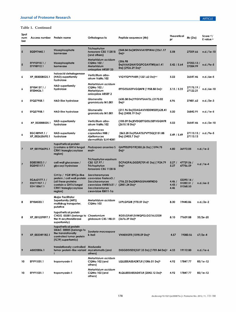
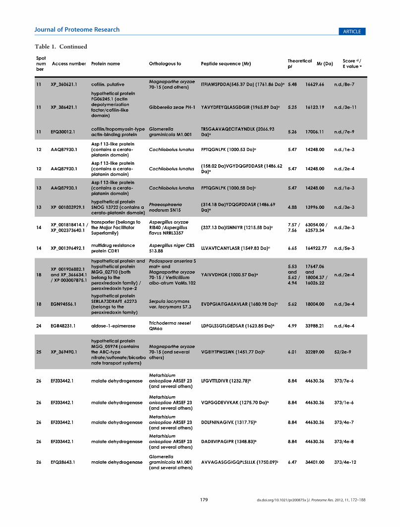
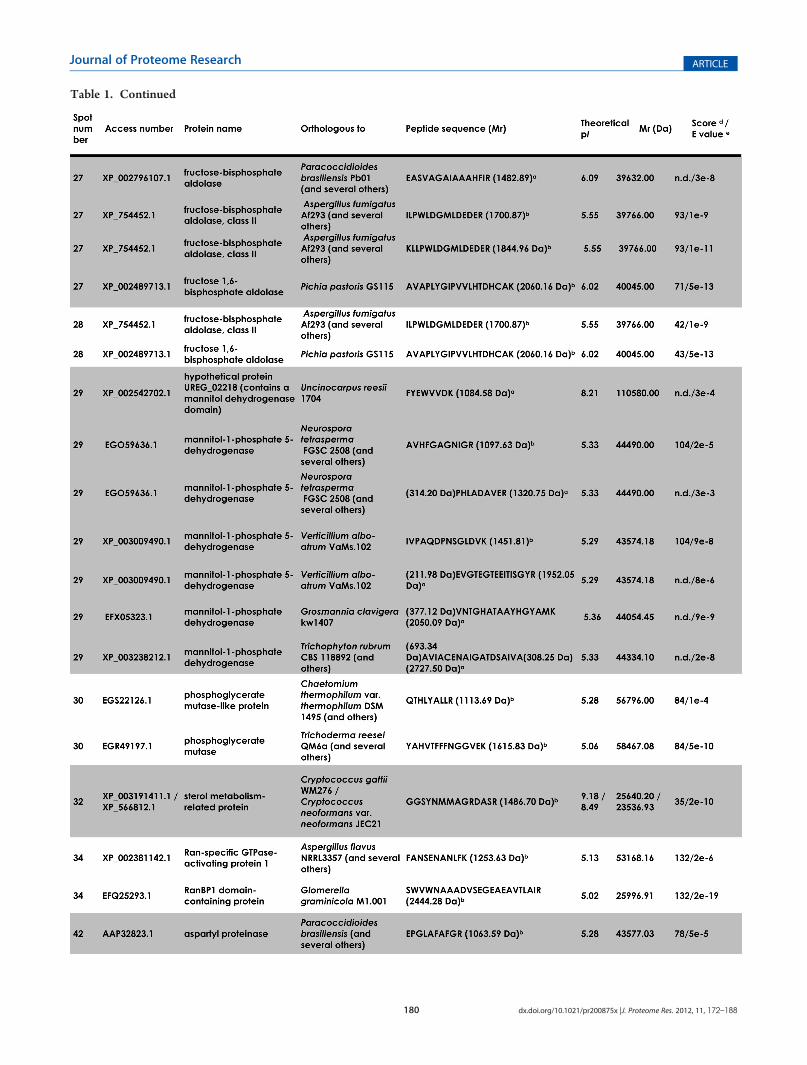
Table 1. Identification of Selected Spots from Pseudallescheria boydii Secretionsf
178 dx.doi.org/10.1021/pr200875x |J. Proteome Res. 2012, 11, 172–188
Journal of Proteome Research ARTICLE
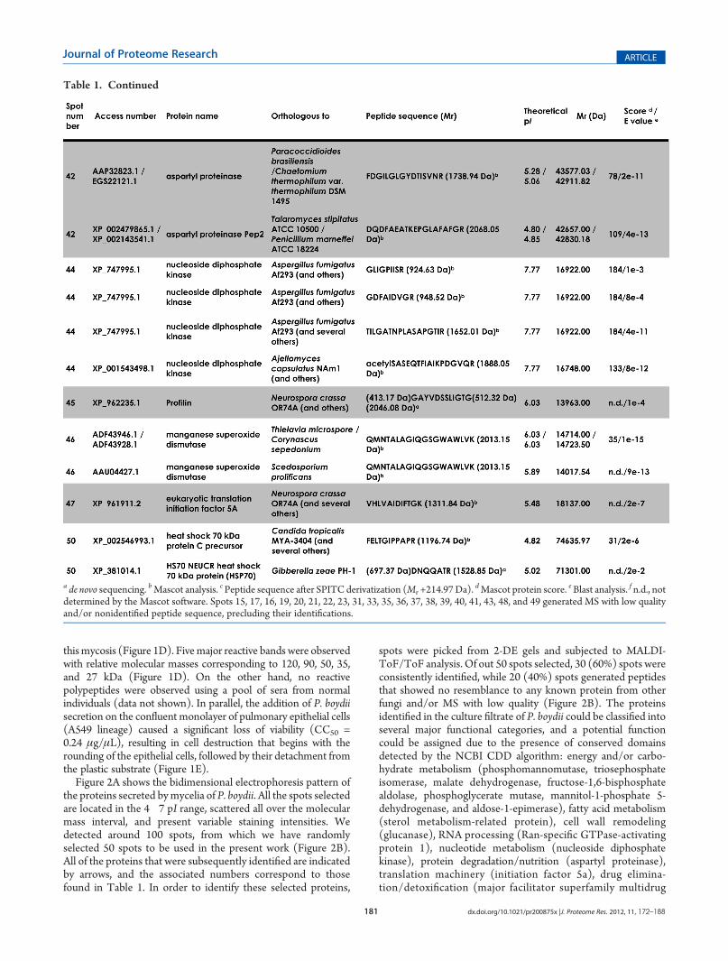
Table 1. Continued
179 dx.doi.org/10.1021/pr200875x |J. Proteome Res. 2012, 11, 172–188
Journal of Proteome Research ARTICLE
Table 1. Continued
180 dx.doi.org/10.1021/pr200875x |J. Proteome Res. 2012, 11, 172–188
Journal of Proteome Research ARTICLE
Table 1. Continued
181 dx.doi.org/10.1021/pr200875x |J. Proteome Res. 2012, 11, 172–188
Journal of Proteome Research ARTICLE
thismycosis (Figure 1D). Fivemajor reactive bands were observedwith relative molecular masses corresponding to 120, 90, 50, 35,and 27 kDa (Figure 1D). On the other hand, no reactivepolypeptides were observed using a pool of sera from normalindividuals (data not shown). In parallel, the addition of P. boydiisecretion on the confluent monolayer of pulmonary epithelial cells(A549 lineage) caused a significant loss of viability (CC50 =0.24 μg/μL), resulting in cell destruction that begins with therounding of the epithelial cells, followed by their detachment fromthe plastic substrate (Figure 1E).
Figure 2A shows the bidimensional electrophoresis pattern ofthe proteins secreted bymycelia of P. boydii. All the spots selectedare located in the 4�7 pI range, scattered all over the molecularmass interval, and present variable staining intensities. Wedetected around 100 spots, from which we have randomlyselected 50 spots to be used in the present work (Figure 2B).All of the proteins that were subsequently identified are indicatedby arrows, and the associated numbers correspond to thosefound in Table 1. In order to identify these selected proteins,
spots were picked from 2-DE gels and subjected to MALDI-ToF/ToF analysis. Of out 50 spots selected, 30 (60%) spots wereconsistently identified, while 20 (40%) spots generated peptidesthat showed no resemblance to any known protein from otherfungi and/or MS with low quality (Figure 2B). The proteinsidentified in the culture filtrate of P. boydii could be classified intoseveral major functional categories, and a potential functioncould be assigned due to the presence of conserved domainsdetected by the NCBI CDD algorithm: energy and/or carbo-hydrate metabolism (phosphomannomutase, triosephosphateisomerase, malate dehydrogenase, fructose-1,6-bisphosphatealdolase, phosphoglycerate mutase, mannitol-1-phosphate 5-dehydrogenase, and aldose-1-epimerase), fatty acid metabolism(sterol metabolism-related protein), cell wall remodeling(glucanase), RNA processing (Ran-specific GTPase-activatingprotein 1), nucleotide metabolism (nucleoside diphosphatekinase), protein degradation/nutrition (aspartyl proteinase),translation machinery (initiation factor 5a), drug elimina-tion/detoxification (major facilitator superfamily multidrug
Table 1. Continued
a de novo sequencing. bMascot analysis. c Peptide sequence after SPITC derivatization (Mr +214.97 Da).dMascot protein score. eBlast analysis. f n.d., not
determined by the Mascot software. Spots 15, 17, 16, 19, 20, 21, 22, 23, 31, 33, 35, 36, 37, 38, 39, 40, 41, 43, 48, and 49 generated MS with low qualityand/or nonidentified peptide sequence, precluding their identifications.

182 dx.doi.org/10.1021/pr200875x |J. Proteome Res. 2012, 11, 172–188
Journal of Proteome Research ARTICLE
transporter, peroxiredoxin, manganese superoxide dismutase,and translationally controlled tumor protein), environmentalstress (heat shock protein), cytoskeleton/movement proteins(cofilin, profilin, and tropomyosin), allergen (Asp f13-likeprotein), miscellaneous reactions (haloacid dehalogenase-super-family hydrolase), and hypothetical proteins without knownfunction but with a characterized domain/region (Forkheadassociated domain involved in signaling events and ABC-typetransport system region). It is obvious that some of theseproteins can be used by fungal cells in different social contexts,and for this reason, different roles can be assigned to eachprotein, as summarized in Figure 3.
As already mentioned, the genome of P. boydii is completelyunknown. For these reasons, we performed some protein/activity identification in order to test the proteomic findings. Inthis context, we measured some enzymatic activities in thesecretions of P. boydii, including the following: mannitol-1-phosphate 5-dehydrogenase, triosephosphate isomerase, malate
dehydrogenase, fructose-1,6-bisphosphate aldolase (Figure 4A),sodium tartrate-sensitive phosphatase activity (Figure 4B), pepsta-tin A-sensitive aspartyl proteinase (Figure 4C), and superoxidedismutase activity (Figure 4D). Subsequently, we searched in thePubMed database for deposited genes belonging to the Pseudal-lescheria/Scedosporium complex, and only a few genes were untilnow sequenced, including the following: 18S rRNA, β-tubulin,manganese superoxide dismutase (Mn-SOD), elongation factor 1α,and calmodulin genes. The unique similar protein detected in thesecretome analysis was theMn-SOD, which presented high identitywith the Mn-SOD of Scedosporium prolificans (E-value thresholdof 9 � 10�13). As described earlier, the secretion in fungal cellsrequires vesicular transport and, consequently, the active participa-tion of cytoskeleton and/or movement proteins. Taking this fact inmind, we searched for the presence of tubulin and actin in P. boydiisecretions. By means of Western blotting assay, the anti-actinantibody reacted with a 45 kDa protein and a protein of 55 kDawas revealed by the anti-tubulin antibody (Figure 4E).
Figure 3. Distribution of the identified proteins in the secretome of Pseudallescheria boydii in different functional categories. The different coloredsquares indicate the possible distinct roles of each identified protein in the P. boydii secretions. The present analysis was based on the well-known roles ofsimilar/homologous proteins published for other several pathogenic fungi. Note that almost all proteins presented more than one recognized biologicalfunction, corroborating the biochemical flexibility of fungal molecules.

183 dx.doi.org/10.1021/pr200875x |J. Proteome Res. 2012, 11, 172–188
Journal of Proteome Research ARTICLE
The mechanisms by which macromolecules such as proteinsreach the extracellular environment and how they are transportedthrough the cell wall, however, have not been rigorously exploredin fungi. Recently, some researchers described that vesicularsecretion is a key and commonmechanism of extracellular deliveryin several fungi.26 Since the profile of protein detection in theP. boydii secretions resembled those found inmammalian exosomesand the content of extracellular vesicles purified from differentpathogenic fungi,26 ultrathin sections of P. boydii mycelial cellswere searched for the presence of vacuole-like structures. TEMimages revealed the presence of vesicles close to the cell surface(Figure 5a�d), which were sometimes found in fusion with theplasma membrane (Figure 5d�g), inside the cell wall (Figure 5hand i), or free in the extracellular environment (Figure 5j and k).
These results add P. boydii to the list of human pathogenic fungithat are able to extracellularly release vesicle-like structures.
4. DISCUSSION
Protein secretion is a universal process of fundamental im-portance for various aspects of cell physiology. However, theprotein secretion pathways used by fungal cells are particularlycomplex and still not well understood, especially in filamentousfungi. Typical studies have focused on the identification, pur-ification, and characterization of single secreted proteins, butstudies on the global analysis of filamentous fungal extracellularproteomes are just beginning to take place. This is quite surprisingwhen considering the clear importance of fungi to mankind.
Figure 4. Measurement of enzymatic activity and immunodetection of some proteins identified in the Pseudallescheria boydii secretions. (A) Detectionof fructose-1,6-bisphosphate aldolase (FBA), malate dehydrogenase (MDH), triosephosphate isomerase (TPI), and mannitol-1-phosphate 5-dehy-drogenase (MPD). (B) Phosphatase activity was measured in the absence (control) or in the presence of 15 mM sodium tartrate (which inhibits theenzymatic activity around 60%). (C) Aspartyl proteinase activity was detected by the cleavage of cathepsin D fluorogenic substrate. As expected,pepstatin A at 1 μM inhibited (∼65%) the hydrolytic activity. (D) Superoxide dismutase activity was determined using the method that involvesgeneration of superoxide and reduction of the tetrazolium dyeMTT to formazan in PBS (control, black bar) and PBS-conditioned medium (white bar).(E) Immunoblotting showing the recognition of both actin and tubulin molecules after incubation with anti-actin and anti-tubulin antibodies,respectively. The numbers on the right indicate the relative molecular mass markers expressed in kilodaltons.

184 dx.doi.org/10.1021/pr200875x |J. Proteome Res. 2012, 11, 172–188
Journal of Proteome Research ARTICLE
Fungal secreted proteins perform several important functions,such as provision of nutrients, cell-to-cell communication, sub-strate colonization, detoxification of the environment, and killingof potential competitors (ecological interactions). Moreover,secreted proteins of pathogenic fungi seem to play critical rolesin virulence. In this sense, fungal secreted proteins can be directlyand/or indirectly involved in the molecular dialogue with thehost cells, enabling their survival, multiplication, and dissemina-tion. Consequently, the secretome can be considered as a realbiological compartment, playing a key role in the fungal infectionstrategy. Herein, we performed a first comprehensive proteome-level study to identify secreted proteins by mycelial cells ofP. boydii, in order to gain a more thorough understanding of thishuman fungal opportunistic pathogen with unknown genome.Having a comprehensive list of P. boydii mycelia secretedproteins will facilitate our understanding about the complexrelationships between this intriguing fungus and the host.Corroborating the relevance of this study, some of the secretedproteins were recognized by antibodies presented in a serumfrom a patient with pseudallescheriosis, which supports ourhypothesis that these proteins are produced during humaninfection. Also, these secreted molecules induced irreversibledamage in pulmonary epithelial cells, which are target cellsduring the infection by P. boydii, culminating in cell death.
A rigorous experimental method to produce a secretome-containing medium, which reflected the P. boydii active exportmolecules, was developed. Nevertheless, some proteins wereidentified with known intracellular locations and functions, but itwas not surprising, since the presence of these proteins is acommon hallmark of published secretome maps of differentfungi. In this sense, enzymes belonging to the energy metabolism
represented the most abundant category in the secretome ofP. boydii, which accounts for 27% of the total proteins identified,including (i) glycolytic enzymes, such as triosephosphate iso-merase and phosphoglycerate mutase, (ii) gluconeogenesis en-zymes, such as fructose-1,6-bisphosphate aldolase, (iii) generalcarbohydrate metabolism enzymes, such as aldose-1-epimerase(which catalyzes the interconversion of α/β-anomers of aldoses,such as glucose and galactose) and phosphomannomutase(which catalyzes the conversion of mannose-6-phosphate tomannose-1-phosphate, which is further converted into guanosinediphosphate (GDP)-mannose, which is required as a mannosesource for the synthesis of oligosaccharide chains in the glyco-sylation pathway),27 (iv) tricarboxylic acid cycle enzymes, such asmalate dehydrogenase (which catalyzes the interconversion ofoxaloacetate and malate linked to the oxidation/reduction ofdinucleotide coenzymes),28 and (v) mannitol metabolism en-zymes, such as mannitol-1-phosphate 5-dehydrogenase.29 Inter-estingly, mannitol is an acyclic hexitol that is found in most fungiand is usually the most abundant of all the soluble carbohydrateswithin the mycelium. Consequently, the physiological role ofmannitol has been widely studied over many years. These rolesinclude carbohydrate storage; a reservoir of reducing power, hightemperature, and oxidative stress tolerance; and spore dislodge-ment/dispersal.30
The earlier cited and several other energy/carbohydratemetabolism enzymes have been detected in secretome maps ofhuman pathogenic fungi, including Candida albicans,31 Crypto-coccus neoformans,32Aspergillus fumigatus,33Histoplasma capsulatum,34
and Paracoccidioides brasiliensis.35 In fungi, proteins frequentlyhave more than a single function and are found in dif-ferent cellular compartments. For instance, in addition to a
Figure 5. Transmission electron micrographs showing the vesicle-like structures in Pseudallescheria boydii. The images revealed the presence of vesiclesclose to the plasma membrane (a�d), which were sometimes found in fusion with this structure (d�g), inside the cell wall (h, i), or free in theextracellular medium (j, k). White arrows evidence the vesicular bodies. cw, cell wall. Bars: 0.1 μm.

185 dx.doi.org/10.1021/pr200875x |J. Proteome Res. 2012, 11, 172–188
Journal of Proteome Research ARTICLE
cytoplasmic location, triosephosphate isomerase of P. brasiliensisis present at the fungal cell wall and is an adhesin putativelyinvolved in binding to both epithelial cells and extracellularmatrix components (e.g., laminin and fibronectin).35 Corroborat-ing these unpredicted roles, some enzymes required for energymetabolism in fungi are highly immunogenic and/or allergenic(Figure 3).31,33,35
During tissue invasion, the fungal cells are exposed to oxidativestress and need to deal with highly toxic reactive oxygen speciesproduced by immune cells. Toxic molecules such as superoxide(O2
•�) and hydrogen peroxide (H2O2) can alter membraneproperties and disrupt membrane-bound proteins and bio-logical macromolecules, leading to cell death.36 Pseudallescheria/Scedosporium species respond to antifungal phagocytic functionto various degrees.37 For instance, serum opsonization of hyphaeof S. apiospermum resulted in a higher level of superoxide anionreleased by human polymorphonuclear leukocytes (PMNs) inresponse to SA54A (an amphotericin B resistant strain) than thatseen in response to SA1216 (amphotericin B susceptible strain);however, PMNs and mononuclear leukocytes induced lesshyphal damage to SA54A than to SA1216.37 Therefore, Pseudal-lescheria/Scedosporium has to maintain its redox homeostasis forsurvival. Scavenger proteins that play important roles in cellsurvival during oxidative stress, such as Mn-SOD, peroxiredoxin,and translationally controlled tumor protein (TCTP) [this lastprotein can act as an antioxidant in a nonenzymatic manner],were identified in P. boydii secretions. Lima and co-workerspurified from mycelial cells of S. apiospermum a Cu,Zn-super-oxide that is stimulated by iron starvation.38 Iron availability hasbeen suggested as a significant controlling factor on expressionlevels of antioxidant enzymes in various microorganisms such asA. fumigatus and A. nidulans.39 TCTP is involved in importantcellular processes, such as cell growth and cell cycle progression,and it can protect cells from various stress conditions andapoptosis due to its biological properties. Interestingly, TCTPwas identified as a glucose-regulated protein,40 and this wasthe condition in which P. boydii cells were submitted to obtainits secretion content. TCTP can complex with microtubulesand actin microfilaments regulating the cell shape in cells. TCTPis also believed to be involved in a variety of inflammatoryprocesses: secretion of histamine and cytokines such as IL-4, IL-8, and IL-13 thereby potentially regulates allergic phenomena.41
For instance, Cladosporium herbarum TCTP is an IgE-bindingantigen and is associated with disease severity.42 TCTP producedby the fungus Madurella mycetomatis, which causes a diseasecharacterized by tumorous swellings in eumycetoma patients, is apredominant immunogenic antigen that is secreted in vitro intothe culture medium. In vivo, TCTP was found to be expressedon hyphae present in developing stages of the eumycetomacharacteristic black grain. Significant IgG and IgM immuneresponses, against the TCTP of M. mycetomatis, were deter-mined, and the antibody levels correlated with lesion size anddisease duration.43
Another interesting and unexpected aspect of the P. boydiisecretome was the presence of the putative eukaryotic translationinitiation factor 5A (eIF5A), which is a highly conserved andessential protein present in all organisms from archaea tomammals, but not in eubacteria.44 Despite being highly con-served and essential, the critical cellular role of eIF5A remainsunclear. Frigieri and co-workers45 revealed that eIF-5A waslocalized in both soluble and membrane fractions of S. cerevisiae,and its membrane association was ribosome-dependent. Those
authors also proposed a link between translation and vesiculartrafficking by using yeast cells mutated in several secretionpathways.
The secretion in fungal cells has been proposed as a result ofvesicular transport. Rodrigues and co-workers32 showed simila-rities in the protein content of extracellular vesicles released byliving C. neoformans cells and mammalian exosome-like struc-tures, which usually contain cytoplasmic proteins such as elonga-tion factors, tubulin, actin, actin-binding proteins, annexins, Rabprotein, molecules responsible for signal transduction, and heatshock proteins such as Hsp70 and Hsp90.46 Sorting of cytosolicproteins into exosomes is normally explained by a randomengulfment of small portions of cytosol during the inwardbudding process of multivesicular bodies.46 Supporting thisstatement, transmission electron microscopic images evidencedthe exosome-like structures in cryptococcal cells, suggesting thatthe extracellular vesicles originate from fungal exosomes.32 Asimilar vesicular transport mechanismwas subsequently reportedin other yeasts and filamentous fungi, suggesting that thisexosome-like structure is a common and well-preserved way toextracellularly release molecules by fungal cells.26 Similarly,vesicles corresponding to endosomes and/or exosomes arefrequently seen in the cytoplasm in close proximity to the cellsurface of P. boydii or free in the extracellular medium.
As it is well-known, the fungal cell wall is a barrier to movementevents. However, the cell wall is a dynamic structure.Wall expansionduring growth requires continuous remodeling of the cell wallpolysaccharide network.47 Synthesis and remodeling of the cell wallpolysaccharide skeleton require not only synthases but also enzymes(hydrolases and transglycosidases) outside the plasma membranethat interconnect and remodel individual cell wall polymers to createa strong but flexible network of macromolecules,47 which permitsthe passage of vesicles through the cell wall polymers. At least aglucanase was identified in the secretions of P. boydii.
As expected, cytoskeleton and movement proteins mustparticipate of the complex vesicular transport in fungal cells. Herein,we detected some of these proteins: cofilin, profilin, tubulin, andactin. The organization of the actin cytoskeleton is important forvarious cellular events, including morphogenesis, migration, andcytokinesis. Eukaryotic cells express various kinds of proteins thatcontrol the architecture and behavior of the actin cytoskeleton.48
Yeast cells contain two prominent actin structures, cables andpatches, both of which are rapidly assembled and disassembled byoperation of other movement proteins, such as cofilin and profilin,which bind to actin monomers (G-actin) and filaments (F-actin)and stimulate depolymerization and fragmentation of F-actin, andtropomysion, which inhibits the dissociation of actin subunits fromthe pointed end of an F-actin.48 In another way, cofilin phosphory-lation/dephosphorylation at Ser3 acts as a simple switch for actinassembly and disassembly/severing.49
Signaling events are essential mechanisms to adaptation ofcells to several conditions and stimuli. Fungal cells respond toextracellular stimuli by signaling pathways that coordinate pro-cesses involved in the activation and/or synthesis of proteinkinases, protein phosphatases, second messengers, and transcrip-tion factors. Membrane-bound protein phosphatases were char-acterized in P. boydii mycelial cells.50 Proteins potentiallyinvolved in signaling cascades were also detected herein in theextracellular content of P. boydii, including a phosphatase in-hibited by sodium tartrate. However, the physiological roles ofthese membrane and secreted phosphatases have not been wellestablished yet. In fungi such asC. neoformans,C. parapsilosis, and

186 dx.doi.org/10.1021/pr200875x |J. Proteome Res. 2012, 11, 172–188
Journal of Proteome Research ARTICLE
Fonsecaea pedrosoi, phosphatase was implicated in the adhesiveprocess to epithelial cells.51,52,24 Moreover, the protein sequenceanalysis revealed a putative adhesion member of the haloaciddehalogenase (HAD) superfamily of hydrolases, which includesdehalogenases, phosphonatases, phosphomutases, phosphatases,and ATPases,53 in the P. boydii secretome. An interesting workevaluated the relevance of a 32 kDa protein (PbHad32p), aputative protein belonging to the HAD superfamily of hydro-lases, in the virulence of P. brasiliensis. Knockdown of PbHAD32did not alter cell vitality or viability but induced morphologicalalterations in yeasts as well as significantly affected their capacityto adhere to human epithelial cells and presented decreasedvirulence in a mouse model of infection.54 The role of nucleosidediphosphate (NDP) kinase in signal transduction systems is wellrecognized.55 NDP kinases catalyze the transfer of the γ-phos-phate from a nucleoside triphosphate to a nucleoside diphos-phate and are important in nucleotide metabolism, providingNTPs as a housekeeping enzyme to sustain growth and differ-entiation; they were also identified in P. boydii. The GTPase Ranthat belongs to the superfamily of small Ras-like GTP-bindingproteins is one of the key player molecules regulating theprocesses inside the eukaryotic nucleus. So far it has been shownto participate in nuclear transport, nuclear pore complex assem-bly, mitotic spindle assembly, and DNA replication.56 Here, wedetected a Ran-binding protein 1(RanBP1) that in S. cerevisiaehas a major role in the cytoplasm, both in recycling of transportreceptors and in release of export cargo.57 Consistent with thisview, S. cerevisiae YRB1, encoding yeast RanBP1, is essential forcell viability and is required for both nuclear protein import andpoly(A)+ RNA export.58
The heat shock proteins (Hsp) are not restricted to heat shockprotection; they also play a role in protein folding, translocation ofproteins across membranes, and gene regulation. Interestingly,microbial Hsps are major targets of host immune responses. Inparticular, members of the 70 kDa Hsp (Hsp70) family are amongthe most immunogenic proteins of human pathogenic microorgan-isms, including fungi.59 P. boydii mycelial cells were able to secreteHsp70 as well as other immunogenic molecules: the allergen Aspf13 and aspartyl proteinase. Especially, aspartyl-type proteinases aremultifaceted molecules carrying out “housekeeping” taskscommon to many eukaryotes as well as functions highly specificto the fungal life cycles, including nutrition, proliferation,differentiation, adhesion to either abiotic or biotic substrates,and escape of host immune responses.60 Several pathogenicfungi secrete aspartyl proteinases such as Aspergillus spp.(named aspergillopepsin), Penicillium spp. (penicillopepsin),Rhizopus spp. (rhizopuspepsin), and Candida spp. (Saps).60
In yeasts, including the pathogenic Candida, an up-regulationof multidrug transporter genes belonging to either the ATP BindingCassette (ABC) or Major Facilitator Superfamily (MFS) is fre-quently observed in the cells exposed to the drugs leading to thephenomenon of multidrug resistance (MDR).61 MDR is a ubiqui-tous biological phenomenon causing serious problems in thetreatment of human cancers and infections of bacterial and fungalorigin. The detection of multidrug transporters can be correlatedwith the intrinsic resistance of P. boydii to several antifungal drugs,including amphotericin B and more-recently introduced antifungalcompounds such as voriconazole and posaconazole.62
A picture of the proteins secreted by P. boydii in cell-freeculture provides a basis for investigation of effector proteins thatmay be active in host cells. Targeting of virulence factors intohost surroundings has been shown to be an effective strategy
used by fungal pathogens in order to remodel the environmentand to influence host cell function. Appreciation of the multi-functional nature of the P. boydii secreted proteins can lead us tospeculate that these molecules may play ancillary roles inpathogenesis or pathogen survival. Furthermore, this first globalsurvey on secreted proteins of P. boydii appeared before thegenome of Pseudallescheria/Scedosporium, making protein iden-tification remarkably challenging. However, the authors reallyhope that this initial analysis can stimulate the researchers aroundthe world to work together in order to obtain the completegenome sequence of P. boydii. Undoubtedly, the knowledge thatwill come from sequencing will lead to a better understanding ofpathogenic mechanisms expressed by this fungus as well aspossible candidates in an antidisease strategy that include eitherdiagnostic or drug targets and potential vaccine candidates.
’AUTHOR INFORMATION
Corresponding Author*Telephone: +55 21 2562 6740. Fax: +55 21 2560 8344. E-mail:[email protected] or [email protected].
Author ContributionsOThese authors contributed equally to the presented work.
’ACKNOWLEDGMENT
This study was supported by grants from the followingBrazilian Agencies: Coordenac-~ao de Aperfeic-oamento de Pessoalde Nível Superior (CAPES), Conselho Nacional de Desenvolvi-mento Científico e Tecnol�ogico (CNPq), Fundac-~ao de Amparo�a Pesquisa no Estado do Rio de Janeiro (FAPERJ), Conselhode Ensino e Pesquisa para Graduados da Universidade Federaldo Rio de Janeiro (CEPG-UFRJ), and Fundac-~ao OswaldoCruz (FIOCRUZ). The authors wish to thank Venicio Veigaand Livia Lopes for technical assistance. The authors dedicatethis publication to Dr. Eliana Barreto-Bergter (IMPPG-UFRJ)for pioneering biochemical studies with this intriguing fungusand the constant encouragement of our research group.
’REFERENCES
(1) Cortez, K. J.; Roilides, E.; Quiroz-Telles, F.; Meletiadis, J.;Antachopoulos, C.; Knudsen, T.; Buchanan, W.; Milanovich, J.; Sutton,D. A.; Fothergill, A.; Rinaldi, M. G.; Shea, Y. R.; Zaoutis, T.; Kottilil, S.;Walsh, T. J. Infections caused by Scedosporium spp. Clin. Microbiol. Rev.2008, 21, 157–197.
(2) O’Bryan, T. A. Pseudallescheriasis in the 21st century. ExpertRev. Anti-Infect. Infect. Ther. 2005, 3, 765–773.
(3) Santos, A. L. S.; Bittencourt, V. C. B.; Pinto, M. R.; Silva, B. A.;Barreto-Bergter, E. Biochemical characterization of potential virulencemarkers in the human fungal pathogen Pseudallescheria boydii. Med.Mycol. 2009, 47, 375–386.
(4) Carberry, S.; Doyle, S. Proteomic studies in biomedically andindustrially relevant fungi. Cytotechnology 2007, 53, 95–100.
(5) Larcher, G.; Cimon, B.; Symoens, F.; Tronchin, G.; Chabasse,D.; Bouchara, J. P. A 33 kDa serine peptidase from Scedosporiumapiospermum. Biochem. J. 1996, 315, 119–126.
(6) Silva, B. A.; Pinto, M. R.; Soares, R. M. A.; Barreto-Bergter, E.;Santos, A. L. S. Pseudallescheria boydii releases metallopeptidases capableof cleaving several proteinaceous compounds. Res. Microbiol. 2006,157, 425–432.

187 dx.doi.org/10.1021/pr200875x |J. Proteome Res. 2012, 11, 172–188
Journal of Proteome Research ARTICLE
(7) Bertrand, S.; Larcher, G.; Landreau, A.; Richomme, P.; Duval,O.; Bouchara, J. P. A hydroxamate siderophores of Scedosporiumapiospermum. Biometals 2009, 22, 1019–1029.(8) Bertrand, S.; Bouchara, J. P.; Venier, M. C.; Richomme, P.;
Duval, O.; Larcher, G. N(α)-methyl coprogen B, a potential marker ofthe airway colonization by Scedosporium apiospermum in patients withcystic fibrosis. Med. Mycol. 2010, 48, S98–107.(9) Hayakawa, Y.; Yamashita, T.; Mori, T.; Nagai, K.; Shin-Ya, K.;
Watanabe, H. Structure of tyroscherin, an antitumor antibiotic againstIGF-1-dependent cells from Pseudallescheria sp. J. Antibiot. (Tokyo)2004, 57, 634–638.(10) Kamigiri, K.; Tanaka, K.; Matsumoto, H.; Nagai, K.; Watanabe,
M.; Suzuki, K. YM-193221, a novel antifungal antibiotic produced byPseudallescheria ellipsoidea. J. Antibiot. (Tokyo) 2004, 57, 569–572.(11) Li, X.; Kim, S. K.; Nam, K. W.; Kang, J. S.; Choi, H. D.; Son,
B. W. A new antibacterial dioxopiperazine alkaloid related to gliotoxinfrom a marine isolate of the fungus Pseudallescheria. J. Antibiot. (Tokyo)2006, 59, 248–250.(12) Ko, W. H.; Tsou, Y. J.; Ju, Y. M.; Hsieh, H. M.; Ann, P. J.
Production of a fungistatic substance by Pseudallescheria boydii isolatedfrom soil amended with vegetable tissues and its significance. Myco-pathologia 2010, 169, 125–131.(13) Pavlaskova, K.; Nedved, J.; Kuzma, M.; Zabka, M.; Sulc, M.;
Sklenar, J.; Novak, P.; Benada, O.; Kofronova, O.; Hajduch, M.; Derrick,P. J.; Lemr, K.; Jegorov, A.; Havlicek, V. Characterization of pseudacy-clins A-E, a suite of cyclic peptides produced by Pseudallescheria boydii.J. Nat. Prod. 2010, 73, 1027–1032.(14) Gilgado, F.; Cano, J.; Gen�e, J.; Guarro, J. Molecular phylogeny
of the Pseudallescheria boydii species complex: proposal of two newspecies. J. Clin. Microbiol. 2005, 43, 4930–4942.(15) Lowry, O. H.; Rebrough, N. J.; Fan, A. L.; Randal, R. J. Protein
measurement with the folin phenol reagent. J. Biol. Chem. 1951,193, 265–275.(16) Laemmli, U. K. Cleavage of structural proteins during the
assembly of the head of bacteriophage Ta. Nature 1970, 277, 680–685.(17) Borenfreund, E.; Puerner, J. A. Toxicity determined in vitro by
morphological alterations and neutral red absorption. Toxicol. Lett.1985, 24, 119–124.(18) Amanchy, R.; Kalume, D. E.; Pandey, A. Stable isotope labeling
with amino acids in cell culture (SILAC) for studying dynamics ofprotein abundance and posttranslational modifications. Sci. STKE 2005,267, l2.(19) Wang, F.; Zhang, Z.; Cui, X.; de B Harrington, P. Identification
of rhubarbs by using NIR spectrometry and temperature-constrainedcascade correlation networks. Talanta 2006, 70, 1170–1706.(20) Steffan, J.; McAlister-Henn, L. Isolation and characterization of
the yeast gene encoding the MDH3 isozyme of malate dehydrogenase.J. Biol. Chem. 1992, 267, 24708–24715.(21) Sola-Penna, M.; dos Santos, A. C.; Alves, G. G.; El-Bacha, T.;
Faber-Barata, J.; Pereira, M. F.; Serejo, F. C.; Da Poian, A. T.; Sorenson,M. A radioassay for phosphofructokinase-1 activity in cell extracts andpurified enzyme. J. Biochem. Biophys. Methods 2002, 50, 129–140.(22) Meira, D. D.; Marinho-Carvalho, M. M.; Teixeira, C. A.; Veiga,
V. F.; Da Poian, A. T.; Holandino, C.; de Freitas, M. S.; Sola-Penna, M.Clotrimazole decreases human breast cancer cells viability throughalterations in cytoskeleton-associated glycolytic enzymes. Mol. Genet.Metab. 2005, 84, 354–362.(23) Iwamoto, K.; Kawanobe, H.; Ikawa, T.; Shiraiwa, Y. Character-
ization of Salt-Regulated Mannitol-1-Phosphate Dehydrogenase in theRed Alga Caloglossa continua. Plant. Physiol. 2003, 133, 893–900.(24) Kneipp, L. F.; Rodrigues, M. L.; Holandino, C.; Esteves, F. F.;
Souto-Padr�on, T.; Alviano, C. S.; Travassos, L. R.; Meyer-Fernandes,J. R. Ectophosphatase activity in conidial forms of Fonsecaea pedrosoi ismodulated by exogenous phosphate and influences fungal adhesion tomammalian cells. Microbiology 2004, 150, 3355–3362.(25) Madesh, M.; Balasubramanian, K. A. Microtiter plate assay for
superoxide dismutase using MTT reduction by superoxide. Indian J.Biochem. Biophys. 1998, 35, 184–188.
(26) Nosanchuk, J. D.; Nimrichter, L.; Casadevall, A.; Rodrigues,M. L. A role for vesicular transport of macromolecules across cell walls infungal pathogenesis. Commun. Integr. Biol. 2008, 1, 37–39.
(27) Quental, R.; Moleirinho, A.; Azevedo, L.; Amorim, A. Evolu-tionary history and functional diversification of phosphomannomutasegenes. J. Mol. Evol. 2010, 71, 119–127.
(28) Goward, C. R.; Nicholls, D. J. Malate dehydrogenase: a modelfor structure, evolution, and catalysis. Protein Sci. 1994, 3, 1883–1888.
(29) Krahulec, S.; Armao, G. C.; Klimacek, M.; Nidetzky, B.Enzymes of mannitol metabolism in the human pathogenic fungusAspergillus fumigatus-kinetic properties of mannitol-1-phosphate 5-de-hydrogenase and mannitol 2-dehydrogenase, and their physiologicalimplications. FEBS J. 2011, 278, 1264–1276.
(30) Solomon, P. S.;Waters,O.D.;Oliver, R. P. Decoding themannitolenigma in filamentous fungi. Trends Microbiol. 2007, 15, 257–262.
(31) Thomas, D. P.; Pitarch, A.; Monteoliva, L.; Gil, C.; Lopez-Ribot, J. L. Proteomics to study Candida albicans biology and patho-genicity. Infect. Disord.: Drug Targets 2006, 6, 335–341.
(32) Rodrigues, M. L.; Nakayasu, E. S.; Oliveira, D. L.; Nimrichter,L.; Nosanchuk, J. D.; Almeida, I. C.; Casadevall, A. Extracellular vesiclesproduced by Cryptococcus neoformans contain protein componentsassociated with virulence. Eukaryotic Cell 2008, 7, 58–67.
(33) Kniemeyer, O.; Lessing, F.; Brakhage, A. A. Proteome analysisfor pathogenicity and new diagnostic markers for Aspergillus fumigatus.Med. Mycol. 2009, 47, S248–S254.
(34) Albuquerque, P. C.; Nakayasu, E. S.; Rodrigues, M. L.; Frases,S.; Casadevall, A.; Zancope-Oliveira, R. M.; Almeida, I. C.; Nosanchuk,J. D. Vesicular transport in Histoplasma capsulatum: an effective mecha-nism for trans-cell wall transfer of proteins and lipids in ascomycetes.Cell. Microbiol. 2008, 10, 1695–1710.
(35) Pereira, L. A.; B�ao, S. N.; Barbosa, M. S.; da Silva, J. L.; Felipe,M. S.; de Santana, J. M.; Mendes-Giannini, M. J.; de Almeida Soares,C. M. Analysis of the Paracoccidioides brasiliensis triosephosphateisomerase suggests the potential for adhesin function. FEMS Yeast Res.2007, 7, 1381–1388.
(36) Lushchak, V. I. Adaptive response to oxidative stress: Bacteria,fungi, plants and animals.Comp. Biochem. Physiol., C: Toxicol. Pharmacol.2011, 153, 175–190.
(37) Gil-Lamaignere, C.; Roilides, E.; Lyman, C.; Simitsopoulou, M.;Stergiopoulou, T.; Maloukou, A.; Walsh, T. J. Human phagocytic cellresponses to Scedosporium apiospermum (Pseudallescheria boydii): variablesusceptibility to oxidative injury. Infect. Immun. 2003, 71, 6472–6478.
(38) Lima, O. C.; Larcher, G.; Vandeputte, P.; Lebouil, A.; Chabasse,D.; Simoneau, P.; Bouchara, J. P. Molecular cloning and biochemicalcharacterization of a Cu,Zn-superoxide dismutase from Scedosporiumapiospermum. Microbes Infect. 2007, 9, 558–565.
(39) Oberegger, H.; Zadra, I.; Schoeser, M.; Haas, H. Iron starvationleads to increased expression of Cu/Zn-superoxide dismutase in Asper-gillus. FEBS Lett. 2000, 485, 113–116.
(40) Diraison, F.; Hayward, K.; Sanders, K. L.; Brozzi, F.; Lajus, S.;Hancock, J.; Francis, J. E.; Ainscow, E.; Bommer, U. A.; Molnar, E.;Avent, N. D.; Varadi, A. Translationally controlled tumour protein(TCTP) is a novel glucose-regulated protein that is important forsurvival of pancreatic beta cells. Diabetologia 2011, 54, 368–379.
(41) Bommer, U. A.; Thiele, B. J. The translationally controlledtumour protein (TCTP). Int. J. Biochem. Cell Biol. 2004, 36, 379–385.
(42) Rid, R.; Simon-Nobbe, B.; Langdon, J.; Holler, C.; Wally, V.;P€oll, V.; Ebner, C.; Hemmer, W.; Hawranek, T.; Lang, R.; Richter, K.;MacDonald, S.; Rinnerthaler, M.; Laun, P.; Mari, A.; Breitenbach, M.Cladosporium herbarum translationally controlled tumor protein(TCTP) is an IgE-binding antigen and is associated with disease severity.Mol. Immunol. 2008, 45, 406–418.
(43) van de Sande, W. W.; Janse, D. J.; Hira, V.; Goedhart, H.; van derZee, R.; Ahmed, A. O.; Ott, A.; Verbrugh, H.; van Belkum, A. Transla-tionally controlled tumor protein fromMadurella mycetomatis, a marker fortumorous mycetoma progression. J. Immunol. 2006, 177, 1997–2005.
(44) Facchiano, A. M.; Stiuso, P.; Chiusano, M. L.; Caraglia, M.;Giuberti, G.; Marra, M.; Abbruzzese, A.; Colonna, G. Homology

188 dx.doi.org/10.1021/pr200875x |J. Proteome Res. 2012, 11, 172–188
Journal of Proteome Research ARTICLE
modelling of the human eukaryotic initiation factor 5A (eIF-5A). ProteinEng. 2001, 14, 881–890.(45) Frigieri, M. C.; Jo~ao Luiz, M. V.; Apponi, L. H.; Zanelli, C. F.;
Valentini, S. R. Synthetic lethality between eIF5A and Ypt1 reveals aconnection between translation and the secretory pathway in yeast.Mol.Genet. Genomics 2008, 280, 211–221.(46) van Niel, G.; Porto-Carreiro, I.; Simoes, S.; Raposo, G. Exo-
somes: a common pathway for a specialized function. J. Biochem. 2006,140, 13–21.(47) Latg�e, J. P. Tasting the fungal cell wall. Cell. Microbiol. 2010,
12, 863–872.(48) Pollard, T. D.; Borisy, G. G. Cellular motility driven by
assembly and disassembly of actin filaments. Cell 2003, 112, 453–465.(49) Huang, T. Y.; DerMardirossian, C.; Bokoch, G. M. Cofilin
phosphatases and regulation of actin dynamics. Curr. Opin. Cell. Biol.2006, 18, 26–31.(50) Kiffer-Moreira, T.; Pinheiro, A. A. S.; Pinto, M. R.; Esteves,
F. F.; Souto-Padr�on, T.; Barreto-Bergter, E.; Meyer-Fernandes, J. R.Mycelial forms of Pseudallescheria boydii present ectophosphatase activ-ities. Arch. Microbiol. 2007, 188, 159–166.(51) Kiffer-Moreira, T.; de S�a Pinheiro, A. A.; Alviano, W. S.;
Barbosa, F. M.; Souto-Padr�on, T.; Nimrichter, L.; Rodrigues, M. L.;Alviano, C. S.; Meyer-Fernandes, J. R. An ectophosphatase activity inCandida parapsilosis influences the interaction of fungi with epithelialcells. FEMS Yeast Res. 2007, 7, 621–628.(52) Collopy-Junior, I.; Esteves, F. F.; Nimrichter, L.; Rodrigues,
M. L.; Alviano, C. S.; Meyer-Fernandes, J. R. An ectophosphataseactivity in Cryptococcus neoformans. FEMS Yeast Res. 2006, 6,1010–1017.(53) Burroughs, A. M.; Allen, K. N.; Dunaway-Mariano, D.; Aravind,
L. Evolutionary genomics of the HAD superfamily: understanding thestructural adaptations and catalytic diversity in a superfamily of phos-phoesterases and allied enzymes. J. Mol. Biol. 2006, 361, 1003–1034.(54) Hern�andez, O.; Almeida, A. J.; Gonzalez, A.; Garcia, A. M.;
Tamayo, D.; Cano, L. E.; Restrepo, A.; McEwen, J. G. A 32-kilodaltonhydrolase plays an important role in Paracoccidioides brasiliensis adher-ence to host cells and influences pathogenicity. Infect. Immun. 2010,78, 5280–5286.(55) Hasunuma, K.; Yabe, N.; Yoshida, Y.; Ogura, Y.; Hamada, T.
Putative functions of nucleoside diphosphate kinase in plants and fungi.J. Bioenerg. Biomembr. 2003, 35, 57–65.(56) Yudin, D.; Fainzilber, M. Ran on tracks—cytoplasmic roles for
a nuclear regulator. J. Cell Sci. 2009, 122, 587–593.(57) Petersen, C.; Orem, N.; Trueheart, J.; Thorner, J. W.; Macara,
I. G. Random mutagenesis and functional analysis of the Ran-bindingprotein, RanBP1. J. Biol. Chem. 2000, 275, 4081–4091.(58) Schlenstedt, G.; Wong, D. H.; Koepp, D. M.; Silver, P. A.
Mutants in a yeast Ran binding protein are defective in nuclear transport.EMBO J. 1995, 14, 5367–5378.(59) Lund, P. A. Microbial molecular chaperones. Adv. Microb.
Physiol. 2001, 44, 93–140.(60) Santos, A. L. S. Aspartic proteases of human pathogenic fungi
are prospective targets for the generation of novel and effectiveantifungal inhibitors. Curr. Enzyme Inhib. 2011, 7, 96–118.(61) Prasad, R.; Kapoor, K. Multidrug resistance in yeast Candida.
Int. Rev. Cytol. 2005, 242, 215–248.(62) Meletiadis, J.; Meis, J. F.; Mouton, J. W.; Rodriquez-Tudela,
J. L.; Donnelly, J. P.; Verweij, P. E. In vitro activities of new andconventional antifungal agents against clinical Scedosporium isolates.Antimicrob. Agents Chemother. 2002, 46, 62–68.
Related Documents