Protein ubiquitination in postsynaptic densities after hypoxia in rat neostriatum is blocked by hypothermia Francisco Capani a, ⁎ , 1 , Gustavo Ezequiel Saraceno a, 1 , Valeria Botti b , Laura Aon-Bertolino a , Diêgo Madureira de Oliveira c , George Barreto d , Pablo Galeano a , Lisandro Diego Giraldez-Alvarez c , Héctor Coirini b a Laboratorio de Citoarquitectura y Plasticidad Neuronal, Instituto de Investigaciones Cardiológicas “Prof. Dr. Alberto C. Taquini” (ININCA), UBA-CONICET, Marcelo T. de Alvear 2270, C1122AAJ, Buenos Aires, Argentina b Laboratorio de Neurobiología, Instituto de Biología y Medicina Experimental (IBYME), CONICET, Vuelta de Obligado 2490, C1428ADN, Buenos Aires, Argentina c Laboratório de Neuroquímica e Biologia Celular, Instituto de Ciências da Saúde, Universidade Federal da Bahia (UFBA), Campus do Canela, 40110-100, Salvador, Bahia, Brazil d Instituto Cajal, C.S.I.C., Avenida Doctor Arce 37, E-28002, Madrid, Spain abstract article info Article history: Received 17 February 2009 Revised 24 May 2009 Accepted 14 June 2009 Available online 23 June 2009 Keywords: Postsynaptic density Hypoxia Neostriatum Ubiquitin Hypothermia Neuroprotection Perinatal asphyxia Synaptic dysfunction has been associated with neuronal cell death following hypoxia. The lack of knowledge on the mechanisms underlying this dysfunction prompted us to investigate the morphological changes in the postsynaptic densities (PSDs) induced by hypoxia. The results presented here demonstrate that PSDs of the rat neostriatum are highly modified and ubiquitinated 6 months after induction of hypoxia in a model of perinatal asphyxia. Using both two dimensional (2D) and three dimensional (3D) electron microscopic analyses of synapses stained with ethanolic phosphotungstic acid (E-PTA), we observed an increment of PSD thickness dependent on the duration and severity of the hypoxic insult. The PSDs showed clear signs of damage and intense staining for ubiquitin. These morphological and molecular changes were effectively blocked by hypothermia treatment, one of the most effective strategies for hypoxia-induced brain injury available today. Our data suggest that synaptic dysfunction following hypoxia may be caused by long-term misfolding and aggregation of proteins in the PSD. © 2009 Elsevier Inc. All rights reserved. Introduction The ubiquitin–proteasome system (UPS) is a protein complex responsible for the degradation of misfolded proteins. Ubiquitination and protein aggregation are important factors in neuronal function and disease (McNaught et al., 2003; Ehlers, 2004; Yi and Ehlers, 2005; DeGracia and Hu, 2007; Ge et al., 2007). Although dysfunction of the UPS and protein aggregation have been implied in neuronal cell death after ischemia (Hu et al., 2000; Mengesdorf et al., 2002; Liu et al., 2006), the mechanisms responsible for neuronal damage after cerebral hypoxia are only beginning to emerge. Several lines of evidence indicate that overactivation of glutamate receptors within synapses might play a role in the damage produced by a hypoxic– ischemic insult (Choi, 1995). Although over-release of glutamate can be seen a few seconds after the hypoxic injury and rapidly returns to the control value, neuronal death can occur several days following the hypoxic episode (Kirino et al., 1984; Van de Berg et al., 2002). Hence, different types of signals may be involved in late neuronal cell death and they could be triggered at the synaptic level. The pioneering work of Miller et al. (1964) and several other reports (Capani et al., 1997, 2003; Gisselsson et al., 2005; Clark et al., 2008; Webster et al., 2009) demonstrated that hypothermia is an effective treatment for the severe consequences of a hypoxic–ischemic insult. We have previously demonstrated that neuronal damage in rat neostriatum after hypoxia is associated with a chain of events that includes the over production of excitatory amino acids, nitric oxide and finally an increased release of reactive oxygen species (ROS) (Capani et al., 1997, 2001, 2003). These events are well correlated with behavioral alterations (Loidl et al., 2000). Electron microscopy observations showed alterations in neuronal subcellular organization like disaggregation of polyribosomes, abnormalities of the Golgi apparatus, edema in the oligodendrocytes and an age-related augmentation in the number of presynaptic boutons in neocortex (Kirino et al., 1984; Petito and Pulsinelli, 1984; Smith et al., 1984; Capani et al., 1997; Martone et al., 1999; Van de Berg et al., 2000; Liu et al., 2005). Recently, changes in postsynaptic density (PSD) thickness (Martone et al., 1999; Liu et al., 2004) and additional dark aggregates throughout the soma and dendrites and PSDs of post- Experimental Neurology 219 (2009) 404–413 ⁎ Corresponding author. Instituto de Investigaciones Cardiológicas “Prof. Dr. Alberto C. Taquini” (ININCA), UBA-CONICET, Marcelo T. de Alvear 2270, C1122AAJ, Buenos Aires, Argentina. Fax: +5411 4508 3880/8. E-mail address: [email protected] (F. Capani). 1 These authors contributed equally to this work. 0014-4886/$ – see front matter © 2009 Elsevier Inc. All rights reserved. doi:10.1016/j.expneurol.2009.06.007 Contents lists available at ScienceDirect Experimental Neurology journal homepage: www.elsevier.com/locate/yexnr

Welcome message from author
This document is posted to help you gain knowledge. Please leave a comment to let me know what you think about it! Share it to your friends and learn new things together.
Transcript
Experimental Neurology 219 (2009) 404–413
Contents lists available at ScienceDirect
Experimental Neurology
j ourna l homepage: www.e lsev ie r.com/ locate /yexnr
Protein ubiquitination in postsynaptic densities after hypoxia in rat neostriatum isblocked by hypothermia
Francisco Capani a,⁎,1, Gustavo Ezequiel Saraceno a,1, Valeria Botti b, Laura Aon-Bertolino a,Diêgo Madureira de Oliveira c, George Barreto d, Pablo Galeano a,Lisandro Diego Giraldez-Alvarez c, Héctor Coirini b
a Laboratorio de Citoarquitectura y Plasticidad Neuronal, Instituto de Investigaciones Cardiológicas “Prof. Dr. Alberto C. Taquini” (ININCA), UBA-CONICET, Marcelo T. de Alvear 2270,C1122AAJ, Buenos Aires, Argentina
b Laboratorio de Neurobiología, Instituto de Biología y Medicina Experimental (IBYME), CONICET, Vuelta de Obligado 2490, C1428ADN, Buenos Aires, Argentinac Laboratório de Neuroquímica e Biologia Celular, Instituto de Ciências da Saúde, Universidade Federal da Bahia (UFBA), Campus do Canela, 40110-100, Salvador, Bahia, Brazild Instituto Cajal, C.S.I.C., Avenida Doctor Arce 37, E-28002, Madrid, Spain
⁎ Corresponding author. Instituto de InvestigacionesC. Taquini” (ININCA), UBA-CONICET, Marcelo T. de AlvearArgentina. Fax: +5411 4508 3880/8.
E-mail address: [email protected] (F. Capani).1 These authors contributed equally to this work.
0014-4886/$ – see front matter © 2009 Elsevier Inc. Aldoi:10.1016/j.expneurol.2009.06.007
a b s t r a c t
a r t i c l e i n f oArticle history:Received 17 February 2009Revised 24 May 2009Accepted 14 June 2009Available online 23 June 2009
Keywords:Postsynaptic densityHypoxiaNeostriatumUbiquitinHypothermiaNeuroprotectionPerinatal asphyxia
Synaptic dysfunction has been associated with neuronal cell death following hypoxia. The lack of knowledgeon the mechanisms underlying this dysfunction prompted us to investigate the morphological changes in thepostsynaptic densities (PSDs) induced by hypoxia. The results presented here demonstrate that PSDs of therat neostriatum are highly modified and ubiquitinated 6 months after induction of hypoxia in a model ofperinatal asphyxia. Using both two dimensional (2D) and three dimensional (3D) electron microscopicanalyses of synapses stained with ethanolic phosphotungstic acid (E-PTA), we observed an increment of PSDthickness dependent on the duration and severity of the hypoxic insult. The PSDs showed clear signs ofdamage and intense staining for ubiquitin. These morphological and molecular changes were effectivelyblocked by hypothermia treatment, one of the most effective strategies for hypoxia-induced brain injuryavailable today. Our data suggest that synaptic dysfunction following hypoxia may be caused by long-termmisfolding and aggregation of proteins in the PSD.
© 2009 Elsevier Inc. All rights reserved.
Introduction
The ubiquitin–proteasome system (UPS) is a protein complexresponsible for the degradation of misfolded proteins. Ubiquitinationand protein aggregation are important factors in neuronal functionand disease (McNaught et al., 2003; Ehlers, 2004; Yi and Ehlers, 2005;DeGracia and Hu, 2007; Ge et al., 2007). Although dysfunction of theUPS and protein aggregation have been implied in neuronal cell deathafter ischemia (Hu et al., 2000; Mengesdorf et al., 2002; Liu et al.,2006), the mechanisms responsible for neuronal damage aftercerebral hypoxia are only beginning to emerge. Several lines ofevidence indicate that overactivation of glutamate receptors withinsynapses might play a role in the damage produced by a hypoxic–ischemic insult (Choi, 1995). Although over-release of glutamate canbe seen a few seconds after the hypoxic injury and rapidly returns tothe control value, neuronal death can occur several days following the
Cardiológicas “Prof. Dr. Alberto2270, C1122AAJ, Buenos Aires,
l rights reserved.
hypoxic episode (Kirino et al., 1984; Van de Berg et al., 2002). Hence,different types of signals may be involved in late neuronal cell deathand they could be triggered at the synaptic level.
The pioneering work of Miller et al. (1964) and several other reports(Capani et al., 1997, 2003; Gisselsson et al., 2005; Clark et al., 2008;Webster et al., 2009) demonstrated that hypothermia is an effectivetreatment for the severe consequences of a hypoxic–ischemic insult.
We have previously demonstrated that neuronal damage in ratneostriatum after hypoxia is associated with a chain of events thatincludes the over production of excitatory amino acids, nitric oxideand finally an increased release of reactive oxygen species (ROS)(Capani et al., 1997, 2001, 2003). These events arewell correlated withbehavioral alterations (Loidl et al., 2000).
Electron microscopy observations showed alterations in neuronalsubcellular organization like disaggregation of polyribosomes,abnormalities of the Golgi apparatus, edema in the oligodendrocytesand an age-related augmentation in the number of presynapticboutons in neocortex (Kirino et al., 1984; Petito and Pulsinelli, 1984;Smith et al., 1984; Capani et al., 1997; Martone et al., 1999; Van de Berget al., 2000; Liu et al., 2005). Recently, changes in postsynaptic density(PSD) thickness (Martone et al., 1999; Liu et al., 2004) and additionaldark aggregates throughout the soma and dendrites and PSDs of post-
405F. Capani et al. / Experimental Neurology 219 (2009) 404–413
ischemic dying neurons have been described (Hu et al., 2000; Liu etal., 2004).
Here, we combine two dimensional (2D) and three dimensional(3D) electron microscopy techniques, ethanolic phosphotungstic acid(E-PTA) staining, immunoelectron microscopy and Western blotanalysis for ubiquitin to study the morphological and molecularmodifications of PSDs in neostriatum 6 months after the induction ofhypoxia. We have employed a rat model of perinatal asphyxia (PA),which reproduces clinical situations when umbilical cord circulationis altered. In this model acidosis, hypercapnia and hypoxia are presentin thewhole body (Lubec et al., 1997; Loidl et al., 2000). Our aimswereto determine: (1) how the duration of hypoxia, i.e. the time ofasphyxia exposure, correlates with alterations in PSD ultrastructure;(2) whether these changes induce a progressive accumulation ofubiquitin-protein conjugates (ubi-proteins) in PSD; and (3) whetherhypothermia (HYP) treatment blocks these alterations. We havedemonstrated that PSDs are highly modified and ubiquitinateddependent on the severity of the hypoxic insult. These long-termPSD alterations may be involved in the hypoxia-induced neuronaldysfunctions. In addition, we present strong evidence that hypother-mia halts synaptic alterations.
Materials and methods
Animals
A total of 51 pregnant Sprague Dawley rats were obtained from thevivarium of the School of Medicine at the Universidad de Buenos Aires.At day 14 of gestation they were placed in individual cages andmaintained in a temperature- (21±2 °C) and humidity- (65±5%)controlled environment on a 12-h light/dark cycle (lights on at 7 a.m.).The animals had ad libitum access to food (Purina chow) and tapwater.One subgroup of animals (n=24) were used as surrogate mothers,another subgroup (n=25) were assigned to PA or PA+HYP proce-dures, and the remaining (n=2) were the mothers of the control pups.All procedures involving animals were approved by the InstitutionalAnimal Care and Use Committee at the University of Buenos Aires(School of Medicine) and conducted according to principles set forth inthe Guide for the Care and Use of Laboratory Animals (NIH PublicationsNo. 80-23, revised 1996). All efforts weremade to reduce the number ofanimals used and to minimize suffering.
Induction of asphyxia
On gestational day 22, 25 full-term pregnant rats were anesthe-tized and rendered unconscious by CO2 inhalation (Dorfman et al.,2006), rapidly decapitated and the uterus hornswere isolated throughan abdominal incision and placed in a water bath at 37 °C for 10 min(slight PA; 6 uterus horns from 3 dams), 15 min (moderate PA; 6uterus horns from 3 dams),19min (subsevere PA; 8 uterus horns from4 dams) and 20 min (severe PA; 24 uterus horns from 12 dams)(Bjelke et al., 1991; Van de Berg et al., 2003). Following the sameprocedure, other dams (n=3) were hysterectomized and their uterushorns were placed in a bath at 15 °C for 20 min (hypothermia duringinsult group [HYP 20 min]). In this hypothermia procedure thetemperature of the pups is expected to be higher than the one set forthe water bath (Engidawork et al., 2001) and, in addition, we andothers have previously obtained 100% survival rate with importantprotective effects using the same protocol (Capani et al., 1997; Loidl etal., 1997, 2000). We chose 20 min as the maximum time of PA because21 or more minutes of PA results in a survival rate lower than 3%(Bjelke et al., 1991). Following asphyxia, the uterus horns were rapidlyopened, the pups were removed, the amniotic fluid was cleaned andthe pups were stimulated to breathe by performing tactile inter-mittent stimulation with pieces of medical wipes for a few minutesuntil regular breathingwas established. The umbilical cordwas ligated
and the animals were left to recover for 1 h under a heating lamp.When their physiological conditions improved, they were given tosurrogate mothers which had delivered normally within the last 24 h.The different groups of pups were marked and mixed with thesurrogate mothers' normal litters. We maintained litters of 10 pupswith each surrogate mother. The different groups of asphyctic animalswas determined based on their different survival rate as described inCapani et al. (1997) and Loidl et al. (2000).
Post-asphyxia procedures
Adult male rats of 6 months of age (n=17–21 animals per group),were anesthetized with 28% (w/v) chloral hydrate, 0.1 ml/100 g ofbody weight, and perfused with 4% paraformaldehyde in phosphatebuffer 0.1 M, pH 7.4 through the abdominal aorta (Gonzalez Aguilarand De Robertis, 1963). Brains were dissected and post-fixed in thesame solution for 2 h, and then immersed overnight in phosphatebuffer 0.1 M, pH 7.4 containing 5% of sucrose. Coronal brain sectionscontaining the neostriatum (40 μm and 200 μm thick) were cut on anOxford vibratome and recovered for electron microscopic studies.Some of these sectionswere stainedwith cresyl violet according to theprocedures described in Capani et al. (1997).
Stereological analysis of calbindin
Striatum was defined according to Paxinos and Watson (1986).Different lines were drawn to define the exact area to be quantified.Medially a line was drawn from the dorsal tip of the left-brain side tothe top of the corpus callosum. Dorsal and lateral boundaries weredefined by the corpus callosum; a line drawn from the ventral tip ofthe lateral ventricle to the rhinal fissure was used as a ventralboundary. Laterally a line was drawn from the ventral tip of the lateralventricle to the corpus callosum. Anterior and posterior boundaries forthe striatumwere set at bregma 1.6 mm and −0.8 mm. (Schmitz andHof, 2000).
For estimates of the total number of immunoreactive (IR) calbindinneurons, every 8th section of the brains of control (n=4), PA (10 min[n=6], 15 min [n=6], 19 min [n=8] and 20 min [n=8]), andhypothermia (n=8) treated animals were analyzed using the opticaldissector.
The CAST-Grid software (Olympus, Denmark) was used forquantification. The IR neurons, which came into focus withinapproximately 450 systematically randomly spaced dissectors, werecounted at a final magnification of ×3600 (distance betweendissectors in mutually orthogonal directions x and y on the sections:250 μm). The optical dissectors had a base area of 1250 μm2. Estimatedtotal numbers of IR neurons were calculated from the number ofcounted neurons and the sampling probability (Schmitz, 1998).Sampling was optimized for prevention of type II error probabilitydue to stereological sampling. The precision of the estimated totalnumbers of neurons was predicted following Schmitz and Hof (2000).
Electron microscopic studies
Coronal brain sections were cut at a thickness of 200 μm with avibratome through the level of the dorsal neostriatum and post-fixedfor 1 h with 4% paraformaldehyde in 0.1 M cacodylate buffer, pH 7.4.Then tissue sections from hypoxic and control animals were stainedeither by 1% E-PTA or applying the conventional osmium–uranium–
lead method. For conventional osmium–uranium–lead staining,sections were post-fixed for 2 h in 1% osmium tetroxide in 0.1 Mcacodylate buffer, rinsed in distilled water, and stained with 1%aqueous uranyl acetate overnight. The sections were then dehydratedin an ascending series of ethanol up to 100%, followed by dry acetone,and embedded in resin (Durcupan ACM, Fluka, Buchs, Switzerland).Thin sections were counterstained with lead citrate before
406 F. Capani et al. / Experimental Neurology 219 (2009) 404–413
examination in the electron microscope. For E-PTA staining, sectionswere dehydrated in an ascending series of ethanol up to 100% andstained for 1 h with 1% PTA. Sections were then embedded in resin(Durcupan ACM, Fluka, Buchs, Switzerland). For conventional electronmicroscopy we have obtained micrographs from the synapses and theneuronal cell body. For E-PTA staining we have focused on the PSDssince this technique is used mainly to identify postsynaptic densities.
Quantitative analyses of thin sections
Tissue sections were cut at thickness of 100 nm and examined andphotographed at 80 keV at a magnification of 8300× with a Zeiss M109electron microscope (Carl Zeiss Inc, Berlin, Germany). For each animal,five micrographs were obtained from neostriatum. The negatives weredigitized into a PC computer. Using an image analyzer software (NIHImage 1.6) PSDs were first manually outlined, and then the maximalthickness, minimum thickness, length, and total area of each PSD weredetermined. All synapses in which the PSD, intracleft line, andpresynaptic grid were clearly visible were chosen for analysis. Sampleswere analyzed from brains of controls (n=8), PA (10 min [n=8],15min [n=8],19min [n=8], 20min [n=8]), and hypothermia (n=8)treated animals. The selection criterion resulted in the analysis ofbetween 40 and 75 PSDs per animal for each neostriatum.
Electron microscopy tomography
Before examination in the electron microscope, 10 nm colloidalgold particles were applied to the E-PTA-stained section (0.5 μm)surface to serve as fiduciary cues for subsequent alignment of images.Then sections were examined by a JEOL 4000EX intermediate high-voltage electron microscope at an accelerating voltage of 400 keV.Data for tomographic reconstructions was acquired using the single-axis tilt method. The micrographs were digitized using a high-resolution 1×1 K CCD camera (Photometrics Inc, Tucson, AZ, USA).The tilt axis was determined, and the images were aligned usingfiducial alignment and correlational techniques implemented in theSUPRIM image-processing library (Schroeter and Bretaudiere, 1996).Finally, the volume was reconstructed from the tilt series using R-weighted back-projection implemented in SUPRIM. Final volumeswere observed and segmentated using the program ANALYZE(Biomedical Imaging Resource, Mayo Foundation, and Rochester,MN, USA). Additional details of the tomographic method used can befound in articles by Perkins et al. (1997).
Immunohistochemistry
Forty micron sections were used to perform immunohistochemistryin control and hypoxic tissues. Sections were washed and rinsed withPBS and PBSwith 0.3% Triton X-100 (PBS-T; pH 7.4). Then sections wereincubated overnight at 4 °C with the calbindin-D28 primary antibody(Sigma, St. Louis, MO, USA) at a dilution of 1:10,000 in PBS-T with 3%bovine serum albumin (BSA; Sigma, St. Louis, MO, USA). After washingseveral times with PBS and PBS-T, the sections were immersed inbiotinylated donkey anti-rabbit IgG (1:800; Jackson ImmunoResearchLaboratories, Inc., West Grove, PA, USA) for 2 h at room temperature(RT), followed by ABC-kit detection (1:800; Vectastain, Burlingame, CA,USA). To develop, the sections were pre-incubated for 8 min in 0.3%3.3′-diaminobenzidine tetrahydrochloride (DAB, Sigma) and stained for5 min in 0.3% DAB containing 0.03% H2O2. Finally the sections werewashed and mounted on gelatine-coated slides.
Immunoelectron microscopy
Immunoelectron microscopy was performed on control and post-asphyctic brain tissues using a pre-embedding protocol. Brains werefixed in 4% paraformaldehyde containing 0.1% glutaraldehyde. Brain
sections were incubated first with an anti-ubiquitin antibody(Chemicon International, Temecula, CA, USA) overnight (diluted1:1000 in PBS). Following several wash steps with PBS, neostriatalsections were incubated with biotinylated anti-mouse secondaryantibody for 1 h (Amersham), followed by incubation with ABCcomplex solution (Vectastain ABC kits, Vector Laboratories Inc.,Burlingame, CA, USA) for 1 h. After washing in PBS, sections weredeveloped with DAB solution until staining was optimal as examinedby light microscopy. Neostriatal sections were post-fixed in 1%osmium tetroxide in 0.1 M cacodylate buffer, rinsed in distilledwater, and stained with 1% aqueous uranyl acetate overnight. Tissuesections were dehydrated in an ascending series of ethanol concen-trations followed by dry acetone, and embedded in resin (DurcupanACM, Fluka, Buchs, Switzerland) as above. Sections were cut at athickness of 0.1 μm and examined with Zeiss M109 electronmicroscope (Carl Zeiss Inc, Berlin, Germany).
Subcellular fractionation and preparation of PSDs
The crude synaptosomal fraction (P2) was prepared according tothemethod described previously by Hu andWieloch (1995) and Liu etal. (2004). Neostriatum tissues were obtained from 5 rats from eachgroup and homogenized with a Dounce homogenizer (25 strokes) in15 vol of ice-cold homogenization buffer containing 15 mM Tris/HClpH 7.6, 1 mM DTT, 0.25 M sucrose, 1 mM MgCl2, 1.25 μg/ml pepstatinA, 10 μg/ml leupeptin, 2.5 μg/ml aproptonin, 0.5 mM PMSF, 2.5 mMEDTA, 1 mM EGTA, 0.1 M Na3VO4, 50 mM NaF, and 2 mM sodiumpyrophosphate. The homogenates were centrifuged at 800 g at 4 °C for10 min, and the supernatants were centrifuged at 10,000 g at 4 °C for15min to obtain P2. This P2 fractionwas loaded onto a sucrose densitygradient of 0.85 M/1.0 M/1.2 M and centrifuged at 82,500 g for 2 h at4 °C. The light membrane (LM) fraction was obtained from the 0.85/1.0 sucrose interface, and the synaptosomal fraction was collectedfrom the 1.0 M/1.2 M sucrose interface. After washing with 1% TritonX-100, synaptosomal pellets were collected by centrifugation and thensubjected to a second 1.0 M/1.5 M/2.0 M sucrose density gradientcentrifugation at 201,000 g, 4 °C for 2 h. The isolated PSD fraction wasobtained from the 1.5 M/2.0 M interface of the sucrose gradients. ThePSD fraction was diluted with an equal volume of 1% Triton X-100/300 mM KCl solution, mixed for 5 min, and centrifuged at 275,000 gfor 1 h. The PSDs were resuspended in a buffer containing 50 mM Tris/HCl, pH 7.4, 0.5 mM DTT, 100 mM KCl, 10 μg/ml leupeptin, 5 μg/mlpepstatin, 5 μg/ml aprotinin, 0.2 mM phenylmethylsulfonyl fluoride,and 0.2 mM sodium orthovanadate. Then PSDs were dissolved in 0.3%SDS for biochemical analysis.
Western blot
Western blot analysis was carried out using PSD fractionsseparated on 8% SDS-PAGE (Hu and Wieloch, 1994). Samplescontaining 20 μg of protein from the control group and experimentalgroups were applied to each lane. After electrophoresis, proteins weretransferred to an Immobilon-P membrane (Amersham). The mem-branes were incubated with a primary antibody that recognizes freeubiquitin and ubi-proteins (Chemicon International, Temecula, CA,USA, 1:2000) overnight at 4 °C. The membranes were incubated withhorseradish peroxidase-conjugated anti-mouse secondary antibodyfor 45 min at RT. The blots were developed with an ECL detection kit(Amersham). The films were scanned, and the optical density ofprotein bands was quantified using Kodak 1D gel analysis software.
Statistical analysis
The results were expressed as the means±standard deviation(SD), unless otherwise noted. Group differences between the meansof the IR calbindin neurons, the area of the PSDs, the length of the
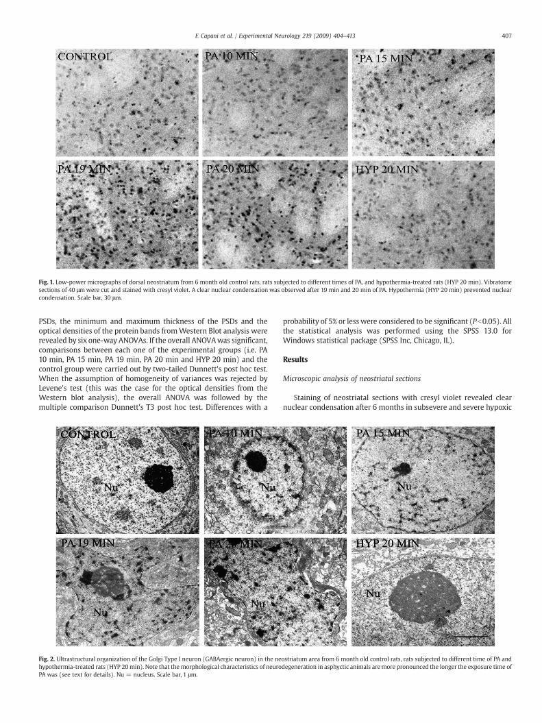
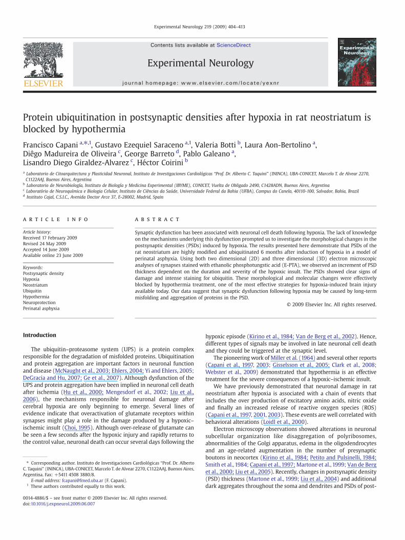
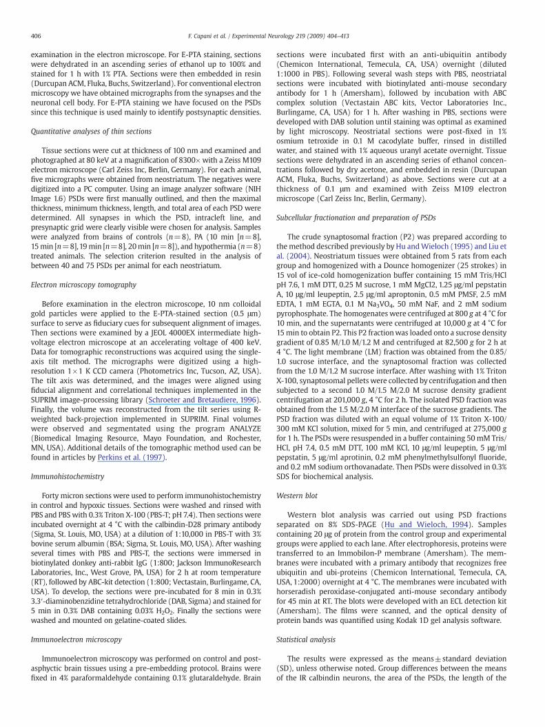
Fig. 1. Low-power micrographs of dorsal neostriatum from 6 month old control rats, rats subjected to different times of PA, and hypothermia-treated rats (HYP 20 min). Vibratomesections of 40 μmwere cut and stained with cresyl violet. A clear nuclear condensation was observed after 19 min and 20 min of PA. Hypothermia (HYP 20 min) prevented nuclearcondensation. Scale bar, 30 μm.
407F. Capani et al. / Experimental Neurology 219 (2009) 404–413
PSDs, the minimum and maximum thickness of the PSDs and theoptical densities of the protein bands fromWestern Blot analysis wererevealed by six one-way ANOVAs. If the overall ANOVAwas significant,comparisons between each one of the experimental groups (i.e. PA10 min, PA 15 min, PA 19 min, PA 20 min and HYP 20 min) and thecontrol group were carried out by two-tailed Dunnett's post hoc test.When the assumption of homogeneity of variances was rejected byLevene's test (this was the case for the optical densities from theWestern blot analysis), the overall ANOVA was followed by themultiple comparison Dunnett's T3 post hoc test. Differences with a
Fig. 2. Ultrastructural organization of the Golgi Type I neuron (GABAergic neuron) in the nehypothermia-treated rats (HYP 20min). Note that the morphological characteristics of neuroPA was (see text for details). Nu = nucleus. Scale bar, 1 μm.
probability of 5% or less were considered to be significant (Pb0.05). Allthe statistical analysis was performed using the SPSS 13.0 forWindows statistical package (SPSS Inc, Chicago, IL).
Results
Microscopic analysis of neostriatal sections
Staining of neostriatal sections with cresyl violet revealed clearnuclear condensation after 6 months in subsevere and severe hypoxic
ostriatum area from 6 month old control rats, rats subjected to different time of PA anddegeneration in asphyctic animals aremore pronounced the longer the exposure time of
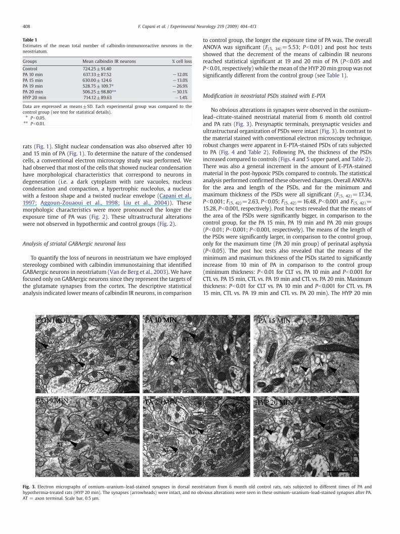
Table 1Estimates of the mean total number of calbindin-immunoreactive neurons in theneostriatum.
Groups Mean calbindin IR neurons % cell loss
Control 724.25±91.40PA 10 min 637.33±87.52 −12.0%PA 15 min 630.00±124.6 −13.0%PA 19 min 528.75±109.7⁎ −26.9%PA 20 min 506.25±98.80⁎⁎ −30.1%HYP 20 min 714.12±89.63 −1.4%
Data are expressed as means±SD. Each experimental group was compared to thecontrol group (see text for statistical details).⁎ Pb0.05.⁎⁎ Pb0.01.
408 F. Capani et al. / Experimental Neurology 219 (2009) 404–413
rats (Fig. 1). Slight nuclear condensation was also observed after 10and 15 min of PA (Fig. 1). To determine the nature of the condensedcells, a conventional electron microscopy study was performed. Wehad observed that most of the cells that showed nuclear condensationhave morphological characteristics that correspond to neurons indegeneration (i.e. a dark cytoplasm with rare vacuoles, nucleuscondensation and compaction, a hypertrophic nucleolus, a nucleuswith a festoon shape and a twisted nuclear envelope (Capani et al.,1997; Aggoun-Zouaoui et al., 1998; Liu et al., 2004)). Thesemorphologic characteristics were more pronounced the longer theexposure time of PA was (Fig. 2). These ultrastructural alterationswere not observed in hypothermic and control groups (Fig. 2).
Analysis of striatal GABAergic neuronal loss
To quantify the loss of neurons in neostriatum we have employedstereology combined with calbindin immunostaining that identifiedGABAergic neurons in neostriatum (Van de Berg et al., 2003). We havefocused only on GABAergic neurons since they represent the targets ofthe glutamate synapses from the cortex. The descriptive statisticalanalysis indicated lowermeans of calbindin IR neurons, in comparison
Fig. 3. Electron micrographs of osmium–uranium–lead-stained synapses in dorsal neosthypothermia-treated rats (HYP 20 min). The synapses (arrowheads) were intact, and no obAT = axon terminal. Scale bar, 0.5 μm.
to control group, the longer the exposure time of PA was. The overallANOVA was significant (F(5, 34)=5.53; Pb0.01) and post hoc testsshowed that the decrement of the means of calbindin IR neuronsreached statistical significant at 19 and 20 min of PA (Pb0.05 andPb0.01, respectively)while themean of the HYP 20min groupwas notsignificantly different from the control group (see Table 1).
Modification in neostriatal PSDs stained with E-PTA
No obvious alterations in synapses were observed in the osmium–
lead–citrate-stained neostriatal material from 6 month old controland PA rats (Fig. 3). Presynaptic terminals, presynaptic vesicles andultrastructural organization of PSDs were intact (Fig. 3). In contrast tothe material stained with conventional electron microscopy technique,robust changes were apparent in E-PTA-stained PSDs of rats subjectedto PA (Fig. 4 and Table 2). Following PA, the thickness of the PSDsincreased compared to controls (Figs. 4 and 5 upper panel, and Table 2).There was also a general increment in the amount of E-PTA-stainedmaterial in the post-hypoxic PSDs compared to controls. The statisticalanalysis performed confirmed these observed changes. Overall ANOVAsfor the area and length of the PSDs, and for the minimum andmaximum thickness of the PSDs were all significant (F(5, 42)=17.34,Pb0.001; F(5, 42)=2.63, Pb0.05; F(5, 42)=16.48, Pb0.001 and F(5, 42)=15.28, Pb0.001, respectively). Post hoc tests revealed that the means ofthe area of the PSDs were significantly bigger, in comparison to thecontrol group, for the PA 15 min, PA 19 min and PA 20 min groups(Pb0.01; Pb0.001; Pb0.001, respectively). The means of the length ofthe PSDs were significantly larger, in comparison to the control group,only for the maximum time (PA 20 min group) of perinatal asphyxia(Pb0.05). The post hoc tests also revealed that the means of theminimum and maximum thickness of the PSDs started to significantlyincrease from 10 min of PA in comparison to the control group(minimum thickness: Pb0.01 for CLT vs. PA 10 min and Pb0.001 forCTL vs. PA 15 min, CTL vs. PA 19 min and CTL vs. PA 20 min. Maximumthickness: Pb0.01 for CLT vs. PA 10 min and Pb0.001 for CTL vs. PA15 min, CTL vs. PA 19 min and CTL vs. PA 20 min). The HYP 20 min
riatum from 6 month old control rats, rats subjected to different times of PA andvious alterations were seen in these osmium–uranium–lead-stained synapses after PA.
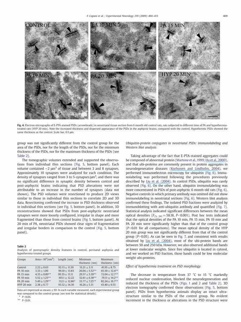
Fig. 4. Electron micrographs of E-PTA-stained PSDs (arrowheads) in neostriatal tissue section from 6 month old control rats, rats subjected to different time of PA and hypothermia-treated rats (HYP 20 min). Note the increased thickness and dispersed appearance of the PSDs in the asphyctic brains, compared with the control. Hypothermic PSDs showed thesame thickness as the control. Scale bar, 0.5 μm.
409F. Capani et al. / Experimental Neurology 219 (2009) 404–413
group was not significantly different from the control group for thearea of the PSDs, nor for the length of the PSDs, nor for the minimumthickness of the PSDs, nor for the maximum thickness of the PSDs (seeTable 2).
The tomographic volumes extended and supported the observa-tions from individual thin sections (Fig. 5, bottom panel). Eachvolume contained ∼2 μm3 of tissue and between 3 and 8 synapses.Approximately 10 synapses were analyzed for each condition. Thedensity of synapses ranged from 3 to 5-synapses/μm3, and there wasno significant difference in synaptic density between control andpost-asphyctic brains indicating that PSD alterations were notattributable to an increase in the number of synapses (data notshown). The PSD volumes were resectioned to produce 2D viewssimilar to those in individual thin sections to correlate 2D and 3Ddata. Resectioning confirmed the increase in PSD thickness observedin individual thin sections (see Fig. 5, bottom panel). In addition, 3Dreconstructions showed that PSDs from post-asphyctic neostriatalsynapses were more loosely configured, irregular in shape and morefragmented than those from control brains (Fig 5, bottom panel). At20 min of PA, neostriatal PSDs showed clear signs of fragmentationand irregular borders in comparison to the control (Fig. 5, bottompanel).
Table 2Analysis of postsynaptic density features in control, perinatal asphyxia andhypothermia-treated groups.
Groups Area×103(nm2) Length (nm) Minimumthickness (nm)
Maximumthickness (nm)
Control 2.22±0.63 92.13±11.59 15.21±3.11 41.95±8.75PA 10 min 3.33±1.00 99.10±13.83 26.04±5.52⁎⁎ 65.50±12.4⁎⁎PA 15 min 4.35±0.89⁎⁎ 99.35±11.11 29.57±5.39⁎⁎ 72.04±12.7⁎⁎PA 19 min 5.52±1.25⁎⁎ 103.1±12.22 32.61±6.38⁎⁎ 79.11±14.2⁎⁎PA 20 min 5.49±1.29⁎⁎ 112.1±12.86⁎ 33.77±7.25⁎⁎ 82.24±16.7⁎⁎HYP 20 min 2.38±0.77 93.12±14.30 16.26±5.31 43.40±9.55
Data are expressed as means±SD. In each variable measured, each experimental groupwas compared to the control group (see text for statistical details).⁎ Pb0.05.⁎⁎ Pb0.01.
Ubiquitin-protein conjugates in neostriatal PSDs: immunolabeling andWestern blot analysis
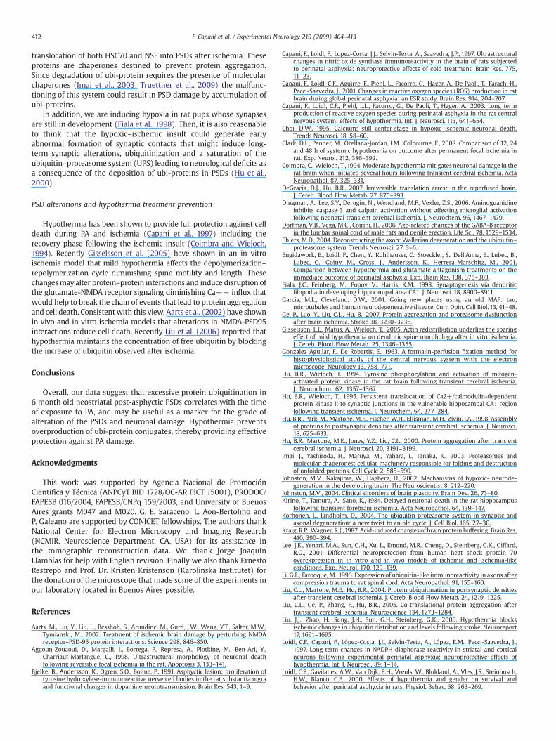
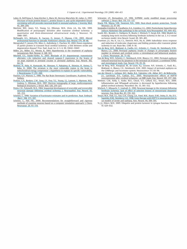
Taking advantage of the fact that E-PTA-stained aggregates couldbe composed of abnormal protein (Martoneet al.,1999;Huet al., 2000),and that ubi-proteins are commonly present in protein aggregates inneurodegenerative diseases (Korhonen and Lindholm, 2004), weperformed immunoelectron microscopy for ubiquitin (Fig. 6). Immu-nolabeling was performed following the procedures previouslydescribed by Liu et al. (2004). In control PSDs, ubiquitin was rarelyobserved (Fig. 6). On the other hand, ubiquitin immunolabeling wasmore concentrated in PSDs of post-asphyctic 6 month old rats (Fig. 6).Negative controls inwhich primary antibody was omitted did not showimmunolabeling in neostriatal sections (Fig. 6). Western blot analysisconfirmed these findings. The isolated PSD fractions were analyzed byimmunoblotting with anti-ubiquitin antibody and quantified (Fig. 7).Statistical analysis indicated significant differences between the meanoptical densities (F(5, 24)=58.18, Pb0.001). Post hoc tests indicatedthat the optical densities of the PA 10 min, PA 15 min, PA 19 min andPA 20 min were significantly higher than that of the control group(Pb0.01 for all comparisons). The mean optical density of the HYP20 min group was not significantly different from that of the controlgroup (PN0.05). As can be seen in Fig. 7, and consistent with resultsobtained by Liu et al. (2004), most of the ubi-protein bands arebetween 50 and 250 kDa. However, we also observed additional bandsat lower molecular weights. Since free ubiquitin is located in cytosol,and we worked on PSD fraction, those bands could be low molecularweight ubi-proteins.
Effect of hypothermia treatment on PSD morphology
The decrease in temperature from 37 °C to 15 °C markedlyreduced nuclear condensation, blocked the neurodegeneration andreduced the thickness of the PSDs (Figs. 1 and 2 and Table 2). 3Delectron tomography confirmed these observations (Fig. 5, bottompanel). PSDs from hypothermia animals display an intact ultra-structure similar to the PSDs of the control group. No evidentincrement in the thickness or alterations in the PSD structure were
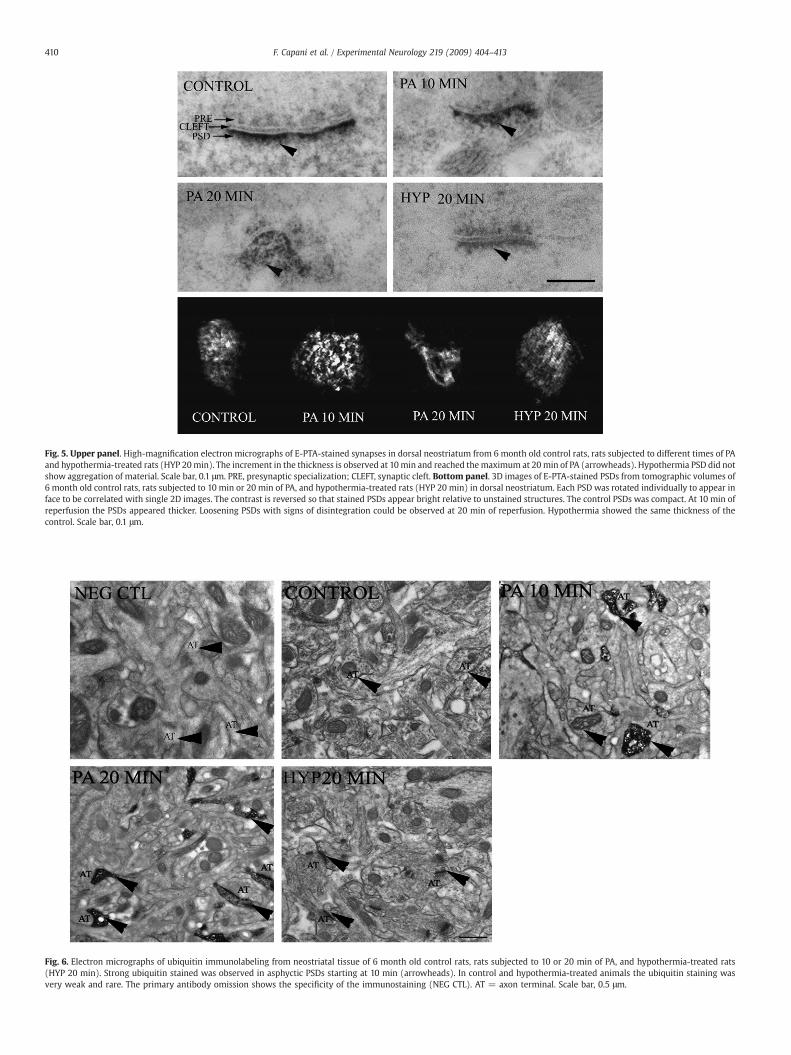
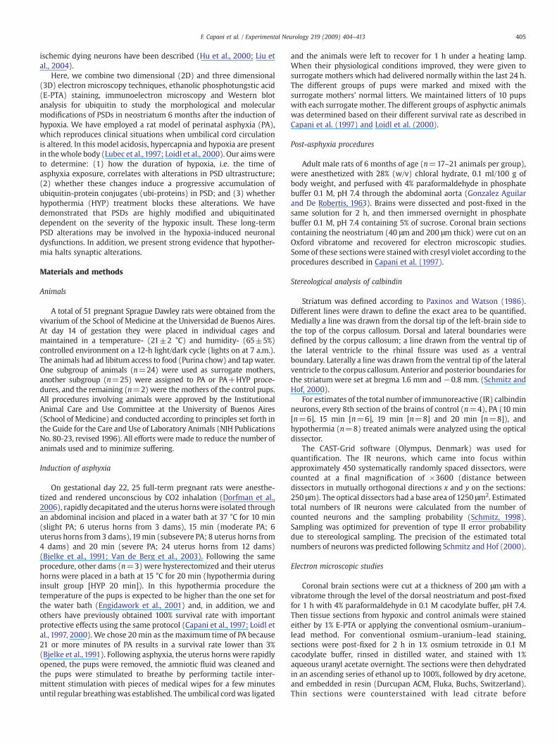
Fig. 6. Electron micrographs of ubiquitin immunolabeling from neostriatal tissue of 6 month old control rats, rats subjected to 10 or 20 min of PA, and hypothermia-treated rats(HYP 20 min). Strong ubiquitin stained was observed in asphyctic PSDs starting at 10 min (arrowheads). In control and hypothermia-treated animals the ubiquitin staining wasvery weak and rare. The primary antibody omission shows the specificity of the immunostaining (NEG CTL). AT = axon terminal. Scale bar, 0.5 μm.
Fig. 5. Upper panel. High-magnification electron micrographs of E-PTA-stained synapses in dorsal neostriatum from 6 month old control rats, rats subjected to different times of PAand hypothermia-treated rats (HYP 20min). The increment in the thickness is observed at 10min and reached themaximum at 20min of PA (arrowheads). Hypothermia PSD did notshow aggregation of material. Scale bar, 0.1 μm. PRE, presynaptic specialization; CLEFT, synaptic cleft. Bottom panel. 3D images of E-PTA-stained PSDs from tomographic volumes of6 month old control rats, rats subjected to 10 min or 20 min of PA, and hypothermia-treated rats (HYP 20 min) in dorsal neostriatum. Each PSD was rotated individually to appear inface to be correlated with single 2D images. The contrast is reversed so that stained PSDs appear bright relative to unstained structures. The control PSDs was compact. At 10 min ofreperfusion the PSDs appeared thicker. Loosening PSDs with signs of disintegration could be observed at 20 min of reperfusion. Hypothermia showed the same thickness of thecontrol. Scale bar, 0.1 μm.
410 F. Capani et al. / Experimental Neurology 219 (2009) 404–413
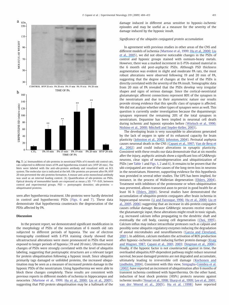
Fig. 7. (a) Immunoblots of ubi-proteins in neostriatal PSDs of 6 month old control rats,rats subjected to different times of PA and hypothermia-treated rats (HYP 20 min). Theblots were labeled with the anti-ubi-protein antibody and visualized with an ECLsystem. Themolecular size is indicated at the left. Ubi-proteins are present after PA. HYP20 min prevented the ubi-proteins formation. A mouse anti-actin monoclonal antibodywas used as an internal loading control. (b) Quantification of ubi-proteins on PSDs.Optical density of immunoblot bands are expressed as mean±SD. ⁎⁎Pb0.01, betweencontrol and experimental groups. PSD = postsynaptic densities; ubi-proteins =ubiquitinated proteins.
411F. Capani et al. / Experimental Neurology 219 (2009) 404–413
seen after hypothermia treatment. Ubi-proteins were hardly detectedin control and hypothermic PSDs (Figs. 6 and 7). These datademonstrate that hypothermia counteracts the degeneration of thePDSs affected by PA.
Discussion
In the present report, we demonstrated significant modification inthe morphology of PSDs of the neostriatum of 6 month old ratssubjected to different periods of hypoxia. The use of electrontomography combined with E-PTA staining clearly showed thatultrastructural alterations were more pronounced in PSDs that wereexposed to longer periods of hypoxia (19 and 20 min). Ultrastructuralchanges of PSDs were strongly correlated with the level of ubiquitinlabeling, suggesting that postsynaptic structures are a relevant targetfor protein ubiquitination following a hypoxic insult. Since ubiquitinprimarily tags damaged or unfolded proteins, the increased ubiqui-tination may be seen as a measure of increased protein damage in thehypoxic PSDs of the neostriatum. Using hypothermia we were able toblock these changes completely. These results are consistent withprevious reports in different models of ischemia in hippocampus andneocortex (Martone et al., 1999; Hu et al., 2000; Liu et al., 2005),suggesting that PSD protein ubiquitination may be a hallmark of the
damage induced in different areas sensitive to hypoxic–ischemicepisodes and may be useful as a measure for the severity of thedamage induced by the hypoxic insult.
Significance of the ubiquitin conjugated protein accumulation
In agreement with previous studies in other areas of the CNS anddifferent models of ischemia (Martone et al., 1999; Hu et al., 2000; Liuet al., 2005), we did not observe noticeable changes in the PSDs ofcontrol and hypoxic groups stained with osmium-heavy metals.However, there was a marked increment in E-PTA-stained material inthe 6 month old post-asphyctic PSDs. Although PSD thicknessaugmentation was evident in slight and moderate PA rats, the mostrobust alterations were observed following 19 and 20 min of PA,suggesting that the degree of changes at the level of the PSDs isdirectly correlatedwith the severity of the PA insult. Tomographic datafrom 20 min of PA revealed that the PSDs develop very irregularshapes and signs of serious damage. Since the cortical-neostriatalglutamatergic afferent connections represent 80% of the synapses inthe neostriatum and due to their asymmetric nature our resultsprovide strong evidence that this specific class of synapse is affected.We did not analyze whether other types of synapses were as well. Thisquestion is currently under investigation because the dopaminergicsynapses represent the remaining 20% of the total synapses inneostriatum. Dopamine has been implied in neuronal cell deathduring ischemic and hypoxic episodes before (Wieloch et al., 1990;Nishino et al., 2000; Mitchell and Snyder-Keller, 2003).
The developing brain is very susceptible to alterations generatedby the lack of oxygen in spite of its enhanced capacity for brainplasticity (Johnston et al., 2002; Johnston, 2004). Perinatal asphyxiacauses neuronal death in the CNS (Capani et al., 1997; Van de Berg etal., 2002) and could induce alterations in synaptic plasticity.Consistent with these results our data demonstrate that at six monthsafter the injury, asphyctic animals show a significant loss of GABAergicneurons, clear signs of neurodegeneration and ubiquitinization ofPSDs (see Table 1 and Figs. 1, 2 and 6). It remains to be proven that theubi-conjugated are one of the causes of the long-term loss in neuronsin the neostriatum. However, supporting evidence for this hypothesiswas provided in several other studies. The UPS has been implied, forinstance, in the process of Wallerian degeneration (Ehlers, 2004).Treatment with inhibitors of the proteosome or when ubiquitinationwas prevented, allows transected axon to persist in good health for atleast 16 h (Ehlers, 2004). Several studies have demonstrated theaccumulation of ubiquitin-protein conjugates after brain ischemia inhippocampal neurons (Li and Farooque, 1996; Hu et al., 2000; Liu etal., 2005, 2006) suggesting that an increase in ubi-protein conjugatescauses cellular damage. Because GABAergic neurons receive most ofthe glutamatergic input, these alterations might result in toxic signals,e.g. increased calcium influx propagating to the dendritic shaft andeventually the cell body, causing cell degeneration (Choi, 1995).Calcium influx may induce intracellular proteases such as calpain andpossibly some ubiquitin regulatory enzymes inducing the degradationof axonal microtubules and neurofilaments (Garcia and Cleveland,2001). In addition, calcium mediates the activation of ROS productionafter hypoxic–ischemic insult inducing further protein damage (Kraigand Wagner, 1987; Capani et al., 2001, 2003; Dingman et al., 2006).Finally, if the hypoxic factor is not counteracted against in time, theoverloaded ubiquitin/ATP-dependent pathwaymight reduce neuronalsurvival, because damaged proteins are not degraded and accumulate,ultimately leading to irreversible cell damage (Korhonen andLindholm, 2004). Consistent with this view, Sinigaglia-Coimbra et al.(2002) have reported an increment of ubiquitination after 6months oftransient ischemia combined with hyperthermia. On the other hand,induction of heat shock proteins (HSPs) protects neurons againstischemic insults (Yenari et al., 1998; Sharp et al., 1999; Lee et al., 2001;van der Weerd et al., 2005). Hu et al. (1998) have reported
412 F. Capani et al. / Experimental Neurology 219 (2009) 404–413
translocation of both HSC70 and NSF into PSDs after ischemia. Theseproteins are chaperones destined to prevent protein aggregation.Since degradation of ubi-protein requires the presence of molecularchaperones (Imai et al., 2003; Truettner et al., 2009) the malfunc-tioning of this system could result in PSD damage by accumulation ofubi-proteins.
In addition, we are inducing hypoxia in rat pups whose synapsesare still in development (Fiala et al., 1998). Then, it is also reasonableto think that the hypoxic–ischemic insult could generate earlyabnormal formation of synaptic contacts that might induce long-term synaptic alterations, ubiquitinization and a saturation of theubiquitin–proteasome system (UPS) leading to neurological deficits asa consequence of the deposition of ubi-proteins in PSDs (Hu et al.,2000).
PSD alterations and hypothermia treatment prevention
Hypothermia has been shown to provide full protection against celldeath during PA and ischemia (Capani et al., 1997) including therecovery phase following the ischemic insult (Coimbra and Wieloch,1994). Recently Gisselsson et al. (2005) have shown in an in vitroischemia model that mild hypothermia affects the depolymerization–repolymerization cycle diminishing spine motility and length. Thesechangesmay alter protein–protein interactions and induce disruption ofthe glutamate-NMDA receptor signaling diminishing Ca++ influx thatwould help to break the chain of events that lead to protein aggregationand cell death. Consistentwith this view, Aarts et al. (2002) have shownin vivo and in vitro ischemia models that alterations in NMDA-PSD95interactions reduce cell death. Recently Liu et al. (2006) reported thathypothermia maintains the concentration of free ubiquitin by blockingthe increase of ubiquitin observed after ischemia.
Conclusions
Overall, our data suggest that excessive protein ubiquitination in6 month old neostriatal post-asphyctic PSDs correlates with the timeof exposure to PA, and may be useful as a marker for the grade ofalteration of the PSDs and neuronal damage. Hypothermia preventsoverproduction of ubi-protein conjugates, thereby providing effectiveprotection against PA damage.
Acknowledgments
This work was supported by Agencia Nacional de PromociónCientífica y Técnica (ANPCyT BID 1728/OC-AR PICT 15001), PRODOC/FAPESB 016/2004, FAPESB/CNPq 159/2003, and University of BuenosAires grants M047 and M020. G. E. Saraceno, L. Aon-Bertolino andP. Galeano are supported by CONICET fellowships. The authors thankNational Center for Electron Microscopy and Imaging Research(NCMIR, Neuroscience Department, CA, USA) for its assistance inthe tomographic reconstruction data. We thank Jorge JoaquínLlambías for help with English revision. Finally we also thank ErnestoRestrepo and Prof. Dr. Kristen Kristenson (Karolinska Institutet) forthe donation of the microscope that made some of the experiments inour laboratory located in Buenos Aires possible.
References
Aarts, M., Liu, Y., Liu, L., Besshoh, S., Arundine, M., Gurd, J.W., Wang, Y.T., Salter, M.W.,Tymianski, M., 2002. Treatment of ischemic brain damage by perturbing NMDAreceptor–PSD-95 protein interactions. Science 298, 846–850.
Aggoun-Zouaoui, D., Margalli, I., Borrega, F., Represa, A., Plotkine, M., Ben-Ari, Y.,Charriaut-Marlangue, C., 1998. Ultrastructural morphology of neuronal deathfollowing reversible focal ischemia in the rat. Apoptosis 3, 133–141.
Bjelke, B., Andersson, K., Ogren, S.O., Bolme, P., 1991. Asphyctic lesion: proliferation oftyrosine hydroxylase-immunoreactive nerve cell bodies in the rat substantia nigraand functional changes in dopamine neurotransmission. Brain Res. 543, 1–9.
Capani, F., Loidl, F., Lopez-Costa, J.J., Selvin-Testa, A., Saavedra, J.P., 1997. Ultrastructuralchanges in nitric oxide synthase immunoreactivity in the brain of rats subjectedto perinatal asphyxia: neuroprotective effects of cold treatment. Brain Res. 775,11–23.
Capani, F., Loidl, C.F., Aguirre, F., Piehl, L., Facorro, G., Hager, A., De Paoli, T., Farach, H.,Pecci-Saavedra, J., 2001. Changes in reactive oxygen species (ROS) production in ratbrain during global perinatal asphyxia: an ESR study. Brain Res. 914, 204–207.
Capani, F., Loidl, C.F., Piehl, L.L., Facorro, G., De Paoli, T., Hager, A., 2003. Long termproduction of reactive oxygen species during perinatal asphyxia in the rat centralnervous system: effects of hypothermia. Int. J. Neurosci. 113, 641–654.
Choi, D.W., 1995. Calcium: still center-stage in hypoxic–ischemic neuronal death.Trends Neurosci. 18, 58–60.
Clark, D.L., Penner, M., Orellana-Jordan, I.M., Colbourne, F., 2008. Comparison of 12, 24and 48 h of systemic hypothermia on outcome after permanent focal ischemia inrat. Exp. Neurol. 212, 386–392.
Coimbra, C., Wieloch, T., 1994. Moderate hypothermia mitigates neuronal damage in therat brain when initiated several hours following transient cerebral ischemia. ActaNeuropathol. 87, 325–331.
DeGracia, D.J., Hu, B.R., 2007. Irreversible translation arrest in the reperfused brain.J. Cereb. Blood Flow Metab. 27, 875–893.
Dingman, A., Lee, S.Y., Derugin, N., Wendland, M.F., Vexler, Z.S., 2006. Aminoguanidineinhibits caspase-3 and calpain activation without affecting microglial activationfollowing neonatal transient cerebral ischemia. J. Neurochem. 96, 1467–1479.
Dorfman, V.B., Vega, M.C., Coirini, H., 2006. Age-related changes of the GABA-B receptorin the lumbar spinal cord of male rats and penile erection. Life Sci. 78, 1529–1534.
Ehlers, M.D., 2004. Deconstructing the axon:Wallerian degeneration and the ubiquitin–proteasome system. Trends Neurosci. 27, 3–6.
Engidawork, E., Loidl, F., Chen, Y., Kohlhauser, C., Stoeckler, S., Dell'Anna, E., Lubec, B.,Lubec, G., Goiny, M., Gross, J., Andersson, K., Herrera-Marschitz, M., 2001.Comparison between hypothermia and glutamate antagonism treatments on theimmediate outcome of perinatal asphyxia. Exp. Brain Res. 138, 375–383.
Fiala, J.C., Feinberg, M., Popov, V., Harris, K.M., 1998. Synaptogenesis via dendriticfilopodia in developing hippocampal area CA1. J. Neurosci. 18, 8900–8911.
Garcia, M.L., Cleveland, D.W., 2001. Going new places using an old MAP: tau,microtubules and human neurodegenerative disease. Curr. Opin. Cell Biol. 13, 41–48.
Ge, P., Luo, Y., Liu, C.L., Hu, B., 2007. Protein aggregation and proteasome dysfunctionafter brain ischemia. Stroke 38, 3230–3236.
Gisselsson, L.L., Matus, A., Wieloch, T., 2005. Actin redistribution underlies the sparingeffect of mild hypothermia on dendritic spine morphology after in vitro ischemia.J. Cereb. Blood Flow Metab. 25, 1346–1355.
Gonzalez Aguilar, F., De Robertis, E., 1963. A formalin-perfusion fixation method forhistophysiological study of the central nervous system with the electronmicroscope. Neurology 13, 758–771.
Hu, B.R., Wieloch, T., 1994. Tyrosine phosphorylation and activation of mitogen-activated protein kinase in the rat brain following transient cerebral ischemia.J. Neurochem. 62, 1357–1367.
Hu, B.R., Wieloch, T., 1995. Persistent translocation of Ca2+/calmodulin-dependentprotein kinase II to synaptic junctions in the vulnerable hippocampal CA1 regionfollowing transient ischemia. J. Neurochem. 64, 277–284.
Hu, B.R., Park, M., Martone, M.E., Fischer,W.H., Ellisman, M.H., Zivin, J.A., 1998. Assemblyof proteins to postsynaptic densities after transient cerebral ischemia. J. Neurosci.18, 625–633.
Hu, B.R., Martone, M.E., Jones, Y.Z., Liu, C.L., 2000. Protein aggregation after transientcerebral ischemia. J. Neurosci. 20, 3191–3199.
Imai, J., Yashiroda, H., Maruya, M., Yahara, I., Tanaka, K., 2003. Proteasomes andmolecular chaperones: cellular machinery responsible for folding and destructionof unfolded proteins. Cell Cycle 2, 585–590.
Johnston, M.V., Nakajima, W., Hagberg, H., 2002. Mechanisms of hypoxic- neurode-generation in the developing brain. The Neuroscientist 8, 212–220.
Johnston, M.V., 2004. Clinical disorders of brain plasticity. Brain Dev. 26, 73–80.Kirino, T., Tamura, A., Sano, K., 1984. Delayed neuronal death in the rat hippocampus
following transient forebrain ischemia. Acta Neuropathol. 64, 139–147.Korhonen, L., Lindholm, D., 2004. The ubiquitin proteasome system in synaptic and
axonal degeneration: a new twist to an old cycle. J. Cell Biol. 165, 27–30.Kraig, R.P., Wagner, R.J., 1987. Acid-induced changes of brain protein buffering. Brain Res.
410, 390–394.Lee, J.E., Yenari, M.A., Sun, G.H., Xu, L., Emond, M.R., Cheng, D., Steinberg, G.K., Giffard,
R.G., 2001. Differential neuroprotection from human heat shock protein 70overexpression in in vitro and in vivo models of ischemia and ischemia-likeconditions. Exp. Neurol. 170, 129–139.
Li, G.L., Farooque, M., 1996. Expression of ubiquitin-like immunoreactivity in axons aftercompression trauma to rat spinal cord. Acta Neuropathol. 91, 155–160.
Liu, C.L., Martone, M.E., Hu, B.R., 2004. Protein ubiquitination in postsynaptic densitiesafter transient cerebral ischemia. J. Cereb. Blood Flow Metab. 24, 1219–1225.
Liu, C.L., Ge, P., Zhang, F., Hu, B.R., 2005. Co-translational protein aggregation aftertransient cerebral ischemia. Neuroscience 134, 1273–1284.
Liu, J.J., Zhao, H., Sung, J.H., Sun, G.H., Steinberg, G.K., 2006. Hypothermia blocksischemic changes in ubiquitin distribution and levels following stroke. Neuroreport17, 1691–1695.
Loidl, C.F., Capani, F., López-Costa, J.J., Selvín-Testa, A., López, E.M., Pecci-Saavedra, J.,1997. Long term changes in NADPH-diaphorase reactivity in striatal and corticalneurons following experimental perinatal asphyxia: neuroprotective effects ofhypothermia. Int. J. Neurosci. 89, 1–14.
Loidl, C.F., Gavilanes, A.W., Van Dijk, E.H., Vreuls, W., Blokland, A., Vles, J.S., Steinbusch,H.W., Blanco, C.E., 2000. Effects of hypothermia and gender on survival andbehavior after perinatal asphyxia in rats. Physiol. Behav. 68, 263–269.
413F. Capani et al. / Experimental Neurology 219 (2009) 404–413
Lubec, B., Dell'Anna, E., Fang-Kircher, S., Marx, M., Herrera-Marschitz, M., Lubec, G.,1997.Decrease of brain protein kinase C, protein kinase A, and cyclin-dependent kinasecorrelating with pH precedes neuronal death in neonatal asphyxia. J. Investig. Med.45, 284–294.
Martone, M.E., Jones, Y.Z., Young, S.J., Ellisman, M.H., Zivin, J.A., Hu, B.R., 1999.Modification of postsynaptic densities after transient cerebral ischemia: aquantitative and three-dimensional ultrastructural study. J. Neurosci. 19,1988–1997.
McNaught, K.S., Belizaire, R., Isacson, O., Jenner, P., Olanow, C.W., 2003. Alteredproteasomal function in sporadic Parkinson's disease. Exp. Neurol. 179, 38–46.
Mengesdorf, T., Jensen, P.H., Mies, G., Aufenberg, C., Paschen,W., 2002. Down-regulationof parkin protein in transient focal cerebral ischemia: a link between stroke anddegenerative disease? Proc. Natl. Acad. Sci. U. S. A. 99, 15042–15047.
Miller, J.A., Miller, F.S., Westin, B., 1964. Hypothermia in the treatment of asphyxianeonatorum. Biol. Neonat. 6, 148–163.
Mitchell, E.S., Snyder-Keller, A., 2003. Blockade of D1 dopaminergic transmissionalleviates c-fos induction and cleaved caspase-3 expression in the brains ofrat pups exposed to prenatal cocaine or perinatal asphyxia. Exp. Neurol. 182,64–74.
Nishino, H., Hida, H., Kumazaki, M., Shimano, Y., Nakajima, K., Shimizu, H., Ooiwa, T.,Baba, H., 2000. The striatum is the most vulnerable region in the brain tomitochondrial energy compromise: a hypothesis to explain its specific vulnerability.J. Neurotrauma 17, 251–260.
Paxinos, G.P., Watson, C., 1986. The Rat Brain Stereotaxic Coordinates. Academic Press,Sydney.
Perkins, G.A., Renken, C.W., Song, J.Y., Frey, T.G., Young, S.J., Lamont, S., Martone, M.E.,Lindsey, S., Ellisman, M.H., 1997. Electron tomography of large, multicomponentbiological structures. J. Struct. Biol. 120, 219–227.
Petito, C.K., Pulsinelli, W.A., 1984. Sequential development of reversible and irreversibleneuronal damage following cerebral ischemia. J. Neuropathol. Exp. Neurol. 43,141–153.
Schmitz, C., 1998. Variation of fractionator estimates and its prediction. Anat. Embryol.(Berl.) 198, 371–397.
Schmitz, C., Hof, P.R., 2000. Recommendations for straightforward and rigorousmethods of counting neurons based on a computer simulation approach. J. Chem.Neuroanat. 20, 93–114.
Schroeter, J.P., Bretaudiere, J.P., 1996. SUPRIM: easily modified image processingsoftware. J. Struct. Biol. 116, 131–137.
Sharp, F.R., Massa, S.M., Swanson, R.A., 1999. Heat-shock protein protection. TrendsNeurosci. 22, 97–99.
Sinigaglia-Coimbra, R., Cavalheiro, E.A., Coimbra, C.G., 2002. Postischemic hyperthermiainduces Alzheimer-like pathology in the rat brain. Acta Neuropathol. 103, 444–452.
Smith, M.L., Bendek, G., Dahlgren, N., Rosén, I., Wieloch, T., Siesjö, B.K., 1984. Models forstudying long-term recovery following forebrain ischemia in the rat. 2. A 2-vesselocclusion model. Acta Neurol. Scand. 69, 385–401.
Truettner, J.S., Hu, K., Liu, C.L., Dietrich, W.D., Hu, B., 2009. Subcellular stress responseand induction of molecular chaperones and folding proteins after transient globalischemia in rats. Brain Res. 1249, 9–18.
Van de Berg, W.D., Blokland, A., Cuello, A.C., Schmitz, C., Vreuls, W., Steinbusch, H.W.,Blanco, C.E., 2000. Perinatal asphyxia results in changes in presynaptic boutonnumber in striatum and cerebral cortex—a stereological and behavioral analysis.J. Chem. Neuroanat. 20, 71–82.
Van de Berg, W.D., Schmitz, C., Steinbusch, H.W., Blanco, C.E., 2002. Perinatal asphyxiainduced neuronal loss by apoptosis in the neonatal rat striatum: a combined TUNELand stereological study. Exp. Neurol. 174, 29–36.
Van de Berg, W.D., Kwaijtaal, M., de Louw, A.J., Lissone, N.P., Schmitz, C., Faull, R.L.,Blokland, A., Blanco, C.E., Steinbusch, H.W., 2003. Impact of perinatal asphyxia onthe GABAergic and locomotor system. Neuroscience 117, 83–96.
van der Weerd, L., Lythgoe, M.F., Badin, R.A., Valentim, L.M., Akbar, M.T., de Belleroche,J.S., Latchman, D.S., Gadian, D.G., 2005. Neuroprotective effects of HSP70overexpression after cerebral ischaemia—an MRI study. Exp. Neurol. 195, 257–266.
Webster, C.M., Kelly, S., Koike, M.A., Chock, V.Y., Giffard, R.G., Yenari, M.A., 2009.Inflammation and NFkappaB activation is decreased by hypothermia followingglobal cerebral ischemia. Neurobiol. Dis. 33, 301–312.
Wieloch, T., Miyauchi, Y., Lindvall, O., 1990. Neuronal damage in the striatum followingforebrain ischemia: lack of effect of selective lesions of mesostriatal dopamineneurons. Exp. Brain Res. 83, 159–163.
Yenari, M.A., Fink, S.L., Sun, G.H., Chang, L.K., Patel, M.K., Kunis, D.M., Onley, D., Ho, D.Y.,Sapolsky, R.M., Steinberg, G.K., 1998. Gene therapy with HSP72 is neuroprotective inrat models of stroke and epilepsy. Ann. Neurol. 44, 584–591.
Yi, J.J., Ehlers, M.D., 2005. Ubiquitin and protein turnover in synapse function. Neuron47, 629–632.
Related Documents