Protein Synthesis Dependence of Growth Cone Collapse Induced by Different Nogo-A-Domains Richard Manns 1 , Andre Schmandke 2. , Antonio Schmandke 2. , Prem Jareonsettasin 1 , Geoffrey Cook 1 , Martin E. Schwab 2 , Christine Holt 1 , Roger Keynes 1 * 1 Department of Physiology, Development and Neuroscience, University of Cambridge, Cambridge, United Kingdom, 2 Brain Research Institute, University of Zurich and Department of Health Sciences and Technology, Swiss Federal Institute of Technology, Zurich, Switzerland Abstract Background: The protein Nogo-A regulates axon growth in the developing and mature nervous system, and this is carried out by two distinct domains in the protein, Nogo-A-D20 and Nogo-66. The differences in the signalling pathways engaged in axon growth cones by these domains are not well characterized, and have been investigated in this study. Methodology/Principal Findings: We analyzed growth cone collapse induced by the Nogo-A domains Nogo-A-D20 and Nogo-66 using explanted chick dorsal root ganglion neurons growing on laminin/poly-lysine substratum. Collapse induced by purified Nogo-A-D20 peptide is dependent on protein synthesis whereas that induced by Nogo-66 peptide is not. Nogo- A-D20-induced collapse is accompanied by a protein synthesis-dependent rise in RhoA expression in the growth cone, but is unaffected by proteasomal catalytic site inhibition. Conversely Nogo-66-induced collapse is inhibited ,50% by proteasomal catalytic site inhibition. Conclusion/Significance: Growth cone collapse induced by the Nogo-A domains Nogo-A-D20 and Nogo-66 is mediated by signalling pathways with distinguishable characteristics concerning their dependence on protein synthesis and proteasomal function. Citation: Manns R, Schmandke A, Schmandke A, Jareonsettasin P, Cook G, et al. (2014) Protein Synthesis Dependence of Growth Cone Collapse Induced by Different Nogo-A-Domains. PLoS ONE 9(1): e86820. doi:10.1371/journal.pone.0086820 Editor: Edward Giniger, National Institutes of Health (NIH), United States of America Received September 20, 2013; Accepted December 13, 2013; Published January 29, 2014 Copyright: ß 2014 Manns et al. This is an open-access article distributed under the terms of the Creative Commons Attribution License, which permits unrestricted use, distribution, and reproduction in any medium, provided the original author and source are credited. Funding: The Wellcome Trust (www.wellcome.ac.uk)project grant no. 08534/Z/08/Z. RM was funded by a graduate studentship from Trinity College, Cambridge. The funders had no role in study design, data collection and analysis, decision to publish, or preparation of the manuscript. Competing Interests: The authors have declared that no competing interests exist. * E-mail: [email protected] . These authors contributed equally to this work. Introduction The protein Nogo-A has been identified as an important regulator of development, plasticity and regeneration in the vertebrate nervous system [1]. Nogo-A (1200 aa, 200 kD) is a member of the Reticulon family of proteins (Reticulon-4, Rtn4), so-called due to the presence of a C-terminal 200 aa RTN homology domain comprising two .35 aa hydrophobic stretches, and the Nogo/Rtn4 gene gives rise to 3 main isoforms (A, B, C), of which Nogo-A is the largest [2]. Consistent with its proposed role as a negative regulator of axon growth, Nogo-A is expressed at the cell surface [3] and causes collapse of a wide variety of growth cones in vitro. Further studies have identified key domains of the protein that elicit collapse [4,5], and two domains in particular have been implicated, Nogo-66 and Nogo-A-D20. Nogo-66 is a 66 amino acid domain that, together with flanking hydrophobic regions, is a component of the RTN homology domain in the C- terminus of all Nogo isoforms [6]. Nogo-66 collapse-inducing activity is associated with high-affinity binding to its receptors NgR1 [7,8], which forms a complex with the transmembrane proteins LINGO1, and p75 or TROY [1,9–11]. Nogo-66 can also bind to the paired immunoglobulin-like receptor PirB [11]. Receptor binding activates the Rho/Rho-associated coiled-coil containing protein kinase (ROCK) pathway, resulting in growth cone collapse through RhoA signalling and destabilization of the actin cytoskeleton [1,12,13]. The other growth cone collapse-inducing domain, Nogo-A-D20 (NiGD20) comprises residues 544–725 of (rat) Nogo-A, and is a component of the extracellular N-terminal domain (residues 1– 979). The cognate receptor(s) for Nogo-A-D20 and the detailed signalling pathways that lead to collapse are less well character- ized. Both integrins [14] and a G protein-coupled receptor [15] have been implicated. Like Nogo-66, Nogo-A-D20 activates the RhoA-ROCK pathway [1,4,12]. Moreover Nogo-A-D20 signal- ling has been shown to inactivate Rac, a GTPase whose regulatory functions on the cytoskeleton oppose those of Rho [12,16]. Nogo- A-D20-induced growth cone collapse also requires endocytosis of a Nogo-A-D20/receptor complex that is retrogradely transported to the cell body in signalling endosomes containing activated Rho. This process is clathrin-independent and mediated by the pinocytotic chaperone protein Pincher [5]. Since Nogo-A-D20 endocytosis is directly linked to reduced levels of phosphorylated neuronal cyclic AMP response element-binding protein (CREB), the process may be a mechanism for Nogo-A-D20 to modulate expression of genes that regulate neuronal growth [5]. PLOS ONE | www.plosone.org 1 January 2014 | Volume 9 | Issue 1 | e86820

Welcome message from author
This document is posted to help you gain knowledge. Please leave a comment to let me know what you think about it! Share it to your friends and learn new things together.
Transcript

Protein Synthesis Dependence of Growth Cone CollapseInduced by Different Nogo-A-DomainsRichard Manns1, Andre Schmandke2., Antonio Schmandke2., Prem Jareonsettasin1, Geoffrey Cook1,
Martin E. Schwab2, Christine Holt1, Roger Keynes1*
1Department of Physiology, Development and Neuroscience, University of Cambridge, Cambridge, United Kingdom, 2 Brain Research Institute, University of Zurich and
Department of Health Sciences and Technology, Swiss Federal Institute of Technology, Zurich, Switzerland
Abstract
Background: The protein Nogo-A regulates axon growth in the developing and mature nervous system, and this is carriedout by two distinct domains in the protein, Nogo-A-D20 and Nogo-66. The differences in the signalling pathways engagedin axon growth cones by these domains are not well characterized, and have been investigated in this study.
Methodology/Principal Findings: We analyzed growth cone collapse induced by the Nogo-A domains Nogo-A-D20 andNogo-66 using explanted chick dorsal root ganglion neurons growing on laminin/poly-lysine substratum. Collapse inducedby purified Nogo-A-D20 peptide is dependent on protein synthesis whereas that induced by Nogo-66 peptide is not. Nogo-A-D20-induced collapse is accompanied by a protein synthesis-dependent rise in RhoA expression in the growth cone, butis unaffected by proteasomal catalytic site inhibition. Conversely Nogo-66-induced collapse is inhibited ,50% byproteasomal catalytic site inhibition.
Conclusion/Significance: Growth cone collapse induced by the Nogo-A domains Nogo-A-D20 and Nogo-66 is mediated bysignalling pathways with distinguishable characteristics concerning their dependence on protein synthesis and proteasomalfunction.
Citation: Manns R, Schmandke A, Schmandke A, Jareonsettasin P, Cook G, et al. (2014) Protein Synthesis Dependence of Growth Cone Collapse Induced byDifferent Nogo-A-Domains. PLoS ONE 9(1): e86820. doi:10.1371/journal.pone.0086820
Editor: Edward Giniger, National Institutes of Health (NIH), United States of America
Received September 20, 2013; Accepted December 13, 2013; Published January 29, 2014
Copyright: � 2014 Manns et al. This is an open-access article distributed under the terms of the Creative Commons Attribution License, which permitsunrestricted use, distribution, and reproduction in any medium, provided the original author and source are credited.
Funding: The Wellcome Trust (www.wellcome.ac.uk)project grant no. 08534/Z/08/Z. RM was funded by a graduate studentship from Trinity College, Cambridge.The funders had no role in study design, data collection and analysis, decision to publish, or preparation of the manuscript.
Competing Interests: The authors have declared that no competing interests exist.
* E-mail: [email protected]
. These authors contributed equally to this work.
Introduction
The protein Nogo-A has been identified as an important
regulator of development, plasticity and regeneration in the
vertebrate nervous system [1]. Nogo-A (1200 aa, 200 kD) is a
member of the Reticulon family of proteins (Reticulon-4, Rtn4),
so-called due to the presence of a C-terminal 200 aa RTN
homology domain comprising two .35 aa hydrophobic stretches,
and the Nogo/Rtn4 gene gives rise to 3 main isoforms (A, B, C), of
which Nogo-A is the largest [2]. Consistent with its proposed role
as a negative regulator of axon growth, Nogo-A is expressed at the
cell surface [3] and causes collapse of a wide variety of growth
cones in vitro. Further studies have identified key domains of the
protein that elicit collapse [4,5], and two domains in particular
have been implicated, Nogo-66 and Nogo-A-D20. Nogo-66 is a 66
amino acid domain that, together with flanking hydrophobic
regions, is a component of the RTN homology domain in the C-
terminus of all Nogo isoforms [6]. Nogo-66 collapse-inducing
activity is associated with high-affinity binding to its receptors
NgR1 [7,8], which forms a complex with the transmembrane
proteins LINGO1, and p75 or TROY [1,9–11]. Nogo-66 can also
bind to the paired immunoglobulin-like receptor PirB [11].
Receptor binding activates the Rho/Rho-associated coiled-coil
containing protein kinase (ROCK) pathway, resulting in growth
cone collapse through RhoA signalling and destabilization of the
actin cytoskeleton [1,12,13].
The other growth cone collapse-inducing domain, Nogo-A-D20(NiGD20) comprises residues 544–725 of (rat) Nogo-A, and is a
component of the extracellular N-terminal domain (residues 1–
979). The cognate receptor(s) for Nogo-A-D20 and the detailed
signalling pathways that lead to collapse are less well character-
ized. Both integrins [14] and a G protein-coupled receptor [15]
have been implicated. Like Nogo-66, Nogo-A-D20 activates the
RhoA-ROCK pathway [1,4,12]. Moreover Nogo-A-D20 signal-
ling has been shown to inactivate Rac, a GTPase whose regulatory
functions on the cytoskeleton oppose those of Rho [12,16]. Nogo-
A-D20-induced growth cone collapse also requires endocytosis of a
Nogo-A-D20/receptor complex that is retrogradely transported to
the cell body in signalling endosomes containing activated Rho.
This process is clathrin-independent and mediated by the
pinocytotic chaperone protein Pincher [5]. Since Nogo-A-D20endocytosis is directly linked to reduced levels of phosphorylated
neuronal cyclic AMP response element-binding protein (CREB),
the process may be a mechanism for Nogo-A-D20 to modulate
expression of genes that regulate neuronal growth [5].
PLOS ONE | www.plosone.org 1 January 2014 | Volume 9 | Issue 1 | e86820

Protein synthesis in the growth cone provides a further
important influence on the signalling events that mediate axon
guidance and regeneration [17,18]. For example growth cone
collapse caused by the axon guidance protein sema3A [19] has
been shown to be protein synthesis-dependent [17,20], and this
dependence varies according to the concentration of sema3A to
which growth cones are exposed [21]. The protein synthesis
dependence of growth cone collapse induced by Nogo-A-D20 and
Nogo-66 is unknown, and this study was therefore undertaken to
elucidate this aspect of Nogo-mediated growth cone signal
transduction. Our main finding is that Nogo-A-D20-inducedcollapse is dependent on protein synthesis whereas Nogo-66-
induced collapse is independent of protein synthesis. This indicates
that these two Nogo-A domains engage differing signalling
pathways that mediate growth cone collapse.
Results
The dependence of Nogo-A-D20-induced growth cone collapse
on protein synthesis was examined using explanted chick dorsal
root ganglion (DRG) neurons growing in the presence of NGF
(40 ng/ml). As shown in Figure 1A, Nogo-A-D20 (150 nM) caused
,45% of all growth cones to collapse 30 minutes after addition to
the cultures, compared with ,15% collapse in control cultures
(addition of PBS). In the presence of 100 nM rapamycin to block
protein translation through mTOR complex 1, Nogo-A-D20-induced collapse was reduced to control levels (addition of PBS
and rapamycin but not Nogo-A-D20). When the Nogo-A-D20concentration was increased 6-fold to 900 nM, collapse increased
to ,65%, and this was again prevented by rapamycin (100 nM),
which reduced collapse to control levels (Figure 1B). Inhibition of
protein translation by the ribosomal inhibitor anisomycin (10 mM)
also reduced Nogo-A-D20-induced collapse to control levels
(Figure 1C).
As a measure of protein synthesis in these experiments we
confirmed that application of 150 nM Nogo-A-D20 increases
growth cone phosphorylation of eukaryotic initiation factor 4E
binding protein 1 (eIF4E-BP1), a key downstream target of mTOR
complex 1. Within 15 minutes of Nogo-A-D20 application
phosphorylation increased significantly over control
(Figure 2A,B). As expected, the combination of 150 nM Nogo-
A-D20 and 100 nM rapamycin reduced the phosphorylation
signal significantly compared with both control and Nogo-A-D20alone, indicating a basal level of mTOR activity in these cultures.
To confirm that rapamycin acts on growth cones independently of
the neuronal nucleus, the assay was repeated using axons acutely
severed from their cell bodies; rapamycin still prevented growth
cone collapse of axotomized axons at 30 minutes (Figure 1B). A
further control experiment, using a separate batch of Nogo-A-D20,showed that the proportion of growth cones of axotomized axons
that collapse in response to Nogo-A-D20 (900 nM) is 44.1% +/22.1 s.e.m.; this was the same as for intact axons using this batch
of Nogo-A-D20 (46.3% +/24.6 s.e.m.). Additionally, we used
azidohomoalanine (AHA) and Click chemistry to show that axonal
protein synthesis increases in growth cones after exposure to
Nogo-A-D20 in response to mTOR activity. Acutely severed DRG
axons were incubated for 1 hour in methionine-free medium with
100 mM AHA, a methionine analogue that can be covalently
coupled to an alkyne-conjugated fluorochrome via Click chemistry
[22,23], before incubation for 1 hour with 150 nM Nogo-A-D20or both Nogo-A-D20 and 100 nM rapamycin. Analysis of AHA-
labelled proteins by SDS gel electrophoresis confirmed that Nogo-
A-D20 induces a rapamycin-inhibitable increase in labeled
proteins within 1 hour (Figure S1).
To assess the time course of Nogo-A-D20-induced growth cone
collapse, DRG axons were exposed to 150 nM Nogo-A-D20 for
periods between 2–30 minutes before fixation, with and without
addition of rapamycin (100 nM). At time points 5 and 9 minutes
post-exposure to Nogo-A-D20, collapse increased to ,30% both
in the presence and absence of rapamycin (Figure 3). Beyond 9
Figure 1. Dependence of Nogo-A-D20-induced growth conecollapse on protein synthesis. A/Nogo-A-D20–induced collapseremains at control levels in the presence of 150 nM rapamycin. B/Nogo-A-D20-induced collapse remains at control levels in the presence of900 nM rapamycin. Collapse is not affected by axotomy prior torapamycin exposure. C/Nogo-A-D20–induced collapse remains atcontrol levels in the presence of 10 mM anisomycin.doi:10.1371/journal.pone.0086820.g001
Nogo-A Domains and Growth Cone Collapse
PLOS ONE | www.plosone.org 2 January 2014 | Volume 9 | Issue 1 | e86820
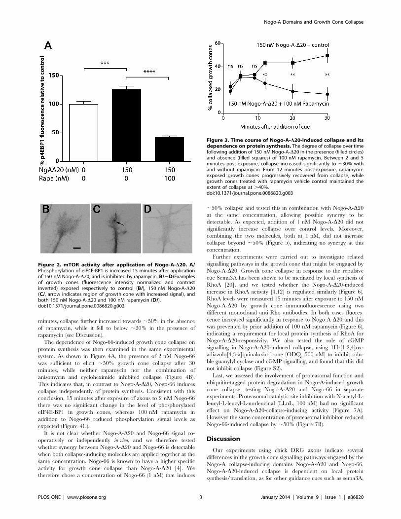
minutes, collapse further increased towards ,50% in the absence
of rapamycin, while it fell to below ,20% in the presence of
rapamycin (see Discussion).
The dependence of Nogo-66-induced growth cone collapse on
protein synthesis was then examined in the same experimental
system. As shown in Figure 4A, the presence of 2 nM Nogo-66
was sufficient to elicit ,50% growth cone collapse after 30
minutes, while neither rapamycin nor the combination of
anisomycin and cycloheximide inhibited collapse (Figure 4B).
This indicates that, in contrast to Nogo-A-D20, Nogo-66 induces
collapse independently of protein synthesis. Consistent with this
conclusion, 15 minutes after exposure of axons to 2 nM Nogo-66
there was no significant change in the level of phosphorylated
eIF4E-BP1 in growth cones, whereas 100 nM rapamycin in
addition to Nogo-66 reduced phosphorylation signal levels as
expected (Figure 4C).
It is not clear whether Nogo-A-D20 and Nogo-66 signal co-
operatively or independently in vivo, and we therefore tested
whether synergy between Nogo-A-D20 and Nogo-66 is detectable
when both collapse-inducing molecules are applied together at the
same concentration. Nogo-66 is known to have a higher specific
activity for growth cone collapse than Nogo-A-D20 [4]. We
therefore chose a concentration of Nogo-66 (1 nM) that induces
,50% collapse and tested this in combination with Nogo-A-D20at the same concentration, allowing possible synergy to be
detectable. As expected, addition of 1 nM Nogo-A-D20 did not
significantly increase collapse over control levels. Moreover,
combining the two molecules, both at 1 nM, did not increase
collapse beyond ,50% (Figure 5), indicating no synergy at this
concentration.
Further experiments were carried out to investigate related
signalling pathways in the growth cone that might be engaged by
Nogo-A-D20. Growth cone collapse in response to the repulsive
cue Sema3A has been shown to be mediated by local synthesis of
RhoA [20], and we tested whether the Nogo-A-D20-inducedincrease in RhoA activity [4,12] is regulated similarly (Figure 6).
RhoA levels were measured 15 minutes after exposure to 150 nM
Nogo-A-D20 by growth cone immunofluorescence using two
different monoclonal anti-Rho antibodies. In both cases fluores-
cence increased significantly in response to Nogo-A-D20 and this
was prevented by prior addition of 100 nM rapamycin (Figure 6),
indicating a requirement for local protein synthesis of RhoA for
Nogo-A-D20-responsivity. We also tested the role of cGMP
signalling in Nogo-A-D20-induced collapse, using 1H-[1,2,4]ox-
adiazolo[4,3-a]quinaloxin-1-one (ODQ, 500 nM) to inhibit solu-
ble guanylyl cyclase and cGMP signalling, and found that this did
not inhibit collapse (Figure S2).
Last, we assessed the involvement of proteasomal function and
ubiquitin-tagged protein degradation in Nogo-A-induced growth
cone collapse, testing Nogo-A-D20 and Nogo-66 in separate
experiments. Proteasomal catalytic site inhibition with N-acetyl-L-
leucyl-L-leucyl-L-norleucinal (LLnL, 100 nM) had no significant
effect on Nogo-A-D20-collapse-inducing activity (Figure 7A).
However the same concentration of proteasomal inhibitor reduced
Nogo-66-induced collapse by ,50% (Figure 7B).
Discussion
Our experiments using chick DRG axons indicate several
differences in the growth cone signalling pathways engaged by the
Nogo-A collapse-inducing domains Nogo-A-D20 and Nogo-66.
Nogo-A-D20-induced collapse is dependent on local protein
synthesis/translation, as for other guidance cues such as sema3A,
Figure 2. mTOR activity after application of Nogo-A-D20. A/Phosphorylation of eIF4E-BP1 is increased 15 minutes after applicationof 150 nM Nogo-A-D20, and is inhibited by rapamycin. B/2D/Examplesof growth cones (fluorescence intensity normalized and contrastinverted) exposed respectively to control (B/), 150 nM Nogo-A-D20(C/, arrow indicates region of growth cone with increased signal), andboth 150 nM Nogo-A-D20 and 100 nM rapamycin (D/).doi:10.1371/journal.pone.0086820.g002
Figure 3. Time course of Nogo-A-D20-induced collapse and itsdependence on protein synthesis. The degree of collapse over timefollowing addition of 150 nM Nogo-A-D20 in the presence (filled circles)and absence (filled squares) of 100 nM rapamycin. Between 2 and 5minutes post-exposure, collapse increased significantly to ,30% withand without rapamycin. From 12 minutes post-exposure, rapamycin-exposed growth cones progressively recovered from collapse, whilegrowth cones treated with rapamycin vehicle control maintained theextent of collapse at .40%.doi:10.1371/journal.pone.0086820.g003
Nogo-A Domains and Growth Cone Collapse
PLOS ONE | www.plosone.org 3 January 2014 | Volume 9 | Issue 1 | e86820

slit2 and netrin 1 [17,18,21]. However, in contrast to sema3A-
induced collapse [21] there is no evidence that collapse induced by
high concentrations of Nogo-A-D20 is independent of protein
synthesis; at both lower (150 nM) and higher (900 nM) concen-
trations, Nogo-A-D20-induced collapse is reduced to control levels
by blockade of mRNA translation. Two further distinctions
between Nogo-A-D20- and sema3A-induced signalling in the
growth cone are also notable. First, Nogo-A-D20-induced collapse
involves Pincher-mediated endocytosis whereas sema3A-induced
collapse does not [5], and second, inhibition of soluble guanylyl
cyclase inhibits collapse induced by sema3A [24–26] but not by
Nogo-A-D20 (this study).
The time course of Nogo-A-D20-induced collapse shows that
some growth cones collapse rapidly following initial exposure to
Nogo-A-D20 (within 10 minutes), and this takes place whether or
not rapamycin is also present (Figure 3). This may reflect the
existence of a sufficient pool of pre-existing protein in these growth
cones to elicit collapse without the requirement for de novo
synthesis, and such rapid collapse is plausible as a physiological
mechanism during axon guidance in vivo. Alternatively, it may
reflect a delay in the onset of action of rapamycin compared with
the initiation of Nogo-A-D20-induced collapse. Our findings
additionally indicate that the subsequent rapamycin-sensitive
phase of Nogo-A-D20-induced growth cone collapse (10–30
minutes) is independent of the cell body, since it also occurs in
acutely axotomized neurites. This is consistent with the study of
Joset et al. [5] showing the requirement for Pincher-mediated
endocytosis in mediating Nogo-A-D20-induced collapse. Using
compartmentalized (rat DRG) cultures, distal neurites but not
proximal neurites or neuronal cell bodies were found to
accumulate Nogo-A-D20-containing endosomes within 30 minutes
of Nogo-A-D20-exposure, while the latter sites contain them only
at later time points [5].
In sharp contrast to Nogo-A-D20, we find that Nogo-66-
induced growth cone collapse takes place independently of protein
synthesis, as confirmed by the absence of phosphorylation of
eIF4E-BP1 after Nogo-66 exposure. Like Nogo-66, collapse due to
high concentrations of sema3A (.500 ng/ml) is independent of
protein synthesis, and the latter pathway has been shown to
Figure 4. Dependence of Nogo-66-induced growth conecollapse on protein synthesis. A/Nogo-66-induced collapse remains
in the presence of 150 nM rapamycin. B/Nogo-66-induced collapse alsoremains in the presence of 2 mM cycloheximide and 5 mM anisomycin.C/Phosphorylation of eIF4E-BP1 is not affected by application of 2 nMNogo-66, but is inhibited by 100 nM rapamycin.doi:10.1371/journal.pone.0086820.g004
Figure 5. Growth cone collapse in the presence of equalconcentrations of Nogo-66 and Nogo-A-D20. 1 nM Nogo-66induces significant growth cone collapse, and this is not altered by thepresence of 1 nM Nogo-A-D20.doi:10.1371/journal.pone.0086820.g005
Nogo-A Domains and Growth Cone Collapse
PLOS ONE | www.plosone.org 4 January 2014 | Volume 9 | Issue 1 | e86820

involve GSK-3b activation [21]. In this respect it is interesting that
a recent study [27] has shown that myelin-associated inhibitors of
axon growth induce phosphorylation and inactivation of GSK-3b,rather than activation. Alabed et al. used a DRG axon outgrowth
assay rather than a growth cone collapse assay, and more detailed
investigation of growth cone regulation by GSK-3b in response to
Nogo-A-derived peptides is therefore warranted.
The finding that de novo synthesis of RhoA in the growth cone is
required for Nogo-A-D20-induced collapse provides another
contrast with Nogo-66-induced collapse, which also involves
RhoA activation [1,12,13] but does not require protein synthesis
(Figure 4). A further difference between the two collapse-inducing
pathways is that proteasomal inhibition reduces Nogo-66- but not
Nogo-A-D20-induced collapse. A possible mediator here is the
scaffold protein Plenty of SH3 (POSH [28]), which is downstream
of Nogo-66/PirB signalling. This has E3 ubiquitin ligase activity,
although the target ubiquitinated downstream of Nogo-66 is
unknown.
While our results indicate that Nogo-66 induces growth cone
collapse independently of mTOR, Nogo-66 has been shown to
activate mTOR in the context of stem cell differentiation,
regulating both astrocyte differentiation from neural progenitor
cells [29] and ES cell pluripotency via regulation of the
transcription factor nanog [30]. Moreover the synthesis of both
glutamate receptors [31] and GABAB receptors [32] is suppressed
by NgR1 signalling via the mTOR pathway, again presumably
through Nogo-66 rather than Nogo-A-D20.Regarding the role of Nogo-A in axon growth regulation in vivo,
Schwab and colleagues have speculated that the primary function
of Nogo-66/NgR signalling may concern axon guidance, since this
system possesses higher specific activity for growth cone collapse
than Nogo-A-D20 [4]. While Nogo-A-D20 may have a similar
role, our evidence indicates that the two domains do not synergize
with respect to growth cone collapse when used together at
concentration (1 nM) that induces ,50% collapse with Nogo-66
alone (Figure 5). The operating concentration range of Nogo-A
in vivo remains unknown, however, and our results do not exclude
the possibility that domain synergy takes place at concentrations
Figure 6. RhoA Levels after application of Nogo-A-D20. A/, B/Levels of RhoA in growth cones detected by anti-RhoA monoclonalantibodies SC-179 (A/) and 26C4 (B/) after 15 minute exposure tocontrol (PBS), 150 nM Nogo-A-D20 and both 150 nM Nogo-A-D20 and100 nM rapamycin, respectively. RhoA increases significantly within 15minutes of exposure to Nogo-A-D20, but rapamycin prevents thisincrease.doi:10.1371/journal.pone.0086820.g006
Figure 7. Proteasome inhibition and Nogo-induced growthcone collapse. A/Proteasome inhibition with Z-LLnL (LLnL) does notinhibit the collapse-inducing activity of 150 nM Nogo-A-D20. B/Proteasome inhibition significantly inhibits collapse-inducing activityof 2 nM Nogo-66 (N66).doi:10.1371/journal.pone.0086820.g007
Nogo-A Domains and Growth Cone Collapse
PLOS ONE | www.plosone.org 5 January 2014 | Volume 9 | Issue 1 | e86820

higher than 1 nM. There is also evidence that Nogo-A-D20 exerts
an additional sustained influence on neuronal gene expression
mediating long-term suppression of axon growth [1,4,5,33]. This is
supported by the study of Chivatakarn et al. [34], who showed
that myelin-induced chronic inhibition of axon outgrowth in vitro is
independent of NgR1 signalling. Our findings revealing several
differences in the growth cone signalling pathways engaged by
these two Nogo-A domains are consistent with this proposed
functional separation.
Materials and Methods
Nogo-66-FC (as a disulfide-linked homodimer) was purchased
from R&D Systems and Nogo-A-D20 was purified as described
previously [4]. Briefly, BL21/DE3 E. coli were transformed with
the pET28 expression vector (Novagen) containing the sequence of
the recombinant His2/T7-tagged protein and cultured at 37uCuntil an OD of 0.8 AU. 1 M IPTG was added for 2 h at 30uC to
induce protein expression. After cell lysis with BugBuster Protein
Extraction Reagent (Novagen) the fusion protein was purified
using Co2+-Talon Metal Affinity Resin (Takara Bio Inc.).
F-12 medium, penicillin/streptomycin and DMEM medium
were obtained from PAA, and B27 supplement, L-15 and Click-
iTH AHA Alexa FluorH 488 protein synthesis reagents from
Invitrogen. Insulin/transferrin/selenite (ITS+3), NGF, glutamine,
laminin from mouse sarcoma, poly-L-lysine, anisomycin, rapamy-
cin and cycloheximide were purchased from Sigma-Aldrich, and
Borosilicate cover-slips from VWR International. 1H-[1,2,4]ox-
adiazolo[4,3-a]quinaloxin-1-one (ODQ) was obtained from Cay-
man Chemical, and N-acetyl-L-leucyl-L-leucyl-L-norleucinal
(LLnL) from Sigma. Anti-p-4EBP1 antibody was purchased from
Cell Signaling Technology, and Alexa Fluor 594 secondary
antibody from Life Technologies. Anti-RhoA monoclonal anti-
bodies SC-179 and 26C4 were obtained from Santa Cruz
Biotechnology.
Coverslips for chick DRG explants were cleaned in acid and
ethanol, and flamed immediately before use. DRG explants were
dissected from E7 chick embryos; no ethical approval was required
for this procedure under English law since it took place within the
first two-thirds of the chick embryo incubation period [The
Guidance on the Operation of the Animals (Scientific Procedures)
Act 1986 (amended 2013)]. Coverslips were coated in 100 mg/ml
poly-L-lysine for 1 h and then 20 mg/ml laminin for 1 h, both
steps at 38uC. E7 DRGs were dissected in medium and grown
overnight at 38uC in DMEM and NGF (80 ng/ml) in 5% CO2.
Inhibitors and inhibitor controls were introduced 1 min prior to
Nogo-A peptide or PBS/vehicle controls, and cultures were
incubated at 38uC in 5% CO2 for 30 min. Axonal transection was
carried out adjacent to the body of the DRG using a hypodermic
needle. Explants were fixed with a solution of 4% w/v
formaldehyde and 15% w/v sucrose in PBS for 2 h at room
temperature. The levels of collapse in blind-coded samples were
assessed by phase contrast microscopy; growth cones with two or
fewer filopodia were designated as collapsed, and at least 6 fields of
view were assessed for each DRG culture. Data groups were
compared using the non-parametric Mann-Whitney U-test and
the Kruskal-Wallis ANOVA test; all percentage values are means.
For each data point growth cone numbers averaged 150,
minimum 50, from at least 3 cultures. Quantitative immunoflu-
orescence was performed on cultures grown in 160 ng/ml NGF (a
high concentration to maintain a spread growth cone morphology
in all samples so that comparative measurements could be made
[20,35]). Anti-p-4EBP1 antibody was used at 1:100, and its
fluorescence signal in growth cones was assessed 15 minutes after
application of Nogo-A-D20. Each growth cone was imaged under
white light and then under fluorescence illumination. The white-
light images were used to define the growth cone outline,
excluding the axon and central zone of the growth cone but
including the lamellipodia and filopodia (peripheral zone) up to
the growth cone transition zone. The central zone was excluded
due to the variable thickness of this part of the growth cone,
causing a significant source of error in a two-dimensional analysis.
The fluorescence intensity was measured as an average across the
growth cone area thus defined, as described by Campbell and Holt
[17]. Inhibition of protein synthesis in growth cones was
monitored using the Click-iTH AHA Alexa FluorH 488 protein
synthesis assay following manufacturer’s instructions.
Supporting Information
Figure S1 AHA-TAMRA labeling of protein synthesisafter exposure of DRG neurons to Nogo-A-D20. A/TAMRA-labeled newly synthesized protein during 1 h exposure
to control (C), 150 nM Nogo-A-D20 (N) and both Nogo-A-D20and 100 nM rapamycin (NR). The rate of protein synthesis
increases markedly across a range of molecular weights after
exposure to Nogo-A-D20, and this increase is prevented by
rapamycin indicating its dependence on mTOR. B/Colorizedversion of image A, showing the gradient spectrum in the lower
right-hand corner (black/blue low intensity, white/red high
intensity); there is a marked increase in protein synthesis due to
Nogo-A-D20 (N) compared with control (C), which is inhibited by
rapamycin (NR). C/Quantification of the total fluorescence in
each lane.
(TIF)
Figure S2 Soluble guanylyl cyclase and Nogo-A-D20-induced growth cone collapse. Inhibition of soluble guanylyl
cyclase with 1H-[1,2,4]oxadiazolo[4,3-a]quinaloxin-1-one (ODQ,
500 nM) does not affect Nogo-A-D20-induced growth cone
collapse.
(TIF)
Author Contributions
Conceived and designed the experiments: RM Andre Schmandke Antonio
Schmandke PJ GC MES CH RK. Performed the experiments: RM Andre
Schmandke Antonio Schmandke PJ GC RK. Analyzed the data: RM
Andre Schmandke Antonio Schmandke PJ GCMES CH RK. Contributed
reagents/materials/analysis tools: Andre Schmandke Antonio Schmandke
GC MES CH RK. Wrote the paper: RM Andre Schmandke Antonio
Schmandke GC MES CH RK.
References
1. Schwab ME (2010) Functions of Nogo proteins and their receptors in the
nervous system. Nat Rev Neurosci 11: 799–811.
2. Oertle T, Klinger M, Stuermer CA, Schwab ME (2003) A reticular rhapsody:
phylogenic evolution and nomenclature of the RTN/Nogo gene family. FASEB J
17: 1238–1247.
3. Dodd DA, Niederoest B, Bloechlinger S, Dupuis L, Loeffler JP et al. (2005)
Nogo-A, -B, and -C are found on the cell surface and interact together in many
different cell types. J Biol Chem 280: 12494–12502.
4. Oertle T, van der Haar ME, Bandtlow CE, Robeva A, Burfeind P et al. (2003)
Nogo-A inhibits neurite outgrowth and cell spreading with three discrete regions.
J Neurosci 23: 5393–5406.
Nogo-A Domains and Growth Cone Collapse
PLOS ONE | www.plosone.org 6 January 2014 | Volume 9 | Issue 1 | e86820

5. Joset A, Dodd DA, Halegoua S, Schwab ME (2010) Pincher-generated Nogo-A
endosomes mediate growth cone collapse and retrograde signaling. J Cell Biol
188: 271–285.
6. GrandPre T, Nakamura F, Vartanian T, Strittmatter SM (2000) Identification of
the Nogo inhibitor of axon regeneration as a Reticulon protein. Nature 403:
439–444.
7. Fournier AE, GrandPre T, Strittmatter SM (2001) Identification of a receptor
mediating Nogo-66 inhibition of axonal regeneration. Nature 409: 341–346.
8. Fournier AE, Gould GC, Liu BP, Strittmatter SM (2002) Truncated soluble
Nogo receptor binds Nogo-66 and blocks inhibition of axon growth by myelin.
J Neurosci 22: 8876–8883.
9. Wong ST, Henley JR, Kanning KC, Huang KH, Bothwell M et al. (2002) A
p75(NTR) and Nogo receptor complex mediates repulsive signaling by myelin-
associated glycoprotein. Nat Neurosci 5: 1302–1308.
10. Mi S, Lee X, Shao Z, Thill G, Ji B et al. (2004) LINGO-1 is a component of the
Nogo-66 receptor/p75 signaling complex. Nat Neurosci 7: 221–228.
11. Atwal JK, Pinkston-Gosse J, Syken J, Stawicki S, Wu Y et al. (2008) PirB is a
functional receptor for myelin inhibitors of axonal regeneration. Science 322:
967–970.
12. Niederost B, Oertle T, Fritsche J, McKinney RA, Bandtlow CE (2002) Nogo-A
and myelin-associated glycoprotein mediate neurite growth inhibition by
antagonistic regulation of RhoA and Rac1. J Neurosci 22: 10368–10376.
13. Fournier AE, Takizawa BT, Strittmatter SM (2003) Rho kinase inhibition
enhances axonal regeneration in the injured CNS. J Neurosci 23: 1416–1423.
14. Hu F, Strittmatter SM (2008) The N-terminal domain of Nogo-A inhibits cell
adhesion and axonal outgrowth by an integrin-specific mechanism. J Neurosci
28: 1262–1269.
15. Grunewald E, Kinnell HL, Porteous DJ, Thomson PA (2009) GPR50 interacts
with neuronal NOGO-A and affects neurite outgrowth. Mol Cell Neurosci 42:
363–371.
16. Deng K, Gao Y, Cao Z, Graziani EI, Wood A et al. (2010) Overcoming amino-
Nogo-induced inhibition of cell spreading and neurite outgrowth by 12-O-
tetradecanoylphorbol-13-acetate-type tumor promoters. J Biol Chem 285: 6425–
6433.
17. Campbell DS, Holt CE (2001) Chemotropic responses of retinal growth cones
mediated by rapid local protein synthesis and degradation. Neuron 32: 1013–
1026.
18. Jung H, Yoon BC, Holt CE (2012) Axonal mRNA localization and local protein
synthesis in nervous system assembly, maintenance and repair. Nat Rev
Neurosci 13: 308–324.
19. Luo Y, Raible D, Raper JA (1993) Collapsin: a protein in brain that induces the
collapse and paralysis of neuronal growth cones. Cell 75: 217–227.
20. Wu KY, Hengst U, Cox LJ, Macosko EZ, Jeromin A et al. (2005) Local
translation of RhoA regulates growth cone collapse. Nature 436: 1020–1024.
21. Manns RP, Cook GM, Holt CE, Keynes RJ (2012) Differing semaphorin 3A
concentrations trigger distinct signaling mechanisms in growth cone collapse.J Neurosci 32: 8554–8559.
22. Dieterich DC, Lee JJ, Link AJ, Graumann J, Tirrell DA et al. (2007) Labeling,
detection and identification of newly synthesized proteomes with bioorthogonalnon-canonical amino-acid tagging. Nat Protoc 2: 532–540.
23. Dieterich DC, Hodas JJ, Gouzer G, Shadrin IY, Ngo JT et al. (2010) In situvisualization and dynamics of newly synthesized proteins in rat hippocampal
neurons. Nat Neurosci 13: 897–905.
24. Castellani V, De Angelis E, Kenwrick S, Rougon G (2002) Cis and transinteractions of L1 with neuropilin-1 control axonal responses to semaphorin 3A.
EMBO J 21: 6348–6357.25. Nangle MR, Keast JR (2011) Semaphorin 3A inhibits growth of adult
sympathetic and parasympathetic neurones via distinct cyclic nucleotidesignalling pathways. Br J Pharmacol 162: 1083–1095.
26. Togashi K, von Schimmelmann MJ, Nishiyama M, Lim CS, Yoshida N et al.
(2008) Cyclic GMP-gated CNG channels function in Sema3A-induced growthcone repulsion. Neuron 58: 694–707.
27. Alabed YZ, Pool M, Ong Tone S, Sutherland C, Fournier AE (2010) GSK3 betaregulates myelin-dependent axon outgrowth inhibition through CRMP4.
J Neurosci 30: 5635–5643.
28. Dickson HM, Zurawski J, Zhang H, Turner DL, Vojtek AB (2010) POSH is anintracellular signal transducer for the axon outgrowth inhibitor Nogo66.
J Neurosci 30: 13319–13325.29. Wang B, Xiao Z, Chen B, Han J, Gao Y et al. (2008) Nogo-66 promotes the
differentiation of neural progenitors into astroglial lineage cells through mTOR-STAT3 pathway. PLoS One 3: e1856.
30. Gao Y, Wang B, Xiao Z, Chen B, Han J et al. (2010) Nogo-66 regulates nanog
expression through stat3 pathway in murine embryonic stem cells. Stem CellsDev 19: 53–60.
31. Peng X, Kim J, Zhou Z, Fink DJ, Mata M (2011) Neuronal Nogo-A regulatesglutamate receptor subunit expression in hippocampal neurons. J Neurochem
119: 1183–1193.
32. Murthy R, Kim J, Sun X, Giger RJ, Fink DJ et al. (2013) Post-transcriptionalregulation of GABAB receptor and GIRK1 channels by Nogo receptor 1. Mol
Brain 6: 30.33. Pernet V, Schwab ME (2012) The role of Nogo-A in axonal plasticity, regrowth
and repair. Cell Tissue Res 349: 97–104.34. Chivatakarn O, Kaneko S, He Z, Tessier-Lavigne M, Giger RJ (2007) The
Nogo-66 receptor NgR1 is required only for the acute growth cone-collapsing
but not the chronic growth-inhibitory actions of myelin inhibitors. J Neurosci 27:7117–7124.
35. Dontchev VD, Letourneau PC (2002) Nerve growth factor and semaphorin 3Asignaling pathways interact in regulating sensory neuronal growth cone motility.
J Neurosci 22: 6659–6669.
Nogo-A Domains and Growth Cone Collapse
PLOS ONE | www.plosone.org 7 January 2014 | Volume 9 | Issue 1 | e86820
Related Documents