REVIEW Protein film voltammetry: electrochemical enzymatic spectroscopy. A review on recent progress Rubin Gulaboski & Valentin Mirčeski & Ivan Bogeski & Markus Hoth Received: 8 March 2011 /Accepted: 26 March 2011 /Published online: 4 May 2011 # Springer-Verlag 2011 Abstract This review is focused on the basic principles, the main applications, and the theoretical models developed for various redox mechanisms in protein film voltammetry, with a special emphasis to square-wave voltammetry as a working technique. Special attention is paid to the thermodynamic and kinetic parameters of relevant enzymes studied in the last decade at various modified electrodes, and their use as a platform for the detection of reactive oxygen species is also discussed. A set of recurrent formulas for simulations of different redox mechanisms of lipophilic enzymes is supplied together with representative simulated voltammograms that illustrate the most relevant voltammetric features of proteins studied under conditions of square-wave voltammetry. Keywords Redox enzymes . Electrochemistry . Modified electrodes . Reactive oxygen species . Mathematical modelling Introduction Life on earth depends almost exclusively on occurrence of redox (redox=reduction oxidation) reactions, which are reactions involving the exchange of electrons between two or more neighboring biochemical systems. The electron charge– transfer reactions are responsible for the function of many proteins and other redox-active compounds in various bio- chemical systems. This is well-known in bioenergetics, where photosynthesis and respiration are fundamental processes in which energy conversion takes place through a complex sequence of electron transfer reactions involving different redox proteins. However, electron transfer also takes place in many other biological processes ranging from cell defense to gene control. Since many diseases are mainly associated with malfunctioning redox biochemistry in humans, the under- standing of these processes has also a big medical significance. One of the fundamental aspects of biological redox chemistry is the physiological importance of the electron transfer organized by proteins in the living systems. Thus, a lot of efforts have been made in the last 30 years to link the thermodynamics and kinetics of electron transfer with the structural and thermodynamic features of various proteins. Since the fundamental principles of electron transfer processes are relatively well understood, the current interest in bioelec- trochemistry is focused largely on the mechanisms by which electron transfers are linked to other important physiological functions. Within all “in vivo” redox processes, two or more redox-active species react with each other in a way of exchanging electrons between them. For the relatively small biochemical systems (small molecules), the voltammetric methods are a common tool of choice that provide insight into the thermodynamics and kinetics of electron transfer reactions, while also giving valuable information about the Dedicated to the 75th birthday of Dr. Nina Fjodorovna Zakharchuk R. Gulaboski (*) Faculty of Agriculture, Department of Chemistry, Goce Delčev University, Štip, Macedonia e-mail: [email protected] V. Mirčeski Faculty of Natural Sciences and Mathematics, Institute of Chemistry, “Ss Kiril i Metodij” University, Skopje, Macedonia I. Bogeski : M. Hoth Faculty of Medicine, Department of Biophysics, University of Saarland, 66421 Homburg, Germany J Solid State Electrochem (2012) 16:2315–2328 DOI 10.1007/s10008-011-1397-5

Welcome message from author
This document is posted to help you gain knowledge. Please leave a comment to let me know what you think about it! Share it to your friends and learn new things together.
Transcript
REVIEW
Protein film voltammetry: electrochemical enzymaticspectroscopy. A review on recent progress
Rubin Gulaboski & Valentin Mirčeski & Ivan Bogeski &Markus Hoth
Received: 8 March 2011 /Accepted: 26 March 2011 /Published online: 4 May 2011# Springer-Verlag 2011
Abstract This review is focused on the basic principles,the main applications, and the theoretical models developedfor various redox mechanisms in protein film voltammetry,with a special emphasis to square-wave voltammetry as aworking technique. Special attention is paid to thethermodynamic and kinetic parameters of relevant enzymesstudied in the last decade at various modified electrodes,and their use as a platform for the detection of reactiveoxygen species is also discussed. A set of recurrentformulas for simulations of different redox mechanisms oflipophilic enzymes is supplied together with representativesimulated voltammograms that illustrate the most relevantvoltammetric features of proteins studied under conditionsof square-wave voltammetry.
Keywords Redox enzymes . Electrochemistry .Modifiedelectrodes . Reactive oxygen species .Mathematicalmodelling
Introduction
Life on earth depends almost exclusively on occurrence ofredox (redox=reduction oxidation) reactions, which arereactions involving the exchange of electrons between two ormore neighboring biochemical systems. The electron charge–transfer reactions are responsible for the function of manyproteins and other redox-active compounds in various bio-chemical systems. This is well-known in bioenergetics, wherephotosynthesis and respiration are fundamental processes inwhich energy conversion takes place through a complexsequence of electron transfer reactions involving differentredox proteins. However, electron transfer also takes place inmany other biological processes ranging from cell defense togene control. Since many diseases are mainly associated withmalfunctioning redox biochemistry in humans, the under-standing of these processes has also a big medical significance.One of the fundamental aspects of biological redox chemistryis the physiological importance of the electron transferorganized by proteins in the living systems. Thus, a lot ofefforts have been made in the last 30 years to link thethermodynamics and kinetics of electron transfer with thestructural and thermodynamic features of various proteins.Since the fundamental principles of electron transfer processesare relatively well understood, the current interest in bioelec-trochemistry is focused largely on the mechanisms by whichelectron transfers are linked to other important physiologicalfunctions. Within all “in vivo” redox processes, two or moreredox-active species react with each other in a way ofexchanging electrons between them. For the relatively smallbiochemical systems (small molecules), the voltammetricmethods are a common tool of choice that provide insightinto the thermodynamics and kinetics of electron transferreactions, while also giving valuable information about the
Dedicated to the 75th birthday of Dr. Nina Fjodorovna Zakharchuk
R. Gulaboski (*)Faculty of Agriculture, Department of Chemistry,Goce Delčev University,Štip, Macedoniae-mail: [email protected]
V. MirčeskiFaculty of Natural Sciences and Mathematics,Institute of Chemistry, “Ss Kiril i Metodij” University,Skopje, Macedonia
I. Bogeski :M. HothFaculty of Medicine, Department of Biophysics,University of Saarland,66421 Homburg, Germany
J Solid State Electrochem (2012) 16:2315–2328DOI 10.1007/s10008-011-1397-5
mechanisms of interactions between such biochemical sys-tems. In the case of proteins, the use of voltammetry as atechnique to understand their redox chemistry is not an easytask. The main problems arise from the huge protein size andthe presence of big “electroinactive” lipophilic tail whichimpede the “access” of electrons to the protein’s redox-activesite(s) through their insulating features.
Nearly 15 years ago, a new voltammetric methodology hadbeen developed, which allows insight into the redox chemistryof various lipophilic redox-active proteins [1–5]. The originalname of this technique coined by Armstrong is “protein filmvoltammetry” (PFV) [1]. This methodology provides accessto a wealth of information on the reactions of redox-activesites in proteins in a quite simple manner. By this technique, agiven protein is attached on an electrode surface mainly byself-assembling from the aqueous electrolyte in which theprotein is initially dissolved. The electrode surface on which aprotein is adsorbed actually acts as a surrogate partner thatmimics the biological redox processes of the proteins. Byapplying a controlled potential to the protein-modifiedelectrode, the redox features of the adsorbed protein can bemonitored by using various voltammetric techniques. Apotentiostat is commonly used to change the potential energyof the electrode which drives electrons onto or removeselectrons from the protein redox-active sites. By applyingcyclic voltammetry (CV) over a wide range of scan rates, forexample, and exploiting the ability to cycle or pulse theelectrode potential between defined values, valuable dataabout the protein’s physiological activity can be obtained.The obtained data are commonly analyzed in terms of plotsof peak potentials, peak currents, or half-peak widths against
the applied scan rates. By such an analysis, one can obtainrelevant thermodynamic and kinetic information about thephysiological functions coupled to protein redox activity. Inthis review, we will focus on the main achievements andchallenges of this methodology, particularly when combinedwith advanced voltammetric techniques as a square-wavevoltammetry (SWV). A special part will be dedicated to theapplication of PFV for studying coupled redox reactions ofrelevant enzymes. In addition, we will discuss recenttheoretical models for various electrode mechanisms underthe conditions of SWV, where we offer a simple way to obtainreliable thermodynamic and/or kinetic information on theredox reactions of studied proteins.
Performing an experiment in protein film voltammetry
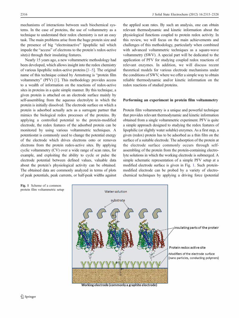
Protein film voltammetry is a unique and powerful techniquethat provides relevant thermodynamic and kinetic informationobtained from a single voltammetric experiment. PFV is quitea simple approach designed to studying the redox features oflipophilic (or slightly water soluble) enzymes. As a first step, agiven (redox) protein has to be adsorbed as a thin film on thesurface of a suitable electrode. The adsorption of the protein atthe electrode surface commonly occurs through self-assembling of the protein from the protein-containing electro-lyte solutions in which the working electrode is submerged. Asimple schematic representation of a simple PFV setup at amodified electrode surface is given in Fig. 1. Such protein-modified electrode can be probed by a variety of electro-chemical techniques by applying a driving force (potential
Fig. 1 Scheme of a commonprotein film voltammetric setup
2316 J Solid State Electrochem (2012) 16:2315–2328
difference) in order to move electrons into and out of theprotein. The movement of the electrons between theelectronic conductor (electrode) and the active center of theredox-active protein is instrumentally detected as a current–potential profile having specific features depending on themeasuring time and the applied potential. From the featuresof the voltammetric outputs, one can obtain information aboutthe thermodynamic and kinetic parameters of the electrontransfer process between the electrode and the redox sites ofthe proteins [1, 4] and in addition, an insight into themechanism of protein interactions with the given substrates[5–10]. To do this, a good theoretical background of theongoing electrode mechanism is required. In order tomimic the physiological activity in the living systems, thenative structural and reactive features of the redox proteinsmust be retained when adsorbed onto the electrodesurface. Hitherto, the main part of the PFV experimentsis performed around neutral pH and at temperaturesbetween 20 and 40 °C, thus mimicking physiological-like conditions. The minimization of the adsorptivesurface denaturation of proteins and keeping the electrodesurface as clean as possible, are essential factors whichfacilitate the direct electron exchange between an elec-trode and the redox site of the proteins. Several metalelectrodes coated with metal oxides, carbon electrodescoated with organic monolayers, and pyrolytic graphiteedge plane electrodes are currently known as the mostsuitable surfaces fulfilling these requirements [5, 11–19].In the last few years, basal plane pyrolytic graphiteelectrodes, glassy carbon electrodes, and even unmodifiedmetallic electrodes have been extensively used in PFV [5].
Pyrolytic graphite edge plane electrodes are especiallysuitable for PFV. These electrodes contain a big fraction ofthe negative carboxylate groups in their three-dimensionalstructure that can interact electrostatically (or by hydrogenbonds) with positively charged amino acids of many proteins[12–19]. This contributes to better sticking of many proteinsto the electrode surface.
Despite many efforts for improving the performances ofPFV, the redox activity of many redox proteins cannot beprobed because the direct electron exchange with theelectrodes is difficult to be achieved. The main cause fortheir redox inactivity is related to the insulating features oftheir side chains. In such cases, there is a common scenarioof co-adsorbing the proteins with some organic linkers likepolymixins, aminocyclitols, alkylthiols, and some othercompounds that are known as good electron shuttlingsystems [5]. Such modifications usually lead to an increaseof the electrochemical reversibility of the electron exchangebetween the proteins and the electrodes. Enzymes can alsobe trapped into some conducting polymer network builtfrom polypyrrole, polyaniline, polythiophene, or polyindolethat are developed on an electrode surface by electrochem-
ical polymerization [5, 13–15]. Different strategies ofpolymerization have been developed in order to enhancethe rate of the electron exchange between the electrode andthe active sites of the proteins, and these are describedelsewhere in more detail [5]. In recent years, there has beenan increased interest in applying the nanoparticle strategy asa tool for enhancing the electrochemical reversibility ofmany proteins. The very high electrical conductivity, goodchemical stability, and structural robustness of carbonnanotubes are crucial physical parameters permitting thesematerials to be exploited in the electrochemistry of manyredox proteins [16–19]. A quite promising electrode forstudying various enzymes is the recently applied diamondelectrode [20]. Nanocrystalline diamond or ultrananocrys-talline diamond thin films have been found quite suitablefor a large variety of substrates [21]. We must note,however, that due to the very high price of this electrode,its use in PFV has been quite limited so far. Many of theaforementioned applications of enzymes in PFV haveinvolved interesting biosensor construction for detectingvarious substrates on nanomodified electrodes, and someof these will be discussed in detail in the subsequentsections.
Representative examples of enzymes studiedwith protein film voltammetry
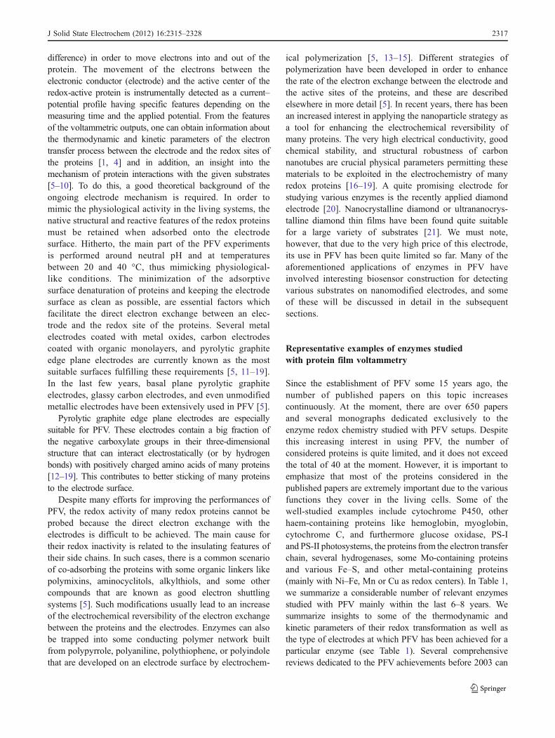
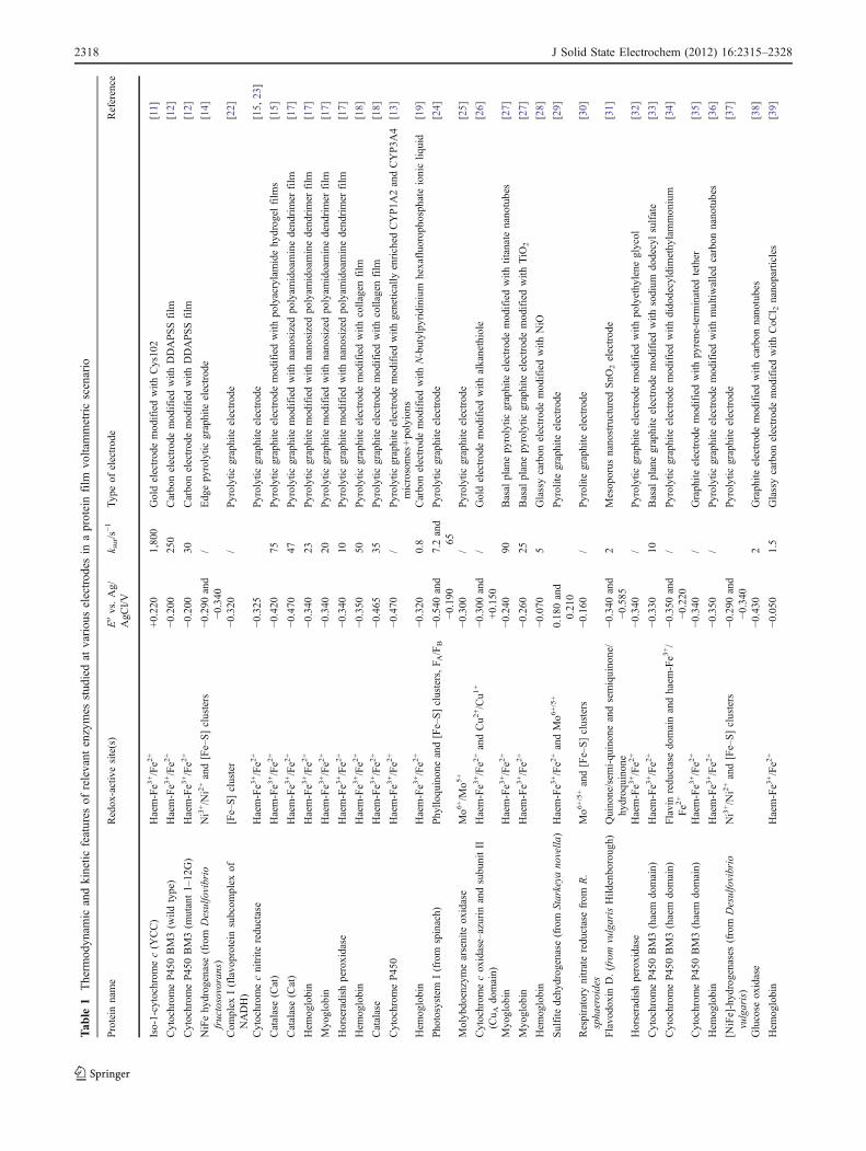
Since the establishment of PFV some 15 years ago, thenumber of published papers on this topic increasescontinuously. At the moment, there are over 650 papersand several monographs dedicated exclusively to theenzyme redox chemistry studied with PFV setups. Despitethis increasing interest in using PFV, the number ofconsidered proteins is quite limited, and it does not exceedthe total of 40 at the moment. However, it is important toemphasize that most of the proteins considered in thepublished papers are extremely important due to the variousfunctions they cover in the living cells. Some of thewell-studied examples include cytochrome P450, otherhaem-containing proteins like hemoglobin, myoglobin,cytochrome C, and furthermore glucose oxidase, PS-Iand PS-II photosystems, the proteins from the electron transferchain, several hydrogenases, some Mo-containing proteinsand various Fe–S, and other metal-containing proteins(mainly with Ni–Fe, Mn or Cu as redox centers). In Table 1,we summarize a considerable number of relevant enzymesstudied with PFV mainly within the last 6–8 years. Wesummarize insights to some of the thermodynamic andkinetic parameters of their redox transformation as well asthe type of electrodes at which PFV has been achieved for aparticular enzyme (see Table 1). Several comprehensivereviews dedicated to the PFV achievements before 2003 can
J Solid State Electrochem (2012) 16:2315–2328 2317
Tab
le1
Therm
odyn
amic
andkinetic
features
ofrelevant
enzymes
stud
iedat
variou
selectrod
esin
aproteinfilm
voltammetricscenario
Protein
name
Redox-activesite(s)
Eovs.Ag/
AgC
l/Vk sur/s
−1Typeof
electrode
Reference
Iso-1-cytochromec(Y
CC)
Haem-Fe3
+/Fe2
++0.220
1,800
Goldelectrodemodifiedwith
Cys102
[11]
Cytochrom
eP450BM3(w
ildtype)
Haem-Fe3
+/Fe2
+−0
.200
250
Carbonelectrodemodifiedwith
DDAPSSfilm
[12]
Cytochrom
eP450BM3(m
utant1–
12G)
Haem-Fe3
+/Fe2
+−0
.200
30Carbonelectrodemodifiedwith
DDAPSSfilm
[12]
NiFehydrogenase(from
Desulfovibrio
fructosovorans)
Ni3+/Ni2+and[Fe–S]clusters
−0.290
and
−0.340
/Edgepyrolytic
graphite
electrode
[14]
Com
plex
I(flavoproteinsubcom
plex
ofNADH)
[Fe–S]cluster
−0.320
/Pyrolytic
graphite
electrode
[22]
Cytochrom
ecnitrite
reductase
Haem-Fe3
+/Fe2
+−0
.325
Pyrolytic
graphite
electrode
[15,
23]
Catalase(Cat)
Haem-Fe3
+/Fe2
+−0
.420
75Pyrolytic
graphite
electrodemodifiedwith
polyacrylamidehydrogel
film
s[15]
Catalase(Cat)
Haem-Fe3
+/Fe2
+−0
.470
47Pyrolytic
graphite
modifiedwith
nanosizedpolyam
idoaminedendrimer
film
[17]
Hem
oglobin
Haem-Fe3
+/Fe2
+−0
.340
23Pyrolytic
graphite
modifiedwith
nanosizedpolyam
idoaminedendrimer
film
[17]
Myoglobin
Haem-Fe3
+/Fe2
+−0
.340
20Pyrolytic
graphite
modifiedwith
nanosizedpolyam
idoaminedendrimer
film
[17]
Horseradish
peroxidase
Haem-Fe3
+/Fe2
+−0
.340
10Pyrolytic
graphite
modifiedwith
nanosizedpolyam
idoaminedendrimer
film
[17]
Hem
oglobin
Haem-Fe3
+/Fe2
+−0
.350
50Pyrolytic
graphite
electrodemodifiedwith
collagenfilm
[18]
Catalase
Haem-Fe3
+/Fe2
+−0
.465
35Pyrolytic
graphite
electrodemodifiedwith
collagenfilm
[18]
Cytochrom
eP450
Haem-Fe3
+/Fe2
+−0
.470
/Pyrolytic
graphiteelectrodemodifiedwith
genetically
enriched
CYP1A
2andCYP3A
4microsomes+polyions
[13]
Hem
oglobin
Haem-Fe3
+/Fe2
+−0
.320
0.8
Carbonelectrodemodifiedwith
N-butylpyridinium
hexafluorophosphateionicliq
uid
[19]
Photosystem
I(from
spinach)
Phyllo
quinoneand[Fe–S]clusters,F A
/FB
−0.540
and
−0.190
7.2and
65Pyrolytic
graphite
electrode
[24]
Molybdoenzymearsenite
oxidase
Mo6
+/M
o5+
−0.300
/Pyrolytic
graphite
electrode
[25]
Cytochrom
ecoxidase–azurin
andsubunitII
(Cu A
domain)
Haem-Fe3
+/Fe2
+andCu2
+/Cu1
+−0
.300
and
+0.150
/Goldelectrodemodifiedwith
alkanethiole
[26]
Myoglobin
Haem-Fe3
+/Fe2
+−0
.240
90Basal
planepyrolytic
graphite
electrodemodifiedwith
titanatenanotubes
[27]
Myoglobin
Haem-Fe3
+/Fe2
+−0
.260
25Basal
planepyrolytic
graphite
electrodemodifiedwith
TiO
2[27]
Hem
oglobin
−0.070
5Glassycarbon
electrodemodifiedwith
NiO
[28]
Sulfite
dehydrogenase(from
Starkeya
novella
)Haem-Fe3
+/Fe2
+andMo6
+/5+
0.180and
0.210
Pyrolite
graphite
electrode
[29]
Respiratory
nitratereductasefrom
R.
sphaeroides
Mo6
+/5+and[Fe–S]clusters
−0.160
/Pyrolite
graphite
electrode
[30]
FlavodoxinD.(from
vulgarisHild
enborough)
Quinone/sem
i-quinoneandsemiquinone/
hydroquinone
−0.340
and
−0.585
2Mesoporus
nanostructured
SnO
2electrode
[31]
Horseradish
peroxidase
Haem-Fe3
+/Fe2
+−0
.340
/Pyrolytic
graphite
electrodemodifiedwith
polyethylene
glycol
[32]
Cytochrom
eP450BM3(haem
domain)
Haem-Fe3
+/Fe2
+−0
.330
10Basal
planegraphite
electrodemodifiedwith
sodium
dodecylsulfate
[33]
Cytochrom
eP450BM3(haem
domain)
Flavinreductasedomainandhaem
-Fe3
+/
Fe2
+−0
.350
and
−0.220
/Pyrolytic
graphite
electrodemodifiedwith
didodecyldim
ethylammonium
[34]
Cytochrom
eP450BM3(haem
domain)
Haem-Fe3
+/Fe2
+−0
.340
/Graphite
electrodemodifiedwith
pyrene-terminated
tether
[35]
Hem
oglobin
Haem-Fe3
+/Fe2
+−0
.350
/Pyrolytic
graphite
electrodemodifiedwith
multiw
alledcarbon
nanotubes
[36]
[NiFe]-hydrogenases(from
Desulfovibrio
vulgaris)
Ni3+/Ni2+and[Fe–S]clusters
−0.290
and
−0.340
Pyrolytic
graphite
electrode
[37]
Glucose
oxidase
−0.430
2Graphite
electrodemodifiedwith
carbon
nanotubes
[38]
Hem
oglobin
Haem-Fe3
+/Fe2
+−0
.050
1.5
Glassycarbon
electrodemodifiedwith
CoC
l 2nanoparticles
[39]
2318 J Solid State Electrochem (2012) 16:2315–2328
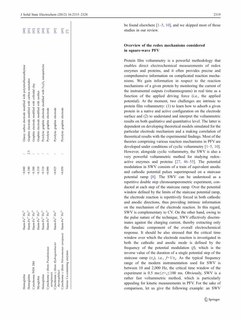
be found elsewhere [1–5, 10], and we skipped most of thosestudies in our review.
Overview of the redox mechanisms consideredin square-wave PFV
Protein film voltammetry is a powerful methodology thatenables direct electrochemical measurements of redoxenzymes and proteins, and it often provides precise andcomprehensive information on complicated reaction mecha-nisms. We gain information in respect to the reactionmechanisms of a given protein by monitoring the current ofthe instrumental outputs (voltammograms) in real time as afunction of the applied driving force (i.e., the appliedpotential). At the moment, two challenges are intrinsic toprotein film voltammetry: (1) to learn how to adsorb a givenprotein in a native and active configuration on the electrodesurface and (2) to understand and interpret the voltammetricresults on both qualitative and quantitative level. The latter isdependent on developing theoretical models simulated for theparticular electrode mechanism and a making correlation oftheoretical results with the experimental findings. Most of thetheories comprising various reaction mechanisms in PFV aredeveloped under conditions of cyclic voltammetry [1–5, 10].However, alongside cyclic voltammetry, the SWV is also avery powerful voltammetric method for studying redox-active enzymes and proteins [27, 46–55]. The potentialmodulation in SWV consists of a train of equivalent anodicand cathodic potential pulses superimposed on a staircasepotential ramp [8]. The SWV can be understood as arepetitive double step chronoamperometric experiment, con-ducted at each step of the staircase ramp. Over the potentialwindow defined by the limits of the staircase potential ramp,the electrode reaction is repetitively forced in both cathodicand anodic directions, thus providing intrinsic informationon the mechanism of the electrode reaction. In this regard,SWV is complementary to CV. On the other hand, owing tothe pulse nature of the technique, SWV effectively discrim-inates against the charging current, thereby extracting onlythe faradaic component of the overall electrochemicalresponse. It should be also stressed that the critical timewindow over which the electrode reaction is investigated inboth the cathodic and anodic mode is defined by thefrequency of the potential modulation (f), which is theinverse value of the duration of a single potential step of thestaircase ramp (ts), i.e., f=1/ts. As the typical frequencyrange of the modern instrumentation used for SWV isbetween 10 and 2,000 Hz, the critical time window of theexperiment is 0.5 ms≤t>s≤100 ms. Obviously, SWV is arather fast voltammetric method, which is particularlyappealing for kinetic measurements in PFV. For the sake ofcomparison, let us give the following example: an SWVT
able
1(con
tinued)
Protein
name
Redox-activesite(s)
Eovs.Ag/
AgC
l/Vk sur/s
−1Typeof
electrode
Reference
Hem
oglobin
Haem-Fe3
+/Fe2
+−0
.040
/Glassycarbon
electrodemodifiedwith
polytetrafluoroethylene
[40]
Hem
oglobin
Haem-Fe3
+/Fe2
+−0
.340
2.5
Graphite
electrodemodifiedwith
carbon
nanotubes
[41]
Cytochrom
eP4502B
4Haem-Fe3
+/Fe2
+−0
.300
/Graphite
electrodemodifiedwith
collo
idal
clay
[42]
Myoglobin
Haem-Fe3
+/Fe2
+−0
.310
/Graphite
electrodemodifiedwith
zeolites
[43]
Hem
oglobin
Haem-Fe3
+/Fe2
+−0
.310
/Pyrolytic
graphite
electrodemodifiedwith
Fe 3O4nanoparticles
[44]
Cytochrom
ec(from
Pseudom
onas
aeruginosa)
Haem-Fe3
+/Fe2
++0.090
/Pyrolytic
graphite
electrode
[45]
Cytochrom
ec(from
Hydrogenobacter
thermophilus)
Haem-Fe3
+/Fe2
++0.025
/Pyrolytic
graphite
electrode
[45]
Cytochrom
ec(from
Nitrosom
onas
europaea)
Haem-Fe3
+/Fe2
++0.050
/Pyrolytic
graphite
electrode
[45]
Various
Cu-containing
enzymes
[7]
J Solid State Electrochem (2012) 16:2315–2328 2319
experiment at f=100 Hz corresponds to a CV experimentconducted at the sweep rate of 60 V/s, assuming a 0.300-Vpotential difference between the initial and the switchingpotential. For most experimental systems, SWV at f=100 Hzprovides undistorted, high-quality voltammetric data, where-as the CV experiment at 60 V/s is still state of the art.
The theoretical background for the SWV of surfaceelectrode processes, where the electroactive form isconfined to the electrode surface in a form of a monolayeris well developed, and it can be effectively utilized in PFV.Over the last decade, significant efforts have been under-taken in modeling and simulations of the surface electrodeprocesses, revealing that SWV is particularly appealing formechanistic, kinetic, and thermodynamic characterizationof surface electrode processes, including proteins andenzymes [27, 46–55]. So far, a plethora of electrodemechanisms have been considered, including simple sur-face electrode reactions [46, 47, 56–61], surface reactionswith uniform interactions [62], surface electrode reactionscoupled with a preceding [63] or following chemicalreaction [64], surface catalytic mechanisms [65, 66], two-step surface reactions [67, 68], and two-step reactionscoupled with an intermediate chemical step (ECE - orelectrochemical-chemical-electrochemical reaction scheme)[69]. The list of surface mechanisms can be easilyextended, as the mathematical modeling of surface pro-cesses, although not easy, is yet simpler than in the case ofcommon diffusion controlled processes. Moreover, thealready existing, rich theory for surface mechanisms underthe conditions of CV [1–5, 10] can be easily adopted forSWV. In the Appendix, we give a list of recurrent formulasfor the simulation of the most frequently encounteredelectrode mechanisms which can be easily implemented invarious software packages. The formulas given in theAppendix are derived based on the Butler–Volmer electrodekinetics, although there are few studies of merit where thesurface processes of immobilized proteins have beenmodeled on the basis of the Marcus kinetic theory toaccount for the intricate voltammetric behavior at largeoverpotentials [47, 50]. Rusling et al. [47] first modeled asurface electrode reaction under conditions of SWV on thebasis of the Markus kinetic theory, in order to studymyoglobin, incorporated into thin films of didodecyldime-thylammonium bromide on the surface of a basal planepyrolytic graphite electrode. Later on, Armstrong et al. [50]utilized the Marcus theory to model the surface electrodereaction of azurin, also called the blue copper protein.
The square-wave (SW) voltammetric response of animmobilized protein is expected to exhibit rather complicatedproperties, even in the case of a simple electron transferprocess. The most remarkable features of almost all quasir-eversible surface electrode reactions are the “quasireversiblemaximum” [8] and the “splitting of the net SW peaks” [70].
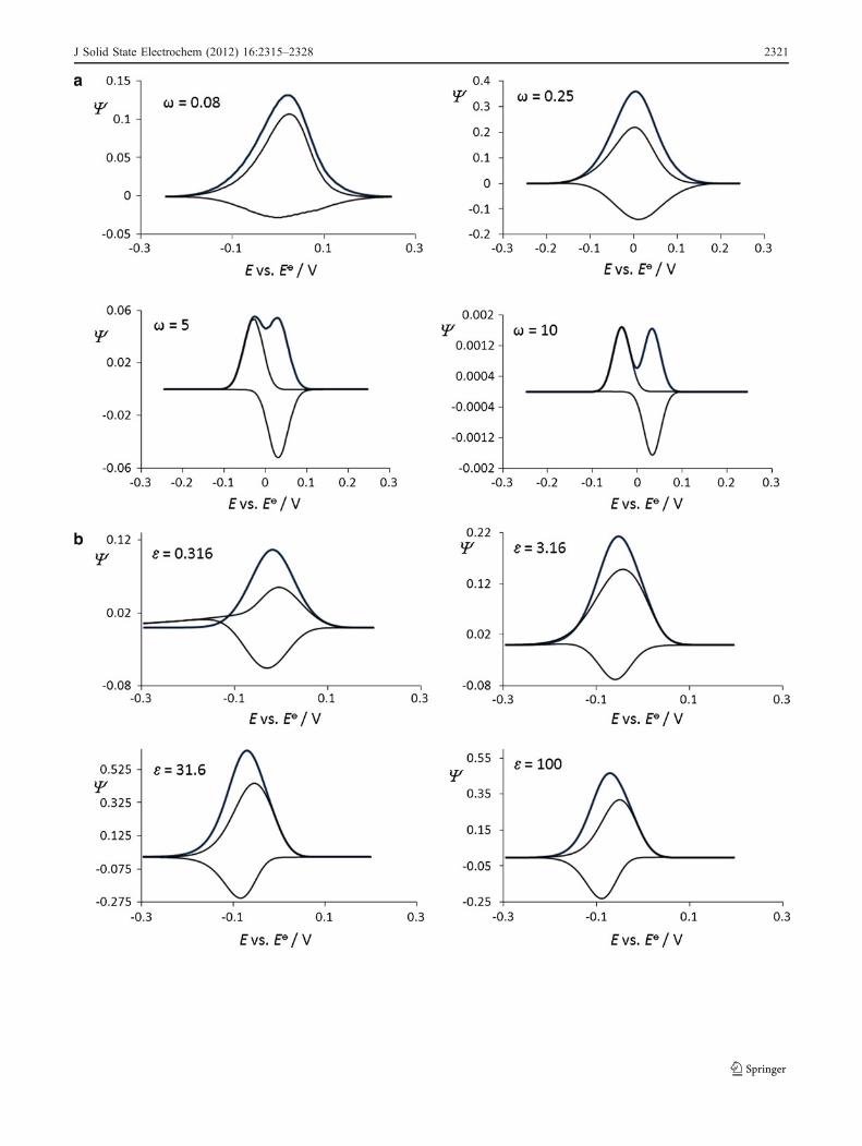
The quasireversible maximum refers to the parabolic depen-dence of the ratio ΔIp/f vs. frequency of the potentialmodulation f, where ΔIp is the net peak current. The originand the physical meaning of the quasireversible maximum iswell understood [8], and its importance stems from the factthat the critical frequency associated with the maximum of theparabola is directly proportional to the standard rate constantof the electrode reaction studied. An important advantage isthat the quasireversible maximum is hardly sensitive to thenumber of electrons involved in the electrode reaction, n, thusenabling kinetic measurements without knowing the latterparameter. Next to the quasireversible maximum, the splittingof the net SW peak is the feature of fast surface electrodereactions [8, 70]. At certain critical amplitudes of the potentialmodulation, the single net SW peak splits into two peaks,symmetrically located around the formal peak potential of thesystem. For given parameters of the potential modulation, thepotential separation between split peaks is highly sensitive tothe standard rate constant and the number of electronsexchanged. The important advantage of the splitting is that itcan be utilized for kinetic measurements of very fast surfaceprocesses only by altering the amplitude of the potentialmodulation, at a fixed low frequency, i.e., at a low sweep rate.Armstrong et al., for example [50], emphasized the utility ofthe splitting of the SW response under large amplitudes for theestimation of the reorganization energy and maximum rateconstant in the case of azurin, adsorbed on edge planepyrolytic graphite and gold electrodes modified with differentself-assembled monolayers of various 1-alkenthiols. In Fig. 2,we provide a set of simulated voltammograms correspondingto relevant mechanisms in PFV studied under conditions ofsquare-wave voltammetry. The readers can certainly benefitfrom the plethora of voltammograms that show how aparticular mechanism in the PFV will be portrayed via theforward–backward and the net current components of the SWvoltammograms.
Fig. 2 a Simple surface redox reaction. Effect of the electron transferkinetic to the features of simulated square-wave voltammograms. Theconditions for the simulations were as follows: electron transfercoefficient α=0.5, square-wave amplitude Esw=40 mV, potential stepdE=5 mV, and temperature T=298 K. b Surface CrE mechanism:Effect of the chemical kinetics to the features of simulated square-wave voltammograms. The value of the equilibrium constant was K=0.1, while the value of the dimensionless kinetic parameter was ω=1.c Surface ECi mechanism: Effect of the chemical kinetics to thefeatures of simulated square-wave voltammograms. The value of thedimensionless kinetic parameter was ω=1. d Surface catalytic EC’:Effect of the catalytic parameter to the features of simulatedvoltammograms. The value of the dimensionless kinetic parameterwas ω=1. e Surface two-step EE mechanism. Effect of the electrontransfer kinetics of both steps to the features of simulated square-wavevoltammograms. f Surface ECE mechanism: Effect of the chemicalstep to the features of simulated voltammograms. The value of thedimensionless kinetic parameters of both electron transfer steps wereω1=ω2=1. Other conditions were the same as those in Fig. 2a
�
2320 J Solid State Electrochem (2012) 16:2315–2328
J Solid State Electrochem (2012) 16:2315–2328 2321
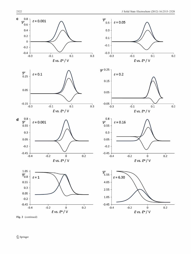
Fig. 2 (continued)
2322 J Solid State Electrochem (2012) 16:2315–2328
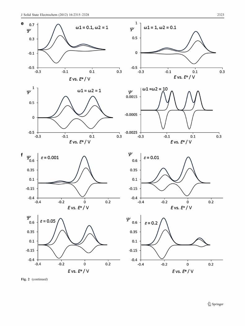
Fig. 2 (continued)
J Solid State Electrochem (2012) 16:2315–2328 2323
Protein film voltammetry of common enzymesas a platform for designing electrochemical sensorsfor various reactive oxygen species
All cells commonly generate energy under aerobic conditionsvia the reduction of molecular oxygen (O2) to water. Bygaining their energy from the oxygen’s reduction, the aerobicorganisms get, at the same time, quite susceptible to thevarious damaging effects of the so-called reactive oxygenspecies (ROS). The ROS are inevitably created during themetabolism of oxygen, especially by its incomplete reductionwith the redox systems engaged in the mitochondrial electrontransfer chain. ROS generated in the living cells can roughlybe classified into two groups: radicals and non-radicals. In the“radical group” one classifies the highly reactive species suchas hydroxyl radical (OH·), superoxide ion radical (O2·
−),peroxyl (ROO·), nitric oxide radical (NO·), and alkoxylradicals (RO·). A common feature of the compoundsbelonging to the radical group is the existence of one unpairedelectron that makes them highly reactive. In the second groupof “non-radical” ROS, one finds compounds produced inrelatively high concentrations in the living cell such ashydrogen peroxide (H2O2), hypochloric acid (HClO), andvarious organic peroxides. Reactive oxygen species producedduring different electron transfer reactions in vivo are usuallyconsidered to be highly harmful to cells. Most of theaforementioned ROS are short-living species that are keen toreact very quickly with redox counter partners from theirsurroundings. The half-lives for most of the ROS from the“radical group” range between 10−3 and 10−10 s, while thehalf-life of the ROS from the non-radical group is typicallymuch higher (it ranges between several seconds to severalhours) [71]. The very high reaction rates of ROS with manybiological compounds from their surroundings make the directdetection of ROS in biological systems a difficult task. In thepast 10 years, the protein film voltammetry has emerged as aviable electrochemical tool for the detection of various ROS. Inmost of the PFV studies, the authors have explored haem-containing proteins as catalase [16–19], hemoglobin andmyoglobin [17, 18, 36, 39–41, 44], cytochrome P450 [13,34], and horseradish peroxidase [32] as platforms for thedetection of oxygen, hydrogen peroxide, trichloroacetic acid,and nitrites. Although good progress has been made in theelectrochemical detection of ROS by protein film voltamme-try, it is worth mentioning that better methods for quantifica-tion of ROS are still required. This is because the very smallspecificity and non-selectivity of the enzymes used for ROSdetection. In addition, the enzymes used as a platform forROS detection are sensitive to rather big concentrations of thesubstrates (i.e., the enzyme sensors can work only in theconcentration regions of ROS of over 50 μM), which maketheir use for the direct detection of ROS in the cells quitelimited. Nonetheless, the use of PFV for ROS detection will
be developed further, mainly because of the simplicity ofachieving “in vivo” detection of ROS by a very simpleexperimental PFV setup.
Outlooks for the future
Protein film voltammetry is a relatively novel methodologicalapproach that allows direct probing of the electrochemistry ofredox enzymes and proteins, while providing a plethora ofinformation on complex reaction mechanisms of various water-insoluble proteins. After the introduction of PFV [1–5], itbecame possible to study the redox features of many enzymesin a common three-electrode voltammetric setup. In this way,much valuable information about the physiological functionsof various enzymes has been collected in the last 15 years.Although significant progress has been made, the number ofconsidered enzymes in the PFV is still quite limited, and itdoes not exceed a total of 40 so far. The main reason for thisis due to the hindered electron exchange between the redoxcenters of many lipophilic proteins with the electrodematerials used in PFV. As many proteins contain long sidechains having insulating features, efforts are permanentlymade to minimize the effect of the protein’s side chains bymodifying the working electrodes in different manners [5].Though various modifications of the electrode surfaces havebeen presented so far [5], the search for a “universal”electrode material that has satisfying electron exchangingfeatures towards different enzymes still goes on. In thisrespect, the diamond electrode [20, 21] is probably the mostpromising material but unfortunately very expensive. Whileplenty of papers have shown that PFV can be successfullyexploited as a sensing tool for ROS detection [5, 13, 16–19,36, 39–41, 44], there are still a lot of limitations to employthis technique for the direct detection and determination ofROS in the cells. The cause is found in the low selectivity ofthe PFV setup and in its low sensitivity at physiological ROSconcentrations. Positive signals in exploring the PFV as a toolfor “in vivo” determination of ROS in the cells come from thesingle enzyme electrochemistry methodology [72]. Byemploying scanning electrochemical microscopy to a singleenzyme by using nano-electrodes, one gets reliable informa-tion about the physiological activity of a given enzyme. Thismethodology [73] appears to be a quite promising applicationof PFV in living cells, and most efforts will be dedicated todevelop this technique in the coming years. Nevertheless, thefuture progress of PFV will also largely depend on thedevelopment of new theoretical models that will help inunderstanding the complex behavior of many enzymes. Inparticular, the theoretical models for proteins containingmultiple redox centers, or redox centers that undergosuccessive redox transformation [5, 52, 74, 75], are neededto elucidate the complex physiological functions of such
2324 J Solid State Electrochem (2012) 16:2315–2328
J Solid State Electrochem (2012) 16:2315–2328 2325
proteins. So far, there is clearly a lack of theories consideringthe voltammetric features of such proteins [76].
Acknowledgments R.G. thanks the Alexander von HumboldtFoundation for providing a Return postdoctoral fellowship. Thiswork is also supported by the Alexander von Humboldt Foundationvia the joint German–Macedonian project from the Research GroupLinkage Programme 3.4-Fokoop-DEU/1128670 (to V.M., R.G. I.B.,and M.H.). M.H. also acknowledges the support by the DeutscheForschungsgemeinschaft (SFB 530, SFB 894, GK 845, GK 1326).
Appendix
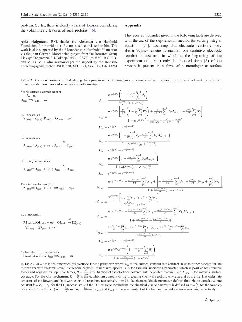
The recurrent formulas given in the following table are derivedwith the aid of the step-function method for solving integralequations [77], assuming that electrode reactions obeyButler–Volmer kinetic formalism. An oxidative electrodereaction is assumed, in which at the beginning of theexperiment (i.e., t=0) only the reduced form (R) of theprotein is present in a form of a monolayer at surface
Table 2 Recurrent formula for calculating the square-wave voltammograms of various surface electrode mechanisms relevant for adsorbedproteins under conditions of square-wave voltammetry
Simple surface electrode reactionksur;aa
RðadsÞ! OðadsÞ þ ne� Ψm ¼w eaa8m 1� 1þe�8m
50
Pm�1
j¼1Ψ j
!
1þ w eaa8m50 1þ e�8mð Þ
CrE mechanismYðadsÞ! RðadsÞ; RðadsÞ! OðadsÞ þ ne� Ψm ¼
w eaa8m K1þK 1� 1
50
Pm�1
j¼1Ψ j
!
þ 1"ð1þKÞ
Pm�1
j¼1Ψ jMm�jþ1 � e�8m
50
Pm�1
j¼1Ψ j
" #
1� w eaa8m � K50 1þKð Þ þ M1
"ð1þKÞ � e�8m50
� �
Mm ¼ e�"50ðmÞ � e�
"50ðm�1Þ
ECi mechanism
RðadsÞ! OðadsÞ þ ne�; OðadsÞ !kf
YðadsÞΨm ¼
w eaa8m 1� 150
Pm�1
j¼1Ψ j� e�8m
"
Pm�1
j¼1Ψ jMm�jþ1
!
1þ w eaa8m 150þ e�8mM1
"
� �
Mm ¼ e�"50ðmÞ � e�
"50ðm�1Þ
EC’ catalytic mechanism
RðadsÞ! OðadsÞ þ ne�; OðadsÞ !kf
RðadsÞ
Ψm ¼w eaa8m 1� 1þe�8m
"
Pm�1
j¼1Ψ jMm�jþ1
!
1þ w eaa8m 1þ e�8mð ÞM1"
Mm ¼ e�"50ðmÞ � e�
"50ðm�1Þ
Two-step mechanism (EE)AðadsÞ! BðadsÞ þ n1e�! CðadsÞ þ n2e� Ψ1;m ¼
w1e�aa;18 1;m � w1e�aa;181;m50
Pm�1
j¼1Ψ1;j þ � ð1þe�81;m Þ50
Pm�1
j¼1Ψ1;j þ e�81;m
50 ðΨ2;m þPm�1
j¼1Ψ2;jÞ
" #
1þ w1eaa;181;m
50 ð1þ e�8 1;m Þ
Ψ2;m ¼w2e
aa;282;m
50 Ψ1;mþPm�1
j¼1Ψ1;j�ð1þe�82;m Þ
Pm�1
j¼1Ψ2;j
� �
1þw2eaa;282;m
50 ð1þe�82;m Þ
ECE mechanism
R1ðadsÞ! O1ðadsÞ þ ne�; O1ðadsÞ !kfR2ðadsÞ
R2ðadsÞ! O2ðadsÞ þ ne�
Ψ1;m ¼w1e�aa;18 1;m � w1e
�aa;181;m Þ50
Pm�1
j¼1Ψ1;j � w1e
ð1�aa;1Þ81;m"
Pm�1
j¼1Ψ1;jMm�jþ1
1þ w1e�aa;181;m50 þ w1e
ð1�aa;1Þ81;m" M1
Ψ2;m ¼w2e�aa;282;m
50
Pm�1
j¼1Ψ1;j�w2e
�aa;282;m"
Pm�1
j¼1Ψ1;jMm�jþ1�w2e
�aa;282;m ð1þe82;m Þ50
Pm�1
j¼1Ψ2;j
1þw2e�aa;282;m
50 ð1þe82;m Þ
Mm ¼ e�"50ðmÞ � e�
"50ðm�1Þ
Surface electrode reaction withlateral interactions RðadsÞ! OðadsÞ þ ne� Ψm ¼
w eaa8me�2aq 1� 1þe�8m50
Pm�1
j¼1Ψ j
!
1þ w eaa8m e�2aq50 1þ e�8mð Þ
In Table 2, w ¼ ksurf is the dimensionless electrode kinetic parameter, where ksur is the surface standard rate constant in units of per second; for the
mechanism with uniform lateral interactions between immobilized species, a is the Frumkin interaction parameter, which is positive for attractiveforces and negative for repulsive forces, q ¼ Γ
»
Γmaxis the fraction of the electrode covered with deposited material, and Γmax is the maximal surface
coverage; For the CrE mechanism, K ¼ kfkb
is the equilibrium constant of the preceding chemical reaction, where kf and kb are the first order rateconstants of the forward and backward chemical reactions, respectively, " ¼ k
f is the chemical kinetic parameter, defined through the cumulative rateconstant k ¼ kf þ kb; for the ECi mechanism and the EC’ catalytic mechanism, the chemical kinetic parameter is defined as " ¼ kf
f ; for the two-stepreaction (EE mechanism) w1 ¼ ksur;1
f and w1 ¼ ksur;2f and ksur,1 and ksur,2 is the rate constant of the first and second electrode reaction, respectively
concentration Γ*. For numerical integration, both time andcurrent are incremented, with the serial number of theincrements designated with m. The time increment is definedas d=1/50f, which means that the duration of each potentialpulse is divided into 25 time increments. The results arepresented in the form of dimensionless current Ψ ¼ I
nFAΓ»f,
where n is the number of electrons, F is the Faraday constant,A is the electrode surface area, and f is the frequency of thepotential modulation. The dimensionless current is thefunction of the dimensionless relative electrode potential8 ¼ nF
RT E � E 6 �� �
, anodic electron transfer coefficient αa, andspecific critical kinetic parameters. Here, E is the electrodepotential, E 6 � is the formal potential of the electrode reaction,R is the gas constant, and T is the thermodynamictemperature. The meaning of the kinetic parameters isexplained below Table 2.
References
1. Armstrong FA (2002) Voltammetry of proteins. In: Bard AJ,Stratmann M, Wilson GS (eds) Encyclopedia of electrochemistry,vol 9. Wiley VCH, Weinheim
2. Armstrong FA (2002) Voltammetric investigations of iron-sulfurclusters in proteins. In: Brajter-Toth A, Chambers JQ (eds)Electroanalytical methods for biological materials. Marcel Dekker,Basel
3. Armstrong FA (2002) J Chem Soc Dalton 5:661–6714. Armstrong FA (1997) Applications of voltammetric methods for
probing the chemistry of redox proteins. In: Lenaz G, Milazzo G(eds) Bioelectrochemistry: principles and practice, vol. 5. Bir-khauser Verlag AG, Basel
5. Barlett PN (2008) Bioelectrochemistry: fundamentals, experimen-tal techniques and application. Wiley, Chichester
6. Jones AK, Lamle SE, Pershad HR, Vincent KA, Albracht SPJ,Armstrong FA (2003) Enzyme Electrokinetics: ElectrochemicalStudies of the Anaerobic Interconversions between Active andInactive States of Allochromatium vinosum [NiFe]-hydrogenase. JAm Chem Soc 125:8505–8514
7. Wijma HJ, Jeuken LJC, Verbeet MP, Armstrong FA, Canters GW(2007) Protein film voltammetry of copper-containing nitrite reduc-tase reveals reversible inactivation. J Am Chem Soc 129:8557–8565
8. Mirceski V, Komorsky-Lovric S, Lovric M (2008) In: Scholz F(ed) Square-wave voltammetry, theory and application. Springer,Berlin
9. Gulaboski R, Lovrić M, Mirčeski V, Bogeski I, Hoth M (2008)Protein-film voltammetry: A theoretical study of the temperatureeffect using square-wave voltammetry. Biophys Chem 137:49–55
10. Hirst J (2006) Elucidating the mechanisms of coupled electrontransfer and catalytic reactions by protein film voltammetry.Biochem Biophys Acta 1757:225–239
11. Heering HA, Wiertz FGM, Dekker C, de Vries S (2004) Directimmobilization of native yeast iso-1 cytochrome c on bare gold:Fast electron relay to redox enzymes and zeptomole protein-filmvoltammetry. J Am Chem Soc 126:11103–11112
12. Udit AK, Hindoyan N, Hill MG, Arnold FH, Gray HB (2005)Protein-surfactant film voltammetry of wild-type and mutantcytochrome P450 BM3. Inorg Chem 44:4109–4111
13. Sultana N, Schenkman JB, Rusling JF (2005) Protein filmelectrochemistry of microsomes genetically enriched in human
cytochrome P450 monooxygenases. J Am Chem Soc 127:13460–13461
14. Léger C, Dementin S, Bertrand P, Rousset M, Guigliarelli B(2004) Inhibition and aerobic inactivation kinetics of Desulfovi-brio fructosovorans NiFe hydrogenase studied by protein filmvoltammetry. J Am Chem Soc 126:12162–12172
15. Gwyer JD, Richardson DJ, Butt JN (2006) Inhibiting Escherichiacoli cytochrome c nitrite reductase: Voltammetry reveals anenzyme equipped for action despite the chemical challenges itmay face in vivo. Biochem Soc Trans 34:133–135
16. Lu H, Li Z, Hu N (2003) Direct voltammetry and electrocatalyticproperties of catalase incorporated in polyacrylamide hydrogelfilms. Biophys Chem 104:623–632
17. Shen L, Hu N (2004) Heme proteins with polyamidoaminedendrimer: Direct electrochemistry and electrocatalysis. BiochemBiophys Acta 1608:23–33
18. Li M, He P, Zhang Y, Hu N (2005) An electrochemicalinvestigation of hemoglobin and catalase incorporated in collagenfilms. Biochem Biophys Acta 1749:43–51
19. Sun W, Gao R, Jiao K (2007) Electrochemistry and electro-catalysis of hemoglobin in nafion/nano- CaCO3 film on a newionic liquid BPPF6 modified carbon paste electrode. J Phys ChemB 111:4560–4567
20. Stutzmann M, Garrido JA, Eickhoff M, Brandt MS (2006) Directbiofunctionalization of semiconductors: A survey. Phys Stat A203:3424–3437
21. Härtl A, Schmich E, Garrido JA, Hernando J, Catharino SCR,Walter S, Feulner P, Kromka A, Steinmüller D, Stutzmann M(2004) Protein-modified nanocrystalline diamond thin films forbiosensor applications. Nat Mater 3:736–742
22. Barker CD, Reda T, Hirst J (2007) The flavoprotein subcomplexof complex I (NADH: ubiquinone oxidoreductase) from bovineheart mitochondria: Insights into the mechanisms of NADHoxidation and NAD+ reduction from protein film voltammetry.Biochem 46:3454–3464
23. Gwyer JD, Richardson DJ, Butt JN (2004) Resolving complexityin the interactions of redox enzymes and their inhibitors:Contrasting mechanisms for the inhibition of a cytochrome cnitrite reductase revealed by protein film voltammetry. Biochem43:15086–15094
24. Munge B, Das SK, Ilagan R, Pendon Z, Yang J, Frank HA,Rusling JF (2003) Electron transfer reactions of redox cofactors inspinach Photosystem I reaction center protein in lipid films onelectrodes. J Am Chem Soc 125:12457–12463
25. Bernhardt PV, Santini JM (2006) Protein film voltammetry ofarsenite oxidase from the chemolithioautotrophic arsenite-oxidiz-ing bacterium NT-26. Biochem 45:2804–2809
26. Fujita K, Nakamura N, Ohno H, Leigh BS, Niki K, Gray HB,Richards JH (2004) Mimicking protein-protein electron transfer:Voltammetry of Pseudomonas aeruginosa azurin and the Thermusthermophilus CuA domain at ω-derivatized self-assembled-mono-layer gold electrodes. J Am Chem Soc 126:13954–13961
27. Liu A, Wei M, Honma I, Zhou H (2005) Direct electrochemistryof myoglobin in titanate nanotubes film. Anal Chem 77:8068–8074
28. Salimi A, Sharifi E, Noorbakhsh A, Soltanian S (2006) Directvoltammetry and electrocatalytic properties of hemoglobin immo-bilized on a glassy carbon electrode modified with nickel oxidenanoparticles. Electrochem Commun 8:1499–1508
29. Aguey-Zinsou K-F, Bernhardt PV, Kappler U, McEwan AG(2003) Direct electrochemistry of a bacterial sulfite dehydroge-nase. J Am Chem Soc 125:530–535
30. Frangioni B, Arnoux P, Sabaty M, Pignol D, Bertrand P,Guigliarelli B, Léger C (2004) In Rhodobacter sphaeroidesrespiratory nitrate reductase, the kinetics of substrate binding
2326 J Solid State Electrochem (2012) 16:2315–2328
favors intramolecular electron transfer. J Am Chem Soc126:1328–1329
31. Astuti Y, Topoglidis E, Briscoe PB, Fantuzzi A, Gilardi G,Durrant JR (2004) Proton-coupled electron transfer of flavodoxinimmobilized on nanostructured tin dioxide electrodes: Thermody-namics versus kinetics control of protein redox function. J AmChem Soc 126:8001–8009
32. Xu Y, Peng W, Liu X, Li G (2004) A new film for the fabrication ofan unmediated H2O2 biosensor. Biosens Bioelectron 20:533–537
33. Udit AK, Hill MG, Gray HB (2006) Electrochemistry ofcytochrome P450 BM3 in sodium dodecyl sulfate films. Lang-muir 25:10854–10857
34. Fleming BD, Tian Y, Bell SG, Wong L-L, Urlacher V, Hill HAO(2003) Redox properties of cytochrome P450 BM3 measured bydirect methods. Eur J Biochem 270:4082–4088
35. Udit AK, Hill MG, Bittner VG, Arnold FH, Gray HB (2004)Reduction of dioxygen catalyzed by pyrene-wired heme domaincytochrome P450 BM3 electrodes. J Am Chem Soc 126:10218–10219
36. Zhao L, Liu H, Hu N (2006) Electroactive films of heme protein-coated multiwalled carbon nanotubes. J Coll Int Sci 296:204–211
37. Vincent KA, Belsey NA, Lubitz W, Armstrong FA (2006) Rapidand reversible reactions of [NiFe]-hydrogenases with sulfide. JAm Chem Soc 128:7448–7449
38. Cai C, Chen J, Lu T (2004) Direct electron transfer of glucoseoxidase on the carbon nanotube electrode. Sci China, Ser BChem 47:113–119
39. Salimi A, Hallaj R, Soltanian S (2007) Immobilization ofhemoglobin on electrodeposited cobalt-oxide nanoparticles: Directvoltammetry and electrocatalytic activity. Biophys Chem130:122–131
40. Lu Q, Hu S (2006) Studies on direct electron transfer andbiocatalytic properties od hemoglobin in polytetrafluoroethylenefilm. Chem Phys Lett 424:167–171
41. Cai C, Chen J (2004) Direct electron transfer and bioelectrocatal-ysis of hemoglobin at a carbon nanotube electrode. Anal Biochem325:285–292
42. Shumyantseva VV, Ivanov YD, Bistolas N, Scheller FW, Arch-akov AI, Wollenberger U (2004) Direct electron transfer ofcytochrome P450 2B4 at electrodes modified with nonionicdetergent and colloidal clay nanoparticles. Anal Chem 76:6046–6052
43. Xie Y, Liu H, Hu N (2007) Layer-by-layer films of hemoglobin ormyoglobin assembled with zeolite particles: Electrochemistry andelectrocatalysis. Bioelectrochem 70:311–319
44. Cao D, Hu N (2006) Direct electron transfer between hemoglobinand pyrolytic graphite electrodes. Biophys Chem 121:209–217
45. Ye T, Kaur R, Wen X, Bren KL, Elliott SJ (2005) Redoxproperties of wild-type and heme-binding loop mutants ofbacterial cytochromes C measured by direct electrochemistry.Inorg Chem 24:8999–9006
46. Reeves JH, Song S, Bowden EF (1993) Application of squarewave voltammetry to strongly adsorbed quasireversible redoxmolecules. Anal Chem 65:683–688
47. Saccucci TM, Rusling JF (2001) Modeling square-wave voltam-metry of thin protein films using Marcus theory. J Phys Chem B105:6142–6147
48. Zhang J, Si-X G, Bond AM, Honeychurch MJ, Oldham KB(2005) Novel kinetic and background current selectivity in theeven harmonic components of fourier transformed square-wavevoltammograms of surface-confined azurin. J Phys Chem B109:8935–8947
49. Jeuken LJC, Jones AK, Chapman SK, Cecchini G, Armstrong FA(2002) Electron-Transfer Mechanisms through Biological RedoxChains in Multicenter Enzymes. J Am Chem Soc 124:5702–5713
50. Jeuken LJC, McEvoy JP, Armstrong FA (2002) Insights intoGated Electron-Transfer Kinetics at the Electrode-Protein Inter-face: A Square Wave Voltammetry Study of the Blue CopperProtein Azurin. J Phys Chem B 106:2304–2313
51. Huang H, Hu N, Zeng Y, Zhou G (2002) Elecrochemistry andelectrocatalysis with heme proteins in chitosan biopolymer films.Anal Biochem 308:141–151
52. Alcantara K, Munge B, Pendon Z, Frank HA, Rusling JF (2006)Thin film voltammetry of spinach photosystem II. Proton-gatedelectron transfer involving the Mn4 cluster. J Am Chem Soc128:14930–14937
53. Xu J, Lu Y, Liu B, Xu C, Kong J (2007) Sensitively probing thecofactor redox species and photo-induced electron transfer ofwild-type and pheophytin-replaced photosynthetic proteins recon-stituted in self-assembled monolayers. J Solid State Electrochem11:1689–1695
54. Ma L, Tian Y, Rong Z (2007) Direct electrochemistry ofhemoglobin in the hyaluronic acid films. J Biochem BiophysMeth 70:657–662
55. Zhou Y, Hu N, Zeng Y, Rusling JF (2002) Layer-by-layerassembly of ultrathin films of hemoglobin and clay nanoparticleswith electrochemical and catalytic activity. Langmuir 18:8573–8579
56. O’Dea JJ, Osteryoung JG (1993) Characterization of quasi-reversible surface processes by square-wave voltammetry. AnalChem 65:3090–3097
57. Komorsky-Lovrić S, Lovrić M (1995) Square-wave voltammetryof quasireversible surface redox reactions. J Electroanal Chem384:115–122
58. Lovrić M (1991) Modelling of surface electrochemical reactions.Elektrokhimija 27:186–195
59. Komorsky-Lovrić Š, Lovrić M (1995) Measurements of redoxkinetics of adsorbed azobenzene by a “quasireversible maximum”in square-wave voltammetry. Electrochim Acta 40:1781–1784
60. Komorsky-Lovrić Š, Lovrić M (1995) Kinetic measurements of asurface confined redox reaction. Anal Chim Acta 305:248–255
61. Mirčeski V, Lovrić M, Jordanoski V (1999) Redox kineticmeasurements of probucole using square-wave voltammetry.Electroanal 11:660–663
62. Mirčeski V, Lovrić M, Gulaboski R (2001) Theoretical andexperimental study of the surface redox reaction involvinginteractions between the adsorbed particles under conditions ofsquare-wave voltammetry. J Electroanal Chem 515:91–100
63. Gulaboski R, Mirčeski V, Lovrić M, Bogeski I (2005) Theoreticalstudy of a surface electrode reaction preceded by a homogeneouschemical reaction under conditions of square-wave voltammetry.Electrochem Commun 7:515–522
64. Mirčeski V, Lovrić M (2000) Adsorption effects in square-wave voltammetry of an EC mechanism. Croat Chem Acta73:305–329
65. Mirčeski V, Gulaboski R (2001) Surface catalytic mechanism insquare-wave voltammetry. Electroanalysis 13:1326–1334
66. Mirčeski V, Gulaboski R (2003) The surface catalytic mechanism:A comparative study with square-wave and staircase cyclicvoltammetry. J Solid State Electrochem 7:157–165
67. O’Dea JJ, Osteryoung JG (1997) Square-wave voltammetry fortwo-step surface redox reactions. Anal Chem 69:650–658
68. Mirčeski V, Gulaboski R (2003) A theoretical and experimentalstudy of a two-step quasireversible surface redox reaction bysquare-wave voltammetry. Croat Chem Acta 76:37–48
69. Gulaboski R (2009) Surface ECE mechanism in protein filmvoltammetry-a theoretical study under conditions of square-wavevoltammetry. J Solid State Electrochem 13:1015–1024
70. Mirceski V, Lovric M (1997) Split square-wave voltammogramsof surface redox reactions. Electroanal 9:1283–1287
J Solid State Electrochem (2012) 16:2315–2328 2327
71. Halliwell B, Gutteridge JM (2001) Free radicals in biology andmedicine, 3rd edn. Oxford University Press, New York
72. Bard AJ (2008) Toward single enzyme molecule electrochemistry.ACS Nano 2:2437–2440
73. Amemiya S, Bard AJ, Fan F-RF, Mirkin MV, Unwin PR (2008)Scanning electrochemical microscopy. Ann Rev Anal Chem 1:95–131
74. McEvoy JP, Brudvig GW (2006) Water-splitting chemistry ofphotosystem II. Chem Rev 106:4455–4483
75. Aguey-Zinsou KF, Bernhardt PV, Leimkuehler S (2003) ProteinFilm Voltammetry of Rhodobacter Capsulatus Xanthine Dehydro-genase. J Am Chem Soc 125:15352–15358
76. Gulaboski R, Mihajlov L (2011) Catalytic mechanism insuccessive two-step protein-film voltammetry. Theoretical studyin square-wave voltammetry. Biophys Chem 155:1–9
77. Nicholson RS (1968) A method based on polynomial approx-imations for numerical solution of volterra integral equations. JElectroanal Chem 16:145–151
2328 J Solid State Electrochem (2012) 16:2315–2328
Related Documents


