Studia Universitatis Studia Universitatis “Vasile Goldiș”, Seria Ştiințele Vieţii (Life Sciences Series), vol. 18, 2008 © 2008 Vasile Goldis University Press http://www.studiauniversitatis.ro 195 PROTECTIVE EFFECTS OF A BIOACTIVE ANTIOXIDANT COMPLEX AGAINST ASPARTAME EXPOSURE DURING GESTATION. BIOCHEMICAL, MORPHOLOGICAL AND ULTRASTRUCTURAL STUDIES ON NEW-BORN RAT BRAIN Constantin PUICĂ 1 *, Constantin CRĂCIUN 2 , Maria BORSA 1 , Mircea RUSU 1 , Ioana ROMAN 1 , Mihai CRISTESCU 3 1 Institute of Biological Research Cluj-Napoca, 2 University Babes-Bolyai Cluj-Napoca, 3 University of Agricultural Sciences and Veterinary Medicine, Cluj-Napoca * Correspondence: Dr. Puică Constantin, “Institutul de Cercetari Biologice Cluj-Napoca”, str. Republicii nr. 48, cod 400015 Tel./Fax +40-(0264)-591238, email: [email protected] Received: march 2008; Published: may 2008 ABSTRACT. 90 days-old nuliparous female Wistar rats were treated by intragastrical gavages with Aspartame at a dose of 0.007 mg/g. bw, dissolved in distilled water (A group), during the entire period of gestation. The animals of group AC were daily treated by oral gavages with Aspartame in each dose, associate with vitamin C in a dose of 0,01 mg/g bw, vitamin E in a dose of 0.002 mg/g bw, and selenium in a dose of 0.005 μg/g bw dissolved in distilled water during the entire period of gestation (days 1-20 of gestation). The new-born brains were examined for their ultrastructure. The rate of some oxidative stress parameters (lipoperoxidation-LPO, non-protein SH and total SH-protein levels) from brain homogenate, were also estimated. The new-born rats prenatally treated with Aspartame showed significant increase of the LPO rate in the brain homogenate (1.17 0.08 nmol MDA/h; +19.3%; p ≤ 0.05), compared to 0.98 ± 0.04 nmol MDA/h value registered in control group. The administration of the bioactive mixture (AC group) showed a moderate, non-significant increase of LPO content in new-born brain homogenate, (1.01 0.05 nmol MDA/h; +3.06%), compared to LPO level in control group. Aspartame treatment during gestation induced a significant depletion of total SH groups in the brain homogenate (14.5 ± 0.27 μmol/g cerebral tissue, - 6.45%, p < 0.5), compared to controls (15.5 ± 0.32 μmol/g). Moderate, but significant decrease of total SH groups was registered in AC group: (14.9 ± 0.25 μmol/g; - 9.61%; p < 0.01). Significant depletion the non- protein SH (new-born brain glutathione content) was registered following Aspartame treatment during gestation: (4.95 ± 0.32 μmol/g. -13.15%; p < 0.001), compared to 5.70 ± 0.32 μmol/g. value registered in controls, and moderate and no significant difference of non-protein SH content, compared to controls, was observed in AC group: (5.48 ± 0.29; - 3.85%; p > 0.25). The study of new-born brain ultrastructure revealed that treatment with Aspartame during gestation has induced decreases with different intensity degrees of both size of neurons and their nuclei in all brain areas. Some nuclei are picnotic. Degenerative aspects of all cellular constituents are typical features of apoptosis: irregular outline of nuclei; picnotic and intense electron dense nuclei; mitochondrial and rough endoplasmic reticulum swellings; demyelization of the myelin sheath; intense vacuolization both of the cytoplasm and axoplasma. The administration of vitamins C, E and selenium produced moderate protective effects against Aspartame treatment, characterized by a some reduction of the lesion-necrotic aspects of the neurons and of all cellular organelles. Our images show groups of neurons with an aspect close to normal of the nuclei and organelles. Ultrastructural and biochemical studies represents an additional element for the establishing the direct embryotoxic/neurotoxic effect of Aspartame-treatment on new-born brain structures and functionality. At the same time, moderate, but positive modulation of the bioactive compound on the ultrastructure and functionality of these brain parameters was registered. Keywords: protective, antioxidant, Aspartame, gestation, brain fetuses INTRODUCTION Aspartame is the name for an artificial, non- saccharide sweetener, aspartyl-phenylalanine-1-methyl ester; i.e., a methyl ester of the dipeptide of the amino acids aspartic acid and phenylalanine. Aspartame is an artificial sweetener because it is 180 times as sweet as sugar in typical concentrations, without the high energy value of sugar. This sweetener is commercialized under a number of trademark names, including Tropicana Slim, Equal, NutraSweet, and Canderel, and is an ingredient of approximately 6,000 consumer foods and beverages sold worldwide. It is commonly used in diet soft drinks

Welcome message from author
This document is posted to help you gain knowledge. Please leave a comment to let me know what you think about it! Share it to your friends and learn new things together.
Transcript
SSttuuddiiaa UUnniivveerrssiittaattiiss
Studia Universitatis “Vasile Goldiș”, Seria Ştiințele Vieţii (Life Sciences Series), vol. 18, 2008 © 2008 Vasile Goldis University Press http://www.studiauniversitatis.ro
195
PPRROOTTEECCTTIIVVEE EEFFFFEECCTTSS OOFF AA BBIIOOAACCTTIIVVEE AANNTTIIOOXXIIDDAANNTT CCOOMMPPLLEEXX
AAGGAAIINNSSTT AASSPPAARRTTAAMMEE EEXXPPOOSSUURREE DDUURRIINNGG GGEESSTTAATTIIOONN.. BBIIOOCCHHEEMMIICCAALL,, MMOORRPPHHOOLLOOGGIICCAALL AANNDD UULLTTRRAASSTTRRUUCCTTUURRAALL
SSTTUUDDIIEESS OONN NNEEWW--BBOORRNN RRAATT BBRRAAIINN
Constantin PUICĂ1*, Constantin CRĂCIUN2, Maria BORSA1, Mircea RUSU1, Ioana ROMAN1, Mihai CRISTESCU3
1Institute of Biological Research Cluj-Napoca, 2University Babes-Bolyai Cluj-Napoca,
3University of Agricultural Sciences and Veterinary Medicine, Cluj-Napoca
* Correspondence: Dr. Puică Constantin, “Institutul de Cercetari Biologice Cluj-Napoca”, str. Republicii nr. 48, cod 400015 Tel./Fax +40-(0264)-591238, email: [email protected] Received: march 2008; Published: may 2008
ABSTRACT. 90 days-old nuliparous female Wistar rats were treated by intragastrical gavages with Aspartame at a dose of 0.007 mg/g. bw, dissolved in distilled water (A group), during the entire period of gestation. The animals of group AC were daily treated by oral gavages with Aspartame in each dose, associate with vitamin C in a dose of 0,01 mg/g bw, vitamin E in a dose of 0.002 mg/g bw, and selenium in a dose of 0.005 μg/g bw dissolved in distilled water during the entire period of gestation (days 1-20 of gestation). The new-born brains were examined for their ultrastructure. The rate of some oxidative stress parameters (lipoperoxidation-LPO, non-protein SH and total SH-protein levels) from brain homogenate, were also estimated. The new-born rats prenatally treated with Aspartame showed significant increase of the LPO rate in the brain homogenate (1.17� 0.08 nmol MDA/h; +19.3%; p ≤ 0.05), compared to 0.98 ± 0.04 nmol MDA/h value registered in control group. The administration of the bioactive mixture (AC group) showed a moderate, non-significant increase of LPO content in new-born brain homogenate, (1.01 � 0.05 nmol MDA/h; +3.06%), compared to LPO level in control group. Aspartame treatment during gestation induced a significant depletion of total SH groups in the brain homogenate (14.5 ± 0.27 µmol/g cerebral tissue, - 6.45%, p < 0.5), compared to controls (15.5 ± 0.32 µmol/g). Moderate, but significant decrease of total SH groups was registered in AC group: (14.9 ± 0.25 µmol/g; - 9.61%; p < 0.01). Significant depletion the non-protein SH (new-born brain glutathione content) was registered following Aspartame treatment during gestation: (4.95 ± 0.32 µmol/g. -13.15%; p < 0.001), compared to 5.70 ± 0.32 µmol/g. value registered in controls, and moderate and no significant difference of non-protein SH content, compared to controls, was observed in AC group: (5.48 ± 0.29; - 3.85%; p > 0.25). The study of new-born brain ultrastructure revealed that treatment with Aspartame during gestation has induced decreases with different intensity degrees of both size of neurons and their nuclei in all brain areas. Some nuclei are picnotic. Degenerative aspects of all cellular constituents are typical features of apoptosis: irregular outline of nuclei; picnotic and intense electron dense nuclei; mitochondrial and rough endoplasmic reticulum swellings; demyelization of the myelin sheath; intense vacuolization both of the cytoplasm and axoplasma. The administration of vitamins C, E and selenium produced moderate protective effects against Aspartame treatment, characterized by a some reduction of the lesion-necrotic aspects of the neurons and of all cellular organelles. Our images show groups of neurons with an aspect close to normal of the nuclei and organelles. Ultrastructural and biochemical studies represents an additional element for the establishing the direct embryotoxic/neurotoxic effect of Aspartame-treatment on new-born brain structures and functionality. At the same time, moderate, but positive modulation of the bioactive compound on the ultrastructure and functionality of these brain parameters was registered. Keywords: protective, antioxidant, Aspartame, gestation, brain fetuses INTRODUCTION
Aspartame is the name for an artificial, non-saccharide sweetener, aspartyl-phenylalanine-1-methyl ester; i.e., a methyl ester of the dipeptide of the amino acids aspartic acid and phenylalanine. Aspartame is an artificial sweetener because it is 180 times as sweet as
sugar in typical concentrations, without the high energy value of sugar.
This sweetener is commercialized under a number of trademark names, including Tropicana Slim, Equal, NutraSweet, and Canderel, and is an ingredient of approximately 6,000 consumer foods and beverages sold worldwide. It is commonly used in diet soft drinks
SSttuuddiiaa UUnniivveerrssiittaattiiss
Studia Universitatis “Vasile Goldiş”, Seria Ştiințele Vieţii (Life Sciences Series), vol. 18, 2008 © 2008 Vasile Goldis University Press
http://www.studiauniversitatis.ro 196
(Adria, Pepsi-Cola light, Coca-Cola light etc. etc), and is often provided as a table condiment.
A Coca-Cola website indicates that a diet soda contains 175 mg of Aspartame (http://www.beverageinstitute.org/ingredients/pdf/Aspartame.pdf, 2007).
It is also used in some brands of chewable vitamin supplements and common in many sugar-free chewing gums. However, Aspartame is not always suitable for baking because it often breaks down when heated and loses much of its sweetness. In the European Union, it is also known under the E number (additive code) E 951.
Aspartame is also one of the sugar substitutes used by people with diabetes.
Aspartame is a subject of a public controversy due to possible health risks and there is some controversy surrounding the rate of breakdown into these various products and the effects that they have on those that consume Aspartame-sweetened foods, (Hull, 2002, Jacobson, 2005; Marshall, 2007; Saito, 2005; Soffritti, 2007).
Under conditions of elevated temperature (more to 350 C) or high pH, like many other peptides, Aspartame is hydrolyzed (break down) to three chemicals, aspartic acid (40%), phenylalanine (50%) and methanol (10%).
In the body, the methanol (wood alcohol that has killed or blinded thousands of skid row drunks) converts to formaldehyde, and then into formic acid (another sting poison). Aspartic acid caused brain lesions in experimental animals. The phenylalanine breaks down into diketopiperazine (DKP), a brain tumor agent. Formaldehyde is deadly embalming fluid, a Class A Carcinogen. Phenylanine and aspartic acid are toxic when unaccompanied by the other amino acids in proteins, (Bowen, 2000; Dow-Edwards Diana et al., 1989; Elsas, 2002; Harder. 2002; Pinto, 1988; Walton, Hudak, and Green-Waite, 1993; Walters, 2001).
Aspartic acid is an amino acid, and it is an aspartate precursor. It is a naturally occurring amino acid which is a component of all proteins. It is classified as a "non-essential" amino acid, meaning that humans do not need to get it from their diets since they can make it from other things in the diet. Aspartic acid is important in the synthesis of new DNA, in urea synthesis, and as a neurotransmitter in the brain. As such, its levels in the body are carefully regulated. If the body needs more aspartic acid, it makes more, using oxaloacetate from the tricarboxylic acid cycle (Krebs cycle) of energy metabolism. If the body has a surplus of aspartic acid, it converts the excess to fumarate, which enters the tricarboxylic acid cycle and provides energy.
Phenylalanine is one of the "essential" amino acids, meaning that humans must get it from their diet. It is a precursor for the synthesis of tyrosine and several neurotransmitters. Excess phenylalanine is broken down to fumarate and acetoacetate, both of which are part of normal energy metabolism. People who lack the enzyme to convert phenylalanine to tyrosine are not able to metabolize phenylalanine
normally. This condition is called phenylketonuria because, in these people, excess phenylalanine is instead converted to phenylketones which appear in the urine. If it is not detected and treated, this condition can lead to mental retardation. This was the first genetic disease for which a routine screening test became available. Persons having this genetic defect must monitor their intake of phenylalanine. A high level of phenylalanine in the brain is extremely harmful and sometimes fatal. For this reason, products containing Aspartame carry an information label for phenylketonurics, (Pinto and Maher, l988; Walters, 2001)
Dr Louis J. Elsas (cited by Gold, 1995), showed that high blood phenylalanine can be concentrated in parts of the brain, and is especially dangerous for infants and fetuses.
In the book, While Waiting: A Prenatal Guidebook by George R. Verrilli, M.D. and Anne Marie Mueser, (cited by Gold, 1995), it is stated that Aspartame is suspected of causing brain damage in sensitive individuals. A fetus may be at risk for these effects.
A recent study has demonstrated that aspartame feeding to animals’ results in an accumulation of formaldehyde within the cells, with evidence of significant damage to cellular proteins and DNA. In fact, the formaldehyde accumulated with prolonged use of aspartame. With this damning evidence, one would have to be suicidal to continue the use of aspartame sweetened foods, drinks and medicines. The use of foods containing excitotoxin additives is especially harmful to the unborn and small children. Starting with age 4 the brain is only 80% formed, at age 8, 90% and at age 16 it is fully formed, but still undergoing changes and rewiring (plasticity). We know that the excitotoxins have a devastating effect on formation of the brain (wiring of the brain) and that such exposure can cause the brain to be "miswired." (Blaylock, 2002; Soffritti et al., 2006).
Methanol is commonly encountered in the diet. Methanol is well known to be poisonous in large quantity. "Large quantity" means tens or hundreds of grams, (Walters, 2001).
Methanol is highly toxic; it is gradually released in the small intestine when the methyl group of the Aspartame encounters the enzyme chymotrypsin, (Stegink and Filer 1984). It has been pointed out that some fruit juices and alcoholic beverages contain small amounts of methanol. It is important to remember, however, that methanol never appears alone. In every case, ethanol is present, usually in much higher amounts; ethanol reduces the effects of methanol toxicity in humans.
Both aspartate and glutamate act as neurotransmitters in the brain, carrying information from neuron to neuron. When there is an excess of neurotransmitter, certain neurons are killed by allowing too much calcium into the cells. This influx causes excessive numbers of free radicals to build up which kill the cells. The neural cell damage that is caused by excessive aspartate and glutamate is the reason they are referred to as ‘excitotoxins’. The excitotoxins are substances, usually acidic amino acids that react with
SSttuuddiiaa UUnniivveerrssiittaattiiss
Studia Universitatis “Vasile Goldiș”, Seria Ştiințele Vieţii (Life Sciences Series), vol. 18, 2008 © 2008 Vasile Goldis University Press http://www.studiauniversitatis.ro
197
specialized receptors in the brain in such a way as to lead to destruction of certain types of neurons, (Blaylock, 2002; Ho et al., 2003).
These two additives - glutamate and Aspartame are two of the more commonly known alimentary excitotoxins and they literally excite the neurons to death, (Blaylock, 1997, 1998, 1999, 2002).
Despite evidence that glutamate, Aspartame and other food neurotoxic amino acids are excitotoxins that can destroy central neurons following oral intake by animals of various species, this amino acid continues to be one of the most vilely and heavily used food additives in the world. MSG is added to several foods to enhance flavor, (Olney, 1995, Puică, 1997a).
Evidence implicating in excitotoxin neuro-degenerative disorders stimulated interest in the possibility that antagonists might prove valuable as neuroprotective in experimental and clinical neurology. Pharmaceutical and/or nutraceutical interventions that target specific pathways in the cascade of cell death may eventually halt the progression of these relentless disorders. Agents that are currently being explored include specific free radical scavengers, antioxidants, mitochondrial substances, antiglutamate compounds and apoptotic inhibitors. It is likely that a cocktail of agents rather than a single factor will be found to be more effective in retarding the pathogenic mechanisms of neurodegenerative disease.
There is a worldwide preoccupation with the counteraction of the negative effects of excitotoxic amino acids, which are utilized as food additives or pharmacologic factors, by attenuating or blocking oxidative stress, the essential cause of many neurodegenerative diseases in animals and men. Agents that are currently being explored include specific free radical scavengers, mitochondrial substances, antiglutamate compounds and apoptotic inhibitors. It is likely that a cocktail of agents rather than a single factor will be found to be more effective in retarding the pathogenic mechanisms of neurodegenerative disease, (Blaylock, 1998; Pedersen and Schmidt, 2000; Sami, 1995; Shenkin, 2001).
Two of the best known anti-oxidants are vitamins C and E, both of which have been shown to be somewhat effective in different models of neurodegeneration (Darr et al., 1996; Lombard, 2002).
In the last decade a considerable interest has been generated concerning the use of natural compounds, antioxidants in particular, in neuroprotection. Antioxidants are substances that prevent or repair oxidative damage of cell constituents. Antioxidants are promising agents in the treatment of neurological disease in humans, but many key questions first need to be addressed concerning their clinical use.
These questions include whether the antioxidants cross the blood brain barrier or achieve adequate concentrations in specific sub cellular locations. The antioxidants that have been most studied to date include vitamin E and specific photochemical such as Ginkgo biloba and carotenoids. Three of the best known anti-oxidants are vitamins C and E, and selenium both of which have been shown to be somewhat effective in different models of
neurodegeneration, (Iacono, 2002; Ikonomidou et al., 1999; Frei and Traber, 2001; Miyamoto et al., 1989).
Although interest is high, and numerous studies have been conducted, serious gaps exists in knowledge of the interactions and possible synergism of endogenous or exogenous antioxidants: B vitamin group, vitamin C, vitamin E, lipoic acid and selenium, etc., and of his preventive and curative potential for multifactor degenerative diseases, as well as in glutamate neurotoxicity, (Ahmad and Jacan, 2001; Beck et al., 2001; Behne and Kyriakopoulos, 2001; Blaylock, 2001; Halliwell, 1996; Iacono et al., 2002; Markowitz, 2001; Meydani, 2001; Puică et al., 2001, 2002; Reinhart, 2000; Sunde, 2002; Wen et al.,1996). MATERIALS AND METHODS
90 days-old nuliparous female Wistar rats weighing 175 g. were used. The animals were divided into three equal groups of eight animals. The first group was the control group (C group) treated daily by oral gavages with distilled water.
Group A received daily by oral gavages Aspartame dissolved in distilled water in a dose of 0.007 mg/g bw during the entire period of gestation.
The animals of group AC were treated during the entire period of gestation (days 1-20 of gestation) daily by oral gavages with Aspartame in a dose of 0.007 mg/g bw, associated with vitamin C in a dose of 0.01 mg/g bw, vitamin E in a dose of 0.002 mg/g bw, and selenium in a dose of 0.005 μg/g. bw, all being dissolved in distilled water. We specify that the 0.007 mg/g bw dose of Aspartame (ICN Biochemical’s-Germany) utilized in our experiment, correspond to concentration of this alimentary additive which can be found in all human alimentary beverages and probably, in other alimentary preparations.
This dose wasn’t utilized until to date in world experimental procedures, and it represents a novelty in the domain of Aspartame neurotoxicity-studies.
The rats were housed in plastic cages, had free
access to drinking water and feed with a common rats-chow. The animals were maintained in standardized conditions, in a room at 230C, and a 12-h light: dark cycle. Care and treatment of rats followed recommended guidelines (NRC 1985, England).
The new-borns were sacrificed by decapitation and the brains were rapidly isolated for biochemical and ultrastructural investigations. The images were analyzed and quantified using a Pentium IV computer-assisted image analysis system.
Biochemical investigations were performed in new-born brain homogenate.
Assay of the rate of lipid peroxidation (LPO) - LPO was assayed by estimating the malondialdehyde (MDA) using thiobarbituric acid (TBA) as described by Wilbur et al., (1949), and presented by Babu et al., (1994). The rate of LPO was expressed as mol MDA formed per hour using molar extinction coefficient for MDA (E532 = 1056 x 105).
Assay of sulphydryl groups (SH) - total and non-protein (free) SH were assayed using the method described by Sedlack and Lindsay (1968). The method is principally based on Ellman’s reaction. The nitro

SSttuuddiiaa UUnniivveerrssiittaattiiss
Studia Universitatis “Vasile Goldiş”, Seria Ştiințele Vieţii (Life Sciences Series), vol. 18, 2008 © 2008 Vasile Goldis University Press
http://www.studiauniversitatis.ro 198
benzoic acid anions give intense yellow color and thus provide accurate and unique method for the estimation of SH groups. Calculations were made by using standard curves plotted with reduced glutathione or cysteine and values were expressed in terms of µmol SH per g. Wet tissue. Statistical evaluation of the data was carried out using Student’s „t” test. A p value of less than 0,05 was considered significant.
Ultrastructural investigations - for ultrastructural investigations, the hippocampus and mediobasal hypothalamus tissues were immediately prefixed in a 2.7% glutaraldehyde solution and postfixes in a 2% osmic acid solution. The dehydration of the samples was performed in acetone and in dehydrates acetone and then embedded in Vestopal W. The ultra fin sections were obtained using an LKB-III ultra microtome and were contrasted with uranyl acetate and
lead citrate. Examination for sections was performed in an Olympus, JAPAN electron microscope. RESULTS Biochemical study
Administration of Aspartame during gestation (group A) showed a significant increase, situated at the limit of statistic significance of LPO rate in new-born brain homogenate (1.17 ± 0.08 nmol. MDA/h, +19.30%, p ≤ 0.05), comparative to C group (0.98 ± 0.04 nmol. MDA/h). The treatment of the pregnant rats with the antioxidant combination (vitamins C, E and selenium) in AC group induced a moderate, non-significant increase of LPO content in new-born brain homogenate, (1.01 ± 0.05 nmol MDA/h; +3.06%), compared to LPO level in control group, (Table 1).
Table 1
LPO content in new-born brain homogenate in all experimental groups
Group C A AC
n 8 8 8
X ± ES nmol.MDA/h 0.98 ± 0.04 1.17 ± 0.08 1.01 ± 0.05
D% - + 19.30 + 3.06
p - ≤ 0.05 p > 0,3
Aspartame treatment during gestation induced a
moderate depletion situated at the limit of statistic significance, of total SH groups in the new-born brain homogenate (14.5 ± 0.27 µmol/g cerebral tissue, - 6.45%, p ≤ 0.5), compared to controls (15.5 ± 0.32 µmol/g.). Moderate, but significant decrease of total SH groups was registered in AC group: (14.9 ± 0.25 µmol/g; - 9.61%; p < 0.05), (Table 2). Significant
depletion the non-protein SH (new-born brain glutathione content) was registered following Aspartame treatment during gestation: (4.95 ± 0.32 µmol/g. -13.15%; p < 0.001), compared to 5.70 ± 0.32 µmol/g. value registered in controls, and moderate and non-significant decrease of non-proteic SH content, compared to controls, was observed in AC group: (5.48 ± 0.29; - 3.85%; p > 0.25), (Table 2).
Table 2
Total-SH and neprotein-SH groups content (μmol/g tissue), in new-born brain homogenate in all experimental groups
Groups C A AC
n 8 8 8
total SH groups
μmol X ± ES 15.5 ± 0.32 14.5 ± 0.27 14.9 ± 0.25
D% - - 6.45 - 9,61
p - ≤ 0.5 < 0.05
non-protein SH groups
μmol X ± ES 5.70 ± 0.10 4.95 ± 0.32 5.48 ± 0.29
D% - - 13.15 - 3.85
p - p < 0.001 > 0.25
Ultrastructural study
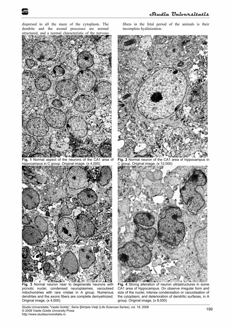
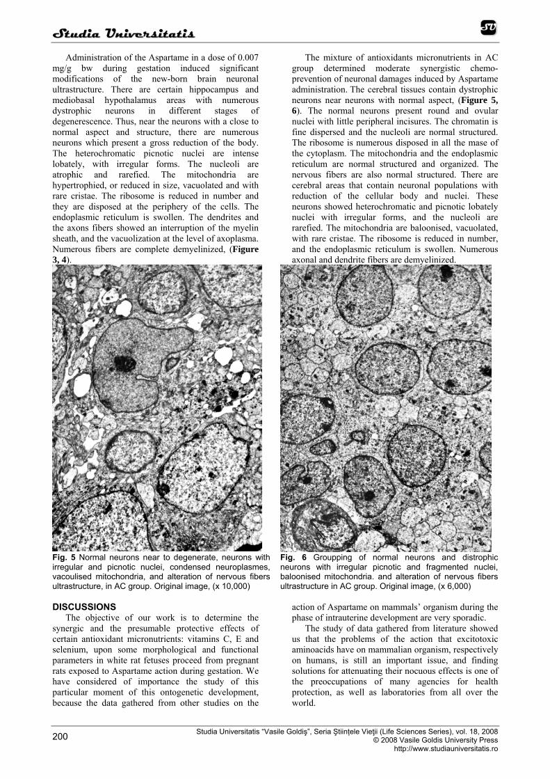
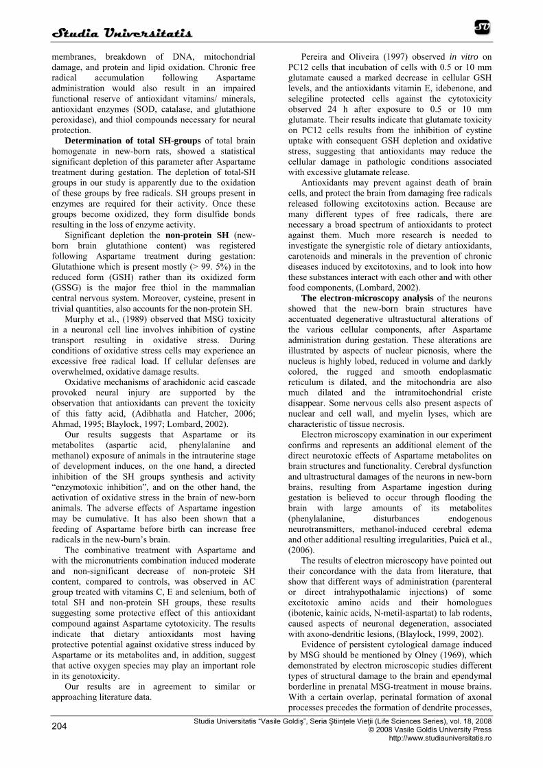
Ultrastructural examination of the new-born cerebral nervous tissues of hippocampus and hypothalamus areas in C group (Figure 1, 2) showed a normal aspect of the neurons, of cellular organites and of nervous dendrite and axonal fibers. The nuclei are spherical or ovular. They are sited in the center of the
cells. The euchromatic nuclei present a fine heterochromatin dispersed in the mase of karyoplasms. They contain one or two nucleoli. In the cytoplasm there are relative few electrondenses mitochondria. The endoplasmic reticulum, very well represented, is arranged in parallel profiles. The ribosome visualized as a spherical particles are very numerous, and they are
SSttuuddiiaa UUnniivveerrssiittaattiiss
Studia Universitatis “Vasile Goldiș”, Seria Ştiințele Vieţii (Life Sciences Series), vol. 18, 2008 © 2008 Vasile Goldis University Press http://www.studiauniversitatis.ro
199
dispersed in all the maze of the cytoplasm. The dendrite and the axonal processes are normal structured, and a normal characteristic of the nervous
fibers in the fetal period of the animals is their incomplete hyalinization.
Fig. 1 Normal aspect of the neurons of the CA1 area of hippocampus in C group. Original image, (x 4,000)
Fig. 2 Normal neuron of the CA1 area of hippocampus in C group. Original image, (x 12,000)
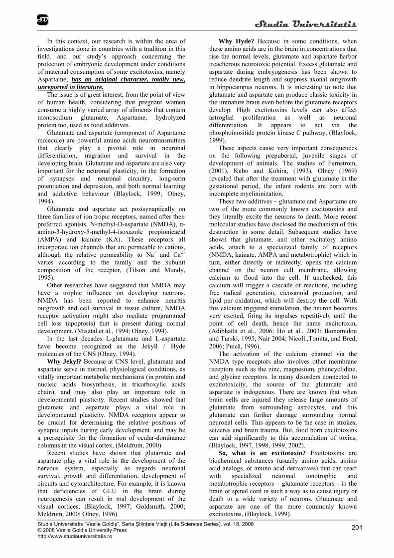
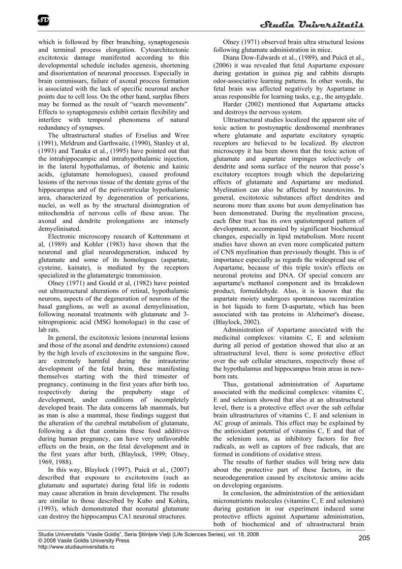
Fig. 3 Normal neuron near to degenerate neurons with picnotic nuclei, condensed neuroplasmes, vacoulised mitochondries with rare cristae in A group. Numerous dendrites and the axons fibers are complete demyelinized. Original image, (x 4,000)
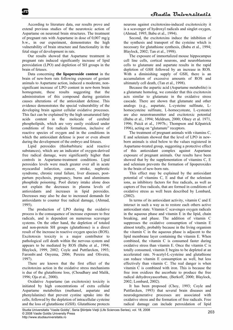
Fig. 4 Strong alteration of neuron ultrastructures in some CA1 area of hippocampus. On observe irregular form and size of the nuclei, intense condensation or vacuolisation of the cytoplasm, and deterioration of dendritic surfaces, in A group. Original image, (x 8,000)
SSttuuddiiaa UUnniivveerrssiittaattiiss
Studia Universitatis “Vasile Goldiş”, Seria Ştiințele Vieţii (Life Sciences Series), vol. 18, 2008 © 2008 Vasile Goldis University Press
http://www.studiauniversitatis.ro 200
Administration of the Aspartame in a dose of 0.007 mg/g bw during gestation induced significant modifications of the new-born brain neuronal ultrastructure. There are certain hippocampus and mediobasal hypothalamus areas with numerous dystrophic neurons in different stages of degenerescence. Thus, near the neurons with a close to normal aspect and structure, there are numerous neurons which present a gross reduction of the body. The heterochromatic picnotic nuclei are intense lobately, with irregular forms. The nucleoli are atrophic and rarefied. The mitochondria are hypertrophied, or reduced in size, vacuolated and with rare cristae. The ribosome is reduced in number and they are disposed at the periphery of the cells. The endoplasmic reticulum is swollen. The dendrites and the axons fibers showed an interruption of the myelin sheath, and the vacuolization at the level of axoplasma. Numerous fibers are complete demyelinized, (Figure 3, 4).
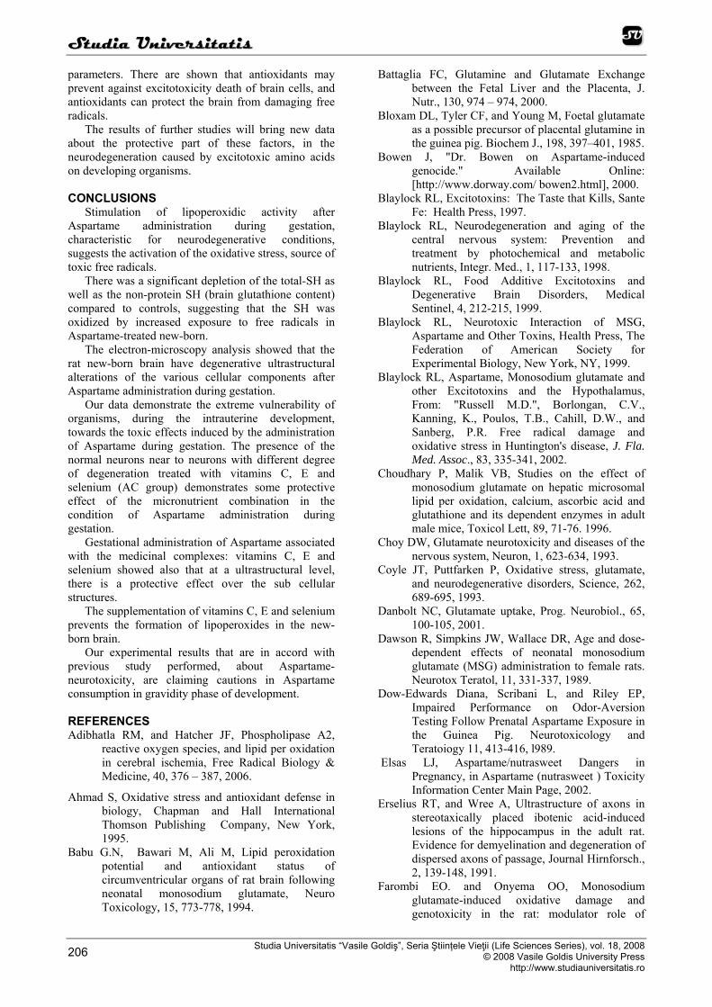
The mixture of antioxidants micronutrients in AC group determined moderate synergistic chemo-prevention of neuronal damages induced by Aspartame administration. The cerebral tissues contain dystrophic neurons near neurons with normal aspect, (Figure 5, 6). The normal neurons present round and ovular nuclei with little peripheral incisures. The chromatin is fine dispersed and the nucleoli are normal structured. The ribosome is numerous disposed in all the mase of the cytoplasm. The mitochondria and the endoplasmic reticulum are normal structured and organized. The nervous fibers are also normal structured. There are cerebral areas that contain neuronal populations with reduction of the cellular body and nuclei. These neurons showed heterochromatic and picnotic lobately nuclei with irregular forms, and the nucleoli are rarefied. The mitochondria are baloonised, vacuolated, with rare cristae. The ribosome is reduced in number, and the endoplasmic reticulum is swollen. Numerous axonal and dendrite fibers are demyelinized.
Fig. 5 Normal neurons near to degenerate, neurons with irregular and picnotic nuclei, condensed neuroplasmes, vacoulised mitochondria, and alteration of nervous fibers ultrastructure, in AC group. Original image, (x 10,000)
Fig. 6 Groupping of normal neurons and distrophic neurons with irregular picnotic and fragmented nuclei, baloonised mitochondria. and alteration of nervous fibers ultrastructure in AC group. Original image, (x 6,000)
DISCUSSIONS
The objective of our work is to determine the synergic and the presumable protective effects of certain antioxidant micronutrients: vitamins C, E and selenium, upon some morphological and functional parameters in white rat fetuses proceed from pregnant rats exposed to Aspartame action during gestation. We have considered of importance the study of this particular moment of this ontogenetic development, because the data gathered from other studies on the
action of Aspartame on mammals’ organism during the phase of intrauterine development are very sporadic.
The study of data gathered from literature showed us that the problems of the action that excitotoxic aminoacids have on mammalian organism, respectively on humans, is still an important issue, and finding solutions for attenuating their nocuous effects is one of the preoccupations of many agencies for health protection, as well as laboratories from all over the world.
SSttuuddiiaa UUnniivveerrssiittaattiiss
Studia Universitatis “Vasile Goldiș”, Seria Ştiințele Vieţii (Life Sciences Series), vol. 18, 2008 © 2008 Vasile Goldis University Press http://www.studiauniversitatis.ro
201
In this context, our research is within the area of investigations done in countries with a tradition in this field, and our study’s approach concerning the protection of embryonic development under conditions of maternal consumption of some excitotoxins, namely Aspartame, has an original character, totally new, unreported in literature.
The issue is of great interest, from the point of view of human health, considering that pregnant women consume a highly varied array of aliments that contain monosodium glutamate, Aspartame, hydrolyzed protein too, used as food additives.
Glutamate and aspartate (component of Aspartame molecule) are powerful amino acids neurotransmitters that clearly play a pivotal role in neuronal differentiation, migration and survival in the developing brain. Glutamate and aspartate are also very important for the neuronal plasticity, in the formation of synapses and neuronal circuitry, long-term potentiation and depression, and both normal learning and addictive behaviour (Blaylock, 1999; Olney, 1994).
Glutamate and aspartate act postsynaptically on three families of ion tropic receptors, named after their preferred agonists, N-methyl-D-aspartate (NMDA), α-amino-3-hydroxy-5-methyl-4-isoxazole propionicacid (AMPA) and kainate (KA). These receptors all incorporate ion channels that are permeable to cations, although the relative permeability to Na+ and Ca2+ varies according to the family and the subunit composition of the receptor, (Tilson and Mundy, 1995).
Other researches have suggested that NMDA may have a trophic influence on developing neurons. NMDA has been reported to enhance neuritis outgrowth and cell survival in tissue culture, NMDA receptor activation might also mediate programmed cell loss (apoptosis) that is present during normal development, (Misztal et al., 1994; Olney, 1994).
In the last decades L-glutamate and L-aspartate have become recognized as the Jekyll / Hyde molecules of the CNS (Olney, 1994).
Why Jekyl? Because at CNS level, glutamate and aspartate serve in normal, physiological conditions, as vitally important metabolic mechanisms (in protein and nucleic acids biosynthesis, in tricarboxylic acids chain), and may also play an important role in developmental plasticity. Recent studies showed that glutamate and aspartate plays a vital role in developmental plasticity. NMDA receptors appear to be crucial for determining the relative positions of synaptic inputs during early development. and may be a prerequisite for the formation of ocular-dominance columns in the visual cortex, (Meldrum, 2000).
Recent studies have shown that glutamate and aspartate play a vital role in the development of the nervous system, especially as regards neuronal survival, growth and differentiation, development of circuits and cytoarchitecture. For example, it is known that deficiencies of GLU in the brain during neurogenesis can result in mal development of the visual cortices, (Blaylock, 1997; Goldsmith, 2000; Meldrum, 2000; Olney, 1996).
Why Hyde? Because in some conditions, when these amino acids are in the brain in concentrations that rise the normal levels, glutamate and aspartate harbor treacherous neurotoxic potential. Excess glutamate and aspartate during embryogenesis has been shown to reduce dendrite length and suppress axonal outgrowth in hippocampus neurons. It is interesting to note that glutamate and aspartate can produce classic toxicity in the immature brain even before the glutamate receptors develop. High excitotoxins levels can also affect astroglial proliferation as well as neuronal differentiation. It appears to act via the phosphoinositide protein kinase C pathway, (Blaylock, 1999).
These aspects cause very important consequences on the following prepubertal, juvenile stages of development of animals. The studies of Fernstrom, (2001), Kubo and Kohira, (1993), Olney (1969) revealed that after the treatment with glutamate in the gestational period, the infant rodents are born with incomplete myelininization.
These two additives – glutamate and Aspartame are two of the more commonly known excitotoxins and they literally excite the neurons to death. More recent molecular studies have disclosed the mechanism of this destruction in some detail. Subsequent studies have shown that glutamate, and other excitatory amino acids, attach to a specialized family of receptors (NMDA, kainate, AMPA and metabotrophic) which in turn, either directly or indirectly, opens the calcium channel on the neuron cell membrane, allowing calcium to flood into the cell. If unchecked, this calcium will trigger a cascade of reactions, including free radical generation, eicosanoid production, and lipid per oxidation, which will destroy the cell. With this calcium triggered stimulation, the neuron becomes very excited, firing its impulses repetitively until the point of cell death, hence the name excitotoxin, (Adibhatla et al., 2006; Ho et al., 2003; Ikonomidou and Turski, 1995; Nair 2004; Nicoll.,Tomita, and Bred, 2006; Puică, 1996).
The activation of the calcium channel via the NMDA type receptors also involves other membrane receptors such as the zinc, magnesium, phencyclidine, and glycine receptors. In many disorders connected to excitotoxicity, the source of the glutamate and aspartate is indogenous. There are known that when brain cells are injured they release large amounts of glutamate from surrounding astrocytes, and this glutamate can further damage surrounding normal neuronal cells. This appears to be the case in strokes, seizures and brain trauma. But, food born excitotoxins can add significantly to this accumulation of toxins, (Blaylock, 1997, 1998, 1999, 2002).
So, what is an excitotoxin? Excitotoxins are biochemical substances (usually amino acids, amino acid analogs, or amino acid derivatives) that can react with specialized neuronal ionotrophic and metabotrophic receptors – glutamate receptors - in the brain or spinal cord in such a way as to cause injury or death to a wide variety of neurons. Glutamate and aspartate are one of the more commonly known excitotoxins, (Blaylock, 1999).

SSttuuddiiaa UUnniivveerrssiittaattiiss
Studia Universitatis “Vasile Goldiş”, Seria Ştiințele Vieţii (Life Sciences Series), vol. 18, 2008 © 2008 Vasile Goldis University Press
http://www.studiauniversitatis.ro 202
These several observations (Olney, 1969, 1974), led to the “excitotoxic” concept that a depolarization mechanism underlies glutamate neurotoxicity and the toxic action is mediated through dendrosomal synaptic receptors specialized for glutamatergic transmission. Excitotoxicity refers to the paradoxical property, shared by glutamate and specific excitatory amino acids (EAA) analogs, of causing acute neuronal degeneration by excessive stimulation of postsynaptic EAA ion tropic receptors-receptors through which glutamate and other analogs functions physiologically as a transmitter.
The acute form of neurotoxicity is characterized by neuronal swelling in the presence of agonists, in particularly glutamate, which leads to osmotic lyses of the neurons, (Choy, 1993; Danbolt, 2000).
Experimental data reveals the role of excitotoxins in neurodegenerative process particularly relevant to the developing central nervous system in early postnatal and in prepubertal period of life, (Choy, 1993; Fernstrom, 2000; Meldrum, 2000; Olney, 1994; Puică, 1996; Puică et al. 1997a, 2002 a, b).
The excess aspartate and glutamate in the blood plasma shortly after ingesting Aspartame or products with free glutamic acid (glutamate precursor) leads to a high level of those neurotransmitters in certain areas of the brain. The effect of glutamate and aspartate in
infant rodents might possibly be related to the lesser capacity of their intestinal epithelium and liver to transaminate glutamate and aspartate, or to a lesser expression of the glial glutamate transporters GLT and GLAST in the hypothalamus at this developmental
stage. Developmental changes in the expression of ion tropic glutamate receptors are known to influence
excitotoxic phenomena and may contribute to the pattern of vulnerability in the neonatal rodent, (Meldrum, 2000).
It is well recognized that the immature brain is four times more sensitive to the toxic effects of the excitatory amino acids as is the mature brain. This means that excitotoxic injury is of special concern from the fetal stage to adolescence. There is also, evidence that the placenta concentrates several of these toxic amino acids on the fetal side of the placenta. (Blaylock, 1999; Battaglia, 2000; Bloxam et al., 1985; Dawson, 1989; Elsas, 2002; Moores et al., 1994).
There are a growing number of clinicians and basic scientists who are convinced that excitotoxins (glutamate, Aspartame, cysteine etc) play a critical role in the development of several neurological disorders, including migraines, seizures, infections, abnormal neural development, certain endocrine disorders, neuropsychiatric disorders, learning disorders in children, AIDS, dementia, episodic violence, lime borreliosis, hepatic encephalopathy, specific types of obesity, and especially the neurodegenerative diseases; a group of diseases which includes: ALS, Parkinson’s disease, Alzheimer’s disease, Huntington’s disease, and olivopontocerebellar degeneration, (Blaylock, 2002; Olney, 1994).
Several experiments have demonstrated that under such conditions, glutamate, aspartate and phenylalanine can by-pass blood barrier system and
enter the brain in toxic concentrations. In experimental condition of elevated level in the brain, such as after systemic administration, these amino acids generally produced brain lesions in immature animals without a fully developed blood-brain barrier, (Goldsmith, 2000; Gu et al., 1994; Gold, 1995; Meldrum, 2000; Olney, 1974, 1994; Puică, 1997a and b, 2002a and b).
These amino acids affects all the developing brain, the hypothalamic regions that lack blood brain barriers, the extrahypothalamic regions (hippocampus, choroids plexus and cortex), because the developing brain are hypervulnerable to the excitotoxic insults, (Blaylock, 1999, 2002; Puică, Crăciun and Rusu, 2004).
In fact, there is some evidence that it may actually be concentrated within the brain with prolonged exposures. There are also several conditions under which the blood-brain barrier (BBB) is made incompetent. Before birth, the BBB is incompetent and will allow glutamate, aspartate and phenyl-alanine to enter the brain. It may be that for a considerable period after birth the barrier may also incompletely developed as well. In fact, as we age the barrier system becomes more porous, allowing excitotoxins in the blood to enter the brain. So there are numerous instances under which excitotoxin food additives can enter and damage the brain, (Goldstein, 1994).
Finally, recent experiments have shown that glutamate and aspartate (as in Aspartame) can open the barrier itself. Another system used to protect the brain against environmental excitotoxins, is a system within the brain that binds the glutamate molecule (called the glutamate transporter) and transports it to a special storage cell (the astrocyte) within a fraction of a second after it is used as a neurotransmitter. This system can be overwhelmed by high intakes of monosodium glutamate (MSG), Aspartame and other food excitotoxins. Glutamate appears to be an important regulator of brain capillary transport and stability, and over-stimulation of blood-brain barrier NMDA receptors through dietary MSG/Aspartame - induced high blood levels of glutamate/aspartate may lead to a lessening of blood-brain barrier exclusion of glutamate and aspartate. It is also known that excitotoxins themselves can cause the generation of numerous amounts of free radicals and that during the process of lipid peroxidation (oxidation of membrane fats) a substance is produced called 4-hydroxynonenal. This chemical inhibits the glutamate transporter, thus allowing glutamate to accumulate in the brain. Excitotoxins destroy neurons partly by stimulating the generation of large numbers of free radicals, (Blaylock, 1997, 1998).
In agreement to early reports of the data of literature (Blaylock, 1997, 1999, 2002; Elsas, 2002; Gold, 1995; Puică et al., 2003; Roberts, 1991; Walton et al., 1993), Aspartame induces some adverse effects in brain following acute or chronic administration.
In 1971 neuroscientist John Olney (whose research on L-monosodium glutamate caused it to be removed from baby foods) proved that Aspartame causes holes in the brains of infant mice.
SSttuuddiiaa UUnniivveerrssiittaattiiss
Studia Universitatis “Vasile Goldiș”, Seria Ştiințele Vieţii (Life Sciences Series), vol. 18, 2008 © 2008 Vasile Goldis University Press http://www.studiauniversitatis.ro
203
According to literature data, our results prove and extend previous studies of the neurotoxic action of Aspartame on neuronal brain structures. The treatment of pregnant rats with Aspartame in dose of 0,007 mg/g b.w., in our experiment, demonstrates the high vulnerability of brain structure and functionality in the fetal stage of development in rats.
Our results showed that Aspartame treatment in pregnant rats induced significantly increase of lipid peroxidation (LPO) and depletion of SH groups in the brain of fetuses.
Data concerning the lipoperoxide content in the brain of new-born rats following exposure of gestant animals to Aspartame action, induced a moderate, non-significant increase of LPO content in new-born brain homogenate, these results suggesting that the administration of this compound during gestation causes alterations of the antioxidant defense. This evidence demonstrates the special vulnerability of the developing brain against cellular oxidative processes. This fact can be explained by the high unsaturated fatty acids content in the molecule of cerebral phospholipids, which are very easily oxidized under conditions of free radicals formation, inclusive of reactive species of oxygen and in the conditions in which the antioxidant defense is poor or even absent during the development of the embryo and fetuses.
Lipid peroxides (thiobarbituric acid reactive substances), which are an indicator of oxygen-derived free radical damage, were significantly higher than controls in Aspartame-treatment conditions. Lipid peroxides levels were much greater over all in acute myocardial infarction, cancer, stroke, nephrotic syndrome, chronic renal failure, liver diseases, post-partum psychosis, pregnancy, burns and aluminums phosphide poisoning. Pool dietary intake alone does not explain the decreases in plasma levels of antioxidants and increases in lipid peroxides. Decreases may also be due to increased demands for antioxidants to counter free radical damage, (Ahmad, 1995).
The production of LPO during the oxidative process is the consequence of increase exposure to free radicals, and is dependent on numerous scavenger systems. On the other hand, the depletion of totaled and non-protein SH groups (glutathione) is a direct result of the increase in reactive oxygen species (ROS). Excitotoxin toxicity is a major contributor to pathological cell death within the nervous system and appears to be mediated by ROS (Babu et al., 1994; Blaylock, 1999, 2002; Coyle and Puttfarcken, 1993; Farombi and Onyema, 2006; Pereira and Oliveira, 1997).
There are known that the first effect of the excitotoxins action in the oxidative stress mechanisms is due of the glutathione loss, (Choudhary and Malik, 1996; Oja et al., 2000).
Oxidative Aspartame (an excitotoxin) toxicity is initiated by high concentrations of extra cellular Aspartame metabolites (methanol, aspartate and phenylalanine) that prevent cystine uptake into the cells, followed by the depletion of intracellular cysteine and the loss of glutathione (GSH). Glutathione protects
neurons against excitotoxins-induced excitotoxicity it is a scavenger of hydroxyl radicals and singlet oxygen, (Ahmad, 1995; Babu et al., 1994).
Second, the excitotoxins induce the inhibition of the synthesis and transport of the cysteine which is necessary for glutathione synthesis, (Babu et al., 1994; Blaylock, 2002; Tan et al., 1998).
The exposure of immortalized mouse hippocampus cell line cells, cortical neurons, and neuroblastoma cells to glutamate and aspartate results in the rapid depletion of GSH followed by an increase in ROS. With a diminishing supply of GSH, there is an accumulation of excessive amounts of ROS and ultimately cell death, (Tan et al., 1998).
Because the aspartic acid (Aspartame metabolite) is a glutamate homolog, we consider that this excitotoxin acts similar to glutamate in the oxidative stress cascade. There are shown that glutamate and other analogs (e.g., aspartate, L-cysteine sulfinate, L-homocysteine sulfinate, L-homocysteate, L-cysteate) are also neurotransmitter and excitotoxic potential (Babu et al., 1994; Meldrum, 2000; Olney et al. 1971, 1996; Puică et al., 1997b.; Thompson and Kilpatrick 1996), acting on “glutamate” receptors.
The treatment of pregnant animals with vitamins C, E and selenium showed that the level of LPO in new-born animals is sited below to the values registered in Aspartame-treated group, suggesting a protective effect of this antioxidant complex against Aspartame exposure of pregnant animals. Our results have been showed that by the supplementation of vitamins C, E and selenium prevents the formation of lipoperoxides in the brain of new-born rats.
This effect may be explained by the antioxidant potential of vitamins C, E and that of the selenium ions, as inhibitory factors for free radicals, as well as captors of free radicals, that are formed in conditions of oxidative stress as well been described by Lombard, (2002).
In terms of its antioxidant activity, vitamin C and E interact in such a way as to restore each others active antioxidant state. Vitamin C scavenges oxygen radicals in the aqueous phase and vitamin E in the lipid, chain breaking, and phase. The addition of vitamin C suppresses the oxidative consumption of vitamin E almost totally, probably because in the living organism the vitamin C in the aqueous phase is adjacent to the lipid membrane layer containing the vitamin E. When combined, the vitamin C is consumed faster during oxidative stress than vitamin E. Once the vitamin C is totally consumed, vitamin E begins to be depleted at an accelerated rate. N-acetyl-L-cysteine and glutathione can reduce vitamin E consumption as well, but less effectively than vitamin C. The real danger is when vitamin C is combined with iron. This is because the free iron oxidizes the ascorbate to produce the free radical dehydroxyascorbate, (Berkoff, 2000; Blaylock, 2002; Lombard, 2002).
It has been proposed (Choy, 1993; Coyle and Puttfarcken, 1993) that several brain diseases and neurodegenerative processes are associated with oxidative stress and the formation of free radicals. Free radical damage can include peroxidation of lipid
SSttuuddiiaa UUnniivveerrssiittaattiiss
Studia Universitatis “Vasile Goldiş”, Seria Ştiințele Vieţii (Life Sciences Series), vol. 18, 2008 © 2008 Vasile Goldis University Press
http://www.studiauniversitatis.ro 204
membranes, breakdown of DNA, mitochondrial damage, and protein and lipid oxidation. Chronic free radical accumulation following Aspartame administration would also result in an impaired functional reserve of antioxidant vitamins/ minerals, antioxidant enzymes (SOD, catalase, and glutathione peroxidase), and thiol compounds necessary for neural protection.
Determination of total SH-groups of total brain homogenate in new-born rats, showed a statistical significant depletion of this parameter after Aspartame treatment during gestation. The depletion of total-SH groups in our study is apparently due to the oxidation of these groups by free radicals. SH groups present in enzymes are required for their activity. Once these groups become oxidized, they form disulfide bonds resulting in the loss of enzyme activity.
Significant depletion the non-protein SH (new-born brain glutathione content) was registered following Aspartame treatment during gestation: Glutathione which is present mostly (> 99. 5%) in the reduced form (GSH) rather than its oxidized form (GSSG) is the major free thiol in the mammalian central nervous system. Moreover, cysteine, present in trivial quantities, also accounts for the non-protein SH.
Murphy et al., (1989) observed that MSG toxicity in a neuronal cell line involves inhibition of cystine transport resulting in oxidative stress. During conditions of oxidative stress cells may experience an excessive free radical load. If cellular defenses are overwhelmed, oxidative damage results.
Oxidative mechanisms of arachidonic acid cascade provoked neural injury are supported by the observation that antioxidants can prevent the toxicity of this fatty acid, (Adibhatla and Hatcher, 2006; Ahmad, 1995; Blaylock, 1997; Lombard, 2002).
Our results suggests that Aspartame or its metabolites (aspartic acid, phenylalanine and methanol) exposure of animals in the intrauterine stage of development induces, on the one hand, a directed inhibition of the SH groups synthesis and activity “enzymotoxic inhibition”, and on the other hand, the activation of oxidative stress in the brain of new-born animals. The adverse effects of Aspartame ingestion may be cumulative. It has also been shown that a feeding of Aspartame before birth can increase free radicals in the new-burn’s brain.
The combinative treatment with Aspartame and with the micronutrients combination induced moderate and non-significant decrease of non-proteic SH content, compared to controls, was observed in AC group treated with vitamins C, E and selenium, both of total SH and non-protein SH groups, these results suggesting some protective effect of this antioxidant compound against Aspartame cytotoxicity. The results indicate that dietary antioxidants most having protective potential against oxidative stress induced by Aspartame or its metabolites and, in addition, suggest that active oxygen species may play an important role in its genotoxicity.
Our results are in agreement to similar or approaching literature data.
Pereira and Oliveira (1997) observed in vitro on PC12 cells that incubation of cells with 0.5 or 10 mm glutamate caused a marked decrease in cellular GSH levels, and the antioxidants vitamin E, idebenone, and selegiline protected cells against the cytotoxicity observed 24 h after exposure to 0.5 or 10 mm glutamate. Their results indicate that glutamate toxicity on PC12 cells results from the inhibition of cystine uptake with consequent GSH depletion and oxidative stress, suggesting that antioxidants may reduce the cellular damage in pathologic conditions associated with excessive glutamate release.
Antioxidants may prevent against death of brain cells, and protect the brain from damaging free radicals released following excitotoxins action. Because are many different types of free radicals, there are necessary a broad spectrum of antioxidants to protect against them. Much more research is needed to investigate the synergistic role of dietary antioxidants, carotenoids and minerals in the prevention of chronic diseases induced by excitotoxins, and to look into how these substances interact with each other and with other food components, (Lombard, 2002).
The electron-microscopy analysis of the neurons showed that the new-born brain structures have accentuated degenerative ultrastuctural alterations of the various cellular components, after Aspartame administration during gestation. These alterations are illustrated by aspects of nuclear picnosis, where the nucleus is highly lobed, reduced in volume and darkly colored, the rugged and smooth endoplasmatic reticulum is dilated, and the mitochondria are also much dilated and the intramitochondrial criste disappear. Some nervous cells also present aspects of nuclear and cell wall, and myelin lyses, which are characteristic of tissue necrosis.
Electron microscopy examination in our experiment confirms and represents an additional element of the direct neurotoxic effects of Aspartame metabolites on brain structures and functionality. Cerebral dysfunction and ultrastructural damages of the neurons in new-born brains, resulting from Aspartame ingestion during gestation is believed to occur through flooding the brain with large amounts of its metabolites (phenylalanine, disturbances endogenous neurotransmitters, methanol-induced cerebral edema and other additional resulting irregularities, Puică et al., (2006).
The results of electron microscopy have pointed out their concordance with the data from literature, that show that different ways of administration (parenteral or direct intrahypothalamic injections) of some excitotoxic amino acids and their homologues (ibotenic, kainic acids, N-metil-aspartat) to lab rodents, caused aspects of neuronal degeneration, associated with axono-dendritic lesions, (Blaylock, 1999, 2002).
Evidence of persistent cytological damage induced by MSG should be mentioned by Olney (1969), which demonstrated by electron microscopic studies different types of structural damage to the brain and ependymal borderline in prenatal MSG-treatment in mouse brains. With a certain overlap, perinatal formation of axonal processes precedes the formation of dendrite processes,
SSttuuddiiaa UUnniivveerrssiittaattiiss
Studia Universitatis “Vasile Goldiș”, Seria Ştiințele Vieţii (Life Sciences Series), vol. 18, 2008 © 2008 Vasile Goldis University Press http://www.studiauniversitatis.ro
205
which is followed by fiber branching, synaptogenesis and terminal process elongation. Cytoarchitectonic excitotoxic damage manifested according to this developmental schedule includes agenesis, shortening and disorientation of neuronal processes. Especially in brain commissars, failure of axonal process formation is associated with the lack of specific neuronal anchor points due to cell loss. On the other hand, surplus fibers may be formed as the result of “search movements”. Effects to synaptogenesis exhibit certain flexibility and interfere with temporal phenomena of natural redundancy of synapses.
The ultrastructural studies of Erselius and Wree (1991), Meldrum and Garthwaite, (1990), Stanley et al, (1993) and Tanaka et al., (1995) have pointed out that the intrahippocampic and intrahypothalamic injection, in the lateral hypothalamus, of ibotenic and kainic acids, (glutamate homologues), caused profound lesions of the nervous tissue of the dentate gyrus of the hippocampus and of the periventricular hypothalamic area, characterized by degeneration of pericarions, nuclei, as well as by the structural disintegration of mitochondria of nervous cells of these areas. The axonal and dendrite prolongations are intensely demyelinisated.
Electronic microscopy research of Kettenmann et al, (1989) and Kohler (1983) have shown that the neuronal and glial neurodegeneration, induced by glutamate and some of its homologues (aspartate, cysteine, kainate), is mediated by the receptors specialized in the glutamatergic transmission.
Olney (1971) and Gould et al, (1982) have pointed out ultrastructural alterations of retinal, hypothalamic neurons, aspects of the degeneration of neurons of the basal ganglions, as well as axonal demyelinisation, following neonatal treatments with glutamate and 3-nitropropionic acid (MSG homologue) in the case of lab rats.
In general, the excitotoxic lesions (neuronal lesions and those of the axonal and dendrite extensions) caused by the high levels of excitotoxins in the sanguine flow, are extremely harmful during the intrauterine development of the fetal brain, these manifesting themselves starting with the third trimester of pregnancy, continuing in the first years after birth too, respectively during the prepuberty stage of development, under conditions of incompletely developed brain. The data concerns lab mammals, but as man is also a mammal, these findings suggest that the alteration of the cerebral metabolism of glutamate, following a diet that contains these food additives during human pregnancy, can have very unfavorable effects on the brain, on the fetal development and in the first years after birth, (Blaylock, 1999; Olney, 1969, 1988).
In this way, Blaylock (1997), Puică et al., (2007) described that exposure to excitotoxins (such as glutamate and aspartate) during fetal life in rodents may cause alteration in brain development. The results are similar to those described by Kubo and Kohira, (1993), which demonstrated that neonatal glutamate can destroy the hippocampus CA1 neuronal structures.
Olney (1971) observed brain ultra structural lesions following glutamate administration in mice.
Diana Dow-Edwards et al., (1989), and Puică et al., (2006) it was revealed that fetal Aspartame exposure during gestation in guinea pig and rabbits disrupts odor-associative learning patterns. In other words, the fetal brain was affected negatively by Aspartame in areas responsible for learning tasks, e.g., the amygdale.
Harder (2002) mentioned that Aspartame attacks and destroys the nervous system.
Ultrastructural studies localized the apparent site of toxic action to postsynaptic dendrosomal membranes where glutamate and aspartate excitatory synaptic receptors are believed to be localized. By electron microscopy it has been shown that the toxic action of glutamate and aspartate impinges selectively on dendrite and soma surface of the neuron that posse’s excitatory receptors trough which the depolarizing effects of glutamate and Aspartame are mediated. Myelination can also be affected by neurotoxins. In general, excitotoxic substances affect dendrites and neurons more than axons but axon demyelination has been demonstrated. During the myelination process, each fiber tract has its own spatiotemporal pattern of development, accompanied by significant biochemical changes, especially in lipid metabolism. More recent studies have shown an even more complicated pattern of CNS myelination than previously thought. This is of importance especially as regards the widespread use of Aspartame, because of this triple toxin's effects on neuronal proteins and DNA. Of special concern are aspartame's methanol component and its breakdown product, formaldehyde. Also, it is known that the aspartate moiety undergoes spontaneous racemization in hot liquids to form D-aspartate, which has been associated with tau proteins in Alzheimer's disease, (Blaylock, 2002).
Administration of Aspartame associated with the medicinal complexes: vitamins C, E and selenium during all period of gestation showed that also at an ultrastructural level, there is some protective effect over the sub cellular structures, respectively those of the hypothalamus and hippocampus brain areas in new-born rats.
Thus, gestational administration of Aspartame associated with the medicinal complexes: vitamins C, E and selenium showed that also at an ultrastructural level, there is a protective effect over the sub cellular brain ultrastructures of vitamins C, E and selenium in AC group of animals. This effect may be explained by the antioxidant potential of vitamins C, E and that of the selenium ions, as inhibitory factors for free radicals, as well as captors of free radicals, that are formed in conditions of oxidative stress.
The results of further studies will bring new data about the protective part of these factors, in the neurodegeneration caused by excitotoxic amino acids on developing organisms.
In conclusion, the administration of the antioxidant micronutrients molecules (vitamins C, E and selenium) during gestation in our experiment induced some protective effects against Aspartame administration, both of biochemical and of ultrastructural brain
SSttuuddiiaa UUnniivveerrssiittaattiiss
Studia Universitatis “Vasile Goldiş”, Seria Ştiințele Vieţii (Life Sciences Series), vol. 18, 2008 © 2008 Vasile Goldis University Press
http://www.studiauniversitatis.ro 206
parameters. There are shown that antioxidants may prevent against excitotoxicity death of brain cells, and antioxidants can protect the brain from damaging free radicals.
The results of further studies will bring new data about the protective part of these factors, in the neurodegeneration caused by excitotoxic amino acids on developing organisms.
CONCLUSIONS
Stimulation of lipoperoxidic activity after Aspartame administration during gestation, characteristic for neurodegenerative conditions, suggests the activation of the oxidative stress, source of toxic free radicals.
There was a significant depletion of the total-SH as well as the non-protein SH (brain glutathione content) compared to controls, suggesting that the SH was oxidized by increased exposure to free radicals in Aspartame-treated new-born.
The electron-microscopy analysis showed that the rat new-born brain have degenerative ultrastructural alterations of the various cellular components after Aspartame administration during gestation.
Our data demonstrate the extreme vulnerability of organisms, during the intrauterine development, towards the toxic effects induced by the administration of Aspartame during gestation. The presence of the normal neurons near to neurons with different degree of degeneration treated with vitamins C, E and selenium (AC group) demonstrates some protective effect of the micronutrient combination in the condition of Aspartame administration during gestation.
Gestational administration of Aspartame associated with the medicinal complexes: vitamins C, E and selenium showed also that at a ultrastructural level, there is a protective effect over the sub cellular structures.
The supplementation of vitamins C, E and selenium prevents the formation of lipoperoxides in the new-born brain.
Our experimental results that are in accord with previous study performed, about Aspartame-neurotoxicity, are claiming cautions in Aspartame consumption in gravidity phase of development.
REFERENCES Adibhatla RM, and Hatcher JF, Phospholipase A2,
reactive oxygen species, and lipid per oxidation in cerebral ischemia, Free Radical Biology & Medicine, 40, 376 – 387, 2006.
Ahmad S, Oxidative stress and antioxidant defense in biology, Chapman and Hall International Thomson Publishing Company, New York, 1995.
Babu G.N, Bawari M, Ali M, Lipid peroxidation potential and antioxidant status of circumventricular organs of rat brain following neonatal monosodium glutamate, Neuro Toxicology, 15, 773-778, 1994.
Battaglia FC, Glutamine and Glutamate Exchange between the Fetal Liver and the Placenta, J. Nutr., 130, 974 – 974, 2000.
Bloxam DL, Tyler CF, and Young M, Foetal glutamate as a possible precursor of placental glutamine in the guinea pig. Biochem J., 198, 397–401, 1985.
Bowen J, "Dr. Bowen on Aspartame-induced genocide." Available Online: [http://www.dorway.com/ bowen2.html], 2000.
Blaylock RL, Excitotoxins: The Taste that Kills, Sante Fe: Health Press, 1997.
Blaylock RL, Neurodegeneration and aging of the central nervous system: Prevention and treatment by photochemical and metabolic nutrients, Integr. Med., 1, 117-133, 1998.
Blaylock RL, Food Additive Excitotoxins and Degenerative Brain Disorders, Medical Sentinel, 4, 212-215, 1999.
Blaylock RL, Neurotoxic Interaction of MSG, Aspartame and Other Toxins, Health Press, The Federation of American Society for Experimental Biology, New York, NY, 1999.
Blaylock RL, Aspartame, Monosodium glutamate and other Excitotoxins and the Hypothalamus, From: "Russell M.D.", Borlongan, C.V., Kanning, K., Poulos, T.B., Cahill, D.W., and Sanberg, P.R. Free radical damage and oxidative stress in Huntington's disease, J. Fla. Med. Assoc., 83, 335-341, 2002.
Choudhary P, Malik VB, Studies on the effect of monosodium glutamate on hepatic microsomal lipid per oxidation, calcium, ascorbic acid and glutathione and its dependent enzymes in adult male mice, Toxicol Lett, 89, 71-76. 1996.
Choy DW, Glutamate neurotoxicity and diseases of the nervous system, Neuron, 1, 623-634, 1993.
Coyle JT, Puttfarken P, Oxidative stress, glutamate, and neurodegenerative disorders, Science, 262, 689-695, 1993.
Danbolt NC, Glutamate uptake, Prog. Neurobiol., 65, 100-105, 2001.
Dawson R, Simpkins JW, Wallace DR, Age and dose-dependent effects of neonatal monosodium glutamate (MSG) administration to female rats. Neurotox Teratol, 11, 331-337, 1989.
Dow-Edwards Diana, Scribani L, and Riley EP, Impaired Performance on Odor-Aversion Testing Follow Prenatal Aspartame Exposure in the Guinea Pig. Neurotoxicology and Teratoiogy 11, 413-416, l989.
Elsas LJ, Aspartame/nutrasweet Dangers in Pregnancy, in Aspartame (nutrasweet ) Toxicity Information Center Main Page, 2002.
Erselius RT, and Wree A, Ultrastructure of axons in stereotaxically placed ibotenic acid-induced lesions of the hippocampus in the adult rat. Evidence for demyelination and degeneration of dispersed axons of passage, Journal Hirnforsch., 2, 139-148, 1991.
Farombi EO. and Onyema OO, Monosodium glutamate-induced oxidative damage and genotoxicity in the rat: modulator role of
SSttuuddiiaa UUnniivveerrssiittaattiiss
Studia Universitatis “Vasile Goldiș”, Seria Ştiințele Vieţii (Life Sciences Series), vol. 18, 2008 © 2008 Vasile Goldis University Press http://www.studiauniversitatis.ro
207
vitamin C, vitamin E and quercetin, Human & Experimental Toxicology, 25, 5, 251-259, 2006.
Fernstrom JD, Glutamate Safety in the Food Supply. Pituitary Hormone Secretion in Normal Male Humans: Acute Responses to a Large, Oral Dose of Monosodium Glutamate, J. Nutr., 130, 1053S–1057S, 2000.
Gold, MD, "Aspartame/Nutrasweet Dangers in Pregnancy."[http://www.holisticmed.com/Aspartame /preg.html], 1995.
Goldstein GW, Blood - brain barrier in toxic encephalopathies, Brain Res., 15, 237-238, 1994.
Gould DH., and Gustine DL, Basal ganglia degeneration, myelin alterations, and enzyme inhibition in mice by the plant toxin 3-nitropropanoic acid, Neuropathol. Appl. Neurobiol., 8, 377-39, 1982.
Harder R, http://www.healthybiz2000.com/articles/; www.shift.to/health ASPARTAME - THE SILENT KILLER, 2002.
Ho PI., Daniela Ortiz, Rogers E, Shea T, Multiple aspects of Homocysteine neurotoxicity: Glutamate excitotoxicity, Kinase hyperactivation and DNA damage, Journal of Neuroscience Research, 70, 694-702, 2003.
Hull Janet Starr, Sweet Poison How The World's Most Popular Artificial Sweetener Is Killing Us - My Story - , New Horizon Press, ISBN; 0-88282-164-4, 2002.
Ikonomidou C. and Turski L, Glutamate in neurodegenerative disorders, in Stone TW (Ed.), Neurotransmitters and Neuromodulators: Glutamate. CRC Press, Boca Raton, 17, 253-272, 1995.
Jacobson M, Liquid Candy-Supplement (Center for Science in the Public Interest). (http://www.cspinet.org/new/pdf/liquid_candy_final_w_new_supplement.pdf, 2005.
Kay D, Techniques for electron microscopy, Secons edition, Blockwell Sci. Pub., Oxford and Edinburgh, 1967.
Kettenmann H, Gilbert P, Schachner M, Depolarisation of cultured oligodendrocytes by glutamate and GABA, Neurosci. Lett., 47, 271-276, 1984.
Kohler C, Neuronal degeneration after intracerebral injections of excitotoxins. A histological and electron-microscopy analysis of kainic acid, ibotenic acid and quinolinic acid lesions in the rat brain, FUXE K., ROBERTS P., and SCHWARTZ R., (Eds.) : Excitotoxins, Macmillan, London, 99-111, 1983.
Kubo T. and Kohira R., Neonatal glutamate can destroy the hippocampus CA1 structure and impair discrimination learning in rats, Brain Res., 616, 311-314, 1993.
Lombard J., Antioxidants and neurodegenerative diseases, Brain Behav. Res., 2, 1-19, 2002.
Meldrum BS., Glutamate as a Neurotransmitter in the Brain: Review of Physiology and Pathology, Journal of Nutrition, 130, 1007S-1015S, 2000.
Meldrum BS., and Garthwaite J, Excitatory amino acid neurotoxicity and neurodegenerative disease, Trends Pharmacol. Sci., 11, 379-386, 1990.
Marshall Mallika, Aspartame's Safety Questioned Again, MMVII, CBS Interactive Inc., 2007.
Moores RR, Vaughn PR, Battaglia FC, Fennessey PV, Wilkening RB, and Meschia G, Glutamate metabolism in fetus and placenta of late-gestation sheep, Am J Physiol Regul Integr Comp Physiol 267, R89-R96, 1994.
Nair PG., N-Methyl-D-Aspartate (NMDA) receptor antagonists as potential therapeutic agents in neurodegenerative diseases, Indian J. Pharmacol., 36, 1, 50-51, 2004.
Nicoll RA.,Tomita S, Bredt DS., Auxiliary Subunits Assist AMPA-Type Glutamate Receptors, Science, 31, 5765, 1253-1256, 2006.
Oja SS, Janaiky R, Varga V, Saransaari P, Modulation of glutamate receptor functions by glutathione, Neurochemistry International 37, 299-306, 2000.
Olney JW, Brain lesions, obesity, and other disturbances in mice treated with monosodium glutamate. Science (Washington, DC) 164, 719–721, 1969.
Olney JW, Glutamate-Induced Neuronal Necrosis in the Infant Mouse Hypothalamus. An Electron Microscopy Study, J. Neuropath. Exp. Neurol., 30, 75-90, 1971.
Olney JW, Toxic Effects of Glutamate and Related Amino Acids on the Developing Central Nervous System, Inherited Disorders of Amino Acid Metabolism, W.N. Nylan, New York, Wiley, 1974.
Olney JW, Excitotoxins in foods. Neurotoxicology 15, 535–544, 1994.
Olney JW, Farber NB, Spitznagel E, Robins LN, Increasing Brain Tumor Rates: Is There a Link to Aspartame?, Journal of Neuropathology and Experimental Neurology, 55, 1115-1123, 1996.
Pereira CMF., and Oliveira CR., Glutamate toxicity on a PC12 cell line involves glutathione (GSH) depletion and oxidative stress, Free Radical Biol Med., 23, 4, 637-647, 1997.
Pinto JMB and Maher TJ, Administration of Aspartame Potentiates Pentlyeneterazole and Fluorothyl-Induced Seizure in Mice. Neuropharmacology, 27, 51-55, l988.
Puică C., Sodium glutamate-neurotoxin factor, experimental model for the study of neurodegenerative disorders in human, vol. I, Current Problems and Techniques in Cellular and Molecular Biology, 1, 338-345, 1996.
Puică C., Excitotoxine în alimentaţie, Clujul Medical, vol 2, 155-159, 1997a,.
Puică C., Transplacental neurotoxic effects in L-monosodium glutamate treated Wistar rats, Roum. Journ. of Biological Sci., IX, 3-5, 1997b.
Puică C., Rusu MA., Cristescu M., Ioana Roman, Mihaela Sabadâş, Histoenzymatical aspects in the fetal brain following some nutritional additives administration during gestation, in white rats, The Bulletin of the Agricultural
SSttuuddiiaa UUnniivveerrssiittaattiiss
Studia Universitatis “Vasile Goldiş”, Seria Ştiințele Vieţii (Life Sciences Series), vol. 18, 2008 © 2008 Vasile Goldis University Press
http://www.studiauniversitatis.ro 208
University of Sciences and Veterinary Medicine, Cluj-Napoca, vol. 57-58, 741-746, 2002a.
Puică C., Crăciun C., Rusu MA., Cristescu M., Mihaela Sabadâş, Structural and ultra structural brain aspects following L-monosodium glutamate administration in juvenile rabbits, The Bulletin of the Agricultural University of Sciences and Veterinary Medicine, Cluj-Napoca, vol. 57-58, p. 735-740, 2002b.
Puică C., Crăciun C., Ahmad MB, Cristescu C., Structural and ultrastructural aspects of hippocampus and pituitary concerning the protective effects of some bioactive compounds in the neurotoxicosis induced ba Aspartame in white juvenile rats, The Nat. Cong. with Int. Com. “Biotechnology, Present and Outlock in the Third Millenium”, Buc., sect. 2, 43-44, 2003.
Puică C., Crăciun C., Rusu M.A., Neurotoxine în alimentaţie, ed. Risoprint, Cluj-Napoca, 2004.
Puică C., Maria Borşa, Rusu MA., Ioana Roman, Cristescu M., Cercetări privind efectul protector al unor compuşi bioactivi asupra dinamicii unor parametri ai stresului oxidativ din creierul nou-nascuţilor, consecutiv administrării Aspartamului, în perioada gestaţională la iepuri, Analele Societăţii Naţionale de Biologie Celulară, vol. XI, 232-243, 2006.
Puică C., Crăciun C., Rusu M., Ioana Roman, Cristescu M., Ultrastructural Studies concerning the Reactivity of the Hipothalamic-Pituitary Axis following L-Monosodium glutamate Administration in Juvenile Rabbits, Studia Universitatis Babeş-Bolyai, Biologia LII, 1, pp. 47-61, 2007.
Roberts HJ, Does Aspartame Cause Human Brain Cancer, Journal of Advancement in Medicine, 4, 231-241, 1991.
Saito I, Aspartame and Combined Toxicity from Formaldehyde & Excitotoxins, Toxicology 210, pp. 235-245, 2005.
Schulz JB, Matthews, RT, Henshaw DR, and Beal MF, Neuroprotective strategies for treatment of
lesions produced by mitochondrial toxins: implications for neurodegenerative diseases. Neuroscience, 71, 1043–1048, 1996..
Stegink L, and Filer LJ, Aspartame Physiology and Biochemistry, University of Iowa College of Medicine. Iowa City, IA Marcel Dekker, Inc. 1984.
Soffritti M, Fiorella Belpoggi, Esposti DD, Lambertini L, Eva Tibaldi, and Anna Rigano "First Experimental Demonstration of the Multipotential Carcinogenic Effects of Aspartame Administered in the Feed to Sprague-Dawley Rats" (reprint), Environmental Health Perspectives, 114, pp. 379-385, 2006.
Soffritti M, Aspartame Toxicity Center, www.holisticmed.com/aspartame, 2007.
Stanley BG, HA LH, Spears LC, DEE GD, Lateral hypothalamic injections of glutamate, kainic acid, D,L-�-amino-3-hydroxy-5-methyl-isoxazole propienic acid or NMDA rapidly elicit intense transient eatings in rats, Brain Res., 613, 88-95, 1993.
Tan Shirlee, Sagara Y, Liu Y, Pamela Maher, and Schubert D., The Regulation of Reactive Oxygen Species Production during Programmed Cell Death, J. Cell Biol., 141, pp. 1423-1432, 1998.
Tanaka K., Shimada M., NakaoK., Kusunoki T., Hypothalamic lesion induced by injection of monosodium glutamate in suckling period and subsequent development of obesity, Exp. Neurol., 62, pp. 191-199, 1995.
Trocho C., Pardo R., Rafecas I., Virgili J., Remesar X., Fernandez-Lopez JA, and Alemany M., Formaldehyde derived from dietary Aspartame binds to tissue components in vivo, Life Sciences 63, pp. 337-349, 1998.
Walters ED., http://www.nutrasweet.com/, 2001. Walton RG, Hudak R, and Green-Waite RJ, Adverse
reactions to Aspartame: Double-blind challenge in patients from a vulnerable population. Biological Psychiatary, 15, pp. 13-17, 1993.
Related Documents