Acta Pharmacologica Sinica (2010) 31: 281–288 © 2010 CPS and SIMM All rights reserved 1671-4083/10 $32.00 www.nature.com/aps npg Introduction Dilated cardiomyopathy (DCM) is a primary heart muscle disease characterized by dilation of the heart chambers and markedly reduced contraction [1] . The estimated prevalence of idiopathic dilated cardiomyopathy is 1 per 2500 of the general population, and appears to be on the rise [2] . Despite recent advances in pharmacological and surgical therapies, it is still a leading cause of death worldwide [3] . Currently, heart trans- plant is thought to be the most effective therapy for end-stage DCM. However, this approach obviously cannot be used for all of the numerous affected patients and is not suitable for patients with a mild disease state. Therefore, there is increas- ing demand for new therapeutic targets and methods for DCM. That familial dilated cardiomyopathy (FDCM) accounts for 20%–48% of DCM is principally a result of mutations of the genes encoding cytoskeletal and sarcomeric proteins in cardiac myocytes [4, 5] . Clinical research has revealed that a missense mutation, R141W, in the strong tropomyosin-binding region of cardiac troponin T (cTnT) causes FDCM [6] . Recently, we reported that a cTnT R141W transgenic mouse manifested pro- gressive chamber dilation and contractile dysfunction, and had a pathologic phenotype similar to that of human DCM [7] . Here, using the cTnT R141W transgenic mouse model of FDCM, tetramethylpyrazine phosphate (TMPP) was selected as a potential medicine for FDCM from 11 kinds of compounds isolated from herbs. Tetramethylpyrazine (TMP) is a biologically active alkaloid isolated in 1957 from the traditional herbal medicine Ligus- ticum wallichii Franch, which has long been used to improve circulation and prevent clot formation in traditional Chinese Protective action of tetramethylpyrazine phosphate against dilated cardiomyopathy in cTnT R141W transge- nic mice Hai-ping ZHAO 1 , Dan LÜ 1 , Wei ZHANG 1 , Li ZHANG 1 , Shu-mei WANG 1 , Chun-mei MA 2 , Chuan QIN 2, * , Lian-feng ZHANG 1, * 1 Key Laboratory of Human Disease Comparative Medicine, Ministry of Health; 2 Department of Pathology, Institute of Laboratory Animal Science, Chinese Academy of Medical Science, Comparative Medicine Center, Peking Union Medical College, Beijing 100021, China Aim: Dilated cardiomyopathy (DCM) is the most common cause of heart failure, and pharmacological intervention is not currently available. Here we investigate the effect of tetramethylpyrazine phosphate (TMPP) on the progression of DCM in the cTnT R141W transge- nic mouse model. Methods: The cTnT R141W transgenic mice aged 2 months were divided into model group and TMPP group, whereas age-matched non- transgenic mice were used as wild-type control. TMPP 45 mg·kg -1 ·d -1 was administered for 7 months. Following assessment of cardiac function by echocardiography, cardiac tissues were prepared for histology and electron microscopy. Levels of molecular markers for cardiomyocyte hypertrophy and fibrosis were detected by RT-PCR. Expression of structural proteins of the sarcomere and intercalated disc was determined by Western blot. Results: TMPP significantly prevented cardiac dilatation and dysfunction with the development of DCM, and decreased mortality by 54%. TMPP decreased HW/BW ratios and expression of hypertrophic markers BNP and ACTA1, as well as reduced interstitial collagen deposition and expression of profibrotic markers Col1a1 and Col3a1. TMPP attenuated ultrastructural disruption caused by cTnT R141W expression and decreased expression of structural proteins myotilin and E-cadherin which were up-regulated in the cTnT R141W heart. Moreover, TMPP reduced the mRNA expression of Calm1 and Camk2b in the cTnT R141W heart. Conclusion: Our results suggest that TMPP could be a promising drug for prevention and treatment of DCM. Keywords: dilated cardiomyopathy; cTnT R141W transgenic mouse; survival; tetramethylpyrazine phosphate Acta Pharmacologica Sinica (2010) 31: 281–288; doi: 10.1038/aps.2010.6; published online 15 February 2010 Original Article * To whom correspondence should be addressed. E-mail [email protected] (Lian-feng ZHANG) [email protected] (Chuan QIN) Received 2009-11-15 Accepted 2010-01-07

Welcome message from author
This document is posted to help you gain knowledge. Please leave a comment to let me know what you think about it! Share it to your friends and learn new things together.
Transcript
Acta Pharmacologica Sinica (2010) 31: 281–288© 2010 CPS and SIMM All rights reserved 1671-4083/10 $32.00www.nature.com/aps
npg
IntroductionDilated cardiomyopathy (DCM) is a primary heart muscle disease characterized by dilation of the heart chambers and markedly reduced contraction[1]. The estimated prevalence of idiopathic dilated cardiomyopathy is 1 per 2500 of the general population, and appears to be on the rise[2]. Despite recent advances in pharmacological and surgical therapies, it is still a leading cause of death worldwide[3]. Currently, heart trans-plant is thought to be the most effective therapy for end-stage DCM. However, this approach obviously cannot be used for all of the numerous affected patients and is not suitable for patients with a mild disease state. Therefore, there is increas-ing demand for new therapeutic targets and methods for
DCM. That familial dilated cardiomyopathy (FDCM) accounts for
20%–48% of DCM is principally a result of mutations of the genes encoding cytoskeletal and sarcomeric proteins in cardiac myocytes[4, 5]. Clinical research has revealed that a missense mutation, R141W, in the strong tropomyosin-binding region of cardiac troponin T (cTnT) causes FDCM[6]. Recently, we reported that a cTnTR141W transgenic mouse manifested pro-gressive chamber dilation and contractile dysfunction, and had a pathologic phenotype similar to that of human DCM[7]. Here, using the cTnTR141W transgenic mouse model of FDCM, tetramethylpyrazine phosphate (TMPP) was selected as a potential medicine for FDCM from 11 kinds of compounds isolated from herbs.
Tetramethylpyrazine (TMP) is a biologically active alkaloid isolated in 1957 from the traditional herbal medicine Ligus-ticum wallichii Franch, which has long been used to improve circulation and prevent clot formation in traditional Chinese
Protective action of tetramethylpyrazine phosphate against dilated cardiomyopathy in cTnTR141W transge-nic mice
Hai-ping ZHAO1, Dan LÜ1, Wei ZHANG1, Li ZHANG1, Shu-mei WANG1, Chun-mei MA2, Chuan QIN2, *, Lian-feng ZHANG1, *
1Key Laboratory of Human Disease Comparative Medicine, Ministry of Health; 2Department of Pathology, Institute of Laboratory Animal Science, Chinese Academy of Medical Science, Comparative Medicine Center, Peking Union Medical College, Beijing 100021, China
Aim: Dilated cardiomyopathy (DCM) is the most common cause of heart failure, and pharmacological intervention is not currently available. Here we investigate the effect of tetramethylpyrazine phosphate (TMPP) on the progression of DCM in the cTnTR141W transge-nic mouse model. Methods: The cTnTR141W transgenic mice aged 2 months were divided into model group and TMPP group, whereas age-matched non-transgenic mice were used as wild-type control. TMPP 45 mg·kg-1·d-1 was administered for 7 months. Following assessment of cardiac function by echocardiography, cardiac tissues were prepared for histology and electron microscopy. Levels of molecular markers for cardiomyocyte hypertrophy and fibrosis were detected by RT-PCR. Expression of structural proteins of the sarcomere and intercalated disc was determined by Western blot.Results: TMPP significantly prevented cardiac dilatation and dysfunction with the development of DCM, and decreased mortality by 54%. TMPP decreased HW/BW ratios and expression of hypertrophic markers BNP and ACTA1, as well as reduced interstitial collagen deposition and expression of profibrotic markers Col1a1 and Col3a1. TMPP attenuated ultrastructural disruption caused by cTnTR141W expression and decreased expression of structural proteins myotilin and E-cadherin which were up-regulated in the cTnTR141W heart. Moreover, TMPP reduced the mRNA expression of Calm1 and Camk2b in the cTnTR141W heart.Conclusion: Our results suggest that TMPP could be a promising drug for prevention and treatment of DCM.
Keywords: dilated cardiomyopathy; cTnTR141W transgenic mouse; survival; tetramethylpyrazine phosphate Acta Pharmacologica Sinica (2010) 31: 281–288; doi: 10.1038/aps.2010.6; published online 15 February 2010
Original Article
* To whom correspondence should be addressed. E-mail [email protected] (Lian-feng ZHANG) [email protected] (Chuan QIN) Received 2009-11-15 Accepted 2010-01-07
282
www.nature.com/apsZhao HP et al
Acta Pharmacologica Sinica
npg
medicine. Its pharmacological actions include vasodilata-tion[8], inhibition of platelet aggregation[9] and strong antioxi-dant effect[10]. TMP has been verified to be a new-type antago-nist of calcium channel[11, 12]. TMPP is one of the derivatives of tetramethylpyrazine. The synthetic TMPP has been put on the market and widely used for the treatment of patients with ischemic cerebrovascular diseases[13] and cardiovascular dis-eases[14]. However, its potential effect on DCM has not been studied either in vivo or in vitro.
In the present study, the effect of long-term treatment with TMPP on the development of dilated cardiomyopathy was examined in the cTnTR141W transgenic mouse model established in our laboratory[7]. We found that TMPP had beneficial effects on the dilated cardiomyopathy caused by cTnTR141W expres-sion, which was demonstrated at organ, tissue and molecular levels.
Materials and methodsAnimalsThe αMHC-cTnTR141W transgenic mouse was established in our laboratory. The mice were maintained on a C57BL/6J genetic background. It develops phenotypic features characteristic of human DCM by 4 months of age. Genotyping of transgenic mice was detected by the PCR using the primers, 5’-GAA-CAGGAGGAAGGCTGAGGATGAG-3’ and 5’-TATTTC-CAGCGCCCGGTGACTTTAG-3’. All the mice were bred in an AAALAC-accredited facility and the use of animals was approved by the Animal Care and Use Committee of the Institute of Laboratory Animal Science of Peking Union Medi-cal College. This investigation conforms with the Guide for the Care and Use of Laboratory Animals published by the US National Institutes of Health (NIH Publication No 85-23, revised 1996).
Groups and treatmentWe performed M-mode echocardiography in 50 αMHC-cTnTR141W transgene-positive mice at the age of 2 months, which is considered to be at the pre-dilated stage of DCM[7]. We determined the mean and the 95% CI of LVIDs in the transgenic mice. We used the upper limit of the 95% CI (LVIDs of >2.93 mm) as indicative of cardiac dilation and thus excluded 2 αMHC-cTnTR141W mice. The remaining 48 trans-genic mice were randomly divided into two groups: Model group (n=24) and TMPP group (n=24). TMPP (Tetrameth-ylpyrazine Phosphate Tablets, Beijing Yanjing Pharmaceutical Co, Ltd) dissolved in water was administered at 45 mg·kg-1·d-1 (human-equivalent dosage) for 7 months. Twenty-four age-matched transgene-negative mice were used as WT group.
EchocardiographyM-mode echocardiography was performed with the small animal echocardiography analysis system (Vevo770, Canada). In brief, mice were anesthetized lightly by intraperitoneal injection of tribromoethanol at a dose of 0.18 mL/10 g body weight. Fur was shaved from the upper sternal and subxiphoid areas, which were then moistened for better acoustic coupling.
Limbs were attached to electrodes for electrocardiographic monitoring and timing of cardiac cycles. M-mode echocar-diography of left ventricular long-axis was recorded at the tip of the mitral valve apparatus with a 30 MHz transducer. Left ventricular end-systolic diameter (LVIDs), left ventricular end-diastolic diameter (LVIDd), interventricular septum during diastole (IVSd), interventricular septum during systole (IVSs), left ventricular posterior wall during diastole (LVPWd), left ventricular posterior wall during systole (LVPWs), heart rate (HR) were measured. Left ventricular end-systolic volume (LVESV), left ventricular end-diastolic volume (LVEDV), ejec-tion fraction (EF%), fractional shortening (FS%) was calcu-lated. To control for the influence of heart rate variability, the dose of mouse-specific anesthetic was strictly maintained.
Survival analysisCumulative percent mortality in each group of mice was cal-culated every month and the data from 2 to 9 months of age were summarized. Upon death of each mouse, the body was autopsied by a pathologist and the morphological and patho-logical changes of the heart were recorded. Heart dilation and mural thrombi were observed on postmortem examination. Kaplan-Meier curves for survival analysis were compared by the log-rank test (Spss 10.0 software).
Heart weight/body weight ratio, H&E, and Masson trichrome stainingMice at the ages of 3 and 6 months were sacrificed and the hearts were excised, washed in PBS, blotted dry on tissue paper, and weighed. The HW/BW (heart weight/body weight) ratio was then calculated. Myocardial tissue was sampled in cross-sections 2 mm in thickness throughout the left and right ventricles. Sections were fixed in formalin and embedded in paraffin. Lengthwise sections were cut in 4 μm thickness. The sections were stained with H&E and Masson Trichrome, and examined under the light microscope by a pathologist who was not given the mouse genotypes.
Reverse transcription-polymerase chain reaction (RT-PCR)Briefly, total RNA was isolated from the hearts of mice aged 6 months using TRIzol Reagent (Invitrogen, Carlsbad, USA); 2 µg of total RNA was used to synthesize the first strand of cDNA using random hexamer primers according to the manu-facturer’s protocol for Superscript III reverse transcriptase (Invitrogen). The genes expression of cardiac hypertrophic markers brain natriuretic peptide (Nppb) and skeletal α–actin (Acta1), cardiac fibrosis markers procollagens COL1 (α1) (Col1a1) and COL3 (α1) (Col3a1), signaling molecules calmod-ulin 1 (Calm1), calcium/calmodulin-dependent protein kinase II beta (Camk2b) were detected by the RT-PCR. GAPDH was used as an internal standard for the determination of targeted mRNA levels. Bands were quantified by the densitometry function of the Quantity One software. The primers used (sense/antisense) for amplification in the RT-PCR: Acta1 5’-CCGACCCCGTCACCAGGGTG-3’,5’-ATCCAACACGAT GCCGGTG-3’; Nppb 5’-ATGGATCTCCTGAAGGTGCTGTC-

283
www.chinaphar.comZhao HP et al
Acta Pharmacologica Sinica
npg
3’,5’-CTACAACAA CTTCAGTGCGTTAC-3’; Col1a1 5’-CCT-GCCTGCTTCGTGTAAACT-3’,5’-TTGG GTTGTTCGTCT-GTTTCC-3’; Col3a1 5’-TTGATGTGCAGCTGGCATTC-3’,5’-GCC ACTGGCCTGATCCATAT-3’; Calm1 5’-CTGACTGAA-GAGCAGATTGCTGAAT-3’,5’-CCCATCCTTGTCAAA-CACTCGG-3’; Camk2b 5’-CGTATGATTTCCCATC CCCT-3’,5’-CCTTGAGCTTCCTCCTTGCA-3’; Gapdh 5’-TCGGTGT-GAACGGAT TTGGC-3’,5’-CAAGTGGGCCCCGGCCTTCT-3’.
Transmission electron microscopyMyocardial biopsy samples of mice 6 months of age were rou-tinely fixed in 2.5% glutaraldehyde in 0.1 mol/L phosphate buffer (pH 7.4) and postfixed in buffered 1% osmium tetroxide for 1 h. Samples were then dehydrated using several changes of ethanol and embedded in Epon 812. The thin sections were stained with uranyl acetate and lead citrate and examined under a JEM-1230 Transmission Electronic Microscope (JEOL, Japan) equipped with a digital camera.
Western blotMice at the ages of 6 months were randomly chosen for pro-tein extraction. Equal amounts of soluble protein were run in 10% SDS-PAGE and transferred onto a nitrocellulose mem-brane (Immobilon NC; Millipore, France). Immunoblotting was carried out with antibodies specific for myotilin (Santa Cruz, USA, 1:200) and E-cadherin (Santa Cruz, USA, 1:200). Primary antibodies were visualized with anti-goat and anti-rabbit HRP conjugate secondary antibodies (Santa Cruz), using a chemiluminescent detection system (Western blotting Luminal Reagent, Santa Cruz). Variations in sample loading were normalized relative to the GAPDH signal. Bands were quantified by the densitometry function of the Quantity One software.
Statistical analysisThe results are presented as means±SEM. Statistical analyses were performed by one-way ANOVA followed by Tukey’s post-hoc test. A value of P<0.05 were considered to be statisti-cally significant.
ResultsTMPP decreased mortality of cTnTR141W transgenic mice with DCMTo evaluate the effect of long-term treatment with TMPP on the mortality due to DCM, survival analysis was assessed. Heart dilation and mural thrombi were observed on post-mortem examination by the pathologist. Zero mortality was observed in the WT group between 2 and 9 months of age. At 9 months of age for the transgenic mice, there was 41% mortal-ity in the model group, and 18% mortality in the TMPP group (Figure 1A). TMPP significantly decreased the mortality of cTnTR141W transgenic mice with DCM (P<0.05 by log-rank test).
TMPP prevented the cardiac dilation and dysfunctionThe heart becomes enlarged and pumps blood less well in DCM. Echocardiography was performed once a month to assess left ventricular dimensions and function, and the data for mice aged 3 and 6 months were presented (Table 1, Fig-ure 1B). The LVIDs of model group was increased by 65.8% (P<0.001) at 6 months, compared with the WT group, and the enlargement of the chamber was reduced with the treatment of TMPP by 41.3% (P<0.05) at 6 months. The increased LVIDd, LVESV and LVEDV in cTnTR141W transgenic mice were also decreased significantly (P<0.05) at 6 months after treatment with TMPP. The EF% of model group was decreased by 59.9% (P<0.001) at 6 months, compared with the WT group, and the pathological reduction of EF% was reversed by treatment with
Figure 1. Effect of TMPP on survival rates and cardiac function in the cTnTR141W mice with DCM. (A) Representative M-mode echocardiographic images of parasternal long-axis of the LV from WT, Model and TMPP mice aged 6 months. They showed the LV chamber dimensions, the LV internal diameter, the thick and motion of LV posterior and septal wall during systole and diastole. (B) Kaplan-Meier survival curves. WT group (n=24), Model group (n=24) and TMPP group (n=24). Deaths of each group were recorded and cumulative every month in the mice from 2 months of age (the beginning of drug administration) to 9 months of age (treatment with TMPP for 7 months). Statistically significant difference was observed between Model group and TMPP group (P<0.05 by log-rank test).
284
www.nature.com/apsZhao HP et al
Acta Pharmacologica Sinica
npg
TMPP by 39.7% (P<0.05) at 6 months. The decreased FS% in cTnTR141W transgenic mice with DCM were also increased significantly (P<0.05) at 6 months after treatment with TMPP. The LVPWs and IVSs in the model group at 6 months were significantly decreased (P<0.05), compared with the WT group, but no statistically significant changes were found after treated with TMPP. There was no significant difference in the HR among three groups at 6 months.
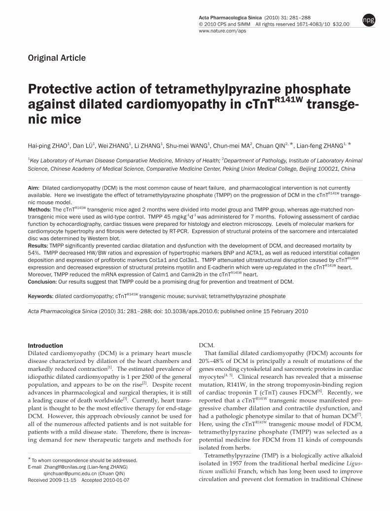
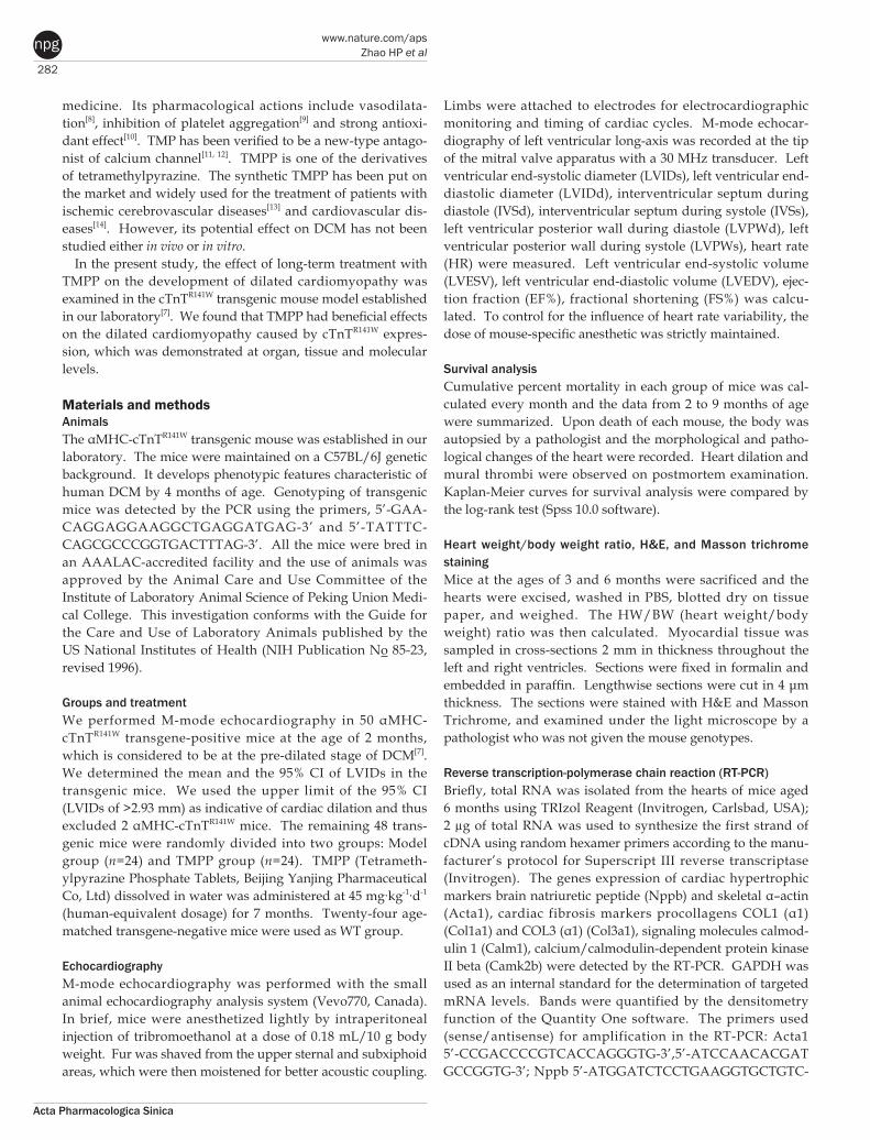
TMPP reduced cardiac hypertrophyDCM hallmark is the combination of dystrophic and hyper-trophic alterations of cardiomyocytes. There was an obvious increase in the size of heart in DCM (Fig ure 2A), and the HW/BW ratio of model group was increased by 43% (P<0.001) at 3 months and by 39% (P<0.001) at 6 months, and this change in heart weight due to DCM was reversed by treatment with TMPP by 55% (P<0.05) at 3 months and by 61% (P<0.05) at 6 months (Figure 2B). The expression levels of molecular mark-ers of cardiac hypertrophy ACTA1 and NPPB were unregu-lated remarkably in the cTnTR141W mice aged 6 months, com-pared with WT mice, and decreased by 63% and 37%, respec-tively after TMPP treatment (Figure 2C, 2D).
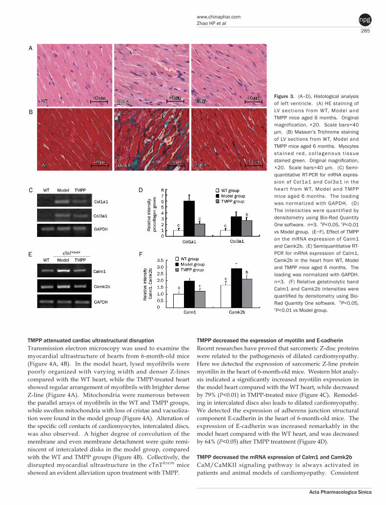
TMPP decreased myofibrillar disarray and interstitial fibrosisLeft ventricular systolic and diastolic dysfunction is related to the extent of myocardial disarray and interstitial fibrosis. HE staining showed that myofibrillar disarray was observed in the model group, but these pathological changes were ameliorated by the treatment with TMPP (Figure 3A). Mas-
son staining showed that a large proportion of collagen was accumulated in interstitial space of the model heart, while the collagen deposition was decreased in TMPP-treated mice (Fig-ure 3B). Expression levels of procollagens Col1a1 and Col3a1 in the model mice were decreased by 78% (P<0.01) and by 27% (P<0.05), respectively, after TMPP treatment, further sub-stantiating the effect of TMPP in reducing collagen expression (Figure 3C, 3D).
Table 1. M-Mode echocardiographic analysis in 6-month-old mice. Values are expressed as the mean±SD. bP<0.05, cP<0.01 vs model group.
6 month (n=15) Parameters WT Model TMPP BW, g 31.80±1.03 28.56±3.11 29.58±3.69 LVIDs, mm 2.46±0.34b 4.08±0.31 3.41±0.38b
LVIDd, mm 3.85±0.26b 4.95±0.29 4.45±0.31b
LVESV, μL 21.97±5.89b 105.51±14.12 86.42±12.43b
LVEDV, μL 68.71±7.00c 144.36±12.29 121.67±18.76b
IVSs, mm 1.27±0.15b 1.03±0.13 1.16±0.14 IVSd, mm 0.80±0.09 0.82±0.09 0.78±0.08 LVPWs, mm 1.03±0.13b 0.71±0.11 0.85±0.09 LVPWd, mm 0.79±0.03 0.81±0.09 0.76±0.07 EF, % 66.30±7.18c 26.56±5.92 42.33±6.85b
FS, % 36.34±5.92c 17.69±3.20 25.84±3.98b
HR 455±28 470±21 465±37
LVIDs, left ventricular internal diameter at end-systole; LVIDd, left ventricular internal diameter at end-diastole; LVESV, left ventricular end-systolic volume; LVEDV, left ventricular end-diastolic volume; IVSs, interventricular septum during systole; IVSd, interventricular septum during diastole; LVPWs, posterior wall during systole; LVPWd, posterior wall during diastole; EF, ejection fraction; FS, fractional shortening; HR, heart rate.
Figure 2. Effect of TMPP on cardiac hypertrophy in the cTnTR141W mice with DCM. (A) Whole-heart from WT, Model and TMPP mice aged 6 months. (B) HW/BW ratio in WT, Model and TMPP mice aged 3 months and 6 months. HW/BW, Heart weight/Body weight. n=7. cP<0.01 vs Model group. (C) Semiquantitative RT-PCR for expression levels of hypertrophic markers including ACTA1 and NPPB. The loading was normalized with GAPDH. (D) The intensities were quantified by densitometry using Bio-Rad Quantity One software. n=3. cP<0.01 vs Model group.
285
www.chinaphar.comZhao HP et al
Acta Pharmacologica Sinica
npg
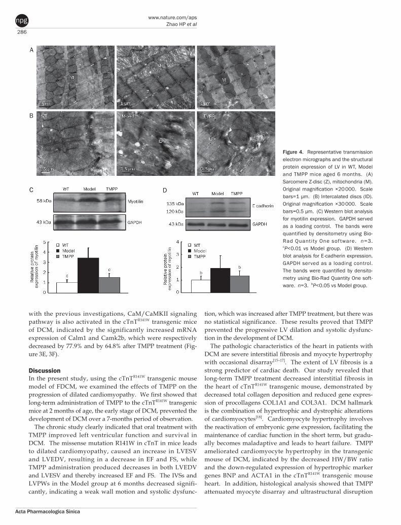
TMPP attenuated cardiac ultrastructural disruptionTransmission electron microscopy was used to examine the myocardial ultrastructure of hearts from 6-month-old mice (Figure 4A, 4B). In the model heart, lysed myofibrils were poorly organized with varying width and denser Z-lines compared with the WT heart, while the TMPP-treated heart showed regular arrangement of myofibrils with brighter dense Z-line (Figure 4A). Mitochondria were numerous between the parallel arrays of myofibrils in the WT and TMPP groups, while swollen mitochondria with loss of cristae and vacuoliza-tion were found in the model group (Figure 4A). Alteration of the specific cell contacts of cardiomyocytes, intercalated discs, was also observed. A higher degree of convolution of the membrane and even membrane detachment were quite remi-niscent of intercalated disks in the model group, compared with the WT and TMPP groups (Figure 4B). Collectively, the disrupted myocardial ultrastructure in the cTnTR141W mice showed an evident alleviation upon treatment with TMPP.
TMPP decreased the expression of myotilin and E-cadherinRecent researches have proved that sarcomeric Z-disc proteins were related to the pathogenesis of dilated cardiomyopathy. Here we detected the expression of sarcomeric Z-line protein myotilin in the heart of 6-month-old mice. Western blot analy-sis indicated a significantly increased myotilin expression in the model heart compared with the WT heart, while decreased by 79% (P<0.01) in TMPP-treated mice (Figure 4C). Remodel-ing in intercalated discs also leads to dilated cardiomyopathy. We detected the expression of adherens junction structural component E-cadherin in the heart of 6-month-old mice. The expression of E-cadherin was increased remarkably in the model heart compared with the WT heart, and was decreased by 64% (P<0.05) after TMPP treatment (Figure 4D).
TMPP decreased the mRNA expression of Calm1 and Camk2bCaM/CaMKII signaling pathway is always activated in patients and animal models of cardiomyopathy. Consistent
Figure 3. (A–D), Histological analysis of left ventricle. (A) HE staining of LV sect ions from W T, Model and TMPP mice aged 6 months. Original magnification, ×20. Scale bars=40 μm. (B) Masson’s Trichrome staining of LV sections from WT, Model and TMPP mice aged 6 months. Myocytes sta ined red, co l lagenous t issue stained green. Original magnification, ×20. Scale bars=40 μm. (C) Semi-quantitative RT-PCR for mRNA expres-sion of Col1a1 and Col3a1 in the hear t from WT, Model and TMPP mice aged 6 months. The loading was normalized with GAPDH. (D) The intensities were quantified by densitometry using Bio-Rad Quantity One software. n=3. bP<0.05, cP<0.01 vs Model group. (E−F), Effect of TMPP on the mRNA expression of Calm1 and Camk2b. (E) Semiquantitative RT-PCR for mRNA expression of Calm1, Camk2b in the heart from WT, Model and TMPP mice aged 6 months. The loading was normalized with GAPDH. n=3. (F) Relative gelatinolytic band Calm1 and Camk2b intensities were quantified by densitometry using Bio-Rad Quantity One software. bP<0.05, cP<0.01 vs Model group.
286
www.nature.com/apsZhao HP et al
Acta Pharmacologica Sinica
npg
with the previous investigations, CaM/CaMKII signaling pathway is also activated in the cTnTR141W transgenic mice of DCM, indicated by the significantly increased mRNA expression of Calm1 and Camk2b, which were respectively decreased by 77.9% and by 64.8% after TMPP treatment (Fig-ure 3E, 3F).
DiscussionIn the present study, using the cTnTR141W transgenic mouse model of FDCM, we examined the effects of TMPP on the progression of dilated cardiomyopathy. We first showed that long-term administration of TMPP to the cTnTR141W transgenic mice at 2 months of age, the early stage of DCM, prevented the development of DCM over a 7-months period of observation.
The chronic study clearly indicated that oral treatment with TMPP improved left ventricular function and survival in DCM. The missense mutation R141W in cTnT in mice leads to dilated cardiomyopathy, caused an increase in LVESV and LVEDV, resulting in a decrease in EF and FS, while TMPP administration produced decreases in both LVEDV and LVESV and thereby increased EF and FS. The IVSs and LVPWs in the Model group at 6 months decreased signifi-cantly, indicating a weak wall motion and systolic dysfunc-
tion, which was increased after TMPP treatment, but there was no statistical significance. These results proved that TMPP prevented the progressive LV dilation and systolic dysfunc-tion in the development of DCM.
The pathologic characteristics of the heart in patients with DCM are severe interstitial fibrosis and myocyte hypertrophy with occasional disarray[15–17]. The extent of LV fibrosis is a strong predictor of cardiac death. Our study revealed that long-term TMPP treatment decreased interstitial fibrosis in the heart of cTnTR141W transgenic mouse, demonstrated by decreased total collagen deposition and reduced gene expres-sion of procollagens COL1A1 and COL3A1. DCM hallmark is the combination of hypertrophic and dystrophic alterations of cardiomyocytes[18]. Cardiomyocyte hypertrophy involves the reactivation of embryonic gene expression, facilitating the maintenance of cardiac function in the short term, but gradu-ally becomes maladaptive and leads to heart failure. TMPP ameliorated cardiomyocyte hypertrophy in the transgenic mouse of DCM, indicated by the decreased HW/BW ratio and the down-regulated expression of hypertrophic marker genes BNP and ACTA1 in the cTnTR141W transgenic mouse heart. In addition, histological analysis showed that TMPP attenuated myocyte disarray and ultrastructural disruption
Figure 4. Representative transmission electron micrographs and the structural protein expression of LV in WT, Model and TMPP mice aged 6 months. (A) Sarcomere Z-disc (Z), mitochondria (M). Original magnification ×20 000. Scale bars=1 μm. (B) Intercalated discs (ID). Original magnification ×30 000. Scale bars=0.5 μm. (C) Western blot analysis for myotilin expression. GAPDH served as a loading control. The bands were quantified by densitometry using Bio-Rad Quantity One sof tware. n=3. cP<0.01 vs Model group. (D) Western blot analysis for E-cadherin expression. GAPDH served as a loading control. The bands were quantified by densito-metry using Bio-Rad Quantity One soft-ware. n=3. bP<0.05 vs Model group.
287
www.chinaphar.comZhao HP et al
Acta Pharmacologica Sinica
npg
involving partially lysed myofibrils and swollen mitochondria in the model heart. The above results suggest that TMPP has beneficial effect on cardiac remodeling in cTnTR141W transgenic mouse, and all of these effects may contribute to improvement of cardiac function of the DCM model.
Sarcomeric Z-disc was related to the pathogenesis of dilated cardiomyopathy, supported by several transgenic animal models and the identification of mutations in various Z-disc proteins in patients[19–21]. We found the Z-lines of the sarcom-ere became denser in the cTnTR141W mouse, just like the change appeared in N-cadherin CKO mouse which also exhibit a DCM phenotype[22]. Then we detected the expression of Z-line protein myotilin, a candidate gene for DCM[23, 24], and showed that its expression was significantly increased in our DCM model. Overexpression of wild-type myotilin in an limb-girdle muscular dystrophy type 1A (LGMD1A) mouse[25, 26] showed more severe muscle degeneration compared with single-transgenic mutant mice[27]. Conversely, loss of myo-tilin does not cause alterations in the heart of newborn or adult myo−/− mice[28]. These findings suggested that strategies aimed at lowering total myotilin levels may be an effective therapeutic approach to the treatment of cardiomyopathy such as LGMD1A and FDCM carrying the cTnT (R141W) mutation. It is likely that the prevention of pathological development of DCM by TMPP was partially associated with depressing myo-tilin expression in cTnTR141W mice.
Remodeling in intercalated discs also leads to dilated cardio-myopathy[29]. Just like the pathological changes of intercalated discs found in the other two DCM models, MLP–/– (muscle LIM protein KO) and TOT (tropomodulin overexpression) mouse[30], a higher degree of convolution of the membrane and even membrane detachment were quite reminiscent of intercalated discs in the cTnTR141W mouse. Components of the adherens junctions at the intercalated discs in MLP–/– and TOT mice are up-regulated. Cadherins are components of adhe-rens junctions, N-cadherin is highly expressed by the develop-ing and mature myocardium, while E-cadherin is distributed mainly in epithelia. A recent study showed that cardiac-specific expression of E-cadherin in adult transgenic mice led to DCM[31], indicating that ectopic E-cadherin expression in the heart is not compatible with normal cardiac function. We detected significantly increased E-cadherin expression in the cTnTR141W mouse heart, which was reduced by TMPP treat-ment. Thus, the alleviation of cardiac dilation and dysfunction in the TMPP-treated mice was in part related to downregula-tion of E-cadherin expression in the heart.
Ca2+/CaM/CaMKII pathway is always activated in patients and animal models of cardiomyopathy[32]. Mice with cardiac overexpression of CaMKIIδc display DCM phenotype[33], and CaMKII inhibition effectively protects against LV dilation and dysfunction caused by myocardial infarction and cat-echolamine toxicity[34]. Consistent with the previous investi-gations, the upregulated expression of calcium/calmodulin-regulated transcripts (Calm1 and Camk2b) is also observed in the cTnTR141W transgenic model. TMPP treatment inhibited the expression of Calm1 and Camk2b and may result in attenua-
tion of cardiac remodeling and dysfunction in DCM.In summary, TMPP significantly prevented the development
of DCM due to R141W mutation in cTnT. The clinical signifi-cance of this finding is underscored by the lack of an effective pharmacological intervention to prevent FDCM in human patients. Present investigation raises the possibility for an early intervention, using a well-established pharmacological agent, in DCM mutation carriers to prevent the development of cardiac phenotype. Our results indicated the potential util-ity of TMPP in preventing FDCM, and it would be interesting to further test TMPP in other appropriate FDCM models.
AcknowledgmentsThe authors thank Dr Harold JAMES for his patiently reading and correcting the English writing of this paper. The present work was supported in part by the National Commonwealth Institute Foundation (No DWS200704) and the Ministry of Health (No 200802036).
Author contributionLian-feng ZHANG designed research; Hai-ping ZHAO per-formed research;, Dan LÜ analyzed data; Wei ZHANG, Li ZHANG and Shu-mei WANG breeded mice; Chuan QIN and Chun-mei MA contributed pathological analysis; Lian-feng ZHANG and Hai-ping ZHAO wrote the paper.
References1 Richardson P, Mckenna W, Bristow M, Maisch B, Mautner B, O’Connell
J, et al. Report of the 1995 world health organization/international society and federation of cardiology task force on the definition and classification of cardiomyopathies. Circulation 1996; 93: 841−2.
2 Maron BJ, Towbin JA, Thiene G, Antzelevitch C, Corrado D, Arnett D, et al. Contemporary definitions and classification of the cardiomyopa-thies. Circulation 2006; 113: 1807−16.
3 Jessup M, Brozena S. Heart failure. N Engl J Med 2003; 348: 2007−18.
4 Kamisago M, Sharma SD, Depalma SR, Solomon S, Sharma P, Mc-Donough B, et al. Mutations in sarcomere protein genes as a cause of dilated cardiomyopathy. N Engl J Med 2000; 343: 1688−96.
5 Towbin JA. The role of cytoskeletal proteins in cardiomyopathies. Curr Opin Cell Biol 1998; 10: 131−9.
6 Li D, Czernuszewicz GZ, Gonzalez O, Tapscott T, Karibe A, Durand JB, et al. Novel cardiac troponin T mutation as a cause of familial dilated cardiomyopathy. Circulation 2001; 104: 2188−93.
7 Juan F, Wei D, Xiongzhi Q, Ran D, Chunmei M, Lan H, et al. The changes of the cardiac structure and function in cTnTR141W transgenic mice. Int J Cardiol 2008; 128: 83−90.
8 Kwan CY, Daniel CE, Chen MC. Inhibition of vasoconstriction by te-tramethylpyrazine: Does it act by blocking the voltage-dependent Ca2+ channel? J Cardiovasc Pharmacol 1990; 15: 157−62.
9 Sheu JR, Kan YC, Hung WC, Lin CH, Yen MH. The antiplatelet activity of tetramethylpyrazine is mediated through activation of NO synthase. Life Sci 2000; 67: 937−47.
10 Zhang Z, Wei T, Hou J, Li G, Yu S, Xin W. Tetramethylpyrazine scaveng-es superoxide anion and decreases nitric oxide production in human polymorphonuclear leukocytes. Life Sci 2003; 72: 2465−72.
11 Pang PK, Shan JJ, Chiu KW. Tetramethylpyrazine, a calcium antago-nist. Planta Med 1996; 62: 431–5.
288
www.nature.com/apsZhao HP et al
Acta Pharmacologica Sinica
npg
12 Hintz KK, Ren J. Tetramethylpyrazine elicits disparate responses in cardiac contraction and intracellular Ca2+ transients in isolated adult rat ventricular myocytes. Vasc Pharmacol 2003; 40: 213–7.
13 Chen KJ, Chen K. Ischemic stroke treated with Ligusticum chuanx-iong. Chin Med J 1992; 105: 870−3.
14 Sutter MC, Wang YX. Recent cardiovascular drugs from Chinese me-dicinal plants. Cardiovasc Res 1993; 27: 1891−901.
15 Aoki J, Ikari Y, Nakajima H, Mori M, Sugimoto T, Hatori M, et al. Clini-cal and pathologic characteristics of dilated cardiomyopathy in hemo-dialysis patients. Kidney International 2005; 67: 333–40.
16 Beltrami CA, Finato N, Rocco M, Feruglio GA, Puricelli C, Cigola E, et al. The cellular basis of dilated cardiomyopathy in humans. J Mol Cell Cardiol 1995; 27: 291−305.
17 Kajstura J, Zhang X, Liu Y, Szoke E, Cheng W, Olivetti G, et al. The cellular basis of pacing-induced dilated cardiomyopathy. Circulation 1995; 92: 2306−17.
18 Branishte TA, Dudnakova TV, Dergilev KV, Severin VV, Naumov VG, Shirinskiĭ VP, et al. Expression of contractile and cytoskeletal proteins in myocardium of patients with dilated cardiomyopathy. Kardiologiia 2004; 44: 31−6.
19 Arber S, Hunter JJ, Ross J Jr, Hongo M, Sansig G, Borg J, et al. MLP-deficient mice exhibit a disruption of cardiac cytoarchitectural orga-nization, dilated cardiomyopathy, and heart failure. Cell 1997; 88: 393−403.
20 Hayashi T, Arimura T, Itoh-Satoh M, Ueda K, Hohda S, Inagaki N, et al. Tcap gene mutations in hypertrophic cardiomyopathy and dilated cardiomyopathy. J Am Coll Cardiol 2004; 44: 2192–201.
21 Duboscq-Bidot L, Xu P, Charron P, Neyroud N, Dilanian G, Millaire A, et al. Mutations in the Z-band protein myopalladin gene and idiopathic dilated cardiomyopathy. Cardiovasc Res 2008; 77: 118−25.
22 Kostetskii I, Li J, Xiong Y, Zhou R, Ferrari VA, Patel VV, et al. Induced deletion of the N-cadherin gene in the heart leads to dissolution of the intercalated disc structure. Circ Res 2005; 96: 346−54.
23 Salmikangas P, van der Ven PF, Lalowski M, Taivainen A, Zhao F, Suila H, et al. Myotilin, the limb-girdle muscular dystrophy 1A (LGMD1A) protein, cross-links actin filaments and controls sarcomere assembly. Hum Mol Genet 2003; 12: 189–203.
24 Zeller R, Ivandic BT, Ehlermann P, Mucke O, Zugck C, Remppis A, et al. Large-scale mutation screening in patients with dilated or hypertro-phic cardiomyopathy: a pilot study using DGGE. J Mol Med 2006; 84: 682−91.
25 Hauser MA, Horrigan SK, Salmikangas P, Torian UM, Viles KD, Dancel R, et al. Myotilin is mutated in limb girdle muscular dystrophy 1A. Hum Mol Genet 2000; 9: 2141–7.
26 Hauser MA, Conde CB, Kowaljow V, Zeppa G, Taratuto AL, Torian UM, et al. Myotilin mutation found in second pedigree with LGMD1A. Am J Hum Genet 2002; 71: 1428–82.
27 Garvey SM, Liu Y, Miller SE, Hauser MA. Myotilin overexpression en-hances myopathology in the LGMD1A mouse model. Muscle Nerve 2008; 37: 663−7.
28 Moza M, Mologni L, Trokovic R, Faulkner G, Partanen J, Carpén O. Targeted deletion of the muscular dystrophy gene myotilin does not perturb muscle structure or function in mice. Mol Cell Biol 2007; 27: 244–52.
29 Perriard JC, Hirschy A, Ehler E. Dilated cardiomyopathy: a disease of the intercalated disc? Trends Cardiovasc Med 2003; 13: 30–8.
30 Ehler E, Horowits R, Zuppinger C, Price RL, Perriard E, Leu M, et al. Alterations at the intercalated disk associated with the absence of muscle LIM protein. J Cell Biol 2001; 153: 763−72.
31 Ferreira-Cornwell MC, Luo Y, Narula N, Lenox JM, Lieberman M, Ra-dice GL. Remodeling the intercalated disc leads to cardiomyopathy in mice misexpressing cadherins in the heart. J Cell Sci 2002; 115: 1623–34.
32 Zhang T, Brown JH. Role of Ca2+/calmodulin-dependent protein kinase II in cardiac hypertrophy and heart failure. Cardiovasc Res 2004; 63: 476–86.
33 Maier LS, Zhang T, Chen L, DeSantiago J, Brown JH, Bers DM. Trans-genic CaMKIIδC overexpression uniquely alters cardiac myocyte Ca2+ handling: reduced SR Ca2+ load and activated SR Ca2+ release. Circ Res 2003; 92: 904−11.
34 Zhang R, Khoo MS, Wu Y, Yang Y, Grueter CE, Ni G, et al. Calmodulin kinase II inhibition protects against structural heart disease. Nat Med 2005; 11: 409–17.
Related Documents