Protection, Support, and Movement

Protection, Support, and Movement. Integumentary Function A. outer covering of body B. protects against injury, dehydration, UV radiation and some pathogens.
Jan 02, 2016
Welcome message from author
This document is posted to help you gain knowledge. Please leave a comment to let me know what you think about it! Share it to your friends and learn new things together.
Transcript

Protection, Support, and Movement
Integumentary• Function
A. outer covering of body
B. protects against injury, dehydration, UV radiation and some pathogens
C. helps regulate body temperature
D. excretes certain wastes
E. receives external stimuli
F. Produces vitamin D

Evolutionary trends in integument with move to land
A. keratinization of outer layer for protection-keritinocytes –produce water resistant protein keratin -melanocytes – produce pigment, melanin, as a barrier to UV radiation
B. recession of glands with ducts to surface
Human Skin
i. largest organ of your bodyii. see figure 37.3, p646iii. stretches, conserves water, fixes small cuts and bruises, helps regulate body temp and protects
against pathogens

Vertebrate Skin
• Two layers
– Upper epidermis
– Lower dermis
• Lies atop a layer
of hypodermis
Figure 37.3Page 646

LayersA. epidermis – stratified epithelial cells with an abundance of cell junctions with no extracellular matrix
-continual mitosis pushes cells from deep layer to surface-outer layers are dead flattened cell that continuously
flake awayB. dermis – dense connective tissue
-stretch resistant elastin fibers-supportive collagen fibers-blood and lymph vessels, and sensory receptors thread
throughout itC. hypodermis – below the skin
-anchors the skin-loose connective tissue and adipose tissue to insulate
and cushion the skin
Skin coloration – combination of several pigments
i. melanocytes – brownish blackii. hemoglobin – pink, found in red blood cells
(sometimes shows through the skin)iii. carotene – yellow orange

Exocrine Glands – their ducts extend through the skini. types
a. mammary glands – produce milkb. sweat glands
-sweat = 99% water with dissolved salts, ammonia and vitamin C -controlled by sympathetic nerves -located on palms, soles, forehead and armpits -decrease body temperaturec. oil glands
-not on palms or soles -called sebaceous glands -lubricates and softens hair and skin -secretions kill many surface bacteria -clogged and infected = acne

HairA. flexible, mostly keratinized (made of keratin - protein) cellsB. cells divide near root, push upwards, flatten
and dieC. influenced by genes, nutrition and hormones
(growth and density)D. root embedded in skin, shaft above its surface
Nails, Claws, Scales, Horns, Hooves and BeaksA. made of keratin
-a fibrous protein

Langerhans Cells
• White blood cells that arise in bone
marrow, migrate to epidermis
• Engulf pathogens and alert immune
system
• UV radiation can damage these cells
and weaken body’s first line of defense

Granstein Cells
• Also occur in epidermis
• Interact with cells that carry out immune response
• Issue suppressor signals that keep immune response under control
• Less vulnerable to UV damage than Langerhans cells
Skeletal System
• Function – human bone function table on page 652
A. Protects and supports the body and organsB. interacts with skeletal muscles for movementC. Produces red blood cells, white blood cells, and plateletsD. provides muscle attachment sitesE. stores minerals (calcium and phosphorus)

• muscles require the presence of some structural element to cause movement
• 3 types of animal skeletonsi. hydrostatic – muscles work against internal body fluid and redistribute it: soft-bodied invert’s such as annelids and sea anemonesii. exoskeleton – rigid, external, receives the applied muscle contraction: arthropodsiii. endoskeleton – internal, receives the applied muscle contraction: vertebrates

Human Skeleton
SKULLcranial bonesfacial bones
sternumRIB CAGE
ribs
VERTEBRAL COLUMN vertebrae
intervertebral disks
PECTORAL GIRDLESAND UPPER EXTREMITIES
clavicle
scapula
humerusradius
ulna
carpalsmetacarpals
phalanges
pelvic girdlefemur
patella
tibiafibula
tarsals
metatarsals
phalanges
PELVIC GIRDLE ANDLOWER EXTREMITIES

• organs consisting of connective tissue and epitheliai. bone tissue consists of mature bone cells
(osteocytes) and collagen fibers in a calcium hardened substance• Long bone structure
i. compact bone – resists mechanical shockii. Haversian system – within compact bone, thin
cylindrical dense layers around canals for blood vessels and nerves
iii. spongy bone – imparts strength but doesn’t weigh much
iv. red marrow –blood cell formation, in some bones
v. yellow marrow – fatty region, converts to red marrow in times of severe blood loss

Long Bone Structure
• Compact bone
• Spongy bone
• Central cavity contains yellow marrow compact bone tissue
spongy bonetissue
nutrient canal
contains yellow marrow
Fig. 37.12 (1)Page 652

Compact Bone Structure
• Mature compact bone consists of many cylindrical Haversian systems
spongy bone tissue
compact bone tissue
outer layer of dense connective tissue
blood vessel
Haversian system
Fig. 37.12 (2)Page 652
Bone Marrow
• Yellow marrow – Fills the cavities of adult long bones – Is largely fat
• Red marrow– Occurs in spongy bone of some bones– Produces blood cells

Joints• Areas of contact or near contact between bones• Fibrous joints
– Short connecting fibers join bones– Ex. newborn skull
• Synovial joints– Move freely; ligaments connect bones– pivot, saddle, hinge, ball and socket– See packet
• Cartilaginous joints– Straps of cartilage allow slight movement– Ex. Breastbone, ribs, vertebrae

Bone formationi. bone forms around cartilage in embryos
a. osteoblasts (bone forming cells) secrete organic substance that becomes mineralized forming osteocytes and the cartilage then breaks down leaving the marrow cavity
ii. bone remodelinga. mineral ions and osteocytes are constantly being removed and replacedb. this adjusts bone strength and helps maintain blood levels of calcium and phosphorusc. osteoblasts deposit new bone, osteoclasts digest (chemically breakdown) bone with enzymesd. free ions can then enter the interstitial fluid and get absorbed by the bloodstream

Bone, Blood, and Calciumi. bone and teeth store all but 1% of the bodies
calciumii. negative feedback of calcitonin and PTH help
regulate blood calcium levelsiii. calcitonin suppresses osteoclast activity (lowers blood calcium levels)iv. PTH enhances osteoclast activity (increases blood calcium levels)

• Osteoporosis is a decrease in bone density– May occur when the action of osteoclasts
outpaces that of osteoblasts
– May also occur as a result of inability to absorb calcium
• Arthritis– Inflammation of joints
Common Disorders

Muscular System
I. Functiona. Movement
i. Limbs and trunk ii. Substances through the body
- blood and foodb. maintains posturec. structure and supportd. generates heat

I. Structure a. 3 types of muscle – see packet picture i. skeletal –we will concentrate on this type
- functional partners of bone- striated (striped)- voluntary- opposing groups (antagonistic,
against) or in pairs (helping) - muscle cells contract or lengthen- humans have over 600, we will
learn 10 (see packet diagram) ii. Smooth – internal organs iii. Cardiac - heart
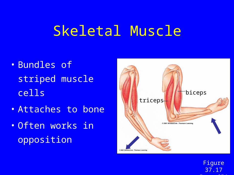
Skeletal Muscle
• Bundles of striped
muscle cells
• Attaches to bone
• Often works in
opposition
bicepstriceps
Figure 37.17Page 654

Major Human Muscles
triceps brachii
pectoralis major
serratus anterior
external oblique
rectus abdominus
adductor longus
sartorius
quadriceps femoris
tibialis anterior
biceps brachii
deltoid
trapezius
latissimus dorsi
gluteus maximus
biceps femoris
gastrocnemius
Figure 37.18Page 655

Skeletal Muscle Structure
• A muscle is made
up of muscle cells
• A muscle fiber is a
single muscle cell
• Each fiber contains
many myofibrils
myofibril
Figure 37.19aPage 656

Muscle Structure
• A muscle fiber is a single, multinucleated muscle cell• A muscle could be made up of hundreds or even
thousands of muscle fibers– Muscle fibers make up most of a muscle but connective
tissue, blood vessels, and nerves are also present– Nerves stimulate the muscle– Arteries supply oxygen and nutrients while veins carry away
metabolic wastes– Connective tissue covers and supports each fiber as well as
the whole muscle

Muscle Fibers
• Fibers consist of bundles of threadlike structures called myofibrils – made up of two protein filaments – Myosin – thick filaments
– Actin – thin filaments
• Myosin and actin are arranged in overlapping patterns that give the appearance of light and dark bands (striations)
• Actin filaments are anchored at their midpoints to a structure called a Z-line
• The region from one z-line to another is called a sarcomere – functional unit of muscle contraction

Sarcomere
Z line Z line Z line
sarcomere sarcomeresarcomere sarcomere
A myofibril is made up of thick and thin filaments arranged in sarcomeres
Figure 37.19bPage 656

Muscle Microfilaments
Actin - Thin filaments• Like two strands of
pearls twisted together
• Pearls are actin
• Other proteins in grooves in filament
Myosin -Thick filaments
• Composed of myosin
• Each myosin molecule has tail and a double head
Figure 37.19cPage 656

a. each muscle cell has many myofibrils (looked striped when stained) containing Z bands and sarcomeres i. cardiac muscle also has these, that is why they are
called striated muscleb. many sarcomeres contract simultaneously causing the muscle to contract and move a bone i. remember, this is the main purpose of skeletal muscle
sliding filament model – see page 657i. actin fibers are attached to the Z –bandsii. myosin fibers run parallel but between actin fibersiii. myosin heads attach to nearby actin filaments creating a cross bridgeiv. myosin heads tilt toward sarcomeres center causing the actin filaments to slide with them and shorten the sarcomere myosin head releases moves back and regrips the actin filament to move again

Sliding-Filament Model
• Myosin heads attach to actin filaments
• Myosin heads tilt toward sarcomere center, pulling actin with them
Fig. 37.20c-gPage 657

Sliding-Filament Model
Sarcomere shortens because the actin filaments are pulled inward, toward the sarcomere center
Fig. 37.20a,bPage 657

Muscle Contraction
• Sliding filament theory– Myosin and Actin filaments interact to shorten the sarcomere
– Knob like projections on myosin form cross-bridges with actin filaments
– When the muscle is stimulated, the filaments move across each other

Contraction Cont.
• When the cross-bridge has moved as far as it can, it is released and the actin filament returns to its original position; the cross-bridge attaches at another point and the cycle is repeated
• The synchronized shortening of sarcomeres causes the entire fiber to contract and thus the muscle shortens
• ATP is required to attach and detach myosin heads from actin
• Without ATP, the filaments would not be able to contract or relax – rigor mortis

Control of Muscle Contraction
• Motor neurons connect to muscles at points called neuromuscular junctions
1. When an action potential reaches the axon terminal, the neurotransmitter acetylcholine diffuse across the synapse and initiate an impulse in the muscle cell
2. Impulse causes the release of calcium ions in the cell
3. Calcium ions affect proteins that regulate the interaction of Actin and Myosin filaments
4. An enzyme, acetylcholinesterase, destroys acetylcholine to stop the impulse

Nervous System Controls Contraction
• Signals from nervous system travel along spinal cord, down a motor neuron
• Endings of motor neuron synapse on a muscle cell at a neuromuscular junction

-Motor neurons stimulate or inhibit contractions with an action potential
-Muscle cells have a specialized structure called the sarcoplasmic reticulum that stores and releases calcium
-When an action potential reaches a muscle cell it travels down the membrane (sarcolemma) and into a T-tubule that carries the action potential inside. This triggers the release of calcium ions from the sarcoplasmic reticulum to the actin filaments


-The calcium clears the binding site for the cross bridge so sarcomere contraction can occur
i. 2 proteins are involved in this action - tropomyosin, blocks the binding site
- tropomyosin is bound to the troponin
- troponin binds the free calcium and changes its shape, this causes the tropomyosin to move - the binding site is therefore unblocked
ii. After the contraction, the calcium is transported back into the sarcoplasmic reticulum for storage

Contraction Requires Energy
• Muscle cells require huge amounts of
ATP energy to power contraction
• The cells have only a very small store of
ATP
• Three pathways supply ATP to power
muscle contraction

i. Phosphorylation
- quick reaction that transfers phosphate from creatine phosphate to ADP, creating ATP
- this gives the cell time to perform cellular respiration (aerobic) to further supply ATP
ii. Cellular respiration
- aerobic
- produces lots of ATP but takes time
- phosphorylation gives the cell time to do this
iii. Glycolysis
- anaerobic respiration
- may kick in if needed (when not enough oxygen for aerobic respiration)
- produces less ATP, but allows processes to continue
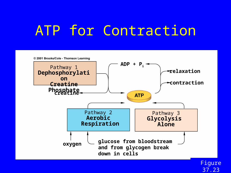
ATP for Contraction
Pathway 1DephosphorylationCreatine Phosphate
Pathway 2Aerobic
Respiration
Pathway 3Glycolysis
Alone
creatine
oxygen glucose from bloodstream and from glycogen break down in cells
ADP + Pi
relaxation
contraction
Figure 37.23Page 659

Motor Unit
• One neuron and all the muscle cells that form junctions with its endings
• When a motor neuron is stimulated, all the muscle cells it supplies are activated to contract simultaneously
• Each muscle consists of many motor units

- tetanus is a sustained contraction resulting from repeated stimulation
i. also the name of a disease that disrupts muscle relaxation
- muscle fatigue
i. decline in a muscles capacity to generate force
ii. decline in tension
iii. after a time of rest, fatigues muscles can contract again
a. the time depends on the amount of fatigue
iv. the molecular mechanism of fatigue is not known

-- cramps
i. involuntary, large, often painful contractions
ii. can persist for up to 15 minutes or more
iii. can be caused by fatigue
- muscular dystrophies
i. genetic disorders where muscles progressively weaken and degenerate
- muscles, exercise, and aging
i. with regular use, muscle cells increase in size and metabolic activity
a. become more resistant to fatigue
ii. muscle tension declines in adults (after age 30 or 40)
a. regular exercise may help
Related Documents