Eur. J. Biochem. 238, 728-736 (1996) 0 FEBS 1996 Proteases from Trypanosoma brucei brucei Purification, characterisation and interactions with host regulatory molecules Linda TROEBERG, Robert N. PIKE, Rory E. MORTY, Ronald K. BERRY, Theresa H. T. COETZER and John D. LONSDALE-ECCLES Department of Biochemistry, University of Natal, Scottsville, South Africa (Received 16 February 1996) - EJB 96 0225/4 African trypanosomes contain proteases that may be released into the bloodstream of their infected hosts. This paper describes a novel, combined isolation of a cysteine proteinase (called trypanopain-Tb) and a serine oligopeptidase (which we call oligopeptidase-Tb) from Trypanosoma brucei brucei, as well as a comparison of the activities of these two enzymes against several host regulatory molecules. The enzymes differed in various respects. Firstly, purified trypanopain-Tb was shown to readily cleave proteins such as gelatin maximally at acidic pH. In contrast, oligopeptidase-Tb, which is optimally active at alkaline pH, did not hydrolyse proteins larger than 4 kDa. However, it readily hydrolysed various polypeptides, including neurotensin and atrial natriuretic factor. The interaction of the two enzymes with mammalian protease inhibitors also differed. Cystatins and a,-macroglobulin effectively inhibited trypanopain-Tb, with the K, values for cystatin C and low-molecu- lar-mass kininogen (-1O-~” M) predicting that trypanopain-Tb is likely to be effectively controlled by these inhibitors if released into the host bloodstream. In contrast, oligopeptidase-Tb was not inhibited by serpins or a,-macroglobulin, suggesting that it may remain active if released into the host bloodstream. In support of these in vitm results, the blood of trypanosome-infected rats displayed no trypanopain-Tb- like activity, but exhibited high oligopeptidase-Tb-like activity. Thus, while trypanopain-Tb seems likely to be confined to an intracellular role within the parasite, oligopeptidase-Tb has the potential to remain active in the host bloodstream and so contribute directly to pathogenesis. Keywords: Trypanosoma brucei; cysteine proteinase; oligopeptidase; cystatin. African trypanosomes are protozoan parasites which cause the diseases nagana and sleeping sickness in cattle and humans, respectively. These diseases are characterised by intermittent fe- ver, progressive anaemia, general loss of condition and, ulti- mately, the death of the infected host. Nagana is still of great economic importance in Africa, where it prevents livestock farming in many areas, and certain parts of Africa are experienc- ing a large resurgence in human trypanosomiasis (Walgate, 1994). While the parasite’s major lysosomal cysteine proteinase (trypanopain) is considered a potentially important factor in the development of the disease (AuthiC et al., 1993; Russo et al., 1994), the role of a cytoplasmic serine oligopeptidase (which we call oligopeptidase-Tb, OP-Tb) has not yet been explored. Trypanopain is lysosomally located (Mbawa et al., 1991) and, as such, is likely to be centrally involved in intracellular digestive and catabolic proteolysis. Additionally, the enzyme is proposed to help the parasite escape opsonisation by degrading internalised antibody-variant surface glycoprotein complexes (Russo et al., 1994). Enzyme released into the host bloodstream Corre.spondence to T. H. T. Coetrer, Department of Biochemistry, Fiix: +27 331 260 5462. Phorre: +27 331 260 5467. AbDrevirrfiorzr. uz-M, cr,-inacroglobulin ; Boc, butoxycarbonyl ; E-64, ~-tr~ns-epoxysuccinyI-leiicylamido(4-~uanidino)butane; HINEtPhSOZF, 4-(2-aminoethyl)-benzenesulfonyl fluoride; iPr,P-F, di-isopropyl fluorophosphate: L-kininogen, low-molecular-mass kininogen; MeO- Suc, methoxysuccinyl; NHMec, arninomethyl coiirnarin; OP-Tb, trypa- nosomal oligopeptidase; PhMeSO,F, phenylmethylsulfoiiyl fluoride; rTM, rat trypanopain modulator; Suc, succinyl ; SBTI, soybean trypsin inhibitor : TosLyKH2C1, tosyllys ylchloromethane ; TosPheCHICI, tosyl- pheiiylalanylcliloroniethaiie; Z, benzyloxycarbonyl. University of Natal, Private Bag XOI, Scottsville, South Africa 3209 has also been proposed to contribute to pathogenesis more di- rectly by degrading various host proteins. Also, since trypanotol- erant cattle infected with Trypanosoma congolense produce anti- bodies to trypanopain-Tc, while susceptible cattle do not, im- mune targeting of the enzyme may help protect infected hosts (AuthiC et al., 1993). While the cDNA of trypanopain-Tb from Tryparzosoma brucei brucei has been sequenced (Mottram et al., 1989), no purification of the enzyme to electrophoretic homo- geneity, or characterisation of its basic enzymatic properties, has been reported. The interactions of trypanopain-Tb with mamma- lian cysteine proteinase inhibitors and other host regulatory mol- ecules also have not been reported before. While OP-Tb has previously been partially purified from 7: b. brucri (Huet et al., 1992; Kornblatt et al., 1992) and Trypuno- soma cruzi (Ashall, 1990), the role of this enzyme in pathogene- sis has not been investigated. Previous data indicates that the enzyme is a serine protease, but its sensitivity to reagents which modify cysteine residues suggests that reduction of a cysteine residue is required for full enzyme activity (Kornblatt et al., 1992). The enzyme also appears to be restricted in its action, cleaving only small peptide substrates. Both these facets of the enzyme’s activity require further investigation. Here we report the joint purification of trypanopain-Tb and OP-Tb from 7: b. brucei, the effects of various potential in vivo inhibitors on the enzymes and the action of the enzymes on po- tcntially relevant host pcptide and protein substrates. MATERIALS AND METHODS Materials. HiLoadTM Q-Sepharose and HiTrapTMBlue Se- pharose were from Pharmacia. DEAE-cellulose was from What-

Welcome message from author
This document is posted to help you gain knowledge. Please leave a comment to let me know what you think about it! Share it to your friends and learn new things together.
Transcript
Eur. J. Biochem. 238, 728-736 (1996) 0 FEBS 1996
Proteases from Trypanosoma brucei brucei Purification, characterisation and interactions with host regulatory molecules
Linda TROEBERG, Robert N. PIKE, Rory E. MORTY, Ronald K. BERRY, Theresa H. T. COETZER and John D. LONSDALE-ECCLES
Department of Biochemistry, University of Natal, Scottsville, South Africa
(Received 16 February 1996) - EJB 96 0225/4
African trypanosomes contain proteases that may be released into the bloodstream of their infected hosts. This paper describes a novel, combined isolation of a cysteine proteinase (called trypanopain-Tb) and a serine oligopeptidase (which we call oligopeptidase-Tb) from Trypanosoma brucei brucei, as well as a comparison of the activities of these two enzymes against several host regulatory molecules.
The enzymes differed in various respects. Firstly, purified trypanopain-Tb was shown to readily cleave proteins such as gelatin maximally at acidic pH. In contrast, oligopeptidase-Tb, which is optimally active at alkaline pH, did not hydrolyse proteins larger than 4 kDa. However, it readily hydrolysed various polypeptides, including neurotensin and atrial natriuretic factor.
The interaction of the two enzymes with mammalian protease inhibitors also differed. Cystatins and a,-macroglobulin effectively inhibited trypanopain-Tb, with the K, values for cystatin C and low-molecu- lar-mass kininogen (-1O-~” M) predicting that trypanopain-Tb is likely to be effectively controlled by these inhibitors if released into the host bloodstream. In contrast, oligopeptidase-Tb was not inhibited by serpins or a,-macroglobulin, suggesting that it may remain active if released into the host bloodstream. In support of these in v i t m results, the blood of trypanosome-infected rats displayed no trypanopain-Tb- like activity, but exhibited high oligopeptidase-Tb-like activity. Thus, while trypanopain-Tb seems likely to be confined to an intracellular role within the parasite, oligopeptidase-Tb has the potential to remain active in the host bloodstream and so contribute directly to pathogenesis.
Keywords: Trypanosoma brucei; cysteine proteinase; oligopeptidase; cystatin.
African trypanosomes are protozoan parasites which cause the diseases nagana and sleeping sickness in cattle and humans, respectively. These diseases are characterised by intermittent fe- ver, progressive anaemia, general loss of condition and, ulti- mately, the death of the infected host. Nagana is still of great economic importance in Africa, where it prevents livestock farming in many areas, and certain parts of Africa are experienc- ing a large resurgence in human trypanosomiasis (Walgate, 1994). While the parasite’s major lysosomal cysteine proteinase (trypanopain) is considered a potentially important factor in the development of the disease (AuthiC et al., 1993; Russo et al., 1994), the role of a cytoplasmic serine oligopeptidase (which we call oligopeptidase-Tb, OP-Tb) has not yet been explored.
Trypanopain is lysosomally located (Mbawa et al., 1991) and, as such, is likely to be centrally involved in intracellular digestive and catabolic proteolysis. Additionally, the enzyme is proposed to help the parasite escape opsonisation by degrading internalised antibody-variant surface glycoprotein complexes (Russo et al., 1994). Enzyme released into the host bloodstream
Corre.spondence to T. H. T. Coetrer, Department of Biochemistry,
Fiix: +27 331 260 5462. Phorre: +27 331 260 5467. AbDrevirrfiorzr. uz-M, cr,-inacroglobulin ; Boc, butoxycarbonyl ; E-64,
~-tr~ns-epoxysuccinyI-leiicylamido(4-~uanidino)butane; HINEtPhSOZF, 4-(2-aminoethyl)-benzenesulfonyl fluoride; iPr,P-F, di-isopropyl fluorophosphate: L-kininogen, low-molecular-mass kininogen; MeO- Suc, methoxysuccinyl; NHMec, arninomethyl coiirnarin; OP-Tb, trypa- nosomal oligopeptidase; PhMeSO,F, phenylmethylsulfoiiyl fluoride; rTM, rat trypanopain modulator; Suc, succinyl ; SBTI, soybean trypsin inhibitor : TosLyKH2C1, tosyllys ylchloromethane ; TosPheCHICI, tosyl- pheiiylalanylcliloroniethaiie; Z, benzyloxycarbonyl.
University of Natal, Private Bag XOI, Scottsville, South Africa 3209
has also been proposed to contribute to pathogenesis more di- rectly by degrading various host proteins. Also, since trypanotol- erant cattle infected with Trypanosoma congolense produce anti- bodies to trypanopain-Tc, while susceptible cattle do not, im- mune targeting of the enzyme may help protect infected hosts (AuthiC et al., 1993). While the cDNA of trypanopain-Tb from Tryparzosoma brucei brucei has been sequenced (Mottram et al., 1989), no purification of the enzyme to electrophoretic homo- geneity, or characterisation of its basic enzymatic properties, has been reported. The interactions of trypanopain-Tb with mamma- lian cysteine proteinase inhibitors and other host regulatory mol- ecules also have not been reported before.
While OP-Tb has previously been partially purified from 7: b. brucri (Huet et al., 1992; Kornblatt et al., 1992) and Trypuno- soma cruzi (Ashall, 1990), the role of this enzyme in pathogene- sis has not been investigated. Previous data indicates that the enzyme is a serine protease, but its sensitivity to reagents which modify cysteine residues suggests that reduction of a cysteine residue is required for full enzyme activity (Kornblatt et al., 1992). The enzyme also appears to be restricted in its action, cleaving only small peptide substrates. Both these facets of the enzyme’s activity require further investigation.
Here we report the joint purification of trypanopain-Tb and OP-Tb from 7: b. brucei, the effects of various potential in vivo inhibitors on the enzymes and the action of the enzymes on po- tcntially relevant host pcptide and protein substrates.
MATERIALS AND METHODS
Materials. HiLoadTM Q-Sepharose and HiTrapTM Blue Se- pharose were from Pharmacia. DEAE-cellulose was from What-
Troeberg et al. (ELM J . Biackern. 238) 729
man. 4-(2-Aminoethyl)-benzenesulfonyl fluoride (H,NEtPh- S02F), BSA and a,-macroglobulin (a,-M) were from Boehringer Mannheim. Chymostatin, leupeptin and antipain were from Cambridge Research Chemicals (UK) and di-isopropyl fluorophosphate (iPr,P-F) was from Fluka. [1-'4C]Acetic anhy- dride was from Amersham. All other inhibitors, benzyloxycar- bony1 phenylanlanyl-arginyl-aminomethyl coumarin (Z-Phe- Arg-NHMec), Z-Arg-Arg-NHMec, pepstatin-A-Sepharose, poly-(L-1ysine)-Sepharose, fibrinogen, rabbit IgG, PercollTM and Sephacryl S-100 HR were from Sigma. The C18 HPLC column was from Vydac. Sheep stefin B was purified as described by Pike et al. (1992). Human recombinant stefin A was from Cal- Biochem and human low-molecular-mass kininogen (L-kinino- gen) was purified from human plasma as described by Gounaris et al. (1984). Recombinant human L-kininogen domain 3 was a gift from Dr Ennes Auerswald (Ludwig-Maximilians-Universitat Miinchen) human cystatin C was a gift from Dr Magnus Abra- hamson (University of Lund, Malmo General Hospital) and ser- pins were a gift from Dr Jan Potempa (Jagiellonian University, Cracow).
Growth and purification of trypanosomes. 7: b. brucei (clone ILTat 1.1) was grown in rats and purified from the in- fected blood by a combination of PercollTM gradient centrifuga- tion (Grab and Bwayo, 1982) and anion-exchange chromatogra- phy on DEAE-cellulose (Lanham and Godfrey, 1970). Purified trypanosomes were stored at -70°C until used.
Enzyme and protein assays. Trypanopain-Tb activity against 5 pM Z-Phe-Arg-NHMec at 37°C was initially measured as de- scribed by Barrett and Kirschke (1981) using an assay buffer of 170 niM sodium acetate, 30 mM acetic acid, 2 mM Na,EDTA, 3 mM dithiothreitol, pH 5.5. Following enzyme characterisation, the assay buffer was amended to 200 mM sodium phosphate pH 7.0 containing 30 mM cysteine and 2 mM Na,EDTA. OP- Tb was similarly assayed against 5 pM Z-Arg-Arg-NHMec in 100 mM Tris/HCl, 10 mM dithiothreitol, pH 8.0 (Kornblatt et al., 1992).
Protein assays were conducted according to the methods of Bradford as modified by Read and Northcote (1981) and Smith et al. (1985).
Isolation of trypanopain-Tb and OP-Tb. Trypanosomes (-5.SX1OY parasites) were thawed and lysed by addition of 0.1 % (by vol.) Triton X-100 (final concentration). Three-phase partitioning (Pike and Dennison, 1989) was performed on the lysate, with all enzyme activity precipitating between 10-2.5 % (mass/vol. of the total volume) ammonium sulfate (results not shown). The pellet was redissolved in buffer A (20 mM sodium acetate, 1 mM Na,EDTA, pH 5.5) and loaded onto HiLoadrM Q- Sepharose (26x110 mm, flow rate 1 ml . min-') equilibrated in buffer A with 100 mM NaCl. After elution of the unbound mate- rial (which contained OP-Tb and was retained for OP-Tb purifi- cation), a linear gradient of 0.1 -1 M NaCl in buffer A was ap- plied over five column volumes. Fractions active against Z-Phe- Arg-NHMec, but inactive against Z-Arg-Arg-NHMec and there- fore identified as containing trypanopain-Tb, were pooled and dialysed against buffer A. Since preliminary studies had iden- tified pepstatin A as a weak inhibitor of trypanopain-Tb, this inhibitor was considered to be a potential affinity chromatogra- phy ligand. Active samples were therefore loaded onto pep- statin-A-Sepharose (13X 10 mm, flow rate 0.26 ml . min-') equilibrated in buffer A containing 500 mM NaCI. Following elution of the unbound material, trypanopain-Tb was eluted with 5 % (masdvol.) Brij-35 in buffer A with 500 mM NaCI. Frac- tions active against Z-Phe-Arg-NHMec were applied to Hi- TrapTM Blue Sepharose (9x23 mm, flow rate 1 ml . min-') equilibrated in 50 mM Tris/HCl pH 8.0 to remove suspected al- bumin contamination. Active fractions were concentrated and
stored at -20°C mixed with glycerol (1 :I) . The purity of the enzyme was analysed by tricinelSDSIPAGE (Shagger and von Jagow, 1987).
The unbound material from HiLoad'" Q-Sepharose was active against Z-Arg-Arg-NHMec and Z-Phe-Arg-NHMec, but insensitive to L-trans-epoxysuccinyl-leucylamido (4-guanidino) butane (E-64), indicating that it contained OP-Tb (Kornblatt et al., 1992). This fraction was dialysed against buffer B (50 mM Tris/HCI, 1 mM dithiothreitol, pH 8.0) and applied to p-aniino- benzamidine-Sepharose (15x50 mm, flow rate 0.3 ml . min--'). After the elution of unbound material, bound OP-Tb was eluted with a step gradient of 250 mM NaCl in buffer B. Following dialysis against buffer B, the sample was applied to HiLoadTM Q-Sepharose equilibrated in buffer B containing 100 mM NaCI, and a five-column-volume gradient of 0.1 -1 M NaCl applied. The active fractions were diluted four-fold in buffer B and ap- plied to poly-(L-1ysine)-Sepharose (55x15 mm, flow rate 1 ml . min-') equilibrated in buffer B containing 100 mM NaC1. A 10-column-volume gradient of 0.1-1.5 M NaCl in buffer B was applied to elute the bound material. Active fractions were con- centrated and applied in buffer B to a Sephacryl S-100 HR gel filtration column (15x850 mm, flow rate 10 ml . h-I). Following analysis of enzyme purity by tricine/SDS/PAGE, the purified enzyme was concentrated and stored at -20°C mixed with glycerol (1 : 1 ) .
Characterisation of enzymes. The activity of trypanopain- Tb and OP-Tb in the presence of various inhibitors was investi- gated by incubating the enzymes with each inhibitor for 15 min at 37 "C before assaying residual activity against Z-Phe-Arg- NHMec or Z-Arg-Arg-NHMec, respectively, as described above. OP-Tb experiments were conducted in the presence of either 10 mM dithiothreitol (indicated as + dithiothreitol in Table 2 ) or in residual dithiothreitol remaining from purification (=40 pM dithiothreitol, indicated as - dithiothreitol in Table 2). Since chloromethanes and alkylating agents are inactivated by reduc- ing agents, care was taken to minimise the relative amount of reducing agents used with these inhibitors. Concentrated en- zymes were thus activated (10 min at 37°C in 30 mM cysteine for trypanopain-Tb; 10 mM dithiothreitol for OP-Tb), before be- ing diluted to the required working concentration in assay buffer containing inhibitor (final reducing agent concentrations of 3 mM cysteine for trypanopain-Tb, 1 mM dithiothreitol for OP-Tb).
The effects of various reducing agents on trypanopain-Tb and OP-Tb activity were investigated by adding dithiothreitol, cysteine . HCI, reduced glutathione or 2-mercaptoethanol (1 - 100 mM for trypanopain-Tb and 1-25 mM for OP-Tb) to the relevant assay buffer. The pH profile of each enzyme against synthetic substrates was investigated by substituting assay buffer with constant-ionic-strength acetate/Mes/Tris buffers (1 00 mM acetate, 200 mM Tris, 100 mM Mes, 4 mM Na,EDTA) of pH 4.0-9.0 (Ellis and Morrison, 1982). Trypanopain-Tb hydrol- ysis of ['4C]gelatin at various pH values was assessed using ace- tate/Mes/Tris buffers as described above, with the [ ''C]gelatin prepared using [ lL1'CC]acetic anhydride as described by Cawston and Barrett (1979). The pH stability of the enzymes was deter- mined by incubating the enzymes i n acetate/Mes/Tris buffers (25 mM acetate, 50 mM Tris, 25 mM Mes, 1 mM Na,EDTA) for 1 h at 37 "C before addition of a pH-7.0 assay buffer and Z-Phe- Arg-NHMec for trypanopain-Tb, or pH-8.0 assay buffer and Z- Arg-Arg-NHMec for OP-Tb. The initial incubation was done either in the presence or absence of reducing agents (30 mM cysteine for trypanopain-Tb and 10 mM dithiothreitol for OP- Tb).
Following determination of the concentration of active trypa- nopain-Tb by titration with E-64 (Barrett and Kirschke, 1981),
730 Troeberg et al. ( E M J. Biochem. 238)
trypanopain-Tb (0.05 pmol) was incubated with readily hy- drolysed synthetic substrates (25 - 100 pM) for 10 min at 37 "C in continuous assays, or with poorly hydrolysed synthetic sub- strates for 4 h at 37°C in stopped time assays. K,,,, V,,, and k,,, were determined from the direct linear plot (Eisenthal and Cor- nish-Bowden, 1974) using the software package Hyper 1.01 (0 1992-1993, J. S. Easterby, UK).
Digestion of protein and peptide substrates by trypano- pain-Tb and OP-Tb. Fibrinogen, BSA and rabbit IgG were di- gested with trypanopain-Tb at 37°C over 1 h at various molar ratios of enzymeisubstrate in 100 mM Tris/HCI, 30 mM cyste- ine, pH 7.4. After the reactions were stopped by the addition of 1 mM E-64, samples were boiled in reducing treatment buffer and analysed by electrophoresis on 10% tricine gels (Shagger and von Jagow, 1987). Trypanopain-Tb digestion of serpins was analysed at 37°C at 1:lOO and 1 : l O molar ratios of enzyme/ inhibitor in 25 mM Tris, 10 mM cysteine, pH 7.4. Reactions were stopped by the addition of 20 pM E-64. To determine the extent of serpin degradation, remaining inhibition of trypsin or chymotrypsin was determined (Potempa et al., 1986) and the degradation products analysed by tricine/SDS/PAGE.
OP-Tb was incubated with oligopeptide substrates at various molar ratios of enzyme/substrate in 0.1 M Tris/HCl, 10 mM di- thiothreitol, pH 8.0 for 1 - 16 h at 37 "C, after which the reaction was stopped by acidification with an equal volume of 5% (by vol.) trifluoroacetic acid. The samples were placed on ice until analysed by HPLC on a C18 column (flow rate 1 ml . min-') using a gradient of 10-90% (by vol.) acetonitrile in water containing 0.1 % (by vol.) trifluoroacetic acid.
Association with cystatins and a,-M. The kinetics of trypa- nopain-Tb inhibition by various cystatins were determined as described by Salvesen and Nagase (1992) with data treatment as described by Henderson (1972). Trypanopain-Tb and OP-Tb association with a,-M was assessed by comparing the elution volumes from Sephacryl S-100 (for trypanopain-Tb) and Seph- acryl S-200 (for OP-Tb) (both 15x300 mni, flow rate 10 cm . h-', equilibrated with 50 mM TrisiHCI pH 8.0 containing 1 mM dithiothreitol for OP-Tb only) of each enzyme alone with that for each enzyme previously incubated with bovine a2-M (= 1 : 1 molar ratio, Mason, 1989).
Measurement of enzyme activity in the blood of infected rats. The blood of trypanosome-infected rats was harvested by cardiac puncture at peak parasitaemia, and centrifuged (3OOOXg, 25 min, 4 "C). The supernatant was microscopically confirmed to be free of both parasites and blood cells, and then assayed against Z-Phe-Arg-NHMec and Z-Arg-Arg-NHMec in the pres- ence of various inhibitors. Since OP-Tb, rat plasma kallikrein and trypanopain-Tb all hydrolyse Z-Phe-Arg-NHMec, soybean trypsin inhibitor (SBTI, 20 pM), H,NEtPhSO,F (1.85 mM) and E-64 (1.7 mM) were used to discriminate between the activities of the three enzymes. SBTI inhibits blood plasma kallikrein (Coleman and Bagdasarian, 1976) but not OP-Tb, while H,NEtPhSO,F inhibits both kallikrein and OP-Tb, so these in- hibitors were used to discriminate between the two activities. Hydrolysis of Z-Arg-Arg-NHMec in the presence of these inhib- itors was additionally investigated to confirm the presence of OP-Tb.
RESULTS
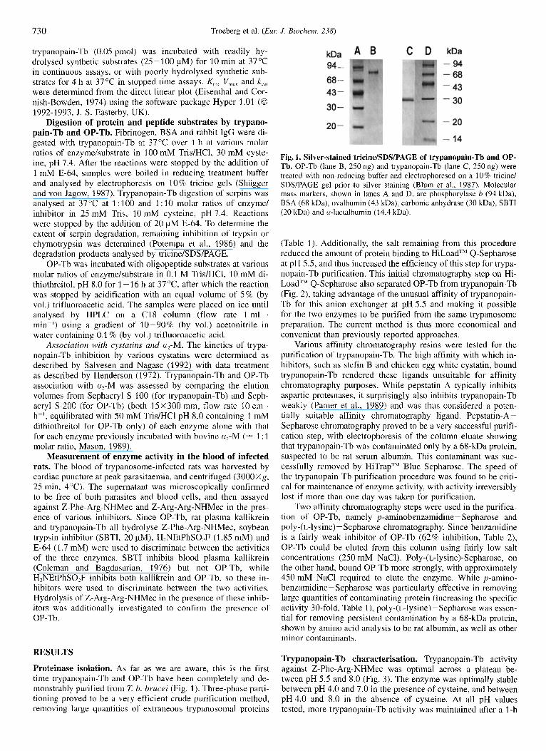
Proteinase isolation. As far as we are aware, this is the first time trypanopaiii-Tb and OP-Tb have been completely and de- monstrably purified from T b. brucei (Fig. 1 ) . Three-phase parti- tioning proved to be a very efficient crude purification method, removing large quantities of extraneous trypanosomal proteins
Fig. I . Silver-stained tricine/SDSIPAGE of trypanopain-Tb and OP- Tb. OP-Tb (lane B, 250 ng) and trypanopain-Tb (lane C, 250 ng) were treated with non-reducing buffer and electrophoresed on a 10% tricine/ SDS/PAGE gel prior to silver staining (Blum et al., 1987). Molecular mass markers, shown in lanes A and D, are phosphorylase b (94 kDa), BSA (68 kDa), ovalbumin (43 kDa), carbonic anhydrase (30 ma), SBTI (20 kDa) and a-lactalbumin (14.4 kDa).
(Table 1 ) . Additionally, the salt remaining from this procedure reduced the amount of protein binding to HiLoadTM Q-Sepharose at pH 5.5, and thus increased the efficiency of this step for trypa- nopain-Tb purification. This initial chromatography step on Hi- LoadTM Q-Sepharose also separated OP-Tb from trypanopain-Tb (Fig. 2), taking advantage of the unusual affinity of trypanopain- Tb for this anion exchanger at pH 5.5 and making it possible for the two enzymes to be purified from the same trypanosome preparation. The current method is thus more economical and convenient than previously reported approaches.
Various affinity chromatography resins were tested for the purification of trypanopain-Tb. The high affinity with which in- hibitors, such as stefin B and chicken egg white cystatin, bound trypanopain-Tb rendered these ligands unsuitable for affinity chromatography purposes. While pepstatin A typically inhibits aspartic proteinases, it surprisingly also inhibits trypanopain-Tb weakly (Pamer et al., 1989) and was thus considered a poten- tially suitable affinity chromatography ligand. Pepstatin-A- Sepharose chromatography proved to be a very successful purifi- cation step, with electrophoresis of the column eluate showing that trypanopain-Tb was contaminated only by a 68-kDa protein, suspected to be rat serum albumin. This contaminant was suc- cessfully removed by HiTrap'" Blue Sepharose. The speed of the trypanopain-Tb purification procedure was found to be criti- cal for maintenance of enzyme activity, with activity irreversibly lost if more than one day was taken for purification.
Two affinity chromatography steps were used in the purifica- tion of OP-Tb, namely p-aminobenzamidine- Sepharose and poly-(IJysine) - Sepharose chromatography. Since benzamidine is a fairly weak inhibitor of OP-Tb (62% inhibition, Table 2), OP-Tb could be eluted from this column using fairly low salt concentrations (250 mM NaC1). Poly-(L-1ysine)-Sepharose, on the other hand, bound OP-Tb more strongly, with approximately 450 mM NaCl required to elute the enzyme. While p-amino- benzamidine- Sepharose was particularly effective in removing large quantities of contaminating protein (increasing the specific activity 30-fold, Table 1), poly-(L-1ysine)-Sepharose was essen- tial for removing persistent contamination by a 68-kDa protein, shown by amino acid analysis to be rat albumin, as well as other minor contaminants.
Trypanopain-Tb characterisation. Trypanopain-Tb activity against Z-Phe-Arg-NHMec was optimal across a plateau be- tween pH 5.5 and 8.0 (Fig. 3). The enzyme was optimally stable between pH 4.0 and 7.0 in the presence of cysteine, and between pH4.0 and 8.0 in the absence of cysteine. At all pH values tested, more trypanopain-Tb activity was maintained after a 1-h

Troeberg et al. (EUK J. Biochern. 238) 731
Q . . . . . . /
Table 1. Purification table for the isolation of trypanopain-Tb and OP-Tb from T. b. brucei
Enzyme Fraction Total Total Specific Purification Yield protein activity activity
mg pmols . s- ' pmol S K ' . mg-' -fold %
Trypanopain-Tb Lysate 213 4562 22 1 100 Three-phase partitioning 9 1144 127 6 25 HiLoadIM Q-Sepharose 0.2 397 1985 90 9 Pepstatiii-A - Sepharose 0.04 252 6 300 286 5.5 HiTrapIM Blue Sepharose 0.01 245 24 500 1114 5.3 Lysate 213 3245 1.5 1 100
p-Aminobenzamidine - Sepharose 0.21 1 1429 6 773 452 44 HiLoadTM Q-Sepharose (pH 8) 0.134 1428 10 657 710 44
Three-phase partitioning 9 2044 227 15 63 HiLoadTM Q-Sepharose (pH 5.5) 8.6 1947 226 15 60
Poly(L-lysine) - Sepharose 0.104 94 1 9 048 603 29 Sephacryl S-100 0.068 909 13 368 89 1 28
OP-Tb
/'
I 0.2 I t '
0 1 , I I I I I , I 0.0 0 50 100 150 200 250 300
Time (mins)
Fig. 2. Elution profile of HiLoad'" Q-Sepharose showing separation of trypanopain-Tb and OP-Tb. HiLoadTM Q-Sepharose (26x1 10 mm, flow rate I ml . min- ') was equilibrated in loading buffer (100 mM NaCl in 20mM sodium acetate, 1 mM Na,EDTA, 1OOinM NaCI, 0.02% (masshol.) NaN,, pH 5.5). The 10-25% (NH,),SO, fraction of the three-phase partitioning was loaded onto the column and the bound ma- terial eluted with a five-column-volume gradient of 0.1-1 M NaCl. While OP-Tb fractions (unbound) were active against both Z-Phe-Arg- NHMec and Z-Arg-Arg-NHMec, trypanopain-Tb-containing fractions (bound) were active against only Z-Phe-Arg-NHMec.
incubation in the absence of cyateine than in its presence, indi- cating that the active enzyme undergoes some autocatalysis. Try- panopain-Tb was active against [14C]gelatin over a far narrower and more acidic pH range than against Z-Phe-Arg-NHMec, with hydrolysis being optimal at pH 5.7 (Fig. 3). While activity de- creased sharply on either side of this pH, trypanopain-Tb was still 44% active at pH 7.4, suggesting that it would be active under physiological pH conditions.
Trypanopain-Tb was only active in the presence of reducing agents, thus displaying typical cysteine proteinase latency. Cys- teine . HC1 was the most effective of the reducing agents tested, with an optimal final concentration of 30 mM. Dithiothreitol was maximally effective at 20 mM, with 2-mercaptoethanol reaching optimum efficiency at much higher concentrations (250 mM). Reduced glutathione was a comparatively poor activator. All subsequent trypanopain-Tb assays against Z-Phe-Arg-NHMec were thus performed at pH 7.0 (in the middle of the optimal
Table 2. Effects of various inhibitors on trypanopain-Tb activity against Z-Phe-Arg-NHMec and OP-Tb activity against Z-Arg-Arg- NHMec at 37°C. Enzymes were incubated with each inhibitor for 15 min at 37OC and then assayed as usual. OP-Tb experiments were conducted in the presence of either 10 mM dithiothreitol (indicated as + dithiothreirol) or in residual dithiothreitol remaining from purification (= 40 yM dithiothreitol, indicated as - dithiothreitol). Where different concentrations of inhibitor were used for OP-Tb and trypanopain-Tb, the concentrations for trypanopain-Tb are given first. IAA, iodoacetate; IAN, iodoacetamide; NEM, N-ethylmaleimide; nd, not determined.
Inhibitor Concen- Activity of tration
Trypano- OP-Tb activity pain-Tb
- dithio- + dithio- threitol threitol
mM % of control
E-64 0.00110.2 0 70 85 IAA 0.1110 0 8 59 NEM 10 0 21 16 IAN 10 0 90 67 Oxidised glutathione 10 100 182 1 00 H,NEtPhSO,F 0.5 100 0 3 iPr,P-F 10 100 14 9 PhMeS0,F 10 100 100 95 TosLysCH,Cl 1 0 5 5 TosPheCH2C1 1 0 78 89 Leu peptin 0.1 0 3 2 Antipain 0.1 0 8 8 Chyinostatin 0.1 1 35 37 Benzamidine 10 nd 33 38 Aprotinin 0.03 nd 56 36 SBTI 0.01 100 100 100 EDTA 10 100 154 103 Pepstatin A 0.1 78 100 100
pH range) using 200 mM sodium phosphate containing 30 mM cysteine and 2 mM Na,EDTA (final concentrations).
Typical cysteine proteinase inhibitors (including E-64 and iodoacetate) inhibited trypanopain-Tb completely, while typical serine and metalloproteinases inhibitors such as iPr,P-F and EDTA, respectively, had no effect on enzyme activity (Table 2). Leupeptin, antipain and chymostatin were all effective inhibitors of the enzyme (99- 100% inhibition). Interestingly, pepstatin A (22 % inhibition) also inhibited trypanopain-Tb.
732 Troeberg et al. (Eur: J. Biochem. 238)
t 100
80
60
40
20
0
A 1 i
A -
1
3 4 5 6 7 8 9 1 0
1
B
i
60
50
40
30
20
10
4 5 6 7 8 9 1 0
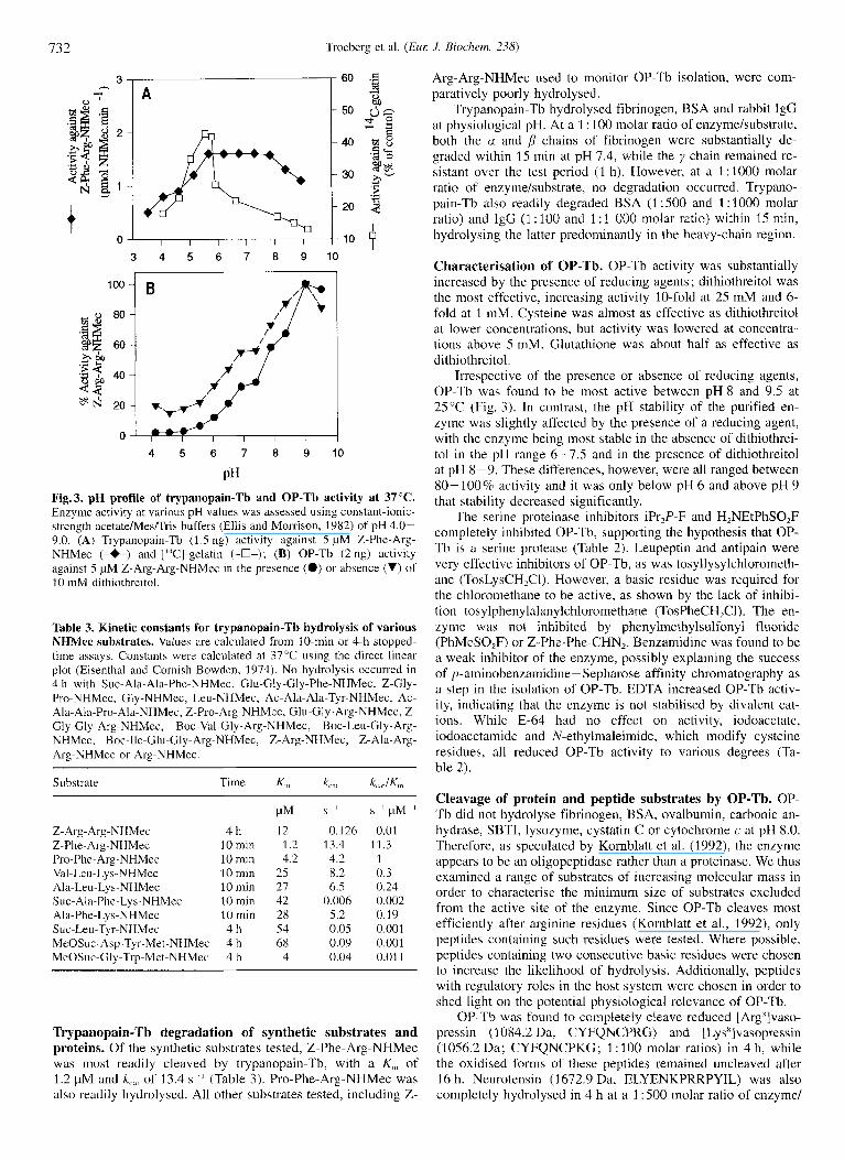
PH Fig.3. pH profile of trypanopain-Tb and OP-Tb activity at 37°C. Enzyme activity at various pH values was assessed using constant-ionic- strength acetate/Mes/Tris buffers (Ellis and Morrison, 1982) of pH 4.0- 9.0. (A) Trypanopain-Tb (1.5 ng) activity against 5 pM Z-Phe-Arg- NHMec (-*-) and ['T-gelatin (-O-); (B) OP-Tb (2 ng) activity against 5 pM Z-Arg-Arg-NHMec in the presence (0) or absence (V) of 10 inM dithiothreitol.
Table 3. Kinetic constants for trypanopain-Tb hydrolysis of various NHMec substrates. Values are calculated from 10-min or 4-h stopped- time assays. Constants were calculated at 37°C using the direct linear plot (Eisenthal and Cornish-Bowden, 1974). No hydrolysis occurred in 4 h with Sue-Ala-Ala-Phe-NHMec, Glu-Gly-Gly-Phe-NHMec, Z-Gly- Pro-NHMec, Gly-NHMec, Leu-NHMec, Ac-Ala-Ala-Tyr-NHMec, Ac- Ala-Ala-Pro-Ala-NHMec, Z-Pro-Arg-NHMec, Glu-Gly-Arg-NHMec, Z- Gly-Gly-Arg-NHMec, Boc-Val-Gly-Arg-NHMec, Boc-Leu-Gly-Arg- NHMec, Boc-lle-Glu-Gly-Arg-NHMec, Z-Arg-NHMec, Z-Ala-Arg- Arg-NHMec or Arg-NHMec.
Z- Arg- Arg-NHMec Z-Phe- Arg-NHMec Pro-Phe-Arg-NHMec Val-Leu-Lys-NHMec Ala-Leu-Lys-NHMec Sue- Ala-Phe-Lys-NHMec Ala-Phe-Lys-NHMec Sue-Leu-Tyr-NHMec MeOSuc- Asp-Tyr-Met-NHMec MeOSuc-Gly-Trp-Met-NHMec
4 h 30 min 10 min 10 min 10 min 10 rnin 10 min 4 h 4 h 4 h
PM 12
I .2 4.2
25 21 42 28 54 68 4
S - I
0.126 13.4 4.2 8.2 6.5
0.006 5.2 0.05 0.09 0.04
s- ' pM-'
0.01 11.3 1 0.3 0.24 0.002 0.19 0.001 0.001 0.01 1
Trypanopain-Tb degradation of synthetic substrates and proteins. Of the synthetic substrates tested, Z-Phe-Arg-NHMec was most readily cleaved by trypanopain-Tb, with a K,,, of 1.2 pM and k,,$, of 13.4 s - ' (Table 3). Pro-Phe-Arg-NHMec was also readily hydrolysed. All other substrates tested, including Z-
Arg-Arg-NHMec used to monitor OP-Tb isolation, were com- paratively poorly hydrolysed.
Trypanopain-Tb hydrolysed fibrinogen, BSA and rabbit IgG at physiological pH. At a 1 : 100 molar ratio of enzyme/substrate, both the a and /I chains of fibrinogen were substantially de- graded within 15 min at pH 7.4, while the y chain remained re- sistant over the test period (1 h). However, at a 1 : 1000 molar ratio of enzyme/substrate, no degradation occurred. Trypano- pain-Tb also readily degraded BSA (1:SOO and 1:lOOO molar ratio) and IgG (1 : 100 and 1 : 1 000 molar ratio) within 15 min, hydrolysing the latter predominantly in the heavy-chain region.
Characterisation of OP-Tb. OP-Tb activity was substantially increased by the presence of reducing agents; dithiothreitol was the most effective, increasing activity 10-fold at 25 mM and 6- fold at 1 mM. Cysteine was almost as effective as dithiothreitol at lower concentrations, but activity was lowered at concentra- tions above 5 mM. Glutathione was about half as effective as dithiothreitol.
Irrespective of the presence or absence of reducing agents, OP-Tb was found to be most active between pH 8 and 9.5 at 25°C (Fig. 3). In contrast, the pH stability of the purified en- zyme was slightly affected by the presence of a reducing agent, with the enzyme being most stable in the absence of dithiothrei- to1 in the pH range 6-7.5 and in the presence of dithiothreitol at pH 8-9. These differences, however, were all ranged between 80-100% activity and it was only below pH 6 and above pH 9 that stability decreased significantly.
The serine proteinase inhibitors iPrlP-F and H,NEtPhSO,F completely inhibited OP-Tb, supporting the hypothesis that OP- Tb is a serine protease (Table 2). Leupeptin and antipain were very effective inhibitors of OP-Tb, as was tosyllysylchlorometh- ane (TosLysCH,Cl). However, a basic residue was required for the chloromethane to be active, as shown by the lack of inhibi- tion tosylphenylalanylchloromethane (TosPheCH,CI). The en- zyme was not inhibited by phenylmethylsulfonyl fluoride (PhMeS0,F) or Z-Phe-Phe-CHN,. Benzamidine was found to be a weak inhibitor of the enzyme, possibly explaining the success of p-aminobenzamidine- Sepharose affinity chromatography as a step in the isolation of OP-Tb. EDTA increased OP-Tb activ- ity, indicating that the enzyme is not stabilised by divalent cat- ions. While E-64 had no effect on activity, iodoacetate, iodoacetamide and N-ethylmaleimide, which modify cysteine residues, all reduced OP-Tb activity to various degrees (Ta- ble 2).
Cleavage of protein and peptide substrates by OP-Tb. OP- Tb did not hydrolyse fibrinogen, BSA, ovalbumin, carbonic an- hydrase, SBTI, lysozyme, cystatin C or cytochrome c at pH 8.0. Therefore, as speculated by Kornblatt et al. (1992), the enzyme appears to be an oligopeptidase rather than a proteinase. We thus examined a range of substrates of increasing molecular mass in order to characterise the minimum size of substrates excluded from the active site of the enzyme. Since OP-Tb cleaves most efficiently after arginine residues (Kornblatt et al., 1992), only peptides containing such residues were tested. Where possible, peptides containing two consecutive basic residues were chosen to increase the likelihood of hydrolysis. Additionally, peptides with regulatory roles in the host system were chosen in order to shed light on the potential physiological relevance of OP-Tb.
OP-Tb was found to completely cleave reduced [Argx]vaso- pressin (1 084.2 Da, CYFQNCPRG) and [Lys']vasopressin (1056.2 Da; CYFQNCPKG; 1 : 100 molar ratios) in 4 h, while the oxidised forms of these peptides remained uncleaved after 16 h. Neurotensin (1672.9 Da, ELYENKPRRPYIL) was also completely hydrolysed in 4 h at a 1 : 500 molar ratio of enzyme/
Troeberg et al. ( E M J. Biochern. 238) 733
Table 4. Equilibrium constant for inhibition, association and dissoci- ation rate constants for cystatin inhibition of trypanopain-Tb (0.05 nM) at 30C. Note that the associations of cystatin C and L-kinino- gen domain 3 with trypanopain-Tb was too rapid for accurate measure- ment and the value given is an estimate.
Inhibitor K , k,,, Predicted ko,,
nM M- ' . s- I s '
Human stefin A 0.045 2.13 X 10' 9.6 X lo-" Sheep stefin B 0.004 6.9 X lo7 2 3 x 1 0 Human cystatin C 0.002 ~ 1 . 7 X I O x 1.7XlO-" Human L-kininogen 0.0035 2.5 X 10' 8.8X Human L-kininogen
domain 3 0.0044 =1.1 X lox 4.8XlO-'
peptide. The point of cleavage in this instance was determined to be after the second arginine residue in the sequence, reinforc- ing the specificity of OP-Tb for two consecutive basic residues in a given sequence. Atrial natriuretic factor (3080.5 Da, SLRRSSCFGGRMDRIGAQSGLGCNSFRY) was effectively cleaved at a I : 100 molar ratio, with the reduced form of the peptide undergoing more cleavage than the oxidised form. The importance of the state of substrate reduction suggests that sub- strate conformation is critical for OP-Tb activity. Peptides such as angiotensin I (1 296.5 Da, DRVYIHPFHL), substance P (1347.6 Da, RPKPQQFFGLM) and oxidised insulin B chain (3495.9 Da, FVNQHLCGSHLVEALYLVCGERGFFYTPKA ; 1 : 100 molar ratios) were not cleaved after 16 h, possibly due to the particular amino acid residues surrounding the basic residues present in these peptides (Kornblatt et al., 1992), although it is possible that a size restriction was the reason for non-cleavage of the insulin B chain. Pancreatic polypeptide (41 81.7 Da) and growth-hormone-releasing factor (5039.7 Da) were also not cleaved, possibly due to the sizes of these peptides. OP-Tb thus does not appear to cleave peptides larger than 4kDa. Further investigation of peptides between 3-4 kDa which contain suit- able consecutive basic residues is needed to clarify the exclusion limit more finely.
Interaction of trypanopain-Tb and OP-Tb with host protein- ase inhibitors. k,,, and predicted k,, values for trypanopain-Tb association with various cystatins were in the 107-10XM-' . s-l
and sS1 range, respectively, while K, values were in the loS" M range (Table 4). Cystatin C and L-kininogen were the most effective inhibitors. Interestingly, very little difference was observed between the inhibition by whole L-kininogen and by domain 3 of the molecule, suggesting that this domain is likely to be predominantly responsible for the inhibition observed with the whole molecule. Assuming a plasma concentration of 7.5 pM for L-kininogen and 80 nM for cystatin C (Abrahamson, 1993), trypanopain-Tb would be expected to associate very rapidly with these inhibitors: the half-life (Bieth, 1980) of the enzyme in the presence of L-kininogen can be calculated as 0.006 s and with cystatin C, 0.08 s. The inhibitor-enzyme com- plexes can also be predicted to dissociate slowly, with a half-life of 131 min for L-kininogen and 67 min for cystatin C. Trypano- pain-Tb is thus likely to be inhibited effectively by cystatin C and (especially) L-kininogen in vivo. Additionally, trypanopain- T b bound to a,-M (= 1 : 1 molar ratio) and is thus also likely to be effectively controlled by this inhibitor in vivo.
Trypanopain-Tb was found to cleave the serpin inhibitor antithrombin 111, but only at enzymdinhibitor ratios of 1 : 10, so it is unlikely that such hydrolysis would occur in vivo. Trypano-
Table 5. Effect of inhibitors on Z-Phe-Arg-NHMec and Z-Arg-Arg- NHMec activity in the blood of rats infected with Z b. brucei. The blood of infected rats was assayed against synthetic substrates in the absence or presence of various inhibitors for 30 min at 37°C. Values are in arbitrary units.
Substrate Rats Total activity with
no in- E-64 SBTI H2NEt hibitors PhSOzF
(1.67 mM) (20 pM) 11.85 mM)
2-Phe-Arg- infected 1521 1585 1573 297 177 193 193 NHMec uninfected 234
Z-Arg-Arg- infected 2244 2372 2284 97 NHMec uninfected 104 91 74 55
pain-Tb did not cleave other serpins, such as a,-proteinase inhib- itor, a,-antichymotrypsin and az-antiplasmin.
OP-Tb did not interact with any of the tested host system inhibitors, including serpins, cystatins and a,-M, presumably due to the limited interaction of the active site with higher-molecu- lar-mass proteins in general.
Measurement of enzyme activity in infected rat blood. High levels of activity against both Z-Phe-Arg-NHMec and Z-Arg- Arg-NHMec were detected in the blood of infected rats, while little activity was detectable in the blood of control, uninfected animals (Table 5) . This activity was insensitive to E-64 and SBTI, but entirely eliminated by H,NEtPhSO,F. OP-Tb is thus likely to be predominantly responsible for the hydrolysis ob- served, suggesting that OP-Tb may be released into the blood- stream of infected rats. Secondly, this study supports the conclu- sion arrived at from in vitro studies with the purified enzymes, namely that while trypanopain-Tb is likely to be effectively con- trolled in the bloodstream, OP-Tb seems to remain active.
DISCUSSION
Isolation of trypanopain-Tb and OP-Tb. A cysteine protein- ase has previously been isolated from 7: congolense (Mbawa et al., 1992) but we have now purified one from 7: b. brucei. OP- Tb from 7: b. brucei has been partially purified and characterised (Kornblatt et al., 1992), but here we report the purification of the enzyme to homogeneity as well as a more complete charac- terisation of certain aspects of its activity. The linked isolation of these proteinases as reported here has the added advantage of maximising the yield of both enzymes from a single parasite preparation.
Characterisation of trypanopain-Tb. Trypanopain-Tb was shown to be a fairly typical cysteine proteinase, similar to trypa- nopain-Tc from 7: congolense (Mbawa et al., 1992) and cruzi- pain from 7: cruzi (Cazzulo et al., 1989). Interestingly, the aspar- tic proteinase inhibitor pepstatin A inhibits trypanopain-Tb slightly (Table 2 ; Pamer et al., 1989). In contrast, pepstatin A inhibits trypanopain-Tc by only 9.7% (Mbawa et al., 1992) and does not affect cruzipain (Murta e t al., 1990; Cazzulo et al., 1989). Pepstatin A appears to be a competitive inhibitor of try- panopain-Tb (data not shown), and may act as a substrate ana- logue. This unusual inhibition was successfully exploited in the purification of trypanopain-Tb, using pepstatin-A- Sepharose.
Trypanopain-Tb was only active in the presence of reducing agents, and appeared to be quite similar to cathepsin L (Den- nison et al., 1992) in its requirement of fairly high concentra-

734 Troeberg et al. ( E m J. Biochern. 238)
tions of reducing agent for optimal activity. It was shown to have cathepsin-L-like specificity for synthetic substrates, hy- drolysing Z-Phe-Arg-NHMec and Pro-Phe-Arg-NHMec most rapidly of the substrates tested. Generally, only substrates with basic residues such as Arg or Lys in P, and hydrophobic residues such as Phe or Leu in P, were hydrolysed (Table 3). The pres- ence of an acidic residue in P, appeared to be unacceptable. Similar specificity has been reported for trypanopain-Tc (Mbawa et al., 1992), cruzipain (Cazzulo et al., 1990) and mammalian cathepsin L (Barrett and Kirschke, 1981 ; Dufour and Ribadeau- Dumas, 1988). Minimal hydrolysis of Z-Arg-Arg-NHMec was observed in the current study (4-h incubation required to demon- strate hydrolysis), allowing this substrate to be used for monitor- ing OP-Tb isolation. This contradicts a previous report by Pamer et al. (1 991 ) that trypanopain-Tb cleaves Z-Arg-Arg-NHMec readily, with a K,,, of 3.2 pM. although it must be noted that only partially purified trypanopain-Tb was used in their study. Trypanopain-Tc does not hydrolyse Z-Arg-Arg-NHMec (Mbawa et al., 1992), while cruzipain cleaves it poorly (KO, 41 pM; Lima et al., 1992).
Of the serpins tested, only antithrombin 111 was cleaved by trypanopain-Tb. Interestingly, cathepsin L, which has been found to be one of the closest mammalian analogues to trypano- pain-Tb (Mottram et al., 1989). cleaves a,-proteinase inhibitor very effectively (Johnson et al., 1986). Cathepsin L also cleaved antithrombin I11 and u2-antiplasmin very effectively at much lower enzymehbstrate ratios than did trypanopain-Tb (data not shown). This indicates that these two enzymes, while very simi- lar in substrate specificity, are not always comparable.
Effect of host proteinase inhibitors on trypanopain-Tb. It has been postulated that if released into the host bloodstream, trypa- nopain-Tb could contribute to pathogenesis by degrading vari- ous host proteins. For this to occur, the enzyme would have to remain active in the presence of host proteinase inhibitors such as cystatins, kininogens and a,-M. Trypanopain-Tb is, however, effectively inhibited by kininogens, various cystatins and a,-M in vitro (Table 4). The physiological concentrations of cystatin C and L-kininogen are more than 10 times Kl, so these inhibitors are likely to inhibit any trypanopain-Tb secreted or released by trypanosomes in vivo (Bieth, 1980). Interestingly, the most abun- dant cysteine proteinase inhibitors in blood are also the most effective in vitro inhibitors of the purified enzyme. Since cysta- tins are relatively conserved between species (Barrett et al., 1986) and the trypanosomal cysteine proteinases sequenced to date are highly similar to each other (Mottram et al., 1989; Pamer et al., 1990; Eakin et al., 1992; Fish et al., 1995), this in vitro approach implies that trypanopains from various trypano- some species are likely to be effectively inhibited by cystatins in the bloodstream of a variety of mammalian hosts. This con- clusion is supported by Nwagwu et al. (1988), who showed that addition of rat serum to isolated parasites inhibited a trypano- pain-Tb-like activity. Additionally, the Z-Phe-Arg-NHMec ac- tivity detected in the blood of infected rats in the present study was not E-64-inhibitable (Table 5), suggesting that there are not significant concentrations of active trypanopain-Tb in the blood of infected animals. Thus while trypanopain-Tb readily degrades IgG and BSA in vitro, it appears that this is unlikely to occur in the host bloodstream.
In apparent conflict with this conclusion, Lonsdale-Eccles et al. (1995) reported that a kininogen-like molecule from rat se- rum enhances trypanopain-Tb activity in fibrinogen zymograms of 7: b. brucei lysates. This molecule (called rat trypanopain modulator, rTM) cross-reacts to some extent with anti-(human kininogen) antibodies and has many other features in common with kininogens, such as its size (68 kDa), its acid and heat sta-
bility, and the presence of 15-kDa proteinase-sensitive domains. Additionally, L-kininogen was shown to enhance trypanopain activity on fibrinogen zymograms in a similar manner to rTM. The current study, however, clearly indicates that purified human L-kininogen is an effective inhibitor of purified trypanopain-Tb with a K, of 0.0035 nM. Pike et al. (1992) reported a similar apparent contradiction in the interaction between a cysteine pro- teinase and its putative inhibitor. While purified stefin B inhib- ited purified cathepsin L as expected, active covalent complexes of the enzyme and inhibitor were shown to form under certain circumstances. An unidentified factor, which may be absent in vitro studies using purified components, may contribute to the formation of these active complexes in vivo. It is possible that this putative factor modifies the interaction between trypano- pain-Tb and L-kininogen in vivo, resulting in the formation of active complexes as reported by Lonsdale-Eccles et al. (1995). Such an interaction may therefore interfere with effective control of trypanopain in the host bloodstream.
Effect of reducing agents and cysteine proteinase inhibitors on OP-Tb. Because OP-Tb is inhibited by iPr,P-F and H,NEtPhSO,F, it is likely to be a serine protease. However, the enzyme is not inhibited by some other serine protease inhibitors, namely PhMeS0,F and SBTI. The lack of inhibition by PhMe- SO,F is not entirely unexpected in view of the subsite specificity of the enzyme as discussed by Kornblatt et al. (1992). SBTI (20.1 kDa), on the other hand, is probably ineffective because i t is too large to gain complete access to the active site of the enzyme.
Additionally, previous work on this enzyme (Kornblatt et al., 1992) and a similar enzyme from 7: cruzi (Ashall, 1990; Ashall et al., 1990) indicated that some compounds which are generally considered to be cysteine protease inhibitors reduce OP-Tb ac- tivity. Here we have been able to delineate more clearly the effect of various cysteine protease inhibitors on this enzyme. While OP-Tb is unaffected by the common cysteine protease inhibitor E-64, the enzyme is inactivated by compounds such as iodoacetate, iodoacetamide and N-ethylmaleimide, that cova- lently modify cysteine residues. It is interesting to note that these three compounds act in rather contrasting manners, depending on the presence or absence of dithiothreitol, possibly reflecting the particular micro-environment of the cysteine residue in- volved (Table 2). This data, together with the fact that OP-Tb is activated by reducing agents, suggests that the enzyme contains a cysteine residue which must be reduced and available for max- imum activity and thus involved i n the control of OP-Tb activity. This may provide an alternative means of controlling this en- zyme in vivo, which would be of particular importance in light of the insensitivity of OP-Tb to more typical means of control by host inhibitors, including serpins and a,-M.
Hydrolysis of peptides by OP-Tb. OP-Tb successfully cleaved reduced [Argx]vasopressin, [Lysx]vasopressin, neurotensin and atrial natriuretic factor, but was found to be inactive against sub- strates of more than 4 kDa. The enzyme is likely to cleave only sinall peptides in vivo. The fact that atrial natriuretic factor, [Argx]vasopressin and [Lys*]vasopressin were more readily cleaved when reduced suggests that access to the OP-Tb active site is probably dependent on conformation as well as size. A similar enzyme from 7: cruzi was found to be vital for the infec- tivity of this organism, since it clcaves and activates a cyto- plasmic factor involved in a calcium signalling mechanism which mediates the entry of trypanosomes into cells (Burleigh and Andrews, 1995). While a similar function for OP-Tb is un- likely since African trypanosomes are not intracellular parasites,
Troeberg et al. ( E M J. Biochem. 238) 735
OP-Tb may play a role in the activation of other hormone-like peptides in 7: b. brucei.
OP-Tb was not inhibited by any mammalian protein protein- ase inhibitor used in this study. In view of the restricted access to the active site of the proteinase, this is not entirely unex- pected. Upon release of this enzyme from the parasite, as we have shown is likely to occur during infections, the enzyme will thus be free to act upon small regulatory molecules in its imme- diate environment. The demonstration here that the enzyme is able to cleave native neurotensin and atrial natriuretic factor, which have diverse roles in the complex mechanisms of control over bodily functions, means that this enzyme has the potential to seriously disrupt control mechanisms of the host system. Symptoms of sleeping sickness are difficult to define in general terms, but altered blood volume, and eventual disruption of neu- rological systems, is not inconsistent with the general symptoms found (Tizard et al.? 1978) ; thus inappropriate cleavage of regu- latory molecules by OP-Tb may have profound implications for the host. It will be of interest to determine the levels of various hormones in hosts infected by 7: b. brucei in order to investigate this possibility more directly.
In conclusion, this study suggests that OP-Tb is more likely than trypanopain-Tb to have an extracellular, pathological role in African trypanosomiasis, making OP-Tb an attractive target for future studies in drug design.
This work was supported by grants from the Foundation for Re- search Development and the University of Natal Research Fund. The authors are particularly grateful to Dr Ennes Auerswald for human L- kininogen domain 3, Dr Magnus Abrahamson for recombinant human cystatin C and to Dr Jan Potempa for serpins.
REFERENCES Abrahamson, M. (1993) Cystatins-protein inhibitors of papain-like cys-
teine proteinases, Cienc. Cult. Soc. Bras. Progr: Cienc. 445, 299- 304.
Ashall, F. (1990) Characterisation of an alkaline peptidase of Trypuno- soma cruzi and other trypanosomatids, Mol. Biochem. Purasitol. 35,
Ashall, F., Harris, D., Roberts, H., Healy, N. & Shaw, E. (1990) Substrate ity and inhibitor sensitivity of a trypanosomatid alkaline pep-
tidase, Biochim. Biophys. Acta 1035, 293 -299. Authit, E., Duvallet, G., Robertson, C. & Williams, D. J. L. (1993)
Antibody response to a 33 kDa cysteine protease of Trypanosoma congolense: relationship to ‘trypanotolerance’ in cattle, Purusite Zm- munol. 15, 465-474.
Barrett, A. J. & Kirschke, H. (1981) Cathepsin B, cathepsin H and ca- thepsin L, Methods Enzyinol. 8OC, 535-561.
Barrett, A. J., Rawlings, N. D., Davies, M. E., Machleidt, W., Salvesen, G. & Turk, V. (1986) Cysteine proteinase inhibitors of the cystatin superfamily, in Profeinase inhibitors (Barrett, A. J. & Salvesen, G., eds) pp. 515 -569, Elsevier, Amsterdam.
Bieth, J. G. (1980) Pathophysiological interpretation of kinetic constants of protease inhibitors, Bull. Eul: Physiopathol. Respil: 16, 183-195.
Blum, H., Beier, H. & Gross, H. J. (1987) Improved silver staining of plant proteins, RNA and DNA in polyacrylamide gels, Electrophore-
Burleigh, B. A. & Andrews, N. W. (1995) A 120-kDa alkaline peptidase from Trypanosoma cruzi is involved in the generation of a novel Ca”-signalling factor for mammalian cells, J . B i d . Chem. 270,
Cawston, T. E. & Barrett, A. J. (1979) A rapid and reproducible assay for collagenase using [l-’4C]acetylated collagen, Anal. Biochem. 99,
Cazzulo, J. J., Couso, R., Raimondi, A,, Wernstedt, C. & Hellman, U. (1 989) Further characterization and partial amino acid sequence of a cysteine proteinase from Trypanosoma cruzi, Mol. Biochem. Purasi-
77-88.
sis 8, 93-99.
51 72-51 80.
340- 345.
tol. 33, 33-42.
Cazzulo, J. J., Cazzulo Franke, M. C., Martinez, J. & Franke de Cazzulo, B. M. (1990) Some kinetic properties of a cysteine proteinase (cruzi- pain) from Trypanosoma cruzi, Biochim. Biophys. Actu 1037, 186- 191.
Coleman, R. W. & Bagdasarian, A. (1976) Human kallikrein and prekal- likrein, in Methods Enzymol. 45B, 303-322.
Dennison, C., Pike, R., Coetzer, T. & Kirk, K. (1992) Characterisation of the activity and stability of single-chain cathepsin L and of proteo- lytically active cathepsin Ucystatin complexes, Biol. Chem. Hoppe- Seyler 373, 419-425.
Dufour, E. & Ribadeau-Dumas, B. (1988) Proteolytic specificity of chicken cathepsin L on bovine p-casein, Biosci. Rep. 8, 185-191.
Eakin, A. E., Mills, A. A., Harth, G., McKerrow, J. H. & Craik, C. S. (1992) The sequence, organisation and expression of the major cyste- ine protease (cruzipain) from Trypanosoma cruzi, J . Bid. Chem. 267,
Eisenthal, R. & Cornish-Bowden, A. (1974) The direct linear plot. A new graphical procedure for estimating enzyme kinetic parameters, Biochem. J . 139, 715-720.
Ellis, K. J. & Morrison, J. F. (1982) Buffers of constant ionic strength for studying pH-dependent processes, Methods Enzyrnol. 87C, 405 - 426.
Fish, W. R., Nkhungulu, Z. M., Muriuki, C. W., Ndegwa, D. M., Lons- dale-Eccles, J. D. & Steyaert, J. (1995) The cDNA and deduced amino acid sequence of a cysteine protease from Trypanoso~na (Nan- n o m ~ n ~ s ) congolense metacyclic forms, Gene 161, 125-1 28.
Gounaris, A. D., Brown, M. A. & Barrett, A. J. (1984) Human plasma a-cysteine proteinase inhibitor, Biochem. J . 221, 445 -452.
Grab, D. J. & Bwayo, J . J. (1982) Isopycnic isolation of African trypano- somes on Percoll gradients formed in situ, Acta Trop. 39, 363- 366.
Henderson, P. J. F. (1972) A linear equation that describes the steady- state kinetics of enzymes and subcellular particles interacting with tightly hound inhibitors, Biochenz. J. 102, 193-202.
Huet, G., Richet, C., Demeyer, D., Bisiau, H., Soudan, B., Tetaert, D., Ki Han, K. & Degand, P. (1992) Characterisation of different proteolytic activities in Trypanosoma hrucei brucei, Biochim. Biophys. Acta 1138, 213-221.
Johnson, D. A,, Barrett, A. J. & Mason, R. W. (1986) Cathepsin L inacti- vates -proteinase inhibitor by cleavage in the active site region, J . Biol. Chem. 261, 14748-14751.
Kornblatt, M. J., Mpimbaza, G. W. N. & Lonsdale-Eccles, J. D. (1992) Characterisation of an endopeptidase of Trypanosoma hrucei brucei, Arch. Biochem. Biophys. 293, 25-31.
Lanham, S. M. & Godfrey, D. G (1970) Isolation of salivarian trypano- somes from man and other mammals using DEAE-cellulose, Exp. Parasitol. 25, 521 -534.
Lima, A. P. C. A,, Scharfstein, J., Storer, A. C. & Mtnard, R. (1992) Temperature-dependent substrate inhibition of the cysteine protein- ase (GP57/5 1) from Trypanosonza cruzi, Mol. Biochenz. Purusitol.
Lonsdale-Eccles, J. D., Mpimbaza, G. W. N., Nkhungulu, 2. R. M., Olobo, J., Smith, L., Tosomba, 0. M. & Grab, D. J. (1995) Trypano- somatid cysteine protease activity may he enhanced by a kininogen- like moeity from host serum, Biochem. J . 305, 549-556.
Mason, R. W. (1989) Interaction of lysosomal cysteine proteinases with a,-macroglobulin: conclusive evidence for the endopeptidase activi- ties of cathepsins B and H, Arch. Biochem. Biophys. 273, 367- 374.
Mbawa, Z. R., Webster, P. & Lonsdale-Eccles, J. D. (1991) Immunolo- calisation of a cysteine protease within the lysosomal system of Try- punosoma congolense, Eul: .I. Cell Biol. 56, 243-250.
Mbawa, Z. R., Gumm, I. D., Shaw, E. & Lonsdale-Eccles, J. D. (1992) Characterisation of a cysteine protease from bloodstream forms of Trypanosomu congolense, Eur: J. Biochem. 204, 371 -379.
Mottram, J. C., North, M. J., Barry, J. D. & Coombs, G. H. (1989) A cysteine protease cDNA from Trypanosoma brucei predicts an en- zyme with an unusual C-terminal extension, FEBS Lett. 258, 21 1 - 21 5.
Murta, A. C. M., Persechini, P. M., de Souto Padron, T., de Souza, W., Guimarles, J. A. & Scharfstein, J. (1990) Structural and functional identification of GP57/51 antigen of Trypano.wna cruzi as a cysteine proteinase, Mol. Biochem. Purusitol. 43, 21 - 38.
741 1-7420.
56, 335-3353,

136 Troeberg et al. ( E m J. Biochem. 238)
Nwagwu, M., Okenu, D. M. N., Olusi, T. A. & Molokwu, R. I. (1988) Trypanosoma brucei releases proteases extracellularly, Trans. R. Soc. Trop. Med. Hyg. 82, 577.
Pamer, E. G., So, M. & Davis, C. E. (1989) Identification of a develop- mentally regulated cysteine protease of Trypanosoma brucei, Mol. Biochem. Para.~itol. 33, 27- 32.
Pamer, E. G., Davis, C. E., Eakin, A. & So, M. (1990) Cloning and sequencing of the cysteine protease cDNA from T ~ p m o s o m a brucei rhodesiense, Nucleic A<.id.~ Re.s. 18, 61 64.
Pamer, E. G., Davis, C. E. B So, M. (1991) Expression and deletion analysis of the Typanosoma hriicei rhodesiense cysteine proteinase in Escherichia coli, Infect. Immun. 59, 1074- 1078.
Pike, R. N. & Dennison, C. (19S9) Protein fractionation by three-phase partitioning (TPP) in aqueousif-butanol mixtures, Biotech. Bioeng.
Pike, R. N., Coetzer, T. H. T. & Dennison, C. (1992) Proteolytically active complexes of cathepsin L and a cysteine proteinase inhibitor; purification and demonstration of their formation in vitro, Arch. Bio- chem. Biophys. 294. 623-629.
Potempa, J., Watorek, W. & Travis, J. (1986) The inactivation of human plasma alpha-1 -proteinase inhibitor by proteinases from Stuphylo- coccus uureus, J . B id . Chem. 261, 14330-14334.
33, 221 -228.
Read, S. M. & Northcote, D. H. (1981) Minimization of variation in the response to different proteins of the Coomassie Blue dye-binding assay for protein, Anal. Biochem. 116, 53-64.
Russo, D. C. W., Williams, D. J. L. & Grab, D. J. (1994) Directional movement of variable surface glycoprotein-antibody complexes in Trypanosoma brucei, Parasitol. Res. 80, 487-492.
Salvesen, G. & Nagase, H. (1992) Inhibition of proteolytic enzymes, in Proteolytic enzymes: a prwticul upprouch (Benyon, R. J. & Bond, J. S., eds) pp. 83-104, IRL Press, Oxford.
Shagger, H. & von Jagow, G. (1987) Tricine-sodium dodecyl sulfate- polyacrylamide gel electrophoresis for the separation of proteins in the range from 1 to 100 kDa, And. Biochem. 166, 368-379.
Smith, P. K., Krohn, R. I., Hermanson, G. T., Mallia, A. K., Gartner, F. H., Provenzano, M. D., Fujimoto, E. K., Goeke, N. M., Olson, 3. J. & Klenk, D. C . (1985) Measurement of protein using bicincho- nonic acid, Anal. Biochem. 150, 76-85.
Tizard, I., Nielsen, K. H., Seed, J. R. & Hall, J. E. (1978) Biologically active products from African trypanosomes, Microbiol. Rev. 42,
Walgate, R. (1994) Sleeping sickness on the boil in Zaire, TDR News 661 -681.
46, 6.
Related Documents