Prosthodontic Management of a Patient with Mandibular Asymmetry: A Case Report Holland, N., & McKenna, G. (2018). Prosthodontic Management of a Patient with Mandibular Asymmetry: A Case Report. Clinical Case Reports, 1-6. DOI: 10.1002/ccr3.1558 Published in: Clinical Case Reports Document Version: Publisher's PDF, also known as Version of record Queen's University Belfast - Research Portal: Link to publication record in Queen's University Belfast Research Portal Publisher rights Copyright 2018 the authors. This is an open access article published under a Creative Commons Attribution-NonCommercial-NoDerivs License (https://creativecommons.org/licenses/by-nc-nd/4.0/), which permits distribution and reproduction for non-commercial purposes, provided the author and source are cited. General rights Copyright for the publications made accessible via the Queen's University Belfast Research Portal is retained by the author(s) and / or other copyright owners and it is a condition of accessing these publications that users recognise and abide by the legal requirements associated with these rights. Take down policy The Research Portal is Queen's institutional repository that provides access to Queen's research output. Every effort has been made to ensure that content in the Research Portal does not infringe any person's rights, or applicable UK laws. If you discover content in the Research Portal that you believe breaches copyright or violates any law, please contact [email protected]. Download date:26. Aug. 2018

Welcome message from author
This document is posted to help you gain knowledge. Please leave a comment to let me know what you think about it! Share it to your friends and learn new things together.
Transcript

Prosthodontic Management of a Patient with Mandibular Asymmetry:A Case Report
Holland, N., & McKenna, G. (2018). Prosthodontic Management of a Patient with Mandibular Asymmetry: ACase Report. Clinical Case Reports, 1-6. DOI: 10.1002/ccr3.1558
Published in:Clinical Case Reports
Document Version:Publisher's PDF, also known as Version of record
Queen's University Belfast - Research Portal:Link to publication record in Queen's University Belfast Research Portal
Publisher rightsCopyright 2018 the authors.This is an open access article published under a Creative Commons Attribution-NonCommercial-NoDerivs License(https://creativecommons.org/licenses/by-nc-nd/4.0/), which permits distribution and reproduction for non-commercial purposes, provided theauthor and source are cited.
General rightsCopyright for the publications made accessible via the Queen's University Belfast Research Portal is retained by the author(s) and / or othercopyright owners and it is a condition of accessing these publications that users recognise and abide by the legal requirements associatedwith these rights.
Take down policyThe Research Portal is Queen's institutional repository that provides access to Queen's research output. Every effort has been made toensure that content in the Research Portal does not infringe any person's rights, or applicable UK laws. If you discover content in theResearch Portal that you believe breaches copyright or violates any law, please contact [email protected].
Download date:26. Aug. 2018

CASE REPORT
Prosthodontic management of a patient with mandibularasymmetry: a case reportNicola Holland* & Gerald McKenna
School of Dentistry, Royal Victoria Hospital, 274 Grosvenor Road, Belfast BT12 6BA, UK
Correspondence
Gerald McKenna, Centre for Public Health,
Institute of Clinical Sciences, Block B, Royal
Victoria Hospital, Queens University Belfast,
Belfast BT12 6BA, UK. Tel: 028 9097 6311;
Fax: 02890235900; E-mail:
Present address
*Aberdeen Dental Hospital and Institute,
Cornhill Road, Aberdeen, UK
Funding Information
No sources of funding were declared for this
study.
Received: 30 November 2017; Revised: 5
March 2018; Accepted: 9 March 2018
doi: 10.1002/ccr3.1558
Key Clinical Message
This case report outlines a conservative treatment approach utilized in the man-
agement of a patient with a transverse left-sided mandibular asymmetry, in an
attempt to obtain a functional and esthetic occlusion using removable intraoral
prostheses. A positive final result was achieved by maintaining close communi-
cation with the on-site dental technician.
Keywords
Asymmetry, dental, mandibular, prosthodontic.
A small degree of facial and dental asymmetry is a com-
mon finding and is normal in the facial dynamics of the
population. However, in some cases, facial asymmetries
can be more obvious and can lead to functional and
esthetic concerns. Management of these patients can be
very challenging.
Case History/Examination
A 62-year-old gentleman presented to the Prosthodontic
department, in the Centre for Dentistry, Queens Univer-
sity Belfast, following referral by his General Dental Prac-
titioner. His presenting complaint was that he was unable
to wear his existing complete removable dentures and
found it almost impossible to eat with them in situ.
The patient had previously worn an upper complete
denture and lower removable partial denture. The lower
denture had been retained with numerous postretained
telescopic crowns; however, these abutment teeth were
extracted a number of years previously due to periodon-
tal involvement. The patient had then been provided
with complete upper and lower removable prostheses
but these had been replaced previously on two occasions
due to difficulties with retention and occlusion. He
reported no history of maxillofacial trauma or radiation
therapy.
Medically, the patient suffered from high cholesterol,
Crohn’s disease, arthritis, and benign prostate hypertro-
phy. His regular medications included simvastatin, mesa-
lamine, omeprazole, pregabalin, tramadol, adalimumab,
alverine citrate, alfuzosin hydrochloride, and fentanyl. He
also reported a history of depressive illness, controlled
with sertraline. He was allergic to penicillin.
On extraoral examination, the patient had a class III
skeletal relationship and left-sided transverse asymmetry
of the mandible. Asymmetry can affect any of the three
planes of space; however, it is most easily assessed in the
frontal view [1]. This patient’s asymmetry was classified
as T1 M3 L3 on the TML classification. There was men-
ton deviation without maxillary or lip cant and deviation
of the menton was in the same transverse direction as the
soft tissue asymmetry (Table 1) [2].
ª 2018 The Authors. Clinical Case Reports published by John Wiley & Sons Ltd.
This is an open access article under the terms of the Creative Commons Attribution-NonCommercial-NoDerivs License, which permits use and
distribution in any medium, provided the original work is properly cited, the use is non-commercial and no modifications or adaptations are made.
1
There were no adverse findings associated with the patient’s
lymph nodes or muscles of mastication on palpation.
Intraorally, the soft tissues appeared healthy with no
evidence of pathology. The class III upper alveolar ridge
was well rounded, firm and showed minimal signs of
alveolar bone resorbtion with adequate height and width.
The class II lower alveolar ridge was moderately resorbed
with recent healing extraction sockets present [3].
Investigations
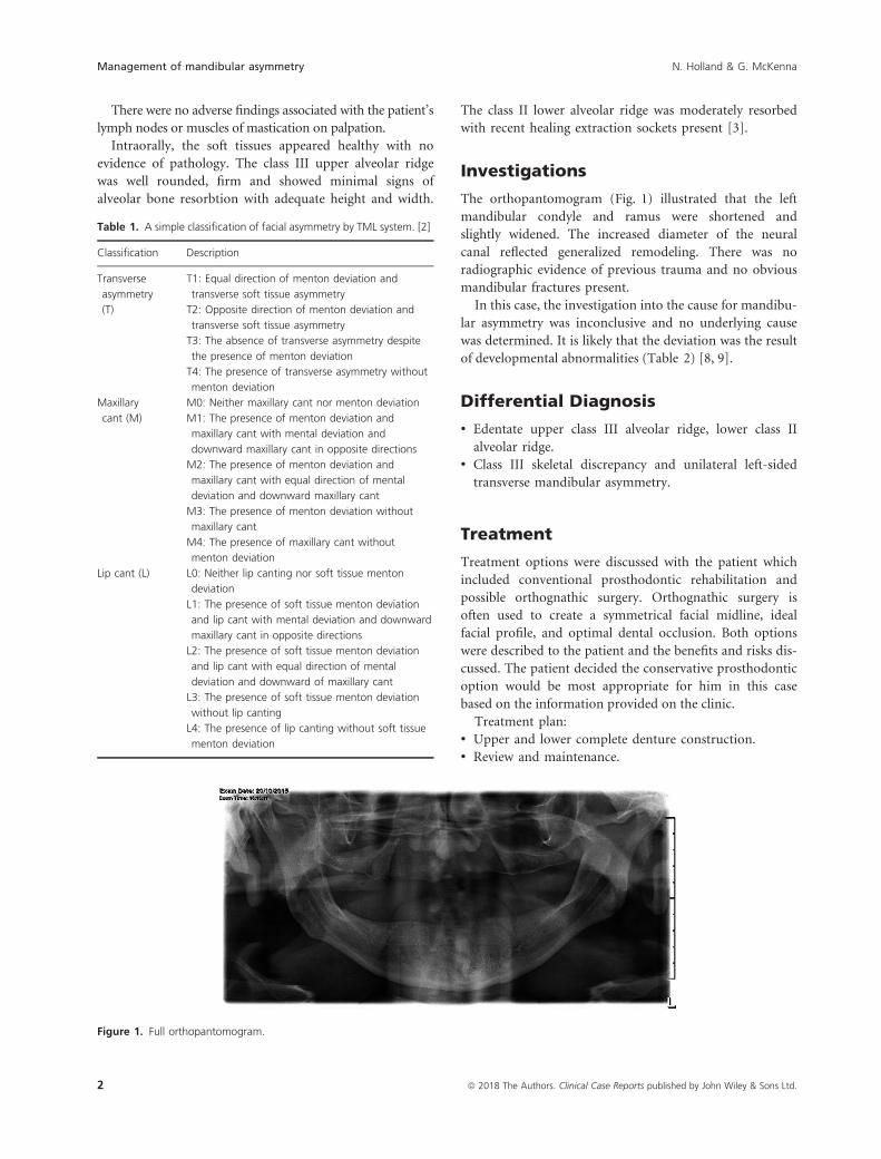
The orthopantomogram (Fig. 1) illustrated that the left
mandibular condyle and ramus were shortened and
slightly widened. The increased diameter of the neural
canal reflected generalized remodeling. There was no
radiographic evidence of previous trauma and no obvious
mandibular fractures present.
In this case, the investigation into the cause for mandibu-
lar asymmetry was inconclusive and no underlying cause
was determined. It is likely that the deviation was the result
of developmental abnormalities (Table 2) [8, 9].
Differential Diagnosis
• Edentate upper class III alveolar ridge, lower class II
alveolar ridge.
• Class III skeletal discrepancy and unilateral left-sided
transverse mandibular asymmetry.
Treatment
Treatment options were discussed with the patient which
included conventional prosthodontic rehabilitation and
possible orthognathic surgery. Orthognathic surgery is
often used to create a symmetrical facial midline, ideal
facial profile, and optimal dental occlusion. Both options
were described to the patient and the benefits and risks dis-
cussed. The patient decided the conservative prosthodontic
option would be most appropriate for him in this case
based on the information provided on the clinic.
Treatment plan:
• Upper and lower complete denture construction.
• Review and maintenance.
Table 1. A simple classification of facial asymmetry by TML system. [2]
Classification Description
Transverse
asymmetry
(T)
T1: Equal direction of menton deviation and
transverse soft tissue asymmetry
T2: Opposite direction of menton deviation and
transverse soft tissue asymmetry
T3: The absence of transverse asymmetry despite
the presence of menton deviation
T4: The presence of transverse asymmetry without
menton deviation
Maxillary
cant (M)
M0: Neither maxillary cant nor menton deviation
M1: The presence of menton deviation and
maxillary cant with mental deviation and
downward maxillary cant in opposite directions
M2: The presence of menton deviation and
maxillary cant with equal direction of mental
deviation and downward maxillary cant
M3: The presence of menton deviation without
maxillary cant
M4: The presence of maxillary cant without
menton deviation
Lip cant (L) L0: Neither lip canting nor soft tissue menton
deviation
L1: The presence of soft tissue menton deviation
and lip cant with mental deviation and downward
maxillary cant in opposite directions
L2: The presence of soft tissue menton deviation
and lip cant with equal direction of mental
deviation and downward of maxillary cant
L3: The presence of soft tissue menton deviation
without lip canting
L4: The presence of lip canting without soft tissue
menton deviation
Figure 1. Full orthopantomogram.
2 ª 2018 The Authors. Clinical Case Reports published by John Wiley & Sons Ltd.
Management of mandibular asymmetry N. Holland & G. McKenna

The patient was seen again in the clinic to start treat-
ment and upper and lower impressions were taken in
nonelastic compound material in edentulous perforated
stock trays for provision of special trays.
Special trays were constructed using polymethyl
methacrylate. The trays were spaced and perforated to
allow for impressions in addition reaction silicone (poly-
vinyl siloxane impression material type 2, medium
Table 2. Potential causes of mandibular asymmetries. [8, 9]
Classification Example Description
Developmental Hemimandibular elongation • Unknown etiology
• Affects the mandible unilaterally
• Presents as transverse displacement of the chin point to contralateral side which presents in early
adulthood
• No vertical asymmetry
• The contralateral mandibular molars deviate lingually in attempt to remain in occlusion
• Cross-bite may develop on the unaffected side
• Radiographic elongation of the condyle or body of mandible on the affected side
Hemimandibular hyperplasia • Horizontal and vertical enlargement on one side of the mandible which involves the condyle,
ramus, and body of the mandible
• The condition usually begins in puberty
• The maxillary dentition on the affected side will overerupt to compensate for the excessive verti-
cal mandibular growth, which results in a characteristic transverse cant of the maxillary occlusal
plane
• If the vertical growth is rapid, then dental eruption may not keep pace and a lateral open bite
will occur on the affected side
• Radiographic elongation of ramus and condylar enlargement can be seen. The lower border of
mandible on the affected side is lower than the unaffected side. There is usually increased dis-
tance between molar roots and inferior alveolar canal on the affected side. The unaffected side
will have normal height
Hemifacial microsomia • Deficiency of hard and soft tissues on one side of the face during embryonic development (con-
genital disorder)
• Chin point displacement is to the affected side
• Hypodontia is commonly noted in these patients
Hemifacial hypertrophy • Asymmetry affects the craniofacial soft and hard tissues
• Intrauterine pressure can lead to shortening of the sternocleidomastiod muscle leading to
mandibular assymetries
• Likely genetic contribution
Hemifacial atrophy (Parry–
Romberg syndrome)
• Uncertain etiology
• Atropy of hard and soft tissues on one side of the face leading to mandibular asymmetry
• May be accompanied by hyperpigmentation of the skin, seizures, and facial pain
Pathological Tumors • For example, benign ameloblastoma
• Condylar head tumors cause deviation of the mandible to the unaffected side with unilateral
condylar enlargement radiographically
Cysts • Dentigerous cysts
• Keratocysts
Infection • Dentoalveolar abscess
• Sialadenitis
Condylar resorption • May be secondary to juvenile rheumatoid arthritis, steroid therapy, or orthognathic surgery
• Unilateral resorption can lead to mandibular asymmetry
Traumatic Condylar Fractures • Trauma to condylar region during childhood can lead to arrest in growth
• Chin point asymmetry toward affected side
Functional Mandibular Displacements • Maxillary narrowing can lead to occlusal interferences leading to lateral displacement of the
mandible
ª 2018 The Authors. Clinical Case Reports published by John Wiley & Sons Ltd. 3
N. Holland & G. McKenna Management of mandibular asymmetry

viscosity). These special trays were then border molded
with green stick used to take master impressions. Care
was taken to ensure that the full width and depth of the
buccal and lingual sulcus was recorded in the master
impressions, along with extension to the hamular notch
in the upper arch and retromolar pad in the lower arch.
These were poured in the laboratory to be used as master
models for construction of registration rims. The master
models are shown in Figures 2 and 3.
Jaw registration proved to be very challenging in this
case and close liason and communication took place with
the on-site dental laboratory. The upper rim was altered
to achieve optimal anterior occlusal plane parallel to the
interpupillary line. The posterior occlusal plane was mea-
sured according to the ala-tragal line [4]. The lower rim
was trimmed to ensure correct interocclusal freeway space
of 2–4 mm based on the occlusal vertical and relaxed ver-
tical dimensions. The midline was marked to be coinci-
dent with the natural facial midline, using the phitrum of
the lip, nose, and labial frenum as a guide. The rims were
registered with polyvinyl siloxane registration paste in
retruded contact position. These were disinfected and
instructions were sent to the laboratory for them to be
articulated on an average value articulator (Average Value
Articulator, 30° condylar angle and flat incisal table)
(Figs. 4 and 5).
The upper teeth were set up as per the registration rim
and based around the midline. This was important esthet-
ically to the patient as the asymmetry was confined to a
left-sided mandibular chin point shift only. Conventional
posterior occlusion was set up on the right side. In order
to maintain occlusal contact, the posterior teeth on the
lower left side were placed lingual to the alveolar ridge in
a balanced occlusal scheme (cross-bite). This was checked
with the patient at trial to ensure the teeth were not
encroaching in the tongue space. Anatomical teeth were
used rather than a flat occlusal table [5]. The upper and
lower wax try in complete dentures were well fitting and
the dentition arrangement was verified. At this stage, the
occlusion was reregistered, again in polyvinyl siloxane
registration paste.
The denture was gum fitted in the lower anterior 3-3
region labially (open-faced denture). Due to the gum fit
anteriorly and the risk of midline fracture of the denture,
processing involved the use of high impact acrylic. This
was spot ground on the articulator prior to fit.
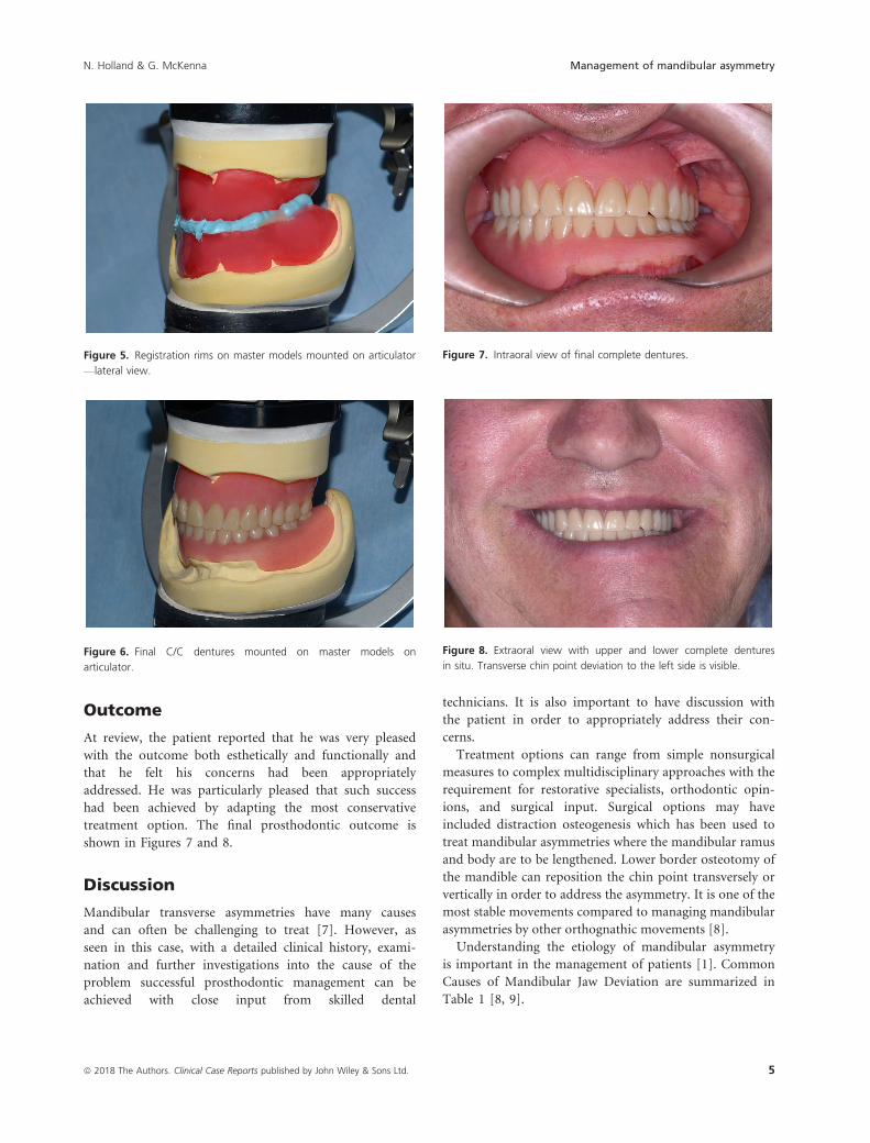
The patient returned for fit of the removable complete
dentures (Figs. 6 and 7). The occlusion and esthetics were
good and at this stage, some adjustment was required on the
lower denture fit surface for the patient’s comfort. The patient
was very pleased with the result. At this stage, oral and written
denture instructions were given and the patient was encour-
aged to make efforts to adapt to the new dentures [6].
Figure 2. Upper master model.
Figure 3. Lower master model.
Figure 4. Registration with models mounted on articulator—anterior
view.
4 ª 2018 The Authors. Clinical Case Reports published by John Wiley & Sons Ltd.
Management of mandibular asymmetry N. Holland & G. McKenna
Outcome
At review, the patient reported that he was very pleased
with the outcome both esthetically and functionally and
that he felt his concerns had been appropriately
addressed. He was particularly pleased that such success
had been achieved by adapting the most conservative
treatment option. The final prosthodontic outcome is
shown in Figures 7 and 8.
Discussion
Mandibular transverse asymmetries have many causes
and can often be challenging to treat [7]. However, as
seen in this case, with a detailed clinical history, exami-
nation and further investigations into the cause of the
problem successful prosthodontic management can be
achieved with close input from skilled dental
technicians. It is also important to have discussion with
the patient in order to appropriately address their con-
cerns.
Treatment options can range from simple nonsurgical
measures to complex multidisciplinary approaches with the
requirement for restorative specialists, orthodontic opin-
ions, and surgical input. Surgical options may have
included distraction osteogenesis which has been used to
treat mandibular asymmetries where the mandibular ramus
and body are to be lengthened. Lower border osteotomy of
the mandible can reposition the chin point transversely or
vertically in order to address the asymmetry. It is one of the
most stable movements compared to managing mandibular
asymmetries by other orthognathic movements [8].
Understanding the etiology of mandibular asymmetry
is important in the management of patients [1]. Common
Causes of Mandibular Jaw Deviation are summarized in
Table 1 [8, 9].
Figure 6. Final C/C dentures mounted on master models on
articulator.
Figure 7. Intraoral view of final complete dentures.
Figure 8. Extraoral view with upper and lower complete dentures
in situ. Transverse chin point deviation to the left side is visible.
Figure 5. Registration rims on master models mounted on articulator
—lateral view.
ª 2018 The Authors. Clinical Case Reports published by John Wiley & Sons Ltd. 5
N. Holland & G. McKenna Management of mandibular asymmetry

Conclusion
This case demonstrated the successful prosthodontic reha-
bilitation of a patient with a significant mandibular asym-
metry. In some cases, treatment can be provided within
primary care; however, in this case, specialist input was
required [1]. There is minimal guidance surrounding the
referral criteria for facial asymmetries; however, due to
the potential for joint maxillofacial, orthodontic and
restorative treatment planning and close liaison with den-
tal laboratories, it may be necessary in most cases to refer
to specialist services.
Furthermore, if the cause of the facial asymmetry is
unknown further investigations may also be required to
rule out pathology, which may not be possible in a pri-
mary care setting. The extent to which the functional and
esthetic concern affects the patient is also likely to influ-
ence the referral decision and treatment choice.
It is hoped that this report will provide further infor-
mation to practitioners in varying care settings and aid in
management of these challenging patients.
Acknowledgments
Robert Thompson—Clinical Case Photography. Gary
McNeilly and the Prosthetics Dental Laboratory Staff RVH.
Treatment costs covered under National Health Service.
Ethics
Informed and valid consent was obtained from the
patient for use of the dental notes, radiographs, and pho-
tographs in the case report.
Authorship
NH: was the first Author and Dental Core Trainee. GMK:
was the Corresponding Author and Clinical Consultant.
Conflict of Interest
None declared.
References
1. Reyneke, J. P., P. Tsakiris, and F. Kienle. 1997. A simple
classification for surgical treatment planning of
maxillomandibular asymmetry. Br. J. Oral Maxillofac. Surg.
35:349–351.
2. Kim, J. Y., H. D. Jung, Y. S. Jung, C. J. Hwang, and H. S.
Park. 2014. A simple classification of facial asymmetry by
TML system. J. Craniomaxillofac. Surg. 42:313–320.3. Cawood, J. L., and R. A. Howell. 1988. A classification of the
edentulous jaws. Int. J. Oral Maxillofac. Surg. 17:232–236.4. Woelfel, J. B., T. Igarashi, and J. K. Dong. 2014. Faculty-
supervised measurements of the face and of mandibular
movements on young adults. J. Adv. Prosthodont. 6:483–490.
5. Abduo, J. 2013. Occlusal schemes for complete dentures: a
systematic review. Int. J. Prosthodont. 26:26–33.
Quintessence Journals
6. Mahajan, T., R. Trivedi, S. Singh, and R. Sangur. 2015.
Prosthetic management of edentulous mandibulectomy
patient with modified occlusion - A case report. Rama
Univ. J. Dent. Sci. 2:42–45.
7. Cheong, Y. W., and L. J. Lo. 2011. Facial asymmetry:
etiology, evaluation, and management, review article.
Chang. Gung. Med. J. 34:341–351.8. Chia, M. S. Y., F. B. Naini, and D. S. Gill. 2008. The
aetiology, diagnosis and management of mandibular
asymmetry. Ortho. Update 1:44–52.
9. Xavier, S. P., T. Santos, E. R. Silv, A. C. Faria, and F. V. de
Mello Filh. 2014. Two-stage treatment of facial asymmetry
caused by unilateral condylar hyperplasia. Braz. Dent. J.
25:257–260. Ribeir~ao Preto
6 ª 2018 The Authors. Clinical Case Reports published by John Wiley & Sons Ltd.
Management of mandibular asymmetry N. Holland & G. McKenna
Related Documents