Anesthesiology 2008; 108:269 –75 Copyright © 2008, the American Society of Anesthesiologists, Inc. Lippincott Williams & Wilkins, Inc. Propofol Restores Brain Microvascular Function Impaired by High Glucose via the Decrease in Oxidative Stress Katsutoshi Nakahata, M.D., Ph.D.,* Hiroyuki Kinoshita, M.D., Ph.D.,† Toshiharu Azma, M.D., Ph.D.,‡ Naoyuki Matsuda, M.D., Ph.D.,§ Keiko Hama-Tomioka, M.D., Masanori Haba, M.D., Yoshio Hatano, M.D., Ph.D.# Background: Vascular dysfunction induced by hyperglyce- mia has not been studied in cerebral parenchymal circulation. The current study was designed to examine whether high glu- cose impairs dilation of cerebral parenchymal arterioles via nitric oxide synthase, and whether propofol recovers this va- sodilation by reducing superoxide levels in the brain. Methods: Cerebral parenchymal arterioles in the rat brain slices were monitored using computer-assisted videomicros- copy. Vasodilation induced by acetylcholine (10 6 to 10 4 M) was obtained after the incubation of brain slices for 60 min with any addition of L-glucose (20 mM), D-glucose (20 mM), or propo- fol (3 10 7 or 10 6 M) in combination with D-glucose (20 mM). Superoxide production in the brain slice was determined by dihydroethidium (2 10 6 M) fluorescence. Results: Addition of D-glucose, but not L-glucose, reduced arteriolar dilation by acetylcholine, whereas the dilation was abolished by the neuronal nitric oxide synthase inhibitor S- methyl-L-thiocitrulline (10 5 M). Both propofol and the super- oxide dismutase mimetic Tempol (10 4 M) restored the arterio- lar dilation in response to acetylcholine in the brain slice treated with D-glucose. Addition of D-glucose increased super- oxide production in the brain slice, whereas propofol, Tem- pol, and the nicotinamide adenine dinucleotide phosphate (NAD[P]H) oxidase inhibitor apocynin (1 mM) similarly inhib- ited it. Conclusions: Clinically relevant concentrations of propofol ameliorate neuronal nitric oxide synthase– dependent dilation impaired by high glucose in the cerebral parenchymal arte- rioles via the effect on superoxide levels. Propofol may be protective against cerebral microvascular malfunction resulting from oxidative stress by acute hyperglycemia. PREVIOUS studies from systemic arteries demonstrated that incubation with high concentrations of glucose im- pairs endothelium-dependent relaxation, indicating that hy- perglycemia causes endothelial dysfunction. 1,2 Nitric oxide synthase is a well-known candidate to confer endothelium- dependent vasodilation in cerebral as well as systemic ar- teries. 3 However, it has not been well studied whether acute exposure toward hyperglycemia reduces cerebral vasodilation related to nitric oxide synthase, although such vasodilation decreased in cerebral arteries from diabetic animals chronically exposed to high glucose. 4,5 Overproduction of superoxide induced by high glucose is one of the crucial causes of vascular dysfunction in acute hyperglycemia as well as diabetes mellitus. Indeed, cumu- lative findings support the view that exposure toward high glucose augments oxidative stress in both endothelial and smooth muscle cells from systemic blood vessels. 6–8 How- ever, there is no direct evidence showing enhanced oxida- tive stress in cerebral blood vessels induced by the acute exposure to high glucose, although a recent study has documented that diabetes mellitus, which is a chronic model of high glucose exposure, increases superoxide pro- duction in pial arterioles. 5 Propofol reportedly possesses potential antioxidant properties caused by its chemical structure similar to that of phenol-based free radical scavengers such as vitamin E. 9 Previous in vivo or in vitro studies docu- mented that this intravenous anesthetic reduces oxida- tive stress in the brain, whereas concentrations or doses of propofol to achieve this phenomenon were mostly beyond the clinical range. 10 –13 These results suggest that this anesthetic may be protective against the cerebrovas- cular dysfunction caused by increased oxidative stress, although it has never been shown. In addition, because these studies were performed using cultured cells, ho- mogenized tissues, or the whole brain, 10 –13 it is still unclear that clinical concentrations of propofol demon- strate the antioxidant effect in situ on which brain structures include neuronal cells and vasculature. Therefore, the current study was designed to examine whether the high concentration of glucose via en- hanced production of superoxide in the cerebral paren- chyma impairs cerebral arteriolar dilation evoked by nitric oxide synthase, and whether propofol ameliorates the dilation via the reduction of superoxide levels in any brain structures. Materials and Methods The animal care and use committee in Wakayama Med- ical University (Wakayama, Japan) approved this study. Male Wistar rats (aged 16 –20 weeks) were obtained from Charles River Japan Inc. (Yokohama, Japan). Rats were anesthetized with inhalation of 3% halothane to perform a midline thoracotomy, and 50 ml saline was infused intracardially into the left ventricle while a right atrial incision was simultaneously made for blood drain- * Assistant Professor, † Associate Professor, Staff, # Professor and Chairman, Department of Anesthesiology, Wakayama Medical University. ‡ Staff, Depart- ment of Anesthesia, Hiroshima General Hospital, Hatsukaichi, Japan. § Associ- ate Professor, Departments of Anesthesiology and Molecular Medical Pharmacol- ogy, Toyama University School of Medicine, Toyama, Japan. Received from the Department of Anesthesiology, Wakayama Medical Univer- sity, Wakayama, Japan. Submitted for publication May 17, 2007. Accepted for publication October 10, 2007. Supported in part by Grant-in-Aid Nos. 18689038 (to K.N.), 19390409 (to H.K.), 18659462 (to H.K.), and 17390432 (to Y.H.) for Scientific Research from the Ministry of Education, Culture, Sports, Science and Technology of Japan, Tokyo, Japan. Presented in part at the Annual Meeting of the American Society of Anesthesiologists, Chicago, Illinois, October 14 –18, 2006. Address correspondence to Dr. Kinoshita: Department of Anesthesiology, Wakayama Medical University, 811-1 Kimiidera, Wakayama, Wakayama 641-0012, Japan. [email protected]. Information on purchasing reprints may be found at www.anesthesiology.org or on the masthead page at the beginning of this issue. ANESTHESIOLOGY’s articles are made freely accessible to all readers, for personal use only, 6 months from the cover date of the issue. Anesthesiology, V 108, No 2, Feb 2008 269 Downloaded From: http://anesthesiology.pubs.asahq.org/ by HIROYUKI KINOSHITA on 04/03/2015

Welcome message from author
This document is posted to help you gain knowledge. Please leave a comment to let me know what you think about it! Share it to your friends and learn new things together.
Transcript

Anesthesiology 2008; 108:269–75 Copyright © 2008, the American Society of Anesthesiologists, Inc. Lippincott Williams & Wilkins, Inc.
Propofol Restores Brain Microvascular Function Impairedby High Glucose via the Decrease in Oxidative StressKatsutoshi Nakahata, M.D., Ph.D.,* Hiroyuki Kinoshita, M.D., Ph.D.,† Toshiharu Azma, M.D., Ph.D.,‡Naoyuki Matsuda, M.D., Ph.D.,§ Keiko Hama-Tomioka, M.D.,� Masanori Haba, M.D.,� Yoshio Hatano, M.D., Ph.D.#
Background: Vascular dysfunction induced by hyperglyce-mia has not been studied in cerebral parenchymal circulation.The current study was designed to examine whether high glu-cose impairs dilation of cerebral parenchymal arterioles vianitric oxide synthase, and whether propofol recovers this va-sodilation by reducing superoxide levels in the brain.
Methods: Cerebral parenchymal arterioles in the rat brainslices were monitored using computer-assisted videomicros-copy. Vasodilation induced by acetylcholine (10�6 to 10�4 M)was obtained after the incubation of brain slices for 60 min withany addition of L-glucose (20 mM), D-glucose (20 mM), or propo-fol (3 � 10�7 or 10�6 M) in combination with D-glucose (20 mM).Superoxide production in the brain slice was determined bydihydroethidium (2 � 10�6 M) fluorescence.
Results: Addition of D-glucose, but not L-glucose, reducedarteriolar dilation by acetylcholine, whereas the dilation wasabolished by the neuronal nitric oxide synthase inhibitor S-methyl-L-thiocitrulline (10�5 M). Both propofol and the super-oxide dismutase mimetic Tempol (10�4 M) restored the arterio-lar dilation in response to acetylcholine in the brain slicetreated with D-glucose. Addition of D-glucose increased super-oxide production in the brain slice, whereas propofol, Tem-pol, and the nicotinamide adenine dinucleotide phosphate(NAD[P]H) oxidase inhibitor apocynin (1 mM) similarly inhib-ited it.
Conclusions: Clinically relevant concentrations of propofolameliorate neuronal nitric oxide synthase–dependent dilationimpaired by high glucose in the cerebral parenchymal arte-rioles via the effect on superoxide levels. Propofol may beprotective against cerebral microvascular malfunction resultingfrom oxidative stress by acute hyperglycemia.
PREVIOUS studies from systemic arteries demonstratedthat incubation with high concentrations of glucose im-pairs endothelium-dependent relaxation, indicating that hy-perglycemia causes endothelial dysfunction.1,2 Nitric oxidesynthase is a well-known candidate to confer endothelium-dependent vasodilation in cerebral as well as systemic ar-teries.3 However, it has not been well studied whetheracute exposure toward hyperglycemia reduces cerebral
vasodilation related to nitric oxide synthase, although suchvasodilation decreased in cerebral arteries from diabeticanimals chronically exposed to high glucose.4,5
Overproduction of superoxide induced by high glucoseis one of the crucial causes of vascular dysfunction in acutehyperglycemia as well as diabetes mellitus. Indeed, cumu-lative findings support the view that exposure toward highglucose augments oxidative stress in both endothelial andsmooth muscle cells from systemic blood vessels.6–8 How-ever, there is no direct evidence showing enhanced oxida-tive stress in cerebral blood vessels induced by the acuteexposure to high glucose, although a recent study hasdocumented that diabetes mellitus, which is a chronicmodel of high glucose exposure, increases superoxide pro-duction in pial arterioles.5
Propofol reportedly possesses potential antioxidantproperties caused by its chemical structure similar tothat of phenol-based free radical scavengers such asvitamin E.9 Previous in vivo or in vitro studies docu-mented that this intravenous anesthetic reduces oxida-tive stress in the brain, whereas concentrations or dosesof propofol to achieve this phenomenon were mostlybeyond the clinical range.10–13 These results suggest thatthis anesthetic may be protective against the cerebrovas-cular dysfunction caused by increased oxidative stress,although it has never been shown. In addition, becausethese studies were performed using cultured cells, ho-mogenized tissues, or the whole brain,10–13 it is stillunclear that clinical concentrations of propofol demon-strate the antioxidant effect in situ on which brainstructures include neuronal cells and vasculature.
Therefore, the current study was designed to examinewhether the high concentration of glucose via en-hanced production of superoxide in the cerebral paren-chyma impairs cerebral arteriolar dilation evoked bynitric oxide synthase, and whether propofol amelioratesthe dilation via the reduction of superoxide levels in anybrain structures.
Materials and Methods
The animal care and use committee in Wakayama Med-ical University (Wakayama, Japan) approved this study.Male Wistar rats (aged 16–20 weeks) were obtainedfrom Charles River Japan Inc. (Yokohama, Japan). Ratswere anesthetized with inhalation of 3% halothane toperform a midline thoracotomy, and 50 ml saline wasinfused intracardially into the left ventricle while a rightatrial incision was simultaneously made for blood drain-
* Assistant Professor, † Associate Professor, � Staff, # Professor and Chairman,Department of Anesthesiology, Wakayama Medical University. ‡ Staff, Depart-ment of Anesthesia, Hiroshima General Hospital, Hatsukaichi, Japan. § Associ-ate Professor, Departments of Anesthesiology and Molecular Medical Pharmacol-ogy, Toyama University School of Medicine, Toyama, Japan.
Received from the Department of Anesthesiology, Wakayama Medical Univer-sity, Wakayama, Japan. Submitted for publication May 17, 2007. Accepted forpublication October 10, 2007. Supported in part by Grant-in-Aid Nos. 18689038(to K.N.), 19390409 (to H.K.), 18659462 (to H.K.), and 17390432 (to Y.H.) forScientific Research from the Ministry of Education, Culture, Sports, Science andTechnology of Japan, Tokyo, Japan. Presented in part at the Annual Meeting ofthe American Society of Anesthesiologists, Chicago, Illinois, October 14–18,2006.
Address correspondence to Dr. Kinoshita: Department of Anesthesiology,Wakayama Medical University, 811-1 Kimiidera, Wakayama, Wakayama 641-0012,Japan. [email protected]. Information on purchasing reprints may befound at www.anesthesiology.org or on the masthead page at the beginning ofthis issue. ANESTHESIOLOGY’s articles are made freely accessible to all readers, forpersonal use only, 6 months from the cover date of the issue.
Anesthesiology, V 108, No 2, Feb 2008 269
Downloaded From: http://anesthesiology.pubs.asahq.org/ by HIROYUKI KINOSHITA on 04/03/2015

age. The animals were then decapitated, and the brainswere rapidly removed and rinsed with artificial cerebro-spinal fluid (control solution) of the following composi-tion: 119 mM NaCl, 4.7 mM KCl, 2.5 mM CaCl2, 1.17 mM
MgSO4, 1.18 mM KH2PO4, 25 mM NaHCO3, and 11 mM
glucose. Brains were cut freehand into blocks containingthe neocortex, followed by immediate sectioning intoslices (150 mm thick) with a mechanical tissue slicer(Vibratomes 1000; Ted Pella, Inc., Redding, CA).Throughout the slicing procedure, brain blocks werecontinuously bathed in the control solution bubbledwith 93% oxygen and 7% carbon dioxide at 4°C.
VideomicroscopyIndividual slice was transferred to a recording chamber
filled with control solution, which is mounted on aninverted microscope (Olympus IX70; Tokyo, Japan). Therecording system was consisted of a recording chamber(3 ml) and a tubing compartment (7 ml) including theperfusion chamber. The slices were continuously super-fused with control solution at the flow rate of 1.5 ml/min, bubbled with 93% oxygen and 7% carbon dioxide(carbon dioxide tension � 40 mmHg, pH � 7.4, 37°C).An intraparenchymal arteriole (4.2–6.5 mm in internaldiameter, 11.0–15.0 mm in external diameter) was lo-cated within the neuronal tissue, and its internal diam-eter was continuously monitored with the live com-puterized videomicroscopy.14–16 The videomicroscopyequipment consisted of an inverted microscope, a 40�objective (Olympus), and a 2.25� video projection lens(Olympus). The image of a parenchymal arteriole wastransmitted to video camera (C6790-81; Olympus) anddisplayed on a computer via a media converter (Physio-Tech, Tokyo, Japan).
The differentiation between the arteriole and thevenule in the brain is based on the previous studiesdocumenting that in the brain, one or several layers ofsmooth muscle cells should be identified in the arterioleand that the venule resembles a large capillary with nomore in its wall than endothelial cells resting on a basallamina, although we only used the arteriole with a singlelayer of smooth muscle.17,18 In our previous study, thesewere confirmed by immunohistochemical analysis usingan anti–�-smooth muscle actin antibody in addition tohematoxylin–eosin stain of the slice.16
We defined the internal diameter as the length be-tween the internal margins of arteriolar walls. Changesof internal diameter in cerebral microvessels were re-corded on computer image files and then analyzed usingthe image analysis software (Physio-Tech).14–16 Mi-crovessel diameters were derived as an average of fourmeasurements taken along approximately 15 �m of ves-sel length.
Each slice was incubated for 60 min with any additionof L-glucose (20 mM), D-glucose (20 mM), or propofol(3 � 10�7 or 10�6
M) in combination with D-glucose (20
mM) to the control solution. Thereafter, these slices weretransferred to the recording chamber, in which vasodi-lation induced by acetylcholine (10�6 to 10�4
M) wasobtained during stable constriction in response to pros-taglandin F2� (5 � 10�7
M). In some experiments, thesuperoxide dismutase mimetic Tempol (10�4
M) or theselective neuronal nitric oxide synthase inhibitor S-meth-yl-L-thiocitrulline (10�5
M) was added 15 min before theconstriction to prostaglandin F2�. Constrictions to pros-taglandin F2� (5 � 10�7
M) were 13–17% of each controldiameter, and these were not different among thegroups studied. Only one concentration–response wasmade for each slice. The duration of experiment for eachslice was within 3 h, because our previous study dem-onstrated that even 3 h after the preparation of brainslice, the vasodilator function mediated by nitric oxidesynthase was intact in our experimental condition15 us-ing the constriction produced by prostaglandin F2� (5 �10�7
M) in each arteriole. Therefore, the percent dilationwas calculated by the following equation: % dilation �100 � (diameter after administration of the vasodila-tor � diameter after administration of prostaglandin F2�
[5 � 10�7M])/(diameter of control condition � diameter
after administration of prostaglandin F2� [5 � 10�7M]).15
Measurements of In Situ Superoxide ProductionThe oxidative fluorescent dye hydroethidine was used
to evaluate levels of superoxide in situ as describedpreviously.19 Cells are permeable to hydroethidine, andin the presence of superoxide, hydroethidine is oxidizedto fluorescent ethidium bromide, in which form it istrapped by intercalation with DNA. This method pro-vides sensitive detection of superoxide levels in situ.Unfixed brain slices, 150 �m thick, were placed on glassslides. Hydroethidine (2 � 10�6
M) in phosphate-buff-ered saline (pH � 7.4) was applied to each slice. Slideswere incubated in a light-protected chamber at 37°Cfor 20 min. Images were obtained with a FLUOVIEWFV300 laser scanning confocal microscope (Olympus)equipped with a krypton–argon laser. Fluorescence wasdetected with a 585-nm long-pass filter. Laser settingswere identical for acquisition of images from all of brainslices.
Before the application of hydroethidine, brain sliceswere incubated with the normal solution, to which Tem-pol (10�4
M), L-glucose (20 mM), or D-glucose (20 mM)was added. In some experiments, a xanthine oxidaseinhibitor allopurinol (10�4
M), Tempol (10�4M), propo-
fol (10�6M), or the nicotinamide adenine dinucleotide
phosphate (NAD[P]H) oxidase inhibitor apocynin (1mM) was also applied. Brain slices exposed to hydroethi-dine in the sole presence of Tempol (10�4
M) served asthe control, and the fluorescence in the brain slice wasexpressed as a ratio.19
270 NAKAHATA ET AL.
Anesthesiology, V 108, No 2, Feb 2008
Downloaded From: http://anesthesiology.pubs.asahq.org/ by HIROYUKI KINOSHITA on 04/03/2015

Immunohistochemical AnalysisThe brain slices were fixed with 10% formalin solution
buffered with phosphate-buffered saline (pH � 7.2) andembedded in paraffin followed by the preparation of 4-to 6-�m sections. After the deparaffinization, sectionswere incubated with phosphate-buffered saline contain-ing 1% normal donkey serum and 1% bovine serumalbumin to reduce nonspecific reactions. The sectionswere further incubated with pairs of rabbit anti–nitricoxide synthase polyclonal antibodies (Santa Cruz Bio-technology, Santa Cruz, CA) at the concentration of 1�g/ml at 4°C, overnight. After washing, the sectionswere incubated with Cy3-conjugated anti-rabbit immu-noglobulin and fluorescein isothiocyanate–labeled anti-mouse immunoglobulin secondary antibodies (15 �g/ml) at 24°C for 1 h. The sections were evaluated undera fluorescence microscopy, and fluorescence imageswere digitally merged. As negative controls, specimenswere incubated with normal rabbit immunoglobulin Gor in the absence of the primary antibodies.
DrugsThe following pharmacologic agents were used: allo-
purinol, apocynin, dimethyl sulfoxide, prostaglandin F2a,S-methyl-L-thiocitrulline, Tempol (Sigma Aldrich Inc., St.Louis, MO), and hydroethidine (Polysciences Inc., War-rington, PA). Drugs were dissolved in distilled watersuch that volumes of less than 60 ml were added to theperfusion system. The stock solutions of apocynin,propofol, and Tempol were prepared in dimethyl sulfox-ide (3 � 10�4
M), and the solution containing hydroethi-dine (7 mg) was produced using N,N-dimethylacetamide(1 ml). The concentrations of drugs are expressed asfinal molar concentration.
Statistical AnalysisData are expressed as mean � SD; n refers to the number
of rats from which the brain slice was taken. Power calcu-lations were performed with responses to the reduction ofsuperoxide levels as the primary endpoint, and we calcu-lated that a sample size of 5 gave 80% power to detect achange of 61% in levels of superoxide at a significance levelof 0.05 (SD � 30%). Statistical analysis was performed withStatView® version 5.0 (SAS Institute Inc., Cary, NC) andSample Power 2.0 (SSPS Japan Inc., Tokyo, Japan). Datawere evaluated using repeated-measures analysis of vari-ance or a factorial analysis of variance as appropriate, fol-lowed by Student-Newman-Keuls test as a post hoc analysis.Differences were considered to be statistically significantwhen P was less than 0.05.
Results
VideomicroscopyIn the cerebral parenchymal arterioles treated with
L-glucose (20 mM, n � 5), acetylcholine (10�6 to 10�4M)
concentration-dependently induced dilation, which wascompletely abolished by the selective neuronal nitricoxide synthase inhibitor S-methyl-L-thiocitrulline (10�5
M,n � 5; fig. 1). Addition of D-glucose (20 mM, n � 5)compared with L-glucose (20 mM, n � 5) significantlyreduced the dilation produced by acetylcholine (fig. 1).Propofol (3 � 10�7 to 10�6
M, n � 5 each) concentra-tion-dependently recovered arteriolar dilation inducedby acetylcholine in the brain slices treated with D-glu-cose (20 mM, n � 5; fig. 2), whereas a solvent forpropofol, dimethyl sulfoxide (1.74 � 10�7
M), did notalter vasodilation in these brain slices (fig. 3). The super-oxide dismutase mimetic Tempol (10�4
M, n � 5) andthe highest concentration of propofol (10�6
M, n � 5)similarly restored this vasodilation (fig. 2). Combinationof Tempol (10�4
M) and propofol (10�6M) did not
further augment vasodilation induced by acetylcholinein the brain slice treated with D-glucose (20 mM, n � 3;data not shown).
Measurements of In Situ Superoxide ProductionRepresentative images of in situ superoxide produc-
tion are shown in figure 4A. D-Glucose (20 mM, n � 5)enhanced ethidium bromide fluorescence, which wasmarkedly reduced to the intensity seen in the brain slicetreated with L-glucose (20 mM, n � 5) by the treatmentwith either propofol (10�6
M, n � 5) or Tempol (10�4M,
n � 5). Importantly, increased intensity of fluorescencein the brain slice treated with D-glucose (20 mM, n � 5)was seen in arteriolar walls as well as neuronal cells (fig.4A). Addition of D-glucose (20 mM, n � 5), but not
Fig. 1. Concentration–response curves of cerebral parenchymalarterioles to acetylcholine in the brain slice treated with anyaddition of L-glucose (20 mM), D-glucose (20 mM), or a selectiveneuronal nitric oxide synthase inhibitor S-methyl-L-thiocitrul-line (10�5 M) in combination with L-glucose (20 mM) to thecontrol solution. * Difference between arterioles treated withL-glucose and arterioles treated with D-glucose, and differencebetween arterioles treated with L-glucose and arterioles treatedwith S-methyl-L-thiocitrulline in combination with L-glucose arestatistically significant (P < 0.05).
271PROPOFOL AND HIGH GLUCOSE IN CEREBRAL ARTERIOLES
Anesthesiology, V 108, No 2, Feb 2008
Downloaded From: http://anesthesiology.pubs.asahq.org/ by HIROYUKI KINOSHITA on 04/03/2015

L-glucose (20 mM, n � 5), increased levels of superoxidein the cerebral parenchyma, whereas propofol (10�6
M,n � 5) and Tempol (10�4
M, n � 5) inhibited them (fig.4B). The NAD(P)H oxidase inhibitor apocynin (1 mM,n � 5) and propofol (10�6
M, n � 5) similarly reducedsuperoxide production in the brain slice treated withD-glucose (20 mM, n � 5), whereas the xanthine oxidase
inhibitor allopurinol (10�4M, n � 5) did not alter the
levels of superoxide (fig. 4B).
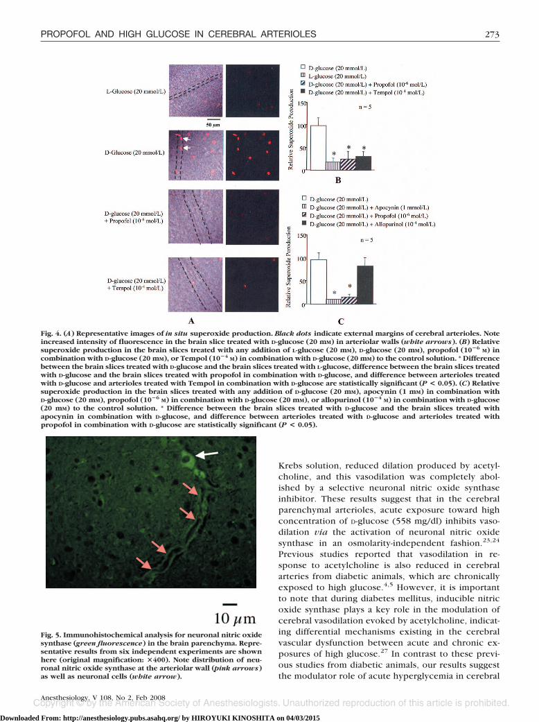
Immunohistochemical AnalysisImmunohistochemical analysis revealed distribution of
neuronal nitric oxide synthase at the arteriolar wall aswell as neuronal cells (fig. 5). The immunoreactivity wasrecognizable only in using each primary antibody butnot in the absence of it.
Discussion
Role of Neuronal Nitric Oxide Synthase in CerebralParenchymal Arteriolar Dilation Induced byAcetylcholineAcetylcholine reportedly induces activation of nitric
oxide synthase followed by cerebral vasodilation in hu-mans a well as animals.20,21 We previously demonstratedthat cerebral parenchymal arteriolar dilation in responseto acetylcholine was completely abolished by the treat-ment with the nonselective nitric oxide synthase inhib-itor NG-nitro-L-arginine methyl ester, indicating the intactfunction of nitric oxide synthases in our model.15 It hasbeen shown that nitric oxide derived through neuronalnitric oxide synthase in the perivascular region can be asource of nitric oxide acting on microvascular smoothmuscle cells.22 These results support the concept thatvasodilator substances possibly produce nitric oxide viathe activation of neuronal nitric oxide synthase withinthe brain slice. Indeed, acetylcholine induced cerebralparenchymal arteriolar dilation, which was completelyabolished by the selective neuronal nitric oxide synthaseinhibitor S-methyl-L-thiocitrulline, indicating that neuro-nal enzyme solely caused nitric oxide synthase–depen-dent vasodilation in the current study.23,24 The involve-ment of neuronal nitric oxide synthase has beenconfirmed by our immunohistochemical analysis, reveal-ing distribution of neuronal nitric oxide synthase withinthe arteriolar wall as well as neuronal cells. Our resultsare consistent with a previous study demonstrating thatnitric oxide synthase–immunoreactive fibers innervatecerebral arterial wall.25 Previous in vivo studies alsoprovided the evidence that nitric oxide from neuronalnitric oxide synthase is a critical link between neuro-transmitters including glutamate and cerebral bloodflow.26 Taken together, neuronal nitric oxide synthaseseems to be a regulator in cerebral microcirculation,although we cannot completely rule out the possibilitythat the endothelial enzyme may contribute to vasodila-tor responses induced by acetylcholine.
Effect of Acute Exposure toward High Glucose onNeuronal Nitric Oxide–dependent Dilation in theCerebral Parenchymal ArteriolesIn the current study, addition of D-glucose (20 mM),
but not the same concentration of L-glucose to normal
Fig. 2. Concentration–response curves of cerebral parenchy-mal arterioles to acetylcholine in the brain slice treated withany addition of D-glucose (20 mM), propofol (3 � 10�7 or 10�6 M)in combination with D-glucose (20 mM), or the superoxidedismutase mimetic Tempol (10�4 M) in combination withD-glucose (20 mM) to the control solution. * Difference be-tween arterioles treated with D-glucose and arterioles treatedwith D-glucose in combination with propofol, and differencebetween arterioles treated with D-glucose and arteriolestreated with D-glucose in combination with Tempol are sta-tistically significant (P < 0.05).
Fig. 3. Concentration–response curves of cerebral parenchymalarterioles to acetylcholine in the brain slice treated with addi-tion of D-glucose (20 mM) in the absence or in the presence ofdimethyl sulfoxide (1.74 � 10�7 M).
272 NAKAHATA ET AL.
Anesthesiology, V 108, No 2, Feb 2008
Downloaded From: http://anesthesiology.pubs.asahq.org/ by HIROYUKI KINOSHITA on 04/03/2015
Krebs solution, reduced dilation produced by acetyl-choline, and this vasodilation was completely abol-ished by a selective neuronal nitric oxide synthaseinhibitor. These results suggest that in the cerebralparenchymal arterioles, acute exposure toward highconcentration of D-glucose (558 mg/dl) inhibits vaso-dilation via the activation of neuronal nitric oxidesynthase in an osmolarity-independent fashion.23,24
Previous studies reported that vasodilation in re-sponse to acetylcholine is also reduced in cerebralarteries from diabetic animals, which are chronicallyexposed to high glucose.4,5 However, it is importantto note that during diabetes mellitus, inducible nitricoxide synthase plays a key role in the modulation ofcerebral vasodilation evoked by acetylcholine, indicat-ing differential mechanisms existing in the cerebralvascular dysfunction between acute and chronic ex-posures of high glucose.27 In contrast to these previ-ous studies from diabetic animals, our results suggestthe modulator role of acute hyperglycemia in cerebral
Fig. 4. (A) Representative images of in situ superoxide production. Black dots indicate external margins of cerebral arterioles. Noteincreased intensity of fluorescence in the brain slice treated with D-glucose (20 mM) in arteriolar walls (white arrows). (B) Relativesuperoxide production in the brain slices treated with any addition of L-glucose (20 mM), D-glucose (20 mM), propofol (10�6 M) incombination with D-glucose (20 mM), or Tempol (10�4 M) in combination with D-glucose (20 mM) to the control solution. * Differencebetween the brain slices treated with D-glucose and the brain slices treated with L-glucose, difference between the brain slices treatedwith D-glucose and the brain slices treated with propofol in combination with D-glucose, and difference between arterioles treatedwith D-glucose and arterioles treated with Tempol in combination with D-glucose are statistically significant (P < 0.05). (C) Relativesuperoxide production in the brain slices treated with any addition of D-glucose (20 mM), apocynin (1 mM) in combination withD-glucose (20 mM), propofol (10�6 M) in combination with D-glucose (20 mM), or allopurinol (10�4 M) in combination with D-glucose(20 mM) to the control solution. * Difference between the brain slices treated with D-glucose and the brain slices treated withapocynin in combination with D-glucose, and difference between arterioles treated with D-glucose and arterioles treated withpropofol in combination with D-glucose are statistically significant (P < 0.05).
Fig. 5. Immunohistochemical analysis for neuronal nitric oxidesynthase (green fluorescence) in the brain parenchyma. Repre-sentative results from six independent experiments are shownhere (original magnification: �400). Note distribution of neu-ronal nitric oxide synthase at the arteriolar wall (pink arrows)as well as neuronal cells (white arrow).
273PROPOFOL AND HIGH GLUCOSE IN CEREBRAL ARTERIOLES
Anesthesiology, V 108, No 2, Feb 2008
Downloaded From: http://anesthesiology.pubs.asahq.org/ by HIROYUKI KINOSHITA on 04/03/2015

vasodilation resulting from the activation of neuronalnitric oxide synthase.
Mechanisms of Superoxide Overproduction by HighGlucose in the BrainOverproduction of superoxide induced by high glu-
cose is one of the crucial causes of vascular dysfunc-tion in acute hyperglycemia as well as diabetes melli-tus. Cumulative findings support the view thatexposure toward high glucose augments oxidativestress in systemic blood vessels.6 – 8 In the cerebralcirculation, a recent study using pial arterioles hassolely documented enhanced superoxide productionduring diabetes mellitus.5 In the current study, thesuperoxide dismutase mimetic Tempol completely re-stored vasodilation in response to acetylcholine and itreduced superoxide levels in the brain slice treatedwith the high concentration of D-glucose, suggestingthe involvement of superoxide in cerebral arteriolarmalfunction induced by acute high glucose.28 In-creased superoxide levels were seen in arteriolar wallsas well as neuronal cells in the brain slice treated withhigh concentration of D-glucose, showing such spatialevidence about the increased production of superox-ide in the brain parenchyma resulting from acute hy-perglycemia. It is of note that the blood– brain barriershould be disrupted in our brain slice model, andtherefore our data are probably relevant during thediseased states such as intracerebral hemorrhage.29
However, it is also true that the blood– brain barrier isvulnerable to hyperglycemia itself, presumably throughthe production of oxygen-derived free radicals includ-ing superoxide.30 Therefore, it is likely that our resultsconfer a piece of information regarding the sites ofsuperoxide production within brain parenchyma dur-ing acute hyperglycemia.
In the next sets of experiments, we evaluated thesources of superoxide production induced by highglucose in the brain parenchyma. The NAD(P)H oxi-dase inhibitor apocynin, similarly to the superoxidedismutase mimetic Tempol, reduced superoxide pro-duction in the brain slice treated with the high con-centration of D-glucose, suggesting a crucial role ofNAD(P)H �oxidase in the cerebral superoxide pro-duction during acute hyperglycemia.30,31 These re-sults are in agreement with our recent study usingsystemic arterioles showing that increased productionof superoxide evoked by high glucose is mediatedby NAD(P)H oxidase.32 Indeed, the expression ofNAD(P)H oxidase has been demonstrated in smaller aswell as larger cerebral arterioles and neurons through-out the brain.33,34 In contrast, our negative resultswith the xanthine oxidase inhibitor allopurinol indi-cate that xanthine oxidase is not responsible for thesuperoxide generation in brain parenchyma afteracute exposure to high glucose.35
Protective Effects of Propofol on ImpairedVasodilation Caused by Oxidative StressPropofol reportedly possesses potential antioxidant
properties caused by its chemical structure similar tothat of phenol-based free radical scavengers such asvitamin E.9 Previous in vivo or in vitro studies docu-mented that this intravenous anesthetic reduces oxida-tive stress in the brain.10–13 These results suggest thatthis anesthetic may be protective against the cerebrovas-cular dysfunction caused by increased oxidative stress.However, we cannot rule out the possibility that theprotective effect of propofol toward oxidative stress maybe due to scavenging of downstream intermediates pro-duced by lipid peroxidation via increased levels of su-peroxide, because such a role was not evaluated in thecurrent study. Propofol (3 � 10�7 to 10�6
M) concen-tration-dependently recovered arteriolar dilation in-duced by acetylcholine in the brain slices treated withthe high concentration of D-glucose. In addition, propo-fol (10�6
M), similarly to a superoxide dismutase mimeticand an NAD(P)H oxidase inhibitor, reduced superoxidelevels within arteriolar walls as well as neuronal cells inthis preparation. The plasma concentration of propofolduring induction of anesthesia in humans has been re-ported as up to 3 � 10�5
M, and burst suppression dosesof propofol (up to 6 � 10�5
M) are also available forcerebral protection.36–38 Effective concentrations ofpropofol (3 � 10�7 to 10�6
M) to restore arteriolarfunction in the cerebral parenchyma are probably withinclinical range if one considers plasma free concentra-tions calculated from both above clinical plasma concen-trations and the substantial binding of this compound toplasma proteins (from 97% to 98%).37 Therefore, ourresults strongly indicate that clinically relevant concen-trations of propofol can confer protection of cerebralblood vessels as well as neuronal cells against oxidativestress caused by acute hyperglycemia.
Conclusions
The current study has demonstrated the following newfindings. First, neuronal nitric oxide synthase solely con-tributes to the cerebral parenchymal arteriolar dilationproduced by acetylcholine. Second, acute exposure ofthe brain slice toward high glucose impairs neuronalnitric oxide synthase–dependent vasodilation by in-creased production of superoxide in the arteriolar wallsvia the activation of NAD(P)H oxidase. Third, clinicallyrelevant concentrations of propofol restore the vasodila-tion impaired by high glucose via reduction of superox-ide in the brain parenchyma. These results suggest themechanisms of increased oxidative stress in the brainparenchyma produced by acute hyperglycemia. Propo-fol may be protective against cerebral microvascularmalfunction resulting from increased oxidative stress.
274 NAKAHATA ET AL.
Anesthesiology, V 108, No 2, Feb 2008
Downloaded From: http://anesthesiology.pubs.asahq.org/ by HIROYUKI KINOSHITA on 04/03/2015

The authors thank Takahito Hayashi, M.D. (Assistant Professor), and ToshikazuKondo, M.D., Ph.D. (Professor and Chairman), in the Department of ForensicMedicine, Wakayama Medical University, Wakayama, Japan, for their help inperforming the immunohistochemical analysis.
References
1. Tesfamariam B, Brown ML, Deykin D, Cohen RA: Elevated glucose pro-motes generation of endothelium-derived vasoconstrictor prostanoids in rabbitaorta. J Clin Invest 1990; 85:929–32
2. Beckman JA, Goldfine AB, Gordon MB, Creager MA: Ascorbate restoresendothelium-dependent vasodilation impaired by acute hyperglycemia in hu-mans. Circulation 2001; 103:1618–23
3. Kinoshita H, Tsutsui M, Milstien S, Katusic ZS: Tetrahydrobiopterin, nitricoxide and regulation of cerebral arterial tone. Prog Neurobiol 1997; 52:295–302
4. Mayhan WG, Simmons LK, Sharpe GM: Mechanism of impaired responses ofcerebral arterioles during diabetes mellitus. Am J Physiol 1991; 260:H319–26
5. Didion SP, Lynch CM, Baumbach GL, Faraci FM: Impaired endothelium-dependent responses and enhanced influence of Rho-kinase in cerebral arteriolesin type II diabetes. Stroke 2005; 36:342–7
6. Kinoshita H, Azma T, Iranami H, Nakahata K, Kimoto Y, Dojo M, Yuge O,Hatano Y: Synthetic peroxisome proliferator-activated receptor-� agonists restoreimpaired vasorelaxation via ATP-sensitive K� channels by high glucose. J Phar-macol Exp Ther 2006; 318:312–8
7. Karashima Y, Oike M, Takahashi S, Ito Y: Propofol prevents endothelialdysfunction induced by glucose overload. Br J Pharmacol 2002; 137:683–91
8. Cosentino F, Hishikawa K, Katusic ZS, Luscher TF: High glucose increasesnitric oxide synthase expression and superoxide anion generation in humanaortic endothelial cells. Circulation 1997; 96:25–8
9. Murphy PG, Myers DS, Davies MJ, Webster NR, Jones JG: The antioxidantpotential of propofol (2,6-diisopropylphenol). Br J Anaesth 1992; 68:613–8
10. Grasshoff C, Gillessen T: The effect of propofol on increased superoxideconcentration in cultured rat cerebrocortical neurons after stimulation of N-methyl-D-aspartate receptors. Anesth Analg 2002; 95:920–2
11. Sagara Y, Hendler S, Khoh-Reiter S, Gillenwater G, Carlo D, Schubert D,Chang J: Propofol hemisuccinate protects neuronal cells from oxidative injury.J Neurochem 1999; 73:2524–30
12. De La Cruz JP, Sedeno G, Carmona JA, Sanchez de la Cuesta F: The in vitroeffects of propofol on tissular oxidative stress in the rat. Anesth Analg 1998;87:1141–6
13. Ozturk E, Demirbilek S, Kadir But A, Saricicek V, Gulec M, Akyol O, OzcanErsoy M: Antioxidant properties of propofol and erythropoietin after closed headinjury in rats. Prog Neuropsychopharmacol Biol Psychiatry 2005; 29:922–7
14. Kinoshita H, Nakahata K, Dojo M, Kimoto Y, Hatano Y: Lidocaine impairsvasodilation mediated by adenosine triphosphate-sensitive K� channels but notby inward rectifier K� channels in rat cerebral microvessels. Anesth Analg 2004;99:904–9
15. Nakahata K, Kinoshita H, Hirano Y, Kimoto Y, Iranami H, Hatano Y: Mildhypercapnia induces vasodilation via adenosine triphosphate–sensitive K� chan-nels in parenchymal microvessels of the rat cerebral cortex. ANESTHESIOLOGY 2003;99:1333–9
16. Nakahata K, Kinoshita H, Tokinaga Y, Ishida Y, Kimoto Y, Dojo M,Mizumoto K, Ogawa K, Hatano Y: Vasodilation mediated by inward rectifier K�channels in cerebral microvessels of hypertensive and normotensive rats. AnesthAnalg 2006; 102:571–6
17. Peters A, Palay SL, Webster HF: Blood vessels, The Fine Structure of Thenervous System: Neurons and Their Supporting Cells, 3rd edition. New York,Oxford University Press, 1991, pp 344–55
18. Frederickson RG, Low FN: Blood vessels and tissue space associated withthe brain of the rat. Am J Anat 1969; 125:123–46
19. Miller FJ, Gutterman DD, Rios CD, Heistad DD, Davidson BL: Superoxideproduction in vascular smooth muscle contributes to oxidative stress and im-paired relaxation in atherosclerosis. Circ Res 1998; 82:1298–305
20. Trauernicht AK, Sun H, Patel KP, Mayhan WG: Enalapril prevents impairednitric oxide synthase-dependent dilatation of cerebral arterioles in diabetic rats.Stroke 2003; 34:2698–703
21. Elhusseiny A, Hamel E: Muscarinic-but not nicotinic-acetylcholine recep-tors mediate a nitric oxide-dependent dilation in brain cortical arterioles: Possiblerole for the M5 receptor subtype. J Cereb Blood Flow Metab 2000; 20:298–305
22. Kavdia M, Popel AS: Contribution of nNOS- and eNOS-derived NO tomicrovascular smooth muscle NO exposure. J Appl Physiol 2004; 97:293–301
23. Li Y-L, Li Y-F, Liu D, Cornish KG, Patel KP, Zucker IH, Channon KM,Schultz HD: Gene transfer of neuronal nitric oxide synthase to carotid bodyreverses enhanced chemoreceptor function in heart failure rabbits. Circ Res2005; 97:260–7
24. Hagioka S, Takeda Y, Zhang S, Sato T, Morita K: Effects of 7-nitroindazoleand N-nitro-L-arginine methyl ester on changes in cerebral blood flow and nitricoxide production preceding development of hyperbaric oxygen-induced seizuresin rats. Neurosci Lett 2005; 382:206–10
25. Yoshida K, Okamura T, Kimura H, Bredt DS, Snyder SH, Toda N: Nitricoxide synthase-immunoreactive nerve fibers in dog cerebral and peripheralarteries. Brain Res 1993; 629:67–72
26. Yang G, Zhang Y, Ross ME, Iadecola C: Attenuation of activity-inducedincreases in cerebellar blood flow in mice lacking neuronal nitric oxide synthase.Am J Physiol 2003; 285:H298–304
27. Kitayama J, Faraci FM, Gunnett CA, Heistad DD: Impairment of dilatorresponses of cerebral arterioles during diabetes mellitus: Role of inducible NOsynthase. Stroke 2006; 37:2129–33
28. Kitayama J, Yi C, Faraci FM, Heistad DD: Modulation of dilator responsesof cerebral arterioles by extracellular superoxide dismutase. Stroke 2006; 37:2802–6
29. Lindsberg PJ, Roine RO: Hyperglycemia in acute stroke. Stroke 2004;35:363–4
30. Stolk J, Hiltermann TJN, Dijkman JH, Verhoeven AJ: Characteristics of theinhibition of NADPH oxidase activation in neutrophils by apocynin, a methoxy-substituted catechol. Am J Respir Cell Mol Biol 1994; 11:95–102
31. Bedard K, Krause K-H: The NOX family of ROS-generating NADPH oxi-dases: Physiology and pathophysiology. Physiol Rev 2007; 87:245–313
32. Kinoshita H, Azma T, Nakahata K, Iranami H, Kimoto Y, Dojo M, Yuge O,Hatano Y: Inhibitory effect of high concentration of glucose on relaxations toactivation of ATP-sensitive K� channels in human omental artery. ArteriosclerThromb Vas Biol 2004; 24:2290–5
33. Sun H, Zheng H, Molacek E, Fang Q, Patel KP, Mayhan WG: Role ofNAD(P)H oxidase in alcohol-induced impairment of endothelial nitric oxidesynthase-dependent dilation of cerebral arterioles. Stroke 2006; 37:495–500
34. Serrano F, Kolluri NS, Wientjes FB, Card JP, Klann E: NADPH oxidaseimmunoreactivity in the mouse brain. Brain Res 2003; 988:193–8
35. Moriwaki Y, Yamamoto T, Suda M, Nasako Y, Takahashi S, Agbedana OE,Hada T, Higashino K: Purification and immunohistochemical tissue localization ofhuman xanthine oxidase. Biochim Biophys Acta 1993; 1164:327–30
36. Kirkpatrick T, Cockshott ID, Douglas EJ, Nimmo WS: Pharmacokinetics ofpropofol (Diprivan) in elderly patients. Br J Anaesth 1988; 60:146–50
37. Servin F, Desmonts JM, Haberer JP, Cockshott ID, Plummer GF, FarinottiR: Pharmacokinetics and protein binding of propofol in patients with cirrhosis.ANESTHESIOLOGY 1988; 69:887–91
38. Newman MF, Murkin JM, Roach G, Croughwell ND, White WD, ClementsFM, Reves JG, CNS Subgroup of McSPI: Cerebral physiologic effects of burstsuppression doses of propofol during nonpulsatile cardiopulmonary bypass.Anesth Analg 1995; 81:452–7
275PROPOFOL AND HIGH GLUCOSE IN CEREBRAL ARTERIOLES
Anesthesiology, V 108, No 2, Feb 2008
Downloaded From: http://anesthesiology.pubs.asahq.org/ by HIROYUKI KINOSHITA on 04/03/2015
Related Documents