Promoter Deletion and Comparative Expression Analysis of the Mirabilis mosaic caulimovirus (MMV) Sub-genomic Transcript (Sgt) Promoter in Transgenic Plants NRISINGHA DEY 1 AND INDU B. MAITI* Molecular Plant Virology and Plant Genetic Engineering Laboratory, Tobacco and Health Research Institute, University of Kentucky, Lexington, KY 40546-0236, USA 35 A sub-genomic transcript (Sgt) promoter was isolated from a genomic clone of the Mirabilis mosaic virus (MMV), a double-stranded DNA plant pararetrovirus belonging to the Caulimoviridae family. The MMV Sgt pro- moter fragment (genomic co-ordinates 4830 to 5840) was mapped by 5′ -3′ -deletion analysis to define the boundaries required for maximal promoter expression. Expression patterns of promoter fragments coupled to GUS reporter gene were evaluated both in protoplast tran- sient expression experiments and in transgenic Nicotiana tabacum cv. Samsun NN plants. A 333 bp MMV Sgt promoter fragment (sequence –306 to +27 from the transcription start site, TSS) was found sufficient for strong promoter activity in protoplast transient expression experiments. The transcription start site (TSS) of the MMV Sgt promoter was determined by primer extension analysis using total RNA isolated from transgenic plants con- taining a MMV Sgt promoter:uidA fusion gene. In seedlings, the level of GUS expression was in the order of leaf ≥ root >stem. Histochemical GUS-staining of seedlings showed highest GUS activity in root tips, leaf midribs, veins and vascular tissues. The MMV Sgt promoter fragment is a strong constitutive promoter, with strength comparable to that of the MMV full-length transcript (FLt) promot- er. MMV Sgt promoter also demonstrated much greater activity, 8-fold more compared to the CaMV 19S promoter and 2-fold stronger than the CaMV 35S promoter. ____________________ 1 Present address: Department of Zoology and Genetics, Iowa State University, 2156 MBB, Ames, IA 50011 *Corresponding author: Molecular Plant Virology and Plant Genetic Engineering Laboratory, Tobacco and Health Research Institute, University of Kentucky, Lexington, KY 40546-0236, USA; Phone: 859-257-3296; Fax: 859-323-1077; e-mail: [email protected] Transgenics, Vol. 4, pp. 35-53 © 2003 Old City Publishing, Inc. Reprints available directly from the publisher Published by license under the OCP Science imprint, Photocopying permitted by license only a member of the Old City Publishing Group.

Welcome message from author
This document is posted to help you gain knowledge. Please leave a comment to let me know what you think about it! Share it to your friends and learn new things together.
Transcript

Promoter Deletion and Comparative Expression Analysis of the Mirabilis mosaic caulimovirus(MMV) Sub-genomic Transcript (Sgt) Promoter in Transgenic Plants
NRISINGHA DEY 1 AND INDU B. MAITI*
Molecular Plant Virology and Plant Genetic Engineering Laboratory, Tobacco and Health Research Institute, University of Kentucky,Lexington, KY 40546-0236, USA
35
A sub-genomic transcript (Sgt) promoter wasisolated from a genomic clone of the Mirabilismosaic virus (MMV), a double-stranded DNAplant pararetrovirus belonging to theCaulimoviridae family. The MMV Sgt pro-moter fragment (genomic co-ordinates 4830 to5840) was mapped by 5′′-3′′-deletion analysisto define the boundaries required for maximalpromoter expression. Expression patterns ofpromoter fragments coupled to GUS reportergene were evaluated both in protoplast tran-sient expression experiments and in transgenicNicotiana tabacum cv. Samsun NN plants. A333 bp MMV Sgt promoter fragment(sequence –306 to +27 from the transcriptionstart site, TSS) was found sufficient for strongpromoter activity in protoplast transient
expression experiments. The transcriptionstart site (TSS) of the MMV Sgt promoter wasdetermined by primer extension analysis usingtotal RNA isolated from transgenic plants con-taining a MMV Sgt promoter:uidA fusiongene. In seedlings, the level of GUS expressionwas in the order of leaf ≥ root >stem.Histochemical GUS-staining of seedlingsshowed highest GUS activity in root tips, leafmidribs, veins and vascular tissues. The MMVSgt promoter fragment is a strong constitutivepromoter, with strength comparable to that ofthe MMV full-length transcript (FLt) promot-er. MMV Sgt promoter also demonstratedmuch greater activity, 8-fold more comparedto the CaMV 19S promoter and 2-foldstronger than the CaMV 35S promoter.
____________________1Present address: Department of Zoology and Genetics, Iowa State University, 2156 MBB, Ames, IA 50011
*Corresponding author: Molecular Plant Virology and Plant Genetic Engineering Laboratory, Tobacco and Health Research Institute,University of Kentucky, Lexington, KY 40546-0236, USA; Phone: 859-257-3296; Fax: 859-323-1077; e-mail: [email protected]
Transgenics, Vol. 4, pp. 35-53 © 2003 Old City Publishing, Inc.Reprints available directly from the publisher Published by license under the OCP Science imprint,Photocopying permitted by license only a member of the Old City Publishing Group.

Key words: caulimovirus, CaMV19S,CaMV35S, mirabilis mosaic virus (MMV),promoter deletion, sub-genomic transcript(Sgt) promoter, transgenic plants
INTRODUCTION
The Mirabilis mosaic virus (MMV) infectsMirabilis plant species (family Nyctaginaceae),a member of the Caulimoviridae family, andhas a circular double-stranded DNA genomeof about 8 Kb with four single-stranded dis-continuities in the DNA, one in the alphastrand and three in the complementary strand[1]. The restriction map of the MMV genomeis quite different from the other members ofthe genus Caulimovirus [1]. The MMV viruswas characterized as a member of the genusCaulimovirus based upon the morphology ofits virions and inclusion bodies [2]. Recently,MMV has been fully sequenced (GenBanknucleotide sequence database accession num-ber AF454635), and homology analysis of itsgenomic DNA has shown that it is a defini-tive member of the genus Caulimovirus[Maiti, unpublished]. However, MMV isserologically distinct from the Cauliflowermosaic virus (CaMV), the type species of thisgenus [2].
Several Caulimoviridae genomes have beenfully sequenced and characterized. Theseinclude Cauliflower mosaic virus (CaMV)[3], Carnation etched ring virus (CERV) [4],Figwort mosaic virus (FMV) [5], Soybeanchlorotic mottle virus (SoCMV) [6], Peanutchlorotic streak virus (PClSV) [7], Casavavein mosaic virus (CVMV) [8], Strawberryvein banding virus (SVBV) [9], Petunia veinclearing virus (PVCV) [10], and Mirabilismosaic virus (MMV) [Maiti, unpublished].
The Caulimovirus genome generally con-
tains two transcriptional promoters, one forthe full-length transcript and the other for thesubgenomic transcript; these are equivalentto the CaMV 35S and 19S transcript respec-tively [6, 11, 12]. A number of strong consti-tutive promoters have been derived fromCaulimoviridae family, particularly from theCauliflower mosaic virus (CaMV): CaMV35Sand 19S promoter [13, 14]. For constructingplant transformation vectors, genetic pro-moters have also been isolated from othermembers of this family, namely Rice tungrobacilliform virus (RTBV) [15], Commelinayellow mottle virus (CYMV) [16], Soybeanchlorotic mottle virus (SoCMV) [6], Figwortmosaic virus (FMV, strain DxS) [17, 18]),FMV strain M3 [19], Cassava vein mosaicvirus (CVMV) [20], Peanut cholotic streakvirus (PClSV) [21] and Mirabilis mosaic virus(MMV) [22, 23]. Transcript promoters fromthe Caulimovirus like CaMV, FMV, PClSV,MMV and FMV are active in all plant organs[13, 18, 21-23], whereas, transcript promot-ers from the Badnavirus like CYMV andRTBV are phloem-specific [15, 16] inexpressing genes in transgenic plants. TheCaMV 35S promoter has been well charac-terized [13, 24-30] and widely used inchimeric gene constructs for heterologousgene expression in transgenic plants [31-33].The CaMV 35S promoter is also active inbacteria [34], in yeast [35], in Hela cells [36]and in Xenopus oocytes [37].
In the present study, we report the identifi-cation and characterization of a subgenomictranscript (Sgt) promoter from the Mirabilismosaic virus (MMV), a newly describedspecies of the genus Caulimovirus. The opti-mal boundaries required for the maximal pro-moter activity were defined by 5′- and 3′- enddeletion analysis of the promoter/leaderregion of the subgenomic transcript promoter
3 6 D E Y A N D M A I T I
T R A N S G E N I C S

S U B G E N O M I C T R A N S C R I P T P R O M O T E R F R O M T H E M I R A B I L I S M O S A I C V I R U S 37
T R A N S G E N I C S
FIGURE 1The DNA sequence of the subgenomic transcript (Sgt) promoter from the Mirabilis mosaic virus (MMV). The nucleotide sequenceof the MMV Sgt promoter (coordinates -646 to +378 in respect to transcription start site; corresponding coordinates in MMVgenome 4829 to 5840), a 1023 bp fragment includes the 3′-end of gene V, followed by the small intergenic region and 5′ portionof gene VI presented from left to right in the 5′ to 3′ direction of the transcript. Modification of promoter sequence resulted frominserting the ‘EcoRI to SmaI’ adapter is shown in lowercase. The end points for the 5′ or the 3′ deletion constructs are also indi-cated above or below the sequence respectively. The tentative regulatory sequence: the TATA-box (TATAA), the CAT box (CAAT)and the initiator ATG codon for gene VI are shown in bold. The transcriptional start site (TSS) is indicated as +1.

of MMV both in transgenic plants and in pro-toplast transient expression experiments. A333 bp MMV Sgt promoter fragment(sequence -306 to +27 from transcription startsite) was found sufficient for maximal pro-moter activity. The strength of the MMV Sgtpromoter is very compatible with the MMVFLt promoter and greater than that of theCaMV 19S and CaMV 35S promoters. This isthe first report, to our knowledge, document-ing the strong and constitutive expressioncharacteristic of the MMV Sgt promoter.
MATERIALS AND METHODS
Protoplasts, Plants and Enzymes
Isolation of protoplasts from the tobaccocell suspension cultures (Xanthi ‘Brad’) andelectroporation of protoplasts with super-coiled DNA containing promoter fragmentfused with GUS gene were done essentially asdescribed earlier [38]. Tobacco plants(Nicotiana tabacum cv. Samsun NN) wereused for plant transformation. Restrictionenzymes, antibiotics, components of plant tis-sue culture medium, and RNA isolation kitswere purchased from commercial sources.Nitrocellulose and Nytran membranes wereobtained from Schleicher & Schuell, USA.
Isolation of MMV Sgt promoter andmodification of its DNA sequence
A full-length genomic clone of MMV inthe plasmid pMMV-B10 [1] was fullysequenced (Maiti, unpublished results), and itwas used as a template for PCR reactions. A1012 bp segment (co-ordinates 4829 to 5840of the MMV genome) was selected for pro-moter deletion analysis, and it was isolated asa BamHI and HindIII fragment by PCR usingthe following primers: (i) Forward primer: 5′-GCG GGC GGA TCC GAA AAA CGG AAACCG TTA-3′ and (ii) Reverse primer: 5′-ATGCAG AAG CTT TTG TTG TGT CTT TACCGG-3′. Promoter sequences in the primersare shown in bold, and restriction enzymessites are underlined. The PCR-fragment wasgel-purified using the QIAgen procedure(Valencia, CA 91355, USA). After digestionwith BamHI and HindIII, the PCR fragmentwas cloned into the corresponding sites ofpBS(KS+) (Strategene, La Jolla, CA 92037,USA). We designated this plasmid as
3 8 D E Y A N D M A I T I
T R A N S G E N I C S
FIGURE 2Determination of the transcriptional start site (TSS) of theMMV Sgt promoter by primer extension analysis. Primerextension was carried out as described in Materials andMethods. Primer extension reaction products were subjectedto electrophoresis on denaturing polyacrylamide gel contain-ing urea, alongside with the sequence reaction of GUS geneconstruct (lane G, A, T and C). The process was performedwith the same labeled primer. The minus strand DNAsequence read on the gel is shown, and the transcriptional startsite (A*) in the corresponding plus strand is indicated by anarrow.
pBSMS1011.
Modification of EcoRI site to a SmaI site:The pBSMS1011 has an EcoRI site at the
MMV genome coordinate 5345. This EcoRIsite located 52 bp upstream of the TATAsequence was modified to insert a SmaI siteusing two primers with the followingsequences: forward primer: 5′-AATTACC-CGGGC-3′ and reverse primer: 5′-AATTGC-CCGGGT-3′. These two primers were mixed
in a 1:1 molar ratio and allowed to anneal byslow cooling (2oC/ min) from 94o C to 24o Cunder a programmed PCR cycle. The plasmidpBSMS1011 was digested with EcoRI andligated with the ‘EcoRI to SmaI’ adapter, fol-lowed by transformation. The resulting plas-mid was designated as pBSMS1011DE. Thischange inserted 12 additional nucleotides (5′-AATTACCCGGGC-3′) into the promotersequence (Fig. 1).
S U B G E N O M I C T R A N S C R I P T P R O M O T E R F R O M T H E M I R A B I L I S M O S A I C V I R U S 39
T R A N S G E N I C S
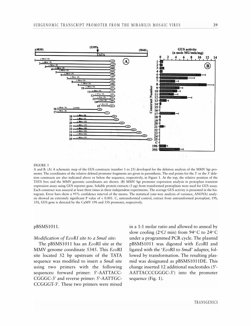
FIGURE 3A and B. (A) A schematic map of the GUS constructs (number 1 to 25) developed for the deletion analysis of the MMV Sgt pro-moter. The coordinates of the relative deleted promoter fragments are given in parenthesis. The end points for the 5′ or the 3′ dele-tion constructs are also indicated above or below the sequence, respectively, in Figure 1. At the top, the relative position of theTATA box and the MMV genomic coordinates are shown. (B) MMV Sgt promoter expression analysis in protoplast transientexpression assay using GUS reporter gene. Soluble protein extracts (5 µg) from transformed protoplasts were used for GUS assay.Each construct was assayed at least three times in three independent experiments. The average GUS activity is presented in the his-togram. Error bars show a 95% confidence interval of the means. The statistical (one-way analysis of variance, ANOVA) analy-sis showed an extremely significant P value of < 0.001. C, untransformed control, extract from untransformed protoplast; 19S,35S, GUS gene is directed by the CaMV 19S and 35S promoter, respectively.

Construction of vectors for transientexpression of genes in protoplasts
A series of promoter fragments included inconstructing the plant transformation vectorwith the subgenomic promoter of MMV weredesigned to study the influence of upstreamand downstream sequence with respect to theTATA box toward the promoter activity. Thedefined MMV Sgt promoter sequence oflength, as indicated (in Fig. 3A) was amplifiedby PCR using pBSMS1011DE as templateand appropriately designed primers to intro-duce an EcoRI site at the 5′-end and a HindIIIsite at the 3′-end of amplified products. PCRamplification was carried out for 31 cyclesunder the following standard conditions:denaturation (92o C for 1 min), annealing(55o C for 1 min), and extension (72o C for 2min) using ELONGASE enzyme mix (recom-binant high fidelity TaqDNA polymerase mixwith proof reading 3′-5′ exonuclease activity)from Gibco-BRL, Maryland, USA. Each PCRamplified promoter fragment from 1 to 25was restricted with EcoRI and HindIII; therestricted fragment was gel-purified andcloned into the corresponding sites ofpUC119 vector and sequenced by thedideoxy chain terminator method [39] usingsynthetic primers.
Subcloning of the MMV Sgt promoter fragments from pUC119 into pUCPMAGUSvector
The sequence of each of the MMV Sgtpromoter fragments cloned in pUC119 wasverified before subcloning to pUCPMAGUS,a protoplast expression vector [22]. TheMMV Sgt promoter fragments were individ-ually gel-purified from the correspondingpUC119 clone after restriction digestion with
EcoRI and HindIII and subcloned into thecorresponding sites of pUCPMAGUS [22].The following promoter deletion plasmidswere developed (Fig. 3). The 5′ and 3′ co-ordinates of the promoter fragment withrespect to the transcription start site (TSS) aregiven in parenthesis: pPMS1GUS (-646 to+27), pPMS2GUS (-606 to +27), pPMS3GUS(-556 to +27), pPMS4GUS (-506 to +27),pPMS5GUS (-456 to +27), pPMS6GUS (-406to +27), pPMS7GUS (-356 to +27),pPMS8GUS (-306 to +27), pPMS9GUS (-256to +27), pPMS10GUS (-206 to +27),pPMS11GUS (-156 to +27), pPMS12GUS(-117 to +27), pPMS13GUS (-94 to +27),pPMS14GUS (-74 to +27), pPMS15GUS (-44to +27), pPMS16GUS (-456 to –74),pPMS17GUS (-456 to –19), pPMS18GUS(-306 to +50), pPMS19GUS (-306 to +77),pPMS20GUS (-306 to +127), pPMS21GUS(-306 to +177), pPMS22GUS (-306 to 227),pPMS23GUS (-306 to 277), pPMS24GUS(-306 to +327) and pPMS25GUS (-306 to+377).
Construction of vectors with MMV Sgt promoter fragments for expression of genes in plants
The MMV Sgt promoter fragments fromconstructs pPMS5GUS (-456 to +27),pPMS8GUS (-306 to +27) and pPMS17GUS(-456 to -19) were isolated by EcoRI andHindIII digestion followed by gel purificationand cloned into the plant expression vectorpKYLX71 [40] individually at its uniquerestriction EcoRI and HindIII sites that flankthe promoter. The following plant geneexpression vectors were developed: pKMS5,pKMS8 and pKMS17. These plant geneexpression vectors have multiple cloning sites(MCS): 5′-HindIII-BamHI-XhoI-SstI-XbaI-
4 0 D E Y A N D M A I T I
T R A N S G E N I C S
3′) with the following unique sites: HindIII,XhoI, SstI and XbaI. The reporter GUS genefrom pBSGUS as an XhoI-SstI fragment wasinserted into these pKYLX-based expressionvectors separately, and the resulting plasmidswere designated as pKMS5GUS, pKMS8GUSand pKMS17GUS.
Isolation of CaMV 19S promoter, and construction of vectors with the CaMV 19Sand 35S promoters for transient and stableexpression of genes in plant cells
A 990 bp SalI to SstI fragment containingthe CaMV 19S promoter was isolated frompCaMV10, a full-length genomic clone of theCaMV strain CM1841 [3]. This Sal1 to SstIrestricted fragment, corresponding to theCaMV genomic coordinates 4833 to 5822,was gel-purified and cloned into the corre-sponding sites of pBS(KS+). The resulting plas-mid was designated as pBSCaMV (4833-5822). An internal EcoRI site in thepBSCaMV(4833-5822) corresponding to theCaMV genomic coordinate 5646 was modi-fied to SmaI site using the synthetic adaptor (asdescribed in earlier section), and the modifiedplasmid was designated pBSCaMV(4833-5822)DE. Using this clone as a template, a 412bp CaMV 19S promoter fragment (CaMVcoordinates 5380 to 5773) was isolated byPCR with the designed primers. The forwardprimer, 5′-CAAGAATTC GTTAACAAGCT-GCAGAAAGGAATTACC-3′, contains EcoRIand HpaI sites (underlined) and CaMVsequence (shown in bold). The reverse primer,5 ′ -CTTAAGCTTGCTTGGAGGTCT-GATTTT-3′, has a HindIII site (underlined)and CaMV sequence (indicated in bold). ThePCR-amplified promoter fragment has EcoRIand HpaI sites at the 5′-end and a HindIII siteat the 3′-end to facilitate cloning. The frag-
ment has the general structure 5′- EcoRI-HpaI-promoter sequence SmaI-TATA-promotersequence-HindIII-3′.
The PCR amplified CaMV 19S promoterfragment (412 bp) was cloned into the corre-sponding EcoRI-HindIII sites of the vectorpUCPMAGUS [22] for the transient expres-sion in protoplasts, and also into the corre-sponding sites of the vector pKYLXGUS forstable transgene expression, as discussedabove. The resulting expression vectors werenamed pPCaSGUS and pKCaSGUS, respec-tively.
Similarly, the CaMV 35S promoter (-940to +27 from TSS; corresponding to theCaMV genomic coordinates 6493 to 7459),was cloned as an EcoRI-HindIII fragmentinto the corresponding sites of the transientexpression vector (pUCPMAGUS) for theexpression of GUS gene in protoplasts. Theresulting plasmid was named pPCa35S-GUS.The GUS reporter gene was inserted as anXhoI-SstI fragment into the correspondingsites of PKYLX71 [40]. In the resulting plantexpression vector pKCa35S-GUS, the GUSreporter gene is directed by the CaMV 35Spromoter (coordinates -940 to +27 fromTSS).
Plant transformation and analysis oftransgenic plants
The constructs pKMS5GUS, pKMS8GUSand pKMS17GUS; developed for expressingGUS gene in plants, were introduced intoAgrobacterium tumefaciens strain C58CI:pGV3850 by triparental mating, and tobacco(Nicotiana tabacum cv. Samsun NN) wastransformed with the engineered Agro-bacterium, as described earlier [41].Regenerated kanamycin-resistant plants weregrown under green house conditions. On
S U B G E N O M I C T R A N S C R I P T P R O M O T E R F R O M T H E M I R A B I L I S M O S A I C V I R U S 41
T R A N S G E N I C S

average, twelve to fourteen independent plantlines were raised for each construct.β-Glucuronidase (GUS) assay
Fluorometric GUS assays to measure GUSactivity in plant tissue or protoplasts extractsand histochemical GUS staining to localize thedistribution of GUS activity in plants were per-formed according to Jefferson et al., [42], asdescribed earlier [18]. Protein in plant extractwas determined according to the method ofBradford [43], using BSA as a standard.
RNA extraction, RNA dot blot andNorthern blot analysis
Total RNA was prepared from trans-formed protoplasts containing individualMMV Sgt promoter construct by extractingwith guanidine thiocyanate [44] solutionusing an Ambion RNA extraction kit(RNAqueous), as described earlier [22]. TheRNA dot blot and northern blot analysiswere performed using a 32P-labeled GUS-probe essentially, as described previously[22].
Determination of transcriptional start site(TSS) of MMV Sgt promoter and DNAsequencing
The transcriptional start site was deter-mined by primer extension analysis. Theextension product was separated on a 7.5%polyacrylamide gel containing 7M-urea [45].Sequencing reactions were carried outaccording to Sanger et al., [39], usingSequenase Version 2.0, USB, as described ear-lier [22].
Automated DNA sequencing was donewith an Applied Biosystem ABI Prism 310Genetic Analyzed (Perkin Elmer), using ABIPrism Dye terminator cycle sequencing ready
reaction kit containing Ampli Taq DNApolymerase, as described earlier [22].
RESULTS AND DISCUSSION
The MMV Sgt promoter sequence and structure
Mirabilis Mosaic virus (MMV) has a dou-ble-stranded circular DNA genome of 8 Kb[1]. The sequence of the MMV Sgt promoteris shown (Fig. 1). This MMV Sgt promotersequence contains several regulatory domainsfound in other caulimovirus promoters: theTATAA sequence (coordinates –65 to –61from TSS) and the CAAT sequence (coordi-nates –110 to –70 from TSS) located 41 bpupstream of the TATA box. In the MMV Sgtpromoter sequence, an ‘as-1’ like enhancerelement (TGACG; coordinates –90 to –88from TSS) and an ‘as-2’ like motif (GATT;coordinates –145 to –142 from TSS) arelocated at the 22 bp and 76 bp upstream ofthe TATA box, respectively. The MMV Sgtpromoter has only one copy of an ‘as-I’ likeor ‘as-2’ like motif; whereas, duplicatedcopies are present in full-length transcriptpromoters of CaMV, FMV and MMV [13,18, 22]. In addition, several direct repetitivesequences are present in the MMV Sgt pro-moter. These are: TCAGGA (-412 to –407and –297 to –292), GAATTAC (-386 to –380and –364 to –358), GGTGA (-244 to –240and –344 to –340), CC(A/T)TTTTTC (–77 to–69 and –19 to –11) and AAACA (-28 to–24, +12 to +16, and +21 to +25) (Fig. 1).They may have some regulatory function.More work will be needed to evaluate theirregulatory role, if any, in promoter function.An EcoRI sites located at 48 bp upstream ofTATA sequence was modified to a SmaI site
4 2 D E Y A N D M A I T I
T R A N S G E N I C S

using a SmaI adaptor. This change inserted 12additional nucleotides (5′-AATTACC-CGGGC-3′), as shown in lowercase (Fig. 1),into the promoter sequence but did not affectpromoter activity. In Caulimovirus, bothsubgenomic and full-length transcripts pro-moters share the same 3′-ends by using thesame poly (A) signal.
Determination of transcriptional start site
The transcriptional start site (TSS) of theMMV Sgt promoter was determined by primerextension analysis using total RNA isolatedfrom transgenic plants developed with theconstruct pKMS5GUS (Fig. 2). The majorextension product was detected and mappedto an adenine residue located 63 nucleotidedownstream of the TATA box in the MMV Sgtsequence and, most likely, it represents the 5′-end of the MMV Sgt transcript (Fig. 2). Thelocation of the TSS reported for othercaulimoviruses: CaMV 35S [46], FMV34S[47], FMV FLt [18], PClSV FLt [7] and MMVFLt [22] is at the 32, 37, 45, 29 and 24nucleotides downstream of respective TATAboxes. The transcription start site of the MMVSgt promoter shows no sequence homologywith that of other caulimovirus promoter.
Transient expression analysis of MMV Sgtpromoter deletion constructs in tobaccoprotoplasts
A deletion analysis scheme of the MMV Sgtpromoter is shown in Fig. 3A. A series of 5′-and 3′- end-deleted promoter fragments (totalof 25 fragments) were included to map theoptimal boundaries required for maximalexpression from the promoter/leader regionand also to analyze the influence of theupstream and downstream cis-sequences with
respect to the TATA box. The designed dele-tion promoter fragments 1 to 25 (Fig. 3A)were amplified by PCR and cloned into theexpressing sites of vector pUCPMAGUS, asdescribed in the Methods section. Results ofthe expression analysis of the MMV Sgt pro-moter are shown in Fig. 3B. In transientexpression assay, the construct 8 (pPMS8GUS)that contains the promoter fragment (coordi-nate –306 to +27 from TSS) gave maximumactivity in protoplasts. The expression levelsof 5′ deletion constructs 1, 2, 3 and 4 was 6%,5%, 9% and 5% respectively relative to theconstruct 8. This suggests that the upstreamsequence region (coordinates –646 to –455from TSS) may contain repressor elements.However, in this context, to obtain maximalpromoter activity this region (coordinates -646 to - 455 from TSS) is not essential. The 5′deletion construct 5, 6 and 7 showed 88%,60% and 32% of maximal activity, respec-tively. In construct 7, 5′ deletion of 50 bp ofsequence (–406 to –356 from TSS) reducedthe promoter activity by 46% relative to con-struct 6 (compared construct 6 and 7) and by63% relative to construct 5 (compare con-struct 5 and 7). These deletions results clearlyshowed the importance of this region (-406 to–357 from TSS) in overall promoter activity.Two direct repeats sequence GAATTC (coor-dinates –386 to –380, and –364 to –358 fromTSS) in this region may be important, butmore work is needed to evaluate the impor-tance of this cis-sequence. Although, construct8 with promoter sequence (–306 to +27 fromTSS) showed maximum activity in protoplasttransient expression experiments, the con-struct 5 with promoter coordinates (-406 to+27 from TSS) exhibited more activity in sta-ble expression assay in transgenic plants, doc-umented in latter section. Construct 9, 10 and11 gave 42%, 37% and 29% of maximal
S U B G E N O M I C T R A N S C R I P T P R O M O T E R F R O M T H E M I R A B I L I S M O S A I C V I R U S 43
T R A N S G E N I C S

activity compared to construct 8, demonstrat-ing the importance of cis sequences between–306 to –255 from TSS, as deletion of thisstretch reduced maximal promoter activity by58% (compare construct 9 with construct 8).Construct 12, containing CAAT sequence(coordinates –110 to –107 from TSS), showed58% of construct 8 activity. However, furtherdeletion of 22 bp (coordinates –117 to –93from TSS) in construct 13 (which contains ‘as-1’ motif) reduced promoter activity to 14% ofthat of construct 8, and to 24% of that ofconstruct 12, suggesting the importance of theCAAT box in this region for promoter func-tion. The 5′ deletion construct 13 that con-tains the ‘as-1’ like motif, construct 14 thatcontains the TATA like element, and construct
15 lacking TATA region, showed 14%, 3%and 1% of maximal activity (compare withconstruct 8). This demonstrates the require-ment of further additional TATA upstreamsequence for full promoter activity. The 3′deletion-construct 16 (promoter coordinates–456 to –74 from TSS), devoid of TATA boxshowed no appreciable promoter activity, sug-gesting the importance of TATAA sequence inthe MMV Sgt promoter function. Although,the MMV Sgt promoter does not containeukaryotic consensus regulatory sequenceTATATAA, this result indicates that TATAAAsequence in MMV Sgt promoter functions asa TATA box. The 3′ deletion construct 17 (-456 to –19 in respect to TSS) showed about33% of maximal promoter activity. In this
4 4 D E Y A N D M A I T I
T R A N S G E N I C S
FIGURE 4A and B: (A) RNA dot-blot analysis of total RNA (10 µg) obtained from transformed protoplasts with construct No 1 to 25 asindicated in Figure 3. (B). Northern blot analysis of total RNA (10 µg) obtained from pPMS8GUS with 32P labeled GUS as probe(lane 2) and RNA obtained from untransformed Samsun NN plant (lane 1).

context, construct 17 may produce transcriptswith different TSS. The constructs 18, 19, 20,21, 22, 23, 24 and 25, with successivelyextended 3′ leader sequence, gave very lessactivity (2%, 1%. 0.7%, 9%, 0.25%, .3%,0.4%, and 0.3%, respectively) of full promot-er activity. These results suggest that, in this
context, the longer leader sequence +50 to+378 has a significant inhibitory effect onpromoter function. In the case of the FLt pro-moter from FMV and MMV, an extendedleader sequence is required for maximum pro-moter activity [18, 22]. Thus, a 320 bp MMVSgt promoter/leader fragment, sequence –306
S U B G E N O M I C T R A N S C R I P T P R O M O T E R F R O M T H E M I R A B I L I S M O S A I C V I R U S 45
T R A N S G E N I C S
FIGURE 5A and B. (A) Comparative expression analysis of the MMV Sgt promoter with the MMV FLt promoters and CaMV promoters(35S and 19S) in (A) transient expression in protoplasts and (B) stable expression in transgenic plants.(A) GUS constructs: pPMS5GUS, pPMS8GUS with the MMV Sgt promoter; pPM1GUS, pPM12GUS and pPM13GUS with MMVFLt promoter, as described earlier [22]; and pPCaSGUS and pPCa35SGUS with CaMV 19S and 35S promoters, respectively, wereassayed in protoplast transient expression experiments. Each construct was assayed at least three times in three independent exper-iments. The average GUS activity is presented in the histogram. Error bars show a 95% confidence interval on the means. The sta-tistical ANOVA analysis showed a P value < 0.001; this is considered to be extremely significant.(B) The MMV Sgt promoter (GUS- constructs pKMS5GUS, pKM8GUS and pKMS17GUS) and CaMV 19 S and 35S promoter(GUS constructs pKCaSGUS and pKCa35SGUS respectively) were compared. The promoter activity was measured in four-week-old seedlings (R1 progeny) grown aseptically on an MS-agar medium in the presence of kanamycin (200 mg/ liter) and 3% sucrose.Soluble protein extract from the whole seedlings were used for the GUS assay. The data are means of five independent experimentsfor each construct; eight to ten independent transgenic lines developed for each construct were assayed. The average GUS activityis presented for each construct in the histogram, with standard deviation from the mean indicated by an error bar. Error bars showa 95% confidence interval on the means. The statistical ANOVA analysis indicated that the P value < 0.001 means extremely sig-nificant. Untransformed control (Control), tissue extract from wild-type N. tabacum cv. SamsunNN.

to +27 from TSS, was found to be sufficientfor maximal GUS expression.
The relative strengths of the MMV Sgtpromoter fragments with the GUS reportergene were evaluated by hybridization analysisof total RNA. Total RNA extracted from thetransformed protoplasts with each of the con-structs (No. 1 to 25, as described in Fig. 3A)was used for RNA dot-blot analysis (Fig. 4A).The P32-labeled GUS coding sequence wasused as a probe. Construct 8 gave the highestsignal and was followed by construct 5, 6, 7,9, 10 and 17. A minimum signal wasobtained from construct 16 that is devoid ofTATA-box (Fig. 4A). The relative transcript
level obtained with these constructs, in gener-al, is in good agreement with the GUS activi-ty. The level of transcript in construct 18 to25 containing longer leader sequence (+50 to+378) was relatively less, and promoter func-tion was reduced probably through its effectof longer untranslated leader sequence ontranscription and subsequent translation.
Northern analysis of total RNA isolatedfrom tobacco protoplasts transformed withconstruct 8 showed a single discrete bandcorresponding to GUS transcripts of theexpected size (2100 nt) (Fig. 4B).
Stable Expression analysis of MMV Sgtpromoter in transgenic plants
Ten to twelve independent primary trans-genic tobacco (Nicotiana tabacum cv. SamsunNN) lines (R0 progeny) were developed foreach of these constructs (pKMS5GUS,pKMS8GUS and pKMS17GUS) and grownunder greenhouse condition. Leaf extractsfrom these R0 plants were used for fluoro-metric GUS assays. Analysis of these linesfrom R0 progeny showed that the GUSexpression level in transgenic plant linesobtained from pKMS5GUS construct is max-imum followed by plant lines obtained fromconstruct pKMS8GUS (82% of pKMS5GUSactivity) and pKMS17GUS (48% ofpKMS5GUS activity), (data not presented forR0 plants). Seeds were collected from self-fer-tilized independent Ro lines. Segregationanalysis for the marker gene (KanR) was per-formed. About 8 to 9 individual R1 transgeniclines, showing the expected segregation ratio(KanR : KanS = 3:1) for the marker KanR foreach construct, were further analyzed. Wholeseedling extracts were used for fluorometricGUS assays. The GUS activity in R1 trans-genic plants (Fig. 5B) is, however, 5 to 8 times
4 6 D E Y A N D M A I T I
T R A N S G E N I C S
FIGURE 6Expression of MMV Sgt promoters in various parts (roots, R;leaves, L; stems, S) of four-week-old seedlings developed forpKMS5GUS, pKMS8GUS and pKMS17GUS. GUS activitywas measured fluorometrically using soluble protein extract (5µg) from roots, stems and leaves of seedlings. The presentedvalue in the histogram, with standard deviation indicated byan error bar, is the average of six samplings from each of theeight independent lines developed for each construct. Errorbar shows a 95% confidence interval on the means. The sta-tistical ANOVA analysis showed that a P value <0.001 meansextremely significant.

higher than the GUS activity obtained in R0
plants. Transgenic plants (R1 progeny) devel-oped for construct pKMS5GUS showed high-est activity followed by pKMS8GUS (-306 to+ 27, 58% of pKMS5GUS) and pKMS17GUS(-456 to –19, 32% of pKMS5GUS).
Histochemical GUS staining was carried outwith whole seedlings separately from thesethree constructs showed comparable intensityof GUS activity (Fig. 7).
It is noted that in transient protoplastexpression experiments the GUS expression
S U B G E N O M I C T R A N S C R I P T P R O M O T E R F R O M T H E M I R A B I L I S M O S A I C V I R U S 47
T R A N S G E N I C S
FIGURE 7Histochemical assay of GUS expression in transgenic tobacco (N. tabacum cv. Samsun NN) seedlings (R1 progeny, 24-day old)developed for the following constructs: pKMS5GUS, pKMS8GUS, pKMS17GUS, pKCaSGUS (19S-GUS) and pKCa35SGUS (35S-GUS). These data were derived from pools of transformed lines with best expressing independent lines shown representing eachconstruct. Untransformed control is shown wild type N. tabacum Samsun NN. See Color Plate VIII at back of issue.

level of the construct pPM8GUS (-306 to+27) was highest followed by constructspPMS5GUS (-456 to +27, 89% ofpPMS8GUS) and pPMS17GUS (-456 to –19,52% of pPMS8GUS) (Fig. 3B). Whereas, intransgenic plants assay, the construct
pKMS5GUS (–456 to +27) showed highestactivity followed by construct pKMS8GUS (-306 to +27, 82% of pKMS5GUS) andpKMS17GUS (–456 to –19, 48% ofpKMS5GUS) (Fig. 5B). The appropriatetransacting factors needed for maximal pro-
4 8 D E Y A N D M A I T I
T R A N S G E N I C S
FIGURE 8Histochemical localization of GUS activity in developing transgenic tobacco plants expressing the GUS reporter gene directed byMMV Sgt promoter. All sections are at 15X magnification. A. Samsun NN tobacco plant (non-transformed) as control; note noGUS staining. B. Matured leaf section from thirteen-weeks-old plants (R1 progeny) developed for the construct pKMS5GUS; notemore GUS staining in vascular tissues (v), midrib and veins. C. Root from four-week-old seedlings (pKMS5GUS, R1 progeny)showing intense staining at the tip and in vascular (v) tissue. D. Top portion of ten-day old seedling (pKMS5GUS, R1 progeny);most GUS activity localized in leaves and apical meristematic (m) region. E. Transgenic tobacco seedling (pKMS5GUS, R1 proge-ny) at day 7 after imbibition, grown axenically on agar plate; GUS activity is localized primarily in root tips, root hairs and in thelower hypocotyls. F and G. Transverse cross section of petiole from control non-transformed Samsun NN (F), no GUS staining;and from four week-old seedlings (pKMS5GUS, R1 progeny), GUS staining is most intense in the vascular (v) cells (G). H, I, J andK. Transverse cross section (H and I) and longitudinal cross section (J and K) of stem from four week old control seedlings non-transformed Samsun NN (H and J respectively), note no GUS activity; and from four week old transformed seedlings(pKMD5GUS, R1 progeny); GUS activity localized mostly in vascular (v) tissues (I and K). L. Transverse section of tobacco flowerpedicel and ovary; M. Stigma (s) and style (St); N. Anther (a); and O. The petal (p) and anther (a) in flower tissues display GUSstaining. See Color Plate IX at back of issue.

moter function in construct 5 may be limitedin protoplasts as compared to whole plantsand this may be a reason, construct 5 gaveless activity in protoplasts.
Comparative expression analysis of MMVSgt promoter with MMV FLt, CaMV 35Sand CaMV19S promoters
In pPCaSGUS or pKCaSGUS, the GUSreporter gene is directed by the CaMV sub-genomic transcript promoter sequence (corre-sponding to CaMV genomic coordinates5380 to 5773).
The MMV Sgt promoter constructspKMS5GUS, pKMS8GUS and pKMS17GUSwere compared with the CaMV promoters(19S and 35S), and three MMV FLt promot-er constructs pKM1GUS, pKM12GUS andpKM13GUS developed earlier [22] both inprotoplast assay (Fig. 5A) and transgenicplant expression analysis (Fig. 5B). In proto-plast transient expression assays, theMMVSgt promoter fragments in constructspPMS5GUS and pPMS8GUS showed moreactivity (5 and 7 fold, respectively), as com-pared to pPCaSGUS, and about 2 fold greateractivity than CaMV 35S promoter (Fig. 5A).MMV Sgt promoter in pPMS5GUS andpPMS8GUS showed very comparable activitywith MMV FLt promoter in pPM12GUS andpPM13GUS (Fig. 5A), suggesting that MMVSgt promoter is comparable in strength to orgreater than that of the MMV FLt promoter.The CaMV 19S is a weak promoter, as com-pared to the 35S promoter [14]. This suggeststhat the MMV Sgt promoter may have a dif-ferent functional mechanism, as compared tothe CaMV 19S promoter.
For stable transformation assays, a numberof independent transgenic tobacco (Nicotianatabacum cv. Samsun NN) lines were generat-
ed. Flurometric GUS assays were carried outwith whole seedling (RI progeny) extracts.The results of transgenic plant analysis areshown in Fig. 5B. In the protoplast assay, theGUS expression with construct pPMS8GUSwas highest. Three MMV Sgt promoter frag-ments analyzed in transgenic plants showedstrong GUS expression compared to bothCaMV 19S and 35S promoter. The level ofexpression of GUS reporter gene inpKMS5GUS (construct giving maximum GUSexpression) is about 8 fold more than theCaMV 19S promoter and 2 fold more thanthe CaMV 35S promoter (Fig. 5B).
Constitutive expression of MMVSgt promoter in different parts of transgenicseedlings
The MMV Sgt promoter activity wasexamined in various tissues during seedlingdevelopment. Transgenic seedlings (R1 prog-eny) were aseptically grown on an MS-agarmedium in presence of kanamycin (240µg/ml) supplemented with 3% sucrose.Seedlings from the independent lines showingsegregation ratio (Kans: Kanr =1:3) for theKanr gene were selected for further analysis.Eight independent lines for each constructwere examined. The relative expression of theGUS reporter gene in 28-day old (4 weeks)seedlings (R1progeny, 2nd generation) trans-formed with pKMS5GUS, pKMS8GUS andpKMS15GUS were monitored by fluoromet-ric GUS assay and by histochemical staining.A relative level of GUS activity in roots,leaves and stems is shown in Fig. 6. On aver-age, GUS activity was slightly more in rootsthan in leaves followed by stems in seedlingsdeveloped for pKMS5GUS. Seedlings devel-oped with pKM8GUS showed little moreactivity in leaves than in roots followed by
S U B G E N O M I C T R A N S C R I P T P R O M O T E R F R O M T H E M I R A B I L I S M O S A I C V I R U S 49
T R A N S G E N I C S
stems. Seedlings developed for constructpKMS17GUS showed more uniform GUSactivity in different parts of seedlings,although slightly more activity in leaves fol-lowed by stems and roots (Fig. 6). The full-length transcript promoter (FLt) from FMV[18] PClSV [21] and MMV [22] showedabout 2-fold more activity in roots comparedto leaves. The expression level of CaMV 35Spromoter in root tissue is about 4-fold higherthan in leaves [30]. The CaMV 35S promot-er contains two ‘as-1’ motif TGACGAresponsible for root specific expression andtwo ‘as-2’ motif (GATA) for leaf specificexpression [30]. The MMV Sgt promoter hasonly one unit of ‘as-1’ like or ‘as-2’ like motif.Further studies will be necessary to evaluatethe impact of these or other cis- sequence, ifany, on MMV Sgt promoter function.Different expression patterns in these pro-moter fragments may be due to the presenceor absence of specific cis-sequence and theircognate interacting factors involved in pro-moter function. This suggests that cauli-movirus promoters may have different func-tional mechanism.
The relative intensity of histochemical GUSstaining of the young seedlings developed forthese constructs pKMS5GUS and pKMS8GUS,pKMS17GUS showed strong promoter activi-ties compared to pKCaSGUS (with CaMV19Spromoter) and pKCa35SGUS (with CaMV35S promoter) (Fig. 7).
Histochemical localization of GUS activity in transgenic plants
The MMV Sgt promoter activity wasmeasured in various tissues during seedling(R1 progeny, second generation) develop-ment. The level of intensity of GUS activitywas measured by histochemical staining of
hand-cut fresh tissue sections of variousorgans of transgenic plants developed for theconstruct pKMS5GUS shown in Figure 8.Strong GUS activity was detected in vasculartissues in midrib and lateral secondary veinsof matured leaves (Fig. 8B), in young leavesand in the apical meristem region of youngseedlings (Fig 8D). Cross section of stems(Fig. 8I) and petioles (Fig. 8G) showedintense staining of the phloem cells. StrongGUS accumulation was detected in vasculartissues of roots and root tips (Fig. 8C). Thenon-transformed tobacco showed no GUSstaining in mature leaves (Fig. 8A), in roottissues (data not shown) or in cross sectionsof stems (Fig 8H), and petioles (Fig. 8F).Histochemical GUS staining of different flo-ral tissues was performed. The petal (corolla)portion of the flower showed light GUS stain-ing (Fig. 8O). Anther containing pollen grainsexhibited intense GUS activity (Fig. 8N). Thestigma and style portion of the flower showedmuch less GUS staining (Fig. 8M). The longi-tudinal cross-section of the flower pediceland ovary (6 days after opening of theflower) showed intense staining of the pediceland the basal vascular part of the ovary (Fig.8L). Differential GUS staining in various flo-ral organs may be due to tissue specificexpression of MMV Sgt promoter. Similartissue specific expression pattern was docu-mented for FLt promoter from CaMV [30],FMV [18], PClSV [21] and MMV [22, 23].
Our studies indicated that the MMV Sgtpromoter is a strong constitutive promoterable to direct foreign gene expression in het-erologous systems including transgenic plantsat a greater level than that of both theCaMV35S and CaMV 19S promoters. Thereis a very limited sequence homology betweenthe MMV Sgt promoter and other cauli-movirus promoters, although they are func-
5 0 D E Y A N D M A I T I
T R A N S G E N I C S

tionally analogous. For metabolic engineer-ing, expression of multiple genes in a singlecell will be necessary. The use of differentpromoters with non-homologous sequencesmay be useful in order to avoid genetic insta-bility due to recombination between identicalpromoter sequences.
ACKNOWLEDGEMENTThis work was supported by Kentucky StateTHRI grants 5-41086 and 5-41132 to IBM.We are much indebted to KTRB for support.We thank Drs. Bruce Webb and SomnathBhattacharyya for critical reading and valu-able comments in improving the manuscript.We thank Dr. Pulak Tripathi for assisting inthe primer-extension analysis, Mr. Willy Wittfor technical assistance and Dr. H. MaelorDavies, Director of THRI, for his continuoushelp and encouragement.
REFERENCES
1. Richins RD, Shepherd RJ: Physical maps of the genomeof dahlia mosaic virus and mirabilis mosaic virus – twomembers of the caulimovirus group. Virology 1983;124: 208-214.
2. Brunt AA, Kitajima EW: Intracellular location and someproperties of mirabilis mosaic virus, a new member ofthe cauliflower mosaic group of viruses. Phytopath Z1973; 76: 265-275.
3. Gardner RC, Howarth AJ, Hahn P, Brown-Leudi M,Shepherd RJ, Messing J: The complete nucleotidesequence of an infectious clone of cauliflower mosaicvirus by M13mp7 shotgun sequencing. Nucl Acids Res1981; 9: 2871-2888.
4. Hull R, Sadler J, Longstaff M: The sequence of carna-tion etched ring viral DNA:comparision with cauli-flower mosaic virus and retroviruses. EMBO J 1986; 5:3083-3090.
5. Richins RD, Scholthof HB, Shepherd RJ: Sequence offigwort mosaic virus DNA (caulimovirus group). NuclAcids Res 1987; 15: 8451-8466.
6. Hasegawa A, Verver J, Shimada A, Saito M, GoldbachR, van Kammen A, Miki K., Kameya-Iwaki M, Hibi T:
The complete sequence of soybean chlorotic mottlevirus DNA and the identification of a novel promoter.Nucl Acids Res 1989; 17: 9993-10013.
7. Richins RD: Organization and expression of the peanutchlorotic streak virus genome. Ph.D. Dissertation, 1993,University of Kentucky, Lexington KY (for the PClSVgenomic sequence: DNA EMBL Data Library GenBankAccession Number U13988).
8. Calvert L, Ospina M, Shepherd, RJ: Characterization ofcassava vein mosaic virus: a distinct plant pararetro-virus. J Gen Virol 1995; 76: 1271-1276.
9. Petrzik K: Strawberry vein banding virus completegenome sequence, 1996, (Document ID 1360608),GenBank Accession Number X97304).
10. Richert-Poggeler KR, Shepherd RJ: Petunia vein-clearingvirus: a plant pararetrovirus with the core sequence foran integrase function. Virology 1997; 236: 137-146.
11. Odell JT, Dudley RK, Howell SH: Structure of the 19SRNA transcripts encoded by the cauliflower mosaicvirus genome. Virology 1981; 111: 377-385.
12. Driesen M, Benito-Moreno RM, Hohn T, Futterer J:Transcription from the CaMV19S promoter and auto-catalysis of translation from CaMV RNA. Virology1993; 195: 203-210.
13. Odell JT, Nagy F, Chua NH: Identification of DNAsequences required for the activity of the of the cauli-flower mosaic virus 35S promoter. Nature 1985; 313:810-812.
14. Lawton MA, Tierney, MA, Nakamura I, Anderson E,Komeda Y, Dube P, Hoffman N, Fraley RT, Beachy, RN:Expression of a soybean b-conglycinin gene under thecontrol of the cauliflower mosaic virus 35S and 19Spromoters in transformed petunia tissues. Plant MolBiol 1987; 9: 315-324.
15. Bhattacharyya-Pakrasi M, Pen J, Elmer JS, Laco G, ShenP, Kaniewska MB, Kononowicz H, Wen F, Hodges TK,Beachy RN: Specificity of a promoter from the rice tun-gro baciliform virus for expression in pholem tissues.Plant J 1993; 4: 71-79.
16. Medberry SL, Lockhart BEL, Olszewski NE: TheCommelina yellow mottle virus promoter is a strongpromoter in vascular and reproductive tissues. PlantCell 1992; 4: 185-192.
17. Gowda S, Wu FC, Herman HB, Shepherd RJ: Gene VIof figwort mosaic virus (caulimorirus group) functionsin posttranscriptional expression of genes on the full-length RNA transcript. Proc Natl Acad Sci USA 1989;86: 9203-9207.
18. Maiti IB, Gowda S, Kiernan J, Ghosh SK, Shepherd RJ:Promoter/leader deletion analysis and plant expressionvectors with the figwort mosaic virus (FMV) full-lengthtranscript (FLt) promoter containing single and doubleenhancer domains. Transgenic Research 1997; 6: 143-156.
S U B G E N O M I C T R A N S C R I P T P R O M O T E R F R O M T H E M I R A B I L I S M O S A I C V I R U S 51
T R A N S G E N I C S

19. Sanger M, Daubert S, Goodman RM: Characteristics ofa strong promoter from figwort mosaic virus: compari-son with the analogous 35S promoter from cauliflowermosaic virus and the regulated mannopine synthase pro-moter. Plant Mol Biol 1990; 14: 433-443.
20. Verdaguer B, de Kochko A, Beachy RN and Fauquet C:Isolation and expression in transgenic tobacco, and riceplants of the cassava vein mosaic virus (CVMV) pro-moter. Plant Mol Biol 1996; 31: 1129-1139.
21. Maiti IB, Shepherd RJ: Isolation and expression analysisof peanut chlorotic streak caulimovirus (PClSV) full-length transcript (FLt) promoter in transgenic plants.Biochem. Biophys Res Commun 1998; 244: 440-444.
22. Dey N, Maiti IB: Structure and promoter/leader deletionanalysis of mirabilis mosaic full-length transcript (FLt)promoter in transgenic plants. Plant Mol Biol 1999; 40:771-782.
23. Dey N, Maiti IB: Further characterization and expres-sion analysis of mirabilis mosaic caulimovirus (MMV)full-length transcript promoter with single and doubleenhancer domains in transgenic plants. Transgenics1999; 3: 61-70.
24. Ow DW, Jacobs JD, Howell SH: Functional regions ofthe cauliflower mosaic virus 35S RNA determined byuse of the firefly luciferase gene as a reporter of promot-er activity. Proc Natl Acad Sci USA 1987; 84: 4870-4874.
25. Benfey PN, Chua NH: The CaMV 35S enhancer con-tains at least two domains which can confer differentdevelopmental and tissue specific expression patterns.EMBO J 1989; 8: 2195-2202.
26. Benfey PN, Chua NH: The cauliflower mosaic virus 35Spromoter: combinatorial regulation of transcription inplants. Science 1990; 250: 959-966.
27. Fang, RX, Nagy F, Sivasubramaniam S, Chua NH:Multiple cis regulatory elements for maximal expressionof the cauliflower mosaic 35S promoter in transgenicplants. Plant Cell 1989; 1: 141-150.
28. Benfey PN, Ren L, Chua NH: Combinatorial and syner-gistic properties of CaMV 35S enhancer subdomains.EMBO J 1990; 9: 1685-1696.
29. Benfey PN, Ren L, Chua, NH: Tissue-specific expressionfrom 35S enhancer subdomains in early stages of plantdevelopment. EMBO J. 1990; 9: 1677-1684.
30. Lam E: Analysis of tissue-specific elements in the CaMV35S promoter, In Nover L (Ed.): Results and problem incells differential, plant promoters and transcription fac-tors. Springer-Verlag, Berlin/Heidelberg, 1994, Vol. 20,pp. 181-196.
31. Holtrof S, Apel K, Bohlmann H: Comparision of differ-ent constitutive and inducible promoters for the overex-pression of transgene in Arabidopsis thaliana. Plant MolBiol 1995: 29: 637-646.
32. Wilmink A, van de Ven BCE, Dons JJM: Activity of
constitutive promoter in various species from theLiliaceae. Plant Mol Biol 1995; 28: 949-955.
33. Mitsuhara I, Ugaki M, Hirochika H, Ohshima M,Murakami T, Gotoh Y, Katayose Y, Nakamura S,Honkura, R., Nishimiya, S., Uneo, K., Mochizuki, A.,Tanimoto, H., Tsugawa, H., Otsuki, Y, Ohashi Y:Efficient promoter cassettes for enhanced expression offoreign genes in dicotyledonous and monocotyledonousplants. Plant Cell Physiol 1996; 37: 49-59.
34. Assaad FF, Singer ER: Cauliflower mosaic virus 35Spromoter activity in Escherichia coli. Mol Gen Genet1990; 223: 517-520.
35. Pobjecky N, Rosenberg GH, Dinter-Gottlieb G, KauferNF: Expression of the b-glucuronidase gene under thecontrol of the CaMV 35S promoter inSchizosaccharomyces pombe. Mol Gen Gent 1990; 220:314-316.
36. Zahm P, Seong-Iyul, R, Klaus G: Promoter activity andexpression of sequence from Ti-plasmid stably main-tained in mammalian cells. Mol Cell Biochem 1989; 90:9-18.
37. Ballas N, Shimshon B, Hermona S, Abrahim L: Efficientfunctioning of plant promoters and polyadenylated sitesin Xenopus oocytes. Nucl Acids Res 1989; 17: 7891-7904.
38. Maiti IB, Richins RD, Shepherd RJ: Gene expressionregulated by gene VI of caulimovirus: transactivation ofdownstream genes of transcripts by gene VI of peanutchlorotic streak virus in transgenic tobacco. Virus Res1998; 57:113-124.
39. Sanger F, Nicklen S, Coulson AR: DNA sequencing withchain terminator inhibitor. Proc. Natl Acad Sci USA1977; 74: 5463-5467.
40. Schardl CL, Byrd AD, Benzion G, Altschuler MA,Hildebrand DF, Hunt AG: Design and construction of aversatile system for the expression of foreign genes inplants. Gene 1987; 61:1-11.
41. Maiti IB, Murphy JF, Shaw JG, Hunt, AG: Plants thatexpress a poty virus proteinase genes are resistant tovirus infection. Proc Natl Acad Sci USA 1993; 90:6110-6114.
42. Jefferson RA, Kavanagh TA, Bevan MW: GUSfusion:b-glucurodinase as a sensitive and versatile gene fusionmarker in higher plants. EMBO J 1987; 6: 3901-3907.
43. Bradford MM: A rapid and sensitive method for quan-tification of microgram quantities of protein utilizingthe principle of protein dye-binding. Anal Biochem1976; 72: 248-254.
44. Chomczynski P, Sacchi N: Single-step method of RNAisolation by acid guanidium thiocyanate-phenol-chloro-form extraction. Anal Biochem 1987; 162: 156-159.
45. Sambrook J, Fritsch EF. Maniatis T: Molecular Cloning:A Laboratory Manual, 1989, 2nd ed., Cold SpringHarbor Laboratory Press, Cold Spring Harbor, NY,
5 2 D E Y A N D M A I T I
T R A N S G E N I C S

46. Guiley H, Dudley RK, Jonard G, Balasze E, RichardsKE: Transcription of cauliflower mosaic virus DNA:detection of promoter sequence and characterization oftranscripts. Cell 1982; 30: 763-773.
47. Sanfacon H: Analysis of figwort mosaic virus (plantpararetrovirus) polyadenylation signal. Virology 1994;198: 39-49.
S U B G E N O M I C T R A N S C R I P T P R O M O T E R F R O M T H E M I R A B I L I S M O S A I C V I R U S 53
T R A N S G E N I C S

5 4 D E Y A N D M A I T I
T R A N S G E N I C S
Related Documents


