Prokaryotic carbonic anhydrases Kerry S. Smith *, James G. Ferry Department of Biochemistry and Molecular Biology, 204 South Frear Laboratory, The Pennsylvania State University, University Park, Pennsylvania, PA 16802, USA Received 1 July 1999; received in revised form 1 December 1999; accepted 1 February 2000 Abstract Carbonic anhydrases catalyze the reversible hydration of CO 2 [CO 2 +H 2 O1HCO 3 3 +H ]. Since the discovery of this zinc (Zn) metalloenzyme in erythrocytes over 65 years ago, carbonic anhydrase has not only been found in virtually all mammalian tissues but is also abundant in plants and green unicellular algae. The enzyme is important to many eukaryotic physiological processes such as respiration, CO 2 transport and photosynthesis. Although ubiquitous in highly evolved organisms from the Eukarya domain, the enzyme has received scant attention in prokaryotes from the Bacteria and Archaea domains and has been purified from only five species since it was first identified in Neisseria sicca in 1963. Recent work has shown that carbonic anhydrase is widespread in metabolically diverse species from both the Archaea and Bacteria domains indicating that the enzyme has a more extensive and fundamental role in prokaryotic biology than previously recognized. A remarkable feature of carbonic anhydrase is the existence of three distinct classes (designated K, L and Q) that have no significant sequence identity and were invented independently. Thus, the carbonic anhydrase classes are excellent examples of convergent evolution of catalytic function. Genes encoding enzymes from all three classes have been identified in the prokaryotes with the L and Q classes predominating. All of the mammalian isozymes (including the 10 human isozymes) belong to the K class; however, only nine K class carbonic anhydrase genes have thus far been found in the Bacteria domain and none in the Archaea domain. The L class is comprised of enzymes from the chloroplasts of both monocotyledonous and dicotyledonous plants as well as enzymes from phylogenetically diverse species from the Archaea and Bacteria domains. The only Q class carbonic anhydrase that has thus far been isolated and characterized is from the methanoarchaeon Methanosarcina thermophila. Interestingly, many prokaryotes contain carbonic anhydrase genes from more than one class ; some even contain genes from all three known classes. In addition, some prokaryotes contain multiple genes encoding carbonic anhydrases from the same class. The presence of multiple carbonic anhydrase genes within a species underscores the importance of this enzyme in prokaryotic physiology ; however, the role(s) of this enzyme is still largely unknown. Even though most of the information known about the function(s) of carbonic anhydrase primarily relates to its role in cyanobacterial CO 2 fixation, the prokaryotic enzyme has also been shown to function in cyanate degradation and the survival of intracellular pathogens within their host. Investigations into prokaryotic carbonic anhydrase have already led to the identification of a new class (Q) and future research will undoubtedly reveal novel functions for carbonic anhydrase in prokaryotes. ß 2000 Federation of European Microbiological Societies. Published by Elsevier Science B.V. All rights reserved. Keywords : Carbonic anhydrase; Zinc; CO 2 ; HCO 3 3 ; Sulfonamide; CO 2 -concentrating mechanism Contents 1. Introduction .......................................................... 336 2. Background .......................................................... 337 2.1. Historical perspective ................................................ 337 2.2. Three evolutionarily distinct classes of carbonic anhydrase ..................... 337 2.3. Proposed mechanism of carbonic anhydrase ............................... 338 3. Carbonic anhydrase is widespread in prokaryotes ............................... 338 4. Properties of isolated carbonic anhydrases from the Bacteria domain ................ 339 4.1. Neisseria .......................................................... 339 4.2. E. coli ........................................................... 341 0168-6445 / 00 / $20.00 ß 2000 Federation of European Microbiological Societies. Published by Elsevier Science B.V. All rights reserved. PII:S0168-6445(00)00030-9 * Corresponding author. Tel.: +1 (814) 863 5822/5688; Fax: +1 (814) 863 6217; E-mail: [email protected] FEMS Microbiology Reviews 24 (2000) 335^366 www.fems-microbiology.org

Welcome message from author
This document is posted to help you gain knowledge. Please leave a comment to let me know what you think about it! Share it to your friends and learn new things together.
Transcript
Prokaryotic carbonic anhydrases
Kerry S. Smith *, James G. FerryDepartment of Biochemistry and Molecular Biology, 204 South Frear Laboratory, The Pennsylvania State University, University Park, Pennsylvania,
PA 16802, USA
Received 1 July 1999; received in revised form 1 December 1999; accepted 1 February 2000
Abstract
Carbonic anhydrases catalyze the reversible hydration of CO2 [CO2+H2O1HCO33 +H�]. Since the discovery of this zinc (Zn)
metalloenzyme in erythrocytes over 65 years ago, carbonic anhydrase has not only been found in virtually all mammalian tissues but is alsoabundant in plants and green unicellular algae. The enzyme is important to many eukaryotic physiological processes such as respiration,CO2 transport and photosynthesis. Although ubiquitous in highly evolved organisms from the Eukarya domain, the enzyme has receivedscant attention in prokaryotes from the Bacteria and Archaea domains and has been purified from only five species since it was firstidentified in Neisseria sicca in 1963. Recent work has shown that carbonic anhydrase is widespread in metabolically diverse species fromboth the Archaea and Bacteria domains indicating that the enzyme has a more extensive and fundamental role in prokaryotic biology thanpreviously recognized. A remarkable feature of carbonic anhydrase is the existence of three distinct classes (designated K, L and Q) that haveno significant sequence identity and were invented independently. Thus, the carbonic anhydrase classes are excellent examples of convergentevolution of catalytic function. Genes encoding enzymes from all three classes have been identified in the prokaryotes with the L and Qclasses predominating. All of the mammalian isozymes (including the 10 human isozymes) belong to the K class; however, only nine K classcarbonic anhydrase genes have thus far been found in the Bacteria domain and none in the Archaea domain. The L class is comprised ofenzymes from the chloroplasts of both monocotyledonous and dicotyledonous plants as well as enzymes from phylogenetically diversespecies from the Archaea and Bacteria domains. The only Q class carbonic anhydrase that has thus far been isolated and characterized isfrom the methanoarchaeon Methanosarcina thermophila. Interestingly, many prokaryotes contain carbonic anhydrase genes from more thanone class; some even contain genes from all three known classes. In addition, some prokaryotes contain multiple genes encoding carbonicanhydrases from the same class. The presence of multiple carbonic anhydrase genes within a species underscores the importance of thisenzyme in prokaryotic physiology; however, the role(s) of this enzyme is still largely unknown. Even though most of the information knownabout the function(s) of carbonic anhydrase primarily relates to its role in cyanobacterial CO2 fixation, the prokaryotic enzyme has alsobeen shown to function in cyanate degradation and the survival of intracellular pathogens within their host. Investigations into prokaryoticcarbonic anhydrase have already led to the identification of a new class (Q) and future research will undoubtedly reveal novel functions forcarbonic anhydrase in prokaryotes. ß 2000 Federation of European Microbiological Societies. Published by Elsevier Science B.V. Allrights reserved.
Keywords: Carbonic anhydrase; Zinc; CO2 ; HCO33 ; Sulfonamide; CO2-concentrating mechanism
Contents
1. Introduction . . . . . . . . . . . . . . . . . . . . . . . . . . . . . . . . . . . . . . . . . . . . . . . . . . . . . . . . . . 3362. Background . . . . . . . . . . . . . . . . . . . . . . . . . . . . . . . . . . . . . . . . . . . . . . . . . . . . . . . . . . 337
2.1. Historical perspective . . . . . . . . . . . . . . . . . . . . . . . . . . . . . . . . . . . . . . . . . . . . . . . . 3372.2. Three evolutionarily distinct classes of carbonic anhydrase . . . . . . . . . . . . . . . . . . . . . 3372.3. Proposed mechanism of carbonic anhydrase . . . . . . . . . . . . . . . . . . . . . . . . . . . . . . . 338
3. Carbonic anhydrase is widespread in prokaryotes . . . . . . . . . . . . . . . . . . . . . . . . . . . . . . . 3384. Properties of isolated carbonic anhydrases from the Bacteria domain . . . . . . . . . . . . . . . . 339
4.1. Neisseria . . . . . . . . . . . . . . . . . . . . . . . . . . . . . . . . . . . . . . . . . . . . . . . . . . . . . . . . . . 3394.2. E. coli . . . . . . . . . . . . . . . . . . . . . . . . . . . . . . . . . . . . . . . . . . . . . . . . . . . . . . . . . . . 341
0168-6445 / 00 / $20.00 ß 2000 Federation of European Microbiological Societies. Published by Elsevier Science B.V. All rights reserved.PII: S 0 1 6 8 - 6 4 4 5 ( 0 0 ) 0 0 0 3 0 - 9
* Corresponding author. Tel. : +1 (814) 863 5822/5688; Fax: +1 (814) 863 6217; E-mail : [email protected]
FEMSRE 682 29-8-00 Cyaan Magenta Geel Zwart
FEMS Microbiology Reviews 24 (2000) 335^366
www.fems-microbiology.org
4.3. Salmonella typhimurium . . . . . . . . . . . . . . . . . . . . . . . . . . . . . . . . . . . . . . . . . . . . . . . 3434.4. Cyanobacteria . . . . . . . . . . . . . . . . . . . . . . . . . . . . . . . . . . . . . . . . . . . . . . . . . . . . . . 3444.5. Rhodospirillum rubrum . . . . . . . . . . . . . . . . . . . . . . . . . . . . . . . . . . . . . . . . . . . . . . . . 3484.6. Acetobacterium woodii . . . . . . . . . . . . . . . . . . . . . . . . . . . . . . . . . . . . . . . . . . . . . . . . 348
5. Properties of isolated carbonic anhydrases from the Archaea domain . . . . . . . . . . . . . . . . 3485.1. M. thermoautotrophicum vH . . . . . . . . . . . . . . . . . . . . . . . . . . . . . . . . . . . . . . . . . . . 3495.2. M. thermophila . . . . . . . . . . . . . . . . . . . . . . . . . . . . . . . . . . . . . . . . . . . . . . . . . . . . . 350
6. Phylogeny of prokaryotic carbonic anhydrases . . . . . . . . . . . . . . . . . . . . . . . . . . . . . . . . . 3516.1. Alpha class . . . . . . . . . . . . . . . . . . . . . . . . . . . . . . . . . . . . . . . . . . . . . . . . . . . . . . . . 3536.2. Beta class . . . . . . . . . . . . . . . . . . . . . . . . . . . . . . . . . . . . . . . . . . . . . . . . . . . . . . . . . 3536.3. Gamma class . . . . . . . . . . . . . . . . . . . . . . . . . . . . . . . . . . . . . . . . . . . . . . . . . . . . . . 356
7. Other potential physiological roles for prokaryotic carbonic anhydrases . . . . . . . . . . . . . . 3588. Conclusions and outlook . . . . . . . . . . . . . . . . . . . . . . . . . . . . . . . . . . . . . . . . . . . . . . . . . 361
Acknowledgements . . . . . . . . . . . . . . . . . . . . . . . . . . . . . . . . . . . . . . . . . . . . . . . . . . . . . . . . . 361
References . . . . . . . . . . . . . . . . . . . . . . . . . . . . . . . . . . . . . . . . . . . . . . . . . . . . . . . . . . . . . . . 362
1. Introduction
The environmental concentration of CO2, a key metab-olite in all living organisms, is seldom high. In addition,CO2 is in equilibrium with HCO3
3 , carbonic acid and car-bonate of which HCO3
3 is the most physiologically impor-tant. HCO3
3 is negatively charged and highly soluble inaqueous solution but poorly soluble in lipids, while CO2
is highly soluble in both aqueous solution and lipids.Therefore, although CO2 can freely di¡use in and out ofthe cell, HCO3
3 must be transported across the cell mem-brane. Above pH 6.3, the equilibrium between the twospecies shifts toward HCO3
3 , thus posing problems in themaintenance of required intracellular CO2 and HCO3
3concentrations. Not surprisingly, the interconversion ofthe planar CO2 and the pyramidal HCO3
3 is slow at phys-iological pH and requires enzymatic catalysis. Conversionof HCO3
3 to CO2 may facilitate its transport into the cellwhile conversion of CO2 to HCO3
3 may be important fortrapping CO2 in the cell. Thus, enzymatic conversion ofCO2 and HCO3
3 not only allows the cell to concentrateCO2 to the levels required for cellular enzymes but alsohelps the cell maintain the proper intracellular levels ofCO2 and HCO3
3 to carry out cellular processes.Carbonic anhydrase is a Zn-containing enzyme that cat-
alyzes the interconversion of carbon dioxide and bicarbon-ate:
CO2 �H2O1HCO33 �H� �1�
This enzyme has been found in virtually all mammals aswell as plants and algae and has been shown to be funda-mental to many eukaryotic biological processes such asphotosynthesis, respiration, CO2 and ion transport, calci-¢cation and acid^base balance. Three evolutionarily dis-tinct classes of carbonic anhydrase have been identi¢edand designated K, L and Q [1]. No signi¢cant sequencesimilarities are observed between representatives of thedi¡erent classes; thus, the carbonic anhydrase classes are
excellent examples of convergent evolution of catalyticfunction.
The enzymology [2^6] and physiology [7^13] of carbonicanhydrases from the Eukarya domain, particularly thosefrom humans, have been well studied. Although ubiqui-tous throughout the Eukarya domain, the enzyme has re-ceived less attention in prokaryotes from the Bacteria andArchaea domains, having been puri¢ed from only ¢ve spe-cies. The availability of antisera to enzymes from eachclass and the emergence of genome sequencing have re-cently been utilized to perform a comprehensive searchfor carbonic anhydrase in metabolically and phylogeneti-cally diverse prokaryotes [14]. The enzyme was shown tobe widespread in both the Bacteria and Archaea domainswith the L and Q classes predominating. All of the mam-malian isozymes belong to the K class ; however, very fewK class carbonic anhydrase genes have thus far been foundin the Bacteria domain and none in the Archaea domain.The L class is comprised of enzymes from the chloroplastsof both plants and algae as well as enzymes from phylo-genetically diverse species from the Archaea and Bacteriadomains. Although the only Q class carbonic anhydrasethat has thus far been isolated and characterized is fromthe methanoarchaeon Methanosarcina thermophila [15,16],additional sequences which could encode Q class enzymeshave been identi¢ed.
Previous reviews have focused on mammalian or plantcarbonic anhydrases with little attention given to the pro-karyotic enzymes. The subject of this review is to examinethe physiology, enzymology and structure of enzymesfrom both the Bacteria and Archaea domains. Many pro-karyotes contain carbonic anhydrase genes from two oreven all three known classes, or, as is the case in mam-mals, some contain multiple genes from the same class.The presence of multiple carbonic anhydrase genes sug-gests this enzyme has a major role in prokaryotic physiol-ogy; however, the roles are still largely unknown. Inves-tigations into prokaryotic carbonic anhydrase have
FEMSRE 682 29-8-00 Cyaan Magenta Geel Zwart
K.S. Smith, J.G. Ferry / FEMS Microbiology Reviews 24 (2000) 335^366336
already led to the identi¢cation of a new class (Q) andfuture research will undoubtedly reveal novel functionsfor this enzyme in prokaryotes. Thus, the purpose ofthis review is to alert the reader to the broad importanceof this enzyme in prokaryotic physiology.
2. Background
2.1. Historical perspective
Carbonic anhydrase was independently discovered in1933 by Meldrum and Roughton [17] and by Stadie andO'Brien [18]. The enzyme was ¢rst characterized as a resultof a search for a catalytic factor that had been theoret-ically determined as necessary for the rapid transit ofHCO3
3 from the erythrocyte to the pulmonary capillary.Meldrum and Roughton puri¢ed the erythrocyte carbonicanhydrase to a relatively high degree [17] and completepuri¢cation from bovine erythrocytes was achieved inthe late 1930s [19^21]. Keilin and Martin [21] demon-strated a speci¢c role for Zn in catalysis by ¢nding thatactivity was directly proportional to the Zn content; thus,carbonic anhydrase was the ¢rst Zn metalloenzyme iden-ti¢ed. The enzyme has subsequently been found in all ani-mals examined for its presence. Initially, there was somedoubt about its existence in plants; however, the use ofsulfhydryl protectants to preserve activity during puri¢ca-tion [22] con¢rmed an earlier report of a plant carbonicanhydrase by Neish [23] in 1939. Carbonic anhydrases inplants were shown to di¡er from those isolated in animalsby size and decreased sensitivity to the sulfonamide familyof inhibitors.
In 1963, Veitch and Blankenship [24] reported the ¢rstcarbonic anhydrase from a prokaryote. They noted car-bonic anhydrase activity in the nasal exudates of patientssu¡ering from respiratory infections. Lactobacillus, ninestrains of Neisseria, and a strain of Streptococcus salivariuswere examined for activity to test the possibility that thecarbonic anhydrase was of microbial origin. Signi¢cantactivities were detected in strains of several species of Neis-seria and in S. salivarius. The enzyme from Neisseria siccastrain 6021 was puri¢ed and found to share many of thesame properties as the human carbonic anhydrases [25,26].Carbonic anhydrase activity in the Archaea domain was¢rst detected in the methanoarchaeon Methanosarcina bar-keri [27], and an enzyme was ¢rst puri¢ed and character-ized from the closely related M. thermophila in 1994 [15].
2.2. Three evolutionarily distinct classes of carbonicanhydrase
The ¢rst carbonic anhydrase (CA) sequences were ob-tained from the 260-residue low activity CA I [28,29] andthe 259-residue high activity CA II [30,31], both isolatedfrom the human erythrocytes. The complete amino acid
sequences of the CA I and CA II isozymes from bovine,equine and other animals have followed over the years[32^35]. In fact, 10 isozymes have now been identi¢ed inhumans that clearly arose from a common ancestral en-zyme [1,36,37]. The crystal structures have been deter-mined for human CA I [38], human CA II [39^41], bovineCA III [42], human CA IV [43] and a truncated form ofmurine CA V [44]. The overall folds of the monomerichuman isozymes are highly similar with mainly antiparal-lel L-sheet as the dominating secondary feature.
The amino acid sequence of a single short peptide frag-ment of a spinach chloroplast carbonic anhydrase wasobtained in 1984 and could not be aligned with the aminoacid sequence of any mammalian enzyme [45]. Subse-quently, the cDNA sequences encoding several plantchloroplast carbonic anhydrases were determined andshown to belong to a genetically distinct class. This wasa somewhat surprising result since the amino acid sequen-ces of two periplasmic carbonic anhydrases from the greenalga Chlamydomonas reinhardtii had previously beenshown to be most similar to those of animal carbonicanhydrases [46,47]. There are no signi¢cant sequence sim-ilarities between representatives of the animal and plantclasses of carbonic anhydrase. A three-dimensional struc-ture has yet to be reported for a plant enzyme, althoughCD spectra suggest a domination of K-helix structure thatis in stark contrast to that of human isozymes that arecomposed mainly of L-sheet structures [48]. The plant classalso appears to be more diverse in their quaternary struc-ture in that the enzymes from dicotyledonous plants arepresumed to be octamers in which two tetramers arelinked by disul¢de bridges [49], while the monocotyledo-nous enzymes have been suggested to be dimers [7]. There-fore, all carbonic anhydrases identi¢ed in the animal king-dom belong to a single gene class that is now referred to asK class carbonic anhydrases, while the plant enzyme classis referred to as the L class carbonic anhydrases [1].
The carbonic anhydrase from N. sicca was originallypuri¢ed in 1972 [25,26], but the ¢rst sequence of a car-bonic anhydrase from a prokaryote was not reported foranother 20 years. In 1992, the product of the cynT gene inEscherichia coli was found to have carbonic anhydraseactivity [50]. When the amino acid sequence was comparedwith those of other known carbonic anhydrases, CynT wasfound to be most similar to members of the plant-type orL class. Shortly thereafter, a gene showing identity to Lclass genes was found to be essential for photosyntheticCO2 ¢xation by Synechococcus PCC7942 [51]. Thus, itappeared that all prokaryotic carbonic anhydrases wouldbelong to the L class.
Alber and Ferry [15] reported the ¢rst isolation of acarbonic anhydrase from the Archaea domain in 1994.When the deduced amino acid sequence of the enzymefrom M. thermophila was compared to the amino acidsequences of known carbonic anhydrases, no signi¢cantsimilarity was detected. Furthermore, the crystal structure
FEMSRE 682 29-8-00 Cyaan Magenta Geel Zwart
K.S. Smith, J.G. Ferry / FEMS Microbiology Reviews 24 (2000) 335^366 337
revealed a trimer with a novel left-handed L-helix fold [52].These results indicated that this carbonic anhydrase rep-resents a third distinct class, which was designated Q [1]. Itwould have been tempting to speculate that all carbonicanhydrases from the Bacteria domain would belong to theL class and all carbonic anhydrases from the Archaeadomain would belong to the Q class. However, the deducedsequence of the M. thermophila enzyme showed 35% se-quence identity to the deduced amino acid sequence of theccmM gene from the cyanobacterium Synechococcus [15].Subsequently, the deduced amino acid sequence of a pu-ri¢ed carbonic anhydrase from Neisseria gonorrhoeae in-dicated that this enzyme belongs to the K class [53]. Morerecently, a putative L class carbonic anhydrase gene wasidenti¢ed [54] from the methanoarchaeon Methanobacte-rium thermoautotrophicum and the enzyme was character-ized and shown to have carbonic anhydrase activity [55].Thus, the L class has now been identi¢ed in all three do-mains of life. Therefore, although the K and L classesclearly predominate in the eukaryotes, all three geneticallydistinct classes of carbonic anhydrase are represented inthe prokaryotes. A summary of the speci¢c properties ofthe characterized prokaryotic carbonic anhydrases isshown in Table 1.
2.3. Proposed mechanism of carbonic anhydrase
The kinetic properties of human isozymes CA I, CA IIand CA III from the K class have been extensively inves-tigated and follow a common `Zn-hydroxide' mechanismfor catalysis [4,6]. The catalytically active group is the Zn-bound water which ionizes to a hydroxide ion. Accordingto the proposed mechanism, the enzyme-catalyzed reactionoccurs in two mechanistically distinct reactions (whereE = enzyme and B = bu¡er). The ¢rst half-reaction is theinterconversion between CO2 and HCO3
3 (Eqs. 2a and 2b)in which the rate is related to the steady-state parameterkcat/Km. The second half-reaction is the regeneration of theactive form of the enzyme (Eqs. 2c and 2d), involving therate-determining intramolecular and intermolecular pro-ton transfer events which are re£ected in the steady-stateparameter kcat.
Eÿ Zn2� ÿOH3 � CO21Eÿ Zn2� ÿHCO33 �2a�
Eÿ Zn2� ÿHCO33 �H2O1Eÿ Zn2� ÿH2O�HCO3
3
�2b�
Eÿ Zn2� ÿH2O1�Hÿ Eÿ Zn2� ÿOH3 �2c��Hÿ Eÿ Zn2� ÿOH� B1Eÿ Zn2� ÿOH� BH�
�2d�
Most of the kinetic properties observed for the L classcarbonic anhydrases and the Q class enzyme from M. ther-mophila are also consistent with this mechanism [48,56^
59]; therefore, all three classes likely have a Zn-hydroxidemechanism.
3. Carbonic anhydrase is widespread in prokaryotes
The sporadic reports of the presence of carbonic anhy-drase in prokaryotes would lead one to expect that thisenzyme is not prevalent in the Bacteria and Archaea do-mains. In the past 20 years, several groups have screenedcertain prokaryotes for the presence of carbonic anhydrase[27,60^65]. Na¢ et al. examined heterotrophic prokaryoteswith antisera raised against the K class N. sicca enzyme[66]. By gel immunodi¡usion, they detected cross-reactivematerial in several species of Neisseria and a few otherprokaryotes. Enzyme activity was only detected in N. siccaand in several strains of N. gonorrhoeae ; however, allNeisseria strains were sensitive to acetazolamide, a car-bonic anhydrase inhibitor. Activity was also detected inseveral phototrophic bacteria and was found to decreasewith a change from photoautotrophic to photoheterotro-phic conditions [63], a result consistent with a function forcarbonic anhydrase in CO2 ¢xation. Carbonic anhydraseactivity was recently detected in prokaryotes that oxidizeacetate [27] and those that produce acetate as an endproduct of fermentation [65]. In addition, carbonic anhy-drases have also been identi¢ed in cyanobacteria [51,67]and E. coli [50] through studies of prokaryotic CO2 ¢xa-tion and cyanate metabolism, respectively.
The recent discovery of the novel Q class in the Archaeadomain [15] and the ¢nding that an enzyme from the Lclass was present in thermophilic chemolithotrophic meth-anoarchaea [55] led to the hypothesis that these enzymesare more central to prokaryotic metabolism than previ-ously recognized. This was tested by performing a surveyof metabolically diverse species from both the Archaeaand Bacteria domains for the presence of carbonic anhy-drase utilizing both activity assays and Western blotting[14]. Using antisera derived against prokaryotic enzymesfrom each of the K, L and Q classes of carbonic anhydrasenow available, Smith et al. [14] determined the extent towhich each of the carbonic anhydrase classes is repre-sented within the prokaryotes. In addition, the advent ofgenome sequencing allowed both completed and un¢n-ished prokaryotic genome sequences to be searched forthe presence of open reading frames encoding putativecarbonic anhydrases. BLASTp and tBLASTn searches[68] using the deduced amino acid sequences from theAnabaena ecaA gene (K class) [67], the M. thermoautotro-phicum cab gene (L class) [69] and the M. thermophila camgene (Q class) [15] as queries identi¢ed six putative K class,26 putative L class and 25 putative Q class carbonic anhy-drase sequences. Evidence for the presence of carbonicanhydrase was obtained for freshwater, marine, meso-philic, thermophilic, aerobic, anaerobic, pathogenic, sym-biotic, methylotrophic, acetotrophic, methanogenic, aceto-
FEMSRE 682 29-8-00 Cyaan Magenta Geel Zwart
K.S. Smith, J.G. Ferry / FEMS Microbiology Reviews 24 (2000) 335^366338
genic, autotrophic, heterotrophic and photosynthetic spe-cies. These results demonstrated that carbonic anhydrasesare not only far more prevalent in prokaryotes and dis-tributed among far more metabolically diverse speciesthan previously recognized, but that the L and Q classesare predominant [14]. In fact, K class carbonic anhy-drases were detected only in a few of the microbes exam-ined from the Bacteria domain and none from the Ar-chaea domain.
4. Properties of isolated carbonic anhydrases from theBacteria domain
Although now known to be nearly ubiquitous in pro-karyotes, carbonic anhydrase has been puri¢ed from only¢ve species in the Bacteria domain since it was ¢rst iden-ti¢ed in N. sicca in 1963 [15,25,65,70,71]. Interestingly, allthree classes of carbonic anhydrase are represented in theBacteria domain and enzymes from the K and L class havebeen puri¢ed and characterized. An overview of the prop-erties of these enzymes is shown in Table 1.
4.1. Neisseria
Carbonic anhydrase activity was ¢rst reported in N.sicca by Veitch and Blankenship and this enzyme wasthe ¢rst carbonic anhydrase puri¢ed from a prokaryote[24]. The gene encoding this enzyme has not been cloned;however, antisera derived against the puri¢ed enzymecross-reacted with the N. gonorrhoeae K class carbonicanhydrase [66]. Furthermore, circular dichroism (CD)
spectra of the two neisserial enzymes were nearly identicalindicating a similar secondary structure [26,53]. Thus, it ishighly likely that the N. sicca carbonic anhydrase belongsto the K class.
The N. sicca enzyme is a monomer with a molecularmass of 28.6 kDa [26]. It contains approximately onemole of Zn, a result consistent with a role for this metalin catalysis. As is the case with the human K class carbonicanhydrases, aromatic and heterocyclic sulfonamides act asstrong inhibitors of the N. sicca enzyme. The N. siccacarbonic anhydrase also has a weak esterase activity(10% of the human CA II activity) with 4-nitrophenylacetate as a substrate, a property characteristic of all Kclass mammalian enzymes.
Adler et al. [25] found that several factors were impor-tant for obtaining optimal expression of the N. sicca en-zyme. Maximal activity was observed when the pH of thegrowth medium was maintained at 7.1 and growth was atlow pCO2 as opposed to a high pCO2 (10% CO2) suggest-ing the enzyme is optimally expressed under low pCO2
conditions. The sulfonamides acetazolamide and ethoxy-zolamide were shown to inhibit growth of N. sicca [72,73].This inhibition was reversed with progressive increases inthe concentration of CO2 (3, 5 and 10%) or excess HCO3
3in the growth medium indicating that carbonic anhydrasefacilitates the exchange of CO2 and HCO3
3 which enhan-ces growth. Carbonic anhydrase activity was associatedwith the inner membrane and was released by mild soni-cation or solubilization with detergents. Although addi-tional research is needed, these results are consistentwith a role for carbonic anhydrase in N. sicca for the ac-quisition and transport of CO2 by converting a freely dif-
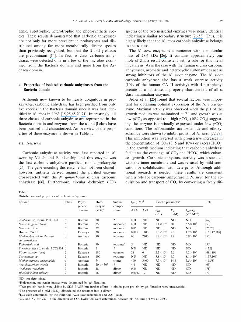
Table 1Distribution and properties of carbonic anhydrases
Enzyme Class Phylo-geneticdomain
Holo-enzyme(kDa)a
Subunitcompo-sition
I50 (WM)d Kinetic parametere Refs.
AZA AZI kcat
(s31)Km
(mM)kcat/Km
(s31 M31)
Anabaena sp. strain PCC7120 K Bacteria 30 ? ND ND ND ND ND [67]Neisseria gonorrhoeae K Bacteria 25 monomer ND ND 1.1U106 20 5.5U107 [53]Neisseria sicca K Bacteria 29 monomer 0.05 ND ND ND ND [25,26]Human CA II K Eukarya 30 monomer 0.015 1100 1.0U106 8.3 1.2U108 [16,142,188]Methanobacterium thermo-autotrophicum
L Archaea 90 tetramer 60 2100 1.7U104 2.9 5.9U106 [55]
Escherichia coli L Bacteria 90 tetramerc 5 ND ND ND ND [50]Synechocystis sp. strain PCC6803 L Bacteria ? ? ND ND ND ND ND [132]Pisum sativum (pea) L Eukarya 180 octamer 28 6 2.3U105 2.5 9.2U107 [48,189]Coccomyxa sp. L Eukarya 100 tetramer ND ND 3.8U105 4.7 8.1U107 [137,164]Methanosarcina thermophila Q Archaea 74 trimer 400 3400 7.7U104 14.8 5.5U106 [16,58]Acetobacterium woodii ? Bacteria 20 or 30b ? 4.4 ND ND ND ND [65]Anabaena variabilis ? Bacteria 48 dimer 0.25 ND ND ND ND [71]Rhodospirillum rubrum ? Bacteria 28 dimer 0.0042 12 ND ND ND [70]
ND, not determined.aHoloenzyme molecular masses were determined by gel ¢ltration.bTwo protein bands were visible by SDS^PAGE but further e¡orts to obtain pure protein by gel ¢ltration were unsuccessful.cThe presence of 5 mM HCO3
3 dissociated the tetramer into a dimer.dI50s were determined for the inhibitors AZA (acetazolamide) and AZI (azide).ekcat and Km for CO2 in the direction of CO2 hydration were determined between pH 8.5 and pH 9.0 at 25³C.
FEMSRE 682 29-8-00 Cyaan Magenta Geel Zwart
K.S. Smith, J.G. Ferry / FEMS Microbiology Reviews 24 (2000) 335^366 339
fusable uncharged gas to a charged species and releasing iton the cytoplasmic side of the membrane.
Although the ¢rst reported prokaryotic carbonic anhy-drase was from N. sicca, no sequence for a neisserial en-
zyme was available until the nucleotide sequence of a genefragment in N. gonorrhoeae, designated ORF2 [74], wasdetected by homology searches of the sequence databases.Chirica et al. [53] cloned the entire gene and found it to
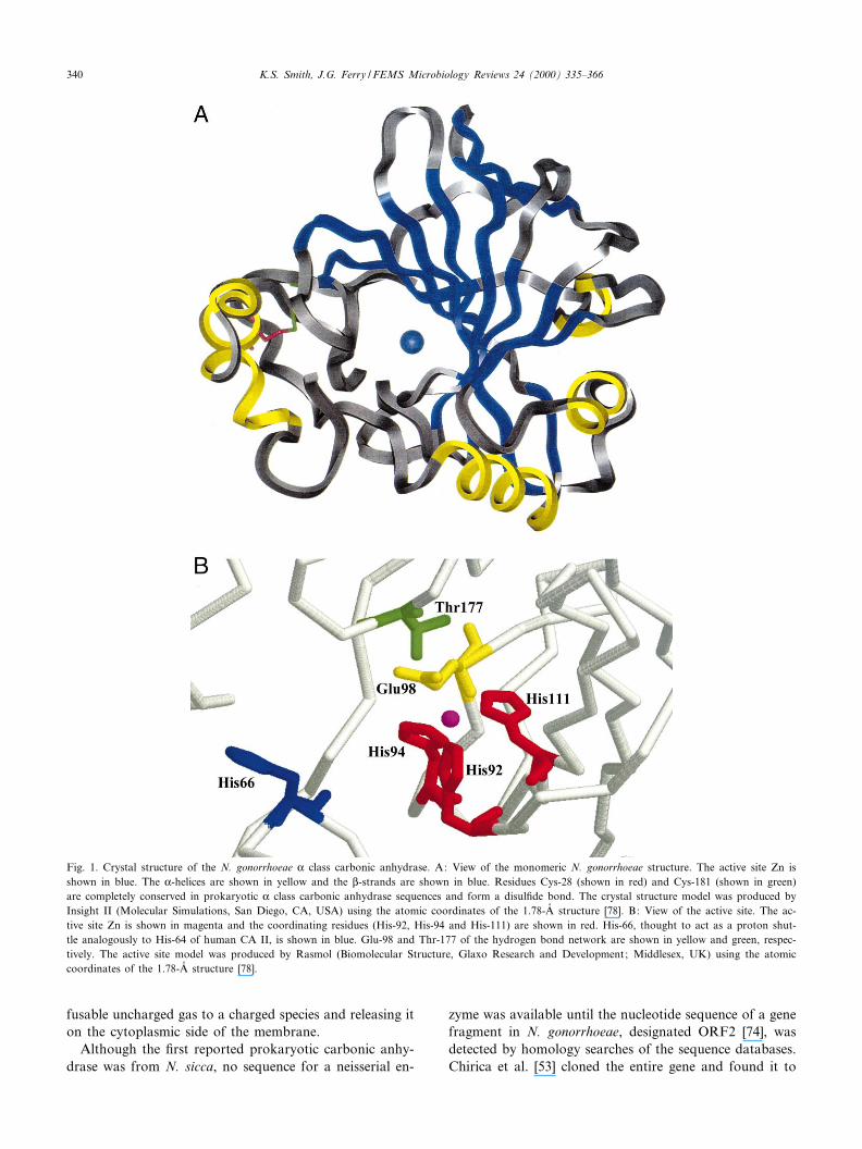
Fig. 1. Crystal structure of the N. gonorrhoeae K class carbonic anhydrase. A: View of the monomeric N. gonorrhoeae structure. The active site Zn isshown in blue. The K-helices are shown in yellow and the L-strands are shown in blue. Residues Cys-28 (shown in red) and Cys-181 (shown in green)are completely conserved in prokaryotic K class carbonic anhydrase sequences and form a disul¢de bond. The crystal structure model was produced byInsight II (Molecular Simulations, San Diego, CA, USA) using the atomic coordinates of the 1.78-Aî structure [78]. B: View of the active site. The ac-tive site Zn is shown in magenta and the coordinating residues (His-92, His-94 and His-111) are shown in red. His-66, thought to act as a proton shut-tle analogously to His-64 of human CA II, is shown in blue. Glu-98 and Thr-177 of the hydrogen bond network are shown in yellow and green, respec-tively. The active site model was produced by Rasmol (Biomolecular Structure, Glaxo Research and Development; Middlesex, UK) using the atomiccoordinates of the 1.78-Aî structure [78].
FEMSRE 682 29-8-00 Cyaan Magenta Geel Zwart
K.S. Smith, J.G. Ferry / FEMS Microbiology Reviews 24 (2000) 335^366340
encode a protein of 252 residues having a molecular massof 28.1 kDa. This carbonic anhydrase was heterologouslyproduced in E. coli as a periplasmic protein lacking the N-terminal 26 residues suggesting that the E. coli processingmachinery recognized this fragment as a signal sequenceand cleaved it. Although the location of the enzyme in N.gonorrhoeae has yet to be ascertained biochemically, theseresults are consistent with a location at or outside thecytoplasmic membrane; therefore, this carbonic anhydrasecould potentially have the same function in CO2 transportas hypothesized for the N. sicca enzyme.
The CO2 hydration activity of the N. gonorrhoeae en-zyme is similar to that of the high activity human CA II[53] (Table 1). The kcat values at pH 9.0 are almost iden-tical, but the Km for the N. gonorrhoeae enzyme is nearly2.5 times greater than reported for human CA II (Table1). The rapid CO2 hydration turnover of approximately106 s31 for human CA II requires the participation of His-64 as a proton shuttle [75^77] and this residue (His-66) isalso conserved in the amino acid sequence of the neisserialcarbonic anhydrase. The N. gonorrhoeae enzyme also hasesterase activity, hydrolyzing 4-nitrophenyl acetate, butthe speci¢c activity is only 5% that of human CA II [53].
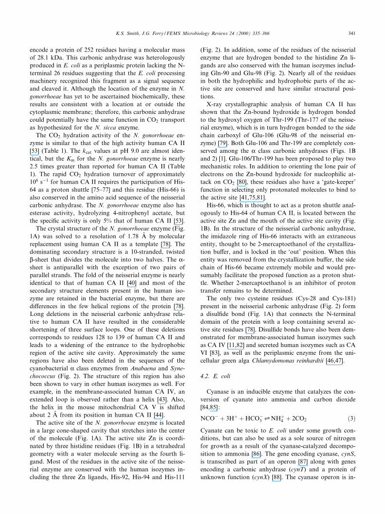
The crystal structure of the N. gonorrhoeae enzyme (Fig.1A) was solved to a resolution of 1.78 Aî by molecularreplacement using human CA II as a template [78]. Thedominating secondary structure is a 10-stranded, twistedL-sheet that divides the molecule into two halves. The K-sheet is antiparallel with the exception of two pairs ofparallel strands. The fold of the neisserial enzyme is nearlyidentical to that of human CA II [40] and most of thesecondary structure elements present in the human iso-zyme are retained in the bacterial enzyme, but there aredi¡erences in the few helical regions of the protein [78].Long deletions in the neisserial carbonic anhydrase rela-tive to human CA II have resulted in the considerableshortening of three surface loops. One of these deletionscorresponds to residues 128 to 139 of human CA II andleads to a widening of the entrance to the hydrophobicregion of the active site cavity. Approximately the sameregions have also been deleted in the sequences of thecyanobacterial K class enzymes from Anabaena and Syne-chococcus (Fig. 2). The structure of this region has alsobeen shown to vary in other human isozymes as well. Forexample, in the membrane-associated human CA IV, anextended loop is observed rather than a helix [43]. Also,the helix in the mouse mitochondrial CA V is shiftedabout 2 Aî from its position in human CA II [44].
The active site of the N. gonorrhoeae enzyme is locatedin a large cone-shaped cavity that stretches into the centerof the molecule (Fig. 1A). The active site Zn is coordi-nated by three histidine residues (Fig. 1B) in a tetrahedralgeometry with a water molecule serving as the fourth li-gand. Most of the residues in the active site of the neisse-rial enzyme are conserved with the human isozymes in-cluding the three Zn ligands, His-92, His-94 and His-111
(Fig. 2). In addition, some of the residues of the neisserialenzyme that are hydrogen bonded to the histidine Zn li-gands are also conserved with the human isozymes includ-ing Gln-90 and Glu-98 (Fig. 2). Nearly all of the residuesin both the hydrophilic and hydrophobic parts of the ac-tive site are conserved and have similar structural posi-tions.
X-ray crystallographic analysis of human CA II hasshown that the Zn-bound hydroxide is hydrogen bondedto the hydroxyl oxygen of Thr-199 (Thr-177 of the neisse-rial enzyme), which is in turn hydrogen bonded to the sidechain carboxyl of Glu-106 (Glu-98 of the neisserial en-zyme) [79]. Both Glu-106 and Thr-199 are completely con-served among the K class carbonic anhydrases (Figs. 1Band 2) [1]. Glu-106/Thr-199 has been proposed to play twomechanistic roles. In addition to orienting the lone pair ofelectrons on the Zn-bound hydroxide for nucleophilic at-tack on CO2 [80], these residues also have a `gate-keeper'function in selecting only protonated molecules to bind tothe active site [41,75,81].
His-66, which is thought to act as a proton shuttle anal-ogously to His-64 of human CA II, is located between theactive site Zn and the mouth of the active site cavity (Fig.1B). In the structure of the neisserial carbonic anhydrase,the imidazole ring of His-66 interacts with an extraneousentity, thought to be 2-mercaptoethanol of the crystalliza-tion bu¡er, and is locked in the `out' position. When thisentity was removed from the crystallization bu¡er, the sidechain of His-66 became extremely mobile and would pre-sumably facilitate the proposed function as a proton shut-tle. Whether 2-mercaptoethanol is an inhibitor of protontransfer remains to be determined.
The only two cysteine residues (Cys-28 and Cys-181)present in the neisserial carbonic anhydrase (Fig. 2) forma disul¢de bond (Fig. 1A) that connects the N-terminaldomain of the protein with a loop containing several ac-tive site residues [78]. Disul¢de bonds have also been dem-onstrated for membrane-associated human isozymes suchas CA IV [11,82] and secreted human isozymes such as CAVI [83], as well as the periplasmic enzyme from the uni-cellular green alga Chlamydomonas reinhardtii [46,47].
4.2. E. coli
Cyanase is an inducible enzyme that catalyzes the con-version of cyanate into ammonia and carbon dioxide[84,85]:
NCO3 � 3H� �HCO33 1NH�4 � 2CO2 �3�
Cyanate can be toxic to E. coli under some growth con-ditions, but can also be used as a sole source of nitrogenfor growth as a result of the cyanase-catalyzed decompo-sition to ammonia [86]. The gene encoding cyanase, cynS,is transcribed as part of an operon [87] along with genesencoding a carbonic anhydrase (cynT) and a protein ofunknown function (cynX) [88]. The cyanase operon is in-
FEMSRE 682 29-8-00 Cyaan Magenta Geel Zwart
K.S. Smith, J.G. Ferry / FEMS Microbiology Reviews 24 (2000) 335^366 341
duced in the presence of cyanate and CynT is found lo-calized in the cytosol [87,88].
The deduced amino acid sequence of CynT shows sig-ni¢cant identity to the spinach and pea sequences, therebymaking it the ¢rst L class carbonic anhydrase sequenceidenti¢ed in the prokaryotes. The subunit molecularmass of the CynT carbonic anhydrase was determined tobe 24 kDa and one Zn is present per subunit [50]. Theisolated enzyme is relatively unstable in the absence ofethylene glycol and appears to aggregate irreversibly.The native molecular mass of CynT is approximately 90kDa indicating that this enzyme is a tetramer, a propertyunlike any plant carbonic anhydrase. When the puri¢edcarbonic anhydrase is stored in 15% ethylene glycol at26³C, a slow cleavage occurs yielding an active fragmentof 22 kDa. Whether this is the result of a contaminatingprotease or an autocatalytic activity has yet to be deter-mined. Cyanate and acetazolamide confer 50% inhibitionat 0.1 and 0.005 mM, respectively [50]. Like the plantcarbonic anhydrases, 4-nitrophenyl acetate does not serveas a substrate [50]. However, carbamate, a product of thebreakdown of cyanate catalyzed by cyanase, is decarboxy-lated by the enzyme [50]. This decarboxylation reactionhas also been shown to be catalyzed by a carbonic anhy-drase from the yeast Saccharomyces cerevisiae [89].
Two pieces of experimental evidence suggest that enzy-
matic activity of CynT may be regulated within the cell.The ¢rst mechanism for regulation is protein degradation.After cyanate is depleted from the culture, CynT is de-graded very rapidly at high pCO2 (3% CO2) but veryslowly at low pCO2 (0.03% CO2) [88]. The processes forincreasing the rate of degradation in the presence of highpCO2 is not yet known. Whether the 22-kDa proteolyticfragment observed upon storage of the puri¢ed enzymerepresents the ¢rst step in the degradation of CynT inthe cell has not been reported. A second regulatory mech-anism may be enzyme dissociation in the presence ofHCO3
3 . Both sucrose density gradient experiments andgel ¢ltration chromatography have shown that the molec-ular mass of the CynT is approximately 50 kDa in thepresence of HCO3
3 , suggesting that the tetramer dissoci-ates into a dimer in the presence of HCO3
3 [50]. By thismechanism, when the intracellular levels of HCO3
3 reach acertain level, the active CynT tetramer would dissociateinto dimers which would presumably be inactive althoughthis has yet to be shown. Subunit dissociation in the pres-ence of HCO3
3 has not been reported for multimeric plantenzymes of the L class or any enzyme from the K or Qclass. Whether this property is unique to CynT or to pro-karyotic enzymes from the L class remains to be deter-mined.
The discovery that cynT encodes a carbonic anhydrase
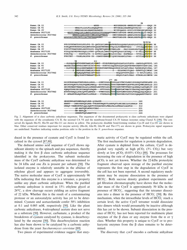
Fig. 2. Alignment of K class carbonic anhydrase sequences. The sequences of the documented prokaryotic K class carbonic anhydrases were alignedwith the sequences of the cytoplasmic CA II, the secreted CA VI and the membrane-bound CA IV human isozymes using Clustal X [190]. The con-served Zn ligands His-92, His-94 and His-111 are shown in yellow. The prokaryotic disul¢de bond-forming residues Cys-28 and Cys-181 are shown inblue. Other conserved residues important for enzyme activity (His-66, Gln-90, Glu-98 and Thr-177) are shown in green. Prokaryotic signal sequencesare underlined. Numbers indicating residue positions refer to the position in the N. gonorrhoeae sequence.
FEMSRE 682 29-8-00 Cyaan Magenta Geel Zwart
K.S. Smith, J.G. Ferry / FEMS Microbiology Reviews 24 (2000) 335^366342
and that cyanase catalyzes the formation of two moleculesof CO2 from cyanate and HCO3
3 suggests that the role ofCynT is to catalyze the hydration of the CO2 generated bycyanase into HCO3
3 , thereby preventing depletion of theHCO3
3 required for further degradation of cyanate or forother metabolic processes [90]. Under low pCO2, it wouldbe expected that CO2 would di¡use out of the cell fasterthan it is hydrated nonenzymatically resulting in depletionof cellular HCO3
3 levels. This hypothesis was tested bythe construction of a mutant with a non-polar deletionmutation within cynT [90]. In the presence of low pCO2
(0.03% CO2), the cynT mutant was extremely sensitiveto inhibition of growth by cyanate. Even though thecyanase wasstill induced, the mutant could not catalyzethe decomposition of cyanate or use cyanate as a solesource of nitrogen. The addition of cyanate to the growthmedia of the cynT deletion mutant resulted in the de-pletion of the cellular HCO3
3 , catalyzed by cyanase,which prevented further degradation of cyanate and ledto an inhibition of growth. When the mutant was grownunder higher pCO2 (3% CO2), the above growth inhibi-tion disappeared. Thus, high pCO2 serves the same role asthe carbonic anhydrase in providing su¤cient levels ofcellular HCO3
3 for growth with cyanate as the nitrogensource.
Two experimental results indicated that the role ofCynT may be more than just to supply HCO3
3 for thedecomposition of cyanate. First, growth of a strain inwhich the entire cyanase operon was deleted was also sus-ceptible to cyanate inhibition at low pCO2 [91]. Growthinhibition was relieved when the cynTSX deletion mutantwas grown in the presence of high pCO2. Second, growthinhibition by cyanate of a cynS deletion strain which stillexpressed carbonic anhydrase activity was the same as thee¡ect on growth of the wild-type strain. The growth inhi-bition was less than that observed for the cynTSX deletionmutant and considerably less than the e¡ect on the cynTdeletion mutant. This result indicates that carbonic anhy-drase alone provided some protection against the inhibi-tion of growth by cyanate. Thus, depletion of HCO3
3 hasmore serious consequences for growth than does the in-hibitory e¡ect of cyanate, implying some essential HCO3
3 -dependent function in E. coli.
The dependence of bacteria on the presence of CO2 forgrowth or for overcoming long lag times (the so-calledsparking phenomenon) has been known for 25 years[92,93]. At low pCO2 and low rates of metabolism, CO2
is expected to di¡use out of the cell faster than it is hy-drated to HCO3
3 , thereby depleting the intracellularHCO3
3 needed for carboxylation reactions. Previous stud-ies have shown that either a large inoculum or the pres-ence of succinate, aspartate or oxaloacetate can overcomethe long lag times observed during growth at low pCO2 byincreasing the citric acid cycle rates and the concentrationof CO2 and HCO3
3 [92,93]. Kozliak et al. [91] showed thatthe growth of wild-type E. coli and the cynS, cynT and
cynTSX deletion mutants is negligible when CO2 is absentfrom the atmosphere. When succinate, fumarate, aspar-tate, K-ketoglutarate or malate are added to the media,growth of both the wild-type and deletion mutant strainsare restored to near normal rates [91]. If growth inhibitionby cyanate of the cynT deletion mutant is due to the de-pletion of HCO3
3 required for replenishing the citric acidcycle intermediates, these intermediates would be expectedto relieve the inhibition of growth by cyanate. However,the presence of citric acid cycle intermediates in thegrowth media failed to overcome the cyanate inhibitionof growth [91]. Another explanation for cyanate inhibitionis that HCO3
3 is required for other carboxylation reactionsbesides those of the citric acid cycle. Kozliak et al. [91]attempted to relieve the inhibition of growth by addingend-product metabolites of other carboxylation reactionssuch as succinate, histidine, adenine, oleate and palmeate[91]. Although some e¡ect was observed, the growth inhi-bition could not be completely overcome.
The above results suggest that CO2 and HCO33 are im-
portant for additional cellular functions that are requiredfor growth and inhibited by cyanate at low pCO2. Un-fortunately, e¡ects on these functions would be observedonly at low pCO2 when the carboxylation reactions arealso limiting [94] which will make identi¢cation of theseputative functions di¤cult. Isocyanic acid (HNNCNO), areactive tautomer of cyanate and a structural and elec-tronic analogue of CO2 (ONCNO), has been used todemonstrate a competition between cyanate and CO2 atsome unknown site thus causing growth inhibition [91].This inhibition could be the result of cyanate either com-peting with CO2 or HCO3
3 for a regulatory or catalyticbinding site or carbamoylating a nucleophilic group (suchas ^SH, ^NH2, ^OH and ^COOH) that might normallyundergo nonenzymatic carboxylation by CO2.
4.3. Salmonella typhimurium
Little is known about what genes are required for intra-cellular growth and survival of pathogens. Valdivia andFalkow devised a selection strategy on the basis of di¡er-ential £uorescence induction for identifying genes whoseexpression is induced when the facultative intracellularpathogen S. typhimurium associates with its host cell[95]. Green £uorescent protein was used as a selectablemarker in conjunction with £uorescence-activated cellsorting. Host macrophages infected with a bacterium hav-ing a transcriptionally active gfp gene fusion were sepa-rated by a £uorescence-activated cell sorter and lysed. Therecovered bacteria were then grown in vitro and those thathad little to no £uorescence were isolated. Fourteen pro-moters with intracellular-dependent activity were identi¢edand the genes present downstream of these promoters werethen isolated. One of the genes identi¢ed was mig-5 (formacrophage-inducible gene), whose expression is induced24-fold in macrophages [95]. The predicted amino acid
FEMSRE 682 29-8-00 Cyaan Magenta Geel Zwart
K.S. Smith, J.G. Ferry / FEMS Microbiology Reviews 24 (2000) 335^366 343
sequence of mig-5 is 24% identical to that of the E. coliCynT L class carbonic anhydrase.
Gene disruptions were constructed for each of the miggenes to determine if the macrophage-inducible proteinshave a role in virulence [95]. Each mutant was tested notonly for the ability to colonize the spleen of BALB/c micein competition assays against wild-type S. typhimurium butalso for the ability to survive in the macrophage-like cellline RAW 264.7. Only 13% of the colony forming unitspresent in the spleen were derived from the mig-5 insertionmutants, but the mig-5 mutant showed no growth defect inthe RAW 264.7 cells. These results suggest that the mig-5gene product is not essential for survival in tissue culturebut is important for survival of S. typhimurium within thehost.
4.4. Cyanobacteria
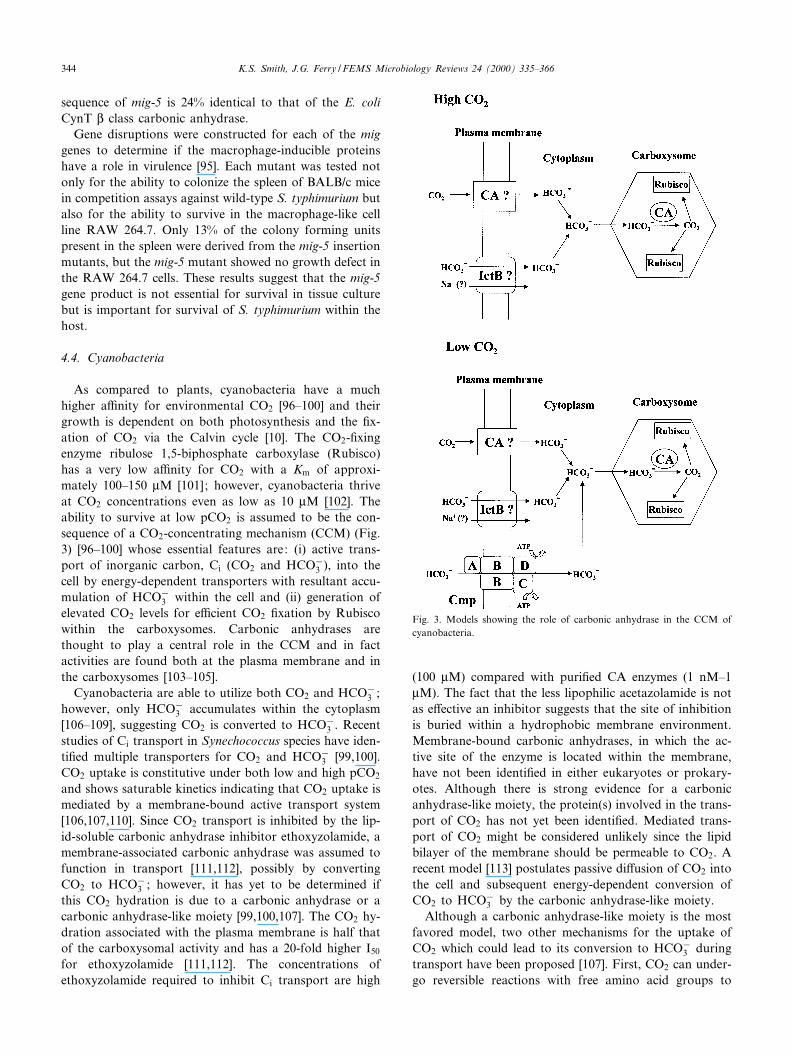
As compared to plants, cyanobacteria have a muchhigher a¤nity for environmental CO2 [96^100] and theirgrowth is dependent on both photosynthesis and the ¢x-ation of CO2 via the Calvin cycle [10]. The CO2-¢xingenzyme ribulose 1,5-biphosphate carboxylase (Rubisco)has a very low a¤nity for CO2 with a Km of approxi-mately 100^150 WM [101]; however, cyanobacteria thriveat CO2 concentrations even as low as 10 WM [102]. Theability to survive at low pCO2 is assumed to be the con-sequence of a CO2-concentrating mechanism (CCM) (Fig.3) [96^100] whose essential features are: (i) active trans-port of inorganic carbon, Ci (CO2 and HCO3
3 ), into thecell by energy-dependent transporters with resultant accu-mulation of HCO3
3 within the cell and (ii) generation ofelevated CO2 levels for e¤cient CO2 ¢xation by Rubiscowithin the carboxysomes. Carbonic anhydrases arethought to play a central role in the CCM and in factactivities are found both at the plasma membrane and inthe carboxysomes [103^105].
Cyanobacteria are able to utilize both CO2 and HCO33 ;
however, only HCO33 accumulates within the cytoplasm
[106^109], suggesting CO2 is converted to HCO33 . Recent
studies of Ci transport in Synechococcus species have iden-ti¢ed multiple transporters for CO2 and HCO3
3 [99,100].CO2 uptake is constitutive under both low and high pCO2
and shows saturable kinetics indicating that CO2 uptake ismediated by a membrane-bound active transport system[106,107,110]. Since CO2 transport is inhibited by the lip-id-soluble carbonic anhydrase inhibitor ethoxyzolamide, amembrane-associated carbonic anhydrase was assumed tofunction in transport [111,112], possibly by convertingCO2 to HCO3
3 ; however, it has yet to be determined ifthis CO2 hydration is due to a carbonic anhydrase or acarbonic anhydrase-like moiety [99,100,107]. The CO2 hy-dration associated with the plasma membrane is half thatof the carboxysomal activity and has a 20-fold higher I50
for ethoxyzolamide [111,112]. The concentrations ofethoxyzolamide required to inhibit Ci transport are high
(100 WM) compared with puri¢ed CA enzymes (1 nM^1WM). The fact that the less lipophilic acetazolamide is notas e¡ective an inhibitor suggests that the site of inhibitionis buried within a hydrophobic membrane environment.Membrane-bound carbonic anhydrases, in which the ac-tive site of the enzyme is located within the membrane,have not been identi¢ed in either eukaryotes or prokary-otes. Although there is strong evidence for a carbonicanhydrase-like moiety, the protein(s) involved in the trans-port of CO2 has not yet been identi¢ed. Mediated trans-port of CO2 might be considered unlikely since the lipidbilayer of the membrane should be permeable to CO2. Arecent model [113] postulates passive di¡usion of CO2 intothe cell and subsequent energy-dependent conversion ofCO2 to HCO3
3 by the carbonic anhydrase-like moiety.Although a carbonic anhydrase-like moiety is the most
favored model, two other mechanisms for the uptake ofCO2 which could lead to its conversion to HCO3
3 duringtransport have been proposed [107]. First, CO2 can under-go reversible reactions with free amino acid groups to
Fig. 3. Models showing the role of carbonic anhydrase in the CCM ofcyanobacteria.
FEMSRE 682 29-8-00 Cyaan Magenta Geel Zwart
K.S. Smith, J.G. Ferry / FEMS Microbiology Reviews 24 (2000) 335^366344
form carbamino adducts or carbamates [114]. An OH3-catalyzed decarboxylation of carbamate would result inthe release of HCO3
3 . A second possible mechanism isthe binding of CO2 to a transition metal. Synthetic tran-sition metal complexes have been shown to bind CO2 andstudies indicate the coordinated CO2 molecule is in a bentcon¢guration [115]. This strained con¢guration enhancessusceptibility of the electrophilic C atom of CO2 to nucle-ophilic attack by OH3 [116]. In both mechanisms, thegeneration of the OH3 is the energy-requiring step andcould occur by raising the pH of the immediate surround-ings either by photosynthetic or respiratory electron trans-port. In fact, CO2 transport has previously been shown tobe dependent on electron transport [117].
Experiments in Synechococcus PCC7942 have identi¢edtwo HCO3
3 transporters [99]. High pCO2-grown cells uti-lize a Na�-dependent HCO3
3 uptake system, while cellsgrown under low pCO2 possess both Na�-dependent andNa�-independent mechanisms for uptake (Fig. 3). Theconstitutive Na�-dependent transporter is inhibited bymonensin, an ionophore that mediates Na�/H� exchange,suggesting a Na�/HCO3
3 symport mechanism [118] ; how-ever, further experiments are needed to con¢rm this mech-anism. A candidate for this transporter is IctB [119], aprotein with nine or 10 membrane spanning domainswhose sequence has identity to a Na�-panthothenate sym-porter (PanF) from E. coli. Disruption of the ictB generesults in a strain requiring high levels of CO2 for growth[119].
CmpABCD is a high a¤nity HCO33 transporter that is
induced during CO2 limitation and appears to be Na�-independent [120]. This transporter belongs to the diversesubfamily of ABC (ATP binding cassette) transportersfound in prokaryotes, also known as tra¤c ATPases[121]. Expression of CmpABCD alone is not su¤cientfor full induction of the high a¤nity state of the CCM.During induction of the CCM, high a¤nity HCO3
3 trans-port occurs within a few minutes of CO2 limitation [122].This fast induction is una¡ected by inhibitors of proteinsynthesis, but does not occur in the presence of inhibitorsof protein kinase [122]. These results suggest that fast in-duction involves post-translational phosphorylation of ex-isting proteins rather than de novo synthesis [122,123]. IfIctB is the Na�-dependent HCO3
3 transporter and its ex-pression is constitutive, it would be a likely target for thekinase-mediated fast induction.
As a result of Ci transport, the steady-state equilibriumbetween CO2 and HCO3
3 is shifted towards HCO33 . The
substrate for the CO2-¢xing enzyme Rubisco is CO2 andnot HCO3
3 [101] ; therefore, an intracellular carbonic an-hydrase would be expected to be present to catalyze thedehydration of HCO3
3 to CO2. Attempts to express humanCA II within the cytoplasm resulted in a decreased rate ofCO2 ¢xation and a requirement for a high pCO2 (5%) forgrowth [124]. This led Price and Badger [125] to theorizethat the intracellular carbonic anhydrase is localized with-
in organelles termed carboxysomes [126]. These polyhedralprotein bodies which contain most if not all the cellularRubisco are thought to serve as a permeability barrier forCO2 to prevent its leakage out of the cell. HCO3
3 in thecytoplasm freely di¡uses into the carboxysome where thecarbonic anhydrase could catalyze its dehydration intoCO2 for consumption by Rubisco. Price et al. [105] iso-lated carboxysomes and identi¢ed an associated carbonicanhydrase activity with some unusual properties, includinginactivation by dithiothreitol and a requirement for 20mM Mg2� for maximal activity. Bedu and Joset[103,104] had previously indicated that the properties ofthe carbonic anhydrase activity associated with the plasmamembrane were distinct from those of the intracellularenzyme(s) based on the di¡erential e¡ects of a carbonicanhydrase inhibitor. These results suggest that di¡erentcarbonic anhydrases have speci¢c and distinct functionsin the transport and ¢xation of CO2.
All three classes of carbonic anhydrase have been iden-ti¢ed in the cyanobacteria. In fact some cyanobacteria (i.e.Synechococcus PCC7942) have genes encoding enzymesfrom all three classes. Each enzyme class and the potentialroles they play are discussed below.
4.4.1. Alpha class carbonic anhydraseA gene ecaA (external carbonic anhydrase alpha class)
was identi¢ed in Anabaena sp. strain PCC7120 which en-codes a protein of approximately 29 kDa [67] with a de-duced amino acid sequence showing identity to severalhuman K class isozymes. Although heterologous expres-sion of EcaA in E. coli resulted in the formation of inclu-sion bodies with no detectable carbonic anhydrase activity,antisera derived against the chicken CA II cross-reactedwith the heterologously produced EcaA. Polyclonal anti-sera derived against the heterologously produced EcaAalso cross-reacted with a 29-kDa protein in whole celllysates of Anabaena.
Expression of EcaA appears to be regulated by the levelof CO2 in the growth medium [67]. High levels of EcaAare present in cells grown at elevated CO2 levels (1% CO2)and low but still detectable levels are observed following24 h of low CO2 (0.01%) exposure. Transcription of ecaAfollows a similar pattern of regulation with high levels ofmRNA being detected only in high CO2-grown cells. Im-munoelectron microscopy studies with antisera derivedagainst EcaA showed that this protein is found outsidethe cell and associated with the cell wall, periplasmic spaceor cytoplasmic membrane [67]. Unfortunately, under opti-mal conditions for EcaA expression, neither whole celllysates nor membrane preparations exhibited carbonic an-hydrase activity; thus, rigorous evidence for the locationof this carbonic anhydrase is not available.
Using primers based on the Anabaena ecaA sequence, ahomologous gene was identi¢ed and isolated from the cy-anobacterium Synechococcus sp. strain PCC7942 [67]. In-terestingly, a search of the entire genome sequence of Sy-
FEMSRE 682 29-8-00 Cyaan Magenta Geel Zwart
K.S. Smith, J.G. Ferry / FEMS Microbiology Reviews 24 (2000) 335^366 345
nechocystis PCC6803 indicated that this microbe lacks agene encoding an K class carbonic anhydrase. The Syne-chococcus ecaA gene encodes a 26-kDa protein with alarge number of positively charged residues in the ami-no-terminal domain suggesting the presence of a signalsequence for membrane targeting. Synechococcus exhibitedthe same pattern of EcaA expression as displayed by Ana-baena with increased levels of EcaA present in cells grownat elevated levels of CO2 [67].
In an e¡ort to identify the role of EcaA in the CCM ofcyanobacteria, deletion mutants of the SynechococcusecaA were constructed [67]. No detectable di¡erence wasobserved between the mutant and the wild-type cells incarbonic anhydrase activity measured at the cell surfaceas determined by the 18O exchange method [127]. ActiveCO2 transport and Na�-dependent and Na�-independentHCO3
3 transport were una¡ected in the ecaA mutant. TheecaA mutant initially grew slower than the wild-type onboth low and high Ci, but the rates were nearly indistin-guishable during steady state. The maximal rates of Ci-dependent photosynthesis (measured by O2 evolution)were also identical ; however, the mutant grown in lowpCO2 displayed a more rapid onset of O2 evolution(29% faster) upon addition of limiting concentrations ofCO2 than the wild-type [67]. Both wild-type and mutantcells exhibited similar patterns of photosynthetic O2 evo-lution when limiting concentrations of HCO3
3 were added.An earlier onset of photosynthesis in the ecaA mutantupon the addition of limiting concentrations of CO2 sug-gests that the immediate external concentration of CO2
available for transport is higher than that for the wild-type. Since CO2 transport is far more rapid than HCO3
3transport, a rapid accumulation of Ci would result. In thewild-type, the activity of EcaA would quickly hydratemuch of the added CO2 ; thus, a lower amount of CO2
would be available and HCO33 would be the major trans-
ported species.These results imply that EcaA does not have a role in
the CCM; however, the possibility that EcaA may play arole under more acidic and alkaline growth conditionscannot be ruled out. Several other potential roles forEcaA in cyanobacteria can be envisaged [67]. EcaA maybe required to fully equilibrate Ci speciation at the site oftransport in order to maximize substrate availability foreither the CO2 or HCO3
3 transporter. Alternatively, EcaAmay serve as a sensor, either detecting or signaling changesin the level of CO2 in the environment [127]. Recent evi-dence suggests that HCO3
3 is the signal for fast inductionof the high a¤nity state of the CCM and therefore detec-tion of the level of CO2 is essential [122,128,129].
4.4.2. Beta class carbonic anhydraseSynechococcus mutants that were able to accumulate Ci
inside the cell but were unable to utilize it for CO2 ¢xationwere isolated [51,130,131]. These mutants required a highlevel of pCO2 (4^5%) for growth and one also exhibited a
30-fold reduction in carboxysomal carbonic anhydrase ac-tivity [51], the complementation of which resulted in theidenti¢cation of the gene isfA [51]. This gene encodes aprotein of 272 amino acids whose sequence is 31% identi-cal to that of the CynT, the L class carbonic anhydrasefrom E. coli.
Inactivation of the isfA gene by insertional mutagenesisresulted in a mutant that accumulated Ci to wild-typelevels internally, but was unable to use the internal Ci
pool for CO2 ¢xation [51]. This observation led Fukuzawaet al. [51] to propose that IsfA is the carboxysomal car-bonic anhydrase whose function is to dehydrate HCO3
3 toCO2 for the CO2-¢xing enzyme Rubisco. Neither physio-logical studies con¢rming the localization of IsfA withinSynechococcus cells nor biochemical characterization ofthe puri¢ed enzyme have been reported.
A DNA fragment was isolated from a Synechocystis sp.strain PCC6803 subgenomic plasmid library that stronglyhybridized to a fragment of the isfA gene from Synecho-coccus sp. strain PCC7942 [132]. The isolated gene, de-noted as ccaA (cyanobacterial carboxysome localized), en-codes a 274-amino acid polypeptide with a predictedmolecular mass of 30.7 kDa. The deduced amino acid se-quence is 55% identical to that of the sequence of theSynechococcus isfA gene product. Heterologous expressionof ccaA in E. coli resulted predominantly in the formationof inclusion bodies. However, carbonic anhydrase activitywas detected in the soluble fraction due to a small amountof soluble CcaA that was sensitive to ethoxyzolamide.
So and Espie [132] used an improved method of subcel-lular fractionation [105] to obtain intact carboxysomesfree of both soluble enzymes and thylakoid membranes.Immunoassays using polyclonal antisera derived againstCcaA revealed that 97% of CcaA was associated withthe isolated intact carboxysomes. These results indicatethat CcaA in Synechocystis is the carboxysomal carbonicanhydrase and provides CO2 for Rubisco. Whether theSynechococcus IsfA is a functional homolog of CcaAand also localized in the carboxysome remains to be de-termined. Previous studies with Synechococcus have indi-cated that the carboxysomal carbonic anhydrase activity isinactivated by dithiothreitol and requires 20 mM Mg2� formaximal activity [105] ; however, these properties were notexamined [132] with the puri¢ed CcaA.
No cross-reactive proteins to antisera derived againstCcaA were detected with the soluble fraction or associatedwith the thylakoid membranes [132] and neither were ex-amined for carbonic anhydrase activity. Whether othercarbonic anhydrases exist in the cytoplasm or are associ-ated with the thylakoid membrane remains to be resolved[133]. No cross-reactive proteins were detected in the par-tially puri¢ed plasma membrane; thus, the carbonic anhy-drase activity present in the plasma membrane is mediatedby an immunologically distinct activity.
An additional open reading frame (slr0051) in Synecho-cystis PCC6803 with a high degree of sequence similarity
FEMSRE 682 29-8-00 Cyaan Magenta Geel Zwart
K.S. Smith, J.G. Ferry / FEMS Microbiology Reviews 24 (2000) 335^366346
to the E. coli CynT L class carbonic anhydrase has beenidenti¢ed [127]. The gene, designated ecaB (external car-bonic anhydrase beta class), encodes a 263-amino acidpolypeptide that has a putative prokaryotic membranelipoprotein lipid attachment site indicating that the poly-protein may either be associated with the membrane orlocated in the periplasm; however, the location of EcaBin Synechocystis PCC6803 has not yet been ascertained.Heterologous expression of EcaB in E. coli resulted inthe formation of inclusion bodies with no detectable car-bonic anhydrase activity [127]. The phenotype of a Syne-chocystis PCC6803 ecaB mutant appeared to be identicalto that of the Synechococccus PCC7042 ecaA mutant inthat no detectable di¡erence was observed between themutant and wild-type cells in enzyme activity, in CO2
and HCO33 transport or in steady state growth [127].
Like the K class enzyme EcaA, EcaB does not play anessential catalytic role in the functioning of the CCM.Thus, the Synechocystis PCC6803 L class carbonic anhy-drase EcaB may have the same function as the Synecho-coccus PCC7942 K class enzyme EcaA. Therefore, thesetwo di¡erent genera of cyanobacteria may use di¡erentclasses of carbonic anhydrase for the same function.
4.4.3. Gamma class carbonic anhydraseA number of genes named ccm (for carbon concentrat-
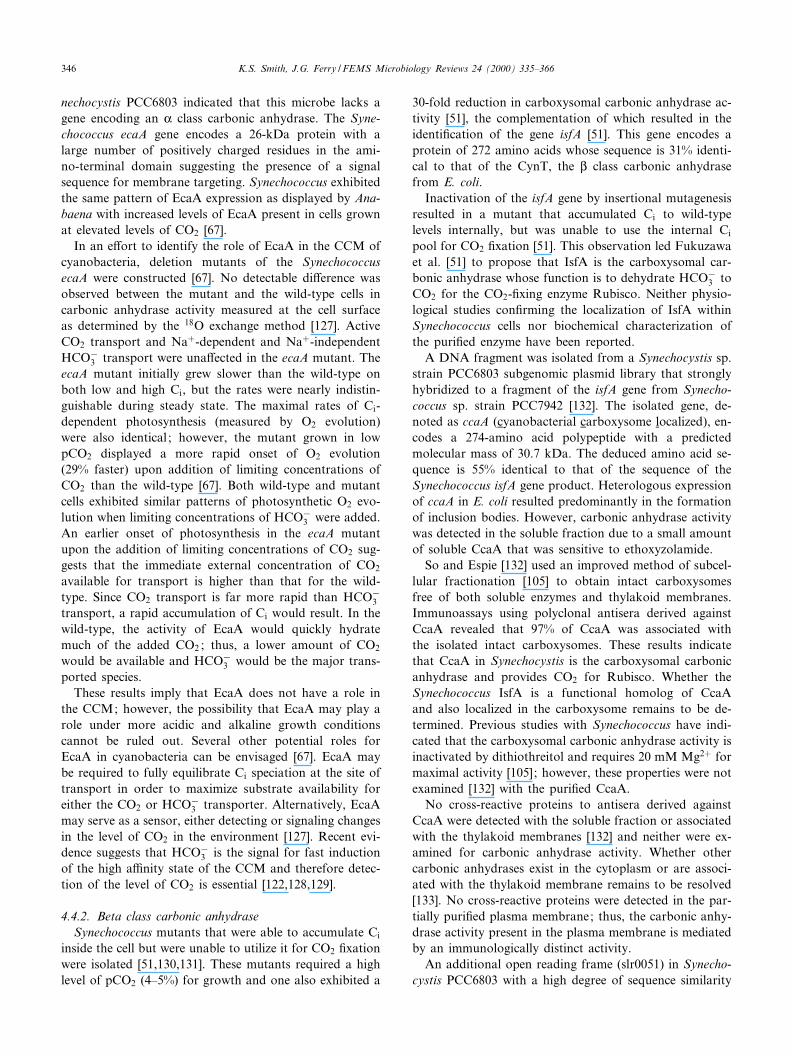
ing mechanism) have been identi¢ed in a region upstreamof the rbcLS operon in Synechococcus PCC7942, whichencodes genes for the subunits of Rubisco [99,134]. Eachof the ccm genes (ccmK, ccmL, ccmM, ccmN and ccmO) isessential for growth at low pCO2 and for correct assemblyof the carboxysome [134]. ccmM encodes a bifunctionalgene product of 539 amino acids of which the ¢rst 219are 35% identical to those of Cam, the prototypical Qcarbonic anhydrase from M. thermophila. Three internalrepeats present in the C-terminal 320 amino acids show45^51% identity to the C-terminal region of the AnabaenaRubisco activase (Fig. 4). Rubisco activase [135] catalyzesthe ATP-dependent activation of the Rubisco^ribulose1,5-biphosphate complex which is energetically coupledto the light reactions of photosynthesis. These internalrepeats also show similarity to the small subunit of Rubis-co (RbcS). The homologous region includes amino acidsArg-86 and Asp-91 of the small subunit of the AnabaenaRubisco (Fig. 4), which are thought to be involved inbinding the small and large subunits of Rubisco together[134]. Genes with similar deduced amino acid sequences,but with four RbcS domains, have also been found inSynechocystis sp. PCC6803 and Synechococcus sp.PCC7002 [99].
Using antisera derived against the 58-kDa CcmM pro-tein from Synechococcus PCC7942 overexpressed in E. coli,
Fig. 4. Alignment of the Synechococcus CcmM sequence with the Anabaena Rubisco activase sequence and the Rubisco small subunit sequences fromT. ferrooxidans and Anabaena sp. Alignments between the CcmM internal repeat regions with the homologous regions of the Rubisco activase (RubAct) and Rubisco small subunit (Rub) sequences are shown. Arg-86 and Asp-91 (referring to the position in the Anabaena Rubisco small subunit se-quence), thought to be involved in binding of the small Rubisco subunit to the large subunits, are highlighted in green.
FEMSRE 682 29-8-00 Cyaan Magenta Geel Zwart
K.S. Smith, J.G. Ferry / FEMS Microbiology Reviews 24 (2000) 335^366 347
CcmM was found to be a moderately abundant proteinlocalized to the carboxysomes although it is not yetknown if it is located inside or on the peripheral shell ofthe carboxysome [99]. The CcmM antisera also cross-re-acts with a 35-kDa protein in the carboxysome-enrichedpreparation, but it is not known if this polypeptide is aprocessed form of CcmM, a second site translation prod-uct or an artifact. Carbonic anhydrase activity has not yetbeen reported for the heterologously expressed CcmM inE. coli and its role in CO2 ¢xation remains to be deter-mined. In addition to a structural role, CcmM may alsoprevent leakage of CO2 out of the carboxysome. AnyCO2 di¡using out of the carboxysome would be convertedto HCO3
3 , which can then be dehydrated by the carbox-ysomal L class carbonic anhydrase to supply CO2 forRubisco.
4.4.4. Unclassi¢edYagawa et al. [71] found carbonic anhydrase activity in
cell extracts from Anabaena variabilis M-2 and M-3 inaddition to A. variabilis ATCC29413. Activity was signi¢-cantly higher in cells grown under low pCO2 than in thosegrown under higher pCO2 (2^4% CO2). The soluble car-bonic anhydrase from A. variabilis ATCC29413 was puri-¢ed greater than 200-fold and major (48 kDa) and minorpeaks (25 kDa) of active carbonic anhydrase were detectedafter gel ¢ltration chromatography of the enzyme. Nativepolyacrylamide gel electrophoresis (PAGE) of both peaksshowed only a single band of approximately 48 kDa, sug-gesting that the major peak of carbonic anhydrase was adimer and the minor peak was the dissociated monomer.The activity of the enzyme from A. variabilis ATCC29413was inhibited by the presence of 5 mM dithiothreitol sim-ilar to the previously described Synechococcus carboxyso-mal carbonic anhydrase activity [105]. These similaritiessuggest this enzyme may be the carboxysomal carbonicanhydrase of A. variabilis ATCC29413, but further experi-ments are clearly required to con¢rm this. The determina-tion of which class of carbonic anhydrase the A. variabilisenzyme belongs to awaits the cloning and sequencing ofthe gene encoding this enzyme.
4.5. Rhodospirillum rubrum
Carbonic anhydrase activity was detected in anaerobi-cally and photosynthetically grown R. rubrum cells [70] ;however, no enzyme activity was detected in cells grownaerobically in the dark. Initial attempts at puri¢cation ofthis soluble enzyme were di¤cult due to a loss of enzymeactivity during the puri¢cation; however, inclusion of Znin the bu¡ers largely resolved the loss of enzyme activity,consistent with a role for this metal in catalysis. The mo-lecular mass of the enzyme was determined to be approx-imately 30 kDa by gel ¢ltration chromatography and su-crose density gradient studies. A single protein band ofapproximately 14 kDa was detected on denaturing SDS^
PAGE indicating that the enzyme is a dimer. In additionto having CO2 hydration activity, the R. rubrum carbonicanhydrase was also shown to have a weak esterase activ-ity; however, this activity was only detectable at enzymelevels 80 times greater than the levels used for the CO2
hydration assay. Thus far, only K class carbonic anhy-drases have been shown to have esterase activity suggest-ing the R. rubrum carbonic anhydrase may belong to thisclass. The cloning and sequencing of the gene encodingthis carbonic anhydrase has not been reported, thus it isimpossible to determine in which class this enzyme be-longs.
4.6. Acetobacterium woodii
The acetogens A. woodii, Acetohalobium arabaticum,Clostridium formicoaceticum and Sporomusa silvaceticawere all found to contain carbonic anhydrase activity[65]. Of the acetogens tested, A. woodii had the highestspeci¢c carbonic anhydrase activity (14 U mg31) and ac-tivity was detected in cell extracts of either glucose- or H2-CO2-grown cells. The carbonic anhydrase was found to becytoplasmic and was puri¢ed 300-fold to a speci¢c activityof 5236 U mg31. After puri¢cation, two prominent bands(approximately 20 and 30 kDa) were visible on SDS^PAGE gel, but further attempts to obtain pure carbonicanhydrase by gel ¢ltration were unsuccessful.
The physiological functions of carbonic anhydrase inacetogens is unknown, but several can be envisioned[65]. Acetogens have a high demand for CO2 in both en-ergy-yielding metabolism and pathways for CO2 ¢xation;thus one function may be to increase the intracellular CO2
levels. Many acetogens cannot grow acetogenically underheterotrophic or autotrophic conditions in the absence ofCO2. A CCM may be essential during autotrophic growthwhen CO2 is used as both a carbon source and an electronacceptor. Acetazolamide (2 mM) had no apparent a¡ecton the growth of A. woodii during heterotrophic condi-tions [65]; however, growth on H2-CO2 was completelyinhibited suggesting an important role for carbonic anhy-drase in autotrophic growth. Other potential roles for theA. woodii carbonic anhydrase include the regulation ofintracellular pH due to the large amount of acetate beingproduced during growth and the coupling to an acetate/HCO3
3 antiporter [65].
5. Properties of isolated carbonic anhydrases from theArchaea domain
Although only two archaeal carbonic anhydrases havebeen documented [16,55], the enzyme appears to be wide-spread in the two kingdoms of the Archaea domain [14].Interestingly, a gene encoding a putative K class carbonicanhydrase has not yet been identi¢ed in the domain, con-sistent with the relatively recent invention of this class.
FEMSRE 682 29-8-00 Cyaan Magenta Geel Zwart
K.S. Smith, J.G. Ferry / FEMS Microbiology Reviews 24 (2000) 335^366348
The two documented archaeal enzymes are discussed indetail below.
5.1. M. thermoautotrophicum vH
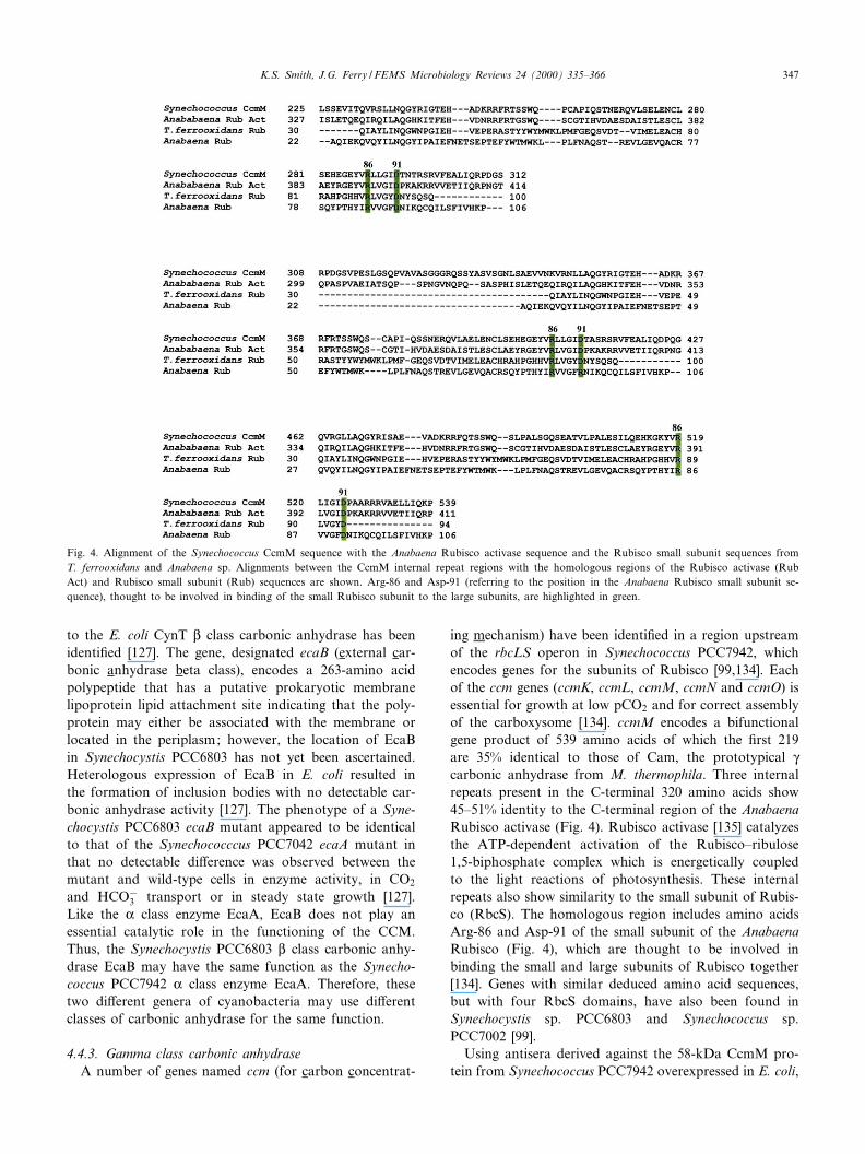
Recently, genome sequencing of the thermophilic, obli-gately chemolithoautotrophic methanoarchaeon M. ther-moautotrophicum vH revealed an open reading framewith a deduced sequence 34% identical to CynT, the Lclass carbonic anhydrase of E. coli [54,69]. The ORF-en-coded protein, Cab (carbonic anhydrase beta), was ex-pressed in E. coli, puri¢ed to electrophoretic homogeneityand found to have carbonic anhydrase activity [55]. Nativegel ¢ltration chromatography estimates the molecularmass of Cab to be approximately 90 kDa [55]. The calcu-lated subunit molecular mass is 18.9 kDa suggesting thatnative Cab is a tetramer in agreement with the E. coliCynT. Analytical ultracentrifugation studies con¢rm thatCab is a tetramer [59]. Thus, the ¢rst two documentedprokaryotic L class carbonic anhydrases are tetramers[50] di¡ering from those characterized from spinach andpea (dicotyledonous plants) which are octameric [49]. CDanalysis and secondary structure predictions of Cab andthe L class spinach enzyme indicate a domination of K-helical structure that is in stark contrast to that of the Kand Q class carbonic anhydrases which are composedmainly of L-sheet structures [59].
Puri¢ed Cab retains full activity after incubation for atleast 15 min at temperatures up to 75³C; thus, Cab is the
most thermostable carbonic anhydrase yet characterized.Iodide, nitrate and azide are potent inhibitors; however,anions such as chloride and sulfate, which inhibit plant Lclass carbonic anhydrases, have no e¡ect on the activity ofCab. The insensitivity to chloride and sulfate has also beenobserved for the Q class carbonic anhydrase from M. ther-mophila [15,16], the only other known methanoarchaealcarbonic anhydrase. Like other characterized carbonic an-hydrases, Cab is also susceptible to inhibition by the sul-fonamides acetazolamide and ethoxyzolamide. As withother characterized L class carbonic anhydrases, Cabshows no detectable esterase activity.
X-ray absorption spectroscopy (XAS) analysis clearlyindicates that the Zn coordination of L class Cab is dis-tinct from that of the K and Q classes of carbonic anhy-drase [59]. Extended X-ray absorption ¢ne structure (EX-AFS) results suggest that the active site Zn is coordinatedby two cysteine residues and two oxygen/nitrogen ligands[59]. One oxygen/nitrogen ligand may derive from histidinewhile the other most likely results from a water moleculewhich is deprotonated and serves as the Zn-hydroxide at-tacking CO2. Residues Cys-32, His-87 and Cys-90 of Cabare completely conserved in the L class carbonic anhydrasesequences (Fig. 5). Although site-directed mutagenesisstudies with the spinach enzyme indicate that alterationsto these three residues result in variants lacking Zn [136],de¢nitive proof that these residues serve as ligands awaitsthe ¢rst crystal structure for any L class carbonic anhy-drase.
Fig. 5. Alignment of L class carbonic anhydrase sequences. The sequences of documented prokaryotic L class carbonic anhydrases were aligned with thesequences of the spinach and pea enzymes using Clustal X [190]. The spinach and pea chloroplast transit peptides (amino acids 1^98 for the spinach en-zyme and 1^70 for the pea enzyme) were not included in the alignment. The conserved Zn ligands Cys-32, His-87 and Cys-90 are indicated in yellow.The completely conserved residues Asp-34 and Arg-36 are shown in green and the highly conserved Gly-50 is shown in blue. Numbers indicating resi-due positions refer to the position in the M. thermoautotrophicum sequence.
FEMSRE 682 29-8-00 Cyaan Magenta Geel Zwart
K.S. Smith, J.G. Ferry / FEMS Microbiology Reviews 24 (2000) 335^366 349
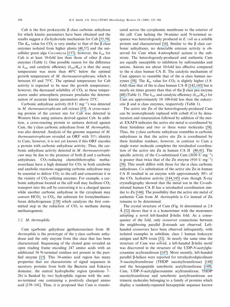
Cab is the ¢rst prokaryotic L class carbonic anhydrasefor which kinetic parameters have been obtained and theresults suggest a Zn-hydroxide mechanism for Cab [55,59].The Km value for CO2 is very similar to that of the L classenzymes isolated from higher plants [48,57] and the uni-cellular green alga Coccomyxa [137]; however, the kcat forCab is at least 10-fold less than those of other L classenzymes (Table 1). One possible reason for the di¡erencein kcat and catalytic e¤ciency (kcat/Km) is that the assaytemperature was more than 40³C below the optimalgrowth temperature of M. thermoautotrophicum, which isbetween 65 and 75³C. The optimal temperature for Cabactivity is expected to be near the growth temperature;however, the decreased solubility of CO2 at these temper-atures under atmospheric pressure precludes the determi-nation of accurate kinetic parameters above 25³C.
Carbonic anhydrase activity (0.8 U mg31) was detectedin M. thermoautotrophicum cell extract [55]. A cross-react-ing protein of the correct size for Cab was detected inWestern blots using antisera derived against Cab. In addi-tion, a cross-reacting protein to antisera derived againstCam, the Q class carbonic anhydrase from M. thermophila,was also detected. Analysis of the genome sequence of M.thermoautotrophicum revealed an ORF with 31% identityto Cam; however, it is not yet known if this ORF encodesa protein with carbonic anhydrase activity. Thus, the car-bonic anhydrase activity detected in M. thermoautotrophi-cum may be due to the presence of both L and Q carbonicanhydrases. CO2-reducing chemolithotrophic metha-noarchaea have a high demand for CO2 in both catabolicand anabolic reactions suggesting carbonic anhydrase maybe essential to deliver CO2 to the cell and concentrate it inthe vicinity of CO2-utilizing enzymes. For example, a car-bonic anhydrase located at the cell wall may facilitate CO2
transport into the cell by converting it to a charged specieswhile another carbonic anhydrase in the cytoplasm mayconvert HCO3
3 to CO2, the substrate for formylmethano-furan dehydrogenase [138] which catalyzes the ¢rst com-mitted step in the reduction of CO2 to methane duringmethanogenesis.
5.2. M. thermophila
Cam (carbonic anhydrase methanosarcina) from M.thermophila is the prototype of the Q class carbonic anhy-drase and the only enzyme from this class that has beencharacterized. Sequencing of the cloned gene revealed anopen reading frame encoding 247 amino acids with anadditional 34 N-terminal residues not present in the puri-¢ed enzyme [15]. This 34-amino acid region has manyproperties that are characteristic of signal sequences insecretory proteins from both the Bacteria and Eukaryadomains; the central hydrophobic region (positions 7^26) is £anked by two hydrophilic regions with the ami-no-terminal one containing a positively charged aminoacid [139^141]. Thus, it is proposed that Cam is translo-
cated across the cytoplasmic membrane to the exterior ofthe cell. Cam lacking the 34-amino acid N-terminal se-quence was heterologously produced in E. coli as a solubleprotein and characterized [16]. Similar to the L class car-bonic anhydrases, no detectable esterase activity is ob-served for Cam when 4-nitrophenyl acetate is the sub-strate. The heterologously-produced and authentic Camare equally susceptible to inhibition by sulfonamides andanions. Anions are about 10-fold less e¡ective comparedto the K class human CA II. The catalytic mechanism ofCam appears to resemble that of the K class human iso-zymes [58]. The Km value for CO2 is slightly higher (1.8-fold) than that of the K class human CA II [142,143] but isnearly six times greater than that of the L class pea enzyme[48] (Table 1). The kcat and catalytic e¤ciency (kcat/Km) forCam are approximately 10^100-fold less than the eukary-otic L and K class enzymes, respectively (Table 1).
The active site Zn of the heterologously-produced Camcan be isomorphously replaced with cobalt (Co) by dena-turation and renaturation followed by addition of the met-al. EXAFS indicates the active site metal is coordinated bythree histidines and two or three water molecules [58].Thus, the Q class carbonic anhydrase resembles K carbonicanhydrases in that the active site Zn is coordinated bythree histidine residues; however, in contrast to Cam asingle water molecule completes the tetrahedral coordina-tion of the active site Zn in human CA II [40,41]. Thespeci¢c activity of the Co-substituted Cam (2020 U mg31)is greater than twice that of the Zn enzyme (910 U mg31)[58]. This result di¡ers with those for the K class carbonicanhydrases. Co substitution of the active site Zn in humanCA II resulted in an enzyme with approximately 50% ofthe CO2 hydration activity [144,145] even though X-raycrystallography showed that the metal ion in the Co-sub-stituted human CA II has a tetrahedral coordination sim-ilar to Zn [146]. The possibility that the active site metal ofauthentic Cam from M. thermophila is Co instead of Znremains to be determined.
The crystal structure of Cam (Fig. 6) determined at 2.8Aî [52] shows that it is a homotrimer with the monomersadopting a novel left-handed L-helix fold. As a conse-quence of the fold, only crossover connections betweenthe neighboring parallel L-strands are observed. Left-handed crossovers have been observed infrequently, withisolated examples in subtilisin, class 1 human leukocyteantigen and KP4 toxin [52]. At nearly the same time thestructure of Cam was solved, a left-handed L-helix motifwas discovered in the structure of the UDP-N-acetylglu-cosamine acyltransferase [147]. More recently, left-handedparallel L-helices were reported for tetrahydrodipicolinateN-succinyltransferase (THDP succinyltransferase) [148]and the hexapeptide xenobiotic acetyltransferase [149].Cam, UDP-N-acetylglucosamine acyltransferase, THDPsuccinyltransferase and xenobiotic acetyltransferase aretrimeric molecules belonging to a family of proteins whichdisplay a tandemly-repeated hexapeptide sequence known
FEMSRE 682 29-8-00 Cyaan Magenta Geel Zwart
K.S. Smith, J.G. Ferry / FEMS Microbiology Reviews 24 (2000) 335^366350
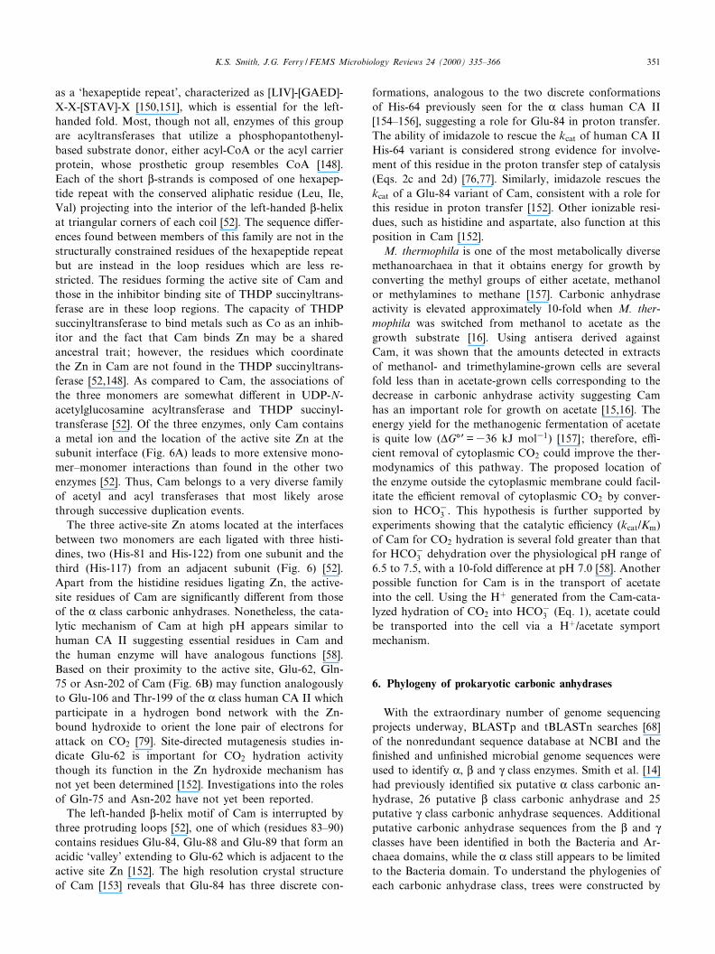
as a `hexapeptide repeat', characterized as [LIV]-[GAED]-X-X-[STAV]-X [150,151], which is essential for the left-handed fold. Most, though not all, enzymes of this groupare acyltransferases that utilize a phosphopantothenyl-based substrate donor, either acyl-CoA or the acyl carrierprotein, whose prosthetic group resembles CoA [148].Each of the short L-strands is composed of one hexapep-tide repeat with the conserved aliphatic residue (Leu, Ile,Val) projecting into the interior of the left-handed L-helixat triangular corners of each coil [52]. The sequence di¡er-ences found between members of this family are not in thestructurally constrained residues of the hexapeptide repeatbut are instead in the loop residues which are less re-stricted. The residues forming the active site of Cam andthose in the inhibitor binding site of THDP succinyltrans-ferase are in these loop regions. The capacity of THDPsuccinyltransferase to bind metals such as Co as an inhib-itor and the fact that Cam binds Zn may be a sharedancestral trait ; however, the residues which coordinatethe Zn in Cam are not found in the THDP succinyltrans-ferase [52,148]. As compared to Cam, the associations ofthe three monomers are somewhat di¡erent in UDP-N-acetylglucosamine acyltransferase and THDP succinyl-transferase [52]. Of the three enzymes, only Cam containsa metal ion and the location of the active site Zn at thesubunit interface (Fig. 6A) leads to more extensive mono-mer^monomer interactions than found in the other twoenzymes [52]. Thus, Cam belongs to a very diverse familyof acetyl and acyl transferases that most likely arosethrough successive duplication events.
The three active-site Zn atoms located at the interfacesbetween two monomers are each ligated with three histi-dines, two (His-81 and His-122) from one subunit and thethird (His-117) from an adjacent subunit (Fig. 6) [52].Apart from the histidine residues ligating Zn, the active-site residues of Cam are signi¢cantly di¡erent from thoseof the K class carbonic anhydrases. Nonetheless, the cata-lytic mechanism of Cam at high pH appears similar tohuman CA II suggesting essential residues in Cam andthe human enzyme will have analogous functions [58].Based on their proximity to the active site, Glu-62, Gln-75 or Asn-202 of Cam (Fig. 6B) may function analogouslyto Glu-106 and Thr-199 of the K class human CA II whichparticipate in a hydrogen bond network with the Zn-bound hydroxide to orient the lone pair of electrons forattack on CO2 [79]. Site-directed mutagenesis studies in-dicate Glu-62 is important for CO2 hydration activitythough its function in the Zn hydroxide mechanism hasnot yet been determined [152]. Investigations into the rolesof Gln-75 and Asn-202 have not yet been reported.
The left-handed L-helix motif of Cam is interrupted bythree protruding loops [52], one of which (residues 83^90)contains residues Glu-84, Glu-88 and Glu-89 that form anacidic `valley' extending to Glu-62 which is adjacent to theactive site Zn [152]. The high resolution crystal structureof Cam [153] reveals that Glu-84 has three discrete con-
formations, analogous to the two discrete conformationsof His-64 previously seen for the K class human CA II[154^156], suggesting a role for Glu-84 in proton transfer.The ability of imidazole to rescue the kcat of human CA IIHis-64 variant is considered strong evidence for involve-ment of this residue in the proton transfer step of catalysis(Eqs. 2c and 2d) [76,77]. Similarly, imidazole rescues thekcat of a Glu-84 variant of Cam, consistent with a role forthis residue in proton transfer [152]. Other ionizable resi-dues, such as histidine and aspartate, also function at thisposition in Cam [152].
M. thermophila is one of the most metabolically diversemethanoarchaea in that it obtains energy for growth byconverting the methyl groups of either acetate, methanolor methylamines to methane [157]. Carbonic anhydraseactivity is elevated approximately 10-fold when M. ther-mophila was switched from methanol to acetate as thegrowth substrate [16]. Using antisera derived againstCam, it was shown that the amounts detected in extractsof methanol- and trimethylamine-grown cells are severalfold less than in acetate-grown cells corresponding to thedecrease in carbonic anhydrase activity suggesting Camhas an important role for growth on acetate [15,16]. Theenergy yield for the methanogenic fermentation of acetateis quite low (vG³P=336 kJ mol31) [157] ; therefore, e¤-cient removal of cytoplasmic CO2 could improve the ther-modynamics of this pathway. The proposed location ofthe enzyme outside the cytoplasmic membrane could facil-itate the e¤cient removal of cytoplasmic CO2 by conver-sion to HCO3
3 . This hypothesis is further supported byexperiments showing that the catalytic e¤ciency (kcat/Km)of Cam for CO2 hydration is several fold greater than thatfor HCO3
3 dehydration over the physiological pH range of6.5 to 7.5, with a 10-fold di¡erence at pH 7.0 [58]. Anotherpossible function for Cam is in the transport of acetateinto the cell. Using the H� generated from the Cam-cata-lyzed hydration of CO2 into HCO3
3 (Eq. 1), acetate couldbe transported into the cell via a H�/acetate symportmechanism.
6. Phylogeny of prokaryotic carbonic anhydrases
With the extraordinary number of genome sequencingprojects underway, BLASTp and tBLASTn searches [68]of the nonredundant sequence database at NCBI and the¢nished and un¢nished microbial genome sequences wereused to identify K, L and Q class enzymes. Smith et al. [14]had previously identi¢ed six putative K class carbonic an-hydrase, 26 putative L class carbonic anhydrase and 25putative Q class carbonic anhydrase sequences. Additionalputative carbonic anhydrase sequences from the L and Qclasses have been identi¢ed in both the Bacteria and Ar-chaea domains, while the K class still appears to be limitedto the Bacteria domain. To understand the phylogenies ofeach carbonic anhydrase class, trees were constructed by
FEMSRE 682 29-8-00 Cyaan Magenta Geel Zwart
K.S. Smith, J.G. Ferry / FEMS Microbiology Reviews 24 (2000) 335^366 351
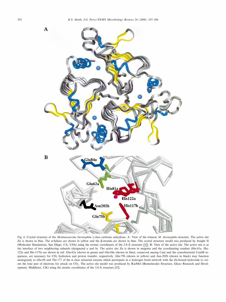
Fig. 6. Crystal structure of the Methanosarcina thermophila Q class carbonic anhydrase. A: View of the trimeric M. thermophila structure. The active siteZn is shown in blue. The K-helices are shown in yellow and the L-strands are shown in blue. The crystal structure model was produced by Insight II(Molecular Simulations, San Diego, CA, USA) using the atomic coordinates of the 2.8-Aî structure [52]. B: View of the active site. The active site is atthe interface of two neighboring subunits (designated a and b). The active site Zn is shown in magenta and the coordinating residues (His-81a, His-122a and His-117b) are shown in red. Glu-62a (shown in green) and Glu-84a (shown in blue), conserved among Cam and the cyanobacterial CcmM se-quences, are necessary for CO2 hydration and proton transfer, respectively. Gln-75b (shown in yellow) and Asn-202b (shown in black) may functionanalogously to Glu-98 and Thr-177 of the K class neisserial enzyme which participate in a hydrogen bond network with the Zn-bound hydroxide to ori-ent the lone pair of electrons for attack on CO2. The active site model was produced by RasMol (Biomolecular Structure, Glaxo Research and Devel-opment, Middlesex, UK) using the atomic coordinates of the 2.8-Aî structure [52].
FEMSRE 682 29-8-00 Cyaan Magenta Geel Zwart
K.S. Smith, J.G. Ferry / FEMS Microbiology Reviews 24 (2000) 335^366352
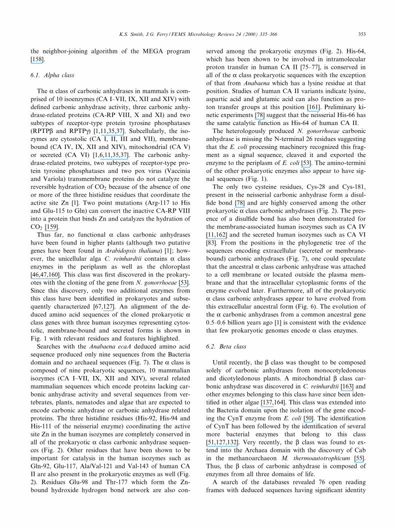
the neighbor-joining algorithm of the MEGA program[158].
6.1. Alpha class
The K class of carbonic anhydrases in mammals is com-prised of 10 isoenzymes (CA I^VII, IX, XII and XIV) withde¢ned carbonic anhydrase activity, three carbonic anhy-drase-related proteins (CA-RP VIII, X and XI) and twosubtypes of receptor-type protein tyrosine phosphatases(RPTPL and RPTPQ) [1,11,35,37]. Subcellularly, the iso-zymes are cytostolic (CA I, II, III and VII), membrane-bound (CA IV, IX, XII and XIV), mitochondrial (CA V)or secreted (CA VI) [1,6,11,35,37]. The carbonic anhy-drase-related proteins, two subtypes of receptor-type pro-tein tyrosine phosphatases and two pox virus (Vacciniaand Variola) transmembrane proteins do not catalyze thereversible hydration of CO2 because of the absence of oneor more of the three histidine residues that coordinate theactive site Zn [1]. Two point mutations (Arg-117 to Hisand Glu-115 to Gln) can convert the inactive CA-RP VIIIinto a protein that binds Zn and catalyzes the hydration ofCO2 [159].
Thus far, no functional K class carbonic anhydraseshave been found in higher plants (although two putativegenes have been found in Arabidopsis thaliana) [1] ; how-ever, the unicellular alga C. reinhardtii contains K classenzymes in the periplasm as well as the chloroplast[46,47,160]. This class was ¢rst discovered in the prokary-otes with the cloning of the gene from N. gonorrhoeae [53].Since this discovery, only two additional enzymes fromthis class have been identi¢ed in prokaryotes and subse-quently characterized [67,127]. An alignment of the de-duced amino acid sequences of the cloned prokaryotic Kclass genes with three human isozymes representing cytos-tolic, membrane-bound and secreted forms is shown inFig. 1 with relevant residues and features highlighted.
Searches with the Anabaena ecaA deduced amino acidsequence produced only nine sequences from the Bacteriadomain and no archaeal sequences (Fig. 7). The K class iscomposed of nine prokaryotic sequences, 10 mammalianisozymes (CA I^VII, IX, XII and XIV), several relatedmammalian sequences which encode proteins lacking car-bonic anhydrase activity and several sequences from ver-tebrates, plants, nematodes and algae that are expected toencode carbonic anhydrase or carbonic anhydrase relatedproteins. The three histidine residues (His-92, His-94 andHis-111 of the neisserial enzyme) coordinating the activesite Zn in the human isozymes are completely conserved inall of the prokaryotic K class carbonic anhydrase sequen-ces (Fig. 2). Other residues that have been shown to beimportant for catalysis in the human isozymes such asGln-92, Glu-117, Ala/Val-121 and Val-143 of human CAII are also present in the prokaryotic enzymes as well (Fig.2). Residues Glu-98 and Thr-177 which form the Zn-bound hydroxide hydrogen bond network are also con-
served among the prokaryotic enzymes (Fig. 2). His-64,which has been shown to be involved in intramolecularproton transfer in human CA II [75^77], is conserved inall of the K class prokaryotic sequences with the exceptionof that from Anabaena which has a lysine residue at thatposition. Studies of human CA II variants indicate lysine,aspartic acid and glutamic acid can also function as pro-ton transfer groups at this position [161]. Preliminary ki-netic experiments [78] suggest that the neisserial His-66 hasthe same catalytic function as His-64 of human CA II.
The heterologously produced N. gonorrhoeae carbonicanhydrase is missing the N-terminal 26 residues suggestingthat the E. coli processing machinery recognized this frag-ment as a signal sequence, cleaved it and exported theenzyme to the periplasm of E. coli [53]. The amino-terminiof the other prokaryotic enzymes also appear to have sig-nal sequences (Fig. 1).
The only two cysteine residues, Cys-28 and Cys-181,present in the neisserial carbonic anhydrase form a disul-¢de bond [78] and are highly conserved among the otherprokaryotic K class carbonic anhydrases (Fig. 2). The pres-ence of a disul¢de bond has also been demonstrated forthe membrane-associated human isozymes such as CA IV[11,162] and the secreted human isozymes such as CA VI[83]. From the positions in the phylogenetic tree of thesequences encoding extracellular (secreted or membrane-bound) carbonic anhydrases (Fig. 7), one could speculatethat the ancestral K class carbonic anhydrase was attachedto a cell membrane or located outside the plasma mem-brane and that the intracellular cytoplasmic forms of theenzyme evolved later. Furthermore, all of the prokaryoticK class carbonic anhydrases appear to have evolved fromthis extracellular ancestral form (Fig. 6). The evolution ofthe K carbonic anhydrases from a common ancestral gene0.5^0.6 billion years ago [1] is consistent with the evidencethat few prokaryotic genomes encode K class enzymes.
6.2. Beta class
Until recently, the L class was thought to be composedsolely of carbonic anhydrases from monocotyledonousand dicotyledonous plants. A mitochondrial L class car-bonic anhydrase was discovered in C. reinhardtii [163] andother enzymes belonging to this class have since been iden-ti¢ed in other algae [137,164]. This class was extended intothe Bacteria domain upon the isolation of the gene encod-ing the CynT enzyme from E. coli [50]. The identi¢cationof CynT has been followed by the identi¢cation of severalmore bacterial enzymes that belong to this class[51,127,132]. Very recently, the L class was found to ex-tend into the Archaea domain with the discovery of Cabin the methanoarchaeon M. thermoautotrophicum [55].Thus, the L class of carbonic anhydrase is composed ofenzymes from all three domains of life.
A search of the databases revealed 76 open readingframes with deduced sequences having signi¢cant identity
FEMSRE 682 29-8-00 Cyaan Magenta Geel Zwart
K.S. Smith, J.G. Ferry / FEMS Microbiology Reviews 24 (2000) 335^366 353
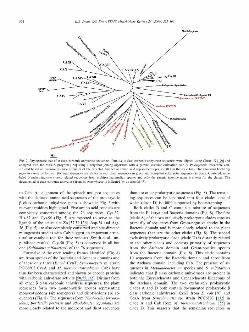
to Cab. An alignment of the spinach and pea sequenceswith the deduced amino acid sequences of the prokaryoticL class carbonic anhydrase genes is shown in Fig. 5 withrelevant residues highlighted. Five amino acid residues arecompletely conserved among the 76 sequences. Cys-32,His-87 and Cys-90 (Fig. 5) are expected to serve as theligands of the active site Zn [57,59,136]. Asp-34 and Arg-36 (Fig. 5) are also completely conserved and site-directedmutagenesis studies with Cab suggest an important struc-tural or catalytic role for these residues (Smith et al., un-published results). Gly-50 (Fig. 5) is conserved in all butone (Sulfolobus solfataricus) of the 76 sequences.
Forty-¢ve of the open reading frames identi¢ed (Fig. 8)are from species of the Bacteria and Archaea domains andof these only three (E. coli CynT, Synechocystis sp. strainPCC6803 CcaA and M. thermoautotrophicum Cab) havethus far been characterized and shown to encode proteinswith carbonic anhydrase activity [50,55,132]. Distinct fromall other L class carbonic anhydrase sequences, the plantsequences form two monophyletic groups representingmonocotyledons (six sequences) and dicotyledons (16 se-quences) (Fig. 8). The sequences from Thiobacillus ferroox-idans, Bordetella pertussis and Rhodobacter capsulatus aremore closely related to the monocot and dicot sequences
than are other prokaryotic sequences (Fig. 8). The remain-ing sequences can be separated into four clades, one ofwhich (clade D) is 100% supported by bootstrapping.
Both clades B and C contain a mixture of sequencesfrom the Eukarya and Bacteria domains (Fig. 8). The ¢rst(clade A) of the two exclusively prokaryotic clades consistsprimarily of sequences from Gram-negative species in theBacteria domain and is more closely related to the plantsequences than are the other clades (Fig. 8). The secondexclusively prokaryotic clade (clade D) is distantly relatedto the other clades and consists primarily of sequencesfrom the Archaea domain and Gram-positive speciesfrom the Bacteria domain (Fig. 8). This clade contains10 sequences from the Bacteria domain and three fromthe Archaea domain, including Cab. The presence of se-quences in Methanobacterium species and S. solfataricusindicates that L class carbonic anhydrases are present inboth the Euryarchaeota and Crenarchaeota kingdoms ofthe Archaea domain. The two exclusively prokaryoticclades A and D both contain documented prokaryotic Lclass carbonic anhydrases, CynT from E. coli [50] andCcaA from Synechocystis sp. strain PCC6803 [132] inclade A and Cab from M. thermoautotrophicum [55] inclade D. This suggests that the remaining sequences in
Fig. 7. Phylogenetic tree of K class carbonic anhydrase sequences. Putative K class carbonic anhydrase sequences were aligned using Clustal X [190] andanalyzed with the MEGA program [158] using a neighbor joining algorithm with a gamma distance estimation (K= 2). Phylogenetic trees were con-structed based on pairwise distance estimates of the expected number of amino acid replacements per site (0.1 in the scale bar). One thousand bootstrapreplicates were performed. Bacterial sequences are shown in red, plant sequences in green and non-plant eukaryotic sequences in black. Clustered, unla-beled branches indicate closely related sequences from multiple mammalian species and only the generic isozyme name is shown for the cluster. Thedocumented K class carbonic anhydrase from N. gonorrhoeae is indicated by an asterisk (*).
FEMSRE 682 29-8-00 Cyaan Magenta Geel Zwart
K.S. Smith, J.G. Ferry / FEMS Microbiology Reviews 24 (2000) 335^366354
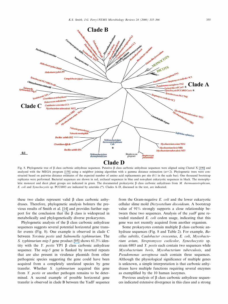
these two clades represent valid L class carbonic anhy-drases. Therefore, phylogenetic analysis bolsters the pre-vious results of Smith et al. [14] and provides further sup-port for the conclusion that the L class is widespread inmetabolically and phylogenetically diverse prokaryotes.
Phylogenetic analysis of the L class carbonic anhydrasesequences suggests several potential horizontal gene trans-fer events (Fig. 8). One example is observed in clade Cbetween Yersinia pestis and Salmonella typhimurium. TheS. typhimurium mig-5 gene product [95] shows 61.3% iden-tity with the Y. pestis YP1 L class carbonic anhydrasesequence. The mig-5 gene is £anked by inverted repeatsthat are also present in virulence plasmids from otherpathogenic species suggesting the gene could have beenacquired from a completely unrelated species by genetransfer. Whether S. typhimurium acquired this genefrom Y. pestis or another pathogen remains to be deter-mined. A second example of possible horizontal genetransfer is observed in clade B between the YadF sequence
from the Gram-negative E. coli and the lower eukaryoticcellular slime mold Dictyostelium discoideum. A bootstrapvalue of 91% strongly supports a close relationship be-tween these two sequences. Analysis of the yadF gene re-vealed standard E. coli codon usage, indicating that thisgene was not recently acquired from another organism.
Some prokaryotes contain multiple L class carbonic an-hydrase sequences (Fig. 8 and Table 2). For example, Ba-cillus subtilis, Caulobacter crescentus, E. coli, Mycobacte-rium avium, Streptomyces coelicolor, Synechocystis sp.strain 6803 and Y. pestis each contain two sequences whileMycobacterium bovis, Mycobacterium tuberculosis, andPseudomonas aeroginosa each contain three sequences.Although the physiological signi¢cance of multiple genesis unknown, a simple interpretation is that carbonic anhy-drases have multiple functions requiring several enzymesas exempli¢ed by the 10 human isozymes.
Previous analysis of L class carbonic anhydrase sequen-ces indicated extensive divergence in this class and a strong
Fig. 8. Phylogenetic tree of L class carbonic anhydrase sequences. Putative L class carbonic anhydrase sequences were aligned using Clustal X [190] andanalyzed with the MEGA program [158] using a neighbor joining algorithm with a gamma distance estimation (K= 2). Phylogenetic trees were con-structed based on pairwise distance estimates of the expected number of amino acid replacements per site (0.1 in the scale bar). One thousand bootstrapreplicates were performed. Bacterial sequences are shown in red, archaeal sequences in blue and non-plant eukaryotic sequences in black. The monophy-letic monocot and dicot plant groups are indicated in green. The documented prokaryotic L class carbonic anhydrases from M. thermoautotrophicum,E. coli and Synechocystis sp. PCC6803 are indicated by asterisks (*). Clades A^D, discussed in the text, are indicated.
FEMSRE 682 29-8-00 Cyaan Magenta Geel Zwart
K.S. Smith, J.G. Ferry / FEMS Microbiology Reviews 24 (2000) 335^366 355
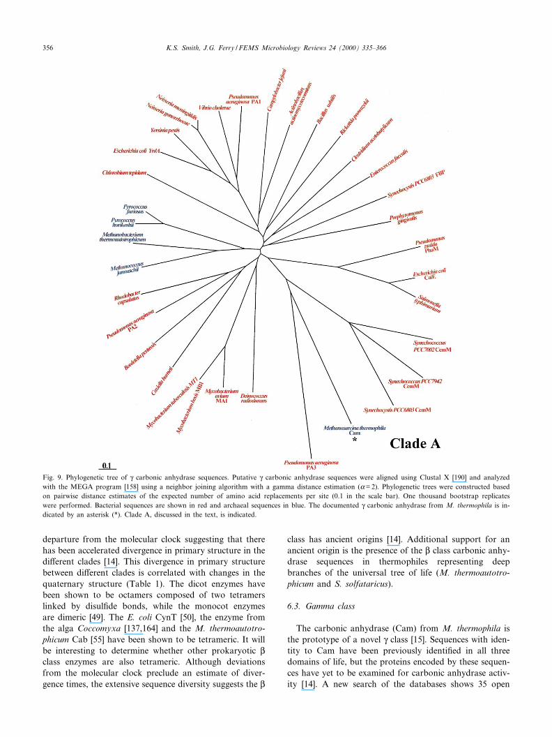
departure from the molecular clock suggesting that therehas been accelerated divergence in primary structure in thedi¡erent clades [14]. This divergence in primary structurebetween di¡erent clades is correlated with changes in thequaternary structure (Table 1). The dicot enzymes havebeen shown to be octamers composed of two tetramerslinked by disul¢de bonds, while the monocot enzymesare dimeric [49]. The E. coli CynT [50], the enzyme fromthe alga Coccomyxa [137,164] and the M. thermoautotro-phicum Cab [55] have been shown to be tetrameric. It willbe interesting to determine whether other prokaryotic Lclass enzymes are also tetrameric. Although deviationsfrom the molecular clock preclude an estimate of diver-gence times, the extensive sequence diversity suggests the L
class has ancient origins [14]. Additional support for anancient origin is the presence of the L class carbonic anhy-drase sequences in thermophiles representing deepbranches of the universal tree of life (M. thermoautotro-phicum and S. solfataricus).
6.3. Gamma class
The carbonic anhydrase (Cam) from M. thermophila isthe prototype of a novel Q class [15]. Sequences with iden-tity to Cam have been previously identi¢ed in all threedomains of life, but the proteins encoded by these sequen-ces have yet to be examined for carbonic anhydrase activ-ity [14]. A new search of the databases shows 35 open
Fig. 9. Phylogenetic tree of Q carbonic anhydrase sequences. Putative Q carbonic anhydrase sequences were aligned using Clustal X [190] and analyzedwith the MEGA program [158] using a neighbor joining algorithm with a gamma distance estimation (K= 2). Phylogenetic trees were constructed basedon pairwise distance estimates of the expected number of amino acid replacements per site (0.1 in the scale bar). One thousand bootstrap replicateswere performed. Bacterial sequences are shown in red and archaeal sequences in blue. The documented Q carbonic anhydrase from M. thermophila is in-dicated by an asterisk (*). Clade A, discussed in the text, is indicated.
FEMSRE 682 29-8-00 Cyaan Magenta Geel Zwart
K.S. Smith, J.G. Ferry / FEMS Microbiology Reviews 24 (2000) 335^366356
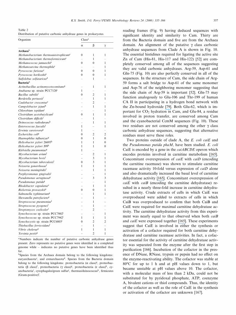
reading frames (Fig. 9) having deduced sequences withsigni¢cant identity and similarity to Cam. Thirty arefrom the Bacteria domain and ¢ve are from the Archaeadomain. An alignment of the putative Q class carbonicanhydrase sequences from Clade A is shown in Fig. 10.The essential histidines required for ligating the active siteZn of Cam (His-81, His-117 and His-122) [52] are com-pletely conserved among all of the sequences suggestingthey are valid carbonic anhydrases. Arg-59, Asp-61 andGln-75 (Fig. 10) are also perfectly conserved in all of thesequences. In the structure of Cam, the side chain of Arg-59 forms a salt bridge to Asp-61 of the same monomerand Asp-76 of the neighboring monomer suggesting thatthe side chain of Arg-59 is important [52]. Gln-75 mayfunction analogously to Glu-106 and Thr-199 of humanCA II in participating in a hydrogen bond network withthe Zn-bound hydroxide [79]. Both Glu-62, which is im-portant for CO2 hydration in Cam, and Glu-84, a residueinvolved in proton transfer, are conserved among Camand the cyanobacterial CcmM sequences (Fig. 10). Thesetwo residues are not conserved among the other Q classcarbonic anhydrase sequences, suggesting that alternativeresidues must serve these roles.
Two proteins outside of clade A, the E. coli caiE andthe Pseudomonas putida phaM, have been studied. E. coliCaiE is encoded by a gene in the caiABCDE operon whichencodes proteins involved in carnitine metabolism [165].Concomitant overexpression of caiE with caiD (encodingthe carnitine racemase) was shown to stimulate carnitineracemase activity 10-fold versus expression of caiD aloneand also dramatically increased the basal level of carnitinedehydratase activity [165]. Concomitant overexpression ofcaiE with caiB (encoding the carnitine dehydratase) re-sulted in a nearly three-fold increase in carnitine dehydra-tase activity. Crude extracts of cells in which CaiE wasoverproduced were added to extracts of cells in whichCaiB was overproduced to con¢rm that both CaiB andCaiE were required for maximal carnitine dehydratase ac-tivity. The carnitine dehydratase activity from this experi-ment was nearly equal to that observed when both caiBand caiE were expressed together [165]. These experimentssuggest that CaiE is involved in either the synthesis oractivation of a cofactor required for both carnitine dehy-dratase and carnitine racemase activities. In fact, a cofac-tor essential for the activity of carnitine dehydratase activ-ity was separated from the enzyme after the ¢rst step inpuri¢cation [166]. Incubation of the cofactor in the pres-ence of DNase, RNase, trypsin or pepsin had no e¡ect onthe enzyme-reactivating ability. The cofactor was stable at80³C for up to 1 h and at pH values down to 1, butbecame unstable at pH values above 10. The cofactor,with a molecular mass of less than 2 kDa, could not besubstituted for by pyridoxal phosphate, ATP, coenzymeA, bivalent cations or thiol compounds. Thus, the identityof the cofactor as well as the role of CaiE in the synthesisor activation of the cofactor are unknown [167].
Table 2Distribution of putative carbonic anhydrase genes in prokaryotes
Organism Classa
K L Q
Archaeab
Methanobacterium thermoautotrophicumc 0 1 1Methanobacterium thermoformicicumc ^ 1 ^Methanococcus jannaschiic 0 0 1Methanosarcina thermophilac ^ ^ 1Pyrococcus furiosusc 0 0 1Pyrococcus horikoshiic 0 0 1Sulfolobus solfataricusd ^ 1 ^Bacteriab
Actinobacillus actinomycetecomitansg ^ 1 1Anabaena sp. strain PCC7120i 1 ^ ^Bacillus subtilisl 0 2 1Bordetella pertussisf ^ 1 1Caulobacter crescentuse ^ 2 ^Campylobacter jejunih ^ 1 1Chlorobium tepidumj ^ ^ 1Clostridium acetobutylicuml 0 1 1Clostridium di¤cilel ^ 1 ^Deinococcus radioduransk ^ 1 1Enterococcus faecalisl 1 ^ 1Erwinia carotovorag 1 ^ ^Escherichia colig 0 2 2Haemophilus in£uenzaeg 0 1 0Helicobacter pylori 26695h 1 1 0Helicobacter pylori J99h 1 1 0Klebsiella pneumoniaeg 1 ^ ^Mycobacterium aviuml ^ 2 1Mycobacterium bovisl ^ 3 1Mycobacterium tuberculosisl 0 3 1Neisseria gonorrhoeaef 1 1 1Neisseria meningitidisf ^ 1 1Porphyromonas gingivalisj ^ ^ 1Pseudomonas aeruginosag ^ 3 3Pseudomonas putidag ^ ^ 1Rhodobacter capsulatuse ^ 1 1Rickettsia prowazekiie 0 0 1Salmonella typhimuriumg ^ 1 1Shewanella putrefaciensg ^ 1 ^Streptococcus pneumoniael ^ 1 ^Streptococcus pyogenesl ^ 1 ^Streptomyces coelicolorl ^ 2 ^Synechococcus sp. strain PCC7002i ^ ^ 1Synechococcus sp. strain PCC7942i 1 1 1Synechocystis sp. strain PCC6803i 0 2 2Thiobacillus ferrioxidansf ^ 1 ^Vibrio choleraeg 1 1 1Yersinia pestisg ^ 2 1
aNumbers indicate the number of putative carbonic anhydrase genespresent. Zero represents no putative genes were identi¢ed in a completedgenome while ^ indicates no putative genes have been identi¢ed thusfar.bSpecies from the Archaea domain belong to the following kingdoms:euryarchaeotac, and crenarchaeotad. Species from the Bacteria domainbelong to the following kingdoms: proteobacteria (K class)e, proteobac-teria (L class)f , proteobacteria (Q class)g, proteobacteria (O class)h, cy-anobacteriai, cytophagales/green sulfurj, thermus/deinococcusk, ¢rmicutes(Gram-positive)l.
FEMSRE 682 29-8-00 Cyaan Magenta Geel Zwart
K.S. Smith, J.G. Ferry / FEMS Microbiology Reviews 24 (2000) 335^366 357
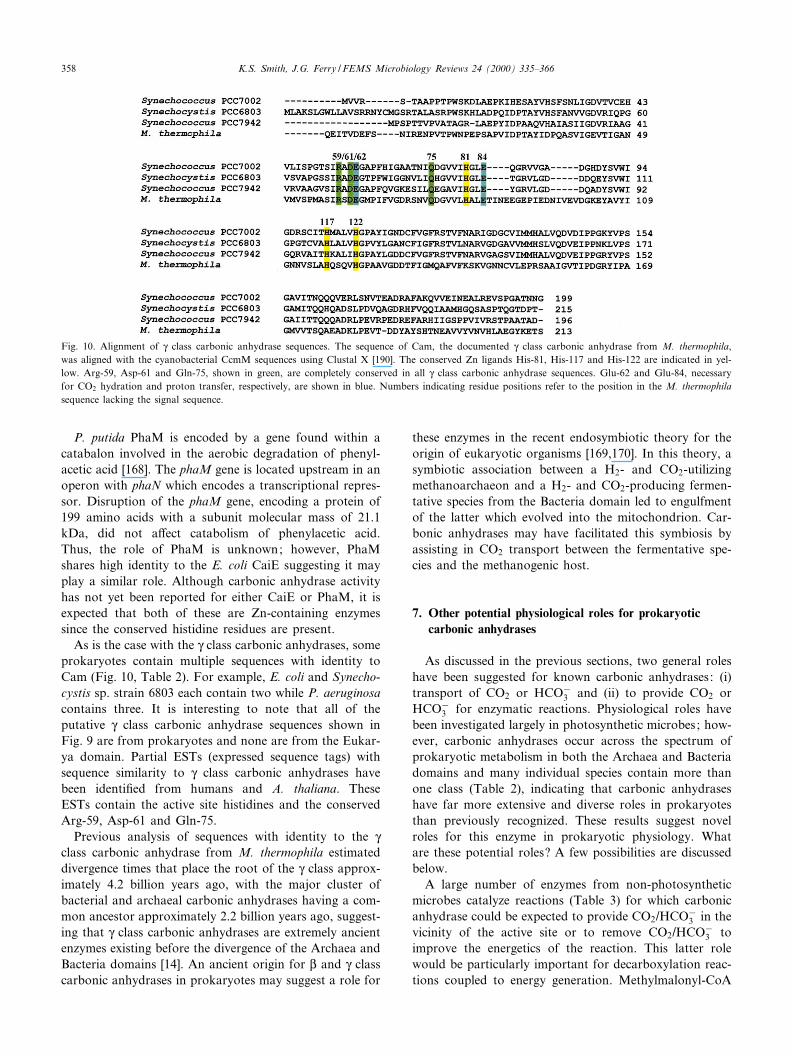
P. putida PhaM is encoded by a gene found within acatabalon involved in the aerobic degradation of phenyl-acetic acid [168]. The phaM gene is located upstream in anoperon with phaN which encodes a transcriptional repres-sor. Disruption of the phaM gene, encoding a protein of199 amino acids with a subunit molecular mass of 21.1kDa, did not a¡ect catabolism of phenylacetic acid.Thus, the role of PhaM is unknown; however, PhaMshares high identity to the E. coli CaiE suggesting it mayplay a similar role. Although carbonic anhydrase activityhas not yet been reported for either CaiE or PhaM, it isexpected that both of these are Zn-containing enzymessince the conserved histidine residues are present.
As is the case with the Q class carbonic anhydrases, someprokaryotes contain multiple sequences with identity toCam (Fig. 10, Table 2). For example, E. coli and Synecho-cystis sp. strain 6803 each contain two while P. aeruginosacontains three. It is interesting to note that all of theputative Q class carbonic anhydrase sequences shown inFig. 9 are from prokaryotes and none are from the Eukar-ya domain. Partial ESTs (expressed sequence tags) withsequence similarity to Q class carbonic anhydrases havebeen identi¢ed from humans and A. thaliana. TheseESTs contain the active site histidines and the conservedArg-59, Asp-61 and Gln-75.
Previous analysis of sequences with identity to the Qclass carbonic anhydrase from M. thermophila estimateddivergence times that place the root of the Q class approx-imately 4.2 billion years ago, with the major cluster ofbacterial and archaeal carbonic anhydrases having a com-mon ancestor approximately 2.2 billion years ago, suggest-ing that Q class carbonic anhydrases are extremely ancientenzymes existing before the divergence of the Archaea andBacteria domains [14]. An ancient origin for L and Q classcarbonic anhydrases in prokaryotes may suggest a role for
these enzymes in the recent endosymbiotic theory for theorigin of eukaryotic organisms [169,170]. In this theory, asymbiotic association between a H2- and CO2-utilizingmethanoarchaeon and a H2- and CO2-producing fermen-tative species from the Bacteria domain led to engulfmentof the latter which evolved into the mitochondrion. Car-bonic anhydrases may have facilitated this symbiosis byassisting in CO2 transport between the fermentative spe-cies and the methanogenic host.
7. Other potential physiological roles for prokaryoticcarbonic anhydrases
As discussed in the previous sections, two general roleshave been suggested for known carbonic anhydrases: (i)transport of CO2 or HCO3
3 and (ii) to provide CO2 orHCO3
3 for enzymatic reactions. Physiological roles havebeen investigated largely in photosynthetic microbes; how-ever, carbonic anhydrases occur across the spectrum ofprokaryotic metabolism in both the Archaea and Bacteriadomains and many individual species contain more thanone class (Table 2), indicating that carbonic anhydraseshave far more extensive and diverse roles in prokaryotesthan previously recognized. These results suggest novelroles for this enzyme in prokaryotic physiology. Whatare these potential roles? A few possibilities are discussedbelow.
A large number of enzymes from non-photosyntheticmicrobes catalyze reactions (Table 3) for which carbonicanhydrase could be expected to provide CO2/HCO3
3 in thevicinity of the active site or to remove CO2/HCO3
3 toimprove the energetics of the reaction. This latter rolewould be particularly important for decarboxylation reac-tions coupled to energy generation. Methylmalonyl-CoA
Fig. 10. Alignment of Q class carbonic anhydrase sequences. The sequence of Cam, the documented Q class carbonic anhydrase from M. thermophila,was aligned with the cyanobacterial CcmM sequences using Clustal X [190]. The conserved Zn ligands His-81, His-117 and His-122 are indicated in yel-low. Arg-59, Asp-61 and Gln-75, shown in green, are completely conserved in all Q class carbonic anhydrase sequences. Glu-62 and Glu-84, necessaryfor CO2 hydration and proton transfer, respectively, are shown in blue. Numbers indicating residue positions refer to the position in the M. thermophilasequence lacking the signal sequence.
FEMSRE 682 29-8-00 Cyaan Magenta Geel Zwart
K.S. Smith, J.G. Ferry / FEMS Microbiology Reviews 24 (2000) 335^366358
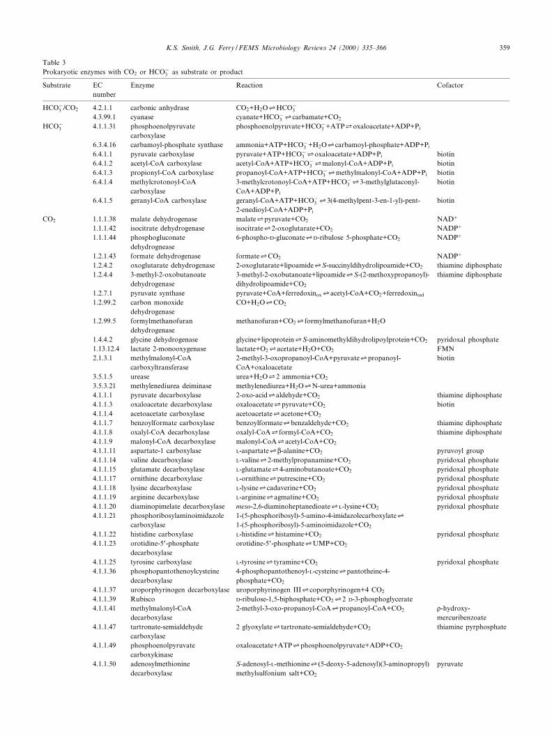
Table 3Prokaryotic enzymes with CO2 or HCO3
3 as substrate or product
Substrate ECnumber
Enzyme Reaction Cofactor
HCO33 /CO2 4.2.1.1 carbonic anhydrase CO2+H2O1HCO3
3
4.3.99.1 cyanase cyanate+HCO33 1carbamate+CO2
HCO33 4.1.1.31 phosphoenolpyruvate
carboxylasephosphoenolpyruvate+HCO3
3 +ATP1oxaloacetate+ADP+Pi
6.3.4.16 carbamoyl-phosphate synthase ammonia+ATP+HCO33 +H2O1carbamoyl-phosphate+ADP+Pi
6.4.1.1 pyruvate carboxylase pyruvate+ATP+HCO33 1oxaloacetate+ADP+Pi biotin
6.4.1.2 acetyl-CoA carboxylase acetyl-CoA+ATP+HCO33 1malonyl-CoA+ADP+Pi biotin
6.4.1.3 propionyl-CoA carboxylase propanoyl-CoA+ATP+HCO33 1methylmalonyl-CoA+ADP+Pi biotin
6.4.1.4 methylcrotonoyl-CoAcarboxylase
3-methylcrotonoyl-CoA+ATP+HCO33 13-methylglutaconyl-
CoA+ADP+Pi
biotin
6.4.1.5 geranyl-CoA carboxylase geranyl-CoA+ATP+HCO33 13(4-methylpent-3-en-1-yl)-pent-
2-enedioyl-CoA+ADP+Pi
biotin
CO2 1.1.1.38 malate dehydrogenase malate1pyruvate+CO2 NAD�
1.1.1.42 isocitrate dehydrogenase isocitrate12-oxoglutarate+CO2 NADP�
1.1.1.44 phosphogluconatedehydrognease
6-phospho-D-gluconate1D-ribulose 5-phosphate+CO2 NADP�
1.2.1.43 formate dehydrogenase formate1CO2 NADP�
1.2.4.2 oxoglutarate dehydrogenase 2-oxoglutarate+lipoamide1S-succinyldihydrolipoamide+CO2 thiamine diphosphate1.2.4.4 3-methyl-2-oxobutanoate
dehydrogenase3-methyl-2-oxobutanoate+lipoamide1S-(2-methoxypropanoyl)-dihydrolipoamide+CO2
thiamine diphosphate
1.2.7.1 pyruvate synthase pyruvate+CoA+ferredoxinox1acetyl-CoA+CO2+ferredoxinred
1.2.99.2 carbon monoxidedehydrogenase
CO+H2O1CO2
1.2.99.5 formylmethanofurandehydrogenase
methanofuran+CO21formylmethanofuran+H2O
1.4.4.2 glycine dehydrogenase glycine+lipoprotein1S-aminomethyldihydrolipoylprotein+CO2 pyridoxal phosphate1.13.12.4 lactate 2-monooxygenase lactate+O21acetate+H2O+CO2 FMN2.1.3.1 methylmalonyl-CoA
carboxyltransferase2-methyl-3-oxopropanoyl-CoA+pyruvate1propanoyl-CoA+oxaloacetate
biotin
3.5.1.5 urease urea+H2O12 ammonia+CO2
3.5.3.21 methylenediurea deiminase methylenediurea+H2O1N-urea+ammonia4.1.1.1 pyruvate decarboxylase 2-oxo-acid1aldehyde+CO2 thiamine diphosphate4.1.1.3 oxaloacetate decarboxylase oxaloacetate1pyruvate+CO2 biotin4.1.1.4 acetoacetate carboxylase acetoacetate1acetone+CO2
4.1.1.7 benzoylformate carboxylase benzoylformate1benzaldehyde+CO2 thiamine diphosphate4.1.1.8 oxalyl-CoA decarboxylase oxalyl-CoA1formyl-CoA+CO2 thiamine diphosphate4.1.1.9 malonyl-CoA decarboxylase malonyl-CoA1acetyl-CoA+CO2
4.1.1.11 aspartate-1 carboxylase L-aspartate1L-alanine+CO2 pyruvoyl group4.1.1.14 valine decarboxylase L-valine12-methylpropanamine+CO2 pyridoxal phosphate4.1.1.15 glutamate decarboxylase L-glutamate14-aminobutanoate+CO2 pyridoxal phosphate4.1.1.17 ornithine decarboxylase L-ornithine1putrescine+CO2 pyridoxal phosphate4.1.1.18 lysine decarboxylase L-lysine1cadaverine+CO2 pyridoxal phosphate4.1.1.19 arginine decarboxylase L-arginine1agmatine+CO2 pyridoxal phosphate4.1.1.20 diaminopimelate decarboxylase meso-2,6-diaminoheptanedioate1L-lysine+CO2 pyridoxal phosphate4.1.1.21 phosphoribosylaminoimidazole
carboxylase1-(5-phosphoribosyl)-5-amino-4-imidazolecarboxylate11-(5-phosphoribosyl)-5-aminoimidazole+CO2
4.1.1.22 histidine carboxylase L-histidine1histamine+CO2 pyridoxal phosphate4.1.1.23 orotidine-5P-phosphate
decarboxylaseorotidine-5P-phosphate1UMP+CO2
4.1.1.25 tyrosine carboxylase L-tyrosine1tyramine+CO2 pyridoxal phosphate4.1.1.36 phosphopantothenoylcysteine
decarboxylase4-phosphopantothenoyl-L-cysteine1pantotheine-4-phosphate+CO2
4.1.1.37 uroporphyrinogen decarboxylase uroporphyrinogen III1coporphyrinogen+4 CO2
4.1.1.39 Rubisco D-ribulose-1,5-biphosphate+CO212 D-3-phosphoglycerate4.1.1.41 methylmalonyl-CoA
decarboxylase2-methyl-3-oxo-propanoyl-CoA1propanoyl-CoA+CO2 b-hydroxy-
mercuribenzoate4.1.1.47 tartronate-semialdehyde
carboxylase2 glyoxylate1tartronate-semialdehyde+CO2 thiamine pyrphosphate
4.1.1.49 phosphoenolpyruvatecarboxykinase
oxaloacetate+ATP1phosphoenolpyruvate+ADP+CO2
4.1.1.50 adenosylmethioninedecarboxylase
S-adenosyl-L-methionine1(5-deoxy-5-adenosyl)(3-aminopropyl)methylsulfonium salt+CO2
pyruvate
FEMSRE 682 29-8-00 Cyaan Magenta Geel Zwart
K.S. Smith, J.G. Ferry / FEMS Microbiology Reviews 24 (2000) 335^366 359
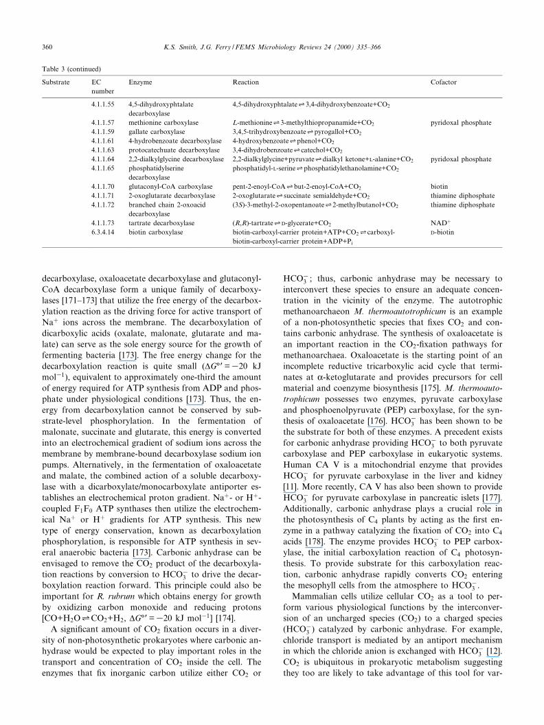
decarboxylase, oxaloacetate decarboxylase and glutaconyl-CoA decarboxylase form a unique family of decarboxy-lases [171^173] that utilize the free energy of the decarbox-ylation reaction as the driving force for active transport ofNa� ions across the membrane. The decarboxylation ofdicarboxylic acids (oxalate, malonate, glutarate and ma-late) can serve as the sole energy source for the growth offermenting bacteria [173]. The free energy change for thedecarboxylation reaction is quite small (vG³P=320 kJmol31), equivalent to approximately one-third the amountof energy required for ATP synthesis from ADP and phos-phate under physiological conditions [173]. Thus, the en-ergy from decarboxylation cannot be conserved by sub-strate-level phosphorylation. In the fermentation ofmalonate, succinate and glutarate, this energy is convertedinto an electrochemical gradient of sodium ions across themembrane by membrane-bound decarboxylase sodium ionpumps. Alternatively, in the fermentation of oxaloacetateand malate, the combined action of a soluble decarboxy-lase with a dicarboxylate/monocarboxylate antiporter es-tablishes an electrochemical proton gradient. Na�- or H�-coupled F1F0 ATP synthases then utilize the electrochem-ical Na� or H� gradients for ATP synthesis. This newtype of energy conservation, known as decarboxylationphosphorylation, is responsible for ATP synthesis in sev-eral anaerobic bacteria [173]. Carbonic anhydrase can beenvisaged to remove the CO2 product of the decarboxyla-tion reactions by conversion to HCO3
3 to drive the decar-boxylation reaction forward. This principle could also beimportant for R. rubrum which obtains energy for growthby oxidizing carbon monoxide and reducing protons[CO+H2O1CO2+H2, vG³P=320 kJ mol31] [174].
A signi¢cant amount of CO2 ¢xation occurs in a diver-sity of non-photosynthetic prokaryotes where carbonic an-hydrase would be expected to play important roles in thetransport and concentration of CO2 inside the cell. Theenzymes that ¢x inorganic carbon utilize either CO2 or
HCO33 ; thus, carbonic anhydrase may be necessary to
interconvert these species to ensure an adequate concen-tration in the vicinity of the enzyme. The autotrophicmethanoarchaeon M. thermoautotrophicum is an exampleof a non-photosynthetic species that ¢xes CO2 and con-tains carbonic anhydrase. The synthesis of oxaloacetate isan important reaction in the CO2-¢xation pathways formethanoarchaea. Oxaloacetate is the starting point of anincomplete reductive tricarboxylic acid cycle that termi-nates at K-ketoglutarate and provides precursors for cellmaterial and coenzyme biosynthesis [175]. M. thermoauto-trophicum possesses two enzymes, pyruvate carboxylaseand phosphoenolpyruvate (PEP) carboxylase, for the syn-thesis of oxaloacetate [176]. HCO3
3 has been shown to bethe substrate for both of these enzymes. A precedent existsfor carbonic anhydrase providing HCO3
3 to both pyruvatecarboxylase and PEP carboxylase in eukaryotic systems.Human CA V is a mitochondrial enzyme that providesHCO3
3 for pyruvate carboxylase in the liver and kidney[11]. More recently, CA V has also been shown to provideHCO3
3 for pyruvate carboxylase in pancreatic islets [177].Additionally, carbonic anhydrase plays a crucial role inthe photosynthesis of C4 plants by acting as the ¢rst en-zyme in a pathway catalyzing the ¢xation of CO2 into C4
acids [178]. The enzyme provides HCO33 to PEP carbox-
ylase, the initial carboxylation reaction of C4 photosyn-thesis. To provide substrate for this carboxylation reac-tion, carbonic anhydrase rapidly converts CO2 enteringthe mesophyll cells from the atmosphere to HCO3
3 .Mammalian cells utilize cellular CO2 as a tool to per-
form various physiological functions by the interconver-sion of an uncharged species (CO2) to a charged species(HCO3
3 ) catalyzed by carbonic anhydrase. For example,chloride transport is mediated by an antiport mechanismin which the chloride anion is exchanged with HCO3
3 [12].CO2 is ubiquitous in prokaryotic metabolism suggestingthey too are likely to take advantage of this tool for var-
Table 3 (continued)
Substrate ECnumber
Enzyme Reaction Cofactor
4.1.1.55 4,5-dihydroxyphtalatedecarboxylase
4,5-dihydroxyphtalate13,4-dihydroxybenzoate+CO2
4.1.1.57 methionine carboxylase L-methionine13-methylthiopropanamide+CO2 pyridoxal phosphate4.1.1.59 gallate carboxylase 3,4,5-trihydroxybenzoate1pyrogallol+CO2
4.1.1.61 4-hydrobenzoate decarboxylase 4-hydroxybenzoate1phenol+CO2
4.1.1.63 protocatechuate decarboxylase 3,4-dihydrobenzoate1catechol+CO2
4.1.1.64 2,2-dialkylglycine decarboxylase 2,2-dialkylglycine+pyruvate1dialkyl ketone+L-alanine+CO2 pyridoxal phosphate4.1.1.65 phosphatidylserine
decarboxylasephosphatidyl-L-serine1phosphatidylethanolamine+CO2
4.1.1.70 glutaconyl-CoA carboxylase pent-2-enoyl-CoA1but-2-enoyl-CoA+CO2 biotin4.1.1.71 2-oxoglutarate decarboxylase 2-oxoglutarate1succinate semialdehyde+CO2 thiamine diphosphate4.1.1.72 branched chain 2-oxoacid
decarboxylase(3S)-3-methyl-2-oxopentanoate12-methylbutanol+CO2 thiamine diphosphate
4.1.1.73 tartrate decarboxylase (R,R)-tartrate1D-glycerate+CO2 NAD�
6.3.4.14 biotin carboxylase biotin-carboxyl-carrier protein+ATP+CO21carboxyl-biotin-carboxyl-carrier protein+ADP+Pi
D-biotin
FEMSRE 682 29-8-00 Cyaan Magenta Geel Zwart
K.S. Smith, J.G. Ferry / FEMS Microbiology Reviews 24 (2000) 335^366360
ious physiological functions such as solute transport. Da-tabase searches have identi¢ed three sequences (S. coeli-color SC2, M. bovis MB3 and M. tuberculosis MT3) thatappear to encode bifunctional proteins (Fig. 8). These se-quences encode putative transmembrane proteins whoseN-terminal halves show similarity to low-a¤nity sulfatetransporters and C-terminal regions show similarity toL class carbonic anhydrases. Thus, carbonic anhydrasemay play a role in the transport of a compound usingeither a symport or antiport mechanism.
In addition to the interconversion of CO2 and HCO33 ,
carbonic anhydrase has been shown to catalyze severalother reactions, such as aldehyde hydration [179] and thehydrolysis of carboxylic esters [180] and various halogenderivatives [181,182]. Esters of phenols [179,180,183] andsulfonic and phosphoric acids [184,185] have been re-ported to act as substrates for this enzyme. Unfortunately,these activities were identi¢ed in studies with the K classhuman isozymes and it is not known whether the L or Qclass enzymes can also catalyze these reactions. With thelarge reservoir of prokaryotic carbonic anhydrases, addi-tional catalytic functions would not be unexpected.
8. Conclusions and outlook
Why is it important to study prokaryotic carbonic an-hydrases when so much is already known about the plantand mammalian enzymes? Past reports of carbonic anhy-drase in microbes from the Archaea and Bacteria domainshave been infrequent implying a narrow function for thisenzyme in prokaryotes. The enzyme has been puri¢edfrom only ¢ve prokaryotic species and previous e¡ortsto detect carbonic anhydrase activity in certain prokary-otes left the impression that carbonic anhydrases are notimportant to prokaryotic physiology. It is now known thatcarbonic anhydrases from prokaryotes are generally ofvery low abundance and strictly regulated, often makingtheir presence di¤cult to detect. The recent discovery ofthe Q class, the availability of antisera to prokaryotic en-zymes representing each of the classes and the availabilityof genome sequences of many prokaryotes has now al-lowed for a more comprehensive search. These analyseshave demonstrated that carbonic anhydrases are indeedwidespread and nearly ubiquitous among metabolicallyand phylogenetically diverse prokaryotes from both theBacteria and Archaea domains, suggesting that this en-zyme plays a major role in prokaryotic physiology.
Genes encoding carbonic anhydrases from all threeclasses have been identi¢ed in the prokaryotes with the Land Q classes predominating (Figs. 6, 8 and 9). Since nosigni¢cant sequence similarities are observed between rep-resentatives of the three classes, carbonic anhydrase pro-vides an excellent example of convergent evolution of cat-alytic function. Why three di¡erent classes have evolvedand what advantage one class may provide over another is
unknown. Interestingly, many prokaryotes contain puta-tive carbonic anhydrase genes from more than one class(Table 2) and some even contain genes from all threeknown classes (Table 2). In addition, some prokaryotescontain multiple genes encoding putative carbonic anhy-drases from the same class (see Section 4.2). Amazingly, P.aeruginosa contains six sequences that could encode car-bonic anhydrase, three from the L class and three from theQ class. The multiplicity of carbonic anhydrases in manyprokaryotes underscores their importance in prokaryoticphysiology and suggests novel roles for the enzyme.
Are there any undiscovered classes of carbonic anhy-drase? Analysis of the completed genome sequences ofBorrelia burgdorferi, Chlamydia trachomatis, Mycoplasmapneumoniae, Mycoplasma genitalium, Thermatoga mariti-ma, and Treponema pallidum from the Bacteria domainand Archaeoglobus fulgidus from the Archaea domain in-dicated an absence of open reading frames with deducedsequences having signi¢cant identity to K, L or Q classcarbonic anhydrases [14]. Whether these organisms possessa di¡erent class of carbonic anhydrase or do not requirethis enzymatic activity remains to be answered. Since car-bonic anhydrases seem to be ancient enzymes and thereare already three known classes that were independentlyinvented, it would not be surprising to ¢nd even moreclasses. One method to identify genes encoding carbonicanhydrase from these prokaryotes would be by geneticcomplementation of an E. coli cynT mutant [88].
In addition to biochemical and physiological studies,carbonic anhydrases have been exploited for biotechnolog-ical purposes such as the design of metal ion sensors andde novo catalytic sites. For example, carbonic anhydrasetransducers have been created that measure picomolarconcentrations of Cu(II), Zn(II), Cd(II), Co(II) andNi(II) in complex media as determined by the changes inthe £uorescence lifetime and intensity of site-speci¢callylabeled £uorescent variants of a human CA II apoenzyme[186,187]. Other technological uses for carbonic anhydrasesuch as in CO2 scrubbers can also be imagined.
Although there are many questions to be answeredabout the enzymology and structure of prokaryotic car-bonic anhydrases, one major question still remains: whatare the roles of carbonic anhydrases in prokaryotic phys-iology? Most of the information known about the func-tion of this enzyme in prokaryotes relates to its role in the¢xation of CO2 by cyanobacteria; however, the researchreviewed here strongly supports that prokaryotes fromdiverse metabolic and phylogenetic origins contain multi-ple carbonic anhydrase genes. Undoubtedly, future re-search will uncover diverse novel functions for this ubiq-uitous prokaryotic enzyme.
Acknowledgements
The work in this laboratory was supported by NIH-
FEMSRE 682 29-8-00 Cyaan Magenta Geel Zwart
K.S. Smith, J.G. Ferry / FEMS Microbiology Reviews 24 (2000) 335^366 361
GM44661 and NASA-Ames Cooperative AgreementNCC2-1057. The authors would like to thank Dr. CherylIngram-Smith for her suggestions and for her critical read-ing of the manuscript.
References
[1] Hewett-Emmett, D. and Tashian, R.E. (1996) Functional diversity,conservation and convergence in the evolution of the K-, L- andQ-carbonic anhydrase gene families. Mol. Phylogenet. Evol. 5, 50^77.
[2] Silverman, D.N. (1982) Carbonic anhydrase: oxygen-18 exchangecatalyzed by an enzyme with rate-contributing proton-transfer steps.Methods Enzymol. 87, 732^752.
[3] Silverman, D.N. and Vincent, S.H. (1983) Proton transfer in thecatalytic mechanism of carbonic anhydrase. CRC Crit. Rev. Bio-chem. 14, 207^255.
[4] Silverman, D.N. and Lindskog, S. (1988) The catalytic mechanism ofcarbonic anhydrase: implications of a rate-limiting proteolysis ofwater. Acc. Chem. Res. 21, 30^36.
[5] Christianson, D.W. and Fierke, C.A. (1996) Carbonic anhydrase:evolution of the zinc binding site by nature and by design. Acc.Chem. Res. 29, 331^339.
[6] Lindskog, S. (1997) Structure and mechanism of carbonic anhydrase.Pharmacol. Ther. 74, 1^20.
[7] Reed, M.L. and Graham, D. (1981) Carbonic anhydrase in plants:distribution, properties, and possible physiological roles. In: Progressin Phytochemistry (Reinhold, L., Harborne, J.B. and Swain, T.,Eds.), pp. 47^94. Pergamon Press, Oxford.
[8] Graham, D., Reed, M.L., Patterson, B.D., Hockley, D.G. andDwyer, M.R. (1984) Chemical properties, distribution, and physiol-ogy of plant and algal carbonic anhydrases. Ann. NY Acad. Sci. 429,222^237.
[9] Tashian, R.E. (1989) The carbonic anhydrases : widening perspectiveson their evolution, expression and function. BioEssays 10, 186^192.
[10] Badger, M.R. and Price, G.D. (1994) The role of carbonic anhydrasein photosynthesis. Annu. Rev. Plant Physiol. Plant Mol. Biol. 45,369^392.
[11] Sly, W.S. and Hu, P.Y. (1995) Human carbonic anhydrases andcarbonic anhydrase de¢ciencies. Annu. Rev. Biochem. 64, 375^401.
[12] Henry, R.P. (1996) Multiple roles of carbonic anhydrase in cellulartransport and metabolism. Annu. Rev. Physiol. 58, 523^538.
[13] Nogradi, A. (1998) The role of carbonic anhydrases in tumors. Am.J. Pathol. 153, 1^4.
[14] Smith, K.S., Jakubzick, C., Whittam, T.S. and Ferry, J.G. (1999)Carbonic anhydrase is an ancient enzyme widespread in prokaryotes.Proc. Natl. Acad. Sci. USA 96, 15184^15189.
[15] Alber, B.E. and Ferry, J.G. (1994) A carbonic anhydrase from thearchaeon Methanosarcina thermophila. Proc. Natl. Acad. Sci. USA91, 6909^6913.
[16] Alber, B.E. and Ferry, J.G. (1996) Characterization of heterologouslyproduced carbonic anhydrase from Methanosarcina thermophila.J. Bacteriol. 178, 3270^3274.
[17] Meldrum, N.N. and Roughton, F.J.W. (1933) Carbonic anhydrase:its preparation and propeties. Nature 80, 113^142.
[18] Stadie, W.C. and O'Brien, H. (1933) The catalysis of the hydration ofcarbon dioxide and dehydration of carbonic acid by the enzyme fromred blood cells. J. Biochem. 103, 521^529.
[19] Keilin, D. and Mann, T. (1939) Carbonic anhydrase. Nature 144,442^443.
[20] Keilin, D. and Mann, T. (1940) Carbonic anhydrase. Puri¢cation andnature of the enzyme. Biochem. J. 34, 1163^1176.
[21] Keilin, D. and Mann, T. (1944) Activity of puri¢ed carbonic anhy-drase. Nature 153, 107^108.
[22] Brad¢eld, J.R.G. (1947) Plant carbonic anhydrase. Nature 159, 467^468.
[23] Neish, A.C. (1939) Studies on chloroplasts. Their chemical composi-tion and the distribution of certain metabolites between the chloro-plasts and the remainder of the leaf. Biochem. J. 33, 300^308.
[24] Veitch, F.P. and Blankenship, L.C. (1963) Carbonic anhydrase activ-ity in bacteria. Nature 197, 76^77.
[25] Adler, L., Brundell, J., Falkbring, S.O. and Nyman, P.O. (1972)Carbonic anhydrase from Neisseria sicca strain 6021 I. Bacterialgrowth and puri¢cation of the enzyme. Biochim. Biophys. Acta284, 298^310.
[26] Brundell, J., Falkbring, S.O. and Nyman, P.O. (1972) Carbonic an-hydrase from Neisseria sicca strain 6021 II. Properties of the puri¢edenzyme. Biochim. Biophys. Acta 284, 311^323.
[27] Karrasch, M., Bott, M. and Thauer, R.K. (1989) Carbonic anhydraseactivity in acetate grown Methanosarcina barkeri. Arch. Microbiol.151, 137^142.
[28] Andersson, B., Nyman, P.O. and Strid, L. (1972) Amino acid se-quence of human erythrocyte CA B. Biochem. Biophys. Res. Com-mun. 48, 670^677.
[29] Lin, K.-T.D. and Deutsch, H.F. (1973) Human carbonic anhydrase.XI. The complete primary structure of carbonic anhydrase B. J. Biol.Chem. 248, 1885^1893.
[30] Henderson, L.E., Henriksson, D. and Nyman, P.O. (1973) Aminoacid sequence of human erythrocyte carbonic anhydrase C. Biochem.Biophys. Res. Commun. 52, 1388^1394.
[31] Lin, K.-T.D. and Deutsch, H.F. (1974) Human carbonic anhydrase.XII. The complete primary structure of carbonic anhydrase C. J. Biol.Chem. 249, 2329^2337.
[32] Tashian, R.E. and Carter, N.D. (1976) Biochemical genetics of car-bonic anhydrase. Adv. Hum. Genet. 7, 1^56.
[33] Tashian, R.E., Hewett-Emmett, D., Stroup, S.K., Goodman, M. andYu, Y.-S.L. (1980) Evolution of structure and function in the car-bonic anhydrase isozymes in mammals. In: Biophysics and Physiol-ogy of Carbon Dioxide (Bauer, G., Gross, G. and Bartels, H., Eds.),pp. 165^176. Springer, Berlin.
[34] Tashian, R.E., Venta, P.J., Nicewander, P.H. and Hewett-Emmett,D. (1990) Evolution, structure, and expression of the carbonic anhy-drase multigene family. Prog. Clin. Biol. Res. 344, 159^175.
[35] Tashian, R.E. (1992) Genetics of the mammalian carbonic anhy-drases. Adv. Genet. 30, 321^356.
[36] Hewett-Emmett, D. and Tashian, R.E. (1991) Structure and evolu-tionary origins of the carbonic anhydrase multigene family. In: TheCarbonic Anhydrases: Cellular Physiology and Molecular Genetics(Dodgson, S.J., Tashian, R.E., Gros, G. and Carter, N.D., Eds.), pp.15^32. Plenum Press, New York.
[37] Mori, K., Ogawa, Y., Ebihara, K., Tamura, N., Tashiro, K., Kuwa-hara, T., Mukoyama, M., Sugawara, A., Ozaki, S., Tanaka, I. andNakao, K. (1999) Isolation and characterization of CA XIV, a novelmembrane-bound carbonic anhydrase from mouse kidney. J. Biol.Chem. 274, 15701^15705.
[38] Kannan, K.K., Notstrand, B., Fridborg, K., Lovgren, S., Ohlsson, A.and Petef, M. (1975) Crystal structure of human erythrocyte carbonicanhydrase B. Three-dimensional structure at a nominal 2.2-Aî resolu-tion. Proc. Natl. Acad. Sci. USA 72, 51^55.
[39] Liljas, A., Kannan, K.K., Bergsten, P.C., Waara, I., Fridborg, K.,Strandberg, B., Carlbom, U., Jarup, L., Lovgren, S. and Petef, M.(1972) Crystal structure of human carbonic anhydrase C. NatureNew Biol. 235, 131^137.
[40] Eriksson, A.E., Jones, T.A. and Liljas, A. (1988) Re¢ned structure ofhuman carbonic anhydrase II at 2.0 angstrom resolution. ProteinStruct. Funct. Genet. 4, 274^282.
[41] Hakansson, K., Carlsson, M., Svensson, L.A. and Liljas, A. (1992)The structure of native and apo carbonic anhydrase-II and some ofits anion-ligand complexes. J. Mol. Biol. 227, 1192^1204.
[42] Eriksson, A.E. and Liljas, A. (1993) Re¢ned structure of bovine
FEMSRE 682 29-8-00 Cyaan Magenta Geel Zwart
K.S. Smith, J.G. Ferry / FEMS Microbiology Reviews 24 (2000) 335^366362
carbonic anhydrase-III at 2.0 angstrom resolution. Protein Struct.Funct. Genet. 16, 29^42.
[43] Stams, T., Nair, S.K., Okuyama, T., Waheed, A., Sly, W.S. andChristianson, D.W. (1996) Crystal structure of the secretory formof membrane-associated human carbonic anhydrase IV at 2.8-Aî res-olution. Proc. Natl. Acad. Sci. USA 93, 13589^13594.
[44] Boriack-Sjodin, P.A., Heck, R.W., Laipis, P.J., Silverman, D.N. andChristianson, D.W. (1995) Structure determination of murine mito-chondrial carbonic anhydrase V at 2.45-Aî resolution: implicationsfor catalytic proton transfer and inhibitor design. Proc. Natl. Acad.Sci. USA 92, 10949^10953.
[45] Hewett-Emmett, D., Hopkins, P.J., Tashian, R.E. and Czelusniak, J.(1984) Origins and molecular evolution of the carbonic anhydraseisozymes. Ann. NY Acad. Sci. 429, 338^358.
[46] Fukuzawa, H., Fujiwara, S., Yamamoto, Y., Dionisio-Sese, M.L. andMiyachi, S. (1990) cDNA cloning, sequence, and expression of car-bonic anhydrase in Chlamydomonas reinhardtii ^ regulation by envi-ronmental CO2 concentration. Proc. Natl. Acad. Sci. USA 87, 4383^4387.
[47] Fukuzawa, H., Fujiwara, S., Tachiki, A. and Miyachi, S. (1990) Nu-cleotide sequences of two genes CAH1 and CAH2 which encodecarbonic anhydrase polypeptides in Chlamydomonas reinhardtii. Nu-cleic Acids Res. 18, 6441^6442.
[48] Johansson, I.M. and Forsman, C. (1993) Kinetic studies of pea car-bonic anhydrase. Eur. J. Biochem. 218, 439^446.
[49] Bjorkbacka, H., Johansson, I.M., Skarfstad, E. and Forsman, C.(1997) The sulfhydryl groups of Cys 269 and Cys 272 are criticalfor the oligomeric state of chloroplast carbonic anhydrase from Pi-sum sativum. Biochemistry 36, 4287^4294.
[50] Guilloton, M.B., Korte, J.J., Lamblin, A.F., Fuchs, J.A. and Ander-son, P.M. (1992) Carbonic anhydrase in Escherichia coli. A productof the cyn operon. J. Biol. Chem. 267, 3731^3734.
[51] Fukuzawa, H., Suzuki, E., Komukai, Y. and Miyachi, S. (1992) Agene homologous to chloroplast carbonic anhydrase (icfA) is essentialto photosynthetic carbon dioxide ¢xation by SynechococcusPCC7942. Proc. Natl. Acad. Sci. USA 89, 4437^4441.
[52] Kisker, C., Schindelin, H., Alber, B.E., Ferry, J.G. and Rees, D.C.(1996) A left-hand beta-helix revealed by the crystal structure of acarbonic anhydrase from the archaeon Methanosarcina thermophila.EMBO J. 15, 2323^2330.
[53] Chirica, L.C., Elleby, B., Jonsson, B.H. and Lindskog, S. (1997) Thecomplete sequence, expression in Escherichia coli, puri¢cation andsome properties of carbonic anhydrase from Neisseria gonorrhoeae.Eur. J. Biochem. 244, 755^760.
[54] Nolling, J. and Reeve, J.N. (1997) Growth- and substrate-dependenttranscription of the formate dehydrogenase (fdhCAB) operon inMethanobacterium thermoformicicum Z-245. J. Bacteriol. 179, 899^908.
[55] Smith, K.S. and Ferry, J.G. (1999) A plant type (L class) carbonicanhydrase from the thermophilic methanoarchaeon Methanobacte-rium thermoautotrophicum. J. Bacteriol. 181, 6247^6253.
[56] Johansson, I.M. and Forsman, C. (1994) Solvent hydrogen isotopee¡ects and anion inhibition of CO2 hydration catalysed by carbonicanhydrase from Pisum sativum. Eur. J. Biochem. 224, 901^907.
[57] Rowlett, R.S., Chance, M.R., Wirt, M.D., Sidelinger, D.E., Royal,J.R., Woodro¡e, M., Wang, Y.F., Saha, R.P. and Lam, M.G. (1994)Kinetic and structural characterization of spinach carbonic anhy-drase. Biochemistry 33, 13967^13976.
[58] Alber, B.E., Colangelo, C.M., Dong, J., Stalhandske, C.M.V., Baird,T.T., Tu, C., Fierke, C.A., Silverman, D.N., Scott, R.A. and Ferry,J.G. (1999) Kinetic and spectroscopic characterization of the gammacarbonic anhydrase from the methanoarchaeon Methanosarcina ther-mophila. Biochemistry 38, 13119^13128.
[59] Smith, K.S., Cosper, N.J., Stalhandske, C.M.V., Scott, R.A. andFerry, J.G., Structural and kinetic characterization of an archaealbeta class carbonic anhydrase. Submitted for publication.
[60] Romanova, A.K., Rusinova, P.G. and Kornitskaya, V.M. (1972) On
the participation of carbonic anhydrase in the assimilation of carbondioxide in the chemosynthesis of Thiobacillus thiooxidans 58R. Dokl.Acad. Sci. USSR 203, 1209^1212.
[61] Ingle, R.K. and Colman, B. (1975) Carbonic anhydrase levels in blue-green algae. Can. J. Bot. 53, 2385^2387.
[62] Ingle, R.K. and Colman, B. (1976) The relationship between carbonicanhydrase activity and glycolate excretion in the blue-green alga Coc-cochloris peniocystis. Planta 128, 217^223.
[63] Ivanovskii, R.N. and Rodova, N.A. (1977) Carbonanhydrase activityof phototrophic bacteria. Mikrobiologiya 46, 409^413.
[64] Jahnke, L.C., Lyman, C. and Hooper, A.B. (1984) Carbonic anhy-drase, carbon dioxide levels and growth of Nitrosomonas. Arch. Mi-crobiol. 140, 291^293.
[65] Braus-Stromeyer, S.A., Schnappauf, G., Braus, G.H., Gossner, A.S.and Drake, H.L. (1997) Carbonic anhydrase in Acetobacterium wood-ii and other acetogenic bacteria. J. Bacteriol. 179, 7197^7200.
[66] Na¢, B.M., Miles, R.J., Butler, L.O., Carter, N.D., Kelly, C. andJe¡ery, S. (1990) Expression of carbonic anhydrase in neisseriaeand other heterotrophic bacteria. J. Med. Microbiol. 32, 1^7.
[67] Soltes-Rak, E., Mulligan, M.E. and Coleman, J.R. (1997) Identi¢ca-tion and characterization of a gene encoding a vertebrate-type car-bonic anhydrase in cyanobacteria. J. Bacteriol. 179, 769^774.
[68] Altschul, S.F., Gish, W., Miller, W., Myers, E.W. and Lipman, D.J.(1990) Basic local alignment search tool. J. Mol. Biol. 215, 403^410.
[69] Smith, D.R., Doucette-Stamm, L.A., Deloughery, C., Lee, H., Du-bois, J., Aldredge, T., Bashirzadeh, R., Blakely, D., Cook, R., Gil-bert, K., Harrison, D., Hoang, L., Keagle, P., Lumm, W., Pothier,B., Qiu, D., Spadafora, R., Vicaire, R., Wang, Y., Wierzbowski, J.,Gibson, R., Jiwani, N., Caruso, A., Bush, D., Safer, H., Patwell, D.,Prabhakar, S., McDougall, S., Shimer, G., Goyal, A., Pietrokovski,S., Church, G.M., Daniels, C.J., Mao, J.-I., Rice, P., Nolling, J. andReeve, J.N. (1997) Complete genome sequence of Methanobacteriumthermoautotrophicum deltaH: functional analysis and comparativegenomics. J. Bacteriol. 179, 7135^7155.
[70] Gill, S.R., Fedorka-Cray, P.J., Tweten, R.K. and Sleeper, B.P. (1984)Puri¢cation and properties of the carbonic anhydrase of Rhodospi-rillum rubrum. Arch. Microbiol. 138, 113^118.
[71] Yagawa, Y., Shiraiwa, Y. and Miyachi, S. (1984) Carbonic anhydrasefrom the blue-green alga (cyanobacterium) Anabaena variabilis. PlantCell Physiol. 25, 775^783.
[72] Sanders, E. and Maren, T.H. (1967) Inhibition of carbonic anhydrasein Neisseria: e¡ects on enzyme activity and growth. Mol. Pharmacol.3, 204^215.
[73] MacLeod, M.N. and DeVoe, I.W. (1981) Localization of carbonicanhydrase in the cytoplasmic membrane of Neisseria sicca (strain 19).Can. J. Microbiol. 27, 87^92.
[74] Black, C.G., Fyfe, J.A.M. and Davies, J.K. (1995) A promoter asso-ciated with the neisserial repeat can be used to transcribe the uvrBgene from Neisseria gonorrhoeae. J. Bacteriol. 177, 1952^1958.
[75] Eriksson, A.E., Kylsten, P.M., Jones, T.A. and Liljas, A. (1988)Crystallographic studies of inhibitor binding sites in human carbonicanhydrase II: a pentacoordinated binding of the SCNG ion to thezinc at high pH. Protein Struct. Funct. Genet. 4, 283^293.
[76] Forsman, C., Behravan, G., Jonsson, B.H., Liang, Z.W., Lindskog,S., Ren, X.L., Sandstrom, J. and Wallgren, K. (1988) Histidine 64 isnot required for high CO2 hydration activity of human carbonicanhydrase II. FEBS Lett. 229, 360^362.
[77] Tu, C.K., Silverman, D.N., Forsman, C., Jonsson, B.H. and Lind-skog, S. (1989) Role of histidine 64 in the catalytic mechanism ofhuman carbonic anhydrase II studied with a site-speci¢c mutant.Biochemistry 28, 7913^7918.
[78] Huang, S., Xue, Y., Sauer-Eriksson, E., Chirica, L., Lindskog, S. andJonsson, B.H. (1998) Crystal structure of carbonic anhydrase fromNeisseria gonorrhoeae and its complex with the inhibitor acetazol-amide. J. Mol. Biol. 283, 301^310.
[79] Xue, Y., Liljas, A., Jonsson, B.H. and Lindskog, S. (1993) Structuralanalysis of the zinc hydroxide-Thr-199-Glu-106 hydrogen-bond net-
FEMSRE 682 29-8-00 Cyaan Magenta Geel Zwart
K.S. Smith, J.G. Ferry / FEMS Microbiology Reviews 24 (2000) 335^366 363
work in human carbonic anhydrase II. Protein Struct. Funct. Genet.17, 93^106.
[80] Merz, K.M. (1990) Insights into the function of the zinc hydroxide-Thr199-Glu106 hydrogen bonding network in carbonic anhydrases.J. Mol. Biol. 214, 799^802.
[81] Lindahl, M., Svensson, L.A. and Liljas, A. (1993) Metal poisoninhibition of carbonic anhydrase. Protein Struct. Funct. Genet. 15,177^182.
[82] Okuyama, T., Waheed, A., Kusumoto, W., Zhu, X.L. and Sly, W.S.(1995) Carbonic anhydrase IV: role of removal of C-terminal do-main in glycosylphosphatidylinositol anchoring and realization ofenzyme activity. Arch. Biochem. Biophys. 320, 315^322.
[83] Murakami, H. and Sly, W.S. (1987) Puri¢cation and characteriza-tion of human salivary carbonic anhydrase. J. Biol. Chem. 262,1382^1388.
[84] Anderson, P.M. (1980) Puri¢cation and properties of the inducibleenzyme cyanase. Biochemistry 19, 2882^2888.
[85] Anderson, P.M. and Little, R.M. (1986) Kinetic properties of cyan-ase. Biochemistry 25, 1621^1626.
[86] Anderson, P.M., Sung, Y.C. and Fuchs, J.A. (1990) The cyanaseoperon and cyanate metabolism. FEMS Microbiol. Rev. 7, 247^252.
[87] Sung, Y.C. and Fuchs, J.A. (1988) Characterization of the cyn op-eron in Escherichia coli K12. J. Biol. Chem. 263, 14769^14775.
[88] Kozliak, E.I., Guilloton, M.B., Gerami-Nejad, M., Fuchs, J.A. andAnderson, P.M. (1994) Expression of proteins encoded by the Es-cherichia coli cyn operon: carbon dioxide-enhanced degradation ofcarbonic anhydrase. J. Bacteriol. 176, 5711^5717.
[89] Shoaf, W.T. and Jones, M.E.L. (1970) Carbonic anhydrase of mi-croorganisms. I. An enzyme from baker's yeast which catalyzes theformation of carbamate from ammonium bicarbonate solution.Arch. Biochem. Biophys. 139, 130^142.
[90] Guilloton, M.B., Lamblin, A.F., Kozliak, E.I., Gerami-Nejad, M.,Tu, C., Silverman, D., Anderson, P.M. and Fuchs, J.A. (1993) Aphysiological role for cyanate-induced carbonic anhydrase in Esche-richia coli. J. Bacteriol. 175, 1443^1451.
[91] Kozliak, E.I., Fuchs, J.A., Guilloton, M.B. and Anderson, P.M.(1995) Role of bicarbonate/CO2 in the inhibition of Escherichiacoli growth by cyanate. J. Bacteriol. 177, 3213^3219.
[92] Neidhardt, F.C., Bloch, P.L. and Smith, D.F. (1974) Culture me-dium for enterobacteria. J. Bacteriol. 119, 736^747.
[93] Repaske, R. and Clayton, M.A. (1978) Control of Escherichia coligrowth by CO2. J. Bacteriol. 117, 652^659.
[94] Charles, H.P. and Roberts, G.A. (1968) Carbon dioxide as a growthfactor for mutants of Escherichia coli. J. Gen. Microbiol. 51, 211^224.
[95] Valdivia, R.H. and Falkow, S. (1997) Fluorescence-based isolationof bacterial genes expressed within host cells. Science 277, 2007^2011.
[96] Aizawa, K. and Miyachi, S. (1986) Carbonic anhydrase and CO2
concentrating mechanisms in microalgae and cyanobacteria. FEMSMicrobiol. Rev. 39, 215^233.
[97] Coleman, J.R. (1991) The molecular and biochemical analyses ofCO2-concentrating mechanisms in cyanobacteria and microalgae.Plant Cell Environ. 14, 861^867.
[98] Kaplan, A., Schwarz, R., Lieman-Hurwitz, J. and Reinhold, L.(1991) Physiological and molecular aspects of the inorganic car-bon-concentrating mechanism in cyanobacteria. Plant Physiol. 97,851^856.
[99] Price, G.D., Sultemeyer, D., Klughammer, B., Ludwig, M. andBadger, M.R. (1998) The functioning of the CO2 concentratingmechanism in several cyanobacterial strains: a review of generalphysiological characteristics, genes, proteins and recent advances.Can. J. Bot. 76, 973^1002.
[100] Kaplan, A. and Reinhold, L. (1999) CO2 concentrating mechanismin photosynthetic microorganisms. Annu. Rev. Plant Physiol. PlantMol. Biol. 50, 539^570.
[101] Badger, M.R. (1980) Kinetic properties of ribulose 1,5-biphosphate
carboxylase/oxygenase from Anabaena variabilis. Arch. Biochem.Biophys. 231, 233^242.
[102] Sultemeyer, D., Schmidt, C. and Fock, H.P. (1993) Carbonic anhy-drase in higher plants and aquatic microorganisms. Physiol. Plant.88, 179^190.
[103] Bedu, S. and Joset, F. (1991) Studies on the carbonic anhydraseactivity in Synechocystis PCC6803 wild type and an acetazol-amide-resistant mutant. Can. J. Bot. 69, 1103^1108.
[104] Bedu, S., Laurent, B. and Joset, F. (1992) Membraneous and solublecarbonic anhydrase activity in a cyanoabacterium. In: Research inPhotosynthesis (Murata, N., Ed.), pp. 819^822. Kluwer Academic,Dordrecht.
[105] Price, G.D., Coleman, J.R. and Badger, M.R. (1992) Association ofcarbonic anhydrase activity with carboxysomes isolated from thecyanobacterium Synechococcus PCC7942. Plant Physiol. 100, 784^793.
[106] Miller, A.G., Espie, G.S. and Canvin, D.T. (1990) Physiologicalaspects of CO2 and HCO3
3 transport by cyanobacteria: a review.Can. J. Bot. 68, 1291^1302.
[107] Miller, A.G., Espie, G.S. and Canvin, D.T. (1991) Active CO2 trans-port in cyanobacteria. Can. J. Bot. 69, 925^935.
[108] Espie, G.S., Miller, A.G. and Canvin, D.T. (1991) High a¤nitytransport of CO2 in the cyanobacterium Synechococcus UTEX625. Plant Physiol. 97, 943^953.
[109] Espie, G.S., Miller, A.G., Kandasami, R.A. and Canvin, D.T.(1991) Active HCO3
3 transport in cyanobacteria. Can. J. Bot. 69,936^944.
[110] Salon, C., Mir, N.A. and Canvin, D.T. (1996) In£ux and e¥ux ofinorganic carbon in Synechococcus UTEX 625. Plant Cell Environ.19, 247^259.
[111] Price, G.D. and Badger, M.R. (1989) Ethoxyzolamide inhibition ofCO2 uptake in the cyanobacterium Synechococcus PCC7942 withoutapparent inhibition of internal carbonic anhydrase activity. PlantPhysiol. 89, 37^43.
[112] Price, G.D. and Badger, M.R. (1989) Ethoxyzolamide inhibition ofCO2-dependent photosynthesis in the cyanobacterium Synechococ-cus PCC7942. Plant Physiol. 89, 44^50.
[113] Fridlyand, L., Kaplan, A. and Reinhold, L. (1996) Quantitativeevaluation of the role of a putative CO2-scavenging entity in thecyanobacterial CO2-concentrating mechanism. Biosystems 44, 229^238.
[114] Johnson, S.L. and Morrison, D.L. (1972) Kinetics and mechanismof decarboxylation of N-arylcarbamates. Evidence for kineticallyimportant zwitterionic carbonic acid species of short lifetime.J. Am. Chem. Soc. 94, 1324^1334.
[115] Fujita, E., Szalda, D.J., Creutz, C. and Sutin, N. (1988) Carbondioxide activation: thermodynamics of CO2 binding and the in-volvement of two cobalt centers in the reduction of CO2 by a co-balt(I) macrocycle. J. Am. Chem. Soc. 110, 4870^4871.
[116] Gambaratto, S., Arena, F., Floriani, C. and Zanazzi, P.F. (1982)Carbon dioxide ¢xation: bifunctional complexes containing acidicand basic sites working as reversible carriers. J. Am. Chem. Soc.104, 5082^5092.
[117] Li, Q. and Canvin, D.T. (1998) Energy sources for HCO33 and CO2
transport in air-grown cells of Synechococcus UTEX 625. PlantPhysiol. 116, 1125^1132.
[118] Espie, G.S. and Kandasamy, R.A. (1994) Monensin inhibition ofNa�-dependent HCO3
3 transport distinguishes it from Na�-inde-pendent HCO3
3 transport and provides evidence for Na�/HCO33
symport in the cyanobacterium Synechococcus UTEX 625. PlantPhysiol. 104, 1419^1428.
[119] Bon¢l, D.J., Ronen-Tarazi, M., Sultemeyer, D., Lieman-Hurwita,J., Schatz, D. and Kaplan, A. (1998) A putative HCO3
3 transporterin the cynaobacterium Syenchococcus sp. strain PCC 7942. FEBSLett. 430, 236^240.
[120] Okamura, M., Price, D., Badger, M., Ogawa, T. and Omata, T.(1997) The cmpABCD genes of the cynaobacterium Synechococcus
FEMSRE 682 29-8-00 Cyaan Magenta Geel Zwart
K.S. Smith, J.G. Ferry / FEMS Microbiology Reviews 24 (2000) 335^366364
sp. PCC7942 encode a HCO33 transporter. Plant Cell. Physiol. 38
(supplement), 30.[121] Higgins, C.F. (1992) ABC transporters : from microorganisms to
man. Annu. Rev. Cell Biol. 8, 67^113.[122] Sultmeyer, D., Klughammer, B., Badger, M.R. and Price, G.D.
(1998) Fast induction of high a¤nity HCO33 transport in cynaobac-
teria. Plant Physiol. 116, 183^192.[123] Sultemeyer, D., Klughammer, B., Badger, M.R. and Price, G.D.
(1998) Protein phosphorylation and its possible involvement in theinduction of the high-a¤nity CO2 concentrating mechanism in cya-nobacteria. Can. J. Bot. 76, 954^961.
[124] Price, G.D. and Badger, M.R. (1989) Expression of human carbonicanhydrase in the cyanobacterium Synechococcus PCC7942 creates ahigh CO2-requiring phenotype. Evidence for a central role for car-boxysomes in the CO2 concentrating mechanism. Plant Physiol. 91,505^513.
[125] Price, G.D. and Badger, M.R. (1991) Evidence for the role of car-boxysomes in the cyanobacterial CO2-concentrating mechanism.Can. J. Bot. 69, 963^973.
[126] Shively, J.M. and English, R.S. (1991) The carboxysome, a prokary-otic organelle: a minireview. Can. J. Bot. 69, 957^962.
[127] So, A.K.C., Van Spall, H.G.C., Coleman, J.R. and Espie, G.S.(1998) Catalytic exchange of 18O from 13C18O-labelled CO2 bywild type cells and ecaA, ecaB, and ccaA mutants of the cyanobac-teria Synechococcus PCC7942 and Synechocystis PCC6803. Can. J.Bot. 76, 1153^1160.
[128] Badger, M.R. and Gallagher, A. (1987) Adaptation of photosyn-thetic CO2 and HCO3
3 accumulation by thr cynaobacterium Syne-chococcus PCC6301 to growth at di¡erent inorganic carbon concne-trations. Aust. J. Plant Physiol. 14, 189^201.
[129] Mayo, W.P., Williams, T.G., Birch, D.G. and Turpin, D.H. (1986)Photosynthetic adaptation by Synechococus leopoliensis in response toexogenous dissolved inorganic carbon. Plant Physiol. 80, 1038^1040.
[130] Price, G.D. and Badger, M.R. (1989) Isolation and characterizationof high CO2-requiring-mutants of the cyanobacterium Synechococ-cus PCC7942. Two phenotypes that accumulate inorganic carbonbut are apparently unable to generate CO2 within the carboxysomes.Plant Physiol. 91, 514^525.
[131] Badger, M.R., Price, G.D. and Yu, J.W. (1991) Selection and anal-ysis of mutants of the CO2-concentrating mechanism in cyanobac-teria. Can. J. Bot. 69, 974^983.
[132] So, A.K. and Espie, G.S. (1998) Cloning, characterization and ex-pression of carbonic anhydrase from the cyanobacterium Synecho-cystis PCC6803. Plant Mol. Biol. 37, 205^215.
[133] Stemler, A.J. (1997) The case for chloroplast thylakoid carbonicanhydrase. Physiol. Plant. 99, 348^353.
[134] Price, G.D., Howitt, S.M., Harrison, K. and Badger, M.R. (1993)Analysis of a genomic DNA region from the cyanobacterium Syn-echococcus sp. strain PCC7942 involved in carboxysome assemblyand function. J. Bacteriol. 175, 2871^2879.
[135] Li, L.-A., Gibson, J.L. and Tabita, F.R. (1993) The Rubisco acti-vase (rca) gene is located downstream from rbcS in Anabaena sp.strain CA and is detected in other Anabaena/Nostoc strains. PlantMol. Biol. 21, 753^764.
[136] Bracey, M.H., Christiansen, J., Tovar, P., Cramer, S.P. and Bartlett,S.G. (1994) Spinach carbonic anhydrase: investigation of the zinc-binding ligands by site-directed mutagenesis, elemental analysis, andEXAFS. Biochemistry 33, 13126^13131.
[137] Hiltonen, T., Bjorkbacka, H., Forsman, C., Clarke, A.K. and Sa-muelsson, G. (1998) Intracellular beta-carbonic anhydrase of theunicellular green alga Coccomyxa. Cloning of the cDNA and char-acterization of the functional enzyme overexpressed in Escherichiacoli. Plant Physiol. 117, 1341^1349.
[138] Vorholt, J.A. and Thauer, R.K. (1997) The active species of `CO2'utilized by formylmethanofuran dehydrogenase from methanogenicArchaea. Eur. J. Biochem. 248, 919^924.
[139] Nielsen, H., Engelbrecht, J., Brunak, S. and von Heijne, G. (1997)
Identi¢cation of prokaryotic and eukaryotic signal peptides andprediction of their cleavage sites. Protein Eng. 10, 1^6.
[140] von Heijne, G. (1985) Signal sequences. The limits of variation.J. Mol. Biol. 184, 99^105.
[141] von Heijne, G. (1986) A new method for predicting signal sequencecleavage sites. Nucleic Acids Res. 14, 4683^4690.
[142] Khalifah, R.G. (1971) The carbon hydroxide hydration activity ofcarbonic anhydrase. I. Stop-£ow kinetic studies on the native humanisozymes B and C. J. Biol. Chem. 246, 2561^2573.
[143] Steiner, H., Jonsson, B.H. and Lindskog, S. (1976) The catalyticmechanism of human carbonic anhydrase C: inhibition of CO2 hy-dration and ester hydrolysis by HCO3
3 . FEBS Lett. 62, 16^20.[144] Bertini, I., Canti, G., Luchinat, C. and Scozzafava, A. (1978) Char-
acterization of cobalt(II) bovine carbonic anhydrase and of its de-rivatives. J. Am. Chem. Soc. 100, 4873^4877.
[145] Ren, X., Sandstrom, A. and Lindskog, S. (1988) Kinetics, anionbinding and mechanism of Co(II)-substituted bovine muscle car-bonic anhydrase. Eur. J. Biochem. 173, 73^78.
[146] Hakansson, K. and Wehnert, A. (1992) Structure of cobalt carbonicanhydrase complexed with bicarbonate. J. Mol. Biol. 228, 1212^1218.
[147] Raetz, C.R.H. and Roderick, S.L. (1995) A left-handed parallel Lhelix in the structure of UDP-N-acetylglucosamine acyltransferase.Science 270, 997^1000.
[148] Beaman, T.W., Binder, D.A., Blanchard, J.S. and Roderick, S.L.(1997) Three-dimensional structure of tetrahydrodipicolinate N-suc-cinyltransferase. Biochemistry 36, 489^494.
[149] Beaman, T.W., Sugantino, M. and Roderick, S.L. (1998) Structureof the hexapeptide xenobiotic acetyltransferase from Pseudomonasaeruginosa. Biochemistry 37, 6689^6696.
[150] Vaara, M. (1992) Eight bacterial proteins, including UDP-N-acetyl-glucosamine acyltransferase (LpxA) and three other transferases ofEscherichia coli, consist of a six-residue periodicity theme. FEMSMicrobiol. Lett. 97, 249^254.
[151] Vuorio, R., Harkonen, T., Tolvanen, M. and Vaara, M. (1994) Thenovel hexapeptide motif found in the acetyltransferases LpxA andLpxD of lipid A biosynthesis is conserved in various bacteria. FEBSLett. 337, 289^292.
[152] Tripp, B.C. and Ferry, J.G., A structure^function study of a protontransport pathway in the gamma class carbonic anhydrase fromMethanosarcina thermophila. Submitted for publication.
[153] Iverson, T.M., Alber, B.E., Kisker, C., Ferry, J.G. and Rees, D.C.,A closer look at the active site of Q-carbonic anhydrases : high res-olution crystallographic studies of the carbonic anhydrase fromMethanosarcina thermophila. Submitted for publication.
[154] Krebs, J.F., Fierke, C.A., Alexander, R.S. and Christianson, D.W.(1991) Conformational mobility of His-64 in the Thr-200-Ser mu-tant of human carbonic anhydrase II. Biochemistry 30, 9153^9160.
[155] Nair, S.K. and Christianson, D.W. (1991) Structural properties ofhuman carbonic anhydrase II at pH 9.5. Biochem. Biophys. Res.Commun. 181, 579^584.
[156] Nair, S.K. and Christianson, D.W. (1991) Unexpected pH-depen-dent conformation of His-64, the proton shuttle of carbonic anhy-drase II. J. Am. Chem. Soc. 113, 9455^9458.
[157] Ferry, J.G. (1997) Enzymology of the fermentation of acetate tomethane by Methanosarcina thermophila. Biofactors 6, 25^35.
[158] Kumar, S., Tamura, K. and Nei, M. (1994) MEGA: molecularevolutionary genetic analysis. Comput. Appl. Biosci. 10, 189^191.
[159] Sjoblom, B., Elleby, B., Wallgren, K., Jonsson, B.H. and Lindskog,S. (1996) Two point mutations convert a catalytically inactive car-bonic anhydrase-related protein (CARP) to an active enzyme. FEBSLett. 398, 322^325.
[160] Kamo, T., Shimogawara, K., Fukuzawa, H., Muto, S. and Miyachi,S. (1990) Subunit constitution of carbonic anhydrase from Chlamy-domonas reinhardtii. Eur. J. Biochem. 192, 557^562.
[161] Engstrand, C., Forsman, C., Liang, Z. and Lindskog, S. (1992)Proton transfer roles of lysine 64 and glutamic acid 64 replacing
FEMSRE 682 29-8-00 Cyaan Magenta Geel Zwart
K.S. Smith, J.G. Ferry / FEMS Microbiology Reviews 24 (2000) 335^366 365
histidine 64 in the active site of human carbonic anhydrase II. Bio-chim. Biophys. Acta 1122, 321^326.
[162] Okuyama, T., Sato, S., Zhu, X.L., Waheed, A. and Sly, W.S. (1992)Human carbonic anhydrase IV-cDNA cloning, sequence compari-son, and expression in COS cell membranes. Proc. Natl. Acad. Sci.USA 89, 1315^1319.
[163] Eriksson, M., Karlsson, J., Ramazanov, Z., Gardestrom, P. andSamuelsson, G. (1996) Discovery of an algal mitochondrial carbonicanhydrase: molecular cloning and characterization of a low-CO2-induced polypeptide in Chlamydomonas reinhardtii. Proc. Natl.Acad. Sci. USA 93, 12031^12034.
[164] Hiltonen, T., Karlsson, J., Palmqvist, K., Clarke, A.K. and Sa-muelsson, G. (1995) Puri¢cation and characterisation of an intra-cellular carbonic anhydrase from the unicellular green alga Cocco-myxa. Planta 195, 345^351.
[165] Eichler, K., Bourgis, F., Buchet, A., Kleber, H.-P. and Mandrand-Berthelot, M.-A. (1994) Molecular characterization of the cai oper-on necessary for carnitine metabolism in Escherichia coli. Mol. Mi-crobiol. 13, 775^786.
[166] Jung, H., Jung, K. and Kleber, H.-P. (1989) Puri¢cation and prop-erties of carnitine dehydratase from Escherichia coli : a new enzymeof carnitine metabolism. Biochim. Biophys. Acta 1003, 270^276.
[167] Kleber, H.-P. (1997) Bacterial carnitine metabolism. FEMS Micro-biol. Lett. 147, 1^9.
[168] Olivera, E.R., Minambres, B., Garcia, B., Muniz, C., Moreno,M.A., Ferrandez, A., Diaz, E., Garcia, J.L. and Luengo, J.M.(1998) Molecular characterization of the phenylacetic acid catabolicpathway in Pseudomonas putida U: the phenylacetyl-CoA catabo-lon. Proc. Natl. Acad. Sci. USA 95, 6419^6424.
[169] Martin, W. and Muller, M. (1998) The hydrogen hypothesis for the¢rst eukaryote. Nature 392, 37^41.
[170] Moreira, D. and Lopez-Garcia, P. (1998) Symbiosis between meth-anogenic archaea and N-proteobacteria as the origin of eukaryotes:the syntropic hypothesis. J. Mol. Evol. 47, 517^530.
[171] Dimroth, P. (1987) Sodium ion transport decarboxylases and otheraspects of sodium ion cycling in bacteria. Microbiol. Rev. 51, 320^340.
[172] Dimroth, P. (1990) Mechanisms of sodium transport in bacteria.Phil. Trans. R. Soc. Lond. B Biol. Sci. 326, 465^477.
[173] Dimroth, P. and Schink, B. (1998) Energy conservation in the de-carboxylation of dicarboxylic acids by fermenting bacteria. Arch.Microbiol. 170, 69^77.
[174] U¡en, R.L. (1976) Anaerobic growth of a Rhodopseudomonas spe-cies in the dark with carbon monoxide as sole carbon and energysubstrate. Proc. Natl. Acad. Sci. USA 73, 3298^3302.
[175] Simpson, P.G. and Whitman, W.B. (1993) Anabolic pathways inmethanogens. In: Methanogenesis : Ecology, Physiology, Biochem-istry, and Genetics (Ferry, J.G., Ed.), pp. 445^472. Chapman andHall, London.
[176] Mukhopadhyay, B., Stoddard, S.F. and Wolfe, R.S. (1998) Puri¢-cation, regulation, and molecular and biochemical characterization
of pyruvate carboxylase from Methanobacterium thermoautotrophi-cum strain deltaH. J. Biol. Chem. 273, 5155^5166.
[177] Parkkila, A.-K., Scarim, A.L., Parkkila, S., Waheed, A., Corbett,J.A. and Sly, W.S. (1998) Expression of carbonic anhydrase V inpancreatic beta cells suggests role for mitochondrial carbonic anhy-drase in insulin secretion. J. Biol. Chem. 273, 24620^24623.
[178] Hatch, M.D. and Burnell, J.N. (1990) Carbonic anhydrase activityin leaves and its role in the ¢rst step of C4 photosynthesis. PlantPhysiol. 93, 380^383.
[179] Pocker, Y. and Meany, J.E. (1965) The catalytic versatility of car-bonic anhydrase from erythrocytes: the enzyme-catalyzed hydrationof acetaldehyde. J. Am. Chem. Soc. 87, 1809^1811.
[180] Pocker, Y. and Stone, J.T. (1965) The catalytic versatility of eryth-rocyte carbonic anhydrase: the enzyme-catalyzed hydrolysis of b-ni-trophenyl acetate. J. Am. Chem. Soc. 87, 5497^5498.
[181] Henkart, P., Guidotti, G. and Edsall, J.T. (1968) Catalysis of thehydrolysis of 1-£uro-2,4-dinitrobenzene by carbonic anhydrase.J. Biol. Chem. 243, 2447^2449.
[182] Whitney, P.L., Folsch, G., Nyman, P.O. and Malmstrom, B.G.(1967) Inhibition of human erythrocyte carbonic anhydrase B bychloroacetyl sulfonamides with labeling of the active site. J. Biol.Chem. 242, 4206^4211.
[183] Tu, C.K., Thomas, H.G., Wynns, G.C. and Silverman, D.N. (1986)Hydrolysis of 4-nitrophenyl acetate catalyzed by carbonic anhydraseIII from bovine skeletal muscle. J. Biol. Chem. 261, 10100^10103.
[184] Kaiser, E.T. and Lo, K.W. (1969) The carbonic anhydrase catalyzedhydrolysis of 2-hydroxy-5-nitro-g-toluenesulfonic acid sultone.J. Am. Chem. Soc. 91, 4912^4918.
[185] Pullan, L.M. and Noltmann, E.A. (1985) Speci¢c arginine modi¢-cation at the phosphatase site of muscle carbonic anhydrase. Bio-chemistry 24, 635^640.
[186] Thompson, R.B., Maliwal, B.P., Feliccia, V.L., Fierke, C.A. andMcCall, K. (1998) Determination of picomolar concentrations ofmetal ions using £uorescence anisotropy: biosensing with a `reagent-less' enzyme transducer. Anal. Chem. 70, 4717^4723.
[187] Thompson, R.B., Maliwal, B.P. and Fierke, C.A. (1999) Selectivityand sensitivity of £uorescence lifetime-based metal ion biosensingusing a carbonic anhydrase transducer. Anal. Biochem. 267, 185^195.
[188] Steiner, H., Jonsson, B.H. and Lindskog, S. (1975) The catalyticmechanism of carbonic anhydrase. Hydrogen-isotope e¡ects on thekinetic parameters of the human C isoenzyme. Eur. J. Biochem. 59,253^259.
[189] Bjorkbacka, H., Johansson, I.M. and Forsman, C. (1999) Possibleroles for His 208 in the active-site region of chloroplast carbonicanhydrase from Pisum sativum. Arch. Biochem. Biophys. 361, 17^24.
[190] Thompson, J.D., Gibson, T.J., Plewniak, F., Jeanmougin, F. andHiggins, D.J. (1997) The Clustal X windows interface: £exible strat-egies for multiple sequence alignment aided by quality analysistools. Nucleic Acids Res. 25, 4876^4882.
FEMSRE 682 29-8-00 Cyaan Magenta Geel Zwart
K.S. Smith, J.G. Ferry / FEMS Microbiology Reviews 24 (2000) 335^366366
Related Documents