Progesterone receptor isoform identification and subcellular localization in endometrial cancer Kimberly K. Leslie a,b,* , Mary-Pat Stein c , Nirmala S. Kumar d , Donghai Dai a , Janet Stephens e , Angela Wandinger-Ness c , and Deborah H. Glueck f a Department of Obstetrics and Gynecology, The University of New Mexico Health Sciences Center, Albuquerque, NM 87131, USA b Department of Biochemistry and Molecular Biology, The University of New Mexico Health Sciences Center, Albuquerque, NM 87131, USA c Department of Pathology, The University of New Mexico Health Sciences Center, Albuquerque, NM 87131, USA d Department of Internal Medicine, The University of Colorado Health Sciences Center, Denver, CO 80220, USA e Department of Pathology, The University of Colorado Health Sciences Center, Denver, CO 80220, USA f Department of Biostatistics, The University of Colorado Health Sciences Center, Denver, CO 80220, USA Abstract Objective—These studies were undertaken to characterize the subcellular localization of the two major isoforms of progesterone receptors (PR), PRA and PRB, in endometrial cancer. Methods—Immunohistochemistry, immunoprecipitation, and confocal microscopy were performed using Hec50co and KLE endometrial cancer cell models expressing PRA or PRB as a consequence of transduction. The location of PRB compared to PRA was determined, and antibodies were tested for specificity with respect to PR isoform recognition. Immunohistochemical analyses of PR expression and subcellular compartmentalization were also performed on 20 formalin-fixed endometrial cancer tumors. Results—Morphological and biochemical evaluations demonstrated that PRA is localized to the nucleus, even in the absence of progesterone. In contrast, a large proportion of PRB is cytoplasmic in the absence of ligand, but is rapidly translocated to the nucleus in the presence of progesterone. The differential distribution of PRA and PRB proved to be a hallmark of malignant and nonmalignant epithelia in 20 samples of archival endometrial tissue from women with the pre-operative diagnosis of endometrial cancer. All endometrial cancer specimens demonstrated cytoplasmic PRB in 50% or more of the cells, and five of the seven tumors that were moderately to poorly differentiated demonstrated no PRB staining in the nuclei. Nuclear PRB was significantly associated with increasing tumor differentiation (P = 0.031). © 2004 Elsevier Inc. All rights reserved *Corresponding author. Department of Obstetrics and Gynecology, University of New Mexico Health Sciences Center, ACC-4, 2211 Lomas Boulevard, NE, Albuquerque, NM 87131. Fax: +1 303 372 6696.. NIH Public Access Author Manuscript Gynecol Oncol. Author manuscript; available in PMC 2009 October 2. Published in final edited form as: Gynecol Oncol. 2005 January ; 96(1): 32–41. doi:10.1016/j.ygyno.2004.09.057. NIH-PA Author Manuscript NIH-PA Author Manuscript NIH-PA Author Manuscript

Welcome message from author
This document is posted to help you gain knowledge. Please leave a comment to let me know what you think about it! Share it to your friends and learn new things together.
Transcript

Progesterone receptor isoform identification and subcellularlocalization in endometrial cancer
Kimberly K. Lesliea,b,*, Mary-Pat Steinc, Nirmala S. Kumard, Donghai Daia, JanetStephense, Angela Wandinger-Nessc, and Deborah H. GlueckfaDepartment of Obstetrics and Gynecology, The University of New Mexico Health Sciences Center,Albuquerque, NM 87131, USAbDepartment of Biochemistry and Molecular Biology, The University of New Mexico Health SciencesCenter, Albuquerque, NM 87131, USAcDepartment of Pathology, The University of New Mexico Health Sciences Center, Albuquerque,NM 87131, USAdDepartment of Internal Medicine, The University of Colorado Health Sciences Center, Denver, CO80220, USAeDepartment of Pathology, The University of Colorado Health Sciences Center, Denver, CO 80220,USAfDepartment of Biostatistics, The University of Colorado Health Sciences Center, Denver, CO80220, USA
AbstractObjective—These studies were undertaken to characterize the subcellular localization of the twomajor isoforms of progesterone receptors (PR), PRA and PRB, in endometrial cancer.
Methods—Immunohistochemistry, immunoprecipitation, and confocal microscopy wereperformed using Hec50co and KLE endometrial cancer cell models expressing PRA or PRB as aconsequence of transduction. The location of PRB compared to PRA was determined, and antibodieswere tested for specificity with respect to PR isoform recognition. Immunohistochemical analysesof PR expression and subcellular compartmentalization were also performed on 20 formalin-fixedendometrial cancer tumors.
Results—Morphological and biochemical evaluations demonstrated that PRA is localized to thenucleus, even in the absence of progesterone. In contrast, a large proportion of PRB is cytoplasmicin the absence of ligand, but is rapidly translocated to the nucleus in the presence of progesterone.The differential distribution of PRA and PRB proved to be a hallmark of malignant and nonmalignantepithelia in 20 samples of archival endometrial tissue from women with the pre-operative diagnosisof endometrial cancer. All endometrial cancer specimens demonstrated cytoplasmic PRB in 50% ormore of the cells, and five of the seven tumors that were moderately to poorly differentiateddemonstrated no PRB staining in the nuclei. Nuclear PRB was significantly associated withincreasing tumor differentiation (P = 0.031).
© 2004 Elsevier Inc. All rights reserved*Corresponding author. Department of Obstetrics and Gynecology, University of New Mexico Health Sciences Center, ACC-4, 2211Lomas Boulevard, NE, Albuquerque, NM 87131. Fax: +1 303 372 6696..
NIH Public AccessAuthor ManuscriptGynecol Oncol. Author manuscript; available in PMC 2009 October 2.
Published in final edited form as:Gynecol Oncol. 2005 January ; 96(1): 32–41. doi:10.1016/j.ygyno.2004.09.057.
NIH
-PA Author Manuscript
NIH
-PA Author Manuscript
NIH
-PA Author Manuscript

Conclusion—In the absence of ligand, PRA is nuclear and PRB is largely cytoplasmic. Thissuggests that PRA may exert ligandindependent nuclear effects, while PRB may have nongenomiccytoplasmic actions in endometrial cancer cells.
KeywordsUterus; Endometrium; Estrogen; Progesterone; Receptors; Human; Trafficking
IntroductionCarcinoma of the uterine endometrium is the most common malignancy of the female genitaltract and the fourth most common site of cancer in women. Numerous studies document thatendometrial cancer is associated with estrogen-induced growth stimulation unopposed by thedifferentiating effects of progesterone: progesterone inhibits endometrial proliferation and canreverse endometrial hyperplasia [1]. Estrogen and progesterone act through intra-nuclearreceptors, ER and PR, which belong to the superfamily of steroid hormone receptors. Theexpression of ER and PR are linked because transcription of the PR gene is induced by estrogenand inhibited by progestins [2,3]. PR is expressed as two major isoforms, PRA and PRB, whicharise from alternative transcriptional start sites within the same gene. PRB is the full-lengthtranscript encoding 933 amino acids. PRA encodes 769 amino acids and is identical to PRBexcept that it lacks the first 164 amino acids of the N-terminus (Fig. 1) [4,5]. Though initiallythought to occur in equimolar amounts, studies have now demonstrated that PRA and PRB aredifferentially expressed and may be functionally distinct [6,7]. The unique N-termini of PRAand PRB confer different functional characteristics on the isoforms: PRB is a strongertranscriptional activator of many genes compared to PRA [7-9], but PRA counters estrogenaction directly by inhibiting ER function in a dominant-negative manner [10]. In endometrialcancer cell lines, both isoforms function to enhance differentiation, with PRA inducing cellsenescence and PRB inducing a secretory phenotype. Both isoforms sensitize endometrialcancer cells to apoptosis and inhibit the cell cycle at the G1 to S transition [11]. However, withrespect to growth inhibition, PRB appears to have the most substantial effects in humanendometrial cancer cells grown in vitro [11,12]. PRB is lost in poorly differentiated endometrialcancer cell lines such as Hec50 and KLE [7], suggesting that this isoform is important formaintenance of endometrial differentiation [7,8], and endometrial cancers appear to down-regulate PRA and PRB [13] or only PRA [14].
In PR knockout mice models, PRB induces cell growth in the absence of PRA [15]. Micedeficient in both isoforms of PR, as well as those deficient in only the A isoform, demonstrateendometrial proliferation [16]. In this model, progesterone treatment causes endometrialgrowth through PRB in the absence of PRA. While these studies are of interest and requirefurther consideration, differences between rodents and humans must be taken into account.Rodents do not develop endometrial cancers, and the proliferative effects of progestin throughPRB do not appear to be malignant in this model, indicating differences between the potentialof endometrial cells to respond to hormone stimulation between the species. Nevertheless, thepossibility that PRB has proliferative effects in humans has not been entirely excluded by anystudies published to date.
Further studies on the expression of PRA relative to PRB and other hormone-dependent genesthat may be biomarkers of clinical response to therapy are warranted to clarify the role ofreceptor expression and endometrial cancer recurrence. Studies now indicate that commerciallyavailable antibodies may not recognize PRA and PRB with equal affinity byimmunohistochemistry (IHC) despite findings to the contrary using immunoblotting. Mote etal. [17] have found that PRB is not recognized by many commonly used antibodies, raisingthe possibility that PRB expression may have been under-reported in the past. In these studies,
Leslie et al. Page 2
Gynecol Oncol. Author manuscript; available in PMC 2009 October 2.
NIH
-PA Author Manuscript
NIH
-PA Author Manuscript
NIH
-PA Author Manuscript

we confirm the findings of others with respect to differential PR isoform recognition bycommercially available antibodies and use these findings to study the unique expressionpatterns and subcellular location of PRA, PRB, and ER-α. Although ER and PR have beenconsidered to be predominantly nuclear proteins, a recent study has shown that when expressionvectors for PRA and PRB are transfected into four cell lines, 82% of PRA localizes to thenucleus while only 59% of PRB localizes to the nucleus [18]. This finding as well as the studiesreported herein suggest a functional and subcellular localization difference between PRA andPRB that may have clinical significance.
Materials and methodsMaterials and antibodies
Detection kits (Cat. 760-001) for the Ventana Nex ES automated IHC stainer were purchasedfrom the Ventana Corporation (Tucson, AZ). Monoclonal antibodies against ER-α (clone6F11) and PRA+B (to common regions of PRA and PRB but preferentially recognizing PRA,clone 1A6) were also purchased from Ventana and Dakocytomation (Carpinteria, CA). Anothermonoclonal antibody, AB52, recognizing both PRA and PRB on immunoblots was a gift ofDr. Kathryn Horwitz, the University of Colorado Health Sciences Center, Denver, CO.Confocal experiments were carried out with Ab-8 (clone hPRa2+hPRa3) from Neo-Markers(Fremont, CA), recognizing both PR isoforms. Differential detection of PRB was accomplishedusing antibodies to the unique 164 amino acids of the N-terminus that is absent in PRA. ThreePRB specific antibodies were used, clone hPRa2 and hPRa6, purchased from Lab Visions(Fremont, CA), and B30, a gift from Dr. Kathryn Horwitz. B30 is useful for immunoblotting,while hPRa2 and hPRa6 are used for immunoblotting as well as potentially, for IHC. ER waslocalized by IHC using antibody clone 1D5 (Ventana), which is specific for ER-α. All primaryantibodies were mouse anti-human and are listed in Table 1.
Cell modelsHec50co cells (obtained from Dr. Erlio Gurpide) and KLE cells (American Type CultureCollection) are poorly differentiated endometrial cancer with very low levels of endogenousPRA and no PRB [7]. T47D and MCF-7 breast cancer cells were obtained from KathrynHorwitz, the University of Colorado. Cells were grown and maintained in the laboratory aspreviously described [7]. To study the PR isoforms, the cells were infected with adenoviralvectors encoding either PRA or PRB along with green fluorescent protein (GFP) under thecontrol of separate promoters using a multiplicity of infection (MOI) of 1 to 10 viral particlesper cell, as previously published [19].
Adenoviral vectorsAs previously described [19], adenoviral vectors were constructed using the pShuttle vector(a gift from Dr. T. C. He), encoding green fluorescent protein (GFP) and containing a polylinkerfor cloning other genes of interest that are under the control of a second, independentcytomegalovirus (CMV) promoter. To make the PR-encoding vectors AdPRA and AdPRB,the PRA and the PRB gene coding sequences were cloned from the vector pSG5-hPR1 (a giftfrom Dr. Pierre Chambon) using BamHI and EcoRI restriction sites, respectively. pShuttle-PRwas generated by insertion of PRA or PRB into the polylinker sequence in the shuttle vectorthrough blunt end ligation. The recombinant adenoviral plasmids were generated byhomologous recombination between the adenovirus backbone plasmid (a gift of Dr. T. C. He)and pShuttle-PR in E. coli BJ5183 cells. The resultant supercoiled plasmid DNA wastransformed into DH10B cells for amplification after confirmation of the plasmid construction.Transfection of 293 cells by the recombinant adenovirus was carried out using a mixture oflinearized plasmid DNA, Lipofectamine (Life Technologies) and OptiMEM I (LifeTechnologies) according to the manufacturer's instructions. Transfected cells were monitored
Leslie et al. Page 3
Gynecol Oncol. Author manuscript; available in PMC 2009 October 2.
NIH
-PA Author Manuscript
NIH
-PA Author Manuscript
NIH
-PA Author Manuscript

for GFP expression, and the viruses expressing PRA (AdPRA) or PRB (AdPRB) wereharvested 7-10 days after transfection. For these experiments, MOIs up to 10 viral particlesper cell were employed to obtain PR expression levels roughly equivalent to the lateproliferative phase of the menstrual cycle.
Patient samplesParaffin-embedded endometrial tissues from pre- and post-menopausal women who hadundergone a hysterectomy for endometrial cancer over the last 4 years were obtained from theUniversity of Colorado's archival tissue bank. Representative samples of normal proliferativeand secretory endometria were used as comparisons. The University of Colorado and theUniversity of New Mexico Institutional Review Boards approved the use of the tissue samples.The most suitable tissue blocks were chosen, and when possible, the blocks includednonmalignant adjacent endometrium and stroma as well as the tumor. Each tumor was gradedas well, moderately, or poorly differentiated, and the nonmalignant glandular epithelium wasclassified as normal, hyperplastic, or atrophic. Twenty patients with the pre-operative diagnosisof endometrial cancer were originally selected for evaluation; however, four of these had noresidual tumor at the time of hysterectomy, leaving 16 endometrial cancer specimens for study.Also, of the 20 original patients, only 13 had surrounding, nonmalignant endometrium forevaluation.
ImmunohistochemistryTissues were fixed in paraffin, cut into 4-μ sections and mounted onto polylysine-coated slides.Slides were deparaffinized through three changes of xylene and through graded alcohols towater. Antigen retrieval was performed by microwaving slides in 1 mM EDTA pH 8.0 in apressure cooker for 15 min. Following a cooling period at room temperature for 5 min, slideswere rinsed twice in Tris wash buffer pH 7.6 (0.5 M Tris HCl and 0.15 M NaCl) for 3 min.Endogenous peroxidase was quenched with 1% hydrogen peroxide in deionized water for 3min. IHC was performed on the Ventana Nex ES automated stainer using the avidin-biotindetection method. All steps were performed at 37°C. The monoclonal primary antibodies forER-α, PRA (both prediluted), and PRB at a concentration of 1:25 or 8 μg/ml were incubatedfor 32 min. The secondary antibody utilized was a goat anti-mouse and was added to theincubation mixture for 10 min. An amplification step was then performed using a reagentprovided by Ventana (Cat # 760-080). The labeled streptavidin-horseradish peroxidase usedfor detection was incubated for 10 min. Finally, the slides were incubated withdiaminobenzidine solution, the chromogen substrate. Slides were rinsed with wash buffer andcounterstained with hematoxylin, dehydrated through graded alcohols, and permounted withCytoseal 60 (Stephens Scientific). Isotype-matched negative controls were included in allcases. MCF-7 breast cancer cells treated with estrogen to down-regulate ER-α served as anadditional negative control. Ishikawa endometrial cancer cells served as positive controls forER-α, PRA, and PRB. T47D breast cancer cells, expressing high levels of both PR isoforms,served as an additional positive control for PRA and PRB. A pathologist reviewed all slides,and the percentage of cells staining positive was calculated over five high power fields.Statistical analysis of differences in PRB subcellular localization as a function of tumordifferentiation was carried out using the Mantel-Haenszel test with modified ridit scores.
ImmunoprecipitationHec50co or KLE endometrial cancer cells expressing either PRA or PRB as a consequence ofPRA or PRBencoding adenoviral (Ad-PRA or Ad-PRB) infection were grown in thelaboratory, and a total protein extraction was performed [19]. Five micrograms ofimmunoprecipitation antibody was added to 1 mg of protein cell lysate. The mixture was rockedat 4°C for 2 h. Fifty microliters of Protein G agarose bead slurry (Gibco Invitrogen Corp.,
Leslie et al. Page 4
Gynecol Oncol. Author manuscript; available in PMC 2009 October 2.
NIH
-PA Author Manuscript
NIH
-PA Author Manuscript
NIH
-PA Author Manuscript

Carlsbad, CA) was added to the immune complexes, and the mixture was rocked for 3 h at 4°C. The beads were pulsed in a microfuge for 10 s, centrifuged at 14,000 × g, and the supernatantwas aspirated and set aside without disturbing the beads. The beads were washed andresuspended in 50 μl of Laemmli sample buffer and boiled for 5 min. The beads were collectedby microfuge pulse, and the supernatant was aliquoted, run on SDS PAGE gels, andimmunoblotted as previously described [7]. Total cell lysate that had not undergoneimmunoprecipitation was loaded for comparison.
ImmunoblottingECL Western blotting kits were purchased from Amersham (Arlington Heights, IL), and thekit instructions were followed. Briefly, 800 μg of protein extract was loaded per lane on a 7.5%SDS-polyacrylamide gel. This was run at 8 mV overnight in Tank buffer (25 mM Tris, 192mM glycine, 0.1% SDS, pH 8.3). The proteins were transferred to nitrocellulose membranes(Amersham). Following primary antibody incubation, the membranes were incubated with agoat anti-mouse secondary antibody (Cappel, Organon Teknika Corp., West Chester, PA)followed by Luminol reagent (Amersham), and chemiluminescence was detected byautoradiography.
Confocal microscopyHec50co and KLE cells were grown to 50% confluence on cover slips and infected with theadenoviral vectors Ad-PRA and Ad-PRB (MOI = 10). Twenty-four hours after infection, thecells were treated with 10-7 M progesterone (P4) dissolved in ethanol as a vehicle or withvehicle alone for 30 min. The cells were washed three times with phosphate buffered saline(PBS), fixed in 3% paraformaldehyde in PBS, and quenched in 50 mM NH4Cl/PBS. Cells werethen permeabilized in 0.1% Triton X-100, washed, and incubated with primary antibody Ab-8(NeoMarkers), which recognizes either PR isoform, diluted 1/500 in PBS with 1% bovineserum albumin (BSA) for 1 h. This was followed by incubation with the secondary antibody(donkey antimouse, Jackson ImmunoResearch Laboratories, West Grove, PA) for 30 min. Thenumber of cells expressing nuclear and cytoplasmic PRA and PRB was determined by scoringthe location of the receptor in 200 cells over multiple representative fields. Cells infected withthe PR-encoding vectors were easily tracked because the coding region for GFP is also presentin the vectors, and GFP is expressed as a separate protein. The infection efficiency of theadenoviral vectors is very high, with total infectivity exceeding 95%.
ResultsImmunoprecipitation (IP) was performed to determine which isoforms of PR are recognizedby commercially available antibodies using KLE and Hec50co endometrial cancer cells inwhich high levels of either PRA or PRB were expressed following AdPRA or AdPRB infection.To determine whether the presence of progesterone affected PR recognition by antibodies, cellswere treated with hormone or with vehicle alone prior to protein extraction. Following IP withthe PR antibody to be tested, cell extracts were run on a Western blot, and both PR isoformsA and B were identified by immunoblotting using AB52. AB52 recognizes PRA and PRB withequal affinity on Western blots. Fig. 2 shows a series of experiments where PR antibodies havebeen used to immunoprecipitate proteins in extracts expressing both PR isoforms, the presenceof which is confirmed by immunoblotting of the extracts prior to immunoprecipitation. Theantibodies used for immunoprecipitation were AB52, Dakocytomation PR and Ventana PR.The antibody used as the probe on the immunoprecipitation and Western blots was AB52. Fig.2A, demonstrates the results when AB52 is used to immunoprecipitate extract containing noPR (lanes 1 and 2) PRA (lanes 3 and 4), or PRB (lanes 5 and 6). Approximately equimolaramounts of PRA and PRB are present on the IP, demonstrating a near equal affinity of AB52for both isoforms. This is in contrast to 2B and 2C where the Dakocytomation antibody against
Leslie et al. Page 5
Gynecol Oncol. Author manuscript; available in PMC 2009 October 2.
NIH
-PA Author Manuscript
NIH
-PA Author Manuscript
NIH
-PA Author Manuscript
PR fails to recognize PRB and the Ventana antibody demonstrates an eightfold lower affinityfor PRB compared to PRA. These results were confirmed by IHC on KLE and Hec50co cellsexpressing PRA or PRB (data not shown) and indicate that some commercially availableantibodies fail to recognize PRB with adequate sensitivity.
Concerns for the lower antibody affinity for PRB with the Dakocytomation and Ventana PRantibodies can be overcome by performing IHC with PRB specific antibodies. Three PRB-specific antibodies were used for these experiments, B30, hPRa2, and hPRa6. AB52 served asthe control antibody on all Western blots, and hPRa2 and hPRa6 served as the IP antibodiesin Figs. 3A and B, respectively. Figs. 3A and B illustrate that by IP, these antibodies are specificfor PRB, as predicted. We found that hPRa6 provided superior intensity of staining for PRBfor IHC (data not shown), and this antibody was used for IHC on human tissues in theexperiments that follow (Figs. 5-7).
Cell models expressing high levels of either the PRA or PRB receptors were used tounequivocally evaluate the subcellular distribution of each receptor in the presence and absenceof ligand. Using confocal microscopy, the differences in PRA compared to PRBcompartmentalization were readily evident (Fig. 4A). In the absence of ligand, PRB is mainlycytoplasmic, while PRA is >90% nuclear (Fig. 4B). In the presence of progesterone, PRBsynchronously translocates to the nucleus within 30 min of ligand addition and becomesexclusively nuclear (Fig. 4B, PRAB + P4). PRA is nuclear irrespective of whether or notprogesterone is present (Fig. 4B, PRA + P4).
The expression and subcellular localization of receptors in tissue samples was evaluated nextto address whether ER-α, PRA, and PRB were differentially expressed and/or localized in themalignant glands compared to the stroma. Receptor expression and localization gradedaccording to tumor differentiation were also evaluated. Fig. 5 shows representativeimmunostaining for ER-α, PRA, and PRB in a well-differentiated endometrial cancer (leftpanels) compared to a poorly differentiated endometrial cancer (right panels). In the well-differentiated tumor on the left, ER-α (Fig. 5A) and PRA (Fig. 5B) are present and nuclear in70-100% of the epithelial cells and the majority of the stromal cells. However, in Fig. 5C, it isclear that PRB is predominantly cytoplasmic in much of the malignant glandular epithelium.In contrast, the majority of PRB in nonmalignant proliferative endometrial glandularepithelium is nuclear (data not shown), just as it is in the stroma underlying the malignantepithelium (Fig. 5C). The tumor on the right is a case of poorly differentiated endometrialcancer in which ER-α and PRA have been downregulated (Figs. 5D and E, respectively), butsomewhat surprisingly, intense staining for cytoplasmic PRB is present (Fig. 5F). Fig. 6A is ahigher magnification to illustrate the extensive cytoplasmic staining for PRB in this tumor.The nuclei of the malignant epithelial cells (indicated by the large arrowheads) are counter-stained blue and are devoid of receptor. In comparison, nuclei of stromal cells are brown (thesmall arrowheads), indicating the continued presence of nuclear PRB in these cells. Fig. 6B isprovided for comparison and is a relatively well-differentiated endometrial tumor in whichnuclear PRB is preserved. Fig. 6C is a negative control for the IHC staining. These cases serveas examples, and Fig. 7 compares the nuclear versus cytoplasmic location of ER-α, PRA, andPRB by immunohistochemistry in well (n = 9), moderately (n = 5), and poorly (n = 2)differentiated additional cancers. ER-α and PRA, when present, reside mainly in the nucleusregardless of the grade of the tumor. This is evident by comparing the bars labeled ER (n) andPRA (n) to ER (c) and PRA (c) in Fig. 7. On the other hand, PRB are lost from the nucleus astumors progress towards a poorly differentiated phenotype. The association between nuclearstaining for PRB and a well-differentiated tumor compared to a poorly differentiated one isstatistically significant (Mantel-Haenszel test with modified ridit scores = 4.936, df = 1, P =0.031), and is indicated by the asterisk in Fig. 7. Hence, the cytoplasmic localization of PRBis not universal, but is a common finding in more poorly differentiated cancers. The fact that
Leslie et al. Page 6
Gynecol Oncol. Author manuscript; available in PMC 2009 October 2.
NIH
-PA Author Manuscript
NIH
-PA Author Manuscript
NIH
-PA Author Manuscript

the anomalous PRB distribution is mimicked in KLE and Hec50co cells expressing the receptorsuggests these cells may serve as models for identifying the cause and effect of such alteredPRB distribution.
DiscussionThese studies have documented differential expression and subcellular localization of PRAand PRB in endometrial cancer cells and tissues. The investigations were restricted toendometrial cancer; however, work by others using breast cancer cells indicates that ourfindings may have general applicability [18,20]. One interesting finding is that some PRantibodies in widespread use do not recognize PRA and PRB with equal affinity, particularlyfor IHC. This is likely due to conformational differences between the isoforms induced by theunique N-terminus of PRB. Our data suggest that older IHC studies utilizing “common” PRantibodies (that actually bind preferentially to PRA) may have underestimated the amount ofPRB present in tissues.
In general, two experimental paradigms were used for these studies: in vitro cell modelsexpressing either PRA or PRB as a consequence of gene transduction, and formalin-fixedparaffin-embedded surgical specimens from women with endometrial cancer where malignantand adjacent nonmalignant endometrial tissues were evaluated. The in vitro experimentsallowed a detailed microscopic analysis of PR isoform subcellular localization in endometrialcancer cells using confocal microscopy that would not have been possible using IHC alone.The consistent differences in localization between the nucleus and the cytoplasm found for thePR isoforms, with PRA being uniquely nuclear and PRB distributed between the nucleus andthe cytoplasm, was then confirmed by IHC in tissues from patients with endometrial cancer.
Confocal microscopic evaluation of endometrial cancer cells expressing either PRA or PRB,as described herein, highlight the difference in cellular compartments occupied by PRA andPRB when ligand is not present. However, when progesterone is added, the entire populationof PRB in the cytoplasm rapidly translocates to the nucleus. Conversely, PRA resides in thenucleus whether or not progesterone is present, and endometrial cancer cells expressingexclusively PRA demonstrate little, if any, cytoplasmic fluorescence. Our work is consistentwith the studies of others, who demonstrated that when an expression vector encoding PRBwas introduced into cells grown in vitro, the subcellular location of the receptor wascytoplasmic until progestin was added [18,20]. In their studies as in ours, addition of progestincaused the rapid nuclear translocation of PRB. This is in contradistinction to PRA, which waspredominantly nuclear whether or not hormone was present.
Our work adds to the body of evidence by extending the in vitro findings to human tissues.The PRB distribution documented in the poorly differentiated Hec50co and KLE cell linesmimics its subcellular compartmentalization in endometrial cancer tissues. With theappropriate antibodies in hand, we show that endometrial adenocarcinomas of all grades (well,moderately and poorly differentiated) express ER-α, PRA, and PRB. PRA, like ER-α, isnuclear. On the other hand, PRB is distributed both in the nuclear compartment as well as thecytoplasm and appears to be excluded from the nucleus entirely in some poorly differentiatedcases. Complete nuclear exclusion of PRB was found in five of the seven moderately to poorlydifferentiated cancers studied, and PRB was cytoplasmic in more than 50% of the tumor cells.It should be noted that the tissues evaluated were endometrial adenocarcinomas, and thesefindings may not apply to other histologic subtypes of endometrial cancer such as clear celland papillary serous.
What are the potential biological implications of our findings? Like glucocorticoid receptors(GRs), PRs are known to shuttle in and out of the nucleus. PR may cross talk with signal
Leslie et al. Page 7
Gynecol Oncol. Author manuscript; available in PMC 2009 October 2.
NIH
-PA Author Manuscript
NIH
-PA Author Manuscript
NIH
-PA Author Manuscript

transduction pathways that could initiate or enhance responses in conjunction with membranebound growth factor receptors or G-protein receptors. PRs contain a proline-rich motif withinthe shared A domain of PRA and PRB, which interacts directly with SH3 domains of c-Srcand its family member Hck [21]. Both PRA and PRB demonstrate this interaction in vitro, butsince PRA is largely restricted to the nucleus in endometrial cancer cells, we predict thatcytoplasmic signaling events depend upon PRB. PRB has also been reported to localize to thecytoplasm in breast cancer cells in the absence of ligand and is driven into the nucleus in thepresence of progesterone or EGF by two distinct mechanisms [22]. This indicates that growthfactor signaling as well as hormone may activate PRB as a transcription factor by targeting itto the nucleus. Could potential interactions between cytoplasmic PRB and other signalingmolecules produce a growth modulatory effect, even a growth-stimulatory effect, inendometrial cancer cells? We speculate that this question could have bearing on theproliferative effects of PRB in knockout mice models; however, these questions remain to bedetermined by future studies. Better established are the effects of EGF on PRB subcellularlocalization. The EGF pathway to nuclear localization requires phosphorylation of PRBthrough the MAP kinase cascade at serine 294 [23]. Since PRA appears to reside in the nucleusconstitutively, phosphorylation by MAP kinase is not required for its nuclear localization. Thedifference in tertiary structure of the two isoforms must account for the requirement that PRB,but not PRA, be phosphorylated by MAP kinase or progesterone to enter the nucleus. Theconstitutive nuclear location of PRA make it the logical candidate to initiate hormone-independent gene transcription, as has recently been proposed for PR [24]. The ability of PR(particularly PRA) to promote gene transcription in the absence of ligand is a new concept thatdeserves further study.
We are now investigating the mechanism(s) underlying PRB cytoplasmic to nuclear shuttling,the molecular details of which are incompletely understood. It has been suggested that adynamic situation exists whereby receptors diffuse into the cytoplasm and are constantly andactively transported back to the nucleus [25]. Nuclear import and export control the functionalactivity and the cellular concentration of the receptors [26]. For PR, nuclear translocationdepends upon two intact nuclear localization signal (NLS) sequences, one that is constitutiveand is located in the hinge region and another located in the DNA binding domain (Fig. 1).The latter can be activated by the binding of hormone or by the deletion of the hormone-bindingdomain. Therefore, either mutations in the NLS sequences or a lack of hormone could renderPR cytoplasmic, and both abnormalities have been shown to result in loss of PR from thenucleus in cell models.
Nuclear shuttling also requires appropriate interactions with the chaperone heat shock protein(HSP) 90 as well as an intact NLS. The functions of heat shock protein 90 with respect to PRand/or GR include (1) the assembly of the receptor into a form capable of productiveinteractions with hormone, (2) the release of the receptor from chromatin following hormonewithdrawal, and (3) the attachment of cytoplasmic receptor to the cytoskeleton to promoterapid nuclear transport [27,28]. It is possible that the PR isoforms interact differently withchaperone proteins such as HSP 90, thus controlling nuclear import; however, no publishedstudies have directly addressed this interesting question. Future investigations should seek toclarify which of a number of mechanisms underlie the different cellular compartmentalizationof the PR isoforms. We predict that the distinct subcellular localization of the PR isoformsrelates to their unique tertiary structures and potentially involves differential phosphorylation,interactions with heat shock protein 90, and NLS function for PRA compared to PRB.
Leslie et al. Page 8
Gynecol Oncol. Author manuscript; available in PMC 2009 October 2.
NIH
-PA Author Manuscript
NIH
-PA Author Manuscript
NIH
-PA Author Manuscript
References[1]. Persson I, Adami HO, Bergkvist L, Lindgren A, Pettersson B, Hoover R, et al. Risk of endometrial
cancer after treatment with oestrogens alone or in conjunction with progestogens: results of aprospective study. BMJ 1989;298(6667):147–51. [PubMed: 2538173]
[2]. Savouret JF, et al. The progesterone receptor. Biological effects of progestins and antiprogestins.Hum Reprod 1994;9(Suppl 1):7–11. [PubMed: 7962473]
[3]. Savouret JF, Rauch M, Redeuilh G, Sar S, Chauchereau A, Woodruff K, et al. Interplay betweenestrogens, progestins, retinoic acid and AP-1 on a single regulatory site in the progesterone receptorgene. J Biol Chem 1994;269(46):28955–62. [PubMed: 7961858]
[4]. Horwitz KB, Alexander PS. In situ photolinked nuclear progesterone receptors of human breastcancer cells: subunit molecular weights after transformation and translocation. Endocrinology1983;113(6):2195–201. [PubMed: 6685620]
[5]. Krett NL, Wei LL, Francis MD, Nordeen SK, Gordon DF, Wood WM, et al. Human progesteroneA-receptors can be synthesized intracellularly and are biologically functional. Biochem BiophysRes Commun 1988;157(1):278–85. [PubMed: 3196338]
[6]. Graham MLD, Smith JA, Jewett PB, Horwitz KB, et al. Heterogeneity of progesterone receptorcontent and remodeling by tamoxifen characterize subpopulations of cultured human breast cancercells: analysis by quantitative dual parameter flow cytometry. Cancer Res 1992;52(3):593–602.[PubMed: 1732047]
[7]. Kumar NS, et al. Selective down-regulation of progesterone receptor isoform B in poorlydifferentiated human endometrial cancer cells: implications for unopposed estrogen action. CancerRes 1998;58(9):1860–5. [PubMed: 9581825]
[8]. Leslie KK, Kumar NS, Richer J, Owen G, Takimoto G, Horwitz KB, et al. Differential expressionof the A and B isoforms of progesterone receptor in human endometrial cancer cells. Onlyprogesterone receptor B is induced by estrogen and associated with strong transcriptional activation.Ann N Y Acad Sci 1997;828:17–26. [PubMed: 9329820]
[9]. Jacobsen BM, Richer JK, Schittone SA, Horwitz KB, et al. New human breast cancer cells to studyprogesterone receptor isoform ratio effects and ligand-independent gene regulation. J Biol Chem2002;277(31):27793–800. [PubMed: 12021276]
[10]. Vegeto E, Shahbaz MM, Wen DX, Goldman ME, O'Malley BW, McDonnell DP, et al. Humanprogesterone receptor A form is a cell-and promoter-specific repressor of human progesteronereceptor B function [see comments]. Mol Endocrinol 1993;7(10):1244–55. [PubMed: 8264658]
[11]. Dai D, Wolf DM, Litman ES, White MJ, Leslie KK, et al. Progesterone inhibits human endometrialcancer cell growth and invasiveness: down-regulation of cellular adhesion molecules throughprogesterone B receptors. Cancer Res 2002;62(3):881–6. [PubMed: 11830547]
[12]. Smid-Koopman E, Blok LJ, Kuhne LC, Burger CW, Helmerhorst TJ, Brinkmann AO, et al. Distinctfunctional differences of human progesterone receptors A and B on gene expression and growthregulation in two endometrial carcinoma cell lines. J Soc Gynecol Investig 2003;10(1):49–57.
[13]. Arnett-Mansfield RL, deFazio A, Wain GV, Jaworski RC, Byth K, Mote PA, et al. Relativeexpression of progesterone receptors A and B in endometrioid cancers of the endometrium. CancerRes 2001;61(11):4576–82. [PubMed: 11389093]
[14]. Fujimoto J, Ichigo S, Hori M, Nishigaki M, Tamaya T, et al. Expression of progesterone receptorform A and B mRNAs in gynecologic malignant tumors. Tumour Biol 1995;16(4):254–60.[PubMed: 7604206]
[15]. Mulac-Jericevic B, Mullinax RA, DeMayo FJ, Lydon JP, Conneely OM, et al. Subgroup ofreproductive functions of progesterone mediated by progesterone receptor-B isoform. Science2000;289(5485):1751–4. [PubMed: 10976068]
[16]. Lydon JP, DeMayo FJ, Funk CR, Mani SK, Hughes AR, Montgomery CA Jr, et al. Mice lackingprogesterone receptor exhibit pleiotropic reproductive abnormalities. Genes Dev 1995;9(18):2266–78. [PubMed: 7557380]
[17]. Mote PA, Johnston JF, Manninen T, Tuohimaa P, Clarke CL, et al. Detection of progesteronereceptor forms A and B by immunohistochemical analysis. J Clin Pathol 2001;54(8):624–30.[PubMed: 11477119]
Leslie et al. Page 9
Gynecol Oncol. Author manuscript; available in PMC 2009 October 2.
NIH
-PA Author Manuscript
NIH
-PA Author Manuscript
NIH
-PA Author Manuscript

[18]. Lim CS, Baumann CT, Htun H, Xian W, Irie M, Smith CL, et al. Differential localization and activityof the A- and B-forms of the human progesterone receptor using green fluorescent protein chimeras.Mol Endocrinol 1999;13(3):366–75. [PubMed: 10076994]
[19]. Dai D, Kumar NS, Wolf DM, Leslie KK, et al. Molecular tools to reestablish progestin control ofendometrial cancer cell proliferation. Am J Obstet Gynecol 2001;184(5):790–7. [PubMed:11303185]
[20]. Kanwal C, Li H, Lim CS. Model system to study classical nuclear export signals. AAPS PharmSci2002;4(3):E18. [PubMed: 12423067]
[21]. Boonyaratanakornkit V, Scott MP, Ribon V, Sherman L, Anderson SM, Maller JL, et al.Progesterone receptor contains a proline-rich motif that directly interacts with SH3 domains andactivates c-Src family tyrosine kinases. Mol Cell 2001;8(2):269–80. [PubMed: 11545730]
[22]. Qiu M, Olsen A, Faivre E, Horwitz KB, Lange CA, et al. Mitogen-activated protein kinase regulatesnuclear association of human progesterone receptors. Mol Endocrinol 2003;17(4):628–42.[PubMed: 12554776]
[23]. Lange CA, Shen T, Horwitz KB. Phosphorylation of human progesterone receptors at serine-294by mitogen-activated protein kinase signals their degradation by the 26S proteasome. Proc NatlAcad Sci U S A 2000;97(3):1032–7. [PubMed: 10655479]
[24]. Richer JK, Jacobsen BM, Manning NG, Abel MG, Wolf DM, Horwitz KB, et al. Differential generegulation by the two progesterone receptor isoforms in human breast cancer cells. J Biol Chem2002;277(7):5209–18. [PubMed: 11717311]
[25]. Guiochon-Mantel A, Lescop P, Christin-Maitre S, Loosfelt H, Perrot-Applanat M, Milgrom E, etal. Nucleocytoplasmic shuttling of the progesterone receptor. EMBO J 1991;10(12):3851–9.[PubMed: 1935904]
[26]. Liu J, DeFranco DB. Protracted nuclear export of glucocorticoid receptor limits its turnover anddoes not require the exportin 1/CRM1-directed nuclear export pathway. Mol Endocrinol 2000;14(1):40–51. [PubMed: 10628746]
[27]. Galigniana MD, et al. Heat shock protein 90-dependent (geldanamycin-inhibited) movement of theglucocorticoid receptor through the cytoplasm to the nucleus requires intact cytoskeleton. MolEndocrinol 1998;12(12):1903–13. [PubMed: 9849964]
[28]. Liu J, DeFranco DB. Chromatin recycling of glucocorticoid receptors: implications for multipleroles of heat shock protein 90. Mol Endocrinol 1999;13(3):355–65. [PubMed: 10076993]
Leslie et al. Page 10
Gynecol Oncol. Author manuscript; available in PMC 2009 October 2.
NIH
-PA Author Manuscript
NIH
-PA Author Manuscript
NIH
-PA Author Manuscript

Fig. 1.Schematic representation of PRB and PRA with nuclear localization signals (NLS). The NLSin PR is a large region extending over the hinge region and the second zinc finger. The C-terminal region is constitutively active and the N-terminal region is active only in the presenceof ligand or by deletion of the hormone binding domain (HBD). DBD = DNA binding domain;BUS = B upstream segment unique to PRB.
Leslie et al. Page 11
Gynecol Oncol. Author manuscript; available in PMC 2009 October 2.
NIH
-PA Author Manuscript
NIH
-PA Author Manuscript
NIH
-PA Author Manuscript
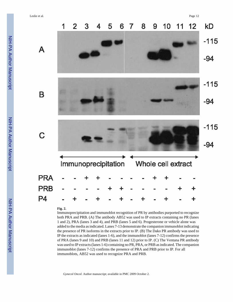
Fig. 2.Immunoprecipitation and immunoblot recognition of PR by antibodies purported to recognizeboth PRA and PRB. (A) The antibody AB52 was used to IP extracts containing no PR (lanes1 and 2), PRA (lanes 3 and 4), and PRB (lanes 5 and 6). Progesterone or vehicle alone wasadded to the media as indicated. Lanes 7-13 demonstrate the companion immunoblot indicatingthe presence of PR isoforms in the extracts prior to IP. (B) The Dako PR antibody was used toIP the extracts as indicated (lanes 1-6), and the immunoblot (lanes 7-12) confirms the presenceof PRA (lanes 9 and 10) and PRB (lanes 11 and 12) prior to IP. (C) The Ventana PR antibodywas used to IP extracts (lanes 1-6) containing no PR, PRA, or PRB as indicated. The companionimmunoblot (lanes 7-12) confirms the presence of PRA and PRB prior to IP. For allimmunoblots, AB52 was used to recognize PRA and PRB.
Leslie et al. Page 12
Gynecol Oncol. Author manuscript; available in PMC 2009 October 2.
NIH
-PA Author Manuscript
NIH
-PA Author Manuscript
NIH
-PA Author Manuscript

Fig. 3.Immunoprecipitation and immunoblot recognition of PRB by antibodies to the unique Nterminus of PRB. (A) hPRab2 was used to IP cell extract containing no PR, PRA, and PRB asindicated. (B) hPRab6 was used to IP cell extract containing no PR, PRA, and PRB. For A andB, the cell extract prior to IP was immunoblotted using AB52, which recognizes both PRA andPRB and is used as a control to demonstrate the presence of both isoforms of PR in the extractprior to IP.
Leslie et al. Page 13
Gynecol Oncol. Author manuscript; available in PMC 2009 October 2.
NIH
-PA Author Manuscript
NIH
-PA Author Manuscript
NIH
-PA Author Manuscript

Fig. 4.Subcellular localization of PRA and PRB with and without ligand. (A) Hec50co cells wereinfected with recombinant adenoviruses encoding GFP and PRA or PRB. Cells expressingGFP and PRA (top) or PRB (bottom) were analyzed by confocal microscopy. The greenfluorescence identifies infected cells expressing GFP in the cytoplasm and the nucleus. Thered fluorescence identifies the localization of the indicated PR isoform. Left panels: In theabsence of ligand, PRA is nuclear and PRB is cytoplasmic in the cells shown. Right panels: Inthe presence of ligand, both PRA and PRB are nuclear. (B) Quantification of cytoplasmic vs.nuclear distributions of PRA and PRB. The subcellular location of PRA and PRB was scoredin 200 cells by confocal microscopy. The number of cells expressing nuclear as compared tocytoplasmic PRA and PRB are plotted for control cells (no ligand) or samples treated withprogesterone (+P4). (For interpretation of the references to colour in this figure legend, thereader is referred to the web version of this article).
Leslie et al. Page 14
Gynecol Oncol. Author manuscript; available in PMC 2009 October 2.
NIH
-PA Author Manuscript
NIH
-PA Author Manuscript
NIH
-PA Author Manuscript

Fig. 5.ER, PRA, and PRB expression in two representative patients, one with a well-differentiatedendometrial cancer (A, B, C) and the other with a poorly differentiated endometrial cancer (D,E, F). IHC for ER-α (A, D), PRA (B, E), and PRB (C, F) was performed as described inMaterials and methods.
Leslie et al. Page 15
Gynecol Oncol. Author manuscript; available in PMC 2009 October 2.
NIH
-PA Author Manuscript
NIH
-PA Author Manuscript
NIH
-PA Author Manuscript

Fig. 6.Detail of PRB staining and nuclear exclusion at high magnification. (A) PRB is cytoplasmicin some poorly differentiated tumors. Note the central blue staining of the nucleus of the tumorcells (large arrowheads), with surrounding brown staining in the cytoplasm. In comparison,the stromal cells surrounding the tumor demonstrate brown nuclear staining for PRB (smallarrowheads). (B) PRB retains nuclear staining in some well-differentiated endometrial tumors.(C) Negative control for IHC staining. (For interpretation of the references to colour in thisfigure legend, the reader is referred to the web version of this article).
Leslie et al. Page 16
Gynecol Oncol. Author manuscript; available in PMC 2009 October 2.
NIH
-PA Author Manuscript
NIH
-PA Author Manuscript
NIH
-PA Author Manuscript

Fig. 7.Percentage of tumor cells demonstrating positive staining for nuclear and cytoplasmic receptorsin well-, moderately- and poorly-differentiated endometrial cancers. ER = estrogen receptorsand PRB = progesterone B receptors. Blue bars = well-differentiated tumors (n = 9), red bars= moderately differentiated tumors (n = 5), and beige bars = poorly differentiated tumors (n =2). *The presence of PRB in the nucleus is significantly correlated with tumor differentiationusing the Mantel-Haenszel test, P = 0.031. (For interpretation of the references to colour inthis figure legend, the reader is referred to the web version of this article).
Leslie et al. Page 17
Gynecol Oncol. Author manuscript; available in PMC 2009 October 2.
NIH
-PA Author Manuscript
NIH
-PA Author Manuscript
NIH
-PA Author Manuscript

NIH
-PA Author Manuscript
NIH
-PA Author Manuscript
NIH
-PA Author Manuscript
Leslie et al. Page 18
Table 1Antibodies used in these studies including their manufacturer and target specificity
Antibody Manufacturer Target Specificity
PR Dakocytomation PR Preferentially PRA
PR Ventana PR Preferentially PRA
AB52 K. Horwitz PR PRA and PRB
B30 K. Horwitz N-terminus PR PRB
hPRa2 Lab Visions N-terminus PR PRB
hPRa6 Lab Visions N-terminus PR PRB
Gynecol Oncol. Author manuscript; available in PMC 2009 October 2.
Related Documents