RESEARCH ARTICLE Progesterone induces neuroprotection following reperfusion- promoted mitochondrial dysfunction after focal cerebral ischemia in rats Syed Suhail Andrabi 1 , Suhel Parvez 1 and Heena Tabassum 2, * ABSTRACT Organelle damage and increases in mitochondrial permeabilization are key events in the development of cerebral ischemic tissue injury because they cause both modifications in ATP turnover and cellular apoptosis/necrosis. Early restoration of blood flow and improvement of mitochondrial function might reverse the situation and help in recovery following an onset of stroke. Mitochondria and related bioenergetic processes can be effectively used as pharmacological targets. Progesterone (P4), one of the promising neurosteroids, has been found to be neuroprotective in various models of neurological diseases, through a number of mechanisms. This influenced us to investigate the possible role of P4 in the mitochondria-mediated neuroprotective mechanism in an ischemic stroke model of rat. In this study, we have shown the positive effect of P4 administration on behavioral deficits and mitochondrial health in an ischemic stroke injury model of transient middle cerebral artery occlusion (tMCAO). After induction of tMCAO, the rats received an initial intraperitoneal injection of P4 (8 mg/kg body weight) or vehicle at 1 h post-occlusion followed by subcutaneous injections at 6, 12 and 18 h. Behavioral assessment for functional deficits included grip strength, motor coordination and gait analysis. Findings revealed a significant improvement with P4 treatment in tMCAO animals. Staining of isolated brain slices from P4-treated rats with 2,3,5- triphenyltetrazolium chloride (TTC) showed a reduction in the infarct area in comparison to the vehicle group, indicating the presence of an increased number of viable mitochondria. P4 treatment was also able to attenuate mitochondrial reactive oxygen species (ROS) production, as well as block the mitochondrial permeability transition pore (mPTP), in the tMCAO injury model. In addition, it was also able to ameliorate the altered mitochondrial membrane potential and respiration ratio in the ischemic animals, thereby suggesting that P4 has a positive effect on mitochondrial bioenergetics. In conclusion, these results demonstrate that P4 treatment is beneficial in preserving the mitochondrial functions that are altered in cerebral ischemic injury and thus can help in defining better therapies. KEY WORDS: Progesterone, Cerebral ischemia, Neurobehavior, Mitochondria, Apoptosis, Neuroprotection INTRODUCTION Stroke is the leading global cause of death and disability as per reports by the World Heart Association (Avasarala et al., 2015). Ischemic stroke is the most common form of stroke, accounting for 87% of all types of strokes (Bennett et al., 2014). In spite of its high prevalence, no effective treatment is currently available that can alter the course of the disease. Tissue plasminogen factor (‘clot buster’) is the only FDA approved drug that is currently available for ischemic stroke patients, but has a narrow therapeutic time window (Peplow, 2015). Thus, there is a dire need for finding a new therapeutic strategy for stroke injury that has a prolonged treatment window. Mitochondria play a crucial role in the pathophysiology of several neurological diseases, including stroke (Andrabi et al., 2015; Rasheed et al., 2017). Mitochondria are very susceptible to any insult, due to their critical role in energy metabolism, the production of reactive oxygen species (ROS), and apoptotic pathways (Waseem et al., 2016). The primary event in ischemia is disruption of the electron transport chain (ETC) due to the impaired delivery of oxygen and glucose (Novgorodov et al., 2016). The mitochondrial membrane potential (MMP), generated via proton transfer across the inner mitochondrial membrane for the production of adenosine triphosphate (ATP), is derailed in cerebral ischemia (Khatri and Man, 2013). Upon reperfusion, the damage becomes severe as a consequence of excess ROS accumulation in mitochondria, leading to attenuated activity of ETC components and favoring the open state of the mitochondrial permeability transition pore (mPTP) (Javadov and Kuznetsov, 2013). These pathological changes eventually cause the release of various apoptotic factors such as cytochrome c and apoptosis inducing factor (AIF), culminating in the initiation of an apoptosis cascade and eventually leading to cell death (Manzanero et al., 2013). Neurotransmitter-based excitotoxicity is another mechanism associated with ischemic injury (Lai et al., 2014). This excitatory neurotransmitter leads to cytosolic Ca 2+ overload and mitochondrial swelling (Nicholls et al., 2015). This swelling causes mitochondrial permeabilization, releasing the apoptotic factors, including cytochrome c, and resulting in cell death in ischemia (Liu et al., 2009). A large set of experimental evidence strongly supports the neuroprotective role of the steroid hormone progesterone (P4) in many CNS injury models (De Nicola et al., 2013). Additionally, preclinical data focused on the neuroprotective role of P4 in ischemic stroke through multiple mechanisms is available (Wong et al., 2013). In addition, clinical trials investigating the neuroprotective role of P4 have also been carried out in traumatic brain injury patients, making it an attractive pharmacological agent for the treatment of ischemic injury (Schumacher et al., 2015; Wright et al., 2014; Xiao et al., 2008). P4 can act multi- mechanically by reducing oxidative damage, inhibiting apoptosis and regulating various signaling pathways in brain damage Received 9 April 2016; Accepted 28 March 2017 1 Department of Medical Elementology and Toxicology, Jamia Hamdard (Hamdard University), New Delhi 110062, India. 2 Department of Biochemistry, Jamia Hamdard (Hamdard University), New Delhi 110062, India. *Author for correspondence ([email protected]) S.P., 0000-0002-6318-6506; H.T., 0000-0001-8699-5309 This is an Open Access article distributed under the terms of the Creative Commons Attribution License (http://creativecommons.org/licenses/by/3.0), which permits unrestricted use, distribution and reproduction in any medium provided that the original work is properly attributed. 787 © 2017. Published by The Company of Biologists Ltd | Disease Models & Mechanisms (2017) 10, 787-796 doi:10.1242/dmm.025692 Disease Models & Mechanisms

Welcome message from author
This document is posted to help you gain knowledge. Please leave a comment to let me know what you think about it! Share it to your friends and learn new things together.
Transcript
RESEARCH ARTICLE
Progesterone induces neuroprotection following reperfusion-promoted mitochondrial dysfunction after focal cerebral ischemiain ratsSyed Suhail Andrabi1, Suhel Parvez1 and Heena Tabassum2,*
ABSTRACTOrganelle damage and increases in mitochondrial permeabilizationare key events in the development of cerebral ischemic tissue injurybecause they cause both modifications in ATP turnover and cellularapoptosis/necrosis. Early restoration of blood flow and improvementof mitochondrial function might reverse the situation and help inrecovery following an onset of stroke. Mitochondria and relatedbioenergetic processes can be effectively used as pharmacologicaltargets. Progesterone (P4), one of the promising neurosteroids, hasbeen found to be neuroprotective in various models of neurologicaldiseases, through a number of mechanisms. This influenced us toinvestigate the possible role of P4 in the mitochondria-mediatedneuroprotective mechanism in an ischemic stroke model of rat. In thisstudy, we have shown the positive effect of P4 administration onbehavioral deficits and mitochondrial health in an ischemic strokeinjury model of transient middle cerebral artery occlusion (tMCAO).After induction of tMCAO, the rats received an initial intraperitonealinjection of P4 (8 mg/kg body weight) or vehicle at 1 h post-occlusionfollowed by subcutaneous injections at 6, 12 and 18 h. Behavioralassessment for functional deficits included grip strength, motorcoordination and gait analysis. Findings revealed a significantimprovement with P4 treatment in tMCAO animals. Stainingof isolated brain slices from P4-treated rats with 2,3,5-triphenyltetrazolium chloride (TTC) showed a reduction in the infarctarea in comparison to the vehicle group, indicating the presence of anincreased number of viable mitochondria. P4 treatment was also ableto attenuatemitochondrial reactive oxygen species (ROS) production,as well as block the mitochondrial permeability transition pore(mPTP), in the tMCAO injury model. In addition, it was also able toameliorate the altered mitochondrial membrane potential andrespiration ratio in the ischemic animals, thereby suggesting that P4has a positive effect on mitochondrial bioenergetics. In conclusion,these results demonstrate that P4 treatment is beneficial in preservingthe mitochondrial functions that are altered in cerebral ischemic injuryand thus can help in defining better therapies.
KEY WORDS: Progesterone, Cerebral ischemia, Neurobehavior,Mitochondria, Apoptosis, Neuroprotection
INTRODUCTIONStroke is the leading global cause of death and disability as per reportsby the World Heart Association (Avasarala et al., 2015). Ischemicstroke is the most common form of stroke, accounting for 87% of alltypes of strokes (Bennett et al., 2014). In spite of its high prevalence,no effective treatment is currently available that can alter the course ofthe disease. Tissue plasminogen factor (‘clot buster’) is the only FDAapproved drug that is currently available for ischemic stroke patients,but has a narrow therapeutic time window (Peplow, 2015). Thus,there is a dire need for finding a new therapeutic strategy for strokeinjury that has a prolonged treatment window.
Mitochondria play a crucial role in the pathophysiology of severalneurological diseases, including stroke (Andrabi et al., 2015;Rasheed et al., 2017). Mitochondria are very susceptible to anyinsult, due to their critical role in energy metabolism, the productionof reactive oxygen species (ROS), and apoptotic pathways (Waseemet al., 2016). The primary event in ischemia is disruption of theelectron transport chain (ETC) due to the impaired delivery ofoxygen and glucose (Novgorodov et al., 2016). The mitochondrialmembrane potential (MMP), generated via proton transfer across theinner mitochondrial membrane for the production of adenosinetriphosphate (ATP), is derailed in cerebral ischemia (Khatri andMan, 2013). Upon reperfusion, the damage becomes severe as aconsequence of excess ROS accumulation in mitochondria, leadingto attenuated activity of ETC components and favoring the openstate of the mitochondrial permeability transition pore (mPTP)(Javadov and Kuznetsov, 2013). These pathological changeseventually cause the release of various apoptotic factors such ascytochrome c and apoptosis inducing factor (AIF), culminatingin the initiation of an apoptosis cascade and eventually leadingto cell death (Manzanero et al., 2013). Neurotransmitter-basedexcitotoxicity is another mechanism associated with ischemicinjury (Lai et al., 2014). This excitatory neurotransmitter leads tocytosolic Ca2+ overload and mitochondrial swelling (Nicholls et al.,2015). This swelling causes mitochondrial permeabilization,releasing the apoptotic factors, including cytochrome c, andresulting in cell death in ischemia (Liu et al., 2009).
A large set of experimental evidence strongly supports theneuroprotective role of the steroid hormone progesterone (P4) inmany CNS injury models (De Nicola et al., 2013). Additionally,preclinical data focused on the neuroprotective role of P4 inischemic stroke through multiple mechanisms is available (Wonget al., 2013). In addition, clinical trials investigating theneuroprotective role of P4 have also been carried out in traumaticbrain injury patients, making it an attractive pharmacological agentfor the treatment of ischemic injury (Schumacher et al., 2015;Wright et al., 2014; Xiao et al., 2008). P4 can act multi-mechanically by reducing oxidative damage, inhibiting apoptosisand regulating various signaling pathways in brain damageReceived 9 April 2016; Accepted 28 March 2017
1Department of Medical Elementology and Toxicology, Jamia Hamdard (HamdardUniversity), New Delhi 110062, India. 2Department of Biochemistry, JamiaHamdard (Hamdard University), New Delhi 110062, India.
*Author for correspondence ([email protected])
S.P., 0000-0002-6318-6506; H.T., 0000-0001-8699-5309
This is an Open Access article distributed under the terms of the Creative Commons AttributionLicense (http://creativecommons.org/licenses/by/3.0), which permits unrestricted use,distribution and reproduction in any medium provided that the original work is properly attributed.
787
© 2017. Published by The Company of Biologists Ltd | Disease Models & Mechanisms (2017) 10, 787-796 doi:10.1242/dmm.025692
Disea
seModels&Mechan
isms
(Deutsch et al., 2013). However, the role of the mitochondria-mediated pathway in neuroprotection provided by P4 in ischemicstroke needs to be thoroughly explored before using it as a target-based therapy. Rodent models of cerebral ischemia are importanttools in experimental stroke research. Such models have proven tobe instrumental for the understanding of injury mechanisms incerebral stroke as well as to identify potential new therapeuticoptions.In the current study, we evaluated the mitochondria-mediated
neuroprotective effects of P4, at clinically relevant concentrations,in vivo in the frontal cortex of a rodent model of cerebral ischemiaestablished in our laboratory. We examined the effects of P4 on theextent of the infarction, the neurobehavioral outcome, andneurotransmitter levels in rats subjected to transient middlecerebral artery occlusion (tMCAO), an in vivo model of focalischemia. Then, to elucidate its mitochondrial mechanism of action,we examined whether or not P4 could act by reducing Ca2+-inducedrat brain mitochondrial swelling, an index of increasedmitochondrial membrane permeability. In addition, we examinedwhether P4 could prevent the other mitochondrial functionalchanges, including loss of membrane potential, and alteration ofand excess ROS production. To further prove our hypothesis, weanalyzed mitochondrial bioenergetics by examining the state 3respiratory control ratio (RCR) along with some ETC components.Finally, we examined the anti-apoptotic action of P4 by elucidatingthe translocation of cytochrome c from mitochondria to cytosolthrough the mPTP, and thereby authenticated our findings.
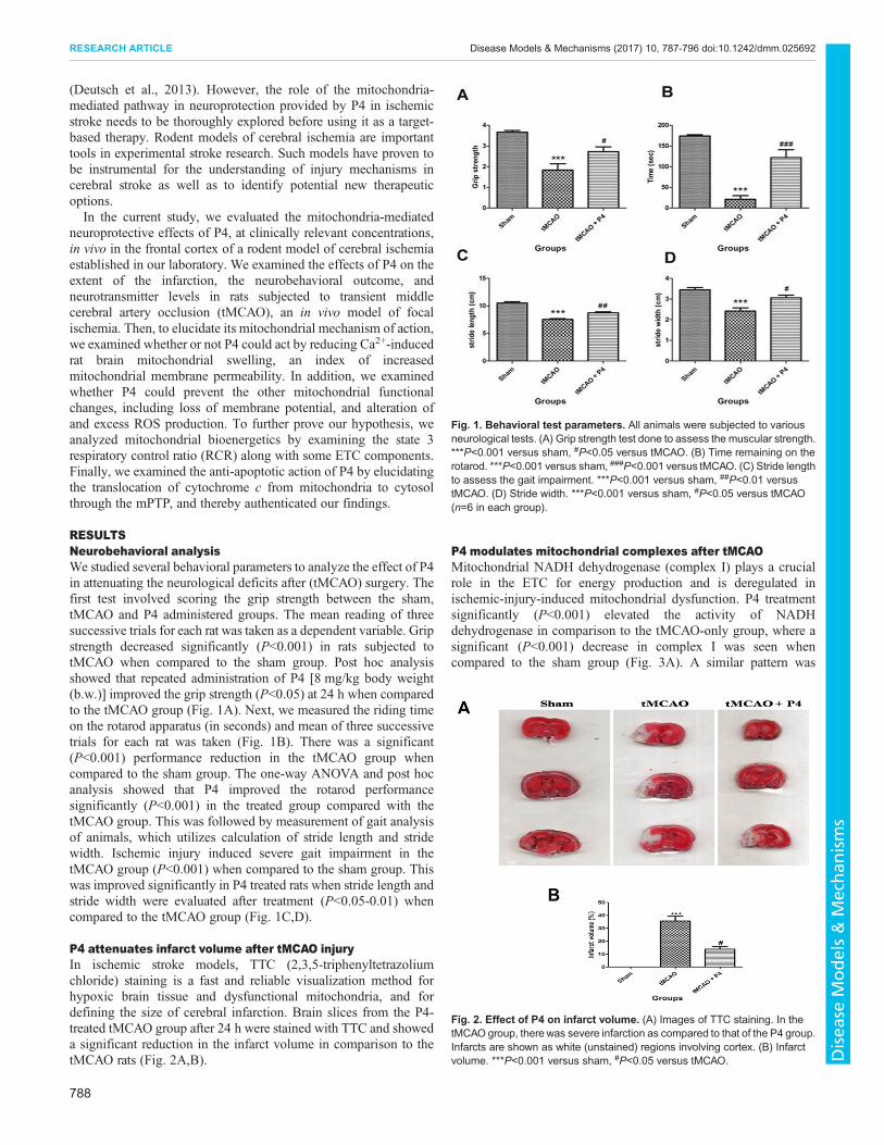
RESULTSNeurobehavioral analysisWe studied several behavioral parameters to analyze the effect of P4in attenuating the neurological deficits after (tMCAO) surgery. Thefirst test involved scoring the grip strength between the sham,tMCAO and P4 administered groups. The mean reading of threesuccessive trials for each rat was taken as a dependent variable. Gripstrength decreased significantly (P<0.001) in rats subjected totMCAO when compared to the sham group. Post hoc analysisshowed that repeated administration of P4 [8 mg/kg body weight(b.w.)] improved the grip strength (P<0.05) at 24 h when comparedto the tMCAO group (Fig. 1A). Next, we measured the riding timeon the rotarod apparatus (in seconds) and mean of three successivetrials for each rat was taken (Fig. 1B). There was a significant(P<0.001) performance reduction in the tMCAO group whencompared to the sham group. The one-way ANOVA and post hocanalysis showed that P4 improved the rotarod performancesignificantly (P<0.001) in the treated group compared with thetMCAO group. This was followed by measurement of gait analysisof animals, which utilizes calculation of stride length and stridewidth. Ischemic injury induced severe gait impairment in thetMCAO group (P<0.001) when compared to the sham group. Thiswas improved significantly in P4 treated rats when stride length andstride width were evaluated after treatment (P<0.05-0.01) whencompared to the tMCAO group (Fig. 1C,D).
P4 attenuates infarct volume after tMCAO injuryIn ischemic stroke models, TTC (2,3,5-triphenyltetrazoliumchloride) staining is a fast and reliable visualization method forhypoxic brain tissue and dysfunctional mitochondria, and fordefining the size of cerebral infarction. Brain slices from the P4-treated tMCAO group after 24 h were stained with TTC and showeda significant reduction in the infarct volume in comparison to thetMCAO rats (Fig. 2A,B).
P4 modulates mitochondrial complexes after tMCAOMitochondrial NADH dehydrogenase (complex I) plays a crucialrole in the ETC for energy production and is deregulated inischemic-injury-induced mitochondrial dysfunction. P4 treatmentsignificantly (P<0.001) elevated the activity of NADHdehydrogenase in comparison to the tMCAO-only group, where asignificant (P<0.001) decrease in complex I was seen whencompared to the sham group (Fig. 3A). A similar pattern was
Fig. 1. Behavioral test parameters. All animals were subjected to variousneurological tests. (A) Grip strength test done to assess the muscular strength.***P<0.001 versus sham, #P<0.05 versus tMCAO. (B) Time remaining on therotarod. ***P<0.001 versus sham, ###P<0.001 versus tMCAO. (C) Stride lengthto assess the gait impairment. ***P<0.001 versus sham, ##P<0.01 versustMCAO. (D) Stride width. ***P<0.001 versus sham, #P<0.05 versus tMCAO(n=6 in each group).
Fig. 2. Effect of P4 on infarct volume. (A) Images of TTC staining. In thetMCAO group, there was severe infarction as compared to that of the P4 group.Infarcts are shown as white (unstained) regions involving cortex. (B) Infarctvolume. ***P<0.001 versus sham, #P<0.05 versus tMCAO.
788
RESEARCH ARTICLE Disease Models & Mechanisms (2017) 10, 787-796 doi:10.1242/dmm.025692
Disea
seModels&Mechan
isms
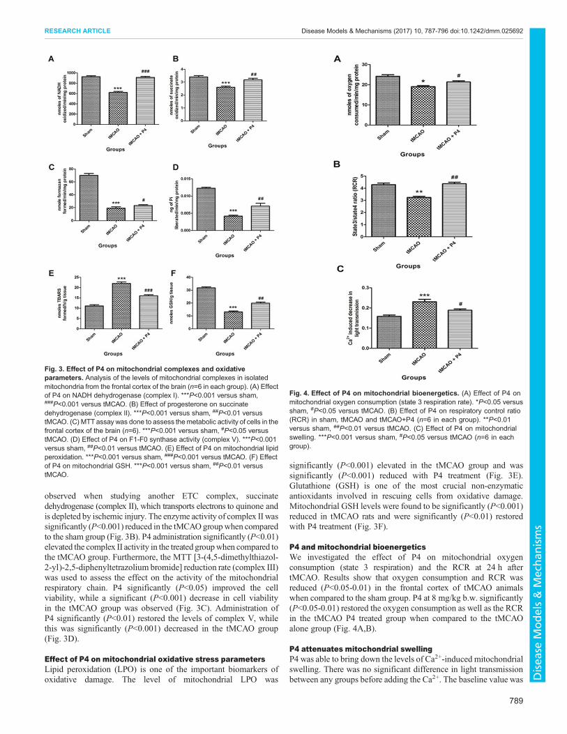
observed when studying another ETC complex, succinatedehydrogenase (complex II), which transports electrons to quinone andis depleted by ischemic injury. The enzyme activity of complex II wassignificantly (P<0.001) reduced in the tMCAOgroupwhen comparedto the sham group (Fig. 3B). P4 administration significantly (P<0.01)elevated the complex II activity in the treated groupwhen compared tothe tMCAO group. Furthermore, the MTT [3-(4,5-dimethylthiazol-2-yl)-2,5-diphenyltetrazolium bromide] reduction rate (complex III)was used to assess the effect on the activity of the mitochondrialrespiratory chain. P4 significantly (P<0.05) improved the cellviability, while a significant (P<0.001) decrease in cell viabilityin the tMCAO group was observed (Fig. 3C). Administration ofP4 significantly (P<0.01) restored the levels of complex V, whilethis was significantly (P<0.001) decreased in the tMCAO group(Fig. 3D).
Effect of P4 on mitochondrial oxidative stress parametersLipid peroxidation (LPO) is one of the important biomarkers ofoxidative damage. The level of mitochondrial LPO was
significantly (P<0.001) elevated in the tMCAO group and wassignificantly (P<0.001) reduced with P4 treatment (Fig. 3E).Glutathione (GSH) is one of the most crucial non-enzymaticantioxidants involved in rescuing cells from oxidative damage.Mitochondrial GSH levels were found to be significantly (P<0.001)reduced in tMCAO rats and were significantly (P<0.01) restoredwith P4 treatment (Fig. 3F).
P4 and mitochondrial bioenergeticsWe investigated the effect of P4 on mitochondrial oxygenconsumption (state 3 respiration) and the RCR at 24 h aftertMCAO. Results show that oxygen consumption and RCR wasreduced (P<0.05-0.01) in the frontal cortex of tMCAO animalswhen compared to the sham group. P4 at 8 mg/kg b.w. significantly(P<0.05-0.01) restored the oxygen consumption as well as the RCRin the tMCAO P4 treated group when compared to the tMCAOalone group (Fig. 4A,B).
P4 attenuates mitochondrial swellingP4 was able to bring down the levels of Ca2+-induced mitochondrialswelling. There was no significant difference in light transmissionbetween any groups before adding the Ca2+. The baseline value was
Fig. 3. Effect of P4 on mitochondrial complexes and oxidativeparameters. Analysis of the levels of mitochondrial complexes in isolatedmitochondria from the frontal cortex of the brain (n=6 in each group). (A) Effectof P4 on NADH dehydrogenase (complex I). ***P<0.001 versus sham,###P<0.001 versus tMCAO. (B) Effect of progesterone on succinatedehydrogenase (complex II). ***P<0.001 versus sham, ##P<0.01 versustMCAO. (C) MTT assay was done to assess themetabolic activity of cells in thefrontal cortex of the brain (n=6). ***P<0.001 versus sham, #P<0.05 versustMCAO. (D) Effect of P4 on F1-F0 synthase activity (complex V). ***P<0.001versus sham, ##P<0.01 versus tMCAO. (E) Effect of P4 on mitochondrial lipidperoxidation. ***P<0.001 versus sham, ###P<0.001 versus tMCAO. (F) Effectof P4 on mitochondrial GSH. ***P<0.001 versus sham, ##P<0.01 versustMCAO.
Fig. 4. Effect of P4 on mitochondrial bioenergetics. (A) Effect of P4 onmitochondrial oxygen consumption (state 3 respiration rate). *P<0.05 versussham, #P<0.05 versus tMCAO. (B) Effect of P4 on respiratory control ratio(RCR) in sham, tMCAO and tMCAO+P4 (n=6 in each group). **P<0.01versus sham, ##P<0.01 versus tMCAO. (C) Effect of P4 on mitochondrialswelling. ***P<0.001 versus sham, #P<0.05 versus tMCAO (n=6 in eachgroup).
789
RESEARCH ARTICLE Disease Models & Mechanisms (2017) 10, 787-796 doi:10.1242/dmm.025692
Disea
seModels&Mechan
isms
taken for 5 min after addition of Ca2+ at the concentration of400 µM. A significant (P<0.001) decrease in light transmission inthe tMCAO group occurred when compared to the sham group withaddition of the Ca2+. This was significantly attenuated by P4administration (P<0.05) when compared to the tMCAO group(Fig. 4C).
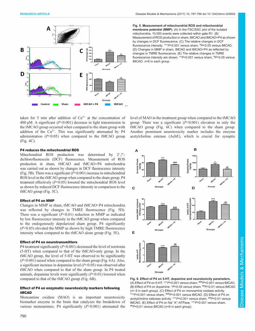
P4 reduces the mitochondrial ROSMitochondrial ROS production was determined by 2′,7′-dichlorofluorescein (DCF) fluorescence. Measurement of ROSproduction in sham, tMCAO and tMCAO+P4 mitochondriawas carried out as shown by changes in DCF fluorescence intensity(Fig. 5B). Therewas a significant (P<0.001) increase in mitochondrialROS level in the tMCAOgroupwhen compared to the sham group. P4treatment effectively (P<0.05) lowered the mitochondrial ROS levelas shown by reduced DCF fluorescence intensity in comparison to thetMCAO group (Fig. 5C).
Effect of P4 on MMPChanges in MMP in sham, tMCAO and tMCAO+P4 mitochondriawas reflected by changes in TMRE fluorescence (Fig. 5D).There was a significant (P<0.01) reduction in MMP as indicatedby low fluorescence intensity in the tMCAO group when comparedto the endogenously depolarized sham group. P4 significantly(P<0.05) elevated the MMP as shown by high TMRE fluorescenceintensity when compared to the tMCAO alone group (Fig. 5E).
Effect of P4 on neurotransmittersP4 treatment significantly (P<0.001) decreased the level of serotonin(5-HT) when compared to that of the tMCAO-only group. In thetMCAO group, the level of 5-HT was observed to be significantly(P<0.001) raised when compared to the sham group (Fig. 6A). Also,a significant increase in dopamine level (P<0.05) was observed aftertMCAO when compared to that of the sham group. In P4 treatedanimals, dopamine levels were significantly (P<0.01) lowered whencompared to that of the tMCAO group (Fig. 6B).
Effect of P4 on enzymatic neurotoxicity markers followingtMCAOMonoamine oxidase (MAO) is an important neurotoxicitybiomarker enzyme in the brain that catalyzes the breakdown ofvarious monoamines. P4 significantly (P<0.001) attenuated the
level of MAO in the treatment group when compared to the tMCAOgroup. There was a significant (P<0.001) elevation in only thetMCAO group (Fig. 6C) when compared to the sham group.Another prominent neurotoxicity marker includes the enzymeacetylcholine esterase (AchE), which is crucial for synaptic
Fig. 5. Measurement of mitochondrial ROS and mitochondrialmembrane potential (MMP). (A) In the FSC/SSC plot of the isolatedmitochondria, 10,000 events were collected within gate R1. (B)Measurement of ROS production in sham, tMCAO and tMCAO+P4 as shownby changes in DCF fluorescence. (C) The relative changes in DCFfluorescence intensity. ***P<0.001 versus sham, #P<0.05 versus tMCAO.(D) Changes in MMP in sham, tMCAO and tMCAO+P4 as reflected bychanges in TMRE fluorescence. (E) The relative changes in TMREfluorescence intensity are shown. **P<0.001 versus sham, #P<0.05 versustMCAO. n=6 in each group.
Fig. 6. Effect of P4 on 5-HT, dopamine and neurotoxicity parameters.(A) Effect of P4 on 5-HT. ***P<0.001 versus sham, ###P<0.001 versus tMCAO.(B) Effect of P4 on dopamine. *P<0.05 versus sham, ##P<0.01 versus tMCAO(n= 6 in each group). (C) Effect of P4 on monoamine oxidase activity.***P<0.001 versus sham, ###P<0.001 versus tMCAO. (D) Effect of P4 onacetylcholine esterase activity. ***P<0.001 versus sham, ##P<0.01 versustMCAO. (E) Effect of P4 on Na+ K+-ATPase. ***P<0.001 versus sham,##P<0.01 versus tMCAO (n=6 in each group).
790
RESEARCH ARTICLE Disease Models & Mechanisms (2017) 10, 787-796 doi:10.1242/dmm.025692
Disea
seModels&Mechan
isms
termination of the nerve impulse bymetabolism of acetylcholine. P4administration significantly (P<0.01) lowered the activity of AchEin animals, in comparison to the tMCAO group. There was asignificant (P<0.001) elevation of AchE enzyme level in thetMCAO group when compared to the sham group (Fig. 6D). Also,decreased levels of Na+ K+-ATPase during brain injury indicateddepletion of ATP. The same was lowered significantly (P<0.001) inthe tMCAO group, when compared to the sham group. This wasobserved to be restored in P4 treated animals at a significant level(P<0.01; Fig. 6E) when compared to the tMCAO group.
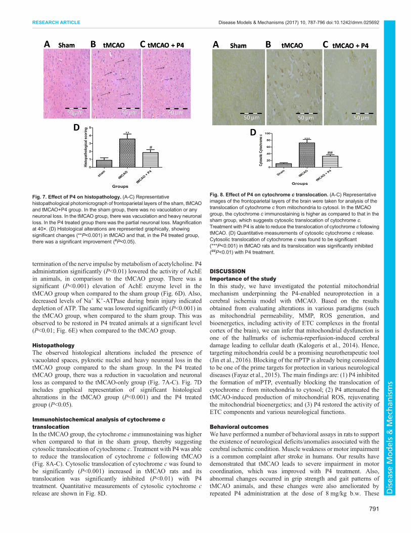
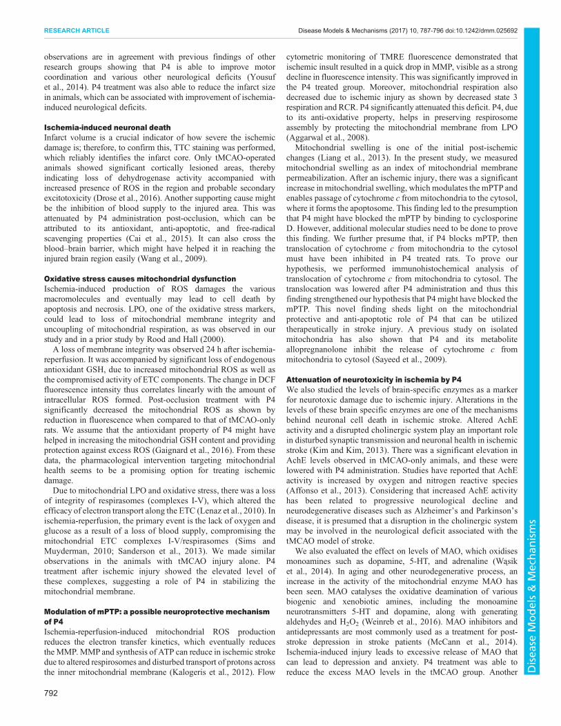
HistopathologyThe observed histological alterations included the presence ofvacuolated spaces, pyknotic nuclei and heavy neuronal loss in thetMCAO group compared to the sham group. In the P4 treatedtMCAO group, there was a reduction in vacuolation and neuronalloss as compared to the tMCAO-only group (Fig. 7A-C). Fig. 7Dincludes graphical representation of significant histologicalalterations in the tMCAO group (P<0.001) and the P4 treatedgroup (P<0.05).
Immunohistochemical analysis of cytochrome ctranslocationIn the tMCAO group, the cytochrome c immunostaining was higherwhen compared to that in the sham group, thereby suggestingcytosolic translocation of cytochrome c. Treatment with P4 was ableto reduce the translocation of cytochrome c following tMCAO(Fig. 8A-C). Cytosolic translocation of cytochrome c was found tobe significantly (P<0.001) increased in tMCAO rats and itstranslocation was significantly inhibited (P<0.01) with P4treatment. Quantitative measurements of cytosolic cytochrome crelease are shown in Fig. 8D.
DISCUSSIONImportance of the studyIn this study, we have investigated the potential mitochondrialmechanism underpinning the P4-enabled neuroprotection in acerebral ischemia model with tMCAO. Based on the resultsobtained from evaluating alterations in various paradigms (suchas mitochondrial permeability, MMP, ROS generation, andbioenergetics, including activity of ETC complexes in the frontalcortex of the brain), we can infer that mitochondrial dysfunction isone of the hallmarks of ischemia-reperfusion-induced cerebraldamage leading to cellular death (Kalogeris et al., 2014). Hence,targeting mitochondria could be a promising neurotherapeutic tool(Jin et al., 2016). Blocking of the mPTP is already being consideredto be one of the prime targets for protection in various neurologicaldiseases (Fayaz et al., 2015). The main findings are: (1) P4 inhibitedthe formation of mPTP, eventually blocking the translocation ofcytochrome c from mitochondria to cytosol; (2) P4 attenuated thetMCAO-induced production of mitochondrial ROS, rejuvenatingthe mitochondrial bioenergetics; and (3) P4 restored the activity ofETC components and various neurological functions.
Behavioral outcomesWe have performed a number of behavioral assays in rats to supportthe existence of neurological deficits/anomalies associated with thecerebral ischemic condition. Muscle weakness or motor impairmentis a common complaint after stroke in humans. Our results havedemonstrated that tMCAO leads to severe impairment in motorcoordination, which was improved with P4 treatment. Also,abnormal changes occurred in grip strength and gait patterns oftMCAO animals, and these changes were also ameliorated byrepeated P4 administration at the dose of 8 mg/kg b.w. These
Fig. 7. Effect of P4 on histopathology. (A-C) Representativehistopathological photomicrograph of frontoparietal layers of the sham, tMCAOand tMCAO+P4 group. In the sham group, there was no vacuolation or anyneuronal loss. In the tMCAO group, there was vacuolation and heavy neuronalloss. In the P4 treated group there was the partial neuronal loss. Magnificationat 40×. (D) Histological alterations are represented graphically, showingsignificant changes (**P<0.001) in tMCAO and that, in the P4 treated group,there was a significant improvement (#P<0.05).
Fig. 8. Effect of P4 on cytochrome c translocation. (A-C) Representativeimages of the frontoparietal layers of the brain were taken for analysis of thetranslocation of cytochrome c from mitochondria to cytosol. In the tMCAOgroup, the cytochrome c immunostaining is higher as compared to that in thesham group, which suggests cytosolic translocation of cytochrome c.Treatment with P4 is able to reduce the translocation of cytochrome c followingtMCAO. (D) Quantitative measurements of cytosolic cytochrome c release.Cytosolic translocation of cytochrome c was found to be significant(***P<0.001) in tMCAO rats and its translocation was significantly inhibited(##P<0.01) with P4 treatment.
791
RESEARCH ARTICLE Disease Models & Mechanisms (2017) 10, 787-796 doi:10.1242/dmm.025692
Disea
seModels&Mechan
isms
observations are in agreement with previous findings of otherresearch groups showing that P4 is able to improve motorcoordination and various other neurological deficits (Yousufet al., 2014). P4 treatment was also able to reduce the infarct sizein animals, which can be associated with improvement of ischemia-induced neurological deficits.
Ischemia-induced neuronal deathInfarct volume is a crucial indicator of how severe the ischemicdamage is; therefore, to confirm this, TTC staining was performed,which reliably identifies the infarct core. Only tMCAO-operatedanimals showed significant cortically lesioned areas, therebyindicating loss of dehydrogenase activity accompanied withincreased presence of ROS in the region and probable secondaryexcitotoxicity (Drose et al., 2016). Another supporting cause mightbe the inhibition of blood supply to the injured area. This wasattenuated by P4 administration post-occlusion, which can beattributed to its antioxidant, anti-apoptotic, and free-radicalscavenging properties (Cai et al., 2015). It can also cross theblood–brain barrier, which might have helped it in reaching theinjured brain region easily (Wang et al., 2009).
Oxidative stress causes mitochondrial dysfunctionIschemia-induced production of ROS damages the variousmacromolecules and eventually may lead to cell death byapoptosis and necrosis. LPO, one of the oxidative stress markers,could lead to loss of mitochondrial membrane integrity anduncoupling of mitochondrial respiration, as was observed in ourstudy and in a prior study by Rood and Hall (2000).A loss of membrane integrity was observed 24 h after ischemia-
reperfusion. It was accompanied by significant loss of endogenousantioxidant GSH, due to increased mitochondrial ROS as well asthe compromised activity of ETC components. The change in DCFfluorescence intensity thus correlates linearly with the amount ofintracellular ROS formed. Post-occlusion treatment with P4significantly decreased the mitochondrial ROS as shown byreduction in fluorescence when compared to that of tMCAO-onlyrats. We assume that the antioxidant property of P4 might havehelped in increasing the mitochondrial GSH content and providingprotection against excess ROS (Gaignard et al., 2016). From thesedata, the pharmacological intervention targeting mitochondrialhealth seems to be a promising option for treating ischemicdamage.Due to mitochondrial LPO and oxidative stress, there was a loss
of integrity of respirasomes (complexes I-V), which altered theefficacy of electron transport along the ETC (Lenaz et al., 2010). Inischemia-reperfusion, the primary event is the lack of oxygen andglucose as a result of a loss of blood supply, compromising themitochondrial ETC complexes I-V/respirasomes (Sims andMuyderman, 2010; Sanderson et al., 2013). We made similarobservations in the animals with tMCAO injury alone. P4treatment after ischemic injury showed the elevated level ofthese complexes, suggesting a role of P4 in stabilizing themitochondrial membrane.
Modulation of mPTP: a possible neuroprotective mechanismof P4Ischemia-reperfusion-induced mitochondrial ROS productionreduces the electron transfer kinetics, which eventually reducestheMMP.MMP and synthesis of ATP can reduce in ischemic strokedue to altered respirosomes and disturbed transport of protons acrossthe inner mitochondrial membrane (Kalogeris et al., 2012). Flow
cytometric monitoring of TMRE fluorescence demonstrated thatischemic insult resulted in a quick drop in MMP, visible as a strongdecline in fluorescence intensity. This was significantly improved inthe P4 treated group. Moreover, mitochondrial respiration alsodecreased due to ischemic injury as shown by decreased state 3respiration and RCR. P4 significantly attenuated this deficit. P4, dueto its anti-oxidative property, helps in preserving respirosomeassembly by protecting the mitochondrial membrane from LPO(Aggarwal et al., 2008).
Mitochondrial swelling is one of the initial post-ischemicchanges (Liang et al., 2013). In the present study, we measuredmitochondrial swelling as an index of mitochondrial membranepermeabilization. After an ischemic injury, there was a significantincrease in mitochondrial swelling, which modulates the mPTP andenables passage of cytochrome c from mitochondria to the cytosol,where it forms the apoptosome. This finding led to the presumptionthat P4 might have blocked the mPTP by binding to cyclosporineD. However, additional molecular studies need to be done to provethis finding. We further presume that, if P4 blocks mPTP, thentranslocation of cytochrome c from mitochondria to the cytosolmust have been inhibited in P4 treated rats. To prove ourhypothesis, we performed immunohistochemical analysis oftranslocation of cytochrome c from mitochondria to cytosol. Thetranslocation was lowered after P4 administration and thus thisfinding strengthened our hypothesis that P4 might have blocked themPTP. This novel finding sheds light on the mitochondrialprotective and anti-apoptotic role of P4 that can be utilizedtherapeutically in stroke injury. A previous study on isolatedmitochondria has also shown that P4 and its metaboliteallopregnanolone inhibit the release of cytochrome c frommitochondria to cytosol (Sayeed et al., 2009).
Attenuation of neurotoxicity in ischemia by P4We also studied the levels of brain-specific enzymes as a markerfor neurotoxic damage due to ischemic injury. Alterations in thelevels of these brain specific enzymes are one of the mechanismsbehind neuronal cell death in ischemic stroke. Altered AchEactivity and a disrupted cholinergic system play an important rolein disturbed synaptic transmission and neuronal health in ischemicstroke (Kim and Kim, 2013). There was a significant elevation inAchE levels observed in tMCAO-only animals, and these werelowered with P4 administration. Studies have reported that AchEactivity is increased by oxygen and nitrogen reactive species(Affonso et al., 2013). Considering that increased AchE activityhas been related to progressive neurological decline andneurodegenerative diseases such as Alzheimer’s and Parkinson’sdisease, it is presumed that a disruption in the cholinergic systemmay be involved in the neurological deficit associated with thetMCAO model of stroke.
We also evaluated the effect on levels of MAO, which oxidisesmonoamines such as dopamine, 5-HT, and adrenaline (Wasiket al., 2014). In aging and other neurodegenerative process, anincrease in the activity of the mitochondrial enzyme MAO hasbeen seen. MAO catalyses the oxidative deamination of variousbiogenic and xenobiotic amines, including the monoamineneurotransmitters 5-HT and dopamine, along with generatingaldehydes and H2O2 (Weinreb et al., 2016). MAO inhibitors andantidepressants are most commonly used as a treatment for post-stroke depression in stroke patients (McCann et al., 2014).Ischemia-induced injury leads to excessive release of MAO thatcan lead to depression and anxiety. P4 treatment was able toreduce the excess MAO levels in the tMCAO group. Another
792
RESEARCH ARTICLE Disease Models & Mechanisms (2017) 10, 787-796 doi:10.1242/dmm.025692
Disea
seModels&Mechan
isms
enzyme, Na+ K+-ATPase, is an ATP-dependent pump for nerveconduction, maintaining the resting potential of cell membranes(de Lores Arnaiz and Ordieres, 2014). Due to alteredmitochondrial bioenergetics in ischemia, physiological processessuch as the functioning of ATP-dependent ion channels can bederailed, leading to alteration of membrane potential as observedin tMCAO injury. P4 was able to substantially elevate the activityof Na+ K+-ATPase by protecting the mitochondrial damage andthus supporting the previous findings that P4 has a positive effecton mitochondrial bioenergetics.
Changes in neurotransmitter levels affects themitochondrial functionsThis study also incorporated the analysis of neurotransmitters thatplay a crucial role in proper functioning of the nervous system. Afterthe onset of ischemic stroke, cessation or reduction of blood flow tothe brain induces neuronal damage. Monoamine neurotransmitterssuch as dopamine and 5-HT are important biogenic amines thatare vital for transmitting nerve impulses for cognitive function ofthe body. Over-release of the above-mentioned neurotransmitters,which can be triggered by many types of neurotoxic insult, suchas stroke, hazardous chemicals, tumors and neurodegenerativediseases, affect their respective receptors in the brain which canfurther induce cytotoxic responses, including altered calciuminflux, free radical damage, oxidative stress, inflammatoryresponses and apoptosis, potentially leading to neuronal celldeath and neurodegeneration (Chen et al., 2014).Dopamine not only works as a neurotransmitter but acts as a good
metal chelator and electron donor in in vivo conditions, generatingtoxic free radicals through the redox reaction. It has a high tendencyto generate H2O2 through Fenton’s chemistry. This, in turn, causesmitochondrial respiratory chain breakdown and oxidative stressleading to cardiomyopathy and neurodegeneration (Uttara et al.,2009). 5-HT has an inhibitory effect on mitochondrial respiration,causing brain ATP depletion. It causes excitotoxic death of nervecells, which involves both limitations of energy production andincreased cellular activation (Chen et al., 2014). Levels of both ofthese neurotransmitters were found to be elevated in the tMCAOgroup and were attenuated by P4 treatment. P4 is known to possessspecific anti-5-HT actions (Morton et al., 2015). P4 can attenuateneuronal excitotoxicity by blocking calcium channels or byupregulating inhibitory neurotransmitter GABA, which in turncan block the release of other excitatory neurotransmitters in theCNS (Wei and Xiao, 2013). Also, we have shown in the presentstudy that P4 inhibits the activity of the enzymeMAO, which breaksdown dopamine to generate toxic products such as H2O2, oxygen-derived radicals, semiquinones and quinones, leading tomitochondrial LPO and neurotoxic effects.
P4 improves anatomical damage in ischemic tissuetMCAO-operated animals showed a series of histologicalalterations, including marked neurodegeneration with pyknoticneurons. P4 successfully ameliorated these histological alterationsas observed by the reduced number of pyknotic neurons.
ConclusionThe current study adds further evidence that P4 is neuroprotective incerebral ischemia. Our results indicate that P4 mediates itsneuroprotection through mitochondrial pathways in male rats aftercerebral ischemia. In conclusion, the results demonstrate that P4 wasable to modulate the functional and cellular deficits associated withischemic injury via mitochondrial pathways. It is important to assess
potential neuroprotective candidates, such as P4, in the group athighest risk of stroke. As this study focused on mitochondrialpathways in male rats, further studies are warranted to study theeffects on female rats.
MATERIALS AND METHODSAnimals and treatment regimenMale Wistar rats (250-300 g) were obtained from the Central Animal HouseFacility of Jamia Hamdard, NewDelhi, India. The animals were kept under a12 h light-dark cycle with free access to food and water. Use of animals andall experimental procedures were conducted according to the proceduresapproved by the Animal Ethics Committee, Jamia Hamdard.
Experimental groupsThe rats were divided randomly into three groups: (1) Sham-operated group,(2) tMCAO group, (3) tMCAO+P4 (8 mg/kg b.w.). The experimenter wasblinded to the treatment group. All parameters were done in the frontalcortex of brain, and six animals were taken for each set of parameters in eachgroup.
Surgical procedureFocal tMCAO was performed according to the method of Vaibhav et al.(2013). Prior to tMCAO surgery, animals were anesthetized with choralhydrate (400 mg/kg b.w.). A midline incision was made on the ventralsurface of neck to expose the right common carotid artery. The externalcarotid artery (ECA) was ligated and internal carotid artery (ICA) wasisolated near to the bifurcation. An intraluminal monofilament of filamentsize 4.0, length 30 mm, and diameter 0.19 mm having a silicon rubber-coated tip was introduced into the ECA and advanced through the ICA up tothe origin of the middle cerebral artery (MCA). The suture was withdrawnslowly after 1 h occlusion of MCA, and rats were returned to their cages forthe period of 23 h for reperfusion. In the sham group, ECA was surgicallyprepared but the filament was not inserted. Animals were returned to theirnormal environment in an air-conditioned room at an ambient temperature(25±2°C) and relative humidity (45-50%) with 12 h light/dark cycles andallowed free access to the pellet diet and purified drinking water.
P4 treatmentP4 (P-0130; Sigma-Aldrich Co.) at a dose of 8 mg/kg b.w. was dissolved in50% dimethyl sulfoxide (DMSO) and 50% saline, which was administeredby intraperitoneal (i.p.) injection 1 h post-occlusion followed subcutaneously(s.c.) at 6, 12, and 18 h post-occlusion. This dose is considered to be the dosefor producing neuroprotection (Wali et al., 2014).
Behavioral testsAll naïve animals were given random training for 5 days for testing motorcoordination, grip strength and gait pattern before the experiment in order toacclimatization and get a maximum possible score.
Assessment of motor impairmentTo assess the sensorimotor coordination, the rats were evaluated in the rotarodtask before sacrifice. In this study, motor function was assessed at 23 h post-occlusion by using a rotarod unit (Omni Rotor, Omnitech Electronics Inc.,Columbus, OH, USA), which consists of a rotating rod of diameter 75 mmwhich is divided into four compartments to test four animals at a time (Ashafaqet al., 2012b). The time that each animal remains on the rotating rod wasrecorded for three trials with a minimum interval between each of 5 min and amaximum trial length of 180 s for each trial. The apparatus automaticallyrecorded the time in tenths of a second until the rat falls to the floor. The speedwas set at 10 rotations per minute and cut-off time was 180 s. The score waspresented as mean of latencies of three trials on the rotating rod.
Assessment of grip strengthGrip test was performed by using the method of Ashafaq et al. (2012a). Theapparatus consists of a string measuring about 50 cm in length, pulled tightbetween two vertical supports and elevated 40 cm from the flat surface. Therats were put on the string at midway and scoring was done according to the
793
RESEARCH ARTICLE Disease Models & Mechanisms (2017) 10, 787-796 doi:10.1242/dmm.025692
Disea
seModels&Mechan
isms
following scoring scale: 0=fall off, 1=hangs onto string by two forepaws,2=hangs onto string by two forepaws and also tries to climb on string,3=hangs onto string by two forepaws along with one or both hind paws,4=hangs onto string by all forepaws along with tail wrapped around thestring, 5=escape.
Gait analysisThe gait patterns were evaluated according to the previously publishedmethod (Ashafaq et al., 2012a) to find the gait-related anomalies at 24 h aftertMCAO. Stride length and stride width were measured by using an enclosedwooden walkway of 12 cm width. The forepaws were stained with greennon-poisonous coloring agent and hindpaws were stained with red color.Stride length was measured as the distance between ipsilateral forepaw andhindpaw. Stridewidth was taken as the side-to-side distance between the twoforepaws, and the two hindpaws, respectively.
Measurement of infarction volumeThe animals were sacrificed after 1 h of occlusion followed by 23 h ofreperfusion (Ashafaq et al., 2016). The brains were dissected out and kept ina brain matrix. 1.5 mm coronal sections of the brains were cut down with thehelp of sharp blades and stained with 0.1% TTC prepared into normal salineat 37°C for 15 min. For imaging, the sections were scanned by a high-resolution scanner. The infarct volume was measured with ImageJ software.
Tissue collection and mitochondrial preparationMitochondria of brain frontal cortex of rat were isolated by thedifferential centrifugation method (Waseem and Parvez, 2016). Ratswere decapitated and frontal cortex were dissected and homogenized byusing a mechanically driven Teflon-fitted Potter-Elvehjem typehomogenizer in ice-cold buffer A. Frontal cortex mitochondria wereisolated in three buffers: A, B and C. Buffer A contained 250 mMsucrose, 10 mM 4-(2-hydroxyethyl)-1-piperazineethanesulfonic acid(HEPES), 1 mM EGTA, and 0.1% fat-free BSA adjusted by Tris to pH7.4 and centrifuged at 1000 g for 8 min at 4°C. The supernatant wascollected and centrifuged at 10,000 g for 10 min at 4°C. Thereafter, theobtained pellet was resuspended and washed twice with washing medium(B), containing 250 mM sucrose, 10 mM HEPES, and 0.1 mM EGTAadjusted by Tris to pH 7.4 and centrifuged at 12,300 g for 10 min.Finally, the pellet was resuspended again in an isolation medium (C),containing 250 mM sucrose, 10 mM HEPES, and 0.1% fat-free BSAadjusted by Tris to pH 7.4 and centrifuged at 12,300 g for 10 min.
Mitochondrial complexes measurementNADH dehydrogenase activity (complex I)NADH dehydrogenase activity was assayed by spectrophotometricallyaccording to Waseem and Parvez (2016). The enzyme activity wasexpressed as micromoles of NADH oxidized per minute per milligramprotein using a molar extinction coefficient of 21,000 M−1 cm−1.
Succinate dehydrogenase activity (complex II)The activity of succinate dehydrogenase was assayed according to themethod described by Waseem and Parvez (2016) by using thespectrophotometer. The enzyme activity was expressed as micromoles ofsuccinate oxidized per minute per milligram protein using a molar extinctioncoefficient of 1000 M−1 cm−1.
Cytochrome c reductase (complex III)Cytochrome c reductase activity was measured as described byWaseem andParvez (2016). The results were expressed as micromoles of formazanformed per minute per milligram protein with a molar excitation coefficientof 51,000 M−1 cm−1.
F1-F0 synthase activity (complex V)ATP synthase is also referred to as mitochondrial complex V. Its activity wasassayed as hydrolysis of ATP into ADP plus inorganic phosphate (Pi) asdescribed by Waseem and Parvez (2016). The enzyme activity wasexpressed as microgram of Pi liberated per minute per milligram protein.
Assessment of oxidative stress damageMitochondrial LPO was assayed according to the procedure of Chaudharyand Parvez (2012) with certain modifications. The results were expressedas nanomoles of thiobarbituric acid-reactive substances (TBARS) formedper hour per gram tissue using a molar extinction coefficient of1.56×10−5 M−1 cm−1. GSH concentration in brain mitochondria wasmeasured according to the method of Tabassum et al. (2007). The GSHcontent was calculated as nanomoles of GSH reduced per gram tissue.
Mitochondrial respiration measurementMitochondrial oxygen consumption was measured by using a Clark-typeoxygen electrode (Hansatech Instrument) (Waseem and Parvez, 2016) at37°C pH 7.4 in a KCl medium containing 0.1 mM EDTA, MgCl2, sucroseand KH2PO4. Rats were sacrificed 24 h after tMCAO and frontal cortex wasisolated for mitochondrial preparations. Mitochondrial respiratory energycoupling was evaluated by determining respiratory control ratio (RCR)calculated as the rate of ADP-induced state 3 respiration to the state 4 ratewithout ADP. The rate of mitochondrial respiration was measured asnanomoles of oxygen (O2)/min/mg of protein.
Mitochondrial swellingMitochondrial swelling caused by the influx of solutes through openpermeability transition pores results in an increase in light transmission (i.e.a reduced turbidity). This turbidity change offers a convenient andfrequently used assay of the mitochondrial permeability transition bymeasurement of absorbance in mitochondrial suspensions. Mitochondrialpermeability was assayed by Ca2+-induced mitochondrial swelling and wasassayed by spectrophotometry (Li et al., 2012). The mitochondrial pelletwas re-suspended in ice-cold BSA-free and EDTA-free sucrose buffer afterthe last step of washing (300 mmol sucrose and 10 mmol/l Tris-Base, pH7.4). The aliquot of 100 µg of mitochondria was added to 1 ml of BSA-freeand EDTA-free buffer and 400 µm Ca2+ was added after 5 min and readingwas taken for 5 min at 540 nm.
Flow cytometric analysis of MMP and mitochondrial ROSFlow cytometry analysis was performed using a FACSCalibur equippedwith a 488 nm argon laser and a 635 nm red diode laser according to Liet al. (2012). Data from the experiments were analyzed using theCellQuest software (BD Bioscience). To exclude debris, samples weregated based on light scattering properties in the side scattering (SSC) andforward scattering (FSC) modes, and 10,000 events per sample within theR1 gate were collected. The mitochondrial sample was suspended inanalysis buffer containing 250 mmol/l sucrose, 20 mmol/l MOPS,10 mmol/l Tris-base, 100 µmol/l Pi(K), and 0.5 mmol/l Mg2+ and5 mmol/l succinate at pH 7.4. The mitochondria were then stained withTMRE (100 nmol/l, excitation at 488 nm and emission at 590 nm) andH2DCFDA (2, 7 dichloro dihydro fluorescien diacetate) (10 mmol/l,excitation at 488 nm and emission at 525 nm), which were used tomeasure the MMP and the production of ROS, respectively.
Neurotransmitter detection using HPLCEstimation of 5-HT and dopamine in the brain frontal cortex was done usinghigh-performance liquid chromatography (HPLC)-electrochemicaldetection (ECD) by the method of Chaudhary and Parvez (2012).Chromatographic analyses were performed at room temperature. Datawere acquired and processed in the Empower Pro Operating System. 5-HTpeaks were identified by comparing their retention time in the sample and itsconcentration was estimated according to the area under the curve using thestraight line equation y=mx+c. Dopamine concentration was represented asng/mg protein.
Neuronal insult markersMAO was measured according to the method reported by Vishnoi et al.(2015). The enzyme activity was assayed as nanomoles of benzyl aminehydrochloride (BAHC) hydrolyzed per minute per milligram protein using amolar extinction coefficient of 7.6925 M−1 cm−1. AchE was estimatedaccording to the method of Vishnoi et al. (2015). The enzyme activity was
794
RESEARCH ARTICLE Disease Models & Mechanisms (2017) 10, 787-796 doi:10.1242/dmm.025692
Disea
seModels&Mechan
isms
measured as nmoles of acetyl thiocholine (ATC) hydrolyzed/min/mgprotein using a molar extinction coefficient of 1.36×10⁴ M−1 cm−1. Na+ K+-ATPase activity was measured according to the method developed byChaudhary and Parvez (2012). The activity was measured as µg of Piliberated/min/mg protein.
HistologyAfter performing neurobehavioral tests, animals were deeply anesthetizedintraperitoneally with chloral hydrate (400 mg/kg b.w.) at 24 h afterischemia-reperfusion and were transcardially perfused through theascending aorta with 0.9% saline according to the method of Ashafaqet al. (2012a). This was followed by ice-cold buffered 4% paraformaldehydeand then the brain was removed and put in paraformaldehyde for 48 h. Brainremoval was followed by dehydration with ethanol. It was then embeddedin paraffin then coronal sections (40 µm thick) were taken for hematoxylinand eosin staining. Histopathological scoring was done with the aid of apathologist in our animal house. Scoring was done on the basis ofmorphological changes in the frontal cortex. According to the morphologicalchanges, sections were given scores as follows: 0 (no change), 1 (minorchanges), 2 (vacuolated spaces), 3 (pyknotic nuclei), 4 (vacuolated spaces andpyknotic nuclei), 5 (heavy neuronal loss).
Cytochrome cImmunohistochemical analysis was performed according to the method ofVishnoi et al. (2015), with some modifications. The brain was removed andfixed in 4% paraformaldehyde. Brain sections were cut from paraffin-embedded, paraformaldehyde-fixed brain tissue and mounted on poly-L-lysine-coated microscopic slides. Sections were deparaffinized three times(5 min) in xylene followed by dehydration in graded ethanol and finallyrehydrated in running tap water. For antigen retrieval, sections were boiledin 10 mM citrate buffer (pH 6.0) for 10-15 min. Sections were incubatedwith hydrogen peroxide for 15 min to minimize non-specific staining andthen rinsed three times (5 min each) with PBST (0.05% Tween-20).Blocking solution was applied for 10 min, then sections were incubated withthe anti-cytochrome c antibody mouse monoclonal (dilution 1:200,Calbiochem®, cat. #QIA87) overnight at 4°C in a humid chamber. Theanti-cytochrome c antibody is a mouse monoclonal (isotype IgG2b) thatreacts with human, mouse and rat cytochrome c. The next day, the slideswere washed with PBST three times and were incubated with secondaryantibody goat anti-mouse IgG for 2 h. The peroxide complex was visualizedwith 3,3-diaminobenzidine (DAB). Next, the slides were counterstainedwith hematoxylin and dried. Finally, the sections were mounted withdibutylphthalate polystyrene xylene and covered with coverslips. The slideswere then ready to be observed under the microscope. The intensity of thecytosolic immunostaining protocol was used for quantitative evaluation ofimmunostaining. Measurements were carried out using an inverted lightmicroscope using objectives with 40× magnifications. Quantification ofimmunohistochemistry slides was performed by ImageJ 1.49 V (WayneRasband National Institute of Health, USA).
Determination of proteinProtein contents in different fractions (supernatant, homogenate andmitochondria) of the brain frontal cortex were assayed by the Bradfordmethod using BSA as a standard.
Statistical analysisAll data are shown as mean±standard error of mean (s.e.m.). All data wereanalyzedbyusing analysis of variance (ANOVA) followed byTukey’s test.Alldata were analyzed by using GraphPad Prism 5 software (GraphPad SoftwareInc., San Diego, CA, USA). Values of P<0.05 were considered significant.
Competing interestsThe authors declare no competing or financial interests.
Author contributionsConceptualization: S.P., H.T.; Methodology: S.S.A., H.T.; Software: S.P., H.T.;Validation: S.P., H.T.; Formal analysis: H.T.; Investigation: S.S.A., S.P., H.T.;Resources: H.T.; Data curation: S.P., H.T.; Writing - original draft: S.S.A., S.P., H.T.;
Writing - review & editing: H.T.; Visualization: H.T.; Supervision: S.P., H.T.; Projectadministration: S.P., H.T.; Funding acquisition: S.P., H.T.
FundingThis work was funded by the Department of Biotechnology , Ministry of Science andTechnology, Government of India, for the financial grant (DBT BioCARe Program,sanction no. BT/Bio-CARe/01/10219/2013-14 to H.T.); Science and EngineeringResearch Board (SERB-EMR grant no. 2016/001070/HS to S.P.). S.S.A. is arecipient of a Senior Research Fellowship fromUniversity Grants Commission-BasicScience Research (UGC-BSR) [grant no. F.25-1/2013-14(BSR)/7-91/2007(BSR)].
ReferencesAffonso, A. C., Machado, D. G., Malgarin, F., Fraga, D. B., Ghedim, F., Zugno, A.,
Streck, E. L., Schuck, P. F. and Ferreira, G. C. (2013). Increased susceptibility ofbrain acetylcholinesterase activity to methylmalonate in young rats with renalfailure. Metab. Brain Dis. 28, 493-500.
Aggarwal, R., Medhi, B., Pathak, A., Dhawan, V. and Chakrabarti, A. (2008).Neuroprotective effect of progesterone on acute phase changes induced by partialglobal cerebral ischaemia in mice. J. Pharm. Pharmacol. 60, 731-737.
Andrabi, S. S., Parvez, S. and Tabassum, H. (2015). Melatonin and ischemicstroke: mechanistic roles and action. Adv. Pharmacol. Sci. 2015, 384750.
Ashafaq, M., Khan, M. M., Shadab Raza, S., Ahmad, A., Khuwaja, G., Javed, H.,Khan, A., Islam, F., Siddiqui, M. S., Safhi, M. M. et al. (2012a). S-allyl cysteinemitigates oxidative damage and improves neurologic deficit in a rat model of focalcerebral ischemia. Nutr. Res. 32, 133-143.
Ashafaq, M., Raza, S. S., Khan, M. M., Ahmad, A., Javed, H., Ahmad, M. E.,Tabassum, R., Islam, F., Siddiqui, M. S., Safhi, M. M. et al. (2012b). Catechinhydrate ameliorates redox imbalance and limits inflammatory response in focalcerebral ischemia. Neurochem. Res. 37, 1747-1760.
Ashafaq, M., Tabassum, H. and Parvez, S. (2016). Modulation of behavioraldeficits and neurodegeneration by tannic acid in experimental stroke challengedWistar rats. Mol. Neurobiol.
Avasarala, J. (2015). Letter “2015 AHA/ASA Focused Update of the 2013Guidelines for the Early Management of Patients With Acute Ischemic StrokeRegarding Endovascular Treatment: A Guideline for Healthcare ProfessionalsFrom the American Heart Association/American Stroke Association”. Stroke 46,e234.
Bennett, D. A., Krishnamurthi, R. V., Barker-Collo, S., Forouzanfar, M. H.,Naghavi, M., Connor, M., Lawes, C. M.M., Moran, A. E., Anderson, L. M., Roth,G. A. et al. (2014). The global burden of ischemic stroke: findings of theGBD 2010study. Glob. Heart 9, 107-112.
Cai, J., Cao, S., Chen, J., Yan, F., Chen, G. and Dai, Y. (2015). Progesteronealleviates acute brain injury via reducing apoptosis and oxidative stress in arat experimental subarachnoid hemorrhage model. Neurosci. Lett. 600,238-243.
Chaudhary, S. and Parvez, S. (2012). An in vitro approach to assess theneurotoxicity of valproic acid-induced oxidative stress in cerebellum and cerebralcortex of young rats. Neuroscience 225, 258-268.
Chen, Y., Garcia, G. E., Huang, W. and Constantini, S. (2014). The involvement ofsecondary neuronal damage in the development of neuropsychiatric disordersfollowing brain insults. Front. Neurol. 5, 22.
de Lores Arnaiz, G. R. and Ordieres, M. G. (2014). Brain Na(+), K(+)-ATPaseactivity in aging and disease. Int. J. Biomed. Sci. 10, 85-102.
De Nicola, A. F., Coronel, F., Garay, L. I., Gargiulo-Monachelli, G., GonzalezDeniselle, M. C., Gonzalez, S. L., Labombarda, F., Meyer, M., Guennoun, R.and Schumacher, M. (2013). Therapeutic effects of progesterone in animalmodels of neurological disorders. CNS Neurol. Disord. Drug Targets 12,1205-1218.
Deutsch, E. R., Espinoza, T. R., Atif, F., Woodall, E., Kaylor, J. andWright, D. W.(2013). Progesterone’s role in neuroprotection, a review of the evidence. BrainRes. 1530, 82-105.
Drose, S., Stepanova, A. and Galkin, A. (2016). Ischemic A/D transition ofmitochondrial complex I and its role in ROS generation. Biochim. Biophys. Acta1857, 946-957.
Fayaz, S. M., Raj, Y. V. and Krishnamurthy, R. G. (2015). CypD: the key to thedeath door. CNS Neurol. Disord. Drug Targets 14, 654-663.
Gaignard, P., Frechou, M., Schumacher, M., Therond, P., Mattern, C., Slama, A.and Guennoun, R. (2016). Progesterone reduces brain mitochondrialdysfunction after transient focal ischemia in male and female mice. J. Cereb.Blood Flow Metab. 36, 562-568.
Javadov, S. and Kuznetsov, A. (2013). Mitochondrial permeability transition andcell death: the role of cyclophilin d. Front. Physiol. 4, 76.
Jin, Z., Wu, J. and Yan, L.-J. (2016). Chemical conditioning as an approach toischemic stroke tolerance: mitochondria as the target. Int. J. Mol. Sci. 17, 351.
Kalogeris, T., Baines, C. P., Krenz, M. and Korthuis, R. J. (2012). Cell biology ofischemia/reperfusion injury. Int. Rev. Cell Mol. Biol. 298, 229-317.
Kalogeris, T., Bao, Y. and Korthuis, R. J. (2014). Mitochondrial reactive oxygenspecies: a double edged sword in ischemia/reperfusion vs preconditioning.Redox. Biol. 2, 702-714.
795
RESEARCH ARTICLE Disease Models & Mechanisms (2017) 10, 787-796 doi:10.1242/dmm.025692
Disea
seModels&Mechan
isms
Khatri, N. and Man, H.-Y. (2013). Synaptic activity and bioenergy homeostasis:implications in brain trauma and neurodegenerative diseases. Front. Neurol. 4,199.
Kim, G., Kim, E. (2013). Effects of treadmill training on limb motor function andand acetylcholinesterase activity in rats with stroke. J. Phys. Ther. Sci. 25,1227-1230.
Lai, T. W., Zhang, S. and Wang, Y. T. (2014). Excitotoxicity and stroke: identifyingnovel targets for neuroprotection. Prog. Neurobiol. 115, 157-188.
Lenaz, G., Baracca, A., Barbero, G., Bergamini, C., Dalmonte, M. E., Del Sole,M., Faccioli, M., Falasca, A., Fato, R., Genova, M. L. et al. (2010). Mitochondrialrespiratory chain super-complex I-III in physiology and pathology. Biochim.Biophys. Acta 1797, 633-640.
Li, J., Ma, X., Yu, W., Lou, Z., Mu, D., Wang, Y., Shen, B. and Qi, S. (2012).Reperfusion promotes mitochondrial dysfunction following focal cerebralischemia in rats. PLoS ONE 7, e46498.
Liang, J.-M., Xu, H.-Y., Zhang, X.-J., Li, X., Zhang, H.-B. and Ge, P.-F. (2013).Role of mitochondrial function in the protective effects of ischaemicpostconditioning on ischaemia/reperfusion cerebral damage. J. Int. Med. Res.41, 618-627.
Liu, Y., Liu, X.-J. and Sun, D. (2009). Ion transporters and ischemic mitochondrialdysfunction. Cell Adh. Migr. 3, 94-98.
Manzanero, S., Santro, T. and Arumugam, T. V. (2013). Neuronal oxidative stressin acute ischemic stroke: sources and contribution to cell injury. Neurochem. Int.62, 712-718.
McCann, S. K., Irvine, C., Mead, G. E., Sena, E. S., Currie, G. L., Egan, K. E.,Macleod, M. R. and Howells, D. W. (2014). Efficacy of antidepressants in animalmodels of ischemic stroke: a systematic review and meta-analysis. Stroke 45,3055-3063.
Morton, R. A., Yanagawa, Y. and Valenzuela, C. F. (2015). Electrophysiologicalassessment of serotonin and GABA neuron function in the dorsal rapheduring the third trimester equivalent developmental period in mice. eNeuro 2,1-14.
Nicholls, D. G., Brand, M. D. and Gerencser, A. A. (2015). Mitochondrialbioenergetics and neuronal survival modelled in primary neuronal culture andisolated nerve terminals. J. Bioenerg. Biomembr. 47, 63-74.
Novgorodov, S. A., Riley, C. L., Keffler, J. A., Yu, J., Kindy, M. S., Macklin, W. B.,Lombard, D. B. and Gudz, T. I. (2016). SIRT3 deacetylates ceramide synthases:implications for mitochondrial dysfunction and brain injury. J. Biol. Chem. 291,1957-1973.
Peplow, P. V. (2015). Neuroimmunomodulatory effects of transcranial laser therapycombined with intravenous tPA administration for acute cerebral ischemic injury.Neural Regen. Res. 10, 1186-1190.
Rasheed, M. Z., Tabassum, H. and Parvez, S. (2017). Mitochondrial permeabilitytransition pore: a promising target for the treatment of Parkinson’s disease.Protoplasma 254, 33-42.
Roof, R. L., Hall, E. D. (2000). Gender differences in acute CNS trauma and stroke:neuroprotective effects of estrogen and progesterone. J. Neurotrauma 17,367-388.
Sanderson, T. H., Reynolds, C.A., Kumar, R., Przyklenk, K., Huttemann, M.(2013). Molecular mechanisms of ischemia-reperfusion injury in brain: pivotal roleof the mitochondrial membrane potential in reactive oxygen species generation.Mol Neurobiol. 47. 9-23.
Sayeed, I., Parvez, S., Wali, B., Siemen, D. and Stein, D. G. (2009). Directinhibition of the mitochondrial permeability transition pore: a possible mechanismfor better neuroprotective effects of allopregnanolone over progesterone. BrainRes. 1263, 165-173.
Schumacher, M., Denier, C., Oudinet, J.-P., Adams, D. and Guennoun, R.(2015). Progesterone neuroprotection: the background of clinical trial failure.J. Steroid Biochem. Mol. Biol. 160, 53-66.
Sims, N.R., Muyderman, H. (2010). Mitochondria, oxidative metabolism and celldeath in stroke. Biochim Biophys Acta 1802, 80-91.
Tabassum, H., Parvez, S., Rehman, H., Dev Banerjee, B., Siemen, D. andRaisuddin, S. (2007). Nephrotoxicity and its prevention by taurine in tamoxifeninduced oxidative stress in mice. Hum. Exp. Toxicol. 26, 509-518.
Uttara, B., Singh, A. V., Zamboni, P. and Mahajan, R. T. (2009). Oxidative stressand neurodegenerative diseases: a review of upstream and downstreamantioxidant therapeutic options. Curr. Neuropharmacol. 7, 65-74.
Vaibhav, K., Shrivastava, P., Tabassum, R., Khan, A., Javed, H., Ahmed, M. E.,Islam, F., Safhi, M. M. and Islam, F. (2013). Delayed administration of zingeronemitigates the behavioral and histological alteration via repression of oxidativestress and intrinsic programmed cell death in focal transient ischemic rats.Pharmacol. Biochem. Behav. 113, 53-62.
Vishnoi, S., Raisuddin, S. and Parvez, S. (2015). Modulatory effects of anNMDAR partial agonist in MK-801-induced memory impairment. Neuroscience311, 22-33.
Wali B., Ishrat, T., Won, S., Stein, D. G., Sayeed, I. (2014). Progesterone inexperimental permanent stroke: a dose-response and therapeutic time-windowstudy. Brain 137, 486-502.
Wang, J., Jiang, C., Li, X., Liu, C., Cheng, N. and Hao, Y. (2009). The protectivemechanism of progesterone on blood-brain barrier in cerebral ischemia in rats.Brain Res. Bull. 79, 426-430.
Waseem, M. and Parvez, S. (2016). Neuroprotective activities of curcumin andquercetin with potential relevance to mitochondrial dysfunction induced byoxaliplatin. Protoplasma 253, 417-430.
Waseem, M., Tabassum, H. and Parvez, S. (2016). Melatonin modulatespermeability transition pore and 5-hydroxydecanoate induced KATP channelinhibition in isolated brain mitochondria. Mitochondrion 31, 1-8.
Wasik, A., Mozdzen, E., Michaluk, J., Romanska, I. and Antkiewicz-Michaluk, L.(2014). 1-Methyl-1,2,3,4-tetrahydroisoquinoline, an endogenous NeuroprotectantandMAO inhibitor with antidepressant-like properties in the rat.Neurotox. Res. 25,323-334.
Wei, J. and Xiao, G.-M. (2013). The neuroprotective effects of progesterone ontraumatic brain injury: current status and future prospects. Acta Pharmacol. Sin.34, 1485-1490.
Weinreb, O., Amit, T., Bar-Am, O. and Youdim, M. B. H. (2016). Neuroprotectiveeffects of multifaceted hybrid agents targeting MAO, cholinesterase, iron andbeta-amyloid in ageing and Alzheimer’s disease. Br. J. Pharmacol. 173,2080-2094.
Wong, R., Renton, C., Gibson, C. L., Murphy, S. J., Kendall, D. A. and Bath,P. M. W. (2013). Progesterone treatment for experimental stroke: an individualanimal meta-analysis. J. Cereb. Blood Flow Metab. 33, 1362-1372.
Wright, D. W., Yeatts, S. D., Silbergleit, R., Palesch, Y. Y., Hertzberg, V. S.,Frankel, M., Goldstein, F. C., Caveney, A. F., Howlett-Smith, H., Bengelink,E. M. et al. (2014). Very early administration of progesterone for acute traumaticbrain injury. N. Engl. J. Med. 371, 2457-2466.
Xiao, G., Wei, J., Yan, W., Wang, W. and Lu, Z. (2008). Improved outcomes fromthe administration of progesterone for patients with acute severe traumatic braininjury: a randomized controlled trial. Crit. Care 12, R61.
Yousuf, S., Sayeed, I., Atif, F., Tang, H.,Wang, J. and Stein, D. G. (2014). Delayedprogesterone treatment reduces brain infarction and improves functionaloutcomes after ischemic stroke: a time-window study in middle-aged rats.J. Cereb. Blood Flow Metab. 34, 297-306.
796
RESEARCH ARTICLE Disease Models & Mechanisms (2017) 10, 787-796 doi:10.1242/dmm.025692
Disea
seModels&Mechan
isms
Related Documents