PROFESSIONAL REQUIREMENTS FOR QUALIFIED MRI PHYSICISTS IN RADIATION THERAPY PHYSICS Geoffrey D. Clarke, PhD UT Health San Antonio 1

Welcome message from author
This document is posted to help you gain knowledge. Please leave a comment to let me know what you think about it! Share it to your friends and learn new things together.
Transcript
PROFESSIONAL REQUIREMENTS FOR
QUALIFIED MRI PHYSICISTS IN RADIATION
THERAPY PHYSICS
Geoffrey D. Clarke, PhDUT Health San Antonio
1
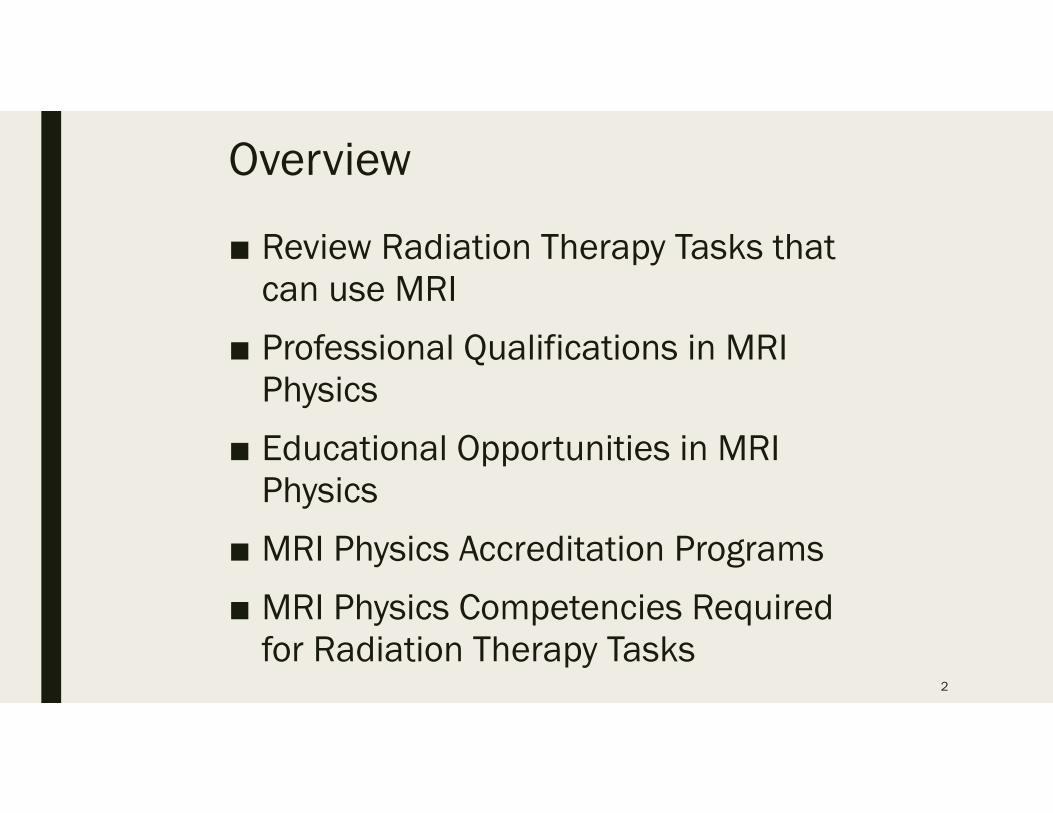
Overview
■ Review Radiation Therapy Tasks that can use MRI
■ Professional Qualifications in MRI Physics
■ Educational Opportunities in MRI Physics
■ MRI Physics Accreditation Programs
■ MRI Physics Competencies Required for Radiation Therapy Tasks
2
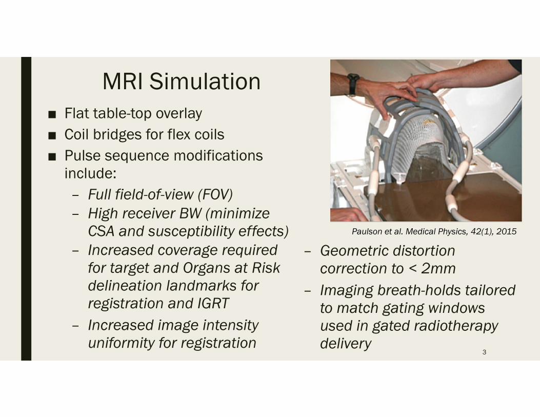
MRI Simulation■ Flat table-top overlay■ Coil bridges for flex coils■ Pulse sequence modifications
include:– Full field-of-view (FOV)– High receiver BW (minimize
CSA and susceptibility effects)– Increased coverage required
for target and Organs at Risk delineation landmarks for registration and IGRT
– Increased image intensity uniformity for registration
3
Paulson et al. Medical Physics, 42(1), 2015
– Geometric distortion correction to < 2mm
– Imaging breath-holds tailored to match gating windows used in gated radiotherapy delivery
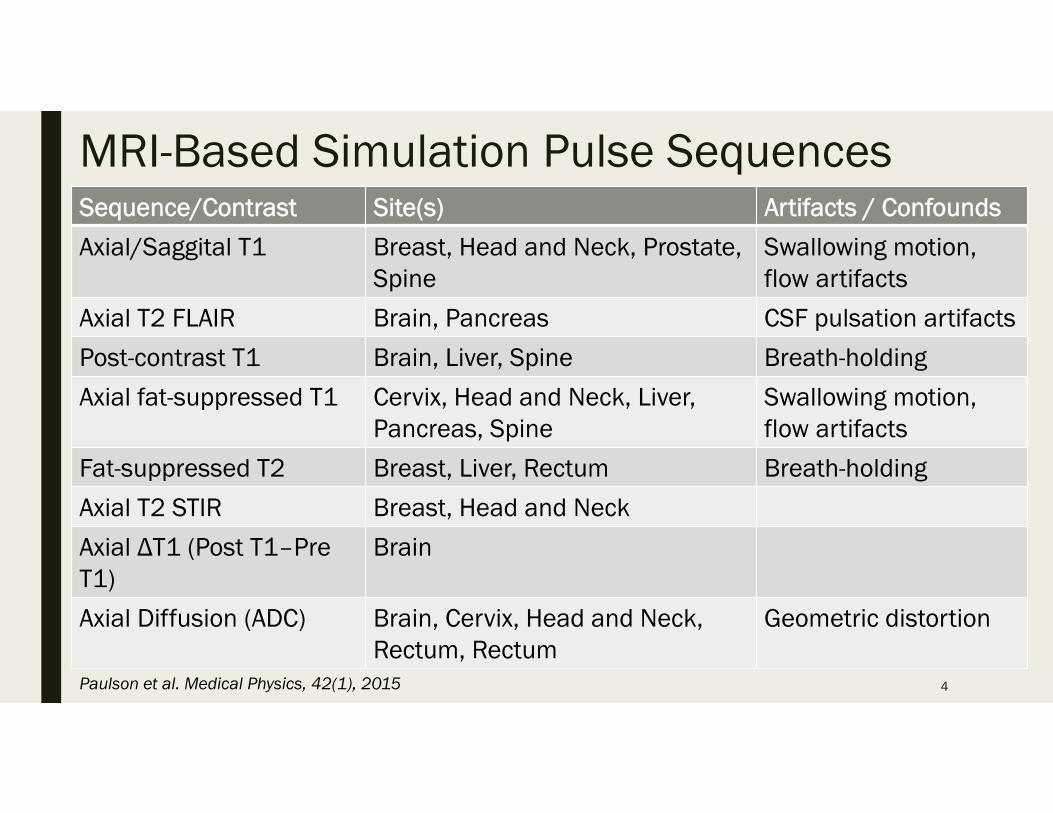
MRI-Based Simulation Pulse SequencesSequence/Contrast Site(s) Artifacts / Confounds
Axial/Saggital T1 Breast, Head and Neck, Prostate, Spine
Swallowing motion, flow artifacts
Axial T2 FLAIR Brain, Pancreas CSF pulsation artifacts
Post-contrast T1 Brain, Liver, Spine Breath-holding
Axial fat-suppressed T1 Cervix, Head and Neck, Liver, Pancreas, Spine
Swallowing motion, flow artifacts
Fat-suppressed T2 Breast, Liver, Rectum Breath-holding
Axial T2 STIR Breast, Head and Neck
Axial ΔT1 (Post T1–PreT1)
Brain
Axial Diffusion (ADC) Brain, Cervix, Head and Neck, Rectum, Rectum
Geometric distortion
4Paulson et al. Medical Physics, 42(1), 2015
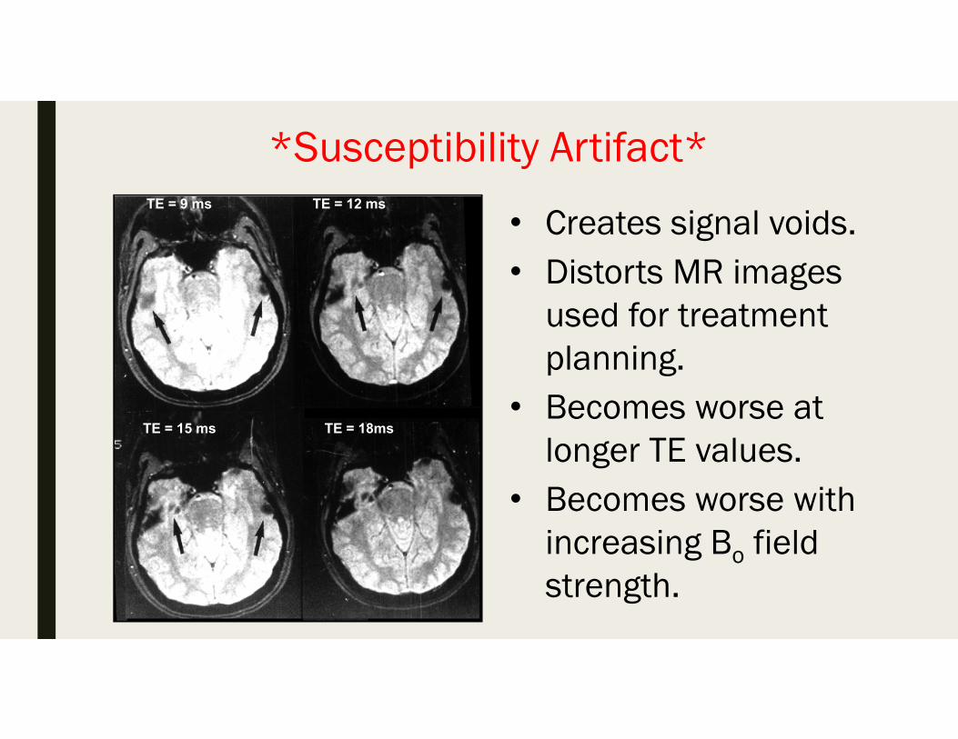
*Susceptibility Artifact*
• Creates signal voids.• Distorts MR images
used for treatment planning.
• Becomes worse at longer TE values.
• Becomes worse with increasing Bo field strength.
TE = 9 ms TE = 12 ms
TE = 15 ms TE = 18ms
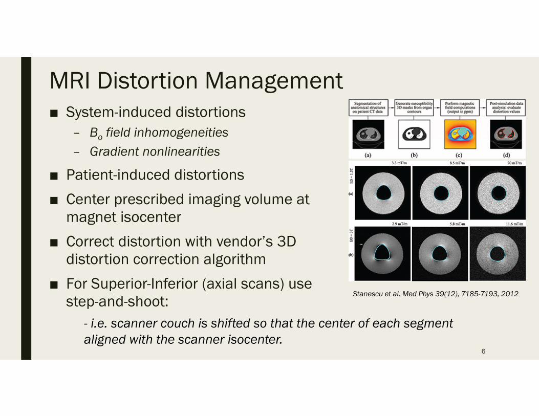
MRI Distortion Management■ System-induced distortions
– Bo field inhomogeneities– Gradient nonlinearities
■ Patient-induced distortions
■ Center prescribed imaging volume at magnet isocenter
■ Correct distortion with vendor’s 3D distortion correction algorithm
■ For Superior-Inferior (axial scans) use step-and-shoot:
6
Stanescu et al. Med Phys 39(12), 7185-7193, 2012
- i.e. scanner couch is shifted so that the center of each segment aligned with the scanner isocenter.
*The Problem with Quantitative MRI*
■ MRI signal intensity cannot be directly related to tissue density, electron density or other tissue parameters
■ Contrast in MR images is mainly dominated by the relaxation times, T1 and T2.
■ Therefore, using MRI for treatment planning is problematic.
■ Special MR acquisition methods must be used.
■ Then MRI gray scale values to Relative Electron Density with quantitative acquisition methods and careful calibration.
7
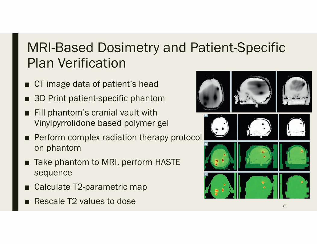
MRI-Based Dosimetry and Patient-Specific Plan Verification■ CT image data of patient’s head
■ 3D Print patient-specific phantom
■ Fill phantom’s cranial vault with Vinylpyrrolidone based polymer gel
■ Perform complex radiation therapy protocol on phantom
■ Take phantom to MRI, perform HASTE sequence
■ Calculate T2-parametric map
■ Rescale T2 values to dose 8
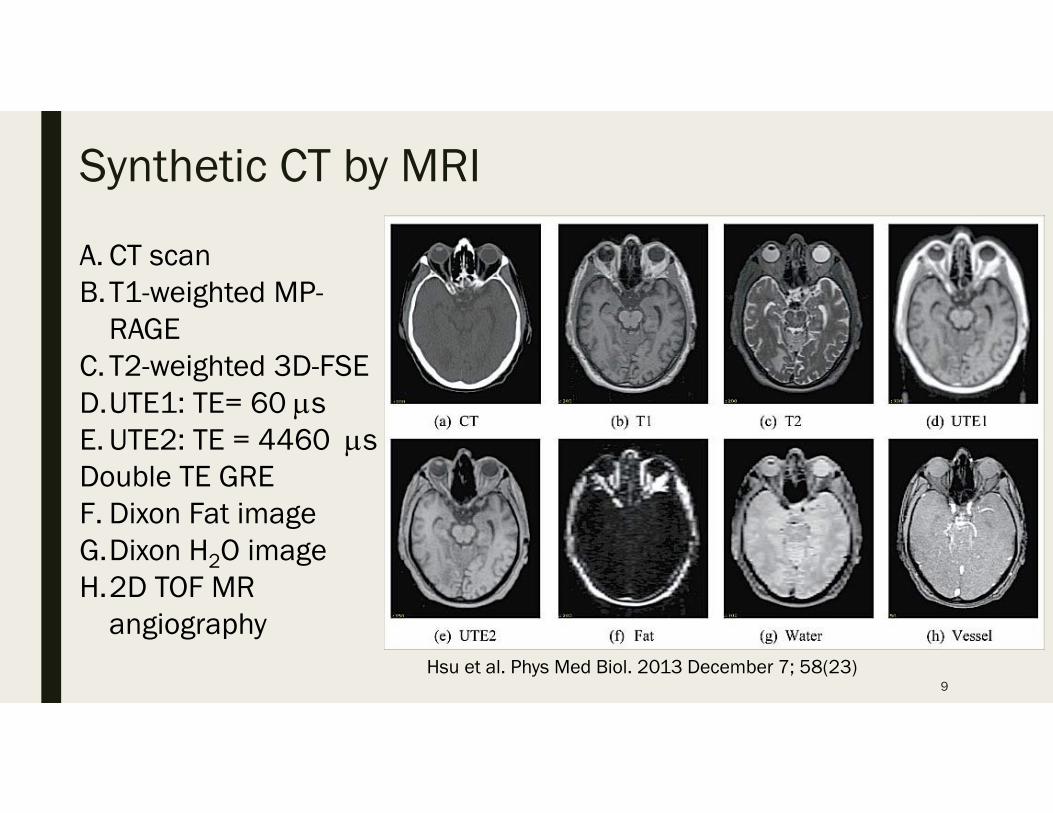
Synthetic CT by MRI
9Hsu et al. Phys Med Biol. 2013 December 7; 58(23)
A. CT scanB.T1-weighted MP-
RAGEC. T2-weighted 3D-FSED.UTE1: TE= 60 sE. UTE2: TE = 4460 sDouble TE GREF. Dixon Fat imageG.Dixon H2O imageH.2D TOF MR
angiography
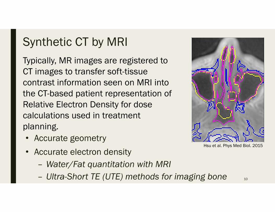
Synthetic CT by MRI
• Accurate geometry
• Accurate electron density– Water/Fat quantitation with MRI– Ultra-Short TE (UTE) methods for imaging bone 10
Hsu et al. Phys Med Biol. 2015
Typically, MR images are registered to CT images to transfer soft-tissue contrast information seen on MRI into the CT-based patient representation of Relative Electron Density for dose calculations used in treatment planning.
4D-MRI Technology■ MR can acquire an image in an arbitrary plane by
changing the direction of magnetic field gradient
■ Two non-coplanar image views provided critical motion characterization, because the most significant displacements are in the cranial-caudal direction
■ Volumetric 4D MRI is practical with parallel acquisition using large RF coil arrays
■ Up to 16-fold acceleration possible for a 32-channel system
■ View-sharing, or TRICKS, further speeds acquisition11
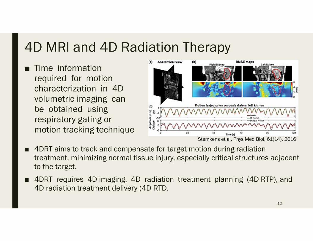
4D MRl and 4D Radiation Therapy■ Time information
required for motion characterization in 4D volumetric imaging can be obtained using respiratory gating or motion tracking technique
12
Stemkens et al. Phys Med Biol, 61(14), 2016
■ 4DRT aims to track and compensate for target motion during radiation treatment, minimizing normal tissue injury, especially critical structures adjacent to the target.
■ 4DRT requires 4D imaging, 4D radiation treatment planning (4D RTP), and 4D radiation treatment delivery (4D RTD.
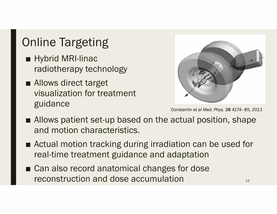
Online Targeting
13
Constantin et al Med. Phys. 38 4174–85, 2011
■ Hybrid MRI-linacradiotherapy technology
■ Allows direct target visualization for treatment guidance
■ Allows patient set-up based on the actual position, shape and motion characteristics.
■ Actual motion tracking during irradiation can be used for real-time treatment guidance and adaptation
■ Can also record anatomical changes for dose reconstruction and dose accumulation
Treatment Response and Adaptations■ Diffusion Weighted MRI
– In a tumor with high cellularity, the motion of water molecules is more restricted.
– Treatments result in increased ADC values due to cell swelling, tumor lysis and necrosis.
■ Dynamic Contrast-Enhanced (DCE) Perfusion Imaging– Maps the distribution of tumor perfusion, blood
volume, & mean transit time.– Have shown prognostic and predictive value for
response of certain cancers to therapy.14
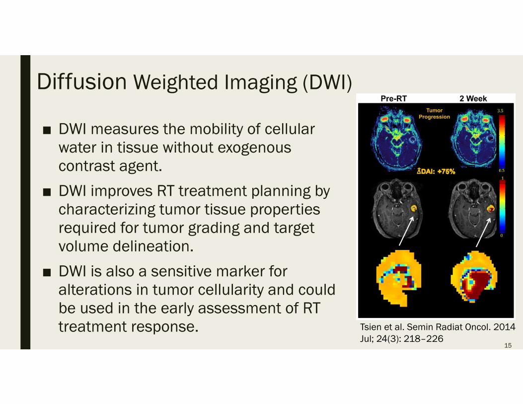
Diffusion Weighted Imaging (DWI)
■ DWI measures the mobility of cellular water in tissue without exogenous contrast agent.
■ DWI improves RT treatment planning by characterizing tumor tissue properties required for tumor grading and target volume delineation.
■ DWI is also a sensitive marker for alterations in tumor cellularity and could be used in the early assessment of RT treatment response.
15
Tsien et al. Semin Radiat Oncol. 2014 Jul; 24(3): 218–226
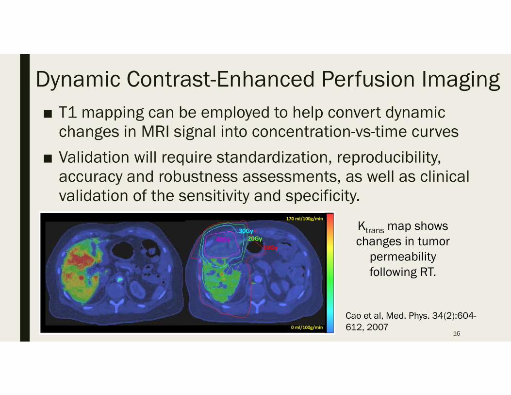
Dynamic Contrast-Enhanced Perfusion Imaging■ T1 mapping can be employed to help convert dynamic
changes in MRI signal into concentration-vs-time curves
■ Validation will require standardization, reproducibility, accuracy and robustness assessments, as well as clinical validation of the sensitivity and specificity.
16
Cao et al, Med. Phys. 34(2):604-612, 2007
Ktrans map shows changes in tumor
permeability following RT.
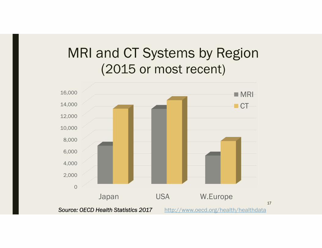
MRI and CT Systems by Region(2015 or most recent)
0
2,000
4,000
6,000
8,000
10,000
12,000
14,000
16,000
Japan USA W.Europe
MRICT
17
Source: OECD Health Statistics 2017 http://www.oecd.org/health/healthdata
18
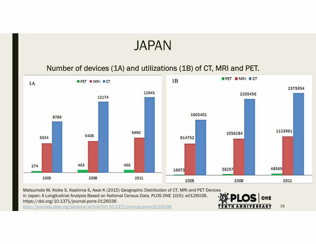
JAPANNumber of devices (1A) and utilizations (1B) of CT, MRI and PET.
Matsumoto M, Koike S, Kashima S, Awai K (2015) Geographic Distribution of CT, MRI and PET Devices in Japan: A Longitudinal Analysis Based on National Census Data. PLOS ONE 10(5): e0126036. https://doi.org/10.1371/journal.pone.0126036http://journals.plos.org/plosone/article?id=10.1371/journal.pone.0126036
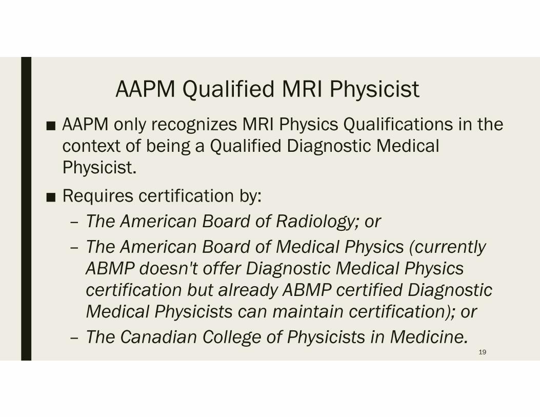
AAPM Qualified MRI Physicist■ AAPM only recognizes MRI Physics Qualifications in the
context of being a Qualified Diagnostic Medical Physicist.
■ Requires certification by:– The American Board of Radiology; or– The American Board of Medical Physics (currently
ABMP doesn't offer Diagnostic Medical Physics certification but already ABMP certified Diagnostic Medical Physicists can maintain certification); or
– The Canadian College of Physicists in Medicine.19
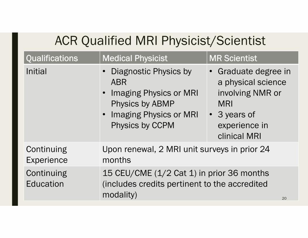
ACR Qualified MRI Physicist/ScientistQualifications Medical Physicist MR Scientist
Initial • Diagnostic Physics by ABR
• Imaging Physics or MRI Physics by ABMP
• Imaging Physics or MRI Physics by CCPM
• Graduate degree in a physical science involving NMR or MRI
• 3 years of experience in clinical MRI
Continuing Experience
Upon renewal, 2 MRI unit surveys in prior 24 months
Continuing Education
15 CEU/CME (1/2 Cat 1) in prior 36 months (includes credits pertinent to the accredited modality) 20
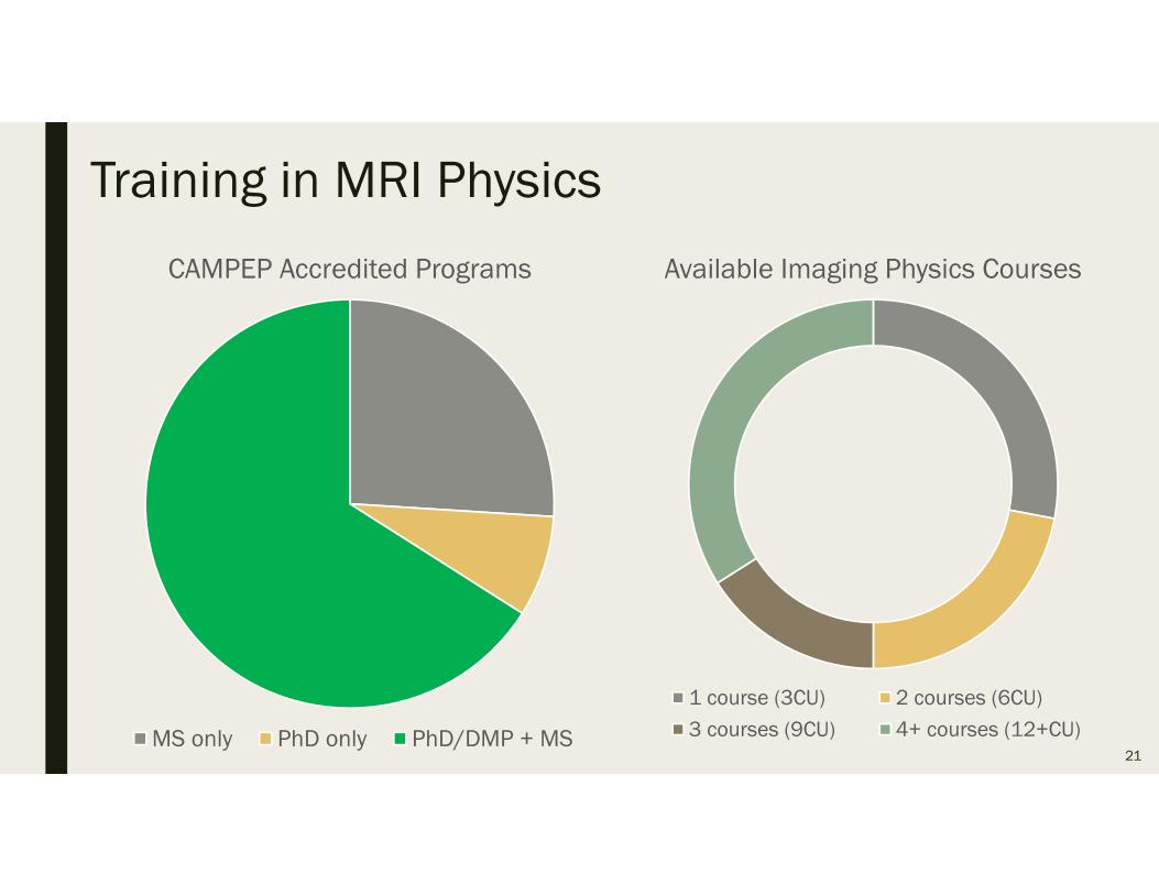
Training in MRI Physics
CAMPEP Accredited Programs
MS only PhD only PhD/DMP + MS21
Available Imaging Physics Courses
1 course (3CU) 2 courses (6CU)
3 courses (9CU) 4+ courses (12+CU)
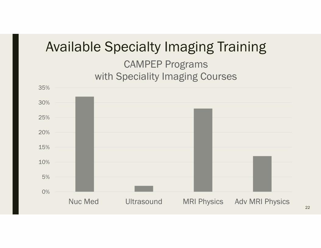
Available Specialty Imaging Training
0%
5%
10%
15%
20%
25%
30%
35%
Nuc Med Ultrasound MRI Physics Adv MRI Physics
CAMPEP Programs with Speciality Imaging Courses
22
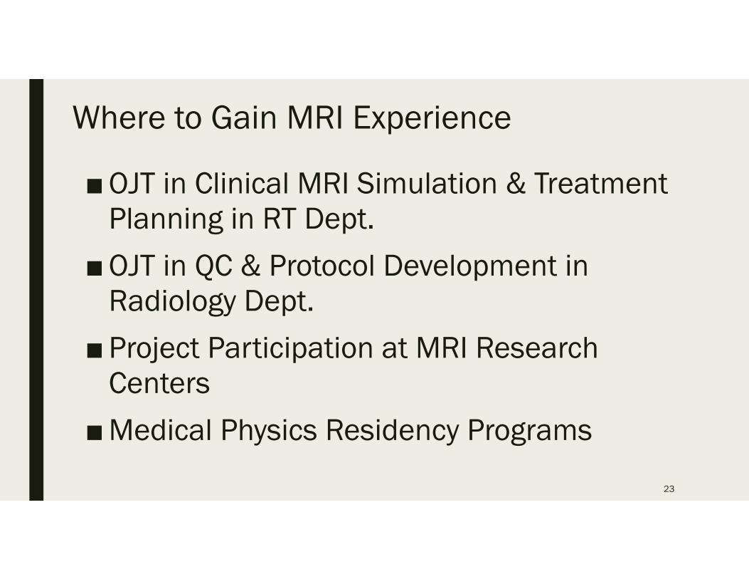
Where to Gain MRI Experience
■OJT in Clinical MRI Simulation & Treatment Planning in RT Dept.
■OJT in QC & Protocol Development in Radiology Dept.
■Project Participation at MRI Research Centers
■Medical Physics Residency Programs
23
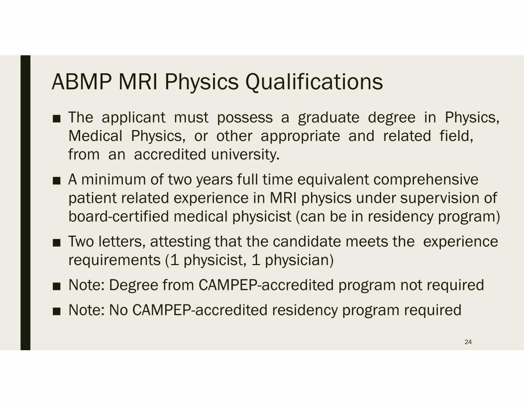
ABMP MRI Physics Qualifications■ The applicant must possess a graduate degree in Physics,
Medical Physics, or other appropriate and related field, from an accredited university.
■ A minimum of two years full time equivalent comprehensive patient related experience in MRI physics under supervision of board-certified medical physicist (can be in residency program)
■ Two letters, attesting that the candidate meets the experience requirements (1 physicist, 1 physician)
■ Note: Degree from CAMPEP-accredited program not required
■ Note: No CAMPEP-accredited residency program required
24
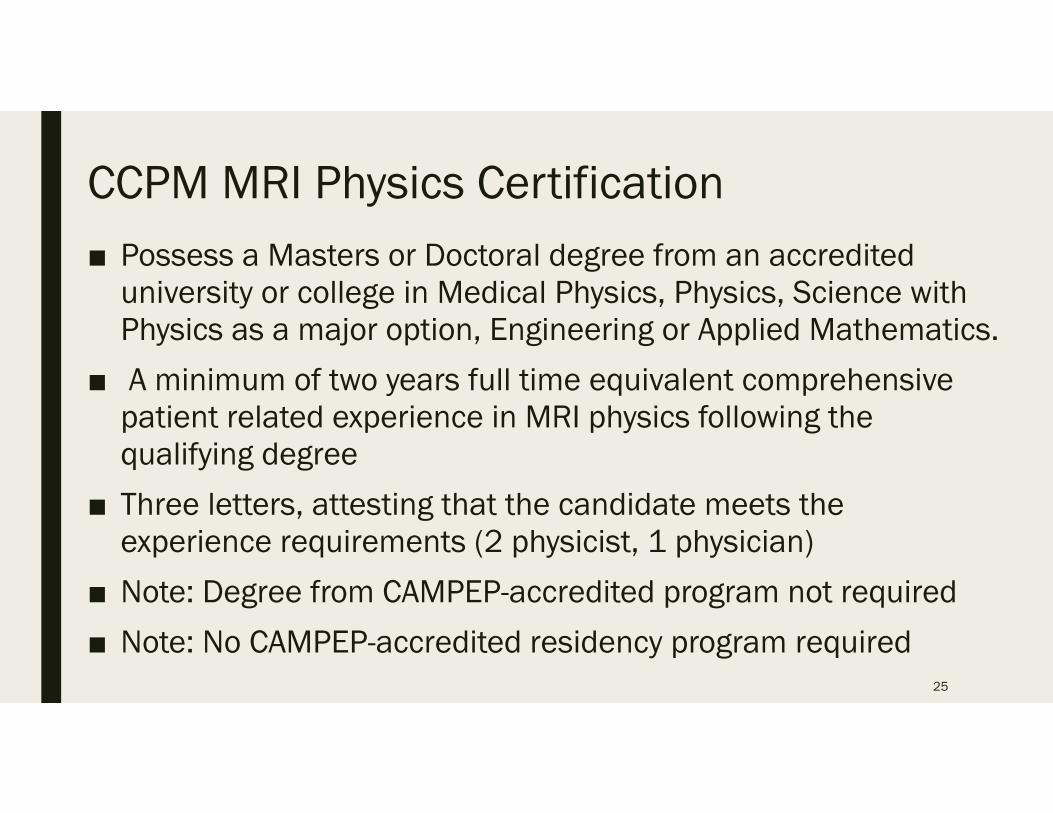
CCPM MRI Physics Certification■ Possess a Masters or Doctoral degree from an accredited
university or college in Medical Physics, Physics, Science with Physics as a major option, Engineering or Applied Mathematics.
■ A minimum of two years full time equivalent comprehensive patient related experience in MRI physics following the qualifying degree
■ Three letters, attesting that the candidate meets the experience requirements (2 physicist, 1 physician)
■ Note: Degree from CAMPEP-accredited program not required
■ Note: No CAMPEP-accredited residency program required25
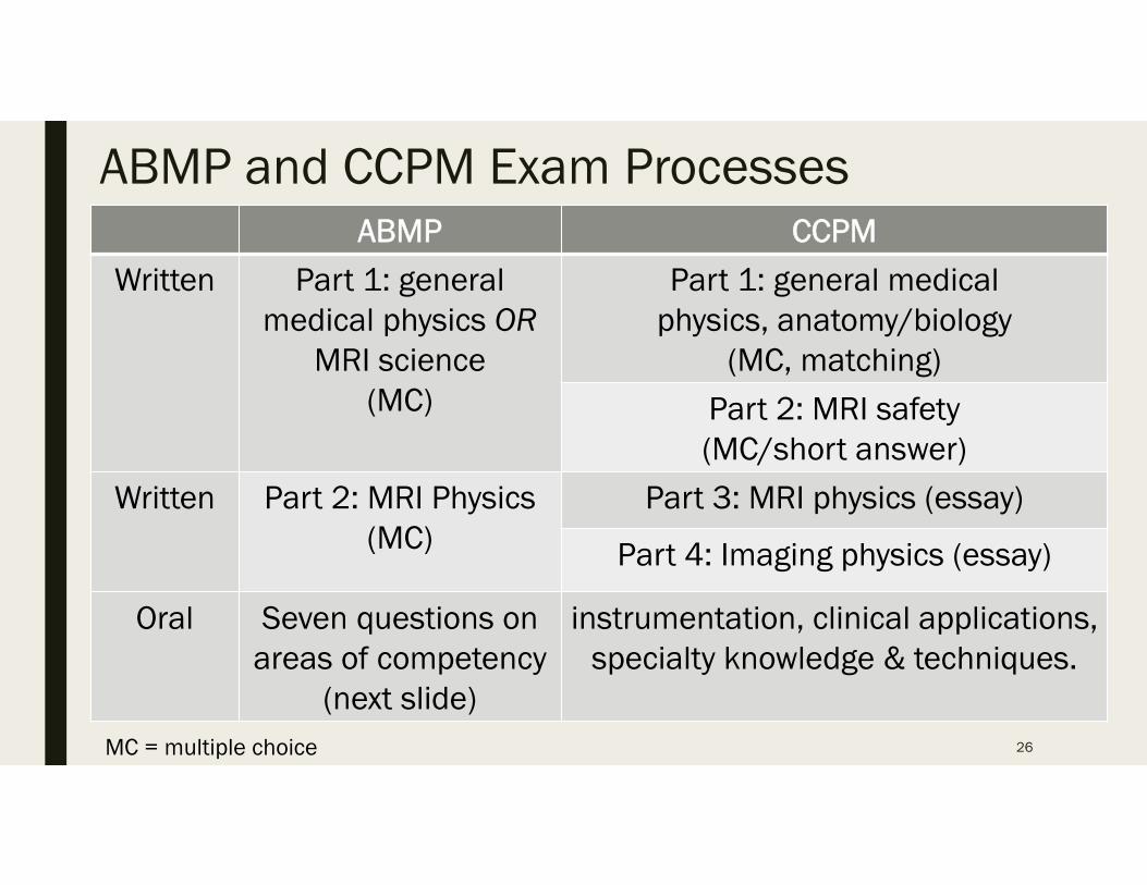
ABMP and CCPM Exam ProcessesABMP CCPM
Written Part 1: general medical physics OR
MRI science(MC)
Part 1: general medical physics, anatomy/biology
(MC, matching)
Part 2: MRI safety (MC/short answer)
Written Part 2: MRI Physics(MC)
Part 3: MRI physics (essay)
Part 4: Imaging physics (essay)
Oral Seven questions on areas of competency
(next slide)
instrumentation, clinical applications, specialty knowledge & techniques.
26MC = multiple choice
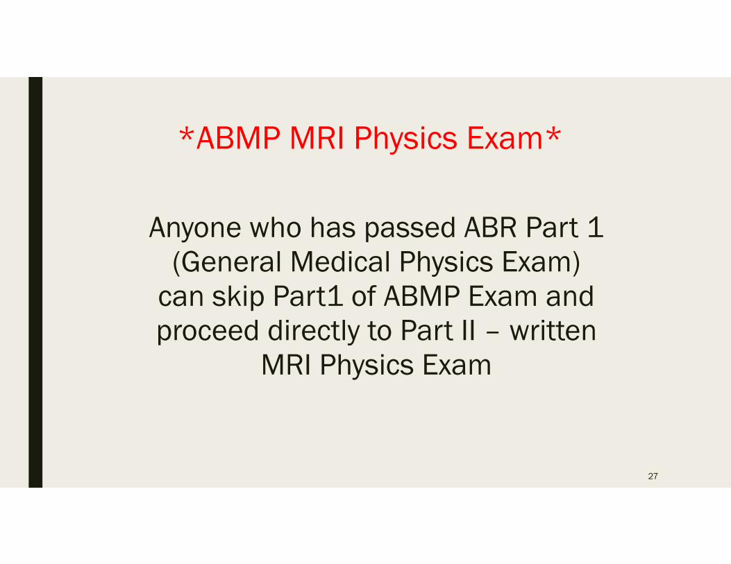
*ABMP MRI Physics Exam*
Anyone who has passed ABR Part 1 (General Medical Physics Exam)
can skip Part1 of ABMP Exam and proceed directly to Part II – written
MRI Physics Exam
27
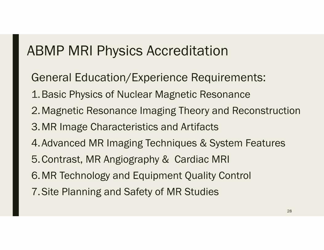
ABMP MRI Physics Accreditation
General Education/Experience Requirements:1.Basic Physics of Nuclear Magnetic Resonance
2.Magnetic Resonance Imaging Theory and Reconstruction
3.MR Image Characteristics and Artifacts
4.Advanced MR Imaging Techniques & System Features
5.Contrast, MR Angiography & Cardiac MRI
6.MR Technology and Equipment Quality Control
7.Site Planning and Safety of MR Studies
28
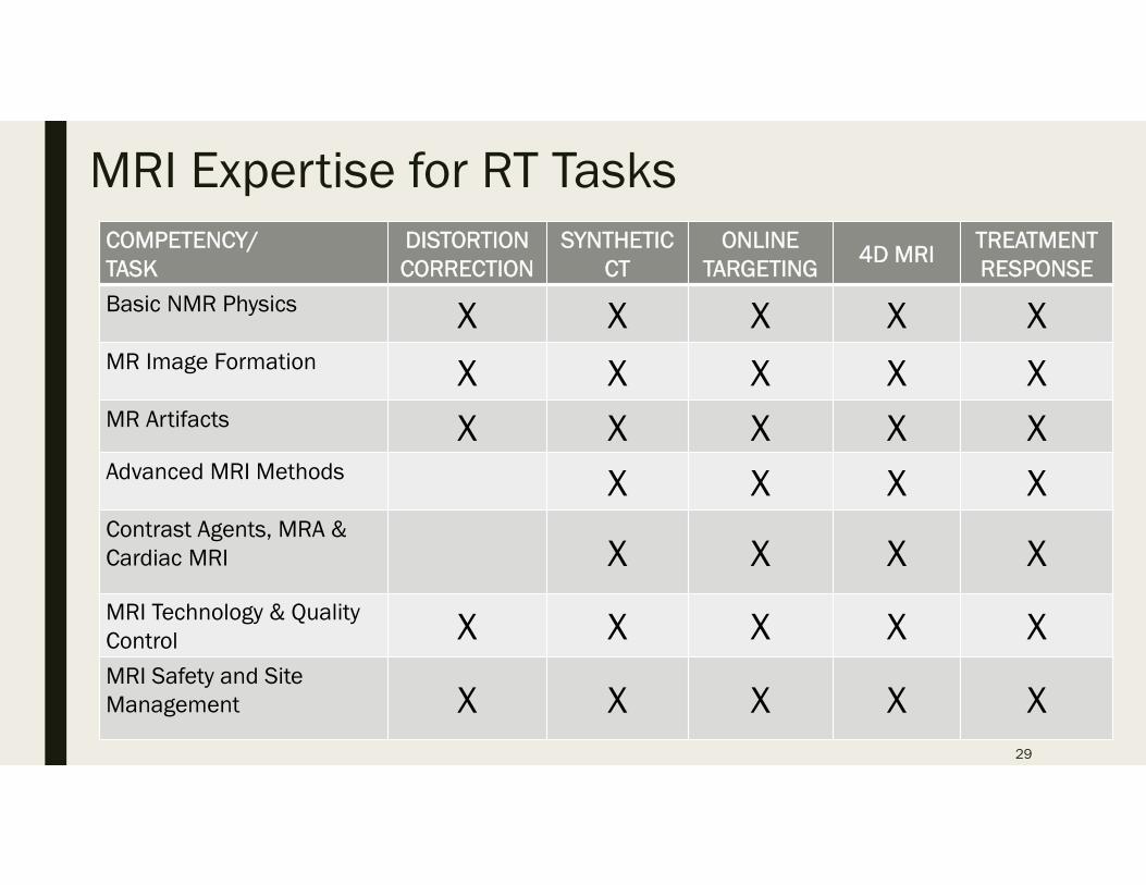
MRI Expertise for RT TasksCOMPETENCY/TASK
DISTORTION CORRECTION
SYNTHETIC CT
ONLINETARGETING
4D MRI TREATMENTRESPONSE
Basic NMR Physics X X X X XMR Image Formation X X X X XMR Artifacts X X X X XAdvanced MRI Methods X X X XContrast Agents, MRA & Cardiac MRI X X X XMRI Technology & Quality Control X X X X XMRI Safety and Site Management X X X X X
29
Summary• MRI can provide exceptional image data for use in multiple phases
of radiation cancer therapy
• However, the use of MRI in RT requires a high degree of competence and understanding of sophisticated MRI methods and procedures.
• Programs are in place for professional credentialing (certification) in MRI Physics however, “MRI Physicist” is not a specialty recognized by the AAPM and
prescribes no special competencies for undertaking MRI physics
MRI physics specific courses are not required in CAMPEP-accredited graduate programs
30
Summary
• Despite plenty of MRI systems across the developed world, there are limited opportunities for medical physicists to gain hands-on, practical experience in MRI
• If these issues are not addressed, there will be a shortage of qualified medical MRI physicists to accommodate future needs for technical expertise in MRI for radiation therapy
31

Fin
32
Related Documents