Basics of Ultrasound Principles of Medical Imaging Prof. Dr. Philippe Cattin MIAC, University of Basel Oct 10th/17th, 2016 Oct 10th/17th, 2016 Principles of Medical Imaging 1 of 60 26.09.2016 08:35 Contents 2 4 5 6 7 8 9 10 11 12 13 15 16 18 19 20 22 23 24 Prof. Dr. Philippe Cattin: Basics of Ultrasound Contents Abstract 1 Introduction Why Ultrasound Applications in Obstetrics Applications in Cardiology Applications in Inner Medicine Applications in Musculoskeletal System Applications of Ultrasound Elastography Advantages and Disadvantages of Ultrasound Challenges for Image Analysis Challenges for Image Analysis (2) Challenges for Image Analysis (3) 2 Research at MIAC using US Myocardial Contrast Echocardiography Myocardial Contrast Enhancement (2) 3 History History History (2) History (3) 4 Properties of Ultrasound Properties of Ultrasound Common Sound Frequencies Physical Principles of Ultrasound Oct 10th/17th, 2016 Principles of Medical Imaging 2 of 60 26.09.2016 08:35

Welcome message from author
This document is posted to help you gain knowledge. Please leave a comment to let me know what you think about it! Share it to your friends and learn new things together.
Transcript
Basics ofUltrasound
Principles of MedicalImaging
Prof. Dr. Philippe Cattin
MIAC, University of Basel
Oct 10th/17th, 2016
Oct 10th/17th, 2016Principles of Medical Imaging
1 of 60 26.09.2016 08:35
Contents
2
4
5
6
7
8
9
10
11
12
13
15
16
18
19
20
22
23
24
Prof. Dr. Philippe Cattin: Basics of Ultrasound
Contents
Abstract
1 Introduction
Why Ultrasound
Applications in Obstetrics
Applications in Cardiology
Applications in Inner Medicine
Applications in Musculoskeletal System
Applications of Ultrasound Elastography
Advantages and Disadvantages of Ultrasound
Challenges for Image Analysis
Challenges for Image Analysis (2)
Challenges for Image Analysis (3)
2 Research at MIAC using US
Myocardial Contrast Echocardiography
Myocardial Contrast Enhancement (2)
3 History
History
History (2)
History (3)
4 Properties of Ultrasound
Properties of Ultrasound
Common Sound Frequencies
Physical Principles of UltrasoundOct 10th/17th, 2016Principles of Medical Imaging
2 of 60 26.09.2016 08:35
25
26
27
28
29
30
31
32
33
34
35
36
37
39
40
41
42
43
44
45
46
47
48
49
50
52
Acquisition Principle
Propagation
Propagation (2)
Propagation (3)
Ultrasound Characteristics
Ultrasound Wavelength Change
Ultrasound Characteristics (2)
Modulus of Elasticity
Acoustic Impedance
Acoustic Impedance (2)
Properties of Different Materials
Power and Intensity
Symbols often used to Characterise Ultrasound
5 Interaction with Matter
Interaction with Matter
Reflection and Refraction
Reflection (2)
Reflection (3)
Reflection Example: Air - Fat
Reflection Example: Liver - Kidney
Diffraction
Attenuation or Absorption
Attenuation or Absorption (2)
Attenuation or Absorption (3)
Decibel Scale
Attenuation or Absorption (4)
6 Safety of Diagnostic Ultrasound
Safety of Diagnostic Ultrasound Oct 10th/17th, 2016Principles of Medical Imaging
3 of 60 26.09.2016 08:35
53
54
55
56
Bioeffects
Thermal Bioeffects
Non-thermal Bioeffects
Output Display Standard Using Thermal andMechanical Indices
Oct 10th/17th, 2016Principles of Medical Imaging
4 of 60 26.09.2016 08:35

Oct 10th/17th, 2016Principles of Medical Imaging
(2)
Prof. Dr. Philippe Cattin: Basics of Ultrasound
Abstract
5 of 60 26.09.2016 08:35
Introduction
Oct 10th/17th, 2016Principles of Medical Imaging
(4)Why Ultrasound
Ultrasound (US) is the most widely used imaging
technology worldwide
Popular due to availability, speed, low cost, patient-
friendliness (no radiation)
Applied in obstetrics, cardiology, inner medicine,
urology,...
Ongoing research to improve image quality, speed and
new application areas such a intra-operative navigation,
tumour therapy,...
6 of 60 26.09.2016 08:35

Oct 10th/17th, 2016Principles of Medical Imaging
Introduction
(5)
Prof. Dr. Philippe Cattin: Basics of Ultrasound
Applications in Obstetrics
Follow fetal development
Detect pathologies
Fig. 4.1: Two-dimensional B-mode
Ultrasound image of a fetus
Fig. 4.2: Three-dimensional image of
the same fetus ~5 months after
conception
7 of 60 26.09.2016 08:35
Oct 10th/17th, 2016Principles of Medical Imaging
Introduction
(6)
Prof. Dr. Philippe Cattin: Basics of Ultrasound
Applications in Cardiology
Blood flow in vessels (Doppler US)
Contraction, Rhythm
Blood flow in the heart (defects on wall muscle, valve
defects)
Assessment of cardiac perfusion
8 of 60 26.09.2016 08:35

Oct 10th/17th, 2016Principles of Medical Imaging
Fig. 4.3: Myocardial contrast echocardiography
(MCE)
Fig. 4.4: Prenatal
diagnostic of the Fallot-
Tetralogie
9 of 60 26.09.2016 08:35
Oct 10th/17th, 2016Principles of Medical Imaging
Introduction
(7)
Prof. Dr. Philippe Cattin: Basics of Ultrasound
Applications in InnerMedicine
Gallstone
Perfusion of renal transplant
Fig. 4.5: Gallstone (red arrow) within
the gallbladder produces a bright
surface echo and causes a dark
acoustic shadow (S)
Fig. 4.6: Perfusion Doppler image of
a renal transplant
10 of 60 26.09.2016 08:35

Oct 10th/17th, 2016Principles of Medical Imaging
Introduction
(8)
Prof. Dr. Philippe Cattin: Basics of Ultrasound
Applications inMusculoskeletal System
Visualisation of tendons, ligaments
Investigation under movement is possible → simplifies the
detection of ruptures, obstructions, ...
Fig. 4.7: The arrows show the large
gap of the rupture Achilles tendon
Fig. 4.8: US image of ISS astronaut
Mike Fincke's biceps tendon, where
"D" denotes the deltoid muscle and
"T" is the proximal intracapsular end
of the long biceps tendon
11 of 60 26.09.2016 08:35
Oct 10th/17th, 2016Principles of Medical Imaging
Introduction
(9)
Prof. Dr. Philippe Cattin: Basics of Ultrasound
Applications of UltrasoundElastography
US Elastography is often used to classify tumours.
Malignant tumours are 10 to 100 times stiffer than the
normal soft tissue around.
Fig. 4.9: Elastogram (of a breast)
indication a mass with a high
probability of being malignant
tumour
12 of 60 26.09.2016 08:35

Oct 10th/17th, 2016Principles of Medical Imaging
Introduction
(10)
Prof. Dr. Philippe Cattin: Basics of Ultrasound
Advantages andDisadvantages of Ultrasound
Advantages
No ionising radiation
Real-time imaging (functional
examinations)
Small, transportable
equipment
Comparatively low cost
Disadvantages
Moderate to poor image
quality
Image interpretation is
difficult and operator
dependent
Acoustic window
Limited penetration depth
(attenuation, total reflection)
Shadows by dense objects
Fig. 4.10: SmartPhone with
Ultrasound capability
Fig. 4.11: Application scenario
13 of 60 26.09.2016 08:35
Oct 10th/17th, 2016Principles of Medical Imaging
Introduction
(11)
Prof. Dr. Philippe Cattin: Basics of Ultrasound
Challenges for ImageAnalysis
Artefacts: speckle pattern, attenuation, shadows,
contours parallel to the beam direction
Lateral resolution depends on the depth
Fig. 4.12: US image of the heart
chambers
Fig. 4.13: US image of a gallstone
14 of 60 26.09.2016 08:35

Oct 10th/17th, 2016Principles of Medical Imaging
Introduction
(12)
Prof. Dr. Philippe Cattin: Basics of Ultrasound
Challenges for ImageAnalysis (2)
Sound propagates at differentspeed in different tissues
Estimation of distances
difficult (
assumed in Ultrasound
devices)
Distortion of contours in
Ultrasound images
Fig. 4.14: Different sound speeds
distort distance measurements
Material
Water
Blood
Brain
Fat
Muscle
Bone
AirTab. 4.1: Different important sound
speed in tissue
15 of 60 26.09.2016 08:35
Oct 10th/17th, 2016Principles of Medical Imaging
Introduction
(13)
Prof. Dr. Philippe Cattin: Basics of Ultrasound
Challenges for ImageAnalysis (3)
Segmentation in the presence of noise (speckle patterns arein a strict sense not noise but interferences of reflections) andartefacts.
Fig. 4.15: Segmentation
example 1
Fig. 4.16: Segmentation
example 2
16 of 60 26.09.2016 08:35
Research at MIACusing US
Oct 10th/17th, 2016Principles of Medical Imaging
17 of 60 26.09.2016 08:35
Oct 10th/17th, 2016Principles of Medical Imaging
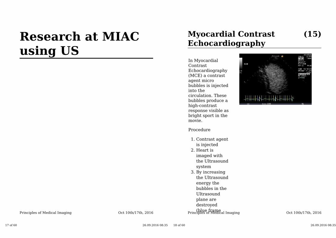
(15)Myocardial ContrastEchocardiography
In MyocardialContrastEchocardiography(MCE) a contrastagent microbubbles is injectedinto thecirculation. Thesebubbles produce ahigh-contrastresponse visible asbright sport in themovie.
Procedure
Contrast agent
is injected
1.
Heart is
imaged with
the Ultrasound
system
2.
By increasing
the Ultrasound
energy the
bubbles in the
Ultrasound
plane are
destroyed
(blue frame
3.
18 of 60 26.09.2016 08:35
Oct 10th/17th, 2016Principles of Medical Imaging
around the US
image)
The time it
takes to refill
the myocard
with bubbles is
a measure of
muscle
perfusion
(health of the
coronary
arteries)
4.
Problem
Analysing andevaluating thesemovies by hand isa time consumingand error pronetask.
19 of 60 26.09.2016 08:35
Oct 10th/17th, 2016Principles of Medical Imaging
Research at MIAC using US
(16)
Prof. Dr. Philippe Cattin: Basics of Ultrasound
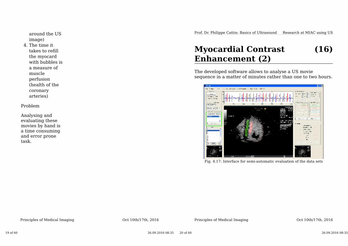
Myocardial ContrastEnhancement (2)
The developed software allows to analyse a US moviesequence in a matter of minutes rather than one to two hours.
Fig. 4.17: Interface for semi-automatic evaluation of the data sets
20 of 60 26.09.2016 08:35
History
Oct 10th/17th, 2016Principles of Medical Imaging
(18)History
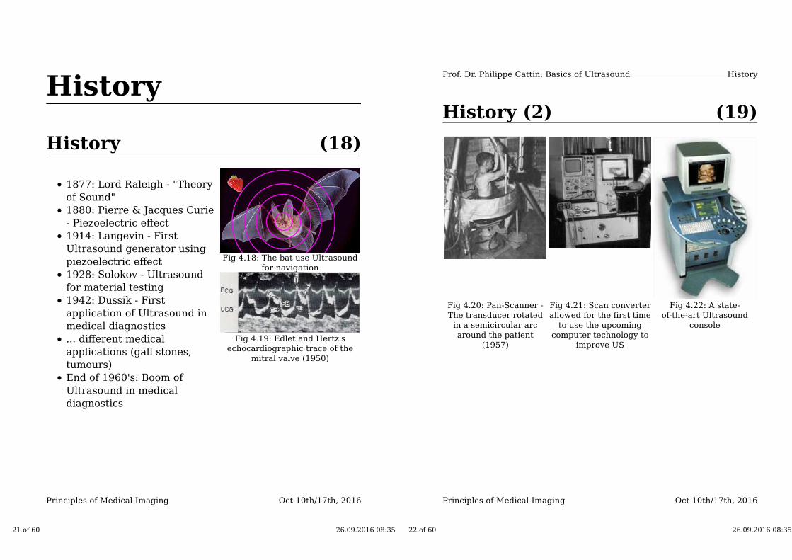
1877: Lord Raleigh - "Theory
of Sound"
1880: Pierre & Jacques Curie
- Piezoelectric effect
1914: Langevin - First
Ultrasound generator using
piezoelectric effect
1928: Solokov - Ultrasound
for material testing
1942: Dussik - First
application of Ultrasound in
medical diagnostics
... different medical
applications (gall stones,
tumours)
End of 1960's: Boom of
Ultrasound in medical
diagnostics
Fig 4.18: The bat use Ultrasound
for navigation
Fig 4.19: Edlet and Hertz's
echocardiographic trace of the
mitral valve (1950)
21 of 60 26.09.2016 08:35
Oct 10th/17th, 2016Principles of Medical Imaging
History
(19)
Prof. Dr. Philippe Cattin: Basics of Ultrasound
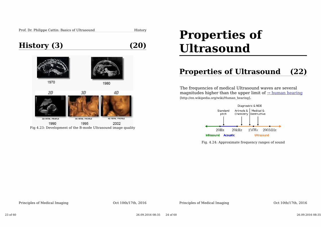
History (2)
Fig 4.20: Pan-Scanner -
The transducer rotated
in a semicircular arc
around the patient
(1957)
Fig 4.21: Scan converter
allowed for the first time
to use the upcoming
computer technology to
improve US
Fig 4.22: A state-
of-the-art Ultrasound
console
22 of 60 26.09.2016 08:35
Oct 10th/17th, 2016Principles of Medical Imaging
History
(20)
Prof. Dr. Philippe Cattin: Basics of Ultrasound
History (3)
Fig 4.23: Development of the B-mode Ultrasound image quality
23 of 60 26.09.2016 08:35
Properties ofUltrasound
Oct 10th/17th, 2016Principles of Medical Imaging
(22)Properties of Ultrasound
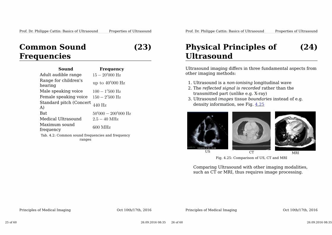
The frequencies of medical Ultrasound waves are severalmagnitudes higher than the upper limit of → human hearing[http://en.wikipedia.org/wiki/Human_hearing].
Fig. 4.24: Approximate frequency ranges of sound
24 of 60 26.09.2016 08:35
Oct 10th/17th, 2016Principles of Medical Imaging
Properties of Ultrasound
(23)
Prof. Dr. Philippe Cattin: Basics of Ultrasound
Common SoundFrequencies
Sound Frequency
Adult audible range
Range for children'shearing
Male speaking voice
Female speaking voice
Standard pitch (ConcertA)
Bat
Medical Ultrasound
Maximum soundfrequency
Tab. 4.2: Common sound frequencies and frequency
ranges
25 of 60 26.09.2016 08:35
Oct 10th/17th, 2016Principles of Medical Imaging
Properties of Ultrasound
(24)
Prof. Dr. Philippe Cattin: Basics of Ultrasound
Physical Principles ofUltrasound
Ultrasound imaging differs in three fundamental aspects fromother imaging methods:
Ultrasound is a non-ionising longitudinal wave1.
The reflected signal is recorded rather than the
transmitted part (unlike e.g. X-ray)
2.
Ultrasound images tissue boundaries instead of e.g.
density information, see Fig. 4.25
3.
US CT MRI
Fig. 4.25: Comparison of US, CT and MRI
Comparing Ultrasound with other imaging modalities,such as CT or MRI, thus requires image processing.
26 of 60 26.09.2016 08:35

Oct 10th/17th, 2016Principles of Medical Imaging
Properties of Ultrasound
(25)
Prof. Dr. Philippe Cattin: Basics of Ultrasound
Acquisition Principle
Ultrasoundmeasures the time apulse takes to travelfrom the transducerto the reflectingsurface and back,see Fig 4.26 →time-of-flight
The depth can be
calculated, if the sound
velocity of the tissue
is known
The magnitude of the
echo is coded as gray
value on the display
Fig. 4.26: Two sound waves reflected
from two surfaces. Echo arrival is
and respectively, assuming a speed
of sound of
27 of 60 26.09.2016 08:35
Oct 10th/17th, 2016Principles of Medical Imaging
Properties of Ultrasound
(26)
Prof. Dr. Philippe Cattin: Basics of Ultrasound
Propagation
Ultrasound is a cyclic longitudinal pressure wave andrequires a medium (gas, liquid, solid)
Fig. 4.27: Three-dimensional
visualisation of a propagating
pressure wave
Fig. 4.28: Propagation of sound
energy as elongation about a center
of equilibrium (particle velocity
about its center is )
Ultrasound waves behave according to theconventional laws associated for light waves exceptthat they require a medium.
28 of 60 26.09.2016 08:35
Oct 10th/17th, 2016Principles of Medical Imaging
Properties of Ultrasound
(27)
Prof. Dr. Philippe Cattin: Basics of Ultrasound
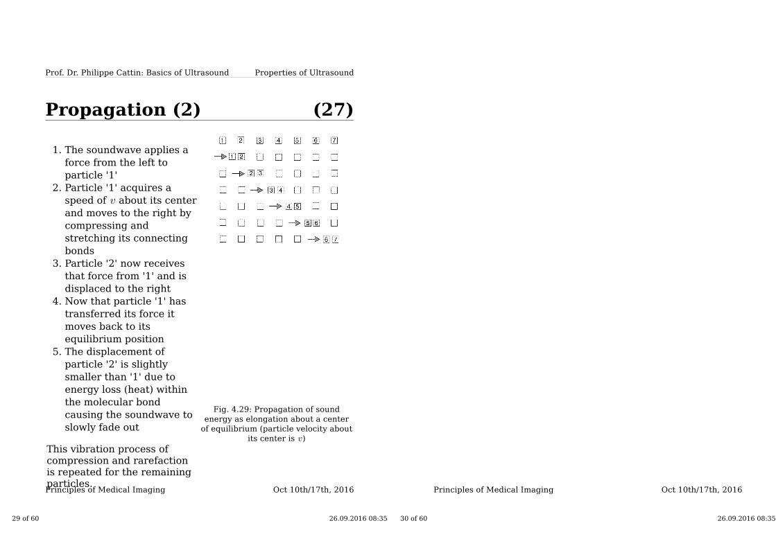
Propagation (2)
The soundwave applies a
force from the left to
particle '1'
1.
Particle '1' acquires a
speed of about its center
and moves to the right by
compressing and
stretching its connecting
bonds
2.
Particle '2' now receives
that force from '1' and is
displaced to the right
3.
Now that particle '1' has
transferred its force it
moves back to its
equilibrium position
4.
The displacement of
particle '2' is slightly
smaller than '1' due to
energy loss (heat) within
the molecular bond
causing the soundwave to
slowly fade out
5.
This vibration process ofcompression and rarefactionis repeated for the remainingparticles.
Fig. 4.29: Propagation of sound
energy as elongation about a center
of equilibrium (particle velocity about
its center is )
29 of 60 26.09.2016 08:35
Oct 10th/17th, 2016Principles of Medical Imaging
30 of 60 26.09.2016 08:35
Oct 10th/17th, 2016Principles of Medical Imaging
Properties of Ultrasound
(28)
Prof. Dr. Philippe Cattin: Basics of Ultrasound
Propagation (3)
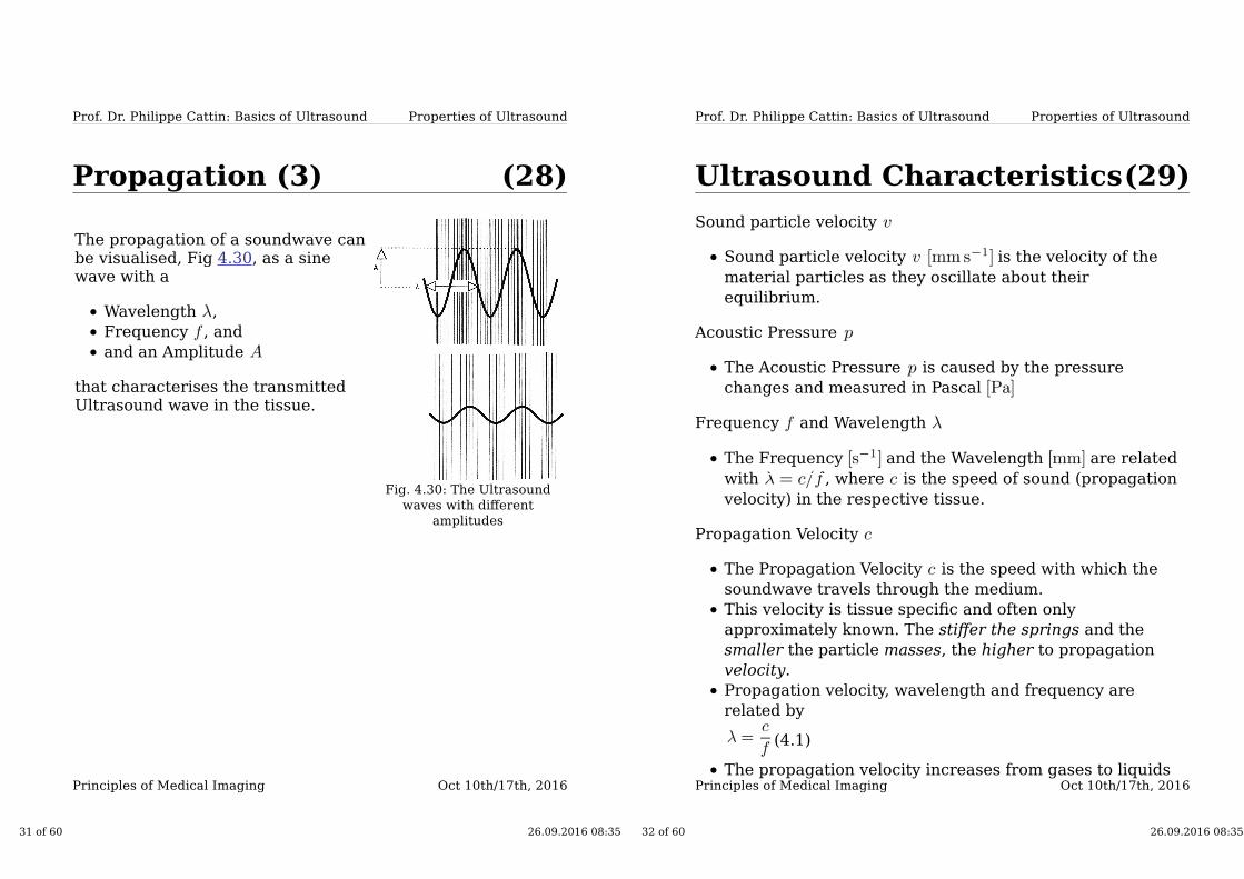
The propagation of a soundwave canbe visualised, Fig 4.30, as a sinewave with a
Wavelength ,
Frequency , and
and an Amplitude
that characterises the transmittedUltrasound wave in the tissue.
Fig. 4.30: The Ultrasound
waves with different
amplitudes
31 of 60 26.09.2016 08:35
Oct 10th/17th, 2016Principles of Medical Imaging
Properties of Ultrasound
(29)
Prof. Dr. Philippe Cattin: Basics of Ultrasound
Ultrasound Characteristics
Sound particle velocity
Sound particle velocity is the velocity of the
material particles as they oscillate about their
equilibrium.
Acoustic Pressure
The Acoustic Pressure is caused by the pressure
changes and measured in Pascal
Frequency and Wavelength
The Frequency and the Wavelength are related
with , where is the speed of sound (propagation
velocity) in the respective tissue.
Propagation Velocity
The Propagation Velocity is the speed with which the
soundwave travels through the medium.
This velocity is tissue specific and often only
approximately known. The stiffer the springs and the
smaller the particle masses, the higher to propagation
velocity.
Propagation velocity, wavelength and frequency are
related by
(4.1)
The propagation velocity increases from gases to liquids
32 of 60 26.09.2016 08:35
Oct 10th/17th, 2016Principles of Medical Imaging
and is highest in solids.
33 of 60 26.09.2016 08:35
Oct 10th/17th, 2016Principles of Medical Imaging
Properties of Ultrasound
(30)
Prof. Dr. Philippe Cattin: Basics of Ultrasound
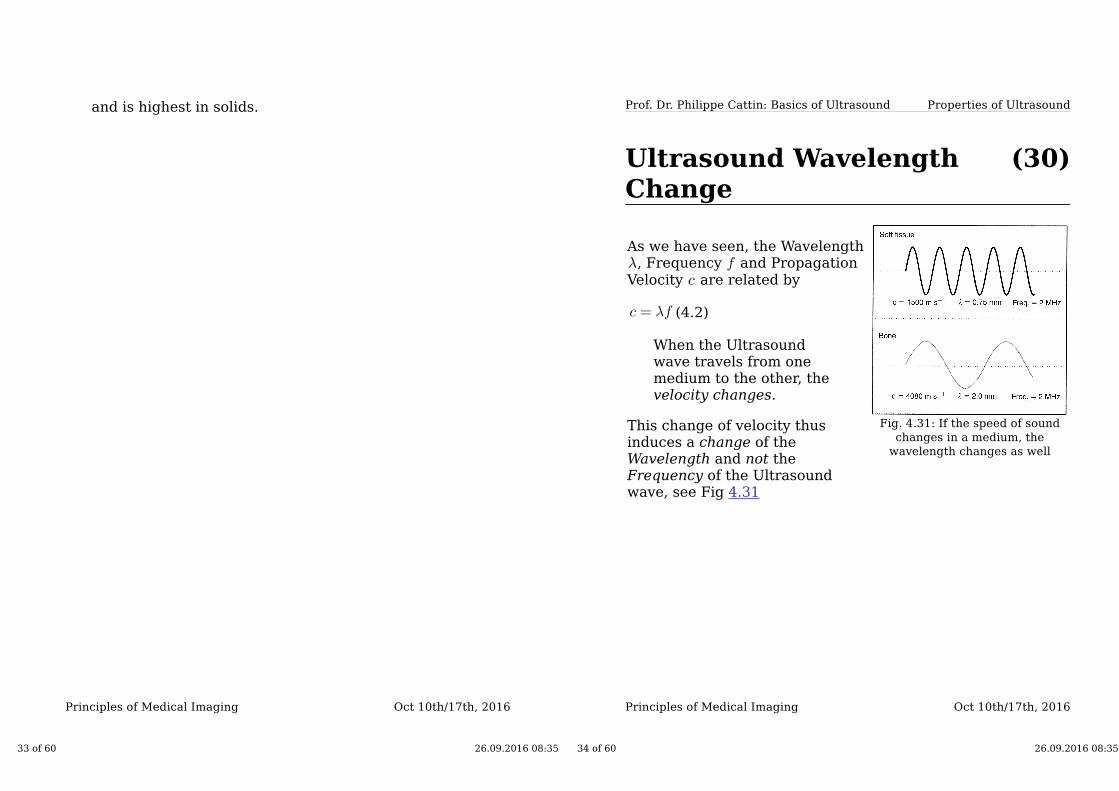
Ultrasound WavelengthChange
As we have seen, the Wavelength, Frequency and Propagation
Velocity are related by
(4.2)
When the Ultrasoundwave travels from onemedium to the other, thevelocity changes.
This change of velocity thusinduces a change of theWavelength and not theFrequency of the Ultrasoundwave, see Fig 4.31
Fig. 4.31: If the speed of sound
changes in a medium, the
wavelength changes as well
34 of 60 26.09.2016 08:35

Oct 10th/17th, 2016Principles of Medical Imaging
Properties of Ultrasound
(31)
Prof. Dr. Philippe Cattin: Basics of Ultrasound
Ultrasound Characteristics(2)
Amplitude
The maximum Amplitude coincides with the compression
peak, see Fig 4.30. Reducing the power level results in a
smaller amplitude.
Power
The Power is the rate of sound energy transfer into the
tissue and is measured in watts
Intensity
The Intensity is a measure of power per unit area and is
commonly measured as .
35 of 60 26.09.2016 08:35
Oct 10th/17th, 2016Principles of Medical Imaging
Properties of Ultrasound
(32)
Prof. Dr. Philippe Cattin: Basics of Ultrasound
Modulus of Elasticity
The rate of transfer from one molecule to the other dependson the molecule's reluctance to motion and the density. Thewave velocity (speed of sound) are connected throughYoung's Modulus of Elasticity .
(4.3)
where is the speed of sound, the material density and Young's Modulus.
The higher the modulus (stiffer springs) and lower
molecule masses, the higher the velocity
The modulus of elasticity is inversely proportional to the
compressibility
The modulus is measured in pressure units e.g.
Fat
Soft tissue
Bone
36 of 60 26.09.2016 08:35

Oct 10th/17th, 2016Principles of Medical Imaging
Properties of Ultrasound
(33)
Prof. Dr. Philippe Cattin: Basics of Ultrasound
Acoustic Impedance
When pressure is applied to a molecule it will moveexerting a pressure on an adjacent molecule. Acousticpressure increases with particle velocity but is also dependson properties of the medium. The equation is similar toelectrical resistance:
Acoustic Impedance Electronic Resistance
(4.4) (4.5)
(4.6) (4.7)
(4.8) (4.9)
The acoustic impedance is measured in often
shortened to rayl.
37 of 60 26.09.2016 08:35
Oct 10th/17th, 2016Principles of Medical Imaging
Properties of Ultrasound
(34)
Prof. Dr. Philippe Cattin: Basics of Ultrasound
Acoustic Impedance (2)
The Acoustic Impedance is also related to the Modulus ofElasticity .
The stiffer the bonding, the greater the pressure exerted
by a molecule moving at a particular velocity, so
(4.10)
A material having a great springiness (low value) has highmolecular motion and will absorb sound energy and less willbe transferred to the next molecule, so
(4.11)
combining Eq 4.10, 4.11 and 4.3 yields
(4.12)
38 of 60 26.09.2016 08:35
Oct 10th/17th, 2016Principles of Medical Imaging
Properties of Ultrasound
(35)
Prof. Dr. Philippe Cattin: Basics of Ultrasound
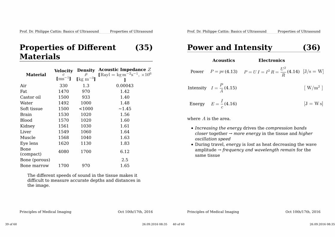
Properties of DifferentMaterials
MaterialVelocity
[ ]
Density
[ ]
Acoustic Impedance
[
]
Air 330 1.3 0.00043
Fat 1470 970 1.42
Castor oil 1500 933 1.40
Water 1492 1000 1.48
Soft tissue 1500 <1000 ~1.45
Brain 1530 1020 1.56
Blood 1570 1020 1.60
Kidney 1561 1030 1.61
Liver 1549 1060 1.64
Muscle 1568 1040 1.63
Eye lens 1620 1130 1.83
Bone(compact)
4080 1700 6.12
Bone (porous) 2.5
Bone marrow 1700 970 1.65
The different speeds of sound in the tissue makes itdifficult to measure accurate depths and distances inthe image.
39 of 60 26.09.2016 08:35
Oct 10th/17th, 2016Principles of Medical Imaging
Properties of Ultrasound
(36)
Prof. Dr. Philippe Cattin: Basics of Ultrasound
Power and Intensity
Acoustics Electronics
Power (4.13) (4.14)
Intensity (4.15)
Energy (4.16)
where is the area.
Increasing the energy drives the compression bands
closer together → more energy in the tissue and higher
oscillation speed
During travel, energy is lost as heat decreasing the wave
amplitude → frequency and wavelength remain for the
same tissue
40 of 60 26.09.2016 08:35

Oct 10th/17th, 2016Principles of Medical Imaging
Properties of Ultrasound
(37)
Prof. Dr. Philippe Cattin: Basics of Ultrasound
Symbols often used toCharacterise Ultrasound
Measurement Symbol Unit Clinical Range
Velocity (speed ofsound)
(for soft
tissue)
Wavelength (for
soft tissue)
Frequency
Elastic modulus (in bone)
Acousticimpedance
Density (for
water)
Pressure
Elongationvelocity
41 of 60 26.09.2016 08:35
Interaction withMatter
Oct 10th/17th, 2016Principles of Medical Imaging
(39)Interaction with Matter
When Ultrasound interacts with matter, they exhibit the samephenomena as visible light:
Reflection (specular and non-specular)
Refraction
Diffraction
Attenuation or absorption
42 of 60 26.09.2016 08:35

Oct 10th/17th, 2016Principles of Medical Imaging
Interaction with Matter
(40)
Prof. Dr. Philippe Cattin: Basics of Ultrasound
Reflection and Refraction
The laws of wave optics also applyfor Ultrasound waves. From energypreservation we know that(neglecting absorption)
(4.17)
the law of reflection states
(4.18)
and → Snell's law [http://en.wikipedia.org
/wiki/Snell_law]
(4.19)
gives the relationship between thesound speeds and angles.
Fig. 4.32: Sound wave at an
angle to the surface
43 of 60 26.09.2016 08:35
Oct 10th/17th, 2016Principles of Medical Imaging
Interaction with Matter
(41)
Prof. Dr. Philippe Cattin: Basics of Ultrasound
Reflection (2)
Given the Impedances of thetwo tissues and the angles andthe incidence intensity , the reflected and transmitted intensitiescan be calculated with
(4.20)
and
(4.21)
from Eq 4.17 we know that
(4.22)
also holds.
Fig. 4.33: Amount of fraction
reflected depending on the
impedance difference
44 of 60 26.09.2016 08:35
Oct 10th/17th, 2016Principles of Medical Imaging
Interaction with Matter
(42)
Prof. Dr. Philippe Cattin: Basics of Ultrasound
Reflection (3)
For a sound wave perpendicular to asmooth surface ( ), Eq 4.20 canbe simplified and yields for thereflected intensity
(4.23)
and similarly the transmitted waveintensity is given by
(4.24)
Fig. 4.34: Sound wave
perpendicular to a smooth
surface
The amount of reflection depends on impedance
differences .
Reflection is greatest when the impedance
difference is large.
45 of 60 26.09.2016 08:35
Oct 10th/17th, 2016Principles of Medical Imaging
Interaction with Matter
(43)
Prof. Dr. Philippe Cattin: Basics of Ultrasound
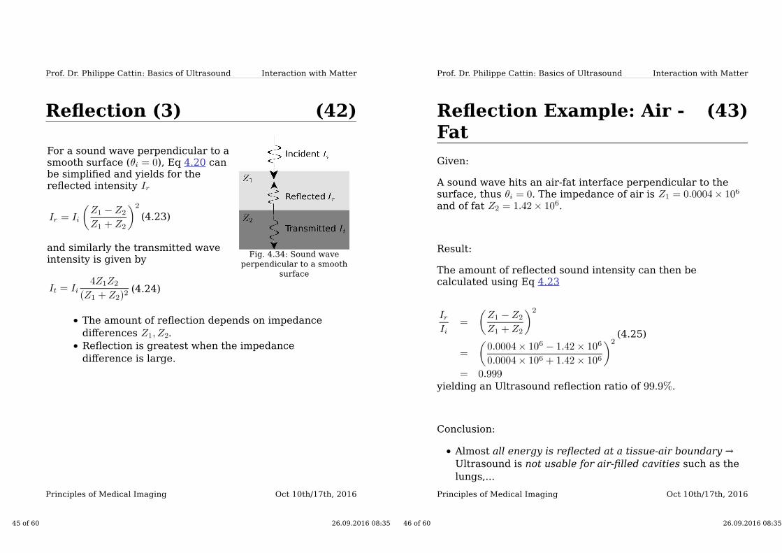
Reflection Example: Air -Fat
Given:
A sound wave hits an air-fat interface perpendicular to thesurface, thus . The impedance of air is and of fat .
Result:
The amount of reflected sound intensity can then becalculated using Eq 4.23
(4.25)
yielding an Ultrasound reflection ratio of .
Conclusion:
Almost all energy is reflected at a tissue-air boundary →
Ultrasound is not usable for air-filled cavities such as the
lungs,...
46 of 60 26.09.2016 08:35
Oct 10th/17th, 2016Principles of Medical Imaging
47 of 60 26.09.2016 08:35
Oct 10th/17th, 2016Principles of Medical Imaging
Interaction with Matter
(44)
Prof. Dr. Philippe Cattin: Basics of Ultrasound
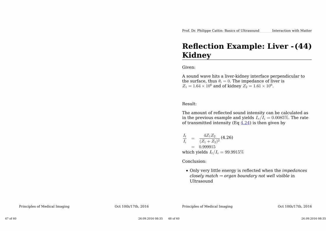
Reflection Example: Liver -Kidney
Given:
A sound wave hits a liver-kidney interface perpendicular tothe surface, thus . The impedance of liver is
and of kidney .
Result:
The amount of reflected sound intensity can be calculated asin the previous example and yields . The rate
of transmitted intensity (Eq 4.24) is then given by
(4.26)
which yields
Conclusion:
Only very little energy is reflected when the impedances
closely match → organ boundary not well visible in
Ultrasound
48 of 60 26.09.2016 08:35
Oct 10th/17th, 2016Principles of Medical Imaging
Interaction with Matter
(45)
Prof. Dr. Philippe Cattin: Basics of Ultrasound
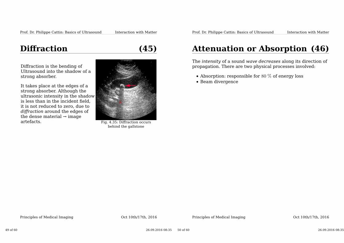
Diffraction
Diffraction is the bending ofUltrasound into the shadow of astrong absorber.
It takes place at the edges of astrong absorber. Although theultrasonic intensity in the shadowis less than in the incident field,it is not reduced to zero, due todiffraction around the edges ofthe dense material → imageartefacts. Fig. 4.35: Diffraction occurs
behind the gallstone
49 of 60 26.09.2016 08:35
Oct 10th/17th, 2016Principles of Medical Imaging
Interaction with Matter
(46)
Prof. Dr. Philippe Cattin: Basics of Ultrasound
Attenuation or Absorption
The intensity of a sound wave decreases along its direction ofpropagation. There are two physical processes involved:
Absorption: responsible for of energy loss
Beam divergence
50 of 60 26.09.2016 08:35
Oct 10th/17th, 2016Principles of Medical Imaging
Interaction with Matter
(47)
Prof. Dr. Philippe Cattin: Basics of Ultrasound
Attenuation or Absorption(2)
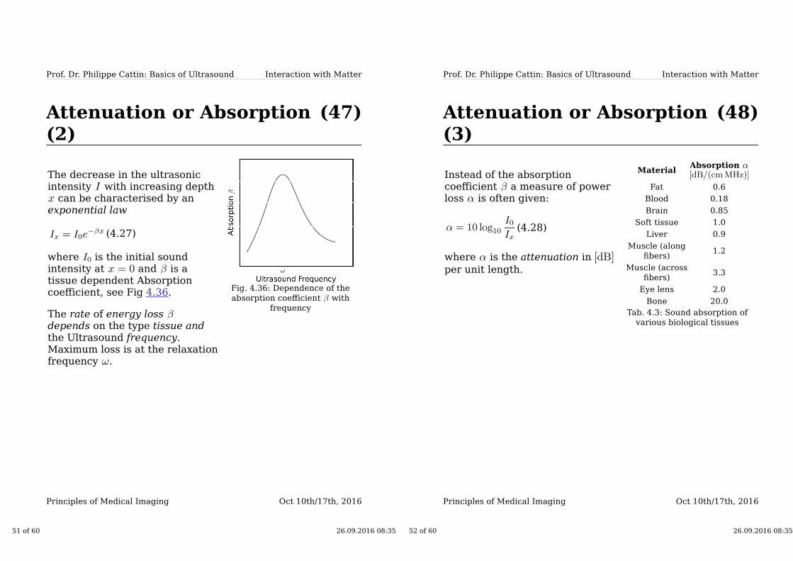
The decrease in the ultrasonicintensity with increasing depth
can be characterised by anexponential law
(4.27)
where is the initial soundintensity at and is atissue dependent Absorptioncoefficient, see Fig 4.36.
The rate of energy loss depends on the type tissue andthe Ultrasound frequency.Maximum loss is at the relaxationfrequency .
Fig. 4.36: Dependence of the
absorption coefficient with
frequency
51 of 60 26.09.2016 08:35
Oct 10th/17th, 2016Principles of Medical Imaging
Interaction with Matter
(48)
Prof. Dr. Philippe Cattin: Basics of Ultrasound
Attenuation or Absorption(3)
Instead of the absorptioncoefficient a measure of powerloss is often given:
(4.28)
where is the attenuation in
per unit length.
MaterialAbsorption
Fat 0.6
Blood 0.18
Brain 0.85
Soft tissue 1.0
Liver 0.9
Muscle (along
fibers)1.2
Muscle (across
fibers)3.3
Eye lens 2.0
Bone 20.0
Tab. 4.3: Sound absorption of
various biological tissues
52 of 60 26.09.2016 08:35
Oct 10th/17th, 2016Principles of Medical Imaging
Interaction with Matter
(49)
Prof. Dr. Philippe Cattin: Basics of Ultrasound
Decibel Scale
Sound power or intensity variations are compared usingthe Decibel Scale
(4.29)
Example:
The power loss of an incident source with and
an echo power of would be
(4.30)
The half power distance or a reduction of is thedistance over which the power is reduced by derived from .
53 of 60 26.09.2016 08:35
Oct 10th/17th, 2016Principles of Medical Imaging
Interaction with Matter
(50)
Prof. Dr. Philippe Cattin: Basics of Ultrasound
Attenuation or Absorption(4)
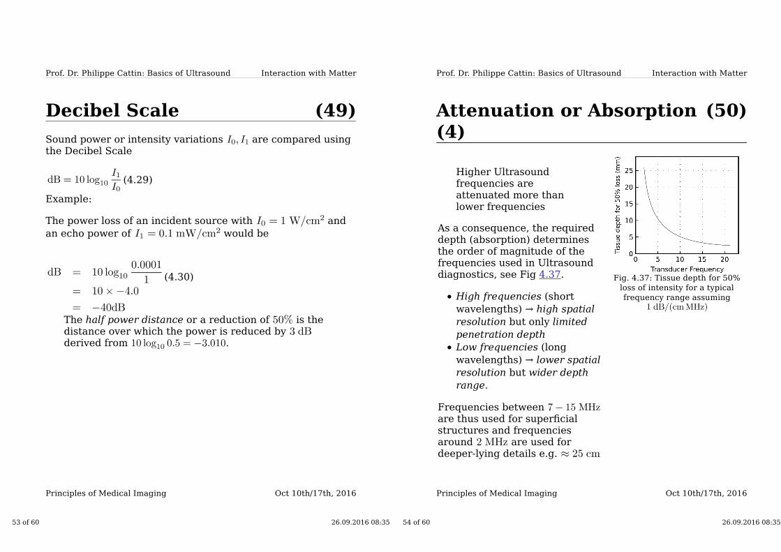
Higher Ultrasoundfrequencies areattenuated more thanlower frequencies
As a consequence, the requireddepth (absorption) determinesthe order of magnitude of thefrequencies used in Ultrasounddiagnostics, see Fig 4.37.
High frequencies (short
wavelengths) → high spatial
resolution but only limited
penetration depth
Low frequencies (long
wavelengths) → lower spatial
resolution but wider depth
range.
Frequencies between are thus used for superficialstructures and frequenciesaround are used fordeeper-lying details e.g.
Fig. 4.37: Tissue depth for 50%
loss of intensity for a typical
frequency range assuming
54 of 60 26.09.2016 08:35

Safety of DiagnosticUltrasound
Oct 10th/17th, 2016Principles of Medical Imaging
(52)Safety of DiagnosticUltrasound
"Diagnostic ultrasound has proven to be a valuabletool in medical practice. An excellent safety recordexists in that, after decades of clinical use, there is noknown instance of human injury as a result ofexposure to diagnostic ultrasound. Evidence exists,however, to indicate that at least a hypothetical riskfor clinical diagnostic ultrasound must be presumed."
1990 Radiological Health Bulletin.
The above report explicitly talks of diagnostic Ultrasound.Ultrasound such as HIFU (High-Frequency FocusedUltrasound) does "harm" the patient by destroying tissue.This is of course intended for cancerous tissue or kidneystones.
55 of 60 26.09.2016 08:35
Oct 10th/17th, 2016Principles of Medical Imaging
Safety of Diagnostic Ultrasound
(53)
Prof. Dr. Philippe Cattin: Basics of
Ultrasound
Bioeffects
Two mechanisms are known to alter biological systems withUltrasound, the
thermal mechanism, and
mechanical mechanism associated with cavitation.
56 of 60 26.09.2016 08:35
Oct 10th/17th, 2016Principles of Medical Imaging
Safety of Diagnostic Ultrasound
(54)
Prof. Dr. Philippe Cattin: Basics of
Ultrasound
Thermal Bioeffects
A major part of the Ultrasound energy emitted duringexamination is absorbed by the tissue. This absorption resultsin the generation of heat. If the absorbed energy exceeds thebody's ability to dissipate heat, the local temperature will rise.If the temperature is too high, tissue is destroyed.
Measuring the absorption of Ultrasound energy is difficult. Itcan, however, be estimated solving the differential equationaka bio-heat equation:
(4.31)
where
Density of tissue and blood
Specific heat of tissue and blood
Temperature of tissue and capillary blood
Volumetric flow rate of blood per unit mass of tissue
Rate of metabolic heat generation
Local specific absorption rate
57 of 60 26.09.2016 08:35
Oct 10th/17th, 2016Principles of Medical Imaging
Safety of Diagnostic Ultrasound
(55)
Prof. Dr. Philippe Cattin: Basics of
Ultrasound
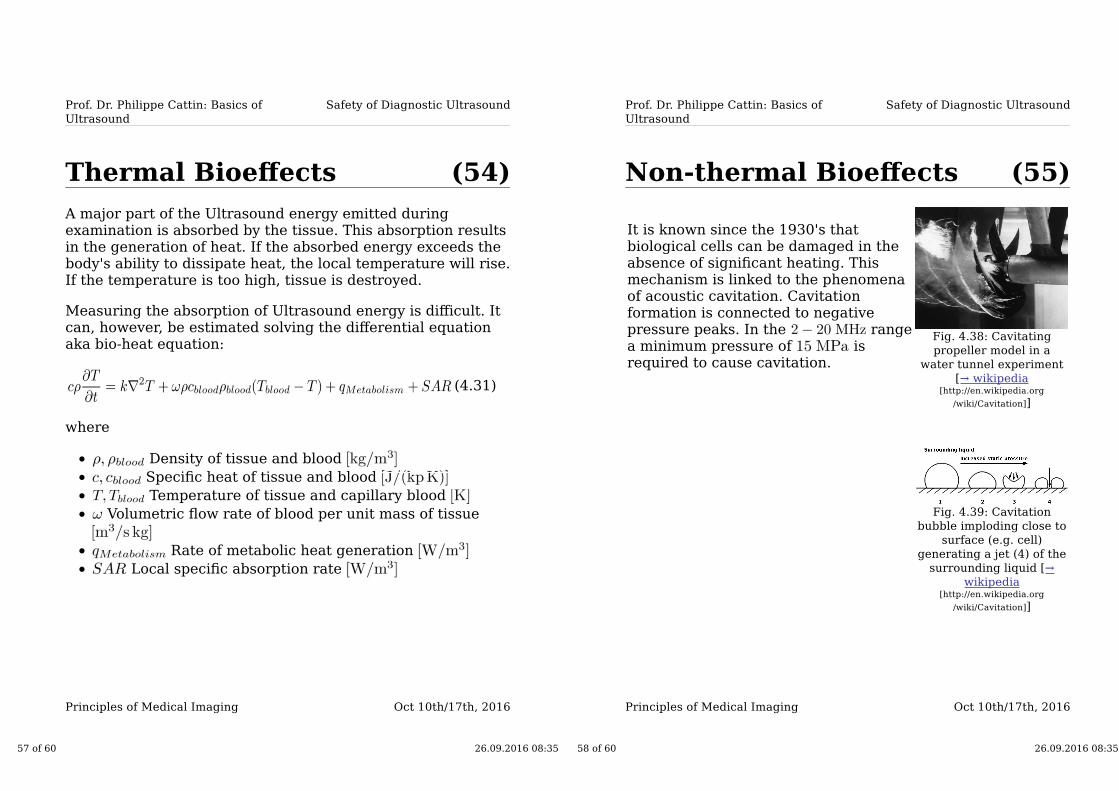
Non-thermal Bioeffects
It is known since the 1930's thatbiological cells can be damaged in theabsence of significant heating. Thismechanism is linked to the phenomenaof acoustic cavitation. Cavitationformation is connected to negativepressure peaks. In the rangea minimum pressure of isrequired to cause cavitation.
Fig. 4.38: Cavitating
propeller model in a
water tunnel experiment
[→ wikipedia[http://en.wikipedia.org
/wiki/Cavitation]]
Fig. 4.39: Cavitation
bubble imploding close to
surface (e.g. cell)
generating a jet (4) of the
surrounding liquid [→
wikipedia[http://en.wikipedia.org
/wiki/Cavitation]]
58 of 60 26.09.2016 08:35
Oct 10th/17th, 2016Principles of Medical Imaging
Safety of Diagnostic Ultrasound
(56)
Prof. Dr. Philippe Cattin: Basics of
Ultrasound
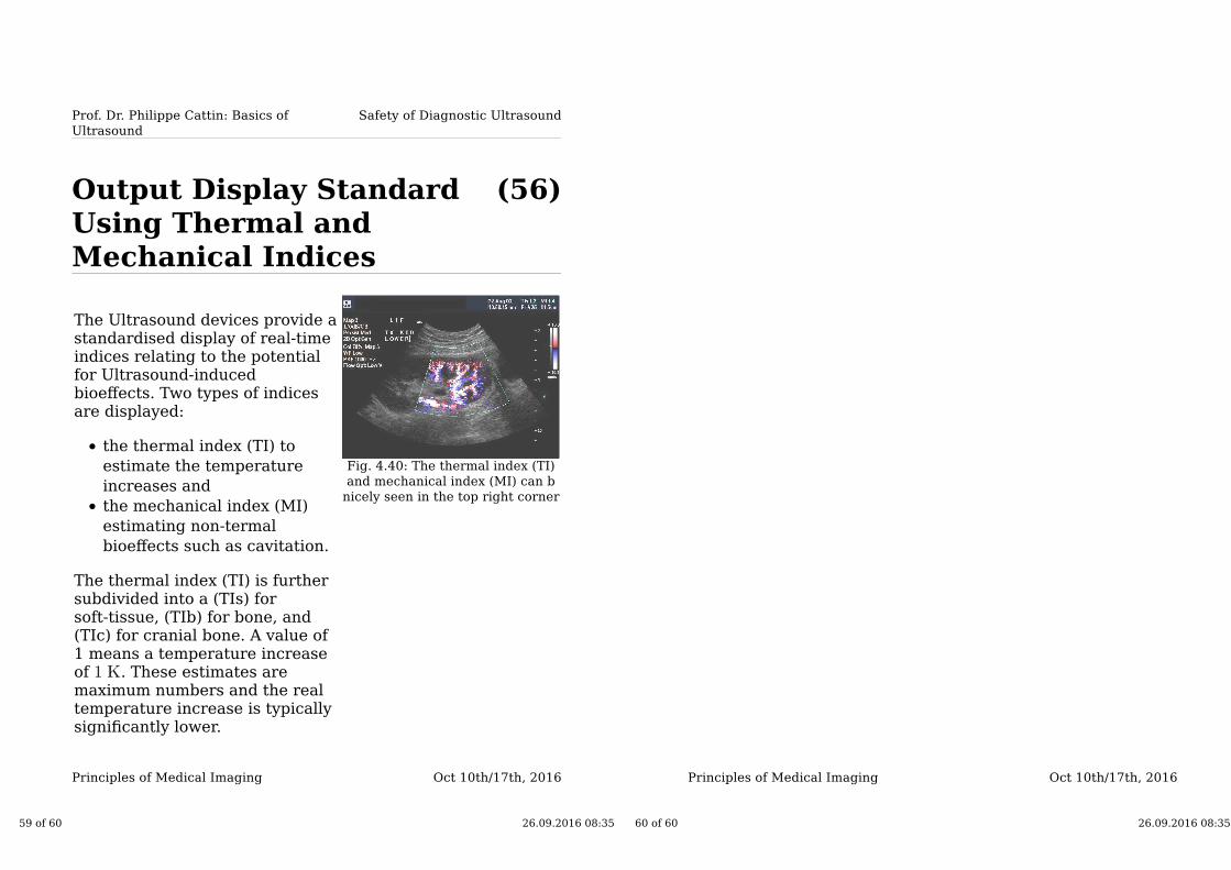
Output Display StandardUsing Thermal andMechanical Indices
The Ultrasound devices provide astandardised display of real-timeindices relating to the potentialfor Ultrasound-inducedbioeffects. Two types of indicesare displayed:
the thermal index (TI) to
estimate the temperature
increases and
the mechanical index (MI)
estimating non-termal
bioeffects such as cavitation.
The thermal index (TI) is furthersubdivided into a (TIs) forsoft-tissue, (TIb) for bone, and(TIc) for cranial bone. A value of1 means a temperature increaseof . These estimates aremaximum numbers and the realtemperature increase is typicallysignificantly lower.
Fig. 4.40: The thermal index (TI)
and mechanical index (MI) can b
nicely seen in the top right corner
59 of 60 26.09.2016 08:35
Oct 10th/17th, 2016Principles of Medical Imaging
60 of 60 26.09.2016 08:35
Related Documents