PRODUCT MONOGRAPH Pr ZOVIRAX ® Acyclovir Cream, Mfr. Std. Acyclovir Ointment, USP 5% w/w Antiviral Agent Bausch Health, Canada Inc. Date of Revision: 2150 St-Elzear Blvd. West December 22, 2020 Laval, Quebec H7L 4A8 Control #: 241907 ZOVIRAX ® is a registered trademark of the GlaxoSmithKline group of companies used under license.

Welcome message from author
This document is posted to help you gain knowledge. Please leave a comment to let me know what you think about it! Share it to your friends and learn new things together.
Transcript

PRODUCT MONOGRAPH
PrZOVIRAX®
Acyclovir Cream, Mfr. Std.
Acyclovir Ointment, USP
5% w/w
Antiviral Agent
Bausch Health, Canada Inc. Date of Revision:
2150 St-Elzear Blvd. West December 22, 2020
Laval, Quebec
H7L 4A8
Control #: 241907
ZOVIRAX® is a registered trademark of the GlaxoSmithKline group of companies used under license.

PrZOVIRAX ® Product Monograph Page 2 of 32
PRODUCT MONOGRAPH
PrZOVIRAX®
Acyclovir Cream, Mfr. Std.
Acyclovir Ointment, USP
5% w/w
ACTIONS AND CLINICAL PHARMACOLOGY
ZOVIRAX (acyclovir), a synthetic acyclic purine nucleoside analog, is a substrate with a high
degree of specificity for herpes simplex and varicella-zoster specified thymidine kinase.
Acyclovir is a poor substrate for host cell-specified thymidine kinase. Herpes simplex and
varicella-zoster specified thymidine kinase transform acyclovir to its monophosphate which is
then transformed by a number of cellular enzymes to acyclovir diphosphate and acyclovir
triphosphate. Acyclovir triphosphate is both an inhibitor of, and a substrate for, herpesvirus-
specified DNA polymerase. Although the cellular α-DNA polymerase in infected cells may also
be inhibited by acyclovir triphosphate, this occurs only at concentrations of acyclovir triphosphate
which are higher than those which inhibit the herpesvirus-specified DNA polymerase. Acyclovir
is selectively converted to its active form in herpesvirus-infected cells and is thus preferentially
taken up by these cells.
Acyclovir has demonstrated a very much lower toxic potential in vitro for normal uninfected cells
because:
1) less is taken up;
2) less is converted to the active form;
3) cellular α-DNA polymerase has a lower sensitivity to the action of the active form of the
drug.
A combination of the thymidine kinase specificity, inhibition of DNA polymerase and premature
termination of DNA synthesis results in inhibition of herpesvirus replication. No effect on latent
non-replicating virus has been demonstrated. Inhibition of the virus reduces the period of viral
shedding, limits the degree of spread and level of pathology, and thereby facilitates healing.
During suppression there is no evidence that acyclovir prevents neural migration of the virus. It
aborts episodes of recurrent herpes due to inhibition of viral replication following reactivation.
INDICATIONS AND CLINICAL USE
Zovirax (acyclovir 5%) cream is indicated for the topical management of initial episodes of
genital herpes simplex infections. The prophylactic use of this preparation has not been
established.
Zovirax (acyclovir 5%) ointment is for the management of initial episodes of genital herpes
simplex infections. It is also indicated in the management of non-life-threatening cutaneous
herpes simplex virus infections in immunocompromised patients. The prophylactic use of this
preparation has not been established.
PrZOVIRAX ® Product Monograph Page 3 of 32
In the treatment of genital herpes, appropriate examinations should be performed to rule out other
sexually transmitted diseases. Therapy should begin as early as possible after the start of an
infection.
Whereas cutaneous lesions associated with herpes simplex infections are often pathognomonic,
TZANCK smears prepared from lesion exudate or scrapings may assist in the diagnosis. Positive
cultures for herpes simplex virus offer the only absolute means for confirmation of the diagnosis.
These indications are based on the results of a number of double-blind, placebo-controlled studies
which examined changes in virus excretion, healing of lesions and relief of pain. Because of the
wide biological variations inherent in herpes simplex infections, the following summary is
presented merely to illustrate the spectrum of responses observed to date. As in the treatment of
any infectious disease, the best response may be expected when therapy is begun at the earliest
possible moment.
The indication regarding the management of initial episodes of genital herpes simplex infections
is based on two multicenter, double-blind, placebo-controlled studies were performed with
ZOVIRAX cream in immunocompetent patients with initial genital herpes. The cream was
applied for up to 10 days or until healing had occurred. Results showed that ZOVIRAX cream
significantly reduced the duration of viral shedding, the formation of new lesions, the time to
crusting and healing of lesions, and the duration of pain.
The indication regarding the management of nonlife-threatening cutaneous herpes simplex virus
infections in immunocompromised patients found that in immunocompromised patients, 93%
were virus negative after 5 days of topical ZOVIRAX ointment therapy, whereas only 35% of
placebo recipients were virus negative at the same time. In patients with herpes labialis, there was
a significantly greater decrease in the amount of virus excreted after one day of therapy in those
receiving ZOVIRAX ointment within 8 hours of the onset of cold sores when compared to
identically treated placebo recipients.
Because complete re-epithelialization of herpes-disrupted integument necessitates recruitment of
several complex repair mechanisms, the physician should be aware that the disappearance of
visible lesions is somewhat variable and will occur later than the cessation of virus shedding. All
immunocompromised patients who received topical ZOVIRAX ointment had healed their lesions
23 days after the initiation of a 10-day course of therapy; 75% of placebo patients had healed
lesions at that point. Some placebo patients continued to have visible lesions for more than 30
days.
Pain associated with herpes infections is highly variable in frequency and intensity. One hundred
percent of the ZOVIRAX ointment treated immunocompromised patients were pain-free by day
23 versus 70% of placebo-treated patients.

PrZOVIRAX ® Product Monograph Page 4 of 32
CONTRAINDICATIONS
ZOVIRAX is contraindicated for patients who develop hypersensitivity or chemical intolerance to
acyclovir, valacyclovir or any of the components of the formulation, such as propylene glycol or
polyethylene glycol.
WARNINGS
ZOVIRAX is intended for topical use only and should not be used in the eye or on mucous
membranes, such as the mouth or vagina.
PRECAUTIONS
General
The recommended dosage, frequency of application and duration of treatment of ZOVIRAX
should not be exceeded (see DOSAGE AND ADMINISTRATION).
There exist no data, at this time, which demonstrate that the use of ZOVIRAX will prevent
transmission of infection to other persons.
Since most cutaneous herpes simplex virus infections result from reactivation of latent virus, it is
unlikely that ZOVIRAX will prevent recurrence of infections when applied in the absence of
signs and symptoms. ZOVIRAX should not be applied in an attempt to prevent recurrences;
application should commence only at the earliest prodromal sign of disease onset.
Although clinically significant viral resistance associated with the use of ZOVIRAX has not been
observed, this possibility exists (see VIROLOGY).
Sexual Function/ Reproduction
There is no information on the effect of acyclovir oral formulations on human female fertility. In
a study of 20 male patients with normal sperm count, oral acyclovir administered at doses of up to
1g per day for up to six months has been shown to have no clinically significant effect on sperm
count, motility or morphology.
Nursing Mothers
Acyclovir, when given systemically, is known to be excreted into human milk. No information is
available on levels of acyclovir which may appear in breast milk after administration of
ZOVIRAX and although evidence suggests that absorption of acyclovir through the skin is
minimal, caution should be exercised when acyclovir is administered to a nursing mother.
PrZOVIRAX ® Product Monograph Page 5 of 32
Use in Pregnancy
ZOVIRAX should not be used during pregnancy unless the physician feels the potential benefit
justifies the risk of possible harm to the fetus.
Teratology studies using acyclovir cream carried out to date in animals have been negative in
general. However, in a non-standard test in rats, there were fetal abnormalities such as head and
tail anomalies, and maternal toxicity; and while since such studies are not always predictive of
human response, the potential for high concentrations of acyclovir to cause chromosome breaks in
vitro should be taken into consideration in making this decision.
All animal studies with the ointment carried out to date on reproduction and teratology have been
negative. However, animal reproduction studies are not always predictive of human response.
A post-marketing acyclovir pregnancy registry has documented pregnancy outcomes in women
exposed to any formulation of ZOVIRAX. The registry findings have not shown an increase in
the number of birth defects amongst ZOVIRAX exposed subjects compared with the general
population, and any birth defects showed no uniqueness or consistent pattern to suggest a
common cause.
Pediatrics Patients (< 18 years of age)
The safe use of ZOVIRAX in pediatric patients less than 18 years of age has not been established
DRUG INTERACTIONS
Clinical experience has identified no interactions resulting from topical or systemic administration
of other drugs concomitantly with ZOVIRAX.
ADVERSE REACTIONS
Because ulcerated genital lesions are characteristically tender and sensitive to any contact or
manipulation, patients may experience discomfort upon application of ZOVIRAX.
There have been very rare reports of immediate hypersensitivity reactions including angioedema
with topical acyclovir.
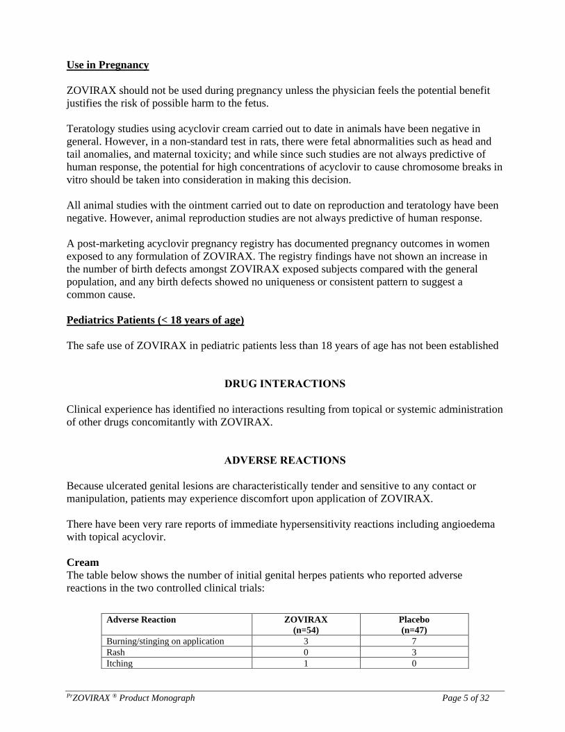
Cream
The table below shows the number of initial genital herpes patients who reported adverse
reactions in the two controlled clinical trials:
Adverse Reaction ZOVIRAX
(n=54)
Placebo
(n=47)
Burning/stinging on application 3 7
Rash 0 3
Itching 1 0

PrZOVIRAX ® Product Monograph Page 6 of 32
Observed During Clinical Practice
Based on worldwide clinical practice experience in patients treated with ZOVIRAX cream, the
adverse events most commonly reported include contact dermatitis, application site reaction,
eczema, allergic reaction, pain, and rash.
Less common events include pruritus, skin discoloration, urticaria, vesiculobullous rash, and
facial edema.
Ointment
In the controlled clinical trials, mild pain (including transient burning and stinging) was reported
by 103 (28.3%) of 364 patients treated with ZOVIRAX ointment and by 115 (31.1%) of 370
patients treated with placebo; treatment was discontinued in 2 of these patients. Other local
reactions among acyclovir-treated patients included pruritus in 15 (4.1%), rash in 1 (0.3%) and
vulvitis in 1 (0.3%). Among the placebo-treated patients, pruritus was reported by 17 (4.6%) and
rash by 1 (0.3%).
In all studies, there was no significant difference between the drug and placebo group in the rate
or type of reported adverse reactions.
There have been very rare reports of immediate hypersensitivity reactions including angioedema
with topical acyclovir.
SYMPTOMS AND TREATMENT OF OVERDOSAGE
Overdosage by topical application of ZOVIRAX is unlikely because of limited transcutaneous
absorption.
For management of a suspected drug overdose, contact your regional poison control centre.
DOSAGE AND ADMINISTRATION
Apply ZOVIRAX liberally to the affected area 4 to 6 times daily for up to 10 days. A sufficient
quantity should be applied to adequately cover all lesions. A finger cot or rubber glove should be
used while applying ZOVIRAX in order to prevent: (1) autoinoculation of other body sites or (2)
transmission of infection to other persons. Therapy should be initiated as early as possible
following onset of signs and symptoms.
Retention of urine 2 2
Meningism 0 2
Paronychia 0 1
Total No. (%) of patients 6 (11%) 15 (32%)
PrZOVIRAX ® Product Monograph Page 7 of 32
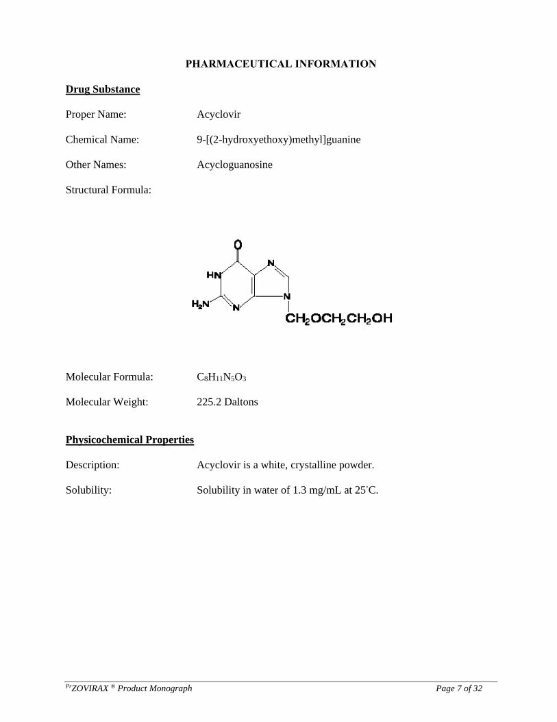
PHARMACEUTICAL INFORMATION
Drug Substance
Proper Name: Acyclovir
Chemical Name: 9-[(2-hydroxyethoxy)methyl]guanine
Other Names: Acycloguanosine
Structural Formula:
Molecular Formula: C8H11N5O3
Molecular Weight: 225.2 Daltons
Physicochemical Properties
Description: Acyclovir is a white, crystalline powder.
Solubility: Solubility in water of 1.3 mg/mL at 25◦C.

PrZOVIRAX ® Product Monograph Page 8 of 32
AVAILABILITY OF DOSAGE FORMS
Cream
Each 1 gram of ZOVIRAX cream contains 50 mg acyclovir and the non-medicinal ingredients
propylene glycol, paraffin, cetostearyl alcohol, poloxamer, sodium lauryl sulfate
ZOVIRAX cream is available in tubes of 5g.
Ointment
Each gram contains 50 mg acyclovir in a polyethylene glycol base.
ZOVIRAX ointment is available in tubes of 4g and 30g.
STORAGE CONDITIONS
Cream
ZOVIRAX cream should be stored between 15ºC and 25ºC and kept dry.
Ointment
ZOVIRAX ointment should be stored between 15ºC to 25°C and kept dry.
VIROLOGY
The quantitative relationship between the in vitro susceptibility of herpes simplex virus (HSV),
varicella-zoster virus (VZV) and cytomegalovirus to acyclovir and the clinical response to therapy
has not been established in man, and virus sensitivity testing has not been standardized.
Prolonged exposure of herpes simplex virus (HSV) to subinhibitory concentrations (0.1 μg/mL)
of acyclovir in cell culture has resulted in the emergence of a variety of acyclovir resistant strains.
The emergence of resistant strains is believed to occur by "selection" of naturally occurring
viruses with relatively low susceptibility to acyclovir. Such strains have been reported in pre-
therapy isolates from several clinical studies.
Two resistance mechanisms involving viral thymidine kinase (required for acyclovir activation)
have been described. These are: (a) selection of thymidine-kinase-deficient mutants that induce
little or no enzyme activity after infection, and (b) selection of mutants possessing a thymidine
kinase of altered substrate specificity that is able to phosphorylate the natural nucleoside
thymidine but not acyclovir. The majority of less susceptible viruses arising in vitro are of the
thymidine-kinase-deficient type which have reduced infectivity and pathogenicity and less
likelihood of inducing latency in animals.
However, an acyclovir resistant HSV infection in an immunosuppressed bone marrow transplant
recipient on extended acyclovir therapy was found to be due to a clinical isolate which had a
normal thymidine kinase but an altered DNA polymerase. This third mechanism of resistance
PrZOVIRAX ® Product Monograph Page 9 of 32
involving herpes simplex virus DNA polymerase is due to the selection of mutants encoding an
altered enzyme, which is resistant to inactivation by acyclovir triphosphate.
Varicella Zoster virus appears to manifest resistance to acyclovir via mechanisms similar to those
seen in herpes simplex virus.
However, limited clinical investigation has revealed no evidence of a significant change in in
vitro susceptibility of varicella zoster virus with acyclovir therapy, although resistant mutants of
this virus can be isolated in vitro in a manner analogous to herpes simplex virus. Analysis of a
small number of clinical isolates from patients who received oral acyclovir or placebo for acute
herpes zoster suggests that in vivo emergence of resistant varicella zoster virus may occur
infrequently. Prolonged acyclovir treatment of highly immunocompromised patients with
acquired immunodeficiency syndrome and severe varicella zoster virus may lead to the
appearance of resistant virus.
Cross-resistance to other antivirals occurs in vitro in acyclovir-resistant mutants. Herpes simplex
virus mutants which are resistant to acyclovir due to an absence of viral thymidine kinase are
cross-resistant to other agents which are phosphorylated by herpesvirus thymidine kinase, such as
bromovinyldeoxyuridine, ganciclovir and the 2'-fluoropyrimidine nucleosides, such as, 2'-fluoro-
5-iodoarabinosyl-cytosine (FIAC).
The clinical response to acyclovir treatment has usually been good for patients with normal
immunity from whom herpes simplex virus having reduced susceptibility to acyclovir has been
recovered either before, during or after therapy. However, certain patient groups, such as the
severely immunocompromised (especially bone marrow transplant recipients) and those
undergoing chronic suppressive regimens have been identified as being most frequently
associated with the emergence of resistant herpes simplex strains, which may or may not
accompany a poor response to the drug. The possibility of the appearance of less sensitive viruses
must be recognized when treating such patients, and susceptibility monitoring of clinical isolates
from these patients should be encouraged.
In summary, the quantitative relationship between the in vitro susceptibility of herpes simplex and
varicella-zoster viruses to acyclovir and the clinical response to therapy has not been clearly
established in man. Standardized methods of virus sensitivity testing are required to allow more
precise correlations between in vitro virus sensitivity and clinical response to acyclovir therapy.
Cream
Sensitivity testing results, expressed as the concentration of drug required to inhibit by 50% the
growth of virus in cell culture (ID50), vary greatly depending upon the particular assay used, the
cell type employed, and the laboratory performing the test. The ID50 of acyclovir against HSV-1
isolates may range from 0.02 μg/mL (plaque reduction in Vero cells) to 5.9-13.5 μg/mL (plaque
reduction in green monkey kidney [GMK] cells). The ID50 against HSV-2 ranges from
0.01 μg/mL to 9.9 μg/mL (plaque reduction in Vero and GMK cells, respectively).
Using a dye-uptake method in Vero cells, which gives ID50 values approximately 5 - to 10-fold
higher than plaque reduction assays, 1417 HSV isolates (553 HSV-1 and 864 HSV-2) from

PrZOVIRAX ® Product Monograph Page 10 of 32
approximately 500 patients were examined over a 5-year period. These assays found that 90% of
HSV-1 isolates were sensitive to ≤ 0.9 μg/mL acyclovir and 50% of all isolates were sensitive to
≤ 0.2 μg/mL acyclovir. For HSV-2 isolates, 90% were sensitive to ≤ 2.2 μg/mL and 50% of all
isolates were sensitive to ≤ 0.7 μg/mL of acyclovir. Isolates with significantly diminished
sensitivity were found in 44 patients. It must be emphasized that neither the patients nor the
isolates were randomly selected and, therefore, do not represent the general population. Most of
the less sensitive HSV clinical isolates have been relatively deficient in the viral thymidine kinase
(TK). Strains with alterations in viral TK or viral DNA polymerase have also been reported.
Prolonged exposure to low concentrations (0.1 μg/mL) of acyclovir in cell culture has resulted in
the emergence of a variety of acyclovir-resistant strains.
The ID50 against VZV ranges from 0.17-1.53 μg/mL (yield reduction, human foreskin fibroblasts)
to 1.85-3.98 μg/mL (foci reduction, human embryo fibroblasts [HEF]). Reproduction of EBV
genome is suppressed by 50% in superinfected Raji cells or P3HR-1 lymphoblastoid cells by
1.5 μg/mL acyclovir. CMV is relatively resistant to acyclovir with ID50 values ranging from 2.3-
17.6 μg/mL (plaque reduction, HEF cells) to 1.82-56.8 μg/mL (DNA hybridization, HEF cells).
The latent state of the genome of any of the human herpesviruses is not known to be sensitive to
acyclovir.
Ointment
In Vitro Activity
The techniques and cell culture types used for determining in vitro susceptibility may influence
the results obtained. Using a quantitative assay to determine the acyclovir concentration
producing 50% inhibition of viral cytopathic effect (ID50), 28 HSV-1 clinical isolates had an ID50
range of 0.02-0.70 μg/mL (mean - 0.17 μg/mL), and 32 HSV-2 clinical isolates had an ID50 range
of 0.01-3.16 μg/mL (mean - 0.46 μg/mL).23 Results from other studies using different assays
have yielded mean ID50 values for clinical HSV-1 isolates of 0.018, 0.03 and 0.043 μg/mL and
for clinical HSV-2 isolates of 0.027, 0.36 and 0.03 μg/mL, respectively.2,3,4.
Using a plaque reduction assay, 9 clinical isolates of VZV had an ID50 range of 0.70-2.32 μg/mL
(mean - 1.52 μg/mL).
A reduced spectrum of in vitro activity was evident with 5 clinical isolates of CMV, where again
using a plaque reduction assay an ID50 range was found of 2.25-12.81 μg/mL (mean -
8.22 μg/mL).
Enzyme Studies - Effects of Acyclovir-TP on Viral and Cellular DNA Polymerases
Time of addition studies with acyclovir showed that the compound was effective early in the
replicative cycle of HSV-1, i.e., prior to 8 hours post-infection. If the compound was added to
HSV-infected Vero cells on monolayers after 8 hours post-infection, no antiviral activity was
observed.2 The formation of acyclovir-TP was detectable at 2 hours and reached a maximum 8
hours after drug addition of HSV-infected cultures. These results suggested that acyclovir was
activated and exerted its inhibitory effect at some point early in the replicative cycle of HSV-1,
most likely during DNA synthesis.
The HSV-specified DNA polymerase has been identified as one of the major enzymes appearing
PrZOVIRAX ® Product Monograph Page 11 of 32
early after infection. It, therefore, seemed a probable target enzyme for the action of acyclovir-
TP. Herpes-specified DNA polymerase was purified from HeLa cells infected with HSV-1, using
DEAE column chromatography followed by phosphocellulose column chromatography. The
cellular DNA polymerase-α from these infected cells was likewise isolated. Kinetic studies were
then performed with various synthetic templates (dC.odG and dA.odT) to verify the identities of
the viral and cellular DNA polymerases. The inhibitory effect of acyclovir-TP on these two DNA
polymerases was then determined. The results obtained showed that acyclovir-TP was a
competitive inhibitor of dGTP for both the viral and cellular DNA polymerases. However, in all
cases, acyclovir-TP was a better inhibitor of the viral DNA polymerase than of the cellular DNA
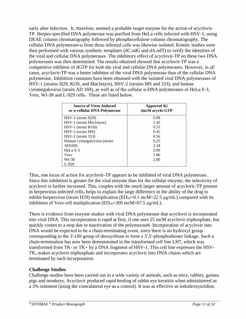
polymerase. Inhibition constants have been obtained with the isolated viral DNA polymerases of
HSV-1 (strains H29, KOS, and MacIntyre), HSV-2 (strains MS and 333), and human
cytomegalovirus (strain AD 169), as well as of the cellular α-DNA polymerases of HeLa S-3,
Vero, WI-38 and L-929 cells. These are listed below.
Source of Virus-Induced
or α-cellular DNA Polymerase
Apparent Ki
(mcM acyclo-GTP
HSV-1 (strain H29)
HSV-1 (strain MacIntyre)
HSV-1 (strain KOS)
HSV-2 (strain MS)
HSV-2 (strain 333)
Human cytmegalovirus (strain
AD169)
HeLa S-3
Vero
WI-38
L-929
0.08
1.42
0.55
0.45
0.56
0.25
2.34
2.09
1.86
2.80
Thus, one locus of action for acyclovir-TP appears to be inhibited of viral DNA polymerase.
Since this inhibition is greater for the viral enzyme than for the cellular enzyme, the selectivity of
acyclovir is further increased. This, couples with the much larger amount of acyclovir-TP present
in herpesvirus-infected cells, helps to explain the large difference in the ability of the drug to
inhibit herpesvirus (strain H29) multiplication (ED50=0.1 mcM=22.5 μg/mL) compared with its
inhibition of Vero cell multiplication (ED50=300 mcM=67.5 μg/mL).
There is evidence from enzyme studies with viral DNA polymerase that acyclovir is incorporated
into viral DNA. This incorporation is rapid at first, if one uses 25 mcM acyclovir triphosphate, but
quickly comes to a stop due to inactivation of the polymerase6. Incorporation of acyclovir into
DNA would be expected to be a chain-terminating event, since there is no hydroxyl group
corresponding to the 3'-OH group of deoxyribose to form a 3',5'-phosphodiester linkage. Such a
chain-termination has now been demonstrated in the transformed cell line LH7, which was
transformed from TK- or TK+ by a DNA fragment of HSV-1. This cell line expresses the HSV-
TK, makes acyclovir triphosphate and incorporates acyclovir into DNA chains which are
terminated by such incorporation.
Challenge Studies
Challenge studies have been carried out in a wide variety of animals, such as mice, rabbits, guinea
pigs and monkeys. Acyclovir produced rapid healing of rabbit eye keratitis when administered as
a 3% ointment (using the contralateral eye as a control). It was as effective as iododeoxyuridine,

PrZOVIRAX ® Product Monograph Page 12 of 32
trifluorothymidine or superior to trifluorothymidine, vidaravine and idoxuridine. Both the topical
and intravenous forms of acyclovir have offered partial to total protection, respectively, from a
fatal outcome due to encephalitis following inoculation of herpes simplex into animal eyes.
A therapeutic effect has been seen in mouse herpes encephalitis using oral doses of 100 mg/kg
twice daily for 5 days,14 or with continuous oral doses of 400 mg/kg for 7 days.
It has also been shown that subcutaneous dosing of 100 mg/kg/day initiated 12 hours after viral
inoculation for 4 consecutive days affects the survival rates and vital titers in the mouse herpes
simplex encephalitis model.
Topical therapy has been shown to be effective in mice against herpes simplex virus skin lesions.
Similar results were shown in guinea pigs.
Intravenous acyclovir has also been shown to be effective in experimental herpes B virus
infection in the rabbit.
Resistance
A colorimetric method has been used to quantitate the inhibition of viral cytopathic effect. From
the data, one can calculate the drug concentration producing a 50% inhibition of viral replication
(ID50) (this assay gives ID50 values which are approximately ten times higher than ID50's obtained
in a plaque reduction assay). McLaren et al. reported a study in which ID50's of clinical isolates
from patients with herpes simplex type 1 or type 2 infections were measured in the CPE-reduction
assay. The mean ID50 value for the HSV-1 isolates was 0.17 μg/mL while the mean of the HSV-2
isolates was somewhat higher at 0.46 μg/mL. Based on initial results, virus isolates with ID50's
greater than 2.0 μg/mL are tentatively regarded as having a significantly lower sensitivity to
acyclovir.
It is possible to isolate strains of HSV which show a reduced sensitivity to the drug, by growing
the virus in tissue cultures treated with acyclovir. Such ‘resistant’ viruses are generally deficient
in thymidine kinase activity (TK). A few mutants with changed DNA-polymerase have also been
isolated in laboratory experiments but with a much lower frequency. Experiments in animals have
indicated that although the TK-deficient mutants of HSV are capable of infecting and inducing
antibody production, they generally have a greatly reduced ability to cause disease and have a
significantly diminished ability to establish latent infections in neuronal ganglia. The clinical
significance of these findings has yet to be determined. To date, no consistent correlation has been
established between resistance identified in the laboratory and clinical expression of HSV disease.
Virus with a diminished sensitivity to acyclovir (an increased ID50) has been detected in seven
patients treated with IV acyclovir and three patients treated with the topical ointment. These
particular virus isolates also showed reduced thymidine kinase activity.
The majority of the patients treated with intravenous drug from whom less sensitive virus was
isolated were severely immunocompromised by virtue of chemotherapy or innate deficiencies. In
two bone-marrow transplant patients TK-deficient HSV was shed asymptomatically for a brief
period after a significant clinical response to intravenous acyclovir therapy. Viruses with reduced
TK-activity were also isolated from two children with severe immunodeficiencies following

PrZOVIRAX ® Product Monograph Page 13 of 32
successful initial therapy with acyclovir; the clinical disease in these children neither improved
nor worsened following the emergence of TK-deficient virus. In the patients who survived their
underlying disease, TK-deficient virus has not been detected during subsequent recurrences nor
has any such isolate apparently spread nosocomially. In immunocompetent patients with genital
HSV-2 infections treated with ZOVIRAX ointment, cessation of virus replication and lesion
healing occurred in a time period similar to that in other patients who had viruses with normal TK
activity.
Among the placebo-treated patients, a change in viral sensitivity was found in two patients treated
with the topical preparation. In only one of these patients was the increase in ID50 associated with
diminished TK activity. Five patients given placebo intravenously exhibited changes in the in
vitro sensitivity of their virus populations during or after administration of the placebo. The TK
activity was reduced in two, remained unchanged in one and was not tested in three of these
patients.
A number of patients have been identified in which the virus presents prior to therapy already
exhibited a relatively high ID50, sometimes associated with low TK activity. In the intravenous
studies, there were six such patients with less sensitive virus; virus with a similar sensitivity was
also recovered from the cervix, but not from other lesions, of a seventh patient.
PHARMACOLOGY
Cream
Dermal Absorption
Two studies were conducted to determine the percutaneous absorption of acyclovir cream.
In the first study, acyclovir (ACV) in dimethyl sulfoxide (DMSO) was evaluated for the treatment
of cutaneous herpes simplex virus infection in guinea pigs and compared with ACV in
polyethylene glycol (PEG). When compared with infection sites treated with the vehicle alone,
ACV-DMSO produced a greater percent reduction than did ACV-PEG in the number, area and
virus titer of the lesions.
The second study investigated penetration through excised human and guinea pig skin of ACV
formulated in three different vehicles: PEG, DMSO, and modified aqueous cream (MAC).
Results showed that ACV-MAC and ACV-DMSO penetrated through human and guinea pig skin
at a faster rate than ACV-PEG.
Ointment
ZOVIRAX administration to adults at 5 mg/kg (approximately 250 mg/m2 BSA) by 1-hour
infusions every 8 hours produces mean steady-state peak and trough concentrations of 9.8 μg/mL
and 0.7 μg/mL, respectively.
Similar concentrations are achieved in pediatric patients over 1 year of age when doses of
250 mg/m2 BSA (body surface area) are given every 8 hours. Concentrations achieved in the

PrZOVIRAX ® Product Monograph Page 14 of 32
cerebrospinal fluid are approximately 50% of plasma values. Plasma protein binding is relatively
low (9 to 33%) and drug interactions involving binding site displacement are not anticipated.
Renal excretion of unchanged drug by glomerular filtration and tubular secretion is the major
route of acyclovir elimination accounting for 62 to 91% of the dose in man as determined by 14C-
labelled drug. The only significant urinary metabolite is 9-carboxymethoxymethylguanine. An
insignificant amount of drug is recovered in feces and expired CO2 and there is no evidence to
suggest tissue retention.
The half-life and total body clearance of acyclovir is dependent on renal function as shown below.
Creatinine Clearance
(mL/min/1.73m2 BSA*)
Half-Life
(hr)
Total Body Clearance
(mL/min/1.73m2 BSA*)
>80
50-80
15-50
0 (Anuric)
2.4
2.9
3.7
18
33225118526
* Body surface area
The half-life and total body clearance of acyclovir in pediatric patients over 1 year of age is
similar to adults with normal renal function.
Two clinical pharmacology studies were performed with ZOVIRAX ointment in adult
immunocompromised patients, at risk of developing mucocutaneous herpes simplex virus
infections or with localized varicella-zoster infections. These studies were designed to evaluate
the dermal tolerance, systemic toxicity and percutaneous absorption of acyclovir. In one of these
studies, which included 16 inpatients, the complete ointment or its vehicle were randomly
administered in a dose of 1cm strips (25 mg acyclovir) four times a day for seven days to an intact
skin surface area of 4.5 square inches. No local intolerance, systemic toxicity or contact
dermatitis were observed. In addition, no drug was detected in blood and urine by
radioimmunoassay30 (sensitivity, 0.01 μg/mL).
The other study included 11 patients with localized varicella-zoster. In this uncontrolled study,
acyclovir was detected in the blood of 9 patients and in the urine of all patients tested. Acyclovir
levels in plasma ranged from 0.01 to 0.28 μg/mL in eight patients with normal renal function, and
from 0.01 to 0.78 μg/mL in one patient with impaired renal function. Acyclovir excreted in the
urine ranged from 0.02 to 53.6 μg/mL (0.02 to 9.4%) of the daily dose. Therefore, systemic
absorption of acyclovir after topical application is minimal.
PrZOVIRAX ® Product Monograph Page 15 of 32
TOXICOLOGY
Acute Toxicity Studies
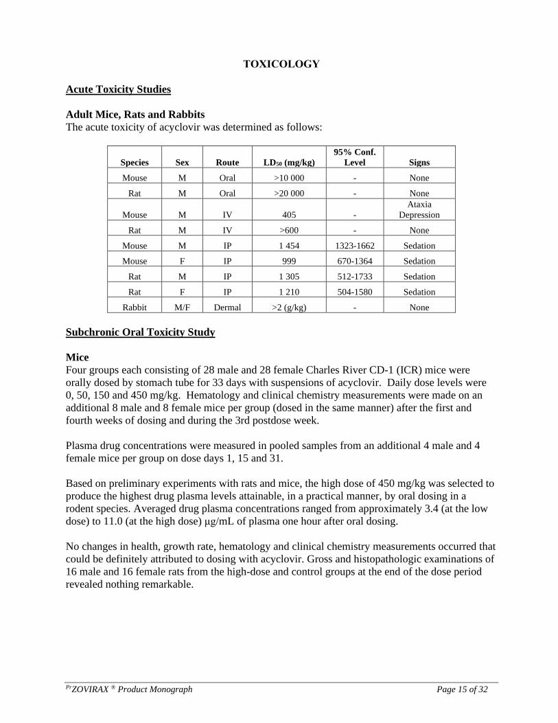
Adult Mice, Rats and Rabbits
The acute toxicity of acyclovir was determined as follows:
Species Sex Route LD50 (mg/kg)
95% Conf.
Level Signs
Mouse M Oral >10 000 - None
Rat M Oral >20 000 - None
Mouse M IV 405 -
Ataxia
Depression
Rat M IV >600 - None
Mouse M IP 1 454 1323-1662 Sedation
Mouse F IP 999 670-1364 Sedation
Rat M IP 1 305 512-1733 Sedation
Rat F IP 1 210 504-1580 Sedation
Rabbit M/F Dermal >2 (g/kg) - None
Subchronic Oral Toxicity Study
Mice
Four groups each consisting of 28 male and 28 female Charles River CD-1 (ICR) mice were
orally dosed by stomach tube for 33 days with suspensions of acyclovir. Daily dose levels were
0, 50, 150 and 450 mg/kg. Hematology and clinical chemistry measurements were made on an
additional 8 male and 8 female mice per group (dosed in the same manner) after the first and
fourth weeks of dosing and during the 3rd postdose week.
Plasma drug concentrations were measured in pooled samples from an additional 4 male and 4
female mice per group on dose days 1, 15 and 31.
Based on preliminary experiments with rats and mice, the high dose of 450 mg/kg was selected to
produce the highest drug plasma levels attainable, in a practical manner, by oral dosing in a
rodent species. Averaged drug plasma concentrations ranged from approximately 3.4 (at the low
dose) to 11.0 (at the high dose) μg/mL of plasma one hour after oral dosing.
No changes in health, growth rate, hematology and clinical chemistry measurements occurred that
could be definitely attributed to dosing with acyclovir. Gross and histopathologic examinations of
16 male and 16 female rats from the high-dose and control groups at the end of the dose period
revealed nothing remarkable.
PrZOVIRAX ® Product Monograph Page 16 of 32
Subchronic Intravenous Toxicity Studies
Beagle Dogs
In a 31-day study in Beagle dogs, acyclovir was administered as a bolus intravenous injection to
groups of 8 dogs (4 male and 4 female) at dosage levels of 0, 25, 50 and 100 mg/kg, b.i.d.
Intravenous bolus doses of 50 or 100 mg/kg, b.i.d. per day in this study produced very high drug
plasma levels [mean values in the range of 45 to 254 μg/mL (200 to 1127 mcM)] which were
obviously highly toxic, whereas the 25 mg/kg, b.i.d. dose resulted in considerably lower plasma
levels in the range of 22.5 to 45 μg/mL (100 to 200 mcM) and was only marginally toxic and
nearly a "no effect" dose.
Primary drug-related changes at the 25 mg/kg b.i.d. dose level included: infrequent retching
and/or emesis, occasional tachycardia, increased urinary output with a decrease in specific
gravity. These effects were reversible and undetectable 15 days after withdrawal of treatment.
At the 50 and 100 mg/kg b.i.d. dosage, additional adverse effects were seen including dyspnea,
hypothermia, hypoactivity, bloody or mucoid diarrhea, dehydration, body weight loss, partial to
total anorexia, leucopenia, slight increases in serum total protein, albumin, creatinine, urea
nitrogen and occasional "loud" heartbeat.
Findings considered directly related to drug treatment with the 50 and 100 mg/kg, b.i.d. dosage
levels included infrequent retching and emesis, occasional tachycardia and "loud" heartbeat,
increased urine output, hyaline droplets in the cytoplasm of the liver parenchymal cells, mild
cytologic changes in the colon mucosa and kidney toxicity. Some other changes, considered
secondary to the effects of drug administration at the 50 and 100 mg/kg b.i.d. levels, were skeletal
muscle and adipose tissue atrophy, depletion of lipid from cortex of adrenal gland, and aspermic
testes.
More seriously, there were tremors, cyanosis, prostration and early death (within the first 8 days
of the study).
Rats
Groups of 15 male and 15 female Sprague-Dawley rats were given one daily intravenous injection
as a bolus of 20, 40 or 80mg/kg of acyclovir for 20 or 21 consecutive days. Drug was formulated
as a 2 % isotonic solution in sterile 0.4 % sodium chloride. A control group was given single daily
injections of a 0.9% solution of sodium chloride.
At all dose levels most or all rats had renal lesions which were considered to be related to
obstruction of the distal nephron by precipitated drug crystals. Lesions increased in severity with
rising dose and were those to be expected with obstruction of the distal nephron: retrograde
tubular dilatation and epithelial degeneration, necrosis and regeneration. There was an
accompanying interstitial inflammatory component in some of the more severely affected
kidneys.
Birefringent drug crystals were visible in sections of kidney which had been frozen before
formalin fixation but were not seen in conventional paraffin sections since they had been
solubilized by the processes of formalin fixation and staining.

PrZOVIRAX ® Product Monograph Page 17 of 32
Examination of kidney sections from rats which had been maintained for a 15-day drug-free
period after administration of the last dose revealed only mild residual reparative changes or no
lesions at all. This indicates the reversibility of the obstructive nephropathy.
Clinical effects of drug were considered to be related to renal changes. These consisted of reduced
body weight, elevated blood urea nitrogen values, increased water intake and urine output plus an
increase in mean absolute and relative kidney weights.
In a second study in rats, lower drug doses were used in an effort to establish a no-effect level.
The doses were 5 or 10mg/kg/day given as a single bolus intravenous injection for 19-20
consecutive days, and there was a control group which received 0.9% sodium chloride solution.
The only finding in the rats, which was considered definitely associated with drug administration,
was very mild dilatation of distal tubules in kidneys of 2 of 20 animals in the 5 mg/kg group. The
dilatation was thought to be due to recent, and perhaps still present, distal nephron obstruction by
drug crystals (though they were not present in paraffin sections for the reason explained above).
Long-Term Toxicity Studies
12-Month Toxicity Study in Dogs
Purebred Beagle dogs were given 0, 15, 45 or 150 mg/kg/day of acyclovir each day for the first
two weeks of a 1-year study. There were 9 male and 9 female dogs in each test group. The dogs
were given gelatin capsules that contained the appropriate dose. They were treated t.i.d., hence the
dosages administered at each of three equally spaced dose periods were 0, 5, 15 and 50mg/kg.
The 45 and 150 mg/kg dose levels induced diarrhea, emesis, decreased food consumption and
weight loss in both male and female dogs during the first two weeks of the study. For this reason,
during the third week of the study the decision was made to decrease the mid and high dosage
levels to 30 and 60 mg/kg/day (10 and 20mg/kg t.i.d.). The low dose of 15mg/kg/day (5mg/kg
t.i.d.) was unchanged. Dogs given 60 mg/kg/day occasionally vomited and occasionally had
diarrhea but did well for the duration of the test and values for body weight gain and food
consumption were comparable to control values.
During the toxicosis induced by the larger doses of acyclovir, plasma levels of the drug were
likely very high (as indicated by initial mean values of 24.0 μg/mL (106.6 mcM) for high-dose
males and 17.4 μg/mL (77.2 mcM) for high-dose females when determined 1 hour after the third
dose on day 1 of the study). When measured on day 15, plasma levels of acyclovir in high-dose
dogs (150 mg/kg/day) were still very high but they decreased later when the dosages were
decreased. Values for plasma levels after 12 months of treatment were generally comparable to
values recorded after 1, 3 and 6 months of treatment. Thus, there was no indication of enhanced
metabolism of acyclovir as a result of chronic treatment.
During the 13th week, some male and female dogs at both the mid and high dosage levels had the
following signs: tenderness in forepaws, breaking of nails and loosening of nails. Regeneration of
lost nails began a few weeks later. Nails regenerated by 6 months (when 3 males and 3 females
from each group were killed for an interim sacrifice) and by the end of the study were of generally
good quality. There were never any signs of an effect on paws or nails in dogs in the low-dose
group (15mg/kg/day).

PrZOVIRAX ® Product Monograph Page 18 of 32
It is accepted that injury of the corial epithelium that produces nail keratin can result in arrested
production of keratin and production of abnormal keratin. The transient toxicosis induced by the
large doses (45 and 150 mg/kg/day) of acyclovir given during the first two weeks of the study
may have affected the corial epithelium. If there was a transient effect on the corial epithelium
(possibly related to direct effects or secondary to drug-induced illness during the first two weeks
of the study) later loss of the nail could be a sequella. No discernible effects upon other keratin-
producing or keratin-containing tissues were observed. It should be emphasized that the
alterations in the nails appeared to be related to the transient toxicosis induced by dose levels of
50 and 150 mg/kg/day tested during the first two weeks of the study and not to the 30 and
60 mg/kg/day dose levels tested subsequently.
There were no important drug-induced alterations in values for serum biochemical tests,
urinalyses and electrocardiographic tests done at appropriate intervals during this study. Values
for serum albumin and total protein were slightly decreased in dogs treated at 30 and
60 mg/kg/day for 6 and 12 months. However, all values for these parameters remained within
limits accepted as normal.
With the exception of residual alterations in old keratin at the tips of the claws, there were no
signs of treatment-related effects in any of the tissues examined by light microscopy. Nor were
there meaningful alterations in values for the organs weighed at necropsy. Thus, dose levels up to
60 mg/kg/day were well tolerated. Except for mild gastrointestinal signs at 60 mg/kg/day, all dose
levels tested for 1 year were “no effect” levels.
52-Week Interim Report of 104-Week Oral Toxicity Study in Rats Given Acyclovir by
Gastric Intubation
Charles River CD (Sprague-Dawley) rats were given suspensions of acyclovir by gavage for 52
weeks of a 104-week study. There were 50 male and 50 female rats at each of the following dose
levels: 0, 50, 150 and 450 mg/kg. After 30 and 52 weeks of treatment, 10 male and 10 female rats
from each group were necropsied. Tissues from control rats and those in the high dose group
were evaluated by light microscopy for both the 30- and 52-week interim sacrifices. Fixed tissues
from rats that were found dead during the first 52 weeks of the study were also evaluated by light
microscopy.
No signs of toxicosis were observed. Mean plasma levels were obtained in high-dose males
(450 mg/kg/day) 1.5 hours after dosing at various sampling times during the study as follows:
1.54, 1.63, 1.39 and 1.60 μg/mL (6.84, 7.26, 6.17 and 7.10 mcM) at days 7, 90, 209 and 365,
respectively.
Corresponding mean values for the high-dose females were 1.76, 2.38, 2.12 and 1.71 μg/mL
(7.82, 10.58, 9.44 and 7.62 mcM). Plasma levels in both males and females at all dose levels after
one year of treatment were generally comparable to plasma levels obtained at earlier samplings.
Values for laboratory tests including hematology, clinical chemistry and ophthalmoscopy were all
within the normal range. There were no drug-induced gross or microscopic lesions in rats killed
for 30- and 52-week interim sacrifices. Most of the relatively few rats found dead or moribund
during the first 52 weeks of this study suffered dosing accidents as evidenced by postmortem
findings of esophageal perforation causing pleural effusion, pneumonia, or mediastinitis.
PrZOVIRAX ® Product Monograph Page 19 of 32
Genotoxicity and Mutagenicity Studies
Acyclovir has been tested for mutagenic potential in a number of in vitro systems: cultured
L5178Y mouse lymphoma cells (3 loci); cultured Chinese hamster ovary (CHO) cells (3 loci);
Ames Salmonella (plate assay); Ames Salmonella (preincubation modification); Rosenkrantz E.
coli polA+/polA- DNA repair assay; and the yeast S. cerevisiae, D-4. Also, the drug has been
tested in the BALB/C-3T3 Neoplastic Transformation Assay, in the C3H/10T ½ Neoplastic
Transformation Assay and for clastogenicity in cultured human lymphocytes. All assays were
done both in the presence and absence of exogenous mammalian metabolic activation except for
the cell transformation tests and the human lymphocyte cytogenetic assay. In vivo, acyclovir has
been examined in a mouse dominant lethal assay, and for clastogenicity in rat and Chinese
hamster bone marrow.
In vitro, acyclovir was negative in all microbial assays; it was also negative at the HGPRT locus
and the Ouabain-resistance marker in the mouse lymphoma system; and in the C3H/1OT ½ assay
for transformation. It was significantly positive at the highest dose tested in the BALB/C-3T3 cell
transformation assay; it gave a moderately positive response at high concentrations at the TK
locus in the mouse lymphoma assay and caused chromosomal breakage in human lymphocytes at
high concentrations. In vivo, no cytogenetic effects were noted at up to nephrotoxic doses
(100 mg/kg) in rats or Chinese hamsters; at higher doses (500 and 1000 mg/kg), chromosome
damage was seen in Chinese hamster bone marrow. Summaries of the various assay results are as
follows:
Microbial
Acyclovir was tested for mutagenic activity in the Ames Salmonella plate assay; in a
preincubation modification of the Ames assay; in the Rosenkrantz E. coli polA+/polA- DNA repair
assay; and in the eukaryote S. cerevisiae, D-4. All studies were performed both in the presence
and absence of exogenous mammalian metabolic activation. Acyclovir gave no positive responses
in any of these systems.
The previous Salmonella studies were extended to extremely high concentrations in order to
achieve toxicity. No positive effects were observed either in the presence or absence of exogenous
mammalian metabolic activation, at concentrations of acyclovir up to 300 mg/plate or 80 mg/mL.
Mammalian Systems
Acyclovir was tested for mutagenic activity in cultured L5178Y mouse lymphoma cells,
heterozygous at the thymidine kinase (TK) locus, by measuring the forward mutation rate to TK-
deficiency (TK+/-→TK-/-); additional studies were performed at the HGPRT locus and the
Ouabain-resistance marker in these same cells. All studies were performed in the presence and in
the absence of exogenous mammalian metabolic activation. The test compound was mutagenic at
the TK locus at high (400-2400 μg/mL) concentrations. (By comparison, acyclovir peak plasma
levels following topical application are 0.27 μg/mL or lower). It was negative at the HGPRT
locus and Ouabain-resistance marker. Metabolic activation did not affect the results at any locus.
Inconclusive results with no apparent dose-related response were obtained when acyclovir
mutagenicity was studied at each of 3 loci (APRT, HGPRT and Ouabain-resistance) in Chinese
hamster ovary (CHO) cells, both in the presence and absence of exogenous metabolic activation.

PrZOVIRAX ® Product Monograph Page 20 of 32
Acyclovir, at a concentration of 50µg/mL (222 mcM) for a 72-hour exposure, has been shown to
cause a statistically significant increase in the incidence of morphologically transformed foci
resulting from treating BALB/C-3T3 cells in vitro in the absence of exogenous metabolic
activation. The morphologically transformed foci have been shown to grow as tumors following
transplantation into immunosuppressed, syngeneic, weanling mice. Tumor tissues were diagnosed
as being either undifferentiated sarcomas or lymphosarcomas.
Acyclovir at concentrations between 8 μg/mL and 64 μg/mL for 18 hours exposure did not induce
any morphologically transformed foci among C3H10 T½ cells treated in vitro in the absence of
exogenous metabolic activation.
Acyclovir, at concentrations of 62.5 and 125 μg/mL for a 48-hour exposure, did not induce any
chromosome aberrations in cultured human lymphocytes in the absence of exogenous metabolic
activation. At higher and toxic concentrations (250 and 500 μg/mL for 48 hours exposure)
acyclovir caused a significant increase in the incidence of chromosome breakage.
Reproduction/Fertility Study
Acyclovir, at single intraperitoneal doses of 25, 50 and 100 mg/kg, failed to induce chromosome
aberrations in bone marrow cells of Chinese hamsters when examined 24 hours after dosing. At
higher doses (500 and 1000 mg/kg), a clastogenic effect was seen. (An intraperitoneal dose of
500 mg/kg produces mean peak plasma levels in Chinese hamsters of 611 μg/mL (2.72 mM)
which is 2200 times higher than human plasma levels following recommended topical
application.)
Acyclovir, at single intravenous doses of 25, 50 and 100 mg/kg, failed to induce chromosome
aberrations in bone marrow cells of male and female rats when examined at 6, 24 and 48 hours
after treatment.
Carcinogenicity Studies
Lifetime Oral Carcinogenicity Study in Rats
There were no signs of toxicosis in Charles River CD (Sprague-Dawley) rats (100 rats/sex/dose
group) given acyclovir by oral gavage at 50, 150 and 450 mg/kg in a lifetime oral carcinogenicity
study. Mean plasma levels obtained in high-dose males 1.5 hours after dosing at various sampling
times during the study were as follows: 1.54, 1.63, 1.39, 1.60 and 1.70 μg/mL (6.84, 7.26, 6.17,
7.10 and 7.56 mcM) at days 7, 90, 209, 369 and 771, respectively. Corresponding mean values for
the high-dose females were 1.76, 2.38, 2.12, 1.71 and 1.81 μg/mL (7.82, 10.58, 9.44, 7.62 and
8.03 mcM).
Values for clinical laboratory tests including hematology, clinical chemistry, urinalysis, body
weight, food consumption and ophthalmoscopy were all within normal ranges. There were no
drug-induced gross or microscopic lesions and there was no evidence that acyclovir affected
survival, temporal patterns of tumor incidence or tumor counts for benign or malignant
neoplasms.

PrZOVIRAX ® Product Monograph Page 21 of 32
Lifetime Oral Carcinogenicity Study in Mice
There were no signs of toxicosis in Charles River CD-1 (ICR) mice (115 mice/sex/dose group)
given acyclovir by oral gavage at 50, 150 and 450 mg/kg/day in a lifetime oral carcinogenicity
study. Mean plasma levels obtained in high-dose males 1.5 hours after dosing at various sampling
times during the study were as follows: 2.83, 3.17 and 1.82 μg/mL (12.59, 14.10 and 8.10 mcM)
at days 90, 365 and 541, respectively. Corresponding mean values for the high-dose females were
9.81, 5.85 and 4.0 μg/mL (43.60, 26.0 and 17.79 mcM).
Values for clinical laboratory tests including hematology, body weight and food consumption
were all within normal ranges. There were no drug-induced gross or microscopic lesions. Female
mice given 150 and 450mg/kg acyclovir survived significantly longer than control female mice;
survival of treated males was comparable to survival of control males. Patterns of tumor incidence
and tumor counts for benign or malignant neoplasms were not affected by treatment with
acyclovir.
Reproduction Studies
Teratology – Rats
Acyclovir was administered to pregnant A.R.S. Sprague-Dawley female rats by subcutaneous
injection during the period of organogenesis (day 6 through day 15 of gestation) at dose levels of
0.0, 6.0, 12.5 and 25.0mg/kg body weight twice daily.
Criteria evaluated for compound effect included maternal body weights, weight gains, appearance
and behavior, survival rates, eye changes, pregnancy rates, and reproduction data. Offspring
viability and development were also evaluated.
In addition to the above measurements, designated animals were sacrificed 1 hour after the first
dose on day 15 in order to collect samples of maternal blood, amniotic fluid and fetuses for
measurements of drug concentration. Mean values from these samples were as follows:
Acyclovir Concentrations
Dose mg/kg
b.i.d., s.c.
Plasma
(μg/mL)
Amniotic Fluid
(μg/mL)
Fetal Homogenate
ng/g (nmoles/g w/w)
6 N = 7
12.5 N = 5
25 N = 5
0.26 ± 0.09
0.69 ± 0.20
1.59 ± 0.55
0.39 ± 0.06
1.13 ± 0.22
2.0 ± 0.53
0.704 (3.13 ± 0.50)
0.963 (4.28 ± 0.67)
1.994 (8.64 ± 2.33)
The values obtained for plasma would represent about 30% of initial plasma levels as judged by
the plasma half-life in rodents.
No effects attributable to the administration of acyclovir were noted in comparisons of maternal
body weight values, appearance and behavior, survival rates, pregnancy rates, or implantation
efficiencies. In addition, no compound-related differences were noted in evaluations of fetal size,
sex, and development.
Although the incidences of resorption and fetal viability were within the range of normal
variability in all of the groups, slightly greater incidences of resorptions were noted in the high-

PrZOVIRAX ® Product Monograph Page 22 of 32
dose animals sacrificed on days 15 and 19 of gestation; however, clear dose-related trends did not
eventuate.
Therefore, acyclovir was not considered teratogenic or embryotoxic when administered to rats at
levels up to 50.0 mg/kg of body weight per day during organogenesis.
Teratology – Rabbits
A teratology study was done in New Zealand White rabbits using essentially the same
experimental design as in the rat, except that dosing was from day 6 through day 18 of gestation.
Also, collection of fetuses, amniotic fluid and samples of maternal blood occurred on day 18
rather than day 15.
No signs of maternal toxicity were observed at any dose, but there was a statistically significant
(p<0.05) lower implantation efficiency in the high-dose group. While there were a few terata
observed in the study (in both control and treated animals), there was no apparent association with
drug treatment. There was, however, an apparent dose-related response in the number of fetuses
having supernumerary ribs. No similar effect was noted in the rat teratology study (see above) or
in a reproduction-fertility experiment in mice.
Concentrations of acyclovir were detected in plasma and amniotic fluid samples, as well as in
homogenates of fetal tissues. All samples were taken one hour after the first dose on day 18 of
gestation. As apparent in the following data, drug concentrations in amniotic fluid were
substantially higher than that of plasma.
Acyclovir Concentrations (Mean and S.E.)
Dose mg/kg
b.i.d., s.c.
Plasma
(μg/mL)
Amniotic Fluid
(μg/mL)
Fetal Homogenate
ng/g (nmoles/g w/w)
6 N = 4
12.5 N = 5
25 N = 5
*N = 5
0.25 ± 0.03
0.25 ± 0.05
1.39 ± 0.12*
0.89 ± 0.18
8.03 ± 6.37
6.16 ± 4.25
0.155 (0.69 ± 0.13)
0.207 (0.92 ± 0.14)
0.315 (1.40 ± 0.19)
Largely reversible adverse effects on spermatogenesis in association with overall toxicity in rats
and dogs have been reported only at systemic doses of acyclovir greatly in excess of those
employed therapeutically. Two-generation studies in mice did not reveal any effect of orally
administered acyclovir on fertility.
Developmental Toxicity Studies
Groups of 10 male and 10 female Charles River CD (Sprague-Dawley) rats were given single
large doses (5 different dose levels) of a solution (pH 11.0) of acyclovir by subcutaneous injection
when they were 3, 10, 28 and 71 days of age. They were observed for 14 days after treatment and
LD50 values were calculated by the Litchfield and Wilcoxon method. This study was done to
determine if age at exposure affects the acute toxicity of acyclovir; there was no evidence that
young rats were more sensitive than older rats to the acute toxic effects of acyclovir.

PrZOVIRAX ® Product Monograph Page 23 of 32
LD50 (mg/kg body weight)
Age When Treated Males Females
3 Days 1070 1281
10 Days 790 496
28 Days 678 750
71 Days 650 1477
There was no apparent relationship between length of survival after treatment and age at which
treatment was given. Clinical signs for the rats treated at 3 and 10 days of age included red and
purple cutaneous blisters, blue areas, scabs, scars, necrotic and sloughed skin, open wounds, body
tremors and alopecia. Decreased activity, lacrimation, closed eyelids, red-brown or brown
material around the eyes, nose and mouth, ataxia, prostration, body tremors, urine stains around
the abdomen or genital area, scabbed or necrotic areas and alopecia were observed in rats treated
at 28 and 71 days of age.
Neonatal Rats - Subchronic Study
Acyclovir dissolved in 0.4% sterile saline was given by subcutaneous injection to Charles River
CD (Sprague-Dawley) neonatal rats for 19 consecutive days, beginning on the 3rd post-partum
day. The dose levels tested were 0, 5, 20 and 80mg/kg body weight. There were 12 litters (each
consisting of 5 male and 5 female neonates nursing the natural dam) at each dose level. The dams
were not treated. Neonates were removed from each group for necropsy and microscopic
evaluation of a wide variety of tissues, including eyes and multiple sections of brain, after they
had been treated for 5, 12 or 19 days and after a 3-week postdose drug-free period (at which time
they were 45 days of age). Hematologic (hemoglobin, packed cell volume, RBC, WBC and
differential cell counts) and clinical chemistry (BUN) tests were done after 16 days of treatment
and repeated 18 days after the last (19th) dose was given.
Blood was collected from some neonates 30 minutes after treatment on day 1, on day 9 and at the
end of the dose period for the determination of concentrations of acyclovir in plasma. The largest
concentration of acyclovir in plasma was 99.1 μg/mL (440.5 mcM) found in pooled plasma
collected from 6 female high-dose (80 mg/kg) neonates 30 minutes after the first dose was given.
Treatment with acyclovir did not increase mortality in the neonatal period.
Rats in the low-dose group gained as much body weight as the respective control rats. Significant
(p<0.05) reductions in mean body weight values were observed in mid- and high-dose group male
and female neonates during the treatment period. Rats in the high-dose group partially
compensated by gaining significantly more body weight than the controls during the postdose
recovery period. There was a minimal but significant increase in BUN for male (p<0.01) and
female (p<0.05) neonates in the high-dose group on dose day 16. This finding may be of
biological importance because there were minimal accumulations of nuclear debris in renal
collecting ducts and loops of Henle in kidney sections taken from high-dose neonates after 19
days of treatment and examined by light microscopy. This was the only time period (and the
kidney was the only organ) in which minimal effects on developing organ systems were detected.
Thus, 5mg/kg was clearly a no effect dose level and 20mg/kg caused only minimal decreases in
body weight gain.
Eye examinations and light microscopy did not reveal adverse effects on ocular development. It
should be emphasized that there was no morphologic or functional evidence of adverse effects on

PrZOVIRAX ® Product Monograph Page 24 of 32
developing brain or other portions of the central nervous system. Thus, acyclovir is distinctly
different than cytosine arabinoside which was reported to produce prominent cerebellar and
retinal dysplasia in neonatal rats.
Immunotoxicology Studies
Acyclovir was subjected to a number of in vitro and in vivo immunological tests.
In two in vitro tests, lymphocyte-mediated cytotoxicity and neutrophil chemotaxis, acyclovir
showed no inhibitory effects at concentrations as high as 135 μg/mL (600 mcM). The compound
inhibited rosette formation approximately 50% at 0.9 μg/mL (4 mcM).
In four in vivo tests in mice which measured cell-mediated immunity (complement-dependent
cellular cytotoxicity, complement-independent cellular cytotoxicity, delayed hypersensitivity and
graft vs. host reaction) acyclovir showed no inhibitory effects at single doses up to 200 mg/kg
given on day 2 after antigenic stimulation.
Four daily doses of 100 mg/kg/day had no significant effect on Jerne hemolysin plaques or
circulating antibody on day 7 after antigenic stimulation. When the Jerne hemolysin plaques and
antibody titers were examined four days after antigenic challenge and one day after the last drug
dose, 100 mg/kg showed only a slight suppressive effect. However, 200 mg/kg produced some
weight loss (-2.2g), a moderate reduction in the number of Jerne hemolysin plaques (PFC/spleen
were reduced to 33% of control, PFC/107 WBC to 46.5% of control). However, there was only a
small reduction in the circulating hemagglutinin titer (from 8.3 to 6.5) and the circulating
hemolysin titer (from 9.5 to 8.3) at 200 mg/kg.
In experiments in mice designed to test whether acyclovir would potentiate the
immunosuppressive effect of azathioprine on antibody formation, it was found that the effects of
the two drugs were no more than additive. Only the 200 mg/kg dose of acyclovir showed an
increased suppression of antibody response when given in combination with azathioprine at doses
above 25 mg/kg.
Studies were carried out to evaluate the influence of acyclovir in vitro on human lymphocyte
function. Inhibitory effects on blastogenesis were seen only in assays examining peak
concentrations of potent mitogens, PHA and Con A, and only at concentrations of drug above
50 μg/mL (222µM) and were much less with monilia and tetanus toxoid antigens, where the
blastogenic response is characteristically less vigorous. There was very little effect on cytotoxicity
or LIF production except at concentrations of 200 μg/mL (890 mcM) for which a direct cytotoxic
effect has been demonstrated before. These inhibitory concentrations are far in excess of
anticipated levels from doses selected for clinical application and over 1000-fold higher than the
concentration required to inhibit herpesvirus multiplication in vitro.
The effect of acyclovir on human cells was measured. A concentration of 11.2- 22.5 μg/mL (50-
100 mcM) inhibits the division of fibroblasts to a variable extent, depending on the experimental
design and the confluency of the monolayer. The magnitude of this effect was less than that
caused by adenine arabinoside or human leukocyte interferon when these three antiviral agents
were compared at clinically relevant concentrations. Acyclovir also inhibited thymidine

PrZOVIRAX ® Product Monograph Page 25 of 32
incorporation by peripheral blood mononuclear cells stimulated by phytohemagglutinin or three
different herpesvirus antigens. A linear dose-response curve was observed with these cells, and
their proliferation was 50% inhibited by 22.5 μg/mL (100 mcM) acyclovir. Inhibition was exerted
on T-cell proliferation without apparent effect on the release of lymphokines or on monocyte
function.
Cream
Dermal Tolerance Studies
Acyclovir was tested in guinea pigs for sensitizing potential in an experiment which followed the
design of Draize. Saline and a known sensitizing agent (1-chloro-2,4-dinitrobenzene) were
administered as negative and positive controls, respectively. A series of ten sensitizing doses were
used, followed 2 weeks later by a challenge dose. All materials were administered intradermally.
Neither acyclovir nor saline produced sensitization, whereas sensitization occurred in all animals
treated with the positive control agent.
Acyclovir cream was evaluated in guinea pigs for sensitizing potential. The cream produced no
signs of dermal sensitization.
Rabbits were treated 3 times a day for 21 consecutive days with acyclovir cream. The rabbits did
not show signs of treatment-related discomfort; erythema at the treatment sites was barely
perceptible in virtually all cases.
Acyclovir cream was applied at a dosage of 0.5 mL/site to the backs of rabbits and the primary
irritation index was calculated at 72 hours post-treatment. Acyclovir cream was found to be
nonirritating.
Guinea pigs were treated with 0.4 mL doses of acyclovir cream, applied to the flank area. No
signs of skin irritancy were noted.
Ointment
Dermal Irritation and Systemic Toxicity
Repeated daily dermal application (4x/day at 4-hour intervals) of 5 and 10% acyclovir in
polyethylene glycol ointment base (200 mg q.i.d.) to the intact and abraded skin (10% body
surface) of guinea pigs for 23-24 days, produced no adverse local effects. There was a decrease
in mean lymphocytes (in abraded female) and in red blood cell counts (abraded male and female)
in animals treated with the 10% acyclovir formulation. Mean drug plasma concentration showed
a considerable variation within any given treatment group (viz: 5% ointment, abraded male,
1.02 μg/mL (4.51 mcM), abraded female, 0.4 μg/mL (1.78 mcM); 10% ointment, abraded male,
0.54 μg/mL (2.41 mcM), abraded female, 1.24 μg/mL (5.52 mcM); 10% ointment, intact male,
0.9 μg/mL (4.00 mcM); intact female 0.81 μg/mL (3.60 mcM). The results indicate that acyclovir
applied as a PEG-base ointment does penetrate the dermal barrier and is absorbed into the
systemic circulation.

PrZOVIRAX ® Product Monograph Page 26 of 32
Epidermal Wound Healing
Spruance and Krueger tested a 5% acyclovir ointment in polyethylene glycol vehicle for potential
effects on epidermal wound healing in domestic pigs. They used a dermatome to make 0.5 to
1.0 cm2 x 0.3 mm deep wounds on the backs of the pigs. The wounds were excised and scored for
healing 4, 5 and 6 days after treatment with acyclovir ointment or the placebo vehicle.
Number Healed/Total Wounds
Treatment Group Day 4 Day 5 Day 6
Untreated 0/15 ND 15/15
PEG 4x/day x 5 days 0/16 15/28* 14/14
5% ACV 4x/day x 5 days 0/14 7/28* 12/16
*p = 0.03
They concluded that topical acyclovir caused a small decrease in the rate of epidermal wound
healing in their pig model.
Eye Irritation
An ointment formulation of acyclovir was tested for eye irritation in rabbits. Concentrations of
1%, 3%, and 6% acyclovir in petrolatum base were instilled into the conjunctival sac 5 times
daily at 90-minute intervals for 21 consecutive days. Groups of 8 rabbits each were dosed with
each concentration, the vehicle and with balanced saline. Eye irritation was evaluated by gross
observation and biomicroscopic, funduscopic and histologic examinations. None of these methods
of examination revealed a significant potential for ophthalmic toxicity.
Ocular Penetration
The instillation of a 1 cm ribbon of a 3% acyclovir ophthalmic ointment in the rabbit eye resulted
in biologically and statistically significant levels of drug in the aqueous humor of the eye within
½ hour. The maximum levels [mean values of 0.48 and 0.57 μg/mL (2.15 and 2.53 mcM)]
occurred at 1 and 2 hours after treatment with a rather marked decrease by the fourth hour [mean
value 0.13 μg/mL (0.59 mcM)]. Plasma levels of acyclovir, where present at detectable amounts,
were below 0.23 μg/mL (1 mcM). In 11 of 15 rabbits the plasma levels were below 0.06 μg/mL
(0.25 mcM) (limit of detectability for the assay procedure).
The study shows that acyclovir can penetrate the corneal surface and produce therapeutic levels of
the drug in the aqueous humor.
Corneal Wound Healing
Masked controlled rabbit studies were done to determine the toxic effects on corneal wound
healing of 3% acyclovir and 0.1% idoxuridine (IDU) in therapeutically effective concentrations.
Acyclovir was found to have no significant detrimental effect (in comparison to controls) on the
quality of regenerating epithelium or the re-epithelialization of epithelial wounds. IDU treatment
of eyes resulted in significant toxic changes in the regenerating epithelium (clinically and
histologically) with a significant delay in epithelial wound healing in comparison to control or
acyclovir-treated eyes. Acyclovir had no significant effect on the collagen content of stromal
wounds as measured by hydroxyproline assay. IDU caused a reduction in collagen content not
significantly different from controls but significantly lower than acyclovir.

PrZOVIRAX ® Product Monograph Page 27 of 32
REFERENCES
1. Bauer DJ, Collins P, Tucker WE, Jr., Macklin AW. Treatment of experimental herpes
simplex keratitis with acycloguanosine. Br J Ophthalmol 1979; 63(6):429-435.
2. Barry DW, Blum MR. Antiviral drugs: acyclovir, in Recent Advances in Clinical
Pharmacology. Turner P, Shand DG (eds) Churchill Livingstone, Edinburgh 1983.
3. Barry DW, Nusinoff-Lehrman S. Viral resistance in clinical practice: summary of five years
experience with acyclovir. Pharmacological and Clinical approaches to Herpesviruses and
Virus Chemotherapy, Aiso, Japan, September 10-13 1984.
4. Barry DW, Nusinoff-Lehrman S. Viral resistance in clinical practice: summary of five years
experience with acyclovir. Proceedings of the International Symposium on Pharmacological
and Clinical Approches to Herpes Viruses and Virus Chemotherapy, Elsever, Amsterdam
1984;269-270.
5. Biron KK, Elion GB. Effect of acyclovir combined with other antiherpetic agents on
varicella zoster virus in vitro. Am J Med 1982; 73(1A):54-57.
6. Boulter EA, Thornton B, Bauer DJ, Bye A. Successful treatment of experimental B virus
(Herpesvirus simiae) infection with acyclovir. Br Med J 1980; 280(6215):681-683.
7. Burns WH, Saral R, Santos GW, Laskin OL, Lietman PS, McLaren C et al. Isolation and
characterisation of resistant Herpes simplex virus after acyclovir therapy. Lancet 1982;
1(8269):421-423.
8. Christophers J, Sutton RN. Characterisation of acyclovir-resistant and -sensitive clinical
herpes simplex virus isolates from an immunocompromised patient. J Antimicrob Chemother
1987; 20(3):389-398.
9. Coen DM, Schaffer PA. Two distinct loci confer resistance to acycloguanosine in herpes
simplex virus type 1. Proc Natl Acad Sci U S A 1980; 77(4):2265-2269.
10. Colby BM, Furman PA, Shaw JE, Elion GB, Pagano JS. Phosphorylation of acyclovir [9-(2-
hydroxyethoxymethyl)guanine] in Epstein-Barr virus-infected lymphoblastoid cell lines. J
Virol 1981; 38(2):606-611.
11. Cole NL, Balfour HH, Jr. Varicella-Zoster virus does not become more resistant to acyclovir
during therapy. J Infect Dis 1986; 153(3):605-608.
12. Collins P, Bauer DJ. The activity in vitro against herpes virus of 9-(2-
hydroxyethoxymethyl)guanine (acycloguanosine), a new antiviral agent. J Antimicrob
Chemother 1979; 5(4):431-436.
13. Collins P, Oliver NM. Sensitivity monitoring of herpes simplex virus isolates from patients
receiving acyclovir. J Antimicrob Chemother 1986; 18 Suppl B:103-112.
PrZOVIRAX ® Product Monograph Page 28 of 32
14. Collins P. Viral sensitivity following the introduction of acyclovir. Am J Med 1988;
85(2A):129-134.
15. Collins P, Larder BA, Oliver NM, Kemp S, Smith IW, Darby G. Characterization of a DNA
polymerase mutant of herpes simplex virus from a severely immunocompromised patient
receiving acyclovir. J Gen Virol 1989; 70 ( Pt 2):375-382.
16. Crumpacker CS, Schnipper LE, Marlowe SI, Kowalsky PN, Hershey BJ, Levin MJ.
Resistance to antiviral drugs of herpes simplex virus isolated from a patient treated with
acyclovir. N Engl J Med 1982; 306(6):343-346.
17. Crumpacker CS, Schnipper LE, Zaia JA, Levin MJ. Growth inhibition by acycloguanosine of
herpesviruses isolated from human infections. Antimicrob Agents Chemother 1979;
15(5):642-645.
18.
Douglas JM, Davis LG, Remington ML, Paulsen CA, Perrin EB, Goodman P et al. A double-
blind, placebo-controlled trial to the effect of chronically administered oral acyclovir on
sperm production in men with frequently recurrent genital herpes. J Infect Dis 1988 Mar;
157:588-93.
19. De Clercq E, Descamps J, Verhelst G, Walker RT, Jones AS, Torrence PF et al. Comparative
efficacy of antiherpes drugs against different strains of herpes simplex virus. J Infect Dis
1980; 141(5):563-574.
20. De Clercq E. Comparative efficacy of antiherpes drugs in different cell lines. Antimicrob
Agents Chemother 1982; 21(4):661-663.
21. Dekker C, Ellis MN, McLaren C, Hunter G, Rogers J, Barry DW. Virus resistance in clinical
practice. J Antimicrob Chemother 1983; 12 Suppl B:137-152.
22. Douglas JM, Davis LG, Remington ML, Paulsen CA, Perrin EB, Goodman P et al. A double-
blind, placebo-controlled trial to the effect of chronically administered oral acyclovir on
sperm production in men with frequently recurrent genital herpes. J Infect Dis 1988 Mar;
157:588-93.
23. Ellis MN, Keller PM, Fyfe JA, Martin JL, Rooney JF, Straus SE et al. Clinical isolate of
herpes simplex virus type 2 that induces a thymidine kinase with altered substrate specificity.
Antimicrob Agents Chemother 1987; 31(7):1117-1125.
24. Elion GB, Furman PA, Fyfe JA, de Miranda P, Beauchamp L, Schaeffer HJ. Selectivity of
action of an antiherpetic agent, 9-(2-hydroxyethoxymethyl) guanine. Proc Natl Acad Sci U S
A 1977; 74(12):5716-5720.
25. Englund JA, Zimmerman ME, Swierkosz EM, Goodman JL, Scholl DR, Balfour HH, Jr.
Herpes simplex virus resistant to acyclovir. A study in a tertiary care center. Ann Intern Med
1990; 112(6):416-422.

PrZOVIRAX ® Product Monograph Page 29 of 32
26. Erlich KS, Mills J, Chatis P, Mertz GJ, Busch DF, Follansbee SE et al. Acyclovir-resistant
herpes simplex virus infections in patients with the acquired immunodeficiency syndrome. N
Engl J Med 1989; 320(5):293-296.
27. Erlich KS, Jacobson MA, Koehler JE, Follansbee SE, Drennan DP, Gooze L et al. Foscarnet
therapy for severe acyclovir-resistant herpes simplex virus type-2 infections in patients with
the acquired immunodeficiency syndrome (AIDS). An uncontrolled trial. Ann Intern Med
1989; 110(9):710-713.
28. Fiddian AP, Kinghorn GR, Goldmeier D, Rees E, Rodin P, Thin RN et al. Topical acyclovir
in the treatment of genital herpes: a comparison with systemic therapy. J Antimicrob
Chemother 1983; 12 Suppl B:67-77.
29. Field HJ, Darby G, Wildy P. Isolation and characterization of acyclovir-resistant mutants of
herpes simplex virus. J Gen Virol 1980; 49(1):115-124.
30. Field HJ, Darby G. Pathogenicity in mice of strains of herpes simplex virus which are
resistant to acyclovir in vitro and in vivo. Antimicrob Agents Chemother 1980; 17(2):209-
216.
31. Freeman DJ, Sheth NV, Spruance SL. Failure of topical acyclovir in ointment to penetrate
human skin. Antimicrob Agents Chemother 1986; 29(5):730-732.
32. Furman PA, St Clair MH, Fyfe JA, Rideout JL, Keller PM, Elion GB. Inhibition of herpes
simplex virus-induced DNA polymerase activity and viral DNA replication by 9-(2-
hydroxyethoxymethyl)guanine and its triphosphate. J Virol 1979; 32(1):72-77.
33. Furman PA, Μguirt PV, Keller PM, Fyfe JA, Elion GB. Inhibition by acyclovir of cell
growth and DNA synthesis of cells biochemically transformed with herpesvirus genetic
information. Virology 1980; 102(2):420-430.
34. Fyfe JA, Keller PM, Furman PA, Miller RL, Elion GB. Thymidine kinase from herpes
simplex virus phosphorylates the new antiviral compound, 9-(2-
hydroxyethoxymethyl)guanine. J Biol Chem 1978; 253(24):8721-8727
35. Kaufman HE, Varnell ED, Centifanto YM, Rheinstrom SD. Effect of 9-(2-
hydroxyethoxymethyl)guanine on herpesvirus-induced keratitis and iritis in rabbits.
Antimicrob Agents Chemother 1978; 14(6):842-845.
36. Kern ER, Richards JT, Overall JC, Glasgow LA. Effective treatment of genital herpes
simplex virus infections of mice and guinea pigs with acycloguanosine. Abs 18th
Interscience Conf Antimicrob Ag Chemother, Atlanta, Oct 1-4 1978;(Abstract#264).
37. Kern ER, Overall JC, Glasgow LA, de Miranda P. Optimal therapy of herpes simplex virus
infections in mice with oral 9-(2-hydroxyethoxymethyl)guanine (acyclovir), in the Human
Herpesviruses - An Interdisciplinary Perspective. Hahmias AJ, Dowdle WR, Schinazi RF
(eds), Atlanta, Georgia Elsevier, New York 1981;673-674.

PrZOVIRAX ® Product Monograph Page 30 of 32
38. Jacobson MA, Berger TG, Fikrig S, Becherer P, Moohr JW, Stanat SC et al. Acyclovir-
resistant varicella zoster virus infection after chronic oral acyclovir therapy in patients with
the acquired immunodeficiency syndrome (AIDS). Ann Intern Med 1990; 112(3):187-191.
39. Lehrman SN, Douglas JM, Corey L, Barry DW. Recurrent genital herpes and suppressive
oral acyclovir therapy. Relation between clinical outcome and in-vitro drug sensitivity. Ann
Intern Med 1986; 104(6):786-790.
40. McLaren C, Ellis MN, Hunter GA. A colorimetric assay for the measurement of the
sensitivity of herpes simplex viruses to antiviral agents. Antiviral Res 1983; 3(4):223-234.
41. McLaren C, Corey L, Dekket C, Barry DW. In vitro sensitivity to acyclovir in genital herpes
simplex viruses from acyclovir-treated patients. J Infect Dis 1983; 148(5):868-875.
42. McLaren C, Sibrack CD, Barry DW. Spectrum of sensitivity of acyclovir of herpes simplex
virus clinical isolates. Am J Med 1982; 73(1A):376-379.
43. Naib ZM, Nahmias AJ, Josey WE, Zaki SA. Relation of cytohistopathology of genital
herpesvirus infection to cervical anaplasia. Cancer Res 1973; 33(6):1452-1463.
44. O'Brien JJ, Campoli-Richards DM. Acyclovir. An updated review of its antiviral activity,
pharmacokinetic properties and therapeutic efficacy. Drugs 1989; 37(3):233-309.
45. Oosterhuis JA, Versteeg J, Kruit PJ, Postma BH. Aciclovir treatment in experimental
herpetic keratitis in rabbits. Ophthalmic Research 1980; 12(1):38-44.
46. Pahwa S, Biron K, Lim W, Swenson P, Kaplan MH, Sadick N et al. Continuous varicella-
zoster infection associated with acyclovir resistance in a child with AIDS. JAMA 1988;
260(19):2879-2882.
47. Parker AC, Craig JI, Collins P, Oliver N, Smith I. Acyclovir-resistant herpes simplex virus
infection due to altered DNA polymerase. Lancet 1987; 2(8573):1461.
48. Park NH, Pavan-Langston D, McLean SL, Albert DM. Therapy of experimental herpes
simplex encephalitis with aciclovir in mice. Antimicrob Agents Chemother 1979; 15(6):775-
779.
49. Parris DS, Harrington JE. Herpes simplex virus variants restraint to high concentrations of
acyclovir exist in clinical isolates. Antimicrob Agents Chemother 1982; 22(1):71-77.
50. Pavan-Langston D, Campbell R, Lass J. Acyclic antimetabolite therapy of experimental
herpes simplex keratitis. Am J Ophthalmol 1978; 86(5):618-623.
51. Preblud SR, Arbeter AM, Proctor EA, Starr SE, Plotkin SA. Susceptibility of vaccine strains
of varicella-zoster virus to antiviral compounds. Antimicrob Agents Chemother 1984;
25(4):417-421.

PrZOVIRAX ® Product Monograph Page 31 of 32
52. Quinn RP, de Miranda P, Gerald L, Good SS. A sensitive radioimmunoassay for the antiviral
agent BW248U [9-(2-hydroxyethoxymethyl)guanine]. Anal Biochem 1979; 98(2):319-328.
53. Schaeffer HJ, Gurwara S, Vince R, Bittner S. Novel substrate of adenosine deaminase. J Med
Chem 1971; 14(4):367-369.
54. Schaeffer HJ, Beauchamp L, de Miranda P, Elion GB, Bauer DJ, Collins P. 9-(2-
hydroxyethoxymethyl) guanine activity against viruses of the herpes group. Nature 1978;
272(5654):583-585.
55. Schnipper LE, Crumpacker CS. Resistance of herpes simplex virus to acycloguanosine: role
of viral thymidine kinase and DNA polymerase loci. Proc Natl Acad Sci U S A 1980;
77(4):2270-2273.
56. Shiota H, Inoue S, Yamane S. Efficacy of acycloguanosine against herpetic ulcers in rabbit
cornea. Br J Ophthalmol 1979; 63(6):425-428.
57. Sibrack CD, Gutman LT, Wilfert CM, McLaren C, St Clair MH, Keller PM et al.
Pathogenicity of acyclovir-resistant herpes simplex virus type 1 from an immunodeficient
child. J Infect Dis 1982; 146(5):673-682.
58. Spruance SL, McKeough M, Sugibayashi K, Robertson F, Gaede P, Clark DS. Effect of
azone and propylene glycol on penetration of trifluorothymidine through skin and efficacy of
different topical formulations against cutaneous herpes simplex virus infections in guinea
pigs. Antimicrob Agents Chemother 1984; 26(6):819-823.
59. Spruance SL, Krueger GG. Effect of topical acyclovir on epidermal wound healing in pigs.
Clinical Research 1981; 29(1):82a.
60. Straus SE, Takiff HE, Seidlin M, Bachrach S, Lininger L, DiGiovanna JJ et al. Suppression
of frequently recurring genital herpes. A placebo-controlled double-blind trial of oral
acyclovir. N Engl J Med 1984; 310(24):1545-1550.
61. Straus SE, Seidlin M, Takiff H, Jacobs D, Bowen D, Smith HA. Oral acyclovir to suppress
recurring herpes simplex virus infections in immunodeficient patients. Ann Intern Med 1984;
100(4):522-524.
62. Tenser RB, Miller RL, Rapp F. Trigeminal ganglion infection by thymidine kinase-negative
mutants of herpes simplex virus. Science 1979; 205(4409):915-917.
63. Vinckier F, Boogaerts M, De Clerck D, De Clercq E. Chronic herpetic infection in an
immunocompromised patient: report of a case. J Oral Maxillofac Surg 1987; 45(8):723-728.
64. Wade JC, Newton B, McLaren C, Flournoy N, Keeney RE, Meyers JD. Intravenous
acyclovir to treat mucocutaneous herpes simplex virus infection after marrow transplantation:
a double-blind trial. Ann Intern Med 1982; 96(3):265-269.

PrZOVIRAX ® Product Monograph Page 32 of 32
65. Wade JC, McLaren C, Meyers JD. Frequency and significance of acyclovir-resistant herpes
simplex virus isolated from marrow transplant patients receiving multiple courses of
treatment with acyclovir. J Infect Dis 1983; 148(6):1077-1082.
Related Documents


