REVIEW Proceedings of the workshop on Cerebellum, Basal Ganglia and Cortical Connections Unmasked in Health and Disorder Held in Brno, Czech Republic, October 17th, 2013 Martin Bareš & Richard Apps & Zora Kikinis & Dagmar Timmann & Gulin Oz & James J. Ashe & Michaela Loft & Stella Koutsikou & Nadia Cerminara & Khalaf O. Bushara & Tomáš Kašpárek # Springer Science+Business Media New York 2014 Abstract The proceedings of the workshop synthesize the experimental, preclinical, and clinical data suggesting that the cerebellum, basal ganglia (BG), and their connections play an important role in pathophysiology of various movement disorders (like Parkinson’ s disease and atypical parkinsonian syndromes) or neurodevelopmental disorders (like autism). The contributions from individual distinguished speakers cov- er the neuroanatomical research of complex networks, neuro- imaging data showing that the cerebellum and BG are con- nected to a wide range of other central nervous system struc- tures involved in movement control. Especially, the cerebel- lum plays a more complex role in how the brain functions than previously thought. Keywords Cerebellum . Basal ganglia . Dentate . Striatum . Imaging . White matter . Magnetic resonance spectroscopy . Movement disorders The Cerebellum and Basal Ganglia: A Developing Story (Martin Bareš and Tomáš Kašpárek) Despite isolated studies disproving the role of the basal ganglia (BG) and the cerebellum in timekeeping [1] recent research provides increasing evidence for the involvement of both of these structures in the processing of temporal information [2]. While both the BG and the cerebellum were found to participate M. Bareš : T. Kašpárek Central European Institute of Technology, CEITEC MU, Behavioral and Social Neuroscience Research Group, Masaryk University, Brno, Czech Republic M. Bareš (*) First Department of Neurology, Faculty of Medicine, Masaryk University and St. Anne’ s Teaching Hospital, Brno, Czech Republic e-mail: [email protected] R. Apps : M. Loft : S. Koutsikou : N. Cerminara School of Physiology and Pharmacology, University of Bristol, Bristol, UK Z. Kikinis Harvard Medical School, Boston, MA, USA D. Timmann Department of Neurology, University Clinic Essen, University of Duisburg-Essen, Hufelandstrasse 55, 45147 Essen, Germany G. Oz Center for Magnetic Resonance Research, Department of Radiology, University of Minnesota, Minneapolis, MN, USA J. J. Ashe : K. O. Bushara Department of Neuroscience, University of Minnesota, Minneapolis, MN, USA J. J. Ashe : K. O. Bushara Department of Neurology, University of Minnesota, Minneapolis, MN, USA J. J. Ashe : K. O. Bushara Neurology Service, VA Medical Center, Minneapolis, MN, USA T. Kašpárek Department of Psychiatry, Faculty of Medicine, Masaryk University and St. Teaching Hospital, Brno, Czech Republic Cerebellum DOI 10.1007/s12311-014-0595-y

Welcome message from author
This document is posted to help you gain knowledge. Please leave a comment to let me know what you think about it! Share it to your friends and learn new things together.
Transcript

REVIEW
Proceedings of the workshop on Cerebellum, Basal Gangliaand Cortical Connections Unmasked in Health and Disorder Heldin Brno, Czech Republic, October 17th, 2013
Martin Bareš & Richard Apps & Zora Kikinis & Dagmar Timmann & Gulin Oz &
James J. Ashe & Michaela Loft & Stella Koutsikou & Nadia Cerminara &
Khalaf O. Bushara & Tomáš Kašpárek
# Springer Science+Business Media New York 2014
Abstract The proceedings of the workshop synthesize theexperimental, preclinical, and clinical data suggesting thatthe cerebellum, basal ganglia (BG), and their connections playan important role in pathophysiology of various movementdisorders (like Parkinson’s disease and atypical parkinsoniansyndromes) or neurodevelopmental disorders (like autism).The contributions from individual distinguished speakers cov-er the neuroanatomical research of complex networks, neuro-imaging data showing that the cerebellum and BG are con-nected to a wide range of other central nervous system struc-tures involved in movement control. Especially, the cerebel-lum plays a more complex role in how the brain functions thanpreviously thought.
Keywords Cerebellum . Basal ganglia . Dentate . Striatum .
Imaging .White matter .Magnetic resonance spectroscopy .
Movement disorders
The Cerebellum and Basal Ganglia: A Developing Story(Martin Bareš and Tomáš Kašpárek)
Despite isolated studies disproving the role of the basal ganglia(BG) and the cerebellum in timekeeping [1] recent researchprovides increasing evidence for the involvement of both ofthese structures in the processing of temporal information [2].While both the BG and the cerebellumwere found to participate
M. Bareš : T. KašpárekCentral European Institute of Technology, CEITEC MU, Behavioraland Social Neuroscience ResearchGroup,MasarykUniversity, Brno,Czech Republic
M. Bareš (*)First Department of Neurology, Faculty of Medicine, MasarykUniversity and St. Anne’s Teaching Hospital, Brno, Czech Republice-mail: [email protected]
R. Apps :M. Loft : S. Koutsikou :N. CerminaraSchool of Physiology and Pharmacology, University of Bristol,Bristol, UK
Z. KikinisHarvard Medical School, Boston, MA, USA
D. TimmannDepartment of Neurology, University Clinic Essen, University ofDuisburg-Essen, Hufelandstrasse 55, 45147 Essen, Germany
G. OzCenter for Magnetic Resonance Research, Department of Radiology,University of Minnesota, Minneapolis, MN, USA
J. J. Ashe :K. O. BusharaDepartment of Neuroscience, University of Minnesota, Minneapolis,MN, USA
J. J. Ashe :K. O. BusharaDepartment of Neurology, University of Minnesota, Minneapolis,MN, USA
J. J. Ashe :K. O. BusharaNeurology Service, VA Medical Center, Minneapolis, MN, USA
T. KašpárekDepartment of Psychiatry, Faculty of Medicine, Masaryk Universityand St. Teaching Hospital, Brno, Czech Republic
CerebellumDOI 10.1007/s12311-014-0595-y
in time encoding, most experiments showed that they playeddifferent roles, such as encoding short versus long time inter-vals, dealing with explicit versus implicit timing or addressingtiming versus temporal order [3, 4]. Many everyday skills, suchas sports and the operation of motor vehicles or machineryrequire precise timing [5]; neurological disorders that disruptmotor timing lead to dysmetric or inaccurate movements [6, 7].Movements involve changes in muscle length over time, thusmotor control and timing are inextricably related [8].
The proceedings of the workshop CEREBELLUM,BASAL GANGLIA AND CORTICAL CONNECTIONSUNMASKED IN HEALTH AND DISORDER held in Brno,Czech Republic on October 17th, 2013; synthesize the exper-imental, preclinical and clinical data suggesting that the cere-bellum, BG and their connections play an important role inpathophysiology of various movement disorders (likeParkinson’s disease, atypical parkinsonian syndromes) orneurodevelopmental disorders (like autism). The contributionsfrom individual distingushed speakers cover the neuroanatom-ical research of complex networks, neuroimaging data showingthat the cerebellum and BG are connected to a wide range ofother central nervous system structures involved in movementcontrol. Especially the cerebellum plays a more complex role inhow the brain functions than previously thought [9].
Cerebellar-Basal Ganglia Communication: PhysiologicalEvidence of a Fast Route for Interaction (Michaela Loft,Stella Koutsikou, Nadia Cerminara and Richard Apps)
The cerebellum is involved in a diverse array of functions,ranging from motor control (for review see [10]) to highercognitive abilities e.g. language [11]. Given its uniformcytoarchitecture, it is generally thought that this functionaldiversity arises predominantly from regional differences inafferent and efferent connections. Inputs to the cerebellumhave been described in considerable detail (e.g. [12]). Inparticular, climbing fibre afferents, which originate exclusive-ly from the inferior olive, have been shown to be highlytopographically organized, with specific olivary subregionsproviding climbing fibres that terminate in longitudinallyarranged cortical zones of Purkinje cells with distinct pheno-type [12]. Some alignment between mossy fibre and climbingfibre projections has also been found (e.g. [13]). In turn,Purkinje cells located within each zone provide a highlyconvergent projection to specific regions of the cerebellarand vestibular nuclei that also receive climbing fibre collat-erals that terminate in the same zone [14].
By comparison, much less is known about the organisation ofcerebellar nuclear outputs to other parts of the CNS. However, amajor target is the thalamus. From there, projections are sent tocerebral structures e.g. motor cortex. In turn, the cerebral cortexsends projections to pre-cerebellar nuclei, thereby forming
multiple ‘cortico-cerebellar’ loops, reminiscent of cortico- basalganglia loops (e.g. [15]). Despite these similarities in organiza-tion, the cortico-basal ganglia and cortico-cerebellar loops arethought to operate largely independently. This is because basalganglia and cerebellar outputs terminate in mainly separate tha-lamic territories [16–18], which subsequently project to differentlayers of the cerebral cortex [17]. However, recent studies havechallenged this view, reporting an anatomical connection be-tween the cerebellar dentate nucleus (DN) and the striatum viathe centrolateral (CL) thalamus in both rodents and primates[19–21]. Presently, nothing is known about the functional signif-icance of this connection.
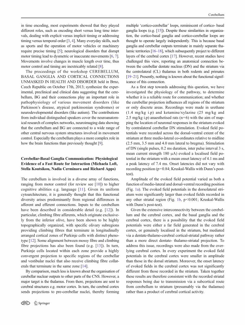
As a first step towards addressing this question, we haveinvestigated the physiology of the pathway, to determinewhether it is a reliable route for communication; and whetherthe cerebellar projection influences all regions of the striatumor only discrete areas. Recordings were made in urethane(1.4 mg/kg i.p) and ketamine/xylazine (25 mg/kg and2.5 mg/kg i.p) anaesthetised rats (n=6) with the aim of map-ping the location of neuronal responses in the striatum evokedby contralateral cerebellar DN stimulation. Evoked field po-tentials were recorded across the dorsal-ventral extent of thestriatum at three medio-lateral co-ordinates relative to midline(2.5 mm, 3.5 mm and 4.0 mm lateral to bregma). Stimulationof DN (single pulses, 0.2 ms duration, inter pulse interval 3 s,mean current strength 180 μA) evoked a localised field po-tential in the striatum with a mean onset latency of 4.1 ms anda peak latency of 7.4 ms. Onset latencies did not vary withrecording position (p=0.84; Kruskal-Wallis with Dunn’s post-test).
Amplitude of the evoked field potential varied as both afunction ofmedio-lateral and dorsal-ventral recording position(Fig. 1a). The evoked field potentials in the dorsolateral stri-atum were significantly larger than evoked fields recorded inany other striatal region (Fig. 1b, p<0.001; Kruskal-Walliswith Dunn’s post-test).
Given the extensive interconnectivity between the cerebel-lum and the cerebral cortex, and the basal ganglia and thecerebral cortex, there is a possibility that the evoked fieldpotentials were either a far field generated in the cerebralcortex, or genuinely localised in the striatum, but mediatedvia a dentate-thalamo-cerebral cortical-striatal pathway ratherthan a more direct dentate- thalamo-striatal projection. Toaddress this issue, recordings were also made from the over-lying cerebral cortex. In every experiment the evoked fieldpotentials in the cerebral cortex were smaller in amplitudethan those in the dorsal striatum. Moreover, the onset latencyof evoked fields in the cerebral cortex was not significantlydifferent from those recorded in the striatum. Taken togetherthese results are therefore consistent with the recorded striatalresponses being due to transmission via a subcortical routefrom cerebellum to striatum (presumably via the thalamus)rather than a product of cerebral cortical activity.
Cerebellum
A small sample of single units (n=3) were also record-ed in the striatum in response to single pulse stimulationof the contralateral DN. Each unit showed a brief, short-latency excitation in response to DN stimulation with ahigh probability of occurrence. The latencies of the re-sponses ranged from 5.9-7.6 ms, with a mean latency of6.7 ms (Fig. 1c).
In conclusion these data reveal that a powerful, short laten-cy pathway connects the DN with the dorsolateral striatum inthe rat. The dorsolateral striatum is known to be involved inhabit formation [23], raising the possibility that the cerebellumplays an important role in modifying basal ganglia activityassociated with habitual behaviour.
Diffusion Tensor Imaging: A Noninvasive Probeinto Normal and Abnormal Brain White Matter(Zora Kikinis)
Magnetic Resonance Diffusion Tensor Imaging (DTI) is amethod to visualize brain white matter in humans in a non-invasive way. DTI has been applied to detect structural chang-es of white matter in healthy subjects from childhood toadulthood [24] and in patients with several diseases, includingschizophrenia [25]. Due to the non-invasive feature of themethod, changes in white matter might be followed over thecourse of the disease or in some instances even prior tosymptom onset. This is of special interest to diseases such as
Fig. 1 a) Topography of evoked striatal responses to contralateral dentate(DN) stimulation. Schematic diagram of a transverse section through thestriatum (adapted from [22] ). The striatum was subdivided into 4 regionsdorso-ventrally. Evoked potentials recorded in the same subdivision werepooled for analysis. The three medio-lateral recording tracks are shown asdashed vertical lines.Waveform averages of evoked striatal field potentials ina single example case. Waveform averages were generated from 10 consec-utive evoked field potentials. The evoked field potentials were largest in themost dorsolateral recording locations, and smallest in more ventromedialrecording locations. For each trace an arrow indicates the stimulus onset. b)Changes in field potential amplitude as a function ofmedio-lateral and dorso-ventral recording position. Bar graph shows pooled mean peak to troughamplitude of evoked striatal fields in each track for all 4 striatal subdivisions(n=6 animals). Peak to trough amplitude was normalised to the largestresponse for each case. On average the largest response was found in thedorsolateral striatum (dorsal striatum, yellow bars, compared across medio-lateral recording sites i.e. 2.5 mm vs. 4.0 mm track and 3.5 mm vs. 4.0 mm
track p<0.001; Kruskal-Wallis with Dunn’s post-test, n=6; field amplitudesrecorded in the dorsal (yellow) region of the lateral 4.0 mm track werecompared to increasingly ventral recording positions shown as orange, pinkand blue bars respectively p<0.001; Kruskal-Wallis with Dunn’s post-test,n=6). The error bars represent standard error of the mean. c) Single unitstriatal responses evoked by contralateral DN stimulation. Left panel showstwo examples of evoked single unit activity in the striatum. Each trace is anoverlay of 3 consecutive sweeps. Arrow indicates stimulus onset. Right handpanel shows peri-stimulus time histograms (PSTHs) displaying the occur-rence of single unit activity after DN stimulation. The PSTHs each represent50 consecutive sweeps. The stimulus was given at time 0 (indicated by thevertical dotted line).d)Histological verification of the site of DN stimulation.Sagittal cerebellar section (adapted from [22]) showing three schematics ofthe DN at different medio-lateral co-ordinates. The black filled circlesindicate the location of the stimulating electrode in the contralateral DN. Inall 6 animals the electrode tip was found to be within the anatomicalboundaries of the dentate nucleus.
Cerebellum

schizophrenia, where changes in white matter prior to symp-tom onset have been reported, and suggested to reflect in-creased risk to develop psychosis.
To characterize structural abnormalities of white matter onehas to reflect upon two features of those changes; namely theirlocation, and biological nature. Those features of white matterpathology are not only disease specific, but can even changeduring the course of the disease. The DTI method is sensitive tosubtle changes in the diffusion properties of water molecules intissue. The outcome measures of DTI include fractional anisot-ropy (FA, which describes directionality of the diffusion), MeanDiffusivity (MD, average diffusion in all directions), axial diffu-sivity (AD, diffusion along the direction of the axon) and radialdiffusion (RD, diffusion perpendicular to the axon). When im-ages of patients are compared to healthy subjects, typically, inchronic schizophrenia patients the FA value decreases and RDincreases, which is thought to reflect abnormalities in the micro-structure of the axon and is interpreted as abnormalities of themyelin sheath [26, 27]. It has been recently speculated that at thetime of first episode of psychosis, decreases in FA and increasesin MD might point to a different scenario, to an acute andreversible inflammatory process [28]. Prior to the onset of psy-chosis, white matter changes have been also reported in subjectswith increased clinical [29], as well as genetic risk for schizo-phrenia. The genetic risk has been explored in siblings of schizo-phrenics, as well as in subjects with 22q11.2 deletion syndromethat have a 30 % incidence of schizophrenia in adulthood. Thelater population has been characterized by reductions in FA andin AD, which presumably reflect changes of white matter, whichmight be interpreted as developmental abnormalities at the levelof the axon [30, 31]. The nature of these DTI abnormalities (lackof either RD or MD changes) suggests intact myelin and lack ofneuroinflammation at this stage.
The localization of the changes might be explored either bycomparing whole brain white matter (using Voxel BasedMorphometry, VBM, or Tract-Based Spatial Statistics, TBSS)or by performing tractography to reconstruct specific whitematter tracts from DTI data. In schizophrenia the localizationof the changes has not been established yet despite close to two-hundred of DTI studies being performed so far. Although ameta-analysis reports mostly changes to fronto-temporal con-nections [32], other summaries of DTI studies favor the viewthat changes are scattered all over the brain [33, 34]. The lack ofthe consensus of the localization is unexpected at the first glance,but might be explained by the heterogeneity of clinical presen-tations of patient group in each study. Schizophrenia is diag-nosed by a number of symptoms, like hallucinations, delusions,disorganized speech or negative symptoms and may thereforecomprise disorders with different trajectories. Thus, the data setof each study is as heterogeneous as the patients’ symptoms andthe diverse areas impacted. Indeed, studies dividing schizophre-nia patients into subgroups based either on symptoms, genetics,Research Domain Criteria (RDoc), hallucinations or movement
deficits [35] are promising approaches to explore whether in ahomogenous patient population the symptomswill be associatedwith changes localized to specific white matter tracts.
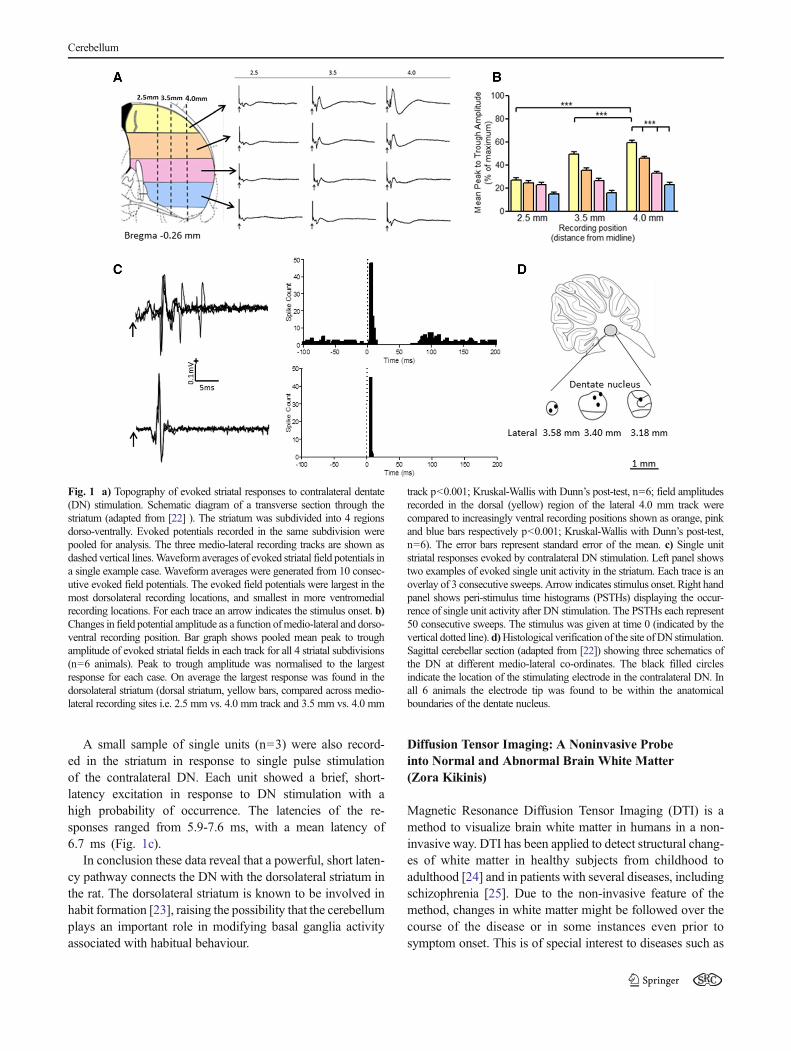
Is DTI a suitable method to explore white matter connectionsbetween the cerebellum, the basal ganglia and the cortex?Images acquired on scanners with magnetic fields of 1.5 and 3Tesla allow the reconstruction of about 17 fiber tracts [36],including three cerebellar tracts, such as the superior cerebellarpeduncle (scp), the middle cerebellar peduncle (mcp) and theinferior cerebellar peduncle (icp) (Fig. 2), but none of thespecific tracts of basal ganglia. Connections to and from thebasal ganglia are extensive and due to the small size of thesubnuclei (few mm in diameter) the reconstruction of the indi-vidual tracts requires image resolution higher than 1mmx 1mmx 1 mm, a better resolution than most of the DTI acquisitionsusing scanners of 3 Tesla can offer today. Sophisticated imagepost-processing methods such as multi-tensor tractography, asopposed to the most commonly used streamline tractography,were applied to explore corticostriatal connections on 3 Tesladata [37], but so far only few studies were successful.
Are connections between the cerebellum, basal ganglia andneocortex abnormal in schizophrenia? It is very likely thatthey are: abnormal corticostriatal connections [37] and abnor-mal connectivity in cerebellar tracts, structural as well asfunctional, were reported in schizophrenia patients [35, 38,39]. So far, only few DTI studies have addressed these con-nections. One reason is that most of the schizophrenia studieshave focused on cortical connections and not cerebellum. Thesecond reason, as mentioned above, is that reconstruction ofthese specific connections in vivo poses technical challengesso far. Advances in imaging methods, either scanning athigher magnetics fields (7 Tesla) or improvement of acquisi-tion techniques and image post-processing methods will allowthe reconstruction of these tracts in the very near future.
Imaging of the Cerebellar Nuclei in Health and Disease(Dagmar Timmann)
Deep cerebellar nuclei have rarely been assessed in humanstudies. Recent studies of our group focused on structural andfunctional magnetic resonance imaging (MRI) of the dentatenuclei in humans. Iron-content of the dentate nuclei is high. Theparamagnetic effect of iron is used to visualize the dentate nucleias hypointensities in susceptibility weighted images (SWI) [40].One future application is to quantify the volume of cerebellarnuclei in patients with degenerative cerebellar disease. This is ofparticular interest in disorders which have known atrophy of thecerebellar nuclei based on histological data. The best knownexamples are spinocerebellar ataxia type 3 (SCA3) andFriedreich’s ataxia (FRDA) [41]. In a first study, we were ableto show atrophy of cerebellar nuclei in patients with FRDAbased on SWI images [42]. Because there is no or little
Cerebellum

degeneration of the cerebellar cortex, standard diagnostic MRIscans frequently show no cerebellar abnormality in FRDA.
Another application of SWI imaging is to determine therelationship between behavioral abnormalities and the locationof lesions of the cerebellar nuclei. In patients with focal cerebellarlesions, for example due to stroke, SWI allows to perform lesion-symptommapping on the level of the dentate nuclei. Here, lesionsare outlined within the dentate nuclei [43]. The same region-of-interest-(ROI)-driven normalization technique is used to performlesion-symptom-mapping, which had initially been developed byDiedrichsen et al. [40] for functionalMRI of the dentate nuclei. Ina first study, we were able to show that more dorsal and rostralparts of the dentate nuclei were related to upper limb ataxia [43].Findings are in good accordance with the dentate hand areashown in recent fMRI studies of our group and anatomical datain monkey discussed below. As yet, in most human cerebellarlesion studies high resolution T1-weighted MR images are avail-able, but not SWI. Cerebellar nuclei are commonly not visible onT1-weighted images. However, some conclusions can still bedrawn on the level of the cerebellar nuclei. Lesions are drawnon the T1-weighted images and include cerebellar cortex andnuclei to various extents. Normalized lesions are superimposedon cerebellar atlas templates which include the cerebellar nuclei,for example the most recent version of the probabilistic atlastemplate of the cerebellum developed by Joern Diedrichsen [40,44]. Using this approach Winfried Ilg’s and our group were ableto show that lesions of the posterolateral hemispheres and thedentate nuclei lead to abnormal treatmill tandem gait. Lesions ofthe medial cerebellum and interposed nuclei, on the other hand,are related to abnormal treatmill walking [45].
On a structural level, dentate nuclei can be visualized usingconventional (1.5 T, 3 T) and ultra-high-field (7 T) MRI.Increasing field strength leads to better spatial resolution [43].On a functional level, it is difficult to achieve robust activationsof the dentate nuclei using conventional field strength. Ultra-high-field MRI together with optimized region-of-interest
(ROI)-based normalizationmethods allow for reliable functionalMRI studies at the level of the dentate nuclei [40]. We foundevidence of a motor somatotopy within the human dentatenuclei [46]. In subsequent studies we were able to show thatdifferent areas within the dentate nuclei contribute to motor andcognitive tasks (verb generation and verbal working memory)[47, 48]. Findings are consistent with Peter Strick’s anatomicaldata in the monkey. His group observed a motor domain withinthe dorso-rostral parts of the dentate nucleus and a non-motordomain within its ventro-caudal parts [49]. In an ongoing studywe investigate different patterns of activations within the cere-bellar cortex and nuclei in spinocerebellar ataxia type 6 (SCA6),which is thought to primarily the cerebellar cortex, and SCA3and FRDA, which affect primarily the cerebellar nuclei [41].
In summary, SWI imaging allows to visualize dentate nucleion individual scans. This has the diagnostic potential to revealatrophy of the cerebellar nuclei in patients with cerebellar degen-eration. Furthermore, lesion-symptom mapping is possible onthe level of subdivisions of the dentate nuclei. Ultra-high-fieldMRI makes reliable fMRI studies of the dentate nuclei possible.In current studies we try to extend fMRI studies to the level ofinterposed nuclei using an eyeblink conditioning paradigm.Ultra-high field fMRI will be a helpful tool to understand theinteractions between the cerebellar cortex and nuclei in futurestudies in healthy subjects and patients with cerebellar disease.
High Field Magnetic Resonance Spectroscopyin Movement Disorders: Biomarker and SurrogateMarker Potential (Gülin Öz)
Proton magnetic resonance spectroscopy (1H MRS) enablesthe non-invasive quantification of a multitude of neurochem-icals in selected brain regions at high and ultra-high fields [50,51]. The neurochemicals that can be reliably quantified at 3tesla (T) and higher fields include neurotransmitters, such asglutamate and γ-aminobutyric acid (GABA), antioxidantsglutathione and ascorbate, and other important metabolitessuch as glutamine, in addition to those that are reliably quan-tified at 1.5 T, namely N-acetylaspartate (NAA), creatine,choline and, depending on acquisition parameters, myo-inosi-tol and lactate. These “neurochemical profiles” are character-istic of brain regions [50] and may provide markers of onset,progression and reversal of pathology in affected brain regionsin many neurological diseases [52] including movement dis-orders. The need for such non-invasive biomarkers of cerebraland cerebellar disease is particularly urgent in the area of pre-clinical and clinical trials for neurodegenerative diseases be-cause they are slowly progressive, show substantial pheno-typic variability and therefore typically necessitate long clin-ical trials with large sample sizes. In addition, clinical outcomemeasures that are routinely used in such trials do not distin-guish symptomatic from disease-modifying effects of drugs.
Fig. 2 Cerebellar tracts. Middle cerebellar peduncle (mcp, colored in red),superior cerebellar peduncle (scp, in blue) and inferior cerebellar peduncle(icp, in green) were reconstructed from DTI images. Structural MRI imageof the brain is in the background (black and white). Posterior view of thebrain in the panel left, side view of the brain in the panel on the right
Cerebellum

A number of recent studies focused on validating suchin vivo markers of neurodegeneration in spinocerebellarataxias (SCAs), hereditary movement disorders that causeatrophy and dysfunction of the cerebellum and in many casesalso the brainstem. An ability to detect neurochemical alter-ations in SCA1, even in spectra from individual patients, wasdemonstrated first at 4 T [53] (Fig. 3) and recently also at thewidely available 3 T platform [54]. In addition, significantcorrelations between the levels of select neurochemicals,namely the neuronal marker NAA, the putative gliosis markermyo-inositol and the neurotransmitter glutamate, and scoreson the validated Scale for the Assessment and Rating ofAtaxia (SARA) were observed [53], demonstrating that theneurochemical levels reflect clinical status in SCA1.Furthermore, MRS has shown potential to distinguish differ-ent SCA genotypes with similar clinical presentation [55].
Parallel studies with mouse models of SCA1 demonstratedthat neurochemical levels measured by 1HMRS are sensitive topre-symptomatic and progressive pathological changes in thecerebellum [56]. Interestingly, the same neurochemicals thatreflected the clinical status in patients with SCA1 (NAA,myo-inositol, glutamate) were also those that showed the stron-gest correlations with semi-quantitative pathology measures inthe mouse model, indicating these as robust markers of theneurodegenerative process [56]. Later, studies with a condition-al transgenic SCA1 model demonstrated the sensitivity of 1HMRS measured neurochemical levels to disease reversal.Namely, upon suppression of transgene expression in this mod-el, both the cerebellar pathology and abnormal neurochemicallevels returned towards normal [57]. Finally, studies with aknock-in SCA1model that displaysmilder cerebellar pathologythan the transgenic line used in prior studies showed thatneurochemical alterations can be detected prior to overt pathol-ogy in SCA1 [58]. To summarize, 1) 1H MRS detects parallelneurochemical alterations in patients with SCA1 and SCA1mouse models, 2) the same neurochemicals (NAA, myo-inosi-tol, glutamate) reflect clinical status and pathological
progression in SCA1, 3) neurochemical alterations are detect-able prior to ataxia onset and overt pathology, and 4) reversal ofneurochemical alterations with treatment is detectable byMRS.
The utility of MRS markers to detect neurochemical alter-ations and treatment response was also shown in Parkinson’sdisease (PD), the most common movement disorder. Early in-volvement of caudal brainstem in PD, even prior to the charac-teristic degeneration of nigrostriatal dopaminergic neurons, wassuggested about 10 years ago based on detailed pathologicalinvestigations [59] and has been gaining wide acceptance.Motivated by this work, neurochemical alterations in thebrainstem and striatum in early-moderate stage PDwere recentlyinvestigated at the ultra-high field of 7 T. This study uncoveredan elevation in the inhibitory neurotransmitter GABA in the pons[60], in addition to elevated striatal GABA levels previouslyshown in postmortem investigations [61]. A more pronouncedGABA elevation in the pons than in the putamen was consistentwith an earlier involvement of the lower brainstem in the diseasepathology. Therefore, the altered GABAergic tone demonstratedby MRS in the lower brainstem and striatum in early-moderatePD may underlie disease pathogenesis and may provide a bio-marker for disease staging. Another recent study focused on apotentially useful antioxidant therapy in PD. Reductions in thelevels of the major antioxidant glutathione (GSH) were reportedin PD [62], indicating the involvement of oxidative stress in thepathophysiology of the disease. In a pilot investigation, ultra-high field MRS was used to monitor increases in brain GSHlevels in patients with PD and healthy controls in response to asingle, intravenous administration of N-acetylcysteine, a knownprecursor to GSH [63]. This study demonstrated the potentialutility of MRS for monitoring treatment response by noninva-sively measuring antioxidant levels in the brain.
The importance of spectral quality for the robustness of theabove findings and reliable neurochemical quantification can-not be overemphasized. Thus, in all the above cited studies, inhouse developed, state-of-the art MRS methods were utilized.The critical need for standardization of MRSmethodology for
Fig. 3 MR spectral differences in a patient with SCA1 relative to ahealthy control. Localized proton MR spectra (STEAM, TE=5 ms,TR=4.5 s, 128 transients) were obtained from the volume-of interest inthe cerebellar hemisphere shown on the T2 weighted images. The
alterations in total N-acetylaspartate (tNAA), glutamate (Glu), myo-ino-sitol (myo-Ins) and total creatine (tCr) visible in the spectra of the patientare shown. (Reprinted, with permission, from reference [27].)
Cerebellum

robust clinical applications was recently emphasized and thefeasibility of implementing advanced MRS methods on clin-ical scanners was demonstrated in a multi-site setting in arecent consensus effort [52].
Autism, Neural Timing, and the Cerebellum(James Ashe and Khalaf Bushara)
Autism is a severe developmental disorder characterized byabnormal social interaction, impaired language use and devel-opment, stereotypical repetitive movements, and resistance tochange. It is remarkable that there is as yet no consensus on theneurological basis of this disorder first described in 1943 butnow affecting more than 1 in every 100 children in the UnitedStates or in Europe. Although abnormalities have been docu-mented in many different regions of the brain in those who diedwith this disorder, the most common abnormality has been a30-40% reduction in the population of Purkinje cells in lobulesVI and VII of the cerebellum. It is not immediately obviousbased on the conventional understanding of cerebellar physiol-ogy how disrupting its function might lead to the types ofbehavioral disturbances in autism. Welsh and colleagues [64]have provided a possible solution to this dilemma by proposingthat disruption of a timing signal within the olivo-cerebellarcircuit in infancy might lead to abnormal development ofmotor, cognitive and emotional processing [65].
There is compelling evidence from experiments in verte-brate animals over a period of 40 years that cells in the inferiorolive provide a rhythmic, synchronous output signal of 8–13Hz that is conveyed via climbing fibres to the Purkinje cellsto produce complex spikes. Despite these data there has beenskepticism about the role and importance of a rhythmic signalgenerated in the inferior olive primarily because the fewexperiments that had addressed the issue in non-human pri-mates have been either inconclusive or unsupportive depend-ing on one’s perspective. We have recently demonstratedusing functional neuroimaging in human subjects that theinferior olive is activated during the perception of the temporal[66] but not the non-temporal features [67] of changing visualstimuli. Furthermore, the sensitivity of the inferior olive tochanges in stimulus timing is independent of awareness [68],which suggests that this structure may contribute to the tem-poral properties that underlie classical conditioning and im-plicit learning.
We believe that lesions of the olivo-cerebellum either pre-natally or in early infancy may disrupt the neural processing oftemporal information and lead to the abnormalities of motor,cognitive and emotional behavior in autism. Our next step inexploring this hypothesis is to attempt to reproduce in micesome of the behavioral abnormalities characteristic of autismby genetically engineering mice to produce focal ablations ofpopulations of Purkinje cells in utero.
Acknowledgements This workshop was supported by the project“CEITEC - Central European Institute of Technology” (CZ.1.05/1.1.00/02.0068) from European Regional Development Fund and byMinistry ofHealth of the Czech Republic / Ministry of Health Departmental Researchand Development Program III (2010–2015) NT/13437.
The second section of this paper was supported by the MRC (grantG1100626 and a studentship to ML) and the BBSRC (Grant BB/G012717/1).
The fourth section of this paper was supported by the DFG (DFG TI239/10-1, 10–2) and EU (Marie Curie Initial Training Network, ITN).
The preparation of the fifth section of this manuscript was supportedby the National Institute of Neurological Disorders and Stroke (NINDS)grant R01 NS070815 (GÖ). The Center for Magnetic Resonance Re-search at the University of Minnesota is supported by National Center forResearch Resources (NCRR) biotechnology research resource grant P41RR008079, National Institute of Biomedical Imaging andBioengineering(NIBIB) grant P41 EB015894 and the Institutional Center Cores forAdvanced Neuroimaging award P30 NS076408.
Conflict of Interest There are no potential conflicts of interest in thesubmission and no financial and personal relationship that might bias ourwork.
References
1. Harrington DL, Lee RR, Boyd LA, Rapcsak SZ, Knight RT. Does therepresentation of time depend on the cerebellum? Effect of cerebellarstroke. Brain. 2004;127(Pt 3):561–74.
2. Beudel M, Galama S, Leenders KL, de Jong BM. Time estimation inParkinson's disease and degenerative cerebellar disease. Neuroreport.2008;19(10):1055–8.
3. Ivry RB, Spencer RM. The neural representation of time. Curr OpinNeurobiol. 2004;14(2):225–32.
4. Dreher JC, Grafman J. The roles of the cerebellum and basal gangliain timing and error prediction. Eur J Neurosci. 2002;16(8):1609–19.
5. Iacoboni M. Playing tennis with the cerebellum. Nat Neurosci.2001;4(6):555–6.
6. JahanshahiM, Jones CR, Zijlmans J, et al. Dopaminergic modulationof striato-frontal connectivity during motor timing in Parkinson'sdisease. Brain. 2010;133(Pt 3):727–45.
7. Bares M, Lungu OV, Liu T, Waechter T, Gomez CM, Ashe J. TheNeural Substrate of Predictive Motor Timing in SpinocerebellarAtaxia. Cerebellum. 2011;10(2):233–44.
8. Mauk MD, Buonomano DV. The neural basis of temporal process-ing. Annu Rev Neurosci. 2004;27:307–40.
9. Vogel M. The cerebellum. Am J Psychiatry. 2005;162(7):1253.10. Manto M et al. Consensus Paper: Roles of the Cerebellum in Motor
Control- The Diversity of Ideas on Cerebellar Involvement inMovement. Cerebellum. 2012;11(2):457–87.
11. Murdoch BE. The cerebellum and language: Historical perspectiveand review. Cortex. 2010;46(7):858–68.
12. Cerminara NL, Aoki H, Loft M, Sugihara I, Apps R. Structural Basis ofCerebellar Microcircuits in the Rat. J Neurosci. 2013;33(42):16427–42.
13. Pijpers A, Apps R, Pardoe J, Voogd J, Ruigrok TJH. Precise SpatialRelationships between Mossy Fibers and Climbing Fibers in RatCerebellar Cortical Zones. J Neurosci. 2006;26(46):12067–80.
14. Voogd J et al. In: Manto M, editor. Cerebellar Nuclei and the InferiorOlivary Nuclei: Organization and Connections, in Handbook of theCerebellum and Cerebellar Disorders. Netherlands: Springer Science:Dordrecht; 2013. p. 377–436.
15. Alexander GE, Delong MR, Strick PL. Parallel organization offunctionally segrated circuits linking basal gangli and cortex. AnnRev Neurosci. 1986;9:357–81.
Cerebellum

16. Percheron GG, Francois C, Talbi B, Yelnik J, Fénelon G. The primatemotor thalamus. Brain Res Rev. 1996;22(2):93–181.
17. Kuramoto E, Furuta T, Nakamura KC, Unzai T, Hioki H, Kaneko T.Two Types of Thalamocortical Projections from the Motor ThalamicNuclei of the Rat: A Single Neuron-Tracing Study Using ViralVectors. Cereb Cortex. 2009;19(9):2065–77.
18. Groenewegen HJ,WitterMP. In: Paxinos G, editor. Thalamus, in Therat nervous system. San Diego: Elsevier; 2004. p. 407–53.
19. Ichinohe N, Iwatsuki H, Shoumura K. Intrastriatal targets of projec-tion fibers from the central lateral nucleus of the rat thalamus.Neurosci Letts. 2001;302(2–3):105–8.
20. Ichinohe N, Mori F, Shoumura K. A di-synaptic projection from thelateral cerebellar nucleus to the laterodorsal part of the striatum viathe central lateral nucleus of the thalamus in the rat. Brain Res.2000;880(1–2):191–7.
21. Hoshi E, Tremblay L, Féger J, Carras PL, Strick PL. The cerebellumcommunicates with the basal ganglia. Nat Neurosci. 2005;8(11):1491–3.
22. Paxinos GWC. The rat brain in stereotaxic co-ordinates. 4th ed.Florida, USA: Academic Press; 1998.
23. Yin HH, Knowlton BJ, Balleine BW. Lesions of dorsolateral striatumpreserve outcome expectancy but disrupt habit formation in instru-mental learning. Eur J Neurosci. 2004;19(1):181–9.
24. Lebel C, Beaulieu C. Longitudinal development of human brainwiring continues from childhood into adulthood. J Neurosci.2011;31(30):10937–47.
25. White T, NelsonM, Lim KO. Diffusion tensor imaging in psychiatricdisorders. Top Magn Reson Imaging. 2008;19(2):97–109.
26. Seal ML, Yucel M, Fornito A, Wood SJ, Harrison BJ, Walterfang M,et al. Abnormal white matter microstructure in schizophrenia: avoxelwise analysis of axial and radial diffusivity. SchizophreniaRes. 2008;101(1–3):106–10.
27. Song SK, Sun SW, Ramsbottom MJ, Chang C, Russell J, Cross AH.Dysmyelination revealed through MRI as increased radial (but un-changed axial) diffusion of water. Neuroimage. 2002;17(3):1429–36.
28. Pasternak O, Westin CF, Bouix S, Seidman LJ, Goldstein JM, WooTU, et al. Excessive extracellular volume reveals a neurodegenerativepattern in schizophrenia onset. J Neurosci. 2012;32(48):17365–72.
29. Clemm von Hohenberg C, Pasternak O, Kubicki M, Ballinger T, VuMA, Swisher T, et al. White Matter Microstructure in Individuals atClinical High Risk of Psychosis: A Whole-Brain Diffusion TensorImaging Study. Schizophrenia Bull. 2014;40(4):895–903.
30. Radoeva PD, Coman IL, Antshel KM, Fremont W, McCarthy CS,Kotkar A, et al. Atlas-based white matter analysis in individuals withvelo-cardio-facial syndrome (22q11.2 deletion syndrome) and unaf-fected siblings. Behav Brain Funct. 2012;8(1):38.
31. Kikinis Z, Asami T, Bouix S, Finn CT, Ballinger T, Tworog-Dube E,et al. Reduced fractional anisotropy and axial diffusivity in whitematter in 22q11.2 deletion syndrome: A pilot study. SchizophreniaRes. 2012;141(1):35–9.
32. Ellison-Wright I, Bullmore E. Meta-analysis of diffusion tensor imagingstudies in schizophrenia. Schizophrenia Res. 2009;108(1–3):3–10.
33. Melonakos ED, Shenton ME, Rathi Y, Terry DP, Bouix S, KubickiM. Voxel-based morphometry (VBM) studies in schizophrenia-canwhite matter changes be reliably detected with VBM? PsychiatryRes. 2011;193(2):65–70.
34. White T, Ehrlich S, Ho BC, Manoach DS, Caprihan A, Schulz SC,et al. Spatial characteristics of white matter abnormalities in schizo-phrenia. Schizophrenia Bull. 2013;39(5):1077–86.
35. Huttlova J, Kikinis Z, Kerkovsky M, Bouix S, Vu MA, Makris N,et al. Abnormalities in Myelination of the Superior CerebellarPeduncle in Patients with Schizophrenia and Deficits in MovementSequencing. Cerebellum. 2014;13(4):415–24.
36. Wakana S, Jiang H, Nagae-Poetscher LM, van Zijl PC, Mori S. Fibertract-based atlas of human white matter anatomy. Radiology.2004;230(1):77–87.
37. Quan M, Lee SH, Kubicki M, Kikinis Z, Rathi Y, Seidman LJ, et al.Whitematter tract abnormalities between rostral middle frontal gyrus,inferior frontal gyrus and striatum in first-episode schizophrenia.Schizophrenia Res. 2013;145(1–3):1–10.
38. Liu H, Fan G, Xu K, Wang F. Changes in cerebellar functionalconnectivity and anatomical connectivity in schizophrenia: a com-bined resting-state functional MRI and diffusion tensor imagingstudy. J Magn Res Imaging. 2011;34(6):1430–8.
39. Kasparek T, Rehulova J, KerkovskyM, Sprlakova A,Mechl M,MiklM. Cortico-cerebellar functional connectivity and sequencing ofmovements in schizophrenia. BMC Psychiatr. 2012;12:17.
40. Diedrichsen J, Maderwald S, Küper M, Thürling M, Rabe K,Gizewski ER, et al. Imaging the deep cerebellar nuclei: a probabilisticatlas and normalization procedure. Neuroimage. 2011;54:1786–94.
41. Koeppen AH, Ramirez RL, Bjork ST, Bauer P, Feustel PJ. Thereciprocal cerebellar circuitry in human hereditary ataxia.Cerebellum. 2013;12:493–503.
42. Rabe K, Kraff O,MinneropM, Beck A, Schöls L, LaddM, TimmannD. Cerebellar pathology in Friedreich’s Ataxia: Atrophied nuclei withnormal iron content. Submitted.
43. Maderwald S, Thürling M, Küper M, Theysohn N, Müller O, BeckA, et al. Direct visualization of cerebellar nuclei in patients with focalcerebellar lesions and its application for lesion-symptom mapping.Neuroimage. 2012;63:1421–31.
44. Diedrichsen J, Balsters JH, Flavell J, Cussans E, Ramnani N. Aprobabilistic MR atlas of the human cerebellum. Neuroimage.2009;46:39–46.
45. Ilg W, Christensen A, Mueller OM, Goericke SL, Giese MA,Timmann D. Effects of cerebellar lesions on working memoryinteracting with motor tasks of different complexities. JNeurophysiol. 2013;110:2337–49.
46. Küper M, Thürling M, Stefanescu R, Maderwald S, Roths J, Elles HG,et al. Evidence for a motor somatotopy in the cerebellar dentate nucleus–an FMRI study in humans. Hum Brain Mapp. 2012;33:2741–9.
47. Thürling M, Küper M, Stefanescu R, Maderwald S, Gizewski ER,LaddME, et al. Activation of the dentate nucleus in a verb generationtask: A 7 T MRI study. Neuroimage. 2011;57:1184–91.
48. Thürling M, Hautzel H, Küper M, Stefanescu MR, Maderwald S,Ladd ME, et al. Involvement of the cerebellar cortex and nuclei inverbal and visuospatial working memory: a 7 T fMRI study.Neuroimage. 2012;62:1537–50.
49. Strick PL, Dum RP, Fiez JA. Cerebellum and nonmotor function.Annu Rev Neurosci. 2009;32:413–34.
50. Emir UE, Auerbach EJ, Moortele PF, Marjańska M, Ugurbil K,Terpstra M, et al. Regional neurochemical profiles in the human brainmeasured by 1HMRS at 7 Tusing local B1 shimming. NMRBiomed.2012;25:152–60.
51. Öz G. MR Spectroscopy in Health and Disease. In: Manto M, GruolDL, Schmahmann JD, Koibuchi N, Rossi F, editors. Handbook of theCerebellum and Cerebellar Disorders. Dordrecht: Springer; 2013. p.713–33.
52. Öz G, Alger JR, Barker PB, Bartha R, Bizzi A, Boesch C, et al.Clinical Proton MR Spectroscopy in Central Nervous SystemDisorders. Radiology. 2014;270(3):658–79.
53. Öz G, Hutter D, Tkáč I, Clark HB, Gross MD, Jiang H, et al.Neurochemical alterations in spinocerebellar ataxia type 1 and theircorrelations with clinical status. Mov Disord. 2010;25:1253–61.
54. Emir UE, Hutter D, Bushara KO, Gomez CM, Eberly LE and Öz G.MRS Biomarkers of Neurodegeneration in Spinocerebellar Ataxiatype 1 (SCA1): Current and Future Potential. 20th Scientific Meetingof the ISMRM. Melbourne, Australia; 2012. pp. 1802.
55. ÖzG, Iltis I, Hutter D, ThomasW, Bushara KO, Gomez CM. DistinctNeurochemical Profiles of Spinocerebellar Ataxias 1, 2, 6, andCerebellar Multiple System Atrophy. Cerebellum. 2011;10:208–17.
56. Öz G, Nelson CD, Koski DM, Henry PG, Marjańska M, DeelchandDK, et al. Noninvasive detection of presymptomatic and progressive
Cerebellum

neurodegeneration in a mouse model of spinocerebellar ataxia type 1.J Neurosci. 2010;30:3831–8.
57. ÖzG, VollmersML, Nelson CD, Shanley R, Eberly LE, Orr HT, et al.In vivo monitoring of recovery from neurodegeneration in condition-al transgenic SCA1 mice. Exp Neurol. 2011;232:290–8.
58. Emir UE, Brent Clark H, Vollmers ML, Eberly LE, Öz G. Non-invasive detection of neurochemical changes prior to overt pathologyin a mouse model of spinocerebellar ataxia type 1. J Neurochem.2013;127:660–8.
59. Braak H, Del Tredici K, Rub U, de Vos RA, Jansen Steur EN, BraakE. Staging of brain pathology related to sporadic Parkinson's disease.Neurobiol Aging. 2003;24:197–211.
60. Emir UE, Tuite PJ, Öz G. Elevated pontine and putamenal GABAlevels in mild-moderate Parkinson disease detected by 7 tesla protonMRS. PLoS ONE. 2012;7:e30918.
61. Kish SJ, Rajput A, Gilbert J, Rozdilsky B, Chang LJ, Shannak K,et al. Elevated gamma-aminobutyric acid level in striatal but notextrastriatal brain regions in Parkinson's disease: correlation withstriatal dopamine loss. Ann Neurol. 1986;20:26–31.
62. Sian J, Dexter DT, Lees AJ, Daniel S, Agid Y, Javoy-Agid F, et al.Alterations in glutathione levels in Parkinson's disease and otherneurodegenerative disorders affecting basal ganglia. Ann Neurol.1994;36:348–55.
63. Holmay MJ, Terpstra M, Coles LD, Mishra U, Ahlskog M, Öz G,et al. N-acetylcysteine Boosts Brain and Blood Glutathione inGaucher and Parkinson Diseases. Clin Neuropharmacol. 2013;36:103–6.
64. Welsh JP, Ahn ES, Placantonakis DG. Is autism due to braindesynchronization? Int J Dev Neurosci. 2005;23:253–63.
65. Schmahmann JD. Disorders of the cerebellum: ataxia, dysmetria ofthought, and the cerebellar cognitive affective syndrome. JNeuropsychiatry Clin Neurosci. 2004;16:367–78.
66. XuD, Liu T, Ashe J, Bushara KO. Role of the olivo-cerebellar systemin timing. The J Neurosci. 2006;26:5990–5.
67. Liu T, Xu D, Ashe J, Bushara KO. Specificity of inferior oliveresponse to stimulus timing. J Neurophysiol. 2008;100:1557–61.
68. Wu X, Ashe J, Bushara KO. Role of olivocerebellar system in timingwithout awareness. Proc Natl Acad Sci U S A. 2011;108:13818–22.
Cerebellum
Related Documents