Welcome message from author
This document is posted to help you gain knowledge. Please leave a comment to let me know what you think about it! Share it to your friends and learn new things together.
Transcript

SMITHSONIAN INSTITUTION
UNITED STATES NATIONAL MUSEUM
PROCEEDINGS
OF THE
UNITED STATES NATIONAL MUSEUM
VOLUME 115
NUMBERS 3476-3493
\m
UNITED STATES
GOVERNMENT PRINTING OFFICE
WASHINGTON : 1965
Publications of the United States National Museum
The scientific publications of the United States National Museum include two
series, Proceedings of the United States National Museum and United States
National Museum Bulletin.
In these series are published original articles and monographs dealing with
the collections and work of the Museum and setting forth newly acquired facts
in the fields of anthropology, biology, geology, history, and technology. Copies
of each publication are distributed to libraries and scientific organizations and
to specialists and others interested in the various subjects.
The Proceedings, begun in 1878, are intended for the publication, in separate
form, of shorter papers. These are gathered in volumes, octavo in size, with
the publication date of each paper recorded in the table of contents of the volume.
In the Bulletin series, the first of which was issued in 1875, appear longer,
separate publications consisting of monographs (occasionally in several parts)
and volumes in which are collected works on related subjects. Bulletins are
either octovo or quarto in size, depending on the needs of the presentation.
Since 1902 papers relating to the botanical collections of the Museum have been
published in the Bulletin series under the heading Contributions from the United
States National Herbarium.
Frank A. Taylor,
Director, United States National Museum.II
CONTENTS
Tapes
Blake, Doris H. Notes on new and old species of Alticinae
(Coleoptera) from the West Indies. Twenty-five figures.
No. 3477, published February 28, 1964 9-30
New genera : Nesaecrepida, Sidfaya.
New species: Aphthona criicijera, A. lepta, A. insularis, A.
lamprocyanea, Longilarsus cylindricus, L. chlanidotus, L. oak-
leyi, L. atypicus, Hermaeophaga jamaicensis, Hamilactica
poitoricensis, Nesaecrepida rufomarginata, Homoschema xaidho-
cyaneum, Chaeiocnema cyanoptera, Pseudoepitrix rugosa, P.
brasilievsis, Sidfaya polutima.
New series: Sidfaya punctatissima.
Clarke, J. F. Gates. Neotropical Microlepidoptera, III.
Restriction of Gonionota inelobaphes Walsingham with
descriptions of new species (Lepidoptera: Oecophoridae)
.
Seven figures and three plates (one color). No. 3480,
published March 17, 1964 61-84
New species: Gonionota coiHrasta, G. dissita, G. deierminata, G.
bourquini, G. incontigua, G. extima, G. periphereia, G. excavata,
G. hyptiotes, G. fimbriata.
Drake, Carl J., and Lattin, John D. American species of
the lacebug genus Acalypta (Hemiptera: Tingidae).
Fifteen plates. No. 3486, published December 31, 1963 . 331-345
Duckworth, W. Donald. A new Brazilian moth of the
genus Gonioterma with notes on related species (Lepidop-
tera: Stenomidae). Three figures. No. 3488, published
March 17, 1964 381-390
New species: Gonioterma exquisita.
New combinations: Gonioterma chlorina, G. aesiocopia, G. argi-
cerauna, G. algosa, G. fastigata, G. bryophanes.
Estrada, Emilio, Meggers, Betty J., and Evans, Clif-
ford. The Jambeli culture of south coastal Ecuador.
Forty-two figures and twelve plates. No. 3492, pub-lished September 25, 1964 483-558
IV PROCEEDINGS OF THE NATIONAL MUSEUM
Pages
Field, William D. Moths of the genus Rhabdatomis Dyar(Arctiidae: Lithosiinae) . Six plates (containing thirty-
three figures). No. 3479, published February 28, 1964 . 47-00
New species: Rhabdatomis dognini, R. draudti, R. knabi, R. ex-
tensa, R. fasseli.
New combinations: Rhabdatomis mandana, R. melinda, R. pueblae,
R. laudamia, R. cora, R. peruviava, R. pusa.
New combination and new status: Rhabdatomis cora coroides.
Flint, Oliver S., Jr. Notes on some Nearctic Psycho-
myiidae with special reference to their larvae (Trichop-
tera). Five figures. No. 3491, published February 28,
1964 467-481
New species: Nydiophylax nephophilus.
New combination: Cyrnelhis fraternus.
New form: Genus C.
Garrick, J. A. F. Additional information on the morphol-
ogy of an embryo whale shark. Four plates. No. 3476,
published February 28, 1964 1-8
Grainger, E. H. Asteroidea of the Blue Dolphin expedi-
tions to Labrador. Four figures. No. 3478, published
February 28, 1964 31-46
Hart, Josephine F. L. Shrimps of the genus Betaeus on
the Pacific Coast of North America with descriptions of
three new species. Eighty figures and two plates. No.
3490, published February 28, 1964 431-466
New species: Betaeus macginitieae, B. gracilis, B. setosus.
Hodges, Ronald W. A review of the North American
moths of the family Walshiidae (Lepidoptera: Gelechio-
idea). Sixty-six figures. No. 3485, published March 17,
1964 289-330
New genera: Neoploca, Synploca, Nepotula, Obithome.
New species: Neoploca corusca, Synploca gumia, Nepotula secura,
Aeaea juvantis, A. dulcedo, A. venifica, A. venatrix, A. stipator,
A. risor, A. victor, A. sagana, A. rhynchosiae, Stilbosis nubila,
Sorhagenia daedala.
New combinations: Aeaea extensa, Sorhagenia nimbosa, Obithome
punctiferella.
KoRMiLEv, Nicholas A. Notes on Aradidae in the U.S.
National Museum : III, Subfamily Mezirinae (Hemiptera)
.
Seven figures. No. 3483, published February 28, 1964 . . 245-258
New species: Qinyphus saileri, Mezira luteonotata, M. championi,
M. mexicana, M. coslalimai, M. carioca, M. guianensis, M.barberi, M. paralata.

CONTENTS V
Pages
Kramer, James P. A generic revision of the leafhopper
subfamily Neocoelidiinae (Homoptera: Cicadellidae).
One hundred and fourteen figures. No. 3484, pubhshed
March 17, 1964 259-288
New genera: Tozzila, Xiqilliba.
New species: Tozzita ips, Xiqilliba bellator, Coelana drakei,
Nelidina iaeniola.
New combination: Neocoelidia fuscodorsata, N. verecunda, Coe-
lidiana coronata, C. croceata.
New status: Coelana DeLong, Cocoelidia DeLong, Nelidina De-
Long, Coelella DeLong.
Lewis, Alan G. Caligoid copepods (Crustacea) of the
Hawaiian Islands: Parasitic on fishes of the family Acan-
thuridae. Twenty-four figures. No. 3482, published
February 28, 1964 137-244
New species: Caligus kala, C. flexispina, C. randalli, C. ligatus,
C. kalumai, Anuretes nienehune, Dentigryps bifurcatus.
Lewis, Alan G. The caligid copepod genus Dentigryps
(Crustacea: Caligoida). Thirteen figures. No. 3487,
published March 17, 1964 347-380
New species: Dentigryps ulna, D. litus.
Shoemaker, Clarence R. Seven new amphipods from the
West Coast of North America with notes on some unusual
Species. Fifteen figures. No. 3489, published March 17,
1964 391-430
New genus: Kyska.
New species: Kyska dalli, Metopa stelleri, Mesometopa sinuata,
Metopelloides dubia, Syrrhoe longifrons, Sympleustes cornigera,
Anisogaminarus schmitli.
Springer, Victor G. A revision of the carcharhinid shark
genera Scoliodon, Loxodon, and Rhizoprionodon. Four-
teen figures and two plates. No. 3493, published Septem-
ber 1, 1964 559-632
New species: Rhizoprionodon {Protozygaena) oligolinx.
Sublette, James E. Chironomid midges of Cahfornia. II,
Tanypodinae, Podonominae, and Diamesinae. Seven
figures. No. 3481, published February 28, 1964 .... 85-136
New species: Pentaneura (Pentaneura) fluminalis, P. (P.) goniodes,
P. (P.) chrysos, P. (P.) comosa, P. (P.) thryptica, P. (P.) lyra,
P. (P.) inyoensis, P. (P.) sequoiaensis, P. (P.) smithae, Ana-topynia {Anatopynia) submarginella, A. {Macropelopia) aclines, A.
{Psectrotanypus) eumorpha, Tanypus carinatus, T. imperialis,
T. parastellatus, T. grodhausi, T. neopunctipennis, Procladius
barbatulus P. freemani, P. denticulatus, Pseudodiamesa (Pseudo-
diamesa) diastena.


Proceedings of
the United States
National MuseumSMITHSONIAN INSTITUTION • WASHINGTON, D.C.
Volume 115 1964 Number 3476
ADDITIONAL INFORMATION ON THE MORPHOLOGYOF AN EMBRYO WHALE SHARK
By J. A. F. Garrick^
An embryo whale shark, Rhincodon typus Smith, kindly loaned
by the Marine Laboratory, Texas Game and Fish Commission, Rock-
port, shows several notable differences in proportional dimensions
and other features as compared with accounts of adult specimens.
To describe these differences is the purpose of this paper.
The embryo, 350 mm. in total length, is one that has been removed
from an egg-case trawled from 31 fathoms in the Gulf of Mexico
about 130 miles south of Port Isabel, Texas, on June 29, 1953. This
specimen, believed to be the only embryo whale shark available,
has been reported previously by Breuer (1954), Baughman (1955),
and Reid (1957), who published dimensions of it and discussed its
trunk ridges and oronasal groove. Reid also presented a figure of
the underside of the head, while Breuer's and Baughman's accounts
each included a photograph of the specimen and its egg-case. Tosupplement these abeady published figures, I submit here five addi-
tional illustrations of the embryo whale shai'k and its dermal denticles.
For these drawings I am greatly indebted to the skill of Mrs. FanuyPhiUips.
' Division of Fishes, U.S. National Museum. This research was supported by contracts between the
Smithsonian Institution and the Atomic Energy Commission (A EC (30-1) 2409) and the Office of Naval
Research (NONR 1354 (09)).

2 PROCEEDINGS OF THE NATIONAL MUSEUM vol. 115
Proportional differences.—In the following account the fea-
tures of the embryo whale shark are compared for the most part
with those of the adult (total length 17' 3") from Acapulco, Mexico,
figm-ed in Bigelow and Schroeder (1948). As dimensions of both
these specimens are already available in their respective accounts,
there is no need to reproduce such measiu-ements here. Instead,
I shall give, in general terms, the major differences between the
embryo and adult, followed in each case by a figm-e in parentheses
which is the proportional difference expressed as a percentage of total
length.
The differences are: the adult is slightly broader headed (0.5), longer
headed (1.4), and noticeably shorter tailed (6.3); the adidt eye is strik-
ingly smaller (0.9), but the gUl-openings are longer (0.3 to 2.2); the first
dorsal (2.6), second dorsal (8.4), anal (7.8), and pelvic fins (3.2)
are fm-ther rearward in the adult; the first dorsal fin is proportionately
higher (1.2), but its base is shorter (0.9) in the adult (however, the
reverse is the case for both height and base length—about 2.0 and 0.3
respectivel}^—in the second dorsal and anal fins); the distance between
the fu'st and second dorsal fins and between the anal fin and subcaudal
origin are longer in the adult (4.8 and 3.4 respectively); the pectoral
fin of the adult has a much longer distal margin (5.5) than that of the
embryo, though the anterior margins are comparable; the lengths of
the upper and lower lobes of the caudal fin are considerably shorter
(7.3 and 4.2 respectively) in the adult than in the embryo.
The above differences are indicative of the growth change which
the whale shark undergoes. Understanding such growth change is
important in studying sharks because of the frequent need to rely on
proportional dimensions to distinguish species. The pattern of growth
change is by no means identical in aU sharks, but there do appear to
be conmion featm-es (Beebe and Tee-Van, 1941, p. 107; Maschlanka,
1955, p. 12; S. Springer, 1960, p. 9; Garrick, 1960, p. 546), such as
accelerated growth in the trunk region, as compared to the head andtail, which thus proportionately decrease with increasing total length;
a tendency for the pectoral fin to increase its relative length or at
least remain stable (though Carcharhinus longimanus (Poey) shows
a relative shortening of pectoral fin from juvenile to adult, as noted
by S. Springer, 1960, p. 9); a broadening of the head region; and a
noticeable decrease in eye diameter. Dorsal fin heights tend to
increase relatively in the galeoid sharks (but C. longimanus is again
an exception), while in the squaloid sharks the reverse generally
holds true (Garrick, 1960, p. 548).
The indicated growth change of the embryo whale shark fits the
above pattern reasonably well except that the head length in the
embryo is relatively shorter than that of the adult. This may be
EMBRYO WHALE SHARK—GARRICK 3
only ail apparent difference for the following reason. The dorsal lobe
of the caudal fin of the embryo is raised only slightly from the
horizontal axis of the body; hence, the posterior margin between the
dorsal and ventral lobes is deeply notched, the angle being less than
90°, In the adult the caudal fin is lunate, the dorsal lobe being raised
steeply from the horizontal axis, with the result that the posterior
margin is only slightly concave. It follows that the change leading
from the slightly raised dorsal lobe in the embryo to the steeply
raised lobe in the adult woidd yield relative total lengths which are
not strictly comparable, and thus this difference woidd provide bias
in proportional lengths of structures, such as head length, if calculated
in terms of the total length. Accordingly, one would expect the head
length of the embryo to have a lower relative value in terms of total
length than is the case for the adult. Better comparison is afforded
by examining the head length in terms of the length to the upper
caudal origin—this shows the relative head length in embryo andadidt to be the same, which is nearer to the actual situation in mostother sharks.
The need for caution in extrapolating proportional dimensions of
small specimens is demonstrated by the different growth rates oper-
ating on the first dorsal fin of the embryo as compared with the second
dorsal and anal. Dimensions of the first dorsal fin in the embryoand in the adult indicate that the rate of vertical growth is proportion-
ately faster than that of horizontal, whereas in the second dorsal
and anal fins the horizontal growth is faster. A similar situation
has been described for Etmoptertis baxteri Garrick (Garrick, 1960,
p. 548) and it may be relatively common. The lengths of the free
rear tips of the dorsal and anal fins compared with theh bases also
show considerable change with growth. In the embryo, these free
tips are relatively short (about 4.0 in base in the first dorsal fin) butin the adidt they are much longer (about 1.4 in base in the first dorsal).
Another change affecting the comparison of all fins is the usual
tendency for fin tips to become relatively pointed in the adidt, whereas
in the embryo they are more rounded or blunt tipped (V. G. Springer,
1961, p. 480, gives an example of this in Mustelus norrisi Springer).
The tip of the dorsal lobe of the caudal fin in the embryo is distinctly
notched, presumably representing the subterminal notch, which is
not evident in the adult.
Dermal ridges.—As noted by Reid (1957, p. 158), the embryowhale shark has a longitudinal dermal ridge originating on each side
of the head and dividing, above the end of the pectoral fin, into tworidges which continue posteriorly. Reid identified this ridge as one
corresponding to an upper divided ridge in the adult. Tiie adult
has, in addition, a lower ridge which extends the whole length of its
4 PROCEEDINGS OF THE NATIONAL MUSEUM vol. iis
body and forms a keel on the peduncle and anterior part of the caudal
fin. I interpret the lower half of the divided ridge in the embryo to
be the same as the lowermost ridge in the adult, since posteriorly
the lower ridge forms the keel on the peduncle and caudal fin.
This means that, at a later date, a third ridge must appear above the
lower one in the embryo. Similar longitudinal ridges occur in somemembers of the family Orectolobidae. The embryo also has a mid-
dorsal ridge which extends from the level of the first gill-opening to
the origin of the first dorsal fin and possibly is present between the
first and second dorsal fins. Adults have been described with and
without a middorsal ridge.
Precaudal pits.—The embryo has a prominent upper precaudal pit,
with a notably wide, transverse front margin. There is also a small
but distinct lower precaudal pit. Adults are described as having the
upper pit but lacking the lower.
Nostrils.—Reid (1957, p. 158) reported that each nostril in the em-
bryo is connected to the mouth by a distinct furrow—a character fre-
quently used to support the view that the whale shark is closely related
to, or belongs in, the family Orectolobidae.
On the basis of an adult specimen, Barnard (1935, p. 649) disputed
this view. Without wishing to enter the controversy, I confirm Reid's
description that, in the embryo, there is no doubt that the nostril is
joined to the mouth by a naked or nearly naked furrow (pi. 4). How-ever, in view of the close proximity of the nostril to the mouth, I won-
der if any significance can be placed upon this connection. Also, in
passing, I would mention that, in the embryo, the distance (in percent-
age of total length) from snout tip to outer nostril (0.6) is about half
that of snout tip to mouth (1.0). Bigelow and Schroeder (1948, p.
189) give the reverse of these figures for the adult they describe.
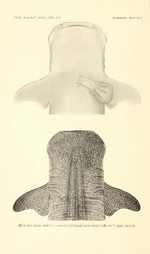
Dermal denticles.—The dermal denticles of the embryo (pi. 3)
closely resemble those of the adult in having ovoid blades, each Avith
three posterior marginal teeth and a strong median longitudinal keel.
Some denticles from the lower longitudinal dermal trunk ridge, how-
ever, are distinctly larger and are arranged in longitudinal pairs, with
the anterior denticle overlapping the one posterior to it. These pairs
are clearly visible not only because of their larger size, but also because
of their darker pigmentation. The posterior denticle of each pair is
similar in shape to the surrounding body denticles, but usually it has a
broader topped longitudinal ridge. The anterior denticle is of the
same size, but it is more nearly oval in shape, with only a median pos-
terior tooth; its longitudinal ridge is broadly expanded and round
topped, and usually it bears several minor ridges which converge
posteriorly to form a single ridge.

ROC. U.S. NAT. MUS . VOL. 115 GARRICK—PLATE 1
(J
Q<
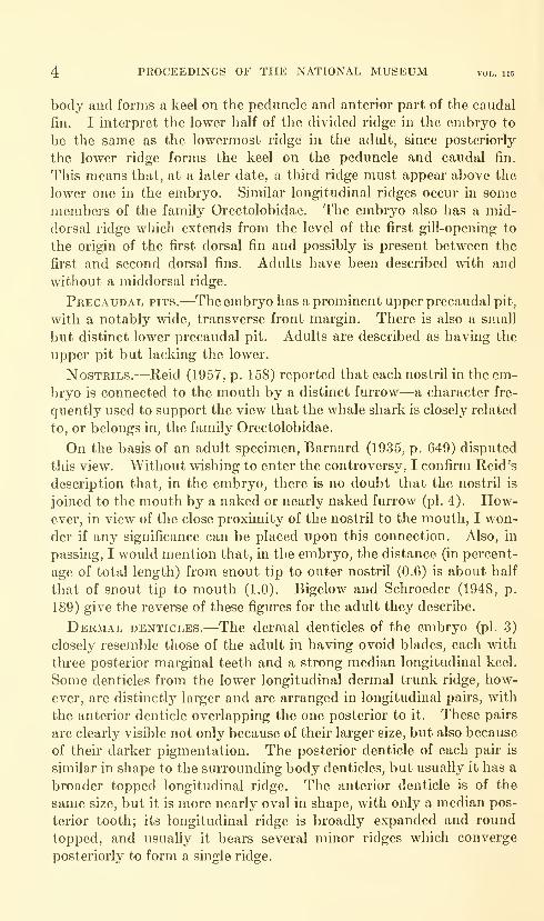
PROC. U.S. NAT. MUS . VOL. 115 GARRICK—PLATE 2
/
Rhincodo7i typus, enihij-o: \ cntral and dorsal \icus (note veilk sac in upper figure).
PROC. U.S. NAT. MUS . VOL. 115 GARRICK—PLATE 3
Rhincodon lypus, embryo: jL-inial denticles from in front of, and a little below, first dorsal
fin. The two pairs of enlarged denticles, overlapping lengthwise (left center and upper
right), are on the lower dermal ridge.
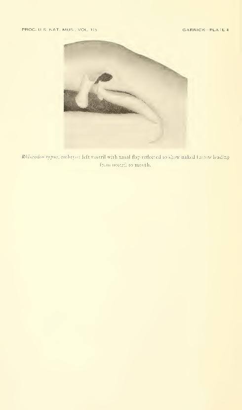
PROC. US, NAT. MUS., VOL. 115 GARRICK -PLATE 4
Rhincodon typus, cmbino: left nostril with nasal flap reflected to show naked furrow leading
from nostril to month.
EMBRYO WHALE SHARK—GARRICK 5
I do not know if similar pairs of enlarged denticles occur on the longi-
tudinal dermal ridges of adults. However, Ford (1921, p. 493) de-
scribed the first denticles to erupt in Scyliorhinus canicula, S. stellaris,
and Galeus melanostomus as being conspicuously larger than the nor-
mal body denticles and "symmetrically arranged in a sequence of
transverse pairs forming two longitudinal rows, one on either side of
the midline in a dorsolateral position." At a later stage, these larger
denticles "lose their individuality eventually owing to the presence of
equally large and similar scales which have grown up around them"
(p. 494).
Teeth.—In the embryo, the teeth are for the most part still covered
by membrane, but those that are visible show little difference from the
teeth of adults.
GiLL-RAKERS.—The plankton-sieving apparatus of the adult whale
shark consists of transverse cartilaginous bars (representing gill-rakers)
which join one gill-arch to the next; these transverse bars are further
connected, one to the other, by a secondary grid of slenderer cross
members. The entire structure is covered on its internal (pharyngeal)
surface by a fine, spongelike lattice or veU derived from dermal den-
ticles. This structure forms the sieving apparatus, with interstices
1 to 3 mm. in diameter.
In the embryo the sieve is still in a very early stage of development,
comprising only the gill-raker elements. These project forward from
each arch to the next, but their tips are still free. On the first arch
there are about 26 rakers on the upper limb and 34 on the lower. Therakers are comparatively stout rods, closely arranged, with virtually
no space between them. Each raker shows faint indications of being
bipinnate, having very short processes developing along the sides.
These processes are presumably the rudiments of the secondary grid
members. There is as yet no obvious sign of the spongy tissue which
will later line their inner surface.
Reid (1957, p. 157) suggests that the relatively advanced stage of
development of the embryo whale shark and the extent to which its
external yolk sac has been absorbed are indications that it is approach-
ing the size at which it would hatch. This is probably correct.
On the other hand, the abdomen is filled almost completely with yolk,
forming an oval mass about 80 mm. long, 50 mm. wide, and 40 mm.deep. This yolk supply seemingly would allow sufficient reserve to
complete development of the pharyngeal sieve either before or after
hatching. Only further specimens will establish whether the juvenile
whale shark feeds from the beginning in the same manner as the
adults.
Color and pattern.—The color of the embryo when first
removed from the egg-case was "bluish grey with white spots, the
6 PROCEEDINGS OF THE NATIONAL MUSEUM vol. us
undersurface white" (Breuer, 1954, p. 29). After preservation, the
embryo is brownish rather than bluish grey, but with the dermal
ridges dusky. Adults have been described as being variously dark
grey to reddish or greenish brown above and white or yellow below.
The color pattern of small spots and narrow transverse bars on the
embryo (pis. 1-2) is remarkably similar to that of adults.
Vertebrae.—Radiographs of the embryo whale shark showvertebral centra very clearly in the body region and the anterior
four-fifths of the tail. The centra are widely spaced, presumably
from incomplete calcification. In the body region anterior to the level
of the origin of the upper caudal lobe, there are 81 vertebrae, while
posteriorly on the caudal axis there are 72 countable vertebrae.
The total number of caudal vertebrae is probably much higher, but
those in the terminal fifth of the caudal axis are calcified or developed
insufficiently to show on the radiographs.
Literature Cited
Barnard, K. H.
1935. Notes on South African marine fishes. Ann. South African Mus., vol.
30, no. 5., pp. 645-G58, pis. 23-25, figs. 1-7.
Baughman, J. L.
1955. The oviparity of the whale shark, Rhineodon typus, with records of
this and other fishes in Texas waters. Copeia, 1955, no. 1, pp.
54-55, pi. 1.
Beebe, W., and Tee-Van, J.
1941. Fishes from the tropical eastern Pacific, Part 2: Sharks. Zoologica,
vol. 26, no. 2., pp. 93-122, pis. 1-2, figs. 1-34.
Bigelow, H. B., and Schroeder, W. C.
1948. Fishes of the western North Atlantic, Part I: Cyclostomes and sharks.
Mem. Sears Found. Mar. Res., no. 1, pp. 29-257, figs. 4r-106.
Breuer, J. P.
1954. The littlest biggest fish. Te.xas Game and Fish, vol. 12, no. 2., pp.
4-5, 29, 3 figs.
Ford, E.
1921. A contribution to our knowledge of the life histories of the dogfishes
landed at Plymouth. Journ. Mar. Biol. Assoc, new ser., vol. 12,
no. 3, pp. 468-505.
Garrick. J. A. F.
1960. Studies on New Zealand Elasmobranchii, Part XII: The species of
Squalus from New Zealand and Australia, and a general account
and key to the New Zealand Squaloidea. Trans. Roy. Soc. NewZealand, vol. 88, no. 3, pp. 519-577, figs. 1-6.
Maschlanka, H.
1955. Die Proportionsanderungen beim Wachstum der Katzenhaie {Scyllio-
rhinus canicula und Sc. stellare). Pubbl. Stazione Zool. Napoli, vol.
26, pp. 12-27, figs. 1-14.
Reid, G. K.
1957. External morphology of an embryo whale shark, Rhineodon typus
Smith. Copeia, 1957, no. 2, pp. 157-158, 1 fig.
Springer, S.
1960. Natural history of the sandbar shark Eidarnia milherli. U.S. Fish
Wildlife Serv., Fishery Bull. 178, vol. 61, pp. 1-36, figs. 1-5.
Springer, V. G.
1961. Notes on and additions to the fish fauna of the Tampa Bay area in
Florida. Copeia, 1961, no. 4, pp. 480-482.
7
U.S. GOVERNMENT PRINTING 0FFICE:I964

Proceedings of
the United States
National MuseumSMITHSONIAN INSTITUTION • WASHINGTON, D.C.
Volume 115 1964 Number 3477
NOTES ON NEW AND OLD SPECIES OF ALTICINAE
(COLEOPTERA) FROM THE WEST INDIES
By Doris H. Blake
The present paper describes 17 new species of beetles of the sub-
family Alticinae: 8 collected in Puerto Rico by R. G. Oakley, 4 in
Jamaica by T. H. Farr, and 5 from other sources. Notes on other
species are given, and the proper placing of species hitherto wrongly
ascribed to Pseudoepitrix is discussed.
Aphthona crucifera, new species
FiauBB 4
About 1.5 mm. in length, oblong oval, shining, the prothorax and
elytra lightly punctate, striate rows of punctures on elytra becoming
faint near apex, pale yellow brown, occiput of head deeper in color,
margins of prothorax usually dark brown and on elytra a brown
humeral spot connecting narrowly along base with a sutural darken-
ing, slightly below the middle a brown fascia, lateral margin more or
less dark.
Head with interocular space a little more than half width of head,
occiput rounded, few punctures near eye, frontal tubercles distinctly
9
10 PROCEEDINGS OF THE NATIONAL MUSEUM vol. 115
marked, narrow carina down lower front; top of head deeper brown,
pale in lower front. Antennae pale yellow brown with outer four
joints slightly darker and wider, not extending much below humeri,
fifth joint longer than fourth. Prothorax with rounded lateral sides,
having oblique angle anteriorly, disc somewhat rounded, shining,
finely punctate, pale yellow with lateral margin usually dark brown.
Scutellum brownish. Elytra moderately convex, without depres-
sions, shining, with rows of fine punctures becoming fainter near apex;
pale yellow brown with dark reddish brown humeral spot connecting
with brown sutural vitta more or less widely across base, sutural
vitta extending to apex and sometimes connecting with dark lateral
margin in darker specimens; in paler specimens only sutural edge
darkened; slightly below middle a brown fascia usually connected
at suture. Body beneath in darker specimens with middle of pro-
sternum and breast chestnut brown, abdomen and legs pale yellow
brown. Hind femora thickened, hind tibiae with short spur. Length
1.3-1.5 mm.; width .8 mm.Type, female, USNM 66194, and 11 paratopes, taken on Myrica
cerifera at Guanica, Puerto Rico, by R. G. Oakley, September 14,
1934.
Remarks.—Dr. E. A. Schwarz originally identified this as Aphthona
maculipennis Jacoby, a Central American species, but that species as
described possesses a very different coloration.
Aphthona lepta, new species
Figure 5
Approximately 1.5 mm. in length, oblong oval, shining, prothorax
very finely punctate, elytra somewhat more distinctly striate punctate,
entirely pale reddish brown with outer antennal joints slightly darker.
Head with interocular space approximately half width of head,
occiput polished, a group of punctures near eye, frontal tubercles
clearly marked, carina down lower front, lower front paler, occiput
and mouth parts deeper brown. Antennae extending below humeri,
fifth joint longer than fourth; joints two, three, and four paler; outer
joints broader and hairier. Prothorax with arcuate sides, oblique
angles anteriorly and tooth at basal angle, smoothly convex, without
depressions, shining, very finely punctate. Scutellum brownish.
Elytra smoothly convex, without depressions, with fine striate punc-
tures; in male specimen more indistinct than in female; entirely
reddish brown. Body beneath and legs reddish brown. Length
1.5 mm.; width .7 mm.Type, male, USNM 66193, and one female paratype, collected at
Guanica, Puerto Rico, by R. G. Oakley, September 27, 1934.
Remarks.—This is approximately the same size as Aphthona
crucifera, which was also collected at Guanica. It lacks the elytral
ALTICINAE FROM THE WEST INDIES—BLAKE Hdark markings and has a narrower aedeagus with a more tapering
point. The striate punctation on the elytra is also less distinct.
Aphthona inornata Blake
Figure 8
Aphthona inornata Blake, Journ. Wash. Acad. Sci., vol. 39, no. 11, p. 308, 1949.
Numerous specimens of this species, which was described from a
single female collected at Rio Piedras, Puerto Rico, have been taken
by R. G. Oakley at Ponce, Puerto Rico. Because of the similarity
in the yellow brown coloration of several species of West Indian
Aphthona, a drawing of the aedeagus is given here for the first time.
Aphthona insularis, new species
Figure 1
About 2 mm. in length, oblong oval, shining, pronotum very finely
punctate, elytra somewhat more coarsely and striately punctate,
with slight depression below basal callosity, deep black with faint
bluish lustre, antennae pale at base, deeper brown towards apex,
femora piceous, tibiae and tarsi sometimes slightly paler.
Head with interocular space a little less than half width of head,
frontal tubercles clearly marked but not swollen, occiput smooth,impunctate, a short carina, slightly produced between antennal
sockets and down lower front, entirely dark. Antennae not reaching
middle of elytra, basal two joints swollen, third joint slightly shorter
than fourth, remainder subequal, becoming somewhat wider anddarker in color. Prothorax moderately convex with arcuate sides
and oblique angles anteriorly, disc without depressions, polished andfinely punctate, entirely dark. Scutellum entirely dark. Elytra
convex, lateral margin almost invisible from above, a slight depression
below basal callosity, lines of fine striate punctures slightly irregular
near suture, between these a line of very fine striate punctures;
entirely dark with faint deep bluish lustre. Body beneath entirely
dark, hind femora enlarged, hind tibiae with short apical sulcus andspur at apex. Length 2-2.4 mm.; width 1-1.3 mm.
Type, male, USNM 66202, and 9 paratypes from Dominica, WestIndies, collected by H. W. Foote of the Yale 1913 Expedition ui Juneand July, 1913.
Remarks.—The dark bluish black coloration of this species dis-
tinguishes it from the smaller yellowish brown species of Aphthonafrom the West Indies.
Aphthona himprocyanea, new species
Figure 9
Between 2 and 3 mm. in length, oblong oval, polished, dark vio-
laceous blue, nearly impunctate, a remnant of striate punctation on
12 PROCEEDINGS OF THE NATIONAL MUSEUM vol. 115
elytra in transverse depression below basal callosity and along suture
at base.
Head with interocular space more than half width of head, occiput
polished, rounded, impunctate, groove from eye to frontal tubercles
with punctures along it, carina short and distinct, head entirely dark.
Antennae extending below humeri, entirely dark, basal joints with
metallic blue sheen, third joint about same length as fourth, remainder
subequal and hairy. Prothorax slightly wider than long, with arcuate
sides and oblique anterior angles, moderately convex, without de-
pressions, polished, impunctate, entirely dark. Scutellum dark.
Elytra moderately convex, below basal callosity a transverse depres-
sion in which are remnants of striate punctation, rest of surface with
very fine confused punctation visible only under high magnification;
surface polished, dark blue. Body beneath and legs shining, entirely
dark blue. Length 2.3-2.9 mm.; width 1.3-1.5 mm.Type, male, USNM 66195, and 13 paratypes, from Ponce and
Aibonito, Puerto Rico, collected on Croton species by R. G. Oakley,
September 7, 1933, August 1933, and July 10, 1934.
Remarks.—It is interesting to note that many species of Aphthona
occur on the Euphorbiaceae.
Longitarsus cylindricus, new species
Figure 2
About 1.5 mm. in length, elongate oval, shining, very finely punc-
tate, elytra without wings or humeral prominences, widest at middle,
yellowish brown, sides of pronotum darker brown; antennae dark with
joints three, four, ten, and eleven pale; outer joints thicker and hairier.
Head with interocular space approximately half width of head,
occiput polished, deeper brown than lower front, a group of punctures
near eye, frontal tubercles clearly marked, carina down lower front.
Antennae fully half length of body; third, fourth, tenth, and eleventh
joints pale; outer joints slightly longer and thicker, Prothorax con-
vex, somewhat wider than long, sides slightly arcuate with oblique
angles anteriorly and tooth at base, a long hair at each corner; very
finely punctate, surface polished, yellow brown with sides deeper
brown, Scutellum brownish. Elytra convex with rounded sides
and no humeral prominences, without wings, surface polished, moredistinctly punctate than prothorax, entirely yellowish brown. Bodybeneath with breast and prosternum usually slightly deeper brownthan abdomen and legs, shining, anterior coxal cavities open, hind
femora enlarged, hind tibiae shallowly channelled with spur at end,
first tarsal joint very long. Length 1.2-1.6 mm.; width .6 mm.Type, male, USNM 66190, and three paratypes, collected on weeds
at Adjuntas, Puerto Rico, by R. G. Oakley, October 11, 1934.
ALTICINAE FROM THE WEST INDIES—BLAKE 13
1. Flp^thona insula ris,n.sp. 2,Lon^itarsus ciilitidr I'eus.n.Sp. 3 Longitoreus chlan ldotus,n,sp.
'I.flphlhona cruclfera,n.S|J, S.flphtliono lepf-a^n.Sp.
<f
6Xonq^\tcirso5 oalilei^i,n.Sp.
/^
(\
3 #y.Longitarsus ati)picu3,n.sp. 8. flphthona I'norrcilo ,n,sp. 9.flplitliona laiTibrocijanc-a.n sp.
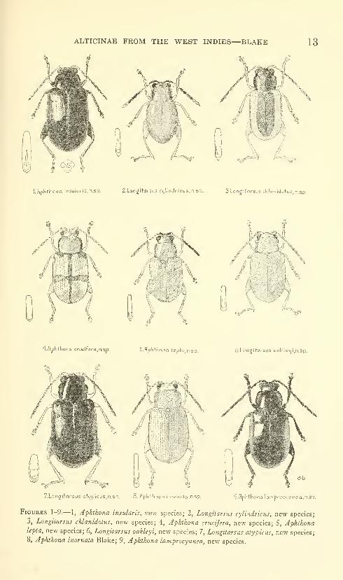
Figures 1-9.— 1, Aphthona insularis, new species; 2, Longitarsus cylindricus, new species;
3, Longitarsus chlanidotus, new species; 4, Aphthona crucifera, new species; 5, Aphthonalepta, new species; 6, Longitarsus oakleyi, new species; 7, Longitarsus atypicus, new species;
8, Aphthona inornata Blake; 9, Aphthona lamprocyanea, new species.
14 PROCEEDINGS OF THE NATIONAL MUSEUM vol. 115
Remarks.—This tiny flealike beetle is distinguished from other
species of Longitarsus in the West Indies by its wingless elytra
lacking humeral prominences and by its black and white antennae.
It is closely related to L. oopterus Harold, another wingless species,
from Colombia, which has an impunctate prothorax, and L. inter-
medius Jacoby from Panama, which is also impunctate but hassimilarly colored antennae. In L. angusticollis Jacoby the antennaeare longer than the body. L. impuncticollis Blatchley from Florida,
another wingless species, is dark chocolate brown with fuscous
antennae.
Longitarsus chlanidotus, new species
Figure 3
About 2 mm. in length, oblong oval, shining, pronotum finely
punctate, elytra more coarsely punctate, yellow brown with a brownhead, pronotum and broad brown sutural vitta, and often brown along
sides of elytra.
Head with interocular space approximately half width of head,
occiput pohshed, a group of punctures near eye, frontal tubercles
shghtly swollen, interantennal area somewhat produced, a carina
down front, shining dark brown, slightly paler in lower front. An-tennae brown with four basal joints paler, extending to middle of
elytra, third joint shorter than fourth, remainder subequal. Pro-
thorax with slightly curved sides, oblique anterior angles and small
tooth at basal angle, disc smooth, without depressions, polished, darkbrown, or in pale specimens paler, distinctly punctate. Elytra
without depressions, humeri not prominent, more coarsely and densely
punctate than pronotum, yellow brown, usually with a wide sutural
vitta covering half the elytra, not reaching apex, usually a brown lateral
vitta between humerus and apex, in paler specimens only sutural
and marginal edges brown. Body beneath brownish with legs
yellowish brown, hind femora enlarged, first hind tarsal joint very long,
hind tibiae grooved, a spur at end, claws appendiculate. Length1.6-2 mm.; width .8-9 mm.Type, male, USNM 66191, and 23 paratypes collected on weeds
in Boringuen Forest, at Guanica, Puerto Rico, by R. G. Oakley,
September 14, 1934. One specimen taken at St. Thomas, 14}^ miles
east of Kingston, Jamaica, by T. H. Farr.
Remarks.—This distinctive little beetle with its brown mantlewas also taken on Tournefortia gnaphalodes on Lower MatecumbeKey, Florida, by Paul W. Oman, July 20, 1939.
ALTICINAE FROM THE WEST INDIES—BLAKE 15
10.LoiWitnr3U5 subtiha Harold 11. Sustena bosalis 12. flitica occidenlalis Suffrion*^ clbcrration Jocqelln DuVol
ISHermoeopha^a jamaicensi's.n.sp. M.Homoschemo xanthocijaneom.n.Sp IS.HemilocticQ porforicensls.n.Sp.
l6-NeSQecre|7.dci Q5|Jiallina (Suftrlor) l7.Ne6aecrepida rufonarj.nato.n5p IS.Choclocn^ma C40.10prcra.nSp
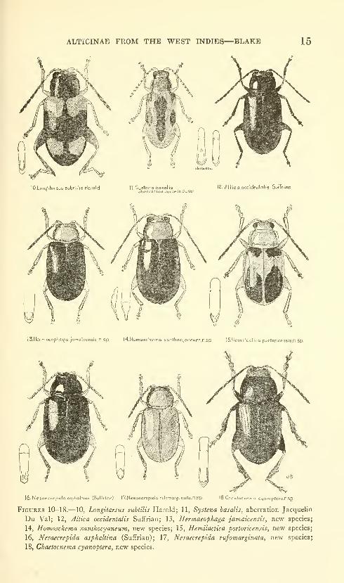
Figures 10-18.— 10, Longitarsus subtilis Harold; 11, Systena basalis, aberration Jacquelin
Du Val; 12, Altica occidentalis Suffrlan; 13, Hermaeophaga jamaicensis, new species;
14, Homosckema xanthocyaneum, new species; 15, Hemilactica fortoricensis, new species;
16, Nesaecrepida asphaltina (Suffrian); 17, Nesaecrepida rufomarginata, new species;
18, Chaetoc7iema cyanopiera, new species.
16 PROCEEDINGS OF THE NATIONAL MUSEUM vol. 115
Longitarsus oakleyif new species
Figure 6
About 1.5 mm. in length, oblong oval, shining, elytra moderatelydensely and strongly punctate, entirely yellow brown.
Head with interocular space slightly more than half width of head,
occiput smoothly rounded, pohshed, a group of pxmctures near eye,
interantennal area broad, somewhat produced, this rather fiat carina
broadening below, entire head pale yellow brown. Antennae extending
to middle of elytra, fifth joint longer than third or fourth, entirely
pale. Prothorax moderately convex with arcuate sides, oblique
angles anteriorly and small tooth at basal angle, surface shining,
without depressions, very finely punctate, entirely pale yellow brown.Scutellum brownish. Elytra moderately convex, without depressions,
shining, strongly and coarsely punctate, yellow brown. Bodybeneath and legs entirely pale, shining, hind legs enlarged, hind
tibiae grooved, spur at end, first hind tarsal joint very long. Length1.3-1.8 mm.; width .7-8 mm.Type, male, USNM 66192, from Ponce, Puerto Rico, collected on
weeds, and also one specimen taken at Guanica, Puerto Rico, bothcollected by R. G. Oakley, September 27, 1934.
Remarks.—This tiny pale species difi^ers from both L. cylindricus
and L. chlanidotus by being entirely pale and having rather coarsely
punctate elytra.
Longitarsus atypicus, new species
Figure 7
About 2 mm. in length, oblong oval, very shining, prothorax finely
and elytra more coarsely punctate, elytra with distinct basal callosity
and depression below, black with four basal joints of antennae pale
and legs pale, posterior femora and lower front of face brownish.
Head with interocular space shghtly more than half width of head,
occiput polished, a gi'oup of punctures near eye, frontal tubercles
slightly swollen, interantennal ai-ea produced, well-mai-ked carina
down lower front, lower front rather long and paler brown. Antennaeabout half as long as body, three basal joints pale, fourth slightly
darker, remainder dark, third joint shorter than fourth. Prothoraxmoderately convex, smoothly rounded, without depressions, sides
slightly arcuate, an oblique anterior angle and small tooth at base,
surface polished black with distinct punctation. Scutellum dark.
Elytra wider than prothorax, with distinct humeri, a basal callosity,
below this a transverse depression, surface very shiny, with coarser
ALTICINAE FROM THE WEST INDIES—BLAKE 17
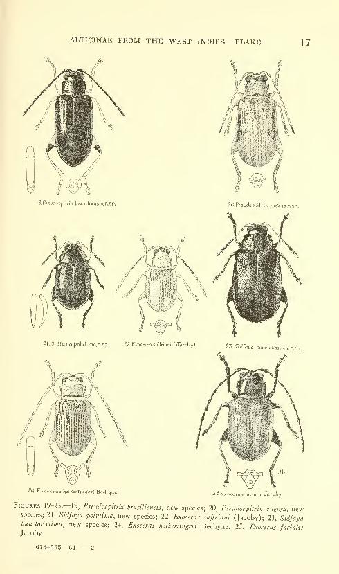
ig.Pscudocpitr,-xbrc,sil;o,is,-s,n,Sp.
2I.Sidfatja l^oluti'ma, n.sp. 22.Ex-ocero3 suffritiii (Jacoby)
20.P5€iidoepltrix rii^osa.nsp.
24.E«ocsrashelKcrtingeri Bechijne 2-5.Cxocero5 focioKs Jaroby
Figures 19-25—19, Pseudoepitrix brasiliensis, new species; 20, Pseudoepitrix rugosa, newspecies; 21, Sidfaya polutima, new species; 22, Exoceras suffriani (Jacoby); 23, Sidfayapunctatissima, new species; 24, Exoceras heikertingeri Bechyne; 25, Exoceras facialisJacoby.
676-565—6-1-
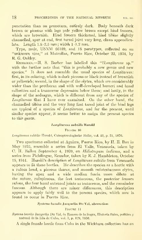
18 PROCEEDINGS OF THE NATIONAL MUSEUM vol. 115
punctation than on pronotum, entirely dark. Body beneath dark
brown or piceous with legs pale yellow brown except hind femora,
which are brownish. Hind femora thickened, hind tibiae slightly
channelled, spur at end, first tarsal joint very long, claws appendicu-
late. Length 1.8-2.5 mm; width 1-1.2 mm.Type, male, USNM 66189, and 18 paratypes, collected on an
"unknown vine," at Matrullas, Puerto Rico, October 22, 1934, byR. G. Oakley.
Remarks.—H. S. Barber has labelled this "?Longitarsns sp."
with the fm'ther note that "this is probably a new genus and newspecies." It does not resemble the usual species of Longitarsus:
first, in its coloring, which is dark piceous or black instead of brownish
or yellowish; second, in the shape of the elytra, which are considerably
wider than the prothorax and with well-developed humeri and basal
callosities and a transverse depression below them; and lastly, in the
shape of the aedeagus, which is different from any of the species of
Longitarsus that I have ever examined. On the other hand, the
channelled tibiae and the very long first tarsal joint of the hind legs
are typical of a species of Longitarsus, and for the present, until
similar species appear, it seems better to assign the present species
to this genus,
Longitarsus subtilis Harold
Figure 10
Longitarsus suhtilis Harold, Coleopterologische Hefte, vol. 15, p. 31, 1876.
Two specimens collected at Aguirre, Puerto Rico, by H. E. Box in
May 1925, resemble a series from El Valle, Venezuela, taken byC. H. Ballou September 4, 1939, on Heliotropum indicum, and a
series from Pichilingue, Ecuador, taken by E. J. Hambleton, October
15, 1944. Harold's description of Longitarsus suhtilis from Venezuela
appears to fit these beetles. He describes the species as a beetle with
a rufous head, a piceous thorax, and smooth rufotestaceous elytra,
having the apex and a wide median fascia more dilute at
the suture, rufopiceous, the feet testaceous, the posterior femora
rufous, the four basal antennal joints as testaceous, and the remainder
fuscous. Although there are minor differences, this description
appears to apply fairly well to the present species, which now is
found to occur in Puerto Rico.
Systena basalis Jacquelin Dii Val, aberration
Figure 11
Systena basalis Jacquelin Du Val, in Ramon de la Sagra, Historia fisica, politica ynatural de la Isla de Cuba, vol. 7, p. 129, 1856.
A single female beetle from Cuba in the Wickham collection has an
ALTICINAE FROM THE WEST INDIES—BLAKE 19
unusual coloration. Instead of being almost entirely piceous with a
bronze lustre, with the elytra having only a trace of a pale vitta near
the base, as is uniformly found in the female of Systena basalis, this
specimen has pale elytra with a humeral dark spot and a median
lateral short vitta, as well as a wide sutural dark vitta, widening
below the scutellum for a short way, then narrowing and extending
down the suture but not reaching the apex. The legs are entirely
pale, the antennae also pale, with a slight darkening at the apex of
each joint. The breast and abdomen are brownish piceous. If it
were a male, in which the usual elytral markings are paler,
with a full-length pale vitta, this would not appear so unusual.
Structurally, however, the specimen does not seem to differ from
Systena basalis, being of the same size and dimensions and possessing
the same sort of punctation. Thus, it seems highly unlikely that
this is a different species.
Altica occidentalis Suffrian
Figure 12
Haltica occidentalis Suffrian, Arch. Naturg., vol. 34, p. 197, 1868.
In describing this species Suffrian was somewhat in doubt as to
whether or not it might be the European A. oleracea L., which it
strongly resembles and which he thought might have been introduced
into Cuba with vegetables. The present species is very similar in
outward appearance to the European species but the aedeagus is
different. This species appears to be endemic in the West Indies and
occurs not only in Cuba but also in Hispaniola, Puerto Rico, the
Virgin Islands, Jamaica, Dominica, and St. Lucia. It is of the same
elongate shape as A. ludoviciana Fall, another Caribbean species
found on the same food plant, Jussiaea, and a species distinguished
by its pale yellow legs.
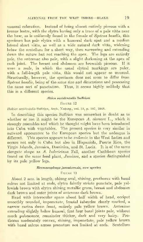
Hermaeophaga jamaicensis, new species
Figure 13
About 2 mm. in length, oblong oval, shining, prothorax with basal
sulcus not limited at ends, elytra faintly striate punctate, pale yel-
lowish brown with elytra shining metallic green, breast and abdomen
dark brown and outer joints of antennae dark brown.
Head with interocular space about half width of head, occiput
smoothly rounded, impunctate, frontal tubercles clearly marked, a
narrow carina down front, entirely pale yellow brown. Antennae
extending slightly below hmneri, first four basal joints pale, without
much pubescence, remainder thicker, dark and very hairy. Pro-
thorax moderately convex, shining, impunctate, pale yellow brown
with basal sulcus across pronotum not limited at ends. Scutellum
20 PROCEEDINGS OF THE NATIONAL MUSEUM vol. 115
pale. Elytra faintly striate punctate, punctures evanescent at apex,
a slight transverse depression below basal callosity, shining, metallic
green. Body beneath with breast and abdomen dark brown, legs
entirely pale. Length 2-2.3 mm.; width 1-1.1 mm.Type, male, USNM 66199, one female paratype, and one male
paratype, in Institute of Jamaica, collected at St. James, about 4
miles northeast of Montego Bay, Jamaica, September 12, 1958, byT. H. Farr.
Remarks.-—This is the second species of Hermaeophaga to be
described from Jamaica, the other being H. cwpraea Blake.
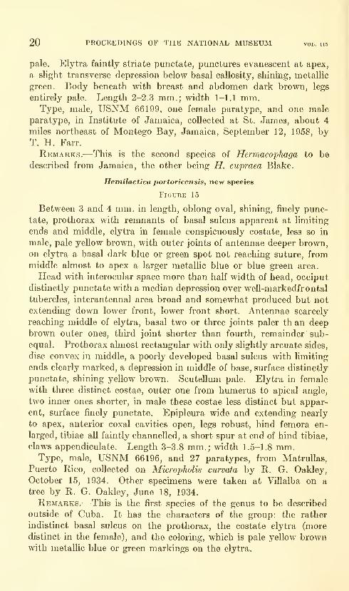
Hemilactica portoricensis, new species
Figure 15
Between 3 and 4 mm. in length, oblong oval, shining, finely punc-
tate, prothorax with remnants of basal sulcus apparent at limiting
ends and middle, elytra in female conspicuously costate, less so in
male, pale yellow brown, with outer joints of antennae deeper brown,on elytra a basal dark blue or green spot not reaching suture, frommiddle almost to apex a larger metallic blue or blue green area.
Head with interocular space more than half width of head, occiput
distinctly punctate with a median depression over well-markedfrontal
tubercles, interantennal area broad and somewhat produced but not
extending down lower front, lower front short. Antennae scarcely
reaching middle of elytra, basal two or three joints paler th an deep
brown outer ones, third joint shorter than fourth, remainder sub-
equal. Prothorax ahnost rectangular with only slightly arcuate sides,
disc convex in middle, a poorly developed basal sulcus with limiting
ends clearly marked, a depression in middle of base, surface distinctly
punctate, shining yellow brown. Scutellum pale. Elytra in female
with three distinct costae, outer one from humerus to apical angle,
two inner ones shorter, in male these costae less distinct but appar-
ent, surface finely punctate. Epipleura wide and extending nearly
to apex, anterior coxal cavities open, legs robust, hind femora en-
larged, tibiae all faintly channelled, a short spur at end of hind tibiae,
claws appendiculate. Length 3-3.8 mm.; width 1.5-1.8 mm.Type, male, USNM 66196, and 27 paratypes, from Matrullas,
Puerto Rico, collected on Micropholis curvata by R. G. Oakley,
October 15, 1934. Other specimens were taken at Villalba on a
tree by R. G. Oakley, June 18, 1934.
Remarks.^—This is the first species of the genus to be described
outside of Cuba. It has the characters of the group: the rather
indistinct basal sulcus on the prothorax, the costate elytra (more
distinct in the female), and the coloring, which is pale yellow brownwith metallic blue or green markings on the elytra.
ALTICINAE FROM THE WEST INDIES—BLAKE 21
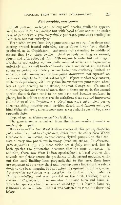
Nesaecrepida, nevr genus
Small (2-3 mm. in length), oblong oval beetles, similar in appear-
ance to species of Crepidodera but with basal sulcus across the entire
base of prothorax, elytra very finely punctate, punctures tending to
be striate but not entirely so.
Head with groove from large puncture near eye running down and
cutting around frontal tubercles, carina down lower front slightly
produced, as in Crepidodera. Antennae not extending to middle of
elytra; first two joints swollen, third scarcely as long as second,
fourth and fifth subequal; from fifth on, joints wider but not longer.
Prothorax moderately convex, with rounded sides, an oblique angle
anteriorly and a small tooth at basal angle, a somewhat sinuate basal
sulcus extending completely across base, not distinctly limited at
ends but with inconspicuous line going downward not upward on
prothorax slightly before lateral margin. Elytra moderately convex,
without depressions, with very fine inconspicuous punctation obso-
lete at apex, tending to be striate, but not always so. (In one of
the two species are traces of more than a dozen striae, in the second
species the striations tend to be geminate and become confused in
places, but in neither species are the striations clear cut and impressed
as in others of the Crepidodera.) Epipleura wide until apical curve,
then vanishing, anterior coxal cavities closed, hind femora enlarged,
hind tibiae shallowly sulcate near apex, a very short spur at tip, claws
appendiculate.
Type of genus, Haltica asphaltina SufFrian.
The generic name is derived from the Greek vrjaaios (nesaios =insular) -f crepida.
Remarks.—The two West Indian species of this genus, Nesaecre-
pida, which is allied to Crepidodera, differ from the other New World
Crepidoderini in having inconspicuous elytral punctation that con-
sists of very fine punctures in many (over 12) striae. In Nesaecre-
pida asphaltina (fig. 16) these striae are slightly confused, but in
both species the punctation becomes obsolete near the apex. In
addition, these two West Indian species have a basal groove that
extends completely across the prothorax to the lateral margins, with-
out the usual limiting lines perpendicular to the base; these lines
are represented by a very short and inconspicuous line from the sulcus
down to the basal margin, but not extending upward from the groove.
Nesaecrepida asphaltina was described by Suffrian from Cuba as
Haltica asjjhaltina and was recorded in the Junk Catalogue as a
species of Crepidodera; it occurs also in Puerto Rico and Jamaica.
The other species, which has been collected by T. H. Farr in Jamaica,
is known also from Cuba, where it was collected on rice; it is described
below.
22 PROCEEDINGS OF THE NATIONAL MUSEUM vol. 115
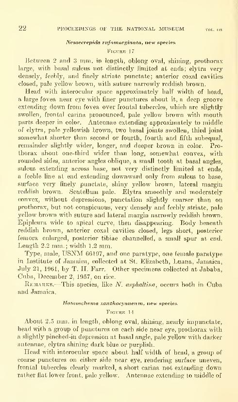
Nesaecrepida rufomarginata, new species
Figure 17
Between 2 and 3 mm. in length, oblong oval, shining, prothorax
large, with basal sulcus not distinctly limited at ends; elytra very
densely, feebly, and finely striate punctate; anterior coxal cavities
closed, pale yellow brown, with suture narrowly reddish brown.
Head with interocular space approximately half width of head,
a large fovea near eye with finer punctures about it, a deep groove
extending down from fovea over frontal tubercles, which are sHghtly
swollen, frontal carina pronounced, pale yellow brown with mouthparts deeper in color. Antennae extending approximately to middle
of elytra, pale yellowish brown, two basal joints swollen, third joint
somewhat shorter than second or fourth, fourth and fifth subequal,
remainder slightly wider, longer, and deeper brown in color. Pro-
thorax about one-third wider than long, somewhat convex, with
rounded sides, anterior angles oblique, a small tooth at basal angles,
sulcus extending across base, not very distinctly limited at ends,
a feeble line at end extending downward only from sulcus to base,
surface very finely punctate, shiny yellow brown, lateral marginreddish brown. Scutellum pale. Elytra smoothly and moderately
convex, without depressions, punctation sHghtly coarser than on
prothorax, but not conspicuous, very densely and feebly striate, pale
yellow brown with suture and lateral margin narrowly reddish brown.Epipleura wide to apical curve, then disappearing. Body beneath
reddish brown, anterior coxal cavities closed, legs short, posterior
femora enlarged, posterior tibiae channelled, a small spur at end.
Length 2.2 mm.; width 1.2 mm.Type, male, USNM 66197, and one paratype, one female paratype
in Institute of Jamaica, collected at St. Elizabeth, Luana, Jamaica,
July 21, 1961, by T. H. Farr. Other specimens collected at Jababa,
Cuba, December 2, 1957, on rice.
Remarks.-—This species, like A^. asphaltina, occurs both in Cubaand Jamaica.
Honioschema xanthocyaneum, new species
Figure 14
About 2.5 mm. in length, oblong oval, shining, nearly impunctate,
head with a group of punctures on each side near eye, prothorax with
a slightly pinched-in depression at basal angle, pale yellow with darker
antennae, elytra shining dark blue or purplish.
Head with interocular space about half width of head, a group of
coarse punctures on either side near eye, rendering surface uneven,
frontal tubercles clearly marked, a short carina not extending downrather flat lower front, pale yellow. Antennae extending to middle of
ALTICINAE FROM THE WEST INDIES—BLAKE 23
elytra, first two joints swollen, third joint not as long as fourth,
remainder subequal, diminishing slightly toward end, basal joints pale,
outer joints becoming dark brown. Prothorax not twice as wide as
long, with rounded sides, oblique anterior angles and a small tooth at
basal angle, a small pinched-in depression near this basal angle, surface
very shiny and very finely punctate, pale yellow. Scutellum reddish
brown. Elytra moderately convex with transverse depression below
basal callosity, polished dark violaceous blue and very finely punctate.
Body beneath and legs pale yellow, anterior coxal cavities open, legs
short, hind tibiae with short spur, claws appendiculate. Length
2.2-2.7 mm.; width 1-1.2 mm.Type, male, USNM 66198, collected at Clarendon, Portland Ridge,
north side, July 23, 1958, by T. H. Farr; one female collected at
Hanover, Bloody Bay, February 15-24, 1952, by F. A. McDermott.
Kemarks."—This is the second species of Homoschema to be found
on Jamaica. It differs from H. jamaicense in having a more trans-
verse prothorax with a little depression on each side near the basal
angle. The aedeagus also is different and, in its long narrow point,
suggests that of H. hoffmani from Haiti.
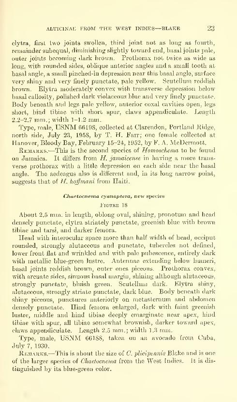
Cliaetocnerna cyanoptera, new species
Figure 18
About 2.5 mm. in length, oblong oval, shining, pronotum and head
densely punctate, elytra striately punctate, greenish blue with brown
tibiae and tarsi, and darker femora.
Head with interocular space more than half width of head, occiput
rounded, strongly alutaceous and punctate, tubercles not defined,
lower front flat and wrinkled and with pale pubescence, entirely dark
with metallic blue-green lustre. Antennae extending below humeri,
basal joints reddish brown, outer ones piceous. Prothorax convex,
with arcuate sides, sinuous basal margin, shining although alutaceous,
strongly punctate, bluish green. Scutellum dark. Elytra shiny,
alutaceous, strongly striate punctate, dark blue. Body beneath dark
shiny piceous, punctures anteriorly on metasternmn and abdomen
densely punctate. Hind femora enlarged, dark with faint greenish
lustre, middle and hind tibiae deeply emarginate near apex, hind
tibiae with spur, all tibiae somewhat brownish, darker toward apex,
claws appendiculate. Length 2.5 mm.; width 1.3 mm.Type, male, USNM 66188, taken on an avocado from Cuba,
July 7, 1930.
Remarks."—This is about the size of C. plicipennis Blake and is one
of the larger species of Chaetocnema from the West Indies. It is dis-
tinguished by its blue-green color.
24 PROCEEDINGS OF THE NATIONAL MUSEUM vol. iis
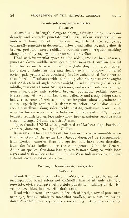
Pseudoepitrix rugosa, new species
Figure 20
About 3 mm. in length, elongate oblong, faintly shining, pronotumdensely and coarsely punctate with basal sulcus very distinct in
middle of base, elytral punctation irregularly striate, somewhatconfusedly punctate in depression below basal callosity, pale yellowish
bro^vn, prothorax more reddish, a reddish brown irregular markingalong side of elytra, legs and antennae pale yellow.
Head with interocular space half its width, front of head coarsely
punctate down middle from occiput to somewhat swollen frontal
tubercles, carina between antennal sockets short and broad, lower
front short. Antennae long and slender, extending below middle of
elytra, pale yellow with terminal joint brownish, third joint shorter
than fourth. Prothorax wider than long with oblique anterior angles
and tooth at basal angle, sides straight, basal sulcus very distinct in
middle, marked at sides by depression, surface coarsely and contig-
uously punctate, pale reddish brown. Scutellum reddish brown.
Elytra long, with well-marked basal callosity and a slight depression
below this, rows of striate punctures irregular, almost geminate at
times, especially confused in depression below basal callosity andabout scutellum, along sides feebly costate, yellowish brown with
irregular brown areas on sides between humerus and middle. Bodybeneath reddish brown, legs pale yellow brown, anterior coxal cavities
closed. Length 2.9 mm.; width 1.3 mm.Type, female, USNM 66201, collected at Hardwar Gap, Portland,
Jamaica, June 28, 1959, by T. H. Farr.
Remarks.-—The characters of this Jamaican species resemble morethe characters of the genus that Jacoby described as Pseudoepitrix
from Central America than the species that have been described
from the West Indies under the same genus. Like the Central
American species, this Jamaican species is more elongate, with long
elytra and with a shorter face than in the West Indian species, and the
anterior coxal cavities are closed.
Pseudoepitrix brasiliensis, new species
Figure 19
About 3 mm. in length, elongate oblong, shining, prothorax with
inconspicuous basal sulcus not distinctly limited at ends, strongly
punctate, elytra elongate with striate pimctation, shining black mthyellow legs, hind femora with dark apex.
Head with interocular space half width of head, a row of punctures
near eye, frontal tubercles somewhat swollen, with distinct carina
down lower front, entirely dark piceous, shining. Antennae extending
ALTICINAE FROM THE WEST INDIES—BLAKE 25
nearly to middle of elytra, third joint shorter than fourth, rest
subequal, entirely dark. Prothorax almost rectangular with nearly
straight sides, anterior obUque angles thickened, a faint basal sulcus,
slightly more distinct at ends, disc somewhat flat, with scattered
not dense coarse punctures, shining black. Scutellum black. Elytra
elongate, tapering at apex, strongly striate punctate, punctures
becoming weaker at apex, shining black. Body beneath black with
legs pale yellow except apex of hind femora, which are dark. Anterior
coxal cavities closed, tibiae not channelled, hind ones with short spur,
claws appendiculate. Length 2.8-3 mm.; width 1.2-1.4 mm.Type, male, USNM 66200, and three paratypes, from Sao Paulo,
Brazil, collected by H. L. Parker on a "labiate plant." Two other
specimens were collected by W. M. Mann in the Baturites Mountains,
Brazil. One of these, which may be immature, has yellow brown
elytra; the head and prothorax are brownish.
Remarks.—This South American beetle has been included amongthe West Indian species to show how the West Indian species assigned
(up to this time) to the genus Pseudoepitrix differ from the Central
and South American species of Pseudoepitrix.
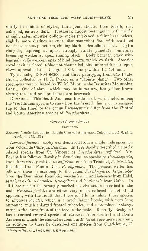
Exoceras facialis Jacoby
FiGUKE 25
Exoceras facialis Jacoby, in Biologia Centrali-Americana, Coleoptera vol. 6, pt. 1,
suppl., p. 273, 1891.
Exoceras facialis Jacoby was described from a single male specimen
from Volcan de Chiriqui, Panama. In 1897 Jacoby described a closely
related species from St. Vincent as Pseudoepitrix suffriani. Mr.
Bryant has followed Jacoby in describing, as species of Pseudoepitrix,
two others closely related to suffriani, one from Trinidad, P. trinitatis,
the other from Puerto Rico, P. hoffmani. The present writer has
followed them in ascribing to the genus Pseudoepitrix: hispaniolae
from the Dominican Republic, punctatissima and hottensis from Haiti,
jamaicensis from Jamaica, tetraspilota and longicornis from Cuba. In
all these species the strongly marked sex characters described in the
male Exoceras facialis are either very much reduced or not at all
apparent, with the result that there is little to relate these species
to Exoceras facialis, which is a much larger beetle, with very long
antennae, much enlarged frontal tubercles, and a prominent enlarge-
ment in the lower front of the face in the male. Recently, Bechyne '
has described several species of Exoceras from Central and South
America in which the characters found in E. facialis are more apparent.
In addition to these he described one species from Guadeloupe, E.
1 Bechyne, Ent. Arb., Band 6, Heft. 1, 1956, ppi 145-148
26 PROCEEDINGS OF THE NATIONAL MUSEUM vol. 115
heikertingeri. The present writer recently has exammed specimens
from Dominica and St. Lucia that are probably this species. Thewriter at once recognized the relationship of these specimens to the
other West Indian species described under Pseudoepitrix although, in
E. heikertingeri (also a larger species), the male characters are very
long antennae, very swollen frontal tubercles, and two well-developed
tubercles in the lower front of the face—all characters to be found in
E. facialis.
In all the West Indian species the head is long, as in E. facialis. In
P. longicornis, described from a male only, the antennae approximate
those oifacialis. In P. suffriani the frontal tubercles over the antennae
are very swollen, as in facialis. To a slightly less extent this mayalso be true in P. trinitatis, as shown in Mr. Bryant's drawing (the present
writer has not examined the type). In the remainder of the West
Indian species (hqffmani, hispaniolae, punctatissima, hottensis, jamai-
censis, and tetraspilota) none of these striking male characters so far
has been observed, but it is evident that all of these species belong to
the genus Exoceras, rather than to Pseudoepitrix.
The West Indian species are all smaller than E. facialis, and all
very similar, forming a homogeneous group. Several have been
collected on tree ferns. Mr. Bryant records trinitatis as destroying
young fronds of the fern Adiantum tenerum. These species do not
have the long elytra common to Pseudoepitrix and, unlike this genus, the
anterior coxal cavities are open. The head in Pseudoepitrix is normal,
not elongate, and there are no traces of tubercles on the lower front
nor enlarged tubercles over the antennal sockets.
Two new species of Pseudoepitrix are included in this paper to
illustrate the differences in the two genera. Drawings have been
made of Exoceras facialis Jacoby (fig. 25), E. suffriani (Jacoby) (fig. 22),
and what is probably E. heikertingeri Bechyne (fig. 24).
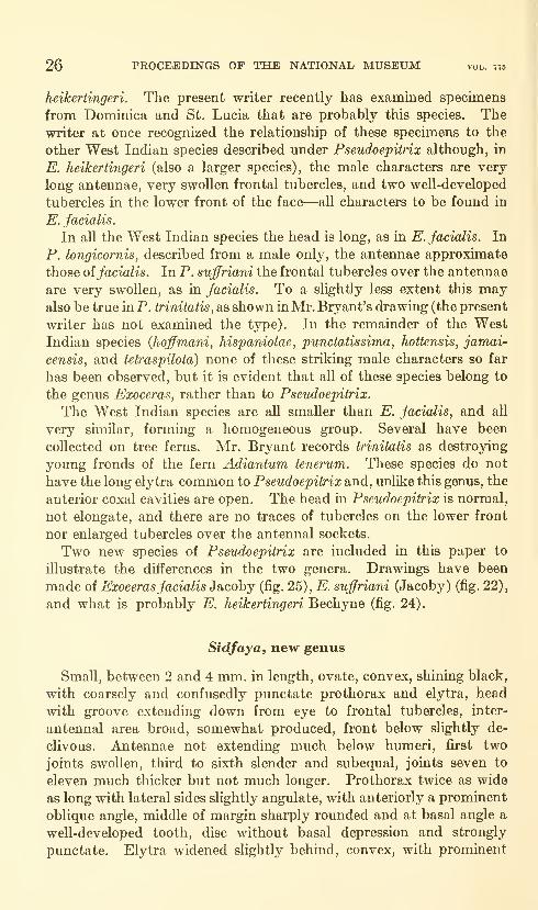
Sidfaya, new genus
Small, between 2 and 4 mm. in length, ovate, convex, shining black,
with coarsely and confusedly punctate prothorax and elytra, head
with groove extending down from eye to frontal tubercles, inter-
antennal area broad, somewhat produced, front below slightly de-
clivous. Antennae not extending much below humeri, first two
joints swollen, third to sixth slender and subequal, joints seven to
eleven much thicker but not much longer. Prothorax twice as wide
as long with lateral sides slightly angulate, with anteriorly a prominent
oblique angle, middle of margin sharply rounded and at basal angle a
well-developed tooth, disc without basal depression and strongly
punctate. Elytra widened slightly behind, convex, with prominent

ALTICINAE FROM THE WEST INDIES—BLAKE 27
basal callosities and transverse depression below them, surface coarsely
and confusedly punctate. Body beneath with prostemum extending
down widely between anterior coxae, hind femora enlarged, hind tibiae
not channelled but rounded, spur at end, claws appendiculate.
Type of genus, Sidfaya polutima, new species.
Remarks.—The beetles are somewhat similar in shape to those of
the genus Heikertingerella but are not so smoothly convex, the elytra
having basal callosities and a transverse depression. In addition, the
strong, even coarse, punctation is unlike any found in Heikertingerella.
The genus is named in memory of Dr. Sidney Fay Blake. This
name also has an interesting parallel in Greek: aidapos (sidaros=iron)
and (f)aco (phao= shining).
Sidfaya polutima, new species
Figure 21
About 2 mm. in length, ovate, convex, shining, with dense and
somewhat confused punctation, dark piceous black with tibiae and
tarsi yellowish brown and basal seven joints of antennae pale yellow,
four terminal joints thickened and dark, hind femora enlarged, hind
tibiae rounded with small spur at end, claws appendiculate.
Head with interocular space half width of head, smoothly rounded,
pohshed and impunctate over occiput, a groove on each side extending
from fovea near eye down to frontal tubercles with several punctures
along it, frontal tubercles small and distinct, interantennal area
sUghtly produced, flat, with no definite carina and somewhat declivous below.
Antennae reaching below humeri, the two basal joints swollen, joints
three to six pale yellow, subequal, joints seven to eleven gradually
becoming wider and last four joints piceous. Prothorax approximately
twice as wide as long with sinuate, almost angulate, lateral margin,
a broad oblique angle anteriorly ending in prominent tooth at lower
end and another tooth at basal angle, moderately convex, without
depressions, surface shining black, with strong punctation. Scutellimi
dark. Elytra moderately convex, with small humeral prominences
and short intrahumeral depression, distinct depression below basal
callosities, surface shiny with strong and not too dense punctation
becoming somewhat less distinct at apex, punctation with tendency
to being striate but not actually so, epipleura vanishing at apical angle.
Body beneath having prosternum extending down widely between
anterior coxae and closing anterior coxal cavities. Hind femora muchenlarged, hind tibiae entire, rounded, a spur at end, claws appendiculate.
Length 1.8-2 mm.; width 1.1 mm.Type, male, MCZ 30491, and one female paratype in the U.S.
National Museum collection, taken at Buenos Aires, Trinidad Moun-tains, Cuba, by P. J. Darlington, Jr.

28 PROCEEDINGS OF THE NATIONAL MUSEUM vol. 115
Remarks.—In shape this species approaches the species of the
genus Heikertingerella, being ovate, moderately convex, with a pro-
thorax about twice as wide as long and without a basal sulcus of anysort. The species differs from that genus, however, in the less
sinuate basal margin of the prothorax, which does not dip down as
much over the scutellum, and in the lateral margin, which is very
sinuate, even angulate, and with a more distinctive oblique angle
anteriorly and a larger tooth basally. The elytra, too, are not as
smoothly convex but have a transverse depression below the basal
callosity. The terminal joints of the antennae are much more enlarged
than in Heikertingerella, and the head has wider interantennal space.
The punctation of this and the species below is much coarser than
that of any species of Heikertingerella.
Sidfaya punctatissimaf new series
Figure 23
Between 2 and 3 mm. in length, oblong ovate, shining piceous black,
pronotum and elytra densely and moderately coarsely punctate, pro-
thorax twice as wide as long with prominent obhque angles anteriorly
and somewhat angulate lateral margin, elytra with transverse depres-
sion below basal callosities, antennae yellowish brown with four
terminal joints wider and darker, not extending much below humeri.
Head with interocular space approximately haK width of head,
occiput rounded, impunctate, pohshed, a groove with several punctures
along it from eye to frontal tubercles, frontal tubercles small and
clearly cut, a broad and somewhat produced interantennal area widen-
ing below, lower front slightly concave. Antennae not extending far
below humeri, joints one and two swollen, joints three to six slender
and subequal, joints seven to eleven giadually wider, but not muchlonger, hairier and darker. Prothorax twice as wide as long with
prominent oblique anterior angles having tooth at lower end, at mid-
point margin sharply rounded, at base a small tooth, disc not very
convex, shiny piceous black with numerous moderately coarse and
deep punctures, not too closely placed in all parts. Scutellum dark
and shiny. Elytra sHghtly wider in apical half, moderately convex,
with prominent basal callosities and transverse depression below them,
surface shining black, coarsely and almost contiguously punctate,
epipleura vanishing at apical curve. Body beneath entirely dark,
prosternum extending down widely between anterior coxae, hind
femora enlarged, hind tibiae not channelled, but rounded, with spur
at apex, claws appendiculate. Length 2.8 mm.; width 1.4 mm.Type, female,MCZ 30492, collected on Main Range, Blue Mountains,
Jamaica, 5000-7388 ft., August 17-19, 1934, by P. J. Darlington, Jr.
ALTICINAE FROM THE WEST INDIES—^BLAKE 29
Remarks.—Although somewhat larger than Sidfaya polutima, the
structure of the head, with the incised line from the eye to the frontal
tubercles, the somewhat declivous face, the wide prothorax, with
the prominent angles, the elytra, with the basal callosities and trans-
verse depression, and the coarse punctation over all the surface—all
cause this species to resemble S. polutima.
U.S. GOVERNMENT PRINTING OFFICE;1964


Proceedings of
the United States
National MuseumSMITHSONIAN INSTITUTION • WASHINGTON, B.C.
Volume 115 1964 Number 3478
ASTEROIDEA OF THE BLUE DOLPHIN EXPEDITIONSTO LABRADOR
By E. H. Grainger
Introduction
During the four summer field seasons of 1949 to 1952 the BlueDolphin expeditions, commanded by David C. Nutt, collected 321specimens of sea stars at 57 stations along the coast of Labrador.Eleven species were taken.
Fevi^ collections of marine invertebrates from the Labrador coast
preceded the Blue Dolphin voyages, and little information exists onthe invertebrate fauna of the region. Only three publications onasteroid collections from Labrador are available, and they refer to
six species in all. Packard (1867) reported from several locations in
southern Labrador Solaster papposus (Linnaeus), S. endeca (Linnaeus),
Henricia sanguinolenta (O. F. M tiller), Leptasterias groenlandica
(Steenstrup) , and L. polaris (M tiller and Troschel), the latter takenalso at Hopedale, about half way along the Labrador coast. Bush(1884) recorded Solaster papposus, Henricia sanguinolenta, Leptasterias
littoralis (Stimpson), and L. polaris, and Rankin (1901) L. polaris only,
all from southern Labrador. Packard (1863, 1867) listed Asterias
vulgaris Verrill from the north shore of the Gulf of St. Lawrence nearthe Quebec-Labrador boundary. The species, however, has not yetbeen reported from the Strait of Belle Isle or north of there, andtherefore it is not properly a member of the Labrador fauna. The
3167&-564—64
32 PROCEEDINGS OF THE NATIONAL MUSEUM vol. us
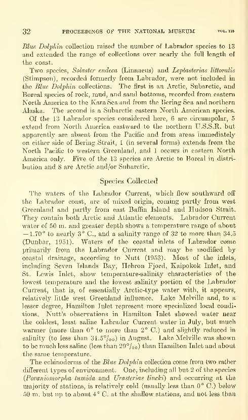
Blue Dolphin collection raised the number of Labrador species to 13
and extended the range of collections over nearly the full length of
the coast.
Two species, Solaster endeca (Linnaeus) and Leptasterias littoralis
(Stimpson), recorded formerly from Labrador, were not included in
the Blue Dolphin collections. The first is an Arctic, Subarctic, and
Boreal species of rock, mud, and sand bottoms, recorded from eastern
North America to the Kara Sea and from the Bering Sea and northern
Alaska. The second is a Subarctic eastern North American species.
Of the 13 Labrador species considered here, 6 are circumpolar, 5
extend from North America eastward to the northern U.S.S.R. but
apparently are absent from the Pacific and from areas immediately
on either side of Bering Strait, 1 (in several forms) extends from the
North Pacific to western Greenland, and 1 occurs in eastern North
America only. Five of the 13 species are Arctic to Boreal in distri-
bution and 8 are Arctic and/or Subarctic.
Species Collected
The waters of the Labrador Current, which flow southward off
the Labrador coast, are of mixed origin, coming partly from west
Greenland and partly from east Bafiin Island and Hudson Strait.
They contain both Arctic and Atlantic elements. Labrador Current
water of 50 m. and greater depth shows a temperature range of about— 1.70° to nearly 3° C, and a salinity range of 32 to more than 34.5
(Dunbar, 1951). Waters of the coastal inlets of Labrador comeprimarily from the Labrador Current and may be modified bycoastal drainage, according to Nutt (1953). Most of the inlets,
including Seven Islands Bay, Hebron Fjord, Kaipokok Inlet, and
St. Lewis Inlet, show temperature-salinity characteristics of the
lowest temperature and the lowest salinity portion of the Labrador
Current, that is, of essentially Arctic-type water with, it appears,
relatively little west Greenland influence. Lake Melville and, to a
lesser degree, Hamilton Inlet represent more specialized local condi-
tions. Nutt's observations in Hamilton Inlet showed water near
the coldest, least saline Labrador Current water in July, but muchwarmer (more than 0° to more than 2° C.) and shghtly reduced in
salinity (to less than 31.5°/oo) in August. Lake Melville was shown
to be much less saline (less than 29%©) than Hamilton Inlet and about
the same temperature.
The echinoderms of the Blue Dolphin collection come from two rather
different types of envhonment. One, including all but 2 of the species
(Poraniomorpha tumida and Urasterias lincki) and occurring at the
majority of stations, is relatively cold (usually less than 0° C.) below
50 m. but up to about 4° C. at the shallow stations, and not less than
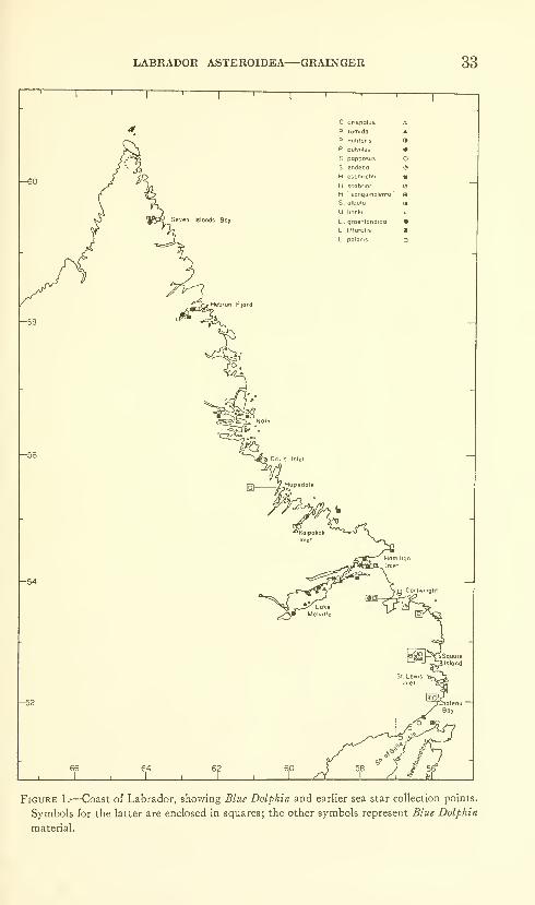
LABRADOR ASTEROIDEA—GRAINGER 33
Seven Islands Boy
1
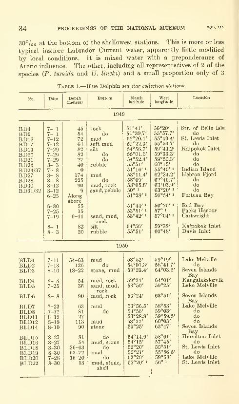
34 PROCEEDINGS OF THE NATIONAL MUSEUM
30°/oo at the bottom of the shallowest stations. This is more or less
typical inshore Labrador Current water, apparently little modified
by local conditions. It is mixed water with a preponderance of
Arctic influence. The other, including all representatives of 2 of the
species (P. tumida and U. lincki) and a small proportion only of 3
Table 1.—Blue Dolphin sea star collection stations.
No. Date Depth(meters)
Bottom Northlatitude
Westlongitude
Location
1949
BD4

LABRADOR ASTEROIDEA—GRAINGER
Table 1.—Blue Dolphin sea star collection stations—Continued
35
No. Date Depth(meters)
Bottom Northlatitude
Westlongitude
Location
1951
BLDl,
36 PROCEEDINGS OF THE NATIONAL MUSEXJM vol. 115
of the Canadian Atlantic provinces and New England. Poranio-
morpha tumida appears not to have been recorded south of the
Labrador location given here. Others, like Pteraster pulvillus and
Henricia scabrior, are evidently rare south of Labrador.
Comparison of this material with Pettibone's (1956) report on the
Blue Dolphin polychaetes from Labrador illustrates interesting dis-
tributional differences between the generally widely dispersed poly-
chaete species and the more restricted sea stars. Of 68 species of
polychaetes, only 9% (compared with 62% of the asteroids) are
Arctic-Subarctic and 37% (38% of the asteroids) are Arctic-to-Boreal
;
the remainder, including 25% with "cosmopolitan" distribution,
show no counterpart among the sea stars.
I am grateful to Dr. F. M, Bayer of the United States National
Museum for making the Blue Dolphin collection available for study
and to Mr. D. C. Nutt for providing station data.
Locations of all collection stations are shown in figure 1. TheBlu£ Dolphin station list is given in table 1.
Ctenodiscus crispatus (Retzius)
Asterias crispata Retzius, 1805, p. 17.
Ctenodiscus crispatus (Retzius).—Fisher, 1911, p. 31, pis. 3 (figs. 1-4), 4 (figs 1-6).
Collected at 9 stations; 27 to 174 m.; mud, silt, rock; 1949: BDI6(47 specmiens), BD17 (1), BD19 (14), BD21 (4), BD27 (4), BD30 (1);
1950: BLD18 (10), BLD19 (23); 1951: BLDl, 11, 34 (13).
All but 4 individuals have the usual 5 rays; 3 from BDI6 and 1
from BD21 have only 4. Two 5-rayed individuals show apparently
regenerating rays, 1 with a single new ray, the other with 2. Diameters
range from 6 to 78 mm., the majority being larger than 30 mm.These specimens were collected (fig. 1) between Hebron and St.
Lewis Inlet, and the species may be expected to occur along the full
length of the Labrador coast. It is limited here to cold water of high
salinity (fig. 2), to water of — 1° C. and less, and of a little less than
31%o to33°/oo. Typically a mud-bottom form, it occurred ahiiost
exclusively on mud in these collections. It is a circumpolar species,
ranging from Arctic to Boreal seas,
Poraniomorpha tumida (Stuxberg)
Solaster tumida Stuxberg, 1878, p. 31, pi. 6.
Asterina tumida tuberculata Danielssen and Koren, 1884, p. 63, pis. 10 (figs. 5-7),
15 (fig. 3).
Paraniomorpha tumida (Stuxberg).—Mortenson, 1910, pp. 257-258, pi. 16 (figs,
6,7).
Collected at 8 stations: 27-146 m.; mud, sand, rock; 1950: BLDl(4 specimens), BLD2 (3), BLD5 (4), BLD7 (2), BLD8 (2); 1951:
BLD41 (2); 1952: I-2a, b (3), 1-17 (2).

LABRADOR ASTEROIDEA—GRAINGER ar
rAAAA/.-
-H scobrior ,
C, crispotus
'-'WW-7o«S 15 20 21 22 23 24 25 26 27 28 29 30 31 32 33
Figure 2.—Temperature-salinity ranges of the Blue Dolphin sea star specimens.
All the specimens show at least some characteristics of the form
tuherculata Danielssen and Koren, described as differing from
tumida in having relatively longer rays, in possessing dorsal
tubercles, and in having only single rather than double rows of
adambulacral spines. Arm radii are from 9 to 36 mm., R :r from 1.7 :1
to 2.7:1. Dorsal tubercles, present in all, vary greatly in their degree
of development, from only faint bulges to strongly developed pro-
tuberances spread conspicuously over the dorsal surface. There is
some evidence of correlation between size and tubercle development,
the largest sea stars frequently showing the most highly developed
tubercles. Adambulacral spines show a rather bewildering range of
variation, most being arranged in only a single row but many occurring
in 2 rows per plate. One-row series range from 3 to 5, having most
frequently 4 or 5 per plate. The majority of specimens show both
1-row and 2-row adambulacral plates, usually irregularly arranged,
the latter with up to 8 spines per plate. While there appears to be
some correlation between size and degree of development (above),
there is none apparent between either size and K,:r values or between
size and adambulacral spine arrangement; that is, large specimens
with well-developed dorsal tubercles (characteristic of form tuherculata)
may have low R:r values and frequent occurrence of 2 rows
of spines per adambulacral plate (characteristic of tumida). Little
work on variation within either form has been done to date. It ap-
pears from this collection that form tuherculata must be defined muchmore widely than was done by Danielssen and Koren, and that its
differences from the stem form are far less distinct than they were
formerly thought to be. Possession of dorsal tubercles and presence
of only a single row of spines on the majority of adambulacral plates
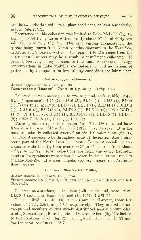
38 PROCEEDINGS OF THE NATIONAL MUSEUM vol. ub
are the two criteria used here to place specimens, at least tentatively,
in form tuberculata.
Occm-rence in this collection was limited to Lake Melville (fig. 1),
that is, to relatively warm water, mostly above 0° C, of fairly lowsalinity, 25 to 28.5 (fig. 2). This is a curious circumstance, the
species being known from North America eastward to the Kara Sea,
in Arctic and Subarctic waters. Its apparent total absence from the
outer coastal waters may be a result of insufiicient collecting. If
present, however, it may be assumed that numbers are small. Largeconcentrations in Lake Melville are undeniable, and indications of
preference by the species for low salinity conditions are fairly clear.
Solaster papposus (Linnaeus)
Asterias papposa Linnaeus, 1767, p. 1098.
Solaster papposus (Linnaeus).—Fisher, 1911, p. 325, pi. 94 (figs. 1-6).
Collected at 24 stations; 13 to 225 m.; mud, rock, rubble; 1949:
BD4 (1 specimen), BD5 (2), BD16 (8), BD24 (1), BD28 (1), BD30(7), Davis Inlet (4); 1950: BLDS (2), BLD4 (1), BLD14 (1), BLD15(1), BLD16 (3), BLD18 (2), BLD19 (1), BLD22 (2); 1951: BLDl,11, 34 (2), BLD3 (2), BLD5 (1), BLD19/39 (3), BLD40 (1), BLD44(2); 1952: I-2a, b (1), I-ll (1), 1-16 (2).
These specimens range in diameter from 7 to 176 mm. and havefrom 9 to 13 rays. More than half (53%) have 12 rays. It is the
most abundantly collected asteroid on the Labrador coast (fig. 1),
as indeed it appears to be throughout most of the eastern Arctic-Sub-
arctic part of the North American coast. Temperature-salinity tol-
erance is wide (fig. 2), from nearly —2° to 4° C, and from about
25°/oo to 33%o. Most collections are from the outer Labradorcoast; a few specimens were taken, however, in the outermost reaches
of Lake Melville. It is a circumpolar species, ranging from Arctic to
Boreal waters.
Pteraster militaris (O. F. Miiller)
Asterias militaris 0. F. Miiller, 1776, p. 234.
Pteraster militaris (O. F. Miiller) .—M. Sars, 1861, p. 48, pis. 3 (figs. 8, 9) 4, 5, 6
(figs. 1-13).
Collected at 3 stations; 82 to 108 m.; silt, sandy mud, stone. 1949:
BD20 (1 specimen), Kaipokok Inlet (1); 1951: BLD3 (1).
The 3 individuals, 148, 118, and 70 mm. in diameter, show R:r
values of 2.4:1, 2.6:1, and 2.3:1 respectively. They are rather un-
exceptional members of this widely distributed nearly circumpolar,
Arctic, Subarctic and Boreal species. Occurrence here (fig. 1) is limited
to two locations which (fig. 2) have high salinity of nearly 33 andlow temperature of near —2° C.
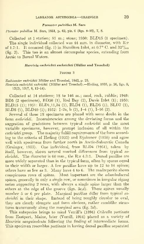
LABRADOR ASTEROIDEA—GRAINGER 39
Pteraster pulvillus M. Sars
Pteraster pulvillus M. Sars, 1861, p. 62, pis. 6 (figs. 4-18), 7, 8.
Collected at 1 station; 81 m.; stone; 1950: BLD15 (1 specimen).
The single individual collected was 44 mm. in diameter, with R:r
of 1.7:1. It occurred (fig. 1) in Hamilton Inlet, at 0.7° C. and 327oo
(fig. 2). This too is an almost circumpolar species, extending from
Arctic to Boreal Waters.
Henricia eschrichti eschrichti (Miiller and Troschel)
Figure 3
Eschinasier eschrichtu Miiller and Troschel, 1842, p. 25.
Henricia eschrichti eschrichti (Mttller and Troschel).—Heding, 1935, p. 26, figs. 8,
12(2), 13(7, 8, 12-14).
Collected at 14 stations; 18 to 146 m.; mud, rock, rubble; 1949:
BD5 (2 specimens), BD30 (1), Red Bay (2), Davis Inlet (1); 1950:
BLD15 (1); 1951: BLDl, 11,34 (1), BLD4 (1), BLD5 (1), BLD7 (1),
BLD8 (1), BLD40 (1); 1952: I-2a, b (2), 1-4 (1), 1-16 (2).
Several of these 18 specimens are placed with some doubt in the
form eschrichti. Inconsistencies among the deviating forms and the
suggestion of gradations between typical eschrichti and the most
variable specimens, however, prompt inclusion of all within the
eschrichti group. The majority fulfill requirements of the form accord-
ing to the criteria of Heding (1935) and Djakonov (1950) and agree
well with specimens from farther north in Arctic-Subarctic Canada
(Grainger, 1955). One individual, from BLD4 (1951), taken by
itself, however, shows several marked differences from typical es-
chrichti. The diameter is 60 mm., the E.:r 4.3:1. Dorsal paxillae are
more widely separated than in the typical form, often by spaces equal
to their width or larger. A few paxillae have up to 12 to 14 spines;
others have as few as 3. Many have 4 to 6. The madreporite shows
conspicuous rows of spines. Most important are the adambulacral
spines, generally in only a single row, or sometimes in irregular zigzag
series suggesting 2 rows, with always a single spine larger than the
others at the edge of the groove (figs. 3c,oJ). These spines usually
number 5 or 6 per plate. Marginal paxillae differ from typical es-
chrichti in their shape. Instead of being roughly circular to oval,
they are clearly elongate and form obvious, rather comblike struc-
tures transversely along the marginal area (fig. 2td).
This subspecies brings to mind Verrill's (1894) Cribrella pectinata
from Eastport, Maine, later (VerriU, 1914) placed as a variety of
Henricia sanguinolenta following the finding of intermediate forms.
This specimen resembles pectinata in having dorsal paxillae separated
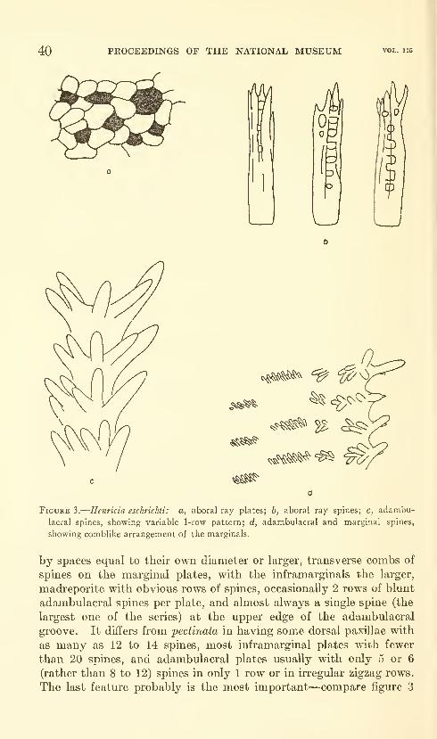
40 PROCEEDINGS OF THE NATIONAL MUSEUM vol. 115
^^'^
^^!^^
^^^"^ 2^ c^
x^^J»^'^^
Figure 3.
—
Henricia eschrichti: a, aboral ray plates; b, aboral ray spines; c, adambu-
lacral spines, showing variable 1-row pattern; d, adambulacral and marginal spines,
showing comblike arrangement of the marginals.
by spaces equal to their own diameter or larger, transverse combs of
spines on the marginal plates, with the inframarginals the larger,
madreporite with obvious rows of spines, occasionally 2 rows of blunt
adambulacral spines per plate, and almost always a single spine (the
largest one of the series) at the upper edge of the adambulacral
groove. It differs from pectinata in having some dorsal paxillae with
as many as 12 to 14 spines, most inframarginal plates with fewer
than 20 spines, and adambulacral plates usually with only 5 or 6
(rather than 8 to 12) spines in only 1 row or in irregular zigzag rows.
The last feature probably is the most important—compare figure 3

LABRADOR ASTEROIDEA—GRAINGER 41
with Verrill (1914, pi. 49, fig. la). This specimen closely resembles
Crihrella pectinata but is not identical with it, nor does the specimen
seem to be a distinct form by itself. Examination of other speci-
mens shows wide variation in marginal comb structure, with forms
intermediate between the advanced comb noted above and the
typical eschrichti comb. Spacing of the dorsal paxUlae varies between
the open pattern above and the fairly close arrangement character-
istic of eschrichti. Adambulacral spines frequently show 2 rows per
plate, with either a single large spine at the upper edge of the gi'oove
or, as in typical eschrichti, 2 more or less equally large spines at the
groove margin. One specimen shows all structures except the marginal
combs closely resembling the extreme form described above. Themarginal spines are typical for eschrichti. In others the character-
istics are blended differently. It seems impossible, therefore, to set
these specimens up as a distinct form ; rather they must be considered
merely as variations of the form eschrichti.
Specimens in this collection (fig. 1) were taken all along the Labrador
coast from Seven Islands Bay to the Strait of Belle Isle and from the
outermost portion of Lake Melville. The ranges of temperature and
salinity (nearly —2° to 4° C. and 25 to 33°/oo) are wide. The range of
form eschrichti cannot yet be described because of the doubtful
identity of many former records of Henricia sanguinolenta, including
two from southern Labrador shown in figure 1. The form eschrichti
appears to be primarily Subarctic and Arctic, and it extends from the
Barents Sea westward at least to eastern North America and south-
ward at least to the coast of New England.
Henricia scabrior (Mikhailovskii)
Figure 4
Cribella sanguinolenta forma scabrior Mikhailovskii, 1902, pp. 478-480.
Henricia scabrior (Mikhailovskii).—Heding, 1935, p. 31, fig. 11, 12(5, 6), 13
(9-11).—Djakonov, 1950, p. 92, pis. 44, 45.
Collected at 6 stations; 36 to 110 m.; silt, mud, rock; 1949: BDl9(3 specimens), Kaipokok Inlet (1); 1950: BLD15 (1), BLD16 (1);
1951: BLD19/39 (1), BLD40 (1).
Among only 8 individuals there is considerable structural variation.
This species is obviously one of the more plastic of this extremely
variable genus. Heding (1935) expanded somewhat on Mikhailovskii's
(1903) original description of the species. Much greater variability
was shown by Djakonov (1950), and it is on the basis of his concept
of the species that the individuals of the present collection are placed
in H. scabrior; this assignment is made with awareness that fm"ther
collecting may show subspecific separations which present material
is not sufficiently extensive to reveal.
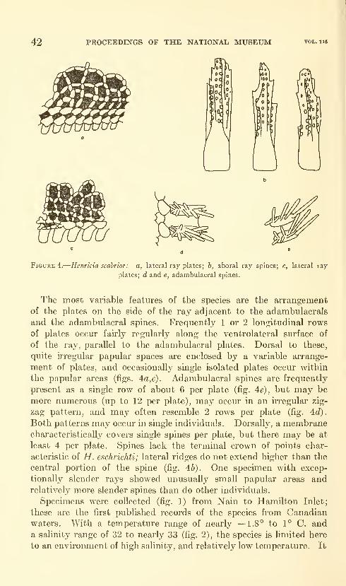
42 PROCEEDENGS OF THE NATIONAL MUSEUM
&SSy^mmn
Figure 4.
—
Henricia scabrior: a, lateral ray plates; h, aboral ray spines; c, lateral ray
plates; d and e, adambulacral spines.
The most variable features of the species are the arrangement
of the plates on the side of the ray adjacent to the adambulacrals
and the adambulacral spines. Frequently 1 or 2 longitudinal rows
of plates occur fairly regularly along the ventrolateral surface of
of the ray, parallel to the adambulacral plates. Dorsal to these,
quite uTegular papular spaces are enclosed by a variable arrange-
ment of plates, and occasionally single isolated plates occur within
the papular areas (figs. 4a,c). Adambulacral spines are frequently
present as a single row of about 6 per plate (fig. 4e), but may be
more numerous (up to 12 per plate), may occur in an irregular zig-
zag pattern, and may often resemble 2 rows per plate (fig. ^d).
Both patterns may occm- in single individuals. Dorsally, a membranecharacteristically covers single spines per plate, but there may be at
least 4 per plate. Spines lack the terminal crown of points char-
acteristic of H. eschrichti; lateral ridges do not extend higlier than the
central portion of the spine (fig. 46). One specimen with excep-
tionally slender rays showed unusually small papular areas andrelatively more slender spines than do other individuals.
Specimens were collected (fig. 1) from Nain to Hamilton Inlet;
these are the first published records of the species from Canadianwaters. With a temperature range of nearly —1.8° to 1° C and
a sahnity range of 32 to nearly 33 (fig. 2), the species is hmited here
to an environment of high salinity, and relatively low temperature. It
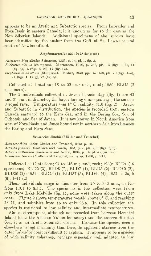
LABRADOR ASTEROIDEA—GRAINGER 43
appears to be an Arctic and Subarctic species. From Labrador and
Foxe Basin in eastern Canada, it is known as far to the east as the
New Siberian Islands. Additional specimens of the species have
been identified by the author from the Gulf of St. Lawrence and
south of Newfoundland.
Stephanasterias albula (Stimpson)
Asteracanthion albulus Stimpson, 1853, p. 14, pi. 1, fig. 5.
Stichaster albilus (Stimpson).—Mortensen, 1910, p. 267, pis. 13 (figs. 1-6), 14
(fig. 8), 15 (figs. 8-10), 17 (fig. 12).
Stephanasterias albula (Stimpson).—Fisher, 1930, pp. 157-159, pis. 70 (figs. 1-5),
71 (figs. 1, la-g), 72 (fig. 5).
Collected at 1 station; 18 to 22 m.; rock, mud; 1950: BLD3 (2
specimens)
.
The 2 individuals collected in Seven Islands Bay (fig. 1) are 42
and 31 mm. in diameter, the larger having 6 unequal rays, the smaller
3 equal rays. Temperature was 1° C, salinity 31.6 (fig. 2). Arctic
and Subarctic in distribution, the species is recorded from eastern
Canada eastward to the Kara Sea, and in the Bering Sea, Sea of
Okhotsk, and Sea of Japan. It is not known in North America from
west of Foxe Basin and Jones Sound nor in northern Asia from between
the Bering and Kara Seas.
Urasterias linckii (Miiller and Troschel)
Asteracanthion linckii Miiller and Troschel, 1842, p. 18.
Asterias gunneri Danielssen and Koren, 1884, p. 7, pis. 2, 3 (figs. 8, 9).
Asterias stellionura Danielssen and Koren, 1884, p. 14, pi. 4 (figs. 1-9).
Urasterias linckii (Miiller and Troschel).—'Fisher, 1930, p. 211.
Collected at 12 stations; 27 to 146 m.; mud, rock; 1950: BLDl (16
specunens), BLD2 (3), BLD5 (7), BLD7 (4), BLD8 (2), BLDl 2 (2),
BLD20 (2); 1951: BLD23 (1), BLD37 (3), BLD41 (4); 1952: I-2a, b
(6), 1-17 (2)._
These individuals range in diameter from 20 to 330 mm., in Il:r
from 4.3:1 to 8.5:1. The specimens in this collection were taken
only from Lake Melville (fig. 1) ; none were taken along the outer
coast. Figure 2 shows temperatures mostly above 0° C. and reaching2° C, and salinities from 15 to only 28.5. In this collection the
species is restricted to low salinity and intermediate temperatures.
Almost circumpolar, although not recorded from between Herschel
Island (near the Alaskan-Yukon boundary) and the eastern Siberian
Sea, it is an Arctic-Subarctic species. Because the species occurs
elsewhere in higher salinity than here, its apparent absence from the
outer Labrador coast is difficult to explain. It appears to be a species
of wide salinity tolerance, perhaps especially well adapted to low
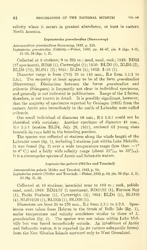
44 PROCEEDINGS OF THE NATIONAL MUSEUM vol. ii5
salinity where it occurs in greatest abundance, at least in eastern
North America.
Leptasterias groenlandica (Steenstrup)
Asteracanthion groenlandicus Steenstrup, 1857, p. 228.
Leptasterias groenlandica (Lutkin).—Fisher, 1930, pp. 45-47, pis. 8 (figs. 1-3),
21-23, 24 (figs. 1, 2).
Collected at 9 stations; 9 to 225 m.; mud, sand, rock; 1949: BD28(17 specimens), BD30 (1), Cartwright (1) ; 1950: BLD3 (5), BLD5 (2),
BLD8 (?1), BLDll (2); 1951: BLD4 (1); 1952: 1-16 (1).
Diameter range is from (?10) 20 to 110 mm., R:r from 3.1:1 to
5.8:1. The majority at least appear to be of the form groenlandica
(Steenstrup). Distinction between the forms groenlandica and
cribraria (wStimpson) is frequently not clear in individual specimens,
and generally is not indicated in publications. Range of the 2 forms,
therefore, is not known in detail. It is possibly significant, however,
that the majority of specimens reported by Grainger (1955) from the
eastern Arctic area immediately to the north of Labrador were called
cribraria.
One small individual of diameter 10 mm., E,:r 5.5:1 could not be
identified with certainty. Another specimen of diameter 41 mm.,
R:r 3.5:1 (station BLD4, July 29, 1951), enclosed 62 young stars
beneath its rays held in the brooding position.
The species was collected at stations along the whole length of the
Labrador coast (fig. 1), including 2 stations just within Lake Melville.
It was found (fig. 2) over a wide temperature range (less than —1°
to 4° C.) and a fairly wide salinity range (about 25%© to 33%o).It is a circumpolar species of Arctic and Subarctic waters.
Leptasterias polaris (Miiller and Troschel)
Asteracanthion polaris Miiller and Troschel, 1842, p. 16.
Leptasterias polaris (Miiller and Troschel).—Fisher, 1930, p. 60, pis. 30 (figs. 1, 2),
32 (fig. 3), 35.
Collected at 10 stations; intertidal zone to 110 m.; rock, pebble
sand, mud; 1949: BD24/37 (1 specimen), BD31/32 (1), Forteau Bay
(1), Packs Harbour (1), Cartwright (3); 1951: BLD4 (1), BLDlO(l),BLDl9/39 (1),BLD35 (1),BLD36 (1).
Diameters are from 25 to 270 mm., R:r from 3.7:1 to 5.2:1. Spec-
imens were taken from Hebron to the Strait of Belle Isle (fig. 1),
under temperature and salinity conditions similar to those of L.
groenlandica (fig. 2). The species was not taken within Lake Mel-
ville but was found immediately outside. An inhabitant of Arctic
and Subarctic waters, it is reported (in its various subspecific forms)
from the New Siberian Islands eastward only to West Greenland.
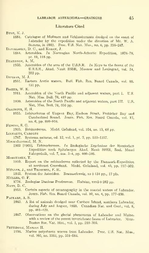
LABRADOR ASTEROIDEA—GRAINGER 45
Literature Cited
Bush, K. J.
1884. Catalogue of MoUusca and Echinodermata dredged on the coast of
Labrador by the expedition under the direction of Mr. W. A.
Stearns, in 1882. Proc. U.S. Nat. Mus., no. 6, pp. 236-247.
Danielssen, D. C, and Koren, J.
1884. Asteroidea. In Norwegian North-Atlantic Expedition, 1876-78,
pt. 11, 118 pp.
Djakonov, a. M.1950. Asteroidea of the seas of the U.S.S.R. In Keys to the fauna of the
U.S.S.R., Akad. Nauk SSSR, Moscow and Leningrad, vol. 34,
202 pp.
Dunbar, M. J.
1951. Eastern Arctic waters. Bull. Fish. Res. Board Canada, vol. 88.
131 pp.
Fisher, W. K.
1911. Asteroidea of the North Pacific and adjacent waters, part I. U.S.
Nat. Mus. Bull. 76, 419 pp.
1930. Asteroidea of the North Pacific and adjacent waters, part III. U.S.
Nat. Mus. Bull. 76, 356 pp.
Grainger, E. H.
1955. Echinoderms of Ungava Bay, Hudson Strait, Frobisher Bay andCumberland Sound. Journ. Fish. Res. Board Canada, vol. 12,
no. 6, pp. 899-916.
Heding, S. G.
1935. Echinoderms. Medd. Gronland, vol. 104, no. 13, 68 pp.Linnaeus, Carolus
1767. Systema naturae, ed. 12, vol. 1, pt. 2, pp. 533-1327.
MikhaIlovskiI, M. N.
1902 [1903]. Echinodermen. In Zoologische Ergebnisse der RussischenExpedition nach Spitzbergen Akad. Nauk SSSR, Zool. MuzeiEzhegodnik, vol. 7, nos. 3-4, pp. 460-546.
Mortensen, T.
1910. Report on the echinoderms collected by the Danmark-Expeditionat northeast Greenland. Medd. Gronland, vol. 45, pp. 237-302.
MtJLLER, J., and Troschel, F. H.
1842. System der Asteriden. Braunschweig, xx+ 134 pp., 12 pis.
MtJLLER, O. F.
1776. Zoologiae Danicae Prodromus. Hafniae, xxxii+ 282 pp.NUTT, D. C.
1953. Certain aspects of oceanography in the coastal waters of Labrador.Journ. Fish. Res. Board Canada, vol. 10, no. 4, pp. 177-186.
Packard, A. S.
1863. A list of animals dredged near Caribou Island, southern Labrador,during July and August, 1860. Canadian Nat. and Geol., vol. 8,
pp. 401-429.
1867. Observations on the glacial phenomena of Labrador and Maine?with a review of the recent invertebrate fauna of Labrador. Mem.Boston Soc. Nat. Hist., vol. 1, pp. 210-303.
Pettibone, Marian H.
1956. Marine polychaete worms from Labrador. Proc. U.S. Nat. Mus.,vol. 105, no. 3361, pp. 531-584.
46 PROCEEDINGS OF THE NATIONAL MUSEUM vol. ii»
Rankin, W. M.1901. Echinoderms collected off the west coast of Greenland by the Prince-
ton Arctic Expedition of 1899. Proc. Acad. Nat. Sci. Philadelphia,
vol. 53, pp. 169-181.
Retzitjs, a. J.
1805. Dissertatio sistens species cognitas Asteriarum. Lundae, 37 pp.Sars, M.
1861. Oversigt af Norges Echinodermer. Christiania, 160 pp.Steenstrup, J. J. S.
1857. Om de ved Gronland forekommende Asteracanthion-arter. Forh.
Skandinaviske Naturf., vol. 7, pp. 228-232.
Stimpson, William1853. Synopsis of the marine invertebrata of Grand Manan. Smithsonian
Contrib. Knowl., vol. 6, art. 5, 66 pp., 3 pis.
Stuxberg, Anton1878. Echinodermer fran Novaja Semljas haf samlade under Nordenskiold-
ska expeditionerna 1875 och 1876. Ofvers. Svenska Vetensk.-
Akad. Forh., Stockholm, vol. 35, no. 3, pp. 27-40, pi. 6.
Verrill, a. E.
1894. Descriptions of new species of starfishes and ophiurans, with a
revision of certain species formerly described, mostly from the
collections made by the United States Commission of Fish andFisheries. Proc. U.S. Nat. Mus., vol. 17, pp. 245-297.
1914. Monograph of the shallow-water starfishes of the North Pacific
coast from the Arctic Ocean to California. Harriman Alaska Ser.,
Smithsonian Inst., vol. 14, 408 pp.
U.S. GOVERNMENT PRINTrNG OFFICE:I'354
Proceedings of
the United States
National MuseumSMITHSONIAN INSTITUTION • WASHINGTON, D.C.
Volume 115 1964 Number 3479
MOTHS OF THE GENUS RHABDATOMIS DYAR(ARCTIIDAE: LITHOSIINAE)
By William D. Field
The genus of moths treated in this paper is neotropical in distribu-
tion and contains 13 species, 5 of which are described as new. One
of these species was included formerly in a second genus, for which a
type designation needs to be made. The following discussion will
clarify the nomenclature.
Hampson (1900, pp. 517, 518) described as new the genus Diar-
hahdosia with two included species: Eustixis minima Butler and
Lithosia laudamia Druce. Although Hampson usually selected a type
species for his genera, he failed to do so for this genus. Hampson 's
second included species, Lithosia laudamia Druce, is congeneric with
Rhabdatomis zaba Dyar, the sole species included in Rhabdatomis by
Dyar (1907, p. 227).
In order to preserve both names, Diarhabdosia and Rhabdatomis^
and also because Hampson used the "first species rule" in selecting
types for the genera of other authors, I designate as type species of
47
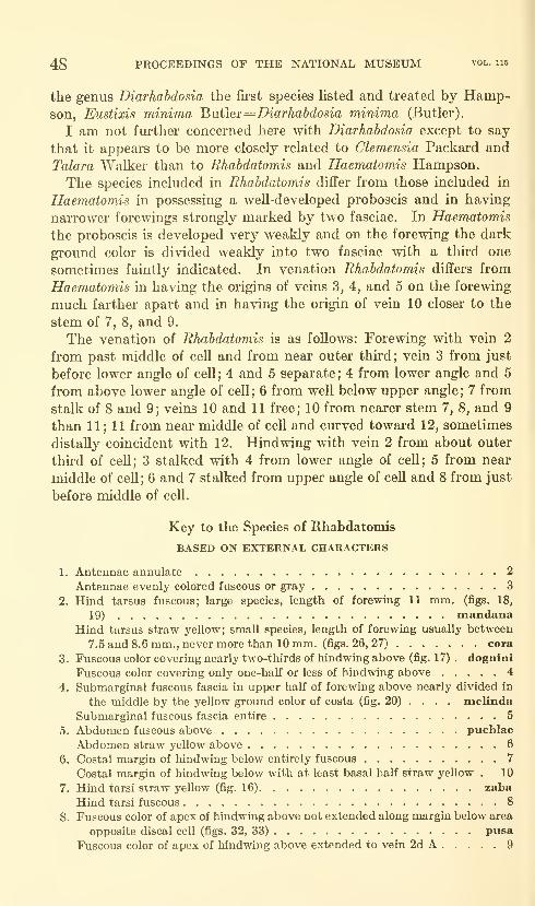
48 PROCEEDENTGS OF THE NATIONAL MUSEUM vol. 115
the genus Diarhabdosia the first species listed and treated by Hamp-son, Eustixis minima Butler=Diarhabdosia minima (Butler).
I am not further concerned here with Diarhabdosia except to say
that it appears to be more closely related to Clemensia Packard and
Talara Walker than to Rhabdatomis and Haematomis Hampson.
The species included in Rhabdatomis differ from those included in
Haematomis in possessing a well-developed proboscis and in having
narrower forewings strongly marked by two fasciae. In Haematomis
the proboscis is developed very wealdy and on the forewing the dark
ground color is divided weakly into two fasciae with a third one
sometimes faintly indicated. In venation Rhabdatomis differs from
Haematomis in having the origins of veins 3, 4, and 5 on the forewing
much farther apart and in having the origin of vein 10 closer to the
stem of 7, 8, and 9.
The venation of Rhabdatomis is as follows: Forewing with vein 2
from past middle of cell and from near outer third; vein 3 from just
before lower angle of ceU; 4 and 5 separate; 4 from lower angle and 5
from above lower angle of cell; 6 from weU below upper angle; 7 from
stalk of 8 and 9; veins 10 and 11 free; 10 from nearer stem 7, 8, and 9
than 11 ; 11 from near middle of cell and curved toward 12, sometimes
distally coincident with 12. Hindwing with vein 2 from about outer
third of cell; 3 stalked with 4 from lower angle of cell; 5 from near
middle of cell; 6 and 7 stalked from upper angle of cell and 8 from just
before middle of cell.
Key to the Species of Rhabdatomis
BASED ON EXTERNAL CHARACTERS
1. Antennae annulate 2
Antennae evenly colored fuscous or gray 3
2. Hind tarsus fuscous; large species, length of forewing 11 mm, (figs. 18,
19) mandanaHind tarsus straw yellow; small species, length of forewing usually between
7.5 and 8.6 mm., nevermore than 10 mm. (figs. 26, 27) cora
3. Fuscous color covering nearly two-thirds of hindwing above (fig. 17) . dognini
Fuscous color covering only one-half or less of hindwing above 4
4. Submarginal fuscous fascia in upper half of forewing above nearly divided in
the middle by the yellow ground color of costa (fig, 20) ... . melinda
Submarginal fuscous fascia entire 5
5. Abdomen fuscous above pueblae
Abdomen straw yellow above 6
6. Costal margin of hindwing below entirely fuscous 7
Costal margin of hindwing below with at least basal half straw yellow . 10
7. Hind tarsi straw yellow (fig. 16) zaba
Hind tarsi fuscous 8
8. Fuscous color of apex of hindwing above not extended along margin below area
opposite discal cell (figs. 32, 33) pusa
Fuscous color of apex of hindwing above extended to vein 2d A 9
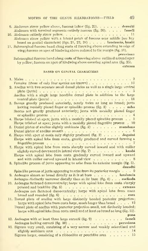
MOTHS OF THE GENUS RHABDATOMIS—FIELD 49
9. Abdomen straw yellow above, fuscous below (fig. 23) draudti
Abdomen with terminal segments entirely iuscous (fig. 30) fasseli
10. Abdomen entirely straw yellow 11
Abdomen straw yellow with a lateral patch of fuscous near middle [see key
based on genital characters] (figs. 21, 22, 24) laudamia, knabi
11. Submarginal fuscous band along costa of forewing above extending to edge of
wing; fuscous on apex of hindwing above reduced to the margin (fig. 31).
peruviana
Submarginal fuscous band along costa of forewing above outlined around apex
by yellow; fuscous on apex of hindwing above covering apical area (fig. 25).
extensa
BASED ON GENITAL CHARACTERS
1. Males 2
Females (those of only four species are known) 13
2. Anellus with two separate small dorsal plates as well as a single large ventral
plate (juxta) 3
Anellus with a single large hoodlike dorsal plate in addition to the large
ventral plate (juxta) 11
3. Saccus greatly produced anteriorly, nearly twice as long as broad; juxta
lacking mesially placed finger or spinelike process (fig. 1) zaba
Saccus not greatly produced anteriorly; juxta with mesially placed finger
or spinelike process 4
4. Harpe bilobed at apex; juxta with a mesially placed spinelike process . . 5
Harpe trilobed at apex; juxta with a mesially placed fingerUke process . 10
5. Dorsal plates of anellus shghtly scobinate (fig. 3) mandanaDorsal plates of anellus smooth 6
6. Harpe with apex at costa only slightly produced (fig. 2) dognini
Harpe with apical lobe from costa, greatly produced and curved forming a
fingerlike process 7
7. Harpe with apical lobe from costa sharply curved inward and with cuiller
shghtly curved downward in lateral view (fig. 7) knabi
Harpe with apical lobe from costa gradually curved inward and upwardand with cuiller curved upward in lateral view 8
8. Spinelike process of juxta appearing to arise from its anterior margin (fig. 5).
draudti
Spinehke process of juxta appearing to arise from its posterior margin ... 9
9. Aedeagus almost as broad distally as it is at base laudamiaAedeagus distinctly narrower distally than at its base (fig. 4) . . . melinda
10. Aedeagus flattened dorsoventrally ; harpe with apical lobe from costa sharply
pointed and beaklike (fig. 8) extensa
Aedeagus not flattened dorsoventrally; harpe with apical lobe from costa
broad and rounded (fig. 6) cora
11. Dorsal plate of anellus with large distinctly hooded posterior projection;
harpe with apical lobe from costa large, much longer than broad .... 12
Dorsal plate of anellus with posterior projection small, not distinctly hooded;
harpe with apical lobe from costa small and at least as broad as long (fig. 1 1)
.
pusa
12. Aedeagus with at least three large cornuti (fig. 9) fasseli
Aedeagus lacking cornuti (fig. 10) peruviana
13. Signum very small, consisting of a very narrow and weakly sclerotized andslightly scobinate area 14
Signum larger, consisting of a ribbonlike or pouchhke area 15
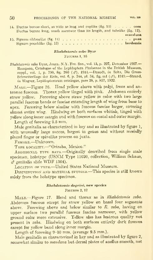
50 PROCEEDINGS OF THE NATIONAL MUSEUM vol. ub
14. Ductus bursae short, as wide as long and cuplike (fig. 15) cora
Ductus bursae long, much narrower than its length, and tubelike (fig. 12).
mandana15. Signum ribbonUke (fig. 14) pusa
Signum pouchlike (fig. 13) laudamia
Rhabdatomis zaba Dyar
Figures 1, 16
Rhabdatomis zaba Dyar, Journ. N.Y. Ent. See, vol. 15, p. 227, December 1907.
—
Hampson, Catalogue of the Lepidoptera Phalaenae in the British Museum,
suppl., vol. 1, p. 796, fig. 260 (cf), 1914.—Draudt, in Seitz, Die Gross-
SchmetterUnge der Erde, vol. 6, p. 244, pi. 34, fig. ml (cf), 1919.—Strand,
in Wagner, Lepidopterorum catalogus, pars 26, p. 837, 1922.
Male.—Figure 16. Head yellow above with palpi, frons and an-
tennae fuscous. Thorax yellow tinged with pink. Abdomen entirely
straw yellow. Forewing above straw yellow in color with two large
parallel fuscous bands or fasciae extending length of wing from base to
apex. Forewing below similar with fuscous fasciae larger, covering
almost entire wing. Hindwing on both surfaces whitish, tinged with
yellow along inner margin and with fuscous on costal and outer margin.
Length of forewing 9.5 mm.Male genitalia as characterized in key and as illustrated by figure 1,
with unusually large saccus, largest in genus and without mesially
placed finger or spinelike process on juxta.
Female.—Unknown.Type locality.—^"Orizaba, Mexico."
Additional type data.—Originally described from single male
specimen, holotype (USNM Type 11020, collection, William Schaus,
cf genitaha shde WDF 1304).
Location of type.—United States National Museum.
Distribution and material studied.—This species is still known
solely from the holotype specimen.
Rhabdatomis dogninif new species
FlGTJEES 2, 17
Male.—Figure 17. Head and thorax as in Rhabdatomis zaba.
Abdomen fuscous except for straw yellow on basal four segments
above. Forewing above and below similar to R. zaba, having on
upper surface two parallel fuscous fasciae narrower, with yellow
ground color more extensive. Yellow also has lustrous quality not
present in zaba. Hindwing on both surfaces entirely dark fuscous
except for yeUow band along inner margin.
Length of forewing 9-10 mm. (average 9.5 mm.).
Male genitalia as characterized in key and as illustrated by figure 2,
somewhat similar to mandana but dorsal plates of anellus smooth, not

MOTHS OF THE GENUS RHABDATOMIS—FIELD 51
scobinate as in that species; short and broad aedeagus and cuiller muchnarrower apically.
Female.—Unknown.
Type locality,—Micay, department of Cauca, Colombia.
Additional type data.—Described from holotype, male (August
1896; USNM Type 34855; Dognin Collection; cf genitaHa sHde WDF1541), and one paratype, male (cf genitalia sHde WDF 1568; other
data as in holotype).
Location of type.—United States National Museum.
Distribution and material studied.—^This species is known solely
from the holotype and paratype.
Kemarks.—Species named for the late Paul Dognin.
Rhabdatomis mandana (Dyar), new combination
Figures 3, 12, 18, 19
Diarhabdosia mandana Dyar, Journ. N.Y. Ent. Soc, vol. 15, p. 228, December1907.—Hampson, Catalogue of the Lepidoptera Phalaenae in the British
Museum, suppl., vol. 1, p. 795, pi. 41, fig. 22 (cf), 1914.—Draudt, in
Seitz, Die Gross-Schmetterhnge der Erde, vol. 6, p. 244, pi. 33, fig. h7
(cf), 1919.—Strand, in Wagner, Lepidopterorum catalogus, pars 26, p. 837,
1922.
Male.—Figure 18. Head and thorax as in R. zaha. Abdomenstraw yellow, paler below. Forewing above and below similar to
zaha, except that fasciae enlarged with fuscous color extending
along both base and apex of costa and to inner margin near anal
angle. Hindwing on both surfaces whitish, tinged with yellow, even
on costal margin below and with small cloud of fuscous at apex muchsmaller on undersurface than above.
Length of forewing 9.5-11 mm. (average 10.1 mm.).
Male genitalia as characterized in key and as illustrated by figure
3. As already mentioned, it is characterized by dorsal plates of
anellus shghtly scobinate, by aedeagus greatly narrowed distally,
and by very broad cuiller from harpe.
Female.—Figure 19. Very similar to male, differing chiefly in
having hindAvings entirely fuscous except for yellow in middle of
outer margin above and below and on costal margin below. Adbomenyellowish brown.
Length of forewing 11 mm.Female genitalia as characterized in key and as illustrated by
figure 12. This species has a signum similar in shape to that of
cora but it is transverse rather than longitudinal in position. Other-
wise it differs greatly from cora in the length and shape of the ductus
bursae.

52 PROCEEDINGS OF THE NATIONAL MUSEUM vol. U6
Type locality.—"Castro, Parana, Brazil."
Additional type data.—Originally described from tliree males
and two females, no single specimen of which was stated to be "the
type" in the original description. All five of these specimens are
therefore syntypes (article 73, paragraphs b and c. International
Code of Zoological Nomenclature, adopted by XV International
Congress of Zoology, London, 1961). Two of these syntypes bear
the USNM type number assigned by Dyar: 10921. In addition, one
of these two specimens bears a written label containing the word
"type" in Dyar's handwiiting. I hereby designate this specimen as
the lectotype and have so labeled it (USNM type 10921; collection,
William Schaus; & genitalia slide WDF 1533).
Location of type.—United States National Museum.Distribution.—Brazil: Castro, State of Parana., and Rio de
Janeiro.
Material studied.—Three males and two females.
Rhabdatomis melinda (Schaus), new combination
Figures 4, 20
Diarhabdosia melinda Schaus, Ann. Mag. Nat. Hist., ser. 8, vol. 7, p. 369, April
1911.—Hampson, Catalogue of the Lepidoptera Phalaenae in the British
Museum, suppl., vol. 1, p. 795, pi. 41, fig. 21 (d"), 1914.—Draudt, in Seitz,
Die Gross-Schmetterlinge der Erde, vol. 6, p. 244, pi. 33, fig. h6 (cf ), 1919.—
Strand, in Wagner, Lepidopterorum catalogus, pars 26, p. 837, 1922-
Male.—Figure 20. Head straw yellow vvdth faint fuscous spot
between antennae, which are also fuscous. Thorax yellow, tinged
with pink. Abdomen entirely straw yellow. Forewing above and
below with fuscous in upper half of wing greatly reduced, entirely
lacking in base of wing but extended toward costa near middle
and also at apex. Hindwing on both surfaces pale straw yellow
with small dark shade near apex and small faint spot on outer mar-
gin of upper surface near anal angle.
Length of forewing 9 mm.Male genitalia as characterized in key and as illustrated by figure 4,
shows close relationship to draudti and laudamia. From both species
it differs greatly in form and shape of cuiller and aedeagus.
Female.—Unknown.Type locality.—"Banana River, Costa Rica."
Additional type data.—Originally described from single male
specimen, holotype (March 1907; USNM Type 17258; collection,
WiUiam Schaus; c? genitaha slide WDF 1532).
Location of type.—United States National Musemn.Distribution and material studied.—This species is still known
solely from the holotype specimen.
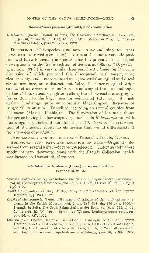
MOTHS OF THE GENUS RHABDATOMIS—FIELD 53
Rhabdatomis pueblae (Draudt), new combination
Diarhabdosia pueblae Draudt, in Seitz, Die Gross-Schmetterlinge der Erde, vol.
6, p. 243, pi. 33, fig. h3 (cf), h4 (9), 1919.—Strand, in Wagner, Lepidop-
terorum catalogus, pars 26, p. 837, 1922.
Discussion.—This species is unknown to me and, since the types
have been destroyed (see below), its true status and taxonomic posi-
tion will have to remain in question for the present. The original
description from the English edition of Seitz is as follows: "D. pueblae
spec. nov. (33 h) is very similar [compared with laudamia Druce, a
discussion of which preceded this description], with longer, moreslender wings, and a more pointed apex; the costal-marginal and discal
stripes are finer, more distinct, not faded, the inner-marginal stripe
somewhat narrower, more uniform. Hindwing at the proximal angle
in the cf less extended, lighter yellow, the whole costal area grey as
far as down to the lower median vein; anal tuft rosy. 9 muchdarker, hindwings quite unicolorously blacldsh-grey. Expanse of
wings: 28 to 30 mm. Described according to several couples from
Tehuacan (Mexico [Puebla])." The illustration of the male shows
this sex as having the forewings very much as in R. laudamia but with
hindwings very dark and more like those of R. dognini. The illustra-
tion of the female shows no characters that would differentiate it
from females of laudamia.
Type locality and distribution.—Tehuacan, Puebla, Mexico.
Additional type data and location of type.—Originally de-
scribed from several pairs, holotype not selected. Unfortunately, these
specimens were destroyed along with the Draudt Collection, which
was located in Darmstadt, Germany,
Rhabdatomis laudamia (Druce), new combination
Figures 13, 21, 22
Lithosia laudamia Druce, in Godman and Salvin, Biologia Centrali-Americana,
vol. 39 (Lepidoptera-Heterocera, vol. 1), p. 131; vol. 41 (vol. 3), pi. 13, fig. 4
(cf ). 1885.
Crambidia laudamia (Druce), Kirby, A synonymic catalogue of Lepidoptera
Heterocera, p. 338, 1892.
Diarhabdosia laudamia (Druce), Hampson, Catalogue of the Lepidoptera Pha-
laenae in the British Museum, vol. 2, pp. 517, 518, fig. 370 (cf), 1900.
—
Draudt, in Seitz, Die Gross-Schmetterlinge der Erde, vol. 6, p. 243, pi. 33,
fig. hi (cf), h2 (9), 1919.—Strand, in Wagner, Lepidopterorum catalogus,
pars 26, p. 837, 1922.
Lithosia pusa Dognin, Hampson not Dognin, Catalogue of the Lepidoptera
Heterocera in the British Museum, vol. 2, p. 518, 1900.—Draudt not Dognin,
in Seitz, Die Gross-Schmetterlinge der Erde, vol. 6, p. 243, 1919.—Strand
not Dognin, in Wagner, Lepidopterorum catalogus, pars 26, p. 837, 1922.
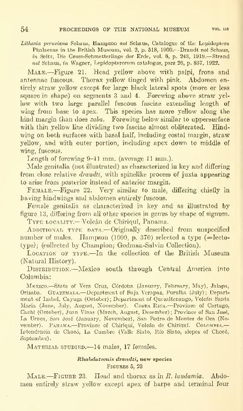
54 PROCEEDINGS OF THE NATIONAL MUSEUM vol. us
Lithosia peruviana Schaus, Hampson not Schaus, Catalogue of the Lepidoptera
Phalaenae in the British Museum, vol. 2, p. 518, 1900.—Draudt not Schaus,
in Seitz, Die Gross-SchmetterUnge der Erde, vol. 6, p. 243, 1919.—Strand
not Schaus, in Wagner, Lepidopterorum catalogus, pars 26, p. 837, 1922.
Male.—Figure 21. Head yellow above with palpi, frons and
antennae fuscous. Thorax yellow tinged with pink. Abdomen en-
tirely straw yellow except for large black lateral spots (more or less
square in shape) on segments 3 and 4. Forewing above straw yel-
low with two large parallel fuscous fasciae extending length of
wing from base to apex. This species has more yellow along the
hind margin than does zaba. Foremng below similar to uppersurface
with thin yellow line dividing two fasciae almost obliterated. Hind-
wing on both surfaces with basal half, including costal margin, straw
yellow, and with outer portion, including apex down to middle of
wing, fuscous.
Length of forewing 9-11 mm. (average 11 mm.).
Male genitalia (not illustrated) as characterized in key and differing
from close relative draudti, with spinelike process of juxta appearing
to arise from posterior instead of anterior margin.
Female.—Figure 22. Very similar to male, differing chiefly in
having hindwings and abdomen entirely fuscous.
Female genitalia as characterized in key and as illustrated byfigure 13, differing from all other species in genus by shape of signum.
Type locality.—Volcan de Chiriqui, Panama.Additional type data.—Originally described from unspecified
number of males. Hampson (1900, p. 370) selected a type (=lecto-
type); (collected by Champion; Godman-Salvin Collection).
Location of type.—In the collection of the British Museum(Natural History).
Distribution.—Mexico south through Central America into
Colombia
:
Mexico.—State of Vera Cruz, C6rdoba (January, February, May), Jalapa,
Orizaba. Guatemala.—Department of Baja Verapaz, Purulha (July) ; Depart-
ment of Izabel, Cayuga (October) ; Department of Quezaltenango, Volcdn Santa
Maria (June, July, August, November). Costa Rica.—Province of Cartago,
Cachi (October), Juan Vinas (March, August, December); Province of San Jos6,
La Uruca, San Jos6 (January, November), San Pedro de Montes de Oca (No-
vember). Panama.—Province of Chiriquf, Volcdn de Chiriqui. Coloaibia.—Intendencia de Choc6, La Cumbre (Valle Siato, Rio Siato, slopes of Choc6,
September)
.
Material studied.—14 males, 17 females.
Rhabdatomis draudti, new species
Figures 5, 23
Male.—Figure 23. Head and thorax as in R. laudamia. Abdo-
men entirely straw yellow except apex of harpe and terminal four

MOTHS OF THE GENUS RHABDATOMIS—FIELD 55
segments underneath, which are fuscous. Forewing above very
similar to R. laudamia, with parallel fuscous fasciae separated byslightly more distinct yellow line. Forewing below as in laudamia.
Hindwing on both sm-faces with costal margin and apical half of outer
margin fuscous, thus differing from lauda7nia which has basal half of
costal margin straw yellow.
Length of forewing 11-12.5 mm. (average, 11.6 mm.).
Male genitalia as characterized in key and as illustrated by figm-e 5.
See remai'ks under R. laudamia.
Female.—Unknown.Type locality.—Tuis, Province of Cartago, Costa Rica.
Additional type data.—Described from holotype, male (Aug.
28, 1908; USNM type 34856; Schaus Collection; cf genitalia slide
WDF 1318), from five male paratypes (same locality as holotype;
Sept. 1, 1908, Aug. 29, 31, 1908; Schaus Collection), and a sixth male
paratype (Cartago, Costa Rica; cf genitalia slide WDF 1558; Schaus
Collection).
Location of type.—United States National Museum.
Distribution and material studied.—This species is knownsolely from the holotype and paratypes listed above.
Remarks.—Species named for Dr. Max Draudt.
lihabdatomis knabi, new species
Figures 7, 24
Male.—Figm-e 24. Head and thorax as in R. laudamia and
R. draudti. Abdomen entu^ely straw yellow except for lateral oblong
patches of fuscous on segments 3 and 4. Both forewing and hindwing,
above and below, entirely like laudamia.
Length of forewing 9.5-10.5 mm.Male genitalia as characterized in key and as illustrated by figure 7
differs from all other species in genus in form of harpe, aedeagus, and
juxta. Hai'pe has apical lobe from costa sharply curved inward and
cuiller slightly curved downward. Juxta has sharp spinelike pro-
jection from posterior margin. Aedeagus has distal lobe projecting
from middle of left lateral sm'face.
Female.—Unknown.Type locality.—Cordoba, State of Vera Cruz, Mexico.
Additional type data.—Described from holotype, male (Jan.
5, 1908; USNM Type 34857; Frederick Knab, collector; cT genitalia
slide WDF 1550), and from one paratype, male (locality as given for
holotype; Feb. 8, 1908; Frederick Knab, collector; <^ genitalia slide
WDF 1549).
Location of type.—United States National Museum.679-426—64 2

56 PROCEEDINGS OF THE NATIONAL MUSEUM vol. ii5
Distribution and material studied.—This species is knownsolely from the holotype and paratype listed above.
Remarks.—Species named for Mr. Frederick Knab.
Rhabdatomis extensa, new species
Figures 8, 25
Male.—Figiu-e 25. Head and thorax as in R. laudamia. Abdomenentirely straw yellow Avith few fuscous scales covering apices of harpes.
Forewing above and below entu-ely like B. laudamia. Hindwing on
both surfaces with basal area and entne costal margin yellow, even
at apex (especially noticeable on undersm-face). In laudamia only
the basal half of costal margin is yellow. Apex of hindwing (except
for costal margin) fuscous as in laudamia.
Length of forewing 9-10 mm.Male genitalia as characterized in key and as illustrated b}" figure
25. This is the only species in the genus mth a beaklike apical lobe
on the costa of the harpe, with an aedeagus that is distinctly flattened
dorsoventrally, and with a uniquely shaped juxta.
Female.—Unknown.Type locality.—Yuntas (near Cali), Department of Valle del
Cauca, Colombia.
Additional type data.—Described from holotype, male (USNMtype 34848; d^ genitalia slide WDF 1554), from one paratype, male
(Esperanza, Guanacaste, Costa Rica; May; cT genitalia slide WDF1556), and second paratype, male (Cayuga, Izabal, Guatemala; April;
Schaus and Barnes, collectors; genitalia slide WDF 1555).
Location of type.—United States National Museum.Distribution and material studied.—Guatemala, Costa Rica,
and Colombia. This species is known solely from the holotype and
paratypes.
Rhabdatomis cora (Dyar), new combination
Figures 6, 26, 27
Diarhabdosia cora Dj'ar, Journ. N.Y. Ent. Soc, vol. 15, p. 227, December 1907.
—
Hampson, Catalogue of the Lepidoptera Phalaenae in the British Museum,
suppl., vol. 1, p. 794, pi. 41, fig. 19 ( cf ), 1914.—Draudt, in Seitz, Die Gross-
Schmetterlinge der Erde, vol. 6, p. 243, pi. 33, fig. g8 (cT), 1919.—Strand,
in Wagner, Lepidopterorum catalogus, pars 26, p. 836, 1922.
Male.—Figure 26. Head yellow above with palpi and frons fus-
cous. Thorax yellow, tinged with pink. Antenna annulate. Abdo-
men entirely straw yellow. Forewing above and below very similar
to R. laudamia except that fuscous-colored fasciae have more lilaceous
cast. Hindwuig straw yellow with outer border fuscous, this color
broken in middle by straw yellow ground color.

MOTHS OF THE GENUS RHABDATOMIS—FIELD 57
Length of forewing 6.5-7.5 mm. (average 6.8 mm.).
Male genitalia as characterized in key and as illustrated by figure 6.
This species, along with extensa, has a harpe that is trilobed at its
apex; it differs from extensa in the shape of all three of these lobes
as well as in the shape of the juxta and aedeagus.
Female.—Figure 27. Very similar to male, differing chiefly in
having hindwing and abdomen entirely fuscous, except for narrowyellow border on costal margin of hindwing below.
Length of forewing 8.5-9 mm. (average 8.8 mm.).
Female genitalia as characterized in key, having verj'^ small signum,
consisting of very narrow and weakly sclerotized and slightly scobi-
nate area and with short cuplike ductus biu-sae.
Type locality.—St. Jean, Maroni River, French Guiana.
Additional type data.—Originally described from 15 males and3 females, no single specimen of which was stated to be "the type"
in the original description. All 18 of these specimens are therefore
syntypes (article 73, paragraphs b and c, International Code of
Zoological Nomenclature, adopted by XV International Congress of
Zoology, London, 1961). Two of these syntypes bear the USNMtype number 10920 assigned by Dyar. In addition, one of these
two specimens bears a wiitten label containing the word "type" in
Dyar's handwriting. I hereby designate this specimen as the lectotype
and have so labeled it (USNM Type 10920; collection, William
Schaus; cf genitaha sHde WDF 1526).
Location of type.—United States National Museum.Distribution,—French Guiana and Brazil:
French guiana.—Maroni District, St. Jean, Maroni River. Brazil.—Pard.
Material studied.—14 males and 4 females.
Rhabdatoniis cora coroides (Schaus), new combination and new status
Figures 28, 29
Diarhabdosia coroides Scliaus, Ann. Mag. Nat. His., ser. 8, vol. 7, p. 368, April
1911.—Hampson, Catalogue of the Lepidoptera Phalaenae in the British
Museum, suppl., vol. 1, pp. 794, 795, pi. 41, fig. 20 (cf), 1914.—Dyar, Proc.
U.S. Nat. Mus., vol. 47, p. 168 (cf), May 20, 1914.—Draudt, w Seitz, DieGross-Schmetterlinge der Erde, vol. 6, p. 243, pi. 33, fig. h5 (cf), 1919.—Strand, in Wagner, Lepidopterorum catalogus, par.? 26, p. 830, 1922.
Male.—Figure 28. The name coroides can be retained to repre-
sent a northern subspecies. It is very similar to Rhabdatomis cora
cora (Dyar), differing slightly in size {see below) and in having a
great deal more fuscous on the hindwings.
Length of forewing 7-8 mm. (average 7.5 mm.).Male genitaha entirely like that of typical subspecies illustrated
by figure 6.
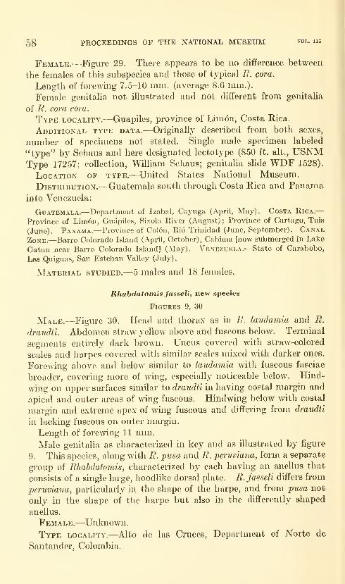
58 PROCEEDINGS OF THE NATIONAL MUSEUM vol. 115
Female.—Figm-e 29. There appears to be no difference between
the females of this subspecies and those of typical B. cora.
Length of forewdng 7.5-10 mm. (average 8.6 mm.).
Female genitalia not illustrated and not different from genitalia
of R. cora cora.
Type locality.—Guapiles, province of Limon, Costa Rica.
Additional type data.—Originallj^ described from both sexes,
number of specimens not stated. Single male specimen labeled
"type" by Schaus and here designated lectotype (850 ft. alt., USNMType 17257; collection, William Schaus; genitaha shde WDF 1528).
Location of type.—United States National Museum.
Distribution.—Guatemala south through Costa Rica and Panama
into Venezuela:
Guatemala.—Department of Izabal, Cayuga (April, May). Costa Rica.—Province of Lini6u, Gudpiles, Sixola River (August) ; Province of Cartago, Tuis
(June). Panama.—Province of Col6n, Rio Trinidad (June, September). Canal
Zone.—Barro Colorado Island (April, October), Cabima [now submerged in Lake
Gatun near Barro Colorado Island] (May). Venezuela.—State of Carabobo,
Las Quiguas, San Esteban Valley (July)
.
Material studied.—5 males and 18 females.
Rhabdatomis fasseli, new species
Figures 9, 30
Male.—Figure 30. Head and thorax as in R. laudamia and R.
draudti. Abdomen straw yellow above and fuscous below. Terminal
segments entirely dark brown. Uncus covered with straw-colored
scales and harpes covered with similar scales mixed with darker ones.
Forewing above and below similar to laudamia with fuscous fasciae
broader, covering more of mng, especially noticeable below. Hind-
wmg on upper surfaces similar to draudti hi having costal mai-gin and
apical and outer areas of wing fuscous. Hindwing below with costal
margin and extreme apex of wing fuscous and differing from draudti
in lacking fuscous on outer margin.
Length of forewing 11 mm.Male genitalia as characterized in key and as illustrated hj figiu'e
9. This species, along with R. pusa and R. jperuviana, form a separate
group of Rhabdatomis, characterized by each having an anellus that
consists of a single large, hoodlike dorsal plate. R. fasseli differs from
peruviana, particularly in the shape of the harpe, and from pusa not
only in the shape of the harpe but also in the differently shaped
anellus.
Female.—Unknown.
Type locality.—Alto de las Cruces, Department of Norte de
Santander, Colombia.
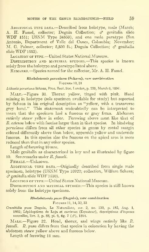
MOTHS OF THE GENUS RHABDATOMIS—FIELD 59
Additional type data.—Described from holotype, male (March;
A. H. Fassel, collector; Dognin Collection; cf genitalia slide
WDF 1551; USNM Type 34859), and one male paratype (San
Antonio, Department of Valle del Cauca, Colombia; November;
M. G. Palmer, collector; 5,800 ft.; Dognin Collection; cf genitalia
slide WDF 1552).
Location of type.—United States National Musemn.Distribution and material studied.—This species is known
solely from the holotype and paratype listed above.
Remarks.—Species named for the collector, Mr. A. H. Fassel.
Rhabdatomis peruviana (Schaus), new combination
Figures 10, 31
Lithosia peruviana Schaus, Proc. Zool. Soc. London, p. 239, March 6, 1894.
Male.—Figm^e 31. Thorax yellow, tinged with pink. Headmissing in holotype (sole specimen available for study) but reported
by Schaus in his original description as "yellow, with a transverse
grey band." This statement undoubtedly can be interpreted to
mean that the specimen had a fuscous or gray frons. Abdomenentirely straw yellow in color. Forewing above most like that of
R. extensa below with fasciae larger than in that species. In hindwing
peruviana differs from all other species in genus by costal margin
colored differently above than below, upperside yellow and underside
fuscous. In this species also the fuscous on the apical area is morereduced than that in any other species.
Length of forewing 10 mm.Male genitalia as characterized in key and as illustrated by figure
10. See remarks under R. fasseli.
Female.—Unknown.Additional type data.—Originally described from single male
specimen, holotype (USNM Type 10922; collection, William Schaus;
d' genitaha sHde WDF 1530).
Location of type.—United States National Museum.Distribution and material studied.—This species is still known
solely from the holotype specimen.
Rhabdatomis pusa (Dognin), new combination
Figures 11, 14,32,33
Crambidia pusa Dognin, Le Naturaliste, ser. 2, no. 130, p. 185, Aug. 1,
1892; L^pidoptferes de Loja et environs (Equateur), descriptions d'^speces
nouveUes, livr. 3, p. 88, pi. 9, fig. 7 (cf), 1894.
Male.—Figure 32. Head, thorax, and wings entirely like R.
fasseli. R. jpusa differs from that species in coloration by having the
abdomen straw yellow above and fuscous below.
Length of forewing 11 mm.
60 PROCEEDINGS OF THE NATIONAL MUSEUM vol. us
Male genitalia as characterized in key and as illustrated by figure 1 1
.
See remarks under B. fasseli.
Female.'—Figure 33. Very similar to male, differing chiefly in
hindwing and abdomen being entirely fuscous. R. jpusa entirely like
female of laudamia in color and pattern.
Length of forewing 12 mm.Female genitalia as characterized in key and as illustrated by
figure 14. R. jpusa has long ribbon-like signum lengthwise in position.
Type locality.'—Loja, province of Loja, Ecuador.
Additional type data.-—Originally described from two female
specimens. One of these females bears a red, numbered USNM type
label and I designate this specimen the lectotype ("Environs de Loja,
Equateur"; November 1887; USNM Type 30580; Dognin Collection;
9 genitaha sHde WDF 1531).
Location of type.-—United States National Museum.Distribution.'—Ecuador.
Literature Cited
Dognin, Paul1892. Descriptions de l^pidoptfires nouveaux. Le Naturaliste, ser. 2, no.
130, p. 185, Aug. 1.
1894. L^pidopteres de Loja et environs (Equateur), descriptions d'^speces
nouvelles. Paris, livr. 3, pp. 71-99, pis. 7-10.
Dratjdt, Max1918-1919. Lithosiinae. In A. Seitz, Die Gross-Schmetterlinge der Erde
Stuttgart, vol. 6, pp. 241-293, pis. 32-37.
Drtjce, Herbert1881-1900. Lepidoptera-Heterocera, vols. 1 and 3. Vols. 39 and 41 in
Godman and Salvin, Biologia Centrali-Americana, 490 pp.; 101 pis.
Dtar, Harrison G.
1907. New American Lepidoptera. Journ. N.Y. Ent. Soc, vol. 15, pp.
226-234, December.
1914. Report on the Lepidoptera of the Smithsonian Biological Survey of
the Panama Canal Zone. Proc. U.S. Nat. Mus., vol. 47, pp.
139-350, May 20.
Hampson, George F.
1900. Catalogue of the Lepidoptera Phalaenae in the British Museum.
London, vol. 2, 589 pp., pis. 17-35.
1914. Catalogue of the Lepidoptera Phalaenae in the British Museum.
London, suppl., vol. 1, 858 pp., pis. 1-41 (1915).
KiRBY, William Forsell1892. A synonymic catalogue of Lepidoptera Heterocera. London, 951 pp.
ScHAus, William1894. On new species of Heterocera from tropical America. Proc. Zool.
Soc. London, pp. 225-243, March 6.
1911. New species of Heterocera from Costa Rica, VII. Ann. Mag. Nat.
Hist., ser. 8, vol. 7, pp. 355-372, April.
Strand, Embrik1922. Arctiidae: Lithosiinae. In H. Wagner, Lepidopterorum catalogus.
BerUn, pars 26, 899 pp.
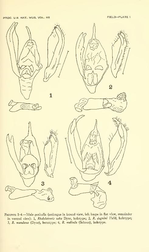
PROC. U.S. NAT. MUS. VOL. 115 FIELD—PLATE 1
Figures 1-4.—Male genitalia (aedeagus in lateral view, left harpe in flat view, remainder
in ventral view): 1, Rhabdatomis zaba Dyar, holotype; 2, R. dognini Field, holotype;
3, R. mandana (Dyar), lectotype; 4, R. melinda (Schaus), holotype.
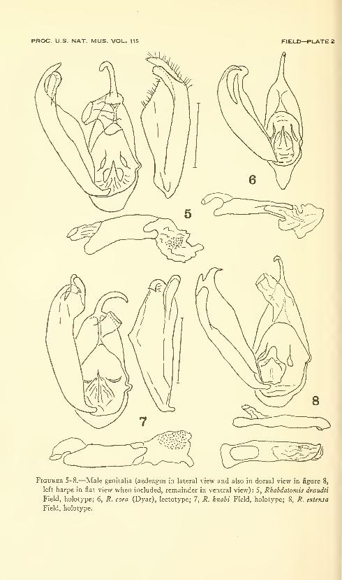
PROC. U.S. NAT. MUS. VOL. 115 FIELD—PLATE 2
Figures S-8.—Male genitalia (aedeagus in lateral view and also in dorsal view in figure 8,
left harpe in flat view when included, remainder in ventral view) : 5, Rhahdatomis draudti
Field, holotype; 6, R. cora (Dyar), lectotype; 7, R. knabi Field, holotype; 8, R. extensa
Field, holotype.

PROC. U.S. NAT. MUS. VOL. 115 FIELD—PLATE 3
Figures 9-11.—Male genitalia (aedeagus in lateral view, left harpe not shown, genital
capsule in ventral view): 9, Rhahdatomisfasseli Field, holotype; 10, R. peruviana (Schaus),
holotype; 11, R. pusa (Dognin), drawn from preparation WDF 1553,
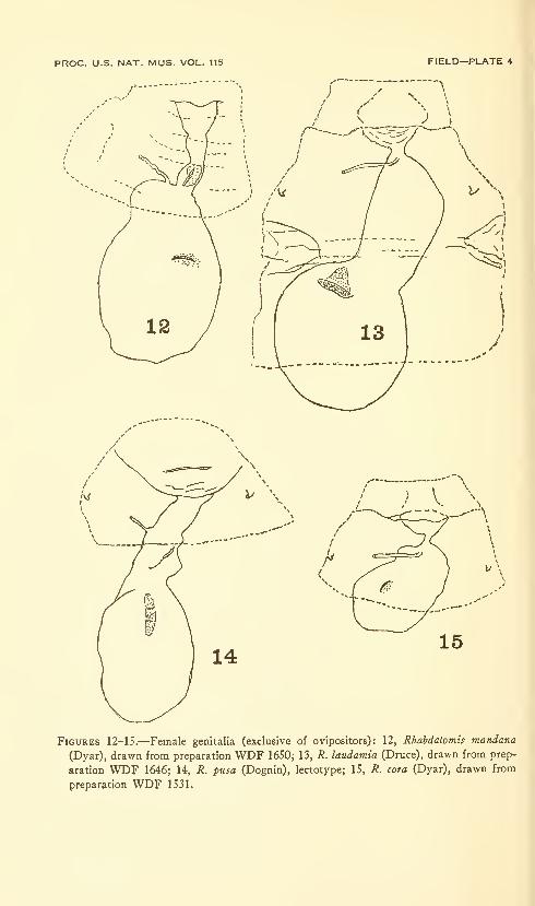
PROC. U.S. NAT. MUS. VOL. 115 FIELD—PLATE 4
; V. \.
Figures 12-15.—Female genitalia (exclusive of ovipositors): 12, Rhahdatomis mandana
(Dyar), drawn from preparation WDF 1650; 13, R. laudamia (Druce), drawn from prep-
aration WDF 1646; 14, R. pusa (Dognin), lectotype; 15, R. cora (Dyar), drawn from
preparation WDF 1531.


Proceedings of
the United States
National MuseumSMITHSONIAN INSTITUTION • WASHINGTON, B.C.
Volume 115 1964 Number 3480
NEOTROPICAL MICROLEPIDOPTERA, III
RESTRICTION OF GONIONOTA MELOBAPIIES WALSINGHAMWITH DESCRIPTIONS OF NEW SPECIES
(LEPIDOPTERA: OECOPHORIDAE)
By J. F. Gates Clarke
Since the description of Gonionota melobaphes Walsingham, a con-
siderable number of species of small moths has been placed under that
name in collections and, indeed, it seems entirely likely that Walsing-
ham's original series consisted of several species. Larger series from
widespread localities indicate that melobaphes is actually confined to
a relatively small geographical area and that other similarly colored
species of this complex are likewise restricted in their ranges.
In the U.S. National Museum there are specimens, determined as
melobaphes by Durrant and Meyrick, which are not referable to
Walsingham's species; moreover, Meyrick suppressed his own vexillata
in favor of melobaphes, yet vexillata is one of the most distinct species
of the group. All of these taxa need reconsideration.
In treating these and other South American forms, Meyrick placed
all of them in the European genus Hypercallia Stephens. Hypercallia
Stephens is based on Phalaena tortrix christiernana Linnaeus, a syno-
nym of H. citrinalis (Scopoli), which is abundantly distinct from any61

62 PROCEEDINGS OF THE NATIONAL MUSEUM vol. 115
of the South American species, and none of the latter are referable
to Hypercallia.
The genus Gonionota was proposed by Zeller ^ for 0. notodontella
Zeller.
Meyrick^ recognized the genus and stated: "I give the characters
of this genus, which was incompletely described by Zeller, who
regarded it as a group of Hypercallia, from which it is in fact very
distinct. . .." Later, in the same paragraph, he stated: "I am
informed by Mr. Busck that the typical species of Gonionota, G.
notodontella, ZelL, has vein 9 of fore-wings rising from the stalk of
7 and 8. . .." Walsingham ^ has pointed out: "Some error has
occurred here—the neuration of the Type of Gonionota notodontella Z.
is: FW: 12 veins; 7-8 stalked, 7 to termen; 9 and other veins separate;
2 twice as remote from 3 as 3 is from 4. HW: 8 veins; 3-4 connate;
5-7 parallel.—Drnt." Obviously Busck was not famihar with
notodontella (there are no specimens in the U.S. National Museum)
because he would not have overlooked such a clear character as the
relation of vein 9 in the forewing to 7 and 8. In 1922 * Meyrick
suppressed Gonionota in favor of Hypercallia and placed all the species
of this group in the latter genus.
Gonionota is a distinct genus, as pointed out by Meyrick in 1909,
and may be distinguished from Hypercallia by the uptm-ned palpus,
the third segment roughened posteriorly, the pubescent or short-
ciliated antenna of the male, the smooth antenna of the female, the
bifurcate uncus, and the presence of a clasper or other ornamentation
on the harpe. Gonionota is most nearly related to the South American
genus Co2)totelia, from which it is distinguished by the strong posterior
thoracic scale-tuft, by vein 9 of the forewing being widely separated
from the stalk of 7 and 8, and by the termen being convex. In the
hind wing, veins 3 and 4 are usually connate, but all species provide
examples with veins 3 and 4 stalked.
Gonionota melobaphes Walsingham
Figure 1; Plates 1 (Fig. 6), 2 (Fig. 1)
Gonionota melobaphes Walsingham, 1912, in Godman and Salvin, Biologia Centrali-
Americana, vol. 42 (Lepidoptera-Heterocera, vol. 4), p. 129, pi. 4, fig. 27.
—
Amsel, 1956, Bol. Ent. Venezolana, vol. 10 (nos. 1 and 2), p. 294, pi. 64
(fig. 5), pi. 108 (fig. 10).
Hypercallia melobaphes (Walsingham), Meyrick, 1922, in Wytsman, Genera
insectorum, fasc. 180, p. 162, no. 22; 1926, Exotic Microlepidoptera, vol. 3,
p. 314; 1930, Ann. Naturhist. Mus. Wien, vol. 44, p. 233.—Gaede, 1939,
in Bryk, Lepidopterorum catalogus, part 92, p. 261.
J 1877, Hor. Soc. Ent. Ross., vol. 13, p. 379.
2 1909, Trans. Ent. Soc. London, 1909, p. 24.
8 1912, in Godman and Salvin, Biologia Centrali-Amerlcana, vol. 42 (Lepidoptera-Heterocera, vol. 4),
p. 128.
* 1922, in Wytsman, Genera insectorum, fasc. 180, p. 161.
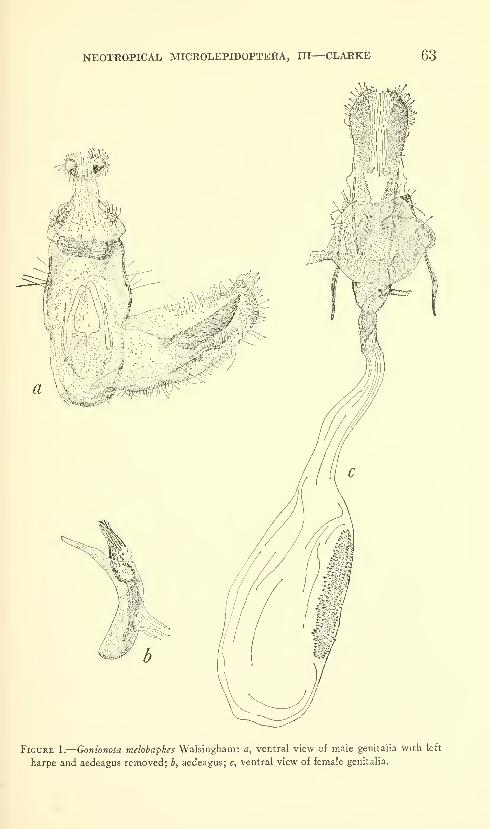
NEOTROPICAL MICROLEPIDOPTERA, III—CLARKE 63
Figure 1.
—
Gonionota melobaphes Walsingham: a, ventral view of male genitalia with left
harpe and aedeagus removed; b, aedeagus; c, ventral view of female genitalia.
64 PROCEEDINGS OF THE NATIONAL MUSEUM vol. us
Type: In the British Museum (Natural History).
Type locaUty: Chiriqui, Panama.Abdomen with strong, median, eversible scale-tuft from first ster-
num of male.
Male genitalia: Harpe broadest about middle, tapering abruptly to
a bluntly pointed cucullus; sacculus with short distal protuberance,
the latter with strong cluster of hairlike setae; from center of harpe a
stout clasper extends toward cucullus for about half the length of
harpe; clasper clothed with very strong setae for almost its whole
length. Anellus subquadrate, posteriorly divided into two long, digi-
tate processes, and with a long, slender, pointed process arising on
each side from about middle. Aedeagus stout, curved, with distal
end depressed, spatulate; vesica armed with strong cluster of cornuti.
Vinculum rounded, Gnathos terminating in a wide, curved plate
clothed with fine setae. Socii small, elongate lobes. Uncus ter-
minating in widely divergent curved processes.
Female genitalia: Posterior third of ductus bursae sclerotized and
dilated before ostium; inception of ductus seminalis dorsal, at point
of constriction of ductus bursae before ostium. Signum an elongate
dentate plate.
Slides examined: cfcf, AB 25.ii.1920; JFGC nos. 4232, 10122,
10123, 5096 (paratype in BM); 99, JFGC nos. 4233, 10125, 10126,
10127.
Although recorded from several widespread localities by Walsingham
and from Venezuela by Amsel, melobaphes appears to be restricted to
Central America. The distribution, according to specimens in the
U.S. National Museum, is as follows:
Costa Rica: Juan Viuas, 3 cf cf, 2 9 9; Sixola River, 399 (Wm. Schaus);
Panama: Cabima, 3 cfcf, 2 9 9 (May, 1911; August Busck).
One of the specimens from the Sixola River is labeled "Gonionota
melobaphes Wlsm." in Walsingham's handwriting.
Gonionota contrasta, new species
Figures 2a-d; Plate 1 (Fig. 4), 2 (Fig. 2)
Alar expanse 19-21 mm.Labial palpus buff yellow; second segment strongly overlaid with
reddish ocherous anteriorly and on outer side; third segment tinged
with reddish ocherous and with a cluster of black scales subapically.
Antennal scape silvery white ventrally, reddish ocherous dorsally;
remainder of antenna reddish ocherous basally shading to fuscous
apically. Proboscis silvery white. Head and thorax buff yeUowheavil}^ overlaid with reddish ocherous; posterior thoracic tuft tipped
fuscous. Forewing ground color sayal brown blotched and suffused
with fuscous; from base of wing broad, elongate, obhque reddish
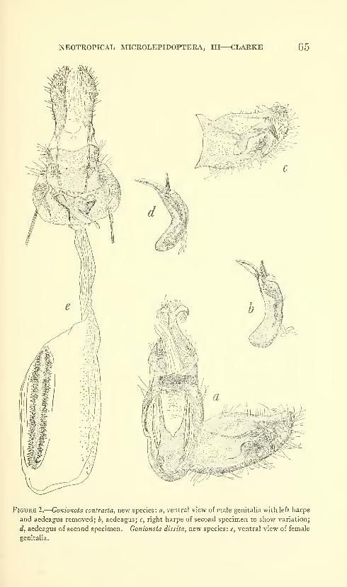
NEOTROPICAL MICROLEPIDOPTERA, III—CLARKE 65
Figure 2.
—
Gonionota contrasta, new species: a, ventral view of naale genitalia with left harpe
and aedeagus removed; b, aedeagus; c, right harpe of second specimen to show variation;
d, aedeagus of second specimen. Gonionota dissita, new species : e, ventral view of female
genitalia.
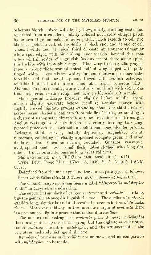
66 PROCEEDINGS OF THE NATIONAL MUSEUM vol. 115
ocherous blotch, mixed with buff yellow, nearly reaching costa and
separated from a smaller similarly colored outwardly oblique patch
by an arm of ground color; in outer patch, which extends to cell, two
blackish spots; in cell, at two-fifths, a black spot and at end of cell
a small white dot; at apical third of costa an elongate triangular
white spot edged with pink along inner margin; beyond this spot
a few whitish scales; cilia grayish fuscous except those along apical
thu-d white with faint pink tinge. Hind wing fuscous; ciha grayish
fuscous except those around apical half of wing tipped with pink-
tinged white. Legs silvery white; forefemur brown on inner side;
foretibia and first tarsal segment tinged with reddish ocherous;
midtibia blotched with brown; hind tibia tinged ocherous white.
Abdomen fuscous dorsally, white ventrally; anal tuft with violaceous
tint; fu'st sternum with strong, median, eversible scale tuft in male.
Male genitalia: Harpe broadest slightly before middle, costal
margin slightly excavate before cucuUus; saccular margin with
slightly curved digitate process extending about one-third distance
across harpe ; clasper a long arm from middle of harpe, terminating in
a cluster of strong setae directed toward and reaching saccular margin.
Anellus rectangular, deeply incised posteriorly forming two long,
pointed processes; on each side an additional long, slender process.
Aedeagus stout, curved, distally depressed, tonguelike; cornuti
numerous, consisting of closely appressed elongate group and stout
dentate series. Vinculum narrow, rounded. Gnathos transverse,
oval, spined knob. Socii small fleshy lobes clothed with long fine
setae. Uncus bifurcate, base as long as lateral arms.
Slides examined: cfcf, JFGC nos. 4630, 5098, 10116, 10121.
Type: Peru, Tingo Maria (Nov. 25, 1949, H. A. Allard), USNM66373.
Described from the male type and three male paratypes as follows:
Peru: 2cf cT, Callao (Mrs. M. J. Pusey); cT, Chanchamayo (Dognin Coll.).
The Chanchamayo specimen bears a label "Hypercallia melobaphes
Wals." in Meyrick's handwriting.
The superficial similarity between contrasta and vexillata is striking,
but the genitalia at once distinguish the two. The anellus of contrasta
exhibits long, slender lateral and terminal processes but vexillata lacks
them. Moreover, midway on the saccular margin of contrasta there
is a pronounced digitate process that is absent in vexillata.
The anellus and aedeagus of contrasta place it nearer melobaphes
than to any other species of this group but the digitate saccular proc-
ess of contrasta, absent in melobaphes, and the arrangement of the
cornuti immediately distinguish the two.
Females of contrasta and vexillata are unknown and no comparison
with melobaphes can be made.
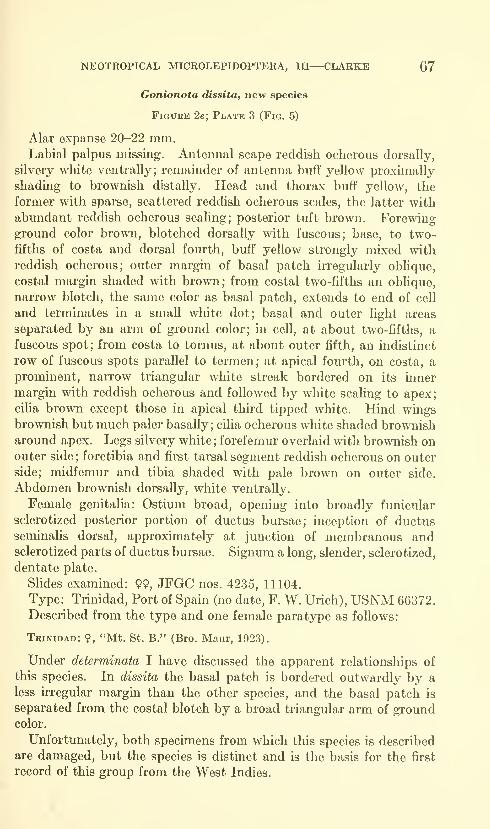
NEOTROPICAL MICROLEPIDOPTERA, III—CLARKE 67
Gonionota dissita, new species
Figure 2e; Plate 3 (Fig. 5)
Alar expanse 20-22 mm.Labial palpus missing. Antennal scape reddish ocherous dorsally,
silvery white ventrally; remainder of antenna buff yellow proximally
shading to brownish distally. Head and thorax buff yellow, the
former with sparse, scattered reddish ocherous scales, the latter with
abundant reddish ocherous scaling; posterior tuft brown. Forewingground color brown, blotched dorsally with fuscous; base, to two-
fifths of costa and dorsal fourth, buff yellow strongly mixed with
reddish ocherous; outer margin of basal patch irregularly oblique,
costal margin shaded with brown; from costal two-fifths an obhque,
narrow blotch, the same color as basal patch, extends to end of cell
and terminates in a small white dot; basal and outer light areas
separated by an arm of ground color; in cell, at about two-fifths, a
fuscous spot; from costa to tornus, at about outer fifth, an indistinct
row of fuscous spots parallel to termen; at apical fourth, on costa, a
prominent, narrow triangular white streak bordered on its inner
margin with reddish ocherous and followed by white scaling to apex;
ciha brown except those in apical third tipped white. Hind wingsbrownish but much paler basally ; cilia ocherous white shaded brownisharound apex. Legs silvery white ; forefemur overlaid with brownish onouter side ; foretibia and first tarsal segment reddish ocherous on outer
side; midfemur and tibia shaded with pale brown on outer side.
Abdomen brownish dorsally, white ventrally.
Female genitalia: Ostium broad, opening into broadly funicular
sclerotized posterior portion of ductus bursae; inception of ductus
seminalis dorsal, approximately at junction of membranous andsclerotized parts of ductus bursae. Signum a long, slender, sclerotized,
dentate plate.
Slides examined: 99, JFGC nos. 4235, 11104.
Type: Trinidad, Port of Spain (no date, F. W. Urich), USNM 66372.
Described from the type and one female paratype as follows:
Trinidad: ?, "Mt. St. B." (Bro. Maur, 1923).
Under determinata I have discussed the apparent relationships of
this species. In dissita the basal patch is bordered outwardly by a
less irregular margin than the other species, and the basal patch is
separated from the costal blotch by a broad triangular arm of groundcolor.
Unfortunately, both specimens from which this species is described
are damaged, but the species is distinct and is the basis for the first
record of this group from the West Indies.
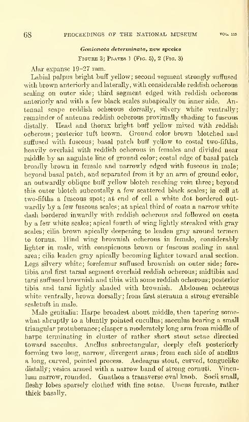
68 PROCEEDINGS OF THE NATIONAL MUSEUM vol. 115
Gonionota determinata, new species
Figure 3; Plates 1 (Fig. 5), 2 (Fig. 3)
Alar expanse 19-27 mm.Labial palpus bright buff yellow; second segment strongly suffused
with brown anteriorly and laterally, with considerable reddish ocherous
scaling on outer side; third segment edged with reddish ocherous
anteriorly and with a few black scales subapically on inner side. An-
tennal scape reddish ocherous dorsally, silvery white ventrally;
remainder of antenna reddish ocherous proximally shading to fuscous
distally. Head and thorax bright buff yellow mixed with reddish
ocherous; posterior tuft brown. Ground color brown blotched and
suffused with fuscous; basal patch buff yellow to costal two-fifths,
heavily overlaid with reddish ocherous in females and divided near
middle by an angulate line of ground color; costal edge of basal patch
broadly brown in female and narrowly edged with fuscous in male;
beyond basal patch, and separated from it by an arm of ground color,
an outwardly oblique buff yellow blotch reaching vein three; beyond
this outer blotch subcostally a few scattered black scales; in cell at
two-fifths a fuscous spot; at end of cell a white dot bordered out-
wardly by a few fuscous scales ; at apical third of costa a narrow white
dash bordered inwardly with reddish ocherous and followed on costa
by a few white scales; apical fourth of wing lightly streaked with gray
scales; ciha brown apically deepening to leaden gray around termen
to tornus. Hind wing brownish ocherous in female, considerably
lighter in male, with conspicuous brown or fuscous scaling in anal
area; ciUa leaden gray apically becoming lighter toward anal section.
Legs silvery white; forefemur suffused brownish on outer side; fore-
tibia and first tarsal segment overlaid reddish ocherous; midtibia and
tarsi suffused brownish and tibia with some reddish ocherous;posterior
tibia and tarsi lightly shaded with brownish. Abdomen ocherous
white ventrally, brown dorsally; from first sternum a strong eversible
scaletuft in male.
Male genitalia: Harpe broadest about middle, then tapering some-
what abruptly to a bluntly pointed cucuUus; sacculus bearing a small
triangular protuberance ; clasper a moderately long arm from middle of
harpe terminating in cluster of rather short stout setae directed
toward sacculus. Anellus subrectangular, deeply cleft posteriorly
forming two long, narrow, divergent arms; from each side of anellus
a long, curved, pointed process. Aedeagus stout, curved, tonguelike
distally; vesica armed with a narrow band of strong cornuti. Vincu-
lum narrow, rounded. Gnathos a transverse oval knob. Socii small,
fleshy lobes sparsely clothed with fine setae. Uncus furcate, rather
thick basally.

NEOTROPICAL MICROLEPIDOPTERA, III—CLARKE 69
>f^mjy
Figure 3.
—
Gonio^iota determinata, new species: a, ventral view of male genitalia with left
harpe and aedeagus removed; b, aedeagus; c, right harpe of second specimen illustrating
variation; d, aedeagus of second specimen; e, ventral view of female genitalia.
Female genitalia: Posterior half of ductus bursae sclerotized,
dilated before ostium; inception of ductus seminalis dorsal and
anterior to dilated portion of ductus bursae. Signum an elongate
dentate plate.
Slides examined: c^d", JFGC nos. 5096, 10128; 99, JFGC nos.
4234, 10120.
Type: British Guiana, Mallali (no date or collector), USNM 66374.
There is a white label attached to this specunen bearing the inscrip-
tion "Hypercallia melobaphes Wals." in Mejrrick's handwriting.
681-521—64-
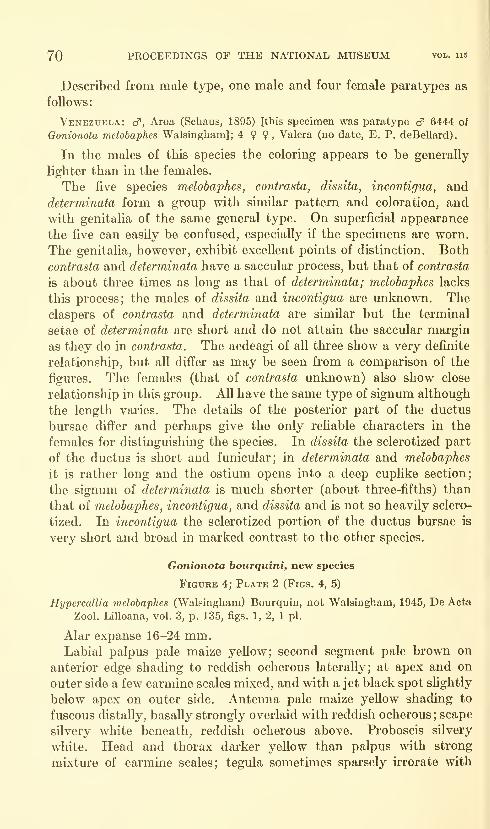
70 PROCEEDINGS OF THE NATIONAL MUSEUM vol. 115
Described from male type, one male and fom." female paratypes as
follows:
Venezuela: cf, Aroa (Schaus, 1895) [this specimen was paratype cf 6444 of
Qonionota melobaphes Walsingham]; 4 ? ?, Valera (no date, E. P. deBellard)
.
In the males of this species the coloring appears to be generally
lighter than in the females.
The five species melobaphes, contrasta, dissita, incontigua, and
determinata form a group with similar pattern and coloration, andwith genitalia of the same general type. On superficial appearance
the five can easily be confused, especially if the specimens are worn.
The genitalia, however, exhibit excellent points of distinction. Both
contrasta and determinata have a saccular process, but that of contrasta
is about three times as long as that of determinata; melobaphes lacks
this process; the males of dissita and incontigua are unknown. Theclaspers of contrasta and determinata are similar but the terminal
setae of determinata are short and do not attain the saccular margin
as they do in contrasta. The aedeagi of all three show a very definite
relationship, but all differ as may be seen from a comparison of the
figures. The females (that of contrasta unknown) also show close
relationship in this group. All have the same type of signum although
the length varies. The details of the posterior part of the ductus
bursae differ and perhaps give the only reliable characters in the
females for distinguishing the species. In dissita the sclerotized part
of the ductus is short and funicular; in determinata and melobaphes
it is rather long and the ostium opens into a deep cuplike section;
the signum of determinata is much shorter (about three-fifths) than
that of melobaphes, incontigua, and dissita and is not so heavily sclero-
tized. In incontigua the sclerotized portion of the ductus bursae is
very short and broad in marked contrast to the other species.
Gonionota bourquini, new species
Figure 4; Plate 2 (Figs. 4, 5)
Hypercallia melobaphes (Walsingham) Bourquin, not Walsingham, 1945, De Acta
Zool. Lilloana, vol. 3, p. 135, figs. 1, 2, 1 pi.
Alar expanse 16-24 mm.Labial palpus pale maize yeUow; second segment pale brown on
anterior edge shading to reddish ocherous laterally; at apex and on
outer side a few carmine scales mixed, and with a jet black spot slightly
below apex on outer side. Antenna pale maize yeUow shading to
fuscous distaUy, basaUy strongly overlaid with reddish ocherous; scape
silvery white beneath, reddish ocherous above. Proboscis silvery
white. Head and thorax darker yellow than palpus with strong
mixture of carmine scales; tegula sometimes sparsely irrorate with
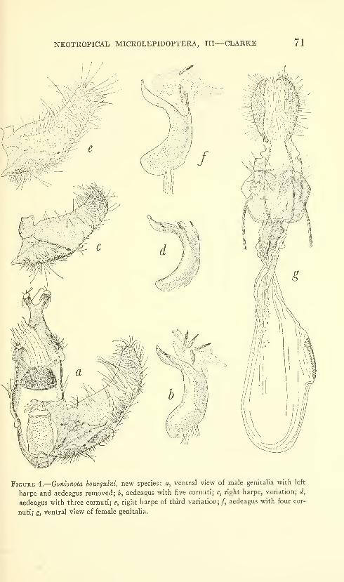
NEOTROPICAL MICROLEPIDOPTERA, III—CLARKE 71
Figure 4.
—
Gonionota bourguini, new species: a, ventral view of male genitalia with left
harpe and aedeagus removed; b, aedeagus with five cornuti; c, right harpc, variation; d,
aedeagus with three cornuti; e, right harpe of third variation;/, aedeagus with four cor-
nuti; g, ventral view of female genitalia.
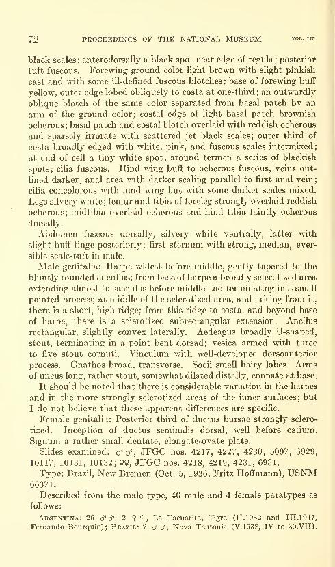
72 PROCEEDINGS OF THE NATIONAL MUSEUM vol. 115
black scales ; anterodorsally a black spot near edge of tegula;posterior
tuft fuscous. Forewing ground color light brown with slight pinkish
cast and with some ill-defined fuscous blotches; base of forewing buff
yellow, outer edge lobed obliquely to costa at one-third; an outwardly
oblique blotch of the same color separated from basal patch by an
arm of the ground color; costal edge of light basal patch brownish
ocherous ; basal patch and costal blotch overlaid with reddish ocherous
and sparsely irrorate with scattered jet black scales; outer third of
costa broadly edged with white, pink, and fuscous scales intermixed;
at end of cell a tiny white spot; around termen a series of blackish
spots; cilia fuscous. Hind wing buff to ocherous fuscous, veins out-
lined darker; anal area with darker scaling parallel to first anal vein;
cilia concolorous with hind wing but with some darker scales mixed.
Legs silvery white ; femur and tibia of foreleg strongly overlaid reddish
ocherous; midtibia overlaid ocherous and hind tibia faintly ocherous
dorsally.
Abdomen fuscous dorsally, silvery white ventrally, latter with
slight buff tinge posteriorly; first sternum with strong, median, ever-
sible scale-tuft in male.
Male genitalia: Harpe widest before middle, gently tapered to the
bluntly rounded cucullus; from base of harpe a broadly sclerotized area
extending almost to sacculus before middle and terminating in a small
pointed process; at middle of the sclerotized area, and arising from it,
there is a short, high ridge; from this ridge to costa, and beyond base
of harpe, there is a sclerotized subrectangular extension. Anellus
rectangular, slightly convex laterally. Aedeagus broadly U-shaped,
stout, terminating in a point bent dorsad; vesica armed with three
to five stout cornuti. Vinculum with well-developed dorsoanterior
process. Gnathos broad, transverse. Socii small hairy lobes. Armsof uncus long, rather stout, somewhat dilated distally, connate at base.
It should be noted that there is considerable variation in the harpes
and in the more strongly sclerotized areas of the inner surfaces; but
I do not believe that these apparent differences are specific.
Female genitalia: Posterior third of ductus bursae strongly sclero-
tized. Inception of ductus seminalis dorsal, well before ostimn.
Signum a rather small dentate, elongate-ovate plate.
Slides examined: d'cf, JFGC nos. 4217, 4227, 4230, 5097, 6929,
10117, 10131, 10132; 99, JFGC nos. 4218, 4219, 4231, 6931.
Type: Brazil, New Bremen (Oct. 5, 1936, Fritz Hoffmann), USNM66371.
Described from the male type, 40 male and 4 female paratypes as
follows:
Argentina: 26 c? cf", 2 ? 9 , La Tacuarita, Tigre (11.1932 and III.1947,
Fernando Bourquin) ; Brazil: 7 cT cf , Nova Teutonia (V.1938, IV to 30.VIII.
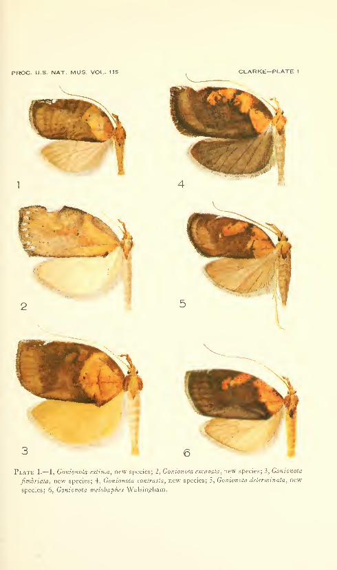
PROC. U.S. NAT. MUS. VOL. 115 CLARKE—PLATE 1
U'4
I
3 6
Plate 1.— 1, Gonionota extima, new species; 2, Gonionota excavata, new species; 3, Gonionota
fimbriata, new species; 4, Gonionota conirasta, new species; 5, Gonionota determinata, new
species; 6, Gonionota melobaphes Walsingham.

PROC. U.S. NAT. MUS. VOL. 115 CLARKE—PLATE 2
'>-
t
^Vr
Plate 2.— 1, Gonionota melobaphes Walsingham; 2, Gonionota contrasta, new species; 3,
Gonionota determinata, new species; 4, Gonionota bourquini, new species; 5, Gonionota
bourquini, new species (dark winged form); 6, Gonionota incontigua, new species.
PROC. U.S. NAT. MUS. VOL. 115 CLARKE—PLATE 3
Plate 3.— 1, Gonionota fimbriata, new species; 2, Gonionota extima, new species; 3, Gonionota
excavata, new species; 4, Gonionota periphereia, new species; 5, Gonionota dissita, new
species; 6, Gonionota hyptiotes, new species.

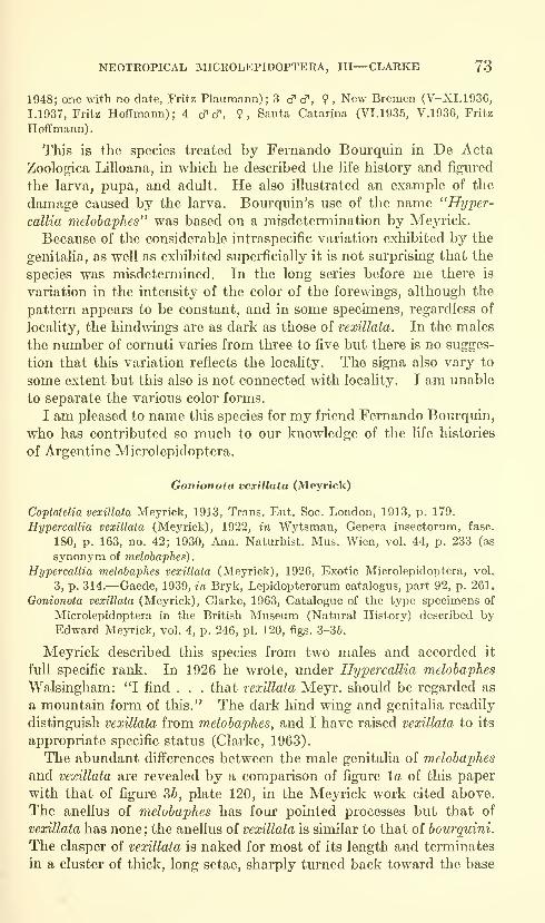
NEOTROPICAL MICROLEPIDOPTERA, III—CLARKE 73
1948; one with no date, Fritz Plaumann) ; 3 cf cf , 9 , New Bremen (V-XI.1936,
1.1937, Fritz Hoffmann); 4 cT cT, 9, Santa Catarina (VI. 1935, V.1936, Fritz
Hoffmann)
.
This is the species treated by Fernando Bourquin in De Acta
Zoologica Lilloana, in which he described the hfe history and figured
the larva, pupa, and adult. He also illustrated an example of the
damage caused by the larva. Bourquin's use of the name "Hyper-
callia melobaphes" was based on a misdetermination by Meyi'ick.
Because of the considerable intraspecific variation exhibited by the
genitalia, as well as exhibited superficially it is not surprising that the
species was misdetermined. In the long series before me there is
variation in the intensity of the color of the forewings, although the
pattern appears to be constant, and in some specimens, regardless of
locality, the hindwings are as dark as those of vexillata. In the males
the number of cornuti varies from three to five but there is no sugges-
tion that this variation reflects the locality. The signa also vary to
some extent but this also is not connected with locality. I am unable
to separate the various color forms.
I am pleased to name this species for my friend Fernando Bourquin,
who has contributed so much to our knowledge of the life histories
of Argentine Microlepidoptera.
Gonionota vexillata (Meyrick)
Coptotelia vexillata Meyrick, 1913, Trans. Ent. Soc. London, 1913, p. 179.
Hypercallia vexillata (Meyrick), 1922, in Wytsman, Genera insectorum, fasc.
180, p. 163, no. 42; 1930, Ann. Naturhist. Mus. Wien, vol. 44, p. 233 (as
synonym of melobaphes).
Hypercallia melobaphes vexillata (Meyrick), 1926, Exotic Microlepidoptera, vol.
3, p. 314.—Gaede, 1939, in Bryk, Lepidopterorum catalogus, part 92, p. 261.
Gonionota vexillata (Meyrick), Clarke, 1963, Catalogue of the type specimens of
Microlepidoptera in the British Museum (Natural History) described byEdward Meyrick, vol. 4, p. 246, pi. 120, figs. 3-36.
Meyrick described this species from two males and accorded it
full specific rank. In 1926 he wrote, under Hypercallia melobaphes
Walsingham: *T find . . . that vexillata Meyr. should be regarded as
a mountain form of this." The dark hind wing and genitalia readily
distinguish vexillata from melobaphes, and I have raised vexillata to its
appropriate specific status (Clarke, 1963).
The abundant differences between the male genitalia of melobaphes
and vexillata are revealed by a comparison of figure la of this paper
with that of figure 36, plate 120, in the Meyrick work cited above.
The anellus of melobaphes has four pointed processes but that of
vexillata has none; the anellus of vexillata is similar to that of bourquini.
The clasper of vexillata is naked for most of its length and terminates
in a cluster of thick, long setae, sharply turned back toward the base
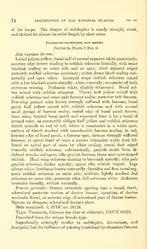
74 PROCEEDINGS OF THE NATIONAL MUSEUM vol. 115
of the harpe. The clasper of melobaphes is nearly straight, stout,
and clothed for almost its entire length by short setae.
Gonionota incontigua, new species
Figure 5a; Plate 2 (Fig. 6)
Alar expanse 26 mm.Labial palpus yellow; basal half of second segment white posteriorly;
anterior edge brown shading to reddish ocherous laterally, with some
blackish scaling on outer side and at apex; third segment edged
narrowly reddish ocherous anteriorly; rather dense black scaling sub-
apically and apex white. Antennal scape reddish ocherous mixed
with a few blackish scales dorsally, white ventrally; remainder of both
antennae missing. Proboscis white, slightly infuscated. Head yel-
low mixed with reddish ocherous. Thorax buff yellow mixed with
reddish ocherous and some pale fuscous scales; posterior tuft fuscous.
Forewing ground color brown strongly suffused with fuscous; basal
patch buff yellow mixed with reddish ocherous and with several
small groups of fuscous scales; costal edge of basal patch brown;
from costa, beyond basal patch and separated from it by a band of
ground color, an outwardly oblique buff yellow and reddish ocherous
blotch extends to end of cell, where it terminates in a white spot;
surface of blotch marked mth considerable fuscous scaling; in cell,
beyond edge of basal patch, a fuscous spot; dorsum strongly suffused
fuscous; at apical third of costa a narrow triangular white dash fol-
lowed on apical part of costa by white scaling; costal dash edged
inwardly reddish ocherous; subterminally, grayish scales form ill-
defined streaks and spots; cilia grayish fuscous, those near apex tipped
whitish. Hind wing ocherous shading to brownish apically; cilia pale
grayish ocherous, darker apically; apical cilia whitish tipped. Legs
silvery white ; forefemur brown outwardly; foretibia and first tarsal seg-
ment reddish ocherous on outer side; midtibia lightly marked dull
ocherous on outer side; posterior tibia dull ocherous white. Abdomenbrownish dorsally, whitish ventrally.
Female genitalia: Ostium crescentic opening into a broad, short,
sclerotized posterior section of ductus bursae; inception of ductus
seminalis dorsal, at anterior edge of sclerotized part of ductus bursae.
Signum an elongate, sclerotized dentate plate.
Slide examined: 9, JFGC no. 10124.
Type: Venezuela, Caracas (no date or collector), USNM 66375.
Described from the unique female type.
Superficially strikingly similar to melobaphes, determinata, and
bourguini, but the brilliance of coloring is subdued by abundant fuscous
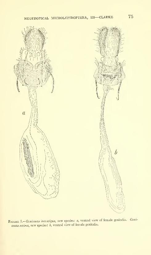
NEOTROPICAL MICROLEPIDOPTERA, III—CLARKE 75
»
Figure S.—Gonionota inconiigua, new species: a, ventral view of female genitalia. Goni-
onota extima, new species: b, ventral view of female genitalia.
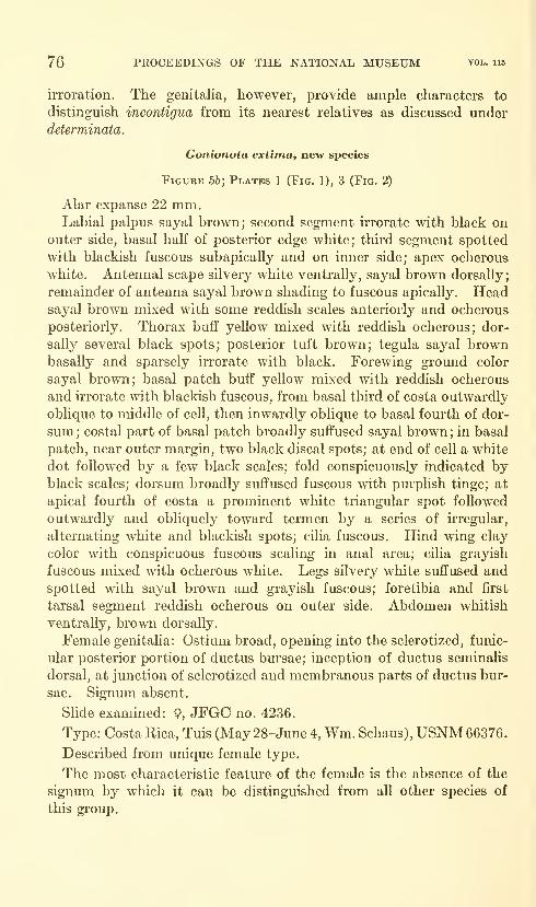
76 PROCEEDINGS OF THE NATIONAL MUSEUM vol. 115
irroration. The genitalia, however, provide ample characters to
distinguish incontigua from its nearest relatives as discussed underdeterminata.
Gonionota extima, new species
Figure 56; Platks 1 (Fig. 1), 3 (Fig. 2)
Alar expanse 22 mm.Labial palpus sayal brown; second segment irrorate with black on
outer side, basal half of posterior edge white; third segment spotted
with blackish fuscous subapically and on inner side; apex ocherous
white. Antennal scape silvery white ventrally, sayal brown dorsally;
remainder of antenna sayal brown shading to fuscous apically. Headsayal brown mixed with some reddish scales anteriorly and ocherous
posteriorly. Thorax buff yellow mixed with reddish ocherous; dor-
sally several black spots; posterior tuft brown; tegula sayal brownbasally and sparsely irrorate with black. Forewing ground color
sayal brown; basal patch buff yellow mixed with reddish ocherous
and irrorate with blackish fuscous, from basal third of costa outwardly
obhque to middle of cell, then inwardly oblique to basal fourth of dor-
sum ; costal part of basal patch broadly suffused sayal brown ; in basal
patch, near outer margin, two black discal spots; at end of cell a white
dot followed by a few black scales; fold conspicuously indicated byblack scales; dorsum broadly suffused fuscous with pm*plish tinge; at
apical fourth of costa a prominent white triangular spot followed
outwardly and obliquely toward termen by a series of irregular,
alternating white and blackish spots; cilia fuscous. Hind wing clay
color with conspicuous fuscous scaling in anal area; cilia grayish
fuscous mixed with ocherous white. Legs silvery white suffused andspotted with sayal brown and grayish fuscous; foretibia and first
tarsal segment reddish ocherous on outer side. Abdomen whitish
ventrally, brown dorsally.
Female genitalia: Ostium broad, opening into the sclerotized, funic-
ular posterior portion of ductus bursae; inception of ductus seminalis
dorsal, at junction of sclerotized and membranous parts of ductus bur-
sae. Signum absent.
Slide examined: 9, JFGC no. 4236.
Type: Costa Rica, Tuis (May 28-June 4, Wm. Schaus), USNM 66376.
Described from unique female type.
The most characteristic feature of the female is the absence of the
signum by which it can be distinguished from all other species of
this group.
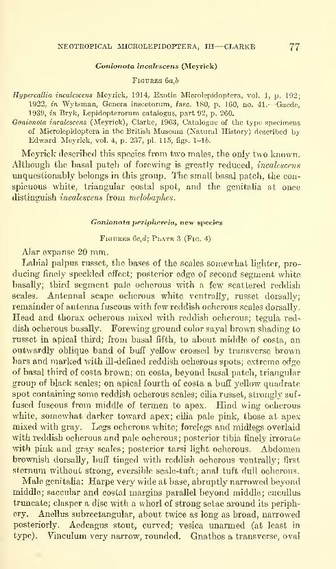
NEOTROPICAL MICROLEPIDOPTERA, III—CLARKE 77
Gonionota incalescens (Meyrick)
Figures 6a,b
Hypercallia incalescens Meyrick, 1914, Exotic Microlepidoptera, vol. 1, p. 192;
1922, in Wytsman, Genera insectorum, fasc. 180, p. 160, no. 41.—Gaede,
1939, in Bryk, Lepidopterorum catalogus, part 92, p. 260.
Oonionota incalescens (Meyrick), Clarke, 1963, Catalogue of the type specimensof Microlepidoptera in the British Museum (Natural History) described byEdward Meyrick, vol. 4, p. 237, pi. 115, figs. 1-16.
Meyrick described this species from two males, the only two known.Although the basal patch of forewing is greatly reduced, incalescens
unquestionably belongs in this group. The small basal patch, the con-
spicuous white, triangular costal spot, and the genitalia at once
distinguish incalescens from melobaphes.
Gonionota periphereia, new species
Figures 6c,d; Plate 3 (Fig. 4)
Alar expanse 20 mm.Labial palpus russet, the bases of the scales somewhat lighter, pro-
ducing finely speckled effect; posterior edge of second segment white
basally; third segment pale ocherous with a few scattered reddish
scales. Antennal scape ocherous white ventrally, russet dorsally;
remainder of antenna fuscous with few reddish ocherous scales dorsally.
Head and thorax ocherous mixed with reddish ocherous; tegula red-
dish ocherous basally. Forewing ground color sayal brown shading to
russet in apical third; from basal fifth, to about middle of costa, an
outwardly oblique band of buff yellow crossed by transverse brownbars and marked with ill-defined reddish ocherous spots; extreme edge
of basal third of costa brown; on costa, beyond basal patch, triangular
group of black scales; on apical fourth of costa a buff yellow quadrate
spot containing some reddish ocherous scales; cilia russet, strongly suf-
fused fuscous from middle of termen to apex. Hind wing ocherous
white, somewhat darker toward apex; cilia pale pink, those at apexmixed with gray. Legs ocherous white; forelegs and midlegs overlaid
with reddish ocherous and pale ocherous;posterior tibia finely irrorate
with pink and gray scales; posterior tarsi light ocherous. Abdomenbrownish dorsally, buff tinged with reddish ocherous ventrally; first
sternum without strong, eversible scale-tuft; anal tuft dull ocherous.
Male genitaHa: Harpe very wide at base, abruptly narrowed beyondmiddle; saccular and costal margins parallel beyond middle; cucullus
trimcate; clasper a disc with a whorl of strong setae around its periph-
ery. Anellus subrectangular, about twice as long as broad, narrowedposteriorly. Aedeagus stout, curved; vesica unarmed (at least in
type) . Vinculum very narrow, rounded. Gnathos a transverse, oval
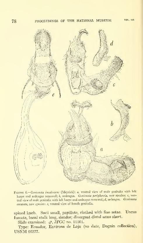
78 PROCEEDINGS OF THE NATIONAL MUSEUM
i\
Figure 6.—Gonionota incalescens (Meyrick): a, ventral view of male genitalia with left
harpe and aedeagus removed; b, aedeagus. Gonionota periphereta, new species: c, ven-
tral view of male genitalia with left harpe and aedeagus removed; d, aedeagus. Gonionota
excavaia, new species: e, ventral view of female genitalia.
spined Imob. Socii small, papillate, clothed with fine setae. Uncus
furcate, basal stalk long, slender; divergent distal arms short.
Slide examined: cf, JFGC no. 11101.
Type: Ecuador, Environs de Loja (no date, Dognin collection),
USNM 66377.
NEOTROPICAL MICROLEPIDOPTERA, III—CLARKE 79
Described from unique male type.
This specimen, among others, was sent to Meyrick for determina-
tion, presumably by Busck. Attached to it is a note in Meyrick's
handwriting which reads: "Sent as second specimen of H. melobaphes
with ocherous h.w., but is new, I tlierefore return, h.w. of melo-
ba2)hes vary from ocherous to dark fuscous, but never white." I
would not describe the hindwing of this specimen as white but it is
certainly lighter than that of any of the other species considered here.
There is some doubt in my mind about placing periphereia in Gonio-
nota. Although the genitalia are not dissimilar to those of other
species associated with melobaphes and the venation easily permits
its retention here, the fact remains that the labial palpus of periphereia
differs from those of the other species. Moreover, the third segment
is short, about one-third the length of the second, and devoid of any
long scaling on the posterior edge. Finally, this is the only species
lacking the eversible tuft from the first sternum, but this can, and
may, be only a specific character.
Gonionota excavata, new species
Figure 6e; Plates 1 (Fig. 2), 3 (Fig. 3)
Alar expanse 22 mm.Labial palpus ocherous white overlaid with brown, especially on
outer side of second segment, sparsely irrorate with fuscous and red-
dish ocherous; third segment similarly colored, apex whitish. An-
tenna ocherous white ventrally, grayish fuscous dorsally. Headbuff yellow mixed with brown and reddish ocherous. Thorax ocher-
ous mixed with brown; tegula brown basally. Forewing ground color
light brown shaded with fuscous costad and with a faint violaceous
tinge in tornal area; basal patch buff yellow mixed with reddish ocher-
ous, extending to two-fifths; costal margin of basal patch broadly
edged with ground color for nearly its entire length; in cell, at outer
edge of basal patch, a blackish spot; several reddish ocherous blotches
in center of wing; at apical thh'd costa excised, bordered white; around
termen a series of white scale patches preceded by ill-defined fuscous
spots; dorsal margin edged with fuscous and with a group of blackish
scales at base of dorsum. Hind wing ocherous white shading to
brownish ocherous apically; in anal area scattered fuscous scales;
cUia grayish fuscous, paler basally. Legs ocherous white lightly in-
fuscated; tarsi white with a few fuscous spots. Abdomen brownish
dorsally, ocherous white ventrally.
Female genitalia: Ostium broad, crescentic. Posterior two-fifths
of ductus bursae sclerotized and bent anteriorly; inception of ductus
seminalis dorsal, near junction of membranous and sclerotized parts of
ductus bursae. Signum a small, bilobed, sclerotized dentate plate.
Slide examined: 9, JFGCno.4003.
80 PROCEEDINGS OF THE NATIONAL MUSEUM vol. 115
Type: Mexico, Orizaba (no date, Wm. Schaus), USNM 66378.
The structure of the genitaha suggests that excavata, jimhriata and
hyptiotes are closely related, but jimhriata lacks the excised costa
of forewing of the other two. The unusually broad ostium and the
signum of hyptiotes distinguish it from excavata. In superficial ap-
pearance excavatamost nearly resembles 0. rhacina Walsingham, which,
however, lacks the row of white subterminal dashes characteristic
of the former species. Also, the hind wing of excavata is paler than
that of rhacina.
Gonionota hyptiotes, new species
Figure 7/; Plate 3 (Fig. 6)
Alan expanse 24 mm.
Labial palpus buckthorn brown; outer side of second segment
sparsely irrorate with fuscous, inner side with some white scaling and
tinged with pink; posterior edge white basally; third segment white
posteriorly, subapical tuft fuscous. Antennal scape buckthorn brown
dorsally, white ventrally ; remainder of antenna brown dorsally shading
to grayish fuscous distally. Head and thorax dull ocherous; tegula
brownish basally. Forewing ground color buckthorn brown; basal
fifth to costal edge of cell at two-fifths dull ocherous; from two-fifths
of costa a transverse bar of dull ocherous extends toward outer extrem-
ity of basal patch but is interrupted by ground color; basal patch
divided at middle by a transverse line of ground color; on outer margin
of basal patch, in cell, two small clusters of fuscous scales; at end of cell
a fuscous spot surrounded by ocherous; costa deeply excised at outer
third, edged with white; inner margin of white edge very narrowly
ocherous ; costa fuscous before and after white-edged excision ; dorsum
broadly edged with fuscous; in terminal third scattered, ill-defined
fuscous spots; cilia grayish fuscous. Hind wing ocherous white
basally, shading to brown apically; in anal area considerable grayish
fuscous scaling; cilia ocherous white shading to grayish fuscous
around termen to apex. Legs ocherous white irrorate with brown
and fuscous. Abdomen ocherous white with sparse fuscous irrora-
tion ventrally; dorsally brownish buff.
Female genitalia: Ostium extremely broad, opening into the scle-
rotized portion of ductus bm'sae on left side. Ductus bursae very
short, sclerotized for about half its length, the sclerotized part dilated
and convoluted; inception of ductus seminalis on right side. Signum
approximately diamond-shaped.
Slide examined: 9, JFGC no. 11103.
Type: Mexico, Orizaba (Sept. 10, K. MuUer), USNM 66380.
Described from the unique female type.
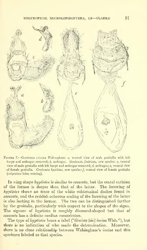
NEOTROPICAL MICROLEPIDOPTERA, III—CLARKE 81
Figure 7.
—
Gonionota cristata Walsingham: a, ventral view of male genitalia with left
harpe and aedeagus removed; b, aedeagus. Gonionota fimbriata, new species: c, ventral
view of male genitalia with left harpe and aedeagus removed; d, aedeagus; e, ventral view
of female genitalia. Gonionota hyptiotes, new species:/, ventral view of female genitalia
(ovipositor lobes missing).
In wing shape hyptiotes is similar to excavata, but the costal excision
of the former is deeper than that of the latter. The forewing of
hyptiotes shows no trace of the white subterminal dashes found in
excavata, and the reddish ocherous scaling of the forewing of the latter
is also lacking in the former. The two can be distinguished further
by the genitaha, particularly with respect to the shapes of the signa.
The signum of hyptiotes is roughly diamond-shaped but that of
excavata has a definite median constriction.
The type oi hyptiotes bears a label ("Goniota [sic] incisa Wish.") , hut
there is no indication of who made the determination. Moreover,
there is no close relationship between Walsingham's incisa and this
specimen labeled as that species.
82 PROCEEDINGS OF THE NATIONAL MUSEUM vol. 115
Gonionota cristata Walsingham
FiGTJKES 7a,b
Gonionota cristata Walsingham, 1912, in Godman and Salvin, Biologia Centrali-
Americana, vol. 42 (Lepidoptera-Heterocera, vol. 4), p. 129.
The male genitalia of cristata have not been figured previously;
they are shown here for comparative reasons.
Slide examined: cf, JFGC no. 3394. No female available.
Male genitalia: Harpe rather abruptly narrowed in distal half;
cucullus truncate; clasper a very small curved ridge about middle of
harpe. Anellus subquadrate, deeply excavated on posterior edge.
Aedeagus very stout, bent slightly before middle; vesica armed with
two groups of strong cornuti, the four or five of the proximal and
smaller group short, dentiform, those of the distal group longer and
more numerous. Vmculum rather narrow, rounded. Gnathos a
narrowly oval spined knob. Uncus with long, slender, proximal stalk
and widely divergent, moderately slender distal arms.
Gonionota fimbriata, new species
FiGUEES 7c-e; Plates 1 (Fig. 3), 3 (Fig. 1)
Alar expanse 17-21 mm.Labial palpus amber brown; second segment ocherous white on
inner side with pale ocherous toward tip; third segment fuscous on
posterior margin subapically; apex white. Antennal scape amber
brown dorsally, silvery white ventrally; remainder of antenna amber
brown dorsally at base shading to grajdsh fuscous ; dull pale ocherous
ventrally. Head russet, the scales narrowly tipped with white; pro-
boscis white. Thorax deep chi'ome mixed with scattered reddish
ocherous scales; anteriorly heavily overlaid with russet; posterior
tuft brown; anterior two-thirds of tegula russet. Forewing ground
color sayal brown faintly blotched with darker brown, basal patch
deep chrome mixed with reddish ocherous and extending to about
basal third of wing; outer margin convex; costal edge of basal patch
sayal brown and across middle of patch a narrow outwardly curved
line of the same color; in cell near outer margin of basal patch two
small black spots, one obliquely above the other; at end of cell a few
white scales, preceded and followed by scattered fuscous scaling, indi-
cate an outer discal spot; at apical third of costa a conspicuous triangu-
lar white dash followed by some white scales; dorsum narrowly edged
with fuscous; cilia grayish fuscous, those below apex faintly tipped
whitish. Hind wing brownish ocherous, cilia slightly darker. Legs
silvery white; forefemur shaded with grayish fuscous on outer side;
foretibia and fhst tarsal segment russet on outer side; midtibia shaded
with brown on outer side; midtarsi irrorate brown; posterior tibia and
NEOTROPICAL MICROLEPIDOPTERA, III—CLARKE 83
tarsi shaded with pale brown. Abdomen sHghtly darker than hind
wing, brownish dorsally, white ventrally; anal tuft cinereous mixed
with ocherous; first sternum with strong, eversible, median scale-tuft
in male.
Male genitalia: Harpe broadest near base, tapering gently to the
rounded cucuUus; clasper arising slightly beyond middle, stout,
distally armed with strong, sharp setae ; sacculus thickened but with-
out appreciable prominence. Anellus rectangular, deeply concave
posteriorly. Aedeagus stout, curved, sharply pointed; vesica armed
with cluster of strong setae. Vinculum narrow, rounded. Gnathos
an oval transverse spined knob. Socii small, fleshy lobes clothed with
fine setae. Uncus furcate, proximal stalk long, slender; distal arms
slender.
Female genitalia: Ostium moderately small, ventral lip ridged,
opening into a cup-shaped posterior sclerotized portion of ductus bursae.
Inception of ductus seminalis approximately at junction of sclerotized
and membranous parts of ductus bursae. Signum a small rectangular
dentate plate.
Slides examined: cT cT, JFGC nos. 3391, 3395, 10791 ; 9, JFGC no.
11102.
Type: Panama, Porto Bello (April 1912, August Busck), USNM66379.
Described from male type, one male and two female paratypes as
follows:
Panama: Barro Colorado Island (d^, Sept. 1940, James Zetek; 2 $ ?, 11.x
and 3.xi, M. Bates coll.).
Superficially, j^m6/'wite and cristata are similar, but the hind wing of
cristata is considerably darker than that oijimhriata. In the forewing
oijimbriata the white costal spot is more pronounced and the ground
color is much lighter than in cristata. The male genitalia also
show marked differences. Although the anneli of both species are of
the same type and are, for practical purposes, indistinguishable, the
clasper, as in the apical half of the harpe oi jimhriata, is absent in the
harpe of cristata. Other obvious differences wUl be seen by a compari-
son of the figures. Unfortunately, no female of cristata is available
for comparison.
U.S. GOVERNMENT PRINTING 0FFICE:I964

Proceedings of
the United States
National MuseumSMITHSONIAN INSTITUTION • WASHINGTON, B.C.
Volume 115 1964 Number 3481
CHIRONOMID MIDGES OF CALIFORNIA
II. TANYPODINAE, PODONOMINAE, AND DIAMESINAE
By James E. Sublette'
Subsequent to publication of Part I of this series (Proc. U.S. Nat.
Mus., vol. 112, no. 3435, 1960), I have received several additional
collections. Rather than delay publication until all the remaining
subfamilies can be treated, I am presenting here the results of study
of three subfamihes. A projected third part will treat the Ortho-
cladiinae and addenda to Parts I and II.
For making the additional collections available to me I should like
to thank George W. Byers, Snow Entomological Museum, University
of Kansas, Lawrence, Kansas; Gail Grodhaus, California Departmentof Public Health, Berkeley, California; Ernest C. Bay and E. I.
Schlinger, University of California, Riverside, California.
Names of collections and collectors which are abbreviated in the
text of this paper are as follows: U.S. National Museum (USNM);British Museum (Natural History) (BMNH) ; University of California
at Davis (UCD) ; University of California at Los Angeles (UCLA)
;
University of California at Riverside (UCR) ; California Departmentof Public Health, Berkeley (CDPH) ; University of Kansas (KU);
lUinois Natural History Survey (INHS); WiUis W. Wirth (W);
R. E. Darby (D) ; J. N. Belkin (B) ; Gail Grodhaus (G) ; Ernest C.
Bay (Bay); E. I. Schlinger (S) ; James E. Sublette (JES).
•Eastern New Mexico University, Portales, New Mexico.
85
86 PROCEEDINGS OF THE NATIONAL MUSEUM vol. 115
All localities mentioned are in California unless otherwise noted.
The deposition of material is indicated immediately before locality
data of the specimens studied.
I should like again to thank my wife Mary Smith Sublette for her
assistance in preparation of the manuscript and Dr. Willis W. Wirthfor reading and constructively criticizing the manuscript.
Subfamily Tanypodinae
Pentaneura {Ablabesmyia) monilis (Linnaeus) Johannsen
Tipula monilis Linnaeus, Systema naturae, ed. 10, p. 587, 1758.
Males: Wing length, range 2.29-3.15; mean 2.85 mm. (14); fore
leg ratio, range 0.77-0.83; mean 0.79 (9); antennal ratio, range
1.50-2.17; mean 1.95 (9); venarum ratio, range 0.80-0.87; mean0.84 (13); aedeagus blade length, range 0.72-0.090; mean 0.081 (10).
Prealar bristles 19 to 23; dorsolateral bristles mostly in single row;
occasionally doubled for 1 or 2 bristles; anterolateral bristles 9 to 13.
Females: Wing length, range 2.29-3.00; mean 2.66 mm. (4); fore
leg ratio, range 0.75-0.85; mean 0.80 (4); venarum ratio, range 0.84-
0.86; mean 0.85 (3).
Material examined: In USNM: 1 male, 1 female, Berkeley, May 1,
1948, W; 2 males, 2 females, Shafter, Kern Co., June, 1946, B. Brook-
man; 1 male. King's River Bridge, Stratford, July 15, 1947, W. In
KU: 3 males. Mammoth Lakes, July 29, 1940, D. E. Hardy; 1 male,
Mammoth Lakes, July 29, 1940, R. H. Beamer; 1 female, Tioga Pass,
July 31, 1940, R. H. Beamer. In UCLA: 1 male. Areata, HumboldtCo., Sept. 9, 1950, B. In UCD: 1 male, Cobb's P.O., Forest Lake,
June 22, 1953, D; 1 male. Pope Valley, Duvall Lake, June 22, 1953,
D. In CDPH: 1 male, Puddmgstone Reservoir, Los Angeles Co.,
June 22, 1952, G; 18 males. Lake Tenaya, Mariposa Co., Aug. 20,
1960, L. L. LewaUen; 2 males, 3 miles south of Woodside, San MateoCo., Apr. 20, 1960, G. In UCR: 3 males, Whittier, Rio Hondo, May15, 20, 1960, Bay.
Pentaneura {Ablabesmyia) mallochi (Walley)
Tanypiis mallochi Walley, Canadian Ent., vol. 57, p. 273, 1925; Ann. Ent. Soc.
Amer., vol. 21, p. 589, 1928.
Males: Wing length, range 2.11-2.52; mean 2.37 mm. (7) ; antennal
ratio, range 2.00-2.28; mean 2.14 (7); fore leg ratio, range 0.78-0.82;
mean 0.79 (6) ; venarum ratio, range 0.79-0.83; mean 0.83 (7) ; antero-
lateral bristles, range 13-19; mean 16 (4).
Female: Wing length 2.26 mm.; fore leg ratio 0.74; venarum ratio
0.90; anterolateral bristles 19.
Material exammed: In USNM: 2 males, 1 female, Stratford, July
8, 1947, W; 1 male, Corcoran, Aug. 22, 1947, W. In D: 3 males, Rio
CHIRONOMID MIDGES OF CALIFORNIA—SUBLETTE 87
Linda, July 25, 27, 31, 1957. In CDPH: 1 male, 2 miles east, 4 miles
north of Manteca, San Joaquin Co., June 3, 1957, G.
I have examined five specimens, four of them paratypes from the
Canadian National Collections, through the kindness of Dr. J. R.
Vockeroth, Entomology Research Institute, Canadian Department of
Agriculture, Ottawa. He also has sent me notes on the holotype.
Measurements for these specimens are:
Males: Wing length, range 2.52-2.85; mean 2.65 mm. (4); fore leg
ratio 0.77-0.85; mean 2.65 (4); antennal ratio, range 2.11-2.22;
mean 2.15 (4).
Female: Wing length 2.26 mm.; fore leg ratio 0.85.
Material examined in Canadian National Collections: 3 males,
paratypes, 1 male, Aylmer, Ont., Aug. 8, 1924, C. H. Curran; 1 male,
Aylmer, Ont., Sept. 7, 1924, C. H. Curran; 1 male, Ottawa, Ont.,
June 31, 1924, C. H. Curran; 1 male, Ottawa, Ont., July 14, 1926,
G. S. Walley; 1 female, locality not recorded.
Measurement data for this species (male) is summarized as follows
(those for the Georgia and New York specimens, fide Roback, 1959,
p. 123):
88 PROCEEDINGS OF THE NATIONAL IVIUSEUM vol. ii6
Pentaneura {Pentaneura) fluminalis, new species
Figures la,h
Pentaneura carnea (Fabricius) Johannsen, Journ. New York Ent. Soc, vol. 54,
p. 279, 280, 1946. Misidentification?
Holotype male: In USNM 65503, Mad River Beach, HumboldtCo., Aug. 12, 1948, Coll. No. 104, reared, W.Head reddish-brown, mouthparts and antennae darkened. Eyes
with thin dorsal extension. Antenna! ratio 1.84.
Thorax yellowish to reddish-brown. Anterior portion of lateral
vittae darkest reddish-brown; pollinose, especially on dorsomedial anddorsolateral bristle rows and on prescutellar area. Supra-alar
bristles 2; prealar bristles 12; dorsomedial bristles in single staggered
row, becoming 2 rows posterior to tubercle at anterior edge of pre-
scutellar area. Dorsolateral bristles in single staggered row, becomingdoubled at edge of prescutellar area and extending back to scutellum.
Dorsomedial and dorsolateral bristles erect, divergent, yellow.
Scutellar bristles numerous; anterior ones strewn; 4 posterior erect
bristles. Halteres white.
Fore leg proportions 60:80:60:34:24:15:8. Fore leg ratio 0.75.
Fore leg with beard 7 times as long as diameter of tarsus; legs uniformly
stramineous. Seven spines in comb on hind tibia; spurs sinuate,
ratio 58:24 Qong spur subsequently broken off at tip); side bristles
not clearly discernible for counting on short spur; on long spur, 3 onone side only. Middle leg ratio 0.72; hind leg ratio 0.63.
Wings very heavily haired; R2+3 scarcely discernible. R4+5 only
sUghtly proximal to termination of M, very far distal to Cui. Ridistal to Cu2; anal lobe rounded. Wing length 2.89 mm.; venarumratio 0.85. Ratio of arculus to m-cu:m-cu to wing tip 0.43.
Abdomen yellowish-brown with reddish-brown fascia on basal part
of each segment, faint on segment I becoming progressively wider
and heavier until segment VI is almost entirely dark.
Allot3T)e: In USNM, reared with holotype.
Wing length 2.70 mm.; fore leg ratio 0.77; middle leg ratio 0.68;
hind leg ratio 0.64 ; venarum ratio 0.90 ; ratio of arculus to m-cu :m-cuto wing tip 0.36.
Paratype: In USNM: 1 female, reared wdth the holotype andallotype.
Wing length 2.66 mm.; fore leg ratio 0.74; middle leg ratio 0.67;
hind leg ratio 0.65 ; venarum ratio 0.85 ; ratio of arculus to m-cu : m-cuto wing tip 0.37.
This species belongs to Edwards' Group C of Pentaneura. It
further belongs to that portion of the group that was not namedwhen Fittkau (1957) erected Thienemannimyia.
In Johannsen (1946) P j^wminaZis, new species, keys in Group C to
P. carnea (Fabricius) and may well be the species that Johannsen
CHIRONOMID MIDGES OF CALIFORNIA—SUBLETTE 89
identified as P. carnea (Fabricius) . However, the wings and genitalia
of this species are distinctly different from those illustrated byGoetghebuer (1936a, pi. 2, fig. 18; 1936b, pi. 5, fig. 71) for P. carnea
(Fabricius), and so I am considering it as new. I have not examined
Johannsen's material identified as P. carnea.
Previous California records: ?Johannsen, 1946, p. 278.
Pentaneiira (JPentaneurd) goniodes, new species
Figure Ic
Holotype male: USNM 65504, Berkeley, Oct. 3, 1947, W.Head yellow; antennal pedicel and flagellum brown; mouthparts
blackish. Eyes with long narrow dorsal extension. Antennal
ratio 2.00.
Thorax stramineous, including legs; pollinose on prescutellar area
and on dorsolateral bristle row. Vittae orange-yellow. Prealar
bristles 14; dorsomedial bristles in staggered single row, which divides
around tubercle, then extends laterally as row on each side of pre-
scutellar area to join single dorsolateral row, thus forming cluster of
bristles on either side of midline just in front of scutellum. Sterno-
pleuron reddish-brown.
Fore leg proportions: 75:92:70:35:25:16:8. Fore leg ratio 0.76;
middle leg ratio 0.50; hind leg ratio 0.65. Sparse hairs on fore tarsus
3 times as long as tarsal diameter.
Wings hyaline, weakly haired on basal half. Ratio of arculus to
m-cu:m-cu to wing tip 0.46. Ri distal to CU2; R2+3 proximal to
Cui; R4+5 distal to Cu.. Squama well haired with small triangular
spot on distal margin. Wing length 3.29 mm.; venarum ratio 0.91.
Abdomen with middorsal brown stripe. Segments II to V with
basal brown fascia; remainder of segments yellow.
Female: Unknown.Paratype: Collected with holotype. Wing length 3.03 mm.; fore
leg ratio 0.71; middle leg ratio 0.54; hind leg ratio 0.65; antennal
ratio 2.00; venarum ratio 0.87; ratio of arculus to m-cu:m-cu to
wing tip 0.42. Both hind legs slide mounted. Both spurs sinuate;
longer spur with 5 lateral spines; shorter spur with 6 lateral spines,
ratio 42:70; comb of 7 long pale bristles.
This species falls in Edwards' Group D of Pentaneura and in the
restricted group which Fittkau (1957) erected as genus Conchapelopia.
It closely resembles P.flavifrons Johannsen, P. alba Roback, P. rurika
Roback, and P. vitellina Johannsen, not Kieffer.^
2 Fittkau (1957, p. 320) clarified the status of vitellina Kieffer and pointed out that Johannsen (1946) had
misidentified the species. Fittkau then proceeded to propose anew name americana for vitellina Johannsen,
not Kieffer, based on Johannsen's figure 13. The propriety of Fittkau's action Is questionable since, accord-
ing to my understanding of the rules of nomenclature, a new name is to be proposed only in the case of
homonomy.
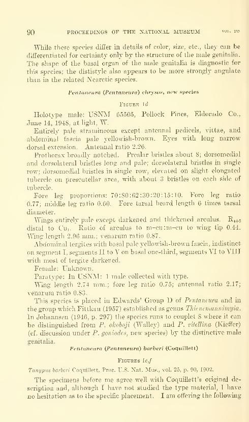
90 PROCEEDINGS OF THE NATIONAL IMUSEUM vol. 115
While these species differ in details of color, size, etc., thej can be
differentiated for certainty only by the structure of the male genitalia.
The shape of the basal organ of the male genitalia is diagnostic for
this species; the dististyle also appears to be more strongly angulate
than in the related Nearctic species.
Pentaneura {Pentaneiira) chrysos, new species
Figure Id
Holotype male: USNM 65505, Pollock Pines, Eldorado Co.,
June 14,"^ 1948, at light, W.Entu-ely pale stramineous except antennal pedicels, vittae, and
abdominal fascia pale yellowish-brown. Fixes with long narrow
dorsal extension. Antennal ratio 2.26.
Prothorax broadl}' notched. Prealar bristles about 8; dorsomedial
and dorsolateral bristles long and pale; dorsolateral bristles in single
row; dorsomedial bristles in single row, elevated on slight elongated
tubercle on prescutellar area, with about 3 bristles on each side of
tubercle.
Fore leg proportions: 70:80:62:30:20:15:10. Fore leg ratio
0.77; middle leg ratio 0.60. Fore tarsal beard length 6 times tarsal
diameter.
Wings entirely pale except darkened and thickened arculus. K4+5
distal to Cui. Katio of arculus to m-cu:m-cu to whig tip 0.44.
Wing length 2.96 mm.; venarum ratio 0.87.
Abdominal tergites wdth basal pale yellowish-brown fascia, indistinct
on segment I, segments II to V on basal one-third, segments VI to VIII
with most of tergite darkened.
Female: Unknowm.Paratype: In USXM: 1 male collected ^^•ith type.
Wing length 2.74 nmi.; fore leg ratio 0.75; antennal ratio 2.17;
venarum ratio 0.83.
This species is placed in Edwards' Group D of Penta/neura and in
the group which Fittkau (1957) established as genus Thienemunnimyia.
In Johannsen (1946, p. 297) the species runs to couplet 8 where it can
be distinguished from P. okoboji (Walley) and P. vitellina (Kieffer)
(cf. discussion under P. goniodes, new species) by the distinctive male
genitalia.
Pentaneura (^Pentaneura) barberi (Coquillett)
Figures le.f
Tanypus barberi Coquillett, Proc. U.S. Nat. Mus., vol. 25, p. 90, 1902.
The specimens before me agree well with CoquiUett's original de-
scription and, although I have not studied the type material, I have
no hesitation as to the specific placement. I am offering the following
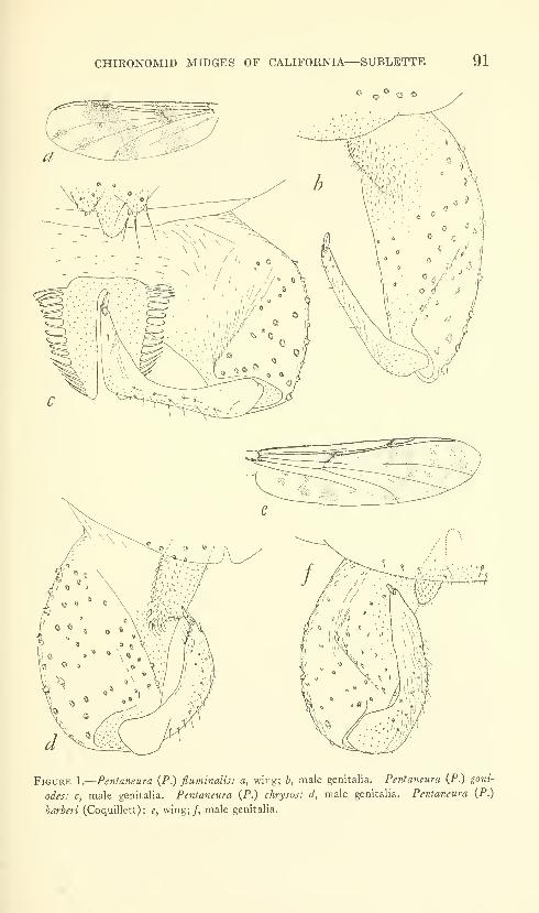
CHIRONOMID MIDGES OF CALIFORNIA—SUBLETTE 91
© ^ o
WV^-^rr
Figure 1.
—
Pentaneura (P.) fluminalis: a, wing; b, male genitalia. Petitaneura {P.) goni-
odes: c, male genitalia. Pentaneura (P.) chrysos: d, male genitalia. Pentaneura (P.)
harberi (Coquillett) : e, wing; /, male genitalia.

92 PROCEEDINGS OF THE NATIONAL MUSEUM vol. ii5
as a more complete description of the species, based on a male speci-
men in USNM: Mono Lake, Mono Co., June 6, 1948, W.Head yellowish-brown, except antennal pedicels and mouthparts
which are blackish-brown. Antenna yellowish with tip infuscate andwith fuscous band near middle. Antennal ratio 1.64. Eyes with
conspicuous narrow dorsal extension.
Thorax, except for yellowish pronotum, humeri, and scutellum, darkbrown ; middle and lateral vittae separated by paler hne along dorso-
lateral bristle row; medial vittae separated by reddish-brown line
along dorsomedial bristle row; dorsum heavily pollinose. Prothorax
narrowed medially, evanescent, considerably inferior to mesonotum;with 6 to 8 very fine lateral bristles. Prealar bristles about 15; dorso-
lateral and dorsomedial bristles in single rows with dorsomedial bristles
doubled for about 6 hairs posteriorly, terminating before scutellum;
anterolateral bristles about 15. Halteres stramineous. Scutellum
rubbed. Sternopleuron dark reddish-brown.
Fore leg proportions: 68:87:58:32:23:14:9. Fore leg ratio 0.67.
Legs stramineous; dark brown annulus at distal end of femur andproximal end of tibia, separated from joint by distance equal to width
of bands; tips of tibiae and tarsal joints 1 to 3 dark brown; distal
half of fourth and all of fifth dark brown. Fore tarsi with beard
slightly less than 3 times tarsal diameter; middle and hind legs with
long pale hairs.
Wings with numerous spots on hyaline background ; Hi+s terminates
slightly distal to Cui ; Cu2 sharply bent downward, terminating half
way between Ri and R2+3- Wing length 3.15 mm. Venarum ratio
0.90.
Each abdominal segment with basal dark brown, rather mottled,
fascia, inconspicuous and narrow on segments I and II, progressively
broadens until segment VII ahnost completely dark. Each abdominal
segment with apical poUinose, mottled white fascia; prominent on
segments I and II, decreasing apicaUy.
Alales: Wing length, range 2.32-3.18; mean 2.88 mm. (8); fore leg
ratio, range 0.64-0.73; mean 0.69 (6); antennal ratio, range, 1.52-
2.08; mean 1.81 (8); venarum ratio, range 0.80-0.92; mean 0.87 (8).
Females: Wing length, range 2.52-3.07; mean 2.73 mm. (3); fore
leg ratio 0.58, 0.66 (2) ; venarum ratio, range 0.88-0.92; mean 0.90 (3).
Material examined: In USNM: 1 male. Mono Lake, June 7, 1948,
W; 1 male, 1 female, Deer Creek Hot Springs, Tulare Co., Aug. 6,
1947, W; 2 males, 2 females, Wheeler's Springs, Ventura Co., June 16,
1948, W; 1 male, Grangeville, Kings Co., July 31, 1947, W; 1 male,
Sanger, Fresno Co., Oct. 14, 1947; 1 male, Reedley, Fresno Co., Oct.,
1947; 1 female, VisaUa, June 30, 1947, W; 1 female, Shafter, Kern Co.,
CHIRONOMID MIDGES OF CALIFORNIA—SUBLETTE 93
June, 1946, B. Brookman. In UCD: 1 male, Bear-Cache CreekJunction, Yolo Co., Apr. 19, 1957, S.
The genus Thienemannimyia Fittkau, 1957, was established for
Edwards' Group C, in part. Fittkau's generic diagnosis (freely trans-
lated) includes a "leg with a brown ring at the end of the femur andon the base of the tibia; the last 2 or 3 tarsal segments brownish. Wingmostly with darkened cross veins and dark flecks or bands. . . .
Hypopygium of a very uniform structure" (Fittkau, 1957, figs. 3, 4).
Of the described North American species only P. barberi (Coquillett),
P. marmorata Johannsen, P. apicalis (Walley), P. ornata (Meigen)
Johannsen, and P. pulchripennis (Lundbeck) definitely belong to this
restricted genus as indicated by characteristics described by Johannsen
(1946).
Roback (1957) has described Pentaneura norena, a species obviously
belonging to Thienemannimyia, as evidenced by the structure of the
male genitalia and by the banded wings; however, the species lacks
leg fasciae. Thienemannimyia must thus be emended to include both
species with fasciate legs and those without fasciae. The remaining
species treated by Johannsen (1946) as members of Group C^—^that
is, those with unhanded legs^—^possibly may include also species that
properly should be placed in Thienemannimyia; however, in the absence
of hypopygial illustrations in the literature, none of these can be
verified at this time. These species of uncertain position include
P. bifasciata (Coquillett), P. Jragilis (Walley), P. futilis (Wulp), P.
carnea (Fabricius) Johannsen, and P. sinuousa (Coquillett). Because
of lack of precise knowledge of many North American types of this
group, I am not using Thienemannimyia at this time but am using a
much more inclusive Pentaneura (Pentaneura) (==Groups B-F,sensu Edwards.)
Pentaneura {Pentaneura) comosa, new species
Figure 2a
Holotype male: USNM 65506, Almn Rock Park, Santa Clara Co.,
July 8, 1948, W.Head pale yellowish-white; posteriorly infuscate. Antennal pedicel
and flagellum dark brown. Eyes with long narrow dorsal extensions.
Palpi long, pale bro^vn, ratio 35:43:45:70. Antennal ratio 1.52.
Thorax brown overlaid with strong white pruinescence ; vittae dark
brown, overlaid with conspicuous greenish pruinescence. Pronotuminfuscate yellowish-white with broad notch completely interrupted
on midline; with 4 fine lateral bristles. Scutellum infuscate yellow.
Postnotum dark brown. Sternopleuron dark brown marked with
infuscate yellow on pleura. Halteres yellow. Supra-alar bristles 2,
1 long, 1 short;prealar bristles 1 1 ; dorsomedial bristles staggered in
94 PROCEEDINGS OF THE NATIONAL MUSEUM vol. iib
single row, anteriorly with tuft of fine appressed bristles; dorsolateral
bristles in 2 staggered rows, converging almost to midline just anterior
to scutellum where the 2 rows are much broadened and staggered;
anterolateral bristles 5, much finer and paler than dorsolateral bristles;
scutellum with single posterior row of 12 long black bristles, anteriorly
16 scattered near midline.
Femora with apical faint brown band, remainder infuscate stramin-
eous. Fore tarsi with long beard 8 times as long as diameter of
tarsus; middle leg and hind leg densely pilose. Fore tibia with single
lyrate spur (similar to that illustrated for P. hjra, new species), com-
posed of 6 spines; main spur spine only slightly heavier than lateral
bristles; middle tibial spurs lyrate, each with 6 spines, main spur spine
heavier than remainder of side bristles; spurs of approximately equal
length. Hind tibial spurs very similar to those of middle leg, lyrate
form not quite so pronounced; comb of 6 spines.
Leg proportions:
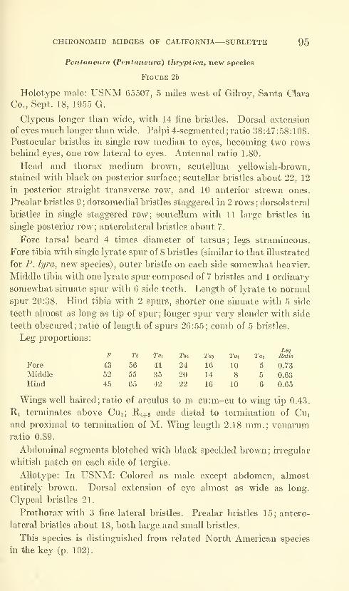
CHIRONOMID MIDGES OF CALIFORNIA—SUBLETTE 95
Pentaneura {Pentaneura) thryptica, new species
Figure 26
Holotj^pe male: USNM 65507, 5 miles west of Gilroy, Santa Clara
Co., Sept. 18, 1955 G.
Clypeus longer than wide, with 14 fine bristles. Dorsal extension
of eyes much longer than wide. Palpi 4-segmented ; ratio 38 :47 : 58 : 1 08
.
Postocular bristles in single row median to eyes, becoming two rowsbehind eyes, one row lateral to eyes. Antennal ratio 1.80.
Head and thorax medium brown, scutellum yellowish-brown,
stained with black on posterior surface; scutellar bristles about 22, 12
in posterior straight transverse row, and 10 anterior strewn ones,
Prealar bristles 9 ; dorsomedial bristles staggered in 2 rows ; dorsolateral
bristles in single staggered row; scutellum with 11 large bristles in
single posterior row; anterolateral bristles about 7.
Fore tarsal beard 4 times diameter of tarsus; legs stramineous.
Fore tibia with single lyrate spur of 8 bristles (similar to that illustrated
for P. lyra, new species), outer bristle on each side somewhat heavier.
Middle tibia with one lyrate spur composed of 7 bristles and 1 ordinary
somewhat sinuate spur with 6 side teeth. Length of lyrate to normal
spur 20:38. Hind tibia with 2 spurs, shorter one sinuate with 5 side
teeth almost as long as tip of spur ; longer spur very slender with side
teeth obscm-ed; ratio of length of spurs 26:55; comb of 5 bristles.
Leg proportions:

96 PROCEEDINGS OF THE NATIONAL MUSEUM vol. 115
Pentaneura {Pentaneura) lyra, new species
Figures 2c, d
Holotype male: USNM 65508, Wheeler's Springs, Ventura Co.,
June 16, 1948, light trap, W.Head infuscate yellow; antennal pedicels dark reddish-brown;
flagellum infuscate. Palpi infuscate; ratio 38:45:55:95. Clypeusslightly longer than broad, with 13 bristles. Postocular bristles
reaching point medial to long narrow doreal extensions of eyes; in
single row.
Thorax reddish-brown; pleura, spaces between vittae, humeri andscutellum pale yellowish-white. Prothorax infuscate yellow; com-pletely divided by rather narrow notch; 2 lateral fine bristles. Post-
notum reddish-brown. Halteres white. Prealar bristles 13; dorso-
medial bristles in staggered single row; dorsolateral bristles in double
staggered row, posterior rows dilated medially, almost reaching
midline; scutellum with 12 long, pale bristles in posterior single row;
anteriorly several fine pale strewn bristles near midline; anterolateral
bristles 9.
Fore legs without beard; middle and hind legs heavily pilose.
Single spur of fore tibia somewhat lyrate with 8 side teeth being almost
as large as main spur continuation; both spurs of middle and hind
tibiae lyrate, very similar to that of fore leg, about equal length,
each with 7 to 9 side teeth; comb of hind tibia of 5 bristles.
Leg proportions:
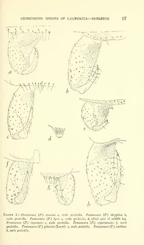
CHIRONOMID MIDGES OF CALIFORNIA—SUBLETTE 97
Figure 2.
—
Pentaneura (P.) comosa: a, male genitalia. Pentaneura (P.) ihryptica: b,
male genitalia. Pentaneura (P.) lyra: c, male genitalia; d, tibial spur of middle leg.
Pentaneura (P.) inyoensis: e, male genitalia. Pentaneura (P.) sequoiaensis: f, male
genitalia. Pentaneura {P.) pilosella (Loew): g, male genitalia. Pentaneura (P.) smithae:
h, male genitalia.
98 PROCEEDINGS OF THE NATIONAL MUSEUM vol. us
venarum ratio, range 0.86-0.93; mean 0.88 (6) ; arculus to m-cu:m-cuto wing tip, range 0.42-0.45; mean 0.43 (6).
This species is distinguished from closely related forms in the key
(p. 102).
Pentaneura (Pentaneura) inyoensis, new species
Figure 2e
Holotype male: USNM 65509, Resting Springs, Inyo Co., May 29,
1955.
Head pale brown; antennal pedicels darker brown. Postocular
bristles in single row reaching medial to dorsal extensions of eyes.
Clypeus longer than broad with 17 bristles. Palpi 35:50:70:62,
Antennal ratio 1.70.
Thorax pale brown overlaid with conspicuous golden green prui-
nescence; vittae and postnotum darker brown. Halteres white.
Pronotum with 4 lateral bristles. Supra-alar bristles 2, 1 large, 1
small; prealar bristles 8; dorsolateral bristles in 2 staggered rows;
anterolateral bristles about 6; scutellum with 8 posterior bristles in
transverse row, anteriorly about 10 smaller strewn bristles.
Legs stramineous; fore tarsal beard 3 times as long as diameter of
tarsus. Fore leg with very short sinuate spur with 2 lateral barbs;
spur length :tibial apical diameter 20:38. Middle leg with two un-
usual spm's of about equal length, each composed of 3 long filaments.
Hind leg with only one visible trifid spur as middle leg; with combof only 4 spines.
Leg proportions:
Fore

CHIRONOMID MIDGES OF CALIFORNIA—SUBLETTE, 99
ratio, range 0.86-0.93; mean 0.91 (3); arculus to m-cu:m-cu to wing
tip, range 0.37-0.41; mean 0.39 (4); hind leg ratio 0.68 (1).
This species is distinguished from related forms in the key (p. 102).
Pentaneura (Pentaneura) seqiioiaensis, new species
Figure 2/
Holotype male: USNM 65510, Stony Creek, Sequoia National
Park, July 13, 1947, W.
Postocular bristles 15, in single row which begins at point medial
to long dorsal extension of eyes. Eye extension with posteromedial
border angulate; the anteromedial border rounded. About 12 clypeal
bristles. Antennal ratio 2.00.
Thoracic vittae, postnotum, and sternopleuron pale brown, re-
mainder of thorax whitish poUinose. Prothorax with 5 fine lateral
bristles. Halteres white. One supra-alar bristle; prealar bristles 10;
dorsolateral bristles in single staggered row; scutellum with 3 trans-
verse rows of bristles, 12 in posterior row, 8 in median row, 3 in
anterior row.
Fore tibial spur lyrate, closely resembling that of P. lyra, newspecies; lateral bristles spatulate, ahnost as long as spine of spur,
giving appearance of comb. Middle leg with spurs short, subequal,
each with apparently 3 lateral somewhat spatulate bristles. Hind
leg with comb of 5 spines ; spurs short, each with apparently 2 lateral
bristles almost as long as spur.
Leg proportions:
100 PROCEEDINGS OF THE NATIONAL MUSEUM vol. 115
Pentaneura (Pentaneura) pilosella (Loew)
Figure 2g
Tanypus pilosellus Loew, Berlin Ent. Zeitschr., vol. 10, p. 5, 1866.
The description that follows is given to supplement the original
description and the one given by Johannsen (1946). It is based on a
male specimen in UCLA: Lee's Lake, Chatsworth, Los Angeles Co.,
July 25, 1950, B.
Head yellowish-white except antennal pedicels reddish-brown andpalpi and antennal flagella infuscate. Postocular bristles in single
row; reaching point medial to dorsal extension of eyes. Eye exten-
sion angulate posteriorly, rounded anteriorly. About 15 clypeal
bristles. Antennal ratio 1.30.
Thorax yellowish-white except for vittae, postnotum, and sterno-
pleuron which are reddish-brown. Mesothoracic vittae with greenish
pruinosity. Halteres yellowish-white. Supra-alar bristles 2, prealar
bristles 8; dorsomedial bristles in 2 rows, diverging posteriorly;
dorsolateral bristles in 2 staggered rows, composed of large and small
bristles, rows diverge posteriorly to join dorsomedial bristles; scutellum
with 8 large bristles in transverse posterior row, anteriorly with manyfine strewn bristles; anterolateral bristles about 8.
Legs infuscate stramineous; beard 3.6 times fore tarsal diameter.
Leg proportions:F Ti Tai T02 Tas Tat Tat Leg Ratio
Fore 25 30 19 12 8 5 4 0.63
Middle 32 27 33 15 4 5 5 1.22
Hind 29 38 30 14 11 6 4 0.79
Wing with ratio of arculus to m-cu:m-cu to wing tip 0.37. Riterminates proximal to Cuj; wing densely haired. Wing length 1.55
mm.; venarum ratio 0.75.
Abdomen with segments I, II, and IV yellowish-white with narrowbasal brown band; segments III, V, and VI largely brown, with nar-
row apical white fascia; segments VII and VIII mounted with
genitalia.
Males: Wing length, range 1.04-1.52; mean 1.26 mm. (7); fore leg
ratio, range 0.56-0.72; mean 0.66 (5); antennal ratio, range 0.64-
1.26; mean 0.91 (7); middle leg ratio, range 1.10-1.28; mean 1.18
(5); hind leg ratio, range 0.82-1.09; mean 0.89 (4).
Material studied: In USNM: 4 males, Orosi, Tulare Co., June 5,
1947, W. In UCLA: 9 males, Lee's Lake, Chatsworth, Los AngelesCo., July 25, 1950, B; 7 males, same locahty, Aug. 10, 1950.
Pentaneura {Pentaneura) smithae, new species
Figure 2h
Holotype male: USNM 65511, Saratoga Springs, March 20, 1955, B.
Head yellowish, occiput and mouthparts infuscate ; antennal pedicel,
thoracic vittae, postnotum, and sternopleuron reddish-brown. Post-

CHIRONOMID MIDGES OF CALIFORNIA—SUBLETTE 101
ocular bristles in single row continuing medially to vertex between
dorsal extensions of eyes. Palpi ratio 13:28:35:20. Antennal ratio
1.59.
Prothorax with 1 large, 2 small lateral bristles. Pronotum, humeri,
small pleural area and scutellum yellowish. Halteres white. Prealar
bristles 12; dorsolateral bristles mostly in double row, posteriorly ex-
panded, becoming multiserial; 12 scutellar bristles in posterior trans-
verse row, about 16 anterior strewn ones; anterolateral bristles about
12.
Fore legs with longest hairs 3.5 times diameter of tarsus, mostly
with short hairs.
Leg proportions:
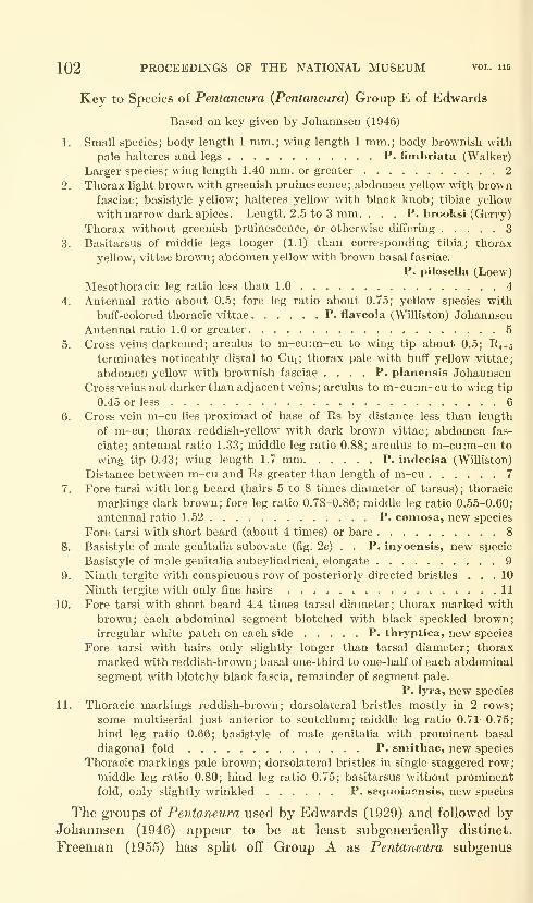
102 PROCEEDINGS OF THE NATIONAL MUSEUM vol. 115
Key to Species of Pentaneura {Pentaneura) Group E of Edwards
Based on key given by Johannsen (1946)
1. Small species; body length 1 mm.; wing length 1 mm.; body brownish with
pale halteres and legs P. fimbriata (Walker)
Larger species; wing length 1.40 mm. or greater 2
2. Thorax light brown with greenish pruinescence; abdomen yellow with brownfasciae; basistyle yellow; halteres yellow with black knob; tibiae yellow
with narrow dark apices. Length 2.5 to 3 mm. . . . P. brooksi (Gerry)
Thorax without greenish pruinescence, or otherwise differing 3
3. Basitarsus of middle legs longer (1.1) than corresponding tibia; tliorax
yellow, vittae brown; abdomen yellow with brown basal fasciae.
P. pilosella (Loew)
Mesothoracic leg ratio less than 1.0 4
4. Antennal ratio about 0.5; fore leg ratio about 0.75; yellow species with
buff-colored thoracic vittae P. flaveola (Williston) Johannsen
Antennal ratio 1.0 or greater 5
5. Cross veins darkened; arculus to m-cu:m-cu to wing tip about 0.5; R4+5
terminates noticeably distal to Cuj; thorax pale with buff yellow vittae;
abdomen yellow with brownish fasciae .... P. planensis Johannsen
Cross veins not darker than adjacent veins; arculus to m-cu:m-cu to wing tip
0.45 or less 6
6. Cross vein m-cu lies proximad of base of Rs by distance less than length
of m-cu; thorax reddish-yellow with dark brown vittae; abdomen fas-
ciate; antennal ratio 1.33; middle leg ratio 0.88; arculus to m-cu:m-cu to
wing tip 0.43; wing length 1.7 mm P. indecisa (Williston)
Distance between m-cu and Rs greater than length of m-cu 7
7. Fore tarsi with long beard (hairs 5 to 8 times diameter of tarsus); thoracic
markings dark brown; fore leg ratio 0.78-0.86; middle leg ratio 0.55-0.60;
antennal ratio 1.52 P. comosa, new species
Fore tarsi with short beard (about 4 times) or bare 8
8. Basistyle of male genitalia subovate (fig. 2e) . . P. inyoensis, new specie
Basistyle of male genitalia subcylindrical, elongate 9
9. Ninth tergite with conspicuous row of posteriorly directed bristles ... 10
Ninth tergite with only fine hairs 11
10. Fore tarsi with short beard 4.4 times tarsal diameter; thorax marked with
brown; each abdominal segment blotched with black speckled brown;
irregular white patch on each side P. thryptica, new species
Fore tarsi with hairs only shghtly longer than tarsal diameter; thorax
marked with reddish-brown; basal one-third to one-half of each abdominal
segment with blotchy black fascia, remainder of segment pale.
P. lyra, new species
11. Thoracic markings reddish-brown; dorsolateral bristles mostly in 2 rows;
some multiserial just anterior to scutellum; middle leg ratio 0.71-0.75;
hind leg ratio 0.66; basistyle of male genitalia with prominent basal
diagonal fold P. smithae, new species
Thoracic markings pale brown; dorsolateral bristles in single staggered row;
middle leg ratio 0.80; hind leg ratio 0.75; basitarsus without prominentfold, only slightly wrinkled P. sequoiaensis, new species
The groups of Pentaneura used by Edwards (1929) and followed byJohannsen (1946) appear to be at least subgenerically distinct.
Freeman (1955) has split off Group A as Pentaneura subgenus

CHIRONOMID MIDGES OF CALIFORNIA—SUBLETTE 103
Ablabesmyia Johannsen, 1905, while retaining Edwards' Groups B to
F as Pentaneura sensu stricto. Fittkau (1957) has named two newgenera, Thienemannimyia and Conchapelopia, which include parts of
Groups C and D of Edwards. There are other species groups within
Edwards' Groups C and D that were not named. Group F appears
to be the equivalent of Nilotanypus Kieffer, 1923. Group E contains
the type-species, Pentaneura grisea Phillipi, sensu Edwards, and is thus
Pentaneura in the strict sense. The remaining Group B is unnamed.Since the status of several types of North American Pentaneura are
described inadequately as yet, I am refraining at this time fromestablishing or using subgeneric units for Edwards' Groups B to F.
Anatopynia (Anatopynia) submarginella, new species
Figure 3a
Holotype male: USNM 65512, Modoc Co., Fandango Pass, May15, 1948, W.
Postocular bristles in double and triple rows reaching from below
eyes up to point medial to dorsal extension of eyes. Clypeus wider
than long, with about 16 bristles. Palpi ratio 15 :25 :20 :30. Antennal
ratio 1.64.
Head, thorax, and abdomen dark brown overlaid with greenish-
white pollen;pronotum, pleural areas, and posterolateral area on each
abdominal segment lighter brown; pollen on abdomen concentrated
on posterior half of each segment, giving, in certain light, vittate
appearance; incisures of abdomen somewhat lighter; pollen absent
between vittae on thorax. Prothorax with 13 fine lateral bristles,
covered all over with fine microtrichae. Sternopleuron with 3 fine
lateral bristles above and 2 below anepisternal suture. Halteres yel-
lowish, stalk basally infuscate. Prealar bristles 26; dorsolateral
bristles multiserial; scutellum with 26 large staggered bristles in pos-
terior transverse row; anteriorly about 14 fine strewn bristles; antero-
lateral bristles about 18.
Fore legs with few long hairs 3 times diameter of tarsus; legs paler
brown than body. Fore tibia with almost straight spur with 9 side
teeth; spur 0.8 as long as apical tibial diameter. Middle tibial spurs
straight with 10 side teeth; spurs of equal length. Spurs of hind leg
straight with 8 teeth on longer, 10 on shorter. Spur ratio 55:45.
Comb of 9 bristles.
Leg proportions
:
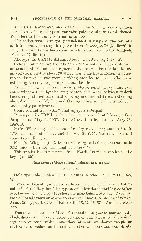
104 PROCEEDINGS OF THE NATIONAL MUSEUM vol. 115
Wings well haired only on distal half; anterior wing veins including
m-cu cross vein brown; posterior veins pale; membrane not darkened.
Wing length 3.37 mm.; venarum ratio 0.88.
The rather short, straight, parallel-sided dististyle of the genitalia
is distinctive, separating this species from A. marginella (Malloch), in
which the dististyle is longer and evenly tapered to the tip (Malloch,
1915, pi. 27, fig. 10).
Allotype: In USNM: Alturas, Modoc Co., July 14, 1948, W.Colored as male except abdomen more solidly blackish-brown;
antennal pedicel and fu'st segment pale brown. Prealar bristles 28;
anterolateral bristles about 30; dorsolateral bristles multiserial; dorso-
medial bristles in two rows, dividing anterior to prescutellar area,
extending laterally to join dorsolateral bristles.
Anterior wing veins dark brown; posterior paler; heavy hairs over
entire wing; with oblique lighting macrotrichia produces h-regular dark
fascia on posterior basal half of wing and second fascia extending
along distal part of M, Cui, and CU2; scutellum somewhat translucent
and slightly paler brown.
Comb of hind tibia with 7 bristles; spurs subequal.
Paratypes: In CBPH: 1 female, 5.3 miles south of Manteca, San
Joaquin Co., May 6, 1957. In UCLA: 1 male, Bradley, Aug. 28,
1949, B.
Male: Wing length 2.66 mm.; fore leg ratio 0.66; antennal ratio
1.78; venarum ratio 0.92; middle leg ratio 0.54; fore tarsal beard 4
times tarsal diameter.
Female: Wing length, 3.18 mm.; fore leg ratio 0.59; venarum ratio
0.82; middle leg ratio 0.49, hind leg ratio 0.58.
This species is differentiated from North American species in the
key (p. 109).
Anatopynia {Macropelopia) acUnes, new species
Figure 3&
Holotype male: USNM 65513, Alturas, Modoc Co., July 14, 1948,
W.Dorsal surface of head yeUowish-brown ; mouthparts black. Anten-
nal pedicel and flagellum black; postocular bristles in double row below
eye, becoming single row for short distance behind eye, then 3 rows at
base of dorsal extension of eye ; rows extend almost to midline of vertex.
About 30 clypeal bristles. Palpi ratio 15:32:38:57. Antennal ratio
2.25.
Thorax and basal four-fifths of abdominal segments marked with
blackish-brown. Ground color of thorax and apices of abdominal
segments yellowish-white, somewhat infuscate on thorax except for
spot of clear yellow on humeri and pleura. Pronotum completely
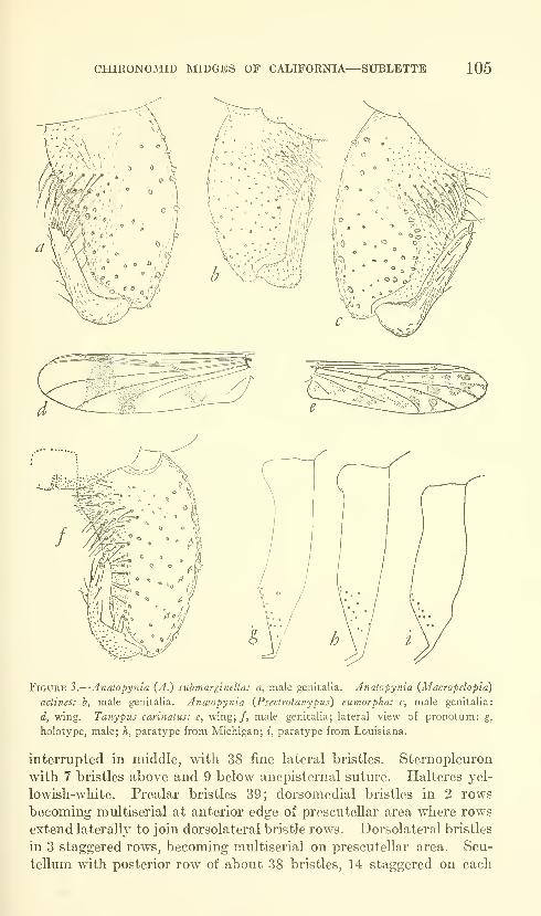
CHIRONOMID MIDGES OF CALIFORNIA—SUBLETTE 105
Figure 3.
—
Anatopynia {A.) submarginella: a, male genitalia. Anaiopynia {Macropelopia)
adines: b, male genitalia. Anatopynia {Pseclrotanypus) eumorpha: c, male genitalia:
d, wing. Tanypus carinatus: e, wing;/, male genitalia; lateral view of pronotum: g,
holotype, male; h, paratype from Michigan; i, paratype from Louisiana.
interrupted in middle, with 38 fine lateral bristles. Sternopleuron
with 7 bristles above and 9 below anepisternal suture. Halteres yel-
lowish-white. Prealar bristles 39; dorsomedial bristles in 2 rows
becoming multiserial at anterior edge of prescutellar area where rows
extend laterally to join dorsolateral bristle rows. Dorsolateral bristles
in 3 staggered rows, becoming multiserial on prescutellar area. Scu-
tellum with posterior row of about 38 bristles, 14 staggered on each
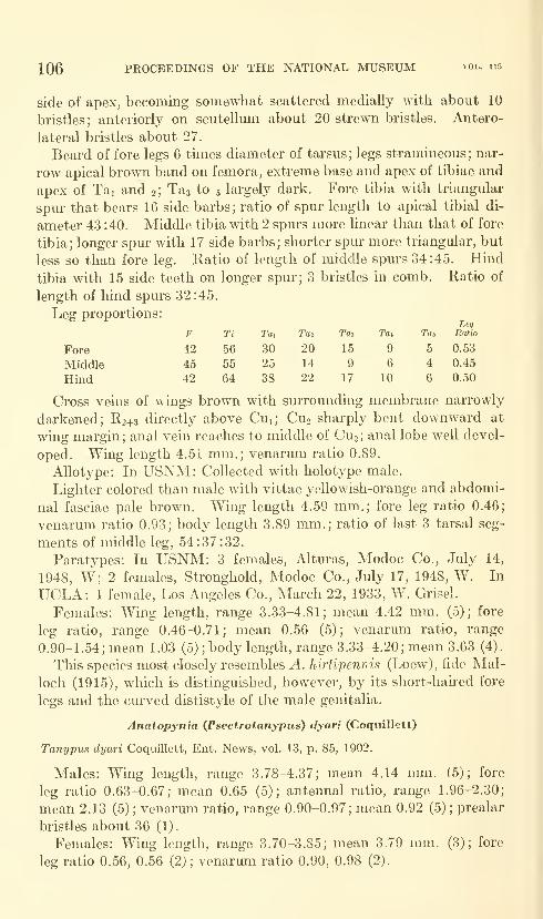
106 PROCEEDINGS OF THE NATIONAL MUSEUM vol. 115
side of apex, becomirig somewhat scattered medially with about 10
bristles; anteriorly on scutellum about 20 strewn bristles. Antero-
lateral bristles about 27.
Beard of fore legs 6 times diameter of tarsus; legs stramineous; nar-
row apical brown band on femora, extreme base and apex of tibiae and
apex of Tai and 2; Tas to 5 largely dark. Fore tibia with triangular
spur that bears 16 side barbs; ratio of spur length to apical tibial di-
ameter 43 :40. Middle tibia with 2 spurs more linear than that of fore
tibia; longer spur with 17 side barbs; shorter spur more triangular, but
less so than fore leg. Ratio of length of middle spurs 34:45. Hind
tibia with 15 side teeth on longer spur; 3 bristles in comb. Ratio of
length of hind spurs 32:45.
eg proportions:
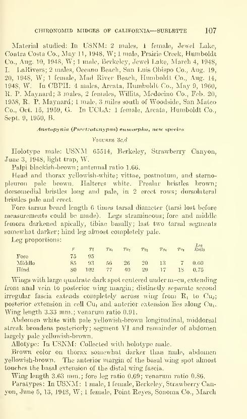
CHIRONOMID MIDGES OF CALIFORNIA—SUBLETTE 107
Material studied: In USNM: 2 males, 1 female, Jewel Lake,
Contra Costa Co., May 11, 1948, W; 1 male, Prairie Creek, HumboldtCo., Aug. 10, 1948, W; 1 male, Berkeley, Jewel Lake, March 4, 1948,
I. LaRivers; 2 males, Oceano Beach, San Luis Obispo Co., Aug. 19,
20, 1948, W; 1 female. Mad River Beach, Humboldt Co., Aug. 14,
1948, W. In CBPH: 4 males. Areata, Humboldt Co., May 9, 1960,
R. P. Maynard; 3 males, 2 females, WilHts, Medocino Co., Feb. 20,
1958, R. P. Maynard; 1 male, 3 miles south of Woodside, San MateoCo., Oct. 15, 1959, G. In UCLA: 1 female, Areata, Humboldt Co.,
Sept. 9, 1950, B.
Anatopynia {Psectrotanypus) eiimorpha, new species
Figures 3c,d
Holotype male: USNM 65514, Berkeley, Strawberry Canyon,
June 3, 1948, light trap, W.Palpi blackish-brown; antennal ratio 1.66.
Head and thorax yellowish-white; vittae, postnotum, and sterno-
pleuron pale brown. Halteres white. Prealar bristles brown;
dorsomedial bristles long and pale, in 2 erect rows; dorsolateral
bristles pale and erect.
Fore tarsus beard length 6 times tarsal diameter (tarsi lost before
measurements could be made). Legs stramineous; fore and middle
femora darkened apically, tibiae basally; last two tarsal segments
somewhat darker; hind leg almost completely pale.
Leg proportions:

108 PROCEEDINGS OF THE NATIONAL MUSEUM vol. 115
16, 1948; 1 female, Strawberry Canyon, Alameda Co., May 24,
1948, W.Males: Wing length 3.11 mm.; venarum ratio 0.90. Abdomen
entirely white; beard length 6 times tarsal diameter.
Females: Wing length, range 3.55-3.70; mean 3.63 mm. (3); fore
leg ratio, range 0.67-0.74; mean 0.70 (3); venarmn ratio, range
0.84-0.91; mean 0.88 (3).
This species is distinguished from related North American species
in the key (p. 109).
Anatopynia (JPsectrotanypus) venusta (Coquillett)
Tanypus venustus Coquillett, Proc. U.S. Nat. Mus., vol. 25, p. 91, 1902.
Males: Wing length, range 3.33-4.67; mean 3.84 mm. (16); fore
leg ratio, range 0.56-0.65; mean 0.60 (14); antennal ratio, range
1.57-1.93; mean 1.70 (14); venarum ratio, range 0.86-0.97; mean0.91 (16); hind leg ratio, range 0.63-0.66; mean 0.64 (3).
Females: Wing length, range 3.51-4.63; mean 4.10 mm. (17); fore
leg ratio, range 0.57-0.63; mean 0.59 (16); venarum ratio, range
0.92-0.98; mean 0.95 (16); hind leg ratio 0.64, 0.67 (2).
Material studied: In USNM: 2 males, 1 female. Deer Creek, HotSprings, Tulare Co., Aug. 6, 1947, W; 1 male, Wheeler's Springs,
Ventura Co., June 16, 1948, light trap, W; 1 male, Visalia, June 30,
1947, W; 1 male, Kern River Canyon, Kern Co., July 26, 1947, B.
Brookman; 1 male. Alum Rock Park, Santa Clara Co., July 8, 1948,
W; 1 male, Hume Lake, Aug. 20, 1947, W; 1 male, San Luis Obispo,
Aug. 21, 1948, W; 1 male, 1 female, San Luis Obispo, Aug. 19, 1948,
W; 1 female, Elderwood, July 18, 1947, bridge, W; 1 female, Buellton,
Santa Barbara Co., June 23, 1948, W; 1 female, Santa Cruz, Santa
Cruz Co., July 8, 1948, W. In CBPH: 1 male. Parks Au- Force
Base, Alameda Co., Oct. 13, 1959; 2 males, 2 females, 2 miles from
Orcutt, Santa Barbara Co., Aug. 2, 1948, W; 1 female, 2.5 miles
north of Manteca, San Joaquin Co., June 19, 1957, G; 1 female,
Wrights, Santa Clara Co., Aug. 25, 1955; 1 male, 1 female, 3 miles
west of Gilroy, Santa Clara Co., March 25, 1956, G; 1 female. Ft.
Barry, Marin Co., March 18, 1954, G. In UCLA: 1 male, 9 females,
Westwood Hills, Los Angeles Co., Apr. 6, 1950; 1 male, 1 female,
Westwood Hills, Los Angeles Co., May 18, 1955, A. Fukushima;
1 male, Santa Monica Canyon, Los Angeles Co., July 22, 1952; 1
female. Sand Canyon, Orange Co., March 30, 1949; 2 females. MurphyCanyon, San Diego Co., May 8, 1949, B and Heid. In KU: 1 male,
Kernville, July 24, 1940, D. E. Hardy. In UCD: 3 males, 1 female,
Monticello, 1 mile south, Napa Co., Oct. 8, 1947, R. M. Bohart;
1 male, 2 females. Green Valley, Solano Co., Aug. 29, 1946, R. M.Bohart and H. E. Cott; 1 female, Green Valley, Solano Co., Apr. 3,
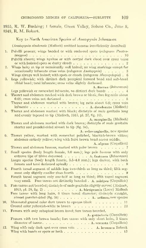
CHIRONOMID MIDGES OF CALIFORNIA—SUBLETTE 109
1955, R. W. Bushing; 1 female, Green Valley, Solano Co., June 8,
1948, E. M. Bohart.
Key to North American Species of Anatopynia Johannsen
{Anatopynia alaskensis (Malloch) omitted because insufficiently described)
1. Pulvilli present; wings banded or witti coalesced spots (subgenus Psectro-
tanypus) 10
Pulvilli absent; wings hyaline or with central dark cloud over cross veins
or with isolated spots or dusky clouds 2
2. Wings hairy at tip or occasionally well haired; no wing markings except for
occasionally infuscate cross veins (subgenus Anatopynia) 3
Wings always well haired; with spots or clouds (subgenus Macropelopia) . 6
3. Legs yellowish; with distinct dark preapical femoral band and sub-basal
tibial band; tarsi infuscate; cross veins slightly darkened.
A. florens (Johannsen)
Legs yellowish or somewhat infuscate, no distinct dark bands 4
4. Thorax and abdomen marked with dark brown or black; fore leg ratio about
0.65; cross veins not darkened 5
Thorax and abdomen marked with brown; leg ratio about 0.8; cross vein
infuscate A. decolorata (Malloch)
5. Thorax and abdomen marked with black; dististyle of male genitalia long
and evenly tapered to tip (Malloch, 1915, pi. 27, fig. 10).
A. marginella (Malloch)
Thorax and abdomen marked with dark brown; dististyle of male genitalia
shorter and parallel-sided almost to tip (fig. 3a).
A. submarginella, new species
6. Thorax yellow, marked with somewhat polished, blackish-brown vittae;
abdomen entirely yellow; wing with faint brown band across middle.
A. algens (Coquillett)
Thorax and abdomen fuscous, marked with paler brown 7
7. Small species (body length female, 3.0 mm.) ; legs pale fuscous with only
extreme tips of tibiae darkened A. fastuosa (Johannsen)
Larger species (body length female, 3.5-4.0 mm.) ; legs darker, with both
femora and tarsi darkened apically 8
8. Fourth tarsal segment of middle legs two-thirds as long as third; fifth seg-
ment only slightly smaller than fourth 9
Fourth tarsal segment only one-half as long as third; fifth tarsal segmentvery small. Fore tarsus not distinctly bearded . A. miripes (Coquillett)
9. Fore tarsus not bearded; dististyle of male genitalia slightly curved (Malloch,
1915, pi. 28, fig. 2) A. hirtipennis (Loew) Malloch
Fore tarsus with long hairs, 6 times tarsal diameter; dististyle straight,
almost parallel-sided (fig. 36) A. aclines^ new species
10. Mesonotal ground color dark brown to opaque black 11
Ground color yellowish-white to brown 12
11. Femora wdth only subapical brown band; fore tarsus bearded.
A. guttularis (Coquillett)
Femora with two brown bands; fore tarsus with only short hairs, 3 times
diameter of tarsus A. veniista (Coquillett)
12. Wing with only dark spot over cross vein A. brunnea RobackWing with bands or spots or both 13
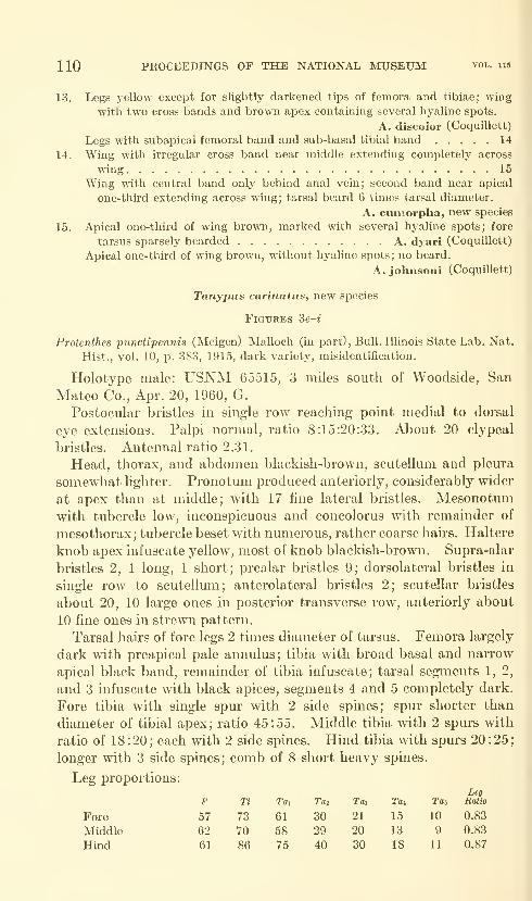
110 PROCEEDINGS OF THE NATIONAL MUSEUM vol. 115
13. Legs yellow except for slightly darkened tips of femora and tibiae; wing
with two cross bands and brown apex containing several hyaline spots.
A. discolor (Coquillett)
Legs with subapical femoral band and sub-basal tibial band 14
14. Wing with irregular cross band near middle extending completely across
wing 15
Wing with central band only behind anal vein; second band near apical
one-third extending across wing; tarsal beard 6 times tarsal diameter.
A. euniorpha, new species
15. Apical one-third of wing brown, marked with several hyaline spots; fore
tarsus sparsely bearded A. dyari (Coquillett)
Apical one-third of wing brown, without hyaline spots; no beard.
A. johnsoni (Coquillett)
Tanypus carinatus, new species
Figures 3e-4
Protenthes punctipennis (Meigen) Malloch (in part), Bull. Illinois State Lab. Nat.
Hist., vol. 10, p. 383, 1915, dark variety, misidentification.
Holotype male: USNM 65515, 3 miles south of Woodside, San
Mateo Co., Apr. 20, 1960, G.
Postocular bristles in single row reaching point medial to dorsal
eye extensions. Palpi normal, ratio 8:15:20:33. About 20 clypeal
bristles. Antennal ratio 2.31.
Head, thorax, and abdomen blackish-brown, scutellum and pleura
somewhat lighter. Pronotum produced anteriorly, considerably wider
at apex than at middle; with 17 fine lateral bristles. Mesonotumwith tubercle low, inconspicuous and concolorus with remainder of
mesothorax; tubercle beset with numerous, rather coarse hairs. Haltere
knob apex infuscate yellow, most of knob blackish-brown. Supra-alar
bristles 2, 1 long, 1 short; prealar bristles 9; dorsolateral bristles in
single row to scutellmn; anterolateral bristles 2; scutellar bristles
about 20, 10 large ones in posterior transverse row, anteriorly about
10 fine ones in strewn pattern.
Tarsal hairs of fore legs 2 times diameter of tarsus. Femora largely
dark with preapical pale annulus; tibia with broad basal and narrow
apical black band, remainder of tibia infuscate; tarsal segments 1, 2,
and 3 infuscate with black apices, segments 4 and 5 completely dark.
Fore tibia with single spur with 2 side spines; spur shorter than
diameter of tibial apex; ratio 45:55. Middle tibia with 2 spurs with
ratio of 18:20; each with 2 side spines. Hind tibia with spurs 20:25;
longer with 3 side spines; comb of 8 short heavy spines.
Leg proportions:
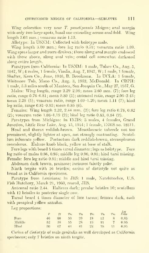
CHIRONOMID MIDGES OF CALIFORNIA—SUBLETTE 111
Wing coloration very near T. punctipennis Meigen; anal margin
with only two large spots, basal one extending across anal fold. Winglength 3.03 mm.; venarum ratio 1.13.
Allotype: In USNM: Collected with holotype male.
Wing length 3.00 mm.; fore leg ratio 0.78; venarmn ratio 1.09.
Wing spots larger and more distinct; those along anal margin coalesced
with those above, along anal vein; costal cell somewhat darkened
along entire length.
Paratypes from California: In USNM: 1 male, Tulare Co., Aug. 5,
1947, W; 4 males, 1 female, Visalia, Aug. 7, 1947, W; 1 male, 1 female,
Shafter, Kern Co., June, 1946, B. Brookman. In UCLA: 1 female,
Whitmore Tub, Mono Co., Aug. 3, 1952, McDonald. In CBPH:1 male, 5.3 miles south of Manteca, San Joaquin Co., May 27, 1957, G.
Males: Wing length, range 2.29-2.96; mean 2.60 mm. (7); fore leg
ratio, range 0.76-0.81 ; mean 0.80 (5); antennal ratio, range 2.05-2.43;
mean 2.28 (5); venarum ratio, range 1.09-1.20; mean 1.11 (7); hind
leg ratio, range 0.81-0.93; mean 0.85 (6).
Females: Wing length 2.32, 2.44 mm. (2); fore leg ratio 0.78, 0.82
(2); venarum ratio 1.08-1.19 (2); hind leg ratio 0.83, 0.84 (2).
Paratypes from Michigan: In INHS: 3 males, 4 females. GrandJunction, Little Bear Lake, Aug. 15, 1914; 1 female, INHS no. 18811.
Head and thorax reddish-brown. Mesothoracic tubercle not too
prominent, slightly lighter at apex, not strongly contrasting. Scutel-
lum infuscate yellow. Postnotum dark reddish-brown, sternopleuron
concolorus. Haltere knob black, yellow at base of stalk.
Fore legs with beard 5 times tarsal diameter; legs as holotype. Fore
leg ratio of males 0.86, 0.93; middle leg 0.90, 0.91; hind tarsi missing.
Female: fore leg ratio 0.91; middle and hind tarsi missing.
Abdomen dark brown, pruinose; incisures faintly paler.
Ninth tergite with 16 bristles; carina of dististyle not quite as
broad as in California specimens.
Paratype from Louisiana: In JES: 1 male, Natchitoches, U.S.
Fish Hatchery, March 21, 1960, reared, JES.
Antennal ratio 2.44. Halteres dark; prealar bristles 10; scuteUum
with 12 bristles in posterior single row.
Tarsal beard 4 times diameter of fore tarsus; femora dark, each
with preapical yellow annulus.
Leg proportions:
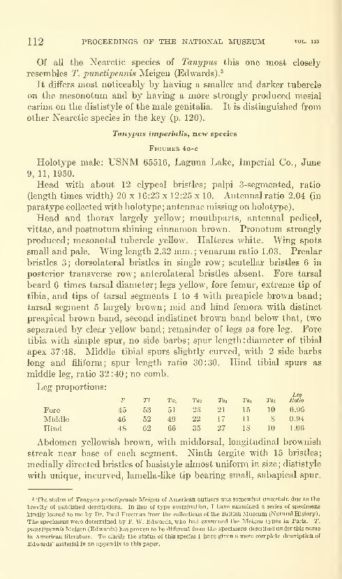
112 PROCEEDINGS OF THE NATIONAL MUSEUM vol. 115
Of all the Nearctic species of Tanypus this one most closely
resembles T. punctipennis Meigen (Edwards).^
It differs most noticeably by having a smaller and darker tubercle
on the mesonotum and by having a more strongly produced mesial
carina on the dististyle of the male genitalia. It is distinguished from
other Nearctic species in the key (p. 120).
Tanypus imperialis, new species
FlGUKES 4o-C
Holotype male: USNM 65516, Laguna Lake, Imperial Co., June
9, 11, 1950.
Head with about 12 clypeal bristles; palpi 3-segmented, ratio
(length times width) 20 x 16:23 x 12:25 x 10. Antennal ratio 2.04 (in
paratype collected with holotype; antennae missing on holotype).
Head and thorax largely yeUow; mouthparts, antennal pedicel,
vittae, and postnotum shining cinnamon brown. Pronotum strongly
produced; mesonotal tubercle yeUow. Halteres white. Wing spots
small and pale. Wing length 2.32 mm. ; venarum ratio 1.03. Prealar
bristles 3; dorsolateral bristles in single row; scutellar bristles 6 in
posterior transverse row; anterolateral bristles absent. Fore tarsal
beard 6 times tarsal diameter; legs yellow, fore femur, extreme tip of
tibia, and tips of tarsal segments 1 to 4 with preapicle brown band;
tarsal segment 5 largely brown; mid and hind femora with distinct
preapical brown band, second indistinct brown band below that, two
separated by clear j^ellow band; remainder of legs as fore leg. Fore
tibia mth simple spur, no side barbs; spur length : diameter of tibial
apex 37:48. Middle tibial spurs slightly curved, with 2 side barbs
long and fihform; spur length ratio 30:30. Hind tibial spurs as
middle leg, ratio 32:40; no comb.
Leg proportions:

CHIRONOMID MIDGES OF CALIFORNIA—SUBLETTE 113
Allotype: In USNM: Fish Springs, Salton Sea, Imperial Co.,
Oct. 18, 19, 1954, Whitney.
Wing pattern and coloration as holotype male. Differs by dorso-
lateral bristles forming group of 4 bristles just anterior to scutellumand by having 11 prealar and 12 anterolateral bristles. Pronotumproduced as in holotype, bearing 5 fine, lateral bristles.
Wing length 2.29 mm.; fore and hind tarsi missing; venarumratio 1.07.
Paratypes: In UCD: 1 female, 12 miles east of Heber, Imperial
Co., May 12, 1956, T. R. Haig. In UCLA: 1 male, collected withholotype; 1 female, Lee's Ranch, Chatsworth, Los Angeles Co.,
July 25, 1950.
Male: Wing length 2.59 mm.; fore leg missing; antennal ratio
2.04; venarum ratio 1.05; hind leg missing.
Females: Wing length 2.59, 2.70 mm. (2); fore leg ratio 0.69 (1);
venarum ratio 1.02, 1.02 (2); hind leg ratio 1.00, 0.80 (2). In femalecollected 12 miles east of Heber, Imperial Co., dorsolateral bristles
in three rows just anterior to scuteUum with about 7 bristles in clump;pronotum produced with 5 lateral bristles; prealar bristles 10; antero-
lateral bristles 10.
In the female from Lee's Ranch, Los Angeles Co., the high leg
ratio and the produced pronotum seem to place it in this species;
however, the prealar bristles are 9, the anterolateral bristles 4, andthe dorsolateral bristles become staggered in 2 rows turning medially
just before the scuteUum.
This species is quite similar to T. neopvnctipennis new species butdiffers in being smaller and by having a strongly produced pronotumand having a distinctly different male genitaha. It is separated fromthe other Nearctic species in the key (p. 120).
Tanypus parastellatus, new species
Figures 4d,e
Holotype male: USNM 65517, Laguna Lake, Imperial Co., June
9, 11, 1950.
Head dark; antennal ratio 2.00.
Pronotum parallel-sided; dark brown, infuscate yellow apicaUy.
Mesonotum blackish-brown, overlaid with strong greyish-green
pruinescence ; tubercle very small and dark. Scutellum dark yeUow-ish-brown. Postnotum shining blackish-brown. Halteres yeUow.Prealar bristles 10; dorsolateral bristles in single row; anterolateral
bristles about 6.
Legs yellowish; black ring above and below knee and at apex of
tibiae and at apex of tarsal joints 1 to 3; 4 and 5 largely dark; base

114 PROCEEDINGS OF THE NATIONAL MUSEUM vol. us
of femora somewhat infuscate, with clear yellow fascia between basal
infuscation and apical dark band.
eg proportions:

CHIRONOMID MIDGES OF CALIFORNIA—SUBLETTE 115
Figure 4.— Tanypus iniperialis: a, lateral view of pronotum; b, wing; c, male genitalia.
Ta7iypus parastellatus: d, male genitalia; e, wing, Tanypus stellatus Coquillett: /,
male genitalia. Tanypus grodhausi: g, paratype, lateral view of pronotum; h, allotype,
lateral view of pronotum; i, wing; /, male genitalia.
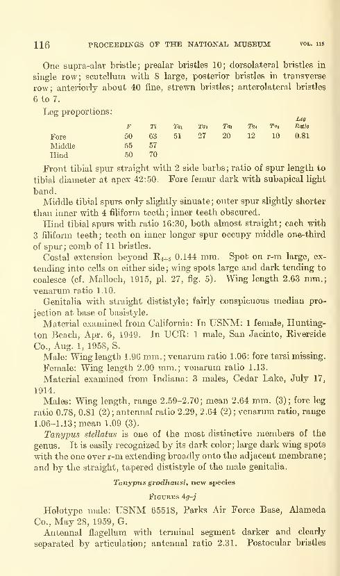
116 PROCEEDINGS OF THE NATIONAL MUSEUM vol. iib
One supra-alar bristle; prealar bristles 10; dorsolateral bristles in
single row; scutellum with 8 large, posterior bristles in transverse
row; anteriorly about 40 fine, strewn bristles; anterolateral bristles
6 to 7.
Leg proportions:
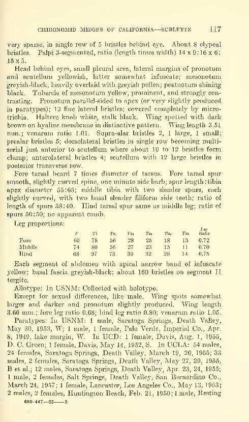
CHIRONOMID MIDGES OF CALIFORNIA—SUBLETTE 117
very sparse, in single row of 5 bristles behind eye. About 8 clypeal
bristles. Palpi 3-segmented, ratio (length times width) 14 x 9 : 16 x 6
:
15x5.Head behind eyes, small pleural area, lateral margins of pronotum
and scutellum yellowish, latter somewhat infuscate; mesonotumgreyish-black, heavily overlaid with greyish pollen; postnotum shining
black. Tubercle of mesonotum yellow, prominent, and strongly con-
trasting. Pronotum parallel-sided to apex (or very slightl}^ produced
in paratypes); 13 fine lateral bristles; covered completely by micro-
trichia. Haltere knob white, stalk black. Wing spotted with dark
brown on hyaline membrane in distinctive pattern. Wing length 3.51
mm.; venarum ratio 1.01. Supra-alar bristles 2, 1 large, 1 small;
prealar bristles 5; dorsolateral bristles in single row becoming multi-
serial just anterior to scutellum where about 10 to 12 bristles form
clump; anterolateral bristles 4; scutellum with 12 large bristles in
posterior transverse row.
Fore tarsal beard 7 times diameter of tarsus. Fore tarsal spur
smooth, slightly curved spine, one minute side barb; spur length :tibia
apex diameter 55:65; middle tibia with two slender spurs, each
slightly curved, with two basal slender filiform side teeth; ratio of
length of spurs 38:40. Hind tarsal spur same as middle leg; ratio of
spurs 50:50; no apparent comb.
Leg proportions:
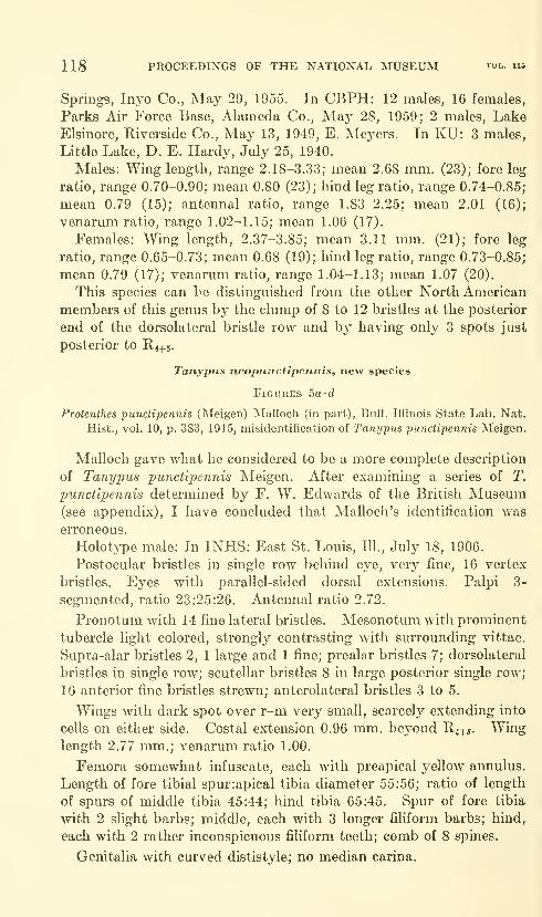
118 PROCEEDINGS OF THE NATIONAL MUSEUM vol. 115
Springs, Inj^o Co., May 29, 1955. In CBPH: 12 males, 16 females,
Parks Air Force Base, Alameda Co., May 28, 1959; 2 males. LakeElsinore, Riverside Co., May 13, 1949, E. Meyers. In KU: 3 males,
Little Lake, D. E. Hardy, July 25, 1940.
Males: Wing length, range 2.18-3.33; mean 2.68 mm. (23); fore leg
ratio, range 0.70-0.90; mean 0.80 (23); hind leg ratio, range 0.74-0.85;
mean 0.79 (15); antenna! ratio, range 1.83-2.25; mean 2.01 (16);
venarum ratio, range 1.02-1.15; mean 1.06 (17).
Females: Wing length, 2.37-3.85; mean 3.11 mm. (21); fore leg
ratio, range 0.65-0.73; mean 0.68 (19); hind leg ratio, range 0.73-0.85;
mean 0.79 (17); venarum ratio, range 1.04-1.13; mean 1.07 (20).
This species can be distinguished from the other North American
members of this genus by the clump of 8 to 12 bristles at the posterior
end of the dorsolateral bristle row and by having only 3 spots just
posterior to R4+5.
Tanypus neopnnctipennis, new species
Figures 5a-d
Protenthes punctipennis (Meigen) Malloch (in part), Bull. Illinois State Lab. Nat.
Hist., vol. 10, p. 383, 1915, misidentification of Tanypus punctipennis Meigen.
Malloch gave what he considered to be a more complete description
of Tanypus 'punctipennis Meigen. After examining a series of T.
punctipennis determined by F. W. Edwards of the British Museum(see appendix), I have concluded that Malloch's identification was
erroneous.
Holot;>T)e male: In INHS: East St. Louis, III, July 18, 1906.
Postocular bristles in single row behind eye, very fine, 16 vertex
bristles. Eyes with parallel-sided dorsal extensions. Palpi 3-
segmented, ratio 23:25:26. Antennal ratio 2.72.
Pronotum with 14 fine lateral bristles. Mesonotum with prominent
tubercle light colored, strongly contrasting with surrounding vittae.
Supra-alar bristles 2, 1 large and 1 fine; prealar bristles 7; dorsolateral
bristles in single row; scutellar bristles 8 in large posterior single row;
16 anterior fine bristles strewn; anterolateral bristles 3 to 5.
Wings with dark spot over r-m very small, scarcely extending into
cells on either side. Costal extension 0.96 mm. beyond R^+j. Winglength 2.77 mm.; venarum ratio 1.00.
Femora somewhat infuscate, each with preapical yellow annulus.
Length of fore tibial spur:apical tibia diameter 55:56; ratio of length
of spurs of middle tibia 45:44; hind tibia 65:45. Spur of fore tibia
with 2 slight barbs; middle, each with 3 longer filiform barbs; hind,
each with 2 rather inconspicuous filiform teeth; comb of 8 spines.
Genitalia with curved dististyle; no median carina.
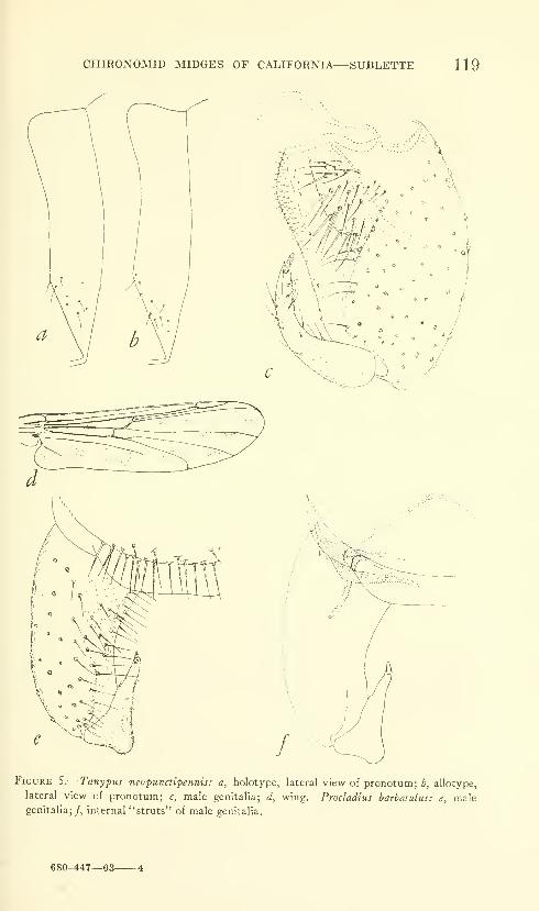
CHIRONOMID MIDGES OF CALIFORNIA
—
SUBLETTE 119
Figure S.— Tanypus neopunctipennis: a, holotype, lateral view of pronotum; b, allotype,
lateral view of pronotum; c, male genitalia; d, wing, Procladius barbaiulus: e, malegenitalia;/, internal "struts" of male genitalia.
680-447—63 4
120 PROCEEDINGS OF THE NATIONAL MUSEUM vol. 113
Allotype: In INHS: Havana, 111., Chautauqua Park, Apr. 29, 1914.
Coloration similar to holotype; wing spots heavier, membrane moredensely haired; stem of M between r-m and m-cu very short, only
about one-half as long as in males; pronotum more strongly produced
anterior^; dorsolateral bristles single to just anterior to scutellum
where row is doubled for about 4 bristles.
Wing length 3.28 mm.; fore leg ratio 0.80; venarum ratio 1.10;
hind leg ratio 0.96.
Paratypes from Illinois: In INHS: 4 males, 3 females, Havana,
Apr. 27, 29, 1914; 1 male. Quiver Lake, Havana, Sept. 19, 1895,
Hart, Newberry, Hempel; 1 female, Momence, July 17, 1914; 1 male,
2 females, St. Joseph, June 9, 1915; 1 female, Urbana, Sept. 5, 1914;
1 male, Vergennes, Aug. 12, 1914.
Paratypes from Texas: In INHS: 1 female. Lake Lomalta, Sept. 27,
1910.
Paratypes from Louisiana: In USNM, L^CLA, JES: 9 males, 2
females, Natchitoches, Chaplain's Lake, Feb. 4, 5, 1957, reared,
JES; 1 male, Natchitoches, pool near Northwestern State College
Dairy, Oct. 22, 1954, reared, JES; 2 males, Natchitoches, U.S. Fish
Hatchery, March 21, 22, 1960, reared, JES.
Paratypes from Alabama: In Dendy, JES: 9 males. Auburn,
June 6, 1956, J. S. Dendy; 1 male. Auburn, June 8, 1956, J. S. Dendy;
1 male, Auburn, June 23, 1955, J. S. Dendy.
Males: Wing length, range 2.44-3.07; mean 2.78 mm. (7); fore leg
ratio, range 0.84-0.98; mean 0.91 (8); hind leg ratio, range 0.88-1.08;
mean 0.97 (7) ; antennal ratio, range 2.01-2.43 ; mean 2.22 (7) ; venarum
ratio, range 1.00-1.08; mean 1.04 (3).
Females: Wing length 2.96, 3.33 mm. (2); fore leg ratio 0.74, 0.88
(2) ; hind leg ratio 0.90 (1) ; venarum ratio 1.02, 1.08 (2).
This species most closely resembles T. imperialis, new species, from
which it can be distinguished by the distinctively different genitalia
and by having the superior pronotal margin only slightly produced
apically. It is distinguished from the other North American species in
the key which follows.
Key to North American Species of Tanypus
1. Tubercle of mesonotum very small and inconspicuous, concolorus with
remainder of mesonotum or only slightly lighter in color; general body
color dark; palpi 4-segniented 2
Tubercle conspicuous, yellow or yellowish-white, strongly contrasting with
remainder of brown to dark brown mesonotum; occasionally tubercle
infuscate yellow; body with yellowish ground color; palpi 3-segmented . 4
CHIRONOMID MIDGES OF CALIFORNIA—SUBLETTE 121
2. Anterior and posterior margins of pronotum nearly parallel; dististyle of
male genitalia straight or slightly curved 3
Anterior margin of pronotum strongly produced at apex; dististyle of malegenitalia with distinctively produced mesial carina (in cleared genitalia
mounts difficult to see) T. carinatu, new species
3. Spot over r-m cross vein large extending broadly into cells on either side;
wing spots dark and tend to coalesce (cf. Malloch, 1915, pi. 27, fig. 5);
dististyle of male genitalia straight ; tapering to tip . T. stellatus (Coquillett)
Spot over r-m smaller extending into adjacent cells only narrowly; wing spots
smaller and more distinct, isolated; dististyle slightly bent, broadenedbefore apex T. parastellatus, new species
4. Dorsolateral bristles in single row to scutellum; with 4 spots in cell behind
R4+5 5
Dorsolateral bristles in single row to just anterior to scutellum where rowexpands to form clump of 8-12 bristles . . . . T. grodhausi, new species
5. Pronotum only slightly produced towards apex; dististyle simple, no carina,
evenly curved T. neopunctipennis, new species
Pronotum strongly produced at apex; dististyle with unique apical carina
bearing subterminal spine T. imperialis, new species
Procladius barbatulus, new species
Figures 5e,/
Holotype male: USNM 65519, Hume Lake, Fresno Co., Sept. 24,
1957, G.
Postocular bristles in 2 staggered rows. Eyes with usual parallel-
sided dorsal extensions. Clypeal bristles 9; palpi ratio 8:13:18:30.
Antennal ratio 1.70.
Head, thorax, and abdomen dark brown, somewhat lighter at
shoulders and on pleura. Halteres dark. Pronotum with 18 fine
lateral bristles. One supra-alar bristle; prealar bristles 13; dorso-
medial bristles 3 on either side of midline on prescutellar area; dorso-
lateral bristles in single row; anterolateral bristles 6; scutellar bristles
about 28, somewhat scattered, posteriorly forming transverse row.
Wings darkened only over r-m and, faintly, over m-cu; membranewell haired with black hairs; anterior veins somewhat darker than
posterior. Wing length 2.22 mm.; venarum ratio 1.50.
Legs uniformly dark brown. Fore tarsus with short beard, 4 times
tarsal diameter. Fore leg with single tibial spur, slightly shorter
than diameter of apex of tibia; middle leg spurs 40:30; hind leg spurs
53:32, comb of 11 spines.
Leg proportions:Leg
F Ti Tai Tai Tai Tat Tm Ratio
Fore 45 59 44 20 15 10 5 0.74
Middle 51 56 37 0.66
Hind 45 61 43 20 16 9 7 0.70
Abdomen almost uniformly darkened with only faint suggestion
of lighter color apically on segments; with about 60 bristles on tergite
II.
122 PROCEEDINGS OF THE NATIONAL MUSEUM vol. 115
Female: Unknown.This species keys to P. choreus (Meigen) in Johannsen (1952).
There is much confusion as to the identity of P. choreus and P. culici-
formis (Linnaeus). Edwards (1929) Hsts them as separate species
although he points out there may be only a varietal difference between
the two. Edwards says the male genitalia of the two species are
identical.
Through the kindness of Dr. Paul Freeman of the British Museum(Natural History) I have examined examples of P. culiciformis and P.
choreus determined by Edwards. While there is a slight color and
size difference, the genitalia of the two appear to be identical. I must
therefore conclude that P. choreus is only a variety of P. culiciformis
and herewith list it as a new synonomy. P. culiciformis is redescribed
from Edwards' material in the appendix to this paper.
P. barbatulus, new species, may be separated from the rest of the
Nearctic species by the distinctive dististyle of the male genitalia.
The genitalia resembles those figured by Freeman (1955) for P.
albitalus Kieffer and P. polytomus (Kieft'er). These species, however,
have distinctively different wing patterns (cf. Freeman, 1955, pi. 1,
figs, h and j). P. sagittalis (Kieffer) is described by Edwards (1929)
as having a dististyle that resembles P. barbatulus, new species ; how-
ever, P. sagittalis lacks a fore tarsal beard and can thus be distinguished.
Procladius freemani, new species
Figures 6a-e
Holotype male: USNM 65520, San Bruno, San Mateo Co., Aug. 23,
1957, R. P. Maynard.
Postocular bristles in 2 staggered rows; reaching medially to dorsal
extension of eye. Clypeal bristles 23. Palpi ratio 15:20:39:40.
Antennal ratio 2.22.
Head and thorax almost entirely black, heavily dusted with white
poUen; heavily infuscate yellow on shoulders, pleura and apex and
lateral margin of prothorax. Prothorax with 13-17 lateral bristles.
Haltere knob white, stalk infuscate. Supra-alar bristles 2, 1 large,
1 small; prealar bristles about 19; dorsomedial bristle row divides just
behind inconspicuous mesonotal tubercle and extends laterally as
two rows, 1 on each side, almost to dorsolateral bristle row. Dorso-
lateral bristles in single row; immediately anterior to scutellum is a
transverse row of 5 bristles on each side, lying at right angles to dor-
solateral row. Scutellar bristles about 40; anterolateral bristles 6.
Wings with preapical shadow as well as one in posterior margin.
Wing length 2.59 mm.
Legs infuscate yellow, apex of tibia and basitarsus black; tarsal
segments 2 to 5 largely black; longest hairs of fore leg 3 times diameter
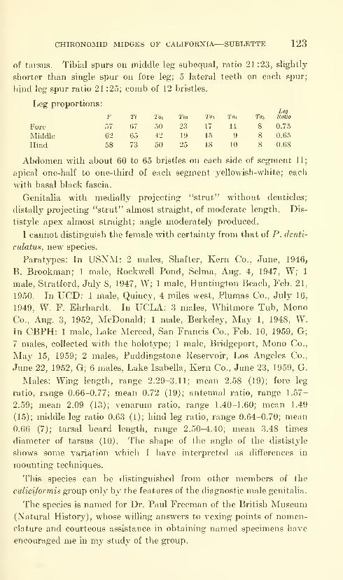
CHIRONOMID MIDGES OF CALIFORNIA—SUBLETTE 123
of tarsus. Tibial spurs on middle leg subequal, ratio 21:23, slightly
shorter than single spur on fore leg; 5 lateral teeth on each spur;
hind leg spur ratio 21 :25; comb of 12 bristles.
Leg proportions:Leg
F Ti Tax Tat Taz Tat Toj Ratio
Fore 57 67 50 23 17 11 8 0.75
Middle 62 65 42 19 15 9 8 0.65
Hind 58 73 50 25 18 10 8 0.68
Abdomen with about 60 to 65 bristles on each side of segment II;
apical one-half to one-third of each segment yellowish-white; each
with basal black fascia.
Genitalia with medially projecting "strut" without denticles;
distally projecting "strut" almost straight, of moderate length. Dis-
tistyle apex almost straight; angle moderately produced.
I cannot distinguish the female with certainty from that of P. denti-
culatus, new species.
Paratjrpes: In USNM: 2 males, Shafter, Kern Co., June, 1946,
B. Brookman; 1 male, Rockwell Pond, Selma, Aug. 4, 1947, W; 1
male, Stratford, July 8, 1947, W; 1 male, Huntington Beach, Feb. 21,
1950. In UCD: 1 male, Quincy, 4 miles west, Plumas Co., July 16,
1949, W. F. Ehrhardt. In UCLA: 3 males, Whitmore Tub, MonoCo., Aug. 3, 1952, McDonald; 1 male, Berkeley, May 1, 1948, W.In CBPH: 1 male, Lake Merced, San Francis Co., Feb. 10, 1959, G;
7 males, collected with the holotype; 1 male, Bridgeport, Mono Co.,
May 15, 1959; 2 males, Pudding-stone Reservoir, Los Angeles Co.,
June 22, 1952, G; 6 males. Lake Isabella, Kern Co., June 23, 1959, G.
Males: Wing length, range 2.29-3.11; mean 2.58 (19); fore leg
ratio, range 0.66-0.77; mean 0.72 (19); antennal ratio, range 1.57-
2.59; mean 2.09 (13); venarum ratio, range 1.40-1.60; mean 1.49
(15); middle leg ratio 0.63 (1); hind leg ratio, range 0.64-0.70; mean
0.66 (7); tarsal beard length, range 2.50-4.40; mean 3.48 times
diameter of tarsus (10). The shape of the angle of the dististyle
shows some variation which I have interpreted as differences in
mounting techniques.
This species can be distinguished from other members of the
culiciformis group only by the features of the diagnostic male genitalia.
The species is named for Dr. Paul Freeman of the British Museum(Natural History), whose willing answers to vexing points of nomen-
clature and courteous assistance in obtaining named specimens have
encouraged me in ni}^ study of the group.
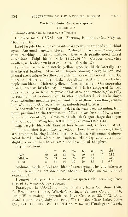
124 PROCEEDINGS OF THE NATIONAL MUSEUM vol. 115
Procladius denticiilatus, new species
Figures 6/-/^
Procladius culiciformis, of authors, not Linnaeus.
Holotype male: USNM 65521, Fortuna, Humboldt Co., May 12,
1960, light trap.
Head largely black but some infuscate yellow in front of and behind
eyes. Antennal flagellum black. Postocular bristles in 2 staggered
rows; reaching almost to midline. Eyes with parallel-sided dorsal
extensions. Palpi black, ratio 15:22:36:50. Clypeus somewhat
swollen, with about 20 bristles. Antennal ratio 1.74.
Pronotum with wide notch; yellow apically, black laterally; 15
fine lateral bristles. Mesonotum largely shining black; humeri and
pleural areas infuscate yellow; greyish pollinose when viewed obliquely;
thoracic bristles shining black. Scutellum, postnotum, and ster-
nopleuron black. Halteres yellow, infuscate basally. One supra-alar
bristle; prealar bristles 23; dorsomedial bristles staggered in two
rows, dividing in front of prescutellar area and extending laterally
to reach almost to dorsolateral bristles; dorsolateral bristles in single
row, extending medially just in front of scutellum to midUne; scutel-
lum with about 48 strewn bristles; anterolateral bristles 6.
Wings with broad triangular black fascia with base extending from
point proximal to the termination of Ri to near the tip of R4+5; apex
at termination of Cuo. Cross veins with dark spot; large dark spot
in anal margin. Wing length 3.00 mm. ; venarum ratio 1.44.
Legs largely blackish; base of fore femur and, to lesser extent,
middle and hind legs infuscate yellow. Fore tibia with single long
straight spur, bearing 5 side spines. Middle leg with spurs of almost
equal length, each with 5 or 6 spines. Hind leg with outer spur
slightly shorter than inner; ratio 48:60; comb of 13 spines.
Leg proportions:
Ti Tai Toj Toa Tat Tat, Ratio
72 59 27 20 13 9 0.82
68 47 23 17 10 8 0.69
84 60 30 20 12 9 0.71
Abdomen black ; apical one-third of each segment heavily infuscate
yellow; basal dark portion pilose, about 65 bristles on each side of
segment II.
I cannot distinguish the female of this species with certainty from
that of P. freemani, new species.
Paratypes: In USNM: 3 males, Shafter, Kern Co., June 1946,
B. Brookman; 1 male, Wheeler's Springs, Ventura Co., June 16,
1948, W; 2 males. Independence, Inyo Co., Aug. 22, 1952, B; 1
male, Hume Lake, July 20, 1947, W; 1 male. Clear Lake, Lake
Co., Oct. 11, 1947, W. In UCLA: 2 males, Huntington Beach,

CHIRONOMID MIDGES OF CALIFORNIA—SUBLETTE 125
Figure 6.
—
Procladius freemani: a, male genitalia; b-e, variations in dististyle of male
genitalia. Procladius denticulatus: /, male genitalia; g, internal "struts" of male geni-
talia; h, dististyle of paratype.
Feb. 21, 1950. In Bay: 7 males, Whittier, Feb. IG, 18, 1960. In
CBPH: 1 male, 3 miles south of Woodside, San Mateo Co., Apr. 20,
1960, G; 1 male, 3.5 miles north of Sierra City, Sierra Co., July 11,
1957, G. In KU: 1 male, Mono Lake, July 31, 1940, E. E. Kenaga.
In UCD: 1 male, Woodland, June 20, 1949, Jack Fowler; 1 male,
Davis, Feb. 13, 1958, G. G. Moore; 1 male, Davis, May 20, 1959,
F. E. Strong; 5 males, Davis, May 10, 11, 18, 1959.
Males: Wing length, range 2.04-3.00; mean 2.47 mm. (15); fore
leg ratio, range 0.67-0.82; mean 0.74 (12); antennal ratio, range
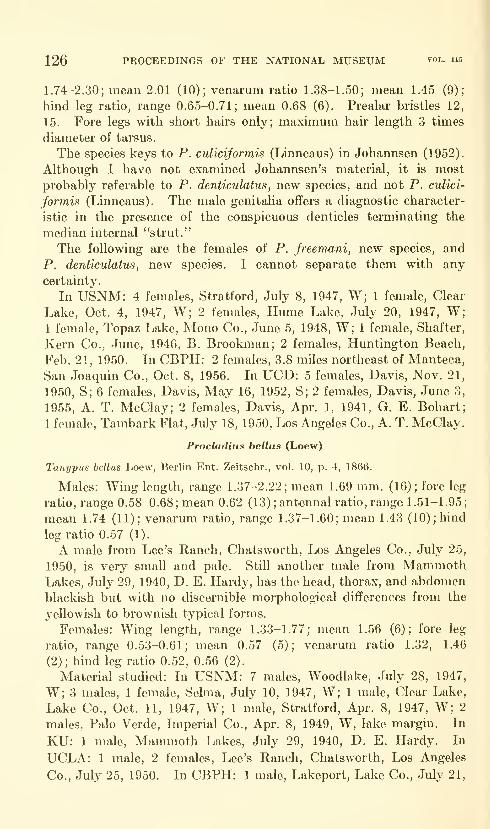
126 PROCEEDINGS OF THE NATIONAL MUSEUM vol. us
1.74-2.30; mean 2.01 (10); venarum ratio 1.38-1.50; mean 1.45 (9);
hind leg ratio, range 0.65-0.71; mean 0.68 (6). Prealar bristles 12,
15. Fore legs with short hairs only; maximum hair length 3 times
diameter of tarsus.
The species keys to P. culicijormis (Linneaus) in Johannsen (1952).
Although I have not examined Johannsen's material, it is most
probably referable to P. denticulatus , new species, and not P. culici-
jormis (Linneaus). The male genitaha offers a diagnostic character-
istic in the presence of the conspicuous denticles terminating the
median internal "strut."
The following are the females of P, freemani, new species, and
P. denticulatus, new species. I cannot separate them with any
certainty.
In USNM: 4 females, Stratford, July 8, 1947, W; 1 female. Clear
Lake, Oct. 4, 1947, W; 2 females, Hume Lake, July 20, 1947, W;1 female, Topaz Lake, Mono Co., June 5, 1948, W; 1 female, Shafter,
Kern Co., June, 1946, B. Brookman; 2 females, Huntington Beach,
Feb. 21, 1950. In CBPH: 2 females, 3.8 miles northeast of Manteca,
San Joaquin Co., Oct. 8, 1956. In UCD: 5 females, Davis, Nov. 21,
1950, S; 6 females, Davis, May 16, 1952, S; 2 females, Davis, June 3,
1955, A. T. McClay; 2 females, Davis, Apr. 1, 1941, G. E. Bohart;
1 female, Tambark Flat, July 18, 1950, Los Angeles Co., A. T. McClay.
Procladius bellus (Loew)
Tanypus bellus Loew, Berlin Ent. Zeitschr., vol. 10, p. 4, 1866.
Males: Wing length, range 1.37-2.22; mean 1.69 mm. (16); fore leg
ratio, range 0.58-0.68; mean 0.62 (13) ;antennal ratio, range 1.51-1.95;
mean 1.74 (11); venarum ratio, range 1.37-1.60; mean 1.43 (10); hind
leg ratio 0.57 (1).
A male from Lee's Ranch, Chatsworth, Los Angeles Co., July 25,
1950, is very small and pale. Still another male from MammothLakes, July 29, 1940, D. E. Hardy, has the head, thorax, and abdomenblackish but with no discernible morphological differences from the
yellowish to brownish typical forms.
Females: Wing length, range 1.33-1.77; mean 1.56 (6); fore leg
ratio, range 0.53-0.61; mean 0.57 (5); venarum ratio 1.32, 1.46
(2); hind leg ratio 0.52, 0.56 (2).
Material studied: In USNM: 7 males, Woodlake, July 28, 1947,
W; 3 males, 1 female, Selma, July 10, 1947, W; 1 male. Clear Lake,
Lake Co., Oct. 11, 1947, W; 1 male, Stratford, Apr. 8, 1947, W; 2
males, Palo Verde, Imperial Co., Apr. 8, 1949, W, lake margin. In
KU: 1 male. Mammoth Lakes, July 29, 1940, D. E. Hardy. In
UCLA: 1 male, 2 females, Lee's Ranch, Chatsworth, Los Angeles
Co., July 25, 1950. In CBPH: 1 male, Lakeport, Lake Co., July 21,

CHIRONOMID MIDGES OF CALIFORNIA—SUBLETTE 127
1955, H. Brydon. In UCR: 1 male, 3 females, San Jacinto, Riverside
Co., Sept. 26, 1958, S; 2 males, 2 females, San Jacinto, Riverside Co.,
July 7, 1958; 1 male, San Jacinto, Riverside Co., Aug. 1, 1958; 1
male, Lancaster, Aug. 18, 1958; 1 female, Lancaster, July 28, 1958, S.
Subfamily Podonominae
Boreochliis persimilis (Johannsen)
Figure 7a
Trichotanypus persimilis Johannsen, Canadian Ent., vol. 58, p. 99, 1926.
Boreochlus persimilis (Johannsen), Edwards, in Edwards and Thienemann,Zool. Anz., vol. 122, p. 153, 1938.
Antennal pedicel dark cinnamon brown; flagelluni pale brown;tip slightly enlarged, bent to one side with cluster of short terminal
hairs. Eyes reniform. Antennal ratio 0.31.
Thorax dark cinnamon brown, somewhat pollinose. Pronotumdetails obscured by pointing glue. Haltere stalk pale brown, knobdark brown. One long, pale prealar bristle; dorsomedial bristles
long, pale, and appressed; dorsolateral bristles long, pale, suberect,
divergent, in single staggered row; scutellar bristles apparently rubbed,
at least 4.
Fore leg proportions, 55:60:32:18:15:9:6; fore leg ratio 0.53.
Hind tibia with single slender spur beset with prickles on basal
half; spur length less than apical diameter of tibia; comb of 6 spines.
Wings densely haired, cuneiform, no anal angle. Squama with 10
hairs; costa greatly elongate (0.088 mm.). Venation as illustrated byJohannsen (1952). Wing length 1.41 mm.; venarum ratio 1.13.
Abdomen pale brown, somewhat darker on dorsal median line (first
segment only, others broken off in genitalia mount).
The genitalia differs from the figure given by Edwards only byhaving the dististyle somewhat excavated on the distal end instead
of being slightly enlarged. Such a difference could be attributed to
variations in mounting technique, and so I do not consider it
significant.
Material examined: 1 male. Fallen Leaf, Lake Tahoe, June 17,
1916, H. G. Dyar.Podonomus species
Material examined: 4 females, Nevada Co., Northwest of Cisco,
May 16, 1948, W.This is apparently a new species closely related to Podonomus
kiefferi (Garrett) and P. arietinus (Coquillett) but in the absence of
the male I prefer not to describe it.
The species appears to differ from the aforementioned species byhaving the halteres entirely black instead of with a whitish pedicel
and by having the abdomen yellowish-brown instead of black.
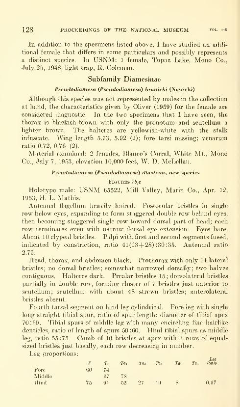
128 PROCEEDINGS OF THE NATIONAL MUSEUM vol. 115
In addition to the specimens listed above, I have studied an addi-
tional female that differs in some particulars and possibly represents
a distinct species. In USNM: 1 female, Topaz Lake, Mono Co.,
July 25, 1948, light trap, R. Coleman.
Subfamily Diamesinae
Pseudodiatnesa {Pseudodianiesa) branicki (Nowicki)
Although this species was not represented by males in the collection
at hand, the characteristics given by Oliver (1959) for the female are
considered diagnostic. In the two specimens that I have seen, the
thorax is blackish-brown with only the pronotum and scutellum a
lighter brown. The halteres are yellowish-white with the stalk
infuscate. Wing length 5.73, 5.92 (2); fore tarsi missing; venarumratio 0.72, 0.76 (2).
Material examined: 2 females, Blanco's Corral, White Mt., MonoCo., July 7, 1953, elevation 10,000 feet, W. D. McLellan.
Pseudodiamesa {Pseudodiamesa) diastena, new species
Figures 7b,c
Holotype male: USNM 65522, Mill Valley, Marin Co., Apr. 12,
1953, H. L. Mathis.
Antenna! flagellum heavily haired. Postocular bristles in single
row below eyes, expanding to form staggered double row behind eyes,
then becoming staggered single row toward dorsal part of head; each
row terminates even with narrow dorsal eye extension. Eyes bare.
About 10 clypeal bristles. Palpi with first and second segments fused,
indicated by constriction, ratio 41(13+ 28) :30:35. Antenna! ratio
2.75.
Head, thorax, and abdomen black. Prothorax with only 14 lateral
bristles; no dorsal bristles; somewhat narrowed dorsally; two halves
contiguous. Halteres dark. Prealar bristles 15; dorsolateral bristles
partially in double row, forming cluster of 7 bristles just anterior to
scutellum; scutellum with about 48 strewn bristles; anterolateral
bristles absent.
Fourth tarsal segment on hind leg cylindrical. Fore leg with single
long straight tibial spur, ratio of spur length: diameter of tibial apex
70:50. Tibial spurs of middle leg with many encircling fine hairlike
denticles, ratio of length of spurs 50:60. Hind tibial spurs as middle
leg, ratio 55:75. Comb of 10 bristles at apex with 3 rows of equal-
sized bristles just basally, each row decreasing in number.
Leg proportions:Leg
Tai Taz Ta* Ta-, Ratio
27 19 8 0.57
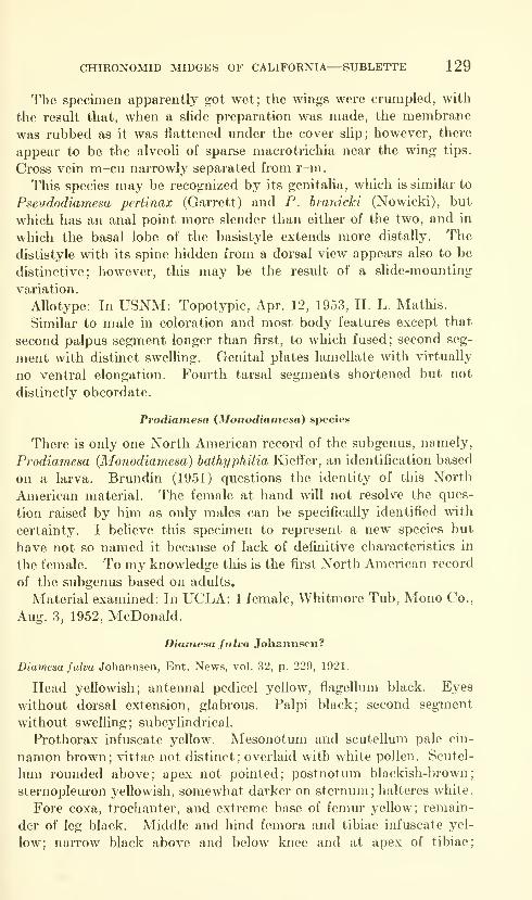
CHIRONOMID MIDGES OF CALIFORNIA—SUBLETTE 129
The specimen apparently got wet; the wings were crumpled, with
the result that, when a slide preparation was made, the membrane
was rubbed as it was flattened under the cover slip; however, there
appear to be the alveoli of sparse macrotrichia near the wing tips.
Cross vein m-cu narrowly separated from r-m.
This species may be recognized by its genitalia, which is similar to
Pseudodiamesa pertinax (Garrett) and P. branicki (Nowicki), but
which has an anal point more slender than either of the two, and in
which tlie basal lobe of the basistyle extends more distally. The
dististyle with its spine hidden from a dorsal view appears also to be
distinctive; however, this may be the result of a slide-mounting
variation.
Allotype: In USNM: Topotypic, Apr. 12, 1953, H. L. Mathis.
Similar to male in coloration and most body features except that
second palpus segment longer than first, to which fused; second seg-
ment with distinct swelling. Genital plates lamellate with virtually
no ventral elongation. Fourth tarsal segments shortened but not
distinctly obcordate.
Prodiamesa {Monodiamesa) species
There is only one North American record of the subgenus, namely,
Prodiamesa (Monodiamesa) bathyphilia Kieffer, an identification based
on a larva. Brundin (1951) questions the identity of this North
American material. The female at hand will not resolve the ques-
tion raised by him as only males can be specifically identified with
certainty. I believe this specimen to represent a new species but
have not so named it because of lack of definitive characteristics in
the female. To my knowledge this is the first North American record
of the subgenus based on adults.
Material examined: In UCLA: 1 female, Whitmore Tub, Mono Co.,
Aug. 3, 1952, McDonald.
Dianiesa fulva Johannsen?
Diamesa fulva Johannsen, Ent. News, vol. 32, p. 229, 1921.
Head yellowish; antennal pedicel yellow, flagellum black. Eyes
without dorsal extension, glabrous. Palpi black; second segment
without swelling; subcylindrical.
Prothorax infuscate yellow. Mesonotum and scutellum pale cin-
namon brown; vittae not distinct; overlaid with white pollen. Scutel-
lum rounded above; apex not pointed; postnotum blackish-brown;
sternopleuron yellowish, somewhat darker on sternum; halteres white.
Fore coxa, trochanter, and extreme base of femur yellow; remain-
der of leg black. Middle and hind femora and tibiae infuscate yel-
low; narrow black above and below knee and at apex of tibiae;
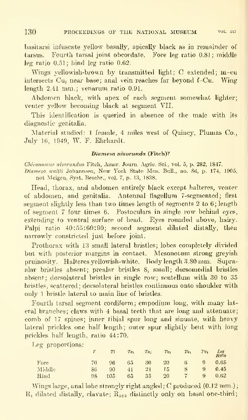
130 PROCEEDINGS OF THE NATIONAL MUSEUM vol. 115
basitarsi infuscate yellow basally, apically black as in remainder of
tarsus. Fourth tarsal joint obcordate. Fore leg ratio 0.81; middle
leg ratio 0.51; bind leg ratio 0.62.
Wings yellowish-brown by transmitted light; C extended; m-cuintersects Cui near base; anal vein reaches far beyond f-Cu. Winglength 2.41 mm.; venarum ratio 0.91.
Abdomen black, with apex of each segment somewhat lighter;
venter yellow becoming black at segment VII.
This identification is queried in absence of the male with its
diagnostic genitalia.
Material studied: 1 female, 4 miles west of Quincy, Plumas Co.,
July 16, 1949, W. F. Ehrhardt.
Diamesa nivorunda (Fitch)?
Chironomus nivorundus Fitch, Amer. Journ. Agric. Sci., vol. 5, p. 282, 1847.
Diamesa waltli Johannsen, New York State Mus. Bull., no. 86, p. 174, 1905,
not Meigen, Syst. Beschr., vol. 7, p. 13, 1838.
Head, thorax, and abdomen entirely black except halteres, venter
of abdomen, and genitalia. Antennal flagellum 7-segmented; first
segment slightly less than two times length of segments 2 to 6 ; length
of segment 7 four times 6. Postoculars in single row behind eyes,
extending to ventral surface of head. Eyes rounded above, haiiy.
Palpi ratio 40:55:60:90; second segment dilated distally, then
narrowly constricted just before joint.
Prothorax \vith 13 small lateral bristles; lobes completely divided
but with posterior margins in contact. Mesonotum strong greyish
pruinosity. Halteres yeUowish-white. Body length 3.80 mm. Supra-
alar bristles absent; prealar bristles 8, small; dorsomedial bristles
absent; dorsolateral bristles in single row; scuteUum with 30 to 35
bristles, scattered; dorsolateral bristles continuous onto shoulder with
only 1 bristle lateral to main line of bristles.
Fourth tarsal segment cordiform; empodium long, with many lat-
eral branches; claws with 4 basal teeth that are long and attenuate;
comb of 17 spines; inner tibial spur long and sinuate, with heavy
lateral prickles one half length; outer spur slightly bent with long
prickles half length, ratio 44:70.
Leg proportions:
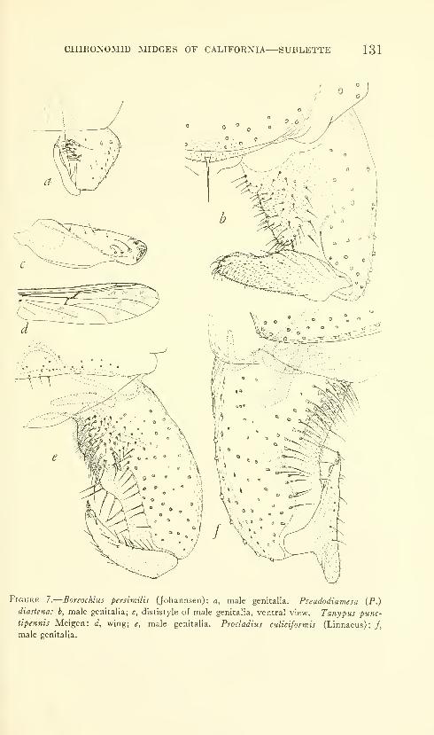
CHIRONOMID MIDGES OF CALIFORNIA—SUBLETTE 131
Figure 7.
—
Boreochlus persimilis (Johannsen): a, male genitalia. Pseudodiamesa (P.)
diastena: b, male genitalia; c, dististyle of male genitalia, ventral view. Tanypus punc-
tipennis Meigen: d, wing; e, male genitalia. Procladius culiciformis (Linnaeus): /,
male genitalia.
132 PROCEEDINGS OF THE NATIONAL MUSEUM vol. iis
R44.5 terminates proximal to M; f-Cu proximal to r-m. Anal vein
reaches almost to wing tip. Wing length 4.63 mm.; venarum ratio
0.92.
Abdomen black except venter and genitalia dark brown.
Females: Wing length, range 4.37-5.11; mean 4.75 mm. (4); leg
ratio, range 0.67-0.72; mean 0.70 (4) ; venarum ratio, range 0.90-0.97;
mean 0.94 (4).
Material examined : In USNM : 2 females, Willow Creek, FandangoPass, Modoc Co., May 15, 1948, W; 1 female, Alturas, Modoc Co.,
July 16, 1948, at light, W; 1 female, Nevada Co., northwest of Cisco,
May 16, 1948, W. In UCD: 1 female, Ukiah, Feb. 23, 1959, S. M.Fidel, light trap.
These specimens agree well with descriptions of D. nivorunda
(Fitch) Johannsen; however, in the absence of the male with its
diagnostic genitalia this identification is not positive.
Appendix
Tanypus punctipennis Meigen (Edwards)
Figures 7d,e
Tanypus punctipennis Meigen, Systematische Beschreibung der Europaischen
zweifliigeligen Insekten, vol. 1, p. 61, 1818.
This description is taken from a male in BM(NH): Beesands,
South Devonshire, June 8, 1920, F. W. Edwards, 1920-229.
Head greyish-brown; antennal pedicel dark brown. Palpi with 4
segments, 1 and 2 subequal; antennal ratio 2.14.
Thorax dark greyish-brown, vittae outlined by whitish pollen,
pleura and scuteilum infuscate yellow. Pronotum strongly produced
apically. Mesonotum with rather low, somewhat elongate tubercle,
dark yellowish-brown; second very small tubercle in center of pre-
scutellar area. Postnotum shining dark brown. Halteres yellowish-
white, slightly infuscate below knob. Prealar bristles about 14;
dorsomedial bristles extend laterally in one row behind tubercle to
join dorsolateral bristles. Dorsolateral bristles in single row forming
group of 3 bristles just anterior to scuteilum. Scuteilum with 16
bristles in posterior transverse row. Anterolateral bristles 3 to 5.
Fore tarsi with beard 6 times tarsal diameter ; femora infuscate with
clear yellow subapical fascia, space between yellow fascia and joint
darker than remainder; thus in certain lights femora appears dusky
with dark brown apical band. Tibiae infuscate yellow, with distinct,
rather broad sub-basal brown band, apex narrowly darkened as are
apices of tarsal segments 1 to 3 ; 4 and 5 largely dark.
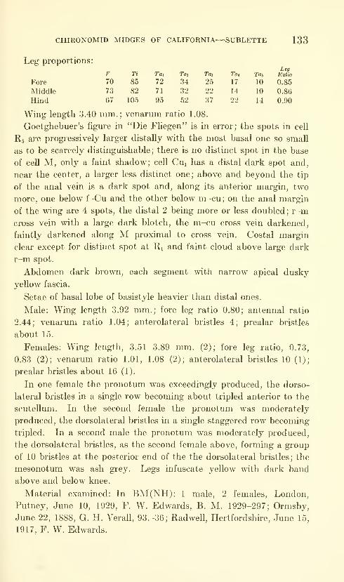
133
Leg proportions:
D MIDGES
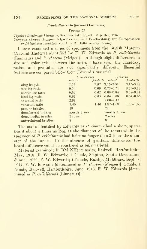
134 PROCEEDINGS OF THE NATIONAL MUSEUM
Procladius culicifortnis (Linnaeus)
Figure 7/
Tipiila culiciforniis Linnaeus, Systema naturae, ed. 12, p. 978, 1767.
Tanypus chorens Meigen, Klassifikazion und Beschreibung der Europaischen
zweifliigeligen Insekten, vol. 1, p. 23, 1804, new synonomy.
I have examined a series of specimens from the British Museum(Natural History) identified by F. W. Edwards as P. culiciformis
(Linnaeus) and P. choreus (Meigen). Although shght differences in
size and color exist between the series I have seen, the chaetoxy,
ratios, and genitalia are not significantly different. Essential
features are compared below from Edward's material.
p. culiciformis P. choreus
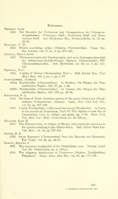
References
Brundin, Lars1951. Zur Keuntnis dor Taxonomie unci Metamorphose tier Chironomi-
deiigattungen : Protanypus Kieff., Prodiamesa Kieff. uud Mono-diamesa Kieff. Inst. Freshwater Res., Drottningholm, no. 33, pp.39-53.
Edwards, F. W.1029. British non-biting midges (Diptera, Chironomidae). Trans. Ent.
Soc. London, vol. 77, no. 2, pp. 279-430.
FiTTKAU, Ernst Josef
1957. Thienemannimyia und Conchapelopla, zwei neue Gattmigen innerhalb
der Ablabesmyia-Costnlis-Gvu.p]ie (Diptera, Chironomidae), VII:
Chironomidenstudien. Arch. Hydrobiol., vol. 53, no. 3, pp. 313-
322.
Freeman, Paul.
1955. A study of African Chironomidae, Part I. Bull. British Mus. (Nat.
Hist.) Ent., vol. 4, no. 1, pp. 1-67.
GOBTGHEBUER, JNIaURICE
1936a. Tendipedidae (Chironomidae). In Lindner, Die Fliegen der Pala-
earktischen Region, Lief. 97, pp. 1-48.
1936b. Tendipedidae (Chironomidae). In Lindner, Die Fliegen der Pala-
earktischen Region, Lief. 100, pp. 49-81.
JOHANNSEN, O. A.
1946. Revision of North American species of the genus Penlaneura (Tendi-
pedidae: Chironomidae, Diptera). Journ. New York Ent. Soc,
vol. 54, pp. 267-289.
1952. Family Tendipedidae {= Chironomidae) except Tendipedini. In Guide
to the insects of Connecticut, Part VI: The Diptera or true flies of
Connecticut, Fasc. 5: Midges and gnats, pp. 3-26. State Geol.
Nat. Hist. Surv. Bull., Connecticut, no. 80, 255 pp.
MaiiLoch, John R.
1915. The Chironomidae, or midges, of Illinois, with particular reference to
the species occurring in the Illinois River. Bull. Illinois State Lab.
Nat. Hist., vol. 10, pp. 275-543.
Oliver, D. R.
1959. Some Diamesini (Chironomidae) from the Nearctic and Palearctic.
Ent. Tidsk., vol. 80, pp. 48-64.
RoBACK, Selwyn S.
1957. The immature tendipedids of the Philadelphia area. Monogr. Acad.
Nat. Sci. Philadelphia, no. 9, 152 pp.
1959. The subgenus Ablabesmyta of Pentaneura (Diptera; Tendipedidae;
Pelopiinae). Trans. Amer. Ent. Soc, vol. 85, pp. 113-135.
135
U.S. eOVERNMCNT PRINTINC OFFICE : I>e4

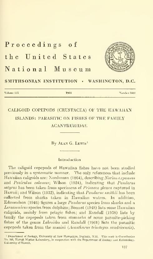
Proceedings of
the United States
National MuseumSMITHSONIAN INSTITUTION • WASHINGTON, D.C.
Volume 115 1964 Number 3482
CALIGOID COPEPODS (CRUSTACEA) OF THE HAWAIIAN
ISLANDS: PARASITIC ON FISHES OF THE FAMILY
ACANTHURIDAE
By Alan G. Lewis ^
Introduction
The caligoid copepods of Hawaiian fishes have not been studied
previously in a systematic manner. The only references that include
Hawaiian caligoids are: Nordmann (1864), describing Norion expansus
and Peniculus calamus; Wilson (1924), indicating that Pandarussatyrus has been taken from specimens of Prionace glauca captured in
Hawaii; and Wilson (1932), indicating that Pandarus smithii has been
collected from sharks taken in Hawaiian waters. In addition,
Edmondson (1946) figures a large Pandarus species from sharks and a
Lernaeenicus species from dolphins; Bonnet (1948) lists some Hawaiiancaligoids, mainly from pelagic fishes; and Randall (1958) lists byfamily the copepods taken from stomachs of some parasite-picking
fishes of the genus Labroides and Randall (1961) lists the parasitic
copepods taken from the manini (Acanthurus triostegus sandvicensis)
.
' Department of Zoology, University of New Hampsbire, Durham, N.H. This work is ContributionNo. 193, Hawaii Marine Laboratory, in cooperation with the Department of Zoology and Entomology,University of Hawaii.
137
138 PROCEEDINGS OF THE NATIONAL MUSEUM vol. us
The author acknowledges with gratitude the assistance and guidance
of Drs. Albert Banner and William Gosline and other staff members
of the Department of Zoology at the University of Hawaii; the
assistance of Samuel Kaolulo, Lester Zukeran, Nick Ferris, the officers
and crew of the U.S. Coast Guard vessel Buttonwood, the Hawaii Board
of Agriculture (Division of Fish and Game), the Honolulu Aquarium,
and many others who assisted in collecting the host material used
in the survey.
The author also is indebted deeply to the staff of the Division of
Marine Invertebrates of the U.S. National Museum and especially to
Drs. Thomas E. Bowman and Fenner A. Chace, Jr., for their assist-
ance and guidance during the period that the author worked at that
institution.
The author further is grateful to Dr. Paul Illg of the University of
Washington for reading the manuscript and offering suggestions for
its improvement.
Finally, the author appreciates the comparison made by Dr. S. M.Shiino, of the Prefectural University of Mie, of some specimens of a
copepod described in this publication with specimens in his personal
collection.
Methods.—The external surface, gill cavities, buccal cavity, and
nasal cavities of the acanthurid fishes collected were examined for
parasitic copepods. Copepods collected from these regions were killed
and preserved in 95 percent ethyl alcohol and later transferred to 70
percent ethyl alcohol. Specimens to be dissected were placed in
85 percent lactic acid to soften and clear them, were stained with
methyl blue dissolved in 85 percent lactic acid, and then were dissected
in 85 percent lactic acid.
Drawings of the total animal were made with a camera lucida and
a Bausch and Lomb arc projector from specimens placed in the
acid and covered by a nine-miUimeter cover slip. Drawings were
made from appendages either in situ on the wholemount or from
others dissected off and mounted in Hoyer's mounting medium.
Measurements were made with an ocular micrometer.
In the following figures the 9 and cf signs are used separately under
each drawing to indicate a difference between the appendage of the
female and that of the male, and together {9&) to indicate similarity
of the appendages. The sex from which the drawing is made is
indicated by underscoring the appropriate symbol.
Terminology.—The term "cephalothorax" is used in the descrip-
tions to designate the fused cephalon and two or more thoracic seg-
ments, the maxilliped-bearing segment being considered as the first
thoracic segment. The term "free thoracic segments" is used to
designate those segments of the thorax not fused with the head.
HAWAIIAN ACANTHURID CALIGOIDS—LEWIS 139
The term "genital segment" designates the fused fifth and sixth
thoracic leg-bearing segments.
In the descriptions of all but two of the species the second antennais described as three-segmented. This is in opposition to many of
the earlier, and some of the present-day, workers. The author is
accepting the viewpoint at this time that the segment attached to
the ventral surface of the cephalothorax along most of its proximalsurface is a distinct segment.
The spinelike projection just lateral and posterior to the base of
the second antenna is called here the postantennal process; the
spinelike projection just lateral to the base of the mouth cone, the
postoral process. With this terminology, the two pairs of appendagesbetween the mouth cone and the first thoracic legs become the max-illae and maxillipeds respectively. Applying the term "postantennal
process" to the structure immediately behind and lateral to the secondantennae is in opposition to Wilson (1905), Heegaard (1947), andothers who apply the term "first maxilla" to the process. Excludingthe word "cuticular" from the term "postantennal process" is in con-
trast with Gurney (1927), Lang (1948), and others, but indicates the
present author's desire neither to accept nor to reject the belief that
the process is not a true appendage. The application of the term"postoral process" to the structure immediately lateral to the base
of the mouth cone is in contrast to all other workers who term the
process a "maxilla." Both the postantennal and postoral processes
are unsegmented, spinelike structures innervated by subesophageal
nerves that arise from the same region of the ganglion (Scott, 1901;
Wilson, 1905; Lewis, unpublished). Each process possesses at least
one setule-bearing nodule. The fact that there is no evidence of
segmentation suggests that these structures are not true appendages.
Although both are innervated by subesophageal nerves, these nerves
arise in close association to each other and in association with a sub-
esophageal nerve that innervates parts of the body musculature andnot the musculature associated with any of the oral appendages.
The presence of at least one setule-bearing nodule may or may not
offer evidence that the processes are appendages, since single setules
are found on parts of the body not associated with any appendage.
The mandible here is considered "parted," not segmented. Theexact derivation of the adult mandible is not known but the lack of
distinct musculature and the indistinct divisions among the parts
indicate that a true segmented condition does not exist in the adult
appendage.
The term "chitinized," as here used, is synonymous with the term
"sclerotized."
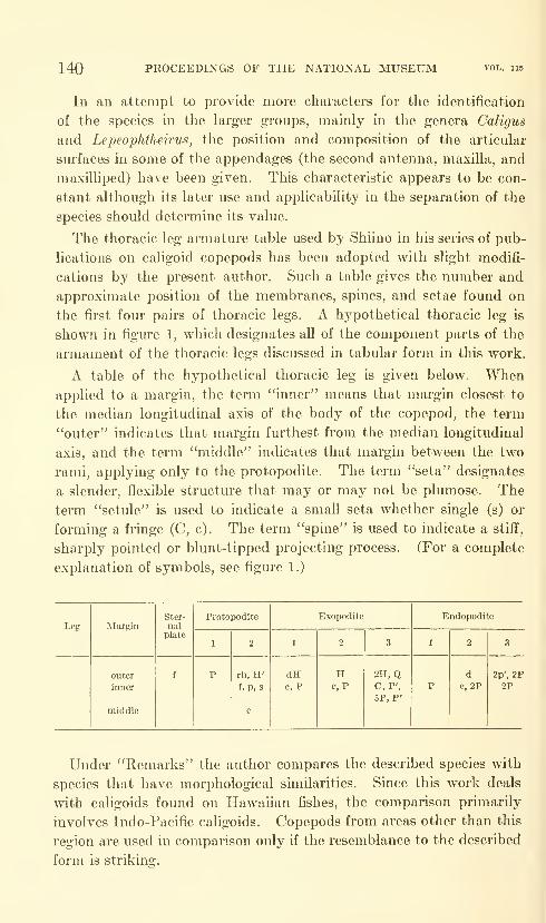
140 PROCEEDINGS OF THE NATIONAL MUSEUM
In an attempt to provide more characters for the identification
of the species in the larger groups, mainly in the genera Caligus
and Lepeophtheirus, the position and composition of the articular
surfaces in some of the appendages (the second antenna, maxilla, and
maxilliped) have been given. This characteristic appears to be con-
stant although its later use and applicability in the separation of the
species should determine its value.
The thoracic leg armature table used by Shiino in his series of pub-
lications on caligoid copepods has been adopted with slight modifi-
cations by the present author. Such a table gives the number and
approximate position of the membranes, spines, and setae found on
the first four pairs of thoracic legs. A hypothetical thoracic leg is
shown in figure 1, which designates all of the component parts of the
armament of the thoracic legs discussed in tabular form in this work.
A table of the hypothetical thoracic leg is given below. Whenapplied to a margin, the term "inner" means that margin closest to
the median longitudinal axis of the body of the copepod, the term
"outer" indicates that margin furthest from the median longitudinal
axis, and the term "middle" indicates that margin between the two
rami, applying only to the protopodite. The term "seta" designates
a slender, flexible structure that may or may not be plumose. The
term "setule" is used to indicate a small seta whether single (s) or
forming a fringe (C, c). The term "spine" is used to indicate a stiff,
sharply pointed or blunt-tipped projecting process. (For a complete
explanation of symbols, see figure 1.)
Leg
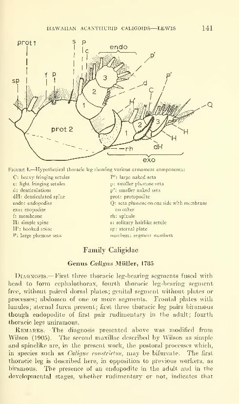
HAWAIIAN ACANTHURID CALIGOIDS—LEWIS 141
prot1 s P
Figure 1.—Hypothetical thoracic leg showing various armament components:
C: heavy fringing setules P': large naked seta
p: smaller plumose setac: light fringing setules
d: denticulations
dH: denticulated spine
endo: endopodite
exo: exopodite
f: membrane
H: simple spine
H': hooked spine
P: large plumose seta
p': smaller naked seta
prot: protopodite
Q: seta plumose on one side with membrane
on other
rh: spinule
s: solitary hairlike setule
sp: sternal plate
numbers: segment numbers
Family Caligidae
Genus Caligus Miiller, 1785
Diagnosis.—First three thoracic leg-bearing segments fused with
head to form cephalothorax, fourth thoracic leg-bearing segment
free, without paired dorsal plates; genital segment without plates or
processes; abdomen of one or more segments. Frontal plates with
lunules; sternal furca present; first three thoracic leg pairs bu'amous
though endopodite of first pair rudimentary in the adult; fourth
thoracic legs uniramous.
Remarks.—The diagnosis presented above was modified from
Wilson (1905). The second maxillae described by Wilson as simple
and spinelike are, in the present work, the postoral processes which,
in species such as Caligus constrictus, may be bifurcate. The first
thoracic leg is described here, in opposition to previous workers, as
biramous. The presence of an endopodite in the adult and in the
developmental stages, whether rudimentary or not, indicates that
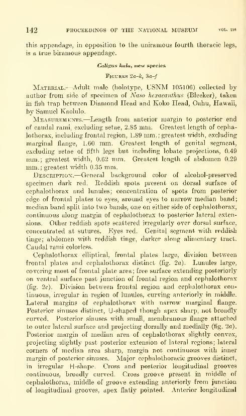
142 PROCEEDINGS OF THE NATIONAL MUSEUM vol. ii5
this appendage, in opposition to the uniramous fourth thoracic legs,
is a true biramous appendage.
Caligus kala, new species
Figures 2a-k, 3a-/
Material.—Adult male (holotype, USNM 105106) collected by-
author from side of specimen of Naso hexacanthus (Bleeker), taken
in fish trap between Diamond Head and Koko Head, Oahu, Hawaii,
by Samuel Kaolulo.
Measurements.—Length from anterior margin to posterior end
of caudal rami, excluding setae, 2.85 mm. Greatest length of cepha-
lothorax, including frontal region, 1.89 mm.;greatest width, excluding
marginal flange, 1.60 mm. Greatest length of genital segment,
excluding setae of fifth legs but including lobate projections, 0.49
mm.; greatest width, 0.62 mm. Greatest length of abdomen 0.29
mm.;greatest width 0.35 mm.
Description.—General background color of alcohol-preserved
specimen dark red. Reddish spots present on dorsal surface of
cephalothorax and lunules; concentration of spots from posterior
edge of frontal plates to eyes, around eyes to narrow median band;
median band split into two bands, one on either side of cephalothorax,
continuous along margin of cephalothorax to posterior lateral exten-
sions. Other reddish spots scattered irregularly over dorsal surface,
concentrated at sutures. Eyes red. Genital segment with reddish
tinge; abdomen with reddish tinge, darker along alimentary tract.
Caudal rami colorless.
Cephalothorax elliptical, frontal plates large, division between
frontal plates and cephalothorax distinct (fig. 2a). Lunules large,
covering most of frontal plate area; free surface extending posteriorly
on ventral surface past junction of frontal region and cephalothorax
(fig. 2c). Division between frontal region and cephalothorax con-
tinuous, irregular in region of lunules, curving anteriorly in middle.
Lateral margins of cephalothorax with narrow marginal flange.
Posterior sinuses distinct, U-shaped though apex sharp, not broadly
curved. Posterior sinuses with small, membranous flange attached
to outer lateral surface and projecting dorsally and medially (fig. 2a).
Posterior margin of median area of cephalothorax slightly convex,
projecting slightly past posterior extension of lateral regions; lateral
corners of median area sharp, margin not continuous with inner
margin of posterior sinuses. Major cephalothoracic grooves distinct,
in irregular H -shape. Cross and posterior longitudinal grooves
continuous, broadly curved. Cross groove present in middle of
cephalothorax, middle of groove extending anteriorly from junction
of longitudinal grooves, apex flatly pointed. Anterior longitudinal

HAWAIIAN ACANTHURID CALIGOIDS—LEWIS 143
0.05mm.
Figure 2.
—
Caligus kala, new species (holotype male): a, dorsal view; b, ventral view show-
ing posterior portion of genital segment, fifth legs, abdomen, and caudal ramus; c, lunule;
d, antennule; e, terminal two segments of second antenna;/, mandible and enlarged distal,
toothed portion; g, postantennal process; h, postoral process; i, maxilla; ;', maxilliped;
k, sternal furca.
grooves extending from junction of longitudinal and cross grooves
laterally and anteriorly, terminating at junction with indistinct,
anteriorly curving cross groove. Apex of indistinct, anteriorly curving
cross groove just posterior to eyes. Lateral strengthening regions
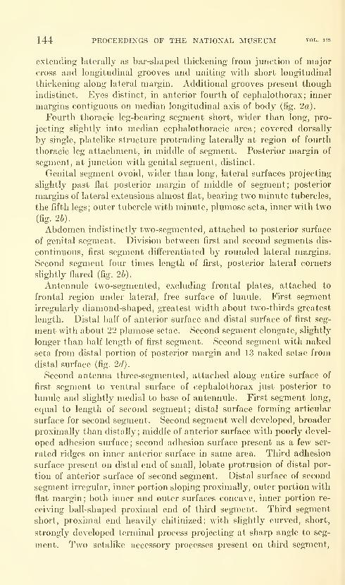
144 PROCEEDINGS OF THE NATIONAL MUSEUM vol. 115
extending laterally as bar-shaped thickening from junction of major
cross and longitudinal grooves and uniting with short longitudinal
thickening along lateral margin. Additional grooves present though
indistinct. Eyes distinct, in anterior fourth of cephalothorax; inner
margins contiguous on median longitudinal axis of body (fig. 2a).
Fourth thoracic leg-bearing segment short, wider than long, pro-
jecting slightly into median cephalothoracic area; covered dorsally
by single, platelike structure protruding laterally at region of fourth
thoracic leg attachment, in middle of segment. Posterior margin of
segment, at junction with genital segment, distinct.
Genital segment ovoid, wider than long, lateral surfaces projecting
slightly past flat posterior margin of middle of segment; posterior
margins of lateral extensions almost flat, bearing two minute tubercles,
the fifth legs; outer tubercle with minute, plumose seta, inner with two
(fig. 26).
Abdomen indistinctly two-segmented, attached to posterior surface
of genital segment. Division between first and second segments dis-
continuous, first segment differentiated by rounded lateral margins.
Second segment four times length of first, posterior lateral corners
slightly flared (fig. 2b).
Antennule two-segmented, excluding frontal plates, attached to
frontal region under lateral, free surface of lunule. First segment
irregularly diamond-shaped, greatest width about two-thirds greatest
length. Distal half of anterior surface and distal surface of first seg-
ment with about 22 plumose setae. Second segment elongate, slightly
longer than half length of fii'st segment. Second segment with naked
seta from distal portion of posterior margin and 13 naked setae from
distal surface (fig. 2d).
Second antenna three-segmented, attached along entire surface of
first segment to ventral surface of cephalothorax just posterior to
lunule and slightly medial to base of antennule. First segment long,
equal to length of second segment; distal surface forming articular
surface for second segment. Second segment well developed, broader
proximally than distally; middle of anterior surface with poorly devel-
oped adhesion surface; second adhesion surface present as a few ser-
rated ridges on inner anterior surface in same area. Third adhesion
surface present on distal end of small, lobate protrusion of distal por-
tion of anterior surface of second segment. Distal surface of second
segment irregular, inner portion sloping proximally, outer portion with
flat margin; both inner and outer surfaces concave, inner portion re-
ceiving ball-shaped proximal end of third segment. Third segment
short, proximal end heavily chitinized; with slightly curved, short,
strongly developed terminal process projecting at sharp angle to seg-
ment. Two setalike accessory processes present on third segment,

HAWAIIAN ACANTHURID CALIGOIDS—LEWIS 145
one on anterior inner surface, second on posterior inner surface.
Division between terminal process and segment distinct (fig. 2e)
.
Mandible four-parted; first part broader proximally than distally.
Second part short, one-fourth length of fii'st part, tapered sharply
to junction with narrower third part. Third part elongate, with
irregularly curved margins. Fourth part curved strongly inwards,
outer margin with slight, chitinous thickening; outer margin with thin
membrane, inner margin with eleven toothlike denticulations (fig. 2/).
Postantennal process simple, falciform, acuminate spine attached
just lateral to proximal end of first segment of second antenna. Base
of process strongly chitinized, ring-shaped; spine continuous with base.
Base with two nodules, each with two minute setules. Two additional
nodules present, medial to base of process, one from plate medial to
anterior end of base, second from ventral surface of cephalothorax
medial to posterior end of base (fig. 2g).
Postoral process simple, spinelike, extending posteriorly and slightly
inward from base just lateral and posterior to proximal end of mouth
cone. Distal end of process rounded. Large nodule present just
anterior to base of process; length of nodule about half length of
process; with three setalike projections, one ahnost as long as nodule,
second and third short, less than half length of first (fig. 2h).
Maxilla two-segmented, attached slightly lateral to base of postoral
process. First segment slightly shorter than second, wider proximally
than distally; inner proximal surface protruding slightly as articular
surface. Distal end of segment abruptly narrowed, extending as
slim, bifurcate process; distal ends of process forming articular surface
for second segment, one part of process on each side, second segment
resting in crotch of process. Second segment elongate, slightly wider
in middle than at either end. Simple, flexible, lobate process present
on second segment, projecting from distal half of inner surface.
Distal surface of second segment with two elongate spines, inner
approximately one and one-third length of outer. Inner spine with
thin membrane along both inner and outer margins ; outer spine with
frilled membrane along both inner and outer margins (fig. 2^).
Maxilliped two-segmented, attached medially and slightly posterior
to maxilla. Fu'st segment strongly developed, with well-developed,
recurved, proximal, articular projection; middle of inner margin with
two small, heavily chitinized, tuberculate projections. Distal margin
irregular, with several heavily chitinized articular surfaces. Second
segment and terminal process falciform; second segment with spine-
like accessory process from anterior surface. Terminal process with
minute nodule projecting from proximal inner margin; division be-
tween terminal process and segment indistinct (fig. 2j).
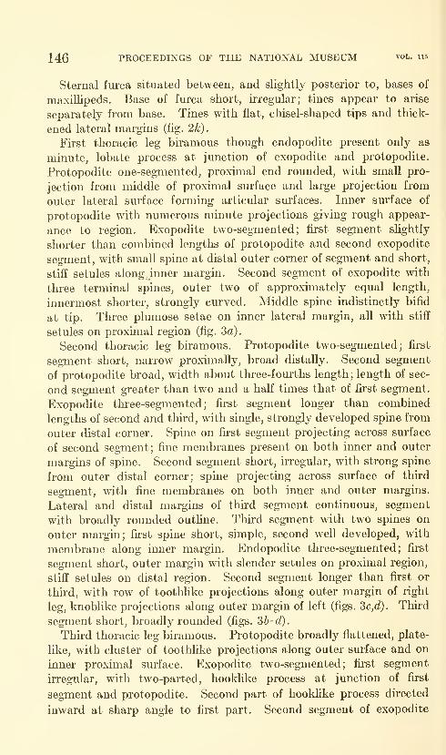
146 PROCEEDINGS OF THE NATIONAL MUSEUM vol. 115
Sternal fui'ca situated between, and slightly posterior to, bases of
maxillipeds. Base of furca short, irregular; tines appear to arise
separately from base. Tines with flat, chisel-shaped tips and thick-
ened lateral margins (fig. 2k).
First thoracic leg biramous though endopodite present only as
minute, lobate process at junction of exopodite and protopodite.
Protopodite one-segmented, proximal end rounded, with small pro-
jection from middle of proximal surface and large projection from
outer lateral surface forming articular surfaces. Inner surface of
protopodite with numerous minute projections giving rough appear-
ance to region. Exopodite two-segmented; first segment slightly
shorter than combined lengths of protopodite and second exopodite
segment, with small spine at distal outer corner of segment and short,
stiff setules along inner margin. Second segment of exopodite with
three terminal spines, outer two of approximately equal length,
innermost shorter, strongly curved. Middle spine indistinctly bifid
at tip. Three plumose setae on inner lateral margin, all with stiff
setules on proximal region (fig. 3a).
Second thoracic leg biramous. Protopodite two-segmented; first
segment short, narrow proximally, broad distally. Second segment
of protopodite broad, width about three-fourths length ; length of sec-
ond segment greater than two and a half times that of first segment.
Exopodite three-segmented; first segment longer than combined
lengths of second and third, with single, strongly developed spine from
outer distal corner. Spine on first segment projecting across surface
of second segment; fine membranes present on both inner and outer
margins of spine. Second segment short, irregular, with strong spine
from outer distal corner; spine projecting across surface of third
segment, with fine membranes on both inner and outer margins.
Lateral and distal margins of third segment continuous, segment
with broadly rounded outline. Third segment with two spines on
outer margin; first spine short, simple, second well developed, with
membrane along inner margin. Endopodite three-segmented; first
segment short, outer margin with slender setules on proximal region,
stiff setules on distal region. Second segment longer than first or
third, with row of toothlike projections along outer margin of right
leg, knoblike projections along outer margin of left (figs. 2>c,d). Third
segment short, broadly rounded (figs. 3b-d).
Thu'd thoracic leg biramous. Protopodite broadly flattened, plate-
like, with cluster of toothlike projections along outer surface and on
inner proximal surface. Exopodite two-segmented; first segment
irregular, with two-parted, hooklike process at junction of first
segment and protopodite. Second part of hooklike process directed
inward at sharp angle to first part. Second segment of exopodite
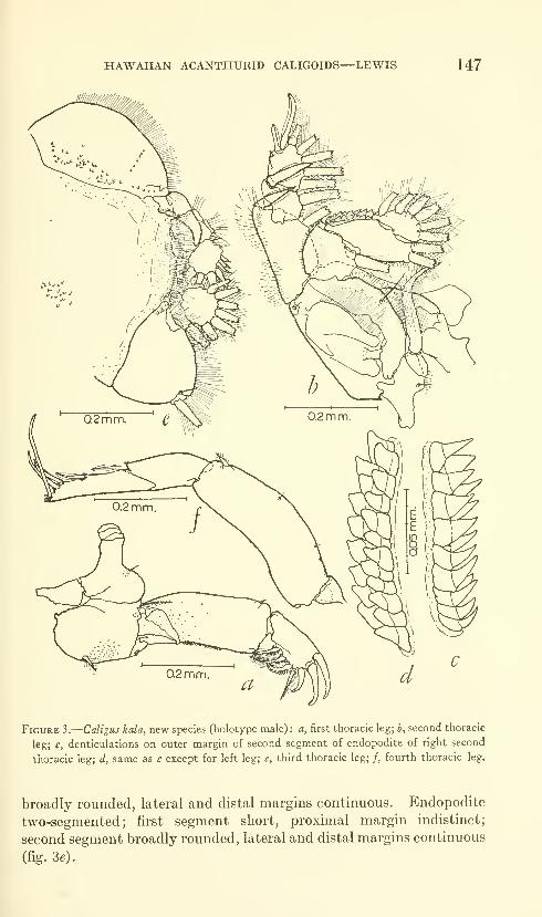
HAWAIIAN ACANTHURID CALIGOIDS—LEWIS 147
Figure 3.
—
Caligus kala, new species (holotype male) : a, first thoracic leg; b, second thoracic
leg; c, denticulations on outer margin of second segment of endopodite of right second
thoracic leg; d, same as c except for left leg; e, third thoracic leg; /, fourth thoracic leg.
broadly rounded, lateral and distal margins continuous. Endopodite
two-segmented; first segment short, proximal margin indistinct;
second segment broadly rounded, lateral and distal margins continuous
(fig. 36).
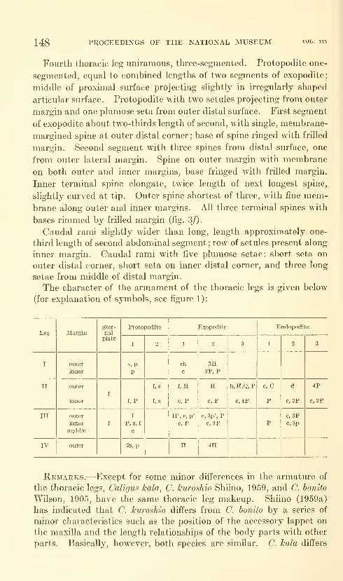
148 PROCEEDINGS OF THE NATIONAL MUSEUM
Fourth thoracic leg uniramous, three-segmented. Protopodite one-
segmented, equal to combined lengths of two segments of exopodite;
middle of proximal surface projecting slightly in irregularly shaped
articular surface. Protopodite with two setules projecting from outer
margin and one plumose seta from outer distal surface. First segment
of exopodite about two-thirds length of second, with single, membrane-
margined spine at outer distal corner; base of spine ringed with frilled
margin. Second segment with three spines from distal surface, one
from outer lateral margin. Spine on outer margin with membrane
on both outer and inner margins, base fringed with frilled margin.
Inner terminal spine elongate, twice length of next longest spine,
slightly curved at tip. Outer spine shortest of three, with fine mem-brane along outer and inner margins. All thi-ee terminal spines with
bases rimmed by frilled margin (fig. 3/).
Caudal rami slightly wider than long, length approximately one-
third length of second abdominal segment ; row of setules present along
inner margin. Caudal rami with five plumose setae: short seta on
outer distal corner, short seta on inner distal corner, and three long
setae from middle of distal margin.
The character of the armament of the thoracic legs is given below
(for explanation of symbols, see figure 1):
Leg
HAWAIIAN ACANTHURID CALIGOIDS—LEWIS 149
from both of the above hi the shape and size of the fourth thoracic
leg-bearing segment, of the genital segment, and of the abdomen,
which is much shorter than that of either C. kuroshio or C. bonito. In
addition, the cephalic appendages of C. kala differ slightly from the
above two species in characteristics such as the single, instead of bifid,
terminal process of the second antenna.
"Kala" is the Hawahan name for fishes of the genus Naso, one of
which, N. hexacanthus, is the host from which the holotype specimen
was taken.
Caligiis flexispina, new species
Figures 4a-o, 5a-/
Material.—Adult female (holotype, USNM 105103), without
egg strings, taken by author from caudal fin of specimen of Acanthurus
triostegus sandmcensis Streets speared by author in Hanauma Bay,
Oahu, Hawaii. Adult male (allotype, USNM 105104), taken by
author from dorsal fin of specimen of A. t. sandvicensis Streets speared
by Robert and Donald Morris near Waikiki, Oahu, Hawaii.
Measurements.—Holotype: Length from anterior end of body
to posterior end of caudal rami, excluding setae, 2.21 mm.; greatest
length of cephalothorax, including frontal region, 1.39 mm.; greatest
width, excluding marginal flange, 0.90 mm. Greatest length of
genital segment 0.62 mm.; greatest width 0.70 mm.Allotype: Length from anterior end of body to posterior end of
caudal rami, excluding setae, 2.30 mm. Greatest length of cephalo-
thorax, including frontal region, 1.56 mm.; greatest width, excluding
marginal flange, 1.23 mm. Greatest length of genital segment,
excluding projections and setae, 0.39 mm.; greatest width 0.37 mm.Description.—Female yellowish brown in alcohol, with no dis-
tinctive pigmentation marks; eyes reddish tan. Male whitish tan in
alcohol, with large j^ellowish splotches over entire body; eyes red.
Female cephalothorax ovoid, widest posteriorly; frontal region
large, lunules large (fig. 4:d). Anterior margin of frontal region with
narrow, membranous flange; division between frontal region and ceph-
alothorax distinct, a trilobed groove, median lobe larger than lateral
lobes. Lateral margins of cephalothorax with broad marginal flange.
Posterior sinuses distinct, U-shaped, entrances open; sinuses without
membranous flange. Middle of cephalothorax extending well behind
posterior margins of lateral areas; posterior margin slightly convex,
with small concavity in middle of margin, at junction with fourth
thoracic leg-bearing segment. Cephalothoracic grooves distinct,
major grooves forming irregular H. Cross groove placed slightly
anterior of middle of cephalothorax, continuous with posterior longi-
tudinal grooves. Cross groove curving anteriorly from junction with

150 PROCEEDINGS OF THE NATIONAL MUSEUM vol. H5
longitudinal grooves, slight indentation present at apex of anterior
curving groove. Anterior longitudinal grooves extending laterally
and anteriorly, terminating blindly slightly posterior to eyes and close
to lateral margins. Lateral thickenings present as narrow bands
running from just anterior to junction of cross and longitudinal grooves
laterally and posteriorly, terminating in region of lateral margin.
Light median groove present, extending from slight indentation at apex
of cross groove to middle of posterior margin of median thoracic area.
Eyes placed in anterior fourth of cephalothorax, between two short,
longitudinal grooves; eye region of female two smaU, rounded areas,
one on either side of two narrow, elongate regions slightly swollen
posteriorly and contiguous on median longitudinal axis of body
(fig. 4a).
Cephalothorax of male slightly different from that of female; widest
in posterior medial region, anterior to widest point in female. Pos-
terior margin of protruding median thoracic area convex, without
median concavity of female. Cephalothoracic grooves similar to
those of female though cross groove without indentation at apex.
Longitudinal bars in eye region not as distinct as in female; contiguous
elongations of female absent in male, eyes with inner margins touching
on median longitudinal axis of body (fig. 4b).
Free fom-th thoracic leg-bearing segment of female short, nar-
rower at junction with third segment than at junction with genital
segment. Fourth thoracic legs attached to widest point of segment,
adjacent to junction with genital segment^ Divisions distinct be-
tween cephalothorax and fourth thoracic leg-bearing segment and
between fourth thoracic leg-bearing segment and genital segment.
Fom-th thoracic legs of male joined to middle of thoracic segment,
not adjacent to junction with genital segment, as in female.
Genital segment of female ovoid, widest posteriorly. Genital seg-
ment with broadly rounded lateral margins; posterior margins convex
laterally, concave medially, at junction with abdomen. Fifth legs
visible as three minute, plumose setae, two arising from slight pro-
jection just lateral to opening of oviduct, third from minute projection
just lateral and anterior to first two (figs. 4a,c).
Genital segment of male slightly broader across posterior end than
across anterior end. Posterior margin convex. Fifth and sixth legs
distinct, fifth as three plumose setae from lateral region of posterior
margin, two arising from single, slight protrusion, third just anteriorly,
from surface of genital segment. Sixth legs of two plumose setae aris-
ing from single, lobate process extending posteriorly slightly past
genital segment; inner seta almost twice length of outer (figs. 4b,c).
Abdomen of female short, length about one-fifth greatest length of
genital segment; width sHghtly greater than greatest length (fig. 4c).
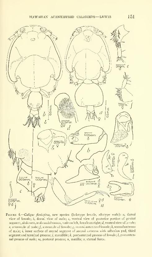
HAWAIIAN ACANTHURID CALIGOIDS—LEWIS 151
Figure 4.
—
Caligus flexispina, new species (holotype female, allotype male): a, dorsal
view of female; b, dorsal view of male; c, ventral view of posterior portion of genital
segment, abdomen, and caudal ramus, male on left, female on right; (^, ventral view of lunule;
e, antennule of male;/, antennule of female; g, secondantennaoffemale; A, second antenna
of male; i, inner surface of second segment of second antenna with adhesion pad, third
segment and terminal process;;', mandible; k, postantennal process of female; /, postanten-
nal process of male; m, postoral process; n, maxilla; o, sternal furca.
152 PROCEEDINGS OF THE NATIONAL MUSEUM vol. 115
Abdomen of male arising from posterior surface of genital segment;
of two distinct segments, first segment short, half length of second;
second flared slightly at posterior end (fig. 4c).
Antennule of female two-segmented, excluding frontal plate,
attached to ventral surface of cephalothorax and frontal region just
medial to lateral frontal margin of frontal region. Greatest length
of first segment one and a half times that of second; greatest width
three-fourths greatest length. Anterior margin angular, apex of angle
at widest point. First segment with twenty plumose setae on distal
two-thirds of anterior margin. Greatest length of second segment
more than two and a half times greatest width; proximal portion of
segment narrowing abrujitly at junction with first segment. Second
segment with one naked seta on middle of posterior margin, twelve
naked setae on distal surface (fig. 4/).
Antennule of male similar to that of female except second segment
longer, fu'st segment only slightly longer than second (fig. 4e)
.
Second antenna of female three-segmented, attached slightly
posterior and medial to base of antennule. Fu'st segment short,
forming articular surface for second segment; sharply pointed,
posteriorly directed process projecting from posterior proximal
surface of segment. Second segment short, strongly developed;
greatest width, at proximal end, slightly less than greatest length,
tapered slightly towards distal end. Inner and outer distal corners
of second segment projecting slightly, heavily chitinized, serving as
articular surfaces for third segment. Third segment with uncinate,
acuminate terminal process; length of combined segment and process
greater than length of preceding two segments. Third segment with
small, spinelike accessory process on posterior proximal surface.
Division between terminal process and segment not visible (fig. 4^).
Second antenna of male three-segmented; first segment as long as
second, attached to ventral surface of cephalothorax along entire
length. Proximal end of first segment with small, posteriorly directed,
pointed projection; anterior end forming concave articular surface
for second segment. Second segment strongly developed, with small
adhesion pad on inner distal lateral surface; distal surface irregular,
with several heavily chitinized regions for articulation with third
segment. Third segment with trifid, spinelike terminal process.
Third segment short, length including terminal process less than
fourth that of second segment, with two setalike accessory processes,
one from posterior medial surface of segment, second from anterior
medial surface. Division between terminal process and segment
distinct (figs. 4h,i).
Female and male mandible four-parted. First part long, broad
proximally, with uneven taper to slightly narrower distal region.
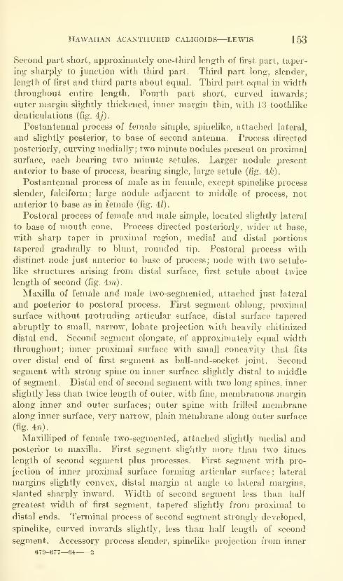
HAWAIIAN ACANTHURID CALIGOIDS—LEWIS 153
Second part short, approximately one-third length of first part, taper-
ing sharply to junction with third part. Third part long, slender,
length of fu-st and third parts about equal. Thu"d part equal in width
throughout entire length. Fourth part short, curved inwards;
outer margin slightly thickened, inner margin thin, with 13 toothlike
denticulations (fig. 4j).
Postantennal process of female simple, spinelike, attached lateral,
and slightly posterior, to base of second antenna. Process directed
posteriorly, cm*ving medially; two minute nodules present on proximal
surface, each bearing two minute setules. Larger nodule present
anterior to base of process, bearing single, large setule (fig. 4^)
.
Postantennal process of male as in female, except spinelike process
slender, falciform; large nodule adjacent to middle of process, not
anterior to base as in female (fig. 41).
Postoral process of female and male simple, located slightly lateral
to base of mouth cone. Process directed posteriorly, wider at base,
wdth sharp taper in proximal region, medial and distal portions
tapered gradually to blunt, rounded tip. Postoral process with
distinct node just anterior to base of process; node with two setule-
like structures arising from distal surface, first setule about twice
length of second (fig. 4m)
.
Maxilla of female and male two-segmented, attached just lateral
and posterior to postoral process. First segment oblong, proximal
surface without protruding articular surface, distal surface tapered
abruptly to small, narrow, lobate projection with heavily chitinized
distal end. Second segment elongate, of approximately equal width
throughout; inner proximal surface with small concavity that fits
over distal end of first segment as ball-and-socket joint. Second
segment with strong spine on inner surface slightly distal to middle
of segment. Distal end of second segment with two long spines, inner
slightly less than twice length of outer, with fine, membranous margin
along inner and outer surfaces; outer spine with frilled membranealong inner surface, very narrow, plain membrane along outer surface
(fig. 4n).
Maxilliped of female two-segmented, attached slightly medial andposterior to maxilla. First segment slightly more than two times
length of second segment plus processes. First segment with pro-
jection of inner proximal surface forming articular surface; lateral
margins slightly convex, distal margin at angle to lateral margins,
slanted sharply inward. Width of second segment less than half
greatest width of first segment, tapered slightl}^ from proximal to
distal ends. Terminal process of second segment strongly developed,
spinelike, curved inwards slightly, less than half length of second
segment. Accessory process slender, spinelike projection from inner
679-677—64 2
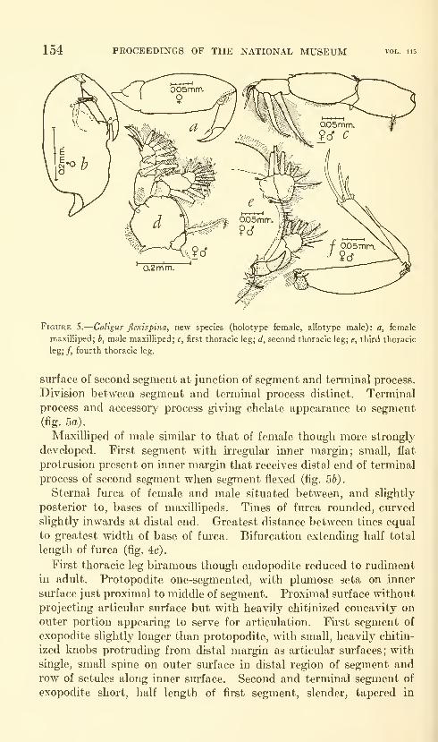
154 PROCEEDINGS OF THE NATIONAL MUSEUM
Figure 5.
—
Caligus flextspina, new species (holotype female, allotype male): a, female
maxilliped; b, male maxilliped; c, first thoracic leg; d, second thoracic leg; e, third thoracic
leg; /, fourth thoracic leg.
surface of second segment at junction of segment and terminal process.
Division between segment and terminal process distinct. Terminal
process and accessory process giving chelate appearance to segment(fig- 5a).
Maxilliped of male similar to that of female though more strongly
developed. First segment with irregular inner margin; small, flat
protrusion present on inner margin that receives distal end of terminal
process of second segment when segment flexed (fig. 56).
Sternal furca of female and male situated between, and slightly
posterior to, bases of maxillipeds. Tines of furca rounded, curved
slightly inwards at distal end. Greatest distance between tines equal
to greatest width of base of furca. Bifurcation extending half total
length of furca (fig. 4c).
First thoracic leg biramous though endopodite reduced to rudimentin adult. Protopodite one-segmented, with plumose seta on inner
surface just proximal to middle of segment. Proximal surface without
projecting articular surface but with heavily chitinized concavity on
outer portion appearing to serve for articulation. First segment of
exopodite slightly longer than protopodite, with small, heavily chitin-
ized knobs protruding from distal margin as articular surfaces; with
single, small spine on outer surface in distal region of segment androw of setules along inner surface. Second and terminal segment of
exopodite short, half length of first segment, slender, tapered in

HAWAIIAN ACANTHURID CALIGOIDS—LEWIS 155
curved manner from broad proximal to narrow distal surface. Second
segment with three flexible processes and long, naked seta on distal
surface, three plumose setae on inner lateral margin (fig. 5c).
Second thoracic leg biramous. Protopodite two-segmented; first
segment short, approximately half length of second segment. Second
segment broad, with single, naked seta from outer distal corner.
Exopodite three-segmented; length of first segment equal to that of
second and third combined, with strongly developed spine directed
inward, across surface of second segment, from outer distal corner of
first segment. Second segment short, with spine similar to that of
first segment though smaller; spine arising from outer distal corner
and directed inward across surface of third segment. Length of
third segment one and a half times that of second; general outline of
segment oval. Third segment with two spines, first small, located
on outer surface of middle of segment; second spine approximately
four times length of first, located just distal to first spine, inner lateral
margin plumose. Endopodite three-segmented; first segment short,
second segment long, slightly shorter than combined lengths of first
and third segments. Third segment short, half length of first seg-
ment, distal and lateral margins continuous (fig. 5d).
Third thoracic leg biramous. Protopodite one-segmented, flattened;
length of protopodite approximately one and a half times width.
Exopodite two-segmented, length of first segment almost twice that
of second, with two-parted, spinelike process curving inward from
junction of exopodite and protopodite. Distance between exopodite
and endopodite bases slightly less than half width of protopodite.
Endopodite two-segmented; first segment irregular, proximal margin
indistinct. Second segment irregularly lobate, distal and lateral
margins continuous (fig. 5e).
Fourth thoracic leg uniramous, three-segmented. Length of one-
segmented protopodite slightly greater than combined lengths of two
segments of exopodite. Inner proximal surface of protopodite
projecting as lobate articular process. Protopodite with single,
plumose seta from outer surface of distal end. First segment of
exopodite less than three-fourths length of second segment, with
elongate spine from outer distal surface. Second segment of ex-
opodite with three simple spines from distal surface, each with frilled
membrane around inner portion of base; friUed membrane attached
to segment, not to spine. Inner spine longest, outer two spines
successively shorter. Outermost spine with fine membrane along
outer surface (fig. 5f).
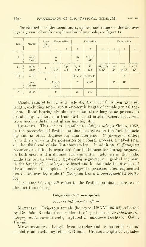
156 PROCEEDINGS OF THE NATIONAL MUSEUM
The character of the membranes, spines, and setae on the thoracic
legs is given below (for explanation of symbols, see figure 1):
Leg
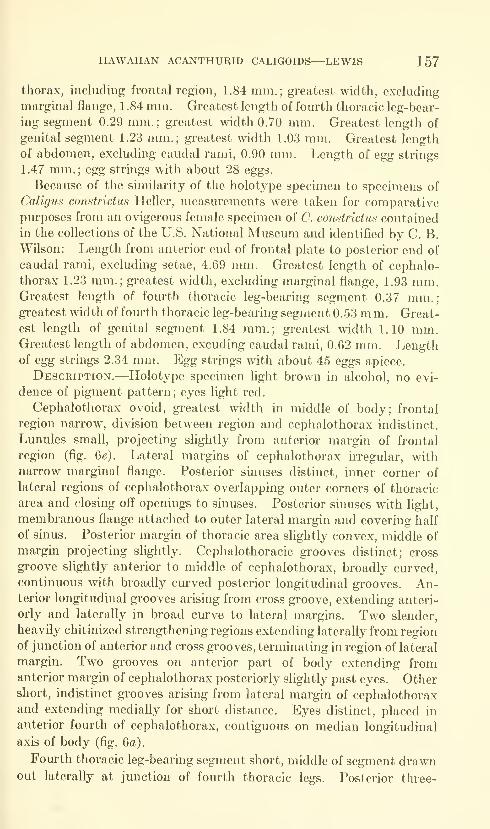
HAWAIIAN ACANTHURID CALIGOIDS—LEWIS 157
thorax, including frontal region, 1.84 mm.; greatest width, excluding
marginal flange, 1 .84 mm. Greatest length of fourth thoracic leg-bear-
ing segment 0.29 mm.; greatest width 0.70 mm. Greatest length of
genital segment 1.23 mm.; greatest width 1.03 mm. Greatest length
of abdomen, excluding caudal rami, 0.90 mm. Length of egg strings
1.47 mm.; egg strings with about 28 eggs.
Because of the similarity of the holotype specimen to specimens of
Caligus constrictus Heller, measurements were taken for comparativepurposes from an ovigerous female specimen of C. constrictus contained
in the collections of the U.S. National Museum and identified by C. B.
Wilson: Length from anterior end of frontal plate to posterior end of
caudal rami, excluding setae, 4.69 mm. Greatest length of cephalo-
thorax 1.23 mm.; greatest width, excluding marginal flange, 1.93 mm.Greatest length of fourth thoracic leg-bearing segment 0.37 mm.;greatest width of fourth thoracic leg-bearing segment 0.53 mm. Great-
est length of genital segment 1.84 mm.; greatest width 1.10 mm.Greatest length of abdomen, excuding caudal rami, 0.62 mm. Lengthof egg strings 2.34 mm. Egg strings with about 45 eggs apiece.
Description.—Holotype specimen light brown in alcohol, no evi-
dence of pigment pattern; eyes light red.
Cephalothorax ovoid, greatest width in middle of body; frontal
region narrow, division between region and cephalothorax indistinct.
Lunules small, projecting slightly from anterior margin of frontal
region (fig. 6e). Lateral margins of cephalothorax irregular, with
narrow marginal flange. Posterior sinuses distinct, inner corner of
lateral regions of cephalothorax overlapping outer corners of thoracic
area and closing off openings to sinuses. Posterior sinuses with light,
membranous flange attached to outer lateral margin and covering half
of sinus. Posterior margin of thoracic area slightly convex, middle of
margin projecting slightly. Cephalothoracic gi'ooves distinct; cross
groove slightly anterior to middle of cephalothorax, broadly cm*ved,
continuous with broadly curved posterior longitudinal grooves. An-terior longitudinal grooves arising from cross groove, extending anteri-
orly and laterally in broad curve to lateral margins. Two slender,
heavily chitinized strengthening regions extending laterally from region
of junction of anterior and cross grooves, terminating in region of lateral
margin. Two grooves on anterior part of body extending fromanterior margin of cephalothorax posteriorly slightly past eyes. Othershort, indistinct grooves arising from lateral margin of cephalothoraxand extending medially for short distance. Eyes distinct, placed in
anterior fourth of cephalothorax, contiguous on median longitudinal
axis of body (fig. 6a).
Fourth thoracic leg-bearing segment short, middle of segment drawnout laterally at junction of fourth thoracic legs. Posterior three-
158 PROCEEDINGS OF THE NATIONAL MUSEUM vol. lis
fourths of segment covered by single, platelike structure distinctly
separate from genital segment. Division between fourth thoracic leg-
bearing segment and genital segment distinct (fig. 66).
Genital segment longer than wide, median and posterior lateral
margins parallel. Posterior lateral sm-faces extending past junction of
abdomen and genital segment; posterior margin forming sharp angle
with lateral margin, middle of posterior margin slightly concave.
Genital segment with two pairs of lobate processes projecting from
posterior ventral surface (fig. 6/).
General outline of abdomen obovoid; length about three-fourths
length of genital segment, greatest width more than half length.
Cephalothorax of C. constrictus circular, not ovoid as in C. randalli.
Fourth thoracic leg-bearing segment of C. constrictus longer than that
of C. randalli, anterior portion similar to C. randalli, posterior portion
forming short, necklike extension; without platelike structure present
in C. randalli (fig. 6c). Posterior lateral surfaces of genital segment
forming lobate instead of angular projections (fig. 6(/). Length of abdo-
men about one-third length of genital segment in contrast to relatively
longer length of abdomen of C. randalli; greatest width four-fifths
greatest length.
Antennule of C. randalli two-segmented, excluding frontal plate,
attached to ventral surface of frontal region and cephalothorax just
lateral to base of lunule ; outer, free margin of lunule overlapping base
of first segment of antennule. First segment of antennule broad,
greatest width two-thirds greatest length. Anterior surface with
approximately twenty plumose setae. Second segment elongate,
greatest length 1.3 times that of first segment. Second segment
with naked seta from distal portion of posterior margin, eleven
naked setae from distal surface (fig. 6A).
Second antenna of C. randalli three-segmented, attached just
posterior and medial to antennule. First segment forming broad,
flat articular surface, with small, laterally directed, lappet-like pro-
trusion. Second segment short, slightly longer than wide; proximal
end slightly wider than distal. Second segment bearing small, lobate
projection from inner distal surface. Third segment, including ter-
minal process, elongate, length greater than two times that of second
segment. Terminal process of third segment broadly curved, division
between segment and process indistinct; inner terminal surface of
process with minute, toothlike projections (fig. 6i).
Second antenna of C. constrictus similar to that of C. randalli though
possessing two seta-like processes on terminal process of third segment;
processes absent on C. randalli.
Mandible of C. randalli four-parted. First part sHghtly broader
proximally than distally. Second part short, approximately two-

HAWAIIAN ACANTHURID CALIGOIDS—LEWIS 159
0.05mm.
Figure 6.
—
Caligus randalli, new species (holotype female): a, dorsal view; b, dorsal view
of fourth thoracic leg-bearing segment (g.s.= genital segment); (c, dorsal view of fourth
thoracic leg-bearing segment of C conslridus) ; d, ventral view of anterior end of cephalo-
thorax; e, lunule;/, ventral view of posterior end of genital segment, abdomen, and caudal
rami of C. randalli; (g, ventral view of posterior end of genital segment, abdomen, and
caudal rami of C. constrictui) ; h, antennule; i, second antenna;;, mandible; k, postantennal
process;/, postoral process; (m, postoral process of C. constrictui); n, maxilla; o, maxilli-
ped; p, sternal furca; q, caudal ramus.
160 PROCEEDINGS OF THE NATIONAL MUSEUM vol. 115
thirds length of first part, tapering sUghtly to junction witli third
part. Third part elongate, slightly swollen at junction of fourth part.
Fourth part short, curving inwards, inner margin with 13 denticula-
tions (fig. 6j).
Postantennal process of C. randalli a simple, curved, spinelike
process situated slightly lateral and posterior to second antenna and
directed ventrally and posteriorly; with small, naked, chitinous pro-
jection and single nodule bearing two long setules present on inner
proximal surface (fig. Qk).
Postantennal process of C. constrictus similar to C. randalli though
with two setule-bearing nodules and two minute spinules on inner
proximal surface.
Postoral process of C. randalli basically single, sharply pointed
process curving laterally from origin adjacent to base of mouth cone,
with membrane continuous around sharply pointed distal end but
with single lateral projection on inner margin giving bifurcate appear-
ance to process. Base of process with nodule bearing two setules
(fig. 60.
Postoral process of C. constrictus with two projections from inner
margin instead of one, without membrane around distal end of process.
Maxilla of C. randalli tv^^o-segmented, attached just lateral and
posterior to postoral process. Length of first segment slightly more
than four times greatest width; lateral margins parallel through
most of length, projection of inner proximal surface forming articular
surface. Distal margin of first segment with several lobes forming
articular surfaces for second segment. Cluster of three lobes on outer
part of surface forming irregular distal concavity for ball-shaped
proximal end of second segment. Second segment elongate, length
slightly greater than that of first segment. Middle of second segment
tapered slightly, bearing folded, membranous margin and row of
setules. Inner of two terminal spines 1.3 times length of outer spine,
with frilled membrane on outer and inner margins. Outer spine also
with frilled membrane on outer and inner margins (fig. Qn).
Maxilla of C. constrictus similar to that of C. randalli though lobate
distal margin replaced by slight depression.
Maxilliped of C. randalli two-segmented, situated slightly posterior
and medial to maxilla. First segment well developed, with large
projection of inner proximal surface serving as articular surface.
Inner surface of segment with concave depression. First segment
with small, lobate projection from posterior surface just proximal
to middle of segment. Distal surface of first segment irregular, with
several heavily chitinized articular surfaces. Second segment short,
with long, strong, falciform terminal process; length of second segment
and terminal process about two-thirds length of first segment. Second
HAWAIIAN ACANTHURID CALIGOIDS—LEWIS 161
segment with small accessory spine at terminal end of segment;
division between segment and process distinct (fig. 60).
Maxilliped of C constridus with more distinct projection from
posterior surface of first segment; with terminal process of second
segment possessing minute denticulations on inner distal surface.
Sternal furca of C. randalli located between posterior portion of
maxilliped bases. Width across base three-fifths width across tines.
Bifurcation extending slightly more than half length of furca. Tines
with round tips; with membrane continuous around tip of each tine
(fig- 62^)-
Tines of sternal furca of C. constridus sharply pointed, not rounded
as in C. randalli.
First thoracic leg biramous though endopodite reduced to rudiment
tipped with two minute processes. Protopodite one-segmented, with
inner proximal surface covered with minute projections giving segment
fuzzy appearance. Two heavily chitinized knobs present as articular
surfaces on proximal surface, projecting slightly from segment. First
segment of exopodite long, greater than combined length of protopo-
dite and second exopodite segment. First segment of exopodite with
articular knob and small spine on outer distal corner, spine surrounded
by frilled margin. Second and terminal segment of exopodite with
irregular inner and distal margins; distal margin with three spines,
outermost longer than inner two, triangular in cross section; inner
margin of outermost spine with row of minute obtuse processes.
Inner two spines on distal margin wavy, distal portion of inner
margins with slight concavity, with fine membrane crossing con-
cavity; inner margins of both spines with row of minute spinules
along entire length. Inner distal corner of second segment with
single, naked seta (fig. 7a).
Second thoracic leg biramous. Protopodite two-segmented, first
segment short, approximately one-third length of second. Second
segment with single naked seta on outer distal corner. Exopodite
three-segmented; first segment slightly more than twice combined
lengths of second and third segments, with single spine on outer
distal corner, projecting across surface of second segment. Spine on
first segment with serrate margins. Second segment of exopodite
short, with slightly curved, serrately margined spine on outer distal
corner. Third segment longer than second, ovoid, with small,
serrately margined spine on outer proximal margin and slightly larger,
serrately margined spine just distal to first. Endopodite three-seg-
mented; first segment broad, outer surface lobate, rimmed with row
of heavy, toothlike setules. Second segment wider than long,
smaller than first segment. Third segment smaller than preceding
two, distal and lateral margins continuous (fig. 7b).
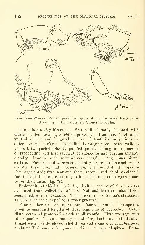
162 PROCEEDINGS OF THE NATIONAL MUSEUM vol. 115
0.2mm,
Figure 7.
—
Caligus randalli, new species (holotype female): a, first thoracic leg; b, second
thoracic leg; c, third thoracic leg; d, fourth thoracic leg.
Third thoracic leg biramous. Protopodite broadly flattened, with
cluster of ten distinct, knoblike projections from middle of inner
ventral surface and longitudinal row of toothlike projections on
outer ventral surface. Exopodite two-segmented, with well-de-
veloped, two-parted, bluntly pointed process arising from junction
of protopodite and first segment of exopodite and curving inwards
distally. Process with membranous margin along inner distal
surface. First exopodite segment slightly larger than second, wider
distally than proximaUy; second segment rounded. Endopodite
three-segmented; first segment short, second and third combined,
forming flat, lobate structure; proximal end of second segment nar-
rower than distal (fig. 7c).
Endopodite of third thoracic leg of all specimens of C. constrictus
examined from coUections of U.S. National Museum also three-
segmented, as in O. randalli. This is contrary to Shiino's statement
(1959b) that the endopodite is two-segmented.
Fourth thoracic leg uniramous, four-segmented. Protopodite
equal to combined lengths of tlu-ee segments of exopodite. Outer
distal corner of protopodite with small spinule. First two segments
of exopodite of approximately equal size, both rounded distally,
tipped with well-developed, slightly curved spine with membranous,
slightly frilled margin along outer and inner margins of spines. Spine

HAWAIIAN ACANTHURID CALIGOIDS—LEWIS 163
on first segment with indistinct frilled membrane around base, frilled
membrane around base of second segment spine distinct. Third
segment with tliree spines on distal surface, all with distinct frilled
membrane around base. Innermost spine longest, outer two of
approximately equal length, inner margins of all three spines lightly
setuliferous (fig. Id).
The character of the membranes, spines, and setae on the thoracic
legs is given below. Except for minor variations such as the length
and spacing of the setae, the thoracic legs of C. constrictus and C.
randalli are identical. Because of this there is no comparison of these
appendages in the two species. (For explanation of symbols, see
figure 1.)
Leg
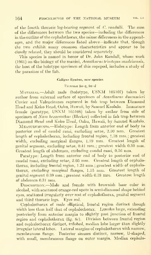
164 PROCEEDINGS OF THE NATIONAL MUSEUM vol. 115
of the fourth thoracic leg-bearmg segment of C. randalli. The sumof the differences between the two species—^including the differences
in the outline of the cephalothorax, the minor differences in the append-
ages, and the major differences hsted above—indicate that, thougli
the two exhibit many common characteristics and appear to be
closely related, they should be considered separately.
This species is named in honor of Dr. John Randall, whose work
(1961) on the biology of the manini, Acanthurus triostegus sandvicensis,
the host of the holotype specimen of this copepod, includes a study of
the parasites of the fish,
Caligus ligatus, new species
Figures Sa-q, 9a-d
Material.—Adult male (holotype, USNM 105107) taken by
author from external surface of specimen of Acanthurus dussumieri
Cuvier and Valenciennes captured in fish trap between DiamondHead and Koko Head, Oahu, Hawaii, by Samuel Kaolulo. Immature
female (paratype, USNM 105108) taken by author from side of
specimen of Naso hexacanthus (Bleeker) collected in fish trap between
Diamond Head and Koko Head, Oahu, Hawah, by Samuel Kaolulo.
Measurements.—Holotype: Length from anterior end of body to
posterior end of caudal rami, excluding setae, 2.30 mm. Greatest
length of cephalothorax, including frontal region, 1.38 mm.; greatest
width, excluding marginal flanges, 1.19 mm. Greatest length of
genital segment, excluding setae, 0.41 mm.; greatest width 0.39 mm.Greatest length of abdomen, excluding caudal rami, 0.36 mm.
Paratype: Length from anterior end of body to posterior end of
caudal rami, excluding setae, 2.05 mm. Greatest length of cephalo-
thorax, including frontal region, 1.23 mm.; greatest width of cephalo-
thorax, excluding marginal flanges, 1.15 mm. Greatest length of
genital segment 0.39 mm.;greatest width 0.38 mm. Greatest length
of abdomen 0.31 mm.Description.—Male and female with brownish base color in
alcohol, with scattered orange-red spots in semi-diamond shape behind
eyes, scattered irregularly over rest of cephalothorax, genital segment
and third thoracic legs. Eyes red.
Cephalothorax of male elliptical, frontal region distinct though
width less than half that of cephalothorax. Lunules large, extending
posteriorly from anterior margin to slightly past junction of frontal
region and cephalothorax (fig. 8^). Division between frontal region
and cephalothorax distinct, trilobed, median lobe larger than slightly
irregular lateral lobes. Lateral margins of cephalothorax with narrow,
membranous flange. Posterior sinuses distinct, narrow, U-shaped,
with small, membranous flange on outer margin. Median cephalo-

HAWAIIAN ACANTHURID CALIGOIDS—LEWIS 165
QOSmm.^9
Figure 8.
—
Caligus ligatus, new species (holotype male, paratype immature female): a,
dorsal view of male; b, dorsal view of immature female; c, ventral view of male fifth and
sixth legs; d, ventral view of immature female fifth legs; e, ventral view of lunule;/, anten-
nule; g, second antenna of male; h, terminal two segments of second antenna of immature
female; i, mandible; /, postantennal process of male; k, postantennal process of immature
female; /, postoral process; m, maxilla; n, maxilliped of male; o, maxilliped of immature
female; p, sternal furca; g, caudal ramus.
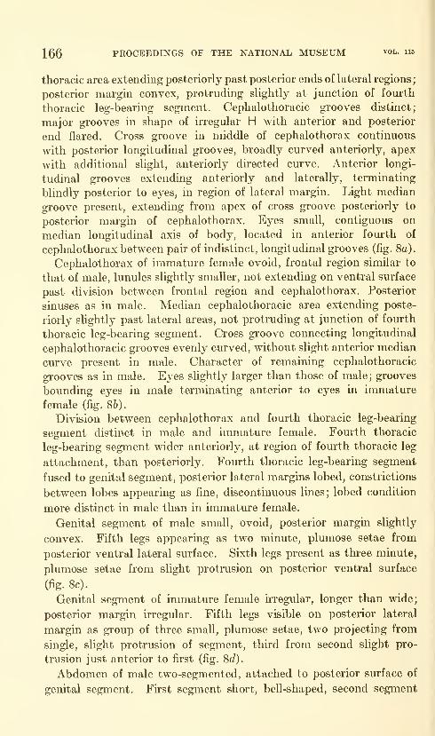
166 PROCEEDINGS OF THE NATIONAL MUSEUM vol. 115
thoracic area extending posteriorly past posterior ends of lateral regions;
posterior margin convex, protruding slightly at junction of fourth
thoracic leg-bearing segment. Cephalothoracic grooves distinct;
major grooves in shape of irregular H with anterior and posterior
end flared. Cross groove in middle of cephalothorax continuous
with posterior longitudinal grooves, broadly curved anteriorly, apex
with additional slight, anteriorly directed curve. Anterior longi-
tudinal grooves extending anteriorly and laterally, terminating
blindly posterior to eyes, in region of lateral margin. Light median
groove present, extending from apex of cross groove posteriorly to
posterior margin of cephalothorax. Eyes small, contiguous on
median longitudinal axis of body, located in anterior fourth of
cephalothorax between pair of indistinct, longitudinal grooves (fig. 8a).
Cephalothorax of immatm-e female ovoid, frontal region similar to
that of male, lunules slightly smaller, not extending on ventral surface
past division between frontal region and cephalothorax. Posterior
sinuses as in male. Median cephalothoracic area extending poste-
riorly slightly past lateral areas, not protruding at junction of fourth
thoracic leg-bearing segment. Cross groove connecting longitudinal
cephalothoracic grooves evenly curved, without slight anterior median
curve present in male. Character of remaining cephalothoracic
grooves as in male. Eyes slightly larger than those of male; grooves
bounding eyes in male terminating anterior to eyes in immature
female (fig. 86).
Division between cephalothorax and fourth thoracic leg-bearing
segment distinct in male and immature female. Fourth thoracic
leg-bearing segment wider anteriorly, at region of fourth thoracic leg
attachment, than posteriorly. Fourth thoracic leg-bearing segment
fused to genital segment, posterior lateral margins lobed, constrictions
between lobes appearing as fine, discontinuous lines; lobed condition
more distinct in male than in immature female.
Genital segment of male small, ovoid, posterior margin slightly
convex. Fifth legs appearing as two minute, plumose setae from
posterior ventral lateral surface. Sixth legs present as three minute,
plumose setae from slight protrusion on posterior ventral surface
(fig. 8c).
Genital segment of immatm'e female irregular, longer than wide;
posterior margin irregular. Fifth legs visible on posterior lateral
margin as group of three small, plumose setae, two projecting from
single, slight protrusion of segment, third from second slight pro-
trusion just anterior to first (fig. 8d).
Abdomen of male two-segmented, attached to posterior surface of
genital segment. First segment short, bell-shaped, second segment
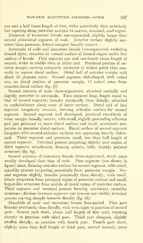
HAWAIIAN ACANTHURID CALIGOIDS—LEWIS 167
one and a half times length of first, wider posteriorly than anteriorly
but tapering along posterior margins to narrow, terminal, anal region.
Abdomen of immature female one-segmented, slightly larger than
second abdominal segment of male. Anterior surface slightly nar-
rower than posterior, lateral margins broadly convex.
Antennule of male and immature female two-segmented, excluding
frontal plate, attached to ventral sm*face of frontal region under free
surface of lunule. First segment one and one-fourth times length of
second; wider in middle than at either end. Proximal portion of an-
terior margin curving concavely outwards to widest point then con-
vexly to narrow distal surface. Distal half of anterior margin with
about 22 plumose setae. Second segment club-shaped, with nakedseta on distal portion of posterior margin, 15 naked setae from
rounded distal surface (fig. 8/).
Second antenna of male three-segmented, attached medially andslightly posterior to antennule. Fh'st segment long, length equal to
that of second segment; broader proximally than distally, attached
to cephalothorax along most of inner surface. Distal end of first
segment uTegularly concave, forming articular surface for second
segment. Second segment well developed, proximal two-thirds of
inner margin broadly convex, with small, slightly protruding adhesion
pad just proximal to inner distal surface and small, acuminate pro-
jection on posterior distal surface. Distal surface of second segment
irregular, with several articular surfaces not appearing heavily chitin-
ized. Third segment and processes small, less than half length of
second segment. Terminal process projecting slightly past region of
third segment attachment, forming minute, bifid, sharply pointed
structure (fig. 8^).
Second antenna of immature female three-segmented, much moreweakly developed than that of male. First segment (not shown in
figure) short, forming articular surface for second segment, with small,
spinelike process projecting posteriorly from posterior margin. Sec-
ond segment slightly broader proximally than distally, with small,
lobate projection from proximal region of posterior surface and small,
lappet-like structure from middle of distal region of posterior surface.
Third segment and terminal process forming acuminate, spinelike
structure ; division between segment and process not visible ; terminal
process curving sharply inwards distally (fig. 8h).
Mandible of male and immature female four-parted. First part
broader proximally than distally, with even taper to junction of second
part. Second part short, about half length of first part, tapering
sharply to junction with third part. Third part elongate, slightly
swollen distally, at junction with fourth part. Fourth part short,
slightly more than half length of third part, curved inward; outer

168 PROCEEDINGS OF THE NATIONAL MUSEUM vol. H5
surface thicker than inner surface, inner surface with 12 toothhke
denticuLations (fig. 8i).
Postantennal process of male simple, spinelike process attached lat-
eral to base of second antenna. Base of process a heavily chitinized
ring, rest of process slender, falciform, acuminate. Two minute
nodules present on basal region, each bearing minute setules. Third
nodule present just posterior and medial to base, arising from small
plate ; third nodule with two minute setules (fig. 8j) .
Postantennal process of immature female without ringlike base,
spine heavier and shorter than that of male (fig. 8k).
Postoral process of male and immature female simple, spinelike
structure attached lateral to base of mouth cone. Process sharply
pointed, with very fine horizontal line dividing process into acuminate
distal and broadly tapering proximal parts. Node present just
anterior to proximal end of process and bearing three setules (fig. 80.
Maxilla of male and immature female two-segmented, attached
adjacent to postoral process. First segment well developed, oblong,
greatest width slightly more than one-third greatest length. Lateral
margins almost parallel; inner proximal surface projecting as articular
surface, distal margin angling sharply inwards from both lateral
margins and projecting in outer medial region as narrow articular
surface with slightly concave distal end. Second segment slightly
longer than first, slender, with little taper throughout length except
at proximal and extreme distal ends. Small, membranous projection
present on inner margin in distal third of second segment. Twoelongate spines on distal end of segment, inner spine one and one-third
times length of outer, with plain, light membrane along both inner
and outer margins; inner spine with frilled membrane along outer
margin, very fine, plain membrane along inner (fig. 8m).
Maxilliped of male two-segmented, located medially and slightly
posterior to maxilla. First segment strongly developed, inner sm-face
of proximal end projecting slightly as broad articular surface, with
heavily chitinized knob on outer proximal corner. Distal end slightly
less than half greatest width of segment. Inner lateral margin with
protrusion of segment in distal region, protrusion receiving terminal
process of third segment when segment flexed. Third segment andprocess short, less than half length of first segment, terminal process
short, acuminate, slightly curved. Seta-like accessory process present
on inner margin of segment at junction of segment and terminal
process. Inner proximal corner with small, heavily chitinized, knob-
like articular process. Division between segment and terminal process
distinct (fig. 8n).
Maxilliped of immature female two-segmented, first segment similar
to that of male except protrusion of inner surface absent. Second

HAWAIIAN ACANTHURID CALIGOIDS—LEWIS 169
segment and terminal process more elongate than in male, more weakly
developed. Accessory process from inner distal margin of segment as
in male; division between segment and terminal process distinct
(fig. 80).
Sternal furca of male and immature female situated between, and
slightly posterior to, bases of maxillipeds. Bifurcation extending
slightly less than half total length of structure. Tines of furca curving
outwards proximally, slightly inwards distally. Distal end of tines
bluntly rounded. Base of sternal furca attached to small, butterfly-
shaped plate (fig. 8p).
First thoracic leg biramous though endopodite reduced to small
protuberance on inner surface at junction of protopodite and exopodite.
Single segment of protopodite slightly less than three-fourths length
of first segment of exopodite, outer proximal margin with slight pro-
trusion serving as articular surface, remaining proximal sm'face slightly
concave, heavily chitinized. Protopodite with short, plumose seta on
inner proximal surface and longer plumose seta from outer distal
corner. First segment of two-segmented exopodite with spinule and
protruding knob on outer distal corner. Greatest length of second
segment slightly less than half that of first; distal surface with three
spines and naked seta, outermost spine simple, longer than inner two,
bifid-tipped spines; inner lateral margin with three plumose setae
(fig. 9a).
Second thoracic leg biramous. Protopodite two-segmented; fii'st
segment short, second segment broad, width slightly greater than
length, with small spine on outer distal corner. Exopodite three-
segmented; first segment slightly longer than combined lengths of
second and third, with spinule projecting from outer distal corner at
base of large, well-developed spine curving inward across surface of
second segment. Second segment short, about half length of third
segment, with large spine projecting from outer distal corner across
surface of third segment. Distal margin of third segment convex;
third segment with single spine with membrane along both margins,
third segment spine, with spinule present at base, on distal lateral
surface. Endopodite three-segmented, first segment with single, small
spine on distal outer surface ; second segment equal to combined lengths
of first and third segments, with row of stift" setules along outer lateral
margin. Third segment short, lateral and distal margins continuous
(fig. 96).
Third thoracic leg biramous. Protopodite broadly flattened, with
cluster of small denticulations on outer surface. Exopodite two-
segmented; first segment with two-parted, spinelike process from
junction of exopodite and protopodite, distal half of spinelike process
curving inwards. First segment of exopodite longer than second
679-677—64 3

170 PROCEEDINGS OF THE NATIONAL MUSEUM
Figure 9.
—
Caligus ligatus, new species (holotype male, paratype immature female):
first thoracic leg; b, second thoracic leg; c, third thoracic leg; d, fourth thoracic leg.
segment; lateral and distal margins of second segment continuous,
segment with semicircular appearance. Endopodite two-segmented,
proximal margin of first segment indistinct; second segment semi-
circular, similar to second exopodite segment in shape (fig. 9c).
Fom'th thoracic leg uniramous, three-segmented. Protopodite one-
segmented, almost equal in length to combined lengths of two segments
of exopodite. Proximal surface of protopodite with small projection
on inner medial portion forming articular surface; distal surface with
small spine on outer portion, knoblike articular sm'face from median
portion. Fhst segment of exopodite short, distal end forming shelf-
hke projection from outer margin of segment and bearing single, weU-
developed spine. Second segment of exopodite with slender, rodlike
process overlying inner margin of first segment. Outer and distal
margins of second segment continuous, with four strongly developed
spines spaced along margin, proximal spine in close proximity to ter-
minal spine of first segment; terminal spine longest, proximal three
spines of approximately equal length. Basal portion of terminal and
adjacent two spines surrounded by frilled margin, that of terminal
spine arising from spinelike projection of inner margin. All major
spines with membrane along inner and outer margins, membranes of
terminal spine minute, discontinuous (fig. QcT).
The character of the membranes, spines, and setae on the thoracic
legs is given below. The thoracic legs of both the male and the
immature female are identical except for the fourth thoracic leg which
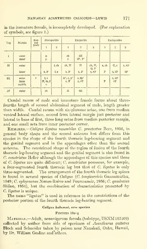
HAWAIIAN ACANTHURID CALIGOIDS—LEWIS 171
in the inunature female, is incompletely developed. (For explanation
of symbols, see figure 1.)
Leg
172 PROCEEDINGS OF THE NATIONAL MUSEUM vol. 115
Measurements.—Length from anterior end of body to posterior
end of caudal rami, excluding setae, 2.94 mm. Greatest length of
cephalothorax, including frontal region, 1.75 mm.; greatest width,
excluding marginal flange, 1.75 mm. Greatest length of genital seg-
ment 0.90 mm.; greatest width 1.11 mm. Greatest length of abdo-
men 0.25 mm.Description.—Body with light brown background coloration in
alcohol, mottled with yellow and with few scattered red and blue
pigment spots over cephalothorax, fourth thoracic leg-bearing seg-
ment, genital and abdominal segments. Eyes reddish.
Cephalothorax orbicular; frontal region well developed, with narrow
anterior marginal flange. Lunules distinct, extending to division
between frontal region and cephalothorax (fig. 10c). Groove between
frontal region and cephalothorax distinct, continuous, curving
anteriorly in smooth, flat curve in middle. Lateral margins of
cephalothorax with membranous flange. Posterior sinuses distinct,
U-shaped. Median cephalothoracic area extending posteriorly slightly
past posterior extensions of lateral regions; posterior margin of
area flattened. Lateral corners of protruding median cephalo-
thoracic area rounded, lateral margins continuous with inner margin
of posterior sinuses. Major cephalothoracic grooves distinct, forming
irregular H ; cross groove continuous with posterior longitudinal
grooves. Middle of cross groove forming flat anteriorly directed
point though apex with slight depression. Posterior longitudinal
grooves slightly convex; anterior longitudinal grooves extending
anteriorly and laterally from junction with cross and posterior longi-
tudinal grooves, terminating blindly posterior to eyes, medial to lateral
margin. Lateral strengthening regions extending laterally from origin
just anterior to junction of longitudinal and cross grooves, curving
posteriorly and tapering out in region of posterior lateral margins.
Two minor grooves present, extending posteriorly from either side
of anterior curvature of groove between frontal region and cephalo-
thorax, terminating just anterior to eyes. Light median longitudinal
groove present extending posteriorly from slight median depression
of major cross groove to posterior margin of thoracic area. Eyes
small, contiguous on median longitudinal axis of body in anterior
fourth of cephalothorax (fig. 10a).
Fourth thoracic leg-bearing segment, genital segment, and abdomenfused. Region of fourth thoracic leg-bearing segment tapered out-
ward sharply from junction of cephalothorax to region of fourth
thoracic leg attachment, adjacent to anterior lateral surface of genital
segment. Fourth thoracic leg-bearing segment tapered inward
slightly from widest point to beginning of genital segment. Division
between fourth thoracic leg-bearing segment and genital segment
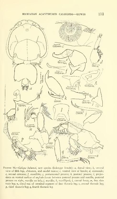
HAWAIIAN ACANTHURID CALIGOIDS—LEWIS 173
Figure 10.
—
Caligus kalumai, new species (holotype female): a, dorsal view; b, ventral
view of fifth legs, abdomen, and caudal ramus; c, ventral view of lunule; d, antennule;
e, second antenna; /, mandible; g, postantennal process; h, postoral process; i, projec-
tions on ventral surface of cephalothorax between postoral process and maxilla, postoral
process on right, maxilla on left; /, maxilla; k, maxllliped; /, sternal furca; w, first tho-
racic leg; n, distal end of terminal segment of first thoracic leg; o, second thoracic leg;
-p, third thoracic leg; q, fourth thoracic leg.

174 PROCEEDINGS OF THE NATIONAL MUSEUM vol. 115
visible as very fine groove extending from junction of segments
medially, on each side, for about one-fourth width of region; no evi-
dence of division present in middle of segment.
Genital segment swollen, lateral margins almost parallel; posterior
lateral regions of segment protruding as irregular lobes; lobes extending
to posterior lateral corners of abdomen. Median posterior surface
depressed. Fifth legs visible dorsally as three small, plumose setae
projecting from posterior lateral margin, single seta from small nodule,
two setae from second nodule just posterior to first (fig. 106).
Abdomen one-segmented, width approximately equal to length;
widest at posterior lateral corners, margins curving medially both
anteriorly and posteriorly, anteriorly in convex curve to region of
fusion of abdomen and genital segment, posteriorly in concave curve
to slightly projecting anal region (fig. 106).
Antennule two-segmented, excluding frontal plate, attached to
frontal region under free surface of lunule, at junction of frontal region
and cephalothorax. Greatest length of first segment slightly morethan that of second. First segment wider in proximal region
than at either end, tapering abruptly to proximal end, gradually to
distal; distal end irregular. Anterior surface with about 23 plumose
setae. Second segment slender, slightly wider in middle than at
either end. Second segment with naked seta on distal region of pos-
terior margin, nine naked setae from distal surface (fig. lOd).
Second antenna three-segmented, attached posterior and slightly
medial to base of antennule. First segment short, forming articular
surface for second segment. Distal surface of first segment con-
cave, posterior surface protruding posteriorly as blunt-tipped, spine-
like projection. Second segment wider proximally than distally,
lateral margins convex. Greatest width of second segment slightly
less than greatest length. Distal margin of segment irregular, inner
portion with concave depression; several points of articulation present
as heavily chitinized regions. Third segment and terminal process
elongate, longer than greatest length of second segment, with two
accessory processes, spinelilve process on posterior sm*face of distal
end of segment and two-parted, sharp-tipped process on anterior
proximal surface of segment. Base of third segment heavily chitin-
ized, protruding slightly proxunally as small, rounded articular sur-
face. Terminal process curving sharply in distal region, distal end
bluntly pointed (fig. lOe).
Mandible four-parted. Fu*st part wider proximally than distally,
with even taper to junction of second part. Second part short,
approximately one-fifth length of first part, tapered slightly to junction
of third part. Thkd part shghtly shorter than first part, expanded
slightly distally. Fourth part short, half length of first part, width
HAWAIIAN ACANTHURID CALIGOIDS—LEWIS 175
greater than that of distal end of third part. Fourth part curved
inward, distal end broadly rounded, inner margin with 11 blunt,
toothlike denticulations (fig. 10/).
Postantennal process simple, blunt-tipped, spinehke process pro-
jecting posteriorly in slight curve from attachment lateral and slightly
posterior to base of second antenna. Proximal surface broad, taper-
ing sharply in middle of process to inward curving distal region.
Proximal region with two nodules, each with two minute setules;
additional nodule present medial to middle of process, projecting
slightly from ventral surface of cephalothorax, bearing two minute
setules (fig. 10^).
Postoral process simple, spinelike, projecting posteriorly from
attachment lateral and slightly posterior to base of mouth cone.
Outer lateral margin irregularly concave, inner lateral margin straight.
Single nodule present anterior to base of process, bearing three setule-
like projections (fig. lOh).
Maxilla two-segmented, attached just lateral to base of postoral
process. Cephalothorax, in region of attachment of maxilla, with
several small, heavily chitinized, spinelike processes (fig. lOi). First
segment with slender projection from inner proximal surface serving
as articular surface. Distal lateral margins angling sharply inward
to sHghtly projecting articular surface. Second segment elongate,
length slightly greater than greatest length of first segment; proximal
surface tapered, inner portion irregular, forming articular surface.
Second segment with sharply pointed spine on distal region of inner
margin, two flexible spines projecting from distal surface of segment.
Inner terminal spine almost twice length of outer, fringed by fine
membrane along both margins; outer spine with fine, setuliferous mem-brane along both margins (fig. IQ;).
Maxilliped two-segmented, situated posterior and medial to first
maxillipeds. First segment obovoid, with strong projection of prox-
imal surface forming articular surface. Middle of inner lateral margin
of first segment with small, convex process. Distal surface concave,
forming region of attachment and articulation of second segment;
distal lateral surface with small, heavily chitinized, knoblike projec-
tion. Second segment slender, slightly wider proximally than dis-
tally; with small concavity on inner proximal margin, fitting over
shght projection of distal lateral surface of fu"st segment when second
segment flexed. Second segment with short, inward-curving terminal
process and two small, spinelike accessory processes, both on inner
surface of distal region of segment. Both accessory processes sur-
rounded by fine, frilled membrane. Division between terminal process
and segment distinct (fig. lOA:).

176 PROCEEDINGS OF THE NATIONAL MUSEUM vol. 115
Sternal furea attached to ventral surface of cephalothorax between,
and slightly posterior to, maxilliped bases. Width of base offurca
equal to distance between outer distal margins of tines. Right tine
broken on holotype specimen; left tine with rounded distal margin.
Tines directed laterally from origin at apex of bifurcation. Bifurca-
tion extending two-fifths length of process; apex flat, length slightly
less than half width of base (fig. lO^.
First thoracic leg biramous though endopodite reduced to simple,
minute lobe on inner distal surface of protopodite. Protopodite
one-segmented, inner proximal surface extending as small articular
process. First segment of two-segmented exopodite slightly longer
than protopodite; proximal and distal margins irregular. First seg-
ment with single, small spine on outer distal margin. Second segment
of exopodite short, less than half length of first segment. Distal
margin of second segment with four flexible processes, innermost
spinelike, second outermost bifid (fig. lOn). Inner lateral margin of
second segment with three plumose setae, distalmost seta with stiff
setules on proximal portion (figs. 10m,n).
Second thoracic leg biramous. Protopodite two-segmented; first
segment short, proximal region slender, proximal end a two-lobed
articular surface. Second segment broad, greatest width twice that
of first segment; greatest length slightly more than twice that of first
segment. Distal end of second segment irregular. Exopodite three-
segmented; greatest length of first segment equal to that of combined
second and third segments. First segment with strong, simple spine
on outer distal corner, directed at angle across outer distal corner of
second segment. Second segment short, with strong, simple spine on
outer distal corner, directed at an angle across middle of third segment.
Third segment longer than second, width greater than length; lateral
margins with irregular convex curve, distal margin forming rounded
apex of union of lateral margins. Outer lateral margin of third
segment with two spines, small spine from middle of segment curving
medially, second spine just distal to fu'st, more strongly developed,
with thin membrane along inner and outer margins. Endopodite
three-segmented; first and third segments shorter than second.
Third segment broadly rounded (fig. lOo).
Third thoracic leg biramous. Protopodite one-segmented, broadly
flattened. Exopodite two-segmented, with three-parted, spinelike
process arising from junction of protopodite and exopodite, and curving
inward. First segment of exopodite almost twice length of second,
second segment broadly rounded, distal and lateral margins contin-
uous. Endopodite two-segmented ; first segment short, with indistinct
proximal margin, second segment broadly rounded laterally, flattened
distally (fig. lOp).

HAWAIIAN ACANTHURID CALIGOIDS—LEWIS 177
Fourth thoracic leg unu*amous, three-segmented. Protopodite one-
segmented, sHghtly longer than combined length of two segments of
expedite. Protopodite slender, with two hairlike setules from outer
sm-face, one in proximal region, second in distal region; single, small,
plumose seta present on outer distal corner. Greatest length of first
exopodite segment slightly less than that of second; with single,
elongate spine from distal surface. Second segment of exopodite with
three spines on flat distal surface, inner spine longest, outer shortest.
All four spines on two segments of exopodite with fine membrane along
inner and outer margins; three terminal spines, on second segment,
with frilled membrane around base (fig. lOg).
The character of the membranes, spines, and setae on the thoracic
legs is given below (for explanation of symbols, see figure 1):
Leg
178 PROCEEDINGS OF THE NATIONAL MUSEUM vol. 115
differ also in the presence of two marginal, hairlike setules on the
protopodite of the fourth thoracic leg of C. kalumai, which are absent
on C. punctatus. In addition, the second segment of the exopodite
of C. punctatus, as shown by Shiino (1955), appears much longer
than the first segment while it is only sUghtly longer than the first
segment in C. kalumai. Other characteristics serve to differentiate
these two species, but it appears that these two, in addition to C.
oviceps, are morphologically similar.
The name "kalumai" is derived from two Hawaiian words. The
first word, "kalmnu," means "to swell." The second, "ma'i," is the
term for genitals. The combination of the two terms is used to in-
dicate the condition of the genital segment in the holotype female
specimen.
Genus Lepeophtheirus Nordmann, 1832
Diagnosis.—Abdomen of one or two segments, usually distinct from
genital segment. Lunules absent. Postoral process simple or
bifurcate; sternal furca present. First three thoracic legs biramous
though endopodite of first rudimentary; fourth leg uniramous,
typically with four segments.
Remarks.—Nordmann does not give a synopsis of the characters
of the genus in his 1832 publication. The characteristics given
above were taken primarily from Wilson (1932) and were modified
from descriptions of members of the genus Lepeophtheirus given by
other authors and from the personal experience of the present author.
Lepeophtheirus dissimulatus Wilson
Figures lla-g, 12a-r
L. dissimulatus Wilson, 1905a, p. 631, pi. 22.—Linton, 1907, p. 120.—Brian,
1924, p. 20.—Heegaard, 1943b, p. 20.—Yeatman, 1957, p. 350, figs. 17-31.—
Shiino, 1959b, p. 305, figs. 15-16.—Causey, 1960, p. 330.
Distribution.—Bermuda, British West Indies, Dry Tortugas
Islands, Galapagos Islands, Revilla Gigedo Islands, Baja CaHfornia,
British Columbia, Mexico, Hawaii.
Hosts.—Epinephalus morio (Cuvier and Valenciennes), E. labri-
formis (Jenyns), E. guaza (Linnaeus), Mycteroperca venennosa apua
(Bloch), Lactophrys trigonus? (Linnaeus), Qadus macrocephalus
Tilesius, Bodianus diplotaenia (Gill), Merluccius productus (Ayres),
Paralichthys californicus (Ayres), Hypsopsetta guttulata (Girard),
Sphyraena argentea Girard, Paralabrax nebulifer (Girard), Sphaeroides
annulatus (Jenyns), Galeichthys guatamelensis (Glinther), Acanihurus
olivaceous Bloch and Schneider, A. dussumieri Cuvier and Valenciennes,
A. guttatus Bloch and Schneider, A. triostegus sandvicensis Streets,
Naso hexacanthus (Bleeker), Chaetodon guadrimaculatus Gray, Zebra-
soma fiavescens (Bennett).
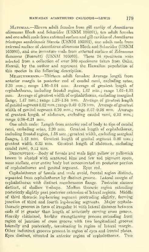
HAWAIIAN ACANTHURID CALIGOIDS—LEWIS 179
Material.—Eleven adult females from gill cavity of Acanthurus
olivaceous Bloch and Schneider (USNM 105091), ten adult females
and one adult male from external surface and gill cavities of Acanthurus
triostegus sandvicensis Streets (USNM 105092), one adult male from
external surface of Acanthurus olivaceous Bloch and Schneider (USNM105093), and one immature male from external surface of Zehrasoma
Havescens (Bennett) (USNM 105090). These 24 specimens were
selected from a collection of over 300 specimens taken from Oahu,
Hawaii, by the author and represent the Hawaiian population of
this species in the following description.
Measurements.—Thirteen adult females: Average length from
anterior margin to posterior end of caudal rami, excluding setae,
2.30 mm.; range 1.93-3.04 mm. Average of greatest length of
cephalothorax, including frontal region, 1.47 mm.; range 1.01-1.93
mm. Average of greatest width of cephalothorax, excluding marginal
flange, 1.47 mm.; range 1.29-1.84 mm. Average of greatest length
of genital segment 0.62 mm. ; range 0.49-0.78 mm. Average of greatest
width of genital segment 0.70 mm.; range 0.57-0.98 mm. Average
of greatest length of abdomen, excluding caudal rami, 0.12 mm.;range 0.08-0.21 mm.One adult male: Length from anterior end of body to tips of caudal
rami, excluding setae, 2.20 mm. Greatest length of cephalothorax,
including frontal region, 1.58 mm.; greatest width, excluding marginal
flanges, 1.42 mm. Greatest length of genital segment 0.40 mm.;greatest width 0.33 mm. Greatest length of abdomen, excluding
caudal rami, 0.12 mm.Description.—Body of female and male light yellow or yellowish
brown in alcohol with scattered blue and few red pigment spots,
some stellate, over entire body but concentrated on posterior portion
of cephalothorax and genital segment. Eyes red.
Cephalothorax of female and male ovoid, frontal region distinct,
separated from cephalothorax by distinct groove. Lateral margin of
cephalothorax with distinct membranous flange. Posterior sinuses
distinct, of shallow V-shape. Median thoracic region extending
posteriorly slightly past posterior extension of lateral regions. Middle
of third thoracic leg-bearing segment protruding slightly, forming
junction of third and fourth leg-bearing segments. Major cephalo-
thoracic grooves in form of irregular H with lateral distance between
ends of H greater than length of anteriorly curving cross groove.
Heavily chitinized, barlike strengthening process extending from
region of junction of cross groove with two longitudinal grooves
laterally and posteriorly, terminating in region of lateral margin.
Other indistinct grooves present in region of eyes and frontal plates.
Eyes distinct, situated in anterior region of cephalothorax. Two
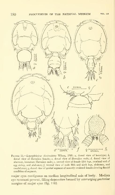
180 PROCEEDINGS OF THE NATIONAL MUSEUM vol. lis
0.2 mm. 0.5 m m.
Figure U.—Lepeophtheirus dissimulatus Wilson, 1905: a, dorsal view of lectotype; b,
dorsal view of Hawaiian female; c, dorsal view of Hawaiian male; d, dorsal view of
aberrant, immature Hawaiian male; e, ventral view of female fifth legs, proximal end of
egg string, and abdomen; /, ventral view of male fifth and sixth legs, abdomen, and
caudal rami; g, dorsal view of genital segment of recently ovulated female showing flaccid
condition of segment.
major eyes contiguous on median longitudinal axis of body. Median
eye remnant present, filling depression formed by converging posterior
margins of major eyes (fig. 116).
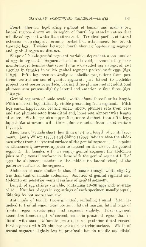
HAWAIIAN ACANTHURID CALIGOIDS—LEWIS 181
Fourth thoracic leg-bearing segment of female and male short,
lateral regions drawn out in region of fourth leg attachment so that
middle of segment wider than either end. Terminal portion of lateral
extension cup-shaped, forming socket-like attachment for fourth
thoracic legs. Division between fourth thoracic leg-bearing segment
and genital segment distinct.
Shape of female genital segment variable, dependent upon numberof eggs in segment. Segment flaccid and ovoid, surrounded by loose
membrane, in females that recently have extruded egg strings; almost
circular in females in which genital segment packed with eggs (figs.
116,^). Fifth legs seen ventrally as lobelike projections from pos-
terior ventral surface of genital segment, just lateral to nodelike
projection of posterior surface, bearing three plumose setae; additional
plumose seta present sHghtly lateral and anterior to first three (figs.
llb,e,g).
Genital segment of male ovoid, width about three-fourths length.
Fifth and sixth legs distinctly visible protruding from segment. Fifth
legs small, lappet-like, bearing single, short, plumose seta from base
and two plumose setae from distal end, inner seta almost twice length
of outer. Sixth legs also lappet-like, more distinct than fifth legs;
lappet-like structure with three plumose setae from distal surface
(fig. 11/).
Abdomen of female short, less than one-third length of genital seg-
ment. Both Wilson (1905) and Shiino (1959) indicate that the abdo-
men arises from the ventral surface of the genital segment. The point
of attachment, however, appears to depend on the size of the genital
segment. In females with an empty genital segment the abdomenjoins to the ventral surface; in those with the genital segment full of
eggs the abdomen attaches to the middle (in lateral view) of the
posterior surface of the segment.
Abdomen of male similar to that of female though width slightly
less than that of female abdomen. Junction of genital segment and
abdomen on posterior ventral surface of genital segment.
Length of egg strings variable, containing 10-30 eggs with average
of 15. Number of eggs in egg strings of each specimen usually equal,
differing by not more than two.
Antennule of female two-segmented, excluding frontal plate, at-
tached to frontal region near posterior lateral margin, lateral edge of
frontal region overlapping first segment slightly. First segment
about two times length of second, wider in proximal region than in
distal, with small, bifm-cate protrusion on posterior distal corner.
Fii'st segment with 20 plumose setae on anterior surface. Width of
second segment slightly less in proximal than in middle and distal
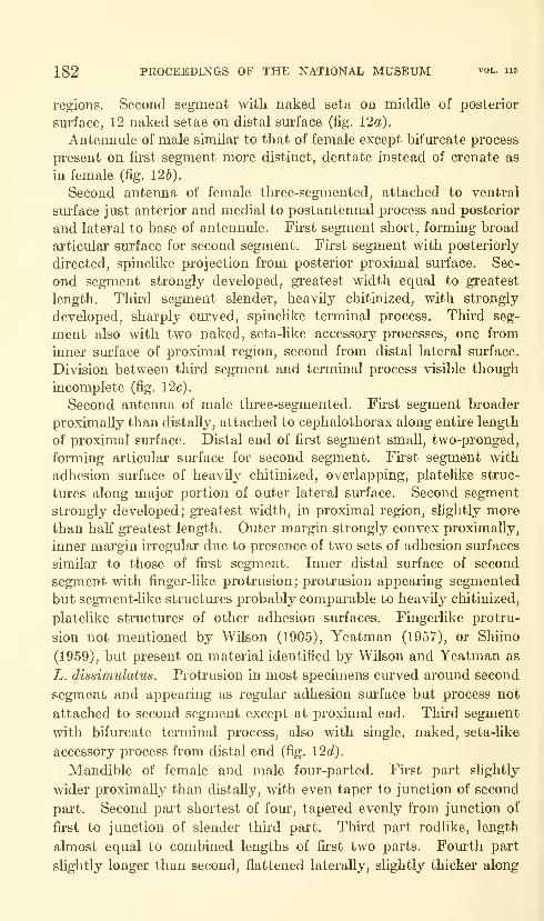
182 PROCEEDINGS OF THE NATIONAL MUSEUM vol. 115
regions. Second segment with naked seta on middle of posterior
surface, 12 naked setae on distal surface (fig. 12a).
Antennule of male similar to that of female except bifurcate process
present on first segment more distinct, dentate instead of crenate as
in female (fig. 126).
Second antenna of female thi'ee-segmented, attached to ventral
surface just anterior and medial to postantennal process and posterior
and lateral to base of antennule. First segment short, forming broad
articular surface for second segment. Fii'st segment with posteriorly
directed, spinelike projection from posterior proximal surface. Sec-
ond segment strongly developed, greatest width equal to greatest
length. Third segment slender, heavily chitinized, with strongly
developed, sharply curved, spinehke terminal process. Third seg-
ment also with two naked, seta-like accessory processes, one from
inner surface of proximal region, second from distal lateral surface.
Division between third segment and terminal process visible though
incomplete (fig. 12c).
Second antenna of male three-segmented. First segment broader
proximally than distally, attached to cephalothorax along entire length
of proximal surface. Distal end of first segment small, two-pronged,
forming articular sm"face for second segment. First segment with
adhesion surface of heavily chitinized, overlapping, plateUke struc-
tures along major portion of outer lateral surface. Second segment
strongly developed; greatest width, in proximal region, slightly more
than half greatest length. Outer margin strongly convex proximally,
inner margin irregular due to presence of two sets of adhesion surfaces
similar to those of first segment. Inner distal sm-face of second
segment with finger-like protrusion; protrusion appearing segmented
but segment-like structures probably comparable to heavily chitinized,
platelike structiu'es of other adhesion surfaces. Fingerlike protru-
sion not mentioned by Wilson (1905), Yeatman (1957), or Shiino
(1959), but present on material identified by Wilson and Yeatman as
L. dissimulatus. Protrusion in most specunens curved around second
segment and appearing as regular adhesion surface but process not
attached to second segment except at proximal end. Third segment
with bifurcate terminal process, also with single, naked, seta-like
accessory process from distal end (fig. \2d).
Mandible of female and male four-parted. Fii'st part slightly
wider proximally than distally, with even taper to junction of second
part. Second part shortest of four, tapered evenly from junction of
first to junction of slender third part. Third part rodlike, length
almost equal to combined lengths of first two parts. Fom"th part
slightly longer than second, flattened laterally, slightly thicker along
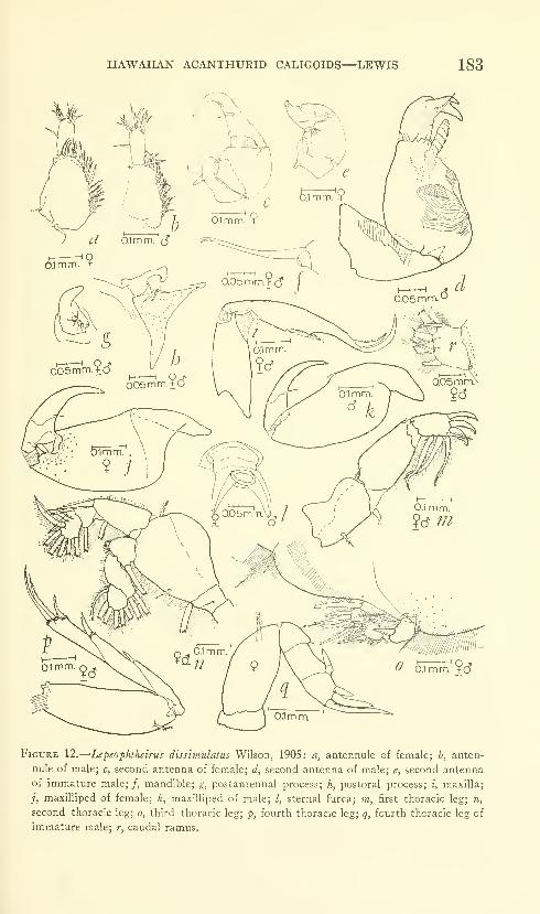
HAWAIIAN ACANTHURID CALIGOIDS—LEWIS 183
Figure 12.
—
Lepeophtheirus dissimulatus Wilson, 1905: a, antennule of female; b, anten-
nule of male; c, second antenna of female; d, second antenna of male; e, second antenna
of immature male;/, mandible; g, postantennal process; h, postoral process; i, maxilla;
j, maxilliped of female; k, maxilliped of male; /, sternal furca; m, first thoracic leg; n,
second thoracic leg; o, third thoracic leg; p, fourth thoracic leg; q, fourth thoracic leg of
immature male; r, caudal ramus.
184 PROCEEDINGS OF THE NATIONAL MUSEUM vol. ii5
outer margin than along inner, distal half curved inwards, inner
margin with 12 toothlike denticulations (fig. 12/).
Postantennal process of female and male simple, spinelike, attached
lateral and sUghtly posterior to second antenna. Spinelike process
curved inward, distal end rounded; two nodules present on inner
proximal region, proximal nodule with three setules, distal nodule
with four (fig. 12^).
Postoral process of female and male flattened, spinelike, situated
just lateral to base of mouth cone and directed posteriorly. Process
with broad base, width slightly less than length, tapered irregularly
to bluntly pointed distal end. Small, conical node present just
anterior to base of process, bearing three setules of approximately
equal length (fig. 12^).
MaxUla of female and male two-segmented, attached just lateral
and posterior to postoral process. First segment short, stocky,
inner proximal end protruding as irregular articular surface. Distal
lateral margins of first segment tapered irregularly, projecting medi-
ally as narrow, lobelike articular surface. Second segment elongate,
lateral margins irregularly convex; inner proximal sm-face irregular,
forming articular surface and place of attachment for muscles moving
segment. Middle of inner margin indented in two places, minute
spine arising from proximal indentation, larger spine, about twice
length of first, arising from distal indentation; membrane present,
connecting both spines. Smaller proximal spine and membrane not
mentioned by Wilson (1905), Yeatman (1957), or Shiino (1959),
though both characteristics present on material identified by Wilson
and Yeatman as L. dissimulatus. (Both membrane and small proxi-
mal spine difficult to see unless appendage stained.) Second segment
with two long, broadly curved terminal, spinelike processes; inner-
most about one and a half times length of outer. Inner process with
membrane along inner margin and rudiments of narrow membrane
along outer; outer process with frilled membrane along outer margin
(fig. 12^).
MaxiUiped of female two-segmented, attached just posterior and
medial to maxUla. First segment large, strongly developed, inner
proximal surface ^vith lobate protrusion forming articular surface.
Outer distal end of first segment irregular, with shghtly projecting,
lobate process serving as articular surface for second segment. Second
segment short, with long, curved, acuminate terminal process and
small, spinehke accessory process from inner distal lateral surface;
division between second segment and terminal process indistinct
(fig. 12i).
HAWAIIAN ACANTHURID CALIGOIDS—LEWIS 185
Maxilliped of male similar to that of female except with small,
shelflike structure present on middle of inner margin of first segment
(fig. 12^).
Sternal furca of female and male attached between, and sHghtly
posterior to, bases of maxillipeds. Bifurcation extending approxi-
mately half length of process; tines with bluntly pointed tips (fig. 12Z).
First thoracic leg bii'amous though endopodite present only as
rudiment at junction of protopodite and exopodite. Protopodite
strongly developed, one-segmented, greatest length almost equal to
that of first segment of exopodite; proximal surface concave, more
heavily chitinized than rest of segment, forming articular surface.
Single, plumose seta present on inner proximal surface of protopodite
and second, plumose seta on outer distal corner. First segment of
two-segmented exopodite wider proximally than distally, proximal
and distal margins irregular, proximal with slightly projecting, heavily
chitinized medial sm'face articulating with protopodite. First seg-
ment with single spinule on outer distal corner and row of setules
along inner margin. Second segment of exopodite palm-shaped,
slightly more than half length of first segment, with three spines
arising from distal margin and curved inwards. Inner two terminal
spines with small, concave depression on distal inner margin across
which fme membrane stretches; with row of stiff, very short setules
along inner margin of all three spines, extending to tip of fii'st spine,
to concavity of second and third (fig. 12to).
Second thoracic leg biramous. Protopodite two-segmented; first
segment small, less than half length of second. Second segment
broad, greatest width about four-fifths greatest length, lateral and
distal margins continuous, distal margin irregular. Exopodite three-
segmented; length of first segment equal to combined lengths of
second and third, with strongly developed, membrane-margined spine
projecting distally from outer distal corner. Second segment short,
irregular, with slender spine projecting distally from slight protrusion
of outer distal corner. Third segment approxunately twice length of
second, with irregular lateral and distal margins. Two well-developed
spines present on outer lateral margin of third segment, first extending
distally from shelflike structure on outer proximal surface, second
extending outward from attachment just distal and medial to base of
first spine. Endopodite three-segmented; second segment longer than
first or thud. First segment with short row of stiff setules on outer
distal margin; third segment rounded distally, lateral and distal
margins distinct (fig. 12n).
Third thoracic leg biramous. Protopodite one-segmented, strongly
flattened, platehke. Exopodite three-segmented; first segment with
two-parted, spinelike process arising from junction of protopodite
679-677—64 4

186 PROCEEDINGS OF THE NATIONAL MUSEUM
and second segment; second, distal, part of process directed inward
at right angles to first. Second segment of exopodite flattened
distally, lateral margins broadly rounded ; third segment almost semi-
circular, lateral and distal margins continuous. Endopodite two-
segmented; first segment broad; proximal margin of segment, as well
as that of first exopodite segment, indistinct. Second segment of
endopodite broadly rounded, distal and lateral margins distinct
(fig. 12o).
Fourth thoracic leg uniramous, four-segmented. Protopodite one-
segmented, with small, bilobed articular surface projecting sUghtly
from inner proximal margin. Exopodite three-segmented, total length
slightly more than that of protopodite. First segment of exopodite
with single spine on outer distal corner; inner margin of spine
with indistinct, frUled membrane. Second segment with larger spine
on outer distal corner; spine with distinct, frilled membrane
along inner and outer margins, base of spine encircled by minute,
stiff setules. Third segment terminating in broad surface bearing
three spines. Outermost spine shortest, about half length of middle
spine, with frilled membrane along inner and outer margins. Middle
spine slightly shorter than inner spine, with frilled membrane along
outer margin only. Inner spine with few, fine setules on inner distal
margin. All three terminal spines encircled by minute, stiff setules
(fig. \2p).
The character of the membranes, spines, and setae of the thoracic
legs is given below. The thoracic legs of both sexes are identical.
(For explanation of symbols, see figure 1.)
Leg
HAWAUAN ACANTHURID CALIGOIDS—LEWIS 187
Aberrant immature male specimen shown in figure lid (second
antenna, fig. 12e; fourth thoracic leg, fig. 12g) differs from adult female
and other immature males in elongate fifth and sixth leg processes.
Fifth and sixth legs visible in all immature males at this stage of
development but in form of small lappets, similar to those of adult
male, not extremely long projections as seen in this specimen.
Remarks.—Wilson (1905) erected this species from a series of
specimens from the Galapagos Islands and from Bermuda. Thematerial that he deposited in the U.S. National Museum as the type
specimens (cotypes) was that from the Galapagos. The lot (USNM42072, numbered 1505 in Wilson's 1905 publication) contained ten
females and one male taken from a specmien of Ejnnephalus labri-
formis (Jenyns) captured at Charles Island. At the time the present publi-
cation was prepared, the original lot contained only six females and one
immature male.
Since WUson did not select a single specimen as the holotype, the
present author has taken the liberty of choosing a lectotype from the
original lot. The specimen chosen was an ovigerous female 3.13 mm.in length from the anterior end of the body to the posterior end of the
caudal rami, excluding the setae. Because the characters of the
specimen chosen as the lectotype are in agreement with those given
by Wilson (1905), only a dorsal view of the specimen is shown (fig.
llo).
Additional measurements of lectotype specimen: Greatest length
of cephalothorax, including frontal region, 2.05 mm.; greatest width,
excluding marginal flanges, 1.93 mm. Greatest length of genital
segment 0.90 mm.; greatest width 1.11 mm.Lepeophtheirus dissimulatus has been taken on both the Atlantic
and Pacific coasts. It appears to be found primarily in tropical and
subtropical waters although the U.S. National Museum possesses a
collection, identified by C. B. Wilson and by the present author as
this species, that was made in British Columbia. The size of the
specimens taken in British Columbia is more than twice that of the
Hawaiian specimens. The British Columbia female specimens have
an average length of 6.26 mm.; the Hawaiian female specimens, an
average length of 2.94 mm. The males from the British Columbia
collection were only slightly larger than the single male used in the
present description (3 mm. in total length to 2.20 mm. for the Hawaiian
specimen)
.
Genus Anuretes Heller, 1865
Diagnosis.—^Abdomen rudimentary, partially or completely fused
to genital segment. Lunules absent. Fourth thoracic legs two-,
three-, or four-segmented.
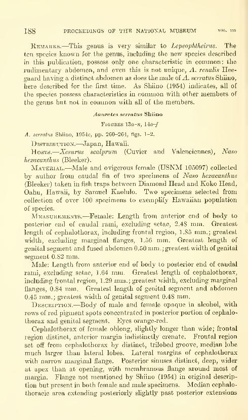
188 PROCEEDINGS OF THE NATIONAL MUSEUM vol. 115
Remarks.—This genus is very similar to Lepeophtheirus. Theten species known for the genus, including the new species described
in this pubhcation, possess only one characteristic in common: the
rudimentary abdomen, and even this is not unique, A. renalis Hee-
gaard having a distinct abdomen as does the male of ^. serratus Shiino,
here described for the first time. As Shiino (1954) indicates, all of
the species possess characteristics in common with other members of
the genus but not in common with all of the members.
Anuretes serratus Shiino
Figures ISa-n, 14o-/
A. serratus Shiino, 1954e, pp. 260-264, figs. 1-2.
Distribution.—Japan, Hawaii.
Hosts.—Xesurus scalprum (Cuvier and Valenciennes), Naso
hexacanthus (Bleeker).
Material.—Male and ovigerous female (USNM 105097) collected
by author from caudal fin of two specimens of Naso hexacanthus
(Bleeker) taken in fish traps between Diamond Head and Koko Head,
Oahu, Hawaii, by Samuel Kaolulo. Two specimens selected from
collection of over 100 specimens to exemplify Hawaiian population
of species.
Measurements.—Female: Length from anterior end of body to
posterior end of caudal rami, excluding setae, 2,48 mm. Greatest
length of cephalothorax, including frontal region, 1,85 mm.; greatest
width, excluding marginal flanges, 1.56 mm. Greatest length of
genital segment and fused abdomen 0.50 mm.;greatest width of genital
segment 0.82 mm.Male: Length from anterior end of body to posterior end of caudal
rami, excluding setae, 1.64 mm. Greatest length of cephalothorax,
including frontal region, 1.29 mm.; gi'eatest width, excluding marginal
flanges, 0.84 mm. Greatest length of genital segment and abdomen
0,45 mm,; greatest width of genital segment 0.48 mm.Description.—Body of male and female opaque in alcohol, with
rows of red pigment spots concentrated in posterior portion of cephalo-
thorax and genital segment. Eyes orange-red.
Cephalothorax of female oblong, slightly longer than wide; frontal
region distinct, anterior margin indistinctly crenate. Frontal region
set off from cephalothorax by distinct, trilobed groove, median lobe
much larger than lateral lobes. Lateral margins of cephalothorax
with narrow marginal flange. Posterior sinuses distinct, deep, wider
at apex than at opening, with membranous flange around most of
margin. Flange not mentioned by Shiino (1954) in original descrip-
tion but present in both female and male specimens. Median cephalo-
thoracic area extending posteriorly slightly past posterior extensions
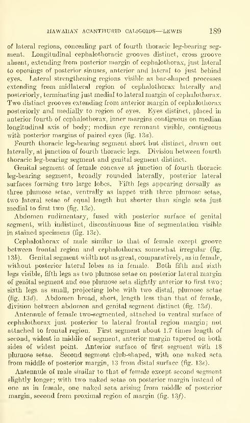
HAWAIIAN ACANTHURID CALIGOIDS— LEWIS 189
of lateral regions, concealing part of fourth thoracic leg-bearing seg-
ment. Longitudinal cephalothoracic grooves distinct, cross groove
absent, extending from posterior margin of cephalothorax, just lateral
to openings of posterior sinuses, anterior and lateral to just behind
eyes. Lateral strengthening regions visible as bar-shaped processes
extending from midlateral region of cephalothorax laterally and
posteriorly, terminating just medial to lateral margin of cephalothorax.
Two distinct grooves extending from anterior margin of cephalothorax
posteriorly and medially to region of eyes. Eyes distinct, placed in
anterior fourth of cephalothorax, inner margins contiguous on median
longitudinal axis of body; median eye remnant visible, contiguous
with posterior margins of paired eyes (fig. 13a).
Fourth thoracic leg-bearing segment short but distinct, drawn out
laterally, at junction of fourth thoracic legs. Division between foiu-th
thoracic leg-bearing segment and genital segment distinct.
Genital segment of female concave at junction of fourth thoracic
leg-bearing segment, broadly rounded laterally, posterior lateral
surfaces forming two large lobes. Fifth legs appearing dorsally as
three plumose setae, ventrally as lappet with three plumose setae,
two lateral setae of equal length but shorter than single seta just
medial to first two (fig. 13c).
Abdomen rudimentary, fused with posterior sm'face of genital
segment, with indistinct, discontinuous line of segmentation visible
in stained specimens (fig. 13c).
Cephalothorax of male similar to that of female except groove
between frontal region and cephalothorax somewhat irregular (fig.
136). Genital segment width not as great, comparatively, as in female,
without posterior lateral lobes as in female. Both fifth and sixth
legs visible, fifth legs as two plumose setae on posterior lateral margin
of genital segment and one plumose seta slightly anterior to first two;
sixth legs as small, projecting lobe with two distal, plumose setae
(fig. 13c?). Abdomen broad, short, length less than that of female,
division between abdomen and genital segment distinct (fig. 13f/).
Antennule of female two-segmented, attached to ventral sm*face of
cephalothorax just posterior to lateral frontal region margin; not
attached to frontal region. First segment about 1.7 times length of
second, widest in middle of segment, anterior margin tapered on both
sides of widest point. Anterior surface of first segment with 18
plumose setae. Second segment club-shaped, with one naked seta
from middle of posterior margin, 13 from distal sm'face (fig. 13c).
Antennule of male similar to that of female except second segment
slightly longer; with two naked setae on posterior margin instead of
one as in female, one naked seta arising from middle of posterior
margin, second from proximal region of margin (fig. 13/).
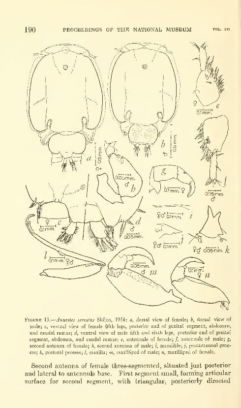
190 PROCEEDINGS OF THE NATIONAL MUSEUM
Figure 13.
—
Anuretes serratus Shiino, 1954: a, dorsal view of female; h, dorsal view of
male; c, ventral view of female fifth legs, posterior end of genital segment, abdomen,
and caudal ramus; d, ventral view of male fifth and sixth legs, posterior end of genital
segment, abdomen, and caudal ramus; e, antennule of female; /, antennule of male; g,
second antenna of female; h, second antenna of male; i, mandible;/, postantennal proc-
ess; k, postoral process; /, maxilla; m, maxilliped of male; n, maxilliped of female.
Second antenna of female three-segmented, situated just posterior
and lateral to antennule base. First segment small, forming articular
surface for second segment, with triangular, posteriorly directed
HAWAIIAN ACANTHURID CALIGOIDS—LEWIS 191
process. Second segment truncate, both proximal and distal surfaces
forming broad articular surfaces, distal lateral surfaces heavily
chitinized. Third segment with curved terminal process and two
spinules, one on proximal, second on medial inner surface. Terminal
process distinct from segment, strongly developed; inner surface of
process flat, lateral and outer surfaces rounded. Inner, flat surface
of terminal process with minute nodules on distal portion, distal end
of process tapered to sharp point (fig. 13g).
Second antenna of male three-segmented. First segment irregular,
attached to cephalothorax along all of proximal and most of inner
lateral surface ; with broad articular surface on distal end. Second
segment strongly developed, proximal region almost three times
width of distal; inner distal surface concave, with several small
adhesion pads. Third segment short, with compound terminal
process and simple spinule serving as accessory process. Compoundterminal process consisting of bifurcate process arising from base of
simple, curved process. Accessory process arising from inner distal
surface of segment (fig. 13h).
Mandible of female and male four-parted. First part broad at
proximal end, tapered to slender distal end; second part short, tapered
to junction of third part. Third part long, length greater than
combined lengths of other three parts, tapered slightly to junction
of fourth part; fourth part short, curved inwards, slightly thickened
along outer margin, distal end rounded, 13 toothlike denticulations
present along inner margin (fig. 13^).
Postantennal process of female and male attached lateral to base
of second antenna. Process spinelike, middle with sharp bend,
distal half pointing posteriorly and medially. Distal end of process
bluntly pointed; inner margin swoUen into distinct lobe, outer margin
with small, rounded knob just proximal to sharp bend. Process with
two small nodes on inner surface, each with several minute setules
(fig. 13i).
Postoral process of female and male situated just lateral to base
of mouth cone, consisting of bifurcate process and two nodules, one
anterior to base of each corner of process, each bearing several setules.
Two tines of bifurcation equal in female, sharply pointed; tines
unequal in male, outer slightly shorter than inner, both with pointed
distal ends. Acuminate condition of inner tine of process in opposition
to Shiino's original description (1954) in which inner spine is indicated
to be broad, distal end rounded (fig. 13^).
MaxiUa of female and male two-segmented, attached lateral to
postoral process. First segment shorter than second, narrower
proximaUy than distally, with proximal end protruding as articular
surface and small, rounded projection present on distal surface and
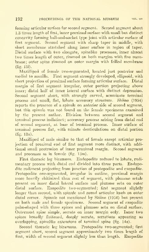
192 PROCEEDINGS OF THE NATIONAL MUSEUM vol. iis
forming articular surface for second segment. Second segment about
1.3 times length of first, inner proximal surface with small but distinct
concavity forming ball-and-socket type joint with articular surface of
first segment. Second segment with sharp taper in middle, with
short membrane stretched along inner surface in region of taper.
Distal surface with two elongate, spinelike processes, inner almost
two times length of outer, rimmed on both margins with fine mem-brane; outer spine rimmed on outer margin with frilled membrane(fig. 130.
Maxilliped of female two-segmented, located just posterior and
medial to maxilla. First segment strongly developed, ellipsoid, with
short projection of proximal surface forming articular surface. Distal
margin of first segment irregular, outer portion projecting above
inner; distal half of inner lateral surface with distinct depression.
Second segment short, with strongly curved, acuminate terminal
process and small, flat, lobate accessory structure. Shiino (1954)
reports the presence of a spinule on anterior side of second segment,
but this spinule was not found on the female specimens examined
by the present author. Division between second segment and
terminal process indistinct; accessory process arising from distal end
of second segment, at base of terminal process. Inner surface of
terminal process flat, with minute denticulations on distal portion
(fig. 13n).
Maxilliped of male similar to that of female except articular pro-
jection of proximal end of flrst segment more distinct, with addi-
tional small protrusion of inner proximal margin. Second segment
and processes as in female (fig. 13m).
First thoracic leg biramous. Endopodite reduced to lobate, rudi-
mentary process with distal end divided into three parts. Endopo-
dite rudiment projecting from junction of protopodite and exopodite.
Protopodite one-segmented, irregular in outline, proximal margin
more heavily chitinized than rest of segment, with plumose setule
present on inner distal lateral surface and plumose seta on outer
distal surface. Exopodite two-segmented; first segment slightly
longer than second, with spinule and round articular knob on outer
distal corner. Spinule not mentioned by Shiino (1954) but present
on both male and female specimens. Second segment of exopodite
palm-shaped with three spines and plumose seta on distal surface.
Outermost spine simple, serrate on inner margin only. Inner two
spines broadly flattened, deeply serrate, serrations appearing as
overlapping, spinelilve extensions of terminal spine (fig. 14a).
Second thoracic leg biramous. Protopodite two-segmented; first
segment short, second segment approximately two times length of
first, width of second segment slightly less than length. Exopodite

HAWAIIAN ACANTHURID CALIGOIDS—LEWIS 193
Figure 14.
—
Anuretes serratus Shiino, 1954: a, first thoracic leg; b, second thoracic leg; c,
ramified spine on first segment of exopodite of female second thoracic leg; d, ramified
spine on first segment of exopodite of male second thoracic leg; e, third thoracic leg; /,
fourth thoracic kg.
three-segmented, first segment longer than combined lengths of sec-
ond and third, with multiramous spine on outer distal corner. Axis
of spine cm'ving inward, rami directed distally (with regard to axis
of exopodite) ; female with eight rami on spine, excluding tip, male
with six (figs. 14c,c?). Second segment of exopodite short, with
single, lightly serrated spine on outer distal corner. Third segment
of exopodite approximately twice length of second, with two simple
spines on outer distal margin; inner and distal margins continuous.
Endopodite two-segmented; second segment more than twice length
of first, with eight plumose setae, all from distal half of segment
(fig. 146).
Third thoracic leg biramous. Protopodite broad, flat plate, semi-
circular in outline. Protopodite in male with adhesion pad on outer
proximal surface. Exopodite one-segmented, with sharply curved,
hooldike process arising from junction of exopodite and protopodite.
Endopodite two-segmented, first segment short, wide; second segment
about two times length of first, half the width; broadly rounded
(fig. Ue).
Fourth thoracic leg uniramous, tlu-ee-segmented. Protopodite slen-
der, with single, plumose seta from outer distal margin. Exopodite
two-segmented, shorter than protopodite; first segment with one
weakly developed terminal spine, second segment with four terminal
spines, outer three short, equal in length, rimmed with minute setules;
inner spine almost four times length of outer three, with several
minute spinules from middle of spine (fig. 14/).
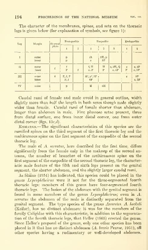
194 PROCEEDINGS OF THE NATIONAL MUSEUM
The character of the membranes, spines, and seta on the thoracic
legs is given below (for explanation of symbols, see figure 1):
Leg

HAWAIIAN ACANTHURID CALIGOIDS—LEWIS 195
Because of the variety of characteristics that are exhibited by membersof this genus, Shiino (1954) has aptly termed it a "waste-basket for
such species that can not be located in Lepeophtheirus." With the
description of more species in the genus, a series of characteristics maybe worked out to delimit the genus more distinctly, or else the charac-
teristics of related genera may be expanded to include the species nowplaced in the genus.
Anuretes menehune, new species
Figures 15a-o, IQa-e
Material.—Ovigerous female (holotype, USNM 105098) collected
by author from dorsal fin of specimen of Naso hexacanthus (Bleeker)
taken in fish trap between Diamond Head and Koko Head, Oahu,
Hawaii, by Samuel Kaolulo. Adult male (allotype,USNM 105099) col-
lected by author from dorsal fin of specimen of Naso hexacanthus (Blee-
ker) taken in fish trap between Diamond Head and Koko Head, Oahu,
Hawaii, by Samuel Kaolulo. Three adult females and three adult
males (paratypes, USNM 105101) taken by author from dorsal fin
of single specimen of Naso hexacanthus (Bleeker) collected in fish trap
between Diamond Head and Koko Head, Oahu, Hawaii, by SamuelKaolulo. Adult ovigerous female (paratype, USNM 105100) taken
by author from side of specimen of Naso unicornis (Forskal) speared
by author at Bird Island, Oahu, Hawaii.
These specimens were taken from a collection of over 100 specimens
and represent the Hawaiian population of the copepod.
Measurements.—Holotype female: Length from anterior end to
posterior end of caudal rami, excluding setae, 2.05 mm. Greatest
length of cephalothorax, including frontal region, 1.44 mm.; greatest
width, excluding marginal flanges, 0.86 mm. Greatest length of fused
genital segment and abdomen 0.50 mm.; greatest width of genital
segment 0.60 mm.Allotype male: Length from anterior end to posterior end of caudal
rami, excluding setae, 1.39 mm. Greatest length of cephalothorax,
including frontal region, 1.07 mm.; greatest width, excluding marginal
flanges, 0.74 mm. Greatest length of genital segment, excluding
protruding fifth and sixth leg setae, 0.29 mm.;greatest width 0.29 mm.
Paratype female specimens: Average length from anterior end to
posterior end of caudal rami, excluding setae, 1.68 mm. Average of
greatest length of cephalothorax, including frontal region, 1.23 mm.;average of greatest width, excluding marginal flanges, 0.74 mm.Average of greatest length of genital segment and abdomen 0.43 mm.;average of greatest width of genital segment 0.49 mm.
Paratype male specimens: Average length from anterior end to
posterior end of caudal rami, excluding setae, 1.31 mm. Average of
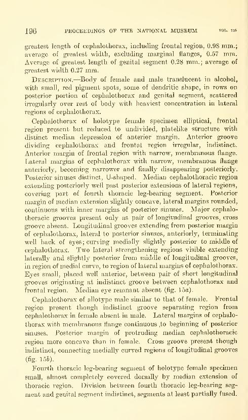
196 PROCEEDINGS OF THE NATIONAL MUSEUM vol. iib
greatest length of cephalothorax, including frontal region, 0.98 mm.;
average of greatest width, excluding marginal flanges, 0.57 mm.Average of greatest length of genital segment 0.28 mm.; average of
greatest width 0.27 mm.Description.-—^Body of female and male translucent in alcohol,
with small, red pigment spots, some of dendritic shape, in rows on
posterior portion of cephalothorax and genital segment, scattered
irregularly over rest of body with heaviest concentration in lateral
regions of cephalothorax.
Cephalothorax of holotype female specimen elliptical, frontal
region present but reduced to undivided, platelike structure with
distinct median depression of anterior margin. Anterior groove
dividing cephalothorax and frontal region irregular, indistinct.
Anterior margin of frontal region with narrow, membranous flange.
Lateral margins of cephalothorax with narrow, membranous flange
anteriorly, becoming narrower and finally disappearing posteriorly.
Posterior sinuses distinct, U-shaped. Median cephalothoracic region
extending posteriorly well past posterior extensions of lateral regions,
covering part of fourth thoracic leg-bearing segment. Posterior
margin of median extension slightly concave, lateral margins rounded,
continuous with inner margins of posterior sinuses. Major cephalo-
thoracic grooves present only as pair of longitudinal grooves, cross
groove absent. Longitudinal grooves extending from posterior margin
of cephalothorax, lateral to posterior sinuses, anteriorly, terminating
well back of eyes ; curving medially slightly posterior to middle of
cephalothorax. Two lateral strengthening regions visible extending
laterally and slightly posterior from middle of longitudinal grooves,
in region of medial curve, to region of lateral margins of cephalothorax.
Eyes small, placed well anterior, between pair of short longitudinal
grooves originating at indistinct groove between cephalothorax and
frontal region. Median eye remnant absent (fig. 15a).
Cephalothorax of allotype male similar to that of female. Frontal
region present though indistinct groove separating region from
cephalothorax in female absent in male. Lateral margins of cephalo-
thorax with membranous flange continuous .to beginning of posterior
sinuses. Posterior margin of protruding median cephalothoracic
region more concave than in female. Cross groove present though
indistinct, connecting medially curved regions of longitudinal grooves
(fig. 156).
Fourth thoracic leg-bearing segment of holotype female specimen
small, almost completely covered dorsally by median extension of
thoracic region. Division between fourth thoracic leg-bearing seg-
ment and genital segment indistinct, segments at least partially fused.
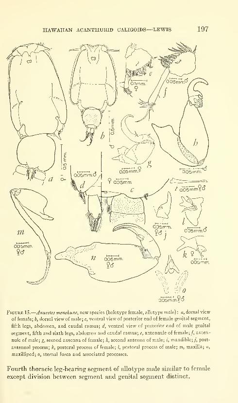
HAWAUAN ACANTHURID CALIGOIDS—LEWIS 197
V\\////
960.05mm
Figure IS.
—
Anuretes menehune, new species (holotype female, allotype male) : a, dorsal view
of female; b, dorsal view of male; c, ventral view of posterior end of female genital segment,
fifth legs, abdomen, and caudal ramus; d, ventral view of posterior end of male genital
segment, fifth and sixth legs, abdomen and caudal ramus; e, antennule of female;/, anten-
nule of male; g, second antenna of female; h, second antenna of male; i, mandible;/, post-
antennal process; k, postoral process of female; /, postoral process of male; m, maxilla; n,
maxilliped; o, sternal furca and associated processes.
Fourth thoracic leg-bearing segment of allotype male similar to female
except division between segment and genital segment distinct.
198 PROCEEDINGS OF THE NATIONAL MUSEUM vol. 115
Genital segment of holotype female specimen slightly wider than
long, lateral margins broadly rounded, posterior lateral regions
forming large lobe. Fifth legs visible as two minute spinules andsingle plumose setule from ventral posterior lateral surface of genital
segment (fig. 15c).
Width of genital segment of allotype male specimen equal to
length, lateral margins broadly rounded, posterior lateral regions
forming smaller lobes than in female. Fifth and sixth legs distinct;
fifth legs as two finely plumose setae from slight indentation of
posterior lateral margin; sixth as two finely plumose setae and third,
short, naked setule from distal end of lobate ventral posterior lateral
surface (fig. I5d).
Abdomen of holotype female specimen rudimentary, fused to
posterior surface of genital segment; line of division indistinct except
in one paratype specimen and discontinuous in this specimen (fig. 15c).
Abdomen of allotype male specimen distinct, not fused to genital
segment, width approximately twice length (fig. 15d).
Spermatophores visible on paratype female specimens as two large,
circular structures attached to genital segment by necklike connection.
Egg strings short, thick, approximately three-fourths length of ceph-
alothorax, with four to six large eggs in each string.
Antennule of holotj^pe female specimen two-segmented, excluding
frontal plate, attached to cephalothorax at posterior edge of frontal
region, appearing not to be attached to frontal region. First segment
of general triangular shape, proximal end wider than length of seg-
ment, anterior margin curving sharply to narrow distal end, bearing
21 plumose setae; distal margin with three plumose setae, anterior
dorsal surface with two nailed setae. Second segment slender, club-
shaped; length equal to length of first segment, with naked seta from
middle of posterior margin and 10 naked setae from distal surface
(fig. 15c).
Antennule of allotype male specimen similar to that of female
though second segment much longer, approximately one and a half
times that of female (fig. 15/).
Second antenna of holotype female specimen three-segmented,
attached posterior to antennule base. First segment forming artic-
ular surface for second. First segment short, well developed, with
long, triangular, distally pointed, posteriorly directed process. Second
segment short, well developed, width approximately two-thirds
length; with depression on distal surface forming articular surface
for third segment. Thud segment with recurved, acuminate terminal
process; segment and process longer than combined lengths of first
two segments, provided with two small, simple, spinelike processes
HAWAIIAN ACANTHURID CALIGOIDS—LEWIS 199
on proximal inner surface. Division between segment and terminal
process indistinct (fig. 15f/).
Second antenna of allotype male three-segmented. Fii-st segment
long, outer surface of distal two-thirds forming articular surface
for second segment, proximal margin forming blunt, posteriorly
directed process. Second segment well developed, with broad base,
tapered to narrow distal margin. Distal margin of second segment
UTCgular, with heavily chitinized regions forming articular surfaces
for third segment. Third segment short, with adhesion surface on
inner margin, and seta-like accessory process on posterior surface
adjacent to adhesion pad. Terminal process of third segment bifiu--
cate; inner part of bifurcation ciurving inward and proximally as
projection of basal region of process, outer part of bifurcation muchlonger than inner, extending distally before recurving in same manner
as first part. Division between segment and terminal process dis-
tinct (fig. 15h).
Mandible of female and male four-parted. First part broader
proximally than distally, lateral margins irregular. Length of second
part approximately equal to that of first, with even taper to junction
of elongate third part. Third part as long as combined lengths of
first two parts, tapered slightly to junction of fourth part. Fourth
part curving medially at distal end, with 11 toothlike denticulations
along inner margin (fig. 15i).
Postantennal process of female and male simple, shghtly curved
spine arising lateral and posterior to attachment of second antenna.
Spine with two minute nodules on inner proximal surface, each with
long setule (fig. I5j).
Postoral process of holotype female specimen a plate with projecting
bifurcate process and node, at anterior end of plate. Node with three
setules of approximately equal length. Inner tine of bifurcate process
longer than outer, both tines with rounded distal ends (fig. 15^).
Postoral process of allotype male specimen similar to holotype
though setule-bearing node with two setules, third setule from base
of bifurcate process (fig. 151).
Maxilla of female and male two-segmented, situated lateral and
slightly posterior to postoral process. First segment short, well
developed, inner proximal surface protruding as lobate articular
process. First segment tapered from broad base to narrow distal end,
middle of distal end projecting as articular surface for second segment.
Second segment elongate, one and a half times length of first segment.
Inner proximal margin with small concavity forming ball-and-socket
joint with articular surface of first segment. Second segment tapered
abruptly in middle, as in A. serratus, but without membrane found in
this species, with two long, spinelike processes on distal surface, inner
200 PROCEEDINGS OF THE NATIONAL MUSEUM vol. 115
process more than twice length of outer, with fine membrane along
inner and outer margins. Outer terminal process without membranes(fig. 15m).
Maxilliped of female and male two-segmented, attached medial and
slightly posterior to maxilla. First segment well developed, with
proximal extension of inner surface forming articular surface. First
segment tapered from wide proxmial to somewhat narrower distal
region. Distal surface irregular, with heavily chitinized indentations
for articulation with second segment. Second segment short, with
strongly developed, curved, clawlike terminal process. Second seg-
ment with flat, lobate process and simple, spinelike process as acces-
sory structures. Lobate accessory structure with minute, toothlike
denticulations on distal surface. Spinelike accessory structure
attached just proximal to middle of base of lobate accessory structure.
Inner surface of curved terminal process flat, with very slight denticu-
lations. Division between segment and terminal process indistinct
(fig. 15n).
Sternal furca of female and male located between, and slightly
posterior to, bases of maxillipeds. Base of furca heavily chitinized
oval structure; center of base depressed, not heavily chitinized.
Bifurcate process single, arrowhead-shaped unit, distinct from oval
base, attached to base at proximal end. Furcal tines widely spread,
with even concave depression between blunt tips; lateral margins
curving medially to attachment with base. Two medially curving
ridges present posterior to sternal furca (fig. 15o).
The arrowhead-shaped furcal process is absent in one of the paratype
specimens (USNM 105100); the only structure present is the heavily
chitinized oval base. In addition, both postoral processes either have
been lost or have developed irregularly, since only a simple, blunt,
spinelike process is present. This variation may lead to taxonomic
difficulties in later work as there is no evidence of the loss of these
two processes in the specimen.
First thoracic leg biramous though endopodite reduced to rudi-
mentary, lobate process at junction of exopodite and protopodite.
Protopodite one-segmented, with slight projection of outer proximal
portion forming articular surface; single, lightly plumose setule present
on inner proximal lateral margin. First segment of two-segmented
exopodite slightly longer than protopodite, proximal and distal margins
irregular, outer distal corner with projecting heavily chitinized knob
and simple spinule. Second segment of exopodite pahn-shaped,
proximal and inner margins continuous, irregular, distal margin with
three spines and plumose seta. Outermost spine lightly denticulate
distally, inner two spines shorter, with small concave depression on
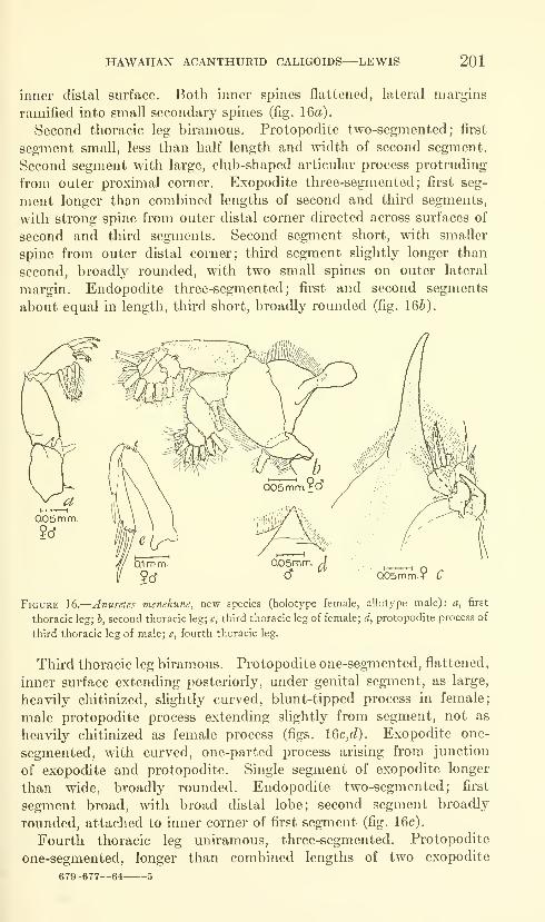
HAWAIIAN ACANTHURID CALIGOIDS—LEWIS 201
inner distal surface. Both inner spines flattened, lateral margins
ramified into small secondary spines (fig. 16a).
Second thoracic leg biramous. Protopodite two-segmented; first
segment small, less than half length and width of second segment.
Second segment with large, club-shaped articular process protruding
from outer proximal corner. Exopodite three-segmented; first seg-
ment longer than combined lengths of second and third segments,
with strong spine from outer distal corner directed across surfaces of
second and third segments. Second segment short, with smaller
spine from outer distal corner; third segment slightly longer than
second, broadly rounded, with two small spines on outer lateral
margin. Endopodite three-segmented; first and second segments
about equal in length, third short, broadly rounded (fig. IQb).
0.05mm9(5
Figure 16.
—
Anuretes menehune, new species (holotype female, allotype male): a, first
thoracic leg; b, second thoracic leg; c, third thoracic leg of female; d, protopodite process of
third thoracic leg of male; e, fourth thoracic leg.
Third thoracic leg biramous. Protopodite one-segmented, flattened,
inner surface extending posteriorly, under genital segment, as large,
heavily chitinized, slightly curved, blunt-tipped process in female;
male protopodite process extending slightly from segment, not as
heavily chitinized as female process (figs. lQc,d). Exopodite one-
segmented, with curved, one-parted process arising from junction
of exopodite and protopodite. Single segment of exopodite longer
than wide, broadly rounded. Endopodite two-segmented; first
segment broad, with broad distal lobe; second segment broadly
rounded, attached to inner corner of first segment (fig. 16c).
Fourth thoracic leg uniramous, three-segmented. Protopodite
one-segmented, longer than combined lengths of two exopodite
679-677—64 5
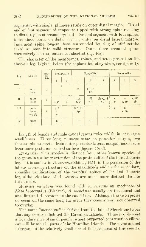
202 PROCEEDINGS OF THE NATIONAL MUSEUM
segments; with single, plumose setule on outer distal margin. Distal
end of first segment of exopodite tipped with strong spine reaching
to distal region of second segment. Second segment with four spines,
inner three borne on distal surface, outer on distal lateral margin.
Innermost spine longest, base surrounded by ring of stiff setules
fused at base into sohd structure. Outer three terminal spines
successively shorter, outermost shortest (fig. IGe).
The character of the membranes, spines, and setae present on the
thoracic legs is given below (for explanation of symbols, see figure 1):
Leg
HAWAIIAN ACANTHURID CALIGOIDS—LEWIS 203
Genus Dentigryps Wilson
Diagnosis.—Fourth thoracic leg-bearing segment and genital seg-
ment fused in female, covered by single, dorsal plate. Lunules absent,
sternal furca present; fifth legs of female elongate, conical, spinelike
processes protruding from genital segment.
Remarks.—The genus was established by Wilson (1913) on the
characteristics presented by the female of D. Curtus Wilson, 1913, the
type-species of the genus. With the description of the new species
below, the second species known for the genus, the diagnosis has been
modified slightly.
Dentigryps bifurcatiis, new species
Figures 17a-c,e-k,m-q, 18a-c,€,f
Material.—Nonovigerous adult female (holotype, USNM 105094)
taken by author from side of specimen of Acanthurus olivaceous Bloch
and Schneider given to author by Mr. Spencer Tinker from collections
of Honolulu Aquarium. Mature male (allotype, USNM 105095) col-
lected by author from caudal fin of specimen of Naso hexacanthus
(Bleeker) captured in fish trap between Diamond Head and KokoHead, Oahu, Hawaii, by Samuel Kaolulo. Mature female (paratype,
USNM 105096) collected by Dr. John Randall from epidermis of
specimen of Acanthurus triostegus sandvicensis Streets captured in
unknown locality on Oahu, Hawaii.
Measurements.-—Holotype female: Length from anterior end to
posterior end of caudal rami, excluding setae, 3.50 mm. Greatest
length of cephalothorax, including frontal region, 2.44 mm,; greatest
width, excluding marginal flanges, 2.30 mm. Greatest length of fused
fourth thoracic leg-bearing segment and genital segment 0.70 mm.;greatest width of genital segment 1.19 mm.; length of left genital seg-
ment spine 0.78 mm. Tip of right spine broken.
Allotype male: Length from anterior end to posterior end of caudal
rami, excluding setae, 1.97 mm. Greatest length of cephalothorax,
including frontal region, 1.48 mm.; greatest width, excluding marginal
flanges, 1.48 mm. Greatest length of fourth thoracic leg-bearing
segment and genital segment, excluding sixth leg projections, 0.50 mm.(measurement taken on ventral surface); greatest width of genital
segment 0.39 mm. Length of projecting fifth legs, excluding setae,
0.18 mm.Paratype female: Length from anterior end to posterior end of
caudal rami, excluding setae, 2.85 mm. Greatest length of cephalo-
thorax, including frontal region, 2.07 mm.; greatest width, excluding
marginal flanges, 1.93 mm. Greatest length of fused fourth thoracic

204 PROCEEDINGS OF THE NATIONAL MUSEUM vol. 115
leg-bearing segment and genital segment 0.59 mm.; greatest width of
genital segment 1.07 mm.; length of genital segment spines 0.66 mm.Description.-—^Body of female with large, yellowish blotches in
irregular pattern in alcohol ; eye spots yellowish orange. Body of male
yellowish in alcohol, with red spots scattered over surface; eyes as in
female,
Cephalothorax of holotype female specimen ovoid, frontal region
small, with thin, membranous flange along anterior margin; frontal
region separated from cephalothorax by distinct, trilobed groove,
median lobe much larger than lateral lobes. Lateral margins of
cephalothorax with narrow, membranous flange. Posterior sinuses
distinct, U-shaped, with two small, membranous flaps attached along
lateral and anterior margins, one extending ventrally, other dorsally.
Inner margin of posterior sinuses continuous with posterior lateral and
posterior margins of median cephalothoracic region. Median cephalo-
thoracic region extending slightly posterior to posterior lateral exten-
sions of lateral cephalothoracic regions; middle of posterior margin
slightly concave. Dorsal grooves of cephalothorax distinct, H -shaped
;
anterior portion of grooves terminating blindly posterior to eyes,
cross groove, in middle of cephalothorax, curving anteriorly, not as
distinct as longitudinal grooves. Lateral strengthening regions
extending from just anterior to junction of cross and longitudinal
grooves laterally and posteriorly, terminating just inside lateral
margins of cephalothorax. Eyes small, situated in anterior third of
cephalothorax, inner margins touching on median longitudinal axis
of body (fig. 17a).
Fourth thoracic leg-bearing segment and genital segment fused in
female, covered by single, dorsal plate. Fourth thoracic leg-bearing
segment more distinct than in D. curtus, genital segment not be-
ginning under posterior portion of cephalothorax as in D. curtus but
slightly behind it. Genital segment expanded, lateral areas broadly
rounded, middle of posterior margin concave, not convex as in D.
curtus, posterior lateral margins without single, small, pointed pro-
trusion present in D. curtus (figs. 17c,d). Fifth legs visible as long,
acuminate, unjointed, spinelike projections from ventral posterior
lateral surface of genital segment. Spinelike projection with three
small, plumose setules, one on proximal lateral surface, one on ventral
medial surface, one on ventral distal surface (figs. 17c,d).
Abdomen of female one-segmented, attached to posterior surface
of genital segment, not ventral surface as in D. curtus. Width of
abdomen approximately same as length. Abdomen without pos-
terior lateral extensions found in D. curtus (figs. 17c,d).
Cephalothorax of allotype male similar to that of female except
frontal region with flatter anterior margin. Region of third thoracic

HAWAIIAN ACANTHtJRID CALlGOIDS—LEWIS 205
01 mm. dFigure 17.
—
Dentigryps bifurcatus, new species (holotype female, allotype male): a, dorsalview of female; b, dorsal view of male; c, ventral view of posterior end of genital segment,fifth leg, abdomen, and caudal ramus of female; (J, ventral view of posterior end of genitalsegment, fifth leg, abdomen, and caudal ramus of female specimen of Z). curtus); e, ventralview of posterior end of genital segment, fifth and sixth legs, abdomen, and caudal ramusof male;/, antennule; g, female second antenna; h, male second antenna; i, mandible;;,postantennal process; k, postoral process of female; (/, postoral process of female specimenof D. curtus); m, postoral process of male; n, maxilla; o, maxilliped of female; p, maxilllpedof male; q, sternal furca.
206 PROCEEDINGS OF THE NATIONAL MUSEUM vol. ii5
leg-bearing segment of thorax, contained in cephalothorax, extended
over fourth thoracic leg-bearing segment. Fourth thoracic leg-
bearing segment partially visible dorsally, separated from genital
segment by distinct line of division; fourth thoracic leg-bearing seg-
ment and genital segment not covered by single, dorsal plate as in
female. Fifth legs visible as conical projections of lateral margins of
genital segment, projections appearing as series of three overlapping,
lappet-like processes, each bearing single, terminal, plumose seta.
Sixth legs visible as conical projections of posterior lateral margins of
genital segment, lappet-like processes visible on fifth legs also present
on sixth though not as distinct; sixth legs with three terminal, plu-
mose setules (figs. 17b,e).
Antennule of male and female two-segmented, excluding frontal
plate, attached to ventral surface of both cephalothorax and frontal
region just medial to lateral margins of frontal region. First seg-
ment broad proximally, tapered on anterior margin to narrow distal
end; greatest length one and one-fourth times greatest width. An-
terior margin of first segment with fifteeen plumose setae. Second
segment slightly more than half length of first, with naked seta on
middle of posterior margin and 13 naked setae from rounded distal
end (fig. 17/).
Second antenna of holotype female three-segmented, attached
posterior and medial to antennule base. First segment short, broad,
forming articular surface for second segment; with triangular, pos-
teriorly directed process. Second segment well developed, greatest
width equal to greatest length, with small adhesion pad on inner distal
surface. Second segment with several small, heavily chitinized de-
pressions on distal margin serving as articular surfaces for third seg-
ment. Third segment with terminal, clawlike process; division
between segment and process indistinct. Terminal process curved
sharply inwards distally. Length of third segment and terminal
process greater than that of second segment; third segment without
accessory processes (fig. 17^).
Second antenna of allotype male three-segmented; first segment
long, narrow proximally, expanded medially, tapered distally; distal
end with projecting, lobate articular process, without posteriorly
projecting process of female. First segment with adhesion surface on
outer distal half of segment. Second segment strongly developed,
swollen proximally, narrow distally. Inner surface of second seg-
ment forming adhesion surface proximally; with small, swollen ad-
hesion surface projecting from inner distal surface; both adhesion sur-
faces on inner surface connected by irregular ridges. Third segment
short, with compound terminal process consisting of simple, curved,
spinelike structure with bifurcate process fused to anterior lateral
HAWAUAN ACANTHURID CALIGOIDS—LEWIS 207
surface of terminal process. Third segment with single, spinelike
accessory process at indistinct junction of terminal process and seg-
ment (fig. 17h).
Mandible of female and male four-parted. First part with broad
base, tapered regularly to slightly narrower distal end; second part
short, distinctly tapered to junction of third part. Third part elon-
gate, length slightly less than combined lengths of first two parts;
fourth part slightly more than half length of third, curving inward,
bearing 11 toothlike denticulations on inner margin (fig. 17i).
Postantennal process of female and male simple, spinelike process
attached just lateral, and slightly posterior to, base of second antenna.
Spinelike process curved posteriorly, terminating in blunt tip, bear-
ing two minute nodules on inner proximal surface, proximal nodule
with two long setules, distal with three (fig. 17j).
Postoral process of paratype female specimen bifurcate, attached
just lateral to base of mouth cone (process broken in holotype).
Postoral process of D. curtus simple, spinelike process, not bifurcate
as in D. hijurcatus (fig. 17Z). Distance across base of bifurcate
process greater than distance between tips, length of tines approxi-
mately one-third length of process; single, slight protrusion pres-
ent just proximal to apex of bifurcation. Single, lobate process
present just anterior to base of spine, with three setules from distal
surface (fig. 17k).
Postoral process of male similar to that of female except with large,
finger-like protrusion on inner margin of bifurcate process (fig. 17m).
Maxilla of female and male two-segmented, attached slightly lat-
eral and posterior to postoral process. Proximal segment strongly
developed, broader at base than at distal end, with articular process
forming extension of inner proximal surface. Distal end of first seg-
ment flat. Second segment elongate, greatest length about one andone-fourth times that of first segment ; inner proximal surface irregular,
proximal end articulating with flat distal end of first segment. Prox-
imal half of second segment slightly swollen, both margins convex,
with fine, membranous flap on inner margin. Spine with attached
membrane present on middle of inner margin of segment. Second
segment terminated by two curved, spinelike processes, inner almost
twice length of outer, with fine membrane along inner margin. Outer
spinelike process on distal end of second segment with frilled mem-brane along outer margin (fig. 17n).
Maxilliped of female two-segmented, attached slightly posterior and
medial to maxilla. First segment well developed, with long articular
process extending from outer proximal portion of segment. Distal
margin with several heavily chitinized surfaces serving as articular
surfaces for second segment. Second segment short, forming, with
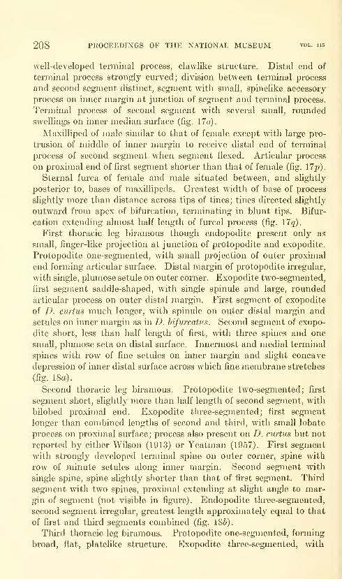
208 PROCEEDINGS OF THE NATIONAL MUSEUM vol. 115
well-developed terminal process, clawlike structure. Distal end of
terminal process strongly cm'ved; division between terminal process
and second segment distinct, segment with small, spinelike accessory
process on inner margin at junction of segment and terminal process.
Terminal process of second segment with several small, rounded
swellings on inner median surface (fig. 17o).
Maxilliped of male similar to that of female except with large pro-
trusion of middle of inner margin to receive distal end of terminal
process of second segment when segment flexed. Ai'ticular process
on proximal end of first segment shorter than that of female (fig. lip).
Sternal furca of female and male situated between, and slightly
posterior to, bases of maxillipeds. Greatest width of base of process
slightly more than distance across tips of tines; tines directed slightly
outward from apex of bifurcation, terminating in blunt tips. Bifur-
cation extending almost half length of furcal process (fig. 17q).
First thoracic leg biramous though endopodite present only as
small, finger-like projection at junction of protopodite and exopodite.
Protopodite one-segmented, with small projection of outer proximal
end forming articular surface. Distal margin of protopodite irregular,
with single, plumose setule on outer corner. Exopodite two-segmented,
first segment saddle-shaped, with single spinule and large, rounded
articular process on outer distal margin. First segment of exopodite
of D. curtus much longer, with spinule on outer distal margin and
setules on inner margin as in D. hijurcatus. Second segment of exopo-
dite short, less than half length of first, with three spines and one
small, plumose seta on distal surface. Innermost and medial terminal
spines with row of fine setules on inner margin and slight concave
depression of inner distal surface across which fine membrane stretches
(fig. 18a).
Second thoracic leg biramous. Protopodite two-segmented; first
segment short, slightly more than half length of second segment, with
bilobed proximal end. Exopodite three-segmented; first segment
longer than combined lengths of second and third, with small lobate
process on proximal surface; process also present on D. curtus but not
reported by either Wilson (1913) or Yeatman (1957). First segment
with strongly developed terminal spine on outer corner, spine with
row of minute setules along inner margin. Second segment with
single spine, spine slightly shorter than that of first segment. Third
segment with two spines, proximal extending at slight angle to mar-
gin of segment (not visible in figure). Endopodite three-segmented,
second segment irregular, greatest length approximately equal to that
of first and third segments combined (fig. 186).
Thu'd thoracic leg biramous. Protopodite one-segmented, forming
broad, flat, platelike structure. Exopodite three-segmented, with
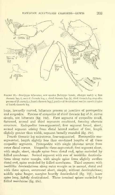
HAWAIIAN ACANTHURID CALIGOIDS—LEWIS 209
005 mm9'
Figure 18.
—
Denligryps bifurcatus, new species (holotype female, allotype male): a, first
thoracic leg; b, second thoracic leg; c, third thoracic leg; {d, third thoracic leg exopodite
process of D. curius);e, fourth thoracic leg;/, portion of denticulated median terminal spine
of fourth thoracic leg.
large, inwardly curved, bifurcate process at junction of protopodite
and exopodite. Process of exopodite of third thoracic leg of D. curtus
simple, not bifurcate (fig. 18c/). First segment of exopodite small,
flattened, second and third segments combined, forming obovate
structure. Endopodite two-segmented; first segment broad, short;
second segment arising from distal lateral surface of first, length
slightly greater than width, segment broadly rounded (fig. 18c).
Fourth thoracic leg uniramous, four-segmented. Protopodite one-
segmented, length slightly less than combined lengths of all three
exopodite segments. Protopodite with single plmnose setule from
outer distal corner. Exopodite three-segmented; first segment short,
with single, short, simple spine from distal end, spine encircled byfrilled membrane. Second segment with row of toothlike denticula-
tions along outer margin, with simple spine from slightly swollen
distal end, spine encircled by frilled membrane. Third segment with
toothlike denticulations along outer margin as in second, distal end
with three spines, outermost short, simple, without denticulations;
middle spine longer, margins heavily denticulated (fig. 18/); inner
spine long, lightly denticulated. Three terminal spines encircled byfrilled membrane (fig. 18e).
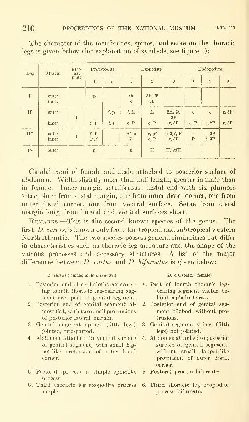
210 PROCEEDINGS OF THE NATIONAL MUSEUM
The character of the membranes, spines, and setae on the thoracic
legs is given below (for explanation of symbols, see figm-e 1):
Leg
HAWAIIAN ACANTHURID CALIGOIDS—LEWIS 211
The name "bifurcatus" applies to the bifurcate condition of the
postoral process and of the third thoracic leg exopodite process.
Family Pandaridae
Genus Nesippus Heller, 1865
Diagnosis.—Cephalothorax consisting of cephalon, maxilliped, and
first thoracic leg-bearing segments; second, third, and fourth thoracic
leg-bearing segments free. Cephalothorax typically longer than wide,
with posteriorly projecting lateral regions. Male with or without
conspicilla placed slightly anterior and lateral to paired eyes. Sec-
ond thoracic leg-bearing segment with lateral and posterior lateral
projecting lobes. Abdomen small, one-segmented. Distal region of
maxillipeds swollen, terminal process knoblike, forceps-like or
flattened and clawlike. All thoracic legs biramous; rami of legs 1-3
two-segmented, fourth leg rami one-segmented.
Remarks.—The diagnosis is mainly from Wilson (1907) with modi-
fication based on the characteristics of the species described subse-
quent to his publication. The adult female and male of this genus
are found primarily on elasmobranch hosts, but the adult male also
is taken in the plankton.
The function of the "conspicilla," Dana's term for the two trans-
parent projections on the dorsal surface in the region of the eyes of
some males, is not known. Dana (1853) indicates that they are dis-
tinct from the cephalothorax and are of a "brittle, though rather hard"
nature.
Nesippus costatus? Wilson
Figures 19a-e, 20a-s, 21a-p
N. costatus Wilson, 1924, pp. 213-214, pi. 20.
Type host.—"A nine-foot shark."
Type locality.—Galapagos Islands.
Material.—Immature male taken by author from cyst on dorsal
fin of specimen of Acanthurus triostegus sandvicensis Streets captured
by U.S. Fish and Wildlife Service, Bureau of Commercial Fisheries,
at Midway Island (USNM 105176). Immature male taken by author
from cyst on anal fin of specimen of Acanthurus nigroris Cuvier and
Valenciennes captured in fish trap in Kaneohe Bay, Oahu, Hawaii,
by Lester Zukeran (USNM 105177). Immature male from cyst on
anal fin of specimen of Acanthurus xanthopterus Cuvier and Valen-
ciennes captured in fish trap in Kaneohe Bay, Oahu, Hawaii, by
Lester Zukeran (USNM 105178). Male specimen collected by Dr.
John Randall from epidermis of specimen of Acanthurus triostegus
sandvicensis Streets from an unknown locality, probably on Oahu,

212 PROCEEDINGS OF THE NATIONAL MUSEUM vol. 115
Hawaii (USNM 105179). Immature male and its exuviiim taken
by Carolyn Lewis from cyst on pectoral fin of specimen of Diodonholocanthus Linnaeus donated by Honolulu Aquarium (USNM105180). Immature male taken by author from cyst on pectoral fin
of specimen of Diodon holocanthus Linnaeus donated by Honolulu
Aquarium (USNM 105181). One cyst containing immature male
taken by author from pectoral fin of specimen of Ctenochaetus strigosus
(Bennett) donated by Honolulu Aquarium (USNM 105182). Im-mature male taken by author from cyst on dorsal fin of specimen of
Acanthurus triostegus sandvicensis Streets speared by N. Ferris in
Kaneohe Bay, Oahu, Hawaii (USNM 105183). Immature male taken
by author from cyst on dorsal fin of specimen of Scarus species speared
by author in Hanauma Bay, Oahu, Hawaii (USNM 105184).
Additional specimens, not used in the following description, were
taken from cysts on fins of specimens of the following families of
fishes: Labridae, Pomacentridae, Zanclidae.
Measurements.—Early encysted stage (two specimens) : Length
from anterior end of body to posterior end of caudal rami, excluding
setae, 1.90 and 2.58 mm. Greatest length of cephalothorax, including
frontal region, 0.82 and 1.23 mm.; greatest width 1.03 and 1.31 mm.Greatest length of free thoracic leg-bearing segments, genital segment,
abdomen, and caudal rami, excluding setae, 1.11 and 1.60 mm. Great-
est length of genital segment 0.18 and 0.34 mm.; greatest width of
genital segment 0.36 and 0.42 mm. Greatest length of abdomen 0.13
and 0.17 mm. Greatest length of caudal rami, excluding setae, 0.25
mm.Late encysted and recently excysted stage (five encysted specimens,
one recently excysted specimen) : Average length from anterior end
of body to posterior end of caudal rami, excluding setae, 3.40 mm.;range 2.94-4.05 mm. Average of greatest length of cephalothorax,
including frontal region, 2.02 mm.; range 1.84-2.12 mm.; average of
greatest width 2.02 mm.; range 1.66-2.21 mm. Average of greatest
length of free thoracic leg-bearing segments, genital segment, abdomen,
and caudal rami, excluding setae, 1.93 mm.; range 1.47-2.30 mm.Average of greatest length of genital segment 0.49 mm.; range 0.42-
0.59 mm.; average of greatest width 0.73 mm.; range 0.64-0.82 mm.Average of greatest length of abdomen and caudal rami, excluding
setae, 0.24 mm.; range 0.15-0.29 mm. Average of greatest length of
abdomen 0.21 mm.; range 0.17-0.27 mm. Average of greatest length
of caudal rami, excluding setae, 0.18 mm.; range 0.15-0.21 mm.Description.—Color of all stages light brown to brownish yellow
in life, with no distinct color pattern. Conspicilla transparent, dis-
tinct on dorsal surface. Eyes reddish brown. Single excysted speci-
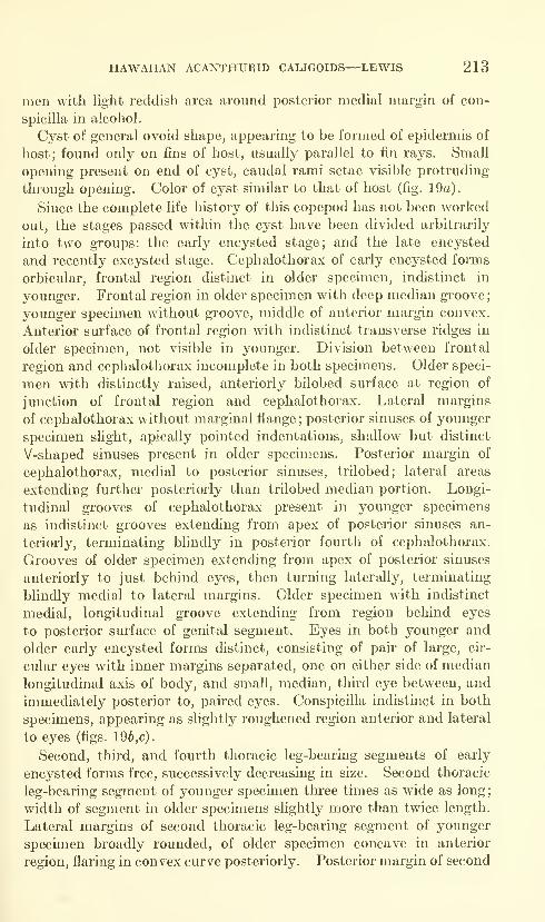
HAWAIIAN ACANTHURID CALIGOIDS—LEWIS 213
men with light reddish area around posterior medial margin of con-
spicilla in alcohol.
Cyst of general ovoid shape, appearing to be formed of epidermis of
host; found only on fins of host, usually parallel to fin rays. Small
opening present on end of cyst, caudal rami setae visible protruding
through opening. Color of cyst similar to that of host (fig. 19a).
Since the complete life history of this copepod has not been worked
out, the stages passed within the cyst have been divided arbitrarily
into two groups: the early encysted stage; and the late encysted
and recently excysted stage. Cephalothorax of early encysted forms
orbicular, frontal region distinct in older specimen, indistinct in
younger. Frontal region in older specimen with deep median groove
;
younger specimen without groove, middle of anterior margin convex.
Anterior surface of frontal region with indistinct transverse ridges in
older specimen, not visible in younger. Division between frontal
region and cephalothorax incomplete in both specimens. Older speci-
men with distinctly raised, anteriorly bilobed surface at region of
junction of frontal region and cephalothorax. Lateral margins
of cephalothorax without marginal flange;posterior sinuses of younger
specimen slight, apically pointed indentations, shallow but distinct
V-shaped sinuses present in older specimens. Posterior margin of
cephalothorax, medial to posterior sinuses, trilobed; lateral areas
extending further posteriorly than trilobed median portion. Longi-
tudinal grooves of cephalothorax present in younger specimens
as indistinct grooves extending from apex of posterior sinuses an-
teriorly, terminating blindly in posterior fourth of cephalothorax.
Grooves of older specimen extending from apex of posterior sinuses
anteriorly to just behind eyes, then turning laterally, terminating
blindly medial to lateral margins. Older specimen with indistinct
medial, longitudinal groove extending from region behind eyes
to posterior surface of genital segment. Eyes in both younger and
older early encysted forms distinct, consisting of pair of large, cir-
cular eyes with inner margins separated, one on either side of median
longitudinal axis of body, and small, median, thu'd eye between, and
immediately posterior to, paked eyes. Conspicilla indistinct in both
specimens, appearing as slightly roughened region anterior and lateral
to eyes (figs. 19b,c).
Second, third, and fourth thoracic leg-bearing segments of early
encysted forms free, successively decreasing in size. Second thoracic
leg-bearing segment of younger specimen three times as wide as long;
width of segment in older specimens slightly more than twice length.
Lateral margins of second thoracic leg-bearing segment of younger
specimen broadly rounded, of older specunen concave in anterior
region, flaring in convex curve posteriorly. Posterior margin of second
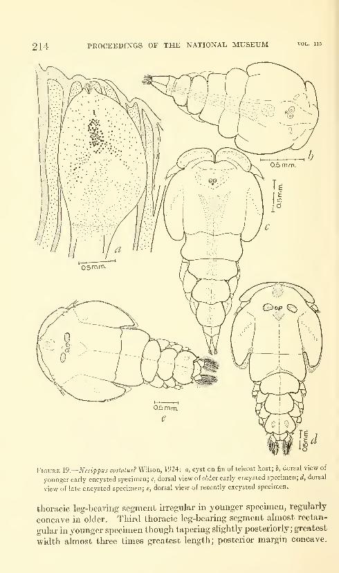
214 PROCEEDINGS OF THE NATIONAL MUSEUM vol. 115
Figure \9.—Nesippus costalus? Wilson, 1924: a, cyst on fin of teleost host; b, dorsal view of
younger early encysted specimen; c, dorsal view of older early encysted specimen; d, dorsal
view of late encysted specimen; e, dorsal view of recently excysted specimen.
thoracic leg-bearing segment irregular in younger specimen, regularly
concave in older. Third thoracic leg-bearing segment almost rectan-
gular in younger specimen though tapering slightly posteriorly;greatest
width almost three times greatest length; posterior margin concave.
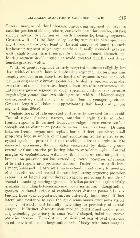
HAWAnAN ACANTHURID CALIGOIDS—LEWIS 215
Lateral margins of thii'd thoracic leg-bearing segment concave in
anterior portion of older specimen, convex in posterior portion, curving
sharply inward to junction of fourth thoracic leg-bearing segment.
Greatest width of third thoracic leg-bearing segment in older specimenslightly more than twice length. Lateral margins of fourth thoracic
leg-bearing segment of younger specimen broadly rounded, greatest
width slightly less than twice greatest length. Fourth thoracic leg-
bearing segment in older specimen ovoid, greatest length about tlu-ee-
fourths greatest width.
Width of genital segment in early encysted specimens slightly less
than width of fourth thoracic leg-bearing segment. Lateral marginsbroadly rounded in anterior three-fourths of segment in younger speci-
men, curving sharply inward posteriorly to slight projection of medialtwo-thirds of segment; greatest length about two-thirds greatest width.
Lateral margins of segment in older specimen flatly convex, greatest
length slightly more than two-thirds greatest width. Abdomen short,
one-segmented; slightly longer in older than in younger specimen.
Greatest length of abdomen approxmiately half length of genital
segment (figs. 20a,b).
Cephalothorax of late encysted and recently excysted forms ovoid.
Frontal region distinct, narrow, anterior margin flatly rounded;
frontal region with distinct transverse ridges across entire surface
though not as distinct on posterior as on anterior surface. Division
between frontal region and cephalothorax distinct, complete; small
projecting lobe in middle of margin separating frontal plates in ex-
cysted specimen present but not reaching anterior margin in late
encysted specimens, though plates separated by distinct groove
extending from anterior projecting lobe to anterior margin. Lateral
margins of cephalothorax with very thin flange on anterior portion,
broader on posterior portion, extending around posterior extensions
of lateral regions into posterior sinuses. Posterior sinuses distinct,
slender, V-shaped. Posterior sinuses extending anteriorly to junction
of cephalothorax and second thoracic leg-bearing segment; posterior
extensions of lateral cephalothoracic regions projecting to middle of
second thoracic leg-bearing segment. Median posterior marginslightly
irregular, extending between apices of posterior sinuses. Longitudinal
grooves on dorsal surface of cephalothorax distinct posteriorly, ex-
tending from apex of posterior sinuses anteriorly, becoming indistinct
lateral and posterior to eyes though discontinuous extensions visible
curving anteriorly and laterally, extending to proximity of lateral
margin. Indistinct, discontinuous median longitudinal groove pres-
ent, extending posteriorly to anus from V-shaped, indistinct groove
posterior to eyes. Eyes distinct, consisting of pair of oval eyes, oneon either side of median longitudinal axis of body, with inner margins
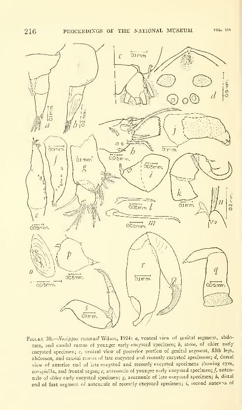
216 PROCEEDINGS OF THE NATIONAL MUSEUM vol. 115
Figure 20.—Nesippus costatus? Wilson, 1924: a, ventral view of genital segment, abdo-
men, and caudal ramus of younger early encysted specimen; b, same, of older early
encysted specimen; c, ventral view of posterior portion of genital segment, fifth legs,
abdomen, and caudal ramus of late encysted and recently excysted specimens; d, dorsal
view of anterior end of late encysted and recently excysted specimens showing eyes,
conspicilla, and frontal organ; e, antennule of younger early encysted specimen;/, anten-
nule of older early encysted specimen; g, antennule of late encysted specimen; h, distal
end of first segment of antennule of recently excysted specimen; i, second antenna of

HAWAIIAN ACANTHURID CALIGOIDS—LEWIS 217
separated; third, median eye present between paired eyes (fig. 20d).
Conspicilla large, distinct, oblong; projecting from dorsal surface of
cepbalothorax just anterior and lateral to paired eyes. Surface of
conspicilla ridged, similar in appearance to adhesion pads of manypandarids (fig. 19d).
Second thoracic leg-bearing segment of late encysted and recently
excysted specimens larger than third or fourth thoracic leg-bearing
segment; lateral margins almost parallel, anterior half forming inner
margin of posterior sinuses, posterior lateral margins curving smoothly
inward, posterior lateral surfaces projecting slightly past anterior end
of third thoracic leg-bearing segment. Posterior margin of second
thoracic leg-bearing segment concave; greatest length of segmentslightly more than half greatest width. Third thoracic leg-bearing
segment with broadly rounded lateral margins, greatest length slightly
more than half greatest width in encysted forms, slightly less in
excysted form. Posterior lateral margins of third thoracic leg-bearing
segment forming small, posteriorly projecting lobes in encysted form,
middle of posterior margin slightly convex; posterior margin of
recently excysted form evenly cm-ved except for slight concavity in
middle. Fourth thoracic leg-bearing segment narrower than third,
greatest length approximately two-thirds greatest width; lateral mar-gins evenly curved. Posterior lateral corners of fourth thoracic leg-
bearing segment of excysted form forming small, posteriorly projecting
lobes, encysted form without lobes.
Genital segment of late encysted and recently excysted forms of
general ovoid shape, slightly wider than fom'th thoracic leg-bearing
segment, greatest length approximately two-thirds greatest width;
internal spermatophores indistinctly visible in encysted, distinctly
visible in excysted specimens. Lateral margins broadly curved;
posterior lateral surfaces of genital segment forming smaU, posteriorly
projecting lobes with fifth legs projecting from apex as single plumose
seta arising from ventral sm-face. Middle of posterior margin convex,
projecting slightly over anterior surface of abdomen. Abdomen short,
one-segmented, greatest length slightly less than greatest width;
widest point in anterior median region of segment, tapered to narrower
anterior and posterior ends of segment. Posterior margin of abdomenindented at anus (fig. 20c).
younger early encysted specimen; /, second antenna of older early encysted specimen;
k, second antenna of late encysted and recently excysted specimens; /, mandible of early
encysted specimens; m, mandible of late encysted and recently excysted specimens;
n, adhesion pad on late encysted and recently excysted specimens (fr= frontal region);
0, postoral process of early encysted specimens; p, postoral process of late encysted and
recently excysted specimens; q, maxilla of early encysted specimens; r, maxilla of late
encysted specimen; j-, maxilla of recently excysted specimen.
679-677—64 6
218 PROCEEDINGS OF THE NATIONAL MUSEUM vol. 115
Antennule of early encysted forms one-segmented, attached to
anterior ventral surface in frontal region. Single segment irregular
in shape, flaccid, tapered to round tip bearing six small, plumose setae.
Middle of antennule with irregular row of six setae, each with several
setules. Two segments of late encysted antennule visible inside early
encysted antennule (figs. 20e,f).
Antennule of late encysted and recently excysted forms two-
segmented. First segment two and a half times length of second in
late encysted specmiens, approxunately two times length of second
segment in recently excysted specimen. Lateral margins almost
parallel in proximal region, with knoblike convexity of anterior sur-
face in distal region. First segment with eight short, plumose setae,
one long plumose seta, two large and two small lobate protrusions,
all from knoblike convexity (fig. 20h).
Second segment of excysted specimen slightly longer than that of
encysted, both with two plmnose setae on distal portion of posterior
margin, three plumose setae from posterior distal smiace, and six
naked setae from anterior distal surface. Setae and lobes on
excysted specimen developed to slightly greater extent than on
encysted specimens (figs. 20g,h).
Second antenna of early encysted specmiens two-segmented,
attached just posterior to base of antennule. Second antenna con-
sisting of large, conical first segment and small, irregular second
segment. First segment of older specimen more slender than that of
younger, with indication of segmentation visible inside appendage but
not externally; both older and younger specimens with small, tubercu-
lar projection from posterior distal surface. Distal segment of older
specimen recurved more than that of younger; younger specimen with
minute, lobate projection from inner distal surface of segment; older
specimen without lobate projection but with minute, irregular exten-
sion of distal surface (figs. 20i,j).
Second antenna of older encysted and recently excysted specimens
three-segmented, attached slightly posterior and medial to antennule
base. First segment short, broad, width and length about equal,
lateral margins irregular, with well-developed articular surface on
outer proximal edge articulating with concave depression of segment-
like extension of ventral surface of cephalothorax. Inner distal surface
of first segment projecting beyond remaining distal surface as broad
articular surface, fitting in heavily chitinized depression on outer
proximal surface of second segment. Second segment strongly devel-
oped, slightly longer than wide, longer than first segment; lateral
margins somewhat irregular, inner margin convex. Inner distal sur-
face of second segment projecting as rodlike structure, articulating
with knobhke process on third segment. Third segment and terminal
HAWAnAN ACANTHURID CALIGOIDS—LEWIS 219
process falciform, more so in excysted than in encysted specimens.
Length of third segment and terminal process slightly shorter than
combined lengths of first two segments. Third segment with two
spinelike accessory processes on inner surface, one on proximal, sec-
ond on distal portions of segment. Division between terminal process
and segment distinct (fig. 20k) .
Mandible of early encysted, late encysted and recently excysted
specimens four-parted. Mandible of late encysted and recently
excysted specimens slightly longer than that of early encysted. First
part of mandible of all specimens broader proximally than distally,
with even taper to junction of second part. Second part short,
slightly more than half length of first part in early encysted specimens,
less than half in late encysted and recently excysted specimens; with
little taper to junction of thu'd part. Third part elongate, almost
twice length of first part, tapered slightly in younger specimens, not
in older. Fourth part short, slightly longer than second part, curved
inward slightly at distal end, with distinct, toothlike denticulations
along inner margin of older specimens, denticulations not visible in
younger specimens (figs. 20l,m).
Postoral process of early encysted specimens mound-shaped swelling
adjacent to base of mouth cone, with two small, lobate protrusions on
anterior surface and single, small, apical projection bearing three
setules on posterior distal surface (fig. 20o)
.
Postoral process of late encysted and recently excysted specimens
bulbous protrusion adjacent to base of mouth cone. Distal surface of
process larger than proximal, with single spine projectingfrom posterior
distal surface, two setules from anterior proximal surface and single
knob from anterior distal surface, arising from slight depression in
surface. Wilson (1924) notes three setules instead of two from the
anterior proximal surface and does not mention the spine found on
the presently described specimens (fig. 20p).
Maxilla of early encysted forms two-segmented, both segments
flaccid. Fh-st segment with almost parallel lateral margins, no
articular surfaces visible. Second segment tapered to blunt tip in
younger specimen, rounded but sharper tip in older specimen; length
of segment about half that of first segment in younger specimen,
two-thirds that of first segment in older specimen. Distal end of
second segment in both specimens tipped by small, pointed protrusion;
younger specimen with additional, setule-like structure on outer
distal corner, structure not present in older specimen (fig. 20g).
Maxilla of late encysted specimens two-segmented. First segment
strongly developed, with long projection of middle of proximal surface
as articular surface. Lateral margins of first segment flatly convex;
middle of distal surface projecting as articular surface, middle of distal

220 PROCEEDINGS OF THE NATIONAL MUSEUM vol. 115
projectiou heavily chitmized, articulating with small concavity on
inner proximal surface of second segment. Second segment with
terminal process slightly shorter than first segment, including proximal
extension of first segment. Second segment tapered slightly to blunt
distal end, tipped by long, slightly curved, dactyliform process with
small-pointed protrusion from tip of process. Terminal process of
late encysted form sheathed with membranous structure covered
with minute, toothlike denticulations; recently excysted specimen
also with sheathed process but denticulations replaced by minute
tubercles. Distal end of segment, just proximal to base of terminal
process, with two accessory processes on anterior margin: first a short,
sharply tipped process; second membranous, irregularly shaped
process surrounded by tuft of setules (figs. 20r,s).
Maxilliped of early encysted form two-segmented, attached poste-
rior to base of maxilla. First segment in younger specimen narrower
in proximal than in distal regions, distal region overlapping proximal
region giving two-parted appearance to segment. Second segment
of younger specimen short, less than one-fom'th length of first segment
;
segment and terminal process continuous, terminal process flaccid,
forming slender projection of segment. First segment of older early
encysted specimen similar to that of younger but distal portion larger,
greatly swollen, with adhesion pad on inner distal sm*face. Second
segment of older specimen similar to that of younger though terminal
process short but distinct clawlike process fitting into adhesion pad
of first segment. Second segment of older specimen with accessory,
spinelike process from inner surface just proximal to base of terminal
process. Division between terminal process and segment in older
specunen distinct (figs. 21a, b).
Maxilliped of late encysted and recently excysted specimens similar
in general appearance to that of older early encysted specimen.
Adhesion pad on inner distal surface of fu'st segment with additional,
loioblike process extending from distal margin. Second segment
more strongly developed, heavily chitinized at articulation with first
segment, with two accessory processes instead of one found in older
early encysted specimen; terminal process not clawlike but rounded
protuberance similar to that of adhesion pad of first segment and
abutting with this process when second segment flexed (figs. 21c,d).
First thoracic leg of early encysted specimens biramous, both rami
dactyliform, without external evidence of segmentation. Both
protopodite and rami flaccid. Exopodite extending beyond distal
margin of endopodite, with three spines in younger specimen, one from
outer distal lateral surface, two from distal margin; four spines on
older specimen, three as in younger specimen, from distal margin,
fourth from inner distal lateral surface. Endopodite with three

HAWAIIAN ACANTHURID CALIGOIDS—LEWIS 221
minute, lobate projections from distal surface. Early encysted
specimens with indistinct evidence of two-segmented exopodite and
endopodite visible inside dactyliform rami (figs. 2le,J).
First thoracic leg of late encysted and recently excysted specimens
biramous. Protopodite irregular in outline, with two plumose setae,
one on either side of rami. Exopodite two-segmented; first segment
longer than second, lateral margins u'regularly convex, with single
spine from outer distal lateral corner. Second segment with narrow
proximal surface, wide distal surface; lateral and distal margins con-
tinuous. Endopodite two-segmented, both segments of approxi-
mately equal length. First segment slightly shorter than second,
with convex lateral margins in encysted specimens, of rectangular
shape in excysted specimen. Second segment with continuous distal
and outer lateral margins (fig. 21^).
Second thoracic leg of early encysted specimens biramous, rami
one-segmented. Both protopodite and rami flaccid; protopodite with
minute projections on inner surface of younger specimen, plumose
setule on older. Exopodite slightly longer than endopodite, tipped
with four minute, lobate projections, one from each distal lateral
surface, two from distal surface. Endopodite with single plumose
setule from middle of inner margin, four minute, sharp projections
from distal margin. Two-segmented exopodite and endopodite of
late encysted specimens visible inside rami (figs. 21h,i).
Second thoracic leg of late encysted and recently excysted specimens
biramous. Protopodite broad, length approximately three-fourths
width, with single naked setule on distal margin next to outer lateral
margin of exopodite and plumose seta on inner proximal margin.
Exopodite two-segmented; first segment longer than second, narrower
proximally than distally, outer surface with patch of short, hairlike
projections giving fuzzy appearance to region. First segment with
single spine on outer distal lateral margin. Second segment of ex-
opodite orbicular, margins continuous, with three small, membrane-margined spines on outer lateral margin. Endopodite two-segmented
;
first segment slightly more than half length of second, second segment
of general ovoid shape, lateral and distal margins distinct (fig. 21j).
Third thoracic legs of early encysted forms biramous, rami one-
segmented. Protopodite and rami flaccid; protopodite of older
specimen with single, plumose setule from inner proximal margin,
with small, knoblike projection outside exopodite of younger specimen.
Both rami of about equal length, exopodite of younger specimen
with three plumose setules from distal margin, one from inner lateral
margin and four minute, spinelike processes from distal outer margin.
Exopodite of older specimen with three plumose setules from distal
margin ; one plumose setule and one minute plumose setule from outer
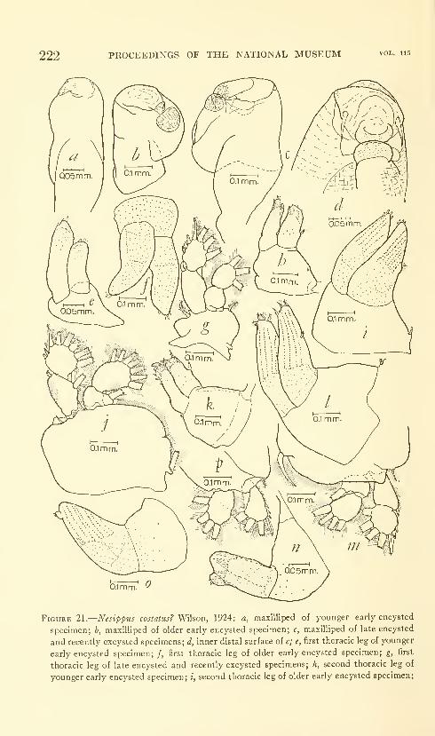
222 PROCEEDINGS OF THE NATIONAL MUSEUM vol. ii5
0.1mm
Figure 21.
—
Nesippus costatus? Wilson, 1924: a, maxilliped of younger early encysted
specimen; b, maxilliped of older early encysted specimen; c, maxilliped of late encysted
and recently excysted specimens; d, inner distal surface of c; e, first thoracic leg of younger
early encysted specimen; /, first thoracic leg of older early encysted specimen; g, first
thoracic leg of late encysted and recently excysted specimens; li, second thoracic leg of
younger early encysted specimen; i, second thoracic leg of older early encysted specimen;
HAWAIIAN ACANTHURID CALIGOIDS—LEWIS 223
distal margin. Endopodite of both specimens with three plumose
setules from distal margin, one plumose setule from middle of inner
margin. Segments of both rami of late encysted forms visible inside
rami of early encysted form (figs. 2\k,l).
Third thoracic leg of late encysted and recently excysted specimens
biramous. Protopodite broad, greatest length about three-fourths
greatest width, with single plumose seta on inner proximal margin,
single plumose setule just outside base of exopodite. Expodite two-
segmented; first segment longer than second, narrower proximally
than distally, with single spine from outer distal corner. Second
segment of exopodite broadly rounded, almost orbicular, lateral and
distal margins continuous; with two membrane-margined spines on
outer margin. Endopodite two-segmented; first segment shorter
than second, flared rapidly from narrow proximal to broad distal
surface. Second segment narrower proximally than distally, lateral
and distal margins distinct (fig. 21m).
Fourth thoracic leg of early encysted forms uniramous, two-
segmented. Protopodite one-segmented, slightly wider than long;
ramus and protopodite flaccid. Ramus with rounded protrusion from
distal portion of outer surface, larger in younger than in older speci-
men; two minute, pointed projections present on protrusion in younger
specimen, three in older. Both exopodite and endopodite of late-
encysted specimens visible inside single ramus of early encysted
specimens (figs. 2ln,o).
Fourth thoracic leg of late encysted and recently excysted forms
biramous, rami one-segmented. Protopodite longer than wide, outer
surface with distinct groove giving bilobed appearance, with single
plumose setule lateral to outer margin of exopodite. Exopodite
narrower at proximal than at distal end, with three spines on distal
portion of outer margin, each with light membranous margin. En-
dopodite oblong, with small but distinct indentation of inner margin,
indistinct, discontinuous groove visible extending part way across
segment from indentation (fig. 2\p).
j, second thoracic leg of late encysted and recently excysted specimens; k, third thoracic
leg of younger early encysted specimen; /, third thoracic leg of older early encysted
specimen; m, third thoracic leg of late encysted and recently excysted specimens; n,
fourth thoracic leg of younger early encysted specimen; o, fourth thoracic leg of older
early encysted specimen; p, fourth thoracic leg of late encysted and recently excysted
specimens.
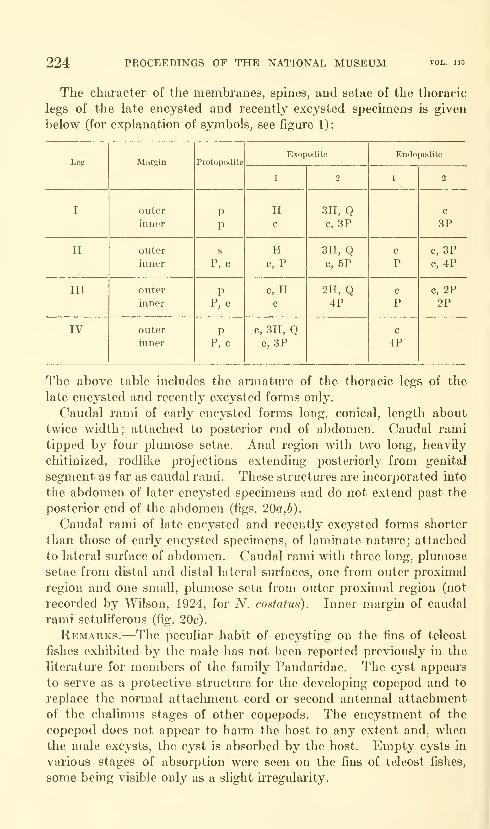
224 PROCEEDINGS OF THE NATIONAL MUSEUM
The character of the membranes, spines, and setae of the thoracic
legs of the late encysted and recently excysted specimens is given
below (for explanation of symbols, see figure 1):
Leg
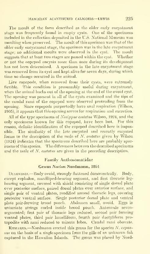
HAWAIIAN ACANTHURID CALIGOIDS—LEWIS 225
The moult of the form described as the older early encystment
stage was frequently found in empty cysts. One of the specimens
included in the collection deposited in the U.S. National Museum was
moulting when preserved. The moult of this specimen was that of the
older early encystment stage, the specimen was in the late encystment
stage; no additional moults were observed in the cyst. The moult
suggests that at least two stages are passed within the cyst. Whether
or not the copepod encysts more than once during its development
has not been determined. A specimen in the late encystment stage
was removed from its cyst and kept alive for seven days, during which
time no change occurred in the animal.
Live copepods, when removed from their cysts, were extremely
flexible. This condition is presumably useful during excystment,
when the animal backs out of the opening at the end of the ovoid cyst.
The opening was present in all of the cysts examined and the tips of
the caudal rami of the copepod were observed protruding from the
opening. Since copepods purportedly have anal respiration (Wilson,
1905), it appears that this opening serves for respiration and excretion.
All of the type specimens of Nesippus costatus Wilson, 1924, and the
only specimens known for this copepod, have been lost. For this
reason, definite identification of the copepod described here is impos-
sible. The similarity of the late encysted and recently excysted
forms to the description of the male of A'', costatus given by Wilson
(1924) indicates that the specimens described here are probably spec-
imens of this species. The differences between the described specimens
and the male of A^. costatus are given in the preceding description.
Family Anthosomatidae
Genus Norion Nordmann, 1864
Diagnosis.—Body ovoid, strongly flattened dorsoventrally. Body,
except cephalon, maxilliped-bearing segment, and first thoracic leg-
bearing segment, covered with shield consisting of single dorsal plate
over posterior surface, pah-ed dorsal plates over anterior surface, and
single pair of ventral plates, modified second thoracic legs, covering
posterior ventral surface. Single posterior dorsal plate and ventral
plate pair-forming brood pouch. Abdomen small, ovoid. Eggs in
uniseriate strings curled inside brood pouch. Antennule seven-
segmented; first pair of thoracic legs reduced, second pair forming
ventral plates, third pair lamelliform, fourth pair dactyliform pro-
topodite with rami reduced to minute lobes. Caudal rami obovate.
Remarks.—Nordmann erected this genus for the species A^. exjMii-
sus on the basis of a single specimen from the gills of an unknown fish
captured in the Hawaiian Islands. The genus was placed by Nord-
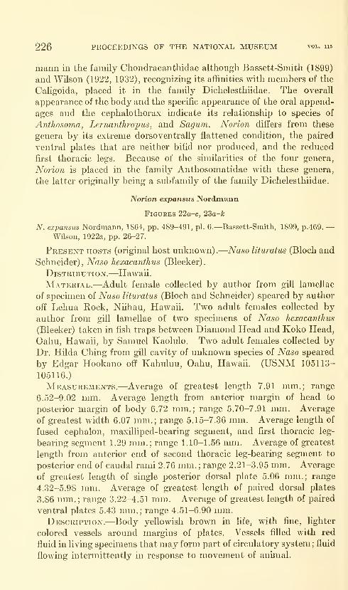
226 PROCEEDINGS OF THE NATIONAL MUSEUM vol. 115
mann in the family Chondracanthidae although Bassett-Smith (1899)
and Wilson (1922, 1932), recognizing its affinities with members of the
Caligoida, placed it in the family Dichelesthiidae. The overall
appearance of the body and the specific appearance of the oral append-
ages and the cephalothorax indicate its relationship to species of
Anthosoma, Lernanthropus, and Sagum. Norion differs from these
genera by its extreme dorsoventrally flattened condition, the paired
ventral plates that are neither bifid nor produced, and the reduced
first thoracic legs. Because of the similarities of the four genera,
Norion is placed in the family Anthosomatidae with these genera,
the latter originally being a subfamily of the family Dichelesthiidae.
Norion expansus Nordmann
FiGtTRES 22a-c, 23a-k
N. expansus Nordmann, 1864, pp. 489-491, pi. 6.—Bassett-Smith, 1899, p.469. —Wilson, 1922a, pp. 26-27.
Present hosts (original host unknown).
—
Naso lituratus (Bloch and
Schneider), Ndso hexacanthus (Bleeker).
Distribution.—Hawaii.
Material.—Adult female collected by author from gill lamellae
of specimen of Naso lituratus (Bloch and Schneider) speared by author
off Lehua Rock, Niihau, Hawaii. Two adult females collected byauthor from gill lamellae of two specimens of Naso hexacanthus
(Bleeker) taken in fish traps between Diamond Head and Koko Head,
Oahu, Hawaii, by Samuel Kaolulo. Two adult females collected byDr. Hilda Ching from gill cavity of unknown species of Naso speared
by Edgar Hookano off Kahuluu, Oahu, Hawaii. (USNM 105113-
105116.)
Measurements.—Average of greatest length 7.91 mm.; range
6.52-9.02 mm. Average length from anterior margin of head to
posterior margin of body 6.72 mm.; range 5.70-7.91 mm. Average
of greatest width 6.07 mm.; range 5.15-7.36 mm. Average length of
fused cephalon, maxilliped-bearing segment, and first thoracic leg-
bearing segment 1.29 mm.; range 1.10-1.56 mm. Average of greatest
length from anterior end of second thoracic leg-bearing segment to
posterior end of caudal rami 2.76 mm. ; range 2.21-3.95 mm. Average
of greatest length of single posterior dorsal plate 5.06 mm.; range
4.32-5.98 mm. Average of greatest length of paired dorsal plates
3.86 mm.; range 3.22-4.51 mm. Average of greatest length of paired
ventral plates 5.43 mm.; range 4.51-6.90 mm.Description.—Body yellowish brown in life, with fine, lighter
colored vessels around margins of plates. Vessels filled with red
fluid in living specimens that may form part of circulatory system ; fluid
flowing intermittently in response to movement of animal.

HAWAIIAN ACANTHURID CALIGOIDS—LEWIS 227
Body ovoid, anterior half, except cephalon, maxilliped-bearing seg-
ment, and fii'st thoracic leg-bearing segment, covered dorsally bypair of plates projecting anteriorly on either side of head and form-
ing U-shaped depression from apex of which uncovered cephalo-
thoracic portion of body projects. Anterior margins of projecting
plates broadly rounded, posterior medial margins irregularly convex;
anterior dorsal plates attached to dorsal and lateral surfaces of sec-
ond thoracic leg-bearing segment. Posterior dorsal portion of bodycovered by single, broad plate projecting posteriorly past caudal
rami for distance equal to length of entire body, excluding plates.
Lateral margins of single dorsal plate tapered to evenly rounded
posterior end. Lateral margins broadly rounded anteriorly; ante-
rior margins slightly concave, curving anteriorly to evenly rounded
anterior median apex. Posterior lateral surfaces of paired dorsal
plates overlapping anterior lateral surfaces of single plate. Single
posterior dorsal plate attached along entire dorsal surface of third
thoracic segment. Second thoracic legs greatly expanded, forming
platelike structures covering entire ventral surface posterior to ceph-
alothorax, extending posteriorly almost as far as posterior dorsal
plate and anteriorly, along ventral surface of pah-ed dorsal plates,
to middle of cephalothorax. Second thoracic legs and posterior dorsal
plate form brood pouch. Dorsal and ventral plates attached along
adjacent margins by membranes. Surface of all plates covered with
minute bmnps (figs. 22a-c).
Cephalon, maxilliped- and first thoracic leg-bearing segments fused,
forming cephalothorax, separated from rest of body by distinct groove
though first thoracic leg-bearing segment overlapping second slightly
on both dorsal and ventral surfaces (figs. 226, 23a). Cephalothorax
oblong, wider than long. Anterior and lateral margins of head irreg-
ular. Ventral surface of cephalon with padlike structures on lateral
and anterior surfaces leaving central depression from which append-
ages and mouth cone arise (fig. 23a). First thoracic leg-bearing seg-
ment slender, anterior margin concave on ventral surface; fu'st
thoracic legs reduced, forming padlilvc structure, with concave depres-
sion on anterior surface to receive maxillipeds (fig. 23a). Second
thoracic leg-bearing segment slender, much wider than long, distinct
from first thoracic leg-bearing segment and third thoracic leg-bear-
ing segment; anterior surface pocket-shaped for rotation of cephalo-
thorax. Posterior ventral surface of second thoracic leg-bearing
segment heavily chitinized in lateral regions, forming base for modi-
fied, platelike second thoracic legs attached to segment by narrow,
pedunculate extension of proximal region of legs. Third thoracic
leg-bearing segment large, expanded medially, larger than all other
segments; lateral margins faint but distinct, tapered into single poste-
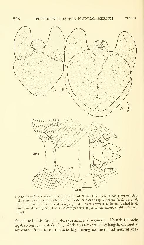
228 PROCEEDINGS OF THE NATIONAL MUSEUM
0.5mm
Figure 22.
—
Norion expansus Nordmann, 1864 (female): a, dorsal view; b, ventral view
of second specimen; c, ventral view of posterior end of cephalothorax (ceph.), second,
third, and fourth thoracic leg-bearing segments, genital segment, abdomen (dashed line),
and caudal rami (parallel lines indicate position of plates and expanded third thoracic
legs).
rior dorsal plate fused to dorsal surface of segment. Foui'th thoracic
leg-bearing segment slender, width greatly exceeding length, distinctly
separated from third thoracic leg-bearing segment and genital seg-
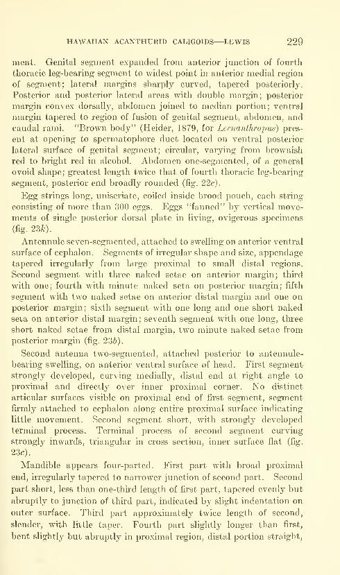
HAWAIIAN ACANTHURID CALIGOIDS—LEWIS 229
ment. Genital segment expanded from anterior junction of fourth
thoracic leg-bearing segment to widest point in anterior medial region
of segment; lateral margins sharply curved, tapered posteriorly.
Posterior and posterior lateral areas with double margin; posterior
margin convex dorsally, abdomen joined to median portion; ventral
margin tapered to region of fusion of genital segment, abdomen, and
caudal rami. "Brown body" (Heider, 1879, for Lernanthropus) pres-
ent at opening to spermatophore duct located on ventral posterior
lateral surface of genital segment; circular, varying from brownish
red to bright red in alcohol. Abdomen one-segjnented, of a general
ovoid shape; greatest length twice that of fourth thoracic leg-bearing
segment, posterior end broadly rounded (fig. 22c).
Egg strings long, uniseriate, coiled inside brood pouch, each string
consisting of more than 300 eggs. Eggs "fanned" by vertical move-ments of single posterior dorsal plate in living, ovigerous specimens
(fig. 23^).
Antennule seven-segmented, attached to swelling on anterior ventral
surface of cephalon. Segments of irregular shape and size, appendage
tapered irregularly from large proximal to small distal regions.
Second segment with three naked setae on anterior margin; third
with one; fourth with minute naked seta on posterior margin; fifth
segment with two naked setae on anterior distal margin and one on
posterior margin; sixth segment with one long and one short naked
seta on anterior distal margin; seventh segment with one long, three
short naked setae from distal margin, two minute naked setae from
posterior margin (fig. 236).
Second antenna two-segmented, attached posterior to antennule-
bearing swelling, on anterior ventral surface of head. First segment
strongly developed, curving medially, distal end at right angle to
proximal and directly over inner proximal corner. No distinct
articular surfaces visible on proximal end of first segment, segment
firmly attached to cephalon along entire proximal surface indicating
little movement. Second segment short, with strongly developed
terminal process. Terminal process of second segment curving
strongly inwards, triangular in cross section, inner surface flat (fig.
23c).
Mandible appears four-parted. First part with broad proximal
end, irregularly tapered to narrower junction of second part. Second
part short, less than one-third length of first part, tapered evenly but
abruptly to junction of third part, indicated b}^ slight indentation on
outer surface. Third part approximately twice length of second,
slender, with little taper. Fourth part slightly longer than first,
bent slightly but abruptly in proximal region, distal portion straight,
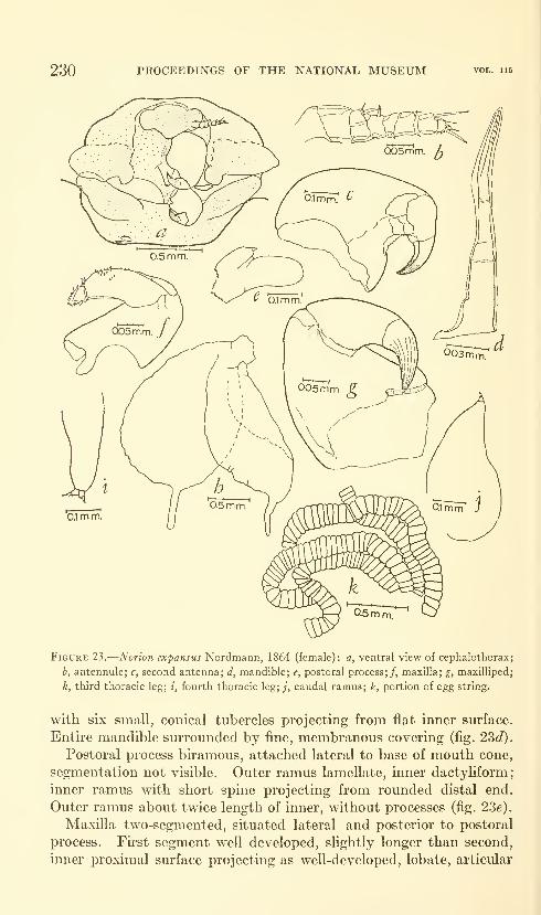
230 PROCEEDINGS OF THE NATIONAL MUSEUM vol. ub
Figure 23.
—
Norion expansus Nordmann, 1864 (female): a, ventral view of cephalothorax;
b, antennule; c, second antenna; d, mandible; e, postoral process;/, maxilla; g, maxilliped;
h, third thoracic leg; i, fourth thoracic leg; ;", caudal ramus; k, portion of egg string.
with six small, conical tubercles projecting from flat inner surface.
Entire mandible surrounded by fine, membranous covering (fig. 22,d).
Postoral process biramous, attached lateral to base of mouth cone,
segmentation not visible. Outer ramus lamellate, inner dactyliform;
inner ramus with short spine projecting from rounded distal end.
Outer ramus about twice length of inner, without processes (fig. 236).
Maxilla two-segmented, situated lateral and posterior to postoral
process. Fu-st segment well developed, slightly longer than second,
inner proximal surface projecting as well-developed, lobate, articular
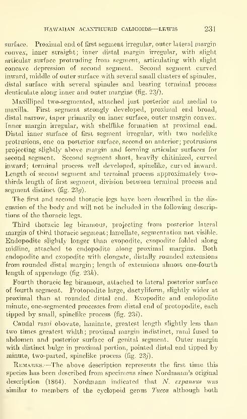
HAWAIIAN ACANTHURID CALIGOIDS—LEWIS 231
surface. Proximal end of first segment irregular, outer lateral margin
convex, inner straight; inner distal margin irregular, with slight
articular surface protruding from segment, articulating with slight
concave depression of second segment. Second segment curved
inward, middle of outer surface with several small clusters of spinules,
distal surface with several spinules and bearing terminal process
denticulate along inner and outer margins (fig. 23/).
Maxilliped two-segmented, attached just posterior and medial to
maxilla. First segment strongly developed, proximal end broad,
distal narrow, taper primarily on inner surface, outer margin convex.
Inner margin irregular, with shelflike formation at proximal end.
Distal inner surface of first segment irregular, with two nodelike
protrusions, one on posterior surface, second on anterior; protrusions
projecting slightly above margin and forming articular surfaces for
second segment. Second segment short, heavily chitinized, curved
inward; terminal process well developed, spinelike, curved inward.
Length of second segment and terminal process approximately two-
thirds length of first segment, division between terminal process and
segment distinct (fig. 23^).
The first and second thoracic legs have been described in the dis-
cussion of the body and will not be included in the following descrip-
tions of the thoracic legs.
Third thoracic leg biramous, projecting from posterior lateral
margin of third thoracic segment; lamellate, segmentation not visible.
Endopodite slightly longer than exopodite, exopodite folded along
midline, attached to endopodite along proximal margins. Both
endopodite and exopodite with elongate, distally rounded extensions
from rounded distal margin ; length of extensions almost one-fourth
length of appendage (fig. 23h).
Fourth thoracic leg biramous, attached to lateral posterior surface
of fom'th segment, Protopodite large, dactyliform, slightly wider at
proximal than at rounded distal end. Exopodite and endopodite
minute, one-segmented processes from distal end of protopodite, each
tipped by small, spinelike process (fig. 23-^).
Caudal rami obovate, laminate, greatest length slightly less than
two times greatest width; proximal margin indistinct, rami fused to
abdomen and posterior surface of genital segment. Outer margin
with distinct bulge in proximal portion, pointed distal end tipped byminute, two-parted, spinelike process (fig. 23^').
Remarks.^—The above description represents the first time this
species has been described from specimens since Nordmann's original
description (1864). Nordmann indicated that A'', expansus was
similar to members of the cyclopoid genus Tucca although both
232 PROCEEDINGS OF THE NATIONAL MUSEUM vol. 115
Bassett-Smith (1899) and Wilson (1922), without seeing the holotype
and only specimen, placed the genus Norton with the Caligoida.
Nordmann (1864) indicates that he found no evidence of bodysegmentation other than the presence of appendages in the cephalo-
thorax. This was probably due to the covering of the posterior
portion of the body by the single dorsal and paired ventral plates.
With sufficient material at hand, this author has been able to examine
members of the species in greater detail than did the original author.
The segmentation present in the specimens collected, the distinct
segmentation of the antennule, the presence of four pairs of thoracic
legs, and the presence of uniseriate egg strings indicate that this
genus, represented solely by A^. expansus, belongs in the Caligoida,
not the Cj^clopoida. The fusion of the first thoracic leg-bearing
segment, the maxilliped-bearing segment, and the cephalon, in
addition to the presence of dorsal plates, indicates that the species is
a member of the family Anthosomatidae. The similarities of the
single dorsal plate covering the posterior surface of the animal and
the paired ventral plates found in A^. expansus to those of some mem-bers of the genus Sagum, S. angulatum (Kr0yer), and those of somemembers of the genus Lernanthropus, L. pagelli Kr0yer and L. chryso-
phrys Shishido, indicate a distinct relationship between these genera.
In addition, the maxilla of N. expansus, especially the distal end and
terminal process of the second segment, is similar to that of membersof the genus Lernanthropus, and the laminate third thoracic leg of
N. expansus is similar to the fourth leg of species of the genus Sagum.
This parasite is comparatively rare in the Hawaiian Islands; the
author collected three from 189 specimens of Naso, the only genus
upon which the parasite was found. The additional two specimens
were collected by Dr. Hilda Ching from a single, large specimen of an
unknown species of Naso.
Family Lernaeoceridae
Genus Peniculus Nordmann, 1832
Diagnosis.—Female: Body elongate; head without horns but
lateral lobes may be present; separated from posterior body by neck
consisting of thoracic leg-bearing segments 1-3 or segments 2 and 3,
one being fused with cephalon in the latter. Fourth thoracic leg-
bearing segment swollen, either separated from fused genital segment
and abdomen by constriction or fused with these segments. Antennule
reduced or absent; second antenna forming organ of attachment,
terminal process clawlike, distinct in immature specimens, indistinct
in mature. Mouth cone retractible; cephalic appendages consist of
second antennae, mandibles, maxillae, and maxillipeds, Thoracic
HAWAIIAN ACANTHURID CALIGOIDS—LEWIS 233
legs platelike, composed solely of protopodite ; fifth legs absent.
Caudal rami armed with setae.
Remarks.-—-The diagnosis was taken from Olsson (1869) and
Wilson (1932) although recent description of new species of this
genus indicates the need for some revision. In addition to distinct
morphological characteristics, the proportional relationships of the
various body parts have been used as specific characteristics. Thevalidity of this method, especially in a degenerated form such as that
of the species in this genus, is dependent upon the degree of difference
between the species and the number of specimens available; the
amount of variation present, at least in the species described below,
is sufficient to warrant careful consideration of the body parts being
compared.Peniculus calamus? Nordmann, 1864
Figures 24a-g
P. calamus Nordmann, 1864, pp. 515-517, no figures.—Wilson, 1917, p. 45
[in key].—Shiino, 1956b, pp. 597, 599, 603.
Present hosts (original host unknown).
—
Acanthurus dussumieri
Cuvier and Valenciennes, Acanthurus mata (Cuvier), Acanthurus
olivaceous Bloch and Schneider, Acanthurus triostegus sandvicensis
Streets, Ctenochaetus strigosus (Bennett), Naso hexacanthus (Bleeker),
Naso unicornis (Forskal).
Type locality.—Hawaii.
Material.—Two matm-e females taken by author from caudal
fin of specimen of Acanthurus dussumieri Cuvier and Valenciennes
taken by poison off Waikiki, Oahu, Hawaii, by Dr. William Gosline
and others. Four matm'e females taken by author from caudal fin
of specimen of A. dussumieri captm*ed in fish trap between DiamondHead and Koko Head, Oahu, Hawaii, by Samuel Kaolulo. Six
matm-e females taken by author from anal and pectoral fins of
specimen of Acanthurus mata (Cuvier) captured in fish trap between
Diamond Head and Koko Head, Oahu, Hawaii, by Samuel Kaolulo.
(USNM 105110-105112.)
Measurements.—Average length from anterior end of head to
posterior end of abdomen, excluding projecting second antennae and
egg strings, 2.85 mm.; range 2.30-3.31 mm. Average of greatest
length of head, excluding projecting second antennae, 0.32 mm.;range 0.29-0.36 mm.; average of greatest width 0.22 mm.; range
0.21-0.25 mm. Average of greatest length of neck (first, second, and
third thoracic leg-bearing segments) 0.20 mm.; range 0.15-0.22 mm.Average of greatest length of combined fourth thoracic leg-bearing
segment, genital segment, and abdomen 2.39 mm.; range 1.84-2.85
mm. Average length of egg strings (eight specimens) 2.94 mm.;range 2.12-3.86 mm.
679-677—64 7
234 PROCEEDINGS OF THE NATIONAL MUSEUM vol. 115
Description.—Color of body brown in alcohol, egg strings yellow;
body with dark bro\\Ti splotches and dorsal black spot, the eye, on
cephalon. The three paii's of spots on the underpart of the head as
described by Nordmann (1864) were not found.
Cephalon ellipsoid dorsally, of general ellipsoid shape laterally,
with ventral surface projecting in region of mouth cone. Dark eye
spot present on dorsal surface, in middle of anterior portion of cephalon.
Division between cephalon and fu'st thoracic leg-bearing segment
distinct, cm*ving anteriorly on dorsal sm^face. First, second, andthird thoracic leg-bearing segments forming neck. First thoracic
leg-bearing segment short, tapered posteriorly, greatest width about
twice greatest length. First segment overlapping second leg-bearing
segment slightly, second segment slightly longer than fii-st, tapered
to junction of third thoracic leg-bearing segment. Third segment
of varying length though generally equal to length of second leg-
bearing segment. Posterior portion of third segment flared slightly
at junction of fourth thoracic leg-bearing segment. Fourth leg-
bearing segment expanded, fused to genital segment ; fourth leg-bearing
segment, genital segment, and abdomen forming elongate posterior
portion of body. Indication of posterior end of fourth leg-bearing
segment present as slight constriction in most specimens though
absent in some. Anterior margins of fourth leg-bearing segment
rounded, ciu-ved medially to junction of third segment (figs. 24:C,d).
Lateral margins of fused fourth thoracic leg-bearing segment, genital
segment, and abdomen elongate, almost parallel; dorsal and ventral
surface with several seta-like projections from epidermis but not
projecting above cuticle in most specimens (fig. 24(i). Abdomensmall, projecting from posterior dorsal surface of genital segment;
caudal projections overlapping caudal rami. The abdomen shows
significant variation in the specimens examined; both long and short
caudal projections were observed in the small series; bulbous and
roimded terminations of the caudal projections were also seen (figs.
24:6,/). Both of these variations were found in about equal propor-
tions though the two do not appear to be related, forms with long
abdomens being found with and without the bulbous terminations
as also were forms with short abdomens (figs. 24a-/).
Egg strings long, with 36-42 eggs in each string. Distal end of
string either rounded or projecting beyond last egg in string in tapered
projection sharply rounded at tip (figs. 24a,6).
Antennule absent in all specimens.
Segmentation of second antenna indistinct, probably two-seg-
mented. Appendage projecting anteriorly from anterior ventral
surface of head, forming organ of attachment. Both second anten-
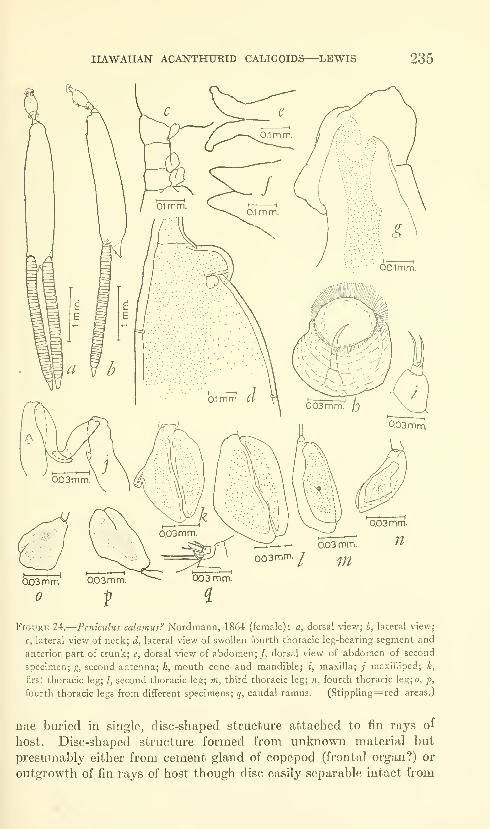
HAWAIIAN ACANTHURID CALIGOIDS—LEWIS 235
0.03mm.
0D3 mm. ^003mm.
I ^0D3mm
Figure 24.
—
Peniculus calamus? Nordmann, 1864 (female): a, dorsal view; b, lateral view;
c, lateral view of neck; d, lateral view of swollen fourth thoracic leg-bearing segment and
anterior part of trunk; e, dorsal view of abdomen;/, dorsal view of abdomen of second
specimen; g, second antenna; h, mouth cone and mandible; i, maxilla; ; maxilliped; k,
first thoracic leg; /, second thoracic leg; m, third thoracic leg; n, fourth thoracic leg; o, p,
fourth thoracic legs from different specimens; q, caudal ramus. (Stippling=red areas.)
nae biu'ied in single, disc-shaped structure attached to fin rays of
host. Disc-shaped structure formed from unknown material but
presumably either from cement gland of copepod (frontal organ?) or
outgrowth of fin rays of host though disc easily separable intact from
236 PROCEEDINGS OF THE NATIONAL MUSEUM vol. 115
fin ray. Distal end of second antenna degenerate in adult, appearing
to have been clawlike in younger stages (fig. 24^').
Mouth cone conical, protruding from median ventral surface of
cephalon, distal surface slightly constricted, with membranous margin
flaring outward from distal margin of cone (fig. 24A).
Mandible visible as elongate, acuminate process ciu"ving inward
distally. Divisions not visible (fig. 24h).
Maxilla two-segmented, attached lateral to mouth cone. First
segment slightly more than four times length of second, gibbous, with
distal sm'face concave. Second segment truncate, with two seta-
like projections from distal surface (fig. 24 i)
•
The modification of this species is such that the naming of the oral
appendages other tlian the mandibles is difficult. The above ap-
pendage is similar to the node on the anterior end of the postoral
process in many caligids. Because of the lack of a complete develop-
mental study and the lack of comparative studies with related groups,
the appendage is termed a maxilla with reservation.
Maxilliped two-segmented, attached posterior to mouth cone.
Fhst segment approximately^ one and a half times length of second,
greatest width slightly less than half greatest length; small protrusion
on middle of inner sm-face. Second segment curving inward distally,
terminating in blunt, rounded tip (fig. 24j).
In specimens preserved in alcohol, all of the thoracic legs possess a
reddish area in the middle of the protopodite; this area is indicated
on the figure of the appendage by stippling.
First thoracic leg ovoid, platelike, attached to anterior lateral
sm-face of segment. Greatest width about two-thirds greatest length,
distinct protrusion on middle of anterior margin and distinct groove
extending from slight indentation on distal margin to proximal region
of appendage (fig. 24:k).
Second thoracic leg one-segmented, of general ovoid shape, attached
to posterior lateral surface of segment; slightly larger than first
thoracic leg. Median groove present as in first thoracic leg, extend-
ing from slight indentation on posterior distal margin proximally to
proximal region of leg. Groove incompletely divides appendage into
two unequal parts, anterior larger than posterior (fig. 24/).
Third thoracic leg ovoid, attached to posterior lateral surface of
segment; \vitliout median groove but with indentation of distal sur-
face. Third leg appears to consist of two lobate processes, smaller
of two fused to lateral surface of larger. Proximal surface projecting
slightly as tuberculate process, process attached to rod-shaped sternal
plate (fig. 24m).
HAWAIIAN ACANTHURID CALIGOIDS—LEWIS 237
Fourth thoracic leg ovoid, much smaller than preceding thoracic
legs. Shape of appendage variable (figs. 247i-2>) ; median groove pres-
ent, extending into leg about one-fourth greatest length of appendage.
Caudal rami minute, projecting slightly from ventral surface of
caudal projections of abdomen. Separated from abdomen by incom-
plete groove; two additional, incomplete transverse grooves in distal
half of appendage. Caudal rami with six naked setae, three from
apex, three from posterior lateral margin (fig. 24g).
Remarks.^—^Nordmann's description of P. calamus (1864) was madefrom a series of specimens. Since the original description, specimens
of this species, including the type specimens, have not been examined.
Although the length of the presently described specimens is con-
siderably less than the 5 mm. noted by Nordmann for the type speci-
mens, the specimens are placed tentatively in the species because of
the general characteristics that they possess in common with those
originally described by Nordmann for P. calamus.
Specimens of P. calamus? collected by the author are similar to P.
minuticaudae Shiino in the general shape of the body, the makeupof the thoracic leg-bearing segments, the cephalic appendages, andthe thoracic legs. The author is indebted to Dr. S. M. vShiino for
comparing specimens of the Hawaiian species with his specimens of
P. minuticaudae. According to Shiino (in correspondence), the Ha-waiian specimens possess ''a longer abdominal process which extends
straight backward and is more deeply and more distinctly bilobed
forming somewhat divergent lobes." In P. minuticaudae "the proc-
ess is shorter, more shallowly bilobed and never extends straight back-
ward, but remams to form a mere triangular stump in lateral view."
These characteristics, in addition to others indicated by Shiino (in
correspondence), suggest that, although the two species possess sim-
ilar morphological characteristics, they are distinct.
References
Baird, W.1850. The natural history of the British Entomostraca. Ray Soc, London,
vii+ 364: pp., 36 pis.
Bassett-Smith, p. W.1896a. A list of the parasitic Copepoda of fish obtained at Plymouth. Journ.
Mar. Biol. Assoc. United Kingdom, vol. 4, no. 2, pp. 155-163.
1896b. Parasitic Copepoda of fish obtained at Plymouth. Ann. Mag. Nat.
Hist., ser. 6, vol. 18, pp. 8-16, pis. 3-6.
1898a. Some new parasitic copepods found on fish at Bombay. Ann. Mag.Nat. Hist., ser. 7, vol. 1, pp. 1-17, pis. 1-7.
1898b. Further new parasitic copepods from fish in tlie Indo-Tropical Region.
Ann. Mag. Nat. Hist., ser. 7, vol. 2, pp. 77-98, pis. 3-6.
1898c. Parasitic copepods from fish in the Indo-Tropical Region. Ann. Mag.Nat. Hist., ser. 7, vol. 2, pp. 357-372, pis. 10-12.
238 PROCEEDINGS OF THE NATIONAL MUSEUM vol. 115
Bassett-Smith, p. W.—Continued
1899. A systematic description of parasitic Copepoda found on fishes with
an enumeration of the kno'wn species. Proc. Zool. Soc. London,
pp. 437-507.
1903. New parasitic copepods from Zanzibar and East Africa. Proc. Zool.
Soc. London, vol. 1, pp. 104-109, figs. 11-12.
Bere, Ruby1936. Parasitic copepods from Gulf of Mexico fish. Amer. Midi. Nat.,
voL 17, no. 3, pp. 577-625, pis. 1-12.
Bonnet, D.
1948. Some parasitic copepods from Hawaiian fishes. Proc. Hawaiian
Acad. Sci., no. 23, p. 7.
Brady, George1899. On the marine Copepoda of New Zealand. Trans. Zool. Soc. London,
vol. 15, no. 2, pp. 31-54, pis. 9-13.
Brian, Alessandro1906. Copepodi parassiti dei pesci d'italia. Genoa, 187 pp., 21 pis.
1914. Copepodes parasites proveuant des recentes campagnes scientifiques
de S.A. le Prince Albert I*'' de Monaco ou deposes dans les collec-
tions du Musee Oceanographique. Bull. Inst. Oceanogr., no. 286,
14 pp., 8 text figs.
1931a. Description d'une nouvelle espece de Caligus (copepoda parasite) de
la MMiterran6e. Bull. Soc. Hist. Nat. Afrique du Nord, vol. 22,
pp. 119-120, pi. 1.
1931a. Sur la synonymie du Caligus argilasi Brian (1931) avec le Caligus
pageti Russel (1925). Bull. Soc. Hist. Nat. Afrique du Nord, vol.
22, p. 157.
1932. Sur une espece rare de Caligidae (copepode parasite) recoltee par le
Docteur Dieuzeide dans la Mediterranee (Lepeophtheuus rotundi-
ventris B.S.). Bull. Trav. Pub. Sta. Aquicult. Peche Castigiione,
pp. 3-14, 1 pi., 2 text figs.
1933. Sur quelques copepodes parasites d'Algerie. Description d'une
espece nouvelle de Caligus (Caligus dieuzeidei) du Diplodes sargus
L. Bull. Trav. Pub. Sta. Aquicult. Peche de Castighone, pp. 3-
16, 1 pi., 2 text figs.
1935. Caligus parassiti dei pesci del Mediterraneo. Estratto Ann. Mus.
Civ. Storia Nat. Genova, vol. 57, no. 7, pp. 1-60, figs. 1-20.
1939. Cop6podes parasites recueillis par M. E. Darteville a I'embouchure
du fleuve Congo. Rev. Zool. Bot. Africaines, vol. 32, no. 2, pp.
176-198, figs. 1-15.
Capart, Andre1953. Quelques copepodes parasites de poissous marins de la region de
Dakar. Bull. Inst. Frangais d'Afrique Noire, vol. 15, no. 2, pp.
647-671, figs. 1-10.
Causey, David1960. Parasitic Copepoda from Mexican coastal fishes. Bull. Mar. Sci.
Gulf Caribbean, vol. 10, no. 3, pp. 323-337, figs. 1-2.
Dana, J. D.
1853; 1855. Crustacea. Vol. 13 in United States Exploring Expedition
during the years 1838, 1839, 1840, 1841, 1842 under the commandof Charles Wilkes, U.S.N. Philadelphia, part 2, pp. 691-1618
(1853); atlas, 27 pp., 96 pis. (1855).
HAWAIIAN ACANTHTRID CALIGOIDS—LEWIS 239
Delamare Deboutteville, Claude, and Nunes-Ruiva, L. P.
1951. Existence de formes biologiques chez Peniculus fistula (Rudolphi)
(Copepoda). Vie et Milieu, vol. 2, no. 4, pp. 448-458, figs. 1-4.
Edmondson, Charles H.
1946. Reef and shore fauna of Hawaii. Bernice P. Bishop Mus. Spec.
Publ. 22, Honolulu, iii+ 381 pp., 223 figs.
Eraser, C.
1920. Copepods parasitic on fish from the Vancouver Island region. Trans.
Roy. Soc. Canada, ser. 3, vol. 13, pp. 45-67, pis. 1-8.
Gadd, Pehr1904. Parasit-Copepoder i Finland. Acta Soc. Fauna Flora Fennica, vol.
26, no. 8, pp. 1-60, pis. 1-2.
Gnanamuthu, C. p.
1947. Caligus sciaenae n. sp. parasitic on Sciaena glauca from Madras.
Proc. Indian Acad. Sci., vol. 25, pp. 43-49, figs. 1-2.
1948. Notes on the anatomy and physiology of Caligus savala n. sp., a
parasitic copepod from Madras plankton. Proc. Zool. Soc. India,
vol. 118, no. 3, pp. 591-606, figs. 1-8.
1949. Two male parasitic copepods from Madras. Ann. Mag. Nat. Hist.,
ser. 12, vol. 2, pp. 359-367, figs. 1-23.
1950a. Three new copepod parasites of the ribbon fish from South India.
Journ. Parasit.. vol. 36, no. 2, pp. 113-119, pis. 1-3.
1950a. Sex differences in the chalimus and adult forms of Caligus polycanthi
sp. nov. (Crustacea: Copepoda) parasitic on Balistes maculatus
from Madras. Rec. Indian Mus., vol. 47, no. 1, pp. 159-170, 6
text figs.
GosLiNE, W. A., and Brock, V. E.
1960. Handbook of Hawaiian fishes. Honolulu, ix+ 372 pp., 277 figs.
Guiart, Jules1913. Crustaces commensaux et parasites de la baie de Concerneau. Bull.
Inst. Oceanogr., no. 264, 11 pp., 2 text figs.
GURNEY, R.
1927. Zoological results of the Cambridge Expedition to the Suez Canal
1924, XXX: Report on the Crustacea: Copepoda (littoral andsemiparasitic) . Trans. Zool. Soc. London, vol. 22, pp. 451-577,
figs. 1-61.
Heegaard, Poul1943a. Some new caligids from the Gilbert Islands. Arkiv. Zool., Stock-
holm, vol. 34A, no. 16, pp. 1-12, figs. 1-3.
1943b. Parasitic copepods mainly from tropical and Antarctic seas. Arkiv.
Zool., Stockholm, vol. 34A, no. 18, pp. 1-37, figs. 1-94.
1945. Some parasitic copepods from fishes in the Uppsala University col-
lections. Arkiv. Zool., Stockholm, vol. 35A, no. 18, pp. 1-27,
figs. 1-54.
1947. Contribution to the phylogeny of the arthropods. Spolia Zool. Mus.Hauniensis, vol. 8, 227 pp., 27 pis.
1955. Parasitic copepods from tropical West Africa. Atlantide Report 3,
pp. 41-56, figs. 1-20.
Heider, Carl1879. Die Gattung Lernanthropus. Vienna, 100 pp., 5 pis.
240 PROCEEDINGS OF THE NATIONAL MUSEUM vol. 115
Heller, Camil1865. Reise dor Osterreichischen Fregatte Novara um die Erde in den
Jahren 1857, 1858, 1859. Zool. Theil, vol. 2, no. 3, pp. 1-280,
pis. 1-25.
1866. Carcinologische Beitrage zur Fauna des adriatischen Meeres. Ver-
handl. Kais.-Konigl. Zool.-bot. Ges. Wien, pp. 723-760.
Humes, Arthur1957. Two new caligoid copepods from Egyptian fishes. Journ. Parasit.,
vol. 43, no. 2, pp. 201-208, pis. 1-3.
Jordan, D. S.; Evermann, B. W.; and Clark, H. W.1930. Check list of the fishes and fishlike vertebrates of North and Middle
America north of the northern boundary of Venezuela and Colombia.
Washington, iv+670 pp.
Kr0yer, H.
1863. Bidrag til Kundskab om Snyltekrebsene. Copenhagen, 352 pp., 18
pis.
Lang, Karl1948. Copepoda "Notodelphyoida" from the Swedish West Coast with an
outline of the systematics of the copepods. Arkiv. Zool., vol. 40a,
no. 14, pp. 1-36, pi. 1, figs. 1-17.
Leigh-Sharpe, W. Harold1930. Resultats scientifiques du voyage aux Indes Orientales Neerlandaises
de LL. AA. RR. le Prince et la Princesse Leopold de Belgique, vol. 3,
no. 2, pp. 1-9, pis. 1-5.
1934. The Copepoda of the Siboga Expedition, Part II: Commensal and
parasitic Copepoda. Siboga Expeditie, monogr. 29b, 40+ 3 pp.,
39 text figs.
Linton, Edwin1907. Notes on parasites of Bermuda fishes. Proc. U.S. Nat. Mus., vol. 33,
no. 1560, pp. 85-126, pis. 1-2.
Milne Edwards, M. H.1840. Histoire naturelle des crustaces, comprenant I'anatomie, la physio-
logie et la classification de ces animaux. Paris, vol. 3, 638 pp.;
plates, 32 pp.+ 42 pis.
Nordmann, Alexander V.
1832. Mikrographische Beitrage zur Naturgeschichte der wirbellosen Thiere.
Berlin, 150 pp., 9 pis.
1864. Neue Beitrage zur Kentniss Parasitischer Copepoden. Bull. Soc.
Imp. Nat. Moscou, vol. 37, no. 4, pp. 461-520, pis. 5-8.
Norman, Alfred, and Scott, Thomas.1906. The Crustacea of Devon and Cornwall. London, xv+ 232 pp., 24 pis.
NuNEs-Ruivo, L., and Fourmanoir, P.
1956. Copepodes parasites de poissons de Madagascar. Mem. Inst. Sci.
Madagascar, ser. A, vol. 10, pp. 69-80, figs. 1-8.
Olsson, Petrus1868-1869. Prodromous faunae copepodorum parasitantium Scandinaviae.
Acta Univ. Lundensis, Lunds Univ. Ars-Skrift, Math. Naturv.,
no. 8, pp. 1-49, pis. 1-2.
1877. Om parasitiska copepoder i Jemtland. Ofv. Svenska Vet.-Akad.
Forh., vol. 34, no. 5, pp. 75-88, pis. 4-6.
HAWAIIAN ACANTHURID CALIGOIDS—LEWIS 241
Randall, John E.
1956. A revision of the surgeon fish genus Acanthurus. Pacific Sci., vol. 10,
no. 2, pp. 159-235, pis. 1-3, figs. 1-23.
1961. The biology of the convict surgeonfish. Pacific Sci., vol. 15, pp. 215-
272, figs. 1-25.
Rangnekar, p.
1953. A^iuretes hranchialis sp. nov., a copepod parasitic on the fish Thynnuspelamys (Linn.). Journ. Zool. Soc. India, vol. 5, no. 2, pp. 239-
242, fig. 1.
1957. Copepod parasites of the families ArguUdae, Caligidae, Dichelesthidae
and Lernaeopodidae. Journ. Univ. Bombay, sect. B, vol. 26,
no. 3, pp. 8-20, figs. 1-6.
Rangnekar, P., and Murti, N. N.
1950. A new caligid copepod parasitic on the fish Clupea tali. Journ.
Univ. Bombay, vol. 18, no. 5, pp. 21-28, pi. 1.
Redkar, M.; Rangnekar, P.; and Murti, N. N.
1949. Four new species of parasitic copepods from the marine fishes of
Bombay. Journ. Univ. Bombay, vol. 18, no. 3, pp. 36-50, pis. 1-4.
Richiardi, D. S.
1880. Catalogo sistematico dei crostacei che vivono sul corpo degli animali
aquatici in Italia, 8 pp.
Scott, A.
1901. Lepeophtheirus and Lernea. Trans. Liverpool Biol. Soc, vol. 15,
pp. 188-241, pis. 1-5.
Scott, Thomas1894. Report on Entomostraca from the Gulf of Guinea. Trans. Linn.
Soc. London, 161 pp., 15 pis.
Scott, Thomas and Andrew1913. The British parasitic Copepoda, I and II. Ray Soc. London, ix+251
pp., A+ 72 pis.
Shiino, Sued M.1952. Copepods parasitic on Japanese fishes, 1: On the species of Caligus
and Lepeophtheirus. Rep. Fac. Fish., Pref. Univ. Mie, vol. 1,
no. 2, pp. 79-112, figs. 1-14.
1954a. On the new copepod, Caligus calotomi n. sp., parasitic on the fish,
Calotomus japonicus (C. & V.). Bull. Japanese Soc. Sci. Fish.,
vol. 20, no. 1, pp. 16-19, figs. 1-2.
1954b. On Caligus latigeniialis n. sp., a copepod parasitic on the fish Sparus
macrocephalus (Basilewsky). Bull. Japanese Soc. Sci. Fish., vol.
20, no. 1, pp. 21-25, fig. 1.
1954c. Note on Caligus quadratus n. sp., a copepod parasitic on the fish
Neothynnus niacropterus (T. & S.). Bull. Japanese Soc. Sci.
Fish., vol. 20, no. 1, pp. 26-29, fig. 1.
1954d. Copepods parasitic on Japanese fishes, 2: On two new species of the
family Trebidae. Rep. Fac. Fish., Pref. Univ. Mie, vol. 1, no. 3,
pp. 247-259, figs. 1-4.
1954e. Copepods parasitic on Japanese fishes, 3 : On two new species of the
genus Anuretes. Rep. Fac. Fish., Pref. Univ. Mie, vol. 1, no. 3,
pp. 260-272, figs. 1-5.
1954f. Copepods parasitic on Japanese fishes, 4: The family Euryphoridae.
Rep. Fac. Fish., Pref. Univ. Mie, vol. 1, no. 3, pp. 273-290, figs.
1-6.
242 PROCEEDINGS OF THE NATIONAL MUSEUM vol. lis
Shiino, Sueo M.—Continued
1954g. Copepods parasitic on Japanese fishes, 5: Five species of the family
Pandaridae. Rep. Fac. Fisli., Pref. Univ. Mie, vol. 1, no. 3, pp.291-332, figs. 1-19.
1954h. Copepods parasitic on Japanese fishes, 6: Two species of the family
Cecropidae. Rep. Fac. Fish., Pref. Univ. Mie, vol. 1, no. 3, pp.333-345, figs. 1-6.
1955a. On the male of Caligus cordiveyitris Shiino. Zool. Mag. (DobutsugakuZasshi), vol. 64, no. 3, pp. 65-67, 1 fig.
1955b. A new piscicola copepod belonging to the genus Caligus from
Matusima Bay. Bull. Biogeogr. Soc. Japan, vols. 16-19, pp.
135-140, figs. 1-3.
1956a. CojDepods parasitic on Japanese fishes, 10: The redescription of three
species of Caligus. Rep. Fac. Fish., Pref. Univ. Mie, vol. 2, no.
2, pp. 233-241, figs. 1-4.
1956b. Copepods parasitic on Japanese fishes, 7: Peniculus and Peniculisa.
Japanese Journ. Zool., vol. 11, no. 5, pp. 593-608, figs. 1-5.
1957a. Copepods parasitic on Japanese fishes, 13: Parasitic copepods collected
off Kesennuma, Miyagi Prefecture. Rep. Fac. Fish., Pref. Univ.
Mie, vol. 2, no. 3, pp. 359-375, figs. 1-6.
1957b. Copepods parasitic on Japanese fishes, 14: Three species from
Spheroides alho-plumheus (Richardson). Rep. Fac. Fish., Pref.
Univ. Mie, vol. 2, no. 3, pp. 376-391, figs. 1-7.
1957c. Copepods parasitic on Japanese fishes, 15: Eudactylinidae andDichelesthiidae. Rep. Fac. Fish., Pref. Univ. Mie, vol. 2, no. 3,
pp. 392-410, figs. 1-7.
1959a. Neuer Artname fiir japanische Exemplare von Caligus bonito. Bull.
Biogeogr. Soc. Japan, vol. 20, no. 11, pp. 51-57, figs. 1-2.
1959b. Ostpazifische Parasitierende Copepoden. Rep. Fac. Fish., Pref.
Univ. Mie, vol. 3, no. 2, pp. 267-333, figs. 1-25.
1959c. Sammlung der parasitischen Copepoden in der Prafekturuniversitat
von Mie. Rep. Fac. Fish., Pref. Univ. Mie, vol. 3, no. 2, pp.
334-374, figs. 1-17.
1959d. On a new species of Caligus (Copepoda Parasitica) from the coast of
California. Pacific Sci., vol. 13, no. 3, pp. 351-356, figs. 1-3.
Steenstrup, J. J. Sm., and Lutken, C. F.
1861. Bidrag til Kundskab om det aabne Havs Snyltekregs eg Lernaeer
samt om nogle andre nye eller hidtil kun ufuldstaendigt kjendte
parasitiske copepoder. Kongel. Danske Vidensk. Selsk. Skrift.,
5te Raekke, Naturh. Math. Afdel., vol. 5, 92 pp., 15 pis.
Thomsen, Ricardo1949. Copepodos pardsitos de los peces marines del Uruguay. Comm.
Zool. Mus. Hist. Nat. Montevideo, vol. 3, no. 54, pp. 1-41, pis.
1-14.
Thomson, George1890. A new parasitic copepod. Trans. New Zealand Inst., vol. 22, pp.
227-229, pi. 23.
Van Beneden, P. J.
1870. Les Poissons des cetes de Belgique, leurs parasites et leurs commen-saux. Brussels, 100 pp., 8 pis.
1892. Le Male de certains caligides et un nouveau genre de cette famille.
Bull. Acad. Roy. Belgique, ser. 3, vol. 23, no. 3, pp. 220-235,
pis. 1-2.
HAWAIIAN ACANTHURID CALIGOIDS—LEWIS 243
Wilson, Charles B.
1905a. North American parasitic copepods belonging to the family Cali-
gidae, Part I: The Caliginae. Proc. U.S. Nat. Mus., vol. 28, no.
1404, pp. 479-672, pis. 5-29.
1905b. New species of parasitic copepods from the Massachusetts Coast.
Proc. Biol. Soc. Washington, vol. 18, pp. 127-132.
1906. Report to the government of Cejdon on the Pearl Oyster Fisheries
of the Gulf of Manaar, Supplementary report 34: On some parasitic
copepods. Ceylon Pearl Oyster Rep., part 5, pp. 189-210, pis.
1-5.
1907a. North American parasitic copepods belonging to the family Caligidae,
Part 2: The Trebinae and Euryphorinae. Proc. U.S. Nat. Mus.,vol. 31, no. 1504, pp. 669-720, pis. 15-20.
1907b. North American parasitic copepods belonging to the family Cahgidae,Parts 3 and 4: Revision of the Pandarinae and Cecropinae. Proc.
U.S. Nat. Mus., vol. 33, no. 1573, pp. 32.3-490, pis. 17-43.
1908a. New genera and species of Caliginae. Proc. U.S. Nat. Mus., vol.
33, no. 1580, pp. 593-627, pis. 49-58.
1908b. North American parasitic copepods: A hst of those found upon thefishes of the Pacific Coast, with descriptions of new genera andspecies. Proc. U.S. Nat. Mus., vol. 35, no. 1652, pp. 431-481,pis. 56-83.
1911a. Notes on Jamaica fish and their parasites. Johns Hopkins Univ.Circ, pp. 1-7.
1911b. North American parasitic copepods: Descriptions of new genera andspecies. Proc. U.S. Nat. Mus., vol. 39, no. 1805, pp. 625-634,
pis. 65-68.
1912a. Parasitic copepods from Nanaimo, British Columbia, including eight
species new to science. Contr. Canadian Biol., 1906-1910, pp.83-102, pis. 3-9.
1912b. Descriptions of new species of parasitic copepods in the collections
of the United States National Museum. Proc. U.S. Nat. Mus.,vol. 42, no. 1900, pp. 233-243, pis. 30-34.
1913. Crustacean parasites of West Indian fishes and land crabs, withdescriptions of new genera and species. Proc. U.S. Nat. Mus.,vol. 44, no. 1950, pp. 189-277, pis. 18-53.
1917. North American parasitic copepods belonging to the Lernaeidae,
with a revision of the entire family. Proc. U.S. Nat. Mus., vol.
53, no. 2194, pp. 1-150, pis. 1-21.
1920. Crustacea, Part I: Parasitic Copepoda. In Canadian Arctic Expedi-tion, vol. 7, pp. 1-16.
1921a. New species and a new genus of parasitic copepods. Proc. U.S.
Nat. Mus., vol. 59, no. 2354, pp. 1-17, pis. 1-7.
1921b. Report on the parasitic Copepoda collected during the survey of the
Juan Fernandez Islands, 1916-1917. The Natural History of
Juan Fernandez and Easter Island, vol. 3, pp. 69-74, pis. 2-4.
1922a. North American parasitic cojiepods belonging to the family Dicheles-
thiidae. Proc. U.S. Nat. Mus., vol. 60, art. 5, no. 2400, pp. 1-100,
pis. 1-13.
1922b. Parasitic copepods in the collection of the Zoological Museum,Kristiania. Nyt Mag. Naturvidensk., vol. 60, no. 4, pp. 1-7.
1923. Parasitic copepods in the collection of the Riksmuseum at Stockholm.Arch. Zool., vol. 15, no. 3, pp. 1-15, pis. 1-2.
244 PROCEEDINGS OF THE NATIONAL MUSEUM vol. 115
Wilson, Charles B.—Continued
1924a. Parasitic copepods from the White Nile and the Red Sea. InResults Swedish Zoological Expedition to Egypt and the WhiteNile, 1901, no. 26B, pp. 1-17, pis. 1-3.
1924b. Parasitic copepods from the Wilhams Galapagos Expedition.
Zoologica, New York Zool. Soc, vol. 5, no. 19, pp. 212-217, pi. 20.
1928. A new species of parasitic copepod (Caligus) from a Siamese catfish.
Journ. Siam Soc. Nat. Hist. Suppl., vol. 7, pp. 191-192, pi. 13.
1932. The copepods of the Woods Hole Region Massachusetts. U.S. Nat.
Mus. Bull., vol. 158, 635 pp., 41 pis., 316 text figs.
1935a. Parasitic copepods from the Dry Tortugas. Carnegie Inst. Washing-ton, Publ. 452, Pap. Tortugas Lab., vol. 29, no. 12, pp. 327-347,
pis. 1-6.
1935b. Parasitic copepods from the Pacific Coast. Amer. Midi. Nat.,
vol. 16, no. 5, pp. 776-797, pis. 25-30, 88 text figs.
1935c. New parasitic copepods. Smithsonian Misc. Coll., vol. 91, no. 19,
pp. 1-9, pis. 1-3.
1936. Two new parasitic copepods from Cuban fish. Mem. Soc. CubanaHist. Nat., vol. 10, no. 2, pp. 107-112, figs. 1-15.
1937a. Some parasitic copepods from Panama Bay. Jouru. Washington
Acad. Sci., vol. 27, no. 10, pp. 423-431, figs. 1-34.
1937b. Parasitic copepods taken during the third Hancock Expedition to
the Galapagos Islands. In Hancock Pacific Expeditions, vol. 2,
no. 4, pp. 23-30, pi. 3, text figs. 1-5.
1942. The copepods of the plankton gathered during the last cruise of
the Carnegie. In Scientific results of Cruise VII of the Carnegie
during 1928-1929 under the command of J. P. Ault. Carnegie
Inst. Washington Publ. 536, iv+ 237 pp., 136 figs.
1944. Parasitic copepods in the United States National Museum. Proc.
U.S. Nat. Mus., vol. 94, no. 3177, pp. 529-582, pis. 20-34.
Yamaguti, SATYtr
1936a. Parasitic copepods from fishes of Japan, Part 2: Caligoida, I. Kyoto,
22 pp., 12 pis.
1936b. Parasitic copepods from fishes of Japan, Part 3 : Caligoida, II. Kyoto,
21 pp., 9 pis.
Yeatman, Harry1957. A redescription of two parasitic copepods from Bermuda. Journ.
Washington Acad. Sci., vol. 47, no. 10, pp. 346-353, figs. 1-31.
Ytj, S. C.
1933. Chinese parastic copepods collected by H. W. Wu, with descriptions
of new genera and species. Bull. Fan Mem. Inst. Biol., vol. 4,
no. 4, pp. 117-138, pis. 1-8.
YtJ, S. C, and Wu, H. W.1932. Parasitic copepods of the flat-fishes from China. Bull. Fan Mem.
Inst. Biol., vol. 3, no. 4, pp. 55-74, pis. 1-8.
U.S. GOVERNMENT PRINTING OFFlCEil964
Proceedings of
the United States
National MuseumSMITHSONIAN INSTITUTION • WASHINGTON, B.C.
Volume 115 1964 Number 3483
NOTES ON ARADIDAE IN THE U.S. NATIONAL MUSEUMIII. SUBFAMILY MEZIRINAE '
(HEMIPTERA)
By Nicholas A. Kormilev
For the privilege of studying the unidentified flat bugs, family
Aradidae, in the collections of the U.S. National Museum, I wish to
express my deep appreciation to Dr. J. F. Gates Clarke, Curator of
Insects, U.S. National Museum, and to Dr. Carl J. Drake, Research
Associate, Smithsonian Institution. The deposition of types of newspecies is stated beneath the descriptions.
In the descriptions, 20 units=l mm. for aU species but Placogenis
brachyptera (Kormilev), in which 30 units=l mm. The first figure in
the ratios represents the length; the second, the width of the measured
part. In the measurements of the pronotum, the figures within
brackets represent the width of the forelobe; the last figure, the width
of the hind lobe across the widest part. The length of the head was
measured from the middle of the posterior border to the tip of the juga
and the width across the eyes, as a more constant unit, and not across
the postocular tubercles or spines, which are variable within the species.
The lengths of the pronotmn, scutellum, and abdomen were taken
on the median line ; the length of the abdomen was taken from the tip
1 See N. A. Kormilev, 1958, Notes on Aradidae in the U.S. National Museum I, Proc.
U.S. Nat. Mus., vol. 109, no. 3413, pp. 209-222, and N. A. Kormilev, 1960, Notes onAradidae In the U.S. National Museum II, Journ. New York Ent. Soc, vol. 68,
pp. 36-47.
245
246 PROCEEDINGS OF THE NATIONAL MUSEUM vol. us
of the scutellum to the tip of the male hypopygium or to segment IX in
the female. The width of the scutellum was taken at the base; of the
abdomen, at the widest part. The proportions of the antennal
segments were taken from the first to the fourth segments.
Subfamily Mezirinae Oshanin, 1908
The collections comprise many specimens of mezirines from the
Neotropical Region. It is significant to note that the numerous newspecies of the genus Mezira Amyot and Serville, mostly from the
tropical areas of South America, vividly indicate the incompleteness
of our present knowledge of the Aradidae from this vast geographical
region.
Genus Placogenis Usinger and Matsuda
Placogenis Usinger and Matsuda, 1959, Classification of Aradidae, British Museum(Natural History), London, p. 342, fig. 100.
Diphyllonotus Kormilev, 1959, Proc. Ent. Soc. Washington, vol. 61, p. 61, figs.
1,2.
Placogenis brachyptera (Kormilev)
Diphyllonotus brachypterus Kormilev, 1956, Anal. Soc. Cient. Argentina, vol.
162, p. 151.
Remarks.—This species was described originally from a single
brachypterous female from Santa Catarina, Brazil. The abbreviated
membrane of the forewing reaches to only the front margin of tergum
VI. In the Drake Collection I found a female specimen with fully
developed forewings that extended backward to the middle of tergum
VII.
Measurements.—Macropterous female: Head almost as long as
wide (27:27.5); proportions of antennal segments 14:10:17:13;
pronotum much shorter than wide (28 : (45) : 53) ; scutellum shorter
than wide at base (19:29); abdomen as long as wide (92:92).
Length 5.3 mm. Width of pronotum 1.76 mm. Width of abdo-
men 3.07 mm. Other characters as in brachypterous form.
New RECORD.—Female, macropterous, Rio de Janeiro, Brazil,
Drake CoUection (USNM).
Genus Cinyphus Stal
Cinyphus St&l, 1865, Hemiptera Africana, vol. 3, p. 31.
Cinyphus saileri, new species
Figures 1, 2
Male.—Elongate ovate, tapering forward; body covered with
curled rusty hairs.

NOTES ON ARADIDAE, III—KORMILEV 247
mi^QK £.. M'h M.
Figures 1-7.— 1, Cinyphus saileri, new species, cf head and pronotum; 2, C. saileri, tip of
cf abdomen; 3, Meziraluteonotata, new species, ? head and pronotum; 4, M. luteonotata,
tip of 9 abdomen; 5, M. costalimai, new species, cf head and pronotum; 6, M. fo//a-
lijnai, tip of cf abdomen; 7, A/, carioca, new species, 9 head and pronotum.
248 PROCEEDINGS OF THE NATIONAL MUSEUM vol. lis
Head longer than width across eyes (31:25); anterior process
(clypeus and juga together) long, slightly widening anteriorly, and
cleft, reaching three-fom"ths first antennal segment. Antenniferous
tubercles long, spiniform, divergent, raised anteriorly, reaching
one-third ftrst antennal segment. Eyes semiglobose, exserted, placed
behind middle of head. Postocular tubercles dentiform, short, not
reaching outer border of eyes. Infraocular carinae low, with bigger
tooth anteriorly. Vertex with setigerous granulation. Antennae
long, slender, twice as long as width of head across eyes (52:25);
proportions of segments 15:11:15:11. Rostrum not reaching to hind
border of long, deep, wide, and transversely rugose rostral groove.
Pronotum almost half as long as width across humeri (30:58);
collum slender, distinct. Anterolateral angles expanded winglike
laterally, with anterior border straight and not reaching foremargin
of collum. Lateral borders of hind lobe denticulate and convergent
anteriorly; lateral notch big, rectangular. Foredisc of pronotum
with four (2+2) longitudinal ridges, two inner ones thicker, two outer
ones thinner; medium line deeply depressed. Hind disc with dis-
persed setigerous granulation. Hind border deeply and widely ex-
cavated at middle.
Scutellum slightly shorter than basal width (27:32); basal border
convex; lateral borders convex anteriorly, sinuate posteriorly; tip of
scutellum notched. Median ridge tapering to tip; disc transversely
rugose. Two (1 + 1) tubercles placed at basal angles.
Hemelytra reaching hind border of tergum VI. Basolateral
borders of corium denticulate, slightly diverging backward, reflexed.
Apical border of corium convex exteriorly, apical angle rounded.
Membrane sepia, with triangular white spot near apical angle of
corium.
Abdomen ovate, almost as long as wide (79 :77) ;postero-exterior
angles of connexiva protruding angularly; postero-exterior angles of
connexivum VII dentiform, divergent. Exterior borders of connexiva
straight; those of connexivum VII convex. Hypopygium with thin
median furrow and with rounded rim on upper side posteriorly.
Spiracles II to VII ventral, far removed from lateral border, those of
VIII lateral and visible from above.
Color.-—Sepia; apical half of antennal segment IV, postero-exterior
angles of connexiva, and rostrum light brown.
Measurements.—Length 8.35 mm. Width of pronotum 2.60
mm. Width of abdomen 3.56 mm.HoLOTYPE.—Male, Vera Cruz, Mexico, May 13, 1946, on orchid
plants, named for Dr. Reece I. Sailer (USNM type 65795).
NOTES ON ARADIDAE, III—KORMILEV 249
Remarks.—This species is allied to Cinyphus squalidus Champion;
it can be separated from the latter by the following characteristics:
antenniferous tubercles acute and divergent; second and fourth
antennal segments equal in length; lateral margins of abdomen evenly
rounded (subangular posteriorly in C. squalidus)
.
Genus Santaremia Kormilev
Santaremia Kormilev, 1960, Journ. New York Ent. Soc, vol. 68, p. 44.
Santaremia robusta Kormilev
Santaremia robusta Kormilev, 1960, Journ. New York Ent. Soc, vol. 68, p. 45,
figs. 6-7.
Female.—Slightly larger than male but of same color and general
aspect. Lobes of VIII (paratergites) short, triangular, reaching to
middle of IX, latter truncate posteriorly. Spiracles II to VI ventral,
placed far from lateral border, VII also ventral but situated a little
nearer to outer margin; VIII lateral and visible from above.
Measurements.—Head shorter than wide across eyes (24:27); pro-
portions of antennal segments 16:10: ? (last two segments missing);
pronotum half as long as wide across humeri (30:(42):60); scutellum
shorter than width at base (25:32); abdomen longer than wide (75:71).
Length 7.85 mm. Width of pronotmn 3.00 mm. Width of
abdomen 3.55 mm.Allotype.—Female, Para, Brazil, in Drake collection (USNM).
Genus Mezira Amyot and Serville
Brachyrhynchiis Laporte, in Gu^rin, Mag. Zool., 1833, vol. 2, p. 54 (preoccupied).
Mezira Amyot and Serville, 1843, Histoire naturelle des insectes, H^mipt^res,
p. 305.
Dusius Bergroth, 1894, Ent. Tidscr., vol. 15, p. 104.
Mezira is the largest genus in the subfamily Mezirinae, and is
worldwide in distribution with the exception of colder areas. In
1959 Usinger and Matsuda removed from Mezira many species
previously assigned therein and created for them the new genera
Daulocoris, Strigocoris, and Oroessa. They also resurrected the genusArictus StM, previously synonymized with Mezira, and left 106
species in the genus Mezira. In its present sense, after revision, the
genus Mezira is still too heterogeneous, for the entire complex of the
so-called "membranacea group" probably should form a separate
genus ; it is dubious that the American and African species of the genus
Mezira could remain in the same genus. At the present time abouthalf of all species classified in Mezira belong to the Neotropical Region.
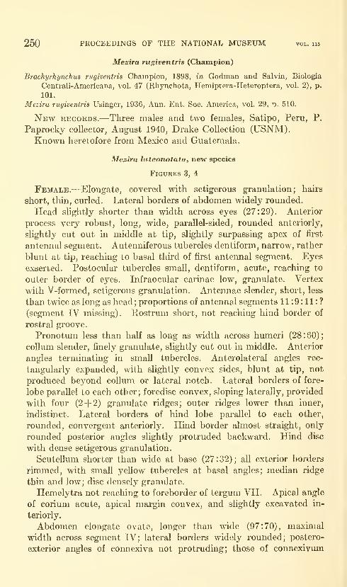
250 PROCEEDINGS OF THE NATIONAL MUSEUM vol. 115
Mesira rugiventris (Champion)
Brachyrhynchus rugiventris Champion, 1898, in Godman and Salvin, Biologia
Central!-Americana, vol. 47 (Rhynchota, Hemiptera-Heteroptera, vol. 2), p.
101.
Mezira rugiventris Usinger, 1936, Ann. Ent. Soc. America, vol. 29, t). 510.
New records.—Three males and two females, Satipo, Peru, P.
Paprocky collector, August 1940, Drake Collection (USNM).Known heretofore from Mexico and Guatemala.
Mezira luteonotata, new species
Figures 3, 4
Female.—Elongate, covered with setigerous granulation; hairs
short, thin, curled. Lateral borders of abdomen widely rounded.
Head slightly shorter than width across eyes (27:29). Anterior
process very robust, long, wide, parallel-sided, rounded anteriorly,
slightly cut out in middle at tip, slightly surpassing apex of first
antennal segment. Antenniferous tubercles dentiform, narrow, rather
blunt at tip, reaching to basal third of first antennal segment. Eyes
exserted. Postocular tubercles small, dentiform, acute, reaching to
outer border of eyes. Infraocular cai'inae low, granulate. Vertex
with V-formed, setigerous granulation. Antennae slender, short, less
than twice as long as head;proportions of antennal segments 11:9:11:?
(segment IV missing). Rostrum short, not reaching hind border of
rostral groove.
Pronotmn less than half as long as width across humeri (28:60);
collum slender, finely granulate, slightly cut out in middle. Anterior
angles terminating in small tubercles. Anterolateral angles rec-
tangularly expanded, with slightly convex sides, blunt at tip, not
produced beyond colhim or lateral notch. Lateral borders of fore-
lobe parallel to each other; foredisc convex, sloping laterally, provided
with four (2-1-2) granulate ridges; outer ridges lower than inner,
indistinct. Lateral borders of hind lobe parallel to each other,
rounded, convergent anteriorly. Hind border almost straight, only
rounded posterior angles slightly protruded backwai'd. Hind disc
with dense setigerous granulation.
Scutellum shorter than wide at base (27:32); all exterior borders
rimmed, with small yellow tubercles at basal angles; median ridge
thin and low; disc densely granulate.
Hemelytra not reaching to foreborder of tergum VII. Apical angle
of corium acute, apical margin convex, and slightly excavated in-
teriorly.
Abdomen elongate ovate, longer than wide (97:70), maxhnal
width across segment IV; lateral borders widely rounded; postero-
exterior angles of connexiva not protruding; those of connexivum
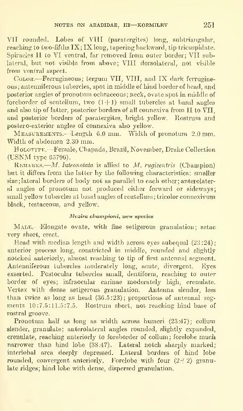
NOTES ON ARADIDAE, III—KORMILEV 251
VII rounded. Lobes of VIII (paratergites) long, subtriangular,
reaching to two-fifths IX; IX long, tapering backward, tip tricuspidate.
Spiracles II to VI ventral, far removed from outer border; VII sub-
lateral, but not visible from above; VIII dorsolateral, not visible
from ventral aspect.
Color.—Ferrugineous ; tergum VII, VIII, and IX dark ferrugine-
ous ; antenniferous tubercles, spot in middle of hind border of head, andposterior angles of pronotum ochraceous ; neck, ovate spot in middle of
foreborder of scutellum, two (1+ 1) small tubercles at basal angles
and also tip of latter, posterior borders of all connexiva from II to VII,
and posterior borders of paratergites, bright yeUow. Rostrum andpostero-exterior angles of connexiva also yeUow.
Measurements.-—^Length 6.0 mm. Width of pronotum 2.0 mm.Width of abdomen 2.30 mm.HoLOTYPE.—^Female, Chapada, Brazil, November, Drake Collection
(USNM type 65796).
Remarks.-—M. luteonotata is allied to M. rugiventris (Champion)
but it difi'ers from the latter by the following characteristics: smaller
size; lateral borders of body not as parallel to each other ; anterolater-
al angles of pronotum not produced either forward or sideways;
small yellow tubercles at basal angles of scutellum ; tricolor connexivumblack, testaceous, and yellow.
Mezira championi, new species
Male.—Elongate ovate, with fine setigerous granulation; setae
very short, erect.
Head with median length and width across eyes subequal (23 : 24)
;
anterior process long, constricted in middle, rounded and slightly
notched anteriorly, almost reaching to tip of first antennal segment.
Antenniferous tubercles moderately long, acute, divergent. Eyesexserted. Postocular tubercles small, dentiform, reaching to outer
border of eyes; infraocular carinae moderately high, crenulate.
Vertex with dense setigerous granulation. Antenna slender, less
than twice as long as head (36.5:23); proportions of antennal seg-
ments 10:7.5:11.5:7.5. Rostrum short, not reaching hind base of
rostral groove.
Pronotum half as long as width across humeri (23:47); collum
slender, granulate; anterolateral angles rounded, slightly expanded,
crenulate, reaching anteriorly to foreborder of coUmn; forelobe muchnarrower than hind lobe (38:47). Lateral notch sharply marked;
interlobal area deeply depressed. Lateral borders of hind lobe
rounded, convergent anteriorly. Forelobe with four (2-f 2) granu-
late ridges; hind lobe with dense, dispersed granulation.

252 PROCEEDINGS OF THE NATIONAL MUSEUM vol. ii8
Scutellum shorter than wide at base (21:25), rimmed on all sides;
disc granulate, with fine, low, granulate, T-shaped, median ridge,
Hemelytra reaching to foreborder of tergum VII ( cf") ; apical border
of corimn convex, strongly rounded, apical angle blunt.
Abdomen ovate, longer than wide (70:56), maximal width across
segment V. Connexivum II granulate; connexiva III to VII almost
without granulation, rather scabrous; postero-exterior angles barely
protruding; those of connexivum VII extending backward as rounded
lobes, extending posteriorly as far as third of hypopygium; lobes of
VIII (paratergites) clavate, reaching basal two-thirds of hypopy-
gium; latter cordate, with median ridge elevated backward and
terminating before reaching hind border. Spiracles II to VI ventral,
placed far from outer margin; those of VII sublateral, not visible from
above; VIII lateral and visible.
Color.—Piceous; connexiva III to VII, and lobes of VIII testa-
ceous; membrane blackish, whitish at base.
Measurements.—Length 4.74 mm. Width of pronotum 1.56
mm. Width of abdomen 1.86 mm.HoLOTYPE.—Male, Huatusco, Veracruz, Mexico, Jan. 2, 1952
(USNM type 65797).
Remarks.—This species is dedicated to the memory of the late
G. C. Champion, who has contributed so much to knowledge of
Central American Aradidae.
The species is allied closely to M. angustata (Champion), but it
differs from the latter by the following characteristics : anterior process
of head relatively longer, almost reaching tip of first antennal segment;
antenniferous tubercles narrower, not so divergent; pronotum less
constricted at sides; abdomen {d^) with sides widely rounded, not
parallel-sided; median ridge of hypopygiun raised backward, not
reaching tip of hypopygium.
Mezira mexicana, new species
Female.—Elongate ovate, densely covered with setigerous granu-
lations; granules fine, each with very short erect setal hair.
Head shorter than width across eyes (27 :30) ; anterior process long,
robust, slightly widening forward, rounded, notched in middle, ante-
riorly reaching to apical third of first antennal segment. Antenniferous
tubercles spiniform, divergent, reaching middle of first antennal
segment. Eyes semiglobose, exserted. Postocular tubercles small,
acute, reaching outer border of eyes; infraocular carinae moderately
high, crenulate; vertex with four rows of setigerous tubercles. An-
tennae slender, less than twice as long as head (46:27), proportions
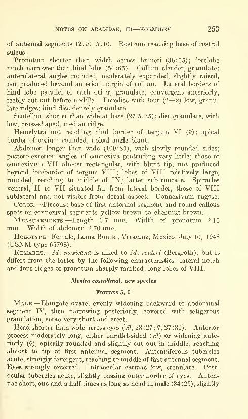
NOTES ON ARADIDAE, III—KORMILEV 253
of antennal segments 12:9:15:10. Rostrum reaching base of rostral
sulcus.
Pronotum shorter than width across humeri (36:65); forelobe
much narrower than hind lobe (54:65). Collum slender, granulate;
anterolateral angles rounded, moderately expanded, slightly raised,
not produced beyond anterior margin of collum. Lateral borders of
hind lobe parallel to each other, granulate, convergent anteriorly,
feebly cut out before middle. Foredisc with four (2+ 2) low, granu-
late ridges; hind disc densely granulate.
Scutellum shorter than wide at base (27.5:35); disc granulate, with
low, cross-shaped, median ridge.
Hemelytra not reaching hind border of tergum VI (9) ; apical
border of corium rounded, apical angle blunt.
Abdomen longer than wide (109:81), with slowly rounded sides;
postero-exterior angles of connexiva protruding very little; those of
connexivum VII almost rectangular, with blunt tip, not produced
beyond foreborder of tergum VIII; lobes of VIII relatively large,
rounded, reaching to middle of IX; latter subtruncate. Spiracles
ventral, II to VII situated far from lateral border, those of VIII
sublateral and not visible from dorsal aspect. Connexivum rugose.
CoLOR.^—^Piceous ; base of first antennal segment and round callous
spots on connexival segments yellow-brown to chestnut-brown.
Measurements.—Length 6.7 mm. Width of pronotum 2.16
mm. Width of abdomen 2.70 mm.Holotype: Female, Loma Bonita, Veracruz, Mexico, July 10, 1948
(USNM type 65798).
Remarks.—M. mexicana is allied to M. reuteri (Bergroth), but it
differs from the latter by the following characteristics: lateral notch
and four ridges of pronotum sharply marked; long lobes of VIII.
Mesira costalimaiy new species
Figures 5, 6
Male.—Elongate ovate, evenly widening backward to abdominal
segment IV, then narrowing posteriorly, covered with setigerous
granulation, setae very short and erect.
Head shorter than wide across eyes ( cf , 23 : 27 ; 9, 27 : 30) . Anterior
process moderately long, either parallel-sided (cf) or widening ante-
riorly (9), apically rounded and slightly cut out in middle; reaching
almost to tip of first antennal segment. Antenniferous tubercles
acute, strongly divergent, reaching to middle of first antennal segment.
Eyes strongly exserted. Infraocular carinae low, crenulate. Post-
ocular tubercles acute, slightly passing outer border of eyes. Anten-
nae short, one and a half times as long as head in male (34 :23), slightly
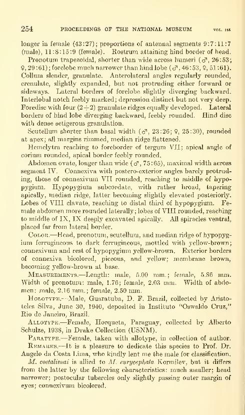
254 PROCEEDINGS OF THE NATIONAL MUSEUM vol. iis
longer in female (43 : 27) ;proportions of antenna! segments 9:7:11:7
(male), 11:8:15:9 (female). Rostrum attaining hind border of head.
Pronotum trapezoidal, shorter than wide across humeri (cf, 26:53;
9, 29 : 61) ; forelobe much narrower than hind lobe ( cf , 46 : 53, 9, 51 :61).
Collum slender, granulate. Anterolateral angles regularly rounded,
crenulate, slightly expanded, but not protruding either forward or
sideways. Lateral borders of forelobe slightly diverging backward.
Interlobal notch feebly marked; depression distinct but not very deep.
Foredisc with four (2+2) granulate ridges equally developed. Lateral
borders of hind lobe diverging backward, feebly rounded. Hind disc
with dense setigerous granulation.
Scutellmn shorter than basal width (cf, 23:26; 9, 25:30), rounded
at apex; all margins rimmed, median ridge flattened.
Hemelytra reaching to foreborder of tergum VII; apical angle of
corium rounded, apical border feebly rounded.
Abdomen ovate, longer than wide (cf , 75:65), maximal width across
segment IV. Connexiva with postero-exterior angles barely protrud-
ing, those of connexivum VII rounded, reaching to middle of hypo-
pygium. Hypopygium subcordate, with rather broad, tapering
apically, median ridge, latter becoming slightly elevated posteriorly.
Lobes of VIII clavate, reaching to distal thu'd of hypopygimn. Fe-
male abdomen more rounded laterally; lobes of VIII rounded, reaching
to middle of IX, IX deeply excavated apicaUy. All spiracles ventral,
placed far from lateral border.
Color.—Head, pronotum, scuteUum, and median ridge of hypopyg-
ium ferrugineous to dark ferrugineous, mottled with yellow-brown;
connexivmn and rest of hypopygium yellow-brown. Exterior borders
of connexiva bicolored, piceous, and yellow; membrane brown,
becoming yellow-brown at base.
Measurements.—Length: male, 5.00 mm.; female, 5.86 mm.Width of pronotum: male, 1.76; female, 2.03 mm. Width of abdo-
men: male, 2.16 mm.; female, 2.50 mm.HoLOTYPE.^—Male, Guaratuba, D. F. Brazil, collected by Aristo-
teles Silva, June 30, 1940, deposited in Instituto "Oswaldo Cruz,"
Rio de Janeiro, Brazil.
Allotype.—Female, Horqueta, Paraguay, collected by Alberto
Schulze, 1938, in Drake Collection (USNM).Paratype.—Female, taken with allotype, in collection of author.
Remarks.—It is a pleasure to dedicate this species to Prof. Dr.
Angelo da Costa Lima, who kindly lent me the male for classification.
M. costalimai is allied to M. eurycephala Kormilev, but it differs
from the latter by the following characteristics: much smaller; headnarrower; postocular tubercles only slightly passing outer margin of
eyes; connexivum bicolored.

NOTES ON ARADIDAE, III—KORMILEV 255
Mezira carioca, new species
Figure 7
Female.—Elongate ovate, lateral borders parallel, gently narrow-
ing anteriorly and posteriorly; body covered with rough setigerous
granulation, with short, erect setae.
Head shorter than width across eyes (28:32); anterior process with
sides subparallel, rounded anteriorly, and excavated at tip, reaching
to, or almost to, tip of first antennal segment. Antenniferous tuber-
cles pointed, slightly divergent, reaching to basal third of first antennal
segment. Eyes exserted. Postocular tubercles strong, pointed, pro-
jecting laterally far beyond outer margin of each eye. Infraocular
carinae moderately high, crenulate; vertex with V-shaped rows
of granulae. Antennae very short, slender, less than one and a half
times length of head (39:28); proportions of antennal segments
12:7.5:12:7.5. Rostrum reaches to base of rostral sulcus.
Pronotum less than half as long as width across humeri (32 : (58) : 67)
.
Collum slender; anterolateral angles roundly expanded, slightly raised
laterally, crenulate; lateral borders slightly cut out in middle, slightly
convex at humeri; hind border also slightly excavated at middle,
Foredisc with four (2+ 2) oblique, granulate ridges; hind disc with
scattered, rough, setigerous granulations.
Scutellum shorter than basal width (25:35), typical of Neotropical
Mezira species.
Hemelytra short, reaching hind border of tergum VI (9); baso-
lateral borders slightly reflexed; apical border of the corium and apex
of apical angle very feebly rounded.
Abdomen longer than wide (95:74), elongate ovate, almost parallel-
sided along middle, tapering in gentle curve posteriorly. Connexivumcovered with setigerous granulation; connexiva with postero-exterior
angles not protruding; lobes of VIII relatively large; rounded, reaching
to middle of IX, latter cut out at tip. All spiracles ventral, placed
far from outer margin.
CoLOR.^—Uniformly ferrugineous.
Measurements.—Length 6.33. Width of pronotum 2.23 mm.Width of abdomen 2.47 mm.HoLOTYPE.—Female, Rio de Janeiro, Brazil, Drake Collection
(USNM type 65799).
Remarks.—M. carioca is allied to M. saltensis Kormilev, but it
differs from the latter by the following characteristics: much smaller
size; lateral borders of pronotum less excavated; anterior process of
head relatively shorter; lobes of VIII relatively larger, reaching to
middle of IX (only to basal fourth of IX in M. saltensis) ; anterolateral
angles of pronotum more rounded and produced farther forward.
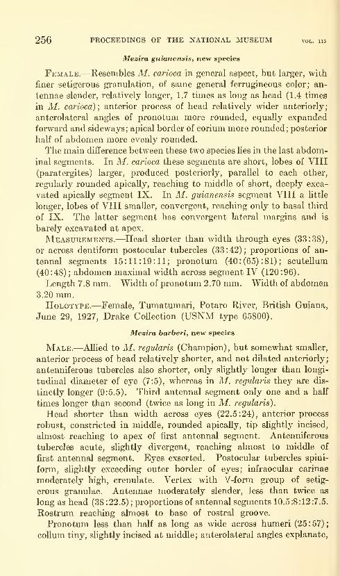
256 PROCEEDINGS OF THE NATIONAL MUSEUM vol. us
Mezira guianensis, new species
Female.—Resembles M. carioca in general aspect, but larger, with
finer setigerous granulation, of same general ferrugineous color; an-
tennae slender, relatively longer, 1.7 times as long as head (1.4 times
in M. carioca) ; anterior process of head relatively wider anteriorly;
anterolateral angles of pronotum more rounded, equally expanded
forward and sideways; apical border of corium more rounded; posterior
half of abdomen more evenly rounded.
The main difference between these two species lies in the last abdom-
inal segments. In M. carioca these segments are short, lobes of VIII
(paratergites) larger, produced posteriorly, parallel to each other,
regularly rounded apicaUy, reaching to middle of short, deeply exca-
vated apically segment IX. In M. guianensis segment VIII a little
longer, lobes of VIII smaller, convergent, reaching only to basal third
of IX. The latter segment has convergent lateral margins and is
barely excavated at apex.
Measurements.—Head shorter than width through eyes (33:38),
or across dentiform postocular tubercles (33:42); proportions of an-
tennal segments 15:11:19:11; pronotum (40 : (65) : 81) ; scuteUum
(40:48); abdomen maximal width across segment IV (120:96).
Length 7.8 mm. Width of pronotum 2.70 mm. Width of abdomen3.20 mm.HoLOTYPE.—Female, Tumatumari, Potaro River, British Guiana,
June 29, 1927, Drake Collection (USNM type 65800).
Mezira barberi, new species
Male.—AUied to M. regularis (Champion) , but somewhat smaller,
anterior process of head relatively shorter, and not dilated anteriorly;
antenniferous tubercles also shorter, only slightly longer than longi-
tudinal diameter of eye (7:5), whereas in M. regularis they are dis-
tinctly longer (9:5.5). Third antennal segment only one and a half
times longer than second (twice as long in M. regularis).
Head shorter than width across eyes (22.5:24), anterior process
robust, constricted in middle, rounded apically, tip slightly incised,
almost reaching to apex of first antennal segment. Antenniferous
tubercles acute, slightly divergent, reaching almost to middle of
first antennal segment. Eyes exserted. Postocular tubercles spini-
form, slightly exceeding outer border of eyes; infraocular carinas
moderately high, crenulate. Vertex with V-form group of setig-
erous granulae. Antennae moderately slender, less than twice as
long as head (38:22.5); proportions of antennal segments 10.5:8:12:7.5.
Rostrum reaching almost to base of rostral groove.
Pronotum less than half as long as wide across humeri (25:57);
collum tiny, slightly incised at middle; anterolateral angles explanate,

NOTES ON ARADIDAE, III—KORMILEV 257
rounded, crenulate, not extending beyond front border of collum.
Lateral notch sharply marked; interlobal depression deep; foredisc
with four (2+ 2) high, granulate ridges; hind lobe wider than fore-
lobe (57:43); lateral margins parallel to each other, roundly con-
vergent anteriorly; hind margins with three excavations in middle
and mesad of rounded hind angles; hind disc roughly granulate.
Scutellum shorter than wide at base (23:30); lateral borders
slightly sinuate near tip; disc granulate; median ridge cross-shaped.
Hemelytra reaching over foreborder of tergum VII ( cf ) or to hmdborder of tergum VI (9).
Abdomen longer than wide (83:65); lateral borders parallel-sided,
roundly convergent posteriorly; connexiva with postero-exterior
angles not protruding. Hypopygium cordiform, median ridge slightly
raised backward. Spiracles II to VII ventral, placed far from lateral
margin, those of VIII lateral and visible from above. Lobes of VIII
in female relatively short, not reaching middle of IX, latter
tricuspidate.
Color.—Ferrugineous, partially piceous; hind borders of connexi-
val segments yellow; membrane brown.
Measurements.—Female, head shorter than wide across eyes
(23:27); proportions of antennal segments 10:7.5:12.5:9; pronotum
shorter than wide across humeri (30:60) ; scutellum shorter than basal
width (25:30); abdomen longer than wide (90:68).
Total length: male, 7.85 mm.; female, 8.65 mm. Width of pro-
notum: male, 2.85 mm.; female, 3.0 mm. Width of abdomen: male,
3.25; female, 3.4 mm.HoLOTYPE.—Male, Hoboken, New Jersey, intercepted on orchid
plants from Venezuela, April 5, 1939 (USNM type 65801).
Allotype (female) and 2 paratypes, same labels as holotype.
Remarks.—This species is dedicated to the late Harry G. Barber,
eminent American hemipterist.
Mezira paralata, new species
Female.—Broadly ovate, abdomen subparallel-sided, almost sub-
rectangular in outline, partially covered with conspicuous, yeUow,
curled hairs.
M. paralata is closely allied to M. lata (Champion) but differs from
it by the following characteristics: anterior process of head constricted
at base, dilated, and distinctly notched at tip, barely reaching to
middle of first antennal segment; abdomen subrectangular (not ovate
as in M. lata) ; lobes of VIII very short, rounded, reaching only to
basal fourth of IX, latter rounded posteriorly ; antenniferous tubercles
blunt, subpai'allel to each other; rostrum short, not reaching to hind
border of rostral groove.
258 PROCEEDINGS OF THE NATIONAL MUSEUM vol. hb
Color.—Striking, bright testaceous; head and ridges of pronotum
ferrugineous ; membrane pale brown; body partially clothed
with golden yellow curly hairs; ventral surface of body coated with
thin layer of white incrustation. Spiracles II to VII ventral, those
of VIII sublateral and not visible from dorsal view.
Measurements.—Head almost as long as wide (25 :26) ;proportions
of antennal segments 17:11 :18:10; pronotum (31 :(47) :60); scutellum
(24:31); abdomen across segment V (90:75).
Length 8.63 mm. Width of pronotum 3.0 mm. Width of ab-
domen 3.75 mm.HoLOTYPE.—Female, Suretka, Lim6n Prov., Costa Rica, in Drake
CoUection (USNM type 65802).
U.S. GOVERNMENT PRINTING OFFICE:l9e4


Proceedings of ^^\the United States IK-S^^National Museum ^'^^^
SMITHSONIAN INSTITUTION • WASHINGTON, D.C.
Volume 115 1964 Number 3484
A GENERIC REVISION OF THE LEAFHOPPER SUBFAMILYNEOCOELIDIINAE
(HOMOPTERA: CICADELLIDAE)
By James P. Kramer'
The leafhopper subfamily Neocoelidiinae was erected by Oman(1943) in order to accommodate the two North American genera,
Neocoelidia and Paracoelidea, which had been placed in the Jassinae
(Deltocephalinae of modern workers) for many years. Evans (1947)
added the Neotropical genera Biza, Chinaia, Coelidiana, and Salvina
in his generic checklist of the subfamily. DeLong (1953) partially
revised the group, adding new genera and subgenera as well as manynew species. He included only one of the genera added by Evans and
attempted to place the Neotropical Neocoelidiinae described by Fowler
and Osborn with reference to the literature alone. More recently newgenera and species have been added by Kramer (1959, 1961, and 1962)
and by Kramer and Linnavuori (1959). Although our knowledge of
Neotropical fauna is still quite meager, it seems appropriate to synthe-
size the information we do have at present in order that future workers
may have a foundation upon which to build.
I Entomology Research Division, Agricultural Research Service, U.S. Department of Agriculture.
259
260 PROCEEDINGS OF THE NATIONAL MUSEUM vol. 115
Subfamily Neocoelidiinae
Description.—Small to large leafhoppers (3-13 mm.). Clypellus
either approximately parallel-sided or widened distally. Lateral
frontal sutures extended near or to ocelli, which are on or near anterior
margin of crown or less often on face. Ocellocular areas developed as
distinct ledges above antennal pits. Antennae long to very long, fre-
quently exceeding entire length of body including forewings at rest.
Face and crown either separated by carina or with carina lacking;
carina, when present, usually limited to distance between ocelli. Headin dorsal view angled or rounded apically, often strongly produced
beyond eyes. Pronotum short, much wider than long, with lateral
margins carinated. Scutellum large and well developed. Venation
of forewings usually obscure except apically and with either two or
three preapical cells. Male genital structures variously modified but
styles usually simple. Ground color various shades of white, yellow,
or orange. There are often contrasting markings of various hues on
the head, thorax, and forewings.
Diagnosis.—The most useful set of characters for recognition of the
Neocoelidiinae consists of the distinct ledge above each antennal base,
the exceptionally long antennae (particularly as found in many of the
Neotropical genera), and the venation of the forewings, which is
usually highly obscure except apically.
Discussion.—As far as it is known, the subfamily is restricted to
the Americas, with good representation in both the temperate and
tropical areas. The available host and food plant records seem to
indicate that most of the species occur on trees or shrubs. According
to DeLong (1953), various Nearctic species have been taken on the fol-
lowing genera of plants: Pinus, Acacia, Rhus, Arctostaphylos, and
Sphaeralcea. Except for one species of Chinaia known to breed on
avocado, the plant relationships of the Neotropical species are
unknown.The key to genera which follows is based almost entu-ely upon char-
acters of the male genitalia. Although most of these structures are
concealed within the genital capsule and require dissection and clear-
ing in order to be observed, they provide the most concrete evidence
for an arrangement of genera within the subfamily. Emphasis on the
male genitalia for the delimiting of genera is not without precedent.
Oman (1949) used this feature extensively in arranging the North
American Deltocephalinae, as did Young (1952) in revising the NewWorld Typhlocybinae. Both men provided generic definitions within
the respective subfamilies that were more definite and better delimited
than any prior to their work. It is hoped that this study will serve
a similar purpose.
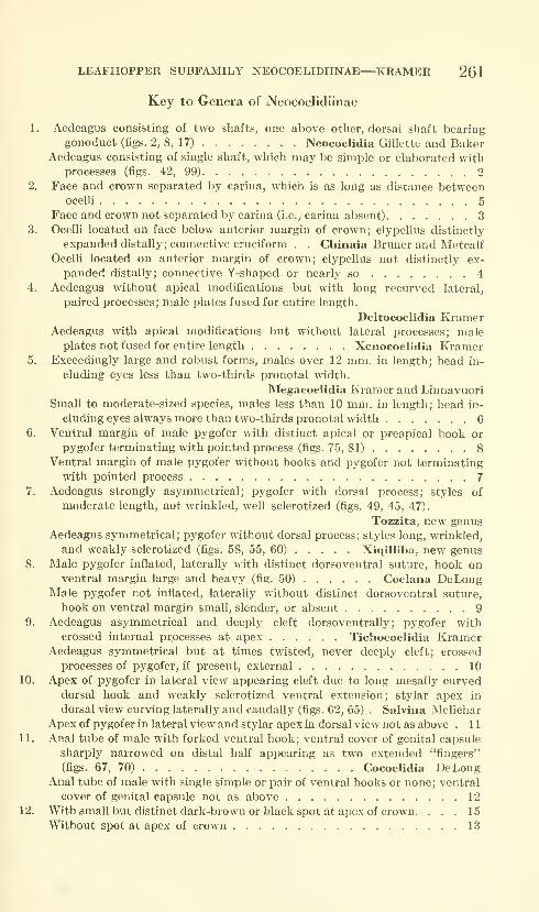
LEAFHOPPER SUBFAMILY NEOCOELIDIINAE—^KRAMER 261
Key to Genera of Neocoelidiinae
1. Aedeagus consisting of two shafts, one above other, dorsal shaft bearing
gonoduct (figs. 2, 8, 17) Neocoelidia Gillette and BakerAedeagus consisting of single shaft, which may be simple or elaborated with
processes (figs. 42, 99) 2
2. Face and crown separated by carina, which is as long as distance betweenocelli 5
Face and crown not separated by carina (i.e., carina absent) 3
3. Ocelli located on face below anterior margin of crown; clypellus distinctly
expanded distally; connective cruciform . . Chinaia Bruner and MetcalfOcelli located on anterior margin of crown; clypellus not distinctly ex-
panded distally; connective Y-shaped or nearly so 4
4. Aedeagus without apical modifications but with long recurved lateral,
paired processes; male plates fused for entire length.
Deltocoelidia KramerAedeagus with apical modifications but without lateral processes; male
plates not fused for entire length Xenocoelidia Kramer5. Exceedingly large and robust forms, males over 12 mm. in length; head in-
cluding eyes less than two-thirds pronotal width.
Megacoelidia Kramer and Linnavuori
Small to moderate-sized species, males less than 10 mm. in length; head in-
cluding eyes always more than two-thirds pronotal width 6
6. Ventral margin of male pygofer with distinct apical or preapical hook or
pygofer terminating with pointed process (figs. 75, 81) 8
Ventral margin of male pygofer without hooks and pygofer not terminating
with pointed process 7
7. Aedeagus strongly asymmetrical; pygofer with dorsal process; styles of
moderate length, not wrinkled, well sclerotized (figs. 49, 45, 47).
Tozzita, new genusAedeagus symmetrical; pygofer without dorsal process; styles long, wrinkled,
and weakly sclerotized (figs. 58, 55, 60) Xiqilliba, new genus
8. Male pygofer inflated, laterally with distinct dorsoventral suture, hook onventral margin large and heavy (fig. 50) Coelana DeLong
Male pygofer not inflated, laterally without distinct dorsoventral suture,
hook on ventral margin small, slender, or absent 9
9. Aedeagus asymmetrical and deeply cleft dorsoventrally;
pygofer withcrossed internal processes at apex Tichocoelidia Kramer
Aedeagus symmetrical but at times twisted, never deeply cleft; crossed
processes of pygofer, if present, external 10
10. Apex of pygofer in lateral view appearing cleft due to long mesally curveddorsal hook and weakly sclerotized ventral extension; stylar apex in
dorsal view curving laterally and caudally (figs. 62, 65) . Salvina Melichar
Apex of pygofer in lateral view and stylar apex in dorsal view not as above . 1
1
11. Anal tube of male with forked ventral hook; ventral cover of genital capsule
sharply narrowed on distal half appearing as two extended "fingers"
(figs. 67, 70) Cocoelidia DeLongAnal tube of male with single simple or pair of ventral hooks or none; ventral
cover of genital capsule not as above 12
12. With small but distinct dark-brown or black spot at apex of crown. ... 15
Without spot at apex of crown 13
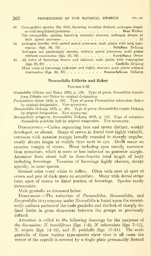
262 PROCEEDINGS OF THE NATIONAL MUSEUM vol. us
13. Cercopidlike species (fig. 114); forewing venation distinct; aedeagus simple
or with long lateral processes Biza Walker
Not cercopidlike species; forewing venation obscure; aedeagus simple or
with apical processes 14
14. Aedeagus slender, with paired apical processes; male plates with long mac-
rosetae (figs. 80, 78) Nelidina DeLongAedeagus not particularly slender, without apical processes; male plates
without macrosetae (figs. 95, 98) Coelidiana Oman15. All veins of forewings brown and distinct; male plates with macrosetae
(figs. 88, 89) Coelella DeLongMost veins of forewings yellowish and highly obscure; male plates without
macrosetae (figs. 81, 85) Neocoelidiana DeLong
Neocoelidia Gillette and Baker
Figures 1-35
Neocoelidia Gillette and Baker 1895, p. 103. Type of genus Neocoelidia tumidi-
frons Gillette and Baker by original designation.
Paracoelidea Baker 1898, p. 292. Type of genus Paracoelidea tuherculata Baker
by original designation. New synonymy.
Stenocoelidia DeLong 1953, p. 104. Type of genus Stenocoelidia virgata DeLongby original designation. New synonymy.
Stenocoelidia subgenus Eurycoelidia DeLong 1953, p. 112. Type of subgenus
Neocoelidia pulchella Ball by original designation. New synonymy.
Description.—Carina separating face and crown distinct, weakly
developed, or absent. Shape of crown in dorsal view highly variable,
extremes with anterior margin broadly rounded to sharply angular,
nearly always longer at middle than next to eye. OceUi occur on
anterior margin of crown. Head including eyes usually narrower
than pronotum, which is more or less indented on posterior margin.
Antennae from about half to three-fourths total length of body
including forewings. Venation of forewings highly obscure, except
apically, in most species.
Ground color ivory white to yellow. Often with spot at apex of
crown and pair of dark spots on scutellum. Many with dorsal stripe
from apex of crown to distal portion of forewings. Species rarely
immaculate.
Male genitaha as discussed below.
Discussion.—The reduction of Paracoelidea, Stenocoelidia, and
Eurycoelidia to synonymy under Neocoelidia is based upon the consist-
ently uniform pattern of the male genitalia and the lack of sharply de-
fined limits in gross characters between the groups as previously
defined.
Attention is called to the following drawings for the pm'poses of
the discussion: N. tumidifrons (figs. 1-6), A^. tuherculata (figs. 7-13),
N. virgata (figs. 14-16), and A^. pulchella (figs. 17-24). The male
genitalia of these various type-species show that in all cases the
venter of the capsule is covered by a single plate presumably formed
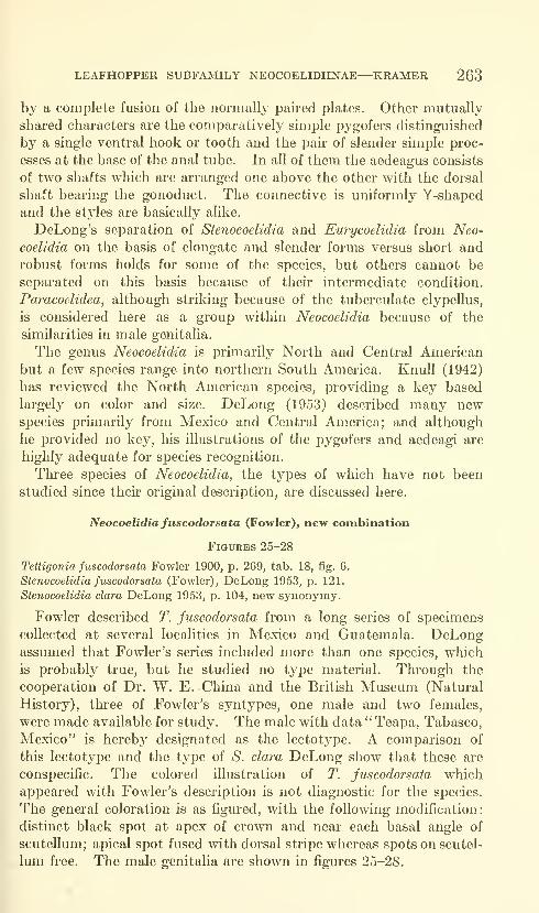
LEAFHOPPER SUBFAMILY NEOCOELIDIINAE—KRAMER 263
by a complete fusion of the normally paired plates. Other mutually
shared characters are the comparatively simple pygofers distinguished
by a single ventral hook or tooth and the pair of slender simple proc-
esses at the base of the anal tube. In all of them the aedeagus consists
of two shafts which are arranged one above the other with the dorsal
shaft bearing the gonoduct. The connective is uniformly Y-shaped
and the styles are basically alike.
DeLong's separation of Stenocoelidia and Eurycoelidia from Neo-
coelidia on the basis of elongate and slender forms versus short and
robust forms holds for some of the species, but others cannot be
separated on this basis because of their intermediate condition.
Paracoelidea, although striking because of the tuberculate clypellus,
is considered here as a group within Neocoelidia because of the
similarities in male genitalia.
The genus Neocoelidia is primarily North and Central Americanbut a few species range into northern South America. Knull (1942)
has reviewed the North American species, providing a key based
largely on color and size. DeLong (1953) described many newspecies primarily from Mexico and Central America; and although
he provided no key, his illustrations of the pygofers and aedeagi are
highly adequate for species recognition.
Three species of Neocoelidia, the types of which have not been
studied since their original description, are discussed here.
Neocoelidia fuscodorsata (Fowler), new combination
Figures 25-28
Tettigonia fuscodorsata Fowler 1900, p. 269, tab. 18, fig. 6.
Stenocoelidia fuscodorsata (Fowler), DeLong 1953, p. 121.
Stenocoelidia clara DeLong 1953, p. 104, new synonymy.
Fowler described T. fuscodorsata from a long series of specimens
collected at several localities in Mexico and Guatemala. DeLongassumed that Fowler's series included more than one species, which
is probably true, but he studied no type material. Through the
cooperation of Dr. W. E. China and the British Museum (Natural
History), three of Fowler's syntypes, one male and two females,
were made available for study. The male with data " Teapa, Tabasco,
Mexico" is hereby designated as the lectotype. A comparison of
this lectotype and the type of S. clara DeLong show that these are
conspecific. The colored illustration of T. fuscodorsata which
appeared with Fowler's description is not diagnostic for the species.
The general coloration is as figured, with the following modification:
distinct black spot at apex of crown and near each basal angle of
scutellum; apical spot fused with dorsal stripe whereas spots on scutel-
lum free. The male genitaha are shown in figures 25-28.

264 PROCEEDINGS OF THE NATIONAL MUSEUM vol. iis
Neocoelidia verecunda (Fowler), new combination
Tettigonia verecunda Fowler 1900, p. 269, tab. 18, fig. 7.
Stenocoelidia verecunda (Fowler), DeLong 1953, p. 122.
Unfortunately this Guatemalan species is knoAvn from females
only, and its generic placement is open to question. I have studied
Fowler's two syntypes in the British Museum and two additional
specimens in the collection of the U.S. National Museum. In none
of the specimens are the longitudinal red markings on the crown,
pronotum, and forewings quite as distinct as illustrated with Fowler's
original description.
Neocoelidia crenulata Osborn
Figures 29-35
Neocoelidia crenulata Osborn 1923, p. 79.
This species was described from a single male from Minca, Colombia.
The type was made available through the courtesy of Dr. G. E. Wal-
lace and the Carnegie Museum. In general it is marked like N.
fuscodorsata except that there are no spots on the scutellum and the
longitudinal dorsal stripe is crenulate on its margins. The genitalia
of the type are illustrated in figures 29-35.
Chinaia Bruner and Metcalf
Figures 37, 113
Chinaia Bruner and Metcalf 1934, p. 120. Type of genus Chinaia bella Bruner
and Metcalf by original designation.
Description.—Without carina separating face and crown. Cly-
pellus expanded distally. Shape of crown in dorsal view broadly
rounded, wider than long. Ocelli on face distinctly below anterior
margin of crown. Head including e^^es narrower than pronotum,
which is not indented on posterior margin. Antenna very long,
exceeding total length of body including forewings. Venation of
forewings obscure except at apex.
Ground color yellowish to pale orange, with orange to bright red-
orange markings on pronotum and extensively on forewings. Dark-
brown markings often found on clavus and apical portion of forewings
as well.
Male genitalia: Valve obscure. Plates deep and scoop-shaped.
Pygofer either with dorsal processes or an elongation of terminus.
Anal tube simple. Connective cruciform and not fused with aedeagus.
Aedeagus moderately straight or recurved with or without lateral
flaps.
Discussion.—This is a Neotropical genus; most of the described
species occur in Central America, but a few are South American.
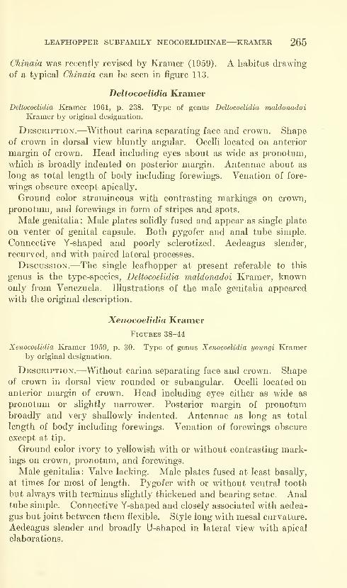
LEAFHOPPER SUBFAMILY NEOCOELIDIINAE—KRAMER 265
Chinaia was recently revised by Kramer (1959). A habitus drawing
of a typical Chinaia can be seen in figure 113.
Deltocoelidia Kramer
Deltocoelidia Kramer 1961, p. 238. Type of genus Deltocoelidia maldonadoi
Ivramer by original designation.
Description.—Without carina separating face and crown. Shapeof crown in dorsal view bluntly angular. Ocelli located on anterior
margin of crown. Head including eyes about as wide as pronotum,
which is broadly indented on posterior margin. Antennae about as
long as total length of body including forewings. Venation of fore-
wings obscure except apically.
Ground color stramineous with contrasting markings on crown,
pronotum, and forewings in form of stripes and spots.
Male genitalia: Male plates solidly fused and appear as single plate
on venter of genital capsule. Both pygofer and anal tube simple.
Connective Y-shaped and poorly sclerotized. Aedeagus slender,
recurved, and with paired lateral processes.
Discussion.—The single leafhopper at present referable to this
genus is the type-species, Deltocoelidia maldonadoi Kramer, knownonly from Venezuela. Illustrations of the male genitaha appeared
with the original description.
Xenocoelidia Kramer
Figures 38-44
Xenocoelidia Eo-amer 1959, p. 30. Type of genus Xenocoelidia youngi Kramerby original designation.
Description.—Without carina separating face and crown. Shapeof crown in dorsal view rounded or subangular. Ocelli located onanterior margin of crown. Head including eyes either as wide as
pronotum or slightly narrower. Posterior margin of pronotumbroadly and very shallowly indented. Antennae as long as total
length of body including forewings. Venation of forewings obscure
except at tip.
Ground color ivory to yellowish with or without contrasting mark-ings on crown, pronotum, and forewings.
Male genitalia: Valve lacldng. Male plates fused at least basally,
at times for most of length. Pygofer with or without ventral tooth
but always with terminus slightly thickened and bearing setae. Analtube simple. Connective Y-shaped and closely associated with aedea-
gus but joint between them flexible. Style long with mesal curvature.
Aedeagus slender and broadly U-shaped in lateral view with apical
elaborations.
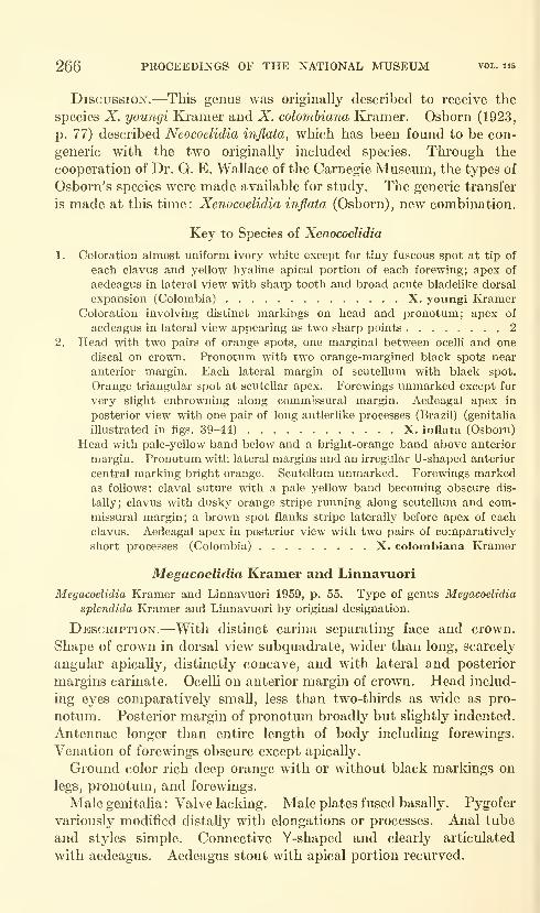
266 PROCEEDINGS OF THE NATIONAL MUSEUM vol. 115
Discussion.—This genus was originally described to receive the
species X. youngi Kramer and X. colombiana Kramer. Osborn (1923,
p. 77) described Neocoelidia inflata, which has been found to be con-
generic with the two originally included species. Through the
cooperation of Dr. G. E. Wallace of the Carnegie Museum, the types of
Osborn's species were made available for study. The generic transfer
is made at this tune: Xenocoelidia inflata (Osborn), new combination.
Key to Species of Xenocoelidia
1. Coloration almost uniform ivory white except for tiny fuscous spot at tip of
each clavus and yellow hyaline apical portion of each forewing; apex of
aedeagus in lateral view with sharp tooth and broad acute bladelike dorsal
expansion (Colombia) X. youngi KramerColoration involving distinct markings on head and pronotum; apex of
aedeagus in lateral view appearing as two sharp points 2
2. Head with two pairs of orange spots, one marginal between ocelh and one
discal on crown. Pronotum with two orange-margined black spots near
anterior margin. Each lateral margin of scutellum with black spot.
Orange triangular spot at scutellar apex. Forewings unmarked except for
very slight enbrowning along commissural margin. Aedeagal apex in
posterior view with one pair of long antlerlike processes (Brazil) (genitalia
illustrated in figs. 39-44) X. inflata (Osborn)
Head with pale-yellow band below and a bright-orange band above anterior
margin. Pronotum with lateral margins and an irregular U-shaped anterior
central marking bright orange. Scutellum unmarked. Forewings markedas follows: claval suture with a pale yellow band becoming obscure dis-
tally; clavus with dusky orange stripe running along scutellum and com-missural margin; a brown spot flanks stripe laterally before apex of each
clavus. Aedeagal apex in posterior view with two pairs of comparatively
short processes (Colombia) X. colombiana Kramer
Megacoelidia Kramer and Linnavuori
Megacoelidia Kramer and Linnavuori 1959, p. 55. Type of genus Megacoelidia
splendida Kramer and Linnavuori by original designation.
Description.—With distinct carina separating face and crown.
Shape of crown in dorsal view subquadrate, wider than long, scarcely
angular apically, distinctly concave, and with lateral and posterior
margins carinate. Ocelli on anterior margin of crown. Head includ-
ing eyes comparatively small, less than two-thirds as wide as pro-
notum. Posterior margin of pronotum broadly but slightly indented.
Antennae longer than entire length of body including forewings.
Venation of forewings obscure except apically.
Ground color rich deep orange with or without black markings on
legs, pronotum, and forewings.
Male genitalia : Valve lacking. Male plates fused basally. Pygofer
variously modified distally with elongations or processes. Anal tube
and styles simple. Connective Y-shaped and clearly articulated
with aedeagus. Aedeagus stout with apical portion recurved.

LEAFHOPPER SUBFAMILY NEOCOELIDIINAE—KRAMER 267
Discussion.—Megacoelidia contains only two known South Amer-ican species, both of which appeared with the original generic descrip-
tion. The genus contains the largest members of the subfamily.
Key to Species of Megacoelidia
Dorsal markings consisting of single narrow black border on posterior margin of
pronotum and on apex of each forewing. Male plates in ventral view pointed
apically and aedeagus with long paired lateral processes (Brazil).
M. splendida Kramer and Linnavuori
Dorsal markings limited to single narrow black border on apex of each forewing.
Male plates in ventral view rounded apically and aedeagus with short paired
lateral processes (Bolivia) M. aurantia Kramer and Linnavuori
Tozzita^ new genus
Figures 45-49
Type of genus Tozzita ips, new species.
Description.—With slender but distinct carina separating face andcrown. Crown in dorsal view much longer than wide, bluntly angular
apically, and strongly produced beyond eyes. Ocelli near anterior
margin of crown but posterior to carina. Head including eyes dis-
tinctly narrower than pronotum. Posterior margin of pronotumbroadly indented. Antennae as long as body including forewings at
rest. Venation of forewings highly obscure except apically.
Ground color stramineous to light brown without definite markings
except for black spot at apex of crown. Forewings stramineous
hyahne.
Male genitalia.—Valve lacking. Male plates fused for nearly
entire length. Pygofer modified only dorsally with processes or exten-
sions. Anal tube with heavily sclerotized portions. Connective moreor less V-shaped and articulated with strongly asymmetrical aedeagus.
Tozzita ips, new species
Figures 45-49
Length.—Male 6.75 mm.Coloration.—Stramineous with indefinite brownish areas on head
and thorax. Only distinct marking is black apical spot on crown.
Forewings stramineous hyaline.
Male genitalia.—Capsule in ventral view with slight apical notch
on ventral cover which is exceeded by the pygofer (fig. 48). Capsule
in lateral view with few tiny spines on ventral margin of pygofer,
dorsum with blunt, sclerotized process; anal tube with two heavily
sclerotized plates, posterior one notched dorsally (fig. 45). Aedeagus
in ventral aspect slender and strongly asymmetrical (fig. 49), with
apex forked and gonopore opening on narrow mesal extension (fig. 46).
681-546—63 2

268 PROCEEDINGS OF THE NATIONAL MUSEUM vol. 115
Aedeagus slender in lateral view with dorsal hump on basal half and
with apical mesal extension slender and recurved (fig. 47).
HoLOTYPE.—Male, Riberalta, Bolivia, W. M. Mann, January,
1921-22, Mulford Biological Expedition. USNM type 65826.
Female unknown.Xiqilliba, new genus
Figure 55-60
Type of genus Xiqilliba hellator, new species.
Description.—With distinct carina separating face and crown.
Crown in dorsal view wider than long and bluntly angular apically.
Ocelli near anterior margin of crown but sHghtly posterior to carina.
Head including eyes narrower than pronotum. Posterior margin of
pronotum mesally indented. Antennae as long as body including
forewings. Venation of forewings comparatively distinct.
Ground color yellow marked with brown or black on head,
pronotum, and forewings.
Male genitalia.—Valve lacking. Male plates fused basally for
about half length. Pygofer simple. Anal tube with long paired proc-
esses extending anteriorly into genital chamber. Styles very long,
wrinkled, slender, and poorly sclerotized. Connective modified
Y-shaped. Aedeagus slender and simple.
Xiqilliba bellator, new species
Figures 55-60
Length.—Male 6.5 mm.Coloration.—Grossly appearing as yellow leafhopper with brown
stripe extending from anterior margin of crown across pronotum and
scutellum on to forewings, where abruptly widens near midpoint of
each clavus extending laterally to costal margin of each forewing,
thus covering entire distal portion of forewings.
Ground color yellow. Black spot at apex of crown below carina.
Crown mesally brown but color vaguely delimited. Pronotum mesally
brown with color widest posteriorly. Scutellum with four very dark
stripes: two wide lateral and two narrow double-toothed central
stripes. Forewings brown except for large yellow patches in anterior
costal area.
Male genitalia.—As defined generically with additional charac-
ters as follows : long paired processes of anal tube moderately slender
but irregular in lateral view (fig. 55), sharply pointed and partially
crossed anteriorly and double-pronged basally where joining anal tube
in ventral view (fig. 56). Partiallj^ crossed anterior portions of
processes form base on which aedeagus rests. Aedeagus uniformly
slender, upturned distally, with apex projecting shghtly caudally (fig. 58)
.
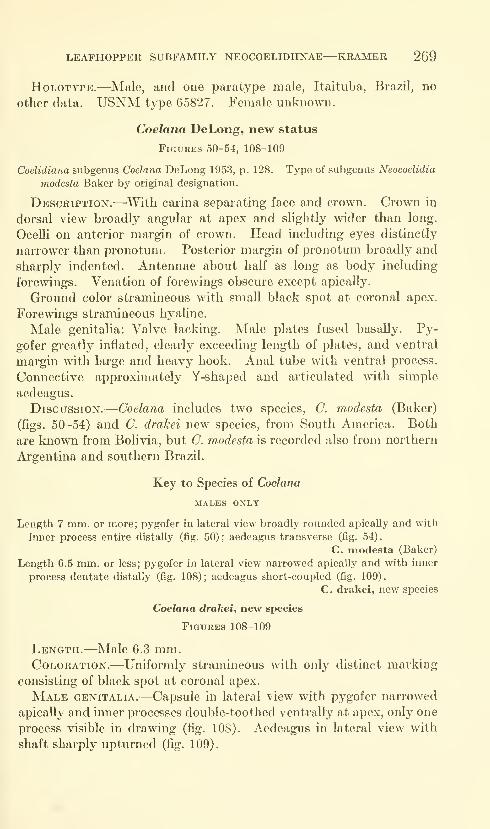
LEAFHOPPER SUBFAMILY NEOCOELIDIINAE—KRAMER 2G9
HoLOTYPE.—Male, and one paratype male, Itaituba, Brazil, no
other data. USNM type 65827. Female unknown.
Coelana DeLong, new status
FiGUBES 50-54, 108-109
Coelidiana subgenus Coelana DeLong 1953, p. 128. Type of subgenus Neocoelidia
modesta Baker by original designation.
Description.—With carina separating face and crown. Crown in
dorsal view broadly angular at apex and slightly wider than long.
Ocelli on anterior margin of crown. Head including eyes distinctly
narrower than pronotum. Posterior margin of pronotum broadly and
sharply indented. Antennae about half as long as body including
forewings. Venation of forewings obscure except apically.
Ground color stramineous with small black spot at coronal apex.
Forewings stramineous hyaline.
Male genitalia: Valve lacking. Male plates fused basally. Py-
gofer greatly inflated, clearly exceeding length of plates, and ventral
margin with large and heavy hook. Anal tube with ventral process.
Connective approximately Y-shaped and articulated with simple
aedeagus.
Discussion.—Ooelana includes two species, G. modesta (Baker)
(figs. 50-54) and C. drakei new species, from South America. Both
are known from Bolivia, but G. modesta is recorded also from northern
Argentina and southern Brazil.
Key to Species of Coelana
MALES ONLY
Length 7 mm. or more; pygofer in lateral view broadly rounded apically and with
inner process entire distally (fig. 50); aedeagus transverse (fig. 54).
C. modesta (Baker)
Length 6.5 mm. or less; pygofer in lateral view narrowed apically and with inner
process dentate distally (fig. 108); aedeagus short-coupled (fig. 109).
C, drakei, new species
Coelana drakei, new species
Figures 108-109
Length.—Male 6.3 mm.Coloration.—Uniformly stramineous with only distinct marking
consisting of black spot at coronal apex.
Male genitalia.—Capsule in lateral view with pygofer narrowed
apically and inner processes double-toothed ventrally at apex, only one
process visible in drawing (fig. lOS). Aedeagus in lateral view with
shaft sharply upturned (fig. 109).
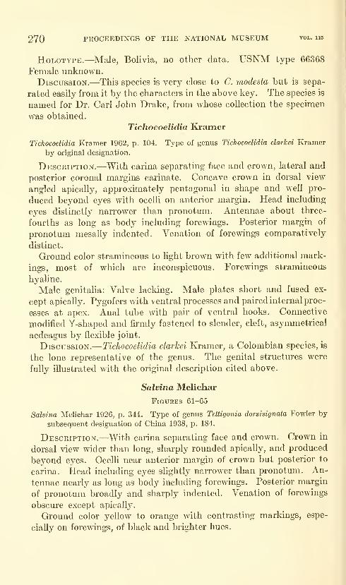
270 PROCEEDINGS OF THE NATIONAL MUSEUM vol. ii5
HoLOTYPE.—Male, Bolivia, no other data. USNM type 66368
Female unknown.
Discussion.—This species is very close to C. modesta but is sepa-
rated easily from it by the characters in the above key. The species is
named for Dr. Carl John Drake, from whose collection the specimen
was obtained.
Tichocoelidia Kramer
Tichocoelidia Kramer 1962, p. 104. Type of genus Tichocoelidia clarkei Kramer
by original designation.
Description.—With carina separating face and crown, lateral and
posterior coronal margins carinate. Concave crown in dorsal view
angled apically, approximately pentagonal in shape and well pro-
duced beyond eyes with ocelli on anterior margin. Head including
eyes distinctly narrower than pronotum. Antennae about three-
fourths as long as body including forewings. Posterior margin of
pronotum mesally indented. Venation of forewings comparatively
distinct.
Ground color stramineous to light brown with few additional mark-
ings, most of which are inconspicuous. Forewings stramineous
hyaline.
Male genitalia: Valve lacking. Male plates short and fused ex-
cept apically. Pygofers with ventral processes and paired internal proc-
esses at apex. Anal tube with pair of ventral hooks. Connective
modified Y-shaped and firmly fastened to slender, cleft, asjrmmetrical
aedeagus by flexible joint.
Discussion.—Tichocoelidia clarkei Kramer, a Colombian species, is
the lone representative of the genus. The genital structures were
fully illustrated with the original description cited above.
Salvina Melichar
Figures 61-65
Salvina Melichar 1926, p. 344. Type of genus Tettigonia dorsisignata Fowler by
subsequent designation of China 1938, p. 184.
Description.—With carina separating face and crown. Crown in
dorsal view wider than long, sharply rounded apically, and produced
beyond eyes. Ocelli near anterior margin of crown but posterior to
carina. Head including eyes slightly narrower than pronotum. An-
tennae nearly as long as body including forewings. Posterior margin
of pronotum broadly and sharply indented. Venation of forewings
obscure except apically.
Ground color yellow to orange with contrasting markings, espe-
cially on forewings, of black and brighter hues.

LEAFHOPPER SUBFAMILY NEOCOELIDIINAE—KRAMER 271
Male genitalia: Valve lacking. Male plates separated only api-
caliy. Pygofer in lateral view with ventral tooth and appearing cleft
at apex due to long mesally curved dorsal hook and weakly sclerotized
ventral extension. Anal tube simple but long. Stylar apex in dorsal
view curving laterally and caudally. Connective heavy and Y-
shaped. Aedeagus simple, compressed and upturned at apex.
Discussion.—The type species was illustrated in color whenoriginally described by Fowler (1900, p. 282, tab. 19, fig. 6). This
illustration is very good for showing the general markings of the
leafhopper; however, in the specunen at hand the ground color is a
brighter yellow-orange and the stripe on the commissural claval area
is of a red-wine shade. This species is Ivnown from three syntypes, all
of which are in the British Museum. Through the kindness of Dr.
W. E. China, one male specimen was made available for this study.
This male with data "Panama, Volcan de Chiriqui 4000 to 6000 feet,
Champion" is hereby designated as the lectotype. The male genitalia
of Salvina dorsisignata (Fowler), the only included species, are illus-
trated in figures 61-65.
Cocoelidia DeLong, new status
Figures 66-74
Neocoelidiana subgenus Cocoelidia DeLong 1953, p. 126. Type of subgenusNeocoelidiana antlera DeLong by original designation.
Desckiption.—With carina separating face and crown. Crown in
dorsal view wider than long, bluntly angular apically, and produced
beyond eyes. Ocelli on anterior margin of crown. Head including
eyes narrower than pronotum. Antennae only about half as long as
body including forewings. Posterior margin of pronotum broadly
indented. Venation of forewings obscure except at apex.
Ground color sordid yellow to light brown with weakly contrasting
yellowish markings on head and dorsum of thorax. Apex of crownwith black spot. Forewings brown hyaline.
Male genitalia: Valve lacking. Ventral cover of genital capsule
sharply narrowed on distal half appearing as two extended "fingers."
Pygofer in lateral view with ventral tooth and dorsal distally hookedprocess. Anal tube with short, forked ventral process. Connective
Y-shaped with stalk bifurcate to receive aedeagus. Style simple.
Aedeagus slender, recurved distally with pair of preapical processes.
Discussion.—The Mexican leafhopper, Cocoelidia antlera (De-
Long), is the only included species. The male genitalia are illustrated
in figures 66-74.
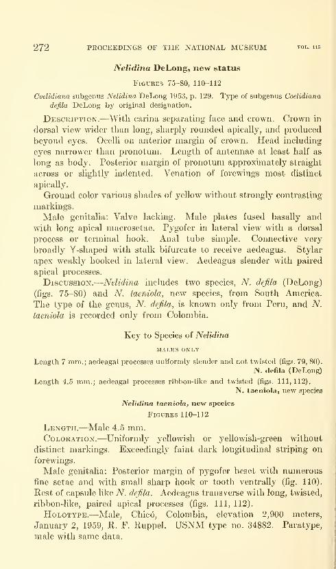
272 PROCEEDINGS OF THE NATIONAL MUSEUM vol. 115
Nelidina DeLong, new status
Figures 75-80, 110-112
Coelidia7ia subgenus Nelidina DeLong 1953, p. 129. Type of subgenus Coelidiana
defila DeLong by original designation.
Description.-—With carina separating face and crown. Crown in
dorsal view wider than long, sharply rounded apically, and produced
beyond eyes. Ocelli on anterior margin of crown. Head including
eyes narrower than pronotum. Length of antennae at least half as
long as body. Posterior margin of pronotmn approxmiately straight
across or slightly indented. Venation of forewings most distinct
apically.
Ground color various shades of yellow without strongly contrasting
markings.
Male genitalia: Valve lacking. Male plates fused basally and
with long apical macrosetae. Pygofer in lateral view with a dorsal
process or terminal hook. Anal tube simple. Connective very
broadly Y-shaped with stalk bifurcate to receive aedeagus. Stylar
apex weakly hooked in lateral view. Aedeagus slender with paired
apical processes.
Discussion.—Nelidina includes two species, A^. defila (DeLong)
(figs. 75-80) and A^. taeniola, new species, from South America.
The type of the genus, A^. defila, is known only from Peru, and A^.
taeniola is recorded only from Colombia.
Key to Species of Nelidina
MALES ONLY
Length 7 mm.; aedeagal processes uniformly slender and not twisted (figs. 79, 80).
N. defila (DeLong)
Length 4.5 mm.; aedeagal processes ribbon-like and twisted (figs. 111,112),
N. taeniola, new species
Nelidina taeniola, new species
Figures 110-112
Length.—Male 4.5 mm.Coloration.—Uniformly yellowish or yellowish-green without
distinct markings. Exceedingly faint dark longitudinal striping on
forewings.
Male genitalia: Posterior margin of pygofer beset with numerous
fine setae and with small sharp hook or tooth ventrally (fig. 110).
Rest of capsule like A^. defila. Aedeagus transverse with long, twisted,
ribbon-Uke, pahed apical processes (figs. Ill, 112).
HoLOTYPE.—Male, Chic6, Colombia, elevation 2,900 meters,
January 2, 1959, R. F. Ruppel. USNM type no. 34882. Paratype,
male with same data.
LEAFHOPPER SUBFAMILY NEOCOELIDIINAE—KRAMER 273
Neocoelidiana DeLong
Figures 81-86
Neocoelidiana DeLong 1953, p. 122. Type of genus Neocoelidia ohscura Bakerby original designation.
Description.—With carina separating face and crown. Crown in
dorsal view wider than long, very broadly or bluntly angled apically,
and produced beyond eyes. Head including eyes narrower than
pronotum. Ocelli on anterior margin of crown. Antennae from half
to three-fourths as long as body including forewings. Posterior
margin of pronotum broadly and often sharply indented. Venation
of forewings highly obscure except apically.
Ground color stramineous to yellow, often with tliree inconspicious
longitudinal stripes of slightly darker shade on crown and pronotum.
Apex of crown with black spot. Forewings yellowish hyaline, at
times with few additional brown marldngs or tinges, with veins
concolorous.
Male genitalia: Valve lacldng. Male plates fused basally for
more than half length, without short macrosetae apically. Pygofer in
lateral view with dorsal process and either ventral process or ventral
hook. Anal tube with single ventral hook. Connective broadly
Y-shaped. Apex of style strongly hooked in lateral view. Aedeagus
in lateral view approximately S-shaped, often somewhat twisted, andwith some sort of apical modifications.
Discussion.—The genus as here defined contains seven species from
western United States and Mexico, all of which were treated byDeLong (1953). The male genital structures of the type-species,
Neocoelidiana ohscura (Baker), are shown in figm-es 81-86.
Coelella DeLong, new status
Figures 87-93
Neocoelidiana subgenus Coelella DeLong 1953, p. 125. Type of subgenus
Neocoelidia distincta Oman by original designation.
Description.—With carina separating face and crown. Crownin dorsal view wider than long, bluntly angled apically, and produced
beyond eyes. Head including eyes narrower than pronotum. Ocelli
on anterior margin of crown. Antennae from half to three-fourths
as long as body including forewings. Posterior margin of pronotumbroadly and sharply indented. Venation of forewing highly distinct.
Ground color stramineous to yellow, usually with three more or less
well-defined longitudinal stripes of slightly darker hue on crown and
pronotum. Apex of crown with black spot. Forewings hyaline with
veins uniformly brown.
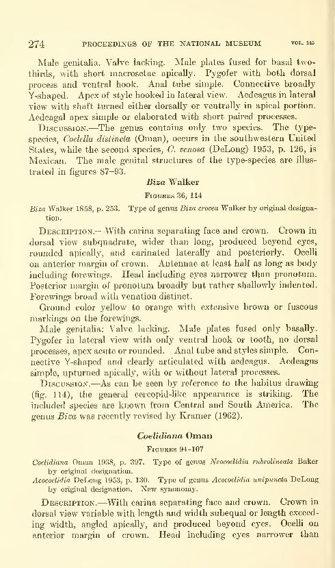
274 PROCEEDINGS OF THE NATIONAL MUSEUM vol. us
Male genitalia. Valve lacking. Male plates fused for basal two-
thirds, with short macrosetae apically. Pygofer with both dorsal
process and ventral hook. Anal tube simple. Connective broadly
Y-shaped. Apex of style hooked in lateral view. Aedeagus in lateral
view with shaft turned either dorsally or ventrally in apical portion.
Aedeagal apex simple or elaborated with short paired processes.
Discussion.—The genus contains only two species. The type-
species, Coelella distincta (Oman), occurs in the southwestern United
States, while the second species, C. venosa (DeLong) 1953, p. 126, is
Mexican. The male genital structures of the type-species are illus-
trated in figures 87-93.
Bisa Walker
Figures 36, 114
Biza Walker 1858, p. 253. Type of genus Biza crocea Walker by original designa-
tion.
Description.—With carina separating face and crown. Crown in
dorsal view subquadrate, wider than long, produced beyond eyes,
rounded apically, and carinated laterally and posteriorly. Ocelli
on anterior margin of crown. Antennae at least half as long as body
including forewings. Head including eyes narrower than pronotum.
Posterior margin of pronotum broadly but rather shallowly indented.
Forewings broad with venation distinct.
Ground color yellow to orange with extensive brown or fuscous
markings on the forewings.
Male genitalia: Valve lacking. Male plates fused only basally.
Pygofer in lateral view with only ventral hook or tooth, no dorsal
processes, apex acute or rounded. Anal tube and styles simple. Con-
nective Y-shaped and clearly articulated with aedeagus. Aedeagus
simple, upturned apically, with or without lateral processes.
Discussion.—As can be seen by reference to the habitus drawing
(fig. 114), the general cercopid-like appearance is striking. The
included species are known from Central and South America. The
genus Biza was recently revised by Kramer (1962).
Coelidiana OmanFigures 94-107
Coelidiana Oman 1938, p. 397. Type of genus Neocoelidia rubrolineata Baker
by original designation.
Acocoelidia DeLong 1953, p. 130. Type of genus Acocoelidia unipunda DeLong
by original designation. New synonomy.
Description.—With carina separating face and crown. Crown in
dorsal view variable with length and width subequal or length exceed-
ing width, angled apically, and produced beyond eyes. Ocelli on
anterior margin of crown. Head including eyes narrower than
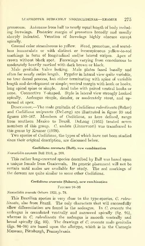
LEAFHOPPER SUBFAMILY NEOCOELIDIINAE—KRAMER 275
pi'onotum. Antennae from half to nearly equal length of body includ-
ing forewings. Posterior margin of pronotum broadly and usually
sharply indented. Venation of forewings highly obscure except
apically.
Ground color stramineous to yellow. Head, pronotum, and scutel-
lum immaculate or with distinct or inconspicuous yellow-to-red
markings in form of longitudinal and/or lateral stripes. Apex of
crown without black spot. Forewings varying from concolorous to
moderately heavily marked with dark brown or black.
Male genitaha: Valve lacking. Male plates fused basally andoften for nearly entire length. Pygofer in lateral view quite variable,
no true dorsal process, but either terminating with spine of variable
length and development or simple; ventral margin with hook or hooks,
long apical spine or simple. Anal tube with paired ventral hooks or
none. Connective Y-shaped. Style in lateral view strongly hookedapically. Aedeagus simple, slender, or moderately stout, and up-turned at apex.
Discussion.—The male genitalia of Coelidiana rubrolineata (Baker)
and Coelidiana unipuncta (DeLong) are illustrated in figure 99 andfigures 100-107. Members of Coelidiana, as here defined, rangefrom southern Mexico to Brazil. DeLong (1953) treated seven
members of this group. C. undata (Linnavuori) was transferred to
this genus by Kramer (1959).
Two species of Coelidiana, the types of which have not been studied
since their original description, are discussed below.
Coelidiana coronata (Ball), new combination
Neocoelidia coronata Ball 1916, p. 208.
This rather long-crowned species described by Ball was based upona unique female from Guatemala. Its generic placement wUl not becertain until males are available for study. The red markings of
the dorsum are quite similar to some other Coelidiana.
Coelidiana croceata (Osborn), new combination
Figures 94-98
Neocoelidia croceata Osborn 1923, p. 78.
This Brazilian species is very close to the type-species, C. rubro-
lineata, also from Brazil. The only characters that will successfully
allow differentiation are found in the aedeagus. In C. croceata the
aedeagus is crenulated ventrally and narrowed apically (fig. 95),
whereas in C. rubrolineata the aedeagus is smooth ventrally andbroad apically (fig. 99). The drawings of C. croceata male genitalia
(figs. 94-98) are based upon the allotype, which is in the CarnegieMuseum, Pittsburgh, Pennsylvania.
276 PROCEEDINGS OF THE NATIONAL MUSEUM vol. lis
Literature Cited
Baker, C. F.
1898. Notes on Jassini with some new species. Canadian Ent., vol. 30,
pp. 289-292.
Ball, E. D.
1916. Some new species of Athysanus and related genera (Homoptera).
Ent. News, vol. 27, pp. 204-208.
Bruner, S. C, and Metcalf, Z. P.
1934. A new Bythoscopidae from Costa Rica. Bull. Brooklyn Ent. Soc,
vol. 29, pp. 120-124.
China, W. E.
1938. Melichar's "Monographie der Cicadellinen." Ann. Mag. Nat. Hist.,
ser. 11, vol. 2 pp. 182-185.
DeLong, D. M.1953. A synopsis of the tribe Neocoelidinii in the Americas (Homoptera-
Cicadellidae). Lloydia, vol. 16, no. 2, pp. 93-131.
Evans, J. W.1947. A natural classification of leaf-hoppers (Jassoidea, Homoptera), Part
3: Jassidae. Trans. Roy. Ent. Soc. London, vol. 98, no. 6, pp.
105-271.
Fowler, W. W.1900. In Godman and Salvin, Biologia Centrali-Americana, vol. 49 (Hemip-
tera-Homoptera, vol. 2), pt. 1, xii+ 339 pp., 21 pis.
Gillette, C. P., and Baker, C. F.
1895. A preliminary list of the Hemiptera of Colorado. Bull. Colorado
Agric. Exper. Sta., vol. 31, pp. 1-137.
Knull, D. J.
1942. The genus Neocoelidia in the United States (Homoptera: Cicadel-
lidae). American Midi. Nat., vol. 28, no. 3, pp. 680-692.
Kraaier, J. P.
1959. An elucidation of the Neotropical genus Chinaia with a key to males
and a new allied genus (Homoptera: Cicadellidae: Neocoelidiinae).
Proc. Biol. Soc. Washington, vol. 72, pp. 23-32.
1961. New Venezuelan leafhoppers of the subfamilies Xestocephalinae
and Neocoehdiinae (Homoptera: CicadeUidae) . Proc. Biol. Soc.
Wasliington, vol. 74, pp. 235-240.
1962. A synopsis of Biza and a new allied genus (Homoptera: CicadeUidae:
Neocoelidiinae). Proc. Biol. Soc. Washington, vol. 75, pp. 101-106.
Kramer, J. P., and Linnavuori, R.
1959. A new genus and two new species of leafhoppers from South America
(Homoptera: Cicadellidae: Neocoehdiinae). Proc. Biol. Soc.
Washington, vol. 72, pp. 55-58.
Melichar, L.
1926. Monographie der CicadeUinen, III. Ann. Mus. Nat. Hungarici,
vol. 23, pp. 273-394.
Oman, P. W.1938. A generic revision of American Bythoscopinae and South American
Jassinae. Univ. Kansas Sci. Bull., vol. 24, pp. 343-420.
1943. A generic revision of the Nearctic CicadeUidae. Summary of doctoral
thesis, George Washington University.
1949. The Nearctic leafhoppers (Homoptera: CicadeUidae): A generic
classification and check list. Ent. Soc. Washington Memoirs,
no. 3, pp. 1-253.

LEAFHOPPER SUBFAMILY NEOCOELIDIINAE—KRAMER 277
OSBOBN, H.
1923. Neotropical Homoptera of the Carnegie Museum: Part 2, Records
and descriptions of five new genera and sixty-five new species of
the subfamily Jassinae. Ann. Carnegie Mus., vol. 15, pp. 27-79.
Walker, F.
1858. List of the specimens of Homopterous insects in the collection of the
British Museum, suj^pl., pp. 1-307.
Young, D. A.
1952. Reclassification of Western Hemisphere Typhlocybinae (Homoptera
Cicadellidae) . Univ. Kansas Sci. Bull., vol. 35, no. 1, pp. 3-217.
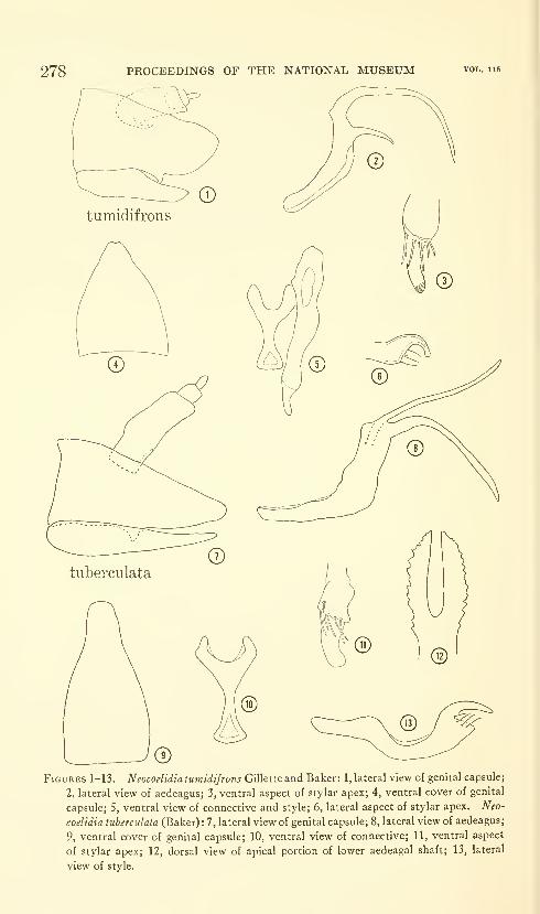
278 PROCEEDINGS OF THE NATIONAL MUSEUM vol. 115
Figures 1-13. Neocoelidia tumtdifrons Gillette and Baker: 1, lateral view of genital capsule;
2, lateral view of aedeagus; 3, ventral aspect of stylar apex; 4, ventral cover of genital
capsule; 5, ventral view of connective and style; 6, lateral aspect of stylar apex. Neo-
coelidia tuherculata (Baker) : 7, lateral view of genital capsule; 8, lateral view of aedeagus;
9, ventral cover of genital capsule; 10, ventral view of connective; 11, ventral aspect
of stylar apex; 12, dorsal view of apical portion of lower aedeagal shaft; 13, lateral
view of style.
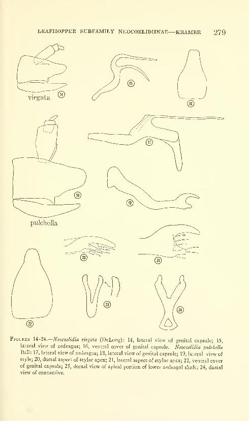
LEAFHOPPER SUBFAMILY NEOCOELIDIINAE—KRAMER 279
Figures 14-24.—Neocoelidia virgata (DeLong): 14, lateral view of genital capsule; 15,
lateral view of aedeagus; 16, ventral cover of genital capsule. Neocoelidia pulchella
Ball: 17, lateral view of aedeagus; 18, lateral view of genital capsule; 19, lateral view ofstyle; 20, dorsal aspect of stylar apex; 21, lateral aspect of stylar apex; 22, ventral coverof genital capsule; 23, dorsal view of apical portion of lower aedeagal shaft; 24, dorsalview of connective.

craspa
Figures 25-36—Neocoelidia fuscodorsata (Fowler): 25, lateral view of genital capsule; 26,
lateral view of aedeagus; 27, ventral cover of genital capsule; 28, dorsal view of style
and lateral view of stylar apex. Neocoelidia crenulata Osborn : 29, lateral view of genital
capsule; 30, ventral cover of genital capsule; 31, lateral view of aedeagus; 32, dorsal
view of apical portion of lower aedeagal shaft; 33, lateral aspect of stylar apex; 34,
ventral view of connective and style; 35, dorsal view of stylar apex. Biza craspa
Kramer; 36, ventral cover of genital capsule.

LEAFHOPPER SUBFAMILY NEOCOELIDIINAE—KRAMER 281
Figures SJ^i.—Chinaia bella Bruner and Metcalf: 37, ventral cover of genital capsule.
Xenocoelidia youngi Kramer: 38, ventral cover of genital capsule. Xenocoelidia inflata
(Osborn): 39, ventral cover of genital capsule; 40, lateral view of genital capsule; 41,ventral view of connective and style; 42, lateral view of aedeagus; 43, posterior view of
aedeagal apex; 44, lateral view of stylar apex.
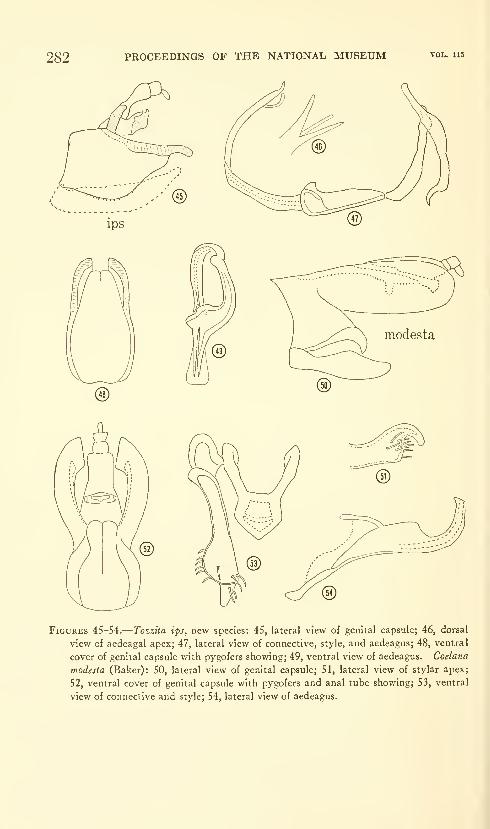
282 PROCEEDINGS OF THE NATIONAL MUSEUM vol. 115
Figures 45-54.
—
Tozzita ips, new species: 45, lateral view of genital capsule; 46, dorsal
view of aedeagal apex; 47, lateral view of connective, style, and aedeagas; 48, ventral
cover of genital capsule with pygofers showing; 49, ventral view of aedeagus. Coelana
modesia (Baker): 50, lateral view of genital capsule; 51, lateral view of stylar apex;
52, ventral cover of genital capsule with pygofers and anal tube showing; 53, ventral
view of connective and style; 54, lateral view of aedeagus.

LEAFHOPPER SUBFAMILY NEOCOELIDIINAE—KRAMER 283
Figures 55-65.
—
Xiqilliba bellator, new species: 55, lateral view of genital capsule; 56,
ventral cover of genital capsule showing processes of anal tube; 57, lateral view of
aedeagal apex; 58, lateral view of aedeagus; 59, lateral view of stylar apex; 60, dorsal
view of connective, style, and aedeagus. Salvina dorsisignata (Fowler): 61, ventral
cover of genital capsule; 62, lateral view of genital capsule; 63, dorsal view of hooksat apex of pygofer; 64, lateral view of aedeagus; 65, dorsal view of connective and style.
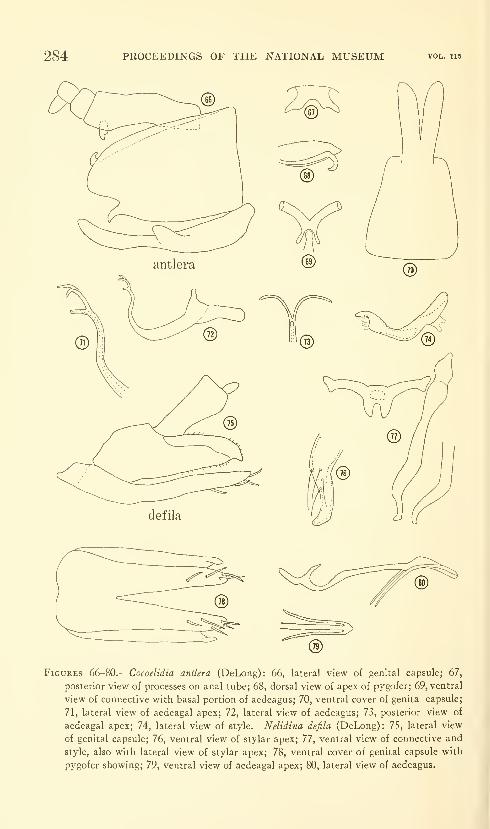
284 PROCEEDINGS OF THE NATIONAL MUSEUM vol. 115
Figures 66-80.
—
Cocoelidia antlera (DeLong): 66, lateral view of genital capsule; 67,
posterior view of processes on anal tube; 68, dorsal view of apex of pygofer; 69, ventral
view of connective with basal portion of aedeagus; 70, ventral cover of genita capsule;
71, lateral view of aedeagal apex; 72, lateral view of aedeagus; 73, posterior view of
aedeagal apex; 74, lateral view of style. Nelidina defila (DeLong): 75, lateral view
of genital capsule; 76, ventral view of stylar apex; 77, ventral view of connective and
style, also with lateral view of stylar apex; 78, ventral cover of genital capsule with
pygofer showing; 79, ventral view of aedeagal apex; 80, lateral view of aedeagus.

LEAFHOPPER SUBFAMILY NEOCOELIDIINAE—KRAMER 285
Figures 81-93.
—
Neocoelidiana obscura (Baker): 81, lateral view of genital capsule; 82,
lateral view of aedeagus; 83, posterior view of aedeagus; 84, posterior view of aedeagal
apex; 85, ventral cover of genital capsule; 86, lateral view of style. Coelella distincta
(Oman): 87, lateral view of style; 88, lateral view of genital capsule; 89, ventral cover
of genital capsule; 90, ventral view of aedeagus; 91, posterioventral view of con-
nective; 92, lateral view of aedeagus; 93, lateral view of aedeagus, a variant.
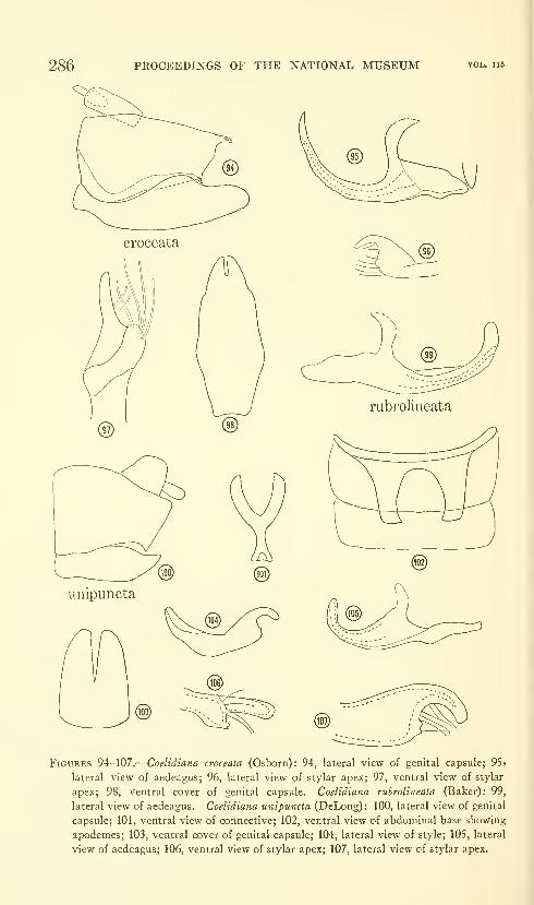
286 PROCEEDINGS OF THE NATIONAL MUSEUM vol. u5
Figures 94-107.
—
Coelidiana croceata (Osborn): 94, lateral view of genital capsule; 95>
lateral view of aedeagus; 96, lateral view of stylar apex; 97, ventral view of stylar
apex; 98, ventral cover of genital capsule. Coelidiana ruhrolineata (Baker): 99,
lateral view of aedeagus. Coelidiana unipuncta (DeLong) : 100, lateral view of genital
capsule; 101, ventral view of connective; 102, ventral view of abdominal base showing
apodemes; 103, ventral cover of genital capsule; 104, lateral view of style; 105, lateral
view of aedeagus; 106, ventral view of stylar apex; 107, lateral view of stylar apex.

LEAFHOPPER SUBFAMILY NEOCOELIDIINAE—KRAMER 287
Chinaia lepida
Biza craspa
114
Figures 108-1 14.—Co^/anA drakei, new species: 108, lateral view of genital capsule; 109,lateral view of aedeagus. Nelidina taeniola, new species : 1 10, lateral view of pygofer andanal tube; 111, lateral view of aedeagus; 112, ventral view of aedeagal apex. Chinaialepida Kramer: 113, habitus in dorsal view. Biza craspa Kramer: 114, habitus indorsal view.
U.S. GOVERNMENT PRINTING OFFlCEil964


Proceedings of
the United States
National MuseumSMITHSONIAN INSTITUTION • WASHINGTON, D.C.
Volume 115 1964 Number 3485
A REVIEW OF THE NORTH AMERICAN MOTHSOF THE FAMILY WALSHIIDAE
(LEPIDOPTERA: GELECHIOIDEA)
By Ronald W. Hodges ^
Introduction
Since most species of Walshiidae are small, inconspicuous moths,
many collectors understandably have overlooked them. As a result,
a relatively small number of specimens has been taken and our knowl-
edge of the group is limited. Until extensive and intensive collecting
has been accomplished, the results of any revisionary study will be
limited, subject to reinvestigation. The purpose of this paper is to
make known the described genera and species, to attempt to define
the genera, and to describe the new species that are at hand. Four
North American genera recently have been revised : Walshia Clemens
(Hodges, 1961), Ithome Chambers (Hodges, 1962a), Periploca Braun(Hodges, 1962b), and Perimede Chambers (Hodges, in press). Theywill not be discussed further in this paper except for a few notes under
"Taxonomic Treatment."
'Entomology Research Division, Agricultural Research Service, U.S. Department of Agriculture,
Washington, D.C.
289
290 PROCEEDINGS OF THE NATIONAL MUSEUM vol. lis
At present not much is known about the immatiu"e stages of most
species of Walshiidae; however, some general statements can be made.
The known larvae of Ithome feed on the florets of species of Legu-
minosae and Polygonaceae. The larvae of Walshia miscecolorella
(Chambers) and W. amor'phella Clemens are stem and root borers;
W. miscecolorella has been reared from several species of Leguminosae
and is potentially a pest of sweet clover in parts of Texas. The larvae
of W. amorphella are gall formers on the stems of Amorpha fruticosa
L. and occasionally on Hydrangea spp. The known larvae of species
of Aeaea Chambers are leaf miners in Ostrya virginiana (Mill.) K.
Koch., Quercus spp., and Rhynchosia tomentosa (L.) H. & A. Species
of Periploca (Braun) are known from Ceanothus spp. (gall formers on
the stems), Gleditsia spp., and Rohinia spp. (borers in the thorns),
Juniperus spp. (found in the fruits), Gymnosporangium spp. (in galls
and as a twig girdler). Two species, Periploca laeta Hodges and P.
nigra Hodges, sometimes are pests of ornamental junipers. Perimede
erransella Chambers has been reared from Taxodium spp. and leaves
of Ulmus spp., but whether this species is a miner during all or part of
the larval stage or whether it is an external feeder is not known. Obithome
punctiferella (Busck) has been found in cages which contained cotton
bolls, but whether the larva feeds on the bolls or is a scavenger is
unknown. The larvae of Stilbosis tesquella Clemens are external feed-
ers on Amphicarpa spp. and Lespedeza spp. The larvae of Chryso-
peleia purpuriella Chambers have been reared from the leaves of
Rohinia pseudo-acacia L., but the exact habits are not known. Sorha-
genia rhamniella (Zeller) and S. nimhosa (Braun) are leaf folders on
Rhajnnus spp. in the larval stage.
Because the species of Aeaea Chambers have similar maculation
and habitus, it was necessary to make genitalic preparations of each
specimen. In some of the female abdomens, single larval head cap-
sules were found. Dm'ing the process of cleaning the specimens, the
head capsules were often removed; but they are present on two slides
(RWH slides 1167 and 2121), Aeaea "d" and Synploca gumia, newgenus and species, respectively. These head capsules indicate that
the species possibly are ovoviviparous, a condition known to occur in
the Coleophoridae (Toll, 1952), Tineidae (Diakonoff, 1952), and
Oecophoridae (teste Clarke).
I wish to thank the following individuals for providing me with
the specimens which form the basis of this study (parentheses en-
close abbreviations used in citing the location of specimens) : Mr.
J. A. G. Rehn, Academy of Natural Sciences, Philadelphia (ANSP);
Dr. C. D. MacNeill, California Academy of Sciences (CAS); Dr.
J. G. Franclemont, Cornell University (CU); Mr. L. M. Martin, Los
Angeles County Museum (LACM); Drs. P. J. Darhngton and
MOTHS OF FAMILY WALSHIIDAE—HODGES 291
H. E. Evans, Museum of Comparative Zoology (MCZ); Dr. J. A. Powell,
University of California at Berkeley (UCB); Dr. A. F. Braun, Cincin-
nati, Ohio (AFB); Mr. M. O. Glenn, Henry, Illinois (MOG); Mr.C. P. Kimball, Barnstable, Massachusetts (CPK); and Dr. A. B. Klots,
New York, New York (ABK). Specimens in the United States
National Museum are designated by USNM. Particular thanks are
given to Drs. Braun, Darlington, Evans, and Mr. Rehn for allowing
me to examine type specimens under their care. In addition, Mr.J. D. Bradley furnished me with specimens of Sorhagenia rhamniella
and Cholotis semnostola Mejn'ick and compared North Americanmaterial with specimens in the British Museum (Natural History).
The photographs of the adult moths were made by J. Scott, Staff
Photographer, Smithsonian Institution.
The specimens of Stilhosis tesquella from Highlands, North Carolina,
were collected during the summer of 1958 when the author was assist-
ing Dr. J. G. Franclemont under the auspices of a grant from the
Penrose Fund of the American Philosophical Society.
Taxonomic Treatment
The name Walshiidae was proposed for a relatively homogeneousgroup of genera (Hodges, 1962a). At some future date, when moreis known about the gelechioids, the group probably will be treated as
a subfamily or tribe within the complex, but for the present it mustbe recognized as being as distinct as several other families.
The characters of the family are as given in Hodges (Revision of
Cosmopterigidae, in press) with the following modifications : the fore-
wing with 11 (no. 2 absent) or 12 veins; the hind wing with 6 (3 and 4
absent) or 8 veins; uncus usually present, absent in Obithome, newgenus, and Periploca. In several of the genera there is a tendency
toward asymmetry in the male genitalia, usually expressed by a twist-
ing of the valvae with a concomitant reduction of one valva and anenlargement of the other.
The three families, Walshiidae, Cosmopterigidae, and Momphidae,have been placed in either Cosmopterigidae, Lavernidae, or Momphi-dae on the basis of wing venation; however, as I have indicated
(Hodges, ibid.), venation does not offer a satisfactory means of sep-
arating these families. For example, by using venation, several genera
of cosmopterigids would go into the Oecophoridae; the walshiids, cos-
mopterigids, and momphids would form a unit; and the Scaeosophi-
dae would be treated as a separate family. Unfortunately, the
female genitalia also do not seem to offer diagnostic characters onthe family (and often not on the generic) level; however, the malegenitalia do present what appear to be reliable means for separating
these families, and it is this system of characters which presently
292 PROCEEDINGS OF THE NATIONAL MUSEUM vol. no
forms the basis of our system of classification. The females will have
to be associated with males before positive family identification can
be made.
Key to Walshiidae, Cosmopterigidae, and Momphidae Based on MaleGenitalia
1. Gnathos present 2
Gnathos absent 3
2. Brachia of gnathos symmetrical MomphidaeBrachia of gnathos asymmetrical Cosmopterigidae
3. Aedeagus ankylosed, heavily sclerotized manica articulating with saccus or
juxta Walshiidae
Aedeagus not ankylosed, manica absent Momphidae
The female genitalia of Ithome Chambers and Obithome are heavily
sclerotized, particularly the apophyses. The apophyses are very
similar to those of Adela Latreille (Pierce and Metcalf, 1935), Ectropro-
ceros Diakonoff (Diakonoff, 1955), and Antispila Hiibner (Kuroko,
1961); however, rather than indicating phyletic relationship, the
sclerotization probably represents a modification for a specialized
type of oviposition.
Meyrick (1915) transferred Stagmatophora ceanothiella Cosens to
the Australian genus Cholotis Meyrick. In 1921 he synonymized
Cholotis with the Palearctic genus Ascalenia Wocke, thereby making
the combination Ascalenia ceanothiella. As I have shown (Hodges,
1962b), A. ceanothiella belongs to the genus Periploca. Genitalic
examination of the type-species Cholotis semnostola indicates that
this genus is distinct from Ascalenia and that it has affinities with
Perimede. Meyrick placed many Central and South American spe-
cies in Cholotis (later Ascalenia) and Prochola Meyrick. Clarke (in
press) subsequently has examined the genitalia of several of Meyrick's
type specimens and has transferred many species to other genera.
Until the genitalia of the known species in these genera have been
examined, the generic combinations must be regarded with caution.
Walshia particornella (Busck) is somewhat intermediate in position
between Walshia and Periploca; however, because of an uncus present
in the male genitalia and the general facies of the genitalia, parti-
cornella is associated with Walshia. The ostium bursae of W. parti-
cornella is in the middle of the seventh sternmii, a characteristic that
is in contrast to the other known species of Walshia, the ostium bursae
of which is on the anterior margin of the seventh sternum ; the ostium
bursae of Periploca is on the anterior margin or medial. The habitus
of W. particornella is similar to that of Periploca species, and the wings
are smooth-scaled; the forewings of all known species of Walshia
have a series of raised scales. At the time of writing I do not feel that
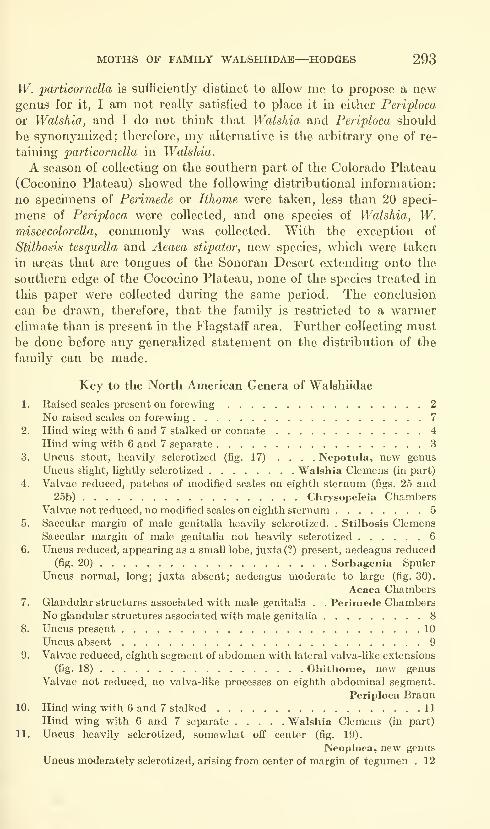
MOTHS OF FAMILY WALSHIIDAE—HODGES 293
W. particornella is sufficiently distinct to allow me to propose a newgenus for it, I am not really satisfied to place it in either Periploca
or Walshla, and I do not think that Walshia and Periploca should
be synonymized; therefore, my alternative is the arbitrary one of re-
taining particornella in Walshia.
A season of collecting on the southern part of the Colorado Plateau
(Coconino Plateau) showed the following distributional information:
no specimens of Perimede or Ithome were taken, less than 20 speci-
mens of Periploca were collected, and one species of Walshia, W.miscecolorella, commonly was collected. With the exception of
Stilbosis tesquella and Aeaea stipator, new species, which were taken
in areas that are tongues of the Sonoran Desert extending onto the
southern edge of the Cococino Plateau, none of the species treated in
this paper were collected during the same period. The conclusion
can be drawn, therefore, that the family is restricted to a warmerclimate than is present in the Flagstaff area. Further collecting mustbe done before any generalized statement on the distribution of the
family can be made.
Key to the North American Genera of Walshiidae
1. Raised scales present on forewing 2
No raised scales on forewing 7
2. Hind wing with 6 and 7 stalked or connate 4
Hind wing with 6 and 7 separate 3
3. Uncus stout, heavily sclerotized (fig. 17) .... Nepotula, new genus
Uncus slight, lightly sclerotized Walshia Clemens (in part)
4. Valvae reduced, patches of modified scales on eighth sternum (figs. 25 and25b) Chrysopeleia Chambers
Valvae not reduced, no modified scales on eighth sternum 5
5. Saccular margin of male genitalia heavily sclerotized. . Stilbosis ClemensSaccular margin of male genitalia not heavily sclerotized 6
6. Uncus reduced, appearing as a small lobe, juxta(?) present, aedeagus reduced
(fig. 20) Sorhagenia Spuler
Uncus normal, long; juxta absent; aedeagus moderate to large (fig. 30).
Aeaea Chambers7. Glandular structures associated with male genitaUa . . Perimede Chambers
No glandular structures associated with male genitalia 8
8. Uncus present 10
Uncus absent 9
9. Valvae reduced, eighth segment of abdomen with lateral valva-Uke extensions
(fig. 18) Obithoine, new genus
Valvae not reduced, no valva-like processes on eighth abdominal segment.
Periploca Braun10. Hind wing with 6 and 7 stalked 11
Hind wing with 6 and 7 separate Walshia Clemens (in part)
11. Uncus heavily sclerotized, somewhat off center (fig. 19).
Neoploca, new genus
Uncus moderately sclerotized, arising from center of margin of tegumen . 12
294 PROCEEDINGS OF THE NATIONAL MUSEUM vol. 115
12. Aedeagus slender, length more than six times width; valvae, often with
basal processes Ithome Chambers
Aedeagus stout, length not more than four times width; valvae without
basal processes (fig. 24) Synploca, new genus
Neoploca, new genus
Figures 7, 11, 59
Type-species: Neoploca corusca Hodges, new species.
Head: smooth-scaled; labial palpus recurved, reaching beyond
vertex, third segment shorter than second, apex acute; maxillary
palpus folded over base of tongue; eye emarginate on anterodorsal
angle; ocellus visible; antenna simple, ciliate, two-thirds length of
forewing, pecten absent. Forewing: lanceolate; 12 veins present; lb
furcate basally; 2 developed toward margin of wing; 3, 4, and 5 sepa-
rate, 3 from angle of cell; 6, 7, and 8 stalked, 7 out of 6 at nearly
five-sLxths; cell open. Hind wing: lanceolate; cell open; 8 veins
present; lb simple; 2, 3, 4, and 5 almost equidistant; 6 and 7 stalked,
diverging at three-fourths, each ending at seven-eighths. Metathoracic
tibia with long scales on dorsal surface, Male genitalia: vinculum
narrow; tegumen relatively broad; valvae symmetrical, no free parts,
setae abundant toward apex and on costal margin; uncus heavily
sclerotized, somewhat asymmetrical; aedeagus loosely helical, no
cornuti present. Female genitalia: ostium bursae slightly beyond
middle of seventh sternum, trapezoidal sclerotized area preceding
ostium bursae; ductus bursae and corpus bursae lightly sclerotized,
ductus bursae coiled before inception of bursa copulatrix; two spine-
shaped signa present.
Neoploca is closest to Periploca, but differs from it in having a well-
developed uncus and a relatively broad tegumen.
Neoploca corusca, new species
Figures 19, 37, 59
Head: pale, shining gold; thorax and forewing shining bronze-
black; cilia shining fuscous; hind wing somewhat shining fuscous,
cilia fuscous. Metathoracic leg shining gold or lead colored, apex
of femur and tibia shining white; base and apex of first tarsal seg-
ment shining white, apex of second segment shining white on external
surface, remaining segments unicolorous, ventral surface of first four
tarsal segments shining white. Abdomen: apices of segments white
with purple reflections, some shining ochreous-brown on segments.
Male genitalia: as in figure 19 (RWH slide 2122). Female genitalia:
as in figure 37 (RWH slide 532). Alar expanse: 9-12 mm.

MOTHS OF FAMILY WALSHIIDAE—HODGES 295
Holotype: cf , Madera Canyon, 4880 feet, Santa Rita Mts., Ariz.,
July 14, 1959, R. W. Hodges (RWH slide 2123), Cornell Univ. type
3889.
ParatA'pes: same locality as type, 1 9 , Aug. 23, 1959 (RWH slide
532), USNM; Pena Blanca Canyon, 4000 feet, Santa Cruz Co., Ariz.,
3 cf, 2 9 , Aug. 8-11, 1959, R. W. Hodges (RWH slides 531, 2122;
RWH wing slide 11), CU, USNM.Superficially, A^. corusca is somewhat similar to Periploca ceanothi-
ella and P. gleditschiaeella, but it differs in having the scales of the
forewings unicolorous and in having a well-developed uncus.
Synploca, new genus
Figures 8, 14, 60
Type-species: Synploca gumia, new species.
Head: smooth-scaled; labial palpus recurved, ahiiost reaching
vertex, second and third segments subequal in length, apex of third
segment acute; tongue scaled basally; maxillary palpus folded over
base of tongue; antenna three-fifths length of forewing, pecten absent;
ocellus visible; anterodorsal margin of eye emarginate. Forewing:
lanceolate; 12 veins present; cell closed or nearly closed; lb fm-cate
basally; 2 from six-sevenths of cell; 3 and 4 distant basally; 4 and 5
approximate basally; 6, 7, and 8 stalked; 9 from end of cell. Hind
wing: 8 veins present; 1 simple; 2, 3, and 4 equidistant basally; 6 and
7 stalked, 6 out of 7 slightly beyond two-thirds length of wing. Meta-
thoracic tibia with long scales dorsally. Male genitalia: valva
relatively broad, apex rounded, costal and saccular areas not free;
aedeagus stout, tapering rapidly to apex; uncus short, bifid. Female
genitalia: ostium bursae near base of seventh sternum; sclerotized
pregenital plate attaining edge of seventh sternum; ductus bursae
and bursa copulatrix lightly sclerotized ; two semicircular signa present,
each sigmun with interiorly projecting extension from broadest point.
Synploca gumia, new species
Figures 24, 38, 60
Labial palpus bronze-black, pale basally; white dot at apex of
second segment ventrally and 11 or 12 white dots forming row on
ventral and anterior surface of third segment; apex white. Base of
tongue and face shming pale silver-gold with purple reflections;
antenna bronze-black, apical nine or ten segments ochreous; vertex
of head, thorax, and forewing bronze-black; cilia of forewing and
hind wing pale ochreous; abdomen shining pale ocher. Legs pale
296 PROCEEDINGS OF THE NATIONAL MUSEUM vol. lis
bronze-black on outer surface, apices of tarsal segments white. Malegenitalia: as in figui'e 24 (RWH slide 564). Female genitalia: as in
figure 38 (RWH slide 571). Alar expanse: 6-9 mm.Holotype: cf, Madera Canyon, 4400 feet, Santa Rita Mts., Ariz.,
Oct. 10, 1959, R. W. Hodges (RWH slide 564), Cornell Univ. type
3888.
Paratypes: same locality as type, 2 cT", Oct. 26, 1959 (RWH slide
2120), CU, USNM; same locality as type except for elevation, 4880
feet, 21 c^, 22 9, June 30-Nov. 1, 1959 (RWH slides 570, 571, 2121),
CU, USNM, BMNH, CNC; same locality as type except for eleva-
tion, 5600 feet, 1 d^, 2 9, Sept. 22-Oct. 15, 1959, CU, USNM.
NcpotulUf new genus
Figures 2, 16, 61
Type-species: Nepotula secura Hodges, new species.
Head: smooth-scaled; labial palpus almost attaining vertex, third
segment shorter than second, apex acute; tongue moderate; maxillary
palpus folded over base of tongue; antenna two-thirds length of
forewing, simple, ciliate, pecten absent (one specimen examined,
possibly deciduous). Forewing: broadly lanceolate, apex acute;
12 veins present; lb furcate basally; 2 extremely faint basally; 4, 5,
and 6 equidistant basally; 7 and 8 stalked. Hind wing: lanceolate,
apex acute; 8 veins present; 6 and 7 separate. Male genitalia:
valva sublinear, costal area a broad fold; vinculum narrow; tegumen
relatively broad; uncus present, short, heavily sclerotized; a lightly
sclerotized area on under surface of tuba analis. Female genitalia:
not known.
Nepotula is closest to Walshia but differs from the latter in having
a heavily sclerotized uncus and a relatively broad tegumen.
Nepotula secura, new species
Figures 17, 61
Labial palpus, tongue, maxillary palpus, head, and thorax covered
with scales which are buff basally and apically , brown-black medially
;
apices of second and third segments of labial palpus buff-white;
undersurface of scape of antenna buff, upper surface and shaft brown-
black. Forewing: brown-black along costal margin, becoming pale
apically; fascia of brown-black scales at one-fourth from costa to
fold, continued beyond fold as series of raised scales; basal one-half
of wing uneven tawny-buft" becoming paler apically and gradually
merging with pale brown-black of apex; three patches of brown-black
raised scales at one-half, two costad of fold, and one from fold to
dorsal margin; series of patches of brown-black raised scales along
MOTHS OF FAMILY WALSHIIDAE
—
HODGES 297
costal and dorsal margins of wing starting between two-thirds and
three-fourths; brown-black spot in middle of wing above end of fold;
cilia pale fuscous-buff basally, fuscous apically. Hind wing: fuscous.
Metathoracic leg: femur buff on dorsal third, brown-black on basal
two-thirds; tibia and tarsus brown-black; white at middle and apex of
tibia and apices of fii'st two tarsal segments; apices of last three
tarsal segments dark buff. Male genitalia: as in figure 17 (RWH slide
2003); apex of valva with curved dorsal extension; connection of
aedeagus with tegumen short. Female genitalia: not known. Alar
expanse: 16 mm.Holotype: &, Pensacola, Fla., Oct. 28, 1961, Shirley Hills (RWH
slide 2003, RWH wing slide 44), USNM type 66354.
Aeaea Chambers
Figures 6, 12, 62
Aeaea Chambers, 1874, Canadian Ent., vol. 6, p. 73; 1878, Bull. U.S. Geol.
Geogr. Surv. Terr., vol. 4, p. 128; 1880, Journ. Cincinnati Soc. Nat. Hist.,
vol. 2, pp. 186, 199, 204.—Riley, in Smith, 1891, List of the Lepicloptera
of boreal America, p. 108 (as synonym of Chrysopeleia)
.
—Dyar, 1902 [1903],
U.S. Nat. Mus. Bull. 52, p. 540 (as synonym of Chrysopeleia) .—Forbes,
1923, Cornell Univ. Agric. Exp. Sta. Mem., no. 68, p. 329 (as synonymof Chrysopeleia).—Fletcher, 1929, Mem. Dep. Agric. India, Ent. Ser., vol.
11, p. 6 (as synonym of Chrysopeleia)
.
Amaurogramma Braun, 1919, Ent. News, vol. 30, p. 261 (type-species: Amauro-gramma extensa Braun, 1919, original designation).—Fletcher, 1929, Mem.Dep. Agric. India, Ent. Ser., vol. 11, p. 11.—McDunnough, 1939, Mem.Southern California Acad. Sci., vol. 2, pt. 1, p. 63. New synonymy.
Type-species: Aeaea ostryaeella Chambers, 1874, monobasic.
Head: smooth-scaled; labial palpus recurved, smooth-scaled, third
segment shorter than second; tongue moderate, scaled for short
distance basally; maxillary palpus folded over base of tongue; ocelli
present; eye emarginate dorsoanteriorly; antenna three-fifths length
of forewing, simple, pecten present, length of scape slightly morethan twice width. Forewing: lanceolate, apex rounded or acute;
patches of raised scales present; cell open or closed; 11 or 12 veins
present; lb furcate basally, dorsal branch weak, Ic absent; 2 usually
absent, sometimes present at margin; 3, 4, and 5 separate, except in
A. extensa; 7 and 8 out of 6; 11 from beyond middle of cell. Hindwing: hnear, apex acute; 5 to 8 veins present; 1 simple or absent; 3, 4,
and 5 absent in rhynchosiae ; 6 out of 7 at three-fourths to four-
fifths; 7 to costa, apex in rhynchosiae. Metathoracic tibia with long
scales dorsally. Male genitalia: symmetrical to asymmetrical; valva
usually with separate costal and saccular areas; aedeagus smooth or
with linear grooves; tegumen narrow; uncus simple or bifurcate.
Female genitalia: usually two signa present; bursa copulatrix and
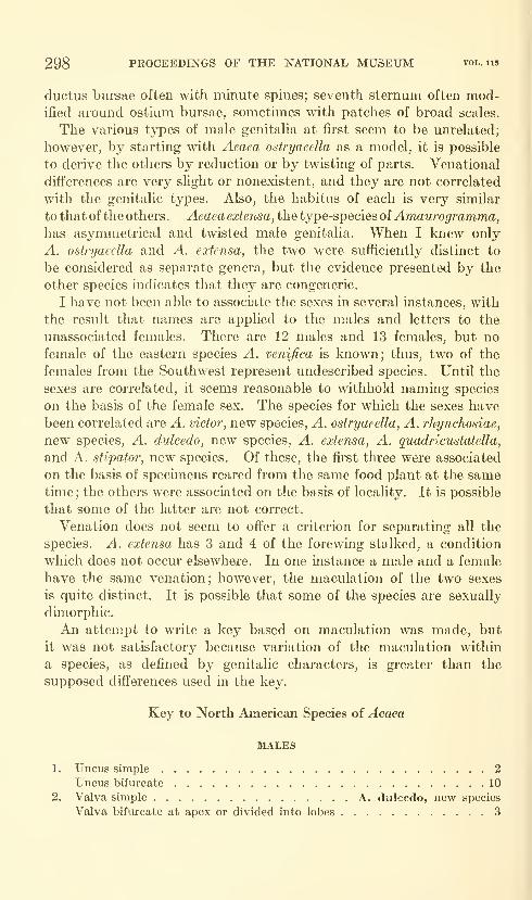
298 PROCEEDINGS OF THE NATIONAL MUSEUM vol. 115
ductus bursae often with minute spines; seventh sternum often mod-ified around ostium bursae, sometimes with patches of broad scales.
The various types of male genitalia at first seem to be unrelated;
however, by starting with Aeaea ostryaeella as a model, it is possible
to derive the others by reduction or by twisting of parts. Venational
differences are very sUght or nonexistent, and they are not correlated
with the genitalic types. Also, the habitus of each is very similar
to that of the others. Aeaeaextensa, the type-species of^wawro^rawma,has asymmetrical and twisted male genitalia. When I knew only
A. ostryaeella and A. extensa, the two were sufficiently distinct to
be considered as separate genera, but the evidence presented by the
other species indicates that they are congeneric.
I have not been able to associate the sexes in several instances, with
the result that names are applied to the males and letters to the
unassociated females. There are 12 males and 13 females, but no
female of the eastern species A. venifica is known; thus, two of the
females from the Southwest represent undcscribed species. Until the
sexes are correlated, it seems reasonable to withhold naming species
on the basis of the female sex. The species for which the sexes have
been correlated are A. victor, new species, A. ostryaeella, A. rhynchosiae,
new species, A. dvlcedo, new species, A. extensa, A. quadricustatella,
and A. stipator, new species. Of these, the first three were associated
on the basis of specimens reared from the same food plant at the sametime ; the others were associated on the basis of locality. It is possible
that some of the latter are not correct.
Venation does not seem to offer a criterion for separating all the
species. A. extensa has 3 and 4 of the forewing stalked, a condition
which does not occur elsewhere. In one instance a male and a female
have the same venation; however, the maculation of the two sexes
is quite distinct. It is possible that some of the species are sexually
dimorphic.
An attempt to wi-ite a key based on maculation was made, but
it was not satisfactory because variation of the maculation within
a species, as defined by genitalic characters, is greater than the
supposed differences used in the key.
Key to North American Species of Aeaea
MALES
1. Uncus simple 2
Uncus bifurcate 10
2. Valva simple A. dulcedo, new species
Valva bifurcate at apex or divided into lobes 3
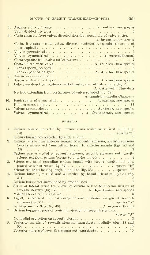
MOTHS OF FAMILY WALSHIIDAE—HODGES 299
3. Apex of valva bifurcate A. venifica, new species
Valva divided into lobes 4
4. Costa separate from valva, directed dorsally; remainder of valva entire.
A. juvantis, new species
Costa, if separate from valva, directed posteriorly; sacculus separate, at
least apically 5
5. Valvae symmetrical 6
Valvae asymmetrical A. extensa (Braun)
6. Costa separate from valva (at least apex) 7
Costa united with valva A. venatrix, new species
7. Uncus tapering to apex 8
Uncus expanded at apex A. stipator, new species
8. Saccus with acute apex 9
Saccus with rounded apex A. risor, new species
9. Lobe extending from posterior part of costa; apex of valva acute (fig. 26).
A. ostryaeella ChambersNo lobe extending from costa; apex of valva rounded (fig. 57).
A. qiiadricustatella Chambers10. Each ramus of uncus bifid A. sagana, new species
Rami of uncus simple 11
11. Valvae symmetrical A. victor, new species
Valvae asymmetrical A. rhynchosiae, new species
FEMALES
1. Ostium bursae preceded by narrow semicircular sclerotized band (fig.
54) species "f"
Ostium bursae not preceded by such a band 2
2. Ostium bursae near anterior margin of seventh sternum, seventh sternum
heavily sclerotized from ostium bursae to anterior margin (figs. 52 and
55) 3
Ostium bursae medial on seventh sternum, seventh sternum not heavily
sclerotized from ostium bursae to anterior margin 4
3. Sclerotized band preceding ostium bursae with strong longitudinal line,
placed to left of center (fig. 52) species "b"Sclerotized band lacking longitudinal hne (fig. 55) species "e"
4. Ostium bursae preceded and succeeded by broad sclerotized plates (fig.
51) 5
Ostium bursae not surrounded by broad plates 7
5. Series of lateral striae from level of ostium bursae to anterior margin of
seventh sternum (fig. 47) A. rhynchosiae, new species
Without series of lateral striae 6
6. Lightly sclerotized flap extending beyond posterior margin of seventh
sternum (fig. 51) species "a"Lacking such a flap (fig. 49) A. extensa (Braun)
7. Ostium bursae at apex of conical projection on seventh sternum.
species "d"No medial projection on seventh sternum 8
8. Posterior margin of seventh sternum emarginate medially (figs. 48 and
50) 9
Posterior margin of seventh sternum not emarginate 10
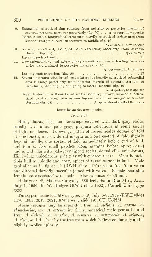
300 PROCEEDINGS OF THE NATIONAL MUSEUM vol. lib
9. Submedial sclerotized flap running from anterior to posterior margin of
seventh sternum, narrower posteriorly (fig. 50) . . A. victor, new species
Without such a longitudinal structure; heavily sclerotized striate area from
anterior margin of seventh sternum to middle (fig. 48).
A. dulcedo, new species
10. Narrow, sclerotized, Y-shaped band extending anteriorly from seventh
sternum (fig. 56) species "c"
Lacking such a band 11
11. Two submedial conical extensions of seventh sternum, extending from an-
terior margin almost to posterior margin (fig. 45).
A. ostryaeella ChambersLacking such extensions (fig. 46) 12
12. Seventh sternum with broad scales laterally; heavily sclerotized submedial
area running posteriorly from anterior margin of seventh sternum to
two-thirds, then angling and going to lateral margins (fig. 46).
A. stipator, new species
Seventh sternum without broad scales laterally; narrow, submedial sclero-
tized band running from ostium bursae to anterior margin of seventh
sternum (fig. 58) A. quadricustatella Chambers
Aeaea juvantis, new species
Figure 32
Head, thorax, legs, and forewings covered with dark gray scales,
usually with apices pale gray, purplish reflections at some angles
of light incidence. Forewing: patch of raised scales dorsad of fold
at one-fourth, one on dorsal margin and one costad of fold slightly
beyond middle, one costad of fold immediately before end of fold,
and four or five small patches along margins before apex; costal
and apical cilia with pale-gray tipped scales, dorsal cilia unicolorous.
Hind wing: unicolorous, pale gray with cinereous cast. Metathoracic
tibia buff at middle and apex, apices of tarsal segments buft'. Male
genitalia: as in figure 32 (RWH slide 1170); costa free from valva
and directed dorsall}^, sacculus joined with valva. Female genitalia:
female not associated with male. Alar expanse: 6-6.5 mm.Holotype: cJ", Madera Canyon, 4880 feet, Santa Rita Mts., Ariz.,
July 1, 1959, R. W. Hodges (RWH slide 1992), Cornell Univ. type
3883.
Paratypes: same locality as type, 5 cf , July 1-9, 1959 (RWH slides
1170, 1993, 2019, 2021 ; RWH wing slide 19), CU, USNM.Aeaea juvantis may be separated from A. stellans, A. sagana, A.
rhynchosiae, and A. extensa by the S3niiimetrical male genitalia; and
from A. dulcedo, A. venifica, A. venatrix, A. ostryaeella, A. stipator,
A. risor, and A. victor by the free costa which is directed dorsally and is
slightly swollen apically.
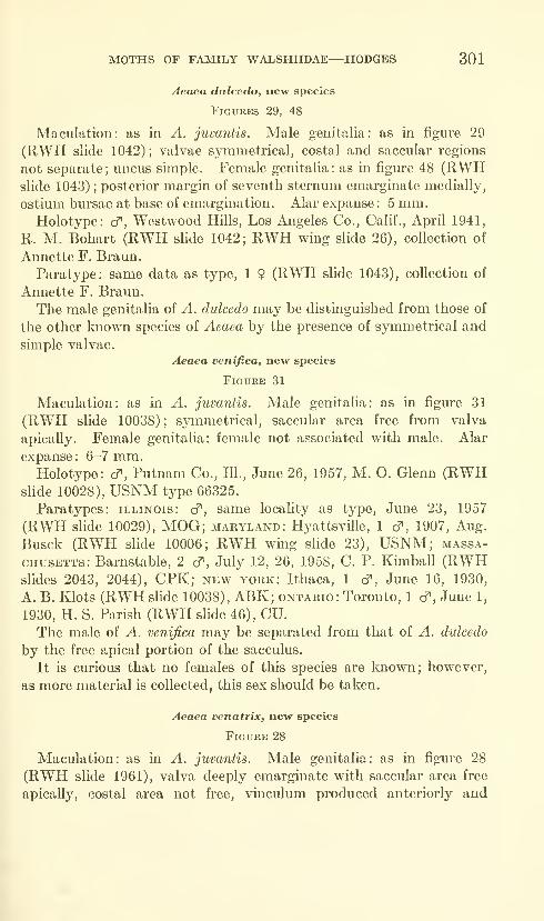
MOTHS OF FAMILY WALSHIIDAE—HODGES 301
Aeaea diilcedo, new species
Figures 29, 48
Maculation: as iii A. juvantis. Male genitalia: as in figure 29
(RWH slide 1042) ; valvae symmetrical, costal and saccular regions
not separate; uncus simple. Female genitalia: as in figure 48 (RWHslide 1043) ;
posterior margin of seventh sternum emarginate medially,
ostium bm'sae at base of emargination. Alar expanse : 5 mm.Holotype: c^, Westwood Hills, Los Angeles Co., Calif., April 1941,
R. M. Bohart (RWH slide 1042; RWH wing slide 26), collection of
Annette F. Braun.
Paratype: same data as type, 1 9 (RWH slide 1043), collection of
Annette F. Braun.
The male genitalia of A. dulcedo may be distinguished from those of
the other known species of Aeaea by the presence of symmetrical and
simple valvae.
Aeaea venifica, new species
Figure 31
Maculation: as in A. juvantis. Male genitalia: as in figure 31
(RWH slide 10038); symmetrical, saccular area free from valva
apically. Female genitalia: female not associated with male. Alar
expanse: 6-7 mm.Holotype: cf , Putnam Co., 111., June 26, 1957, M. O. Glenn (RWH
shde 10028), USNM type 66325.
Paratypes: Illinois: cf, same locality as type, June 23, 1957
(RWH slide 10029), MOG; Maryland: Hyattsviile, 1 cf , 1907, Aug.
Busck (RWH slide 10006; RWH wing slide 23), USNM; Massa-
chusetts: Barnstable, 2 d", July 12, 26, 1958, C. P. Kunball (RWHslides 2043, 2044), CPK; new york: Ithaca, 1 cf, June 16, 1930,
A. B. Klots (RWH slide 10038), ABK; Ontario: Toronto, 1 cT, June 1,
1930, H. S. Parish (RWH slide 46), CU.The male of A. venifica may be separated from that of A. dulcedo
by the free apical portion of the sacculus.
It is curious that no females of this species are known; however,
as more material is collected, this sex should be taken.
Aeaea venatrix, new species
Figure 28
Maculation: as in A. juvantis. Male genitalia: as in figure 28
(RWH slide 1961), valva deeply emarginate with saccular area free
apically, costal area not free, vinculum produced anteriorly and
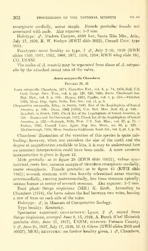
302 PROCEEDINGS OF THE NATIONAL MUSEUM vol. us
emarginate medially, uncus simple. Female genitalia: female not
associated with male. Alar expanse : 5-7 mm.Holotype: cT, Madera Canyon, 4880 feet, Santa Rita Mts., Ariz.,
July 17, 1959, R. W. Hodges (RWH slide 2022), Cornell Univ. type
3884.
Paratypes: same locality as type, 7 cf, July 2-30, 1959 (RWHslides 1169, 1961, 1962, 1966, 1971, 1976, 1994; RWH wing slide 18),
CU, USNM.The males of A. venafrix may be separated from those of A. ostryae-
ella by the attached costal area of the valva.
Aeaea ostryaeella Chambers
Figures 26, 45
Aeaea ostryaeella Chambers, 1874, Canadian Ent., vol. 6, p. 74; 1878, Bull. U.S.
Geol. Geogr. Surv. Terr., vol. 4, pp. 121, 128; 1880, Journ. Cincinnati Soc.
Nat. Hist., vol. 2, p. 199.—Hagen, 1884, Papilio, vol. 4, p. 154.—Fletcher,
1929, Mem. Dep. Agric. India, Ent. Ser., vol. 11, p. 6.
Chrysopeleia ostryaeella, Riley, in Smith, 1891, List of the Lepidoptera of boreal
America, p. 108.—Dyar, 1902 [1903], U.S. Nat. Mus. Bull. 52, p. 540.—
Kearfott, in Smith, 1903, Check list of the Lepidoptera of boreal America, p.
118.—Barnes and McDunnough, 1917, Check list of the Lepidoptera of boreal
America, p. 152.—Heinrich, 1920, Proc. U.S. Nat. Mus., vol. 57, p. 71.—
Forbes, 1923, Cornell Univ. Agric. Exp. Sta. Mem., no. 68, p. 329.—
McDunnough, 1939, Mem. Southern California Acad. Sci., vol. 2, pt. 1, p. 63.
Chambers' illustration of the venation of this species is quite mis-
leading; however, when one considers the size of the moth and the
degree of magnification available to him, it is easy to understand howan incorrect interpretation could have been made. A more accurate
interpretation is given in figure 12,
Male genitalia: as m figure 26 (RWH slide 10027), valvae sym-
metrical, costa free, anterior margin of vinculum emarginate medially,
uncus emarginate. Female genitalia: as in figm*e 45 (RWH slide
1162); seventh sternum with two heavUy sclerotized areas starting
anteromedially, runnmg posteromedially, free from sternum apically;
ostium bursae at center of seventh sternum. Alar expanse: 5-7 mm.Food plant: Ostrya virginiana (Mill.) K. Koch. According to
Chambers (1874), the larva mines the leaf between two veins, leaving
a row of frass on each side of the mine.
Holotype: cf , in Museum of Comparative Zoology.
Type locality: Kentucky.
Specimens examined: Connecticut: Lyme, 2 cf, reared from
Ostrya virginiana, emerged June 9, 15, 1916, A. Busck (Carl Heinrich
genitalia shde, June 27, 1917), USNM; Illinois: Putnam County,
2 cf , June 28, 1957, July 17, 1959, M. O. Glenn (RWH slides 2039 and
10027), MOG; Kentucky: no fm-ther locality given, 1 cf , Chambers,
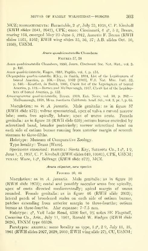
MOTHS OF FAMILY WALSHIIDAE—HODGES 303
MCZ; MASSACHUSETTS: Barnstable, 2 d^, July 25, 1958, C. P. Kimball
(RWH slides 2041, 2042), CPK; ohio: Cincinnati, 4 cT", 5 9, Braun,
rearing 164, emerged May 23-Jmie 5, 1912, Annette F. Braun (RWHslides 1161, 1162; RWH wing slides 25, 36, 37; A.B. slides Oct. 10,
1930), USNM.
Aeaea quadricustatella Chambers
Figures 57, 58
Aeaea quadricustatella Chambers, 1880, Journ. Cincinnati Soc. Nat. Hist., vol. 2,
p. 186.
Aeaea quadricristatella, Hagen, 1884, Papilio, vol. 4, p. 154.
Chrysopeleia quadricristatella, Riley, in Smith, 1891, List of the Lepidoptera of
boreal America, p. 108.—Dyar, 1902 [1903], U.S. Nat. Mus. Bull. 52,
p. 540.—Kearfott, in Smith, 1903, Check list of the Lepidoptera of boreal
America, p. 118.—Barnes and McDunnough, 1917, Check Hst of the Lepidop-
tera of boreal America, p. 152.
Amaurogramma quadricristatella, Braun, 1919, Ent. News, vol. 30, p. 262.
—
McDunnough, 1939, Mem. Southern Cahfornia Acad. Sci., vol. 2, pt. 1, p. 63.
Maculation: as in yl. juvantis. Male genitalia: as in figure 57
(RWH slide 872) ; valvae symmetrical, apex of valva a curved narrow
lobe; costa free apically, lobate; apex of uncus acute. Femalegenitalia : as in figure 58 (RWH slide 659) ; ostium bursae encircled bysclerotized band, broader posteriorly; narrow sclerotized band oneach side of ostium bursae running from anterior margin of seventh
sternum to three-fifths.
Holotype: Museum of Comparative Zoology.
Type locality: Texas [Waco].
Specimens exammed: Florida: Siesta Key, Sarasota Co., Icf, 19,
June 1, 2, 1957, C. P. Kmiball (RWH slides 649, 10041), CPK, USNM;TEXAS: Waco, 1 cf , Belfrage (RWH slide 872), MCZ.
Aeaea stipator, new species
Figures 30, 46
Maculation: as in A. juvantis. Male genitalia: as in figure 30
(RWH slide 1972); costal and possibly saccular areas free apically,
apex of costa directed medioventrally; apical margin of uncus
rounded. Female genitalia: as in figure 46 (RWH slide 2032);
lateral patch of broadened scales on each side of ostium bursae,
patches extending from anterior margin to three-fourths; ostium
bursae at three-fourths. Alar expanse: 7-7.5 mm.Holotype: cf, Vail Lake Road, 6500 feet, 9% miles SE Flagstaff,
Coconmo Co., Ariz., July 11, 1961, Ronald W. Hodges (RWH slide
2028), USNM type 66326.
Paratypes: Arizona: same locality as type, Icf, 2 9, July 11, 18,
1961 (RWH slides 2027, 2029, 2030; RWH wmg slide 27), CU, USNM;
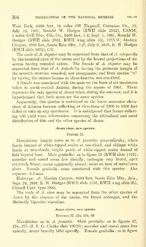
304 PROCEEDINGS OF THE NATIONAL MUSEUM vol. ii5
West Fork, 6500 feet, 16 miles SW Flagstaff, Coconino Co., 19,
July 15, 1961, Ronald W. Hodges (RWH slide 2032), USNM;4 miles ESE Pine, Gila Co., 5400 feet, 1 9, Sept. 1, 1961, Ronald W.Hodges (RWH slide 2031, RWH wing slide 35), USNM; Madera
Canyon, 4880 feet, Santa Rita Mts., 1 cf , July 9, 1959, R. W. Hodges
(RWH slide 1972), CU.The male of A. stipator may be separated from that of A. ostryaeella
by the rounded apex of the uncus and by the lateral projections of the
saccus having rounded apices. The female of A. stipator may be
separated from that of A. dulcedo by having the posterior margin of
the seventh sternum rounded, not emarginate; and from species "c"
by having the ostium bursae at three-fourths, not one-third.
A female was associated with the male on the basis of six specimens
taken m north-central Arizona during the season of 1961. These
represent the only species of Aeaea taken during the summer, and it is
hypothesized that both sexes are the same species.
Apparently, this species is restricted to the lower montaine eleva-
tions of Arizona because collecting at elevations of 7200 to 8500 feet
failed to turn up any specimens. It is anticipated that future collect-
ing will yield more information concerning the altitudinal and areal
distribution of this and the other species of Aeaea.
Aeaea risoVf new species
Figure 33
Maculation: largely same as in A. juvantis; perpendicular, white
fascia formed of white-tipped scales at one-third, and oblique white
fascia at two-thirds; bright patch of white-tipped scales dorsad of
fold beyond base. Male genitalia: as in figure 33 (RWH slide 1166)
;
saccular and costal areas free distally; aedeagus very broad, apex
relatively blunt; uncus apparently absent; setae on base of costal area
short. Female genitalia: none associated with this species. Alar
expanse: 6.5 mm.Holotype: cf, Madera Canyon, 4880 feet, Santa Rita Mts., Ariz.,
Sept. 24, 1959, R. W. Hodges (RWH slide 1166, RWH wing slide 28),
Cornell Univ. type 3885.
The male of A. risor may be separated from the other species of
Aeaea by the absence of the uncus, the broad aedeagus, and the
distinctly bipartite vinculum.
Aeaea victor, new species
Figures 27, 27a, 27b, 50
Maculation: as in A. juvantis. Male genitalia: as in figures 27,
27a, 27b (J. F. G. Clarke slide 10226); saccular and costal areas free
apically, uncus broadly bifid apically. Female genitalia : as in figure
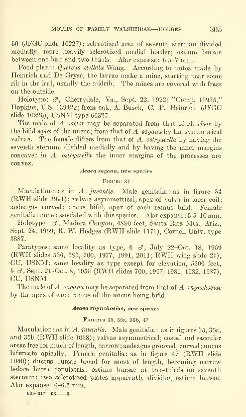
MOTHS OF FAMILY WALSHIIDAE—HODGES 305
50 (JFGC slide 10227); scleroiized area of seventh sternum divided
medially, more heavily sclerotized medial border; ostium bursae
between one-half and two-thirds. Alar expanse: 6.5-7 mm.Food plant: Qiiercus stellata Wang. According to notes made by
Heinrich and De Gryse, the larvae make a mine, starting near somerib in the leaf, usually the midrib. The mines are covered with frass
on the outside.
Holotype: cf, Cherrydale, Va., Sept. 22, 1922; "Comp. 13935,"
Hopkins, U.S. 13942g; from oak, A. Busck, C. P. Heinrich (JFGCslide 10226), USNM type 66327.
The male of A. victor may be separated from that of A. risor bythe bifid apex of the uncus ; from that of A. sagana by the symmetrical
valvae. The female differs from that of A. ostryaeella by having the
seventh sternum divided medially and by having the inner marginsconcave; in A. ostryaeella the inner margins of the processes are
convex.
Aeaea sagana, new species
FlGURK 34
Maculation: as in A. juvantis. Male genitalia: as in figure 34
(RWH slide 1991); valvae asymmetrical, apex of valva in loose coil;
aedeagus curved; uncus bifid, apex of each ramus bifid. Femalegenitalia : none associated with this species. Alar expanse : 5.5-10 mm.
Holotype: d^, Madera Canyon, 4880 feet, Santa Rita Mts., Ariz.,
Sept. 24, 1959, R. W. Hodges (RWH slide 1171), Cornell Univ. type
3887.
Paratypes: same locality as type, 6 d^, July 22-Oct. 18, 1959
(RWH slides 536, 585, 700, 1977, 1991, 2011; RWH wing slide 21),
CU, USNM; same locality as type except for elevation, 5600 feet,
5 cT, Sept. 21-Oct. 8, 1959 (RWH slides 700, 1967, 1981, 1982, 1987),
CU, USNM.The male of A. sagana may be separated from that of A. rhynchosiae
by the apex of each ramus of the uncus being bifid.
Aeaea rhynchosiae, new species
Figures 35, 35a, 35b, 47
Maculation : as in A. juvanHs. Male genitalia : as in figm-es 35, 35a,
and 35b (RWH slide 1038); valvae asymmetrical; costal and saccular
areas free for much of length, narrow; aedeagus grooved, curved ; uncusbifurcate apically. Female genitalia: as in figure 47 (RWH slide
1040) ; ductus bursae broad for most of length, becoming narrowbefore bursa copulatrLx; ostium bursae at two-thirds on seventhsternum; two sclerotized plates apparently dividing ostium bursae.
Alar expanse: 6-6.5 mm.685-617—63 2
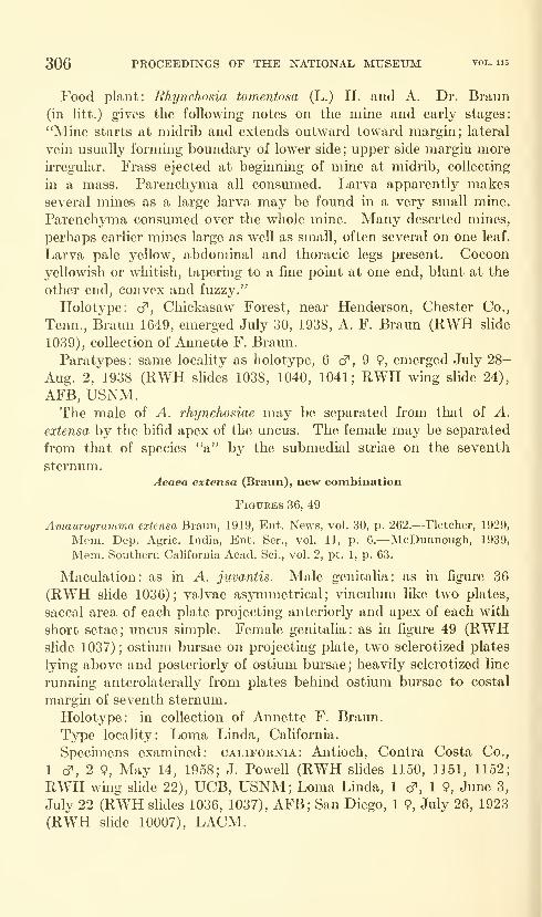
306 PROCEEDINGS OF THE NATIONAL MUSEUM vol. 115
Food plant: Rhynchosia tomentosa (L.) H. and A. Dr. Braun
(in litt.) gives the following notes on the mine and early stages:
"Mine starts at midrib and extends outward toward margin; lateral
vein usually formmg boundary of lower side; upper side margin more
irregular. Frass ejected at beginning of mine at midrib, collecting
in a mass. Parenchyma all consmned. Larva apparently makesseveral mines as a large larva may be found in a very small mine.
Parenchyma consumed over the whole mine. Many deserted mines,
perhaps earlier mines large as well as small, often several on one leaf.
Larva pale yellow, abdominal and thoracic legs present. Cocoon
yellowish or whitish, tapermg to a fme point at one end, blunt at the
other end, convex and fuzzy."
Holotype: cf, Chickasaw Forest, near Henderson, Chester Co.,
Tenn., Braun 1649, emerged July 30, 1938, A. F. Braun (RWH slide
1039), collection of Annette F. Braun.
Paratypes: same locality as holotype, 6 cT, 9 9, emerged July 28-
Aug. 2, 1938 (RWH slides 1038, 1040, 1041; RWH wmg slide 24),
AFB, USNM.The male of A. rhynchosiae may be separated from that of A.
extensa by the bifid apex of the uncus. The female may be separated
from that of species "a" by the submedial striae on the seventh
sternum.Aeaea extensa (Braun), new combination
Figures 36, 49
Amaurogramma extensa Braun, 1919, Ent. News, vol. 30, p. 262.—Fletcher, 1929,
Mem. Dep. Agric. India, Ent. Ser., vol. 11, p. 6.—McDunnough, 1939,
Mem. Southern California Acad. Sci., vol. 2, pt. 1, p. 63.
Maculation: as in A. juvantis. Male genitalia: as in figure 36
(RWH slide 1036); valvae asymmetrical; vinculum like two plates,
saccal area of each plate projecting anteriorly and apex of each with
short setae; uncus simple. Female genitalia: as in figure 49 (RWHslide 1037) ; ostium bursae on projecting plate, two sclerotized plates
lying above and posteriorly of ostium bursae; heavily sclerotized line
running anterolaterally from plates behind ostium bursae to costal
margin of seventh sternum.
Holotype: in collection of Annette F. Braun.
Type locality: Loma Linda, California.
Specimens examined: California: Antioch, Contra Costa Co.,
1 c^, 2 9, May 14, 1958; J. Powell (RWH slides 1150, 1151, 1152;
RWH wing slide 22), UCB, USNM; Loma Linda, 1 c?", 1 9, June 3,
July 22 (RWH slides 1036, 1037), AFB; San Diego, 1 9, July 26, 1923
(RWH slide 10007), LACM.

MOTHS OF FAMILY WALSHIIDAE—HODGES 307
Unnamed Species of Aeaea
The remaining females are those which have not been associated
with males. As I have stated previously, at least two of them repre-
sent new species, but until reared material with associated adults
is available, the best course is to illustrate the genitalia and to give
alphabetic denotations to the specimens. Each has approximately
the same habitus as A. juvantis except for "d," which is smiilar to
A. risor. The specimens will be retained temporarily in the USNM,
Aeaea species a
Figure 61
Female genitalia: as in figure 51 (RWH slide 1164) ; ostium bursae
at middle of seventh sternmn; heavily sclerotized plate anterior to
ostium bursae, slightly emarginate posteriorly; two heavily sclerotized
plates posterior to ostium bursae; third plate posterior to ostium bursae
with convex apex. Alar expanse: 9 mm.Specimen examined: Madera Canyon, 4880 feet, Santa Rita Mts.,
Ariz., 1 9, July 24, 1959, R. W. Hodges (RWH slide 1164, RWHwing slide 34).
Aeaea species b
Figure 52
Female genitalia: as in figure 52 (RWH slide 1172); ostium bursae
at one-third on seventh sternum; heavily sclerotized plate anterior to
and laterad of ostium bursae, well-defined Ime dividing medial andlateral sections of this plate. Alar expanse: 7-7.5 mm.
Specmiens examined: Madera Canyon, 4880 feet, Santa Rita
Mts., Ariz., 3 9, Sept. 19-29, 1959, R. W. Hodges (RWH slides
1990, 2017, 2018; RWH wmg slide 31); same locality except for ele-
vation, 5600 feet, 1 9, Sept. 23, 1959 (RWH slide 1172).
Aeaea species c
Figures 56 and 62
Female genitalia: as in figm-e 56 (RWH slide 1965); ostium bursae
at one-third; seventh sternum with broad scales arranged in semi-
circular patches beside ostium bursae; one signum visible. Alar
expanse: 7-7.5 mm.Specimens examined: Madera Canyon, 4880 feet, Santa Rita
Mts., Ai'iz., 4 9, July 10-27, 1959, R. W. Hodges (RWH slides 1965.
1974, 2023; RWH wmg slide 33).
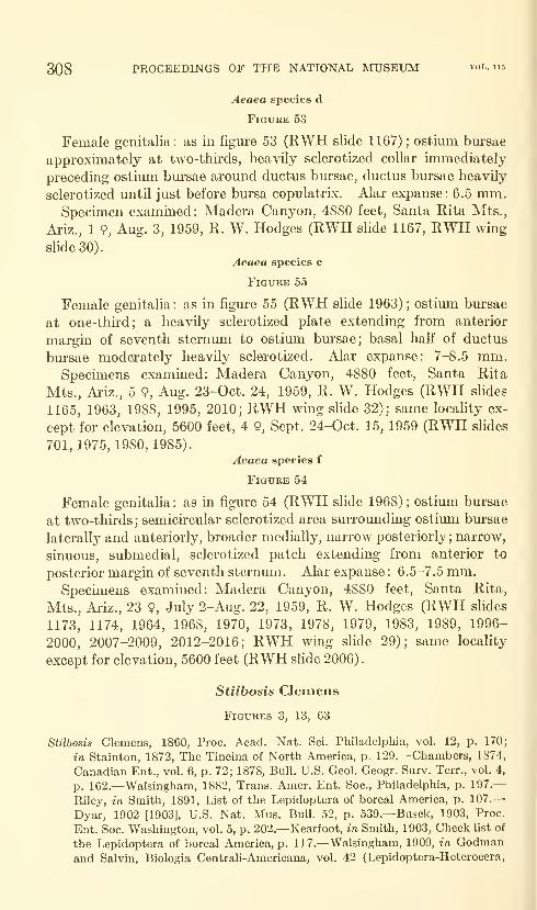
308 PROCEEDINGS OF THE NATIONAL MUSEUM vol. 115
Aeaea species d
Figure 53
Female genitalia: as in figure 53 (RWH slide 1167); ostium bursae
approximately at two-thirds, heavily sclerotized collar immediately
preceding ostium bm-sae around ductus bursae, ductus bursae heavily
sclerotized until just before bursa copulatrix. Alar expanse: 6.5 mm.Specimen examined: Madera Canyon, 4880 feet, Santa Rita Mts.,
Ariz., 1 9, Aug. 3, 1959, R. W. Hodges (RWH slide 1167, RWH wmgslide 30).
Aeaea species e
Figure 55
Female genitalia: as in figure 55 (RWH slide 1963); ostium bursae
at one-third; a heavily sclerotized plate extending from anterior
margin of seventh sternum to ostium bursae; basal haK of ductus
bursae moderately heavily sclerotized. Alar expanse: 7-8.5 mm.Specunens examined: Madera Canyon, 4880 feet, Santa Rita
Mts., Ariz., 5 9, Aug. 23-Oct. 24, 1959, R. W. Hodges (RWH slides
1165, 1963, 1988, 1995, 2010; RWH wing slide 32); same locality ex-
cept for elevation, 5600 feet, 4 9, Sept. 24-Oct. 15, 1959 (RWH slides
701, 1975, 1980, 1985).
Aeaea species f
Figure 54
Female genitalia: as in figure 54 (RWH slide 1968); ostium bursae
at two-thirds; semicircular sclerotized area surrounding ostium bursae
laterally and anteriorly, broader medially, narrow posteriorly; narrow,
sinuous, submedial, sclerotized patch extending from anterior to
posterior margin of seventh sternum. Alar expanse : 6.5-7.5 mm.Specunens examined: Madera Canyon, 4880 feet, Santa Rita,
Mts., Ariz., 23 9, July 2-Aug. 22, 1959, R. W. Hodges (RWH slides
1173, 1174, 1964, 1968, 1970, 1973, 1978, 1979, 1983, 1989, 1996-
2000, 2007-2009, 2012-2016; RWH wing slide 29); same locality
except for elevation, 5600 feet (RWH slide 2006).
Stilbosis Clemens
Figures 3, 13, 63
Stilbosis Clemens, 1860, Proc. Acad. Nat. Sci. Philadelphia, vol. 12, p. 170;
in Stainton, 1872, The Tineina of North America, p. 129.—Chambers, 1874,
Canadian Ent., vol. 6, p. 72; 1878, Bull. U.S. Geol. Geogr. Surv. Terr., vol. 4,
p. 1G2.—Walsingham, 1882, Trans. Amer. Ent. Soc, Philadelphia, p. 197.
—
Riley, in Smith, 1891, List of the Lepidoptera of boreal America, p. 107.
—
Dyar, 1902 [1903], U.S. Nat. Mus. Bull. 52, p. 539.—Busck, 1903, Proc.
Ent. Soc. Washington, vol. 5, p. 202.—Kearfoot, in Smith, 1903, Check list of
the Lepidoptera of boreal America, p. 117.—Walsingham, 1909, in Godmanand Salvin, Biologia Centrali-Americana, vol. 42 (Lepidoptera-Heterocera,
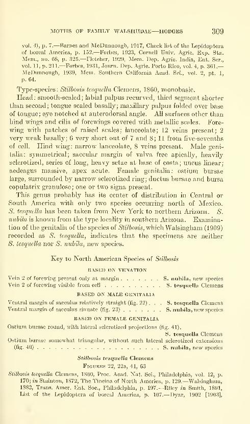
MOTHS OF FAMILY WALSHIIDAE—HODGES 309
vol. 4), p. 7.—Barnes and McDunnough, 1917, Check list of the Lepidopteraof boreal America, p. 152.—Forbes, 1923, Cornell Univ. Agric. Exp. Sta.
Mem., no. 68, p. 325.—Fletcher, 1929, Mem. Dep. Agric. India, Ent. Ser.,
vol. 11, p. 211.—Forbes, 1931, Journ. Dep. Agric. Porto Rico, vol. 4, p. 361.
—
McDunnough, 1939, Mem. Southern California Acad. Sci., vol. 2, pt. 1,
p. 64.
Type-species: Stilbosis tesquella Clemens, 1860, monobasic.
Head : smooth-scaled ; labial palpus recm"ved, third segment shorter
than second; tongue scaled basally; maxillary palpus folded over base
of tongue; eye notched at anterodorsal angle. All surfaces other than
hind wings and cilia of forewings covered with metallic scoles. Fore-
wing with patches of raised scales; lanceolate; 12 veins present; 2
very v>^eak basally; 6 very short out of 7 and 8; 11 from five-sevenths
of cell. Hind wing: narrow lanceolate, 8 veins present. Male geni-
talia: symmetrical; saccular margin of valva free apically, heavily
sclerotized, series of long, heavy setae at base of costa; uncus linear;
aedeagus massive, apex acute. Female genitalia: ostium bursae
large, surrounded by narrow sclerotized ring; ductus bursae and bursa
copulatrix granulose ; one or two signa present.
This genus probably has its center of distribution in Central or
South America with only two species occurring north of Mexico.
S. tesqudla has been taken from New York to northern Arizona. S.
nuhila is laiown from the type locality in southern Arizona. Examina-tion of the genitalia of the species of Stilbosis, which Walsingham (1909)
recorded as S. tesquella, indicates that the specimens are neither
S. tesquella nor S. nuhila, new species.
Key to North American Species of Stilbosis
BASED ON VENATION
Vein 2 of forewing present only at margin S. nubila, new species
Vein 2 of forewing visible from cell S. tesquella Clemens
BASED ON MALE GENITALIA
Ventral margin of sacculus relatively straight (fig. 22) . . . S. tesquella ClemensVentral margin of sacculus sinuate (fig. 23) S. nubila, new species
BASED ON FEMALE GENITALIA
Ostium bursae round, with lateral sclerotized projections (fig. 41).
S. tesquella ClemensOstium bursae somewhat triangular, without such lateral sclerotized extensions
(fig. 40) S. nubila, new species
Stilbosis tesquella Clemens
Figures 22, 22a, 41, 63
Stilbosis tesquella Clemens, 1860, Proc. Acad. Nat. Sci., Philadelphia, vol. 12, p.
170; in Stainton, 1872, The Tineina of North America, p. 129.—Walsingham,1882, Trans. Amer. Ent. Soc, Philadelphia, p. 197.—Riley in Smith, 1891,
List of the Lepidoptera of boreal America, p. 107.—Dyar, 1902 [1903],

310 PROCEEDINGS OF THE NATIONAL MUSEUM vol. 115
U.S. Nat. Mus. Bull. 52, p. 539.—Busck, 1903, Proc. Ent. Soc. Washington,
vol. 5, p. 202.—Kearfott, in Smith, 1903, Check list of the Lepidoptera of
boreal America, p. 117.—Walsingham, 1909, in Godman and Salvin, Biologia
Central!-Americana, vol. 42 (Lepidoptera-Heterocera, vol. 4), p. 7.—Barnesand McDiinnough, 1917, Check list of the Lepidoptera of boreal America,
p. 152.—Forbes, 1923, Cornell Univ. Agric. Exp. Sta. Mem., no. 68, p.
325.—Fletcher, 1929, Mem. Dep. Agric. India, Ent. Ser., vol. 11, p. 211.—McDunnough, 1939, Mem. Southern California Acad. Sci., vol. 2, pt. 1,
p. 64.
Stilbosis tesquatella, Chambers, 1878, Bull. U.S. Geol. Geogr. Surv. Terr., vol. 4,
p. 162, misspeUing.—Riley, in Smith, 1891, List of the Lepidoptera of boreal
America, p. 107 (as synonym of S. tesauella).—Walsingham, 1909, in Godmanand Salvin, Biologia Centrali-Americana, vol. 42 (Lepidoptera-Heterocera,
vol. 4), p. 8 (as synonym of S. tesquella).
Laverna(?) quinquicristatella Chambers, 1881, Journ. Cincinnati Soc. Nat. Hist.,
vol. 3, p. 293.
Laverna ? quinquecristatella, Walsingham, 1882, Trans. Amer. Ent. Soc, Phil-
adelphia, p. 197 (correction of spelling; as synonym of S. tesquella).
Stilbosis quinque-cristatella, Riley, in Smith, 1891, List of the Lepidoptera of
boreal America, p. 107 (as synonym of S. tcsqtiella).
Stilbosis quinquecristatella, Dyar, 1902 [1903], U.S. Nat. Mus. Bull. 52, p. 539
(as synonym of S. tesquella).—Walsingham, 1909, in Godman and Salvin,
Biologia Centrali-Americana, vol. 42 (Lepidoptera-Heterocera, vol. 4), p. 8
(as synonym of S. tesquella).—Barnes and McDunnough, 1917, Check list
of the Lepidoptera of boreal America, p. 152 (as synonym of S. tesquella).—Forbes, 1923, Cornell Univ. Agric. E.\p. Sta. Mem., no. 68, p. 325 (as synonymof S. tesquella).—McDunnough, 1939, Mem. Southern Cahfornia Acad. Sci.,
vol. 2, pt. 1, p. 64 (as synonym of S. tesquella).
The male and female genitalia of this common and relatively well-
known species are figm-ed for the first time: Figures 22 and 22a
(AB slide 14.XI.1930) and figure 41 (RWH slide 577).
Lectotype: S. tesquella, present designation, 9, bearing following
labels: "1. 88; 2. Type, Stilbosis tesquella B. Clemens, 7482; 3. Stilbosis
tesquella Clemens, Type!, AB 1902." Academy of Natural Sciences,
Philadelphia, quinquicristatella, lost.
Type localities: S. tesquella, Pennsylvania(?); S. quinquicristatella,
Amherst, Massachusetts.
Food plant: Amphicarpa bracteata (L.) Fern, and Lespedeza spp.
in the East.
Spechnens examined: Arizona: Fort Valley, 7350 feet, 7}^ miles
NW Flagstaff, Coconino Co., 1 d", July 19, 1961, Ronald W. Hodges,
USNM; Hart Prairie, 8500 feet, 10 miles NNW Flagstaff, Coconino
Co., 2 d", July 1, 21, 1961, Ronald W. Hodges, USNM; West Fork,
6500 feet, 16 miles SW Flagstaff, Coconino Co., 63 d,9 9, July 4-19,
1961, Ronald W. Hodges (RWH slides 1655, 1656; RWH wmg slide
42), CU, USNM; Kansas: Onaga, 3 9 (RWH slide 10008), MCZ.MARYLAND: Plummcrs Island, 4 cf, May-July, A. Busck, R. C.
Sherman, USNM; District of Columbia: 3 9, ex hog peanut, emerged
June 31, July 6, USNM; Minnesota: no further locality, 1 cf , mining
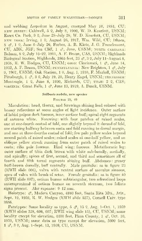
MOTHS OF FAMILY WALSHIIDAE—HODGES 311
and webbing Lespedeza in August, emerged May 10, 1884, CU;NEW jersey: Caldwell, 5 9, July 8, 1900, W. D. Kearfott, USNM;Essex Co. Park, 9 9, June 28-July 28, W. D. Kearfott, CU, USNM;NEW YORK: Irving, 1 9, August 26, 1917, Wm. Wild, CU; Ithaca,
8 cT, 1 9, June 6-July 26, Forbes, A. B. Klots, J. G. Franclemont,
CU, ABK, JGF; Sea Cliff, 1 d^, June, USNM; north Carolina:
Balsam, 8 9, July 16-22, 1911, A. F. Braun, CAS, USNM; Highlands
Biological Station, Highlands, 3865 feet, 25 cf , 3 9, July 11-August 8,
1958, R. W. Hodges, CU, USNM; ohio: Cincinnati, 1 cf, June 14,
1952, A. F. Braun, USNM; Pennsylvania: New Brighton, 1 cf , July
9, 1907, USNM; Oak Station, 1 9, Aug. 1, 1910, F. Marloff, USNM;Pittsburgh, 1 cf, 3 9, July 19, 20, Henry Engel, USNM; Tennessee:
Monteagle, 1 9, June 8, 1930, Richards, CU; utah: 2 9, CAS;VIRGINIA: Great Falls, 1 d", June 13, 1919, A. Busck, USNM.
Stilbosis niibila, new species
Figures 23, 40
Maculation: head, thorax, and forewings shining lead colored with
brassy reflections at some angles of light incidence. Outer surface
of labial palpus dark fuscous, inner surface buff; apical eight segments
of antenna white. Forewing: with four patches of raised scales,
one at one-fourth costad of fold, one slightly beyond it dorsad of fold,
one starting halfway between costa and fold running to dorsal margin,
and one at three-fourths costad of fold ; few pale yellow scales beyond
second patch of raised scales; raised scales at one-half yellow, and an
oblique yellow streak running from outer patch of raised scales to
costa; cilia pale fuscous. Hind wing: fuscous. Metathoracic leg:
outer surface of tibia dark brown with white sub-basally, medially,
and apically; apices of first, second, and third and sometimes all of
fourth and fifth tarsal segments shining buff". Abdomen: greasy
buff-brown dorsally, buff ventrally. Male genitalia: as in figure 23
(RWH slide 606), valva with ventral surface of sacculus sinuous,
apex of valva with brush of setae. Female genitalia: as in figure 40
(RWH slide 607), ostium bursae subtriangular, two sclerotized loops
anteroproximal of ostium bursae on seventh sternum, two foliate
signa present. Alar expanse: 9-12 mm.Holotype: cf , Madera Canyon, 4880 feet, Santa Rita Mts., Ariz.,
Sept. 19, 1959, R. W. Hodges (RWH sKde 527), Cornell Univ. type
3886.
Paratypes: Same locality as type, 5 cT, 13 9, Aug. 1-Oct. 1, 1959
(RWH slides 528, 606, 607; RWH wing slide 15), CU, USNM; samelocality except for elevation, 4400 feet, Pima County, 1 cf, Oct. 10,
1959, USNM; same data as type except for elevation, 5600 feet,
1 cf, 3 9, Aug. 1-Sept. 13, 1959, CU, USNM.
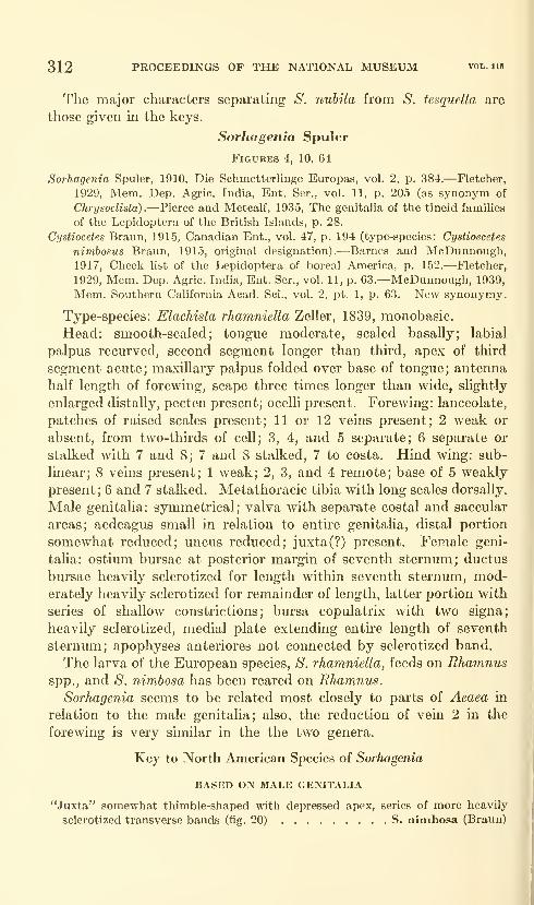
312 PROCEEDINGS OF THE NATIONAL MUSEUM vol. 115
The major characters separating S. nubila from S. tesquella are
those given in the keys.
Sorhagenia Spuler
Figures 4, 10, 64
Sorhagenia Spuler, 1910, Die Schmetterlinge Europas, vol. 2, p. 384.—Fletcher,
1929, Mem. Dep. Agric. India, Ent. Ser., vol. 11, p. 205 (as synonym of
ChrysocUsta)
.
—Pierce and Metcalf, 1935, The genitalia of the tineid families
of the Lepidoptera of the British Islands, p. 28.
Cystiocetes Braun, 1915, Canadian Ent., vol. 47, p. 194 (type-species: Cystioecetes
nimbosus Braun, 1915, original designation).—Barnes and McDunnough,1917, Check list of the Lepidoptera of boreal America, p. 152.—Fletcher,
1929, Mem. Dep. Agric. India, Ent. Ser., vol. 11, p. 63.—McDunnough, 1939,
Mem. Southern California Acad. Sci., vol. 2, pt. 1, p. 63. New synonymy.
Type-species: Elachista rhamniella Zeller, 1839, monobasic.
Head: smooth-scaled; tongue moderate, scaled basally; labial
palpus recurved, second segment longer than third, apex of third
segment acute; maxillary palpus folded over base of tongue; antenna
half length of forewing, scape three times longer than wide, slightly
enlarged distaUy, pecten present; ocelli present. Forewing: lanceolate,
patches of raised scales present; 11 or 12 veins present; 2 weak or
absent, from two-thirds of cell; 3, 4, and 5 separate; 6 separate or
stalked with 7 and 8; 7 and 8 stalked, 7 to costa. Hind wing: sub-
linear; 8 veins present; 1 weak; 2, 3, and 4 remote; base of 5 weakly
present; 6 and 7 stalked. Metathoracic tibia with long scales dorsally.
Male genitalia: symmetrical; valva with separate costal and saccular
areas; aedeagus small in relation to entire genitalia, distal portion
somewhat reduced; uncus reduced; juxta(?) present. Female geni-
talia: ostium bursae at posterior margin of seventh sternum; ductus
bursae heavily sclerotized for length within seventh sternum, mod-erately heavily sclerotized for remainder of length, latter portion with
series of shallow constrictions; bursa copulatrix with two signa;
heavily sclerotized, medial plate extending entire length of seventh
sternum; apophyses anteriores not connected by sclerotized band.
The larva of the European species, S. rhamniella, feeds on Rhamnusspp., and S. nimbosa has been reared on Rhamnus.
Sorhagenia seems to be related most closely to parts of Aeaea in
relation to the male genitalia; also, the reduction of vein 2 in the
forewing is very similar in the the two genera.
Key to North American Species of Sorhagenia
BASED ON MALE GENITALIA
"Juxta" somewhat thimble-shaped with depressed apex, series of more heavily
sclerotized transverse bands (fig. 20) S. nimbosa (Braun)
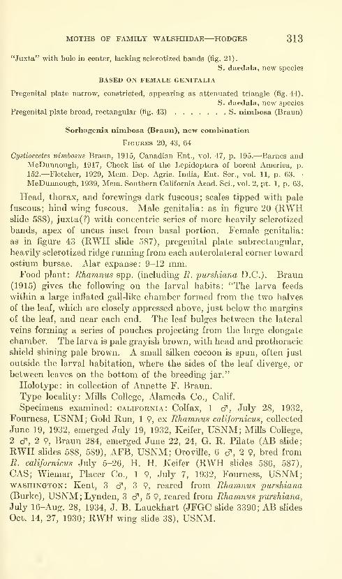
MOTHS OF FAMILY WALSHIIDAE—HODGES 313
"Juxta" with hole in center, lacliing sclerotized bands (fig. 21).
S. daedala, new species
BASED ON FEMALE GENITALIA
Pregenital plate narrow, constricted, appearing as attenuated triangle (fig. 44).
S. daedala, new species
Pregenital plate broad, rectangular (fig. 43) S. nimbosa (Braun)
Sorhagenia nimbosa (Braun), new combination
Figures 20, 43, 64
Cystioecetes nimbosus Braun, 1915, Canadian Ent., vol. 47, p. 195.—'Barnes andMcDunnough, 1917, Check list of the Lepidoptera of boreal America, p.
152.—Fletcher, 1929, Mem. Dep. Agric. India, Ent. Ser., vol. 11, p. 63.—McDunnough, 1939, Mem. Southern California Acad. Sci., vol. 2, pt. 1, p. 63.
Head, thorax, and forewings dark fuscous; scales tipped with pale
fuscous; hind wing fuscous. Male genitalia: as in figure 20 (RWHslide 588), juxta(?) with concentric series of more heavily sclerotized
bands, apex of uncus inset from basal portion. Female genitalia:
as in figure 43 (RWH slide 587), pregenital plate subrectangular,
heavily sclerotized ridge running from each anterolateral corner toward
ostium bursae. Alar expanse: 9-12 mm.Food plant: Rhamnus spp. (including B. purshiana D.C.). Braun
(1915) gives the following on the larval habits: ''The larva feeds
within a large inflated gall-like chamber formed from the two halves
of the leaf, which are closely appressed above, just below the margins
of the leaf, and near each end. The leaf bulges between the lateral
veins forming a series of pouches projecting from the large elongate
chamber. The larva is pale grayish brown, with head and prothoracic
shield shining pale brown. A small silken cocoon is spun, often just
outside the larval habitation, where the sides of the leaf diverge, or
between leaves on the bottom of the breeding jar."
Holotype: in collection of Annette F. Braun.
Type locality: Mills College, Alameda Co., Calif.
Specimens examined: California: Colfax, 1 cf, July 28, 1932,
Fourness, USNM; Gold Run, 1 9, ex Rhamnus californicus, collected
June 19, 1932, emerged July 19, 1932, Keifer, USNM; Mills College,
2 c?", 2 9, Braun 284, emerged June 22, 24, G. R. Pilate (AB slide;
RWH slides 588, 589), AFB, USNM; Oroville, 6 cT, 2 9, bred from
R. californicus July 5-26, H. H. Keifer (RWH shdes 586, 587),
CAS; Wiemar, Placer Co., 1 9, July 7, 1932, Fourness, USNM;WASHINGTON: Kent, 3 d^, 3 9, reared from Rhamnus purshiana
(Burke), USNM; Lynden, 3 cf, 5 9, reared from Rhamnus purshiana,
July 16-Aug. 28, 1934, J. B. Lauckhart (JFGC sHde 3390; AB slides
Oct. 14, 27, 1930; RWH wing slide 38), USNM.
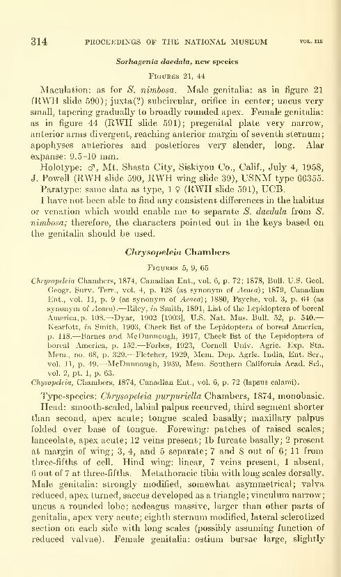
314 PROCEEDINGS OF THE NATIONAL MUSEUM vol. 115
Sorhagenia daedala, new species
Figures 21, 44
Maculation: as for S. nimbosa. Male genitalia: as in figure 21
(KWH slide 590); juxta(?) subcircular, orifice in center; uncus very
small, tapering gradually to broadly rounded apex. Female genitalia:
as in figure 44 (RWH slide 591); pregenital plate very narrow,
anterior arms divergent, reaching anterior margin of seventh sternum
;
apophyses anteriores and posteriores very slender, long. Alar
expanse: 9.5-10 mm.Holotype: d^, Mt. Shasta City, Siskiyou Co., CaUf., July 4, 1958,
J. Powell (RWPI shde 590, RWH wing slide 39), USNM type 66355.
Paratype: same data as type, 1 9 (RWH slide 591), UCB.I have not been able to find any consistent differences in the habitus
or venation which would enable me to separate S. daedala from S.
nimbosa; therefore, the characters pointed out in the keys based on
the genitalia should be used.
Chrysopeleia Chambers
Figures 5, 9, 65
Chrysopeleia Chambers, 1874, Canadian Ent., vol. 6, p. 72; 1878, Bull. U.S. Geol.
Geogr. Surv. Terr., vol. 4, p. 128 (as synonym of Aeaea) ; 1879, Canadian
Ent., vol. 11, p. 9 (as synonym of Aeaea); 1880, Psyche, vol. 3, p. 64 (as
synonym of Aeaea).—Riley, in Smith, 1891, List of the Lepidoptera of boreal
America, p. 108.—Dyar, 1902 [1903], U.S. Nat. Mus. Bull. 52, p. 540.—
Kearfott, in Smith, 1903, Check list of the Lepidoptera of boreal America,
p. 118.—Barnes and McDunnough, 1917, Check list of the Lepidoptera of
boreal America, p. 152.—Forbes, 1923, Cornell Univ. Agric. Exp. Sta.
Mem., no. 68, p. 329.—Fletcher, 1929, Mem. Dep. Agric. India, Ent. Ser.,
vol. 11, p. 49.—McDunnough, 1939, Mem. Southern California Acad. Sci.,
vol. 2, pt. 1, p. 63.
Chysopeleia, Chambers, 1874, Canadian Ent., vol. 6, p. 72 (lapsus calami).
Type-species: Chrysopeleia purpuriella Chambers, 1874, monobasic.
Head: smooth-scaled, labial palpus recurved, third segment shorter
than second, apex acute; tongue scaled basally; maxillary palpus
folded over base of tongue. Forewing: patches of raised scales;
lanceolate, apex acute; 12 veins present; lb furcate basally; 2 present
at margin of wing; 3, 4, and 5 separate; 7 and 8 out of 6; 11 from
three-fifths of cell. Hind wing: linear, 7 veins present, 1 absent,
6 out of 7 at three-fifths. Metathoracic tibia with long scales dorsally.
Male genitalia: strongly modified, somewhat asymmetrical; valva
reduced, apex turned, saccus developed as a triangle; vinculum narrow;
uncus a rounded lobe; aedeagus massive, larger than other parts of
genitalia, apex very acute; eighth sternum modified, lateral sclerotized
section on each side with long scales (possibly assuming function of
reduced valvae). Female genitalia: ostium bursae large, slightly

MOTHS OF FAMILY WALSHIIDAE—HODGES 315
distad of middle of eighth sternum; ductus bursae heavily sclerotized
for proximal three-fifths; corpus bursae with two signa.
Chrysopeleia purpuriella has the habitus of species of Aeaea; how-ever, the male and female genitalia are very distinct from the latter,
and for this reason I am treating them as separate genera. Only one
species of Chrysopeleia is known, and it may be an offshoot from Aeaea.
Chrysopeleia purpuriella Chambers
Figures 25, 25a, 25b, 42, 65
Chrysopeleia purpuriella Chambers, 1874, Canadian Ent., vol. 6, p. 73.—Riley,
in Smith, 1891, List of the Lepidoptera of boreal America, p. 108.—Dyar,
1902 [1903], U.S. Nat. Mus. Bull. 52, p. 540.—Kearfott, in Smith, 1903,
Check list of the Lepidoptera of boreal America, p. 118.—Barnes and Mc-Dmmough, 1917, Check hst of the Lepidoptera of boreal America, p. 152.—Forbes, 1923, Cornell Univ. Agric. Exp. Sta. Mem., no. 68, p. 329.—Fletcher, 1929, Mem. Dep. Agric. India, Ent. Ser., vol. 11, p. 49.^McDun-nough, 1939, Mem. Southern California Acad. Sci., vol. 2, pt. 1, p. 63.
Aeaea purpuriella, Chambers, 1878, Bull. U.S. Geol. Geogr. Surv. Terr., vol. 4,
p. 128; 1879, Canadian Ent., vol. 11, p. 9; 1880, Psyche, vol. 3, p. 64.—Hagen,1884, Papilio, vol. 4, p. 154.
Chysopeleia [sic] purpuriella, Chambers, 1874, Canadian Ent., vol. 6, p. 73 (lapsus
calami)
.
Maculation: as in figure 65; labial palpus dark fuscous-black with
deep purple reflections, dorsal surface of second and third segments
and apex of third segment shining pale gray; head, thorax, forewings,
legs, and abdomen dark fuscous-black with deep purple reflections,
most scales unicolorous; undersurface of scape of antenna shining
pale gray, forewing sometimes with pale, oblique fascia starting at
four-fifths, angled at fold. Hind wing: pale fuscous. Matathoracic
tibia with white fascia on outer sm'face at middle and outer tibial
spurs; apices of tarsal segments white. Male genitalia: as in figure
25 (JFGC slide 10225). Female genitaha: as in figure 42 (RWHslide 1160). Alar expanse: 6-8 mm.Food plant: Chambers (1880) has reared one adult from Bobinia
pseudo-acacia L.; however, he did not separate the larva from those
of Lithocolletis robinieUa Clemens when the leaves were gathered.
Chapman has reared the adults of C. pur^puriella from cocoons which
were attached to the smaller branches of apple; but, even though
the cocoons were very abundant, no feeding larvae were found.
Holotype: Museum of Comparative Zoology.
Type locality: Kentucky.
Specmiens examined: Illinois: Putnam Co., 1 cf. May 17, 1959,
M. O. Glenn (RWH slide 2038), MOG. Kentucky: no further
locality, 2 d^. Chambers (RWH shde 871), MCZ, USNM. newYORK: Geneva, 2 cf, 1 9, emerged March 29, 1961 (RWH slides
1657-1659), USNM; Ithaca, 1 9, June 25, 1931, A. B. Klots (RWH
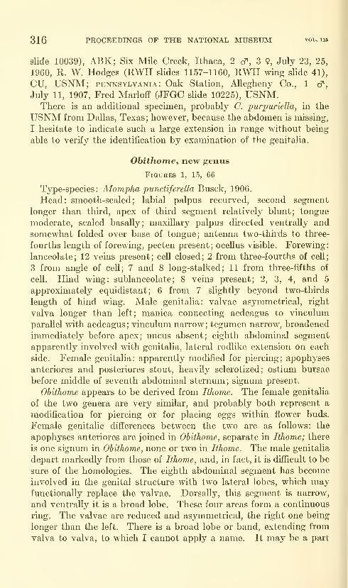
316 PROCEEDINGS OF THE NATIONAL MUSEUM vol. 115
slide 10039), ABK; Six Mile Creek, Ithaca, 2 cf , 3 9, July 23, 25,
1960, R. W. Hodges (RWH slides 1157-1160, RWH wing slide 41),
CU, USNM; PENNSYLVANIA: Oak Station, Allegheny Co., 1 cf,
July 11, 1907, Fred Marloff (JFGC slide 10225), USNM.There is an additional specimen, probably C. purpuriella, in the
USNM from Dallas, Texas; however, because the abdomen is missing,
I hesitate to indicate such a large extension in range without being
able to verify the identification by examination of the genitalia.
Obithome, new genus
Figures 1, 15, 66
Type-species: Mompha punctiferella Busck, 1906.
Head: smooth-scaled; labial palpus recurved, second segment
longer than third, apex of third segment relatively blunt; tongue
moderate, scaled basally; maxillary palpus directed ventrally and
somewhat folded over base of tongue; antenna two-thirds to three-
fourths length of forewing, pecten present; ocellus visible. Forewing:
lanceolate; 12 veins present; cell closed; 2 from three-fourths of cell;
3 from angle of cell; 7 and 8 long-stalked; 11 from three-fifths of
cell. Hind wing: sublanceolate; 8 veins present; 2, 3, 4, and 5
approximately equidistant; 6 from 7 slightly beyond two-thirds
length of hind wing. Male genitalia: valvae asymmetrical, right
valva longer than left; manica connecting aedeagus to vinculum
parallel with aedeagus; vinculum narrow; tegumen narrow, broadened
immediately before apex; uncus absent; eighth abdominal segment
apparently involved with genitalia, lateral rodlike extension on each
side. Female genitalia: apparently modified for piercing; apophyses
anteriores and posteriores stout, heavily sclerotized; ostium bursae
before middle of seventh abdominal sternum; signum present.
Obithome appears to be derived from Ithome. The female genitalia
of the two genera are very similar, and probably both represent a
modification for piercing or for placing eggs within flower buds.
Female genitalic differences between the two are as follows: the
apophyses anteriores are joined in Obithome, separate in Ithome; there
is one signum in Obithome, none or two in Ithome. The male genitalia
depart markedly from those of Ithome, and, in fact, it is difficult to be
sure of the homologies. The eighth abdominal segment has become
involved in the genital structure with two lateral lobes, which mayfunctionally replace the valvae. Dorsally, this segment is narrow,
and ventrally it is a broad lobe. These four areas form a continuous
ring. The valvae are reduced and asymmetrical, the right one being
longer than the left. There is a broad lobe or band, extending from
valva to valva, to which I cannot apply a name. It may be a part
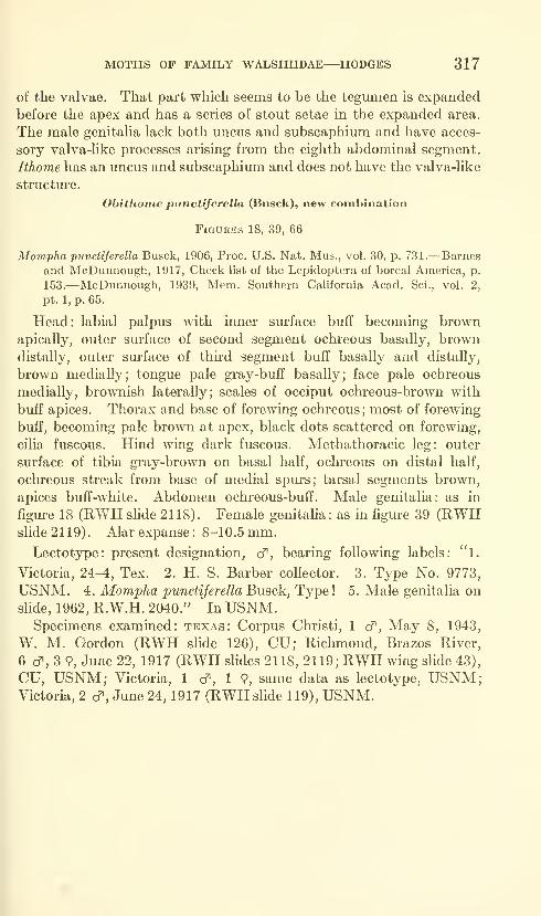
MOTHS OF FAMILY WALSHIIDAE—HODGES 317
of the valvae. That part which seems to be the tegumen is expanded
before the apex and has a series of stout setae in the expanded area.
The male genitaha lack both uncus and subscaphium and have acces-
sory valva-like processes arising from the eighth abdominal segment.
Ithome has an uncus and subscaphium and does not have the valva-like
structure.
Obithoine punctiferella (Busck), new combination
Figures 18, 39, 66
Mompha punctiferella Busck, 1906, Proc. U.S. Nat. Mus., vol. 30, p. 731.—Barnes
and McDunnough, 1917, Check list of the Lepidoptera of boreal America, p.
153.—McDunnough, 1939, Mem. Southern Cahfornia Acad. Sci., vol. 2,
pt. 1, p. 65.
Head: labial palpus with inner surface buff becoming brownapically, outer surface of second segment ochreous basally, browndistally, outer surface of third segment buff basally and distally,
brown medially; tongue pale gray-buff basally; face pale ochreous
medially, brownish laterally; scales of occiput ochreous-brown with
buff apices. Thorax and base of forewing ochreous; most of forewing
buff, becoming pale brown at apex, black dots scattered on forewing,
cilia fuscous. Hind wing dark fuscous. Methathoracic leg: outer
surface of tibia gray-brown on basal half, ochreous on distal half,
ochreous streak from base of medial spm-s; tarsal segments brown,
apices buff-white. Abdomen ochreous-buff. Male genitalia: as in
figure 18 (RWH slide 2118). Female genitalia: as in figure 39 (RWHslide 2119). Alar expanse: 8-10.5 mm.
Lectotype: present designation, cf, bearing following labels: "l.
Victoria, 24-4, Tex. 2. H. S. Barber collector. 3. Type No. 9773,
USNM. 4. Mompha punctiferella Busck, Type! 5. Male genitalia on
slide, 1962, R.W.H. 2040." In USNM.Specmiens examined: texas: Corpus Christi, 1 cf. May 8, 1943,
W. M. Gordon (RWH slide 126), CU; Richmond, Brazos River,
6 cf, 3 9, June 22, 1917 (RWH slides 2118, 2119; RWH wing slide 43),
CU, USNM; Victoria, 1 cf, 1 9, same data as lectotype, USNM;Victoria, 2 cf , June 24, 1917 (RWH slide 119), USNM.
Literature Cited
Braun, Annette F.
1915. New genera and species of Tineina. Canadian Ent., vol. 47, pp.
188-197.
Chambers, V. T.
1878. Index to the described Tineina of the United States and Canada.
Bull. U.S. Geol. Geogr. Surv. Terr., vol. 4, pp. 125-167.
1880. Notes on some tineid larvae. Psyche, vol. 3, pp. 63-68.
Clarke, J. F. Gates.
[In press.] Catalogue of the type specimens of Microlepidoptera in the
British Museum (Natural History) described by Edward Meyrick,
vol. 4.
DiAKONOFF, A.
1952. Viviparity in Lepidoptera. Trans. Ninth Interuat. Congr. Ent., vol.
1, pp. 91-96.
1955. Microlepidoptera of New Guinea, part V. Verh. Akad. Wet. Amster-
dam, vol. 50, pp. 1-210.
Hodges, Ronald W.1961. A review of the genus Walshia Clemens with descriptions of new
species (Lepidoptera: Gelechioidea) . Bull. Brooklyn Ent. Soc,
vol. 56, pp. 66-80.
1962. The genus Ithoine in North America north of Mexico (Walshiidae).
Journ. Lepidopterists' Soc, vol. 15, pp. 81-90.
1962b. A review of the genus Periploca with descriptions of nine new species
(Lepidoptera: Gelechioidea). Fan-Pacific Ent., vol. 38, pp.
83-97.
[In press.] A revision of the Cosmopterigidae of America north of Mexico,
with a definition of the Momphidae and Walshiidae (Lepidoptera:
Gelechioidea)
.
[In press.] The genus Perimede Chambers in North America north of Mexico
(Lepidoptera: Walshiidae).
KUROKO, HiROSHI.
1961. The genus Aniispila from Japan with descriptions of seven newspecies (Lepidoptera, Heliozelidae) . Esakia, vol. 3, pp. 11-24,
pis. 4-9.
Meyrick, Edward.1915. Exotic Microlepidoptera, vol. 1, pt. 11, pp. 321-352.
1921. Descriptions of South African Microlepidoptera. Ann. Transvaal
Mus., vol. 8, pp. 49-148.
Toll, S.
1952. Eupistidae (Coleophoridae) of Poland. Docum. Physiogr. Polon.,
Krakow, no. 32, pp. 1-292.
318

MOTHS OF FAMILY WALSHIIDAE—HODGES 319
^#;>.
2. NEPOTULA
3. STILBOSIS
5. CHRYSOPELEIA 6. AEAEAFigures 1-6.—Lateral view of heads of types of genera: 1, Obithome punctiferella (Busck);
2, Nepotula secura, new species; 3, Stilbosis tesquella Clemens; 4, Sorhagenia rhamniella
(Zeller); 5, Chrysopeleia purpuriella Chambers; 6, Aeaea ostryaeella Chambers.
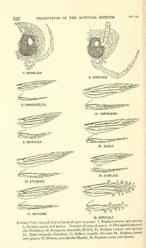
320 PROCEEDINGS OF THE NATIONAL MUSEUM vol. ii5
7. NEOPLOCA
9. CHRYSOPELEIA
10. S0RHA6ENIA
II. NEOPLOCA•12. AEAEA
13. STILBOSIS
15. OBITHOME16. NEPOTULA
Figures 7-16.-Lateral view of heads of types of genera: 7, Neoploca corusca, new species;
8 Synploca gumia, new species. Venation of types of genera: 9, Chrysopdeta purpun-
ella Chambers; 10, Sorhagenia rhamniella (Zeller); 11, Neoploca corusca, new species;
12, Aeaea ostryaeella Chambers; 13, Stilhosis tesquella Clemens; 14, Synploca gumia,
new species; 15, Obithome punctiferella (Busck); 16, Nepotula secura, new species.
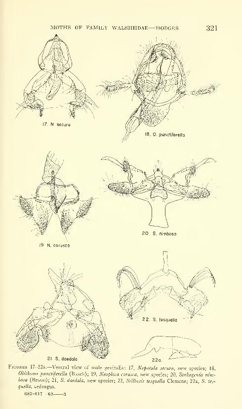
MOTHS OF FAMILY WALSHIIDAE—HODGES 321
20. S. nimbosa
19. N. corusca
21. S. daedala^*=^ '"
22a.
Figures 17-22a.—Ventral view of male genitalia: 17, Nepotula secura, new species; 18,
Obithome punctiferella (Busck); 19, Neoploca corusca, new species; 20, Sorhagenia nim-bosa (Braun); 21, S. daedala, new species; 22, Stilbosis tesquella Clemen?; 22a, S. tes-
quella, aedeagus.
685-617—63 -3
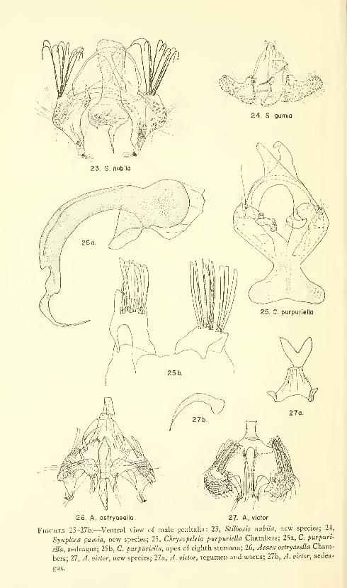
26. A. ostryoeella 27. A. victor
Figures 23-27b.—Ventral view of male genitalia: 23, Stilbosis nuhila, new species; 24,
Synploca gumia, new species; 25, Chrysopeleia purpuriella Chambers; 25a, C. purpuri-
ella, aedeagus; 25b, C. purpuriella, apex of eighth sternum; 26, Aeaea ostryaeella Cham-
bers; 27, A. victor, new species; 27a, A. victor, tegumen and uncus; 27b, A. victor, aedea-
gus.
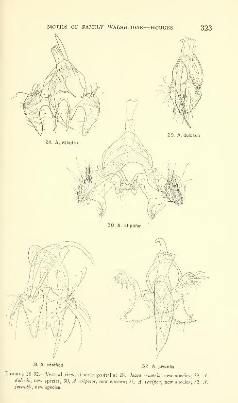
MOTHS OF FAMILY WALSHIIDAE—HODGES
31. A venifica 32. A. juvantis
Figures 28-32.—Ventral view of male genitalia: 28, Aeaea venatrix, new species; 29, A.dulcedo, new species; 30, A. stipator, new species; 31, A. venifica, new species; 32, A.juvantis, new species.
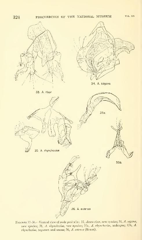
324 PROCEEDINGS OF THE NATIONAL MUSEUM vol. ns
36 A extensa
Figures 33-36.—Ventral view of male genitalia: 33, Aeaea risor, new species; 34, J. sagana,
new species; 35, A. rhynchosiae, new species; 35a, A. rhynchosiae, aedeagus; 35b, A.
rhynchosiae, tegumen and uncus; 36, A. extensa (Braun).
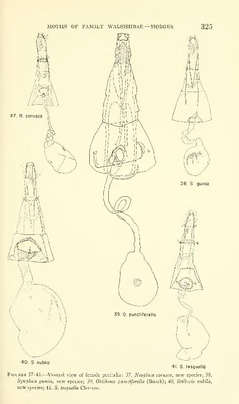
MOTHS OF FAMILY WALSHIIDAE—HODGES 325
37. N, corusca
39. 0. punctiferella
38. S. gumia
40. S. nubila41. S. tesquella
Figures 37-41.—Ventral view of female genitalia: 37, Neoploca corusca, new species; 38,
Synploca gumia, new species; 39, Ohithome punctiferella (Busck); 40, Stilbosis nubila,
new species; 41, S. tesquella Clemens.
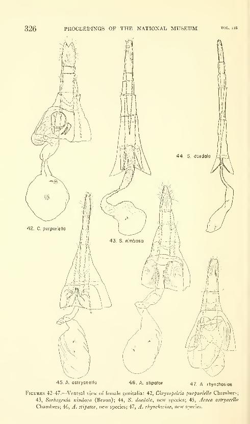
326 PROCEEDINGS OF THE NATIONAL MUSEUM
45. A, ostryaeella 46. A. stipotor 47. A. rhynchoslae
Figures 42-47.—Ventral view of female genitalia: 42, Chrysopelcia piirpuriella Chambers;
43, Sorhagenia nimbosa (Braun); 44, S. daedala, new species; 45, Aeaea ostryaeella
Chambers; 46, A. stipator, new species; 47, A. rhynchosiae, new species.
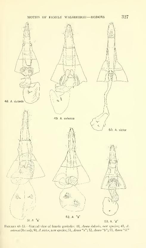
MOTHS OF FAMILY WALSHIIDAE—HODGES 327
51. A V52. A. "b"
53. A. "d"
Figures 48-53.—Ventral view of female genitalia: 48, Aeaea dulcedo, new species; 49, A.
exUtisa (Braun); 50, A. victor, new species; 51, Aeaea "a"; 52, Aeaea "b"; 53, Aeaea "d."
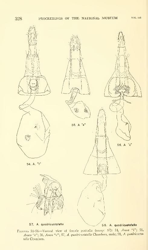
328 PROCEEDINGS OF THE NATIONAL MUSEUM
57. A. quadricustotella \_^ y 53 ^^ quadricustatella
Figures 54-58.—Ventral view of female genitalia (except 57): 54, Aeaea "f"; 55,
Aeaea "e"; 56, Aeaea "c"; 57, A. quadricustatella Chambers, male; 58, A. quadricusta-
tella Chambers.

MOTPTS OF FAMILY WALSHITDAE—ITODCES 329
59 60
63
66
Figures 59-66.—Left wings of species of Walshiidae: 59, Neoploca corusca, new species,
Pefia Blanca Canyon, Santa Cruz County, Arizona; 60, Synploca gumia, new species,
Madera Canyon, Santa Rita Mountains, Arizona; 61, Nepotula secura, new species,
Pensacola, Florida; 62, Aeaea "c," Madera Canyon, Santa Rita Mountains, Arizona;
63, Stilbosis tesquella Clemens, West Fork, 16 miles SW Flagstaff, Arizona; 64, Sorha-
genia nimbosa (Braun), Oroville, California; 65, Chrysopeleia purpuriella Chambers,
Ithaca, New York; 66, Obithome pmictiferella (Busck), Victoria, Texas.
U.S. GOVERNMENT PRINTING OFFICE: 1964

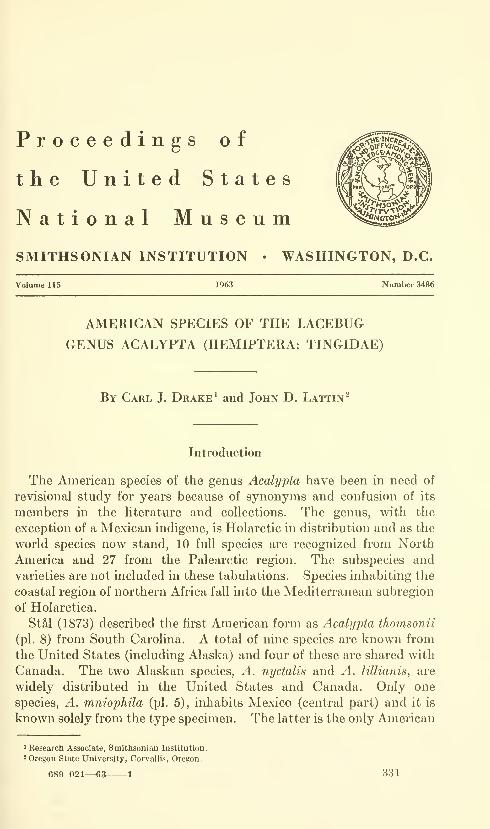
Proceedings of
the United States
National MuseumSMITHSONIAN INSTITUTION • WASHINGTON, D.C.
Volume 115 1963 Number 34R6
AMERICAN SPECIES OF THE LACEBUG
GENUS ACALYPTA (HEMIPTERA: TINGIDAE)
By Carl J. Drake ^ and John D. Lattin^
Introduction
The American species of the genus Acalypta have been in need of
revisional study for years because of synonyms and confusion of its
members in the Hterature and collections. The genus, with the
exception of a Mexican indigene, is Holarctic in distribution and as the
world species now stand, 10 full species are recognized from North
America and 27 from the Palearctic region. The subspecies and
varieties are not included in these tabulations. Species inhabiting the
coastal region of northern Africa fall into the Mediterranean subregion
of Holarctica.
StM (1873) described the first American form as Acalypta thomsonii
(pi. 8) from South Carolina. A total of nine species are known from
the United States (including Alaska) and four of these are shared with
Canada. The two Alaskan species, A. nyctalis and A. lillianis, are
widely distributed in the United States and Canada. Only one
species, A. mniophila (pi. 5), inhabits Mexico (central part) and it is
known solely from the type specimen. The latter is the only American
' Research Associate, Smithsonian Institution.
2 Oregon State University, Corvallis, Oregon.
689-021—63 1 331

332 PROCEEDINGS OF THE NATIONAL MUSEUM vol. 115
representative of the genus not known to occur in the Nearctic region.
None of the species is common to both the Old and New Worlds.
Fossil forms are unknown.
Acalypta comprises a well-defined, homogenous group of species
with similar facies, especially the brachypterous forms (pis. 3-11, 13,
14). The macropterous form (pi. 12) is apparently rather uncommonand only known for three of the American species. The differences in
habitus between the dimorphic forms of a species are depicted in the
illustrations of A. barberi (pis. 11, 12). The macropterous forms like-
wise are similar in general aspect to one another.
The present paper is based largely upon the collections of the
authors, which include all the American species and most of the
European forms. The material in the U.S. National Museum, where
all the American species are represented, and in the private collection
of Mr. Joe Schuh, Kalamath Falls, Oreg., have also been studied.
We are indebted to Patricia J. Hogue, Arlington, Va., and Liza
Biganzoli, Washington, D.C., for the fine illustrations. The latter
also prepared the map (pi. 2) showing the distribution of Acalypta spp.
in the northwestern United States and bordering provinces of Canada.
This work is a byproduct of a tingid project being conducted with the
aid of a National Science Foundation grant.
Genus Acalypta Westwood
Acalypta Westwood 1840 (synopsis) p. 121.—StM 1873, pp. 118, 122; 1874,
p. 51.—Horvdth 1906, pp. 13, 24.—Oshanin 1908, p. 406; 1912, p. 42.—Banks
1910, p. 55.—Van Duzee 1916, p. 25; 1917, p. 211.—Osborn and Drake 1916a,
p. 220.—Parshley 1923, pp. 696, 698.—Blatcbley 1926, p. 480.—Drake
1928b, pp. 1-9.—China 1943, p. 245.—Hurd 1946, p. 462.—Bailey 1951,
p. 32.—Kiritshenko 1951, pp. 240, 244.—Drake and Ruhoflf 1959, p. 138;
1960, p. 31.
Orthosteira Fieber 1844, p. 46.
Monanthia (Orthosteira): Flor 1860, pp. 330, 331.
Orthostira [sic]: Fieber 1861, p. 130.—Lethierry and Severin 1896, p. 6.
Fenestrella Osborn and Drake 1916a, p. 222.—Parshley 1917a, p. 14.
Drakella Bergroth 1922, p. 152.—Parshley 1923, pp. 696, 698.—Blatchley 1926,
p. 481.
Type species: Tingis carinata Panzer.
Brachypterous form.—Small, ovate, obovate, or oblong (pis. 3,
7, 8), pronotum subdepressed, elytra more or less convex, general
color brownish testaceous to blackish fuscous. Size small, ranging
from 1.65-2.70 mm. long.
Head.—Short, only shghtly produced in front of eyes, usually
armed with one pair of stout, porrect, frontal spines, other spines
wanting; eyes large, granulate; bucculae foUaceous, areolate, open or
closed in front, extending backwards beneath anterior part of
prosternum; sternal laminae of rostral sulcus uniseriate, present on all

LACEBUG GENUS ACALYPTA—DRAKE AND LATTIN 333
three sternal divisions, open at base. Labium long, 4-segmented,
extending to middle of or slightly beyond metasternum. Antennae
long, slender; segment I short, stout, slightly longer and stouter than
11; III longest, slenderest; IV moderately long, fusiform to subclavate.
Pronotum.—Subdepressed, punctate, unicarinate (pis. 3-6) or
tricarinate (pis. 7-15); collar tectiformily raised at middle, produced
forward over base of vertex; calli small, impunctate; median carina
percurrent on hood and pronotmn, ridgelike and without cells on
hood, foliaceous and uniseriate on pronotum; lateral carinae cus-
tomarily slightly lower than median carina, frequently raised
anteriorly, composed of one row of areolae, either parallel or slightly
divergent posteriorly; paranotum explanate, long, subrectangular,
slightly reflexed, two or tkree areolae deep in front, only one or two
wide opposite humeral angle; posterior process triangular, areolate.
Legs moderately long, femora slightly swollen, tarsi 2-segmented.
Elytra.—Abruptly widened at base to that of pronotal width
across paranota, slightly longer than and thus covering abdomen,
with inner margins connivent, apices rounded and separated from each
other; claval area not clearly defined, concealed beneath backward
projection of pronotum; major divisions of costal, subcostal, discoidal,
and sutural areas sharply defined by boundary veins from one another
(pi. 1a); sutural area much reduced, narrow. Metathoracic wings
obsolete. Hypocostal lamina (pi. If) of elytron either composed of
one complete row of areolae or slightly wider and biseriate in basal
third to half.
Abdomen.—Segmentation, spiracles, male and female genital
segments as figured and labeled in plate 1.
Macropterous form.^—Oblong. Pronotum swoUen, convex across
humeral angles, coarsely punctate. Anterior pair of coxal cavities
more distant longitudinally from middle pair than in brachyptery.
Elytra (pis. 1, 12) long, much longer as well as wider than abdomen,
the sutural areas large, overlapping each other in repose so that their
apices lie jointly rounded at rest; claval area (pi. 1) small, sharply
defined, always concealed beneath backward projection of pronotumin resting position; costal, subcostal, discoidal, and sutural areas
separated from one another by prominent boundary veins;
metathoracic wings large, functional. Venation of elytron (pi. Id)
and hind wing (pi. IE) as labeled in the illustrations. Other structures
similar to those in brachypterous form.
Certain structural characters, which usually remain fairly constant
in most tingid genera, vary considerably between the pterygopoly-
morphic forms within a species. These dimorphic differences include
such features as: size and convexity of pronotum, spacing longitudinally
of coxal cavities, form and length of elytra, absence or presence of
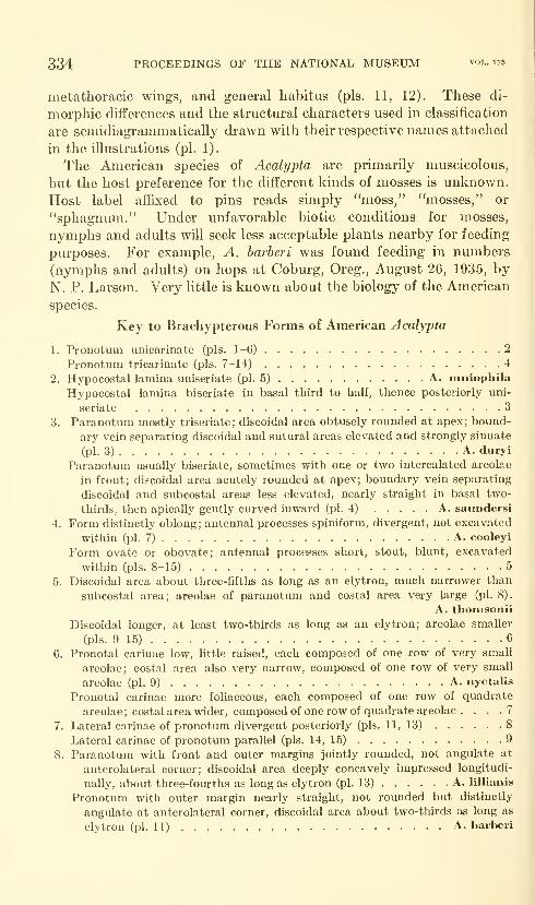
334 PROCEEDINGS OF THE NATIONAL MUSEUM vol. us
metathoracic wings, and general habitus (pis. 11, 12). These di-
morphic differences and the structural characters used in classification
are semidiagrammatically drawn with their respective names attached
in the illustrations (pi. 1).
The American species of Acalypta are primarily muscicolous,
but the host preference for the different kinds of mosses is unknown.
Host label affixed to pins reads simply "moss," "mosses," or
"sphagnum." Under unfavorable biotic conditions for mosses,
nymphs and adults will seek less acceptable plants nearby for feeding
purposes. For example, A. harheri was found feeding in numbers
(nymphs and adults) on hops at Coburg, Oreg., August 26, 1935, by
N. P. Larson. Very httle is known about the biology of the American
species.
Key to Brachypterous Forms of American Acalypta
1. Pronotum unicarinate (pis. 1-6) 2
Pronotum tricarinate (pis. 7-14) 4
2. Hypocostal lamina uniseriate (pi. 5) A. inniophila
Hypocostal lamina biseriate in basal third to half, thence posteriorly uni-
seriate 3
3. Paranotum mostly triseriate; discoidal area obtusely rounded at apex; bound-
ary vein separating discoidal and sutural areas elevated and strongly sinuate
(pi. 3) A. duryi
Paranotum usually biseriate, sometimes with one or two intercalated areolae
in front; discoidal area acutely rounded at apex; boundary vein separating
discoidal and subcostal areas less elevated, nearly straight in basal two-
thirds, then apically gently curved inward (pi. 4) A. saundersi
4. Form distinctly oblong; antennal processes spiniform, divergent, not excavated
within (pi. 7) A. cooleyi
Form ovate or obovate; antennal processes short, stout, blunt, excavated
within (pis. 8-15) 5
5. Discoidal area about three-fifths as long as an elytron, much narrower than
subcostal area; areolae of paranotum and costal area very large (pi. 8).
A. thomsonii
Discoidal longer, at least two-thirds as long as an elytron; areolae smaller
(pis. 9-15) 6
6. Pronotal carinae low, little raised, each composed of one row of very small
areolae; costal area also very narrow, composed of one row of very small
areolae (pi. 9) A. nyctalis
Pronotal carinae more foliaceous, each composed of one row of quadrate
areolae; costal area wider, composed of one row of quadrate areolae . . . .7
7. Lateral carinae of pronotum divergent posteriorly (pis. 11, 13) 8
Lateral carinae of pronotum parallel (pis. 14, 15) 9
8. Paranotum with front and outer margins jointly rounded, not angulate at
anterolateral corner; discoidal area deeply concavely impressed longitudi-
nally, about three-fourths as long as elytron (pi. 13) A. lillianis
Pronotum with outer margin nearly straight, not rounded but distinctly
angulate at anterolateral corner, discoidal area about two-thirds as long as
elytron (pi. 11) A. barberi
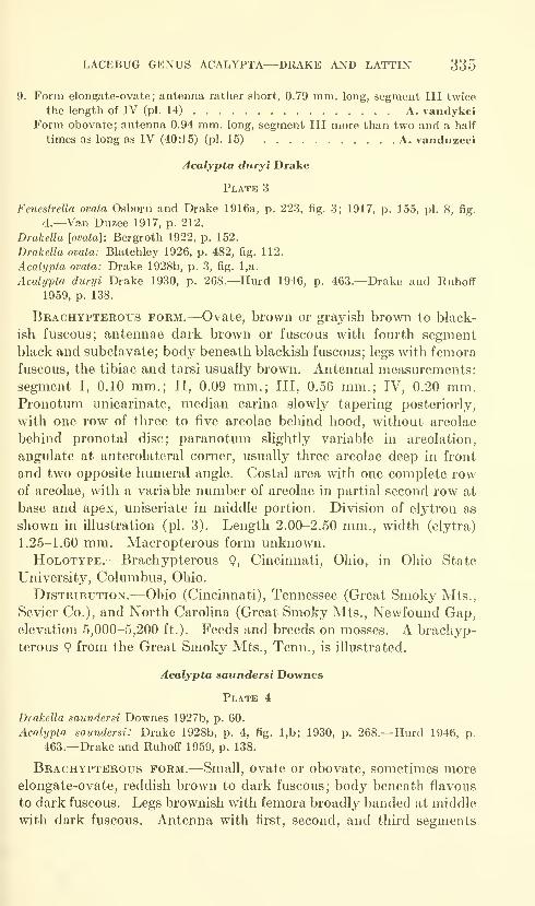
LACEBUG GENUS ACALYPTA—DRAKE AND LATTIN 335
9. Form elongate-ovate; antenna rather short, 0.79 mm. long, segment III twice
the length of IV (pi. 14) A. vandykeiForm obovate; antenna 0.94 mm. long, segment III more than two and a half
times as long as IV (40:15) (pi. 15) A. vanduzeei
Acalypta duryi Drake
Plate 3
Fenestrella ovata Osborn and Drake 1916a, p. 223, fig. 3; 1917, p. 155, pi. 8, fig.
d.—Van Duzee 1917, p. 212.
Drakella [ovata]: Bergroth 1922, p. 152.
Drakella ovata: Blatchley 1926, p. 482, fig. 112.
Acalypta ovata: Drake 1928b, p. 3, fig. l,a.
Acabjpla duryi Drake 1930, p. 268.—Hurd 1946, p. 463.—Drake and Ruhoff
1959, p. 138.
Brachypterous form.—Ovate, brown or grayish brown to black-
ish fuscous; antennae dark brown or fuscous with fourth segmentblack and subclavate; body beneath blackish fuscous; legs with femora
fuscous, the tibiae and tarsi usually brown. Antennal measurements:
segment I, 0.10 mm.; II, 0.09 mm.; Ill, 0.56 mm.; IV, 0.20 mm.Pronotum unicarinate, median carina slowly tapering posteriorly,
with one row of three to five areolae behind hood, without areolae
behind pronotal disc; paranotum slightly variable in areolation,
angulate at anterolateral corner, usually three areolae deep in front
and two opposite humeral angle. Costal area with one complete rowof areolae, with a variable number of areolae in partial second row at
base and apex, uniseriate in middle portion. Division of elytron as
shown in illustration (pi. 3). Length 2.00-2.50 mm., width (elytra)
1.25-1.60 mm. Macropterous form unknown.HoLOTYPE.—Brachypterous 9, Cincinnati, Ohio, in Ohio State
University, Columbus, Ohio.
Distribution.—Ohio (Cincinnati), Tennessee (Great Smoky Mts.,
Sevier Co.), and North Carohna (Great Smoky Mts., Newfound Gap,elevation 5,000-5,200 ft.). Feeds and breeds on mosses. A brachyp-
terous 9 from the Great Smoky Mts., Tenn., is illustrated.
Acalypta saundersi Downes
Plate 4
Drakella saundersi Downes 1927b, p. 60.
Acalypta saundersi: Drake 1928b, p. 4, fig. l,b; 1930, p. 268.—Hurd 1946, p.
463.—Drake and Ruhoff 1959, p. 138.
Brachypterous form.—Small, ovate or obovate, sometimes moreelongate-ovate, reddish brown to dark fuscous; body beneath flavous
to dark fuscous. Legs brownish with femora broadly banded at middle
with dark fuscous. Antenna with first, second, and third segments
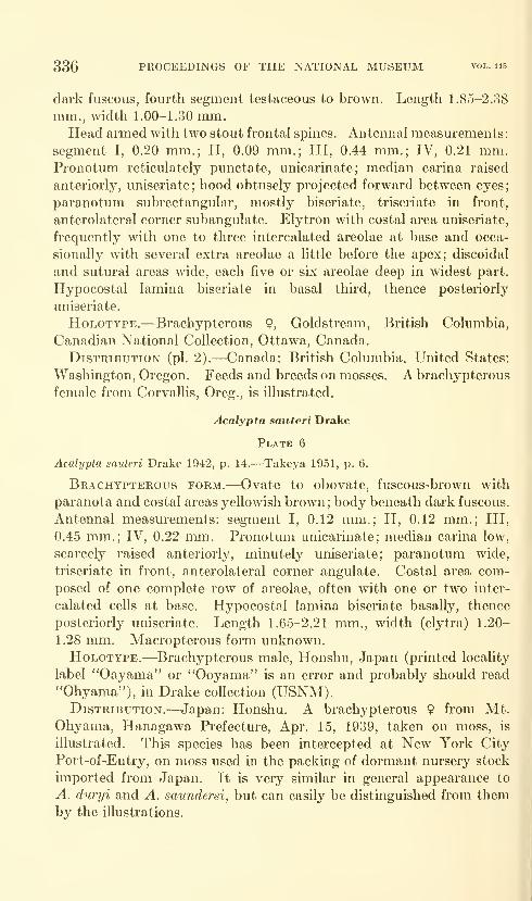
336 PROCEEDINGS OF THE NATIONAL MUSEUM vol. 115
dark fuscous, fourth segment testaceous to brown. Length 1.85-2.38
mm., width 1.00-1.30 mm.Head armed with two stout frontal spines. Antennal measurements:
segment I, 0.20 mm.; II, 0.09 mm.; Ill, 0.44 mm.; IV, 0.21 mm.Pronotum reticulately punctate, unicarinate; median carina raised
anteriorly, uniseriate; hood obtusely projected forward between eyes;
paranotum subrectangular, mostly biseriate, triseriate in front,
anterolateral corner subangulate. Elytron with costal area uniseriate,
frequently with one to three intercalated areolae at base and occa-
sionally with several extra areolae a little before the apex; discoidal
and sutural areas wide, each five or six areolae deep in widest part.
Hypocostal lamina biseriate in basal third, thence posteriorly
uniseriate.
HoLOTYPE.—Brachypterous 9, Goldstream, British Columbia,
Canadian National Collection, Ottawa, Canada.
DisTKiBUTioN (pi. 2).—Canada: British Columbia. United States:
Washington, Oregon. Feeds and breeds on mosses. A brachypterous
female from Corvallis, Oreg., is illustrated.
Acalypta sauteri Drake
Plate 6
Acalypta sauteri Drake 1942, p. 14.—Takeya 1951, p. 6.
Brachypterous form.—Ovate to obovate, fuscous-brown with
paranota and costal areas yellowish brown; body beneath dark fuscous.
Antennal measurements: segment I, 0.12 mm.; II, 0.12 mm.; Ill,
0.45 mm.; IV, 0.22 mm. Pronotum unicarinate; median carina low,
scarcely raised anteriorly, minutely uniseriate; paranotum wide,
triseriate in front, anterolateral corner angulate. Costal area com-
posed of one complete row of areolae, often with one or two inter-
calated cells at base. Hypocostal lamina biseriate basally, thence
posteriorly uniseriate. Length 1.65-2.21 mm., width (elytra) 1.20-
1.28 mm. Macropterous form unknown.HoLOTYPE.—Brachypterous male, Honshu, Japan (printed locality
label "Oayama" or "Ooyama" is an error and probably should read
"Ohyama"), in Drake collection (USNM).Distribution.—Japan: Honshu. A brachypterous 9 from Mt.
Ohyama, Hanagawa Prefecture, Apr. 15, 1939, taken on moss, is
illustrated. This species has been intercepted at New York City
Port-of-Entry, on moss used in the packing of dormant nursery stock
imported from Japan. It is very similar in general appearance to
A. duryi and A. saundersi, but can easily be distinguished from themby the illustrations.
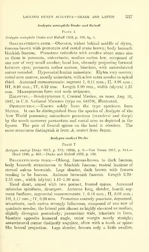
LACEBUG GENUS ACALYPTA—DRAKE AND LATTIN 337
Acalypta tnniophila Drake and Ruhoff
Plate 5
Acalypta mniophila Drake and Ruhoff 1959, p. 136, fig. 1.
Brachypterous form.—Obovate, widest behind middle of elytra,
fuscous-brown with pronotum and costal areas brown; body beneath
blackish fuscous. Pronotum reticulate with areolae about same size
as those in paranota, unicarinate; median carina low, composed of
one row of very small areolae; hood low, obtusely projecting forward
between eyes; paranota rather narrow, biseriate, with anterolateral
corner rounded. Hypocostal lamina uniseriate. Elytra very convex;
costal area narrow, mostly uniseriate, with a few extra areolae in apical
third. Antennal measurements: segment I, 0.11 mm.; II, 0.08 mm.;Ill, 0.40 mm.; IV, 0.22 mm. Length 2.00 mm., width (elytra) 1.25
mm. Macropterous form and male unknown.
HoLOTYPE.—Brachypterous 9, Central Mexico, on moss, Aug. 16,
1947, in U.S. National Museum (type no. 64870), illustrated.
Distribution.—Known solely from the type specimen from
Mexico. It can be distinguished from the species of Acalypta of the
New World possessing unicarinate pronotum (saundersi and duryi)
by the much narrower paranotum and costal area as depicted in the
figures. The pair of frontal spines on the head is obsolete. Thesame structures distinguish it from A. sauteri from Japan.
Acalypta cooleyi Drake
Plate 7
Acalypla cooleyi Drake 1917, p. 213; 1928b, p. 9.—Van Duzee 1917, p. 814.—Hurd 1946, p. 463.—Drake and Ruhoff 1959, p. 138.
Brachypterous form.—Oblong, fuscous-brown to dark fuscous,
body beneath stramineous to blackish fuscous; rostral laminae of
sternal sulcus brownish. Legs slender, dark brown with femora
tending to be fuscous. Antenna brownish fuscous. Length 2.20-
2.55 mm., width (elytra) 1.12-1.30 mm.Head short, armed with two porrect, frontal spines. Antennal
tubercles spiniform, divergent. Antenna long, slender, fourth seg-
ment fusiform, segmental measurements: I, 0.18 mm.; II, 0.08 mm.;Ill, 1.11 mm.; IV, 0.20 mm. Pronotum coarsely punctate, depressed,
tricarinate, each carina strongly foliaceous, composed of one row of
quadrate areolae, the lateral pair almost as highly elevated as median,
slightly divergent posteriorly; paranotum wide, triseriate in form,
biseriate opposite humeral angle, outer margin nearly straight;
anterolateral corner distinctly angulate, often with a tooth or spine-
like lateral projection. Legs slender, femora only a little swollen.
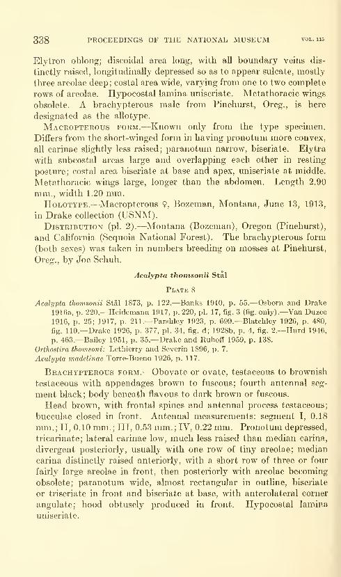
338 PROCEEDINGS OF THE NATIONAL MUSEUM vol. 115
Elytron oblong; discoidal area long, with all boundary veins dis-
tinctly raised, longitudinally depressed so as to appear sulcate, mostly
three areolae deep; costal area wide, varying from one to two complete
rows of areolae. Hypocostal lamina uniseriate. Metathoracic wings
obsolete. A brachypterous male from Pinehurst, Oreg., is here
designated as the allotype.
Macropterous form.-—Known only from the type specimen.
Differs from the short-winged form in having pronotum more convex,
all carinae slightly less raised; paranotum narrow, biseriate. Elytra
with subcostal areas large and overlapping each other in resting
posture; costal area biseriate at base and apex, uniseriate at middle.
Metathoracic wings large, longer than the abdomen. Length 2.90
mm., width 1.20 mm.HoLOTYPE.—Macropterous 9, Bozeman, Montana, June 13, 1913,
in Drake collection (USNM).Distribution (pi. 2).—Montana (Bozeman), Oregon (Pinehurst),
and California (Sequoia National Forest). The brachypterous form
(both sexes) was taken in numbers breeding on mosses at Pinehurst,
Oreg., by Joe Schuh,
Acalypta thomsonii Stal
Plate 8
Acalypta thomsonii Stai 1873, p. 122.—Banks 1910, p. 55.—Osborn and Drake
1916a, p. 220.—Heidemann 1917, p. 220, pi. 17, fig. 3 (fig. only).—Van Duzee
1916, p. 25; 1917, p. 211.—Parshley 1923, p. 699.—Blatchley 1926, p. 480,
fig. 110.—Drake 1926, p. 377, pi. 34, fig. d; 1928b, p. 4, fig. 2.—Kurd 1946,
p. 463.—Bailey 1951, p. 35.—Drake and Ruhoff 1959, p. 138.
Orthostira thomsoni: Lethierry and Severin 1896, p. 7.
Acalypta madelinae Torre-Bueno 1926, p. 117.
Brachypterous form.—Obovate or ovate, testaceous to brownish
testaceous with appendages brown to fuscous; fourth antenna! seg-
ment black; body beneath flavous to dark brown or fuscous.
Head brown, with frontal spines and antennal process testaceous;
bucculae closed in front. Antennal measurements: segment I, 0.18
mm. ; II, 0.10 mm. ; III, 0.53 mm. ; IV, 0.22 mm. Pronotum depressed,
tricarinate; lateral carinae low, much less raised than median carina,
divergent posteriorly, usually with one row of tiny areolae; median
carina distinctly raised anteriorly, with a short row of three or four
fairly large areolae in front, then posteriorly with areolae becoming
obsolete; paranotum wide, almost rectangular in outline, biseriate
or triseriate in front and biseriate at base, with anterolateral corner
angulate; hood obtusely produced in front. Hypocostal lamina
uniseriate.
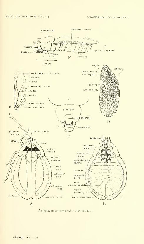
PROC US NAT. MUS. VOL. 115 DRAKE AND LATTIN. PLATE 1
nypocostal lomino
B
Acalypta, structures used in classification.
CS9-021—63-
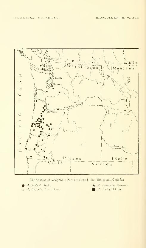
PROC. US. NAT. MUS. VOL. 115 DRAKE AND LATTIN. PLATE 2
Distribution of Aralypta In Northwestern United Stales and Canada:
# .7. harhrri Drake Vk" -^- ^aundersi Downes
^ J. lillianis Torre-Bucno A. cooleyi Drake
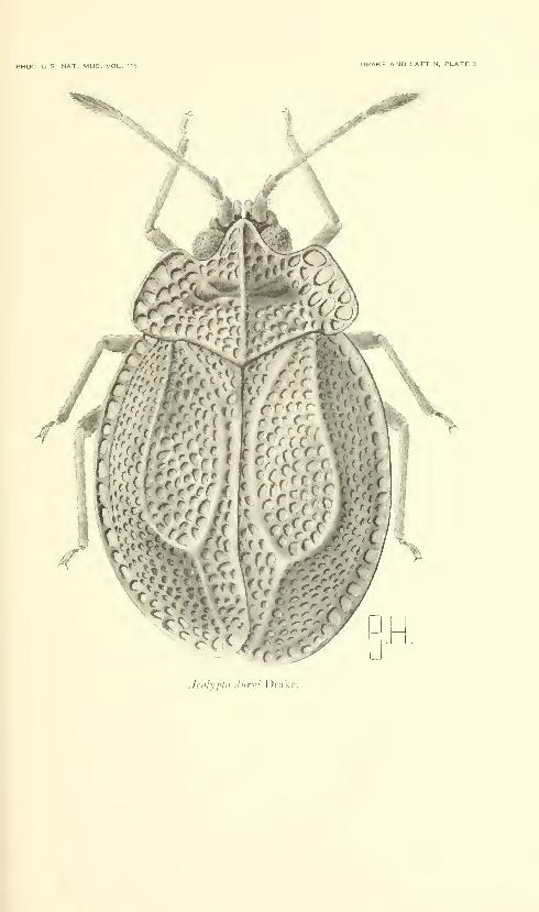
PROC. U.S. NAT. MUS. VOL.. 115 DRAKE AND LATTIN. PLATE 3
AcalypUi diiryi Drake.
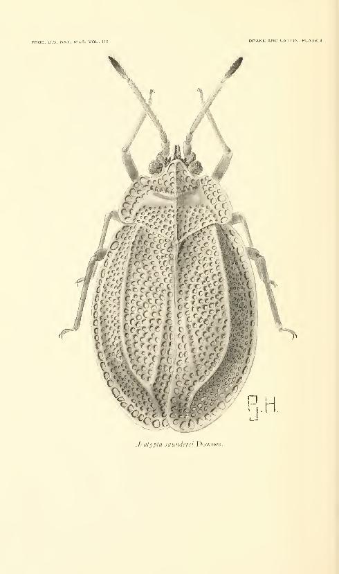
PROC, U.S. NAT. MUS. VOL. 115 DRAKE AND LATTIN. PLATE 4
Acalypla saundersi Downes.
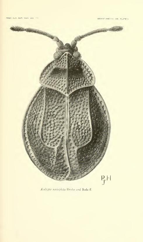
PROC. U.S. NAT. MUS. VOL. 115 DRAKE AND UATTIN, PLATE 5
Acalypta iiiiiiopliHa Drake and Ruhoff.
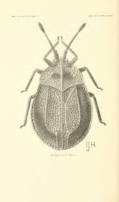
PROC. U.S. NAT. MUS. VOL. 115 DRAKE AND LATTIN, PLATE 6
Acalypta sauten Drake.
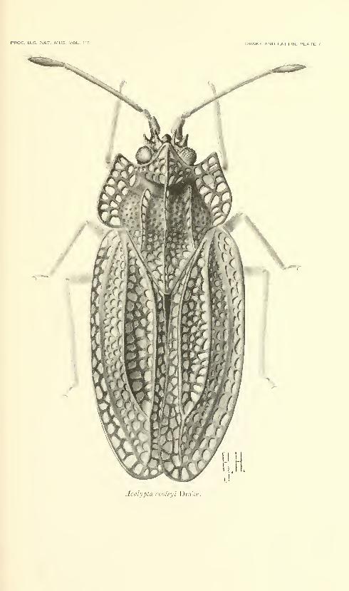
PROC. U.S. NAT. MUS. VOL. 115 DRAKE AND LATTIN. PLATE 7
Acalypta conleyi Drake.
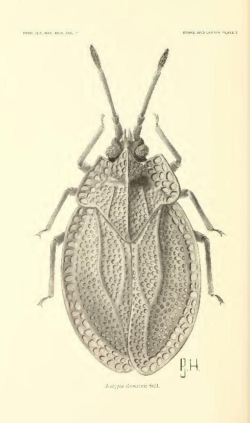
PROC. U.S. NAT. MUS. VOL. 115 DRAKE AND UATTIN. PLATE 3
^ CC^i^'mAcalypta thomsonii Stal.
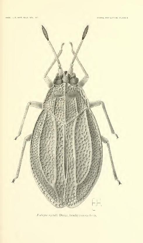
PROC. U.S. NAT. MUS VOL. 11 DRAKE AND LATTIN. PLATE 9
A
V
Acalypta nyctalis Drake, brachypterous form.
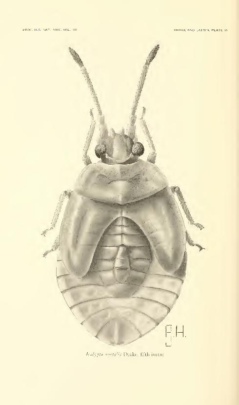
PROC. U.S. NAT. MUS. VOL. 115 DRAKE AND LATTIN. PLATE
Acalypta iiyctalis Drake, fifth instar.
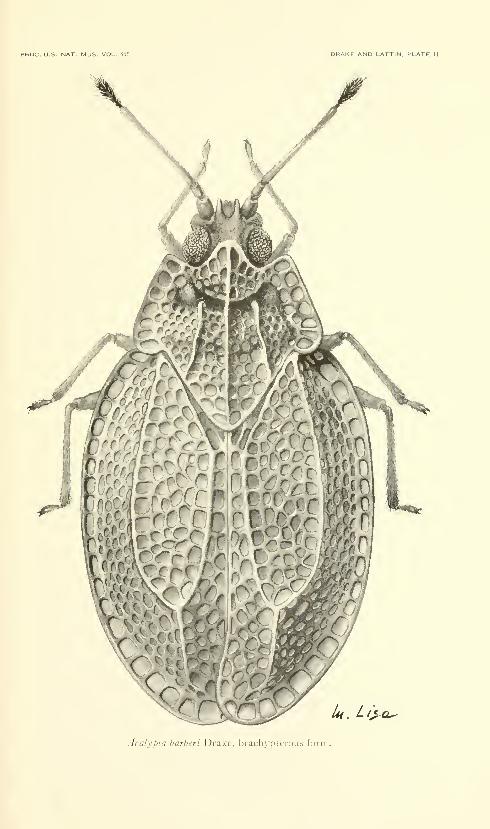
PROC. U.S. NAT. MUS. VOL. 115 DRAKE AND LATTIN. PLATE 11
/r ( ;;[0
^. Z/jj-e^
Acalypta barheri Drake, brach_\'pterous form.
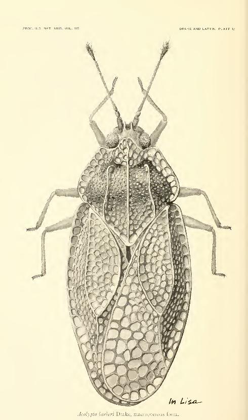
PROC. U.S. NAT. MUS. VOL. 115 DRAKE AND LATTIN. PLATE 12
^
/^ Li^.a^
Acalypta barheri Drake, macroptcrous form.
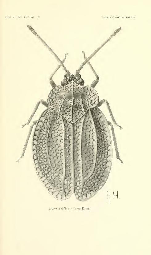
PROC. US. NAT. MUS. VOL. 115 DRAKE AND LATTIN. PLATE 13
Acalypta lillianis Torre-Bueiic
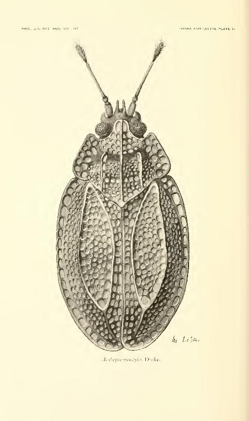
PROC. U.S. NAT. MUS. VOL. 115 DRAKE AND LATTIN, PLATE 11
>A^
AcalypUi Vandyke. Drake.
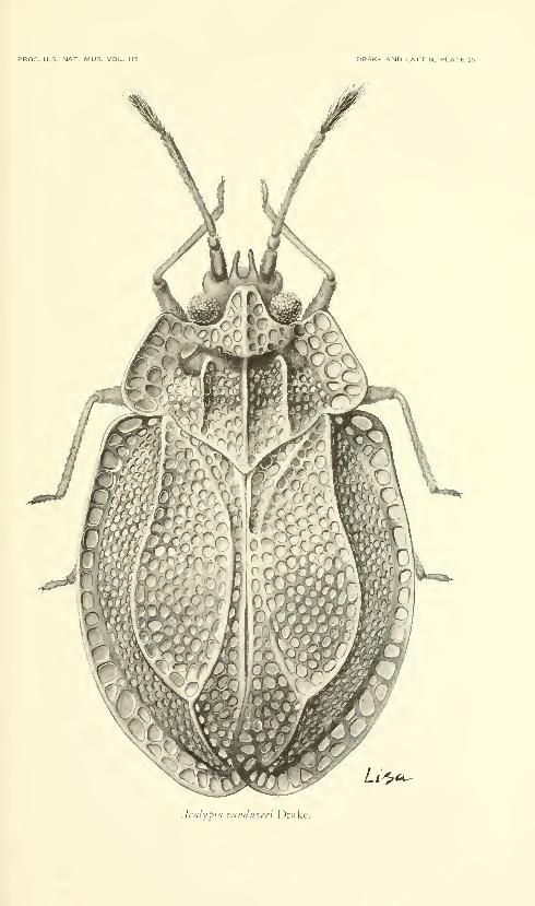
PROC. U.S. NAT. MUS. VOU. 115 DRAKE AND LATTIN. PLATE 15
Acalypta vunduzeci Drake.


LACEBUG GENUS ACALYPTA—DRAKE AND LATTIN 339
Elytra with fairly large areolae; costal area wide, varying in almost
all degrees from one to two complete rows of large areolae, usually
with one and a partial second row of areolae; discoidal area narrow,
shorter than in other members of the genus, narrower than subcostal
area, acutely angulate at base and apex, three or four areolae deep in
widest part. Length 2.30-2.70 mm., width (elytra) 1.25-1.55 mm.Macropterous form unknown.
HoLOTYPE.—Brachypterous cf, "Carolina meridionalis" (S.C.),
in Naturhistoriska Riksmuseum, Stockholm, Sweden.
Distribution.—Massachusetts, Rhode Island, New Jersey, Mary-
land, District of Columbia, Virginia, Georgia, North Carolina, South
Carolina, and Florida. Known only from the eastern coastal states.
Adults and nymphs were collected on mosses, the only known host.
It has also been collected during the winter months on mosses. Abrachypterous 9 from Rhode Island is illustrated.
Acalypta nyctalis Drake
Plates 9, 10
Acalypta nyctalis Drake 1928b, pp. 3, 5.—Hurd 1946, p. 463.—Bailey 1951,
p. 35.—Lindberg 1958, p. 13.—Drake and Ruhoflf 1959, p. 139.
Brachypterous form.—Elongate-ovate, brownish testaceous,
antenna testaceous with fourth segment black, legs testaceous.
Antennal measurements: segment I, 0.15 mm.; II, 0.10 mm.; Ill,
0.25 mm.; IV, 0.25 mm. Head with frontal spines rather slender;
bucculae open in front. Pronotum reticulate, tricarinate; carinae
low, outer pair less elevated than median and slightly divergent
posteriorly, each carina slightly raised and becoming uniseriate
anteriorly; paranotum rather narrow, mostly biseriate, with a few
extra areolae in front; hood small, feebly roundly produced anteriorly.
Elytron with very long discoidal area, as wide or slightly wider than
subcostal area; costal area usually entirely uniseriate, with small
areolae. Hypocostal lamina uniseriate. Length 2.60-2.72 mm.,
width (elytra) 1.22 mm. Macropterous form and male unknown.
HoLOTYPE.—Brachypterous 9, Franconia, New Hampshire, in
U.S. National Museum (type no. 51715).
Distribution.—United States: New Hampshire, Alaska (Point
Richardson and Fairbanks); Canada: Newfoundland and Alberta.
This is our most northerly distributed member of the genus. Abrachypterous 9 from Point Richardson and the last nymphal instar
from Fairbanks, Alaska, are illustrated.
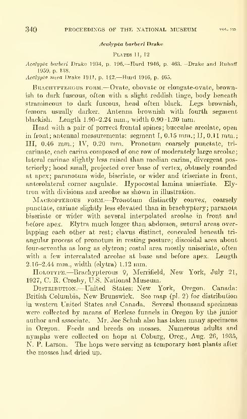
340 PROCEEDINGS OF THE NATIONAL MUSEUM vol. iio
Acalypta barberi Drake
Plates 11, 12
Acalypta barberi Drake 1934, p. 196.—Hurd 1946, p. 463.—Drake and R.uhoff
1959, p. 138.
Acalypta mera Drake 1941, p. 142.—Hurd 1946, p. 465.
Brachypterous form.-—Ovate, obovate or elongate-ovate, brown-
ish to dark fuscous, often with a shght reddish tinge, body beneath
stramineous to dark fuscous, head often bhxck. Legs brownish,
femora usually darker. Antenna brownish with fourth segment
blackish. Length L90-2.24 mm., width 0.90-L20 mm.Head Avith a pair of porrect frontal spines; bucculae areolate, open
in front; antennal measurements: segment I, 0.15 mm.; II, 0.11 mm.;Ill, 0.46 mm.; IV, 0.20 mm. Pronotum coarsely punctate, tri-
carinate, each carina composed of one row of moderately large areolae
;
lateral carinae slightly less raised than median carina, divergent pos-
teriorly; hood small, projected over base of vertex, obtusely rounded
at apex; paranotum wide, biseriate, or wider and triseriate in front,
anterolateral corner angulate. Hypocostal lamina uniseriate. Ely-
tron with divisions and areolae as shown in illustration.
Macropterous form.-—^Pronotum distinctly convex, coarsely
punctate, carinae slightly less elevated than in brachyptery; paranota
biseriate or wider with several interpolated areolae in front and
before apex. Elytra much longer than abdomen, sutural areas over-
lapping each other at rest; clavus distinct, concealed beneath tri-
angular process of pronotum in resting posture; discoidal area about
four-sevenths as long as elytron; costal area mostly uniseriate, often
with a few intercalated areolae at base and before apex. Length
2.16-2.44 mm., width (elytra) 1.12 mm.HoLOTYPE.—Brachypterous 9, Merrifield, New York, July 21,
1927, C. R. Crosby, U.S. National Museum.Distribution.-—United States: New York, Oregon. Canada:
British Columbia, New Brunswick. See map (pi. 2) for distribution
in western United States and Canada. Several thousand specimens
were collected by means of Berlese funnels in Oregon by the junior
author and associate. Mr. Joe Schuh also has taken many specimens
in Oregon. Feeds and breeds on mosses. Numerous adults and
nymphs were collected on hops at Coburg, Oreg., Aug. 26, 1935,
N. P. Larson. The hops were serving as temporary host plants after
the mosses had dried up.
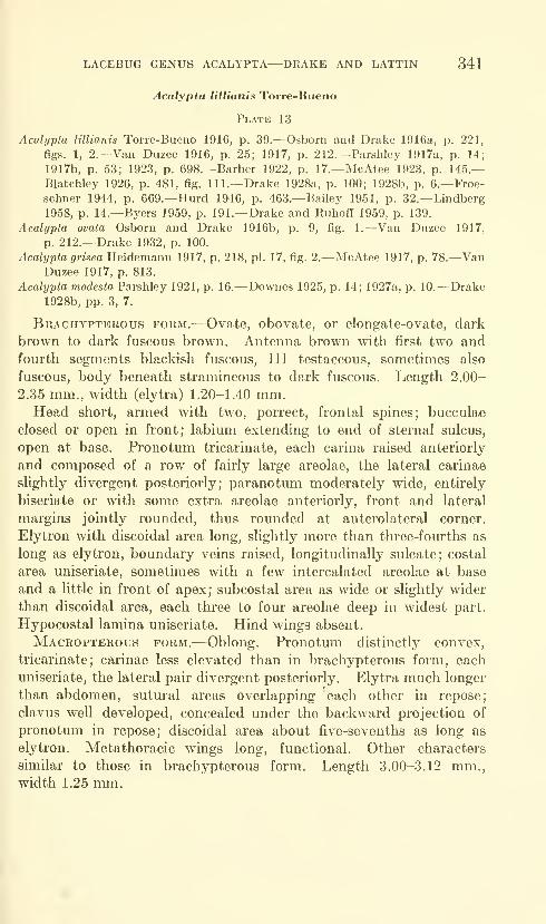
LACEBUG GENUS ACALYPTA—DRAKE AND LATTIN 341
Acalypta lillianis Torre-Bueno
Plate 13
Acalypta lillianis Torre-Bueno 1916, p. 39.—Osborn and Drake 1916a, p. 221,
figs. 1, 2.—Van Duzee 1916, p. 25; 1917, p. 212.—Parshley 1917a, p. 14;
1917b, p. 53; 1923, p. 698.—Barber 1922, p. 17.—McAtee 1923, p. 145.—Blatchley 1926, p. 481, fig. 111.—Drake 1928a, p. 100; 1928b, p. 6.—Froe-schner 1944, p. 669.—Hurd 1946, p. 463.—Bailey 1951, p. 32.—Lindberg1958, p. 14.—Byers 1959, p. 191.—Drake and Ruhoff 1959, p. 139.
Acalypta ovata Osborn and Drake 1916b, p. 9, fig. 1.—Van Duzee 1917,
p. 212.—Drake 1932, p. 100.
Acalypta grisea Heidemann 1917, p. 218, pi. 17, fig. 2.—McAtee 1917, p. 78.—VanDuzee 1917, p. 813.
Acalypta modesta Parshley 1921, p. 16.—Downes 1925, p. 14; 1927a, p. 10.—Drake1928b, pp. 3, 7.
Brachypterous form.^—Ovate, obovate, or elongate-ovate, dark
brown to dark fuscous brown. Antenna brown with first two and
fourth segments blackish fuscous, III testaceous, sometimes also
fuscous, body beneath stramineous to dark fuscous. Length 2.00-
2.35 mm., width (elytra) 1.20-1.40 mm.Head short, armed with two, porrect, frontal spines; bucculae
closed or open in front; labium extending to end of sternal sulcus,
open at base. Pronotum tricarinate, each carina raised anteriorly
and composed of a row of fairly large areolae, the lateral carinae
slightly divergent posteriorly; paranotum moderately wide, entirely
biseriate or with some extra areolae anteriorly, front and lateral
margins jointly rounded, thus rounded at anterolateral corner.
Elytron with discoidal area long, slightly more than three-fourths as
long as elytron, boundary veins raised, longitudinally sulcate; costal
area uniseriate, sometimes ^vith a few intercalated areolae at base
and a little in front of apex; subcostal area as wide or slightly wider
than discoidal area, each three to four areolae deep in widest part.
Hypocostal lamina uniseriate. Hind wings absent.
Macropterous form.^—Oblong. Pronotum distinctly convex,
tricarinate; carinae less elevated than in brachypterous form, each
uniseriate, the lateral pair divergent posteriorly. Elytra much longer
than abdomen, sutural areas overlapping each other in repose;
clavus well developed, concealed under the backward projection of
pronotum in repose; discoidal area about five-sevenths as long as
elytron. Metathoracic wings long, functional. Other characters
similar to those in brachypterous form. Length 3.00-3.12 mm.,width 1.25 mm.

342 PROCEEDINGS OF THE NATIONAL MUSEUM vol. 115
Lectotype.—9, White Plains, New York, in Snow Museum, Uni-
versity of Kansas.
Distribution.-—United States: Maine, New Hampshire, Mas-
sachusetts, Vermont, Connecticut, New York, Pennsylvania, Ohio,
Indiana, Illinois, Iowa, Michigan, Minnesota, North Dakota, Wis-
consin, Nebraska, Vu-ginia, New Jersey, North Carolina, Rhode
Island, Idaho, Tennessee, Maryland, District of Columbia, Alaska.
Canada: Quebec, Ontario, Newfoundland, British Columbia
The furcate discoidal area and rounded outer margins of the
pronotum separate A. lillianis from closely allied species. Host
records are from mosses.
Acalypta vandykei Drake
Plate 14
Acalypta vandykei Drake 1928b, pp. 3, 8.—Hurd 1946, p. 463.—Drake and Ruhoff
1959, p. 139.
Brachypterous form.^—^Elongate-ovate, dark reddish brown with
head and body beneath dark fuscous. Antenna with fom'th
segment subclavate, measurements: segment I, 0.13 mm.; II, 0.08
mm.; Ill, 0.38 mm.; IV, 0.20 mm. Pronotum with lateral carinae
parallel, slightly less raised than median carina, with nearly quadrate
areolae; paranotum fairly wide, triseriate in front, biseriate opposite
humeral angle. Hypocostal lamina uniseriate. Size and arrange-
ment of areolae and divisions of elytron as shown in illustration.
Length 1.88 mm., width (elytra) 1.10 mm. Macropterous form
unknown.HoLOTYPE.—Brachypterous cf , San Francisco County, CaUfornia,
in California Academy of Sciences.
Distribution.-—California.
This species can be separated from ^4. barheri by the characters
employed in the key. A. lillianis difTers from the above species by
having a longer and more sulcate discoidal area and jointly rounded
front and lateral margins of the paranotum. Collected on moss.
The allotype is figured.
Acalypta vanduzeei Drake
Plate 15
Acalypta vanduzeei Drake 1928b, pp. 3, 8.—Hurd 1946, p. 463.—Drake and
Ruhoff 1959, p. 139.
Brachypterous female.—Ovate, brown with head blackish, body
beneath yellowish brown. Pronotum rather short, tricarinate;
carinae raised anteriorly, each composed of one row of nearly quadrate
areolae; lateral carinae parallel, slightly less raised than median;
hood obtuse at apex; paranotum biseriate opposite humeral angle,
LACEBUG GENUS ACALYPTA—DRAKE AND LATTIN 343
triseriate in front, angulate at anterolateral corner. Hypocostal
lamina uniseriate. Antenna with segment IV fusiform, measm-ements:
segment I, 0.13 mm.; II, 0.08 mm.; Ill, 0.38 mm.; IV, 0.20 mm.Elytron with divisions and areolae as in illustration. Length 2.25
mm., width (elytra) 1.30 mm.HoLOTYPE.—Brachypterous cf, Green Point Ranch, Humbbldt
County, California; in California Academy of Sciences, Golden GatePark, San Francisco, Calif.
Distribution.—^Known only from the two type specimens. Theform and longer third antennal segment separate this species from
A. vandykei. Inhabits moss. The allotype is illustrated.
Literature Cited
Bailey, Norman S.
1951. The Tingoidea of New England and their biology. Ent. Amer., vol.
31, pp. 1-140, 6 figs.
Banks, Nathan1910. Catalogue of the Nearctic Hemiptera-Heteroptera. Amer. Ent. Soc,
Philadelphia. 103 pp.
Barber, Harry Gardner1922. List of the superfamily Tingidoidea of New Jersey with synonymy
and food plants. State of New Jersey Dep. Agric, Bur. Statistics
and Inspection, Circ. 54, pp. 16-17.
Bergroth, Ernst Evald1922. On some Neotropical Tingidae (Hem.). Ann. Soc. Ent. Belgique,
vol. 62, pp. 149-152.
Blatchley, Willis Stanley1926. Heteroptera or true bugs of eastern North America, with especial
references to the faunas of Indiana and Florida. Nature Publishing
Co., Indianapolis, 1116 pp., 215 text figs., 12 pis.
Byers, George W.1959. Types of Tingidae described by Torre-Bueno (Hemiptera). Ent.
News, vol. 70, No. 7, pp. 191-193.
China, William E.
1943. The generic names of the British Hemiptera-Heteroptera, with a
check list of the British species. In The generic names of British
insects prepared by the Committee on Generic Nomenclature of
Royal Entomological Society of London, pt. 8, pp. 211-342.
Downes, W.1925. Lace bugs of British Columbia. Proc. Ent. Soc. British Columbia,
No. 22, pp. 12-19, 1 pi.
1927a. A preliminary list of the Heteroptera and Homoptera of British
Columbia. Proc. Ent. Soc. British Columbia, No. 23, pp. 1-22.
1927b. A new species of Drakella (Heteroptera-Tingitidae). Canadian Ent.,
vol. 59, p. 60.
Drake, Carl John1917. New and noteworthy Tingidae from the United States. Ohio Journ.
Sci., vol. 17, No. 6, pp. 213-216.
1926. The North American Tingitidae (Heteroptera) described by St&l.
Ann. Carnegie Mus., vol. 16, Nos. 3-4, pp. 375-380, 1 pi.
344 PROCEEDINGS OF THE NATIONAL MUSEUM vul. 115
Drake, Carl John—Continued
1928a. A list of the insects of New York State, families Piesmidae and Tingi-
tidae. Cornell Univ. Agric. Exper. Station, Mem. 101, pp. 99-103.
1928b. A synopsis of the American species of Acalypta (Hemip.-Tingitidae).
Bull. Brooklyn Ent. Soc, vol. 23, No. 1, pp. 1-9, 2 figs.
1930. Notes on American Tingitidae (Hemiptera). Bull. Brooklyn Ent.
Soc, vol. 25, No. 5, pp. 268-272.
1932. Notes on some American Tingitidae (Hemiptera). Psyche, vol. 39,
No. 4, pp. 100-102.
1934. An undescribed Acalypta from New York (Tingitidae: Hemiptera).
Bull. Brooklyn Ent. Soc, vol. 29, No. 5, p. 196.
1941. New American Tingitidae (Hemiptera). Journ. Washington Acad.
Sci., vol. 31, No. 4, pp. 141-145.
1942. New Tingitidae (Hemiptera). Iowa State Coll. Journ. Sci., vol. 17,
No. 1, pp. 1-21.
Drake, Carl J., and Ruhoff, Florence A.
1959. A new moss-feeding tingid from Mexico (Hemiptera). Bull. Brooklyn
Ent. Soc, vol. 54, pp. 136-139, 1 fig.
1960. Lace-bug genera of the world (Hemiptera: Tingidae). Proc. U.S.
Nat. Mus., vol. 112, pp. 1-105, 5 figs., 9 pis.
FiEBER, Franz Xavier1844. Entomologische Monographien. Leipzig. 138 pp., 10 pis.
1861. Die Europaischen Hemiptera. Halbfliigler. (Rhynchota Heterop-
tera). Vienna, 444 pp., 2 pis.
Flor, Gustav1860. Die Rhynchoten Livlands in systematischen Folge beschrieben. Vol.
1, 826 pp.
Froeschner, Richard C.
1944. Contributions to a synopsis of the Hemiptera of Missouri, pt. IIL
The Amer. Mid. Nat., vol. 31, No. 3, pp. 638-683, 3 pis.
Heidemann, Otto1917. Two new species of lace-bugs (Heteroptera: Tingidae). Proc. Ent.
Soc. Washington (1916), vol. 18, No. 4, pp. 217-220, 3 figs.
HORVATH, G^ZA1906. Synopsis tingitdarum regionis palaearcticae. Ann. Mus. Nat.
Hungarici, vol. 4, pp. 1-118, 1 pi.
HuRD, Margaret Poor1946. Generic classification of North American Tingoidea (Hemiptera-
Heteroptera) . Iowa State Coll. Journ. Sci., vol. 20, No. 4, pp.
429-492, 8 figs.
Kiritshenko, Aleksandr N,
1951. True bugs of the European part of the U.S.S.R. Zool. Inst. Akad.
Nauk U.S.S.R., vol. 42, 423 pp., 416 figs.
Lethierry, Lucten, and Severin, Guillaume1896. Catalogue General des H6mipteres. Vol. 3, 275 pp.
Lindberg, Hakan1958. Hemiptera Heteroptera from Newfoundland, collected by the Swedish-
Finnish Expedition of 1949 and 1951. Acta Zool. Fennica, vol. 96,
pp. 1-25.

LACEBUG GENUS ACALYPTA—DRAKE AND LATTIN 345
McAtee, W. L.
1917. A few notes chiefly on the names of Nearctic Tingidae. Bull. BrooklynEnt. Soc, vol. 12, No. 4, pp. 78-79.
1923. Tingitoidea of the vicinity of Washington, D.C. (Heterdptera).
Proc. Ent. Soc. Washington, vol. 25, Nos. 7-8, pp. 143-151.
OsBOBN, Herbert, and Drake, Carl J.
1916a. The Tingitoidea or "lace bugs" of Ohio. Ohio Biol. Surv., vol. 2,
No. 4,'pp. 217-251, 9 figs., 4 pis.
1916b. Some new species of Nearctic Tingidae. Ohio Journ. Sci., vol. 17,
No. 1, pp. 9-15, 3 figs.
1917. Notes on Tingidae. Psyche, vol. 24, No. 5, pp. 155-161, 5 figs., 1 pi.
OsHANiN, Basil
1908. Verzeichnis der Palaearktischen Hemipteren, 1, Heteroptera, 2.
Tingididae-Acanthiidae, pp. 395-586.
1912, Katalog der palaarktischen Hemipteren (Heteroptera, Homoptera-Auchenorhyncha und Psylloideae) . 187 pp.
Parshley, Howard Madison1917a. Notes on North American Tingidae (Hemiptera). Psyche, vol. 24,
No. 1, pp. 13-24, 2 figs.
1917b. Fauna of New England. 14. List of the Hemiptera-Heteroptera.Occas. Pap. Boston Soc. Nat. Hist., vol. 7, pp. 1-125.
1921. A report on some Hemiptera from British Columbia. Proc. British
Columbia Ent. Soc, vol. 18, pp. 13-24.
1923. Family Tingidae. In Britton, Guide to the insects of Connecticut.State Geol. Nat. Hist. Surv., Connecticut, Bull. 34, pp. 695-707,figs. 164-165, 1 pi.
Stal, Carl1873. Enumeratio hemipterorum, 3. K. Svenska Vetensk.-Akad. Handl.,
vol. 11, No. 2, pp. 1-162.
1874. Genera tingitidarum europae disposuit. K. Vetensk.-Akad. Forh.,
vol. 3, pp. 43-60.
Takeya, Choku1951. A tentative list of Tingidae of Japan and her adjacent territories
(Hemiptera). Kurume Univ. Journ. (Nat. Sci.), vol. 4, No. 1,
pp. 5-28.
Torre-Bueno, Jose B.
1916. A new tingid from New York State. Bull. Brooklyn Ent. Soc, vol.
11, No. 2, pp. 39-40.
1926. Some remarks, Al Vuelo, on tingitid names. Bull. Brooklyn Ent.Soc, vol. 21, No. 3, pp. 116-117.
Van Duzee, Edward1916. Check list of the Hemiptera (excepting the Aphididae, Aleurodidae
and Coccidae) of America, north of Mexico. New York Ent. Soc,111 pp.
1917. Catalogue of the Hemiptera of America north of Mexico exceptingthe Aphididae, Coccidae and Aleurodidae. Univ. California
Publ., Tech. Bull., vol. 2, 902 pp.Westwood, John O.
1840. Synopsis of the genera of British Insects. London. 158 pp.

Proceedings of
the United States
National MuseumSMITHSONIAN INSTITUTION • WASHINGTON, D.C.
Volume 115 1964 Number 3487
THE CALIGID COPEPOD GENUS DENTIGRYPS(CRUSTACEA: CALIGOIDA)
By Alan G. Lewis ^
Introduction
The genus Dentigryps was erected by C. B. Wilson (1913) for D.curtus, which Wilson described on the basis of six female specimens
taken from the mouth and gill cavities of Mycteroperca venenosa apua(Bloch) in Bermuda. Only one additional species has since been
described in the genus, D. bifurcatus Lewis (1964), although several
closely allied species have been described in other genera without
any comment on their affinities with Dentigryps.
Wilson (1913) indicates that Dentigryps may be a connecting link
between several genera which include representatives of the families
Caligidae and Euryphoridae. This implication is discussed following
the description of the four species included in this survey.
This work is based largely on material, including the types of the
new species, deposited in the collections of the United States National
Museum. The specimens were collected from fishes taken in Florida,
the Hawaiian Islands, and at Eniwetok Atoll in the Marshall Islands.
The author was assisted by a Sigma Xi RESA grant-in-aid for the
summer of 1961. He is grateful for this and for the aid given to him
' Department of Zoology, University of New Hampshire.
347

348 PROCEEDINGS OF THE NATIONAL MUSEUM
by the U.S. Atomic Energy Commission and the Eniwetok Marine
Biological Laboratory during the time that the Eniwetok Atoll
collections were being made. The loan of some specimens of Denti-
gryps curtus by the division of marine invertebrates, U.S. National
Museum, is also deeply appreciated. Additionally, the assistance
given to the author by Eugene Shinn, David Au, John Shoup, Samuel
Kaolulo, and Lester Zukeran in the collection of the fish hosts greatly
facilitated the study.
The drawings included in the figures were made in one of two ways:
The entire animal was drawn, with the aid of a Bausch and LombTri- simplex microprojector provided by the University of New Hamp-shire Central University Research Fund, from specimens stained in
Methyl Blue, placed in 85 percent lactic acid and covered with a 9-mm.cover slip; the appendages and processes were drawn, with the aid of
a camera lucid a, either in situ on the wholemount or removed and
mounted in Hoyer's mounting medium. Measurements of the cope-
pods and their component parts were made with an ocular micrometer.
In the figures the cf and 9 signs are used separately under each
drawing to indicate a difference between the appendage of the female
and that of the male, and together (cT?) to indicate similarity; the
sex from which the drawing was made is indicated by a line under the
appropriate symbol.
The terminology used is basically the same as that in Lewis (1964).
To facilitate the use of the thoracic leg tables, a hypothetical thoracic
leg is shown in figure 1 , and the component parts of the armament of
the legs are shown in tabular form in table 1.
Table 1.
—
Armature of hypothetical thoracic leg shown in figure 1.
Leg
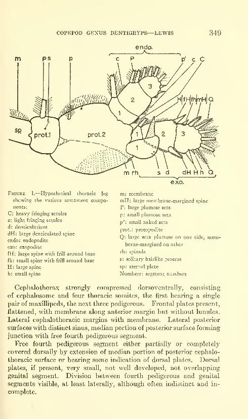
COPEPOD GENUS DENTIGRYPS—^LEWIS 349
Figure 1.—Hypothetical thoracic leg
showing the various armament compo-
nents:
C: heavy fringing setules
c: light fringing setules
d: denticulations
dH: large denticulated spine
endo: endopodite
exo: exopodite
fH: large spine with frill around base
fh: small spine with frill around base
H: large spine
h: small spine
m: membrane
mH: large membrane-margined spine
P: large plumose seta
p: small plumose seta
p': small naked seta
prot.: protopodite
Q: large seta plumose on one side, mem«brane-margined on other
rh: spinule
s: solitary hairlike process
sp: sternal plate
Numbers: segment numbers
Cephalothorax strongly compressed dorsoventrally, consisting
of cephalosome and four thoracic somites, the first bearing a single
pair of maxillipeds, the next three pedigerous. Frontal plates present,
flattened, with membrane along anterior margin but without lunules.
Lateral cephalothoracic margins with membrane. Lateral posterior
surfaces with distinct sinus, median portion of posterior surface forming
junction with free fourth pedigerous segment.
Free fourth pedigerous segment either partially or completely
covered dorsally by extension of median portion of posterior cephalo-
thoracic surface or bearing some indication of dorsal plates. Dorsal
plates, if present, very small, not well developed, not overlapping
genital segment. Division between fourth pedigerous and genital
segments visible, at least laterally, although often indistinct and in-
complete.
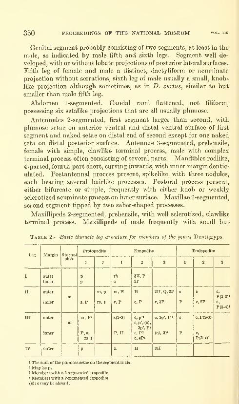
350 PROCBEDENGS OF THE NATIONAL MUSEUM
Genital segment probably consisting of two segments, at least in the
male, as indicated by male fifth and sixth legs. Segment well de-
veloped, with or without lobate projections of posterior lateral sm-faces.
Fifth leg of female and male a distinct, dactyliform or acuminate
projection without serrations, sixth leg of male usually a small, knob-
like projection although sometimes, as in D. curtus, similar to but
smaller than male fifth leg.
Abdomen 1-segmented. Caudal rami flattened, not filiform,
possessing six setalike projections that are all usually plumose.
Antennules 2-segmented, first segment larger than second, with
plumose setae on anterior ventral and distal ventral surface of first
segment and naked setae on distal end of second except for one naked
seta on distal posterior surface. Antennae 3-segmented, prehensile,
female with simple, clawlike terminal process, male with complex
terminal process often consisting of several parts. Mandibles rodlike,
4-parted, fourth part short, curving inwards, with inner margin dentic-
ulated. Postantennal process present, spikelike, with three nodules,
each bearing several hairlike processes. Postoral process present,
either bifurcate or simple, frequently with either knob or weakly
sclerotized acuminate process on inner surface. Maxillae 2-segmented
,
second segment tipped by two saber-shaped processes.
Maxillipeds 2-segmented, prehensile, with well sclerotized, clawlike
terminal process. Maxillipeds of male frequently with small but
Tab
COPEPOD GENUS DENTIGRYPS—LEWIS 351
distinct knoblike protrusion from inner surface of first segment that
appears to receive distal end of clawlike terminal process of second
segment when segment flexed. Sternal fm-ca present, situated
between and slightly posterior to maxilliped bases.
First thoracic leg biramous although endopodite reduced to minute,
knoblike rudiment in adult. Protopodite 1-segmented, exopodite
2-segmented, second segment tipped by three spines and one seta
(see table 2 for remaining basic armature), inner two spines frequently
with small, wealdy sclerotized, acuminate projection from inner
surface. Second thoracic leg distinctly biramous, protopodite 2-seg-
mented, exopodite and endopodite both 3-segmented. Thhdthoracic leg bhamous, protopodite broad, laminate, 1-segmented,
exopodite either 2- or 3-segmented, endopodite 2-segmented. Fourth
thoracic leg uniramous, protopodite 1-segmented, exopodite (pre-
sumably) 3-segmented, denticulations present either on outer surface
of second and third exopodite segments, on one or more terminal
spines of third exopodite segment or on both segments and spines.
Dentigryps ulna, new species ^
Figures 2; 3; 4; \2a,e,i; 13a
(Ulua is the Hawaiian name for many of the carangid fishes.)
Material.—One adult, nonovigerous female designated as the
holotype (USNM 107864) taken by the author from the external
surface of specimen of Caranx melampygus? Cuvier and Valenciennes,
captured in trap between Diamond Head and Koko Head, Oahu,
Hawaii, by Samuel Kaolulo. One adult male designated as the
allotype (USNM 107865) taken from holotype host. Two ovigerous,
two nonovigerous adult females, and 1 adult male designated as
paratypes (USNM 107866) taken from holotype host. Four ovigerous
females, six nonovigerous adult females, and ten adult males designated
as paratypes (retained by author) taken from external surface of
several specimens of Caranx melampygus? captured in traps between
Diamond Head and Koko Head, Oahu, Hawaii, by Samuel Kaolulo.
Measurements.—Holotype: Greatest length, excluding caudal
setae and fifth legs, 6.90 mm. Greatest length of cephalothorax,
including frontal region, 4.50 mm.; greatest width, excluding marginal
flanges, 3.60 mm. Greatest length of genital segment, excluding
fifth legs, 1.65 mm.; greatest width 1.80 mm. Greatest length of
abdomen 0.68 mm. Greatest length of fifth legs 1.37 mm. Greatest
length of fifth-leg terminal process 0.07 mm.
2 For a comparison of this species with Lepeophtheims spinifer Kirtlsinghe, see Discussion (p. 368).
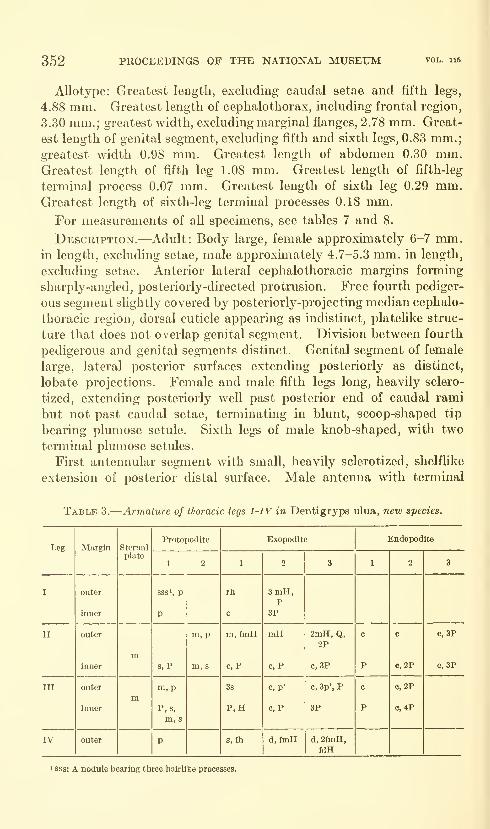
352 PROCEEDINGS OF THE NATIONAL MUSEUM
Allotype: Greatest length, excluding caudal setae and fifth legs,
4.88 mm. Greatest length of cephalothorax, including frontal region,
3.30 mm.; greatest width, excluding marginal flanges, 2.78 mm. Great-
est length of genital segment, excluding fifth and sixth legs, 0.83 mm.;greatest width 0.98 mm. Greatest length of abdomen 0.30 mm.Greatest length of fifth leg 1.08 mm. Greatest length of fifth-leg
terminal process 0.07 mm. Greatest length of sixth leg 0.29 mm.Greatest length of sixth-leg terminal processes 0.18 mm.
For measurements of all specimens, see tables 7 and 8.
Description.—Adult: Body large, female approximately 6-7 mm.in length, excluding setae, male approximately 4.7-5.3 mm. in length,
excluding setae. Anterior lateral cephalothoracic margins forming
sharply-angled, posteriorly-directed protrusion. Free fourth pediger-
ous segment slightly covered by posteriorly-projecting median cephalo-
thoracic region, dorsal cuticle appearing as indistinct, plateHke struc-
ture that does not overlap genital segment. Division between fourth
pedigerous and genital segments distinct. Genital segment of female
large, lateral posterior surfaces extending posteriorly as distinct,
lobate projections. Female and male fifth legs long, heavily sclero-
tized, extending posteriorly well past posterior end of caudal rami
but not past caudal setae, terminating in blunt, scoop-shaped tip
bearing plumose setule. Sixth legs of male knob-shaped, with two
terminal plumose setules.
First antennular segment with small, heavily sclerotized, shelflike
extension of posterior distal surface. Male antenna with terminal
Table 3.-
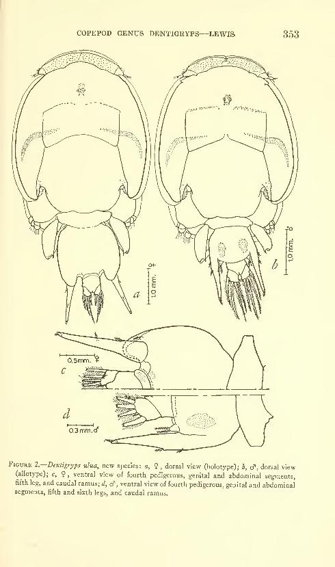
COPEPOD GENUS DENTIGRYPS—LEWIS 353
Figure l.—Dentigryps ulua, new species: a, ? , dorsal view (holotype); b, cf, dorsal view(allotype); c, 9, ventral view of fourth pedigerous, genital and abdominal segments,fifth leg, and caudal ramus; d, cf , ventral view of fourth pedigerous, genital and abdominalsegments, fifth and sixth legs, and caudal ramus.
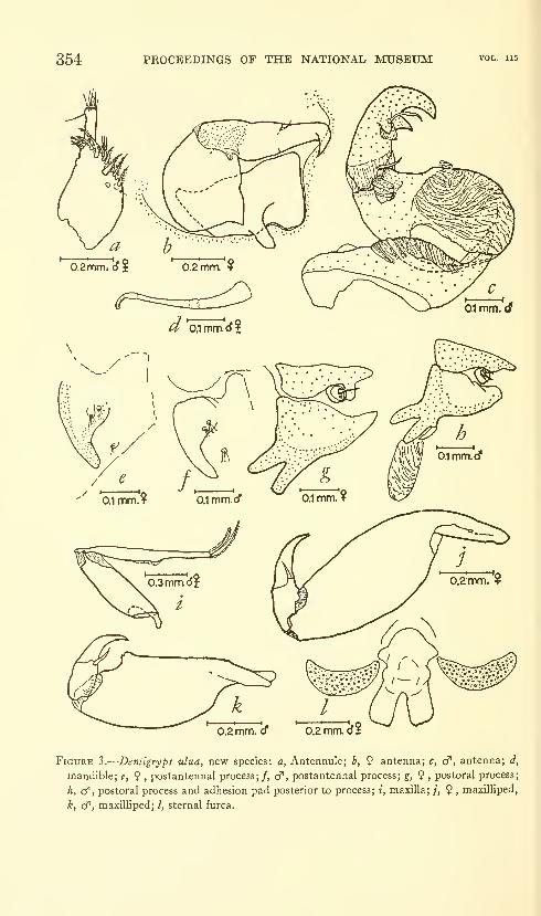
354 PROCEEDINGS OF THE NATIONAL MUSEUM
0.2 mm. d 0.2 mm I
Figure 3.
—
Dentigryps ulua, new species: a, Antennule; b, 9 antenna; c, cf, antenna; d,
mandible; e, ? ,postantennal process;/, cf", postantennal process; g, 9 ,
postoral process;
h, c?, postoral process and adhesion pad posterior to process; i, maxilla; /, 9 » maxIUiped,
k, cf , maxilliped; /, sternal furca.
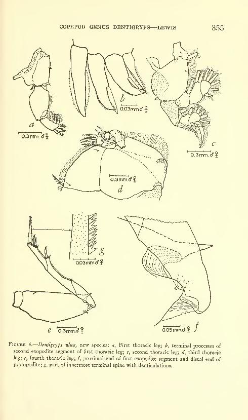
COPEPOD GENUS DENTIGRYPS—LEWIS 355
e 'oSmrrTd'? 0.05mmim.<^ ? }
Figure i.—Denttgryps ulua, new species: a, First thoracic leg; b, terminal processes ofsecond exopodite segment of first thoracic leg; c, second thoracic leg; d, third thoracicleg; e, fourth thoracic leg; /, proximal end of first exopodite segment and distal end ofprotopodite; £, part of innermost terminal spine with denticulations.

356 PROCEEDINGS OF THE NATIONAL MUSEUM
process consisting of bifurcate claw bearing lappet-shaped, membrane-covered process at level of proximalmost fiircal ramus. Male with
pair of rugose adhesion pads just posterior to mouth cone. Postoral
process of female consists of bifurcate protrusion and process-bearing
node, of male sunilar except protrusion constricted at apex of bifurca-
tion and giving rise to short, dactylifomi process on inner surface.
Sternal furca with chisel-shaped tines. Exopodite of third thoracic
leg 3-segmented. Protopodite of fourth thoracic leg with spikelike
protrusion of cuticle on inner distal corner.
Dentigryps bifurcatus Lewis, 1964
Figures 5; 126,/j; 13&
Dentigryps bifurcatus Lewis, 1964, p. 203, figs. 17, 18.
Locality.—Hawaii.
Hosts.—Acanihurus olivaceous Bloch and Schneider, Acanthurus
triostegus sandvicensis Streets, Naso hexacanthus (Bleeker).
Material.—Two nonovigerous adult females and two adult males
taken by the author from the external surface of a specunen of Myri-
pristis sp. captured by spear in Hanauma Bay, Oahu, Hawaii, byBruce Walsh. Two adult males taken by the author from the external
surface of specunen of Fistularia petimba Lacepede taken by trap
between Diamond Head and Koko Head, Oahu, Hawaii, by SamuelKaolulo. One nonovigerous adult female taken by the author from
Table
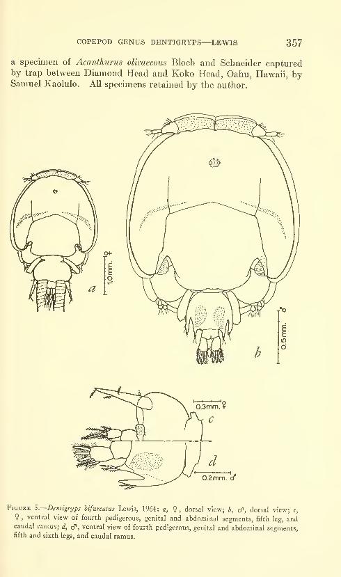
COPEPOD GENUS DENTIGRYPS—LEWIS 357
a specimen of Acanthurus olivaceous Bloch and Schneider capturedby trap between Diamond Head and Koko Head, Oahu, Hawaii, bySamuel Kaolulo. All specimens retained by the author.
0.2mm. (f
Figure S.—Denligryps bifurcatus Lewis, 1964: a, ? , dorsal view; b, d', dorsal view; c,
? ,ventral view of fourth pedigerous, genital and abdominal segments, fifth leg, and
caudal ramus; d, cp, ventral view of fourth pedigerous, genital and abdominal segments,fifth and sixth legs, and caudal ramus.
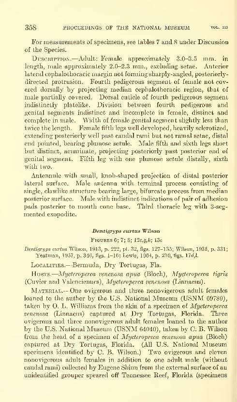
358 PROCEEDINGS OF THE NATIONAL MUSEUM vol. 115
For measurements of specimens, see tables 7 and 8 under Discussion
of the Species.
Description.—Adult: Female approximately 3.0-3.5 mm. in
length, male approximately 2.0-2.3 mm., excluding setae. Anterior
lateral cephalothoracic margin not forming sharply-angled, posteriorly-
directed protrusion. Fourth pedigerous segment of female not cov-
ered dorsally by projecting median cephalothoracic region, that of
male partially covered. Dorsal cuticle of fourth pedigerous segment
indistinctly platelike. Division between fourth pedigerous and
genital segments indistinct and incomplete in female, distinct and
complete in male. Width of female genital segment slightly less than
twice the length. Female fifth legs well developed, heavily sclerotized,
extending posteriorly well past caudal rami but not ramal setae, distal
end pointed, bearing plumose setule. Male fifth and sixth legs short
but distinct, acuminate, projecting posteriorly past posterior end of
genital segment. Fifth leg with one plumose setule distally, sixth
with two.
Antennule with small, knob-shaped projection of distal posterior
lateral surface. Male antenna with terminal process consisting of
single, clawUke structure bearing large, bifurcate process from median
posterior surface. Male with indistinct indications of pair of adhesion
pads posterior to mouth cone base. Third thoracic leg with 3-seg-
mented exopodite.
Dentigryps curtus Wilson
Figures 6; 7; 8; 12c,g,k; 13c
Dentigryps curtus Wilson, 1913, p. 222, pi. 32, figs. 127-135; Wilson, 1935, p. 331;
Yeatman, 1957, p. 346, figs. 1-16: Lewis, 1964, p. 210, figs. nd,l.
Localities.—Bermuda, Dry Tortugas, Fla.
Hosts.—Mycteroperca venenosa apua (Bloch), Mycteroperca tigris
(Cuvier and Valenciennes), Mycteroperca venenosa (Linnaeus).
Material.—One ovigerous and three nonovigerous adult females
loaned to the author by the U.S. National Museum (USNM 69789),
taken by O. L. Williams from the skin of a specimen of Mycteroperca
venenosa (Linnaeus) captured at Dry Tortugas, Florida. Three
ovigerous and three nonovigerous adult females loaned to the author
by the U.S. National Museum (USNM 64040), taken by C. B. Wilson
from the head of a specimen of Mycteropreca venenosa apua (Bloch)
captured at Dry Tortugas, Florida. (All U.S. National Museumspecimens identified by C B. Wilson.) Two ovigerous and eleven
nonovigerous adult females in addition to one adult male (without
caudal rami) collected by Eugene Shinn from the external surface of an
unidentified grouper speared off Tennessee Reef, Florida (specimens
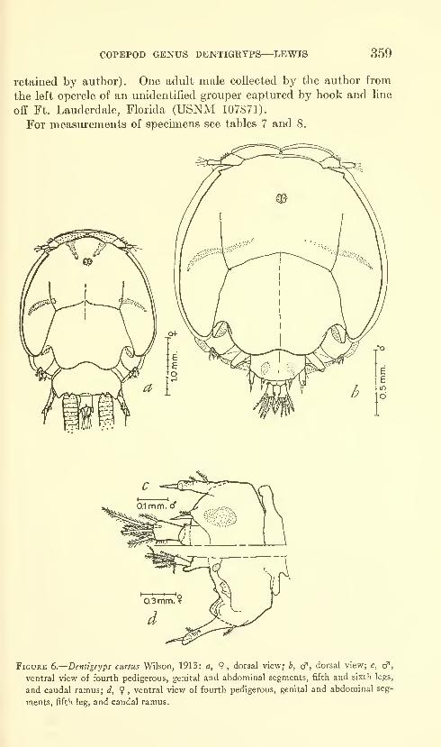
COPEPOD GENUS DENTIGRYPS—LEWIS 359
retained by author). One adult male collected by the author from
the left opercle of an unidentified grouper captured by hook and line
off Ft. Lauderdale, Florida (USNM 107871).
For measurements of specimens see tables 7 and 8.
Figure 6.
—
Dentigryps curtus Wilson, 1913: a, ?, dorsal view; b, cf, dorsal view; c, cf,
ventral view of fourth pedigerous, genital and abdominal segments, fifth and sixth legs,
and caudal ramus; d, 9 , ventral view of fourth pedigerous, genital and abdominal seg-
ments, fifth leg, and caudal ramus.
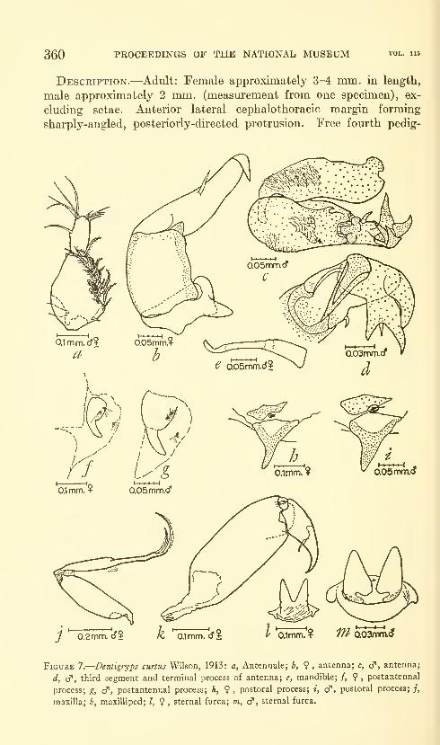
360 PROCEEDINGS OF THE NATIONAL MUSEUM
Description.—Adult: Female approximately 3-4 mm. in length,
male approximately 2 mm. (measurement from one specimen), ex-
cluding setae. Anterior lateral cephalothoracic margin forming
sharply-angled, posteriorly-directed protrusion. Free fourth pedig-
0.1mm.? 0.05mm.d"
0.1mm. ? 0.05mm.<J
0.1mm. ¥ moB^i
Figure 7.
—
Dentigryps curtus Wilson, 1913: a, Antennule; b, ? , antenna; c, cf, antenna;
d, cf , third segment and terminal process of antenna; e, mandible; /, 9 ,postantennal
process; g, cf, postantennal process; h, 9, postoral process; i, cf, postoral process; ;,
maxilla; k, maxilliped; I, 9 , sternal furca; m, cT, sternal furca.

COPEPOD GENUS DENTIGRYPS—LEWIS 361
erous segment of female covered completely or almost completely bymedian cephalothoracic region, male completely covered by region
which also covers anterior end of genital segment. Division betweenfourth pedigerous and genital segments indistinct in female, distinct
in male. Genital segment of female more than twice as wide as long,
with pair of small but distinct projections on each lateral posterior
dorsal sm'face. Male genital segment without posterior processes.
Female and male fifth legs appearing 2-parted, consisting of well-
h Qoimrnd'l C 0.2mm. (J?
<5 N^«>^ssfV WisJy
Figure 8.
—
Dentigryps curtus Wilson, 1913: a, First thoracic leg; b, distal end and ter-
minal processes of second exopodite segment of first thoracic leg; c, second thoracic leg;
d, third thoracic leg; e, fourth thoracic leg; /, proximal end of first exopodite segment
and distal end of protopodite of fourth thoracic leg; g, denticulations on outer margin
of middle terminal spine of fourth thoracic leg; h, denticulations on outer margin of inner
terminal spine of fourth thoracic leg.
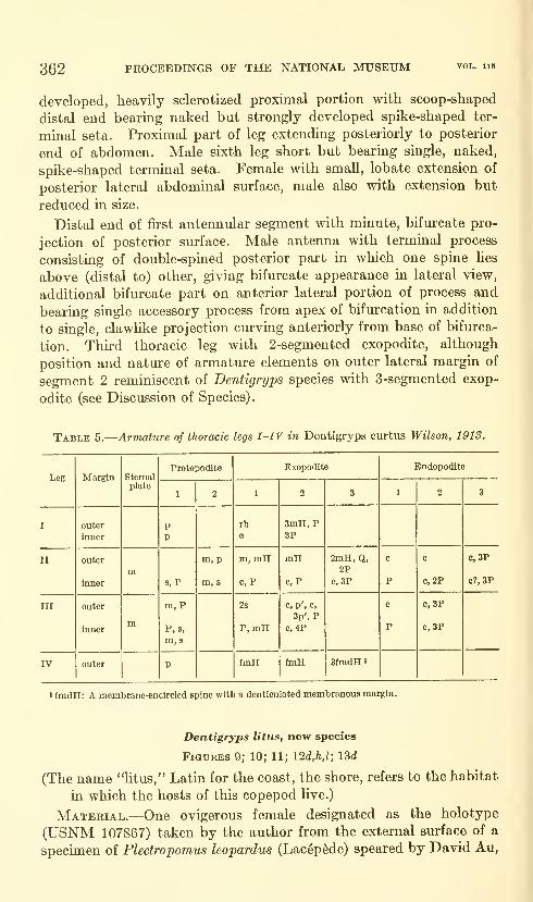
362 PROCEEDINGS OF THE NATIONAL MUSEUM
developed, heavily sclerotized proximal portion with scoop-shaped
distal end bearing naked but strongly developed spike-shaped ter-
minal seta. Proximal part of leg extending posteriorly to posterior
end of abdomen. Male sixth leg short but bearing single, naked,
spike-shaped terminal seta. Female with small, lobate extension of
posterior lateral abdominal surface, male also with extension but
reduced in size.
Distal end of first antennular segment with minute, bifurcate pro-
jection of posterior surface. Male antenna with terminal process
consisting of double-spined posterior part in which one spine lies
above (distal to) other, giving bifurcate appearance in lateral view,
additional bifurcate part on anterior lateral portion of process and
bearing single accessory process from apex of bifurcation in addition
to single, clawlike projection curving anteriorly from base of bifurca-
tion. Third thoracic leg with 2-segmented exopodite, although
position and nature of armature elements on outer lateral margin of
segment 2 reminiscent of Dentigryps species with 3-segmented exop-
odite (see Discussion of Species)
.
Table 5.
—
Armature of thoracic legs I-IV in Dentigryps curtus Wilson, 1913.
COPEPOD GENUS DENTIGRYPS—LEWIS 363
John Shoup, and Charles King at Eniwetok Atoll. One adult maledesignated as the allotype (USNM 107868) taken by the author
from the external surface of a specimen of Epineyhelus fuscoguttatus
(ForskSl) captured by hook and line at Eniwetok Atoll. Twoovigerous and one nonovigerous adult females in addition to one adult
male designated as paratypes (USNM 107869) taken by the author
from the external sm'face of the allotype host. Two ovigerous adult
females and one nonovigerous adult female designated as paratypes
(USNM 107870) taken by the author from the external surface of a
specimen of Aulostomus chinensis (Linnaeus) captured by hook andline at Eniwetok Atoll. Three ovigerous females and one adult
male designated as paratypes (retained by the author) taken fromthe external surface of a specimen of Balistoides viridescens (Bloch)
captured by hook and line at Eniwetok Atoll.
Measurements.—Holotype: Greatest length, excluding caudal
setae and fifth legs, 3.56 mm. Greatest length of cephalothorax,
including frontal region, 2.80 mm.; greatest width excluding marginal
flanges, 2.64 mm. Greatest length of genital segment, excluding
fifth legs, 0.58 mm.; greatest width 1.10 mm. Greatest length of
abdomen 0.35 mm. Greatest length of fifth leg 0.68 mm. Greatest
length of fifth-leg terminal process 0.22 mm.Allotype: Greatest length, excluding caudal setae and fifth legs, 2.78
mm. Greatest length of cephalothorax, including frontal region,
2.33 mm.; greatest width excluding marginal flanges, 2.18mm. Great-
est length of genital segment, excluding fifth and sLxth legs, 0.30 mm.;greatest width 0.53 mm. Greatest length of abdomen 0.11 mm.Greatest length of fifth leg 0.32 mm. Greatest length of fifth-leg
terminal process 0.14 mm. Greatest length of sixth leg 0.18 mm.Greatest length of sixth-leg terminal process 0.11 mm.For measurements of all specimens, see tables 7 and 8.
Description.—Adult: Female approximately 3.4-4.0 mm. in
length, male approximately 2.4-2.8 mm., excluding setae. Anterior
lateral cephalothoracic margin forming sharply-angled, posteriorly-
directed protrusion. Free fourth pedigerous segment of female andmale ahnost, if not completely, covered dorsally by median cephalo-
thoracic region. Division between fourth pedigerous and genital
segments indistinct in female, distinct in male. Genital segment of
female almost twice as wide as long, without posterior dorsal pro-
jections but with two small, irregular ventral projections, in region of
oviducal opening. Male genital segment irregularly bell-shaped.
Female- and male fifth legs appearing 2-parted, consisting of well-
developed, heavily sclerotized proximal portion with scoop-shaped
distal end bearing plumose, strongly developed, spike-shaped terminal
seta. Proximal part of female fifth leg extending posteriorly past
693-050—64 2
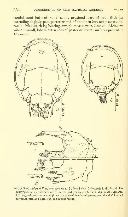
364 PROCEEDINGS OF THE NATIONAL MUSEUM
caudal rami but not ramal setae, proximal part of male fifth leg
extending slightly past posterior end of abdomen but not past caudal
rami. Male sixth leg bearing two plumose terminal setae. Abdomenwithout small, lobate extensions of posterior lateral surfaces present in
D, curtus.
Figure 9.
—
Dentigryps liius, new species: a, ? , dorsal view (holotype); b, cf, dorsal view
(allotype); c, ? , ventral view of fourth pedigerous, genital and abdominal segments,
fifth leg, and caudal ramus; d, c?", ventral view of fourth pedigerous, genital and abdominal
segments, fifth and sixth legs, and caudal ramus.

COPEPOD GENUS DENTIGRYPS—LEWIS 365
Anterior lateral surface of first antennular segment with single,
spike-shaped projection proximally, junction of distal and posterior
lateral surfaces forming sharp, slightly projecting point. Maleantenna with terminal process consisting of double-spined inner part
, ^—^
-
I' 0.1mm. cT
Figure 10.
—
Dentigryps litus, new species : a, Dorsal view of anterior lateral surface of
cephalothorax showing projection of cephalothorax and process-bearing nodule; b,
antennule; c, 9 , antenna; d, cf, antenna; e, cf , third segment and terminal process of
antenna;/, mandible; g, postantennal process; h, $, postoral process; i, cf, postoral
process.
693-050—64 3
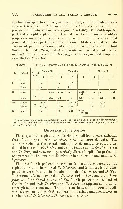
366 PROCEEDINGS OF THE NATIONAL MUSEUM
in which one spine lies above (distal to) other, giving bifui'cate appear-
ance in lateral view. Additional armature of male antenna terminal
process a bifurcate part in distal region, overlying first, double-spined,
part and at right angles to it. Second part bearing single, knoblike
projection on anterior surface and one on posterior surface, just
proximal to distal part of terminal process. Male with distinct indi-
cations of pau" of adhesion pads posterior to mouth cone. Third
thoracic leg with 2-segmented exopodite but armature of second
segment not reminiscent of Dentigryps with 3-segmented exopodite
as is that of D. curtus.
Table 6.—
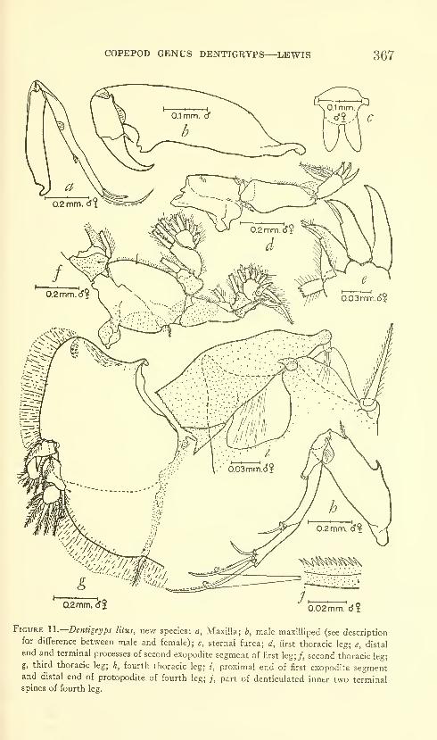
COPEPOD GENUS DENTIGRYPS—LEWIS 367
I 1 1 -0.02mm. cJi
Figure U.—Dentigryps litus, new species: a, Maxilla; b, male maxilliped (see descriptionfor difference between male and female); c, sternal furca; d, first thoracic leg; e, distal
end and terminal processes of second exopodite segment of first leg;/, second thoracic leg;
g, third thoracic leg; h, fourth thoracic leg; i, proximal end of first exopodite segmentand distal end of protopodite of fourth leg; /, part of denticulated inner two terminalspines of fourth leg.

368 PROCEEDINGS OF THE NATIONAL MUSEUM vol. 115
The female genital segment of D. ulua has distinct posterior lateral
lobes. That of D. curtus has two sets of distinct knobs on the pos-
terior sm-face, the posterior lateral of which may be compared to the
posterior lateral lobes of D. ulua.
The fifth and sixth legs of the male members of the genus determine
at least part of the shape of the segment, due to the association of the
legs with the segment. Because of this, no distinct similarities or
differences can be shown without discussing the fifth and sixth legs.
The female fifth leg (figs. I2a-d) is long and possesses a short
terminal process in both D. ulua and D. hijurcatus and is long and
possesses a long and well-developed terminal process in both D. curtus
and D. litus. The only major difference between the female fifth leg
of D. curtus and that of D. litus is that the terminal process of D. curtus
is naked while that of D. litus is plumose.
The male fifth leg (figs. 12e-h) of D. bifurcatus is s^ikelike while th&t
of D. curtus and D. litus is similar, in general makeup, being rather
stocky, with a scoop-shaped distal end which gives rise to a naked
terminal process in D. curtus and a plumose process in D. litus, as in
the female. The fifth leg of D. ulua is much longer than that of
D. curtus and D. litus but, like them, has a scoop-shaped distal end
which gives rise to a short process that, in D. ulua, is plumose.
The male sixth leg (figs. 12i-Z) of all four species is short and, in
D. ulua and D. hijurcatus, is of a more generalized nature than the
fifth leg. The sixth leg of D. ulua is knob-shaped while that of
D. hijurcatus is spikelike, although with a broad base. The sixth leg
of both D. curtus and D. litus is short and generally knob-shaped but
has a scoop-shaped distal end similar to that of the fifth leg. Thedistal end of the sixth leg of D. curtus bears two naked, setalike
processes and one plumose seta while that of D. litus bears two plumose,
setalike processes.
The abdomen of all four species is 1-segmented. The female of
D. curtus possesses an abdomen with a distinct lobate projection on
each posterior lateral surface, while the female of the other three
species has either a flat posterior sm-face, except for the anal indenta-
tion, or a biconcave posterior surface which forms the attachment
and articulation sm-face for the caudal rami.
The caudal rami of all four species are fairly similar and do not
present differences that appear sufficient to warrant any specific
comment.
The antennule appears to present relatively few comparable charac-
teristics, with the exception of one, that of the projection of the
posterior distal surface of the first segment present in D. hijurcatus,
D. curtus, and D. litus. This projection is heavily sclerotized and,
in D. hijurcatus, is small and knob-shaped while in both D. curtus and
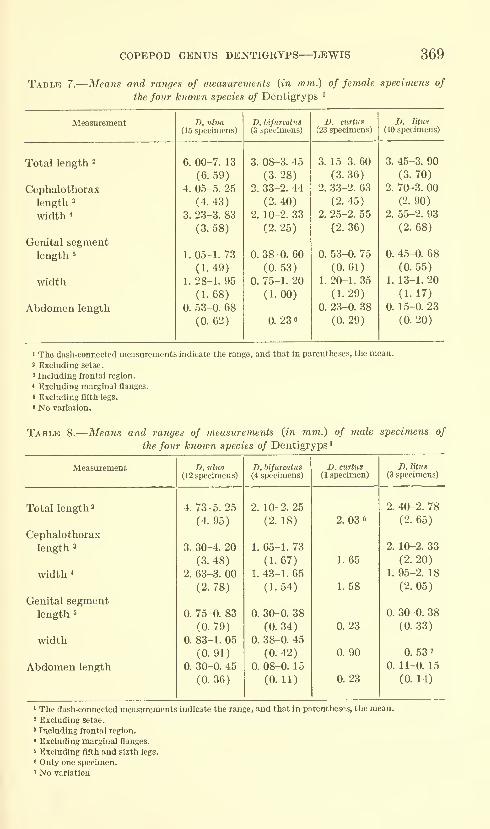
COPEPOD GENUS DENTIGRYPS—LEWIS 369
Table 7.
—
Means and ranges of measurements {in mm.) of female specimens of
the four known species of Dentigryps '
Measurement
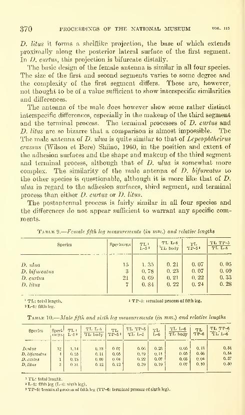
370 PROCEEDINGS OF THE NATIONAL MUSEUM
D. litus it forms a shelflike projection, the base of which extends
proximally along the posterior lateral sm"face of the first segment.
In D. curtus, this projection is bifurcate distally.
The basic design of the female antenna is similar in all fom* species.
The size of the first and second segments varies to some degree and
the complexity of the first segment differs. These are, however,
not thought to be of a value sufficient to show interspecific similarities
and differences.
The antenna of the male does however show some rather distinct
interspecific differences, especially in the makeup of the third segment
and the terminal process. The terminal processes of D. curtus and
D. litus are so bizarre that a comparison is almost impossible. TheThe male antenna of D. ulua is quite similar to that of Lepeophtheirus
crassus (Wilson et Bere) Shiino, 1960, in the position and extent of
the adhesion surfaces and the shape and makeup of the third segment
and terminal process, although that of D. ulua is somewhat morecomplex. The similarity of the male antenna of D. hifurcatus to
the other species is questionable, although it is more like that of D.
ulua in regard to the adhesion surfaces, third segment, and terminal
process than either D. curtus or D. litus.
The postantennal process is fairly similar in all four species and
the differences do not appear sufficient to warrant any specific com-
ments.
Table 9.
—
Female fifth leg measurements (in mm.) and relative lengths
Species
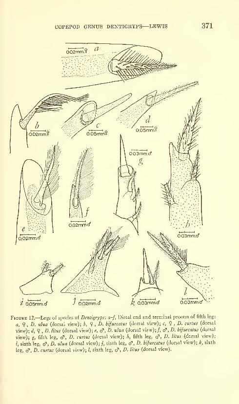
COPEPOD GENUS DENTIGRYPS
—
^LEWIS 371
t O.OSmm.ci' ).02mm.d'
Figure 12.—Legs of species of Dentigryps: a-f, Distal end and terminal process of fifth leg:
a, 9 , D. ulua (dorsal view); b, 9 , Z). hifurcatus (dorsal view); c, 9 , Z). curtus (dorsal
view); d,9 ,D. litus (dorsal view); e, cf , D. ulua (dorsal view);/, cf, D. hifurcatus (dorsal
view); g, fifth leg, cf, £>. cttri«J (dorsal view); h, fifth leg, cT, Z). /j/mj (dorsal view);
i, sixth leg, cf, Z>. ulua (dorsal view);;, sixth leg, c?, £». hifurcatus (dorsal view); i&, sixth
leg, d', Z). curtus (dorsal view); /, sixth leg, cf, Z). /iZmj (dorsal view).
372 PROCEEDINGS OF THE NATIONAL MUSEUM vol. us
One of the structures that has been used in applying the nameappendage to the postantennal process is the nodule, arising fromthe platelike area of sclerotization, which Heegaard (1947) calls
"the basal joint" of the first maxilla. In both the male and female
of D. litus there is a nodule bearing hairlike processes (as do all three of
the nodules of the postantennal process) on the dorsal anterior lateral
surface of the cephalothorax, just posterior to the division between the
frontal region and the cephalothorax (fig. 10a) and well separated
from any appendage or process. Its presence in this position onD. litus, however, implies only that a nodule of this type does not
necessarily have to be found in association with a process or an
appendage.
The female postoral process of D. curtus is simple and not bifurcate
while that of D. ulua, D. hifurcatus, and D. litus is bifurcate. Themale postoral process of D curtus is not bifurcate while, as in the fe-
male, that of D. ulua, D. bifurcatus, and D. litus is bifurcate. In the
male of the last three species, however, a poorly sclerotized acuminate
or dactyliform process arises from the inner margin of the inner
tine at the level of the apex of the bifurcation.
A pair of small but distinct adhesion pads on the ventral cephalo-
thoracic sm-face of the male of D. ulua extends posteriorly and medi-
ally from the region of the distal end of the postoral process to the
region just posterior to the mouth cone. Distinct indications of this
adhesion surface appear on the male of D. litus and indistinct indica-
tions of it on the male of D. bifurcatus. D. curtus is the only species
in which the male does not exhibit any indication of these adhesion
surfaces.
The basic structures comprising the maxilla—the two segments,
two saber-shaped terminal processes, and the membrane on the second
segment—are similar in all four species. The interspecific differences
in this appendage are caused by the presence or absence of an acces-
sory structure—a spine or spine-shaped process found in the region
of the sharp indentation in the middle of the second segment, and bythe minor differences in segment shape. D. ulua and D. curtus pos-
sess the membrane common to all four species but do not possess the
spine, while D. bifurcatus and D. litus possess both the membrane and
the spine.
The maxilliped, like the maxilla, is basically similar in all four
species, the only major difference being the presence or absence of a
distally indented swelling on the inner surface of the male appendage.
The male of D. ulua, D. bifurcatus, and D. litus all possess this swelling
while the male of D. curtus does not.
The sternal furca of D. ulua has chisel-shaped tines while the sternal
furca of all of the other species has pointed or rounded tines. In ad-
COPEPOD GENUS DENTIGRYPS—LEWIS 373
dition, D. ulua has a platelike process on either side of the sternal
furca while the others either do not have this process or else it is not
distinct from the surrounding ventral cephalothoracic cuticle.
The adult fu'st thoracic leg is basically similar in all four species,
consisting of a 1-segmented protopodite, a 2-segmented exopodite,
and a rudimentary endopodite that gives the leg an indistinctly
biramous condition. The differences that occm- do so in what could
be called secondary armature elements, elements such as the mem-branes that are present on spines, the individual or small groups of
hairlike processes that occur on the segments, the plumose conditions
of some of the spines, and small dactyliform or acuminate projections
found on some of the spines. These are in contrast to the primary
elements such as the spines and setae. The single secondary armature
element that appears to be of enough importance to be included in this
comparison is a small, acuminate, poorly sclerotized process that pro-
jects from the inner surface of each of the inner two terminal spines
of the second exopodite segment of D. ulua, D. curtus, and D. litus.
Although D. bifurcatus does not possess this process there is, in the
same region as the acuminate process on the other species, a distinct
membrane, that appears folded and that projects very slightly past
the distal end of the terminal spine (the projecting portion is believed
to be the folded part of the membrane). As with the first thoracic leg,
the comparable difference in the second thoracic leg of all four species
occurs in the secondary armatm'e elements. In the second leg how-
ever these differences are of such a nature that no distinct trend or
grouping can be indicated.
The third thoracic leg (fig. 13, table 11) has a protopodite-exopodite-
endopodite segment number of 1-3-2 in D. ulua and D. bifurcatus
and of 1-2-2 in D. curtus and D. litus. The difference in the numberof exopodite segments is important in that it suggests a distinct break
between D. curtus - D. litus and D. ulua - D. bifurcatus. This break
may not be as distinct as it seems, however, as part of the armamentof the exopodite of D. curtus is unique and suggests a trarsition
between the two groups. The outer margin of the distal two exopodite
segments in both groups has four small and naked setae that appear
to be divided into two parts. In the 1-3-2 group, the second exopodite
segment bears one of these setae on its outer distal lateral sm'face, the
remaining three are located on the third segment and are separated
from the second segment seta by the division between the segments
and by a plumose space. In D. curtus, a member of the 1-2-2 group,
all four setae are on the second or distalmost segment but there is a
distinct space between the proximalmost of the four and the succeeding
three setae. This space is plumose. The separation of the proximal-
most from the three succeeding setae and the presence of a plumose

374 PROCEEDINGS OF THE NATIONAL MUSEUM
space is, in D. curtus, strongly suggestive of the members of the 1-3-2
group except that there is no indication of segmentation between the
proximahnost seta and the other three.
Along the inner lateral surface of the two distal exopodite segments
of the 1-3-2 group are fom- plumose setae, one on the inner-distal
lateral surface of the second segment and three on the lateral sm-face
of the third. In addition, the proximal lateral margin of the third
segment is plumose. Although both of the species in the 1-2-2
group, D. curtus and D. litus, have four plumose setae along the second
or distal exopodite segment, there is no indication of a plumose
sm'face between the first or proximalmost seta and the second. The
spinelike process projecting inward from the inner surface of the
first exopodite segment is distinctly separated from the segment by
a line of division in all of the species except D. hijurcatus. Other
than these two characteristics, the differences between the species
are the shape of the segments and the variation in the position of the
armatm-e components.
Whether the difference in the exopodite makeup of the third leg
indicates a fusion of segments (from a 1-3-2 to 1-2-2 condition), a
division of segments (from a 1-2-2 to a 1-3-2 condition), or simply
a peculiar series in the pertinent armature elements could not be
ascertained. The indication is, however, that the distinct difference
in the number of segments may possibly be explained by the position
and arrangement of the armature.
The fourth thoracic leg protopodite has a distinct, spikelike projec-
tion of the inner distal surface in D. ulua and a distmct, bluntly
tipped projection in D. litus. The general armature of the leg and
Table 11.
—
Armature of third thoracic legs of four species of Dentigryps.
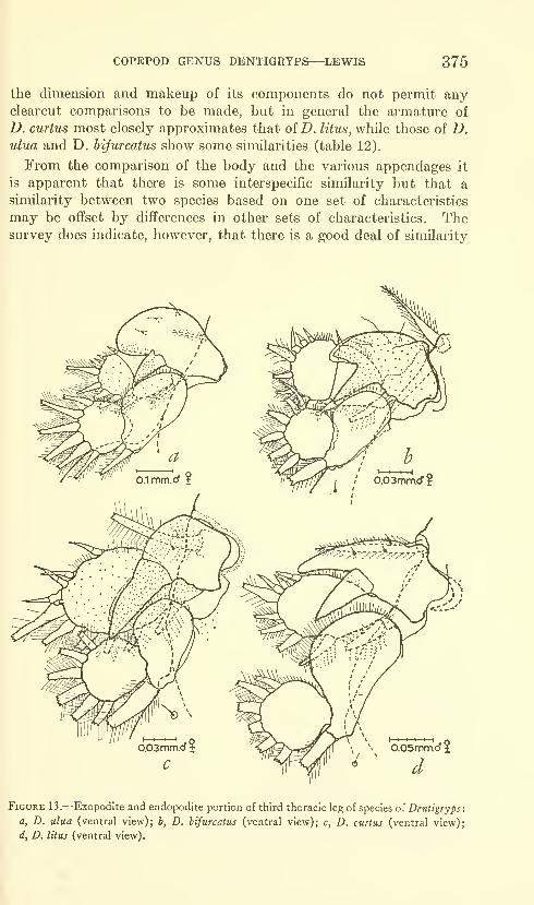
COPEPOD GENUS DENTIGRYPS—LEWIS 375
the dimension and makeup of its components do not permit anyclearcut comparisons to be made, but in general the armature of
D. curtus most closely approximates that of D. litus, while those of D.
ulua and D, bifurcatus show some similarities (table 12).
From the comparison of the body and the various appendages it
is apparent that there is some interspecific similarity but that a
similarity between two species based on one set of characteristics
may be offset by differences in other sets of characteristics. Thesurvey does indicate, however, that there is a good deal of similarity
' 'I -o
o.05mm.o t
Figure 13.—Exopodite and endopodite portion of third thoracic leg of species of Denttgryps:
a, D. ulua (ventral view); b, D. bifurcatus (ventral view); c, D. curtus (ventral view);
d, D. litus (ventral view).
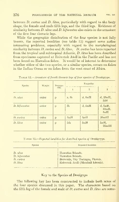
376 PROCEEDINGS OF THE NATIONAL MUSEUM
between D. curtus and D. litus, particularly with regard to the bodyshape, the female and male fifth legs, and the third legs. Evidence of
similarity between D. ulua and D. bifurcatus also exists in the armature
of the fii'st fom' thoracic legs.
While the geographic distribution of the four species is not fully
known, the reported localities (see table 13) suggest some rather
interesting problems, especially with regard to the morphological
similarity between D. curtus and D. litus. D. curtus has been reported
from the tropical and subtropical Atlantic, D. litus has been described
from specimens captured at Eniwetok Atoll in the Pacific and has not
been found on Hawaiian fishes. It would be of interest to determine
whether either of the two species, or a similar species, occurs on fishes
in the Indian Ocean or on fishes from the west coast of Africa.
Table 12.
—
Armature of fourth thoracic legs of four species of Dentigryps.
Species
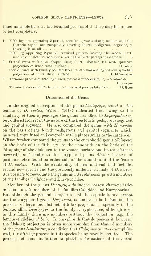
COPEPOD GENUS DENTIGRYPS—LEWIS 377
times unusable because the terminal process of that leg may be broken
or lost completely.
1. Fifth leg not appearing 2-parted, terminal process short; median cephalo-
thoracic region not completely covering fourth pedigerous segment, if
covering it at all 2
Fifth leg appearing 2-parted, terminal process forming the second part;
median cephalothoracic region covering the fourth pedigerous segment . . 3
2. Sternal furca with chisel-shaped tines; fourth thoracic leg with spilcehke
projection of inner distal surface D. uluaSternal furca with bluntly pointed tines ; fourth thoracic leg without spikelike
projection of inner distal surface D. bifurcatus
3. Terminal process of fifth leg naked; postoral process simple, not bifurcate.
D. curtus
Terminal process of fifth leg plumose; postoral process bifurcate . . . D. litus
Discussion of the Genus
In the original description of the genus Dentigryps, based on the
female of D. curtus, Wilson (1913) indicated that owing to the
similarity of their appendages the genus was allied to Lepeophtheirus,
but differed from it in the nature of the free fourth pedigerous segment
and genital segment. He also compared the genus with Homoiotes
on the basis of the fom^th pedigerous and genital segments which,
he noted, were fused and covered "with a plate similar to the carapace."
Wilson further compared the genus to the euryphorid genus Gloiopotes
on the basis of the fifth legs, to the pandarids on the basis of the
"dropping of the abdomen to the ventral surface and its transference
forward," and finally to the euryphorid genus Alehion because of
posterior lobes found on either side of the caudal rami of the female
of D. curtus. With the availabilit}^ of new material that includes
several new species and the previously undescribed male of D. curtus,
it is possible to reevaluate the genus and its relationships with membersof the families Caligidae and Em-yphoridae.
Members of the genus Dentigryps do indeed possess characteristics
in common with members of the families Caligidae and Euryphoridae.
But although the general composition of the cephalothorax, except
for the euryphorid genus Dysgamus, is similar in both families, the
presence of large and distinct fifth-leg projections, especially in the
female, allies Dentigryps to the family Euryphoridae, although even
in this family there are members without the projection (e.g., the
female of Alehion glaher). In euryphorids that do possess it, however,
the fifth-leg projection is often more complex than that of membersof the genus Dentigryps, a condition that Gloiopotes ornatus exemplifies
well, the fifth-leg process in this species being heavily serrated. Thepresence of some indication of platelike formations of the dorsal
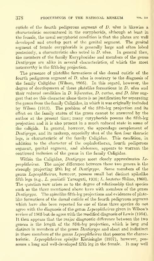
378 PROCEEDINGS OF THE NATIONAL MUSEUM vol. ub
cuticle of the fourth pedigerous segment of D. ulua is Hkewise a
characteristic encountered in the euryphorids, although at least in
the female, the usual euryphorid condition is that the plates are well
developed and overlap part of the genital segment. The genital
segment of female em-yphorids is generally large and often lobed
posteriorly, a characteristic also noted in D. ulua. In general then,
the members of the family Euryphoridae and members of the genus
Dentigryps are alike in several characteristics, of which the most
noteworthy is the fifth-leg projection.
The presence of platelike formations of the dorsal cuticle of the
fourth pedigerous segment of D. ulua is contrary to the diagnosis of
the family Caligidae (Wilson, 1905). In this regard, however, the
degree of development of these platelike formations in D. ulua and
their reduced condition in D. hijurcatus, D. curtu^, and D. litus sug-
gest that on this character alone there is no justification for separating
the genus from the family Caligidae, in which it was originally included
by Wilson (1913). The problem of the fifth-leg projection and its
effect on the family status of the genus cannot be answered by the
author at the present time; many euryphorids possess the fifth-leg
projection and it is also present in a much reduced state in some of
the caligids. In general, however, the appendage complement of
Dentigryps, and its makeup, especially that of the first four thoracic
legs, is characteristic of the family Caligidae. This condition, in
addition to the character of the cephalothorax, fourth pedigerous
segment, genital segment, and abdomen, appears to warrant the
continued inclusion of the genus in the family Caligidae,
Within the Caligidae, Dentigryps most closely approximates Le-
peophtheirus. The major difference between these two genera is the
strongly projecting fifth leg of Dentigryps. Some members of the
genus Lepeophtheirus, however, possess small but distinct spikelike
fifth legs (e.g., L. goniistii Yamaguti, 1936; L. hastatus Shiino, 1960).
The question now arises as to the degree of relationship that species
such as the three mentioned above have with members of the genus
Dentigryps. The spinelike fifth-leg projections and evidences of plate-
like formations of the dorsal cuticle of the fourth pedigerous segment
which have also been reported for one of these three species do not
agree with the diagnosis of the genus Lepeophtheirus given in Wilson's
review of 1905 but do agree with the modified diagnosis of Lewis (1964).
It then appears that the major diagnostic difference between the two
genera is the length of the fifth-leg projection, which is long and
distinct in members of the genus Dentigryps and short and indistinct
in those members of the genus Lepeophtheirus that possess the charac-
teristic. Lepeophtheirus spinifer Kh-tisinghe (1937), however, pos-
sesses a long and well-developed fifth leg in the female. It may well
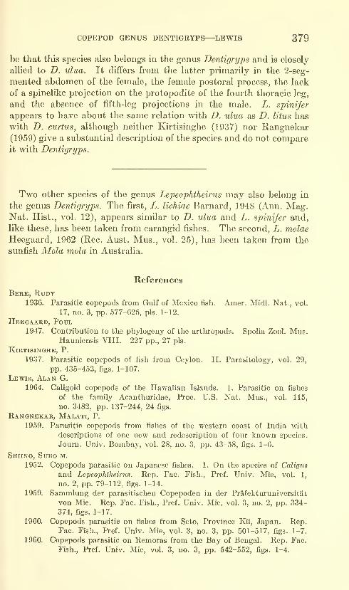
COPEPOD GENUS DENTIGRYPS—^LEWIS 379
be that this species also belongs in the genus Dentigryps and is closely
allied to D. ulua. It differs from the latter primarily in the 2-seg-
mented abdomen of the female, the female postoral process, the lack
of a spinelike projection on the protopodite of the fourth thoracic leg,
and the absence of fifth-leg projections in the male. L. spinifer
appears to have about the same relation with D. ulua as D. litus has
with D. curtus, although neither Kirtisinghe (1937) nor Rangnekar
(1959) give a substantial description of the species and do not compareit with Dentigryps.
Two other species of the genus Lepeophtheirus may also belong in
the genus Dentigryps. The first, L. lichiae Barnard, 1948 (Ann. Mag.Nat. Hist., vol. 12), appears similar to D. ulua and L. spinifer and,
like these, has been taken from carangid fishes. The second, L. molae
Heegaard, 1962 (Rec. Aust. Mus., vol. 25), has been taken from the
sunfish Mola mola in Australia.
References
Bere, Rudt1936. Parasitic copepods from Gulf of Mexico fish. Amer. Midi. Nat., vol.
17, no. 3, pp. 577-625, pis. 1-12.
Heegaard, Poul1947. Contribution to the phylogeny of the arthropods. Spolia Zool. Mus.
Hauniensis VIII. 227 pp., 27 pis.
Kirtisinghe, P.
1937. Parasitic copepods of fish from Ceylon. II. Parasitology, vol. 29,
pp. 435-452, figs. 1-107.
Lewis, Alan G.
1964. Caligoid copepods of the Hawaiian Islands. 1. Parasitic on fishes
of the family Acanthuridae, Proc. U.S. Nat. Mus., vol. 115,
no. 3482, pp. 137-244, 24 figs.
Rangnekar, Malati, P.
1959. Parasitic copepods from fishes of the western coast of India with
descriptions of one new and redescription of four known species.
Journ. Univ. Bombay, vol. 28, no. 3, pp. 43-58, figs. 1-6.
Shiino, Sued m.
1952. Copepods parasitic on Japanese fishes. 1. On the species of Caligus
and Lepeophtheirus. Rep. Fac. Fish., Pref. Univ. Mie, vol. 1,
no. 2, pp. 79-112, figs. 1-14.
1959. Sammlung der parasitischen Copepoden in der Prafekturuniversitat
von Mie. Rep. Fac. Fish., Pref. Univ. Mie, vol. 3, no. 2, pp. 334-
374, figs. 1-17.
1960. Copepods parasitic on fishes from Seto, Province Kii, Japan. Rep.Fac. Fish., Pref. Univ. Mie, vol. 3, no. 3, pp. 501-517, figs. 1-7.
1960. Copepods parasitic on Remoras from the Bay of Bengal. Rep. Fac.
Fish., Pref. Univ. Mie, vol. 3, no. 3, pp. 542-552, figs. 1-4.
380 PROCEEDINGS OF THE NATIONAL MUSEUM vol. 115
Wilson, Charles B.
1905. North American parasitic copepods belonging to the family Caligidae.
Part I, The CaUginae. Proc. U.S. Nat. Mus., vol. 28, no. 1404,
pp. 479-672, pis. 5-29.
1913. Crustacean parasites of West Indian fishes and land crabs, with
descriptions of new genera and species. Proc. U.S. Nat. Mus.,
vol. 44, no. 1950, pp. 189-277, pis. 18-53.
1935. Parasitic copepods from the Dry Tortugas. Carnegie Inst. Wash-ington, Publ. 452, Pap. from Tortugas Lab., vol. 29, no. 12, pp. 327-
347, pis. 1-6.
Yamaguti, SATYtr
1936. Parasitic copepods from fishes of Japan: Part 2, Caligoida, I. KyotoImperial Univ., Japan, 22 pp., 12 pis.
Yeatman, Harry1957. A redescription of two parasitic copepods from Bermuda. Journ.
Washington Acad. Sci., vol. 47, no. 10, pp. 346-353, figs. 1-31.
U.S. GOVERNMENT PRINTING 0FF1CE:I964


Proceedings of
the United States
National MuseumSMITHSONIAN INSTITUTION • WASHINGTON, D.C.
Volume 115 1964 Number 3488
A NEW BRAZILIAN MOTH OF THE GENUS GONIOTERMAWITH NOTES ON RELATED SPECIES
(LEPIDOPTERA: STENOMIDAE)
By W. Donald Duckworth
This description is published to provide a name for a new species
of Microlepidoptera for which Dr. Laiiro P. Travassos F. of Sao Paulo,
Brazil, has ecological data to be published.
During the course of the present study I found that the new species
is related very closely to several that have been associated improperly
with the genus Sienoma Zeller. These species have been studied,
features critical to their recognition have been discussed and figured,
and the species have been placed in their proper position in the genus
Gonioterma Walsingham.
When fresh, the species examined are rather distinct in coloration,
but because they fade rapidly, specific recognition on the basis of
color alone is difficult. For this reason the genitalia are described and
381
382 PROCEEDINGS OF THE NATIONAL MUSEUM vol. 115
specific differences are noted from this character. A complete revision
of the genus Gonioterma is planned, but the present study at least will
permit recognition of the included species.
Because, in some instances, characters used to distinguish the fe-
males involve differences in size, the illustrations of the female genitalia
were all drawn to the same magnification. Mr. Andre Pizzini madethe illustrations for this paper.
Gonioterma exquisita, new species
Figures la, 6; 2a,
e
Alar expanse 21-23 mm.Antenna whitish sprinlded with brown basally, whitish annulated
with brown beyond. Head white medially with transverse band of
gray between bases of antennae; second segment of palpus white,
shaded with brown on outer side, white on inner side. Legs whitish
ocherous; forelegs heavily shaded with fuscous, mid- and hindlegs
lightly shaded with brown. Tegula and dorsoanterior portion of
thorax white, shaded with gray, posterior portion of thorax gray.
Abdomen ocherous. Forewing white; base of costa narrowly fuscous;
three triangular fuscous costal spots, one at basal third, one near
middle, one at apical fourth; from outer corner of apical spot a row of
fuscous dots extends from apex along termen to tornus; black spot at
base of cell from which a wide band of olivaceous brown extends to
inner angle; from apex of middle costal spot an irregular patch of gray
extends transversely around basal spot to inner margin; crescent-
shaped olivaceous brown spot at end of cell; irregular patch of gray,
shaded with olivaceous brown, extends from tornus to apex of apical
costal spot; cilia fuscous basally, white beyond. Hindwing light gray
with whitish costal margin in males, heavily shaded with fuscous in
females; cilia white with gray subbasal line.
Male genitaha: See figures la,h (shde WDD 2502, paratype).
Uncus reduced, curved ventrad; gnathos complete, basal process well
developed, rather long, recurved; division of gnathal arms at base
of process; tegumen moderately constricted; harpe broad, saccular
lobe rather narrow, bluntly pointed; anellar lobes broad, bluntly
rounded at apex; aedeagus short, broad, vesica armed with two
elongate, convoluted clusters of small, heavy cornuti.
Female genitaha: See figure 2a (slide WDD 2503, paratype).
Genital plate membranous; ostium small, ostium bursae enlarged;
ductus bursae short, less than half length of corpus bursae, which has
two dentate signa; anterior margin of eighth abdominal tergum with
rectangular indentation.
Type: Brazil, Lassance, Minas Geraes, Nov. 9-12, 1919, USNM66418.
A NEW BRAZILIAN MOTH—DUCKWORTH 383
Described from male holotype, one male paratype, three female
paratypes. All paratypes with following data: Brazil, Sao Paulo,
S.P., Ypiranga (April 4, 1960, W. Bokemann). One female paratype
is in the Departamento de Zoologia, Secretaria da Agricultm-a, Sao
Paulo, Brazil.
This species and the others included in this study
—
chlorina, aesiocopia,
algosa, fastigata, argicerauna, bryophanes—form a very closely related
group within the genus Gonioterma. The males of G. exquisita are
readily separable from the males of the previously mentioned species
by the absence, along vein la in the hindwing, of a hair pencil that is
present in the other species. Also, the forewing of G. exquisita has a
straight costa and white ground color, whereas the other species
mentioned above possess a strongly arched costa in the forewing and
lack a white ground color.
The genitalia, although indicating an unusually close relationship
between G. exquisita and the other species included in this study, pro-
vide a number of distinguishing characters. The complete gnathos
and the broad harpe, with narrow, bluntly pointed saccular lobe,
serve to distinguish the males of G. exquisita. The short ductus bursae
in the female genitalia serves to separate the females.
Gonioterma chlorina (Kearfott), new combination
FiGUBES lc,d; 26
Stenoma chlorina Kearfott, 1911, Ent. News, vol. 22, p. 126.
Type locality: Sao Paulo, Brazil.
Male genitaha: See figures lc,d (slide WDD 2052, Sao Paulo,
Brazil, type). Uncus reduced, curved sharply ventrad; gnathos in-
complete, basal process well developed, long, recurved, pointed;
division of gnathal arms well below process; tegumen narrowly con-
stricted; harpe narrow, saccular lobe broad, blunt; anellar lobes broad,
bluntly rounded at apex; aedeagus narrow, vesica armed with one
elongate convoluted cluster of small, heavy cornuti.
Female genitalia: See figure 26 (slide WDD 2501, Sao Paulo,
Brazil). Genital plate membranous; ostiimi large, ostium bursae en-
larged; ductus bursae longer than in G. exquisita, more than half length
of corpus bursae; corpus bursae with two dentate signa; anterior mar-
gin of eighth abdominal tergum as in G. exquisita.
This species and the following one, G. aesiocopia, are very similar in
coloration and general appearance. However, the long, reciu'ved,
pointed basal process and the division of the gnathal arms well below
the process in the male genitalia serve to separate the males of G.
chlorina from those of G. aesiocopia. The females are more difficult to
separate, but figures 26 and c indicate the size differences that will
distinguish the species in a majority of instances.

384 PROCEEDINGS OF THE NATIONAL MUSEUM vol. lis
Gonioterma aesiocopia (Walsingham), new combination
FiGUBES Ig, 2c
Sienoma aesiocopia Walsingham, 1913, in Godman and Saivin, Biologia Centrali-
Americana, vol. 42 (Lepidoptera-Heterocera, vol. 4), p. 179.
Sienoma aphrogramma Mej-rick, 1929, Trans. Ent. Soc. London, vol. 76, p. 515.
Type localities: Veracruz, Mexico {G. aesiocopia)', Taboga Isl.,
Panama, 200-1,000 ft. (6^. aphrogramma).
Male genitalia: See figure Ig (slide WDD 2051, Kio Tempas,
Honduras). Uncus reduced, curved ventrad; gnathos incomplete,
basal process poorly developed, short, blunt; division of gnathal arms
near apex of process; tegumen moderately constricted; harpe rather
broad, saccular lobe broad, rounded at apex; anellar lobes broad,
pointed at apex; aedeagus narrow, vesica armed with two elongate,
convoluted clusters of small, heavy cornuti.
Female genitalia: See figure 2c (slide WDD 2554, Porto Bella,
Panama). Genital plate membranous; ostium large, ostimn bursae
enlarged; ductus bursae long, wider than in G. exquisita or G. chlorina;
corpus bursae with two dentate signa; anterior margin of eighth ab-
dominal tergum as in G. exquisita.
The short, blunt basal process and the division of the gnathal arms
near the apex of the basal process in the male genitalia distinguish the
males of G. aesiocopia. The differences in the females have been
discussed in the remarks under G. chlorina.
Gonioterma argicerauna (Meyrick), new combination
Figures le, f
Stenonia argicerauna Meyrick, 1925, Exotic Microlepidoptera, vol. 3, p. 221.
Type locality: Cantinero, Colombia, 1300 ft.
Male genitalia: See figures lej (slide WDD 2555, Colombia).
Uncus reduced, curved sharply ventrad; gnathos incomplete, basal
process poorly developed, short, blunt; division of gnathal arms well
below process; constriction of tegumen parallel-sided; harpe broad,
saccular lobe long, tapering to very sharp, slightly recm-ved point;
anellar lobes broad, sharply pointed at apex; aedeagus short, broad,
vesica armed with one elongate, convoluted cluster of small, heavy
cornuti.
Female genitalia: Unknown.The parallel-sided constriction of the tegumen and the long narrow
saccular lobe that tapers to a sharply pointed apex serve to distinguish
the males from the other species.

A NEW BRAZILIAN MOTH—DUCKWORTH 385
Gonioterma algosa (Meyrick), new combination
Figures lh,i; 2d
Stenoma algosa Meyrick, 1916, Exotic Microlepidoptera, vol. 1, p. 537.
Type locality: Rio Maroai, Frencli Guiana.
Male genitalia: See figures lh,i (slide WDD 2549, Rio Maroni,
French Guiana). Uncus reduced, curved sharply ventrad; gnathos
incomplete, basal process poorly developed, short, blunt; division of
gnathal arms at base of process; tegumen moderately constricted;
harpe narrow, saccular lobe broad, rounded; anellar lobes narrow,
vesica armed with one elongate, convoluted cluster of small, heavy
cornuti.
Female genitalia: Unknown.This species is readily identifiable by the narrow anellar lobes
that taper to sharply pointed apices. Also, the lateral portions of
the second and third abdominal terga have patches of large pores
around which the cuticle is very heavily sclerotized (fig. 2d), Theother species included in this study possess these patches of large
pores (fig. 2e), but the heavy sclerotization occurs only in G. algosa.
Gonioterma fastigata (Meyrick), new combination
Figure 3a
Stenoma fastigata Meyrick, 1915, Exotic Microlepidoptera, vol. 1, p. 430.
Type locality: Bartica, British Guiana.
Male genitalia: Unknown.Female genitalia: See figure 3a (slide WDD 2551, Bartica, British
Guiana). Genital plate membranous; ostium small, ostium bursae
enlarged; ductus bursae moderately long; corpus bursae with two
dentate signa; anterior margin of eighth abdominal tergum with two
lateral hooldike lobes curving toward midline.
The two lateral hooklike lobes from the anterior margin of the
eighth abdommal tergum serve to distinguish the females of G.
fastigata.
Gonioterma bryophanes (Meyrick), new combination
Figures Ij, 36
Stenoma bryophanes Meyrick, 1915, Exotic Microlepidoptera, vol. 1, p. 477.
Type locality: Rio Maroni, French Guiana.
Male genitalia: See figure Ij (slide WDD 2838). Uncus long,
curved slightly ventrad; gnathos incomplete, basal process v/ell
developed, long, truncate at apex; division of gnathal arms near base
of process; tegumen moderately constricted; harpe broad, saccular
lobe only slightly developed; anella,r lobes broad, bluntly pointed at
386 PROCEEDINGS OF THE NATIONAL MUSEUM vol. lis
apex; aedeagus short, vesica armed with two elongate, convoluted
clusters of small, heavy cornuti.
Female genitalia: See figure 36 (slide WDD 2839). Genital plate
membranous; ostium large, ostium bmsae enlarged; ductus bursae
moderately long; corpus bursae with two dentate signa; anterior
margin of eighth abdominal tergum with heart-shaped indentation
and two pointed, medially directed lobes.
The long uncus and basal process in the male genitalia and the
heart-shaped indentation of the anterior margin of the eighth abdomi-
nal tergum in the female genitalia serve to distinguish G. bryophanse
from the other species discussed.
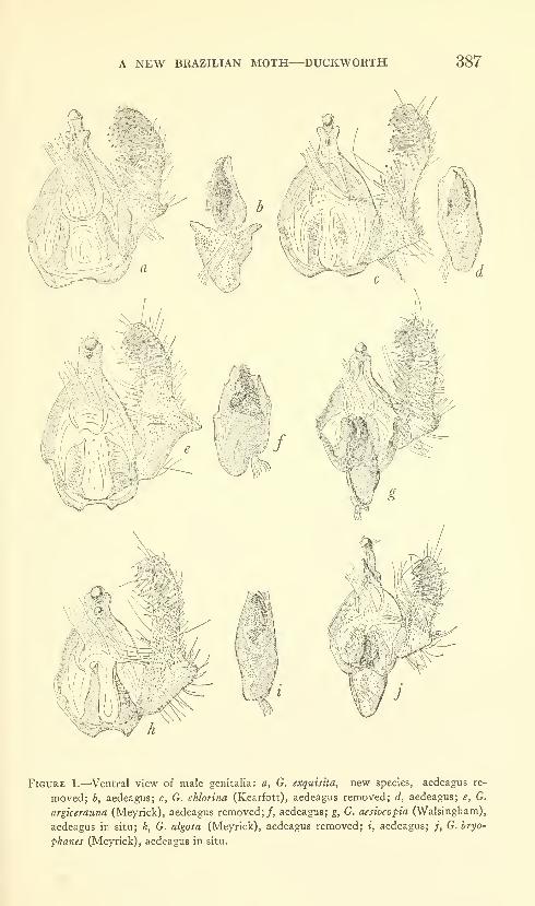
A NEW BRAZILIAN MOTH—DUCKWORTH 387
Figure 1.—^Ventral view of male genitalia: a, G. exquiska, new species, aedeagus re-
moved; h, aedeagus; c, G. chlorina (Kearfott), aedeagus removed; d, aedeagus; e, G.
argicerauna (Meyrick), aedeagus removed;/, aedeagus; g, G. aesiocopia (Walsingham),
aedeagus in situ; h, G. algosa (Meyrick), aedeagus removed; i, aedeagus; /, G. bryo-
phanes (Meyrick), aedeagus in situ.
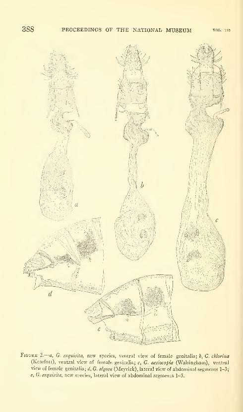
388 PROCEEDINGS OF THE NATIONAL MUSEUM
Figure 2.
—
a, G. exquisita, new species, ventral view of female genitalia; b, G. chlorina
(Kearfott), ventral view of female genitalia; c, G. aesiocopia (Walslngham), ventral
view of female genitalia; d, G. algosa (Meyrick), lateral view of abdominal segments 1-3;
e, G. exquisita, new species, lateral view of abdominal segments 1-3.
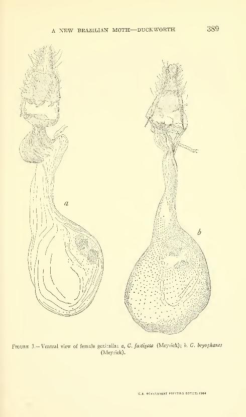
A NEW BRAZILIAN MOTH—DUCKWORTH 389
Figure 3.—Ventral view of female genitalia: a, G. fastigata (Meyrick); b. G. bryophanes
(Meyrick).
U.S. GOVERNMENT PRINTING 0FF1CE:1964


Proceedings of
the United States
National MuseumSMITHSONIAN INSTITUTION . WASHINGTON, B.C.
Volume 115 1964 Number 3489
SEVEN NEW AMPHIPODSFROM THE WEST COAST OF NORTH AMERICAWITH NOTES ON SOME UNUSUAL SPECIES
By Clarence R. Shoemaker^
While studying the unidentified amphipods from the west coast of
North America in the collections of the Smithsonian Institution, I
have noted undescribed species from time to time. Seven of these
species are here described and illustrated; also redescriptions andsupplementary figures are given of seven other species.
Family Lysianassidae
Kyska, new genus
Antennae 1 and 2 rather short, stout. Eyes large, pyriform.
Mandible, incisor smooth with small tooth at inner end ; molar conical,
without triturating surface; palp S-jointed. Maxilla 1, inner plate
small, with 2 terminal setae; outer plate with 9 terminal spine-teeth,
1 Died December 28, 1958. This paper, nearly completed, was prepared for
publication by Thomas E. Bowman, U.S. National Museum. J. Laurens
Barnard of the Beaudette Foundation read the manuscript critically and addedseveral recent references.
391
392 PROCEEDINGS OF THE NATIONAL MUSEUM vol. 115
2 lateral teeth; palp 2-jointed. Maxilla 2, inner plate shorter than
outer. Maxilliped, inner plate with 3 terminal teeth; outer plate
without marginal teeth; palp 4-jointed. Gnathopod 1 chelate.
Gnathopod 2 slender, normal. Pereopods 3-5 increasing consecu-
tively in length; basipod weU expanded. Pleon segment 3 with
lower hind corner sharply upturned. Branchiae plaited on both
sides. Telson cleft beyond middle.
Type-species.—Kyska dalli, new species.
This new genus is very much like Anonyx, and the type-species bears
a close superficial resemblance to Anonyx nugax. The characters that
differentiate Kyska from Anonyx are the comparative shortness of
the antennae, the lack of calceoli in the male, and the possession of
chelate gnathopod 1 in both sexes ; otherwise, the characters of Kyskaare the same as those given by Stebbing (1906) for Anonyx.
Kyska dalli, new species
Figure 1
Male.—Eye black, pyriform. Antenna 1 about as long as head andpereon segment 1 combined, about % as long as antenna 2 ; first joint
large, second and third joints very short; flagellum about as long as
peduncle, composed of 17 joints; accessory flagellum }^ as long as
primary and composed of 9-10 joints. Antenna 2, fourth joint
slightly longer than fifth, flagellum longer than peduncle and composedof about 28-30 joints. Epistome not projecting beyond upper lip.
Other mouthparts as shown in figures.
Coxal plate 1 expanded below, projecting slightly forward. Gna-thopod 1, second joint about as long as remaining joints combined;
fifth joint short, cup-shaped; sixth joint greatly developed, lower
distal end produced forward, forming chela with short, curved, oppos-
ing seventh joint (figs. la,h). Gnathopod 2 normal.
Pereopods 1 and 2 similar, subequal in length. Pereopods 3-5
rather long, slender, increasing in length consecutively; second joints
considerably expanded; fourth joint well expanded in pereopod 3,
less expanded in pereopods 4 and 5. Seventh joint in all pereopods
long, slightly curved.
Pleon segment 1, lower hind corner not produced; segment 2 rather
sharply produced; segment 3 sharply upturned. Urosome segment 1
with slight dorsal depression. Uropod 1 reaching back farther than
uropod 2, about as far as uropod 3. Uropod 3, outer ramus longer than
inner and with small second joint. All uropods bearing only few short
spinules. Telson cleft nearly to base, reaching back slightly beyondpeduncle of uropod 3.
Gills plaited on both sides. Gill of pereopods 1 and 2 v/ith ap-
pendage, that of pereopods 3 and 4 with pointed finger-like appendage.
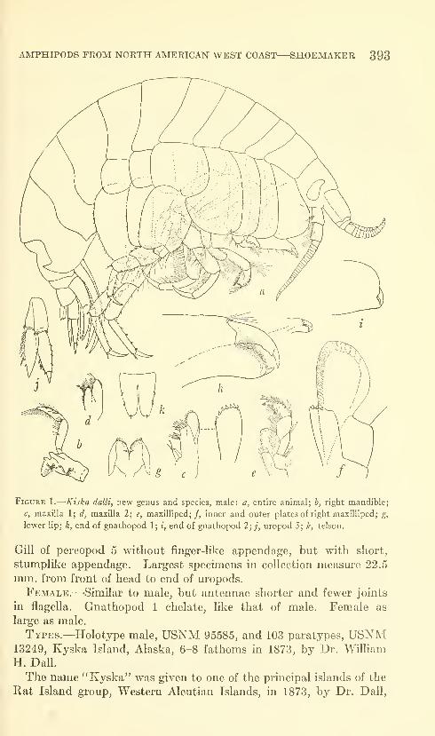
AMPHIPODS FROM NORTH AMERICAN WEST COAST—SHOEMAKER 393
Figure 1.
—
Kiska dalli, new genus and species, male: a, entire animal; b, right mandible;
c, maxilla 1; d, maxilla 2; e, maxilliped; /, inner and outer plates of right maxilliped; g,
lower lip; h, end of gnathopod 1; i, end of gnathopod 2; ;', uropod 3; k, telson.
Gill of pereopod 5 without jBnger-like appendage, but with short,
stumplike appendage. Largest specimens in collection measure 22.5
mm. from front of head to end of uropods.
Female.—-Similar to male, but antennae shorter and fewer joints
in flagella. Gnathopod 1 chelate, like that of male. Female as
large as male.
Types.—Holotype male, USNM 95585, and 103 paratypes, USNM13249, Kyska Island, Alaska, 6-8 fathoms in 1873, by Dr. William
H. DaU.
The name "Kyska" was given to one of the principal islands of the
Rat Island group, Western Aleutian Islands, in 1873, by Dr. Dall,
394 PROCEEDINGS OF THE NATIONAL MUSEUM vol. im
who collected a number of specimens of this amphipod in the harbor
at that time.
In addition to the specimens taken by Dr. Dall, a single specimen of
this species was taken by W. G. Hall in 1872 at Nagai Island, another
of the larger islands of the Rat Island group.
Family Stenothoidae
Metopa stelleri, new species
Figure 2
Male.—Head not quite as long as first 2 body segments combined;
lateral lobe broadly rounded. Eye medium size, round, very light
straw-colored in alcohol. Antenna 1 nearly as long as antenna 2,
which is nearly as long as body. Antenna 1, first joint longer than
second, which is about 5 times as long as third; flagellum longer than
peduncle and composed of about 25 joints. Antenna 2, third joint
about Yz as long as fourth, which is about as long as fifth; flagellum
little more than ){ length of fifth peduncular joint, composed of about
11 joints, first of which almost as long as remaning joints combined.
Mandible normal, spine row of 15-16 spines; palp with very small third
joint. Maxilla 1 normal, palp 1-jointed. Maxilla 2 normal; inner
lobe much shorter than outer. Maxilliped with characters of genus.
Gnathopod 1 slender, second joint slightly expanded, without lobes,
nearly as long as fifth and sixth joints combined; fifth joint about as
long and as wide as sixth; sixth joint slightly expanded distally, palm
oblique, straight, bearing very fine short spinules throughout, no
defining spines, but row of long slender spines on rounding margin,
where palm merges into hind margin of joint (fig. 2d); seventh joint
fitting palm, armed on inner margin with very fine teeth and short
spinules. Gnathopod 2 large, strong; second joint not as long as
sixth, with rounding lobe on lower front margin; fifth joint less than
){ as long as sLxth, with lobe between fourth and sixth joints; sixth
joint large, strong, front and hind margins convex, palm very oblique,
convex, armed with irregular low teeth and defined by tooth bearing
slender spine on its inner margin; seventh joint stout, sHghtly shorter
than palm and bearing minute setules on inner margin.
Pereopod 1 long, slender. Pereopod 2 not as long as pereopod 1,
but stouter. Pereopod 3 longer than pereopods 4 or 5, which are
subequal in length; second joint slender, without lower lobe. Pereo-
pods 4 and 5 strong; second joint broadly expanded; fourth joint
large, broadly expanded with hind margin produced shghtly beyond
end of fifth joint. Seventh joints of pereopods 1-5 rather short,
stout, wiih inner margins bearing very low forward-pointing teeth

AMPHIPODS FROM NORTH AMERICAN WEST COAST—SHOEMAKER 395
(fig. 2/). Uropod 1 reaching back slightly farther than uropod 2,
2 slightly farther than 3. Uropods 1 and 2 scantily armed with short
spines. Uropod 3, peduncle longer than ramus, armed on upper
margin with 8 or 9 short spines, Telson broadly oval, reaching
slightly beyond end of third urosome segment, bearing 5 short spines
on either lateral margin. Pereon segments 6 and 7 and pleon seg-
ments 1 and 2 each produced dorsally into backward-pointing tooth.
Coxal plates 2-4 much deeper than their body segments. Coxal
plate 4 only of moderate size for genus Metopa. Length to 9 mm.Female.—Very much like male. Antennae shorter than in male.
Fifth and sixth joints of gnathopod 1 shorter. Sixth joint of gnatho-
pod 2 not so large; palm less oblique, with fine teeth and shorter
defining tooth. Female as large as male.
Types.—Holotype male, USNM 96489, and 11 paratypes, USNM107860, taken at Albatross station 4803, Cape RoUin, Simushir Island,
Kuriles (46°42' N, 15r45' E) in 229 fathoms, June 24, 1906. Single
Figure 2.—Metopa stelleri, new species, male: a, entire animal; b, mandibular palp; c,
gnathopod 1; d, end of gnathopod 1; e, gnathopod 2;/, end of pereopod 2; female: g,
gnathopod 1; h, gnathopod 2.
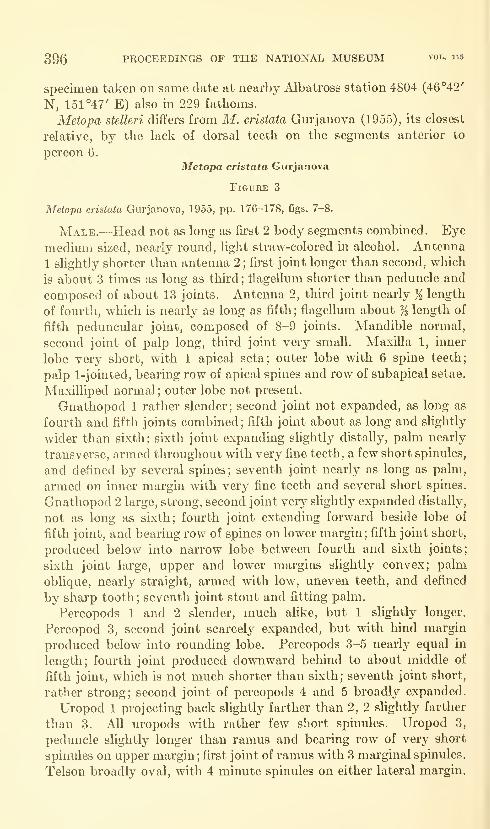
396 PROCEEDINGS OF THE NATIONAL MUSEUM vol. 115
specimen taken on same date at nearby Albatross station 4804 (46*'42'
N, 151 "47' E) also in 229 fathoms.
Metopa stelleri differs from M. cristata Gurjanova (1955), its closest
relative, by the lack of dorsal teeth on the segments anterior to
pereon 6.
Metopa cristata Gurjanova
Figure 3
Metopa cristata Gurjanova, 1955, pp. 176-178, figs. 7-8.
Male.—Head not as long as first 2 body segments combined. Eye
medium sized, nearly round, light straw-colored m alcohol. Antenna
1 slightly shorter than antenna 2 ; first joint longer than second, which
is about 3 times as long as third ; fiagellum shorter than peduncle and
composed of about 13 joints. Antenna 2, third joint nearly ji length
of fourth, which is nearly as long as fifth ; flagellum about % length of
fifth peduncular joint, composed of 8-9 joints. Mandible normal,
second joint of palp long, third joint very small. Maxilla 1, inner
lobe very short, with 1 apical seta; outer lobe with 6 spine teeth;
palp 1-jointed, bearing row of apical spines and row of subapical setae.
Maxilliped normal; outer lobe not present.
Gnathopod 1 rather slender; second joint not expanded, as long as
fourth and fifth joints combined; fifth joint about as long and slightly
wider than sixth: sixth joint expanding slightly distally, palm nearly
transverse, armed throughout with very fine teeth, a few short spinules,
and defined by several spines; seventh joint nearly as long as palm,
armed on inner margin with very fine teeth and several short spines.
Gnathopod 2 large, strong, second joint very slightly expanded distally,
not as long as sixth; fourth joint extending forward beside lobe of
fifth joint, and bearing row of spines on lower margin; fifth joint short,
produced below into narrow lobe between fourth and sixth joints;
sixth joint large, upper and lower margins slightly convex; palm
oblique, nearly straight, armed with low, uneven teeth, and defined
by sharp tooth; seventh joint stout and fitting palm.
Pereopods 1 and 2 slender, much alike, but 1 slightly longer.
Pereopod 3, second joint scarcely expanded, but with hind margin
produced below into rounding lobe. Pereopods 3-5 nearly equal in
length; fourth joint produced downward behind to about middle of
fifth joint, which is not much shorter than sixth; seventh joint short,
rather strong; second joint of pereopods 4 and 5 broadly expanded.
Uropod 1 projecting back slightly farther than 2, 2 slightly farther
than 3. All uropods with rather few short spinules. Uropod 3,
peduncle slightly longer than ramus and bearing row of very short
spinules on upper margin ; first joint of ramus with 3 marginal spinules.
Telson broadly oval, with 4 minute spinules on either lateral margin.
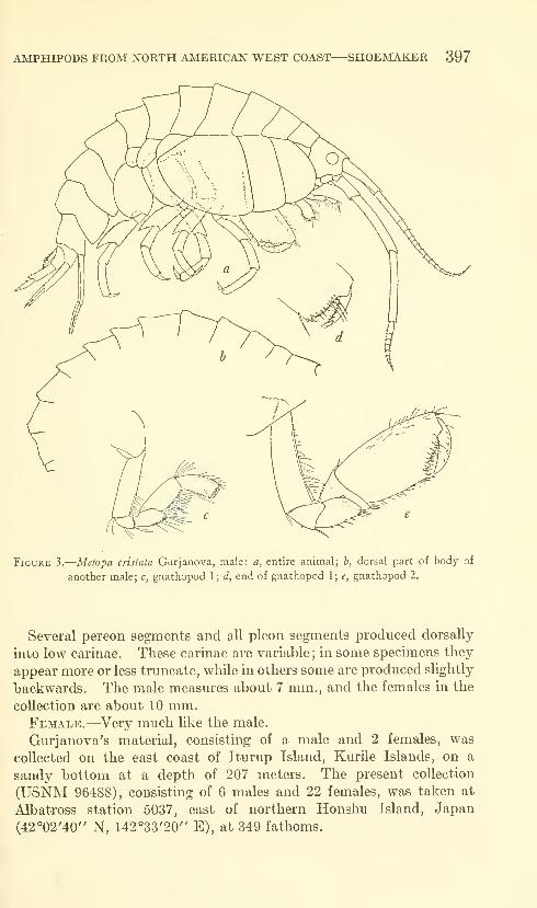
AMPHIPODS FROM NORTH AMERICAN WEST COAST—SHOEMAKER 397
Figure 3.
—
Metopa crisiata Gurjanova, male: a, entire animal; b, dorsal part of body of
another male; c, gnathopod 1; d, end of gnathopod 1; e, gnathopod 2.
Several pereon segments and all pleon segments produced dorsally
into low carinae. These carinae are variable; in some specimens they
appear more or less truncate, while in others some are produced slightly
backwards. The male measures about 7 mm., and the females in the
collection are about 10 mm.Female.—Very much like the male.
Gurjanova's material, consisting of a male and 2 females, was
collected on the east coast of Iturup Island, Kurile Islands, on a
sandy bottom at a depth of 207 meters. The present collection
(USNM 96488), consisting of 6 males and 22 females, was taken at
Albatross station 5037, east of northern Honshu Island, Japan
(42°02'40" N, 142°33'20" E), at 349 fathoms.

398 PROCEEDINGS OF THE NATIONAL MUSEUM vol. 115
Mesometopa sinuata, new species
Figure 4
Male.—Head not as long as first 2 body segments combined;
rostrum short, triangular from above; lateral lobes triangular; eye
small, oval; epistome very prominent, deeply cleft (fig. 46). Antenna
1 about }i length of body, peduncle rather stout, first joint longer than
second and third combined, flagellum shorter than peduncle and
composed of 10 or 11 joints, each of which bears sensory filaments on
lower distal corner. Antenna 2 much longer than antenna 1, third
joint ji length of fourth, which is as long as fifth; flagellum slightly
shorter than fifth peduncular joint and composed of 1 long and 4
shorter joints.
Mandible, cutting-edge, and accessory plate toothed, 7-8 spines in
spine row, palp small, consisting of basal joint bearing lateral spine, and
smaller conical second joint bearing apical spine (fig. id). Maxilla 1,
inner plate without setae, outer plate with 5 large, 1 small spine, palp
1-jointed, bearing 7 terminal spines. Maxilliped, inner plate not
reaching to base of first joint of palp; outer plate consisting of only
short triangular extension of inner margin of third peduncular joint;
palp long, slender, fourth joint with very fine teeth on inner margin.
Gnathopod 1 rather short, slender; second joint very slightly ex-
panded; fourth joint reaching to about middle of fifth; fifth joint as
long as, but wider than, sixth; sixth joint with front and hind margins
slightly convex, palm very oblique, convex, armed throughout with
fine sharp teeth, few slender spinules, and defined by group of spines;
seventh joint fitting palm, bearing on inner margin row of fine spinules,
and small forward-pointing tooth near apex (fig. Ag). Gnathopod 2
with outside front margin of second joint expanded into thin trans-
parent lobe on outer and inner front margin; fourth joint with tri-
angular lobe on inside surface projecting above upper margin of fifth
joint, lower margin of joint produced forward on outside of lobe of
fifth joint; fifth joint normal; sixth joint large, longer than wide, palm
very oblique with deep oblong sinus between defining tooth and distal
portion of palm, which is provided with several blunt teeth and few
setules (fig. 4:h) ; seventh joint stout, curved, shorter than palm and
bearing closely set setae on inner margin and a few setae on outer
margin.
Pereopods 1 and 2 about equal in length, but 2 stouter; fourth
joint with front margin produced slightly downward. Pereopod 1
almost entirely devoid of spines. Pereopods 3-5 consecutively shorter.
Pereopod 4, second joint linear; fourth joint expanded with lower
hind corner reaching to about middle of fifth joint; fifth joint shorter
than sixth; seventh joint over % length of sixth. Pereopod 5, second
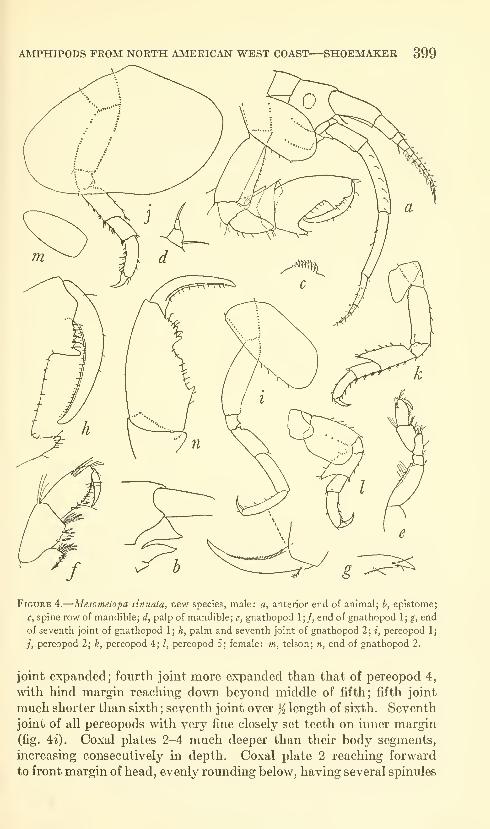
AMPHIPODS FROM NORTH AMERICAN WEST COAST—SHOEMAKER 399
Figure 4.
—
Mesometopa sinuata, new species, male: a, anterior end of animal; b, epistome;
c, spine row of mandible; d, palp of mandible; e, gnathopod 1;/, end of gnathopod 1; g, end
of seventh joint of gnathopod 1; h, palm and seventh joint of gnathopod 2; i, pereopod 1;
/, pereopod 2; k, pereopod 4; /, pereopod 5; female: m, telson; n, end of gnathopod 2.
joint expanded; fourth joint more expanded than that of pereopod 4,
with hind margin reaching down beyond middle of fifth; fifth joint
much shorter than sixth ; seventh joint over ji length of sixth. Seventh
joint of all pereopods with very fine closely set teeth on inner margin
(fig. 4^). Coxal plates 2-4 much deeper than their body segments,
increasing consecutively in depth. Coxal plate 2 reaching forward
to front margin of head, evenly rounding below, having several spinules
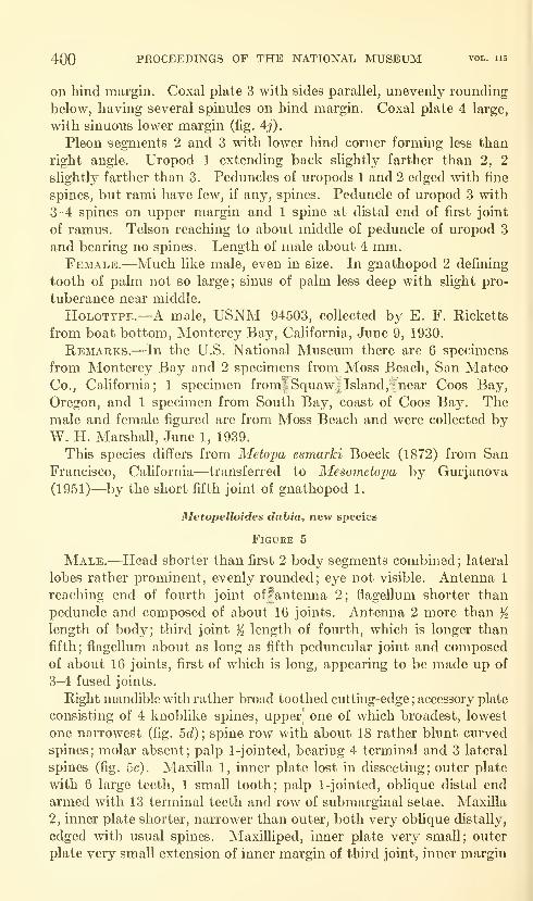
400 PROCEEDINGS OF THE NATIONAL MUSEUM vol. ii5
on hind margin. Coxal plate 3 with sides parallel, unevenly rounding
below, having several spinules on hind margin. Coxal plate 4 large,
with sinuous lower margin (fig. 4j).
Pleon segments 2 and 3 with lower hind corner forming less than
right angle. Uropod 1 extending back slightly farther than 2, 2
slightly farther than 3. Peduncles of uropods 1 and 2 edged with fine
spines, but rami have few, if any, spines. Peduncle of uropod 3 with
3-4 spines on upper margin and 1 spine at distal end of first joint
of ramus. Telson reaching to about middle of peduncle of uropod 3
and bearing no spines. Length of male about 4 mm.Female.—Much like male, even in size. In gnathopod 2 defining
tooth of palm not so large; sinus of palm less deep with slight pro-
tuberance near middle.
HoLOTYPE.—A male, USNM 94503, collected by E. F. Ricketts
from boat bottom, Monterey Bay, California, June 9, 1930.
Remarks.—In the U.S. National Museum there are 6 specimens
from Monterey Bay and 2 specimens from Moss Beach, San MateoCo., California; 1 specimen from|Squaw|Island,fnear Coos Bay,
Oregon, and 1 specimen from South Bay, coast of Coos Bay. Themale and female figured are from Moss Beach and were collected byW. H. Marshall, June 1, 1939.
This species differs from Metopa esmarki Boeck (1872) from San
Francisco, California—transferred to Alesometopa by Gurjanova
(1951)—by the short fifth joint of gnathopod 1.
Metopelloides diibia, new species
Figure 5
Male.—Head shorter than first 2 body segments combined; lateral
lobes rather prominent, evenly rounded; eye not visible. Antenna 1
reaching end of fourth joint offantenna 2; flagellum shorter than
peduncle and composed of about 16 joints. Antenna 2 more than Klength of body; third joint ji length of fourth, which is longer than
fifth; flagellum about as long as fifth peduncular joint and composedof about 16 joints, first of which is long, appearing to be made up of
3-4 fused joints.
Right mandible with rather broad toothed cuttmg-edge ; accessory plate
consisting of 4 knoblike spines, upper- one of wliich broadest, lowest
one narrowest (fig. 5d) ; spine row with about 18 rather blunt curved
spines; molar absent; palp 1-jointed, bearing 4 terminal and 3 lateral
spines (fig. 5c), Maxilla 1, inner plate lost in dissecting; outer plate
with 6 large teeth, 1 small tooth; palp 1-jointed, oblique distal end
armed with 13 terminal teeth and row of submarginal setae. Maxilla
2, inner plate shorter, narrower than outer, both very oblique distally,
edged with usual spines. Maxilliped, inner plate very small; outer
plate very small extension of inner margin of third joint, inner margin
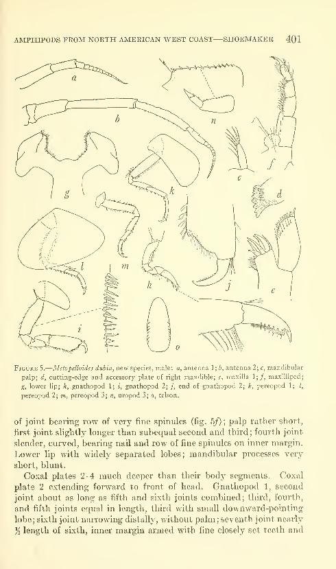
AMPHIPODS FROM NORTH AMERICAN WEST COAST—SHOEMAKER 401
Figure S.—Metopelloides dubia, new species, male: a, antenna 1; b, antenna 2; c, mandibular
palp; d, cutting-edge and accessory plate of right mandible; e, maxilla 1;/, maxilliped;
g, lower lip; h, gnathopod 1; i, gnathopod 2; ;, end of gnathopod 2; k, pereopod 1; /,
pereopod 2; m, pereopod 3; n, uropod 3; o, telson.
of joint bearing row of very fine spinules (fig. 5/) ;palp rather short,
first joint slightly longer than subequal second and third; fourth joint
slender, curved, bearing nail and row of fine spinules on inner margin.
Lower lip with widely separated lobes; mandibular processes very
short, blunt.
Coxal plates 2-4 much deeper than their body segments. Coxal
plate 2 extending forward to front of head. Gnathopod 1, second
joint about as long as fifth and sixth joints combined; third, fourth,
and fifth joints equal in length, third with small downward-pointing
lobe; sixth joint narrowing distally, without palm; seventh joint nearly
ji length of sixth, inner margin armed with fine closely set teeth and
402 PROCEEDINGS OF THE NATIONAL MUSEUM vol. 115
about 6 setae, and with rather deep angular sinus near apex (fig. 5h).
Gnathopod 2, second joint not as long as fifth and sixth combined;
third, fourth, and fifth joints about equal in length, third bearing
small downward-projecting angular lobe; sixth joint twice length of
fifth, about twice as long as wide, expanding slightly distally; palmoblique, convex, finely crenulate throughout, defined by rather blunt
tooth and 2 spines (fig. 5j); seventh joint stout, curved, fitting palm.
Pereopods 1 and 2 slender, pereopod 1 slightly longer (fig. 5k). Pere-
opods 3-5 alike, 4 slightly the longest; second joint linear. Uropod1 reaching posteriorly farther than 2, 2 farther than 3. Peduncles
of all uropods with many fine, closely set spines. Uropod 1, outer
ramus with 5 marginal spines; inner ramus with 3 marginal spines.
Uropod 2, outer ramus with 4 marginal spines; inner ramus with 3.
Uropod 3, peduncle armed with row of short spines on upper outer
margin; first joint of ramus with 1 median, 1 distal spine. No ter-
minal spines on any rami. Telson long, narrow, reaching back about
Ys length of peduncle of uropod 3, and bearing row of 7-8 spinules on
either lateral margin. Length, from front of head to end of uropods,
about 13 mm.HoLOTYPE.—Male, USNM 95660, taken by U.S. Bureau of Fish-
eries at St. Paul Island, Pribilof Islands, Jan. 13, 1918, from stomach
of duck, Somateria voniger.
Remarks.—Because the specimen is from the stomach of a duck,
neither gills nor marsupial plates are present, but it is inferred from
the great development of the second antenna that the specimen is a
male. There is a close resemblance in some of the characters to
Metopelloides shoemaJceri Gurjanova (1938). Her specimen, ap-
parently a female, measured only 4 mm., but the present specimen,
presumably a male, measures 13 mm. Whether the difference
between these two species is due to the immaturity of Gurjanova's
specimen or to the difference in sex is impossible to determine. Fur-
ther and more abundant material probably will decide the question,
but for the present it seems best to regard them as distinct species,
Proboloides pacifica (Holmes)
Figure 6
Metopa pacifica Holmes, 1908, p. 524, figs. 30-31.
Meiopella pacifica.—Gurjanova, 1951, p. 478, fig. 315.
Male.—Head nearly as long as first 2 body segments combined;
lateral lobes bluntly triangular with apex rounding. Eye not present
in alcoholic type. Antenna 1 almost as long as antenna 2, peduncle
reaching slightly beyond fourth joint of antenna 2; first joint shorter
than second; third joint about ji as long as second; flagellum shorter
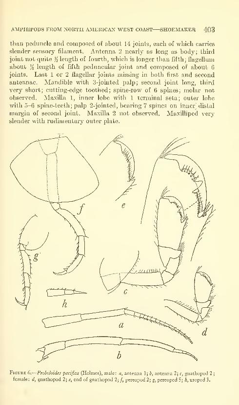
AMPHIPODS FROM NORTH AMERICAN WEST COAST—SHOEMAKER 403
than peduncle and composed of about 14 joints, each of which carries
slender sensory filament. Antenna 2 nearly as long as body; third
joint not quite ji length of fourth, which is longer than fifth; flagellum
about Yi length of fifth peduncular joint and composed of about 6
joints. Last 1 or 2 flagellar joints missing in both first and second
antennae. Mandible with 3-jointed palp; second joint long, third
very short; cutting-edge toothed; spine-row of 6 spines; molar not
observed. Maxilla 1, inner lobe with 1 terminal seta; outer lobe
mth 5-6 spine-teeth; palp 2-jointed, bearing 7 spines on inner^ distal
margin of second joint. Maxilla 2 not observed. Maxilliped very
slender with rudimentary outer plate.
Figure 6.
—
Proholoides pacifica (Holmes), male: a, antenna 1; b, antenna 2; c, gnathopod 2
female: d, gnathopod 2; e, end of gnathopod 2; /, pereopod 2; g, pereopod 5; h, uropod 3.
404 PROCEEDINGS OF THE NATIONAL MUSEUM vol. 115
Gnatliopocl 1 with fifth joint longer than sixth, which is distinctly
subchelate. Sixth joint with palm obhque, convex, defined by spines,
and about as long as hind margin of joint. Seventh joint fitting
palm and armed on inner margin with minute spinules. Gnathopod
2 large (fig. 6c); palm very oblique, defined by stout sharp tooth,
with large tooth in middle, on either side of which are small teeth;
seventh joint strong, fitting palm. Pereopods 1 and 2 slender, 1
slightly slenderer than 2; fourth joint not produced downward in
front; seventh joint rather long, slender, shghtly curved. Pereopods
4 and 5 with second joint expanded; fourth joint very slightly ex-
panded, only slightly produced downward; seventh joint about %
length of sixth. Pleon segment 3 about as shown by Sars (1895,
pi. 94, fig. 2) for Metopa invalida. Uropod 1 reaches back slightly
farther than uropod 2, 2 slightly farther back than 3. Uropod 3,
peduncle slightly shorter than ramus, first joint of which is shorter
than second (fig. 6/0- Telson narrowly oval with narrow rounding
apex, and bearing 3 short spines on either lateral margin. Length
6 mm.Female.—Gnathopod 2 not as large as in male; palm oblique,
convex, defined by tooth and 2 spines (fig. 5e), and armed throughout
with small rounding teeth, one of which near middle is shghtly larger
than rest; seventh joint fitting palm.
Remarks.—The above description is based on the 2 syntypes,
a male and a female, USNM 38550, from Albatross station 4516,
Monterey Bay, California, May 24, 1904, 718-756 fathoms, among
hydroids on the back of a spider crab, Hyas species.
Holmes' species is here placed in the genus Proboloides Delia Valle, as
it differs from Metopa by having a 2-jointed palp to maxilla 1 and not a
1 -jointed palp as in Metopa. It was assigned to Metopella Sars by
Gurjanova (1951), perhaps on the basis of Holmes' statement (1908):
"Last three peraeopods with the merus not widely expanded"—but,
as shown herein, the merus is widely expanded on pereopods 4 and 5.
Family Tironidae
Syrrhoe longifrons, new species
FiGUBE 7
This species differs from S. crenulata Goes (1866) as follows: Head
elongate, about as long as first 5 body segments combined, produced
slightly forward, evenly rounding. Rostrum pointing downward and
backward toward body, not straight downward as in S. crenulata.
Eyes reddish brown in alcohol, elongate, about )i as long as head,
united above and occupying almost entire front of head. Lateral
angle of head truncate, not angular. Lower front corner of fkst 3
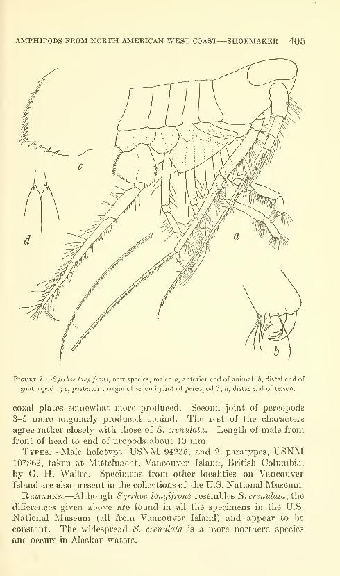
AMPHIPODS FROM NORTH AMERICAN WEST COAST
—
SHOEMAKER 405
a
Figure 7.
—
Syrrhoe longifrons, new species, male: a, anterior end of animal; b, distal end of
gnathopod 1; c, posterior margin of second joint of pereopod 3; d, distal end of telson.
coxal plates somewhat more produced. Second joint of pereopods
3-5 more angularly produced behind. The rest of the characters
agree rather closely with those of S. crenulata. Length of male from
front of head to end of uropods about 10 mm.Types.—Male holotype, USNM 94235, and 2 paratypes, USNM
107862, taken at Mittelnacht, Vancouver Island, British Columbia,
by G. H. Wailes. Specimens from other localities on Vancouver
Island are also present m the collections of the U.S. National Museum.Remarks.—Although Syrrhoe longifrons resembles S. crenulata, the
differences given above are found in all the specimens in the U.S.
National Museum (all from Vancouver Island) and appear to be
constant. The widespread S. crenulata is a more northern species
and occurs in Alaskan waters.

406 PROCEEDINGS OF THE NATIONAL MUSEUM vol. ii5
Family Calliopiidae
Halirages bungei Gurjanova
Figure 8
Halirages bungei Gurjanova, 1951, pp. 611-612, fig. 414.
Female.—Head as long as first 2 body segments combined; rostrmn
very short, broad; lateral lobe obliquely trimcate; lower corner broadly
romiding; eye long, consisting of narrow black central area bordered
by colorless ocelli. Antennae subequal, but 1 perhaps slightly shorter.
Antenna 1 about as long as head and first 5 body segments combined
;
peduncle short, joints decreasmg consecutively in length; accessory
flagellum rudimentary (fig. 86) ;primary flagellum consisting of about
44 joints, every other one of which bears 2 sensory filaments on lower
distal end. Antenna 2 with fifth joint longer than fourth; flagellum
consisting of about 38 joints.
Mandible with cutting-edge narrow, toothed; accessory plate narrow
with bifid apex; spine-row of 9 spines ; molar strong with well-developed
triturating surface and armed on upper edge with long slender teeth;
palp strong with second joint longest. Upper lip symmetrically
rounding. Maxilla 1, inner plate long, armed apically with 5 plumose
setae; outer plate armed with 11 serrate spine-teeth; palp 2-jointed,
rather broad, bluntly rounding apex armed with 14 serrate spine-
teeth and on outside with 6 subapical setae. Maxilla 2, inner plate
slightly longer, armed apically with spines, and on inner margin with
spines, 4 of which are longer than rest ; outer plate armed with apical
spines. Lower lip without inner lobes; mandibular processes rather
short, apically rounding. MaxUliped, second joint with several spines
on outer margin ; inner lobe rather long, reaching slightly beyond
middle of outer lobe, armed distally with curved plumose spines or
setae and 3 short spine-teeth, 2 innermost being separated from third
by slight sinus; outer plate reaching middle of second joint of palp,
bearing distally a closely set row of slender curved plumose spines, andbearing on inner margin transparent lamella, from base of which arises
row of slightly curved serrate spine-teeth; palp rather short, stout,
third joint bearing group of spines near middle of outer margin andspines and small rounding lobe distally; fourth joint rather slender,
slightly curved, bearing distally sharp nail and few setules.
Gnathopods 1 and 2 alike in form and size, though 1 may possibly
be slightly longer; second joint scarcely expanded distally; fifth joint
not expanded, about % as long as sixth; sixth joint long, narrow, of
equal width throughout, bearing groups of spines on hind margin;
palm convex, very oblique, edged with narrow smooth transparent
lamella and merging into hind margm \\'ithout defining angle, but
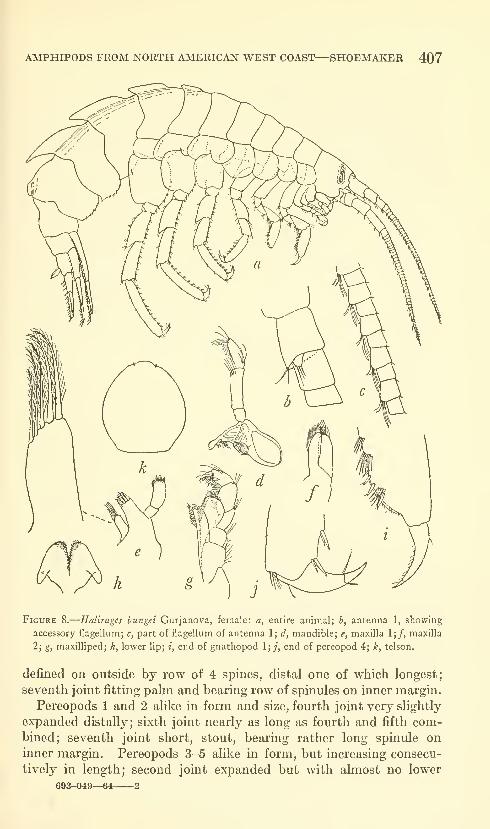
AMPHIPODS FROM NORTH AMERICAN WEST COAST—SHOEMAKER 407
Figure 8.
—
Halirages bungei Gurjanova, female: a, entire animal; b, antenna 1, showing
accessory flagellum; c, part of flagellum of antenna 1; d, mandible; e, maxilla 1;/, maxilla
2; g, maxilliped; h, lower lip; i, end of gnathopod 1; /, end of pereopod 4; k, telson,
defined on outside by row of 4 spines, distal one of which longest;
seventh joint fitting palm and bearing row of spinules on inner margin.
Pereopods 1 and 2 alike in form and size, fourth joint very slightly
expanded distally; sixth joint nearly as long as fourth and fifth com-bined; seventh joint short, stout, bearing rather long spinule on
inner margin. Pereopods 3-5 alike in form, but increasing consecu-
tively in length; second joint expanded but with almost no lower
693-049—64 2
408 PROCEEDINGS OF THE NATIONAL MUSEUM vol. 115
posterior lobe. Coxal plates 1-4 shallow, increasing consecutively
in size, fourth excavate behind.
Pereon segment 6 with small backward-pointing dorsal carina.
Pereon segment 7 with larger dorsal carina. Pleon segments 1 and2 each with strong backward-pointing dorsal carina; segment 3 with
small dorsal carina which is scarcely backward pointing. Pleon
segments 1 and 2 with lower posterior corner sharply produced,
lower margin bearing few spinules; segment 3 with lower posterior
corner blunt-angled or without angle and lower margin bearing rowof spinules.
Uropods 1 and 2 reaching back about same distance. Uropod 1,
peduncle longer than rami, outer ramus shorter; both peduncle andrami margined with many fine short spines. Uropod 2, peduncle
slightly longer than outer ramus, but shorter than inner; both peduncle
and rami margined with fine short spines. Uropod 3 not reaching
back quite as far as 2 ;peduncle slightly shorter than rami and bearing
4 spines on upper margin which are longer than those on rami. Telson
reaching to about middle of peduncle of uropod 3, broadly oval,
slightly longer than wide, bearing 2 minute apical spinules. Femalemeasures about 13 mm. from front of head to end of uropods.
Remarks.—Gurjanova's specmiens, 22 in number, were taken onthe east coast of Kamchatka, among algae and on fine sand, up to a
depth of 60 m. The U.S. National Museum collections contain 12
specimens from Bering Island, Commander Islands, 9 of them col-
lected by Leonard Stejneger in 1882-83, and 3 specimens collected
by N. Grebnitzky on August 6, 1888.
Family Pleustidae
Sympleustes cornigera, new species
Figure 9
Male.—Head with short rostrum, appearing triangular from
above; lateral lobe triangularly produced, with narrow rounding apex;
lower corner narrowly, sharply produced; eye large, broadly reniform,
rather colorless in alcohol. Antenna 1 much longer than antenna 2;
first joint longer than second, which is about twice as long as third;
flagellum consisting of about 90 joints, first of which is longest.
Mandible with rather broad cutting-edge, but without accessory
plate; spine-row of about 14 spines (fig. 7c); molar conical with rather
small triturating surface; palp about as shown for Sympleustes latipes
by Sars (1895, pi. 127, fig. m) {=Stenopleustes, according to Barnardand Given, 1960). Maxilla 1, inner plate broad, without plumose
setae, but fringed with fine setules; outer plate armed with 9 spine-
teeth; palp armed with distal row of 7 spines and submarginal row
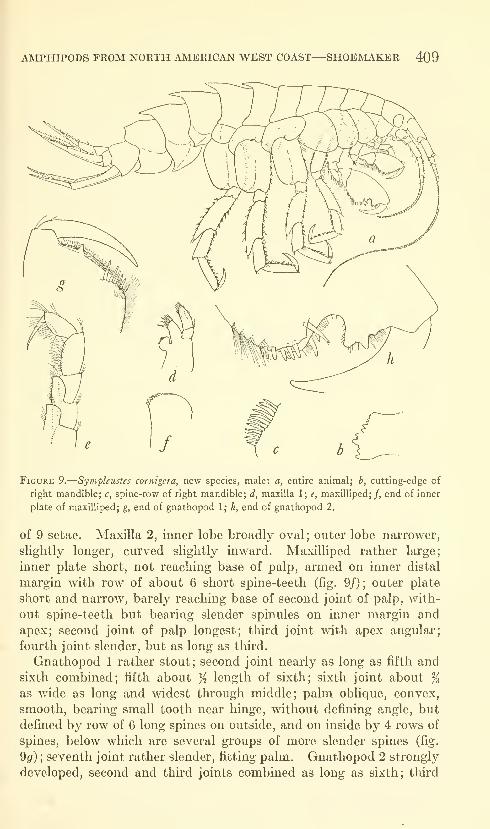
AMPHIPODS FROM NORTH AMERICAN WEST COAST
—
SHOEMAKER 409
Figure 9.
—
Sympleustes cornigera, new species, male: a, entire animal; h, cutting-edge of
right mandible; c, spine-row of right mandible; d, maxilla 1; e, maxilliped;/, end of inner
plate of maxilliped; g, end of gnathopod 1; h, end of gnathopod 2.
of 9 setae. Maxilla 2, inner lobe broadly oval; outer lobe narrower,
slightly longer, curved slightly inward. Maxilliped rather large;
inner plate short, not reaching base of palp, armed on inner distal
margin with row of about 6 short spine-teeth (fig. 9/); outer plate
short and narrow, barely reaching base of second joint of palp, with-
out spine-teeth but bearing slender spinules on inner margin and
apex; second joint of palp longest; third joint with apex angular;
fourth joint slender, but as long as third.
Gnathopod 1 rather stout; second joint nearly as long as fifth and
sixth combined; fifth about K length of sixth; sixth joint about %as wide as long and widest ttu-ough middle; palm oblique, convex,
smooth, bearing small tooth near hinge, without defining angle, but
defined by row of 6 long spines on outside, and on inside by 4 rows of
spines, below which are several groups of more slender spines (fig.
9^) ; seventh joint rather slender, fitting palm. Gnathopod 2 strongly
developed, second and third joints combined as long as sixth; third
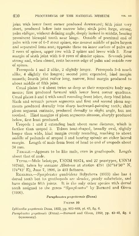
410 PROCEEDINGS OF THE NATIONAL MUSEUM vol. 115
joint with lower front corner produced downward; fifth joint very
short, produced below into narrow lobe; sixth joint large, strong,
palm oblique, without defining angle, deeply incised in middle, bearing
prominent bicuspid tooth near hinge. Outside of proximal end of
palm with row of 6-7 stout spines, one nearest middle of palm largest
and separated from rest; opposite these on inner surface of palm are
2 rows of spines, upper row with 2 spines and lower with 3. Rearmargin of sixth joint with 8-9 rows of slender spines. Seventh joint
strong and, when closed, rests between edge of palm and outside rowof spines.
Pereopods 1 and 2 alike, 2 slightly longer. Pereopods 3-5 muchalike, 4 slightly the longest; second joint expanded, hind margin
smooth; fom'th joint rather long, narrow, hind margin produced to
about middle of fifth joint.
Coxal plates 1-4 about twice as deep as their respective body seg-
ments; first produced forward with lower front corner quadrate.
Coxal plates 5 and 6 with small rounding front lobes, deep hind lobes.
Sixth and seventh pereon segments and first and second pleon seg-
ments produced dorsally into sharp backward-pointing teeth; third
pleon segment carinate, produced dorsally to slight angle, but not
toothed. Hind margins of pleon segments sinuous, sharply produced
below, first least produced.
Uropods 1 and 2 extending back about same distance, which is
farther than uropod 3. Telson boat-shaped, broadly oval, slightly
longer than wide, hind margin evenly rounding, reaching to about
middle of peduncle of uropod 3 and bearing spinule on either lateral
margin. Length of male from front of head to end of uropods about
24 mm.Female.—Appears to be like male, even in gnathopods. Length
about that of male.
Types.—Male holotype, USNM 93874, and 27 paratypes, USNM107863, taken by steamer Albatross at station 4781 (52°14'30" N,
174°13' E), June 7, 1906, in 482 fathoms.
Remarks.—Sympleustes quadridens Bulycheva (1955) also has 4
dorsal teeth but its gnathopods are slender, poorly subchelate, and
have elongate fifth joints. It is the only other species with dorsal
teeth assigned to the genus "Sympleustes^' by Barnard and Given
(1960).
Parapleustes pugettensis (Dana)
Figure 10
Iphimedia pugettensis Dana, 1853, pp. 932-933, pi. 63, fig. 6.
Parapleustes pugettensis (Dana).—Barnard and Given, 1960, pp. 43-45, fig. 4
(synonomy) .
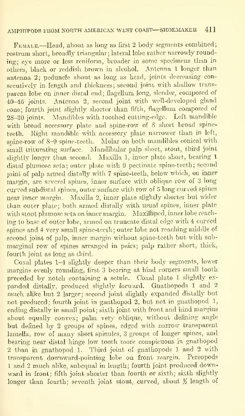
AMPHIPODS FROM NORTH AMERICAN WEST COAST—SHOEMAKER 41
1
Female.—Head, about as long as first 2 body segments combined;
rostrimi short, broadly triangular; lateral lobe rather narrowly round-
ing; eye more or less reniform, broader in some specimens than in
others, black or reddish brown in alcohol. Antenna 1 longer than
antenna 2; peduncle about as long as head, joints decreasing con-
secutively in length and thickness; second joint with shallow trans-
parent lobe on inner distal end ; flagellum long, slender, composed of
40-46 joints. Antenna 2, second joint with well-developed gland
cone; fourth joint slightly shorter than fifth, flagellum composed of
28-30 joints. Mandibles with toothed cutting-edge. Left mandible
with broad accessory plate and spine-row of 8 short broad spine-
teeth. Right mandible with accessory plate narrower than in left,
spine-row of 8-9 spine-teeth. Molar on both mandibles conical with
small triturating surface. Mandibular palp short, stout, third joint
slightly longer than second. Maxilla 1, inner plate short, bearing 1
distal plumose seta; outer plate with 9 pectinate spine-teeth; second
joint of palp armed distally with 7 spine-teeth, below which, on inner
margin, are several spines, inner surface with oblique row of 3 long
curved subdistal spines, outer surface with row of 5 long curved spines
near inner margin. Maxilla 2, inner plate slightly shorter but wider
than outer plate; both armed distally with usual spines, inner plate
with stout plumose seta on inner margin. MaxUliped, inner lobe reach-
ing to base of outer lobe, armed on truncate distal edge with 4 curved
spines and 4 very small spine-teeth; outer lobe not reaching middle of
second joint of palp, inner margin without spine-teeth but with sub-
marginal row of spines arranged in pairs; palp rather short, thick,
fourth joint as long as third.
Coxal plates 1-4 slightly deeper than their body segments, lower
margins evenly rounding, first 3 bearing at hind corners small tooth
preceded by notch containing a setule. Coxal plate 1 slightly ex-
panded distally, produced slightly forward. Gnathopods 1 and 2
much alike but 2 larger; second joint slightly expanded distally but
not produced; fourth joint in gnathopod 2, but not in gnathopod 1,
ending distally in small point; sixth joint with front and hind margins
about equally convex; palm very oblique, without defining angle
but defined by 2 groups of spines, edged with narrow transparent
lamella, row of many short spinules, 3 groups of longer spines, and
bearing near distal hinge low tooth more conspicuous in gnathopod
2 than in gnathopod 1. Third joint of gnathopods 1 and 2 with
transparent downward-pointing lobe on front margin. Pereopods
1 and 2 much alike, subequal in length; fourth joint produced down-
ward in front; fifth joint shorter than fourth or sixth; sixth slightly
longer than fourth; seventh joint stout, curved, about K length of
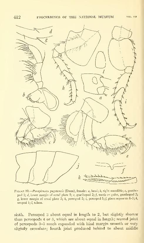
412 PROCEEDINGS OF THE NATIONAL MUSEUM vol. ii5
Figure 10.
—
Parapleustes -pugettensis (Dana), female: a, head; b, right mandible; c, gnatho-
pod 1; d, lower margin of coxal plate 1; e, gnathopod 2;/, tooth on palm, gnathopod 2;
g, lower margin of coxal plate 2; h, pereopod 2; i, pereopod S;;, pleon segments 1-3; k,
uropod 1 ; /, telson.
sixth. Pereopod 3 about equal in length to 2, but slightly shorter
than pereopods 4 or 5, which are about equal in length; second joint
of pereopods 3-5 much expanded with hind margin smooth or very
slightly crenulate; fourth joint produced behind to about middle

AMPHIPODS FROM NORTH AMERICAN WEST COAST—SHOEMAKER 413
of fifth; sixth jomt longer than fourth; seventh joint nearly }^ length
of sixth.
Pleon segments 1-3 very slightly produced at lower hind corner.
Uropods 1 and 2 extending back about same distance, slightly farther
than uropod 3. Uropod 1, peduncle slightly longer than rami,
spinose on outer and inner margins and bearing rather long stout
spine at outer distal corner; rami with rows of spines on upper edges.
Uropod 2 shorter than 1, with similar armature but with fewer spines.
Uropod 3, peduncle about K length of inner ramus; outer ramus
about % length of inner, both rami and peduncle armed on upper
edges with few spines. Telson keeled below, narrowly oval, bearing
2 minute setae distally, extending back slightly beyond peduncle
of uropod 3. None of body segments dentate. Reaches length of
about 9 mm.Remarks.—Parapleustes pugettensis appears to be subject to
considerable variation. It has a wide distribution in the North
Pacific. Specimens in the U.S. National Museum come from La JoUa
northward along the coast of California to Dillon Beach; Cape Arago,
Oregon; Vancouver Island; Queen Charlotte Islands; the Alaskan
Peninsula and the west coast of Alaska (62°54' N, 166°38' W).
Barnard and Given (1960) provide information on depth and type
of bottom preferred by this species in southern California waters.
Small specimens of 4 or 5 mm. are much less spinose or setose than
larger specimens, such as the 9 mm. female from Dillon Beach here
figured. Paramphithoe hairdi, described by Boeck in 1872 from the
coast of California, is in all probability a synonym of Dana's Iphimedia
pugettensis. Boeck does not give the size of his species, but the
description and figures contain nothing that can distinguish it from
Dana's species. Gurjanova's species (1938), Neopleustes derzhavini,
measuring 4 mm., from the Sea of Japan, appears to agree fairly well,
so far as her figures show, with Dana's species. A small tooth and
seta are shown on her figure (pi. 31, fig. 3) of the first coxal plate,
but not on the second; the hind margin of the third pleon segment
(pi. 31, fig. 4) is as here shown for Parapleustes pugettensis (fig. lOj).
The teeth on the first 3 coxal plates of many specimens from the
west coast of America are so small and inconspicuous that they can
be easily overlooked.
Barnard's fine figures (1952) are of a male, while those given here
are of a female, showing some of the appendages not illustrated by
him. The gnathopods of the 9-mm. female are very much more
hirsute than those of the 5-mm. male figured by him.

414 PROCEEDINGS OF THE NATIONAL MUSEUM vol. ii5
Family Stilipedidae
Stilipes distincta Holmes
Figure 11
Stilipes distincta Holmes, 1908, p. 536, figs. 41-44.—Gurjanova, 1952, p. 194,
fiff. 17.
Stilipes distincta was described by Holmes from San Nicolas Island
off southern California, and he created the family Stilipedidae to
receive it. Giirjanova (1952) recorded and figured this species from
the western Bering Sea. The genus Cacao, erected by K. H. Barnard
(1932, p. 153) for C. lacteus from the South Atlantic, has the samecharacters as Stilipes and is here considered a synonym of Holmes'
genus. Cacao sanguineus Hurley (1954, p. 803), from the southeast
coast of New Zealand, now becomes Stilipes sanguineus (Hurley).
Barnard placed Cacao in the family Tironidae with some reservation,
but it does not agree with that family in several of its characters.
The genus Stilipes is very widely distributed, but up to the present
time it is represented by only 3 species, S. distincta, S. lactea, and
S. sanguinea.
A sexually mature male and an ovigerous female were taken by the
Albatross at station 4751 at the southern end of Alaska (55°56'50" N,132°04'20" W) in 288 fathoms, Aug. 30, 1905. The description and
figures given herein are of this male.
Male.—Head and pereon broad, smooth, evenly arched. Headprominent, tumid, as long as first 3 pereon segments combined, bearing
peculiar downward-projecting rostrum (fig. 116). Oval bulging area
on lower front margin of head represents eye, but no visual elements
can be discerned, animal having been in alcohol so long. Antenna 1
about % length of body and about % length of antenna 2; peduncle
short, first joint longer than second and third combined; flagellum
composed of many joints, first of which is scarcely any longer than
rest; second and third peduncular joints and first 7-10 flagellar joints
with brushes of forward-curving setae or spines on lower surfaces.
Antenna 2 with prominent gland-cone; third joint short; fourth joint
shorter than fifth; fifth joint narrower than fourth; both fourth and
fifth joints bearing groups of short setae on upper margin and longer
setae on lower margin; flagellum composed of many short joints.
Mandible short, stout, without molar or spine-row; cutting-edge
chitinous, very broad, without teeth; right mandible without accessory
plate, but left with broad plate edged distally with low teeth; palp with
second joint twice as long as third. Maxilla 1, inner plate conical,
bearing group of terminal setae; outer plate broad, armed with row
of 16-17 stout, compoundly curved spine teeth, and brush of setae on
inner rounding corner (figs. lld,e)', palp with very broadly expanded
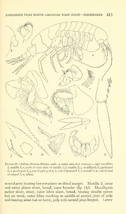
AMPHIPODS FROM NORTH AMERICAN WEST COAST—SHOEMAKER 415
Figure 11.
—
Stilipes distincta Holmes, male: a, entire animal; b, rostrum; c, right mandible;
d, maxilla 1; e, teeth of outer plate of maxilla 1;/, maxilla 2; g, maxilliped; h, gnathopod
1; i, gnathopod 2; ;, end of pereopod 4; k, end of pereopod 5; /, uropod 3; m, end of rami
of uropod 3; n, telson.
second joint bearing low serrations on distal margin. Maxilla 2, inner
and outer plates short, broad, inner broader (fig. 11/), Maxillipeds
rather short, stout; inner lobes short, broad, bearing slender spines
but no teeth; outer lobes reaching to middle of second joint of palp
and bearing setae but no teeth;palp with second joint longest. Lower

416 PROCEEDINGS OF THE NATIONAL MUSEUM vol. 115
lip with outer lobes slightly cui"ved, converging distally; no inner
lobes; lateral processes long, narrow.
Gnathopods 1 and 2 simple, much alike; 1 shorter. Gnathopod 1,
second joint about as long as fourth to sixth combined; fifth joint
slightly longer than wide, nearly twice as long as narrower sixth joint;
sixth joint slightly longer than wide, without palm; seventh joint
short, weak; second to sixth joints bearing many long slender spines.
Gnathopod 2 like 1 except joints are all proportionately longer.
Pereopods 1 and 2 alike, shorter than pereopod 3; fourth joint very
sUghtly expanded; seventh joint small, nearly straight. Pereopods
3-5 increasing consecutively in length; 3 and 4 much alike, second
joint not much expanded, twice as long as wide, with lower hind
margin slightly lobed; fourth joint slightly expanded ; sixth joint nearly
twice as long as fifth; seventh joint rather short, nearly straight.
Pereopod 5 stouter, longer than 4; second joint nearly twice as long as
wide, with rather deep lower hind lobe; fourth joint slightly expanded,
scarcely produced behind; fifth joint slightly shorter than fourth;
sixth joint slightly longer than fourth; seventh joint greatly developed,
as long and wide as sixth and entirely different from that of any other
pereopods, being paddle-like, very thin, converging to very sharp
apex, and bearing row of short spinules on front margin (fig. Ilk).
Coxal plates 1-4 deeper than their body segments and decreasing
consecutively in depth. Coxal plate 1 greatly expanded distally with
front margin reaching front margin of head. Coxal plates 2 and 3
about as wide as their body segments and with sides parallel. Coxal
plate 4 nearly as wide as second and third combined, with lower margin
curving obliquely upward and upper hind margin excavated to fit
front margin of coxal plate 5. Coxal plates 5-7 not quite as deep as
their body segments (fig. 11a).
Branchiae present on gnathopod 2 and pereopods 1-5, all more or
less irregularly folded, crumpled, or lobed.
Pleon segment 1, lower hind corner not produced. Pleon segment
2 sharply produced; segment 3 more sharply produced than 2. Uro-
some segment 1 with dorsal hump and slight lateral ridges.
All uropods reaching back about same distance. Uropod 1,
peduncle slightly longer than inner ramus, which is slightly longer
than outer ramus. Uropod 2, peduncle slightly shorter than outer
ramus, which is shorter than inner ramus. Peduncle and rami of
uropods 1 and 2 edged with many short spines. Uropod 3, peduncle
about Yi as long as subequal rami, which are lanceolate, thin, flat,
edged with short spines (figs. llZ,w). Telson reaching slightly
beyond peduncle of uropod 3, slightly longer than wide, with sides
converging to slightly excavated end (fig. lln). Length of male
from front of head to end of uropods about 15 mm.
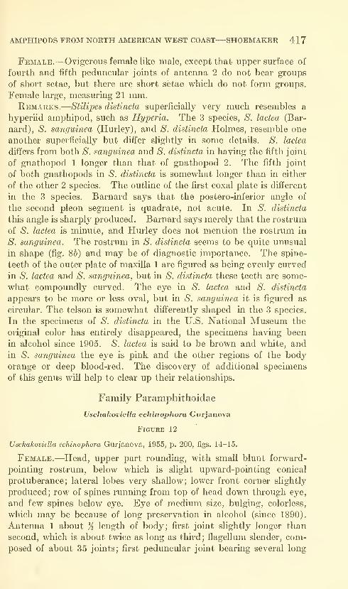
AMPHIPODS FROM NORTH AMERICAN WEST COAST—SHOEMAKER 41
7
Female.—Ovigerous female like male, except that upper sui'face of
fom-th and fifth peduncular joints of antenna 2 do not bear groups
of short setae, but there are short setae which do not form groups.
Female large, measiu"ing 21 mm.Remarks.—Stilipes distincta superficially very much resembles a
hyperiid amphipod, such as Hyperia. The 3 species, S. lactea (Bar-
nard), S. sanguinea (Hurley), and *S'. distincta Holmes, resemble one
another superficially but differ slightly in some details. S. lactea
differs from both *S'. sanguinea and S. distincta in having the fifth joint
of gnathopod 1 longer than that of gnathopod 2. The fifth joint
of both gnathopods in S. distincta is somewhat longer than in either
of the other 2 species. The outline of the first coxal plate is different
in the 3 species. Barnard says that the postero-inferior angle of
the second pleon segment is quadrate, not acute. In *S'. distincta
this angle is sharply produced. Barnard says merely that the rostrum
of ^S. lactea is minute, and Hurley does not mention the rostrum in
S. sanguinea. The rostrum in S. distincta seems to be quite unusual
in shape (fig. Sb) and may be of diagnostic importance. The spine-
teeth of the outer plate of maxilla 1 are figured as being evenly curved
in S. lactea and S. sanguinea, but in S. distincta these teeth are some-
what compoundly curved. The eye in S. lactea and S. distincta
appears to be more or less oval, but in S. sanguinea it is figured as
circular. The telson is somewhat differently shaped in the 3 species.
In the specimens of S. distincta in the U.S. National Museum the
original color has entirely disappeared, the specimens having been
in alcohol since 1905. S. lactea is said to be brown and white, andin S. sanguinea the eye is pink and the other regions of the bodyorange or deep blood-red. The discovery of additional specimens
of this genus will help to clear up their relationships.
Family Paramphithoidae
Uschakoviella echinophora Gurjanova
Figure 12
Uschakoviella echinophora Gurjanova, 1955, p. 200, figs. 14-15.
Female.—Head, upper part rounding, with small blunt forward-
pointing rostrum, below which is slight upward-pointing conical
protuberance; lateral lobes very shallow; lower front corner slightly
produced; row of spines running from top of head down through eye,
and few spines below eye. Eye of medium size, bulging, colorless,
which may be because of long preservation in alcohol (since 1890).
Antenna 1 about % length of body; first joint slightly longer than
second, which is about twice as long as third; flagellum slender, com-
posed of about 35 joints; first peduncular joint bearing several long

418 PROCEEDINGS OF THE NATIONAL MUSEUM vol. lis
forward-curving spines and several low ridges; second joint bearing
2 or 3 forward-pointing spines, one of which projects considerably
beyond third joint. Antenna 2 slightly longer than 1, fourth and
fifth joints about equal in length; flagellum composed of about 40
joints; peduncle without spines such as present on antenna 1.
Upper lip broad, symmetrical, not incised. Mouth parts projecting
considerably downward, suggesting parasitic mode of life. Mandible
with forward part prolonged; cutting-edge rather narrow with blunt
teeth; accessory plate not close to cutting-edge; spine-row consisting
of close cluster of 4-5 short stout spines; molar strong, prominent,
situated at about middle of mandible, triturating surface narrow; palp
situated opposite molar, thu'd joint shorter than second. Maxilla 1,
inner plate with 2 apical setae clothed in minute setules, giving themvelvety appearance; outer plate with 11 simple spine-teeth; palp slen-
der, with 3 apical velvety spines. Maxilla 2, inner plate shorter,
broader than outer, both armed distally with velvety spines. Max-niiped, inner plate broad, reaching to middle of outer plate, armed
distally and on inner margin with velvety spines; outer plate very
broad, reaching nearly to end of second joint of palp, armed distally
and on outer margin with short spines, and on inner margin with
short spines which appear to be imbedded in flesh; palp rather short,
slender, second joint longest, fourth joint short, stout, bearing minute
spinules. Lower lip without inner lobes; mandibular processes broad,
strong.
Gnathopods much alike, subequal in size, rather short, slender;
second joint nearly as long as third to sixth joints combined; fifth
joint longer than sixth; sixth joint slightly expanded distally, with
lower distal corner slightly produced, thus forming slight chela with
stout seventh joint.
Pereopods 1 and 2 alike in size and form, fifth and sixth joints
slightly expanding distally; seventh joint stout, strongly curved, very
sharp. Pereopods 3-5 increasing slightly in length consecutively;
second joint not much expanded, but that of fifth pereopod broadest;
fourth and fifth joints expanded distally with lower hind corner
considerably and sharply produced; sixth joint slightly expanded
distally; seventh joint stout, very sharp.
Pleon segments rounding below, not produced backward. Uropod1 reaching back slightly farther than 2 and uropod 3 slightly farther
than 1 ; outer rami of all uropods slightly shorter than inner. Uropod
3, rami somewhat expanded, with narrow pointed apices and manyvery small marginal spinules. Telson reaching considerably beyond
peduncle of uropod 3, about as wide as long, slightly expanding dis-
tally, bearing shallow triangular excavation which separates 2 rounding
lobes bearing few minute spinules. All gills simple, without plaits or
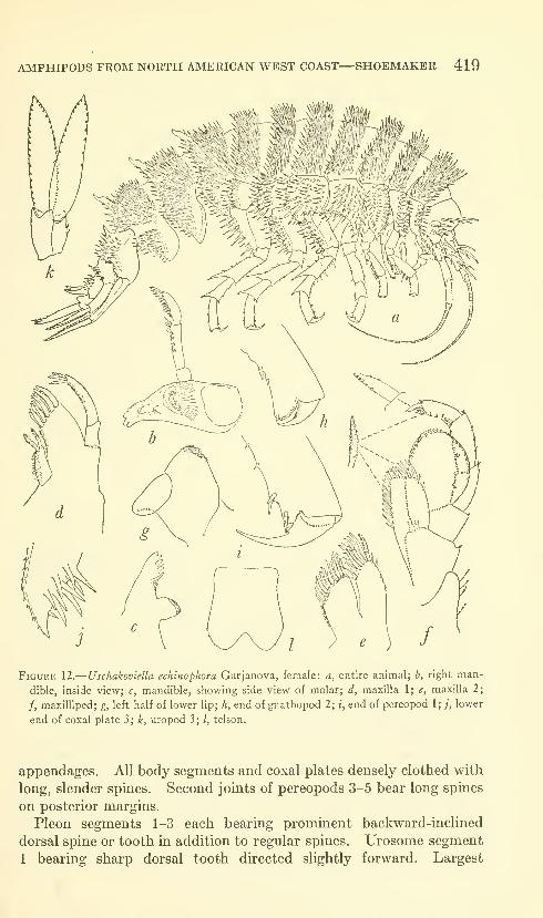
AMPHIPODS FROM NORTH AMERICAN WEST COAST—SHOEMAKER 419
Figure 12.
—
Uschakoviella echinophora Gurjanova, female: a, entire animal; h, rlglit man-
dible, inside view; c, mandible, showing side view of molar; d, maxilla 1; e, maxilla 2;
/, maxilliped; g, left half of lower lip; h, end of gnathopod 2; i, end of pereopod 1; ;, lower
end of coxal plate 3 ; k, uropod 3 ; /, telson.
appendages. All body segments and coxal plates densely clothed with
long, slender spines. Second joints of pereopods 3-5 bear long spines
on posterior margins.
Pleon segments 1-3 each bearing prominent backward-inclined
dorsal spine or tooth in addition to regular spines. Urosome segment
1 bearing sharp dorsal tooth directed slightly forward. Largest
420 PROCEEDINGS OF THE NATIONAL MUSEUM vol. 115
female in collection measures 30 mm. from front of head to end of
m-opods.
Male.—All males in collection small, only about }^ length of largest
females. Males, however, fully developed sexually. Whethermales are normally much smaller than females cannot be
determined from the present material. All of the males and the
smaller females are much less spinose than the large females.
The specimens that Gurjanova studied were taken east of Sturup
Island, the northernmost of the Kuril Islands. The specimens taken
by the steamer Albatross were from the Bering Sea and the vicinity of
Otter Island, the westernmost of the Aleutian Islands.
The specimens taken by the Albatross were from the following
stations: 3231 (53°33'30'' N, 167°15'40" W), June 18, 1890, in 54
fms., 2 specimens; station 3599 (52°05'00' N, 177°40'00' E), June 9,
1894, 1 specimen; station 4782 (52°55' N, 173°27' E), June 9, 1906,
in 65 fms., 3 specimens; station 4784 (52°55'40" N, 173°26'00" E),
June 11, 1906, in 135 fms., 2 specimens. One vial without locality,
21 small specimens.
Remarks.—Echiniphimedia hodgsoni (Walker, 1907) also has the
body clothed with spines, but the resemblance is only superficial.
Family Gamniaridae
Anisogammarus schmitti, new species
Figure 13
Male.—Head about as long as first 2 body segments combined;
side lobe with front margin more or less straight, upper and lower
corners rounding, shallow notch near upper corner. Eye not very
large, reniform, rather narrow and brownish-black in alcohol. Anten-
nae 1 and 2 about equal in length. Antenna 1, peduncular joints
decreasing in length consecutively; flagellum perhaps slightly longer
than peduncle, composed of about 20 joints; accessory flagellum of
5 joints. Antenna 2, fom-th and fifth joints about equal in length;
flagellum nearly as long as fourth and fifth joints combined, composedof about 14 joints.
Right mandible, cutting edge toothed and rather narrow; accessory
plate sharply toothed; spine-row of 4 or 5 simple spines and several
plumose setae. Molar strong, jwith .plumose seta at rear margin.
Maxilla 1, inner plate broad, with closely set row of marginal plumose
setae; outer plate with 11 pectinate spine-teeth; palp broad, with 7
apical teeth and diagonal row of 5 distal submarginal setae on outside.
Maxilla 2, lobes rather broad, bearing usual distal curved spines;
inner plate slightly the shorter, bearing diagonal row of 9 plumose
spines, first 2 of which longer than rest. Maxilliped rather long.
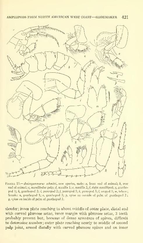
AMPHIPODS FROM NORTH AMERICAN WEST COAST—SHOEMAKER 421
Figure 13.
—
Anisogammarus schmitli,. new species, male: a, front end of animal; b, rear
end of animal; c, mandibular palp; d, maxilla 1; e, maxilla 2;/, right maxilliped; g, gnatho-
pod 1; h, gnathopod 2; i, pereopod 2;j, pereopod 3; k, pereopod 5; /, uropod 3; m, telson
;
female: n, gnathopod 1; o, gnathopod 2; p, spine on outside of palm of gnathopod 2\
q, spine on inside of palm of gnathopod 2.
slender; inner plate reaching to about middle of outer plate, distal end
with ciu-ved plumose setae, inner margin with plumose setae, 3 teeth
probably present but, because of dense armature of spines, difficult
to determine nmnber; outer plate reaching nearly to middle of second
palp joint, armed distally with curved plumose spines and on inner
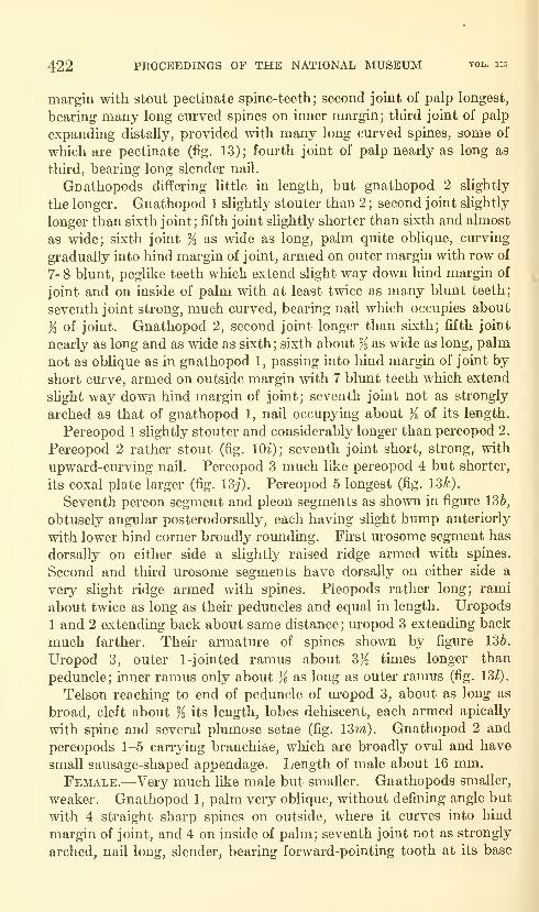
422 PROCEEDINGS OF THE NATIONAL MUSEUM vol. lis
margin with stout pectinate spine-teeth; second joint of palp longest,
bearing many long curved spines on inner margin; third joint of palp
expanding distally, provided with many long curved spines, some of
which are pectinate (fig. 13); fom^th joint of palp nearly as long as
third, bearing long slender naU.
Gnathopods differing little in length, but gnathopod 2 slightly
the longer. Gnathopod 1 slightly stouter than 2; second joint slightly
longer than sixth joint; fifth joint slightly shorter than sixth and almost
as wide; sixth joint % as wide as long, palm quite oblique, curving
gradually into hind margin of joint, armed on outer margin with row of
7-8 blunt, peglike teeth which extend slight way down hind margin of
joint and on inside of palm with at least twice as many blunt teeth;
seventh joint strong, much cm'ved, bearing nail which occupies about
% of joint. Gnathopod 2, second joint longer than sixth; fifth joint
nearly as long and as wide as sixth ; sixth about % as wide as long, palm
not as oblique as in gnathopod 1, passing into hind margin of joint by
short curve, armed on outside margin with 7 blunt teeth which extend
sHght way down hind margin of joint; seventh joint not as strongly
arched as that of gnathopod 1, nail occupying about ji of its length.
Pereopod 1 slightly stouter and considerably longer than pereopod 2.
Pereopod 2 rather stout (fig. lOi); seventh joint short, strong, with
upward-ciu-ving nail, Pereopod 3 much like pereopod 4 but shorter,
its coxal plate larger (fig. ISj). Pereopod 5 longest (fig. 13^).
Seventh pereon segment and pleon segments as shown in figure 136,
obtusely angular posterodorsally, each having slight hump anteriorly
with lower hind corner broadly rounding. First urosome segment has
dorsally on either side a slightly raised ridge armed with spines.
Second and third urosome segments have dorsally on either side a
very slight ridge armed with spines. Pieopods rather long; rami
about twice as long as their peduncles and equal in length. Uropods
1 and 2 extending back about same distance; uropod 3 extending back
much farther. Their armature of spines shown by figure 136.
Uropod 3, outer 1 -jointed ramus about 3K times longer than
peduncle; inner ramus only about ji as long as outer ramus (fig. 131).
Telson reaching to end of peduncle of lu-opod 3, about as long as
broad, cleft about % its length, lobes dehiscent, each armed apically
with spine and several plumose setae (fig. 13m). Gnathopod 2 and
pereopods 1-5 carrying branchiae, which are broadly oval and have
small sausage-shaped appendage. Length of male about 16 mm.Female.—Very much like male but smaller. Gnathopods smaller,
weaker. Gnathopod 1, palm very oblique, without defining angle but
with 4 straight sharp spines on outside, where it curves into hind
margin of joint, and 4 on inside of palm; seventh joint not as strongly
arched, nail long, slender, bearing forward-pointing tooth at its base
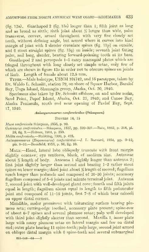
AMPHIPODS FROM NORTH AMERICAN WEST COAST—SHOEMAKER 423
(fig. 13n). Gnathopod 2 (fig. 13o) longer than 1; fifth joint as long
and as broad as sixth; sixth joint about % longer than wide, palm
transverse, convex, armed thi'oughout with very fine closely set
teeth, without defining angle, but armed where it curves into hind
margin of joint with 5 slender crenulate spines (fig. 13^) on outside,
and 6 stout straight spines (fig. I3q) on inside; seventh joint fitting
palm, naU long, slender, bearing forward-pointing tooth at its base.
Gnathopod 2 and pereopods 1-3 carry marsupial plates which are
fringed throughout with long closely set simple setae, only few of
which are shown in figure 13o in order not to obscure spines on joints
of limb. Length of female about 12.5 mm.Types.—Male holotype, USNM 101742, and 15 paratypes, taken by
Dr. Waldo L. Schmitt, station 79, on shore of Squaw Harbor, Baralof
Bay, Unga Island, Shumagin group, Alaska, Oct. 20, 1940.
Specimens also taken by Dr. Schmitt offshore, on and under rocks,
Sand Point, Popof Island, Alaska, Oct. 25, 1940; and Canoe Bay,
Alaska Peninsula, south end near opening of Pavlof Bay, Sept.
17, 1940.
Anisogammarus confervicolus (Stirnpson)
Figures 14, 15
Mara confervicola Stirnpson, 1856, p. 90.
Gammarus confervicolus.—Stirnpson, 1857, pp. 520-521.—Bate, 1862, p. 218, pi.
38, fig. 9.—Holmes, 1904, p. 239.
Melita confervicola.—Stebbing, 1906, p. 428.
Anisogammarus (Eogammarus) confervicolus.—J. L. Barnard, 1954, pp. 9-12,
pis. 9-10.—Bousfield, 1958, p. 86, fig. 10.
Male.—Head, lateral lobe obliquely truncate with front margin
slightly concave; eye reniform, black, of medium size. Antennae
about K length of body. Antenna 1 slightly longer than antenna 2
;
first joint slightly longer than second and bearing 1-2 rather stout
spines on lower margin; third joint about ji length of second; flagellum
much longer than peduncle and composed of 26-30 joints; accessory
flagellum composed of 5-6 joints and minute terminal joint. Antenna
2, second joint with well-developed gland cone; fourth and fifth joints
equal in length; flagellum about equal in length to fifth peduncular
joint and composed of 11-15 joints, first 7-8 of which bear calceoli
on upper distal corners.
Mandible, molar prominent with triturating surface bearing plu-
mose seta; cutting-edge toothed, accessory plate present; spine-row
of about 6-7 spines and several plumose setae; palp well developed
with third joint slightly shorter than second. Maxilla 1, inner plate
large with many plumose setae on lateral margin and few on distal
end; outer plate bearing 11 spine-teeth; palp large, second joint armed
on oblique distal margin with 6 spine-teeth and several submarginal
693-049—64 3
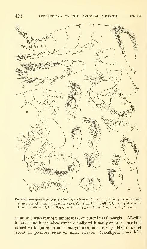
424 PROCEEDINGS OF THE NATIONAL MUSEUM vol. 115
Figure 14.
—
Anisogammarus confermcolus (Stimpson), male: a, front part of animal;
b, hind part of animal; c, right mandible; d, maxilla 1; e, maxilla 2;/, maxilliped; g, outer
lobe of maxilliped; h, lower lip; i, gnathopod 1;;, gnathopod 2; k, uropod 3; /, telson.
setae, and with row of plumose setae on outer lateral margin. Maxilla
2, outer and inner lobes armed distally with many spines; inner lobe
armed with spines on inner margin also, and having oblique row of
about 11 plumose setae on inner surface. Maxilliped, inner lobe
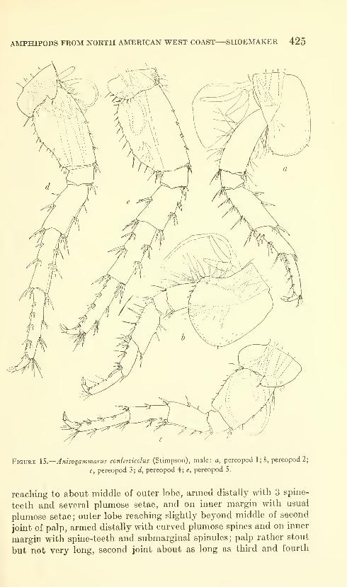
AMPHIPODS FROM NORTH AMERICAN WEST COAST—SHOEMAKER 425
Figure IS.
—
Anisogammarus confervicolus (Stimpson), male: a, pereopod 1; ^, pereopod 2;
c, pereopod 3; d, pereopod 4; e, pereopod 5.
reaching to about middle of outer lobe, armed distally with 3 spine-
teeth and several plumose setae, and on inner margin with usual
plumose setae ; outer lobe reaching slightly beyond middle of second
joint of palp, armed distally with curved plumose spines and on inner
margin with spine-teeth and submarginal spinules; palp rather stout
but not very long, second joint about as long as third and fourth
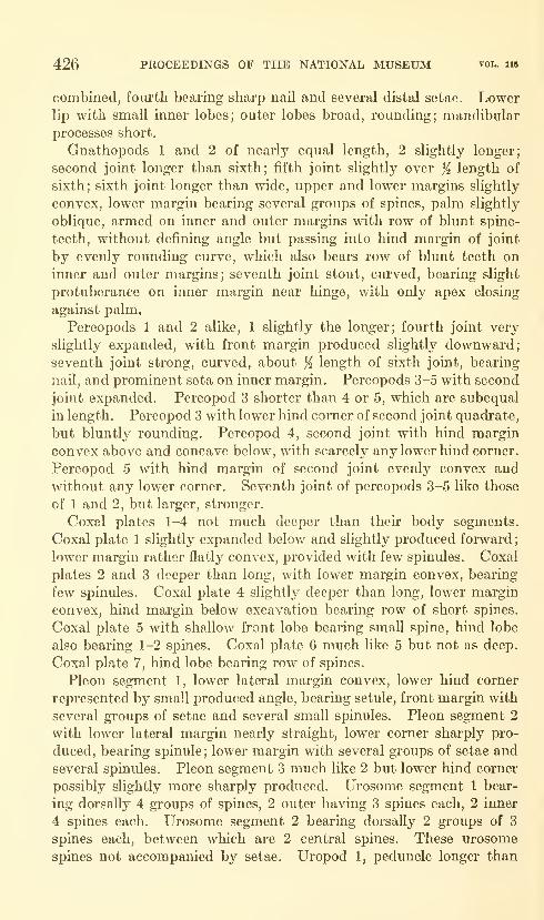
426 PROCEEDINGS OF THE NATIONAL MUSEUM vol. 115
combined, fourth bearing sharp nail and several distal setae. Lowerlip with small inner lobes; outer lobes broad, rounding; mandibular
processes short.
Gnathopods 1 and 2 of nearly equal length, 2 slightly longer;
second joint longer than sixth; fifth joint slightly over % length of
sixth; sixth joint longer than wide, upper and lower margins slightly
convex, lower margin bearing several groups of spines, palm slightly
oblique, armed on inner and outer margins with row of blunt spine-
teeth, without defining angle but passing into hind margin of joint
by evenly rounding curve, which also bears row of blunt teeth on
inner and outer margins; seventh joint stout, curved, bearing slight
protuberance on inner margin near hinge, with only apex closing
against palm.
Pereopods 1 and 2 alike, 1 slightly the longer; fourth joint very
slightly expanded, with front margin produced slightly downward;seventh joint strong, curved, about % length of sixth joint, bearing
nail, and prominent seta on inner margin. Pereopods 3-5 with second
joint expanded. Pereopod 3 shorter than 4 or 5, which are subequal
in length. Pereopod 3 with lower hind corner of second joint quadrate,
but bluntly rounding. Pereopod 4, second joint with hind margin
convex above and concave below, with scarcely any lower hind corner.
Pereopod 5 with hind margin of second joint evenly convex and
without any lower corner. Seventh joint of pereopods 3-5 like those
of 1 and 2, but larger, stronger.
Coxal plates 1-4 not much deeper than their body segments.
Coxal plate 1 slightly expanded belov/ and slightly produced forward
;
lower margin rather flatly convex, provided with few spinules. Coxal
plates 2 and 3 deeper than long, with lower margin convex, bearing
few spinules. Coxal plate 4 slightly deeper than long, lower margin
convex, hind margin below excavation bearing row of short spines.
Coxal plate 5 with shallow front lobe bearing small spine, hind lobe
also bearing 1-2 spines. Coxal plate 6 much like 5 but not as deep.
Coxal plate 7, hind lobe bearing row of spines.
Pleon segment 1, lower lateral margin convex, lower hind corner
represented by small produced angle, bearing setule, front margin with
several groups of setae and several small spinules. Pleon segment 2
with lower lateral margin nearly straight, lower corner sharply pro-
duced, bearing spinule; lower margin with several groups of setae and
several spinules. Pleon segment 3 much like 2 but lower hind corner
possibly slightly more sharply produced. Urosome segment 1 bear-
ing dorsally 4 groups of spines, 2 outer having 3 spines each, 2 inner
4 spines each. Urosome segment 2 bearing dorsally 2 groups of 3
spines each, between which are 2 central spines. These urosome
spines not accompanied by setae. Uropod 1, peduncle longer than

AMPHIPODS FROM NORTH AMERICAN WEST COAST—SHOEMAKER 427
rami and armed on upper outer margin with 5 spines, 2 of which are
apical; upper inner margin with 3 spines, 1 of which is apical; outer
ramus slightly longer than inner ramus, each armed with spines
(fig. 5b). Uropod 2, peduncle longer than rami, upper outer marginwith 3-4 spines, one of which apical, inner margin with 2 spines, oneof which is apical ; outer ramus slightly the shorter, each armed with
spines (fig. 56). Uropod 3 extending back farther than 1; peduncle
about ji as long as outer ramus, which bears groups of spines con-
taining few plumose setae on outer and inner margin, second joint
small, narrow; inner ramus about K as long as outer, and bearing fewspines on inner margin and group of spines and plumose setae apically.
Telson reaching slightly beyond end of peduncle of uropod 3, cleft
about Yz its length, armed with spine and plumose seta on either
lateral margin, 2 spines and few spinules on apex of each lobe. GUIsof gnathopod 2 and pereopods 1-3 each with 2 finger-like appendages.
Gill of pereopod 4 with 3 finger-like appendages, gill of pereopod 5
with 1 appendage.
Stimpson gave .4 to .5 of an inch (about 13 mm.) as the length of
this species, but there are specimens in the U.S. National Museummeasuring up to 21 mm. that were taken from a brackish pond in
Snohomish County, Washington, a pond which receives an inflow from
Puget Sound at exceptionally high tides. Apparently Stimpson's
description was taken from rather small specimens.
Female.—Much like male but smaller. Antennae somewhatshorter, flagellum of antenna 2 bears calceoli which are smaller than
those of male. Gnathopod 1 stronger but shorter than gnathopod 2
and like that of male. Sixth joint of gnathopod 2 comparatively
narrow, with palm slightly oblique or nearly transverse. Uropodsshorter than those of male.
Remarks.—Anisogammarus confervicolus appears to inhabit bays,
estuaries, brackish tidepools, and mouths of streams where the water
is brackish. In the U.S. National Museum there are specimens fromSanta Catalina Island, San Diego, and various localities northward
along the coasts of California, Oregon, and Washington. The species
appears to be conmion in Puget Sound and has been taken at Van-
couver Island. There are 3 collections from Alaska: 1 from Saginaw
Bay, Kuiu Island, Frederick Sound, Alexander Archipelago; 1 froma stream entering Orzenoi Bay, southwest Alaska (stream probably
bracldsh, at least at high tide) ; and 1 from Sitka. Barnard (1954)
gives several localities in Oregon and 1 in Alaska (Alinchak Bay),
and Bousfield (1958) records the species from a number of Canadian
Pacific localities.

428 PROCEEDINGS OF THE NATIONAL MUSEUM vol. 115
Literature Cited
Baknard, J. Laurens1952, Some Amphipoda from central California. Wasmann Journ. Biol.,
vol. 10, no. 1, pp. 9-36.
1954. Marine Amphipoda of Oregon. Oregon State Monogr. Stud. Zool.,
no. 8, 102 pp., 33 pis.
Barnard, J. Laurens, and Given, Robert R.
1960. Common pleustid amphipods of southern California with a projected
revision of the family. Pacific Naturalist, vol. 1, no. 17, pp. 37-48.
Barnard, K. H.
1932. Amphipoda. Discovery reports, vol. 5, 326 pp., 174 text figs., 1 color
pi.
Bate, C. Spence1862. Catalogue of the specimens of amphipodous crustaceans in the collection
of the British Museum, 399 pp., 58 pis.
BoECK, Axel1872. Bidrag til Californiens Amphipodefauna. Vidensk.-Selsk. Forhandl.
1871, pp. 32-51.
Bousfield, E. L.
1958. Fresh water amphipod crustaceans of glaciated North America. Cana-
dian Field-Naturalist, vol. 72, no. 2, pp. 55-113.
Bulycheva, a. I.
1955. New species of gammarideans (Amphipoda, Gammaridea) from the
Sea of Japan, II. Trudy Zool. Inst. Akad. Nauk SSSR, vol. 21, pp.
193-207 [in Russian].
Dana, James Dwight1853. United States Exploring Expedition during the years 1838, 1839, 1840,
1841, 1842 under the command of Charles Wilkes, U.S.N., vol. 14
(Crustacea), pt. 2, pp. 685-1618.
Goes, A.
1866. Crustacea Amphipoda maris Spitzbergiam alluentis cum speciebus aliis
arcticis. Ofv. Vet. Akad. Forhandl., vol. 22, pp. 517-536, pis. 35-41.
GURJANOVA, EUPRAXIE F.
1938. Amphipoda, Gammaroidea of Siaukhu Bay and Sudzukhe Bay (Japan
Sea). Rep. Japan Sea Hydrobiol. Exped. in 1934, Zool. Inst. Acad.
Sci. USSR, pt. 1, pp. 241-404, 59 figs, [in Russian].
1951. Gammarideans of the seas of the USSR and adjacent waters (Amphi-
poda-Gammaridea). Akad. Nauk SSSR, Opred. po Faune SSSR,vol. 41, 1029 pp. [in Russian].
1952. New species of gammarideans (Amphipoda, Gammaridea) from far-
eastern seas. Trudy Zool. Inst. Akad. Nauk. SSSR, vol. 12, pp.
171-194 [in Russian].
1955. New species of gammarideans (Amphipoda, Gammaridea) from the
northern part of the Pacific Ocean. Trudy Zool. Inst. Akad. Nauk.
SSSR, vol. 18, pp. 166-218 [in Russian].
Holmes, Samuel J.
1904. Amphipod crustaceaiis of the expedition. HaiTiman Alaska Expedi-
tion, vol. 10, pp. 233-246.
1908. The Amphipoda collected by the U.S. Bureau of Fisheries steamer
Albatross off the west coast of North America, in 1903 and 1904, with
descriptions of a new family and several new genera and species.
Proc. U.S. Nat. Mus., vol. 35, no. 1654, pp. 489-543.
AMPHIPODS FROM NORTH AMERICAN WEST COAST—SHOEMAKER 429
Hurley, Desmond E.
1954. Studies on the New Zealand amphipodan fauna, no. 10: A new species
of Cacao. Trans. Roy. Soc. New Zealand, vol. 82, pt. 3, pp. 803-811.
Sars, G. O.
1895. An account of the Crustacea of Norway, vol. 1 (Amphipoda), 711 pp.,
240 pis., 8 suppl. pis.
Stebbing, T. R. R.
1906. Amphipoda, I: Gammaridea. Das Tierreich, Lief. 21, SOG+ xxxix pp.
Stimpson, William1857. On the Crustacea and Echinodermata of the Pacific shores of North
America. Boston Journ. Nat. Hist., vol. 6, no. 4, pp. 444-532, pis.
18-23.
Walker, A. 0.
1907. Crustacea, III: Amphipoda. National Antarctic {Discovery) Expedi-
tion 1901-1904. Nat. Hist., vol. 3, pp. 1-39, 13 pis.
U.O. GOVCRNMtNT PRINTING 0FFICE:I964

Proceedings of
the United States
National MuseumSMITHSONIAN INSTITUTION • WASHINGTON, D.C.
Volume 115 1964 Number 3490
SHRIMPS OF THE GENUS BETAEUS
ON THE PACIFIC COAST OF NORTH AMERICA
WITH DESCRIPTIONS OF THREE NEW SPECIES
By Josephine F. L. Hart
Introduction
Shrimps of the genus Betaeus are members of the section Caridea
and the family Alpheidae (or Crangonidae in much of the NorthAmerican literature). Members of this genus are characterized bythe lack of a rostrum in the adult and by the inversion of the "hands,"
with the result that the dactyls are on the lower side. The terms
"visored shrimps" and "hooded shrimps" are sometimes used because
the carapace projects forward to overhang the eyes. Up to the present
time four species have been recognized as occurring in the area from
Mexico to Alaska: Betaeus harrimani Rathbun, B. longidactylus
Lockington, B. ensenadensis GlasseU, and B. harfordi (Kingsley).
However, these species have not been well defined.
The sole published record of Betaeus harrimani is the original
description by M. J. Rathbun (1904) based on a single female taken
in southern Alaska. The examination of a series of both males andfemales from a number of more southerly locations has made it possible
to elaborate on this description. Such an elaboration is especially
431693-433—63 1

432 PROCEEDINGS OF THE NATIONAL MUSEUM vol. 115
important because of the overlap of the range of the closely alhed form
B. longidactylus Lockington (1877) in California. These two species,
as well as B. ensenadensis Glassell (1938) of southern CaUfornia and
Mexico, are figured and redescribed here in some detail.
In the 1870's W. N. Lockington and J. S. Kingsley each described
two species of hooded shrimps from California. They engaged in
considerable controversy in their papers as to whether or not Betaeus
was a valid genus, and occasionally they referred species of Betaeus
to the genus Alpheus; as a result, in the literature both Alpheus and
Betaeus are used in referring to these shrimps. Lockington (1877b,
1878) described two species briefly, without figures, and, unfortunately,
named one Betaeus equimanus, a name that had been used previously
by Dana (1852, p. 560; 1855, pi. 35) for a shrimp from New Zealand.
Early in 1878 Kingsley published a description of apparently the
same shrimp and called it Alpheus harfordi but indicated that if
"aequimanu^'^ should prove to be different, the shrimp would have to
be called ''aequalis.'' Later in the same year Lockington called it
B. egualis and enlarged his description by incorporating Kingsley's
description of B. harfordi. He was convinced that they were identical
but because of his use of a preoccupied name, he lost the honor of
naming the species. Betaeus harfordi (Kingsley) is therefore the
valid name.
Holmes (1900) gave a description with more details than those
given by the early writers, but unfortunately he added new habitats
that perhaps have helped to mask the presence of four distinct forms:
two commensal and two free-living.
In 1934 several hooded shrimps were taken in tide pools on the west
coast of Vancouver Island by E. G. Hart. These agreed mth pub-
lished descriptions of B. harfordi except for a few minor differences.
Since shrimps of the family Alpheidae often show considerable vari-
ation in relative proportions according to age and sex, no suspicion of
significant differences arose until individuals from California were
compared with the northern specimens. As a result of a detailed
comparative study, it now seems that those found commensal within
the mantle cavity of abalones are referable to B. harfordi and that
the remainder represent three undescribed species. The similar, but
quite distinct, form that lives in association with sea urchins I propose
to call B. macginitieae in recognition of Nettie MacGinitie, who, with
her husband Professor G. E. MacGinitie, has contributed so much to
our knowledge of the genus. For the two free-hving forms I have
chosen the names B. gracilis and B. setosus.
In view of the fact that the published descriptions and figures of
the known species are inadequate, an attempt is made herewith to
supplement them with more detailed descriptions and illustrations.
SHRIMPS OF THE GENUS BETAEUS—HART 433
The figures are drawn to three magnifications : scales A, B (two times
A), C (ten times A). The whole animal, the chelipeds, and chelae
are scale A; the dactyli, scale C; and the remainder, scale B. Newkeys and comparative tables incorporating the new species are pre-
sented. In this aspect the work of L. B. Holthuis (1952) on twospecies found in Chile has been very useful.
In classification, emphasis in the past has been placed on the pro-
portions of the segments of the antennular peduncle, and on the size,
shape, and dentition of the large chelae. These features, however,
have been found to differ with age, sex, and extent of regeneration.
In this paper, therefore, an attempt is made to use as diagnosis those
characters that do not vary appreciably during the life of the
individual.
Sexual dimorphism also is noted in this account. Differences dueto sex are most obvious in large males of B. longidactylus, in whichthe chelipeds are much larger in relative proportion to the rest of the
body than in smaller males or in females. All the other species
dealt with here also show sexual dimorphism but only to a minorextent such as stouter appendages and narrower pleura on the
abdomens of the males.
Acknowledgments
My thanks are due to Dr. Waldo L. Schmitt and Dr. Fenner A.
Chace, Jr., of the United States National Museum for the loan of
comparative material, for making comparisons with the type of B.
harrimani, and for much other help. In addition. Dr. Chace read the
manuscript and gave invaluable advice. The late Dr. Belle A. Stevens
was most helpful in the earl}^ stages of the study. I wish also to thankboth Dr. Paul L. Illg of the Department of Zoology, University of
Washington, and Mrs. G. E. MacGinitie, retired from Kerckhoff
Marine Laboratory, for much help and encouragement, as well as
for specimens. Thanks also are due to Mr. Keith W. Cox, California
Department of Fish and Game, who has given me specimens andmuch useful data on Betaeus harfordi obtained in connection with
his work on Haliotis.
For the use of material I am indebted to the following: Mr. T. H.Butler and the Fisheries Research Board of Canada; Dr. Joel W.Hedgpeth (Pacific Marine Station) ; Dr. Rolf Bolin and Dr. DonaldP. Abbott (Hopkins Marine Station) ; Dr. Olga Hartman (University
of Southern California, Allan Hancock Foundation) for the loan of
the late S. A. Glassell's collection; Dr. James E. Lynch (University
of Washington) for collections by Dr. John S. Laurie; and Dr. R. U.Gooding of the University of Washington at the time.
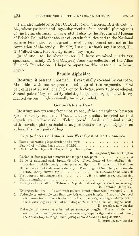
434 PROCEEDINGS OF THE NATIONAL MUSEUM vol. us
I am also indebted to Mr. C. B. Rowland, Victoria, British Colum-bia, whose patience and ingenuity resulted in successful photographs
of the living shrimps. I am grateful also to the Provincial Museumof British Columbia for the use of certain facilities and to the National
Science Foundation for a grant providing equipment and aid in the
completion of the study. Finally, I want to thank my husband, Dr.
G. Clifford Carl, for his help in so many ways.
In addition to the above material I have examined nearly 200
specimens (mainly B. longidactylus) from the collection of the AUanHancock Foundation. I hope to report on this material in a futm-e
paper.
Family Alpheidae
Rostrum, if present, unarmed. Eyes usually covered by carapace.
Mandibles with incisor process and palp of two segments. First
pair of legs often with one chela, or both chelae, powerfully developed.
Second pair of legs minutely chelate, long, slender, equal, with seg-
mented carpus. Telson usually broad, rounded.
Genus Betaeus Dana
Rostrum not present; front not spined, either emarginate between
eyes or evenly rounded. Chelae usually similar, inverted so that
dactyls are on lower side. Telson broad. Sixth abdominal somite
with movable plate articulated at posterolateral angle. Epipods on
at least first two pairs of leg's.
Key to Species of Betaeus from West Coast of North America
1. Dactyli of walking legs slender and simple 2
Dactyli of walking legs stout and bifid 4
2. Chelae of first legs with fingers longer than palm.
B. longidactylus Lockington
Chelae of first legs with fingers not longer than palm 3
3. Blade of antennal scale broad distally. Fixed finger of first cheliped de-
creasing in width evenly to sharp curved tip . . B. harrimani RathbunBlade of antennal scale narrow distally. Fixed finger of first cheliped truncate
before sharp curved tip B. ensenadensis Glassell
4. Front curved, not emarginate B. macginitieae, new species
Front emarginate 5
5. Emargination shallow. Telson with posterolateral spines small or missing.
B. harfordi (Kingsley)
Emargination deep. Telson with posterolateral spines well developed . . 6
6. Peduncle of antennule less than one-half carapace length. Merus of cheliped
Math lower inner ridge with long bristles, upper ridge ending in sharp tooth;
chela with fingers subequal to palm; chela is three times as long as wide.
B. gracilis, new species
Peduncle of antennule subequal to carapace length. Merus of cheliped
with lower inner ridge usually tuberculate, upper ridge with tuft of hairs;
chela Math fingers longer than palm; chela is twice as long as wide.
B. setosiis, new species
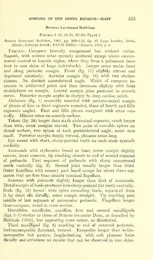
SHRIMPS OF THE GENUS BETAEUS—^HART 435
Betaeus harrimani Rathbun
Figures 1-26, 29-31, 37-39; Plate 1
Betaeus harrimani Rathbun, 1904, pp. 108-110, fig. 49 (type locality, Sitka,
Alaska; holotype female, USNM 25692).—Banner, 1953, p. 5.
Female.—Carapace laterally compressed but without carina.
Smooth, with minute setae sparsely scattered except where concen-
trated ventral to hepatic region, where they form a pubesence (seen
best in cast skins of large individuals). Longer setae under front
and along posterior margin. Front (fig. 17) slightly curved anddepressed anteriorly. Anterior margin (fig. 18) with two shallow
sinuses. No distinct anterolateral angle. Width of carapace in-
creases to midlateral point and then decreases slightly with faint
undulations on margin. Lateral margin joins posterior in smoothcurve. Posterior margin angles in sharply to deep cardiac notch.
Abdomen (fig. 1) smoothly rounded with posteroventral marginof pleura of first to third segments rounded, those of fourth and fifth
segments angled. First and fifth pleura margined with setae ven-
trally. Minute setae on smooth surface.
Telson (fig. 26) longer than sixth abdominal segment, much longer
than wide, outer margins curved. Two pairs of movable spines ondorsal surface, two spines at each posterolateral angle; outer ones
small. Posterior margin deeply curved, plumose setae long.
Eye round with short, sharp-pointed tooth on each stout eyestalk
medially.
Antennule with stylocerite broad at base; outer margin slightly
convex, inner concave, tip reaching almost to end of second segmentof peduncle. First segment of peduncle with sharp compressed
tooth ventrally (fig. 2). Second joint usually longer than third.
Outer flagellum with sensory part fused except for about three seg-
ments that are free from slender terminal flagellum.
Antenna with peduncle slightly longer than that of antennule.
Distal margin of basis produced into sharp-pointed flat tooth ventrally.
Scale (fig. 19) broad: wide spine exceeding blade, separated from
it by short slit distally, outer margin straight. Tip reaching past
middle of last segment of antennular peduncle. Flagellum longer
than carapace, ovoid in cross section.
Mandibles, maxillules, maxillae, first and second maxillipeds
(figs. 3-7) similar to those of Betaeus truncatus Dana, as described byHolthuis (1952), but appearing more setose, as illustrated.
Third maxilliped (fig. 8) reaching to end of antennal peduncle.
Ischiomeropodite flattened, twisted. Exopodite longer than ischio-
meropodite but narrow, fragile-looking, with soft plumose hairs
distally and striations on cuticle that can be observed in cast skins.
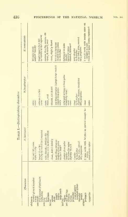
436 PROCEEDINGS OF THE NATIONAL MUSEUM
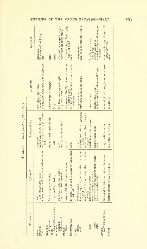
SHRIMPS OF THE GENUS BETAEUS—^HART 437
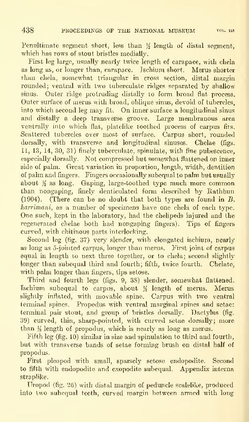
438 PROCEEDINGS OF THE NATIONAL MUSEUM vol. 115
Penultimate segment short, less than j^ length of distal segment,
which has rows of stout bristles medially.
First leg large, usually nearly twice length of carapace, with chela
as long as, or longer than, carapace. Ischium short. Merus shorter
than chela, somewhat triangular in cross section, distal marginrounded; ventral with two tuberculate ridges separated by shallow
sinus. Outer ridge protruding distally to form broad flat process.
Outer surface of merus with broad, oblique sinus, devoid of tubercles,
into which second leg may fit. On inner surface a longitudinal sinus
and distally a deep transverse groove. Large membranous area
ventrally into which flat, platelike toothed process of carpus fits.
Scattered tubercles over most of surface. Carpus short, rounded
dorsally, with transverse and longitudinal sinuses. Chelae (figs.
11, 13, 14, 30, 31) finely tuberculate, spinulate, with fine pubescence,
especially dorsally. Not compressed but somewhat flattened on inner
side of pahn. Great variation in proportion, length, width, dentition
of palm and fingers. Fingers occasionally subequal to palm but usually
about Yz as long. Gaping, large-toothed type much more commonthan nongaping, finely denticulated form described by Rathbun(1904). (There can be no doubt that both types are found in B.
harrimani, as a number of specimens have one chela of each type.
One such, kept in the laboratory, had the chehpeds injured and the
regenerated chelae both had nongaping fingers). Tips of fingers
curved, with chitinous parts interlocking.
Second leg (fig. 37) very slender, with elongated ischium, nearly
as long as 5-jointed carpus, longer than merus. First joint of carpus
equal in length to next three together, or to chela; second shghtly
longer than subequal third and fourth; fifth, twice fourth. Chelate,
with palm longer than fingers, tips setose.
Third and fourth legs (figs. 9, 38) slender, somewhat flattened.
Ischium subequal to carpus, about ^ length of merus. Merusslightly inflated, with movable spine. Carpus with two ventral
terminal spines. Propodus with ventral marginal spines and setae:
terminal pair stout, and group of bristles dorsally. Dactylus (fig.
39) curved, thin, sharp-pointed, with curved setae dorsally; morethan K length of propodus, which is nearly as long as merus.
Fifth leg (fig. 10) similar in size and spinulation to third and fourth,
but with transverse bands of setae forming brush on distal half of
propodus.
First pleopod with smaU, sparsely setose endopodite. Second
to fifth with endopodite and exopodite subequal. Appendix interna
straplike.
Uropod (fig. 26) with distal margin of peduncle scalelike, produced
into two subequal teeth, curved margin between armed with long

PROC US. NAT. MUS. VOL. 115 HART—PLATE 1

PROC. U.S. NAT. MUS. VOL 115 HART—PLATE 2
q;
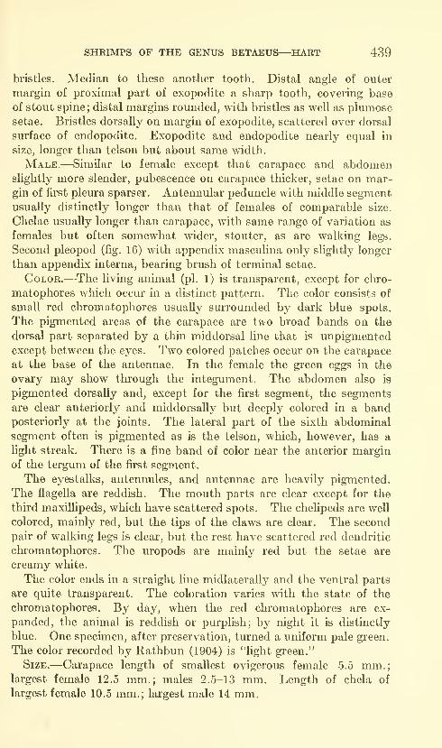
SHRIMPS OF THE GENUS BETAEUS—HART 439
bristles. Median to these another tooth. Distal angle of outer
margin of proximal part of exopodite a sharp tooth, covering base
of stout spine; distal margins rounded, with bristles as well as plumosesetae. Bristles dorsally on margin of exopodite, scattered over dorsal
surface of endopodite. Exopodite and endopodite nearly equal in
size, longer than telson but about same width.
Male.—Similar to female except that carapace and abdomenslightly more slender, pubescence on carapace thicker, setae on mar-gin of first pleura sparser. Antennular peduncle with middle segmentusually distinctly longer than that of females of comparable size.
Chelae usually longer than carapace, with same range of variation as
females but often somewhat wider, stouter, as are walking legs.
Second pleopod (fig. 16) with appendix masculina only shghtly longer
than appendix interna, bearing brush of terminal setae.
Color.—-The living annual (pi. 1) is transparent, except for clu"0-
matophores which occur in a distinct pattern. The color consists of
small red chromatophores usually surrounded by dark blue spots.
The pigmented areas of the carapace are two broad bands on the
dorsal part separated by a thin middorsal line that is unpigmentedexcept between the eyes. Two colored patches occur on the carapace
at the base of the antennae. In the female the green eggs in the
ovary may show through the integument. The abdomen also is
pigmented dorsally and, except for the first segment, the segments
are clear anteriorly and middorsally but deeply colored in a bandposteriorly at the joints. The lateral part of the sixth abdominal
segment often is pigmented as is the telson, which, however, has a
light streak. There is a fine band of color near the anterior margin
of the tergum of the first segment.
The eyestalks, antennules, and antennae are heavily pigmented.
The flagella are reddish. The mouth parts are clear except for the
third maxillipeds, which have scattered spots. The chelipeds are well
colored, mainly red, but the tips of the claws are clear. The second
pair of walking legs is clear, but the rest have scattered red dendritic
chromatophores. The uropods are mainly red but the setae are
creamy white.
The color ends in a straight line midlaterally and the ventral parts
are quite transparent. The coloration varies with the state of the
chromatophores. By day, when the red chromatophores are ex-
panded, the animal is reddish or purplish; by night it is distinctly
blue. One specimen, after preservation, turned a uniform pale green.
The color recorded by Kathbun (1904) is "fight green."
Size.—Carapace length of smallest ovigerous female 5.5 mm.;largest female 12.5 mm.; males 2.5-13 mm. Length of chela of
largest female 10.5 mm.; largest male 14 mm.

440 PROCEEDINGS OF THE NATIONAL MUSEUM vol. 115
Range.—Newport Harbor, Calif., to Sitka, Alaska.
Habitat.—Intertidal, possibly partly commensal in the burrows of
the mud shrimp Upogebia pugettensis (Dana) or the ghost shrimp
Callianassa californiensis Dana. Individuals have been collected frompools formed in the depressions left by boulders on a gravelly beach;
the openings of the burrows of the mud shrimps occur in the sides of
these pools. Others have been found by chance when excavations
were being made for mud or ghost shrimps, and one was found out of
water between two slabs of sandstone. In only one locality have the
shrimps been found in any numbers: in the oyster dike area of Oak-land Bay, Puget Sound, where they were under logs, shells, anddebris near the low tide mark, and in the trickle of water leaking fromdikes (Dr. J. S. Laurie).
Material.—The following specimens were examined:
California.—Newport Harbor, N. MacGinitie, 1 male.—Monterey Bay,Elkhorn Slough, Aug. 2, 1927, G. E. MacGinitie, 1 ovigerous female (HopkinsMarine Station).—Monterey Bay, in muddy sand with Upogebia, July 3, 1951,
C. Hand, 1 male (USNM 92660)^
Washington.—Willapa Bay, muddy sand, in burrow of Callianassa californi-
ensis Dana, May 4, 1958, F. Clogston and R. U. Gooding, 1 male.—Puget Sound:
Oakland Bay, near Shelton, in oyster dikes under wood debris, June 28, 1942,
A. H. Banner, 1 ovigerous female; Oakland Bay, shore, Sept. 26, 1951, J. S.
Laurie, 1 female, 1 male; Oakland Bay, from under board, Aug. 4, 1952, J. S.
Laurie, 1 male; Oakland Bay, Aug. 19, 1952, J. S. Laurie, 2 males; Oakland Bay,
State Reserve Dikes, Sept. 16, 1952, J. S. Laurie, 43 females (1 ovigerous), 26 males,
1 juvenile; Oakland Bay, Ersudt's Ground, Nov. 2, 1952, J. S. Laurie, 9 females,
7 males; Seabeck, Sept. 18, 1928, 1 female; Warm Beach, June 8, 1931, I male.
—
San Juan Archipelago: San Juan Island, Kanaka Bay, with Callianassa californi-
ensis Dana, July 31, 1956, R. U. Gooding, 1 ovigerous female; San Juan Island,
Garrison Bay, with Upogebia pugeltensis (Dana), Aug. 22, 1957, R. U. Gooding,
I male; Brown Island, with Upogebia pugeltensis (Dana), July 13, 1961, J. F. L.
Carl, 1 male.
British Columbia.—Victoria: Cadboro Point, under boulder, April 7, 1950,
J. F. L. Carl, 1 female; Cadboro Point, in pool formed while digging for mudshrimps, July 27, 1958, G. C. and J. F. L. Carl, 1 ovigerous female, 1 male.—East
coast of Vancouver Island: Departure Bay, north of Nanaimo, from stomach of
flounder, Pleuronichthys coenosus Girard, July 9, 1934, E. Kuitenen, 1 ovigerous
female (damaged); Departure Bay, under boulders, July 29, 1938, J. F. L. Carl,
3 females (2 ovigerous); Departure Bay, Aug. 8, 1938, J. F. L. Carl, 1 female;
Departure Bay, Aug. 10, 1938, J. F. L. Carl, 3 females, 2 males; Departure Bay,
Aug. 24, 1938, J. F. L. Carl, 5 females, 2 males; Departure Bay, March 23, 1939,
J. F. L. Carl, 1 female, 1 male; Hammond Bay, north of Nanaimo, muddy gravel,
with Upogebia pugeltensis (Dana), June 22, 1962, J. F. L. Carl, 11 females (3
ovigerous), 8 males; Hornby Island, between slabs of sandstone, July 7, 1959,
A. D. Carl, 1 male.—West coast of Vancouver Island, Clayoquot Sound, near
Kakawis, Meares Island, June 14, 1946, E. F. Ricketts, 1 male (USNM 84397);
Kyuquot, mudflat pool, July 10, 1958, G. C. Carl, 1 ovigerous female.
Notes.—This is a very agile shrimp, inclined to "play possum" and
then to move very quicldy. Even in clear water, to see the animal
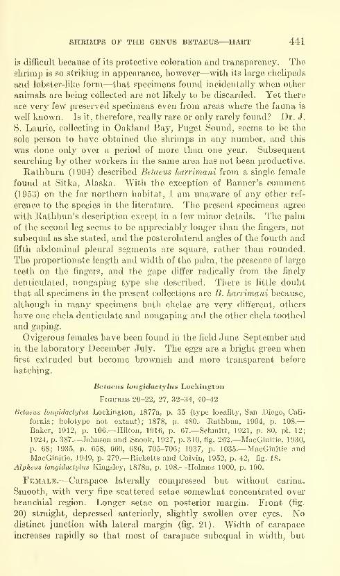
SHRIMPS OF THE GENUS BETAEUS—HART 441
is difficult because of its protective coloration and transparency. Theshrimp is so striking in appearance, however—with its large chelipeds
and lobster-like form—that specimens found incidentally when other
animals are being collected are not likely to be discarded. Yet there
are very few preserved specimens even from areas where the fauna is
well kno\vn. Is it, therefore, really rare or only rarely found? Dr. J.
S. Laurie, collecting in Oakland Bay, Puget Sound, seems to be the
sole person to have obtained the shrimps in any number, and this
was done only over a period of more than one year. Subsequent
searching by other workers in the same area has not been productive.
Rathburn (1904) described Betaeus harrimani from a single female
found at Sitka, Alaska. With the exception of Banner's comment(1953) on the far northern habitat, I am unaware of any other ref-
erence to the species in the literature. The present specimens agi"ee
with Rathbun's description except in a few minor details. The palmof the second leg seems to be appreciably longer than the fingers, not
subequal as she stated, and the posterolateral angles of the fourth and
fifth abdominal plem'al segments are square, rather than rounded.
The proportionate length and width of the palm, the presence of large
teeth on the fingers, and the gape differ radically from the finely
denticulated, nongaping type she described. There is little doubt
that all specimens in the present collections are B. harrimani because,
although in many specimens both chelae are very different, others
have one chela denticulate and nongaping and the other chela toothed
and gaping.
Ovigerous females have been found in the field June-September andin the laboratory December-July. The eggs are a bright green whenfii'st extruded but become brownish and more transparent before
hatching.
Betaeus longidactylus Lockington
Figures 20-22, 27, 32-34, 40-42
Betaeus longidactylus Lockington, 1877a, p. 35 (type locality, San Diego, Cali-
fornia; holotype not extant); 1878, p. 480.—Rathbun, 1904, p. 108.—Baker, 1912, p. 106.—Hilton, 1916, p. 67.—Schmitt, 1921, p. 80, pi. 12;
1924, p. 387.—Johnson and Snook, 1927, p. 310, fig. 262.—MacGinitie, 1930,
p. 68; 1935, p. 658, 660, 686, 705-706; 1937, p. 1035.—MacGinitie andMacGinitie, 1949, p. 279.—Ricketts and Calvin, 1952, p. 42, fig. 18.
Alpheus longidactylus Kingsley, 1878a, p. 198.—Holmes 1900, p. 190.
Female.—Carapace laterally compressed but without carina.
Smooth, with very fine scattered setae somewhat concentrated over
branchial region. Longer setae on posterior margin. Front (fig.
20) straight, depressed anteriorly, slightly swollen over eyes. Nodistinct junction with lateral margin (fig. 21). Width of carapace
increases rapidly so that most of carapace subequal in width, but

442 PROCEEDINGS OF THE NATIONAL MUSEUM vol. 115
lateral margin somewhat undulate. Posterolateral margin curved,
posterior margin angled below deep cardiac notch.
Abdomen smoothly rounded, with posteroventral margins of first
to third segments rounded, those of fourth and fifth angled. First
and fifth pleura ventrally margined with plumose setae. Minutesetae on smooth surface.
Telson (fig. 27) longer than sixth abdominal segment, nearly twice
as long as wide, outer margin slightly undulate. Two pairs of movable
spines on dorsal sm'face, two spines on each posterolateral angle;
outer spines very small. Posterior margin deeply curved; plumose
setae relatively short.
Eye round. Sharp cone-shaped tooth on median part of short,
stout eyestalk.
Antennule with stout peduncle of three subequal segments. Broad-
based scimitar-shaped stylocerite reaching almost to end of second
segment. Outer flagella with stout fused portion before division into
short sensory part and much longer, slender flagellum. Peduncle
with large tooth ventrally.
Antenna with stout peduncle slightly longer than antennular
peduncle. Distal margin of basis produced into sharp tooth. Scale
(fig. 22) broad; wide spine exceeding blade, and separated by distinct
slit distally, outer margin straight. Scale reaches nearly to end of
antennular peduncle. Flagellum longer than carapace, flattened ovoid
in cross section.
Third maxilliped reaching to last segment of antennular peduncle.
Ischiomeropodite flat, relatively narrow, curved over mouth parts,
slightly shorter than exopodite, about twice as long as ultimate, four
times penultimate segment. Medial surfaces all armed with bristles,
which are particularly dense on distal segment.
First legs (fig. 32) usually similar in size and shape, with chela muchshorter than carapace. Ischium short. Merus slender, increasing in
size distally, somewhat triangular in outline with scattered tubercles.
Shallow groove on outer side where second leg fits. Carpus short,
smooth, cup-shaped, with sHght dorsal and larger ventral flattened
projection. Chela narrow elongate, covered with fine spines. Fingers
longer than palm, narrow gape, with one large flat tooth medially on
fixed finger, another proximally, which meshes with similar one on
dactylus. Curved corneous tips cross and intermesh. Chela sub-
rectangular, but deepest at base of dactylus. There may not be any
large teeth nor any gape when fingers are closed, particularly in small
individuals (fig. 33).
Second leg (fig. 40) slender, with ischium subequal to merus. First
joint of carpus slightly longer than next three together, subequal to
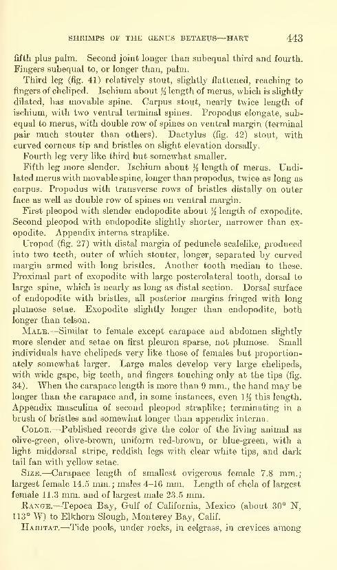
SHRIMPS OF THE GENUS BETAEUS—HART 443
fifth plus palm. Second joint longer than subequal third and fourth.
Fingers subequal to, or longer than, palm.
Third leg (fig. 41) relatively stout, slightly flattened, reaching to
fingers of cheliped. Ischium about % length of merus, which is slightly
dilated, has movable spine. Carpus stout, nearly twice length of
ischium, with two ventral terminal spines. Propodus elongate, sub-
equal to merus, with double row of spines on ventral margin (terminal
pair much stouter than others). Dactylus (fig. 42) stout, with
curved corneus tip and bristles on slight elevation dorsally.
Fom'th leg very like third but somewhat smaller.
Fifth leg more slender. Ischium about K length of merus. Undi-
lated merus with movable spine, longer than propodus, twice as long as
carpus. Propodus with transverse rows of bristles distally on outer
face as well as double row of spines on ventral margin.
First pleopod with slender endopodite about % length of exopodite.
Second pleopod with endopodite slightly shorter, narrower than ex-
opodite. Appendix interna straplike.
Uropod (fig. 27) with distal margin of peduncle scalelike, produced
into two teeth, outer of which stouter, longer, separated by curved
margin armed with long bristles. Another tooth median to these.
Proximal part of exopodite with large posterolateral tooth, dorsal to
large spine, which is nearly as long as distal section. Dorsal surface
of endopodite with bristles, all posterior margins fringed with long
plumose setae. Exopodite slightly longer than endopodite, both
longer than telson,
Male.—Similar to female except carapace and abdomen slightly
more slender and setae on first pleuron sparse, not plumose. Small
individuals have chelipeds very like those of females but proportion-
ately somewhat larger. Large males develop very large chelipeds,
with wide gape, big teeth, and fingers touching only at the tips (fig.
34). When the carapace length is more than 9 mm., the hand may be
longer than the carapace and, in some instances, even Iji this length.
Appendix masculina of second pleopod straplike; terminating in a
brush of bristles and somewhat longer than appendix interna.
Color.—Published records give the color of the living animal as
olive-green, olive-brown, uniform red-brown, or blue-green, with a
Hght middorsal stripe, reddish legs with clear white tips, and dark
tail fan with yellow setae.
Size.—Carapace length of smallest ovigerous female 7.8 mm.;
largest female 14.5 mm.; males 4-16 mm. Length of chela of largest
female 11.3 mm. and of largest male 23.5 mm.Range.—Tepoca Bay, Gulf of California, Mexico (about 30° N,
113° W) to Elkhorn Slough, Monterey Bay, Calif.
Habitat.—Tide pools, under rocks, in eelgrass, in crevices among
444 PROCEEDINGS OF THE NATIONAL MUSEUM vol. 115
materials on boat bottoms, and paired in burrows of echiuroid wormUrechis caupo Fisher and MacGinitie or mud shrimp Upogebia puget-
tensis (Dana).
Material.—The following specimens were examined:
Mexico.—San Felipo, May 11, 1937, S. A. Glassell, 1 female.
California.—San Diego, dredged, March 9, 1949, T. E. Bowman, 2 males
(USNM 98059).—La Jolla, from kelp holdfasts on beach, Aug. 28, 1918, W. L.
Schmitt, 2 females (USNM 53910).—La Jolla, Sept. 21, 1918, W. L. Schmitt,
1 female, 2 males (USNM 53914).—La Jolla, tide pools, Sept. 22, 1918, W. L.
Schmitt, 1 ovigerous female, 9 males (USNM 53925).—La Jolla, Bird Rock,
Nov. 3, 1942, 1 female, 1 male (Pacific Marine Station, 846, 847).—La Jolla, kelp
beds, March 16, 1954, C. Limbaugh, 1 male (USNM 96415).—Laguna Beach,
W. A. Hilton, 1 ovigerous female, 2 males (USNM 48982).—Laguna Beach, W. A.
Hilton, 1 ovigerous female, 2 males (USNM 50582).—Laguna Beach, W. A. Hilton,
2 ovigerous females (USNM 50586).—Laguna Beach, W. A. Hilton, 1 male
(USNM 50590).)—Laguna Beach, under stones, W. A. Hilton, 1 ovigerous female,
2 males, pale olive-green (USNM 52756).—Newport Bay, Jan. 20, 1929, Dec.
16, 1930, G. E. MacGinitie, 1 female, 1 male, 12 juveniles (Hopkins Marine
Station).—Santa Catalina Island, west shore of Catalina Harbor, Dec. 12, 1912,
Anton Dohrn, 3 females (USNM 49981).—Santa Catalina Island, Nov. 28, 1913,
Anton Dohrn, 5 females, 10 males (USNM 49980).—Catalina Island, Isthmus
Harbor, Nov. 27, 1913, Anton Dohrn, 9 females, 8 males (USNM 49982).—CatalinaIsland, west shore of Catalina Harbor, Nov. 28, 1913, Anton Dohrn, 20 juveniles
(USNM 50012).—Long Beach, H. N. Lowe, 2 males (USNM 53018).—SanPedro, C. F. Baker, 1 male (USNM 29309).—San Pedro, Rocky Point, Feb. 2,
1912, Anton Dohrn, 2 males (USNM 49978).—San Pedro, Portuguese Bend,
June 26, 1914, Anton Dohrn, 1 male (USNM 49979).—San Pedro, GovernmentBreakwater and Point Fermin, littoral, April 2, 1913, Anton Dohrn, 3 females,
6 males (USNM 49983).—San Pedro, first rocks north of Port Los Angeles, Dec.
5, 1911, Anton Dohrn, 3 females, 2 males (USNM 49993).—San Pedro, May4, 1919, E. P. Chace, 1 female (USNM 54048).—San Pedro, June 25, 1895,
5. J. Holmes (?), Univ. of Cahfornia, 20 females (14 ovigerous, 5 parasitized),
6 males (USNM 87439).—Santa Monica, near long wharf, Venice Marine Bio-
logical Station, 1 male (USNM 46118).—Santa Monica, April 1923, E. P. Chace,
1 male (USNM 57174).—Morro Bay, Feb. 2, 1939, S. A. Glassell, 1 male.—SantaMonica, 8 miles north, open beach in Upogebia burrow, Feb. 2, 1931, G. E.
MacGinitie, 1 female (Hopkins Marine Station).—Monterey Bay, Elkhorn
Slough, April 3, 1930, G. E. MacGinitie, 1 female (Hopkins Marine Station).
Notes.—Betaeus longidactylus is a stouter species than B. harrimani
and may be separated easily on the basis of the chelipeds. Theranges overlap in the region from Newport Bay to Monterey, but
few specimens of either species are present in collections from this
area. Ricketts and Calvin (1952) state that B. longidactylus is very
plentiful in southern California, where it occurs on the outer coast,
but in the northern part of its range the species is restricted to quiet
waters. Berried females have been taken in January, June, August,
and September.

SHRIMPS OF THE GENUS BETAEUS—HART 445
Betaeus ensenadensis Glassell
Figures 23-25, 28, 35, 36, 43-45
Betaeus, new species, MacGinitie, 1934, pp. 173-174; 1937, pp. 1035-1036.
Betaexis ensenadensis Glassell 1938, pp. 416-419, pi. 28 (type locality, Estero de la
Punta Banda, Ensenada, Baja California, Mexico; holotype male, San Diego
Soc. Nat. Hist. No. 1121).—MacGinitie and MacGinitie, 1949, p. 279.
Female.—Carapace laterally compressed but without carina;
smooth, naked. Front (fig. 23) shghtly curved, depressed anteriorly.
Anterior margin without sinuses or with shallow ones. Anterolateral
angle obtuse (fig. 24). Width of carapace increases rapidly to base of
third maxilliped, followed by slight undulation over base of first legs.
Posterolateral region rounded. Blunt angle beneath cardiac notch.
Abdomen smoothly rounded with posteroventral margins of pleura
of first four segments rounded, fifth bluntly angled. Ventral margin
of first pleuron sparsely setose.
Telson (fig. 28) subequal to sixth segment, nearly twice as long as
wide, with outer margins slightly undulate. Two pairs of movable
spines on dorsal sm'face, two spines at each posterolateral angle, outer
ones very small. Posterior margin slightly curved, armed with plu-
mose setae.
Eye somewhat oval, with small flat tooth on eyestalk.
Antennule with stout scimitar-shaped stylocerite, reaching to about
distal third of second segment of peduncle. Subterminally first seg-
ment of peduncle produced into thin tooth ventrally. Second segment
of peduncle longer than fu'st, which is longer than third. Flattened
fused part of outer flagella shorter than peduncle; distally divided
into few free segments and slender flagellum subequal to fused portion.
Inner flagellum considerably longer.
Antennal peduncle subequal to antennular peduncle, with ventral
distal part of basis produced into sharp tooth. Scale (fig. 25) with
spine much longer than narrow blade, slit between minute; outer
margin almost straight. Scale reaching to middle of third segment of
antennular peduncle. Flagellum short, wide, much flattened, reaching
slightly past posterior margin of carapace.
Thu'd maxilhped reaching to middle of second joint of antennular
peduncle. Ischiomeropodite sparsely setose, slender, flattened, some-
what curved, subequal to two distal segments together in length.
Exopodite longer than ischiomeropodite, relatively stout. Penulti-
mate segment shortest, sparsely setose. Distal segment banded
medially with many short bristles.
First legs (fig. 35) similar. Ischium short with ridge flatly tubercu-
lated ventrally. Stout merus with deep groove into which second leg
fits. Two evenly toothed ridges ventrally, transverse sulcus distally.

446 PROCEEDINGS OF THE NATIONAL MUSEUM vol. us
Carpus short, rounded dorsally, smooth, except for two tubercles or
teeth on inner margin. Platehke ridge ventrally with somewhat rough
edge. Chela covered with fine denticles, particularly on outer sur-
faces. Inner side of chela flat, outer rounded. Dactylus slightly
shorter than palm, with three teeth: one small, near junction with palm
and meshing with similar tooth on fixed finger; large conical tooth
near middle; terminal tooth meshing with truncate terminal part of
fixed finger. Fixed finger stout with wide cutting surface bordered on
each side with denticulated, elevated edge; both fingers end in sharp
tips that cross when closed. Distinct gape.
Second leg (fig. 43) slender. Merus and ischium subequal, slightly
shorter than 5-jointed carpus, which has fu-st joint longer than fifth
and about equal to second, third, and foiu'th together, which are
subequal to each other. Fingers subequal to palm.
Third (fig. 44) and fourth legs flattened with dilated merus. Is-
chium nearly ji merus in length. Distinct movable spines on merus
and ischium. Sparsely setose; single spine and setae on distal ventral
part of carpus, usually three equidistant spines and terminal pair on
ventral part of propodus. Few bristles dorsaUy. Needle-sharp
curved dactylus (fig. 45), more than % length of propodus.
Fifth leg without movable spine on ischium, but one on undilated
merus. Kather sparse brush of bristles on distal part of propodus.
Dactylus similar to that of thu'd or fourth leg.
First pleopod with round-tipped endopodite less than ji length of
exopodite. Second to fifth pleopods with exopodite slightly longer
than endopodite, both slender. Appendix interna stout and straplike.
Uropod (fig. 28) broad and considerably longer than telson. Pe-
duncle with straight margin; two small teeth separated by serrated
spines graduated in length with longest medial. Proximal margin
of exopodite with serrated spines of varying lengths forming thatch
between small lateral tooth and large distal spine.
Male.—With straight front. Cheliped (fig. 36) proportionately
heavier, wider than that of female (median tooth on dactylus maybe missing). Appendix masculina on second pleopod only slightly
longer than appendix interna, blunt tip armed with bristles of varied
lengths.
Color.—"The carapace, abdomen and chelipeds are covered with
light tinted chromatophores in reds and blues, the fingers and telson
are tinted a light purple" (GlasseU, 1938). "Minute red and blue
spots" (MacGinitie and MacGinitie, 1949).
Size.—Carapace length of smallest ovigerous female 5 mm.; largest
female 7 mm.; males 4.5-6.8 mm. Length of chela of largest female
6 mm.; of largest male 5.6 mm.
SHRIMPS OF THE GENUS BETAEUS—^HART 447
Range.—El Estero de la Punta Banda, Ensenada, Baja California,
Mexico, to False Bay, San Diego, Calif.
Habitat.—MacGinitie (1937), MacGinitie and MacGinitie (1949),
and Glassell (1938) record the shrimps living in pairs in the burrowsof the ghost shrimp Callianassa californiensis Dana at Ensenadaand in the burrows of the mud shrimp Uiiogehia pugettensis (Dana)at San Diego,
Material.—The following specimens were examined:
Mexico.—El Estero de la Punta Bauda, Ensenada, Baja California, Dec. 19,
1930, G. E. MacGinitie, 1 ovigerous female, 1 male.—El Estero de la PuntaBanda, Ensenada, Baja California, Jan. 21, 1932, G. E. MacGinitie, 7 females
(1 ovigerous), 3 males.
California.— Mission Bay, in burrow, December 1949, T. E. Bowman, 1 speci-
men (USNM 102442).—False Bay, San Diego, with Upogebia, Nov. 18, 1937,
G. E. MacGinitie, 3 females (1 ovigerous).
Notes.-—A small, slight shrimp of the same type as Betaeus
harrimani and B. longidadylus, but easily distinguished from these bythe shape of the antennal scale, the truncate tip of the fixed finger,
the large teeth on the dactylus of the chela, tlie presence of a movablespine on the ischium of the third and fourth walkuig legs, and the
characteristics of the uropod. Ovigerous females have been taken in
November, December, and January.
Betaeus harfordi (Kingsley)
Figures 46, 47, 54, 58-61, 73, 74
Betaeus eqidmanus Lockington, 1877b, p. 43 (type locality, Cataliua Island,
California; types not extant).
Alpheiis harfordi Kingsley, 1878a, pp. 198-199 (type localities, Santa Barbara andCatalina Island, California; types not located); 1878b, pp. 58-59; 1882,
p. 124, pi. 2, fig. 4.
Alpheus aequalis Kingsley, 1878a, p. 199.
Betaeus aequalis Lockington, 1878, pp. 479-480.
Alpheus (Betaeus) aequalis Holmes, 1900, p. 189, pi. 3, fig. 47 (part).
Betaeus harfordi Rathbun, 1904, p. 108.—Schmitt, 1921, p. 79, fig. 55.—MacGinitieand MacGinitie, 1949, p. 279 (part).—Ricketts and Calvin, 1952, pp. 323-324(part)
.
Female.—Carapace laterally compressed but without carina;
smooth, naked. Front (fig. 46) shallowly emarginate, produced to
cover eyes. Anterior margin of carapace (fig. 47) shallowly cmwed,meeting lateral margin at blunt angle just ventral to base of antenna.
Width of carapace increasing to base of third maxillipeds, gradually
decreasing to curved posterolateral margin. Posterior margin pro-
trudes slightly before decreasing ventral to cardiac notch.
Abdomen smooth, evenly rounded. Naked except for plumosesetae on ventral margin of pleura of first segment. Pleura with
693-433—63 2
448 PROCEEDINGS OF THE NATIONAL MUSEUM vol. us
posterolateral margins evenly rounded except fifth, which is somewhatacute.
Telson (fig. 58) wide, flat, shorter than uropods. One and one-half
to twice length of sixth segment. Lateral margins curved. Distally
about % as wide as proximally. Two pairs of small movable spines
on distal half of dorsal sm-face, but posterolateral spines vestigial or
missing. Posterior margin cm'ved, bearing long plumose setae.
Eye large, round, on short stalk with small median tooth.
Antennule with stylocerite narrow, sharp-pointed, reaching to distal
quarter of second segment of peduncle. Ventrally a slender scimitar-
shaped tooth on distal part of first segment of peduncle. Peduncle
slender, about % carapace length. Second segment about twice length
of subequal first and third segments. Flagella slender, short: outer
flagella fused for most of length with only terminal tip of sensory part
free and remaining flagellmn short, slender.
Antenna slender. Peduncle with wide tooth ventrally on margin of
first segment; slightly longer than antennular peduncle. Scale (fig.
54) narrow, with stout spine longer than narrow blade, separated from
it with slit for about % of length. Flagcllum longer than carapace,
ovoid in cross section.
Third maxilliped reaching to end of second segment of antennular
pedmicle. Relatively narrow, with flattened ischiomeropodite sub-
equal to distal segment, sparsely bristled. Penultimate segment
sparsely setose, slightly shorter than distal, which is armed medially
with bands of stout bristles and scattered longer setae. Exopodite
slightly longer than ischiomeropodite.
First legs subequal, large. Chela slightly shorter than carapace.
Ischimn short. Merus naked except for few setae on inner margin:
triangular in cross section, wdth dorsal carina smooth, sharp, ending in
curved tooth distally, outer ventral ridge smooth, inner one \vith someflattened tubercles. Carpus short, smooth, naked, cup-shaped with
small ventral plate. Chela (fig. 60) smooth, flattened laterally: few
fine scattered setae on dorsal ridge, stifT setae near tips of fingers and
on cutting surfaces. Palm and fingers subequal but fixed finger muchwider than dactjdus. Somewhat flattened teeth of cutting surface
may mesh without any gape or may have hiatuses on both fingers
resulting in a round foramen. Chelae usually alike in dentition.
Dorsal ridge of fixed finger and palm compressed, so that margin
knife-edged.
Second leg (fig. 61) slender. Ischium longer than merus; both flat-
tened laterally. Carpus with first joint longest, fifth next, others
shorter, subequal. Chela longer than first joint, fingers subequal to
palm. Cutting smface of fingers spinulate.
Third leg (fig. 73) stout, flattened laterally, with scattered fine
SHRIMPS OF THE GENUS BETAEUS—^HART 449
setae. Ischium and merus comprise }^ length of appendage. Large
movable spine on merus. Carpus, with minute terminal spines, sub-
equal in length to propodus. Propodus armed ventrally with small
setae and vestigial spines. Terminal pair larger. Dactylus (fig. 74)
short, stout, bifid, but claws masked by two median bands of short
stiff bristles.
Fourth leg similar to third, but all segments except ischium slightly
smaller.
Fifth leg distinctly shorter than fourth. Subequal merus and
propodus each longer than carpus. Brush of 4-5 bands of short stiff
setae on distal part of propodus. Dactylus similar to that of third
and fourth legs. No movable spine on merus.
First pleopod with round-tipped endopodite, nearly ji length and
width of slender exopodite. Second pleopod with subequal slender
endopodite and exopodite. Appendix interna straplike, nearly %length of endopodite.
Uropod (fig. 58) with posterior margin of peduncle produced into
two sharp subequal teeth, separated by concave margin with few small
setae. Distal angle of proximal part of exopodite a sharp tooth,
covering base of short stout spine. Uropods longer than telson.
Male.—Similar to female except that chelae (fig. 59) are propor-
tionately larger, subequal in length to carapace. Appendix masculina
with bristles on margin of distal half and on blunt tip; twice as long
as appendix interna, nearly as long as endopodite.
Color.—Dark purple (Lockington, 1878; Holmes, 1900), blue-black
(specimen label, E. P. Chace), deep blue (specimen label, W. A.
Hilton) and deep blue, occasionally reddish-brown on sides (Mrs.
G. E. MacGinitie).
Size.—Carapace length of smallest ovigerous female 6 mm.; largest
female 10.7 mm; males 3.3-8 mm. Length of chela of largest female
8.5 mm.; of largest male 8 mm.Range.—^Magdalena Bay, Mexico, to Fort Bragg, Calif, (personal
communication, Mr. Keith W. Cox) ; intertidal to depth of 12 fathoms.
This is apparently a very elusive shrimp that leaves its host with little
provocation, with the residt that, unless precautions are taken to
prevent its escape, it may be consistently missed when abalones are
collected. UntU Mr. Cox was consulted, no specimens had been
recorded in any collections taken north of Santa Monica, even with
Mr. D. Montgomery checking approximately 350 specimens of
Haliotis from the vicinity of San Luis Obispo. On the other hand,
Dr. Howard Teas, on April 4, 1953, collected at El Estero de PuntaBanda, Ensenada, Baja California, Mexico, and reported to the Mac-Ginities that about four out of every five Haliotis fulgens had a
Betaeus in the mantle cavity.
093-433—63 3

450 PROCEEDINGS OF THE NATIONAL MUSEUM vol. ii5
Habitat.—Found living in mantle cavity of ab alone and occasion-
ally free, latter probably temporary state, owing to distm-bance of
normal host. The specimens examined have occm^red most commonlywith the pink ab alone, Haliotis corrugata Gray, but also with the red
abalone, H. rufescens Swainson, the black abalone, H. cracherodii
Leach, and the green abalone, H. fulgens PhUlipi. Mr. Keith Coxreports them also from H. kamtschatkana Jonas, H. wallalensis
Stearns, H. sorenseni Bartsch, and H. assimilis Dall.
Material.—The following specmiens were examined:
Mexico.—Magdalena Bay, Belchers Point, on shore, Jan. 30, 1938, S. A.
Glassell, 1 specimen, dried.
California.—La Jolla Cove, July 14, 1942, 1 female (Pacific Marine Station
845).—La Jolla, kelp beds, March 16, 1954, C. Limbaugh, 1 ovigerous female, I
male (USNM 96414).—La Jolla, with Haliotis rufescens Swainson, March 28,
1952, E. Dodge, 1 ovigerous female.—San Clemente Island, living beneath mantle
of Haliotis, January 1936, Nell Murbarger, 2 ovigerous females, 1 male (USNM77743).—San Clemente Island, from abalone. May 8, 1888, Albatross, 2 females
(USNM 63453).—Laguna Beach, from abalone, Sept. 19, I9I8, W. A. Hilton,
I female, deep blue (LTSNM 53598).—Laguna Beach, shale reef ]/i mile offshore,
midway to Corona del Mar, 12 fathoms, from mantle cavity of Haliotis corrugata
Gray, April 25, 1958, Robert Given, from N. MacGinitie, I ovigerous female.
—
Laguna Beach, off Salt Creek, 8 fathoms, from Haliotis corrugata Gray, May 21,
1958, Dale Seemen, from N. MacGinitie, 20 females (7 ovigerous), 5 males;
May 26, 1958, 7 females (5 ovigerous).—Laguna Beach, 8 fathoms, from Haliotis
corrugata (2 specimens from Haliotis rufescens), April 28, 1958, Dale Seeman,
from N. MacGinitie, 4 females (I ovigerous), 10 males.—Catalina Island, com-mensal in shell of green abalone, Venice Marine Biological Station, 1 ovigerous
female (USNM 46119).—Catalina Island, Little Harbor, Dec. 27, 1912, AntonDohrn, 2 males (USNM 49977).—San Pedro, Rocky Point, from mantle of black
abalone, Feb. 2, 1912, Anton Dohrn, 1 ovigerous female (USNM 49975).—SanPedro, in gill chamber of Haliotis cracherodii Leach, June 1930, E. P. Chace,
1 ovigerous female, blue-black when alive (USNM 64087).—Santa Barbara
Island, from Haliotis rufescens, 8 fathoms, Sept. 11, 1962, Keith W. Cox, 22
ovigerous females, 7 males.—Santa Monica Bay, Malibu Point, under mantle of
Haliotis washed up on beach, Dec. 19, 1937, A. Camp, from R. Crocker, 1 female
(Hopkins Marine Station).—Southern California, Nov. 27, 1913, Anton Dohrn,
1 female (USNM 49976).
Notes.—Mr. Keith W. Cox informs me that there seems to be a
definite correlation in size between the shrimps and their hosts:
large shrimps in large abalones, small in small. The usual number is
one shrimp per host. Exceptions do occiu* and on several occasions
up to four have been found in the cavity between mantle and shell.
MacGinitie and MacGinitie (1949, p. 279) state:
We have found this shrimp with its purplish-red color form living in the mantle
cavity of the green abalone Haliotis fulgens. For over a year one has been living
under the protectorate of an abalone in one of our aquariums. It usually stays
with its head near the region of the mouth of the abalone, and although it is byno means inactive, its host does not give any evidence of being disturbed by its
movements. On rare occasions we have seen it make short excursions onto the
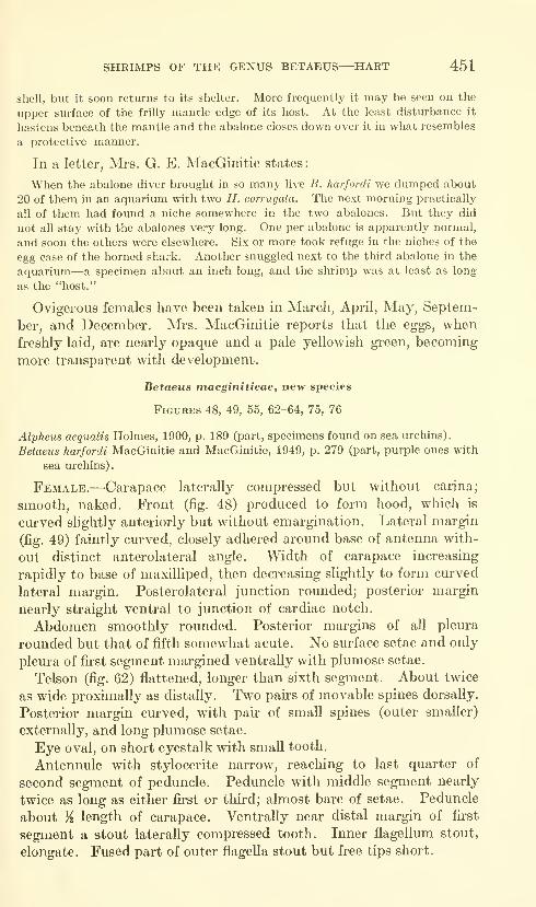
SHRIMPS OF THE GENUS BETAEUS—HART 451
shell, but it soon returns to its shelter. More frequently it may be seen on the
upper surface of the frilly mantle edge of its host. At the least disturbance it
hastens beneath the mantle and the abalone closes down over it in what resembles
a protective manner.
In a letter, Mrs. G. E. MacGinitie states:
When the abalone diver brought in so many live B. harfordi we dumped about
20 of them in an aquarium with two H. corrugaia. The next morning practically
all of them had found a niche somewhere in the two abalones. But they did
not all stay with the abalones very long. One per abalone is apparently normal,
and soon the others were elsewhere. Six or more took refuge in the niches of the
egg case of the horned shark. Another snuggled next to the third abalone in the
aquarium—a specimen about an inch long, and the shrimp was at least as long
as the "host."
Ovigerous females have been taken in March, April, May, Septem-
ber, and December. Mrs. MacGinitie reports that the eggs, when
freshly laid, are nearly opaque and a pale yellowish green, becoming
more transparent with development.
Betaeus macginitieae, new species
Figures 48, 49, 55, 62-64, 75, 76
Alpheus aequalis Holmes, 1900, p. 189 (part, specimens found on sea urchins).
Betaeus harfordi MacGinitie and MacGinitie, 1949, p. 279 (part, purple ones with
sea urchins).
Female.—Carapace laterally compressed but without carina;
smooth, naked. Front (fig. 48) produced to form hood, which is
curved slightly anteriorly but without emargination. Lateral margin
(fig. 49) faintly curved, closely adhered around base of antenna with-
out distinct anterolateral angle. Width of carapace increasing
rapidly to base of maxilliped, then decreasing slightly to form curved
lateral margin. Posterolateral junction rounded; posterior margin
nearly straight ventral to junction of cardiac notch.
Abdomen smoothly rounded. Posterior margins of all pleura
rounded but that of fifth somewhat acute. No surface setae and only
pleura of first segment margined ventrally with plumose setae.
Telson (fig. 62) flattened, longer than sixth segment. About twice
as wide proximally as distally. Two pairs of movable spines dorsally.
Posterior margin curved, with pair of small spines (outer smaller)
externally, and long plumose setae.
Eye oval, on short eyestalk with small tooth,
Antennule with stylocerite narrow, reaching to last quarter of
second segment of peduncle. Peduncle with middle segment nearly
twice as long as either first or third; almost bare of setae. Peduncle
about /^ length of carapace. Ventrally near distal margin of first
segment a stout laterally compressed tooth. Inner flagellum stout,
elongate. Fused part of outer flagella stout but free tips short.
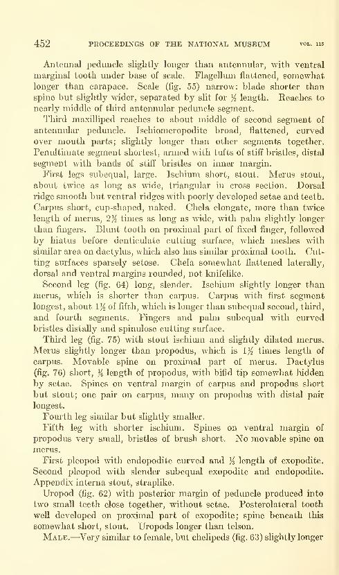
452 PROCEEDESTGS OF THE NATIONAL MUSEUM vol. 115
Antennal peduncle slightly longer than antennular, with ventral
marginal tooth under base of scale. Flagellum flattened, somewhatlonger than carapace. Scale (fig. 55) narrow: blade shorter than
spine but slightly wider, separated by slit for ji length. Reaches to
nearly middle of third antennular peduncle segment.
Thkd maxilliped reaches to about middle of second segment of
antennular peduncle. Ischiomeropodite broad, flattened, curved
over mouth parts; slightly longer than other segments together.
Penultimate segment shortest, armed with tufts of stiff bristles, distal
segment with bands of stiff bristles on inner margin.
First legs subequal, large. Ischium short, stout. Merus stout,
about twice as long as wide, triangular in cross section. Dorsal
ridge smooth but ventral ridges with poorly developed setae and teeth.
Carpus short, cup-shaped, naked. Chela elongate, more than twice
length of merus, 2^ times as long as wide, wdth palm slightly longer
than fingers. Blunt tooth on proximal part of fixed finger, followed
by hiatus before denticulate cutting siu-face, which meshes with
similar area on dactylus, which also has similar proximal tooth. Cut-
ting surfaces sparsely setose. Chela somewhat flattened lateraUy,
dorsal and ventral margins rounded, not knifelike.
Second leg (fig. 64) long, slender. Ischium slightly longer than
merus, which is shorter than carpus. Carpus with fu'st segmentlongest, about 1}^ of fifth, which is longer than subequal second, third,
and fom'th segments. Fingers and palm subequal with curved
bristles distally and spinulose cutting surface.
Third leg (fig. 75) with stout ischium and slightly dflated merus.
Merus slightly longer than propodus, which is 1% times length of
carpus. Movable spine on proximal part of merus. Dactylus
(fig. 76) short, }i length of propodus, with bifid tip somewhat hidden
by setae. Spines on ventral margin of carpus and propodus short
but stout; one pair on carpus, many on propodus with distal pair
longest.
Fourth leg similar but slightly smaller.
Fifth leg with shorter ischimn. Spines on ventral margin of
propodus very small, bristles of brush short. No movable spine onmerus.
First pleopod with endopodite curved and ji length of exopodite.
Second pleopod with slender subequal exopodite and endopodite.
AppendLx interna stout, straplike.
Uropod (fig. 62) with posterior margin of peduncle produced into
two small teeth close together, without setae. Posterolateral tooth
well developed on proximal part of exopodite; spine beneath this
somewhat short, stout. Uropods longer than telson.
Male.—^Very similar to female, but chelipeds (fig. 63) slightly longer
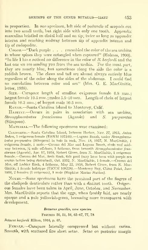
SHRIMPS OF THE GENUS BETAEUS
—
^HART 453
in proportion. In one specimen, left side of peduncle of uropods cut
into two small teeth, but right side with only one tooth. Appendixmasculina bristled on distal half and on tip; twice as long as appendixinterna and reaching midway between tip of appendix interna andtip of endopodite.
Color.—"Dark purple . . . resembled the color of the sea urchins
in whose spines they were entangled when captured" (Holmes, 1900).
"In life I have noticed no difference in the color of B. harfordi and the
last one we are sending you from the sea lu-chin. For the most part,
they are mostly blue, but sometimes along the side the color is a
reddish brown. The claws and tail are almost always entirely blue
regardless of the color along the sides of the abdomen. I could find
no correlation between color and sex" (Mrs. G. E. MacGinitie,
letter, 1958).
Size.—Carapace length of smallest ovigerous female 6.8 mm.;largest female 10.5 mm. ; males 5.5-10 mm. Length of chela of largest
female 10.3 mm.; of largest male 10.5 mm.Range.^—Santa Catalina Island to Monterey, Calif.
Habitat.—Occurs in pairs in association with sea urchins,
Strongylocentrotus franciscana (Agassiz) and S. purpuratus
(Stimpson)
.
Material.—-The following specimens were examined
;
California.—Santa Catalina Island, Isthmus Harbor, Nov. 27, 1913, AntonDohrn, 1 ovigerous female (USNM 107546).—Laguna Beach, under Strongylocen-
trotus purpuratus (Stimpson) in hole in rock, Nov. 18, 1937, N. MacGinitie, 1
ovigerous female, 1 male.—Corona del Mar and Laguna Beach, shale reef mid-way between, ]i mile offshore, 5 fathoms, from beneath Strongylocentrotus fran-
ciscana (Agassiz), Apr. 17, 1958, Robert Given, from N. MacGinitie, 1 ovigerous
female.—Corona del Mar, Arch Rock, tide pool (may have been with purple sea
urchin before being disturbed), Oct. 1932, N. MacGinitie, 1 female.—Corona del
Mar, off Ladder Rock, 4 fathoins. May 22, 1958, Robert Given, from N. Mac-Ginitie, 1 female holotype (USNM 108228).— Monterey Bay, China Point, June
1908, 2 females (1 ovigerous), 1 male (Hopkins Marine Station).
Notes.—Some specimens have the proximal part of the fingers of
the chelipeds denticulate rather than with a distinct tooth. Oviger-
ous females have been taken in April, June, October, and November.Mrs. MacGinitie reports that the eggs, when freshly laid, are nearly
opaque and a pale yeUowish-green, becoming more transparent with
development.
Betaeus gracilis, new species
Figures 50, 51, 66, 65-67, 77, 78
Betaeus harfordi Hilton, 1916, p. 67.
Female.—Carapace laterally compressed but without carina.
Smooth, with scattered fine short setae. Setae on posterior margin
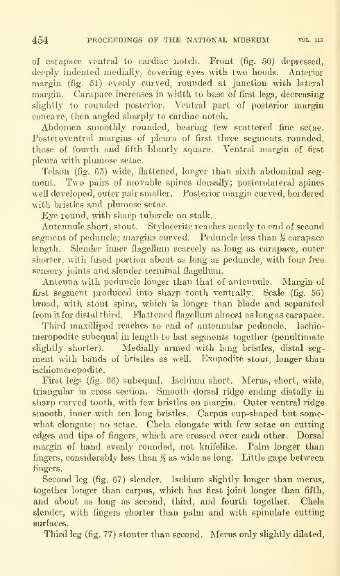
454 PROCEEDINGS OF THE NATIONAL MUSEUM vol. 115
of carapace ventral to cardiac notch. Front (fig. 50) depressed,
deeply indented medially, covering eyes with two hoods. Anterior
margin (fig. 51) evenly curved, rounded at junction with lateral
margin. Carapace increases in width to base of first legs, decreasing
slightly to rounded posterior. Ventral part of posterior margin
concave, then angled sharply to cardiac notch.
Abdomen smoothly rounded, bearing few scattered fine setae.
Posteroventral margins of pleura of fu-st three segments rounded,
those of fourth and fifth bluntly square. Ventral margin of fu-st
pleura with plumose setae.
Telson (fig. 65) wide, flattened, longer than sixth abdominal seg-
ment. Two pairs of movable spines dorsally; posterolateral spines
well developed, outer pair smaller. Posterior margin curved, bordered
with bristles and plumose setae.
Eye round, with sharp tubercle on stalk.
Antennule short, stout. Stylocerite reaches nearly to end of second
segment of peduncle; margins curved. Peduncle less than % carapace
length. Slender inner flagellum scarcely as long as carapace, outer
shorter, with fused portion about as long as peduncle, with four free
sensory joints and slender terminal flagellum.
Antenna with peduncle longer than that of antennule. Margin of
fu-st segment produced into sharp tooth ventrally. Scale (fig. 56)
broad, with stout spine, which is longer than blade and separated
from it for distal thhd. Flattened flagellmn ahnost as long as carapace.
Third maxilliped reaches to end of antennular peduncle. Ischio-
meropodite subequal in length to last segments together (penultimate
slightly shorter). Medially armed with long bristles, distal seg-
ment with bands of bristles as well. Exopodite stout, longer than
ischiomeropodite.
First legs (fig. 66) subequal. Ischium short. Merus, short, wide,
triangular in cross section. Smooth doreal ridge ending distally in
sharp curved tooth, with few bristles on margin. Outer ventral ridge
smooth, inner with ten long bristles. Carpus cup-shaped but some-
what elongate; no setae. Chela elongate with few setae on cutting
edges and tips of fingers, which are crossed over each other. Dorsal
margin of hand evenly rounded, not knifelike. Palm longer than
fingers, considerably less than ji as wide as long. Little gape between
fingers.
Second leg (fig. 67) slender. Ischium slightly longer than merus,
together longer than carpus, which has first joint longer than fifth,
and about as long as second, third, and fourth together. Chela
slender, with fingers shorter than palm and with spinulate cutting
surfaces.
Third leg (fig. 77) stouter than second. Merus only slightly dilated.
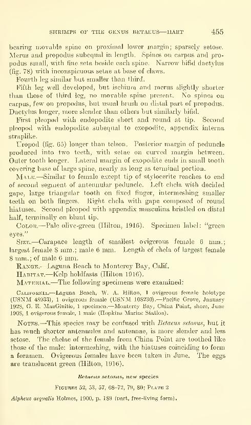
SHRIMPS OF THE GENUS BETAEUS—HART 455
bearing movable spine on proximal lower margin; sparsely setose.
Merus and propodus subequal in length. Spines on carpus and pro-
podus small, with fine seta beside each spine. Narrow bifid dactyliis
(fig. 78) with inconspicuous setae at base of claws.
Fourth leg similar but smaller than third.
Fifth leg well developed, but ischium and merus slightly shorter
than those of third leg, no movable spine present. No spines on
carpus, few on propodus, but usual brush on distal part of propodus.
Dactylus longer, more slender than others but similarly bifid.
First pleopod with endopodite short and round at tip. Second
pleopod with endopodite subequal to exopodite, appendLx interna
straplike.
Uropod (fig. 65) longer than telson. Posterior margin of peduncle
produced into two teeth, with setae on curved margin between.
Outer tooth longer. Lateral margin of exopodite ends in small tooth
covering base of large spine, nearly as long as terminal portion.
Male.—Similar to female except tip of stylocerite reaches to end
of second segment of antennular peduncle. Left chela with decided
gape, large triangular tooth on fixed finger, intermeshing smaller
teeth on both fingers. Right chela with gape composed of round
hiatuses. Second pleopod with appendix masculina bristled on distal
half, terminally on blunt tip.
Color.—Pale olive-green (Hilton, 1916). Specimen label: "green
eyes."
Size.—Carapace length of smallest ovigerous female 6 mm.;largest female 8 mm.; male 6 mm. Length of chela of largest female
8 mm. ; of male 6 mm.Range.—Laguna Beach to Monterey Bay, Calif.
Habitat.—Kelp holdfasts (Hilton 1916).
Material.—The following specimens were examined:
California.—Laguna Beach, W. A. Hilton, 1 ovigerous female holotype
(USNM 48933), 1 ovigerous female (USNM 108230).—Pacific Grove, January
1928, G. E. MacGinitie, 1 specimen.—Monterey Bay, China Point, shore, June
1908, 1 ovigerous female, 1 male (Hopkins Marine Station).
Notes.—^This species may be confused with Betaeus setosus, but it
has much shorter antennules and antennae, is more slender and less
setose. The chelae of the female from China Point are toothed like
those of the male: intermeshing, with the hiatuses coinciding to form
a foramen. Ovigerous females have been taken in June. The eggs
are translucent gi"een (HUton, 1916).
Betaeus setosus, new species
Figures 52, 53, 57, 68-72, 79, 80; Plate 2
Alpheus aequalis Holmes, 1900, p. 189 Cpart, free-living form).
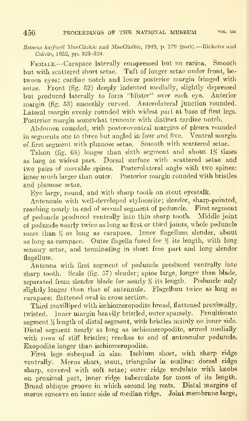
456 PROCEEDINGS OF THE NATIONAL MUSEUM vol. 115
Betaeus harfordi MacGinitie and MacGinitie, 1949, p. 279 (part).—Ricketts and
Calvin, 1952, pp. 323-324.
Female.—Carapace laterally compressed but no carina. Smooth
but with scattered short setae. Tuft of longer setae under front, be-
tween eyes; cardiac notch and lower posterior margin fringed with
setae. Front (fig. 52) deeply indented medially, slightly depressed
but produced laterally to form "blister" over each eye. Anterior
margin (fig. 53) smoothly curved. Anterolateral junction rounded.
Lateral margin evenly rounded with widest part at base of first legs.
Posterior margin somewhat truncate with distinct cardiac notch.
Abdomen rounded, with posteroventral margins of pleura rounded
in segments one to thi-ee but angled in four and five. Ventral margin
of first segment with plumose setae. Smooth with scattered setae.
Telson (fig. 68) longer than sixth segment and about 1% times
as long as widest part. Dorsal surface with scattered setae and
two pairs of movable spines. Posterolateral angle with two spines:
inner much larger than outer. Posterior margin rounded with bristles
and plumose setae.
Eye large, round, and with sharp tooth on stout eyestalk.
Antennule with well-developed stylocerite; slender, sharp-pointed,
reaching nearly to end of second segment of peduncle. First segment
of peduncle produced ventrally into thin sharp tooth. Middle joint
of peduncle nearly twice as long as first or third joints, whole peduncle
more than K as long as carapace. Inner flagellum slender, about
as long as carapace. Outer flagella fused for )^ its length, with long
sensory setae, and terminating in short free part and long slender
flagellum.
Antenna with fu-st segment of peduncle produced ventrally into
sharp tooth. Scale (fig. 57) slender; spine large, longer than blade,
separated from slender blade for nearly }^ its length. Peduncle only
slightly longer than that of antennule. Flagellum twice as long as
carapace: flattened oval in cross section.
Third maxilliped with ischiomeropodite broad, flattened proximally,
twisted. Inner margin heavily bristled, outer sparsely. Penultimate
segment K length of distal segment, with bristles mainly on inner side.
Distal segment nearly as long as ischiomeropodite, armed medially
with rows of stiff bristles; reaches to end of antennular peduncle.
Exopodite longer than ischiomeropodite.
Fu'st legs subequal in size. Ischium short, with sharp ridge
ventrally. Merus short, stout, triangular in outlme: dorsal ridge
sharp, covered with soft setae; outer ridge undulate with knobs
on proximal part, inner ridge tuberculate for most of its length.
Broad oblique groove in which second leg rests. Distal margins of
merus concave on inner side of median ridge. Joint membrane large,
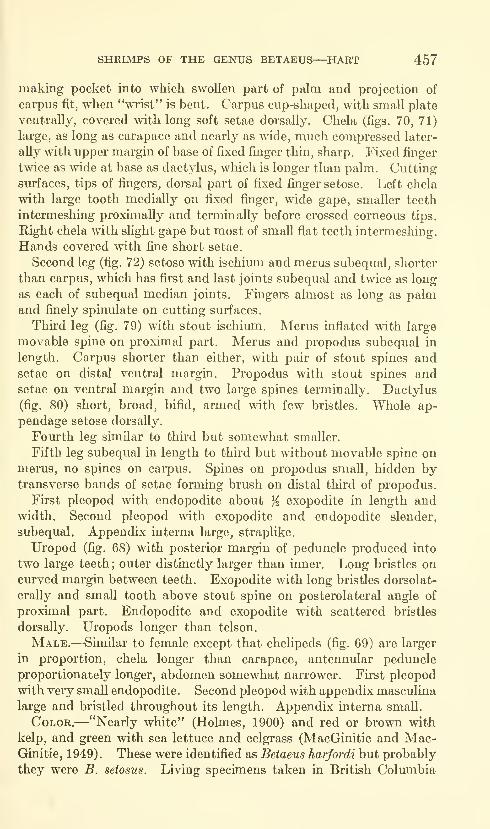
SHRIMPS OF THE GENUS BETAEUS—HART 457
making pocket into which swollen part of palm and projection of
carpus fit, when "wrist" is bent. Carpus cup-shaped, with small plate
ventrally, covered with long soft setae dorsally. Chela (figs. 70, 71)
large, as long as carapace and nearly as wide, much compressed later-
ally with upper margin of base of fixed finger thin, sharp. Fixed finger
twice as wide at base as dactylus, which is longer than palm. Cutting-
surfaces, tips of fingers, dorsal part of fixed finger setose. Left chela
with large tooth medially on fixed finger, wide gape, smaller teeth
intermeshing proximally and terminally before crossed corneous tips.
Right chela with slight gape but most of small flat teeth intermeshing.
Hands covered mth fine short setae.
Second leg (fig. 72) setose with ischimn and merus subequal, shorter
than carpus, which has first and last joints subequal and twice as long
as each of subequal median joints. Fmgers almost as long as palmand finely spinulate on cutting surfaces.
Third leg (fig. 79) with stout ischium. Merus inflated with large
movable spine on proximal part. Merus and propodus subequal in
length. Carpus shorter than either, with pair of stout spines andsetae on distal ventral margin. Propodus with stout spines andsetae on ventral margin and two large spines terminally. Dactylus
(fig. 80) short, broad, bifid, armed with few bristles. Whole ap-
pendage setose dorsally.
Fom"th leg similar to thu-d but somewhat smaller.
Fifth leg subequal in length to third but without movable spine onmerus, no spines on carpus. Spines on propodus small, hidden bytransverse bands of setae forming brush on distal third of propodus.
First pleopod with endopodite about K exopodite in length andwidth. Second pleopod with exopodite and endopodite slender,
subequal. Appendix interna large, straplike.
Uropod (fig. 68) with posterior margin of peduncle produced into
two large teeth; outer distinctly larger than inner. Long bristles oncurved margin between teeth. Exopodite with long bristles dorsolat-
erally and small tooth above stout spine on posterolateral angle of
proximal part. Endopodite and exopodite with scattered bristles
dorsally. Uropods longer than telson.
Male.—Similar to female except that chelipeds (fig. 69) are larger
in proportion, chela longer than carapace, antennular peduncle
proportionately longer, abdomen somewhat narrower. First pleopod
with very small endopodite. Second pleopod with appendix masculina
large and bristled throughout its length. Appendix interna small.
Color.—"Nearly white" (Holmes, 1900) and red or brown with
kelp, and green with sea lettuce and eelgrass (MacGinitie and Mac-Ginitie, 1 949) . These were identified as Betaeus harfordi but probably
they were B. setosus. Living specimens taken in British Columbia
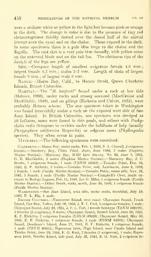
458 PROCEEDINGS OF THE NATIONAL MUSEUM vol. u5
were a uniform white or yellow in the light but became pink or orange
in the dark. The change in color is due to the presence of tiny red
chromatophores thicldy dotted over the dorsal half of the animal
(except over the eyes) and on the chelae. These expand in the dark.
In some specimens there is a pale blue tinge to the chelae and the
flagella. The cast skin is a very pale blue dorsally, with yellow setae
on the antennal blade and on the tail fan. The chitinous tips of the
dactyli of the legs are yellow.
Size.—Carapace length of smallest ovigerous female 4.8 mm.;largest female 8.5 mm.; males 3-7 mm. Length of chela of largest
female 9 mm.; of largest male 9 mm.Range.—Morro Bay, Calif., to Hecate Strait, Queen Charlotte
Islands, British Coliunbia.
Habitat.—The ''B. harfof^di" fomid under a rock at low tide
(Holmes, 1900), under rocks and among seaweed (MacGinitie and
MacGinitie, 1949), and on pilings (Ricketts and Calvin, 1952), were
probably Betaeus setosus. The one specimen taken in Washington
was found intertidally under a rock on the exposed west side of San
Juan Island. In British Columbia, one specimen was dredged at
10 fathoms, some were found in tide pools, and others with Pachy-
cheles rudis Stimpson in cavities mider the holdfasts of kelp (usually
Pterygophora californica Euprecht) or eelgrass roots (Phyllospadix
species). They often occur in pairs.
Material.—The following specimens were examined:
California.—Morro Bay, under rocks, Feb. 1, 1939, S. A. Glassell, 2 ovigerous
females.—Monterey Bay, China Point, shore, June 1908, 2 males (Hopkins
Marine Station).—Monterey Bay, HMS Line Sui-vey, No. 86, June 2, 1931,
G. E. MacGinitie, 2 males (Hopkins Marine Station).—Monterey Bay, J. C.
Brown, 1 ovigerous female, 1 male (USNM 23932).—Tomales Point, Dec. 26,
1939, E. F. Ricketts, 2 males.—Tomales Point, reef, Laviinaria, June 9, 1948,
1 female, 1 male (Pacific Marine Station).—Tomales Point, ocean side, Nov. 28,
1947, 1 female, 1 male (Pacific Marine Station).—Campbell's Cove, inside en-
trance to Bodego Lagoon, Feb. 11, 1949, Lee O. Miles, 1 ovigerous female (Pacific
Marine Station).-—Dillon Beach, rocks, north, June 30, 1946, 1 ovigerous female
(Pacific Marine Station).
Washington.—San Juan Island, west side, under rocks, intertidal, July 10,
1960, P. L. lUg, 1 male.
British Columbia.—Vancouver Island, west coast: Clayoquot Sound, FrankIsland, Cox Bay, Tofino, July 10, 1960, J. F. L. Carl, 3 ovigerous females, 1 male;
Clayoquot Sound, July 29, 1961, J. F. L. Carl, 1 female holotype (USNM 108229),
5 females (3 ovigerous), 3 males; Clayoquot Sound, Round Island, June 30, 1945.
E. F. Ricketts, 2 ovigerous females (USNM 80630); Clayoquot Sound, May 30,
1946, E. F. Ricketts, 1 ovigerous female, 1 male (USNM 84395); Clayoquot
Sound, Deadman's Island, June 27, 1945, E. F. Ricketts, 1 ovigerous female,
1 male (USNM 80635); Esperanza Inlet, High Island, near Catala Island andTatchu Point, June 28, 1934, E. G. Hart, 2 females (1 ovigerous), 1 male; Esper-
anza Inlet, Nootka Island, tide pool, July 25, 1934, E. G. Hart, 2 ovigerous fe-

SHRIMPS OF THE GENUS BETAEUS—HART 459
males, 1 male.—Hecate Strait, Queen Charlotte Islands, 4 miles northeast RosePoint, 10 fathoms, June 24, 1956, T. H. Butler, 1 female.
Notes.—There are a number of variations to be seen in this species.
The inner ridge of the merus of the chelipeds may be almost smooth,
or there may be a few tubercles, or there may be a row of uniform
beadlike knobs. The dentition of the chelae varies considerably
from flat intermeshed teeth, with no gape, to others with a foramen
and/or a large square tooth on the fixed finger, but with the tips of
the fingers always crossed (figs. 70, 71). Most have similar subequal
chelae. To judge from specimens kept in the laboratory, regenerated
chelae may be without teeth and a nmnber of moults may be required
to regain normal size.
Ovigerous females have been taken February-June in California
and May-August in British Columbia. The ovaries and freslily
laid eggs are a pale olive-green, becoming lighter and more trans-
parent with development.
Literature Cited
Baker, B. M.1912. Notes on the Crustacea of Laguna Beach. 1st Ann. Rep. Laguna
Marine Lab., pp. 100-117, figs. 53-64.
Banner, Albert H.
1953. The Crangonidae, or snapping shrimp, of Hawaii. Pacific Sci., vol.
7, no. 1, pp. 2-147, 1 pL, 50 figs.
Dana, James Dwight1852; 1855. Crustacea. Vol. 13 in United States Exploring Expedition
during the years 1838, 1839, 1840, 1841, 1842 under the
command of Charles Wilkes, U.S.N. Philadelphia, part 1,
685 pp. (1852); atlas, 27 pp., 96 pis. (1855).
Glassell, Steve A.
1938. New and obscure decapod Crustacea from the West American coasts.
Trans. San Diego Soc. Nat. Hist. vol. 8, no. 33, pp. 411-454, pis.
27-36.
Hilton, W. A.
1916. Crustacea from Laguna Beach. Journ. Ent. Zool., Pomona College,
California, vol. 8, pp. 65-73, figs. 1-19.
Holmes, Samuel J.
1900. Synopsis of California stalk-eyed Crustacea. Occas. Pap. California
Acad. Sci., vol. 7, 262 pp., 4 pis.
Holthuis, Lipke B.
1952, The Crustacea Decapoda Macrura of Chile. No. 5 in Reports of
the Lund University Chile Expedition 1948-49. Lunds Univ.
Arsskr., new ser., avd. 2, bd. 47, no. 10, 110 pp., 19 figs.
Johnson, M. E., and Snook, J. H.
1927. Seashore animals of the Pacific coast, 659 pp., figs.
460 PROCEEDLNGS OF THE NATIONAL MUSEUM vol. 115
KiNGSLEY, J. S.
1878a. A synopsis of the North American species of the genus Alpheus,
VII. BuU. U.S. Geol. Geogr. Surv. Terr., vol. 4, no. 1, pp.
189-199.
1878b. List of the North American Crustacea belonging to the suborder
Caridea. Bull. Essex Inst., vol. 10, pp. 53-70.
18S2. Carcinological notes, no. V. Bull. Essex Inst. vol. 14, nos. 7-12,
pp. 105-132, 2 pis.
LOCKINGTON, W. N.
1877a. Remarks on the Crustacea of the Pacific coast, with descriptions of
some new species. Proc. California Acad. Sci., vol. 7, pp. 28-36.
1877b. Description of seventeen new species of Crustacea. Proc. California
Acad. Sci., vol. 7, pp. 41-48.
1878. Remarks on some new Alphei, with a synopsis of the North-American
species. Ann. Mag. Nat. Hist., ser. 5, vol. 1, no. 6, pp. 465-480.
MacGinitie, G. E.
1930. Notice of extension of range and of new species of various inverte-
brates. Ann. Mag. Nat. Hist., ser. 10, no. 6, p. 68.
1934. The natural history of Callia7iassa californiensis Dana. Amer.
Midi. Nat., vol. 15, no. 2, pp. 166-177, pis. 5-6.
1935. Ecological aspects of a California marine estuary. Amer. Midi.
Natur., vol. 16, no. 5, pp. 629-765, figs. 1-21.
1937. Notes on the natural history of several marine Crustacea. Amer.
Midi. Natur., vol. 18, no. 6, pp. 1031-1037, 1 pi.
MacGinitie, G. E., and MacGinitie, Nettie
1949. Natural history of marine animals, 473 pp., 282 figs.
Rathbun, M. J.
1904. Decapod crustaceans of the northwest coast of North America.
Harriman Alaska Expedition, vol. 10, 210 pp., 10 pis, 95 figs.
Ricketts, Edward F., and Calvin, Jack.
1952. Between Pacific tides, 3rd ed., 502 pp., 134 figs., 46 pis.
Schmitt, Waldo L.
1921. The marine decapod Crustacea of California. Univ. California
Publ. Zool., vol. 23, 470 pp., 165 figs., 50 pis.
1924. Crustacea (Macrura and Anomura). In Expedition of the California
Academy of Science to the Gulf of Cahfornia in 1921. Proc.
California Acad. Sci., ser. 4, vol. 13, pp. 381-388.
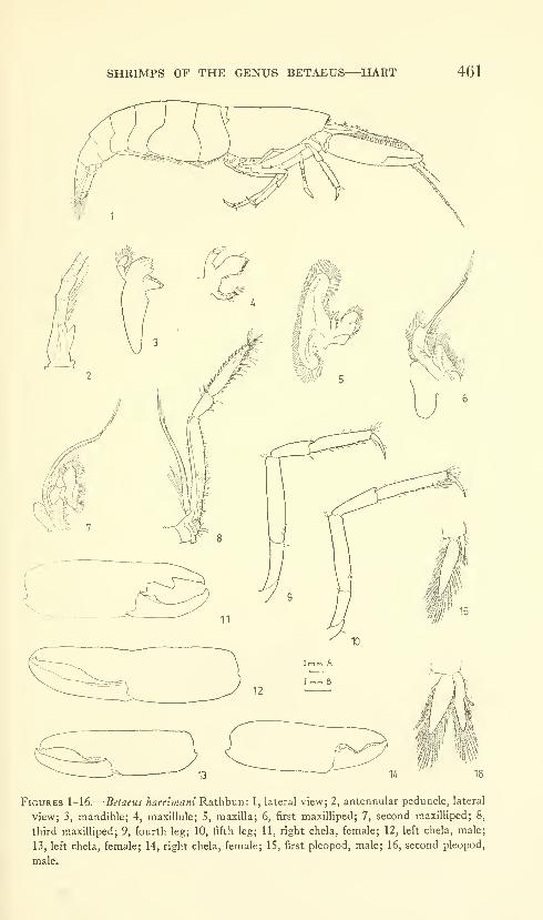
SHRIMPS OF THE GENUS BETAEUS—HART 461
Figures 1-16.
—
Betaeus harrimani Rathbun: 1, lateral view; 2, antennular peduncle, lateral
view; 3, mandible; 4, maxillule; 5, maxilla; 6, first maxilliped; 7, second maxilliped; 8,
third maxilliped; 9, fourth leg; 10, fifth leg; 11, right chela, female; 12, left chela, male;
13, left chela, female; 14, right chela, female; 15, first pleopod, male; 16, second pleopod,
male.

462 PROCEEDINGS OF THE NATIONAL MUSEUM
Figures ll-l'i.—Betaeus harrimani Rathbun: 17, frontal region, dorsal view, female; 18,
same, lateral view; 19, antennal scale; 26, tail fan. Betaeus longidactylus Lockington:
20, frontal region, dorsal view, female; 21, same, lateral view; 22, antennal scale; 27, tail
fan. Betaeus ensenadensis Glassell: 23, frontal region, dorsal view, male; 24, same, lateral
view; 25, antennal scale; 28, tail fan.
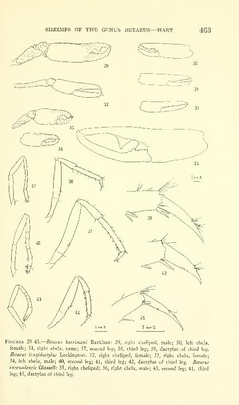
SHRIMPS OF THE GENUS BETAEUS—HART 463
Figures 29^S.—Betaeus harrimani Rathbun: 29, right cheliped, male; 30, left chela,
female; 31, right chela, same; 37, second leg; 38, third leg; 39, dactylus of third leg.
Betaeus longidactylus Lockington: 32, right cheliped, female; 33, right chela, female;
34, left chela, male; 40, second leg; 41, third leg; 42, dactylus of third leg. Betaeus
ensenadensis Glassell: 35, right cheliped; 36, right chela, male; 43, second leg; 44, third
leg; 45, dactylus of third leg.

464 PROCEEDINGS OF THE NATIONAL MUSEUM
Figures ^G-S1.—Betaeus harfordi (Kingsley): 46, frontal region, dorsal view, female; 47,
same, lateral view; 54, antennal scale. Betaeus macginitieae, new species: 48, frontal
region, dorsal view, female; 49, same, lateral view; 55, antennal scale. Betaeus gracilis,
new species: 50, frontal region, dorsal view, female; 51, same, lateral view; 56, antennal
scale. Betaeus setosus, new species: 52, frontal region, dorsal view, female; 53, same,
lateral view; 57, antennal scale.
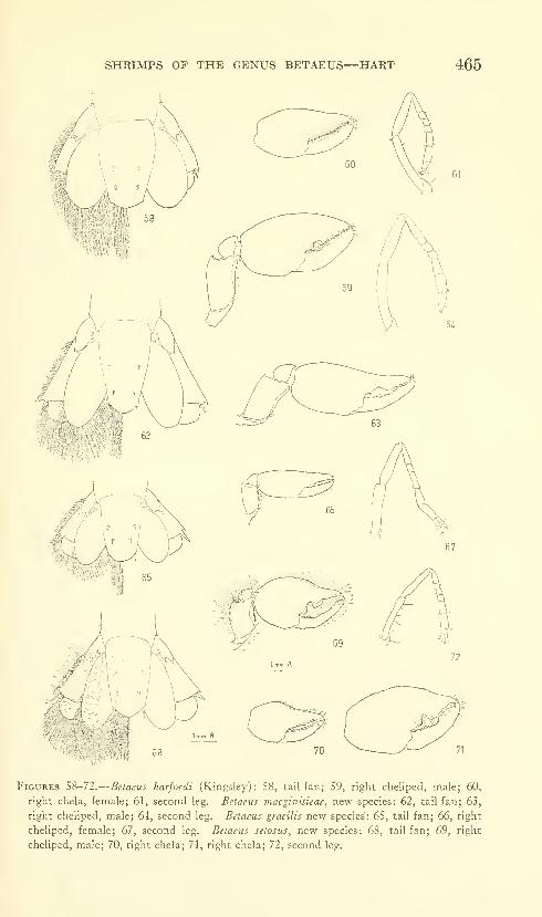
SHRIMPS OF THE GENUS BETAEUS—HART 465
Figures 58-72.
—
Betaeus harfordi (Kingsley): 58, tail fan; 59, right cheliped, male; 60,
right chela, female; 61, second leg. Betaeus macginiiieae, new species: 62, tail fan; 63,
right cheliped, male; 64, second leg. Betaeus gracilis new species: 65, tail fan; 66, right
cheliped, female; 67, second leg. Betaeus setosus, new species: 68, tail fan; 69, right
cheliped, male; 70, right chela; 71, right chela; 72, second leg.
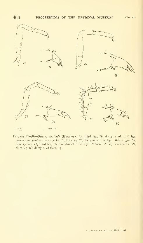
466 PROCEEDINGS OF THE NATIONAL MUSEUM vol. lis
Figures 73-80.
—
Betaeus harfordi (Kingsley): 73, third leg; 74, dactylus of third leg.
Betaeus inacginitieae, new species: 75, third leg; 76, dact)'lus of third leg. Betaeus gracilis,
new species: 77, third leg; 78, dactylus of third leg. Betaeus setosus, new species: 79,
third leg; 80, dactylus of third leg.
U.S. GOVERNMENT PRIMTLNG 0FFICE:I964

Proceedineis of z^^^^^hv
the United States |®^7^IN a t 1 o n a 1 Museum ^^^^^^
SMITHSONIAN INSTITUTION . WASHINGTON, D.C.
Volume 115 1964 Number 3491
NOTES ON SOME NEARCTIC PSYCHOMYIIDAEWITH SPECIAL REFERENCE TO THEIR LARVAE
(TRICHOPTERA)
By Oliver S. Flint, Jr.
Introduction
The caddis-fly family Psychomyiidae is represented in Americanorth of Mexico by 10 genera containing 61 species. The larvae of
this family construct fixed retreats that vary from simple silken
covers to long tubes and complex trap nets. Until 1944, when Rosspublished a key to the genera for the larvae and pupae, it was vir-
tually impossible to identify the immature stages of our species,
even to genus in most cases. The subsequent modifications of that
key (Ross, 1959, and here) permit generic determination of mostlarvae, but specific determination in our larger genera cannot yet be
made with any degree of certainty.
Since the publication of Ross' key (1959), several papers have
appeared and a few critical associations have been established that
make expansion and modification of his key necessary. Edwards(1961) described the larvae of Xiphocentron mexico, and the collection
of a metamorphotype (sensu MUne, 1938) of Ross' Genus B (1944,
p. 74) establishes its synonymy with Nyctiophylax vestitus. A critical
examination of more material also has necessitated the synonymyof Genus A (Ross 1944, p. 73) with Nyctiophylax. The collection
467693-434—63
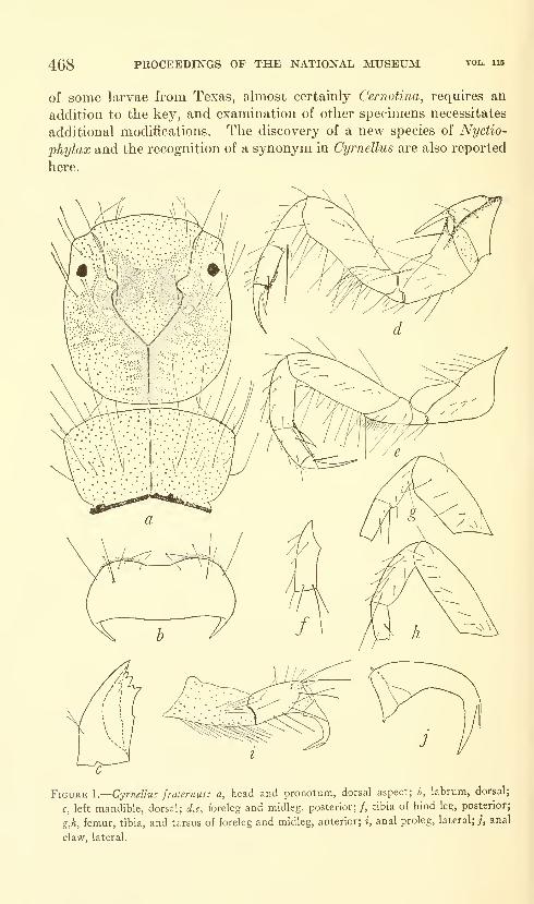
468 PROCEEDINGS OF THE NATIONAL MUSEUM
of some larvae from Texas, almost certainly Cernotina, requires an
addition to the key, and examination of other specimens necessitates
additional modifications. The discovery of a new species of Nyctio-
phylax and the recognition of a synonym in Cyrnellus are also reported
here.
Figure 1.
—
Cyrnellus fraternus: a, head and pronotum, dorsal aspect; h, labrum, dorsal;
c, left mandible, dorsal; d,e, foreleg and midleg, posterior;/, tibia of hind leg, posterior;
g,h, femur, tibia, and tarsus of foreleg and midleg, anterior; i, anal proleg, lateral; /, anal
claw, lateral.
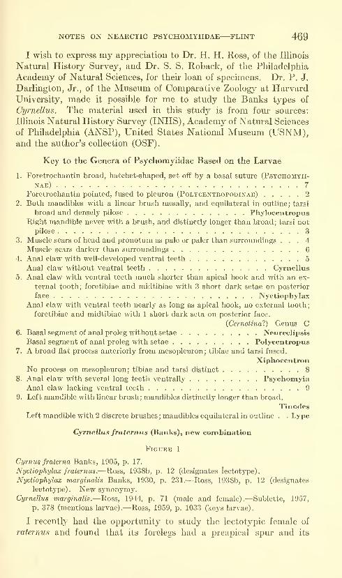
NOTES ON NEARCTIC PSYCHOMYIIDAE—FLINT 469
I wish to express my appreciation to Dr. H. H. Ross, of the Illinois
Natural History Survey, and Dr. S. S. Roback, of the Philadelphia
Academy of Natural Sciences, for their loan of specimens. Dr. P. J.
Darlington, Jr., of the Museum of Comparative Zoology at HarvardUniversity, made it possible for me to study the Banks types of
Cymelius. The material used in this study is from four sources:
Illinois Natural History Survey (INHS), Academy of Natural Sciences
of Philadelphia (ANSP), United States National Museum (USNM),and the author's collection (OSF).
Key to the Genera of Psychomyiidae Based on the Larvae
1. Foretrochantin broad, hatchet-shaped, set off by a basal suture (Psychomyii-
nae) 7
Foretrochantin pointed, fused to pleuron (Polycentropodinae) 2
2. Both mandibles with a linear brush mesally, and equilateral in outline; tarsi
broad and densely pilose PhylocentropusRight mandible never with a brush, and distinctly longer than broad; tarsi not
pilose 3
3. Muscle scars of head and pronotum as pale or paler than surroundings ... 4
Muscle scars darker than surroundings 6
4. Anal claw with well-developed ventral teeth 5
Anal claw without ventral teeth Cyrnellus
5. Anal claw with ventral teeth much shorter than apical hook and with an ex-
ternal tooth; foretibiae and midtibiae with 3 short dark setae on posterior
face NyctiophylaxAnal claw with ventral teeth nearly as long as apical hook, no external tooth;
foretibiae and midtibiae with 1 short dark seta on posterior face.
{Cernotinai) Genus C6. Basal segment of anal proleg without setae Neureclipsis
Basal segment of anal proleg with setae Polycentropus
7. A broad fiat process anteriorly from mesopleuron; tibiae and tarsi fused.
XiphocentronNo process on mesopleuron; tibiae and tarsi distinct 8
8. Anal claw with several long teeth ventrally PsychomyiaAnal claw lacking ventral teeth 9
9. Left mandible with linear brush; mandibles distinctly longer than broad.
TinodesLeft mandible with 2 discrete brushes; mandibles equilateral in outline . . Lype
Cyrnellus fraternus (Banks), new combination
Figure 1
Cyrnus fraterna Banks, 1905, p. 17.
Nyctio-phylax fraternus.—Ross, 1938b, p. 12 (designates lectotype).
Nyctiophylax marginalis Banks, 1930, p. 231.—Ross, 1938b, p. 12 (designates
lectotype). New synonymy.Cyrnellus marginalis.—Ross, 1944, p. 71 (male and female).—Sublette, 1957,
p. 378 (mentions larvae).—Ross, 1959, p. 1033 (keys larvae).
I recently had the opportunity to study the lectotypic female of
raternus and found that its forelegs had a preapical spm* and its
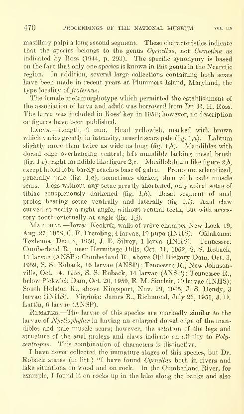
470 PROCEEDINGS OF THE NATIONAL MUSEUM vol. 115
maxillary palpi a long second segment. These characteristics indicate
that the species belongs to the genus Cyrnellus, not Cernotina as
indicated by Ross (1944, p. 293). The specific synonymy is basedon the fact that only one species is known in this genus in the Nearctic
region. In addition, several large collections containing both sexes
have been made in recent years at Plummers Island, Maryland, the
type locality oi fraternus.
The female metamorphotype which permitted the establishment of
the association of larva and adult was borrowed from Dr. H. H. Ross.
The larva was included in Ross' key in 1959; however, no description
or figures have been published.
Larva.—Length, 9 mm. Head yellowish, marked with brownwhich varies greatly in intensity, muscle scars pale (fig. l,a). Labrumslightly more than twice as wide as long (fig. 1,6). Mandibles with
dorsal edge overhanging ventral; left mandible lacking mesal brush
(fig. 1 ,c) ; right mandible like figure 2,c. Maxillolabium like figure 2,6,
except labial lobe barely reaches base of galea. Pronotum sclerotized,
generally pale (fig. l,a), sometimes darker, then with pale muscle
scars. Legs without any setae greatly shortened, only apical setae of
tibiae conspicuously darkened (fig. 1,^). Basal segment of anal
proleg bearing setae ventrally and laterally (fig. l,i). Anal claw
curved at nearly a right angle, without ventral teeth, but with acces-
sory tooth externally at angle (fig. 1,^).
Material.—Iowa: Keokulc, walls of valve chamber New Lock 19,
Aug. 27, 1958, C. R. Fremling, 4 larvae, 19 pupa (INHS). Oklahoma:Texhoma, Dec. 8, 1950, J. E. Silvey, 1 larva (INHS). Tennessee:
Cumberland R., near Hermitage Hills, Oct. 11, 1962, S. S. Roback,11 larvae (ANSP); Cumberland R., above Old Hickory Dam, Oct. 3,
1959, S. S. Roback, 16 larvae (ANSP); Tennessee R., New Johnson-
vUle, Oct. 14, 1958, S. S. Roback, 14 larvae (ANSP); Tennessee R.,
below Pickwick Dam, Oct. 20, 1959, R. M. Sinclau', 10 larvae (INHS);South Holston R., above Kingsport, Nov. 29, 1945, J. S. Dendy, 3
larvae (INHS). Virginia: James R., Richmond, July 26, 1951, J. D.Lattin, 6 larvae (ANSP).Remarks.—The larvae of this species are markedly similar to the
larvae of Nyctiophylax in having an enlarged dorsal edge of the man-dibles and pale muscle scars; however, the setation of the legs andstructure of the anal prolegs and claws indicate an affinity to Poly-
centropus. This combination of characters is distinctive.
I have never collected the immature stages of this species, but Dr.
Roback states (in litt.) 'T have found Cyrnellus both in rivers andlake situations on wood and on rock. In the Cumberland River, for
example, I found it on rocks up in the lake along the banks and also
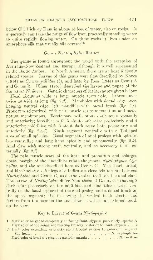
NOTES ON NEARCTIC PSYCHOMYIIDAE—FLINT 471
below Old Hickory Dam in about 15 feet of water, also on rocks. It
apparently can take the range of flow from practically standing water
to quite rapidly flowing water. On these rocks it lives under an
amorphous silk mat usually silt covered."
Genus Nyctiophylax Brauer
The genus is found throughout the world with the exception of
Australia-New Zealand and Europe, although it is well represented
in the Baltic Amber. In North America there are at least 5 closely
related species. Larvae of this genus were first described by Noyes
(1914) as Cyrnus pallidus (?), and later by Ross (1944) as Genus Aand Genus B. Ulmer (1957) described the larvae and pupae of the
Sumatran A^". -flavus. Generic characters of the larvae are given below:
Head about as wide as long; muscle scars pale. Labrum over
twice as wide as long (fig. 2,d). Mandibles with dorsal edge over-
hanging ventral edge, left mandible with mesal brush (fig. 2,c).
Pronotum sclerotized, with pale muscle scars; mesonotum and meta-
notum membranous. Forefemora with stout dark setae ventrally
and anteriorly; foretibiae with 3 stout dark setae posteriorly and 4
anteriorly; midtibiae with 3 stout dark setae both posteriorly and
anteriorly (fig. 2,e~i). Ninth segment ventrally with a T-shaped
area of small spicules. Basal segment of anal prolegs with spicules
basoventrall}^; and long hairs apically and apicomesally (fig. 2,k).
Anal claw with strong teeth ventrally, and an accessory tooth ex-
ternally (fig. 2.j).
The pale muscle scars of the head and pronotum and enlarged
dorsal margin of the mandibles relate the genera Nyctiophylax, Cyr-
nellus, and the one described here as Genus C. The short, broad,
and black setae on the legs also indicate a close relationship between
Nyctiophylax and Genus C, as do the ventral teeth on the anal claw.
The larvae of Nyctiophylax dift'er from those of Genus C in having 3
dark setae posteriorly on the midtibiae and hind tibiae, setae ven-
trally on the basal segment of the anal proleg, and a dorsal brush on
the apical segment; also in having the ventral teeth shorter and
farther from the base on the anal claw as weU as an external tooth
on the claw.
Key to Larvae of Genus Nyctiophylax
1. Dark color on genae completely enclosing frontoclypeus posteriorly, species ADark color of the genae not meeting broadly posterior to frontoclypeus . . 2
2. Dark color extending anteriorly along frontal sutures to anterior margin of
the head N. nephophilus
Dark color of head not reaching anterior margin N. vestitus

472 PROCEEDINGS OF THE NATIONAL MUSEUM vol. 115
Nyctiophylax vestitus (Hagen)
Figure 2
Polycentropiis vestihis Hagen, 1861, p. 293.
Nijctiophylax vestitics.—^Ross, 1938b, p. 13 (designates lectotype); 1944, p. 70
(male and female).
Genus b Ross, 1944, p. 74; 1959, p. 1032.
Although the adults of this species are commonly collected at
lights throughout much of eastern North America, the larvae have
remained unknown until recently. Ross (1944) gave the first de-
scription of the larvae under the name Psychomyiidae Genus B.
In the summer of 1961 I collected a male metamorphotype of the
species, finally establishing the correlation of stages.
Larva.—Length 8 mm. Head yellowish, with brown area neither
reaching anterior margin nor extending posteriad of frontoclypeus
(fig. 2,a).
Material.—Illinois: Kankakee R., Momence, May 26, 1936,
H. H. Ross, 1 larva (INHS). Virginia: Broad Run, Thoroughfare
Gap, Fauquier County, July 22, 1961, O. S. FHnt, 1 larva, Icfpupa
(USNM); May 27, 1961, 27 larvae, 4 pupae (OSF); June 14, 1961,
1 larva, 1 pupa (USNM); Thornton R., above Sperryville, Mar.
4, 1961, O. S. Flint, 9 larvae (USNM); Apr. 15, 1961, 7 larvae
(USNM). North Carolina: Deep Creek, 2,000 ft., near Bryson City,
May 19, 1959, O. S. Flint, 1 larva (OSF); June 7, 1961, 1 larva
(USNM).Remarks.—There is considerable variation in the shape of the
clasper of the male. It may well be that further study will showthat our present concept of the species includes several closely related
species. For this reason, I give figure 21, showing the clasper from
the metamorphotype. In all likelihood the name vestitus, based on a
female, is correctly associated with this male as the type locality is
Washington, D.C., and this is the only clasper shape I have seen
from the area.
The larva of this species is most like that of nephoj^hilus, differing
from it in the coloration of the head. In vestitus the dark area does
not reach the anterior margin of the head.
The larvae are found on rocks in streams generally about a meter
or two wide, near one or the other end of the pools, generally shunning
the fastest water of the rapids and cascades. They construct, in
some angle on a rock, a silken shelter of considerable rigidity beneath
which they hide. The shelter is open at both ends, but as far as
can be seen no trap net is constructed. At pupation time the shelter
is strengthened and closed except for a cylindrical aperture at one
end. The end of the cylinder inside the shelter is partially closed
by a silken sieve.
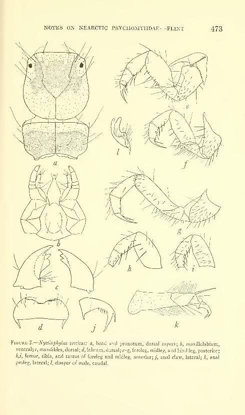
NOTES ON NEARCTIC PSYCHOMYIIDAE—FLINT 473
Figure 2.
—
Nyctiophylax vestitus: a, head and pronotum, dorsal aspect; b, maxillolabium,ventral; c, mandibles, dorsal; d, labrum, dorsal; ^-g, foreleg, midleg, and hind leg, posterior;
h,i, femur, tibia, and tarsus of foreleg and midleg, anterior; ;, anal claw, lateral; k, anal
proleg, lateral; /, clasper of male, caudal.
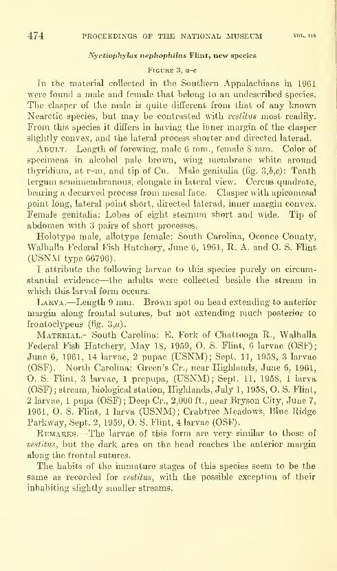
474 PROCEEDINGS OF THE NATIONAL MUSEUM vol. ne
Nyctiophylax nephophiliis Flint, new species
Figure 3, a-c
In the material collected in the Southern Appalachians in 1961
were found a male and female that belong to an undescribed species.
The clasper of the male is quite different from that of any knownNearctic species, but may be contrasted with vesfitus most readily.
From this species it differs in having the inner margin of the clasper
slightly convex, and the lateral process shorter and directed laterad.
Adult.—Length of forewing, male 6 mm., female 8 mm. Color of
specimens in alcohol pale brown, wing membrane white around
thyridium, at r-m, and tip of Cu. Male genitalia (fig, 3,6,c): Tenthtergmn semimembranous, elongate in lateral view. Cercus quadrate,
bearing a decm-ved process from mesal face. Clasper with apicomesal
point long, lateral point short, directed laterad, inner margin convex.
Female genitalia: Lobes of eight sternum short and wide. Tip of
abdomen with 3 pairs of short processes.
Holotype male, allotype female: South Carolina, Oconee County,
Walhalla Federal Fish Hatchery, June 6, 1961, R. A. and O. S. Flint
(USNM type 66796).
I attribute the following larvae to this species purely on circum-
stantial evidence—the adults were collected beside the stream in
which this larval form occurs.
Larva.—Length 9 mm. Brown spot on head extending to anterior
margin along frontal sutm'es, but not extending much posterior to
frontoclypeus (fig. 3,a).
Material.—South Carolina: E. Fork of Chattooga R., Walhalla
Federal Fish Hatchery, May 18, 1959, O. S, Flint, 6 larvae (OSF);
June 6, 1961, 14 larvae, 2 pupae (USNM); Sept. 11, 1958, 3 larvae
(OSF). North Carolina: Green's Cr., near Highlands, June 6, 1961,
O, S. Flint, 3 larvae, 1 prepupa, (USNM); Sept. 11, 1958, 1 larva
(OSF) ; stream, biological station, Highlands, July 1, 1958, O. S, Flint,
2 larvae, 1 pupa (OSF) ; Deep Cr., 2,000 ft., near Bryson City, June 7,
1961, O. S. Fhnt, 1 larva (USNM); Crabtree Meadows, Blue Ridge
Parkway, Sept. 2, 1959, O. S. Flint, 4 larvae (OSF),
Remarks,—The larvae of this form are very similar to those of
vestitus, but the dark area on the head reaches the anterior margin
along the frontal sutures.
The habits of the immature stages of this species seem to be the
same as recorded for vestitus, with the possible exception of their
inhabiting slightly smaller streams.
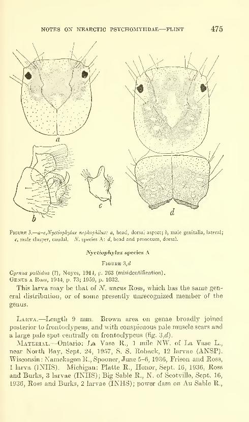
NOTES ON NEARCTIC PSYCHOMYIIDAE—FLINT 475
Figure 3.
—
a-c,Nyctiophylax nephophilus: a, head, dorsal aspect; b, male genitalia, lateral;
c, male clasper, caudal. N. species A: d, head and pronotum, dorsal.
Nyctiophylax species A
Figure 3,d
Cyrnus pallidus (?), Noyes, 1914, p. 263 (misidentification) .
Genus a Ross, 1944, p. 73; 1959, p. 1032.
This larva may be that of A^. uncus Ross, which has the same gen-
eral distribution, or of some presently unrecognized member of the
genus.
Larva.—Length 9 mm. Brown area on genae broadly joined
posterior to frontoclypeus, and ^vith conspicuous pale muscle scars and
a large pale spot centrally on frontoclypeus (fig. 3,d).
Material.—Ontario: La Vase R., 1 mile NW. of La Vase L.,
near North Bay, Sept. 24, 1957, S. S. Roback, 12 larvae (ANSP).
Wisconsin : Namekagon R., Spooner, June 5-6, 1936, Frison and Ross,
1 larva (INHS). Michigan: Platte R., Honor, Sept. 16, 1936, Ross
and Burks, 3 larvae (INHS); Big Sable R., N. of Scotville, Sept. 16,
1936, Ross and Burks, 2 larvae (INHS); power dam on Au Sable R.,
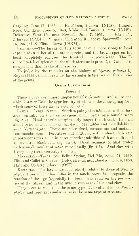
476 PROCEEDINGS OF THE NATIONAL MUSEUM vol. lis
Grayling, June 17, 1935, T. H. Frison, 1 larva (INHS). Illinois:
Rock Cr., Erie, June 5, 1940, Mohr and Bm-ks, 1 larva (INHS).
Delaware: West Cr., near Newark, June 7, 1951, T, Dolan IV,
2 larvae (ANSP). Virginia: Thornton R., above Sperryville, Apr.
15, 1961, O. S. Flint, 1 larva (USNM).Remarks.—The larvae of this form have a more elongate head
capsule than either of the other species, and the brown spot on the
head completely encloses the frontoclypeus posteriorly. The T-
shaped patch of spicules on the ninth sternum is present, but much less
conspicuous than in the other species.
To judge by the remarks on the biology of Cyrnus pallidus by
Noyes (1914), the larvae must have similar habits to the other species
of the genus.
Genus C, new form
Figure 4
These larvae are almost unquestionably Cernotina, and quite pos-
sibly C. astern Ross the type locality of which is the same spring from
which some of these larvae were collected.
Larva.—Length 8 mm. Sclerites pale yellowish; head with a dark
area centrally on the frontoclypeus which bears pale muscle scars
(fig. 4,a). Head capsule conspicuously longer than broad. Labrum
about twice as wide as long (fig. 4,6). Mandibles and maxillolabium
as in Nyctiophylax. Pronotum sclerotized, mesonotum and metano-
tum membranous. Foretibiae and midtibiae with 1 short, dark seta
in posterior series and 4 in anterior series; midtibia with an additional
apicoventral black seta (fig. 4,c-fif). Basal segment of anal proleg
vrith a small number of setae apicomesally (fig. 4,h). Anal claw with
4 very long teeth ventrall}^ (fig. 4,%).
Material.—Texas: San Felipe Spring, Del Rio, Sept. 21, 1960,
Flint and CoUette, 8 larvae (OSF); stream, near Bandera, Oct. 9, 1960,
Flint and Collette, 9 larvae (OSF).
Remarks.—The larvae are most closel}^ related to those of Nyctio-
phylax, from which they differ in the much longer head capsule, the
setation of the legs (especialh^ the fewer dark setae on the posterior
row of the tibiae), and in the unique structure of the anal claw.
They seem to construct the sam.e type of larval shelter as Nyctio-
phylax, and frequent similar areas in the same type of streams.
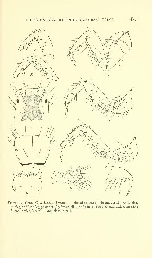
NOTES ON NEARCTIC PSYCHOMYIIDAE—FLINT 477
Figure 4.—-Genus C: a, head and pronotum, dorsal aspect; b, labrum, dorsal; c-e, foreleg,
midleg, and hind leg, posterior; /,g, femur, tibia, and tarsus of foreleg and midleg, anterior;
h, anal proleg, lateral; i, anal claw, lateral.
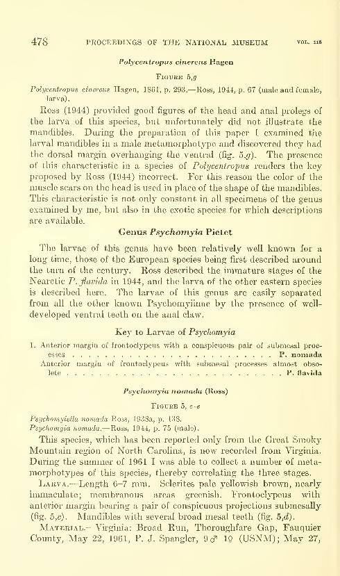
478 PROCEEDINGS OF THE NATIONAL MUSEUM vol. ii5
Polycentropus cinereiis Hagen
Figure 5,g
Polycentropus cinereus Hagen, 1861, p. 293.—Ross, 1944, p. 67 (male and female,
larva)
.
Ross (1944) provided good figiu'es of the head and anal prolegs of
the larva of this species, but unfortunately did not illustrate the
mandibles. During the preparation of this paper I examined the
larval mandibles in a male metamorphotype and discovered they hadthe dorsal margin overhanging the ventral (fig. b,g). The presence
of this characteristic in a species of Polycentropus renders the keyproposed by Ross (1944) incorrect. For this reason the color of the
muscle scars on the head is used in place of the shape of the mandibles.
This characteristic is not only constant in all specimens of the genus
examined by me, but also in the exotic species for which descriptions
are available.
Genus Psychomyia Pictet
The larvae of this genus have been relatively well known for a
long time, those of the European species bemg fii*st described aroundthe turn of the centmy. Ross described the immature stages of the
Nearctic P.flavida in 1944, and the larva of the other eastern species
is described here. The larvae of this genus are easily separated
from all the other loiown Psychomyiinae by the presence of well-
developed ventral teeth on the anal claw.
Key to Larvae of Psychomyia
1. Anterior margin of frontoclypeus with a conspicuous pair of submesal proc-
esses P. nomadaAnterior margin of frontoclypeus with submesal processes almost obso-
lete P. flavida
Psychomyia nomada (Ross)
Figure 5, c-e
PsTjchomyiella nomada Ross, 1938a, p. 138.
Psychomyia nomada.—Ross, 1944, p. 75 (male).
This species, which has been reported only from the Great SmokyMountain region of North Carolina, is now recorded from Virginia.
During the summer of 1961 I was able to collect a number of meta-
morphotypes of this species, thereby correlating the three stages.
Larva.—Length 6-7 mm. Sclerites pale yellowish brown, nearly
immaculate; membranous areas greenish. Frontoclypeus with
anterior margin bearing a pair of conspicuous projections submesally
(fig. 5,c). Mandibles with several broad mesal teeth (fig. b,d).
Material.—Virginia: Broad Run, Thoroughfare Gap, Fauquier
County, May 22, 1961, P. J. Spangler, 9d^ 19 (USNM); May 27,
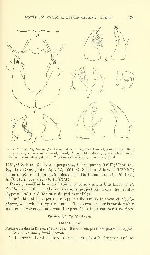
NOTES ON XEARCTIC PSYCHOMYIIDAE—FLINT 479
Figure 5.
—
a,b, Psychomyia flavida: a, anterior margin of frontoclypeus; b, mandibles,
dorsal, c-e, P. nomada: c, head, dorsal; d, mandibles, dorsal; e, anal claw, lateral.
Tinodes: f, mandibles, dorsal. Polyceniropuj cinereus: g, mandibles, dorsal.
1961, O. S. Flint, 2 larvae, 4 prepupae, 2c^ 49 pupae (OSF); ThorntonR., above Sperryville, Apr. 15, 1961, O. S. Flint, 3 larvae (USNM);Jefferson National Forest, 5 miles east of Buchanan, June 19-20, 1961,
A. B. Gurney, many cf 9 (USNM).Remarks.—The larvae of this species are much like those of P.
flavida, but differ in the conspicuous projections from the fronto-
clypeus, and the differently shaped mandibles.
The habits of this species are apparently similar to those of Nyctio-
phylax, with which they are found. The larval shelter is considerably
smaller, however, as one would expect from their comparative sizes.
Psychomyia flavida Hagen
Figure 5, a,b
Psychomyia flavida Hagen, 1861, p. 294.—Ross, 1938b, p. 14 (designates lectotype);
1944, p. 75 (male, female, larva).
This species is widespread over eastern North America and as
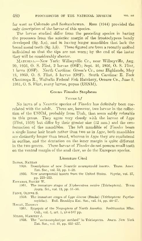
480 PROCEEDINGS OF THE NATIONAL MUSEUM vol. ii5
far west as Colorado and Saskatchewan. Ross (1944) provided the
only description of the larvae of this species.
The larvae studied differ from the preceding species in having
the processes from the anterior margin of the frontoclypeus barely
developed (fig. 5,a), and in having longer mandibles that lack the
broad mesal teeth (fig. 5,6) . Those figured are from a recently molted
individual so that the tips are not worn; by the end of the instar
they will be considerably shorter.
Material.—New York: Willseyville Cr., near Willseyville, Aug.
20, 1956, O. S. Flint, 2 larvae (OSF); Sept. 16, 1956, O. S. Flint,
1 larva (OSF). North Carolina: Green's Cr., near Highlands, May18, 1959, O. S. Flint, 5 larvae (OSF). South Carolina: E. Fork
Chattooga R., Walhalla Federal Fish Hatchery, Oconee Co., June 6,
1961, O. S. Flint, many larvae, pupae (USNM).
Genus Tinodes Stephens
Figure 5,/
No larva of a Nearctic species of Tinodes has definitely been cor-
related with the adult. There are, however, two larvae in the collec-
tion of the USNM, probably from Utah, that seem safely referable
to this genus. They agree very closely with the larvae of Lype
(Flint, 1959) but differ by their greater size (12 mm.) and the con-
formation of the mandibles. The left mandible of Tinodes bears
a single linear hair brush rather than two as in Lype, both mandibles
are distinctly longer than broad, whereas in Lype they are equilateral
in outline, and the dentation on the inner margin is quite different
in the two genera. These larvae of Tinodes do not possess small teeth
on the ventral margin of the anal claw, as do the European species.
Literature CitedBanks, Nathan
1905. Descriptions of new Nearctic neuropteroid insects. Trans. Amer.
Ent. Soc, vol. 32, pp. 1-20.
1930. New neuropteroid insects from the United States. Psyche, vol. 37,
pp. 223-233.
Edwards, Sidney W.1961. The immature stages of Xiphocentron niexico (Trichoptera) . Texas
Journ. Sci., vol. 13, pp. 51-56.
Flint, Oliver S.
1959. The immature stages of Lype diversa (Banks) (Trichoptera: Psycho-
myiidae). Bull. Brooklyn Ent. Soc, vol. 54, pp. 44-47.
Hagen, Herman1861. Synopsis of the Neuroptera of North America. Smithsonian Misc.
Coll., vol. 4, art. 1, xx+ 347 pp.
Milne, Marjery J.
1938. The "metamorphotype method" in Trichoptera. Journ. New YorkEnt. Soc, vol. 46, pp. 435-437.
NOTES ON NEARCTIC PSYCHOMYIIDAE—FLINT 481
Notes, Alice A.
1914. The biology of the net-spinning Trichoptera of Cascadilla Creek.
Ann. Ent. Soc. America, vol. 7, pp. 251-272.
Ross, Herbert H.
1938a. Descriptions of Nearctic caddis flies. Bull. Illinois Nat. Hist. Surv.,
vol. 21, pp. 101-183.
1938b. Lectotypes of North American caddis flies in the Museum of Com-parative Zoology. Psyche, vol. 45, pp. 1-61.
1944. The caddis flies, or Trichoptera, of Illinois. Bull. Illinois Nat. Hist.
Surv., vol. 23, pp. 1-326.
1959. Trichoptera. In Edmondson, Fresh-water Biology. Pp. 1024-1049.
New York.
Sublette, James E.
1957. The ecology of the macroscopic bottom fauna of Lake Texoma(Denison Reservoir), Oklahoma and Texas. Amer. Midi. Nat.,
vol. 57, pp. 371-402.
Ulmer, Georg1957. Kocherfliegen (Trichopteren) vondenSunda-Inseln. Teillll. Archiv
fur hydrobiologie, vol. 23 suppl., pp. 109-470.
U.S. GOVERNMENT PS1NT1N5 OFriCE:l964

Proceedings of
the United States
National MuseumSMITHSONIAN INSTITUTION • WASHINGTON, D.C.
Volume 115 1964 Number 3492
THE JAMBELI CULTURE OF SOUTH COASTAL ECUADOR
By Emilio Estrada, Betty J. Meggers, and Clifford Evans
This brief report is the result of fieldwork from 1958 to 1961 in
the mangrove swamps and salitres of the southern Ecuadorian coast.
The survey of the JambeK islands was undertaken during the summerof 1958 by Estrada, Meggers, and Evans. In 1960, Estrada tested
stratigraphically a site, identified as G-84, within the present town of
Posorja, Early in 1961, he made a preliminary survey of the salitres
of Lagarto and El Morro. During the summer of that year, the
three authors revisited this area and the sites on the southeastern
coast of Puna Island. The strata cut at G-86 was also part of the
final season's work. All the pottery was classified at the MuseoVictor Emilio Estrada, and notes taken there are the basis for the
type descriptions. Except for type samples of sherds, all pottery and
other artifacts were deposited in the Museo Estrada.
Most of the area occupied by the Jambeli Phase sites is not under
private ownership, but we wish to express our appreciation to the
many local residents for their guidance and assistance with excavation.
Particular thanks are due Jorge Swett, who secured permission for us
to investigate G-86, one of three sites that has sufficient depth for
stratigraphic excavation. In Guayaquil, Walter Molina assisted in
processing the sherds for analysis. In Washington, we are indebted
483
484 PROCEEDINGS OF THE NATIONAL MUSEUM
to George Robert Lewis for the line drawings of specimens, and to
Judith Hill for typing the manuscript.
For support of certain of the field expenses durmg the 1958 season,
the junior authors received Grant 2370 from the Penrose Fund of the
American Philosophical Society. The 1961 investigations were part
of Project J of the program organized by the Institute of Andean
Research on "Interrelationships of New World Cultures," financed
by a grant from the National Science Foundation. To all three
organizations, we wish to express our deep appreciation.
Completion of this report was delayed by the unexpected death of
the senior author in November 1961. Although he did not see the
final version of the manuscript, we have retained the authorship wehad agreed upon together, not only because we believe that Estrada
would accept its content, but because without his persistence the
Jambeli culture, represented by some of the most unproductive and
uninviting sites to be found on the Ecuadorian coast, would have
remained only superficially known.
Geographical Distribution and Site Characteristics
Sites of the Jambeli culture occur on the coastal portions of the
Provinces of El Oro and Guayas, extending from the Peruvian border
up the coast and around the Gulf of Guayaquil to the vicinity of
Playas (fig. 1). All the sites included in this survey are located on the
peninsula between Playas and the Canal del Morro, on Puna Island or
on the islands off the coast of El Oro. Sherds of Jambeli pottery types
are reported from Tendales in El Oro Province, but this part of the
mainland and the portion of Guayas Province extending along the
eastern side of the Gulf of Guayaquil have not been surveyed.
Today, this region does not present the uniform appearance that it
must have had during the existence of the Jambeli culture. The
Playas-Posorja area has suffered noticeable desiccation within the
memory of the residents, so that only small remnants are left of the
mangrove swamps that once occupied about 50 percent of the area.
Their former extent is indicated by the clotted lines representing the
boundaries of the large salitres that have taken their place (fig. 2).
This part of the coast now receives little rain, forcing the abandonment
of once prosperous cattle ranches and reducing to ghost towns formerly
populous communities. The same environmental situation exists on
Puna Island except for the southeastern part, which is low and broken
into small islands of mangrove giving it an appearance comparable to
that of the islands of El Oro to the south (fig. 3).
The distribution and character of the Jambeli Phase sites indicates
that the southern portion of the area conserves the appearance that
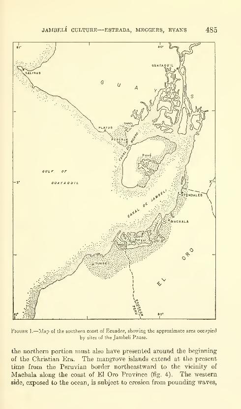
JAMBELI CULTURE—ESTRADA, MEGGERS, EVANS 485
Figure 1.—Map of the southern coast of Ecuador, showing the approximate area occupied
by sites of the Jambeli Phase.
the northern portion must also have presented around the beginning
of the Christian Era, The mangrove islands extend at the present
time from the Peruvian border northeastward to the vicinity of
Machala along the coast of El Oro Province (fig. 4). The western
side, exposed to the ocean, is subject to erosion from pounding waves,
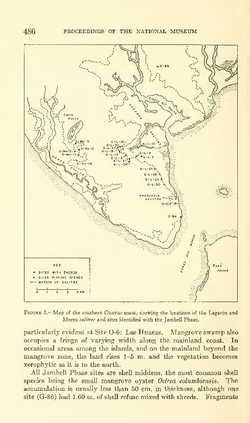
486 PROCEEDESTGS OF THE NATIONAL MUSEUM
• SITES WITH SherdsA SITES WITHOUT SHERDS
MARGIN OF SALITRE
4 KM
Figure 2.—Map of the southern Guayas coast, showing the locations of the Lagarto and
Morro lalitres and sites identified with the Jambeli Phase.
particularly evident at Site 0-6: Las Huacas. Mangrove swamp also
occupies a fringe of varying width along the mainland coast. In
occasional areas among the islands, and on the mainland beyond the
mangrove zone, the land rises 1-5 m, and the vegetation becomesxerophytic as it is to the north.
All Jambeli Phase sites are shell middens, the most common shell
species being the small mangrove oyster Ostrea columbiensis. Theaccumulation is usually less than 50 cm. in thickness, although one
site (G-86) had 1.60 m. of shell refuse mixed with sherds. Fragments
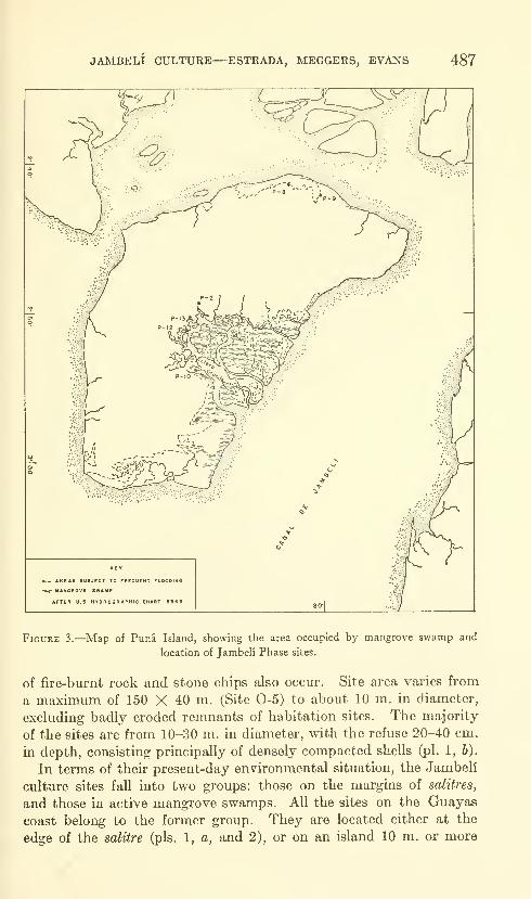
JAMBELI CULTURE—ESTRADA, MEGGERS, EVANS 487
Figure 3.—Map of Puna Island, showing the area occupied by mangrove swamp and
location of Jambeli Phase sites.
of fire-burnt rock and stone chips also occur. Site area varies from
a maximum of 150 X 40 m. (Site 0-5) to about 10 m. in diameter,
excluding badly eroded remnants of habitation sites. The majority
of the sites are from 10-30 m, in diameter, with the refuse 20-40 cm.
in depth, consisting principally of densely compacted shells (pi. 1, b).
In terms of their present-day environmental situation, the Jambeli
culture sites fall into two groups: those on the margins of salitres,
and those in active mangrove swamps. All the sites on the Guayas
coast belong to the former group. They are located either at the
edge of the salitre (pis. 1, a, and 2), or on an island 10 m. or more
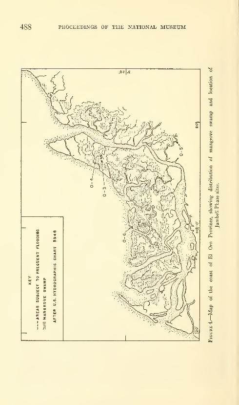
488 PROCEEDINGS OF THE NATIONAL MUSEUM
s- ^
o P-,
(U
U
fe
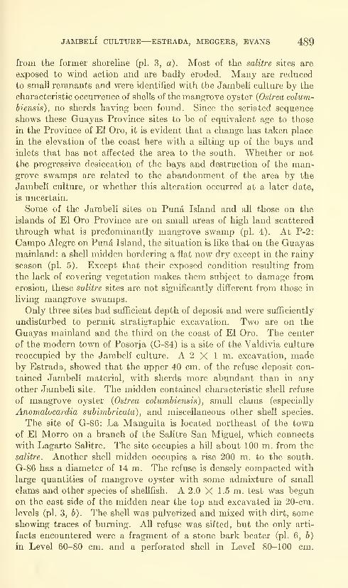
JAMBELI CULTURE—ESTRADA, MEGGERS, EVANS 489
from the former shoreline (pi. 3, a). Most of the salitre sites are
exposed to wind action and are badly eroded. Many are reduced
to small remnants and were identified with the Jambeli culture by the
characteristic occurrence of shells of the mangrove oyster {Ostrea colum-
biensis), no sherds having been found. Since the seriated sequence
shows these Guayas Province sites to be of equivalent age to those
in the Province of El Oro, it is evident that a change has taken place
in the elevation of the coast here with a silting up of the bays andinlets that has not affected the area to the south. Whether or not
the progressive desiccation of the bays and destruction of the man-grove swamps are related to the abandonment of the area by the
Jambeli culture, or whether this alteration occurred at a later date,
is uncertain.
Some of the Jambeli sites on Puna Island and all those on the
islands of El Oro Province are on small areas of high land scattered
through what is predominantly mangrove swamp (pi. 4). At P-2:
Campo Alegre on Pund Island, the situation is like that on the Guayasmainland: a shell midden bordering a fiat now dry except in the rainy
season (pi. 5). Except that their exposed condition resulting from
the lack of covering vegetation makes them subject to damage fromerosion, these salitre sites are not significantly different from those in
living mangrove swamps.
Only three sites had sufficient depth of deposit and were sufficiently
undisturbed to permit stratigraphic excavation. Two are on the
Guayas mainland and the third on the coast of El Oro. The center
of the modern town of Posorja (G-84) is a site of the Valdivia culture
reoccupied by the Jambeli culture. A 2 X 1 m. excavation, madeby Estrada, showed that the upper 40 cm. of the refuse deposit con-
tained Jambeli material, with sherds more abundant than in anyother Jambeli site. The midden contained characteristic shell refuse
of mangrove oyster {Ostrea columbiensis) , small clams (especially
Anomalocardia subimbricata) , and miscellaneous other shell species.
The site of G-86: La Manguita is located northeast of the townof El Morro on a branch of the Salitre San Miguel, which connects
with Lagarto Salitre. The site occupies a hill about 100 m. from the
salitre. Another shell midden occupies a rise 200 m. to the south.
G-86 has a diameter of 14 m. The refuse is densely compacted with
large quantities of mangrove oyster with some admixture of small
clams and other species of shellfish. A 2.0 X 1.5 m. test was begun
on the east side of the midden near the top and excavated in 20-cm.
levels (pi. 3, b). The shell was pulverized and mixed with dirt, someshowing traces of burning. All refuse was sifted, but the only arti-
facts encountered were a fragment of a stone bark beater (pi. 6, 6)
in Level 60-80 cm. and a perforated shell in Level 80-100 cm.

490 PROCEEDINGS OF THE NATIONAL MUSEUM
Several large fire-burnt stones came from Level 120-140 cm. Sterile
black clay at a depth of 160 cm. established the bottom of the middenaccumulation.
The site of 0-5: Embarcadero is about 5 km. inland from the mouthof the Estero Embarcadero (fig. 4), where the left bank rises to 2 m.
above high tide level. Shell refuse was visible for a distance of about
150 m. along this bank (pi. 4, a). The midden area slopes off to the
surrounding land, which measures roughly 1 m. above high tide, a
contour that is easily seen from the water. Along the eroded bank,
vertical columns of shell about 10 cm. in diameter and about a meter
long could be seen in several places, possibly representing former post
holes. The site is densely overgrown but the midden deposit could
be traced for 30-40 m. inward from the bank. Miscellaneous testing
showed that sherds were fairly abundant throughout. A 1 X 1 m.
stratigraphic excavation, designated as Cut 1 and controlled in 20-cm.
levels, was placed 50 cm. in from the edge of the bank. The refuse
consisted of hard, lumpy clay with an abundant admixture of shell,
sherds, some fishbones, and small broken rocks. Sherds were present
to a depth of 80 cm., where a dense layer of shells 20-25 cm. thick
was encountered. Below this was sticky clay, the natural soil of
the bank. The only unusual artifacts were a pottery figurine armfrom Level 20-40., cm. and anther figurine fragment from Level 40-60 cm.
None of the sites investigated in the Playas-Posorja region pro-
duced any human skeletal remains. White-on-red pottery was
collected by Estrada from Site P-4, which consisted of a small circle
of erect stones with two extended burials in the center (Estrada, 1957
a, p. 28 and fig. 10). Site 0-7: Tendales, reported by local residents
and not visited by any of the authors, produced human skeletal
remains in association with ceramics of Jambeli types. This site,
discovered during excavation of irrigation trenches, is reported to be
several hundred square meters in area.
For ease in reference throughout the report, sites belonging to the
Jambeli culture are listed below. Group ''A" includes those from
which sherd collections have been classified and which appear in
Appendix Table 1. Group "B" includes sites in the ^alitres where
no sherds were recovered, but in which the character of the site, its
location, and the composition of the shell refuse are typical of the
Jambeli Phase.
A. jAMBELf Sites With Pottery:
El Oro Province:
0-3: Estero Chiven'a No. 1
0-4: Estero Chiven'a No. 2
0-5: Embarcadero0-6: Las Huacas0-7: Tendales
JAMBELI CULTURE—ESTRADA, MEGGERS, EVANS 4!)1
Fund Island, Guayas Province:
P-2: Campo Alegre No. 1
P-8
P-9
P-12: Los Chalacos
P-13: Jer6ninio
Guayas Province:
G-84: Posorja
G-86: La Manguita
G-109Lagarto Salitre, Guayas Province:
G-L-2G-L-3
G-L-7
G-L-9
G-L-12
G-L-27
G-L-28
G-L-29
G-L-30: Cangrejito
G-L-34
El Morro Salitre, Guayas Province
:
G-Mo-3G-Mo-11G-Mo-15G-Mo-17
B. JambelI Sites Without Potteuy:
Lagarto Salitre, Guayas Province:
G-L-13
G-L-16
G-L-18G-L-20G-L-21
EI Morro Salitre, Guayas Province:
G-Mo-16
Artifacts
As is typical of cultures of the Regional Developmental Period on
the Ecuadorian coast (cf. Estrada, 1962, p. 52), the Jambeli culture
is characterized by a variety of small ornaments, figurines, and
objects of uncertain utility. The most frequently used material is
shell, except for figurines, the majority of which are of pottery. Stone
was rarely employed, possibly in part because of the absence of
suitable raw materials in the mangrove swamp area. Bone and woodwere probably also utilized, but conditions of preservation have pro-
hibited their siu*vival.
Shell Artifacts
Shell artifacts are of two kinds: small complete shells carved or
perforated for suspension, and objects fashioned from Spondylus

492 PROCEEDINGS OF THE NATIONAL MUSEUM
shell. The latter are the more abundant and variable. All are well
made, with good symmetry, Sm-faces vary from highly polished to
slightly blemished.
Perforated complete shells.—All three examples of this type
come from the surface of 0-6: LasHuacas. Two are cone shells perfo-
rated on one side near the pointed end by sawing a horizontal notch
^r^\ -
'
•'
v " ^• '^'
<!!:Ji' t?-^-p?»'<vv'-;'^-^??p»^
i CM
Figure S.—Shell ornaments: a-c, pendants of worked shell from 0-6, surface; ^, small
complete shell perforated for suspension from 0-6, surface.
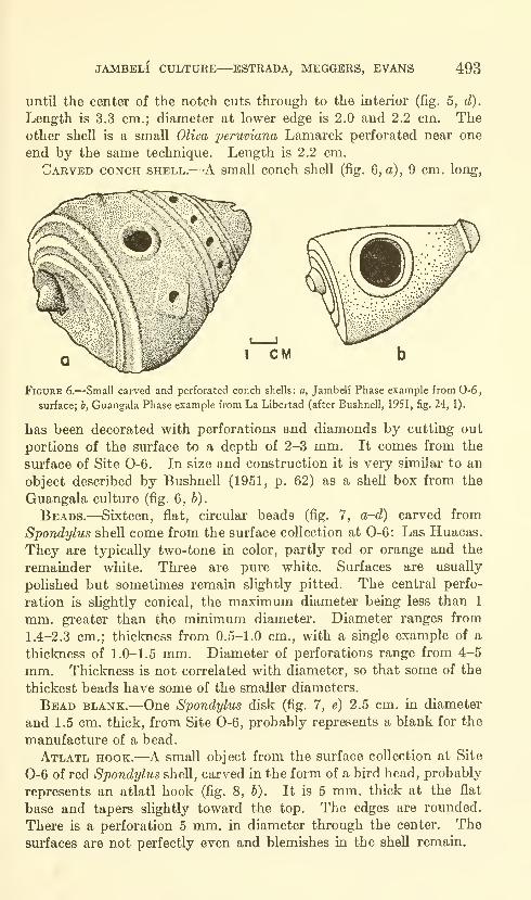
JAMBELI CULTURE—ESTRADA, MEGGERS, EVANS 493
until the center of the notch cuts through to the interior (fig. 5, d).
Length is 3.3 cm.; diameter at lower edge is 2.0 and 2.2 cm. Theother shell is a small OUoa peruviana Lamarck perforated near one
end by the same technique. Length is 2.2 cm.
Carved conch shell.—-A small conch shell (fig. 6, o), 9 cm. long,
Figure 6.—Small carved and perforated conch shells: a, Jambeli Phase example from 0-6,
surface; b, Guangala Phase example from La Libertad (after Bushnell, 1951, fig. 24, 1).
has been decorated with perforations and diamonds by cutting out
portions of the surface to a depth of 2-3 mm. It comes from the
surface of Site 0-6. In size and construction it is very similar to an
object described by Bushnell (1951, p. 62) as a shell box from the
Guangala culture (fig. 6, 6).
Beads.—Sixteen, flat, circular beads (fig. 7, a-d) carved from
Spondylus shell come from the surface collection at 0-6: Las Huacas.
They are typically two-tone in color, partly red or orange and the
remainder white. Three are pure white. Surfaces are usually
polished but sometimes remain slightly pitted. The central perfo-
ration is slightly conical, the maximum diameter being less than 1
mm. greater than the minimum diameter. Diameter ranges from
1.4-2.3 cm.; thickness from 0.5-1.0 cm., with a single example of a
thickness of 1.0-1.5 mm. Diameter of perforations range from 4-5
mm. Thickness is not correlated with diameter, so that some of the
thickest beads have some of the smaller diameters.
Bead blank.—One Spondylus disk (fig. 7, e) 2.5 cm. in diameter
and 1.5 cm, thick, from Site 0-6, probably represents a blank for the
manufacture of a bead.
Atlatl hook,—A small object from the surface collection at Site
0-6 of red Spondylus shell, carved in the form of a bird head, probably
represents an atlatl hook (fig, 8, b). It is 5 mm. thick at the flat
base and tapers slightly toward the top. The edges are rounded.
There is a perforation 5 mm. in diameter through the center. The
surfaces are not perfectly even and blemishes in the shell remain.
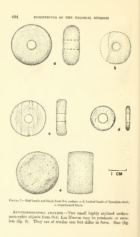
494 PROCEEDINGS OF THE NATIONAL MUSEUM
I CM
Figure 7,—Shell beads and blank from 0-6, surface: a-d, finished beads of Spondylus shell;
e, unperforated blank.
Anthropomorphic amulets.—Two small highly stylized anthro-pomorphic objects from 0-6: Las Huacas may be pendants or amu-lets (fig. 9). They are of similar size but differ in form. One (fig.
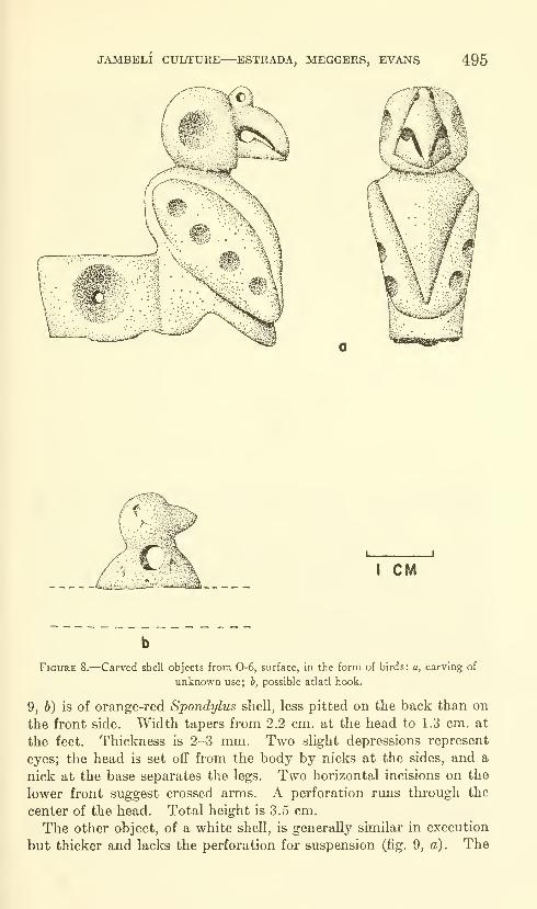
JAMBELI CULTURE—ESTRADA, MEGGERS, EVANS 495
I CM
Figure 8.—Carved shell objects from 0-6, surface, in the form of birds: a, carving of
unknown use; h, possible atlatl hook.
9, 6) is of orange-red Spondylus shell, less pitted on the back than on
the front side. Width tapers from 2.2 cm. at the head to 1.3 cm. at
the feet. Thickness is 2-3 mm. Two slight depressions represent
eyes; the head is set off from the body by nicks at the sides, and a
nick at the base separates the legs. Two horizontal incisions on the
lower front suggest crossed arms. A perforation runs through the
center of the head. Total height is 3.5 cm.
The other object, of a white shell, is generally similar in execution
but thicker and lacks the perforation for suspension (fig. 9, a). The
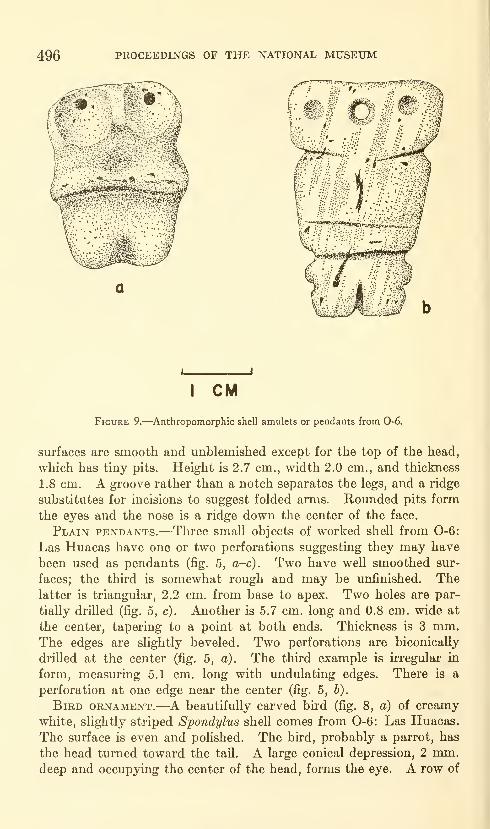
496 PROCEEDINGS OF THE NATIONAL MUSEUM
I CM
Figure 9.—Anthropomorphic shell amulets or pendants from 0-6.
surfaces are smooth and unblemished except for the top of the head,
which has tiny pits. Height is 2.7 cm., width 2.0 cm., and thickness
1.8 cm. A groove rather than a notch separates the legs, and a ridge
substitutes for incisions to suggest folded arms. Rounded pits form
the eyes and the nose is a ridge down the center of the face.
Plain pendants.—Three small objects of worked shell from 0-6:
Las Huacas have one or two perforations suggesting they may have
been used as pendants (fig. 5, a-c). Two have well smoothed sur-
faces; the third is somewhat rough and may be unfinished. Thelatter is triangular, 2.2 cm. from base to apex. Two holes are par-
tially drilled (fig. 5, c). Another is 5.7 cm. long and 0.8 cm. wide at
the center, tapering to a point at both ends. Thickness is 3 mm.The edges are slightly beveled. Two perforations are biconically
drilled at the center (fig. 5, a). The third example is irregular in
form, measuring 5.1 cm. long with undulating edges. There is a
perforation at one edge near the center (fig. 5, b).
Bird ornament.—A beautifully carved bird (fig. 8, a) of creamy
white, slightly striped Spondylus shell comes from 0-6: Las Huacas.
The surface is even and polished. The bird, probably a parrot, has
the head turned toward the tail. A large conical depression, 2 mm.deep and occupying the center of the head, forms the eye. A row of
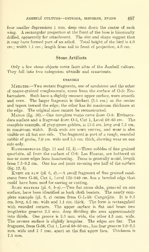
JAMBELI CULTURE—ESTRADA, MEGGERS, EVANS 497
four smaller depressions 1 mm. deep runs down the center of each
wing. A rectangular projection at the front of the base is biconically
drilled, apparently for attachment. The size and shape suggest that
it may have formed part of an atlatl. Total height of the bird is 4.0
cm.; width 1.1 cm.; length from tail to front of projection, 4.0 cm.
Stone Artifacts
Only a few stone objects come from sites of the Jambeli culture.
They fall into two categories: utensils and ornaments.
UTENSILS
Metates.—Two metate fragments, one of sandstone and the other
of coarse-gi'ained conglomerate, come from the surface of 0-5: Em-barcadero. Both have a slightly concave upper surface, worn smooth
and even. The larger fragment is thickest (3.5 cm.) at the center
and tapers toward the edge; the other has its maximum thickness at
the edge. The original sizes cannot be reconstructed.
Manos (fig. 10).—One complete mano came from 0-5: Embarca-
dero surface and a fragment from 0-5, Cut 1, Level 60-80 cm. Thecomplete example, of gray-green gabbro, is 13.0 cm. long and 7.5 cm.
in maximum width. Both ends are worn convex, and wear is also
visible on all but one side. The fragment is part of a rough, rounded
cobble of gneiss, 7.0 cm. wide and 5.5 cm. thick, showing wear on one
side only.
Hammerstones (figs. 11 and 12, 6).—Three cobbles of fine grained
quartzite, all from the surface of 0-6: Las Huacas, are battered on
one or more edges from hammering. Form is generally ovoid, length
from 7.5-9.2 cm. One has red paint covering one half of the surface
(fig. 12, b).
Knife or saw (pi. 6, d).—A small fragment of fine grained sand-
stone from G-86, Cut 1, Level 120-140 cm. has a beveled edge that
could have been used for sawing or cutting.
Bark beaters (pi. 6, b-c).—Two flat stone slabs, grooved on one
surface, have been identified as bark cloth beaters. The nearly com-
plete example (pi. 6, c) comes from G-L-30: Cangrejito. It is 6.5
cm. long, 4.5 cm. wide and 1.1 cm. thick. The form is rectanguloid
with rounded corners. The upper surface is flat and bears two
lengthwise grooves 2.5 mm. deep dividing the area approximately
into thirds. One groove is 3.5 mm. wide, the other 4.5 mm. wide.
The reverse surface is shghtly irregular. The edges are flat. Thefragment, from G-86, Cut 1, Level 60-80 cm., has four grooves 2.0-2.5
mm. wide and 5-7 mm. apart on the flat upper face. Thickness is
7.5 mm.
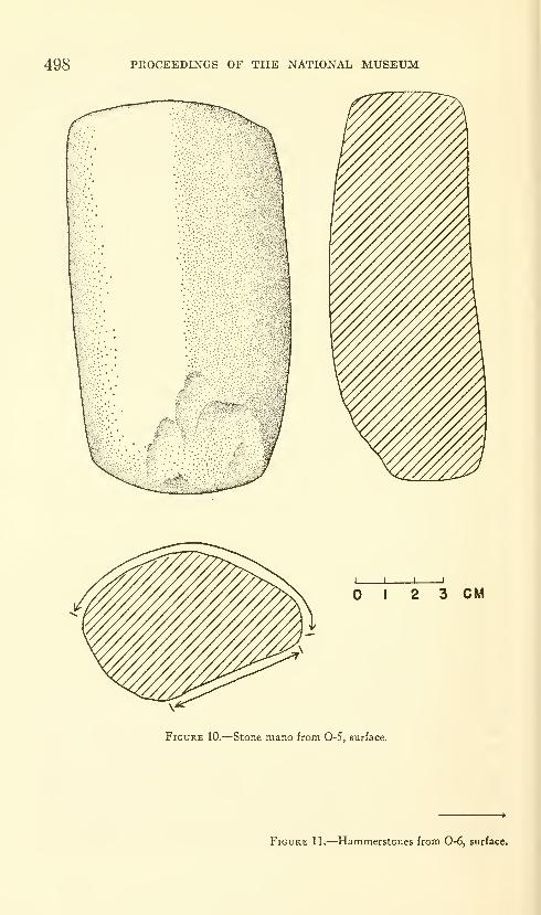
498 PROCEEDINGS OF THE NATIONAL MUSEUM
1 2 3 CM
Figure 10.—Stone mano from 0-5, surface.
Figure 11.—Hammerstones from 0-6, surface.
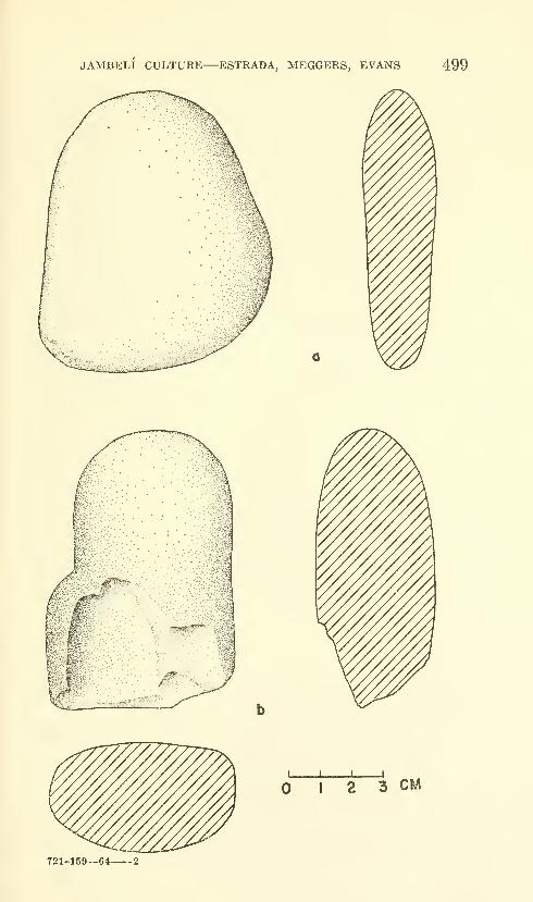
JAMBELI CULTURE—ESTRADA, MEGGERS, EVANS 499
I 1 1 1
I 2 3 CM
721-159—64 2
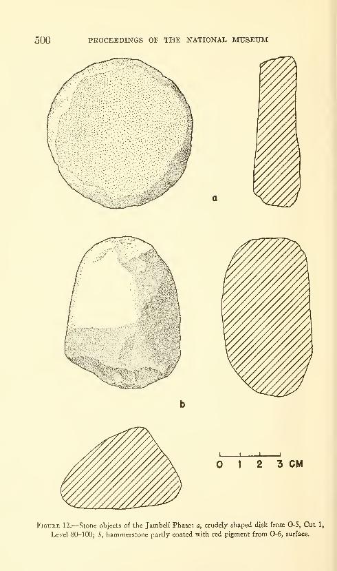
500 PROCEEDINGS OF THE NATIONAL MUSEUM
I 2 3 CM
Figure 12.—Stone objects of the Jambeli Phase: a, crudely shaped disk from 0-5, Cut 1,
Level 80-100; h, hammerstone partly coated with red pigment from 0-6, surface.

JAMBELI CULTURE—ESTRADA, MEGGERS, EVANS 501
Worked stone.—A fragment of serpentine, 6.5X5.5 cm. and 2.0
cm. thick, shows grooves and circular marks produced by cord saw-
ing. A small ovoid projection left from the sawing remains at the
center. The object is from G-86, Cut 1, Level 0-20 cm.
DiscoiDAL SHAPED STONE (fig. 12, o)
.
—A piecc of fine conglomerate
has been shaped into a disk 7 cm. in diameter and 1.8-2.5 cm. thick.
The edges are rounded, and one surface is even while the other is
slightly irregular. The object is from 0-5, Cut 1, Level 80-100 cm.
ORNAMENTS
Beads (fig. 13).—Basalt, shale, serpentine and chlorite schist were
used for the manufacture of beads. One example of asymmetrical
I CM
Figure 13.—Stone beads showing variation in diameter and thickness; all from 0-6, surface.
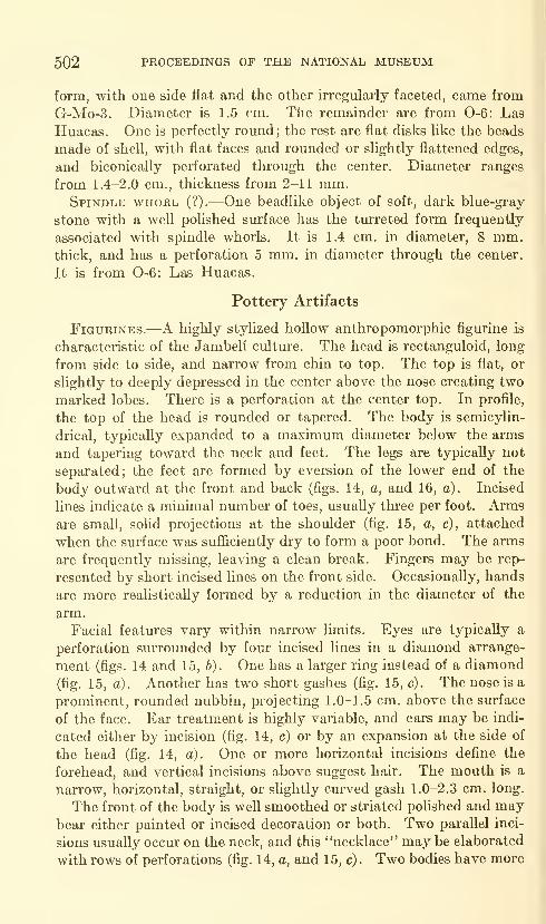
502 PROCEEDINGS OF THE NATIONAL MUSEUM
form, with one side flat and the other irregularly faceted, came from
G-Mo-3. Diameter is 1.5 cm. The remainder are from 0-6: Las
Huacas. One is perfectly round; the rest are flat disks like the beads
made of shell, with flat faces and rounded or slightly flattened edges,
and biconically perforated through the center. Diameter ranges
from 1.4-2.0 cm., thickness from 2-11 mm.Spindle whorl (?).—One beadlike object of soft, dark blue-gray
stone with a well polished surface has the tmTcted form frequently
associated with spindle whorls. It is 1.4 cm. in diameter, 8 mm.thick, and has a perforation 5 mm. in diameter through the center.
It is from 0-6: Las Huacas.
Pottery Artifacts
Figurines.—A highly stylized hollow anthropomorphic figurine is
characteristic of the Jambeli culture. The head is rectanguloid, long
from side to side, and narrow from chin to top. The top is flat, or
slightly to deeply depressed in the center above the nose creating two
marked lobes. There is a perforation at the center top. In profile,
the top of the head is rounded or tapered. The body is semicylin-
drical, typically expanded to a maximum diameter below the arms
and tapering toward the neck and feet. The legs are typically not
separated; the feet are formed by eversion of the lower end of the
body outward at the front and back (figs. 14, a, and 16, a). Incised
lines indicate a minimal number of toes, usually three per foot. Armsare small, solid projections at the shoulder (fig. 15, a, c), attached
when the surface was sufficiently dry to form a poor bond. The arms
are frequently missing, leaving a clean break. Fingers may be rep-
resented by short incised lines on the front side. Occasionally, hands
are more realistically formed by a reduction in the diameter of the
arm.
Facial features vary within narrow limits. Eyes are typically a
perforation surrounded by four incised lines in a diamond arrange-
ment (figs. 14 and 15, b). One has a larger ring instead of a diamond
(fig. 15, a). Another has two short gashes (fig. 15, c). The nose is a
prominent, rounded nubbin, projecting 1.0-1.5 cm. above the surface
of the face. Ear treatment is highly variable, and ears may be indi-
cated either by incision (fig. 14, c) or by an expansion at the side of
the head (fig. 14, a). One or more horizontal incisions define the
forehead, and vertical incisions above suggest hair. The mouth is a
narrow, horizontal, straight, or slightly curved gash 1.0-2.3 cm. long.
The front of the body is well smoothed or striated polished and maybear either painted or incised decoration or both. Two parallel inci-
sions usually occur on the neck, and this "necklace" may be elaborated
with rows of perforations (fig. 14, a, and 15, c) . Two bodies have more
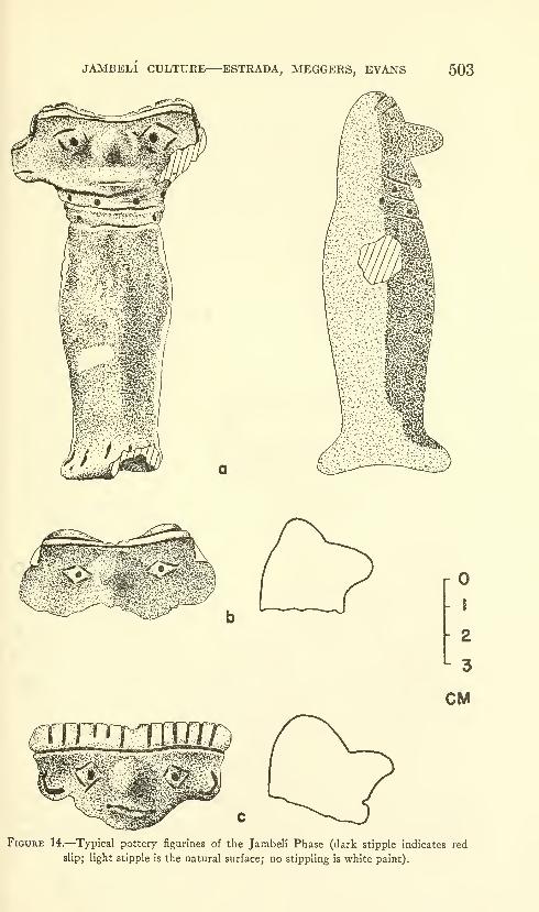
JAMBELI CULTURE—ESTRADA, MEGGERS, EVANS 503
r^
-
1
- 2
'- 3
CM
Figure 14.—Typical pottery figurines of the Jambeli Phase (dark stipple indicates red
slip; light stipple is the natural surface; no stippling is white paint).
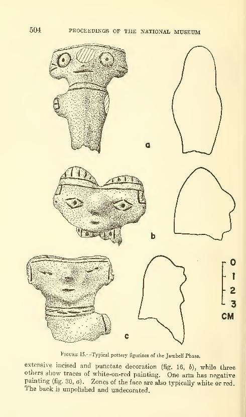
504 PROCEEDINGS OF THE NATIONAL MUSEUM
rO-
r
- 2
- 3
CM
Figure 15.—Typical pottery figurines of the Jambeli Phase.
extensive incised and punctate decoration (fig. 16, b), while threeothers show traces of white-on-red painting. One arm has negativepainting (fig. 30, a). Zones of the face are also typically white or red.The back is unpolished and undecorated.

JAMBELI CULTURE—ESTRADA, MEGGERS, EVANS 505
Only two of the figurines are complete. One of these is 15.5 cm.
tall, the other 6.2 cm. Judging from the proportions of the frag-
mentary examples, these probably represent the size range. Com-plete heads range from 3.0-8.5 cm. in width, with the majority from
6.2-8.5 cm. Arms project 1.0-2.3 cm. outward from the body wall.
Imported figurines.—One atypical figurine body fragment comes
from Site 0-3 (fig. 17, a). It is solid, with flat surfaces, and is 1 .3-1 .6 cm.
thick. One arm curves from the shoulder to join the body at the hip.
The front has incised and red painted decoration. Two other frag-
ments from the same site appear to represent the same style. In
J I
I 2 CM
I 2 3 CM
b
Figure 16.—Typical pottery figurines of the Jambeli Phase (dark stipple indicates red slip)
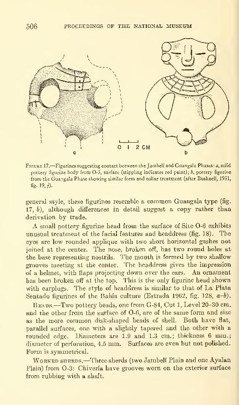
506 PROCEEDINGS OF THE NATIONAL MUSEUM
I 2 CM
Figure 17.—Figurines suggesting contact between the Jambeli and Guangala Phases: a, solid
pottery figurine body from 0-3, surface (stippling indicates red paint); b, pottery figurine
from the Guangala Phase showing similar form and collar treatment (after Bushnell, 19S1,
fig. 19,;).
general style, these figurines resemble a common Guangala type (fig.
17, b), although differences in detail suggest a copy rather than
derivation by trade.
A small pottery figurine head from the surface of Site 0-6 exhibits
unusual treatment of the facial features and headdress (fig. 18). Theeyes are low rounded applique with two short horizontal gashes not
joined at the center. The nose, broken off, has two round holes at
the base representing nostrils. The mouth is formed by two shallow
grooves meeting at the center. The headdress gives the impression
of a helmet, with flaps projecting down over the ears. An ornament
has been broken off at the top. This is the only figurine head shown
with earplugs. The style of headdress is similar to that of La Plata
Sentado figurines of the Bahia culture (Estrada 1962, fig. 128, a-b).
Beads.—Two pottery beads, one from G-84, Cut 1, Level 20-30 cm.
and the other from the surface of 0-6, are of the same form and size
as the more common disk-shaped beads of shell. Both have flat,
parallel surfaces, one with a slightly tapered and the other with a
rounded edge. Diameters are 1.9 and 1.3 cm.; thickness 6 mm.;diameter of perforation, 4.5 mm. Surfaces are even but not polished.
Form is symmetrical.
Worked sherds.—Three sherds (two Jambeli Plain and one Ayalan
Plain) from 0-3: Chiveria have grooves worn on the exterior surface
from rubbing with a shaft.
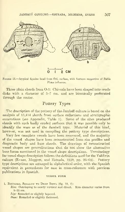
JAMBELI CULTURE—ESTRADA, MEGGERS, EVANS 507
I 2 CM
Figure 18.—Atypical figurine head from 0-6, surface, with features suggestive of Bahia
Phase influence.
Three plain sherds from 0-3: Chiveria have been shaped into crude
disks with a diameter of 5-7 cm. and are biconically perforated
through the center.
Pottery Types
The description of the pottery of the Jambeli culture is based on the
analysis of 15,414 sherds from surface collections and stratigraphic
excavations (see Appendix, Table 1). Some of the sites produced
sherds with such badly eroded surfaces that it was possible only to
identify the ware as of the Jambeli type. Material of this kind,
however, was not used in compiling the pottery type descriptions.
Very few complete vessels have been recovered, and the majority
of the vessel shapes have been reconstructed from rim profiles anddiagnostic body and base sherds. The drawings of reconstructed
vessel shapes are generalizations that do not show the alternative
base forms mentioned in the vessel shape descriptions. Terminology
for vessel shape description follows the definitions used for the Valdivia
culture (Evans, Meggers, and Estrada, 1959, pp. 26-68). Pottery
type descriptions are arranged in alphabetical order, with the Spanish
equivalent in parentheses for ease in cross-reference with previous
publications in Spanish.
VESSEL FORM
1. Rounded, Shallow to Deep Bowl (fig. 19, 1):
Rim: Outsloping to nearly vertical and direct. Rina diameter varies from8-28 cm.
Lip: Rounded or slightly tapered.
Base: Rounded or slightly flattened.
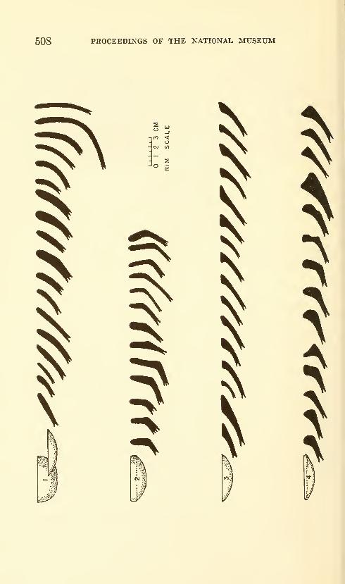
508 PROCEEDINGS OF THE NATIONAL MUSEUM
-itO <o
- CM «1
rv

JAMBELI CULTURE—ESTRADA, MEGGERS, EVANS 509
\VV
\
-00
LLl
>
a.
B
I
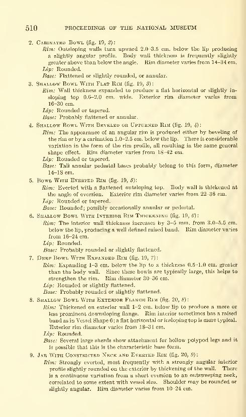
510 PROCEEDINGS OF THE NATIONAL MUSEUM
2. Cakinated Bowl (fig. 19, 2) :
Uim: Outsloping walls turn upward 2.0-3.5 cm. below the lip producing
a slightly angular profile. Body wall thickness is frequently slightly
greater above than below the angle. Rim diameter varies from 14-34 cm.
Ltp; Rounded.
Base: Flattened or slightly rounded, or annular.
3. Shallow Bowl With Flat Rim (fig. 19, S)
:
Rim: Wall thickness expanded to produce a flat horizontal or slightly in-
sloping top 0.6-2.0 cm. wide. Exterior rim diameter varies from16-30 cm.
Ldp: Rounded or tapered.
Base: Probably flattened or annular.
4. Shallow Bowl With Beveled or UPTtrRNED Rim (fig, 19, 4)
Rim: The appearance of an angular rim is produced either by beveling of
the rim or by a carination 1.0-2.5 cm. below the lip. There is considerable
variation in the form of the rim profile, all resulting in the same general
shape effect. Rim diameter varies from 18-42 cm.
Ldp: Rounded or tapered.
Base: Tall annular pedestal bases probably belong to this form, diameter
14-18 cm.
5. Bowl With Everted Rim (fig. 19, 6):
Rim: Everted with a flattened outsloping top. Body wall is thickened at
the angle of eversion. Exterior rim diameter varies from 22-36 cm.
Lip: Rounded or tapered.
Base: Rounded; possibly occasionally annular or pedestal.
6. Shallow Bowl With Interior Rim Thickening (fig. 19, 6)
:
Rim: The interior wall thickness increases by 3-5 mm. from 3.0-5.5 cm.
below the lip, producing a well defined raised band. Rim diameter varies
from 16-24 cm.
Lip: Rounded.
Base: Probably rounded or slightly flattened.
7. Deep Bowl With Expanded Rim (fig. 19, 7)
:
Ri7n: Expanding 1-3 cm. below the lip to a thickness 0.5-1.0 cm. greater
than the body wall. Since these bowls are typically large, this helps to
strengthen the rim. Rim diameter 30-36 cm.
Lip: Rounded or slightly flattened.
Base: Probably rounded or slightly flattened.
8. Shallow Bowl With Exterior Flange Rim (fig. 20, 8) :
Rim: Thickened on exterior wall 1-2 cm. below lip to produce a more or
less prominent downsloping flange. Rim interior sometimes has a raised
band as in Vessel Shape 6 ; a flat horizontal or insloping top is more typical.
Exterior rim diameter varies from 18-34 cm.
Lip: Rounded.Base: Several large sherds show attachment for hollow polypod legs and it
is possible that this is the characteristic base form.
9. Jar With Constricted Neck and Everted Rim (fig. 20, 9) :
Rim: Strongly everted, most frequently with a strongly angular interior
profile slightly rounded on the exterior by thickening of the wall. There
is a continuous variation from a short eversion to an outsweeping neck,
correlated to some extent with vessel size. Shoulder may be rounded or
slightly angular. Rim diameter varies from 10-24 cm.
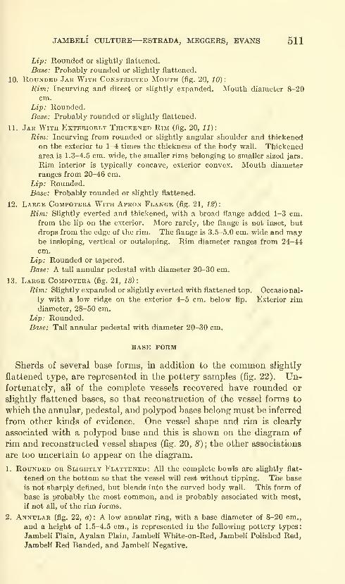
JAMBELI CULTURE—ESTRADA, MEGGERS, EVANS 511
Lip: Rounded or slightly flattened.
Base: Probably rounded or slightly flattened.
10. Rounded Jar With Constricted Mouth (fig. 20, 10)
:
Rim: Incurving and direct or slightly expanded. Mouth diameter 8-20
cm.
Lip: Rounded.
Base: Probably rounded or slightly flattened.
11. Jar With Exteriorly Thickened Rim (fig. 20, 11):
Rim: Incurving from rounded or slightly angular shoulder and thickened
on the exterior to 1-4 times the thickness of the body wall. Thickenedarea is 1.3-4.5 cm. wide, the smaller rims belonging to smaller sized jars.
Rim interior is typically concave, exterior convex. Mouth diameter
ranges from 20-46 cm.
Lip: Rounded.
Base: Probably rounded or slightly flattened.
12. Large Compotera With Apron Flange (fig. 21, 12):
Rim: Slightly everted and thickened, with a broad flange added 1-3 cm.
from the lip on the exterior. More rarely, the flange is not inset, but
drops from the edge of the rim. The flange is 3.5-5.0 cm. wide and maybe insloping, vertical or outsloping. Rim diameter ranges from 24-44
cm.
Lip: Rounded or tapered.
Base: A tall annular pedestal with diameter 20-30 cm.
13. Large Compotera (fig. 21, 13)
:
Rim: Slightly expanded or slightly everted with flattened top. Occasional-
ly with a low ridge on the exterior 4-5 cm. below lip. Exterior rimdiameter, 28-50 cm.
Lip: Rounded.Base: Tall annular pedestal with diameter 20-30 cm.
BASE FORM
Sherds of several base forms, in addition to the common slightly
flattened type, are represented in the pottery samples (fig. 22). Un-fortunately, all of the complete vessels recovered have rounded or
slightly flattened bases, so that reconstruction of the vessel forms to
which the annular, pedestal, and polypod bases belong must be inferred
from other kinds of evidence. One vessel shape and rim is clearly
associated with a polypod base and this is shown on the diagram of
rim and reconstructed vessel shapes (fig. 20, 8); the other associations
are too uncertain to appear on the diagram.
1. Rounded or Slightly Flattened: All the complete bowls are slightly flat-
tened on the bottom so that the vessel will rest without tipping. The base
is not sharply deflned, but blends into the curved body wall. This form of
base is probably the most common, and is probably associated with most,
if not all, of the rim forms.
2. Annular (fig. 22, a): A low annular ring, with a base diameter of 8-20 cm.,
and a height of 1.5-4.5 cm., is represented in the following pottery types:
Jambeli Plain, Ayalan Plain, Jambelf White-on-Red, Jambell Polished Red,
Jambeli Red Banded, and Jambeli Negative.
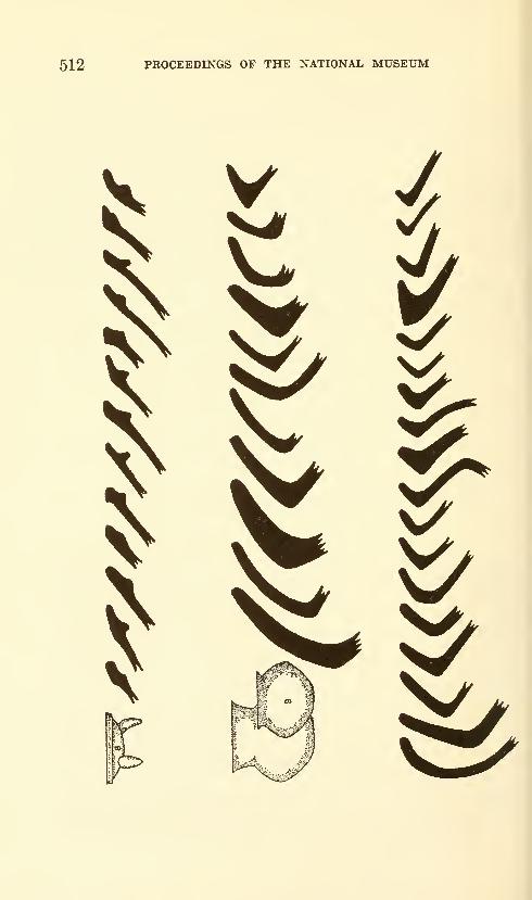
512 PROCEEDINGS OF THE NATIONAL MUSEUM
V
V^
iW^m V̂
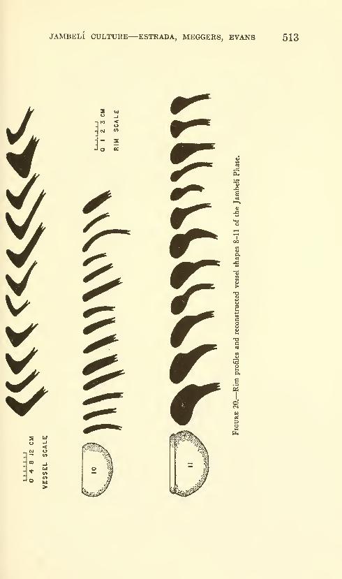
JAMBELI CULTURE—ESTRADA, MEGGERS, EVANS 513
V
Z uj
n to <o
- 2o n:
V^«g 111
o -J
<— «
>
PiJ
b
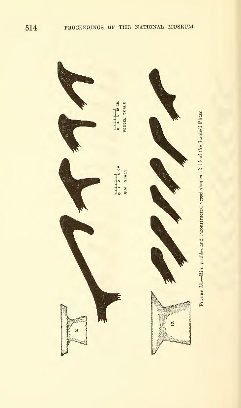
514 PROCEEDINGS OF THE NATIONAL MUSEUM
Oh
P<
U^
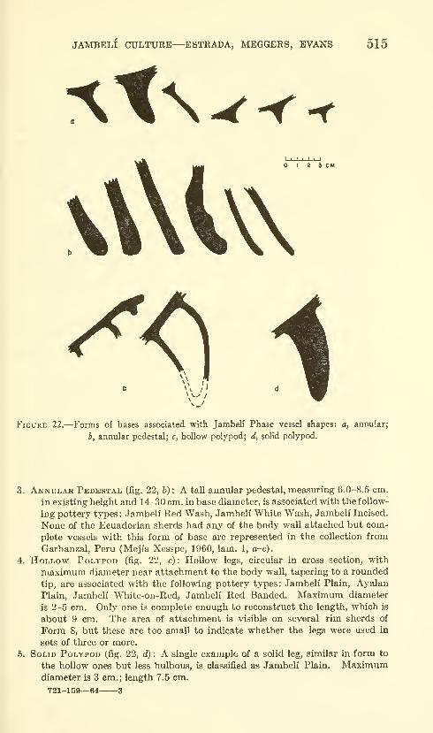
JAMBELI CULTURE—ESTRADA, MEGGERS, EVANS 515
^t^I 2 3 CM
\\ I,
Figure 22.—Forms of bases associated with Jambeli Phase vessel shapes: a, annular;
b, annular pedestal; c, hollow polypod; d, solid polypod.
3. Annular Pedestal (fig. 22, b) : A tall annular pedestal, measuring 6.0-8.5 cm.
in existing height and 14-30 cm. in base diameter, is associated with the follow-
ing pottery types: Jambeli Red Wash, Jambeli White Wash, Jambeli Incised.
None of the Ecuadorian sherds had any of the body wall attached but com-
plete vessels with this form of base are represented in the collection from
Garbanzal, Peru (Meji'a Xesspe, 1960, lam. 1, a-c).
4. Hollow Polypod (fig. 22, c) : Hollow legs, circular in cross section, with
maximum diameter near attachment to the body wall, tapering to a rounded
tip, are associated with the following pottery types: Jambeli Plain, Ayalan
Plain, Jambeli White-on-Red, Jambeli Red Banded. Maximum diameter
is 2-5 cm. Only one is complete enough to reconstruct the length, which is
about 9 cm. The area of attachment is visible on several rim sherds of
Form 8, but these are too small to indicate whether the legs were used in
sets of three or more.
5. Solid Polypod (fig. 22, d) : A single example of a solid leg, similar in form to
the hollow ones but less bulbous, is classified as Jambeli Plain. Maximumdiameter is 3 cm. ; length 7.5 cm,
721-159—64 3
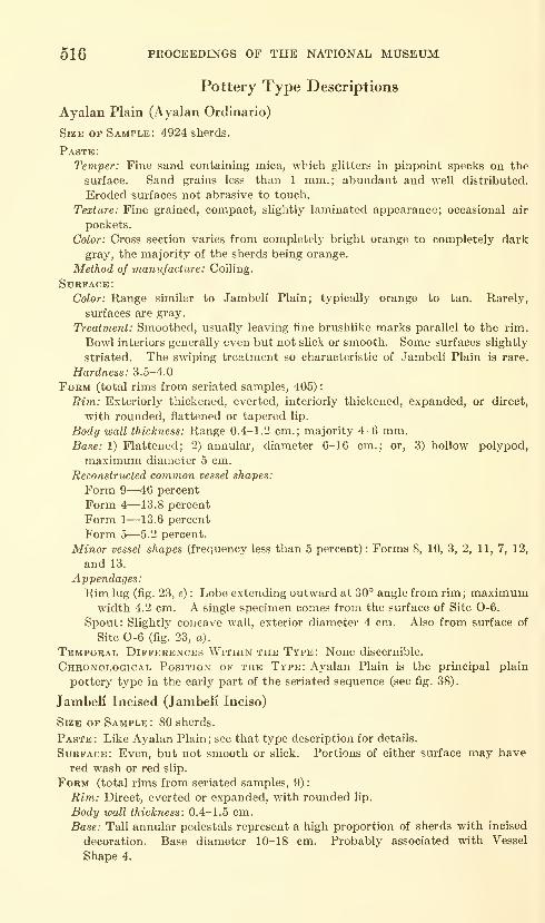
516 PROCEEDINGS OF THE NATIONAL MUSEUM
Pottery Type Descriptions
Ayalan Plain (Ayalan Ordinario)
Size of Sample: 4924 sherds.
Paste:
Temper: Fine sand containing mica, which glitters in pinpoint specks on the
surface. Sand grains less than 1 mm.; abundant and well distributed.
Eroded surfaces not abrasive to touch.
Texture: Fine grained, compact, slightlj^ laminated appearance; occasional air
pockets.
Color: Cross section varies from completely bright orange to completely dark
gray, the majority of the sherds being orange.
Method of manufacture: Coiling.
Surface:
Color: Range similar to Jambeli Plain; typically orange to tan. Rarely,
surfaces are gray.
Treatment: Smoothed, usually leaving fine brushlike marks parallel to the rim.
Bowl interiors generally even but not slick or smooth. Some surfaces slightly
striated. The swiping treatment so characteristic of Jambeli Plain is rare.
Hardness: 3.5-4.0
Form (total rims from seriated samples, 405)
:
Ri7n: Exteriorly thickened, everted, interiorly thickened, expanded, or direct,
with rounded, flattened or tapered lip.
Body wall thickness: Range 0.4-1.2 cm.; majority 4-6 mm.Base: 1) Flattened; 2) annular, diameter 6-16 cm.; or, 3) hollow polypod,
maximum diameter 5 cm.
Reconstructed common vessel shapes:
Form 9—46 percent
Form 4—13.8 percent
Form 1—13.6 percent
Form 5—5.2 percent.
Minor vessel shapes (frequency less than 5 percent): Forms S, 10, 3, 2, 11, 7, 12,
and 13.
Appendages:
Rim lug (fig. 23, e) : Lobe extending outward at 30° angle from rim; maximumwidth 4.2 cm. A single specimen comes from the surface of Site 0-6.
Spout: Slightly concave wall, exterior diameter 4 cm. Also from surface of
Site 0-6 (fig. 23, a).
Temporal Differences Within the Type: None discernible.
Chronological Position of the Type: Ayalan Plain is the principal plain
pottery type in the early part of the seriated sequence (see fig. 38).
Jambeli Incised (Jambeli Inciso)
Size of Sample: 80 sherds.
Paste: Like Ayalan Plain; see that type description for details.
Surface: Even, but not smooth or slick. Portions of either surface may have
red wash or red slip.
Form (total rims from seriated samples, 9)
:
Rim: Direct, everted or expanded, with rounded lip.
Body wall thickness: 0.4-1.5 cm.
Base: Tall annular pedestals represent a high proportion of sherds with incised
decoration. Base diameter 10-18 cm. Probably associated with Vessel
Shape 4.
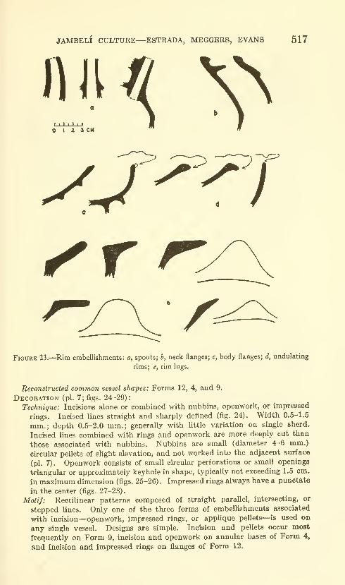
JAMBELI CULTURE ESTRADA, MEGGERS, EVANS 517
n n //O I 2 a CM ^
Figure 23.—Rim embellishments: a, spouts; h, neck flanges; c, body flanges; d, undulating
rims; e, rim lugs.
Reconstructed common vessel shapes: Forms 12, 4, and 9.
Decoration (pi. 7; figs. 24-29):
Technique: Incisions alone or combined with nubbins, openwork, or impressed
rings. Incised lines straight and sharply defined (fig. 24). Width 0.5-1.5
mm.; depth 0.5-2.0 mm.; generally with little variation on single sherd.
Incised lines combined with rings and openwork are more deeply cut than
those associated with nubbins. Nubbins are small (diameter 4-6 mm.)
circular pellets of slight elevation, and not worked into the adjacent surface
(pi. 7). Openwork consists of small circular perforations or small openings
triangular or approximately keyhole in shape, typically not exceeding 1.5 cm.
in maximum dimension (figs. 25-26). Impressed rings always have a punctate
in the center (figs. 27-28).
Motif: Rectilinear patterns composed of straight parallel, intersecting, or
stepped lines. Only one of the three forms of embellishments associated
with incision—openwork, impressed rings, or applique pellets—is used on
any single vessel. Designs are simple. Incision and pellets occur moat
frequently on Form 9, incision and openwork on annular bases of Form 4,
and incision and impressed rings on flanges of Form 12.
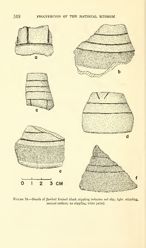
518 PROCEEDINGS OF THE NATIONAL MUSEUM
J L
I 2 3 CM
Figure 24.—Sherds of Jambeli Incised (dark stippling indicates red slip; light stippling,
natural surface; no stippling, white paint).
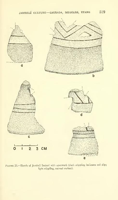
JAMBELI CULTURE—ESTRADA, MEGGERS, EVANS 519
'.nf.l-itiHi;'^'—'-''"^''
g,m^'-
I I I 1
I 2 3 CM
Figure 25.—Sherds of Jambeli Incised with openwork (dark stippling indicates red slip;
light stippling, natural surface).
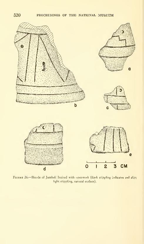
520 PROCEEDINGS OF THE NATIONAL MUSEUM
J I
2 3 CM
Figure 26.—Sherds of Jambeli Incised with openwork (dark stippling indicates red slip;
light stippling, natural surface).

JAMBELI CULTURE—ESTRADA, MEGGERS, EVANS 521
I 2 3 CM
'!i..'%.--si~5a.,.
Figure 27.—Sherds of Jambeli Incised with impressed rings on the flange of Vessel Form 12.
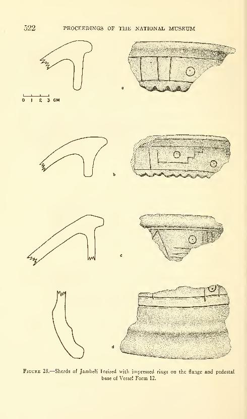
522 PROCEEDINGS OF THE NATIONAL MUSEUM
I \ L.
I 2 3 CM
^^«il**W^**'A,^sAi.^^
^^j-jm^UflVJ'
'- ' Ai '^f^^n•^•'r i
Figure 28.—Sherds of Jambeli Incised with impressed rings on the flange and pedestal
base of Vessel Form 12.
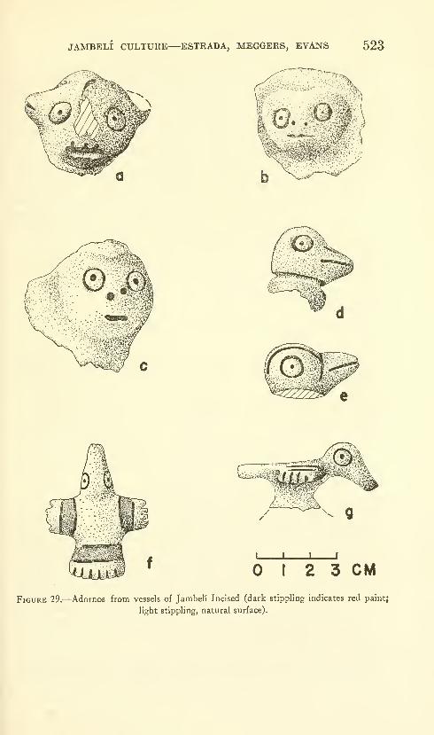
JAMBELI CULTURE—ESTRADA, MEGGERS, EVANS 523
J I I
I 2 3 CM
Figure 29.—Adornos from vessels of Jambeli Incised (dark stippling indicates red paint;
light stippling, natural surface).
524 PROCEEDINGS OF THE NATIONAL MUSEUM
Associated techniques: Seven small pottery adornos were collected from the
surface of Site 0-6. Several complete vessels from Garbanzal, Peru, show
this kind of adorno on the upper part of tall pedestal bases with incised
decoration (fig. 42: 55-56; Mejia Xesspe, 1960, lam. 1, a, c,), and the adornos
from Site 0-6 must have occupied the same position. The surface is gen-
erally even, but unpolished, and only one adorno shows traces of slip or paint.
Three are identifiable as bird heads (fig. 29, d-g) ; the remaining four are
generalized but probably represent an animal (fig. 29, a-c) . All have the same
type of eye: a ring with a punctate in the center. The animal heads have
slightly to highly projecting noses, with two punctates at the base to repre-
sent nostrils. The mouth is an irregular horizontal gash. These heads
are attached at the back to the vessel wall. Two are solid and the other two
are hollow. Of the three birds, two are heads and the other a complete
bird with outstretched wings. The latter has zoned red paint between the
pairs of incisions on the wing and tail.
Temporal Differences Within the Type: None discernible.
Chronological Position of the Type: Present in small frequency throughout
the seriated sequence (see fig. 38).
Jambeli Negative (Jambeli Negative)
Size of Sample: 45 sherds.
Paste: Majority like Ayalan Plain; a few like Jambeli Plain. See those type
descriptions for details.
Surface: Painting applied to plain or red slipped surface. Plain surfaces even
but not polished. Red slip varies from thin unpolished coating to polished
red slip comparable to Jambeli Polished Red. Rare sherds are white slipped
on the rim, then painted.
Form (total rims from seriated samples, 17)
:
Rim: Direct with rounded or tapered lip. Rarely expanded with flat top.
Body wall thickness: 3-7 mm.Base: Typically flattened; rarely annular 14-16 cm. diameter.
Reconstructed common vessel shapes:
Form 1—82.5 percent
Form 3—11.8 percent
Form 10—5.9 percent.
Decoration (figs. 30 and 31):
Technique: Black paint applied by the resist or negative technique to a plain,
red slipped or white sUpped portion of the surface. Frequently combined
with white paint on a red slipped surface, either as an integrated design in
which the negative painting is alternated with or superimposed on the
white painted design, or on the opposite surface (i.e., the interior is negative
painted and the exterior white-on-red). Where well preserved, the black is
dark and covers the red sHp completely. Edges of painted areas are sharply
defined. Black lines are more unequal in width than unpainted lines left
by the application of resist material. Negative paint is fugitive and fre-
quently difficult to detect even when the surface is wet, suggesting that this
technique of decoration may have been more common than the eroded
condition of the sherds now indicates.
Motif: Lines and dots, independently or in combination. Dots are unpainted
areas to which resist material was originally applied. They occur in a single
row in the center of a band 8 mm. wide, or several adjacent rows cover a
JAMBELf CULTURE—ESTRADA, MEGGERS, EVANS 525
larger area. Dots are 3-7 mm. in diameter, with little variation on a single
design. Smaller dots are more circular than larger ones, which may be
slightly ovoid. Rarely, dots are black and surrounded by a black ring.
Separation is from 1-3 mm. Unpainted stripes range from 2-8 mm. in
width; black stripes from 1-9 mm. Lines are straight or curved, and parallel
lines are commonly employed.
Temporal Differences Within the Type: None discernible.
Chronological Position of the Type: Present in minor frequency throughout
the seriated sequence (see fig. 38).
Jambeli Plain (Jambeli Ordinario)
Size of Sample: 6488 sherds.
Paste:
Temper: Abundant coarse white quartz sand with abundant pinpoint flecks of
mica. Quartz grains 1-2 mm. frequent, some up to 5 mm., and well dis-
tributed in paste.
Texture: Sandy, giving abrasive feeling to eroded surfaces; compact; not friable.
Tendency to show lamination parallel to surface.
Color: Typically, completely oxidized so that the cross section has the same
color as the surface. Thick rim may have medium to dark gray core.
Method of manufacture: Coiling; sometimes fractured along coil junctions.
Surface:
Color: Rather uniformly orange to orange-tan or orange-brown; occasional
medium gray fire clouds.
Treatment: Smoothing varies with vessel shape. Bowl interiors relatively even,
showing fine brush marks adjacent to the rim, but never slick to touch;
bowl exteriors typically swiped, leaving characteristic elongated marks from
dragged temper grains parallel to rim. Jar necks have brushlike marks on
interior and exterior; bodies poorly smoothed leaving uneven and irregular
pitted surfaces.
Hardness: 4-4.5
Form (total rims from seriated samples, 561)
:
Rim: Exteriorly thickened, everted, interiorly thickened, expanded or direct,
with rounded, flattened, or tapered lip.
Body wall thickness: Range 0.4-1.2 cm.; majority 5-7 mm.Base: 1) flattened; 2) annular, diameter 10-28 cm.; 3) hollow polypod; maxi-
mum diameter 4-5 cm.; 4) solid polypod, diameter 3 cm., length 7.5 cm.
Reconstructed common vessel shapes:
Form 9—46 percent
Form 4—19.7 percent
Form 11—14.8 percent.
Minor vessel shapes (frequency less than 5 percent): Forms 1, 5, 10, 3, 2, 12,
and 13.
Appendages:
Rim lug: Trianguloid lobe projection extending horizontally outward 3 cm.
at rim. Examples are from the surface of Sites 0-3 and 0-6 (fig. 23, e).
Temporal Differences Within the Type: None discernible.
Chronological Position of the Type: Increases from a frequency of 3.6 percent
at the earliest site to become the dominant plain type in the latter part of the
seriated sequence (see fig. 38).
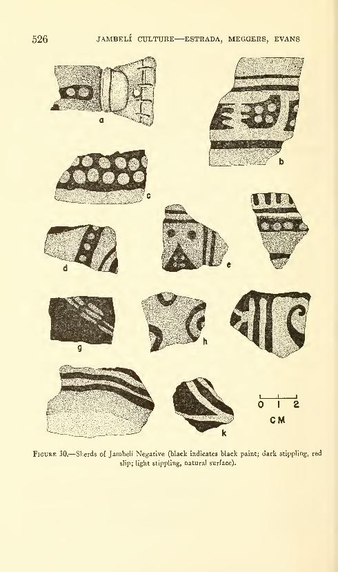
526 JAMBELI CULTURE—ESTRADA, MEGGERS, EVANS
Figure 30.—Sherds of Jambeli Negative (black indicates black paint; dark stippling, red
slip; light stippling, natural surface).
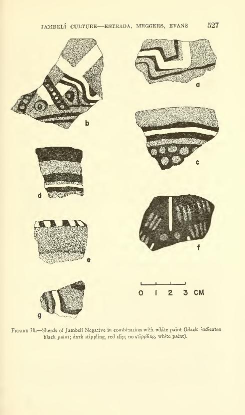
JMOELI CULTURE—ESTRADA, MEGGERS, EVANS 527
I 2 3 CM
Figure 31.—Sherds of Jambeli Negative in combination with white paint (black indicates
black paint; dark stippling, red slip; no stippling, white paint).
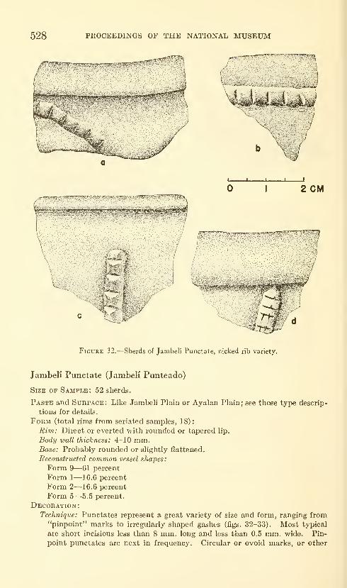
528 PROCEEDINGS OF THE NATIONAL MUSEUM
2 CM
Figure 32.—Sherds of Jambeli Punctate, nicked rib variety.
Jambeli Punctate (Jambeli Punteado)
Size of Sample: 52 sherds.
Paste and Surface: Like Jambeli Plain or Ayalan Plain; see those type descrip-
tions for details.
Form (total rims from seriated samples, 18)
:
Rim: Direct or everted with rounded or tapered lip.
Body wall thickness: 4-10 mm.Base: Probably rounded or slightly flattened.
Reconstructed common vessel shapes:
Form 9—61 percent
Form 1—16.6 percent
Form 2—16.6 percent
Form 5—5.5 percent.
Decoration:Technique: Punctates represent a great variety of size and form, ranging from
"pinpoint" marks to irregularly shaped gashes (figs. 32-33). Most typical
are short incisions less than 8 mm. long and less than 0.5 mm. wide. Pin-
point punctates are next in frequency. Circular or ovoid marks, or other
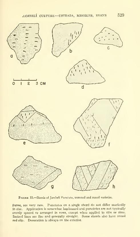
JAMBELI CULTURE—ESTRADA, MEGGERS, EVANS 529
I ] I I
I 2 3 CM
Figure 33.—Sherds of Jambeli Punctate, unzoned and zoned varieties.
forms, are very rare. Punctates on a single sherd do not differ markedly
in size. Application is somewhat haphazard and punctates are not typically
evenly spaced or arranged in rows, except when applied to ribs or rims.
Incised lines are fine and generally straight. Some sherds also have zoned
red slip. Decoration is always on the exterior.
530 PROCEEDINGS OF THE NATIONAL MUSEUM
Motif: Punctate decoration can be divided into four classes: 1) Nicked rim
—
a row of nicks along the edge of the lip, or more rarely around the neck of
vessels of Form 9; 2) Nicked rib—a row of nicks along an applique rib onthe upper vessel wall (fig. 32) ; 3) Punctate areas—overall application of
punctation to the entire surface, or large areas thereof (the latter differs fromzoned punctate in the absence of a bordering incision) ; 4) Zoned punctate
—
bands or triangular areas outlined by incised lines and filled with puncta-tions, which may be randomly distributed or in rows.
Temporal Differences Within the Type: None discernible. All four
motifs occur throughout the seriated sequence.
Chronological Position of the Type: Present in small frequency throughoutthe seriated sequence (see fig. 38).
Jambeli Polished Red (Jambeli Rojo Pulido)
Size OF Sample : 1320 sherds.
Paste: Like Ayalan Plain; see that type description for details.
Surface:
Color: Unslipped surfaces light orange to tan, very uniform in hue. Slipped
surfaces rich red.
Treatment: Unslipped surfaces even, occasionally smooth; majority show fine
smoothing marks and minor defects; more eroded than slipped surfaces.
Slipped surfaces (exterior of jars and deep bowls; both surfaces of shallow
bowls), well smoothed, even, polished. SHp appears thinner than on Jambeli
White-on-Red, but this may result from greater erosion.
Hardness: 3.5--4.0
Form (total rims from seriated samples, 330)
:
Rim: Direct, interiorly thickened, everted, with rounded or tapered lip.
Body wall thickness: 0.3-1.4 cm.; majority 5-8 mm.Base: Typically slightly flattened; occasionally annular; diameter 8-20 cm.
Reconstructed common vessel shapes:
Form 1—48.2 percent
Form 9—24.2 percent
Form 6—7.0 percent
Form 8—6.4 percent.
Minor vessel shapes (frequency less than 5 percent): Forms 5, 4, 10, 3, and 13.
Temporal Differences Within the Type. None discernible.
Chronological Position of the Type. Present with relatively uniform
frequency throughout the seriated sequence (see fig. 38).
JambeK Red Banded (Jambeli Rojo en Bandas)
Size OP Sample: 183 sherds.
Paste: Like Ayalan Plain; see that type description for details.
Surface:
Color: Light orange to light tan.
Treatment: Smoothed, producing an even but not polished finish; fine hori-
zontal smoothing marks visible on interior, rim, or exterior, usually not
present on bowl interiors.
Hardness: 3.5-4.0
Form (total rims from seriated samples, 83)
:
Ri7n: Direct, everted, interiorly thickened, or expanded with rounded or
tapered lip.
Body wall thickness: 5-8 mm.
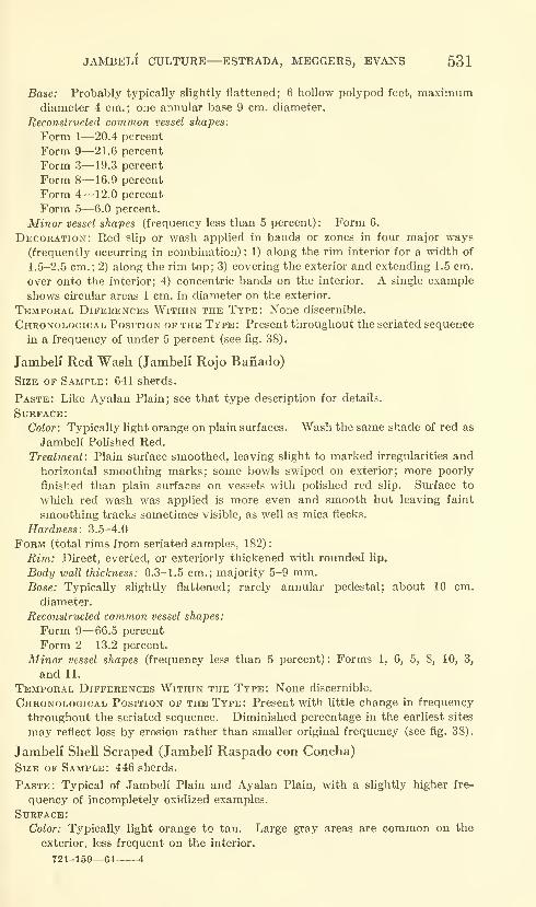
JAMBELI CULTURE—ESTRADA, MEGGERS, EVANS 531
Base: Probably typically slightly flattened; 6 hollow polypod feet, maximumdiameter 4 cm.; one annular base 9 cm. diameter.
Reconstructed common vessel shapes:
Form 1—20.4 percent
Form 9—21.6 percent
Form 3—19.3 percent
Form 8—16.9 percent
Form 4—12.0 percent
Form 5—6.0 percent.
Minor vessel shapes (frequency less than 5 percent) : Form 6.
Decoration: Red slip or wash applied in bands or zones in four major ways
(frequently occurring in combination) : 1) along the rim interior for a width of
1.5-2.5 cm. ; 2) along the rim top; 3) covering the exterior and extending 1.5 cm.
over onto the interior; 4) concentric bands on the interior. A single example
shows circular areas 1 cm. in diameter on the exterior.
Temporal Diferences Within the Type: None discernible.
Chronological Position of the Type: Present throughout the seriated sequence
in a frequency of under 5 percent (see fig. 38).
Jambeli Red Wash (Jambeli Rojo Baiiado)
Size op Sample: 641 sherds.
Paste: Like Ayalan Plain; see that type description for details.
Surface:
Color: Typically light orange on plain surfaces. Wash the same shade of red as
Jambeli Polished Red.
Treatment: Plain surface smoothed, leaving slight to marked irregularities andhorizontal smoothing marks; some bowls swiped on exterior; more poorly
finished than plain surfaces on vessels with polished red slip. Surface to
which red wash was applied is more even and smooth but leaving faint
smoothing tracks sometimes visible, as well as mica flecks.
Hardness: 3.5-4.0
Form (total rims from seriated samples, 182)
:
Rim: Direct, everted, or exteriorly thickened with rounded lip.
Body wall thickness: 0.3-1.5 cm.; majority 5-9 mm.Base: Typically slightly flattened; rarely annular pedestal; about 10 cm.
diameter.
Reconstructed common vessel shapes:
Form 9—66.5 percent
Form 2— 13.2 percent.
Minor vessel shapes (frequency less than 5 percent) : Forms 1, 6, 5, 8, 10, 3,
and 11.
Temporal Differences Within the Type: None discernible.
Chronological Position of the Type: Present with little change in frequency
throughout the seriated sequence. Diminished percentage in the earliest sites
may reflect loss by erosion rather than smaller original frequency (see fig. 38).
Jambeli Shell Scraped (Jambeli Raspado con Concha)Size of Sample: 446 sherds.
Paste: Typical of Jambeli Plain and Ayalan Plain, with a slightly higher fre-
quency of incompletely oxidized examples.
Surface:Color: Typically light orange to tan. Large gray areas are common on the
exterior, less frequent on the interior.
721-159—64 4

532 PROCEEDINGS OF THE NATIONAL MUSEUM
Treatment: One surface scraped with the edge of a ribbed clamshell producing
broad parallel grooves more or less 1.5 mm. wide and 1.5 mm. apart, varying
with the size of the shell used as the tool. The majority show scraping in
one direction only, but intersecting or overlapping patterns occur. About95 percent of the sherds are scraped on the interior only, suggesting that this
is a technique of surface finish rather than one of intentional decoration since
the vessels are constricted mouthed jars. Exterior scraping is restricted to
the necks of jars of Form 9, and the marks run vertically, probably for
decorative effect (pi. 8).
Hardness: 3.5-4.5
Form (total rims from seriated samples, 36)
:
Rim: Everted and slightly to markedly thickened at the angle of eversion, or
direct with rounded or flattened lip.
Body wall thickness: 0.3-1.1 cm.; majoritj' 4-6 mm.Base: Probably slightly flattened.
Reconstructed common vessel shapes: Form 9—94.3 percent.
Minor vessel shapes (frequency less than 5 percent): Forms 1 and 10.
Temporal Differences Within the Type: None discernible.
Chronological Position of the Type: Present throughout the seriated se-
quence with no apparently significant change in frequency (see fig. 38).
Jambeli White-on-Red (Jambeli Blanco Sobre Rojo Pulido)
Size of Sample: 599 sherds.
Paste :
Temper: Sand abundantly mixed with minute grains of mica; sand typically
fine but with occasional coarser inclusions up to 1.5 mm. Abundant and
well mixed.
Texture: Fine grained, sandy; tendency to laminated appearance accentuated
by slit air pockets parallel to surface.
Color: Range from completely oxidized light orange, to orange along the surface
with medium to dark gray core, to gray through entire cross section.
Surface:Color: Decorated areas covered with a thin red slip, typically a dark rich red
of relatively uniform hue. Undecorated surfaces light orange or tan. Grayfire clouds may occur on interior or exterior.
Treatment: Slipped surfaces smooth, even, and where uneroded show a gloss.
Unslipped surfaces even, sometimes showing smoothing marks, but not
polished. Sparkling pinpoint mica grains are visible on slipped and un-
slipped surfaces.
Hardness: 3.5-4.0
Form (total rims from seriated samples, 227)
:
Rim: Direct, interiorly thickened, everted, with rounded or tapered lip.
Body wall thickness: 0.3-1.1 cm.; majority 3-5 mm.Base: 1) Slightly flattened; 2) Annular, diameter 16-18 cm.; 3) Hollow polypod,
maximum diameter 4-5 cm.
Reconstructed common vessel shapes:
Form 1—62 percent
Form 3—15 percent
Form 6-7.9 percent.
Minor vessel shapes (frequency less than 5 percent) : Forms 5, 4, 9, 8, 10, 12,
and 13.
Additional rare vessel shapes: 1) Jar with spout and bridge handle: Spout
length 4 cm., exterior diameter 1.3 cm., interior diameter 0.8-0.9 cm.,
maximum body diameter 12 cm. (fig. 23, a).

JAMBELI CULTURE—ESTRADA, MEGGERS, EVANS 533
1 CM
Figure 34.—Sherds of Jambeli White-on-Red (dark stippling indicates red slip; no stippling,
white paint).
Decoration:Technique: 1) White stripes and zones on red slip. Stripes typically 2-7 mm.
wide, occasionally 1.3-1.8 cm. wide; some variation in width due to poor
control of brush; termination may be tapered to point or thickened by excess
paint; paint sometimes thick and easily chipped off. Solid areas are
rectanguloid, up to 4 by 6 cm., or circular. Unequal thickness of paint
makes color vary from white to pinkish in thin places where underlying red

534 PROCEEDINGS OF THE NATIONAL MUSEUM
% .M,^
, ^/.^fj« -' *
^'Syi^gwvsTC'-:;
^m^
r-i..
-AS
Mmi,
<j,,"^ .i^^^
I L J I
I 2 3 CM
Figure 35.—Rolled out designs from complete vessels of Jambeli White-on-Red (dark
stippling indicates red slip; no stippling, white paint).
m^^m-
I 2 3 CM
Figure 36.—Sherds of Jambeli White-on-Red, white and red variety (dark stippling
indicates red slip; light stippling, natural surface; no stippling, white paint).

JAMBELI CULTURE—ESTRADA, MEGGERS, EVANS 535
slip shows through. Solid areas were outlined and then filled in (figs. 34-35;pis. 9-10). 2) Alternating bands of red and white, white applied after red
and may overlap slightly onto red areas (fig. 36).
Motif: Rectilinear patterns composed of single or parallel lines formingrectangular or diamond-sliaped areas or stepped designs are most typical of
white-on-red decoration. Curvilinear designs are rare. Solid areas are
typically rectangular, rarely circular, and most frequent on the exterior.
White and red designs are usually concentric bands, alternating white and red,
with red bands slightly wider than the white ones. A rare variant consists of
red and white spots on an unslipped surface (fig. 36, a). Painting is applied
to interior of shallow bowls, exterior of deep bowls and jars; occasionally to
both surfaces.
Temporal Differences Within the Type: None discernible. Both white-
on-red and white and red techniques occur tliroughout the seriated sequence.
Chronological Position of the Type: Characteristic throughout the seri-
ated sequence (see fig. 38).
Jambeli White Painted (Jambeli Blanco Pintado)
Size of Sample: 74 sherds.
Paste: About 50 percent like Ayalan Plain; the remainder like Jambeli Plain;
see those type descriptions for details.
Surface:
Color: Red-orange, orange, brownish, or gray as a result of differential firing.
A few sherds have a thin red wash; none has a polished red slip.
Treatment: Smoothed, sometimes leaving horizontal smoothing marks; surface
remains somewhat uneven and irregular with small defects. Broad scraping
tracks on interior. Mica flecks glisten on both surfaces.
Hardness: 3-4
Form (total rims from seriated samples, 25)
:
Rim: Direct or everted with rounded lip.
Body wall thickness: 0.3-1.0 cm.; majority, 3-7 mm.Base: Probably slightly flattened.
Reconstructed common vessel shapes:
Form 9—88 percent
Form 2—12 percent.
Additional rare vessel shapes: 1) Miniature vessel with rounded body, slightly
constricted mouth spanned by strap handle; height including handle 7.7
cm., maximum body diameter 5.6 cm., mouth diameter 3.5 cm. (pi. 11, /).
2) Jar of Form 9 with a horizontal flange around the neck 2 cm. below the
rim; mouth diameter 9 cm. (fig. 23, b).
Appendages:
Horizontal Rim Lobe (pi. 11, d). A bowl of Form 2 has a prominent lobed
lug extending horizontally from the rim exterior. Width at point of
attachment, 6 cm.; outward projection, 3.5 cm. The upper surface has
two white bands perpendicular to the mouth. The specimen is from the
surface of Site 0-6.
Decoration (fig. 37; pi. 11):
Technique: White painted lines 2-5 mm. wide, with variation in width ona single sherd. Differences in paint thickness produce color range fromwhite to pinkish. Spacing of parallel lines not uniform.
Motif: Parallel straight lines in groups of three or more, usually running
vertically down from the rim. Rare examples have parallel horizontal lines
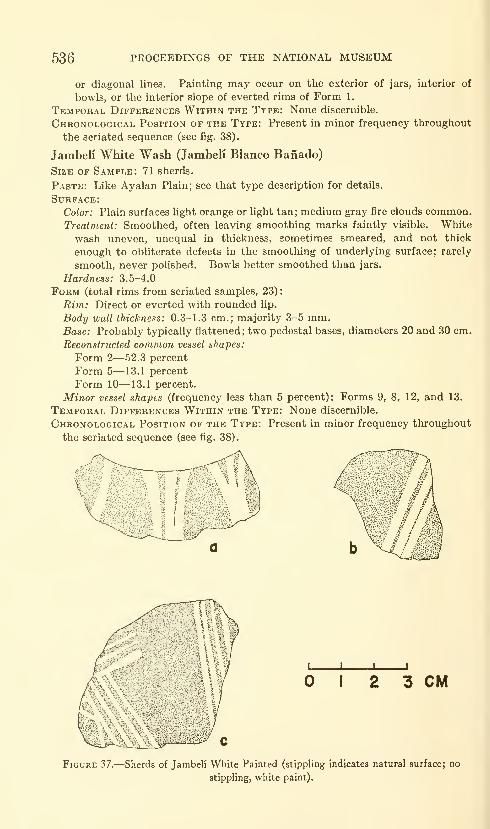
536 PROCEEDINGS OF THE NATIONAL MUSEUM
or diagonal lines. Painting may occur on the exterior of jars, interior of
bowls, or the interior slope of everted rims of Form I.
Temporal Differences Within the Type: None discernible.
Chronological Position of the Type: Present in minor frequency throughout
the seriated sequence (see fig. 38).
Jarabeli White Wash (Jambeli Blanco Baiiado)
Size of Sample: 71 sherds.
Paste: Like Ayalan Plain; see that type description for details.
Surface:
Color: Plain surfaces light orange or light tan; medium gray fire clouds common.
Treatment: Smoothed, often leaving smoothing marks faintly visible. White
wash uneven, unequal in thickness, sometimes smeared, and not thick
enough to obliterate defects in the smoothing of underlying surface; rarely
smooth, never polished. Bowls better smoothed than jars.
Hardness: 3.5-4.0
Form (total rims from seriated samples, 23)
:
Rim: Direct or everted with rounded lip.
Body wail thickness: 0.3-1.3 cm.; majority 3-5 mm.Base: Probably typically flattened ; two pedestal bases, diameters 20 and 30 cm.
Reconstructed common vessel shapes:
Form 2—52.3 percent
Form 5—13.1 percent
Form 10—13.1 percent.
Minor vessel shapes (frequency less than 5 percent): Forms 9, 8, 12, and 13.
Temporal Differences Within the Type: None discernible.
Chronological Position of the Type: Present in minor frequency throughout
the seriated sequence (see fig. 38).
i 2 3 CM
Figure 37.—Sherds of Jambeli White Painted (stippling indicates natural surface; no
stippling, white paint).
JAMBELI CULTURE—ESTRADA, MEGGERS, EVANS 537
Posorja Polished Plain (Posorja Pulido)
Size of Sample: 458 sherds.
Paste:
Temper: Fine sand, grains not easily visible.
Texture: Fine grained, compact but often laminated, with long fine air pockets.
Color: Medium gray to tannish gray.
Surface:
Color: Dusty gray tan where polish eroded; polished areas vary from dark grayto tan.
Treatment: Even and, where uneroded, slick to touch. Fine horizontal brushmarks faintly visible on interior, more rarely on exterior.
Hardness: 3.5-4.0
Form (total rims from seriated samples, 22)
:
Rim: Direct, everted, or interiorly thickened with rounded lip.
Body wall thickness: 4-7 mm.Base: Probably rounded or slightly flattened.
Reconstructed common vessel shapes:
Form 9—22.7 percent
Form 10—18.2 percent
Form 2—18.2 percent
Form 1—9 percent.
Minor vessel shapes (frequency less than 5 percent): Forms 8 and 11.
Temporal Differences Within the Type: None discernible.
Chronological Position of the Type: Present only during the early half of
the seriated sequence (see fig. 38).
Trade Sherds
Several sites of the Jambeli Phase produced sherds of plain or
decorated types belonging to two other cultures found in the Province
of Guayas. The largest number are of Guangala Phase origin; a few
represent the early Manteno Phase.
Guangala Phase Types.—Sherds of Guangala Phase origin
include plain types and the following decorated types (Appendix
Table 2): Barcelona Painted Dark Line (Barcelona Linea Oscura),
Guangala Burnished Line (Guangala Brunido), Guangala Finger
Painted (Guangala Pintado con Dedo) and Chorrera Iridescent
(Chorrera Iridiscente). Although Guangala pottery was identified
at only a few sites, its seriated position (fig. 38) suggests that con-
tact between the Jambeli and Guangala Phases occurred throughout
the duration of the Jambeli Phase as it is now known.
Mantei?o Phase Types.—A few plain sherds from Site G-L-30
were identified by Estrada as representing the fine kaolinite paste
of Playas Gray Polished (Playas Gris Pulido). Two decorated
sherds from Site 0-3 are Playas Engi-aved (Playas Grabado) (Estrada,
1957 a, p. 72). These types are characteristic of the Mantenoculture, which is later than the Regional Developmental Period,
Since these sherds are from surface collections, they are most easily

538 PROCEEDINGS OF THE NATIONAL MUSEUM
explained as intrusions resulting from later Mantefio occupation
of the area around the Gulf of Guayaquil.
Unclassified Decorated
Most of the decorated sherds left unclassified were too small or too
badly eroded to identify. The remainder consisted principally of
applique nubbins.
Applique Nubbins.—One rim of Form 9 from Site 0-5 has two
rounded pellets attached side by side under the everted rim. Traces
of red wash are visible on the upper surface of the rim, which has a
diameter of 14 cm. Another rim from the same provenience has a
nubbin with a punctate in the center in the same position on the
rim exterior.
Several sherds from tiie surface of Site 0-3 are ornamented with a
horizontal row of low applique nubbins 2-5 mm. high, applied either
to the surface or to a low ridge.
Implications of the Seriated Sequence
The seriated sequence of the Jambeli culture is based on trends in
change of frequency in the plain types derived from analysis of the
sherds from stratigraphic excavations at sites G-84, G-86 and 0-5.
Into this framework, surface collections from 10 additional sites were
seriated. Finally, the attempt was made to include sites represented
by a sample of less than 100 sherds, and 7 of these fit the pattern of
change sufficiently well to be incorporated into the graph (fig. 38).
The two principal plain types exhibit a rather smooth pattern of
change, in which Jambeli Plain, tempered with coarse micaceous
sand, increases in popularity while Ayalan Plain, tempered with fine
micaceous sand, shows a corresponding decline in frequency. Someof the samples exhibiting a larger amount of both types than is
consistent with the pattern of the curve reflect an unusually high
degree of surface erosion. This loss of surface results in the classi-
fication of some sherds as plain that might originally have had a
slipped or painted surface. Sites P-2 and G-L-3 are examples of
this kind of distortion.
The only real time marker, in terms of presence or absence, is
Posorja Polished Plain, which is restricted to the early half of the
sequence. It perhaps represents a survival of the polished plain
ware tradition characteristic of the preceding Formative Period.
None of the decorated types exhibits any consistent pattern of
change. Each type maintains a relatively uniform frequency from the
earliest to the latest site, with fluctuations that can be attributed
either to differential amounts of surface erosion or to errors resulting
from the small size of the sherd sample. Attempts were made to
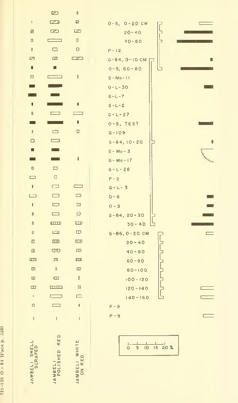
I
B
D
ED
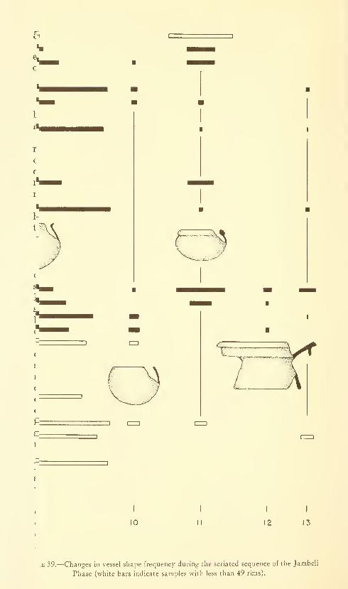
r
c
c
1
•E 39.—Changes in vessel shape frequency during the seriated sequence of the Jambeh'
Phase (white bars indicate samples with less than 49 rims).

..J CUT I, 0-20CM
20-40
40-60
P-18
G-84, CUT 1,0-10 CM
0-S, CUT I, 60-80
t'Mo-ll
1-1.-30
S-L-T
e-i"!
S-l-27
S-i, TEST
(-109
«-e4,CUT 1,10-20
1^ ^ -^ ^ jy J'^ ^J
\y^ ^y' y' ^ y^ y -' I
^yy y / / y/ y /\ a B
i-«4, CUT
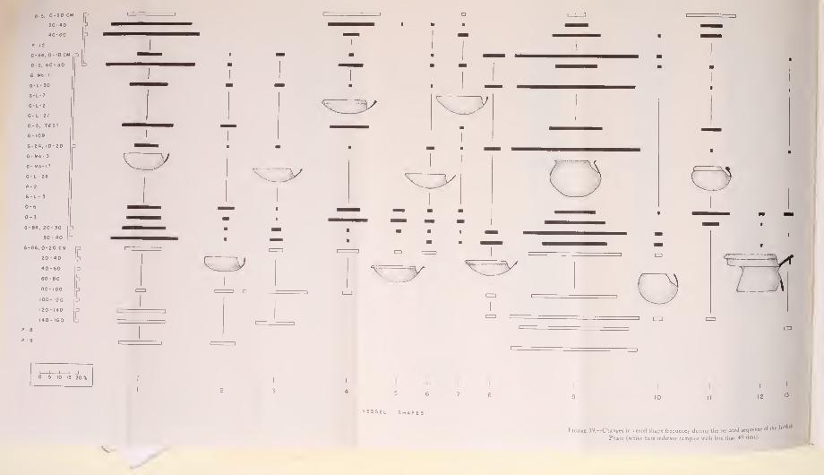
c

JAMBELI CULTURE—ESTRADA, MEGGERS, EVANS 539
detect temporal differences in the decorative motifs, but no distinctions
could be found that could not be attributed to deficiencies in the size
of the sample.
Analysis of the rim forms and reconstructed vessel shapes showedthe same picture of cultural uniformity through time (fig. 39). Onlytwo forms have a restricted distribution: Form 5, a bowl with aneverted rim, and Form 12, a large compotera. Both are limited to
the early half of the sequence. Annular bases are present throughout,
and this is probably also true of hollow legs since their absence from
the earliest and latest levels can be accounted for by the small size of
the sherd sample. The only solid leg comes from an early site.
Spouts, although very rare, appear to occur throughout the sequence.
Discussion of the temporal differences in the frequency of various
types of artifacts is hampered by the fact that these are relatively
rare in comparison to sherds. The majority of those artifacts included
in this report are from Site 0-6, where the constant erosion of the site
by the sea, and the existence of a resident population able to salvage
objects as they were revealed, provides a combination of collecting
circumstances not met with at any other Jambeli site. In view of the
relatively small amount of excavation undertaken, it consequently is
difficult to determine whether the absence of figurine fragments from
the lower third of the sequence reflects a real absence or the inadequacy
of collections from early sites. The two bark beater fragments are
from sites that occupy an early and a late position, suggesting that
this trait was present throughout the sequence.
Of considerable interest is the question of whether the entire
area in which the Jambeli culture remains are found was occupied at
the same time, or whether a movement from north to south or south to
north can be inferred. The seriated position of the sites does not
provide a clear-cut conclusion, but the absence of any El Oro Province
sites in the lower third of the seriated sequence may reflect a morerecent movement into that part of the area from the north. In the
upper third of the sequence, the situation is reversed. The largest
sites are in El Oro or southeastern Puna Island, and those in the
Playas-Posorja region are typically small with sparse pottery refuse.
If this interpretation of a shift in population concentration from
north to south is correct, it may be correlated with the gradual
desiccation of the northern portion of the area, which caused the
extinction of the mangrove swamp on which the subsistence of the
Jambeli Phase was largely dependent.
In summary, it can be said that all the evidence derived from an
examination of the chronological distribution of ceramic traits,
artifacts, and other cultural features of the Jambeli culture presents a
picture of unifonnity and consistency. There is no indication that
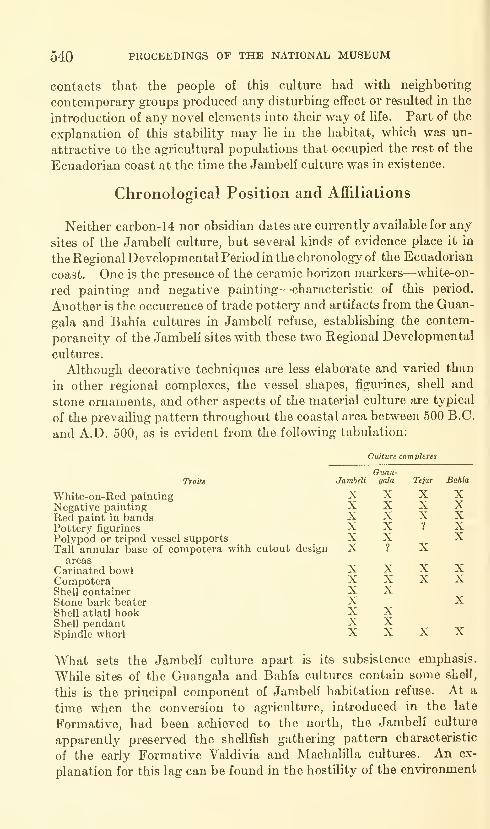
540 PROCEEDINGS OF THE NATIONAL MUSEUM
contacts that the people of this culture had with neighboring
contemporary groups produced any disturbing effect or resulted in the
introduction of any novel elements into their way of life. Part of the
explanation of this stability may lie in the habitat, which was un-
attractive to the agricultural populations that occupied the rest of the
Ecuadorian coast at the time the Jambeli culture was in existence.
Chronological Position and Affiliations
Neither carbon-14 nor obsidian dates are currently available for any
sites of the Jambeli culture, but several kinds of evidence place it in
the Regional Developmental Period in the chronology of the Ecuadorian
coast. One is the presence of the ceramic horizon markers—'white-on-
red painting and negative painting—-characteristic of this period.
Another is the occurrence of trade pottery and artifacts from the Guan-
gala and Bahia cultures in Jambeli refuse, establishing the contem-
poraneity of the Jambeli sites with these two Regional Developmental
cultures.
Although decorative techniques are less elaborate and varied than
in other regional complexes, the vessel shapes, figurines, shell and
stone ornaments, and other aspects of the material culture are typical
of the prevailing pattern throughout the coastal area between 500 B.C.
and A.D. 500, as is evident from the following tabulation:
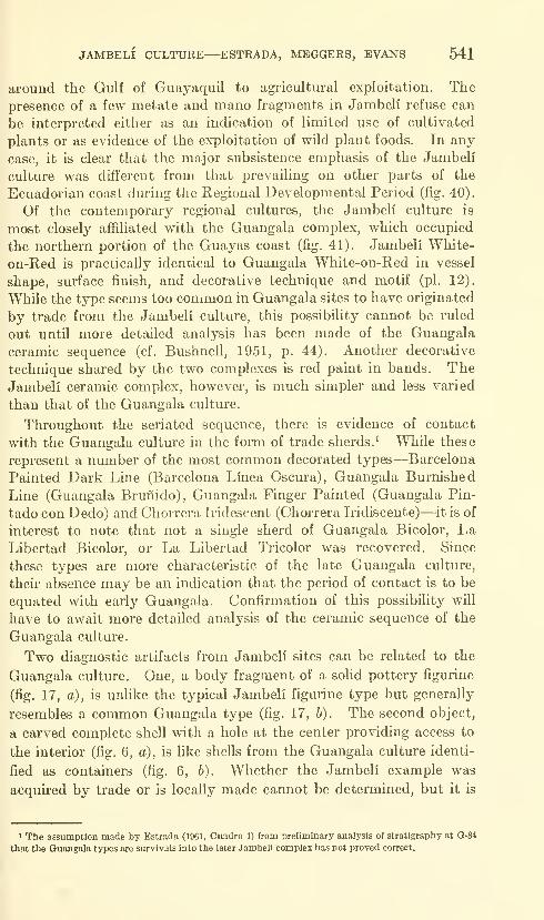
JAMBELI CULTURE—ESTRADA, MEGGERS, EVANS 541
around the Gulf of Guayaquil to agricultural exploitation. Thepresence of a few metate and mano fragments in Jambeli refuse can
be interpreted either as an indication of limited use of cultivated
plants or as evidence of the exploitation of wild plant foods. In any
case, it is clear that the major subsistence emphasis of the Jambeli
culture was different from that prevailing on other parts of the
Ecuadorian coast during the Regional Developmental Period (fig. 40).
Of the contemporary regional cultures, the Jambeli culture is
most closely affiliated with the Guangala complex, which occupied
the northern portion of the Guayas coast (fig. 41). Jambeli White-
on-Red is practically identical to Guangala White-on-Red in vessel
shape, surface finish, and decorative technique and motif (pi. 12).
While the type seems too common in Guangala sites to have originated
by trade from the Jambeli culture, this possibility cannot be ruled
out until more detailed analysis has been made of the Guangala
ceramic sequence (cf. Bushnell, 1951, p. 44). Another decorative
technique shared by the two complexes is red paint in bands. TheJambeli ceramic complex, however, is much simpler and less varied
than that of the Guangala culture.
Throughout the seriated sequence, there is evidence of contact
with the Guangala culture in the form of trade sherds.^ While these
represent a number of the most common decorated types—Barcelona
Painted Dark Line (Barcelona Linea Oscura), Guangala Burnished
Line (Guangala Brunido), Guangala Finger Painted (Guangala Pin-
tado con Dedo) and Chorrera Iridescent (Chorrera Iridiscente)—^it is of
interest to note that not a single sherd of Guangala Bicolor, LaLibertad Bicolor, or La Libertad Tricolor was recovered. Since
these types are more characteristic of the late Guangala culture,
their absence may be an indication that the period of contact is to be
equated with early Guangala. Confirmation of this possibility will
have to await more detailed analysis of the ceramic sequence of the
Guangala culture.
Two diagnostic artifacts from Jambeli sites can be related to the
Guangala culture. One, a body fragment of a solid pottery figurine
(fig. 17, a), is unlike the typical Jambeli figurine type but generally
resembles a common Guangala type (fig. 17, b). The second object,
a carved complete shell with a hole at the center providing access to
the interior (fig. 6, a), is like shells from the Guangala culture identi-
fied as containers (fig. 6, 6). Whether the Jambeli example was
acquired by trade or is locally made cannot be determined, but it is
> The assumption made by Estrada (1961, Cuadro 1) from preliminary analysis of stratigraphy at Q-84
that the Guangala types are survivals into the later Jambeli complex has not proved correct.

542 PROCEEDINGS OF THE NATIONAL MUSEUM
GEOGRAPHICAL SUBDIVISIONS
ESMERALDASNORTH SOUTH
MANABINORTH SOUTH
ATACAMES
GUAYASCOAST
MANTENO
CHIRIJE
GUAYASBASIN
EL ORO
MILAGRO -1000
1500
500
Z 5O Q.
500
CHORRERA
MACHA-LILLA
-1000
-1500
-2000
•2500
-3000
Figure 40.—Chronological sequence of archeological complexes on the Ecuadorian coast.
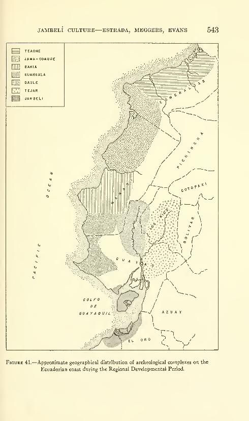
JAMBELI CULTURE—ESTRADA, MEGGERS, EVANS 543
Figure 41.—Approximate geographical distribution of archeological complexes on the
Ecuadorian coast during the Regional Developmental Period.
544 PROCEEDINGS OF THE NATIONAL MUSEUM
a rare enougli object to be interpreted as evidence of contact between
the two groups regardless of its origin.
Unfortunately, no carbon-14 dates are yet available for sites of
the Guangala culture. Absolute dates have been obtained from
obsidian at two Guangala sites, and these cover a range from 340
B.C. to A.D. 360 (Evans and Meggers, 1960, fig. 19).
Another link to absolute chronology is the stone bark beater (pi.
6, b-c), which is known elsewhere on the Ecuadorian coast only fromthe Bahia culture (pi. 6, a). The site of M-7: Esteros from which
the specimen came, has two carbon-14 dates: 2150±240 or 190 B.C.
(Sample W-833), and 2200 ±240 or 240 B.C. (Sample W-834)(Rubin and Alexander, 1960, p. 181). Since the trait is likely to
have reached the Jambell culture from the Bahia area, the dates
may be slightly earlier than the appearance of bark beaters farther
to the south, although this is not necessarily the case. An atypical
pottery figurine head has a headdress with ear flaps (fig. 18) resembling
those frequently worn by La Plata Sentado figurines of the Bahia
culture (Estrada, 1957, figs. 70 center and 90) providing another
possible link with the north.
To the south, the only archeological materials showing strong
affinities with the Jambell culture are those from Garbanzal, 9 km.
south of Tumbes on the right bank of the R.10 Tumbes in Peru (Ishida
et al., 1960, p. 423). Two sites are reported, one on the alluvial
terrace and another on a higher terrace. Although the investigators
have considered both to belong to the same cultural complex, correla-
tions with Ecuadorian archeology suggest that a distinction should
be made. The site on the lower level produced 23 complete vessels
and additional fragments readily classifiable into pottery types of the
Jambell Phase, including Jambeli Incised, Jambeli White-on-Red,
Jambell White Painted, Jambeli Polished Red, and Jambeli Negative
(op. cit., p. 120, fig. 54-56). The sand and mica temper is also typical
of Jambell wares (Mejia Xesspe, 1960, lam. 1, F). Vessel shapes
duplicate those representing the Jambeli Phase (fig. 42).
The characteristics of the second Garbanzal site, on the upper
terrace, appear to be different. Artifacts were recovered from shaft
and chamber tombs (Mejia Xesspe, 1960, fig. 2), and included globular
jars and 34 pieces of copper ax money (Ishida et al., p. 423 and Ap-pendix 1, fig. 2). In Ecuador, both shaft tombs and ax money are
associated with cultures of the later Integration Period.
A distinction between the two Garbanzal sites is important for
establishing the absolute dating of the Jambeli culture because a
carbon-14 date has been obtained for Tomb 2 at the site on the upper
terrace of Garbanzal by the Radiocarbon Laboratory of the Depart-
ment of Physics and Chemistry, Gakushuin University. The sample
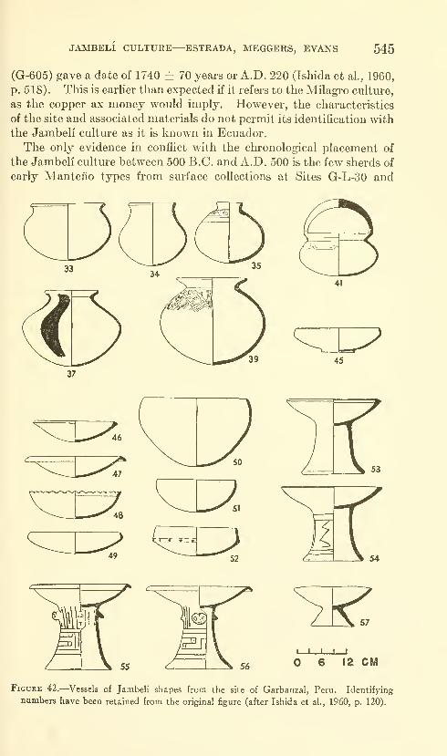
JAMBELI CULTURE—ESTRADA, MEGGERS, EVANS 545
(G-605) gave a date of 1740 ± 70 years or A.D. 220 (Ishida et al, 1960,
p. 518). This is earlier than expected if it refers to the Milagro culture,
as the copper ax money would imply. However, the characteristics
of the site and associated materials do not permit its identification with
the Jambeli cultm-e as it is known in Ecuador.
The only evidence in conflict with the chronological placement of
the Jambeli culture between 500 B.C. and A.D. 500 is the few sherds of
early Manteiio types from surface collections at Sites G-L-30 and
12 CM
Figure 42.—Vessels of Jambeli shapes from the site of Garbanzal, Peru. Identifying
numbers have been retained from the original figure (after Ishida et al., 1960, p. 120).
546 PROCEEDINGS OF THE NATIONAL MUSEUM
0-3. Bushnell (1951, p. 48) identifies this material with the Guangala
culture also, and if he is correct it may be added to other evidence of
trade relations with the Guangala Phase. The alternative is to
attribute these few surface sherds to later visits by the Manteno, whoare known to have occupied the Gulf of Guayaquil during the Integra-
tion Period (Estrada, 1957 a).
JAMBELI CULTURE—ESTRADA, MEGGERS, EVANS 547
Literature Cited
BUSHNELL, G. H. S.
1951. The archaeology of the Santa Elena Peninsula in South-west Ecuador.
Cambridge.
Estrada, Emilio
1957a. Los Huancavilcas; Ultimas civilizaciones pre-hist6ricas de la costa
del Guayas. Publicaci6n del Museo Victor Emilio Estrada No. 3.
Guayaquil.
1957b. Prehistoria de Manabf. Publicaci6n del Museo Victor Emilio
Estrada No. 4. Guayaquil.
1961. Correlaciones entre la arqueologia de la costa del Ecuador y Perd.
Humanitas II, No. 2, pp. 31-61. Quito.
1962. Arqueologia de Manabl Central. Publicaci6n del Museo Victor
EmiUo Estrada No. 7. Guayaquil.
Evans, Clifford, and Meggers, Betty J.
1960. Anew dating method using obsidian: Part II. An archaeological
evaluation of the method. Amer. Antiq., vol. 25, pp. 523-537.
Evans, Clifford, Meggers, Betty J., and Estrada, Emilio
1959. Cultura Valdivia. Publicacion del Museo Victor Emilio Estrada
No. 6. Guayaquil.
IsHiDA, EiicHiRO, and others
1960. Andes: the report of the University of Tokyo Scientific Expedition to
the Andes in 1958. Tokyo.
MEjfA Xesspe, Toribio
1960. Algunos nuevos elementos de la civilizaci6n Recuay-Pasto en el extremo
norte del Utoral peruano. Antiguo Peru, Espacio y Tiempo, pp.
205-217. Lima.
Rubin, Meyer, and Alexander, Corrinne1960. U.S. Geological Survey Radiocarbon Dates V. American Journal of
Science Radiocarbon Supplement, vol. 2, pp. 129-185. New Haven.
721-159—64-
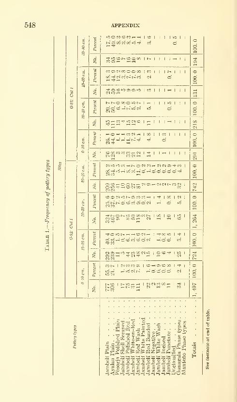
548 APPENDIX
lOOfOCOCOi—I--I \ roI
eool(^^GOoooolco|i it^i i i
o
^O^^COlMr-HlOOl ICOl I I I
(M lO lO CO rH t^ O IM <M rH IM (M Ol TtH CO |
Oc0^OOt^^(M05'-HiMC<H^C0iM |O m rfH ,-1 CD (M 00 CO
CD O (M lO r^ O CO CO tHI ^ I
COIiM
-^t^Ot^iOOOOCOt^ICO lO lie
IIM002 OOlOrH <M ^ 1-H COCO ^ rH
*0»Ct^.-lrHCDC<li-HI^COiO I Tt^
I
o CO t-H o «o CO CO o" c<i r-I o o" CO
(M05'-H»OTt<COOO(NiO IOCO-* |iOI
05 CO T-( Tt< (N •* i-H ,-1 <M
cot-
to Ol
(MCOCOCiICOrHOlcDOO
I•*
I
t-h' iC im' l>^ ^ O O o" O C^'
l>CO It^iO'-^'-H |(N(MCOCO^ I"*t^O i-(l^CO^ (M "-H >-H COt^ CO r-l
.S fl'^ — -2
(Inp;^ PL| m fin
-tj'd "^
t/2 03
S T3 "G Td M'GCO o
|§^t—I Ph q-
03 2^0 03 031-5 < P-l h, 1-5
CD OJ OJ O O O
£ s a s s s^ c3 o3 (^ 03 (^•-5 1-5 (-S l-i »-S 1-5
OJ cu cc
sag03 o3£1-5 -Sp
^ oMO§ s
03-1-3
oH

APPENDIX 549
3a'+3
doO
(2

550 APPENDIX
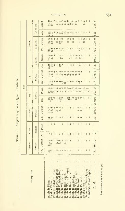
APPENDIX 551
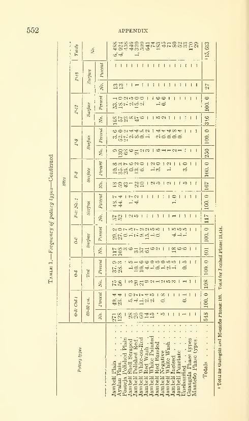
552 APPENDIX
O
<
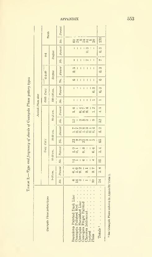
APPENDIX 553
e
Oh
s
1
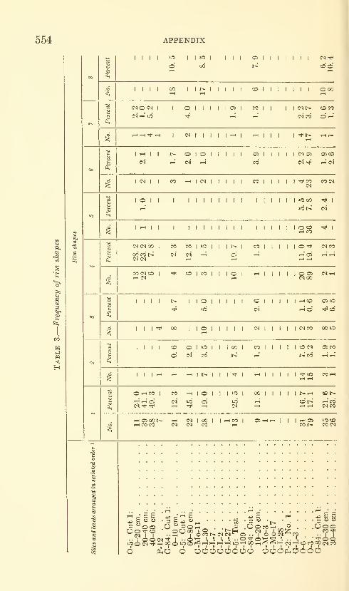
554 APPENDIX
«s>
n
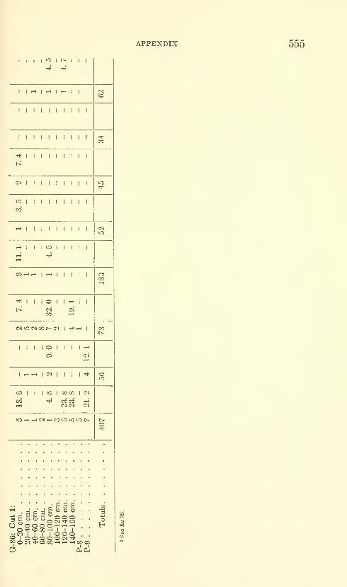
APPENDIX 555
1
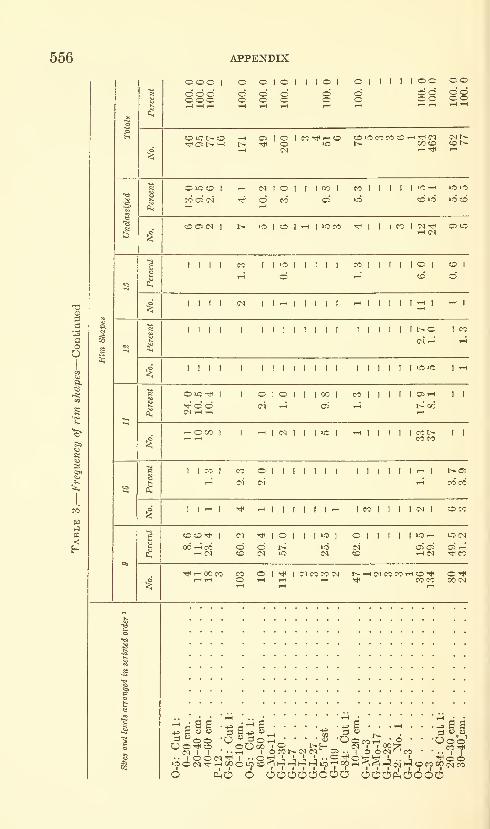
556 APPENDIX

APPENDIX 557
I I I I oo
I I I I I
^ I I I I 1 I 1M(N
I I I t^ I
I I 1 I I I ^
I I I I I I I
I I I I I I I I I
I I I I
I I I I'H
I 1 I
I t- 1 I I
T-( 1 .-ITtHI CO
aBBB- ^ - fl o o o
R " " '^OiM^CO"0000'-<'-<'-i0-* COQO'-' 1 I i(M I
OH
<*0(M-*COC30'-*'-l'-*°P'7
P5I I

558 APPENDIX
Table 4.—Frequency of base forms, unusual vessel appendages, figurines, and barkbeaters at sites of the Jambeli Phase
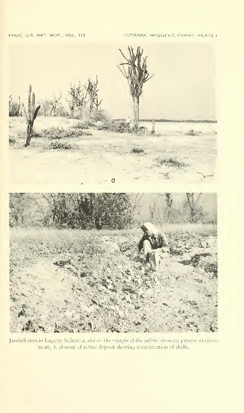
PROC. US. NAT. MUS.. VOL. 115 ESTRADA. MEGGERS, EVANS— PLATE 1
/^m-- *
siSi^%st:'
^^ it
V *^^
t. \ ^ „
r v\ i-^
Janibeli sites in Lagarto Salitrc: a, site on the margin of the sali!re, showing present environ-
ment; b, closeup of refuse deposit showing concentration of shells.
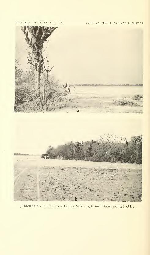
PROC. US NAT MUS , VOL. 115 ESTRADA, MEGGERS, EVANS—PLATE 2
iM.>^0^iim
1 A--I. -('3*'**' BiaiiiiiMiiiiiiai
•#&-
•^M^-^'-'v^^"^m
fKKt-
b -
Jaiiiiieli sites Dii tile margin ot Layarto Salitrc: «, testing refuse dep.)Sit; b, G-1,-/
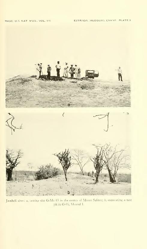
PROC. U.S. NAT. MUS.. VOL. 115 ESTRADA. MEGGERS. EVANS—PLATE 3
I-^--
•^cA
xV
JanibL-li biCcs: a, testing site Cj-Mm^1.t i;i iLc center ..,; Al ,::. .^.tliirc; b, c-xcaxatii;.:: a le-t
pit in G-86, Mound 1.
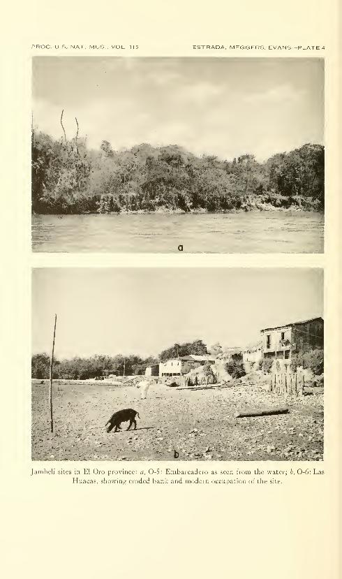
PROC. US. NAT. MUS , VOL 115 ESTRADA, MEGGERS, EVANS—PLATE 4
Janibeli sites in El Oro province: a, 0-5: Embarcadero as seen from the water; b, 0-6: Las
Huacas, showing eroded bank and modern occupation of the site.
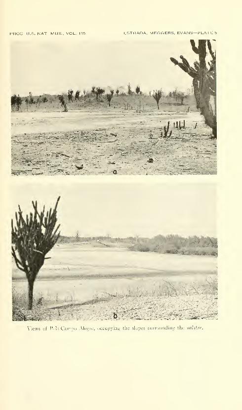
PROC. U.S. NAT. MUS.. VOL. 115 ESTRADA. MEGGERS. EVANS—PLATES
1.
^.i f # ^. Ifer«
..'^'^'^fc-li ,._
5^^
1)
Views i)t 1^-2: Campo Alegre, occupying the slopes surrounding the saliire.
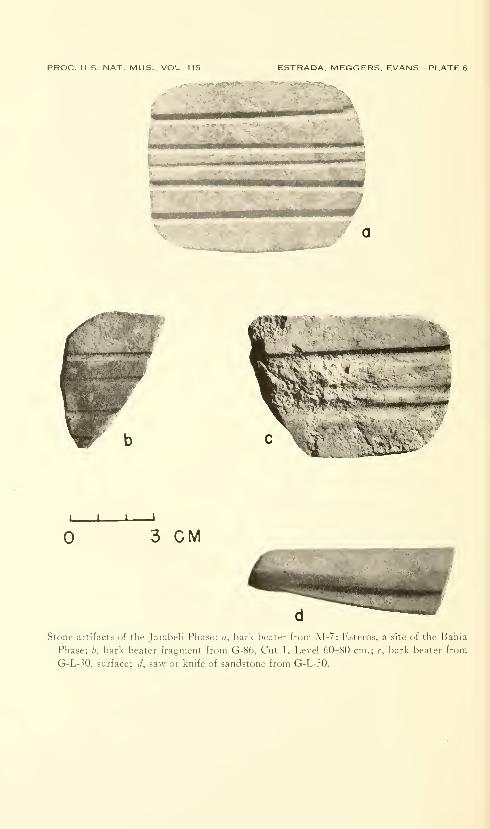
PROC. U.S. NAT. MUS.. VOL. 115 ESTRADA. MEGGERS, EVANS—PLATE 6
'i(j;-<f«j>*«E>i'-s(»fe««,»'-v-' • • «m*^m»n»m^
iimmmmtmKmmmmmmiitmmmmm
tm.' >:
I I—I—
I
3 CM
Stone artifacts of the Jainbcli Phase: a, bark beater from M-7: Esteros, a site of the Bahia
Phase; b, bark beater fragment from G-86, Cut 1, Level 60-80 cm.; c, bark beater from
G-L-30, surface; d, saw or knife of sandstone from G-L-30.
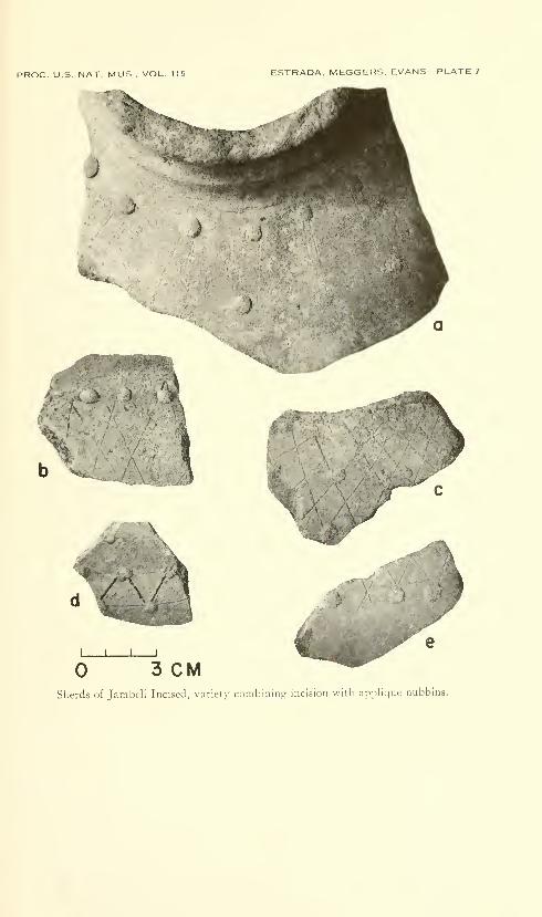
PROC. U.S. NAT MUS . VOL 115 ESTRADA, MEGGERS, EVANS—PLATE 7
J
^1
b Vv
,/ .\/:
I I \ I
3 CMSherds of Jambeli Incised, variety combining incision witli applique nubbins.
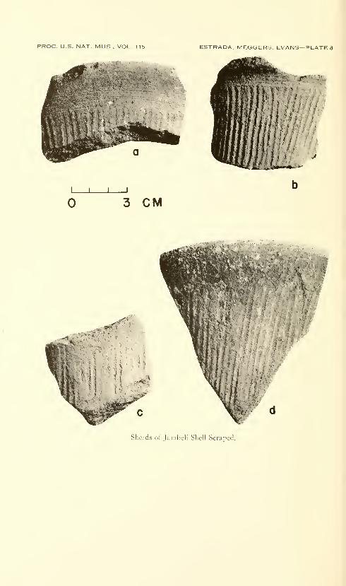
PROC. U.S. NAT. MUS , VOL. 115 ESTRADA. MEGGERS. EVANS
—
PLATE!
I I I
3 CM
Sherds of Jainbeli Shell Scraped.
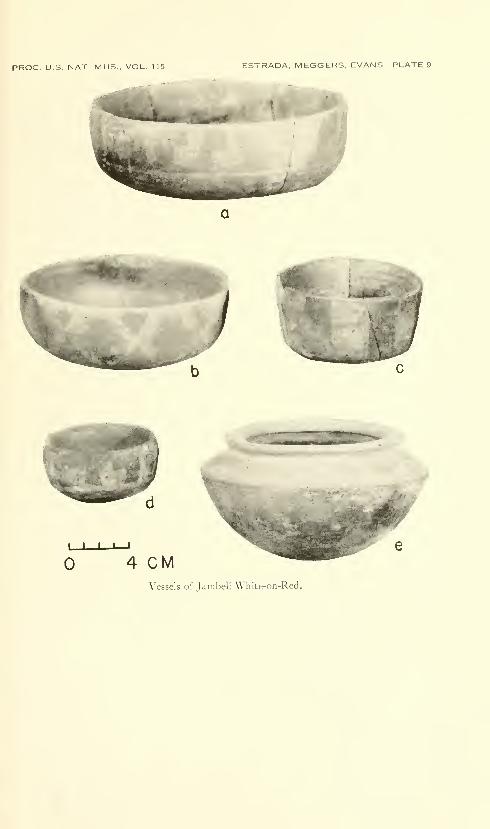
PROC. U.S. NAT. MUS., VOL. 115 ESTRADA. MEGGERS. EVANS—PLATE 9
/
)
L_J I I—
i
4 CMVessels of Jambeli White-on-Red.
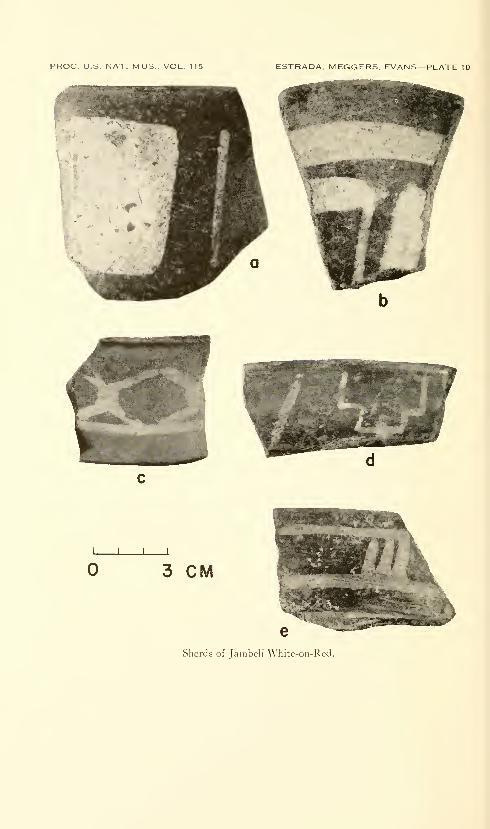
PROC. U.S. NAT. MUS., VOL. 115 ESTRADA. MEGGERS. EVANS—PLATE 10
I I I I
3 CM
Sherds of Jambeli White-on-Red.
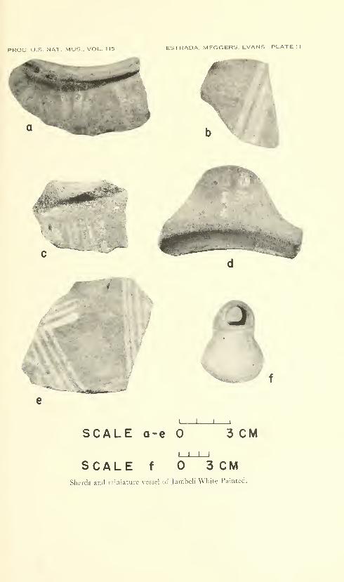
PROC U.S. NAT. MUS.. VOL. 115 ESTRADA, MEGGERS. EVANS—PLATE 11
/'^#;**^
"^-.-..^
/^^^'
# •>4
I I \ I
SCALE a-e 3 CMI t I I
SCALE f 3 CMSherds and miniature vessel of Jambeli White Painted.
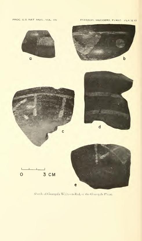
PROC. U.S. NAT. MUS., VOL. 115 ESTRADA. MEGGERS. EVANS— PLATE 12
J \ I
3 CM
Sherds of Guangala White-on-Red, of the Guantjala Phase.
Proceedings of
the United States
National MuseumSMITHSONIAN INSTITUTION • WASHINGTON, D.C.
Volume 115 1964 Number 3493
A REVISIONOF THE CARCHARHINID SHARK GENERA
SCOLIODON, LOXODON, AND RHIZOPRIONODON
By Victor G. Springer
Introduction
The purpose of this paper is to revise the genera Scoliodon, Loxodon,
and Rhizoprionodon. The most recent studies allocate all the species
included in this report to a single genus, Scoliodon, and it is for this
reason, together with their superficial resemblance to each other, that
the three genera are treated together. While these genera do notseem to form a natural group, judgment of their relationships is de-
ferred until the other genera of the family Carcharhinidae are morecompletely known.
Among the three genera, Scoliodon is quite distinct from the other
two and easily distinguishable from all other Carcharhinidae. Loxodonand Rhizoprionodon perhaps are closely related, and it is possible that
the two subgenera under Rhizoprionodon should be elevated to generic
status. Differences separating the genera, as they are here recog-
nized, are presented in the key, and their characteristics are dis-
cussed under the respective generic headings.
The characters previously used in assigning the species herein to a
559
560 PROCEEDINGS OF THE NATIONAL MUSEUM vol. 115
single genus and separating them primarily from Carcharhinus have
never been adequately evaluated. These characters are the smooth
and oblique teeth, the nature of the labial furrows, and, in Loxodon,
the presence of a spiracle (see diagnosis under Loxodon). In addition
to these characters, and more important, I find that the group can
be conveniently, though not naturally, separated from other carcha-
rhinids in having the origin of the second dorsal fin never in advance
of the middle of the anal base and frequently over or behind the anal
axil. The group differs from most carcharhinids in having the cusps
of most teeth smooth although in the larger specimens of some Rhizo-
prionodon species the cusps become serrulate.
These characters are not unique; a few other carcharhinids, e.g.,
Carcharhinus porosus (Ranzani) and C. borneensis (Bleeker), have a
posteriorly placed second dorsal origin (but never posterior to a ver-
tical line through the middle of the anal base). These species, how-
ever, have noticeably serrated teeth, the upper teeth possessing very
large basal serrations on the posterior (lateral) margins, and thus differ
from all the species treated in the present study. Because Carcha-
rhinus poros^is and C. borneensis lack a long upper labial furrow, they
might be confused superficially with Scoliodon laticaudus, Loxodon
macrorhinus, Rhizoprionodon oligolinx, or R. taylori. Aside from the
nature of the teeth, Scoliodon (q.v.) can be distinguished by its unique
fin positions; Loxodon (q.v.), by its short dorsal fin base, by the second
dorsal fin usually originating posterior to a vertical line through the
anal axil, and by having a notched orbital rim; Rhizoprionodon oligo-
linx and R. taylori, by having the origin of the second dorsal fin over,
or posterior to, a vertical line through the posterior third of the
anal base.
Several species assigned to the genus Scoliodon have been described
from fossil teeth. I choose not to treat them here as I believe the
affinities of fossils based only on teeth are at present indecipherable
and possibly will remain so in most cases. This viewpoint is based on
the close similarity of teeth within the living genera included in the
present study, as well as a similarity to the teeth of the distantly
related Sphyrnidae. The problem is further confused by the
heretofore unrecognized dental sexual dimorphism that is found in
several carcharhinids, including Rhizoprionodon and Scoliodon. Re-
lationships presently based only on teeth, therefore, are open to
question.
The species of Scoliodon, Loxodon, and Rhizoprionodon are dis-
tributed primarily through the shallow tropical marine waters of the
world, with some forms straying over moderate depths and into tem-
perate areas. None of the species is known to occur in the Mediter-
ranean or in Oceania.
REVISION OF CARCHARHINID SHARK GENERA—SPRINGER 561
All the species treated herein, with the possible exceptions of
Rhizoprionodon terraenovae and R. taylori, are used commonly as food,
and one, Scoliodon laticaudus, is considered a delicacy. The two ex-
ceptions noted should be edible also, but they have received little
attention as food, possibly because of local prejudices in the areas
where they occur.
The following abbreviations have been used in the text
:
AMNH—American Museum of Natural History, New YorkAMS—Australian Museum, Sydney
ANSP—Academy of Natural Sciences of Philadelphia
BMNH—British Museum (Natural History), LondonCAS—California Academy of Sciences, San Francisco
CNHM—Chicago Natural History MuseumDHMB—Department of Harbors and Marine, Brisbane
DIRU—Department of Ichthyology, Rhodes University, Grahamstown,South Africa
GVF—George Vanderbilt Foundation (to be incorporated with SU—see
below), Stanford, California
HUI—Hebrew University, Jerusalem, Israel
IFAN—Institute Francais d'Afrique Noire, Dakar, Senegal
IRSN—Institut Royal de Sciences Naturelles de Belgique, Brussels
ISH—Institut fiir Seefischeri, HamburgISZZ—Institut fiir Spezielle Zoologie und Zoologisches Museum, Berlin
MCZ—Museum of Comparative Zoology, Harvard University
MNHN—Museum National d'Histoire Naturelle, Paris
MRAC—Musee Royal de I'Afrique Centrale, Tervuren, Belgium
NFIS—Natur-Museum und Forschungs-Institut Senckenberg, Frankfurt
NMV—Naturhistorisches Museum, Vienna
QMB—Queensland Museum, Brisbane
RNH—Rijksmuseum van Natuurlijke Historic, Leiden
SIO—Scripps Institution of Oceanography, La JoUaSMNS—Staatliches Museum fiir Naturkunde, Stuttgart
SMW—Stadtisches Museum WiesbadenSU—Division of Systematic Biology, Stanford University
UCLA—University of California at Los Ajigeles
UMML—University of Miami Institute of Marine Science
UMM25—University of Michigan Museum of Zoology, Ann Arbor
UND—University of Natal, Durban, South Africa
UWS—University of Washington, Seattle
USNM—United States National Museum, WashingtonUZMK—Universitetets Zoologiske Museum, KobenhavnZSZM—Zoologisches Staatinstitut und Zoologisches Museum, Hamburg
During the course of this study, I was afforded the opportunity
of examining and radiographing specimens from the collections of a
large number of American and foreign institutions; I was also fur-
nished radiographs and data on several specimens I did not see.
To my colleagues, I wish to express my gratitude for their generous
aid and extreme patience: D. E. Rosen (AMNH); G. P Whitley
(AMS) ; J. E. Bohlke (ANSP) ; A. C. Wheeler and G. Palmer (BMNH)
;
W. I. FoUett (CAS) ; L. P. Woods (CNHM) ; T. C. Marshall (DHMB)
;
562 PROCEEDINGS OF THE NATIONAL MUSEUM vol. us
J. L. B. Smith (DIRU) ; R. R. Rofen, A. Fehlman, and W. L. Chan(GVF); A. Ben-Tuvia (HUI); T. Monod and F. Paraiso (IFAN);
J. P. Gosse (IRSN); G. Krefft (ISH); G. Deckert (ISZZ); G. W.Mead and M. Dick (MCZ); M. L. Bauchot (MNHN); M. Poll
(MRAC); W. Klausewitz and F. Rossel (NFIS); P. Kahsbauer(NMV) ; G. Mack and A. Bartholomai (QMB) ; M. Boeseman (RNH)
;
R. Rosenblatt (SIO); H. Janus (SMNS) ; C. FHl (SMW); G. S.
Myers (SU) ; B. W. Walker (UCLA) ; C. R. Robins and W. R. Court-
enay, Jr. (UMML) ; R. M. Bailey (UMMZ) ; J. D'Aubrey (UND);J. E. Randall (University of Puerto Rico); J. Nielsen (UZMK);W. Ladiges (ZSZM).
Radiographs were made in Europe and Australia with the help of
the following, to whom I express my sincere appreciation: F. Meier,
Roentgen Institut, Stuttgart; R. Pobisch, Tierarzliche Hochschule,
Vienna; J. P. Gasc, Laboratoire d'Anatomie Compare, Paris; Drs.
Leloup and Vastesaeger, Hopital d'lxelles, Belgium; P. DuUemeijer
and Mr. Simons, Zoologisch Laboratorium, Leiden, Holland; P.
R0nne, Biofysisk Lab, Copenhagen; R. Prevot and K. J. Thiemann,
Eppendorf, Hamburg; K. L. Schmidt and R. Lingemann, St. Mallins
Krankenhaus, Frankfurt; N. Mackintosh and assistants. University
of Sydney; Dr. Tod and assistants, Brisbane General Hospital,
Australia.
The drawings of the sharks were done by my generous colleague
J. A. F. Garrick, and the shark teeth, by Dorothea B. Schultz.
Stewart Springer (no relation) read the manuscript and offered
valuable criticisms.
During the course of the study I had the good fortune of having
numerous discussions with, and suggestions from, J. A. F. Garrick.
His perceptive comments and criticisms were of great value in im-
proving the work and the manuscript.
This investigation was made possible by funds supplied on a con-
tract, ONR 1354(09), between the Biology Branch of the Office of
Naval Research and the Smithsonian Institution, and administered byLeonard P. Schultz. Dr. Schultz suggested the problem and greatly
facilitated my work by his encouragement and his expeditious han-
dhng of the many administrative matters that arose.
Methods and Definitions
Measurements.—Measurements on sharks are notoriously difficult
to obtain with accuracy, and it is rare that one investigator can
reproduce exactly another's measurements or even his own; never-
theless, proportions based on measurements are one of the few types
of characters available for the description of sharks. But manyerrors originate in the twisted and distorted shapes that result from
REVISION OF CARCHARHINID SHARK GENERA—SPRINGER 563
preservation procedures. The snout tip is one of the most important
reference points on a shark, yet in numerous preserved specimens
the tip has been pushed in, mashed, or crushed beyond reconstruction
because the specimen has been forced into too small a bottle.
During the course of this study it became apparent that any given
character—counts, fin shapes, fin positions, etc.—could vary widely
within a species. Usually there would be at least one character in
which each specimen of a particular species differed widely from all the
remaining. The natm^e and extent of this variation was greater than
I expected from my experience with teleostean fishes. For this reason,
the reader should make allowances for variability when trying to fit his
own specimens to my descriptions.
Total length (t.l.) : Each specimen was laid on its side in a normal
position on a standard fish measuring board (i.e., the caudal fin was
not stretched to its fullest extent toward the midline of the body axis
but was placed at what appeared to be its normal angle to the body)
.
The total length is the distance from the tip of the snout (along the
projected midline of the body axis) to a point on a vertical line through
the tip of the upper lobe of the caudal fin.
This measurement contains two errors: (1) Because a shark in
contour has a broad head and a narrow tail, the sagittal plane of its
body is not parallel to the measuring board when the side of the shark
is laid flat on the board. The total length measm'ed along this in-
clined plane is shorter than the total length measured when the tail of
the specimen is raised to make the sagittal plane parallel to the meas-
uring board. The amount of error depends on the relative thicknesses
of the head and tail.
(2) Total lengths of various sized specimens cannot be measured
comparably because in many of the larger specimens there is a tend-
ency for the axis of the upper caudal lobe to become raised in relation
to the horizontal axis of the body. Thus, proportional measurements
based on total lengths of large specimens appear greater than those
based on the total lengths of smaller specimens, which may not actu-
ally be the case. (The angle also varies somewhat in sharks of the
same size.)
Snout tip to
—
outer nostrils: A straight pencil line was drawn between the
anterior margins of each nostril. One point of a pair of dividers wasplaced on the tip of the snout and the other on the center of the line.
The distance between the two points of the dividers was measured.
eye: Pencil lines were extended ventrally from the anterior rim
of each orbit and joined on the underside of the head. One point of
the dividers was placed on the tip of the snout and the other on the
center of the line. The distance between the two points of the dividers
was measured.
564 PROCEEDINGS OF THE NATIONAL MUSEUM vol. us
mouth: One point of the dividers was placed on the tip of the
snout and the other on the tip of the lower jaw. The distances be-
tween the two points was measured.
gill-opening: (This and the next seven measurements weremade with the specimen lying on its side on the measuring board.)
A line was drawn across the underside of the head connecting the
first gill-opening of each side at the point of its junction with the
body. A right-angled triangle was placed vertically on the measuring
board with the vertical edge crossing the midpoint of the line. Thedistance measured was the point where the base of the triangle inter-
sected the scale on the measuring board.
pectoral origin : I found the origin of each pectoral fin by pressing
my thumbnail at the junction of the anterior margin of that fin with
the body and by locating the point of insertion of the hard internal
element comprising that margin. This point on each fin was con-
nected with a line across the ventral surface of the specimen, andthe midpoint of this line was crossed by the edge of the triangle as
above. The distance measured was the point where the base of the
triangle intersected the scale on the measuring board. Frequently,
because of deformities or poor preservation, one point of origin wasmuch in advance of the other. By measuring the distance to the
midpoint of the connecting fine, I obtained the approximate distance
to the true origin.
pelvic origin: This was determined and measured in a wa}''
similar to that of the pectoral origin.
first dorsal origin: The origin was found in the same manneras that used for the pectorals and pelvics. A line perpendicular to
the measuring board (parallel to the anterior end of the board) wasextended ventrally over the side of the shark and the vertical edge of
the triangle was placed on this line ; the distance along the measuring
board was recorded.
second dorsal origin: The second dorsal origin is a difficult
point to establish and can vary considerably with each investigator.
I used the point which I subjectively considered to be the point wherethe sharp anterior edge of the fin first faded out as it passed into the
dorsal body contour. A fine was drawn ventrally from this point andthe distance to it measured in the same manner as it was done for
the first dorsal fin.
anal origin: This was determined and measured in the samemanner as for the second dorsal origin except that it was necessary
only to mark the origin and to place the vertical edge of the triangle
on it.
upper caudal origin: This origin was determined to be at the
point where the comparatively thin anterior dorsal edge of the upper

REVISION OF CARCHARHINID SHARK GENERA—SPRINGER 565
caudal lobe faded into the dorsal body contour. The point is usually
close to the posterior end of the upper precaudal pit. A line wasextended ventrally, and, using the triangle, I measured the distance to
this line.
lower caudal origin : This was obtained in a way similar to that
used for the upper caudal origin except that it was necessary only to
mark the origin.
Distance between the inner corners of the nostrils: Measured as
the least distance between the two nostrils.
Mouth width: The distance between the corners of the mouth.
Mouth length: The distance from the tip of the lower jaw to the
midpoint of a line connecting the corners of the mouth.
Upper labial furrow length: The distance from the anterior end of
the upper furrow to the posterior tip of the fold formed by the furrow.
Lower labial furrow length: The distance from the anterior end of
the lower furrow to the posterior tip of the fold formed by the upper
furrow.
Gill-opening length: Measured by placing the divider points at the
dorsal and ventral points of junction of the gill-opening with the body.
Eye diameter: The length of the horizontal diameter of the rim
of the orbit.
First dorsal fin, second dorsal fin, and anal fin—total length: Measured from the origin to the tip of the posterior
margin of the fin. This measurement usually is slightly smaller
than the sum of the next two measurements because of the difficulty
of determining the exact anterior margin of the axil.
length of base: Measured from the origin to the axil. Thelatter point is determined by raising the posterior margin of the fin
and placing one point of the dividers on the most posterior extension
of the base. This point is somewhat posterior to the anteriorly
extending furrow of the posterior margin.
length of posterior margin: The distance from the axil to the
posterior tip of the fin.
height: A measurement perpendicular to the base of the fin
made by placing one point of the dividers at the axil and the other
at an imaginary point on the level of the tip of the anterior margin
of the fin.
Pectoral and pelvic fins—inner corner: The angle formed by the junction of the distal
and posterior margins of the fin.
length of base: Measured in same manner as the vertical fins
above.
length of anterior margin: Measured as the length of a straight
line from the origin to the most distal point on the anterior margin
of the fin.
566 PROCEEDINGS OF THE NATIONAL MUSEUM vol. its
length of distal margin: Measured as the length of a straight
line from the most distal point on the anterior margin to the tip of
the inner corner.
Pectoral width: Measured as the length of a line perpendicular
to the anterior margin and extending to the tip of the inner corner.
Clasper length: Measured as the distance between the two points
of a pair of dividers with one point in the axil of the pelvic fin and the
other on the tip of the clasper.
During the course of this study I noticed the lack of male specimens
with developing claspers. All males I examined had either juvenile
or completely formed claspers. Plotting clasper lengths (as percent
t.l.) against total length (see species accounts) established this im-
pression. An explanation of the phenomenon probably lies in the
fact that claspers do not mature gradually with the growth of the
individual. Their growth to maturity begins abruptly and is com-pleted in a very brief period of time and generally simultaneously
within a given population (year class) of males. Some males with
intermediately developed claspers undoubtedly will be collected, but
only few in comparison with those having juvenile or completely
developed claspers. Thus, puberty in males is of a much shorter
duration than periods of complete immaturity or maturity. Pleath
(1960) noted that the claspers of Squalus acanthias and Mustelus
canis mature rapidly with little increase in body length at time of
maturation.
Caudal fin
—
length of the upper lobe: Measured as the length of a straight
Une from the upper caudal origin to the distal tip of the upper lobe,
length of the lower lobe: Measured as the length of a straight
line from the lower caudal origin to the distal tip of the lower lobe.
dorsal tip to notch: Measured as a straight line from the tip
of the upper lobe to the distal tip of the notch.
notch depth: Measured from the distal tip of the notch to the
proximal end of its incision.
Dorsal-pectoral ratio.—This ratio is found by dividing the total
length of the first dorsal fin by the length of the anterior margin of
the pectoral fin and multiplying by one hundred (table 3).
Counts.—Teeth: In this study only the upper jaw is considered
to have a symphysial tooth, and there is always one, never more.
The teeth on the outer margin of each jaw form a "row" and a single
tooth of the row with those teeth aligned inward to it comprise a
"series."
It was found that total counts of teeth in a row of either jaw gave
more constant results than counts from a single ramus of either jaw.
The most posterior teeth of a ramus frequently are very small
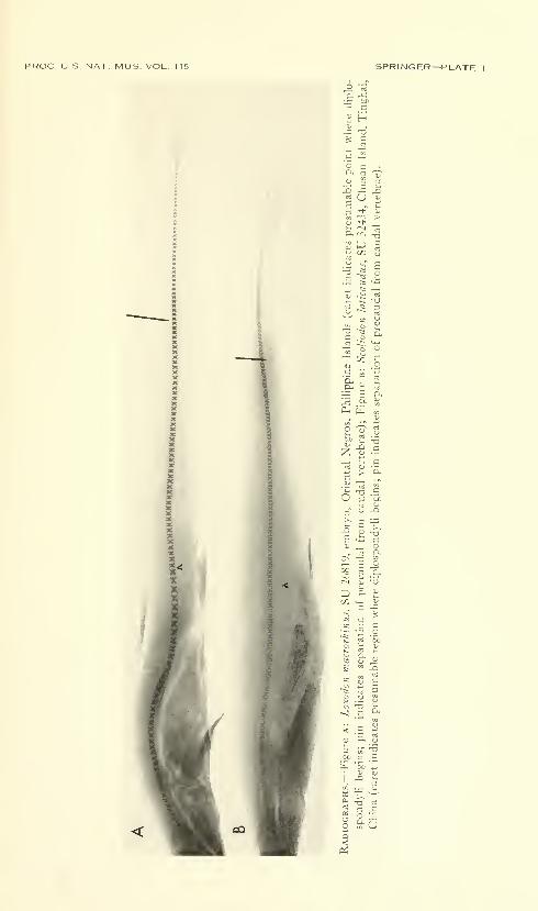
PROC. U.S. NAT. MUS. VOL. 115 SPRINGER—PLATE I
QJ r-t nj—^ 2 '-'^ ^ J3
— 3D.
2CT3 <U • —
6
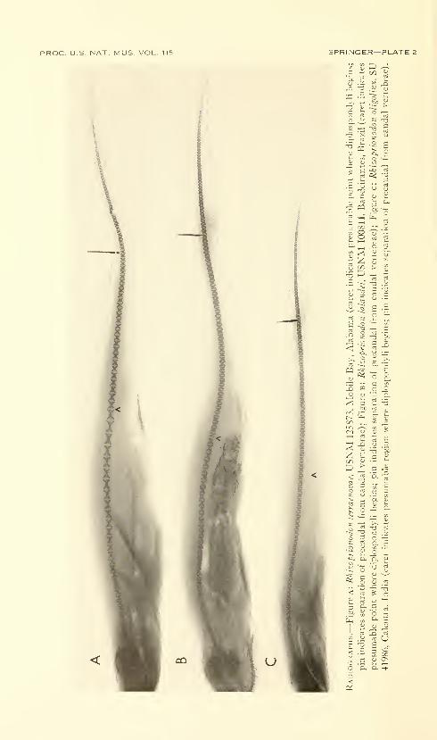
PROC. U.S. NAT, MUS. VOL. 115 SPRINGER—PLATE 2
JS -Q
\^-§ 5
a. cj
rt Qi -O
5 -^ fe
o O rt «
P _
-2

REVISION OF CARCHARHINID SHARK GENERA—SPRINGER 567
(sometimes a series consists of a single tooth) and extremely difficult
to count. Competent colleagues, not engaged in elasmobranch
studies, were given specimens on which to count the teeth and moreoften than not made significant errors. Caution, as well as magni-
fication and good illumination, is recommended strongly for such
counts.
Enlarged hyomandibular pores : In the genus Rhizoprionodon, there
is a discrete group of comparatively large and noticeable pores of
the hyomandibular series opening near the upper labial furrow and
extending dorsoposteriorly a short distance (see figures of the various
species). Frequently these pores are in a straight line, sometimes
irregularly biserial. Some difficulty in counting was found whenthe pores were clogged or when they had not broken through the
denticles (in small specimens). Counts should be made under
magnification and any large gaps between pores should be searched
for obscured members of the series. Sometimes the most anterior
pore is well separated from the remaining ones. Scoliodon may have
a few enlarged pores, but they are frequently difficult to find. (See
also description of Loxodon.)
Vertebrae : Radiographs of sharks were made using a very fine-grain
industrial film. The vertebral count was separated into two parts:
(1) precaudal vertebrae, which includes all complete centra anterior
to the forward edge of the upper precaudal pit; (2) caudal vertebrae,
which includes those centra posterior to the precaudal vertebrae.
Precaudal vertebral counts are sometimes subject to an error of
plus or minus two vertebrae. One reason for this is that to establish
accurately the position of the first vertebra is not always possible
because of masking by other structm*es behind the head, including
the occipital condyles. Another is that sometimes an error in estab-
lishing the last precaudal vertebra occurs because of the angle with
which the X-rays entered the body in the region of the precaudal pit;
a two-dimensional parallax is created on the film. This last problem
was circumvented in most instances by sticking a pin, to mark the
first caudal vertebra, into the vertebral column immediately posterior
to the anterior margin of the precaudal pit. The errors affect the
counts very little.
At the tip of the caudal fin the last few vertebrae are frequently
and irregularly fused (each fusion was counted as one vertebra), or
they are too small to give good resolution on the film. Caudal verte-
bral counts were made under high illumination and magnification,
and they are reasonably accurate for the purposes of this study.
In embryos, precaudal vertebrae are formed early, but caudal
vertebrae are not completely formed until shortly before birth;
therefore, precaudal, but not caudal, vertebral counts from embryos
can be used (in Loxodon, for which I had few specimens, caudal counts
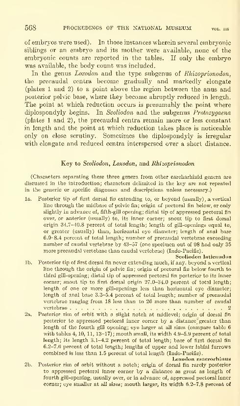
568 PROCEEDINGS OF THE NATIONAL MUSEUM vol. 115
of embryos were used). In those instances wherein several embryonicsiblings or an embryo and its mother were available, none of the
embryonic counts are reported in the tables. If only the embryowas available, the body count was included.
In the genus Loxodon and the type subgenus of Rhizoprionodon,
the precaudal centra become gradually and markedly elongate
(plates 1 and 2) to a point above the region between the anus andposterior pelvic base, where they become abruptly reduced in length.
The point at which reduction occurs is presumably the point where
diplospondyly begins. In Scoliodon and the subgenus Protozygaena
(plates 1 and 2), the precaudal centra remain more or less constant
in length and the point at which reduction takes place is noticeable
only on close scrutiny. Sometimes the diplospondyly is irregular
with elongate and reduced centra interspersed over a short distance.
Key to Scoliodon, Loxodon, and Rhizoprionodon
(Characters separating these three genera from other carcharhinid genera are
discussed in the introduction; characters delimited in the key are not repeated
in the generic or specific diagnoses and descriptions unless necessary.)
la. Posterior tip of first dorsal fin extending to, or beyond (usually) , a vertical
line through the midbase of pelvic fin; origin of pectoral fin below, or only
slightly in advance of, fifth-gill opening; distal tip of appressed pectoral fin
over, or anterior (usually) to, its inner corner; snout tip to first dorsal
origin 34.7-40.8 percent of total length; length of gill-openings equal to,
or greater (usually) than, horizontal eye diameter; length of anal base
6.9-8.4 percent of total length; number of precaudal vertebrae exceeding
number of caudal vertebrae by 43-57 (one specimen out of 98 had only 35
more precaudal vertebrae than caudal vertebrae) (Indo-Pacific)
.
Scoliodon laticauduslb. Posterior tip of first dorsal fin never extending much, if any. beyond a vertical
line through the origin of pelvic fin; origin of pectoral fin below fourth to
third gill-opening; distal tip of appressed pectoral fin posterior to its inner
corner; snout tip to first dorsal origin 27.0-34.0 percent of total length;
length of one or more gill-openings less than horizontal eye diameter;
length of anal base 3.3-5.4 percent of total length; number of precaudal
vertebrae ranging from 18 less than to 26 more than number of caudal
vertebrae 2
2a. Posterior rim of orbit with a slight notch at midlevel; origin of dorsal fin
posterior to appressed pectoral inner corner by a distance* greater than
length of the fourth gill opening; eye larger at all sizes (compare table 6
with tables 4, 10, 11, 13-17) ; mouth small, its width 4.9-5.9 percent of total
length; its length 3.1-4.2 percent of total length; base of first dorsal fin
6.2-7.6 percent of total length; lengths of upper and lower labial furrows
combined is less than 1.5 percent of total length (Indo-Pacific).
Loxodon macrorhinus2b. Posterior rim of orbit without a notch; origin of dorsal fin rarely posterior
to appressed pectoral inner corner by a distance as great as length of
fourth gill-opening, usually over, or in advance of, appressed pectoral inner
corner; eye smaller at all sizes; mouth larger, its width 6.2-7.8 percent of

REVISION OF CARCHARHINID SHARK GENERA—SPRINGER 569

570 PROCEEDINGS OF THE NATIONAL MUSEUM
S5
00°
CO™
I «<
^«
^2
j,s
S2'
-H M ^
o r^- (N t^
1-H 00 t^ -S"
C< r-c
I ?o 00 >om C4 M
" ^ S '=»
I t- 00 CO
-a "S •==^ » a a
g
Ss
e^ 1 c^ 1
C^ I (N CO
CO 1 fN c>
<N I C^ CI
I -^ to CJ
rt I* to rt rt I
l-H
Ii-< lO Ol <N IN -<
( —I t- CO CO
rt M CO t^
I C<I CO ^ >0 CO
3 SC o 5, '2 6 g. § g I
"
od ^4 aj (ij Qi ft; ft! oi o^'
REVISION OF CARCHARHINID SHARK GENERA—SPRINGER 571
total length: its length 4.1-5.3 percent (usually more than 4.2 percent) of
total length; base of first dorsal fin 7.8-10.9 percent of total length (only
one specimen out of 70 below 8.3 percent) ; lengths of upper and lower
labial furrows combined is more than 1.5 percent of total length.
3 Rhizoprionodon3a. Upper labial furrow short or virtually absent, 0-1.3 percent of total length
(rarely more than 1 percent), usually shorter than lower labial furrow;
teeth modally fewer (see tables 1-3) 4
3b. Upper labial furrow longer, always present, more than 1.1 percent of total
length (usually more than 1.3 percent), rarely shorter than lower labial
furrow; teeth modally more (see tables 1-3) 5
4a. Total enlarged hyomandibular pores on both sides of head 7-16 (only 2 of
54 specimens with more than 14) ;precaudal vertebrae 84-91 (Indo-Pacific
north of Australia) Rhizoprionodon oligolinx
4b, Total enlarged hyomandibular pores on both sides of head 15-22; precaudal
vertebrae 73-80 (Australia) Rhizoprionodon taylori
5a. Total teeth in outer row of both jaws 53-58; total teeth in outer row of lower
jaw 26-28; total teeth in outer row of upper jaw 27-30; snout tip to outer
nostrils usually longer at any given size (fig. 1), ranging from 4.5-6.0
percent of total length (Eastern Pacific) . . . Rhizoprionodon longurio5b. Total teeth in outer row of both jaws 47-53 (only 2 specimens out of 74 of
R. acutus with 53) ; total teeth in outer row of lower jaw 22-26 (only 1
specimen out of 19 of R. terraenovae and 4 of R. acutus with 26) ; total
teeth in outer row of upper jaw 23-27 (1 specimen each of R. porosus
and R. terraenovae and 7 of R. acutus with 27) ; snout tip to outer nostrils
usually shorter at any given size (fig. 1), 3.2-4.5 percent of total length
in R. porosus and R. terraenovae, 4.4-5.1 percent in R. lalandei, 4.0-5.4
percent in R. acutus (other than Eastern Pacific) 6
6a. Tip of appressed pectoral fin fails to reach level of middle of first dorsal
base; dorsal-pectoral ratio 100-123 (only 1 out of 33 specimens with less
than 105) ; clasper length 5.9 percent or more of total length in males
500 mm. or larger; largest specimen seen: 640 mm.; precaudal vertebrae
79-90 (rarely less than 84), outnumbering caudal vertebrae 5-20 (usually
7 or more) ; centra in posterior monospondylous region scarcely longer
than those in anterior diplospondylous region (pi. 2b) (Western Atlantic)
.
Rhizoprionodon lalandei6b. Tip of appressed pectoral fin reaches to, or beyond, level of middle of first
dorsal base (for dorsal-pectoral ratio, see 7a and 7b) ; clasper length 3.9
percent or less in males less than 620 mm.; attaining a length of over 1000mm.; precaudal vertebrae 55-79, equal to, or fewer in number than, caudal
vertebrae (one specimen of R. acutus with one more caudal vertebra than
body vertebrae) ; centra in posterior monospondylous region markedlylonger than those in anterior diplospondylous region (pi. 2a) 7
7a. Snout tip 4.0-5.4 percent of total length, usually greater at any given size
(fig. 1); dorsal-pectoral ratio usually larger (91-114, most specimens 100
or more, table 3) (Africa to Austraha) Rhizoprionodon acutus7b. Snout tip 3.2-4.5 percent of total length, usually smaller at any given size
(fig. 1); dorsal-pectoral ratio usually smaller (81-104, few specimens over
100, table 3) (Western Atlantic) 8
8a. Precaudal vertebrae 58-66 (1 specimen out of 74 with 66, North Carolina)
(Atlantic and Gulf coasts of the United States and Mexico).
Rhizoprionodon terraenovae8b. Precaudal vertebrae 66-75 (2 specimens out of 58 with 66, Brazil) (Western
Atlantic except United States and Mexico) . . Rhizoprionodon porosus

572 PROCEEDINGS OF THE NATIONAL MUSEUM
Q OO Ooo -^ o
o o oo o
o
o-o
o-o
* o* o o o
oo o
**t ?
80 °§§oo o ^
00 o 0^0^
^ *o o
u
i
0^0
•o
oO§= §0 o
§0 o@ o^ o
3 "
O B^zV)
l#«
REVISION OF CARCHARHINID SHARK GENERA—SPRINGER 573
Scoliodon Miiller and Henle
Scoliodon Miiller and Henle, 1837, Sitzb. Akad. Wiss. Berlin, p. 114 (name only).
Scoliodon Miiller and Henle, 1837, Arch. Naturg., vol. 3, no. 1, p. 397 (name anddiagnosis).
Scoliodon Miiller and Henle, 1838, L'Institut, vol. 6, no. 244, p. 64 (name anddiagnosis)
.
Scoliodon Miiller and Henle, 1838, Mag. Nat. Hist., new. ser., vol. 2, p. 35 (nameand diagnosis).
Scoliodon Miiller and Henle, 1841, Systematische Beschreibung der Plagiostomen,
2nd page 27 (a subgenus with three included species; type species Carcharias
(Scoliodon) laticaudus Miiller and Henle, by subsequent designation, Gill,
1862, Ann. New York Lye, vol. 8, p. 401).
Physodon Valenciennes in Miiller and Henle, 1841, Systematische Beschreibung
der Plagiostomen, p. 30 (a subgenus; type species Carcharias (Physodon)
mulleri Valenciennes by monotypy).
In several copies of Miiller and Henle (1841), except that in the
British Museum, seen by me, there are two pages numbered 27 and
two numbered 28. The first page 27 lists Scoliodon as a new genus and
the description terminates and is complete at the bottom of the first
page 28. The second page 27 lists Scoliodon as a subgenus and the
description continues from page 28 to page 29. Obviously, the first
pair of pages were not meant to be included and their elimination from
the text causes no discontinuity. This is not so with the second pair.
For nomenclatural purposes I disregard the first pages 27 and 28.
Bigelow and Schroeder (1948, p. 292) included in their synonymyof Scoliodon (which embodies all three genera treated in my study)
the genus Cynocephalus (not Cynocephalus Gill, 1862) which they
attributed to Bleeker (1879). They designated Carcharias (Scoliodon)
macrorhynchus Bleeker (1852, but 1858 in their work) as type species.
However, Bleeker (1878) first used the name Cynocephalus for a group
of shark species that did not include C. (S.) macrorhynchus; therefore,
Bigelow and Schroeder were in error in dating the genus from 1879 and
C. (S.) macrorhynchus could not be designated as type species for the
genus.
Cynocephalus was first used validly in 1768 for a genus of mammals;Bleeker's usage of the name was apparently an extrapolation from
Gill (1862), who gave a key to shark genera and designated Squalus
glaucus Linnaeus as type species of Cynocephalus, listing no other species.
For these reasons, I do not believe Bleeker was creating a genus, and
future workers should refrain from selecting a type from Bleeker
and erecting an additional junior homonym.Diagnosis.—Small sharks (largest specimen seen 581 mm. t.l.)
distinct from all other Carcharhinidae in having the tip of the posterior
margin of the first dorsal fin extending posteriorly beyond the level of

574 PROCEEDINGS OF THE NATIONAL MUSEUM vol. 115
the origin of the pelvic fin to a point over, or beyond (usually), the
middle of the pelvic base; distinct from all Carcharhinidae except
Aprionodon isodon, Carcharhinus oxyrhynchus, and C. temmincki in
having the origin of the pectoral fin below, or only slightly in advanceof, the level of the fifth gill-opening. Differing from most Carcharhi-
nidae in having the outer tip of the appressed pectoral fin over, or in
advance of, its inner corner and from all Carcharhinidae in havingboth the tip and inner corner well in advance of the level of the origin
of the first dorsal fin. The origin of the second dorsal fin ranges fromabove the posterior third of the anal base to over the anal axil. It is
usually over the posterior fifth of the anal base.
A single common species distributed from the coasts of Japan andsoutheast Asia to India and the Dutch East Indies, exclusive of NewGuinea, and absent also from Australia, the Philippines, and Oceania.
At the present time I know of no close relative to this genus among the
other carcharhinid genera.
Scoliodon laticaudus Miiller and Henle
Figures 2, 3; Plate 1b
"Pala Sorra" Russell, 1803, Descriptions and figures of two hundred fishes . . .
Coromandel, vol. 1, p. 9, fig. 14 (a common name).
Carcharias (Scoliodon) laticaudus Miiller and Henle, 1841, Systematische Beschrei-
bung der Plagiostomen, p. 28, pi. 8 (India).
Carcharias (Physodon) mulleri Valenciennes in Miiller and Henle, 1841, ibid., p. 30,
pi. 19, fig. 1 (Bengal).
Carcharias (Scoliodon) macrorchynchos Bleeker, 1852, Verh. Bataviaasch Gen.,
vol. 24, p. 31, pi. 1, fig. 1 (Batavia, spelled macrorhynchos in other parts of the
paper and in subsequent literature).
Carcharias palasorra Bleeker, 1853, Verh. Bataviaasch Gen., vol. 25, p. 9 (Coro-
mandel, based on "Pala Sorra" Russell, see discussion below).
Diagnosis.—Distinctive characters are those of the genus (p. 568).
Description (see also table 4).—Body vertebrae 97-112 (table 5),
centra not markedly elongate in posterior monospondylous region
(pi. 1b); caudal vertebrae 50-62, total vertebrae 148-171; teeth
smooth-edged, upper teeth 12-1-12 to 16-1-16; lower teeth 12-12
to 17-17 (higher, or lower, counts in upper and lower jaws correlated)
;
enlarged hyomandibular pores 0-5 on each side of head (rarely morethan 2; frequently difiicult to see).
Denticles imbricate, 3-ridged and 3-toothed in young, 3- to 5-ridged
and 3- to 4-toothed in adults.
Color of preserved specimens: Buff brown, purplish brown, or gray
brown, darker above, pale below; fins sometimes darker than body;
edges of fins at all sizes without dark margins except sometimes the
dorsal and distal edges of the upper caudal lobe; in adult males the
distal third of clasper abruptly paler than remainder.
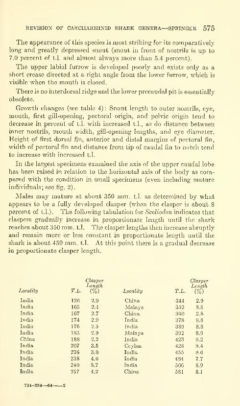
REVISION OF CARCHARHINID SHARK GENERA—SPRINGER 575
The appearance of this species is most striking for its comparatively
long and greatly depressed snout (snout in front of nostrils is up to
7.0 percent of t.l. and almost always more than 5.4 percent).
The upper labial furrow is developed poorly and exists only as a
short crease directed at a right angle from the lower furrow, which is
visible when the mouth is closed.
There is no interdorsal ridge and the lower precaudal pit is essentially
obsolete.
Growth changes (see table 4) : Snout length to outer nostrils, eye,
mouth, first gill-opening, pectoral origin, and pelvic origin tend to
decrease in percent of t.l. with increased t.l., as do distance betweeninner nostrils, mouth width, gill-opening lengths, and eye diameter.
Height of first dorsal fin, anterior and distal margins of pectoral fin,
width of pectoral fin and distance from tip of caudal fin to notch tend
to increase with increased t.l.
In the largest specimens examined the axis of the upper caudal lobe
has been raised in relation to the horizontal axis of the body as com-pared with the condition in small specimens (even including matureindividuals; see fig. 2).
Males may mature at about 350 mm. t.l. as determined by whatappears to be a fully developed clasper (when the clasper is about 8
percent of t.l.). The following tabulation for Scoliodon indicates that
claspers gradually increase in proportionate length until the shark
reaches about 350 mm. t.l. The clasper lengths then increase abruptly
and remain more or less constant in proportionate length until the
shark is about 450 mm. t.l. At this point there is a gradual decrease
in proportionate clasper length.
Locality
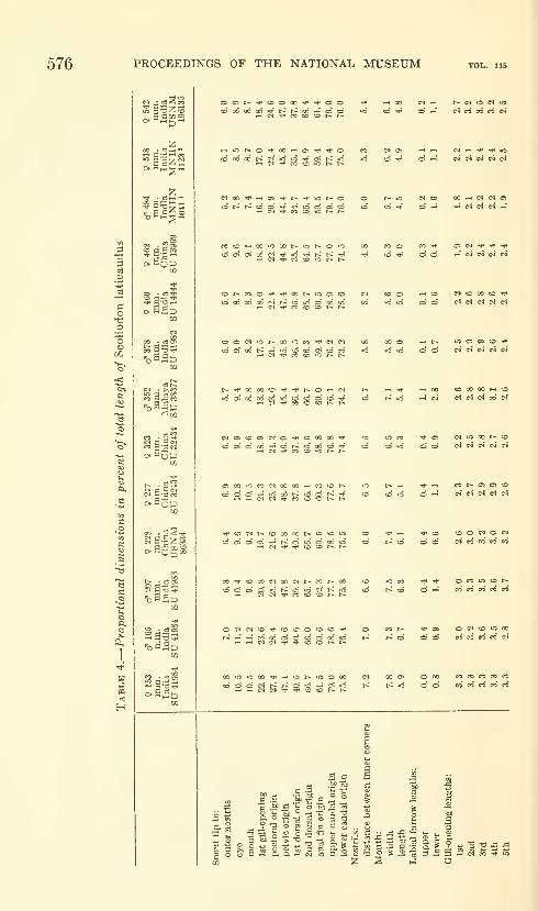
576 PROCEEDINGS OF THE NATIONAL MUSEUM

REVISION OF CARCHARHINID SHARK GENERA—SPRINGER 577
•>»< tt>t~i-4 t0«00 •^«« COtOOOO tOC^OO OOrttDOO
Oi OO <D
t^ ^ 00
^ TJH 00
00 « C4
O rH O t~-
tO i-i 0> 00
CO <o c»
00 CO CO
00 o> t^
ei CO i-I
(N o «00 CO c<
CO O 00 oo
»o o t* t-*
o c» •<»<
O lO '^
o eo o>
CO fli i-i
o o t~
00 CO <N
eo »- eo oCO >-* 00 00
CD O OlO lo ici
c^ CO 00 eo
eo* OS * '*'
05 t^ ^ CO
O 00 >0 Tl<
c^ 00 t>.
oi -^ 00
N N 00
eo" * rt
m t~ CO
t>; eo' <N
^ CO IM OCO »-i' 00 oi
»0 lO CO CO
CO o> »o o
CO CO r~
03 «»> t>-"
lO f* C^ lO
CO -^* 00 00
Tj* lO CO *c
id io -^ 00
O CO oCO V t>^
o »-• *o
CO co' ci
lO 00 •'J^ oCO O 00 oi
eo o eo
id id -^
c^ O 00
eo' ^ i-i
1-1 lO OS oCO O CO 00
T(< C<1 CO
•d >d eo'
CO Tt< t~ lO
eo oi >d «<
Oi CD eo CO
<m" oJ id id
•«»> eo i-H
c<i oJ id
05 t^ ^t^ CO CO
iO 05 iO
co' eo' r-i
05 i-l 00
t-: co' -i
1}. CD O •*
CO 05 t-^ 1^
CO CO iO
id ^ CO
C4 CO >s
OS ^ t^
O CO ot~: eo' CJ
05 ^ lO Nid o r-I 00
00 CO eo
00 eo' CO
CO iO 00 <r -H o OJ
id oi t^ t^
CO N eo iO
id id •<*< CO
iO rfi ^^ rj«
00 N r-1 00!-<' oi id V
O iO CO 00
eo' 00 Tii •»<
N OJ o>
id id eo'
t^ eo iO c^
c4 OS * id
578 PROCEEDINGS OF THE NATIONAL MUSEUM vol. 115
Sexual dimorpMsm: There is a striking difference in dentition of
adult males as compared with females or juvenile males (fig. 3). In
adult males the cusps of the teeth are long and slender and the middle
and posterior teeth of each side of the lower jaw are erect. The
anterior teeth are greatly elongate and flexuous and somewhat round
in cross section. Several rows of the anterior lower teeth in the adult
male appear to be functional. Sexual dimorphism of dentition in
sharks has been reported only for the squalid genus Deania (see
Bigelow and Schroeder, 1957, and Garrick, 1960). Dental sexual
dimorphism also exists in Rhizoprionodon lalandei, R. oligolinx, and
probably R. taylori; it also exists to a much lesser degree in Loxodon.
The phenomenon is probably widespread and unrecognized in sharks.
Physodon is based on an adult male S. laticaudus and the type descrip-
tion of iS'. laticaudus is based on an adult female.
This species is free-living at sizes of at least 126 mm. t.l. (130-150
mm. according to Setna and Sarangdhar, 1949). Maximum size in
Indian waters is about 26 inches (approximately 660 mm.). Breeding
occurs all year with peak parturition from November through January.
Up to 12 embryos occur per uterus, but usually no more than 14 young
per female. Mature eggs are small, about one mm. in diameter
(Setna and Sarangdhar, 1949).
Thillayampalam (1928) has given a detailed anatomical description
of S. laticaudus (as Scoliodon sorrakowa) . Some portions of the work
are based on other species {Rhizoprionodon), but the study is an
important one.
Distribution (see "Material").—This is a common species and its
absence from Australia, the Philippines, the Celebes, Moluccas,
New Guinea, and Oceania I believe to be real. The distribution seems
to indicate that the species is essentially a continentally restricted one,
able to traverse only the narrowest passes between coastlines. Setna
and Sarangdhar (1949) reported that the species (as S. sorrakowa)
lived in rocky areas up to three miles offshore in Bombay waters.
NoMENCLATURAL DISCUSSION.—As mentioned above, Physodon
mulleri was based on an adult male of S. laticaudus. Its dentition is
markedly different from that of the female upon which Miiller and
Henle's description of C. (S.) laticaudus was based. There are adult
males in the syntypic series of S. laticaudus but undoubtedly these were
not examined for dentition. This is indicated by the fact that, in the
first pair of pages 27 and 28 in Miiller and Henle's Plagiostomen (see
discussion on page 573), the description of C. (S.) laticaudus is of a
single stuffed female specimen in the Berlin Museum. In the second
pair of pages 27 and 28, additional material (including alcohol-
preserved specimens) is listed, but the description is identical with the
previous one.
REVISION OF CARCHARHINID SHARK GENERA—SPRINGER 579
In order to insure the stability of the name C. (S.) laticaudus, I
believe that a lectotype should be designated. In the present situation
the normal procedure would be to select the single stuffed specimen
upon which the original description was based. However, in view of
the importance of vertebral characters for shark classification, I have
chosen to select one of the alcohol-preserved specimens in the Paris
Museum (see "Material").
The type of C. (S.) macrorhynchos Bleeker is missing, but the descrip-
tion and figure are sufficient for placing it in synonymy. Giinther
(1870) first synonymized Bleeker's species with S. laticaudus, and
Bleeker, in an unpublished manuscript (Ennumeratio nova revisa) in
the Leiden museum, accepted this as correct.
There is a history of confusion surrounding the trivial namessorrakowa and palasorra, both of which have been applied to
S. laticaudus. RusseU (1803), not binomial nomenclature, described
"Pala Sorra" and "Sorra Kowah" from Vizgapatam, Coromandel,
India. The names used were native common names. Cuvier (1829)
added the following in a footnote to the last word under Le Bleu (Sq.
glaucus L.), which is the last species he mentions under Les Requins:
Ajoutez le Squ. ustus, Dum. (Sq. carcharia minor, Forsk.), Lac, I,
VIII, 1; Requin a nageoires noires, Quoy et Gaym., Zool. de Freyc, pi. 43,
f. 1; le Sq. glauque, Lac, I, ix, 1, qui est different de celui de Bl.; le Sq,
ciliaris, Schn., pi. 31, dont les cil marquent seulement I'extreme jeunesse,
Le palasorrah [sic] et le sorrakowah [sic], Russ., XIV et un assez grand
nombre e'espece nouvelles que nous decrirons dans notre histoire de
poissons.
There is no reason to believe from the information contained above
that Cuvier was intending to name Russell's forms. Rather it
seems he was merely citing the native names. In support of this is the
fact that the generic name (Sq.) that appears before the other species is
omitted before palasorrah and sorrakowah. Nevertheless, on the
basis of the above quotation most subsequent authors credited Cuvier
(1829) with authorship of Carcharias palasorra and C. sorrakowah.
Fowler (1936) first called attention to the fact that the first of these
two names could not date from Cuvier (1829) because it was not
proposed in binomial form. He did not credit the name, however,
nor did he mention sorrakowah. Klausewitz (1960) also recognized
that palasorrah as it appeared in Cuvier (1829) was not a valid name.
He assigned authorship of Carcharias palasorrah to Bleeker (1853)
and placed the name in synonymy with C. acutus Ruppell but gave
no reasons for his actions. He did not discuss sorrakowah.
Miiller and Henle (1841), in their monograph of the Plagiostoma,
list Russell (1803) in their literature references and mention the
"Pana Sorrats" and the "Sorra Kowats" as doubtful synonyms of their
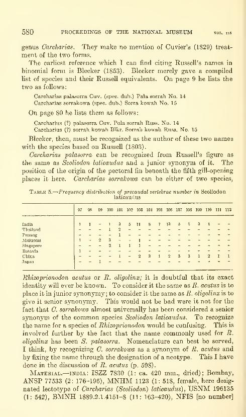
580 PROCEEDINGS OF THE NATIONAL MUSEUM vol. us
genus Carcharias. They make no mention of Cuvier's (1829) treat-
ment of the two forms.
The earhest reference which I can find citing Russell's names in
binomial form is Bleeker (1853). Bleeker merely gave a compiled
list of species and their Russell equivalents. On page 9 he lists the
two as follows:
Carcharias palasorra Cuv. (spec, dub.) Pala sorrah No. 14
Carcharias sorrakowa (spec, dub.) Sorra kowah No. 15
On page 80 he lists them as follows:
Carcharius (?) palasorra Cuv. Pola sorrah Russ. No. 14
Carcharius (?) sorrah kowah Blkr. Sorrah kowah Russ. No. 15
Bleeker, then, must be recognized as the author of these two nameswith the species based on Russell (1803).
Carcharias palasorra can be recognized from Russell's figure as
the same as Scoliodon laticaudus and a junior synonym of it. Theposition of the origin of the pectoral fin beneath the fifth gill-opening
places it here. Carcharias sorrakowa can be either of two species,
Table 5.
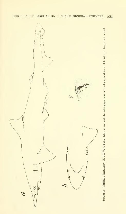
REVISION OF CARCHARHINID SHARK GENERA—SPRINGER 581
•§
CO
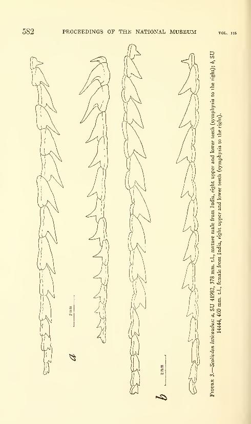
582 PROCEEDINGS OF THE NATIONAL MUSEUM
t, Ot)
*-"
^ .2O w
V M
r ^
c '2
^ o
CO o
?* ^Q —I
CO
K^
REVISION OF CARCHARHINID SHARK GENERA—SPRINGER 583
(2: 170-365); Alibag, ZSZM H1391 (3: ca. 167-ca. 281), ZSZM H1374(1: ca. 402), ZSZM H1375 (8: 264-282), ISH 3/61 (6: 176-506);
Kanara, BMNH 1889.2.1.4160 (1: 312); Karwar, ZSZM H1392 (1:
208), ZSZM H1376 (3: 126-218), ISH 4/61 (1: 173) ; Calicut, BMNH1899.2.1.4159 (1: 455); Malabar Coast, MNHN 1066 (3: 167-257),
MNHN 1122 (1: 423); Pondicherry, UZMK 378 (1: 330); EmurFisheries Station, Madras Presidenc}^, SU 41983 (1: 207); Madras,
BMNH 1889.2.1.4161-3 (2: 337-389); Puri Orissa State, SU 41982
(1: 378); Vizagapatam, SU 1444 (1: 460); Calcutta, SU 41984 (3:
153-165); Bengal, BMNH [no number] (2: 174-240), MNHN 1041
(1 : 484, holotype of Physodon mulleri) ; Ceylon, NMV [no number]
(1 : 175), NMV [no number] (1 : 426). gulf of Thailand: off MenamRiver, UZMK [no number] (1 : 321) ; ca. 12°11' N. and ca. 100°41' E.,
GVF 2470 (1: 255). Thailand: Bangkok, ANSP 60404 (1: 283);
Bandon Bight, USNM 86905 (2: 100-137, embryos), malaya:
Penang, UZMK 377 (1: 370); Singapore, SU 38377 (3: ca. 328-293),
MCZ 182 (1: 333), CNHM 15652 (1: 459). java: Batavia, USNM72479 (1: 422); Straits of Molucca, UZMK 404-409 (6: ca. 210-ca.
253), ANSP 517 (1: 186). east Indian archipelago: BMNH1867.11.28.190 (1: 467, female; this specimen is cataloged as holotype
of C. (S.) macrorhynchos, but since Bleeker listed only a male, 218
mm., this specimen cannot be a type; the holotype is apparently lost).
SARAWAK: BMNH 1895.2.28.74-75, in part (1: ca. 143). china:
BMNH 1851.12.27.253-4 (3: 162-245); Chinai, Chusan Island,
Tinghai, SU 32434 (4: 258-333) ; Foochow, USNM 86970 (1: ca. 212),
USNM 86334 (1: 228); San-tu, Fukien, ANSP 89616 (1: 323); Wen-chow, Chekiang, CAS 875 (1: 275); Amoy, BMNH 1863.2.23.13-16
(4: 159-165), BMNH 1860.7.20.32 (1: 344); Hong Kong, UZMK 376
(1: 581), BMNH 1939.3.23.1 (1: 360), ANSP 76687 (1: 349), SU13770 (1: ca. 522), SU 13969 (1: 462). japan: BMNH 1862.11.1.30
(1: 155) "TRIESTE" [error]: NMV [no number] (1: 405).
Loxodon Miiller and Henle
Loxodon Miiller and Henle, 1838, Mag. Nat. Hist., new ser., vol. 2, p. 36 (nameand diagnosis).
Loxodon Miiller and Henle, 1838, Arch. Naturg., vol. 4, no. 1, p. 84 (name anddiagnosis).
Loxodon Miiller and Henle, 1841, Systematische Beschreibung der Plagiostomen,
p. 61 (type Loxodon macrorhinus Miiller and Henle, 1841, by monotypy).
Diagnosis.—Sharks of moderate size (reaching at least 905 mm.t.l.) distinguished from other carcharinid genera by the following
combination of characters: origin of second dorsal fin ranges from
over, and just in advance of, anal axil to slightly behind anal axil
(usually); cusps of teeth smooth and obliquely directed rearward;
labial furrow on upper jaw poorly developed, usually shorter than
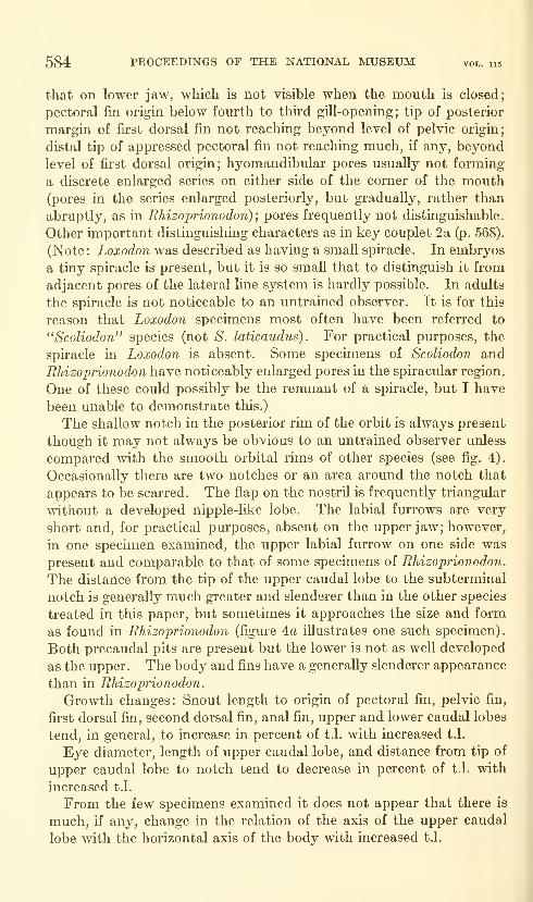
584 PROCEEDINGS OF THE NATIONAL MUSEUM vol. 115
that on lower jaw, which is not visible when the mouth is closed;
pectoral fin origin below fourth to third gill-opening; tip of posterior
margin of first dorsal fin not reaching beyond level of pelvic origin;
distal tip of appressed pectoral fin not reaching much, if any, beyond
level of first dorsal origin; hyomandibular pores usually not forming
a discrete enlarged series on either side of the corner of the mouth(pores in the series enlarged posteriorly, but gradually, rather than
abruptly, as in Rhizoprionodon);pores frequently not distinguishable.
Other important distinguishing characters as in key couplet 2a (p. 068).
(Note: ioaroc^oTi was described as having a small spiracle. In embryos
a tiny spiracle is present, but it is so small that to distinguish it from
adjacent pores of the lateral line system is hardly possible. In adults
the spiracle is not noticeable to an untrained observer. It is for this
reason that Loxodon specimens most often have been referred to
"Scoliodon" species (not S. laticaudus). For practical purposes, the
spiracle in Loxodon is absent. Some specimens of Scoliodon and
Rhizoprionodon have noticeably enlarged pores in the spiracular region.
One of these could possibly be the remnant of a spiracle, but I have
been unable to demonstrate this.)
The shallow notch in the posterior rim of the orbit is always present
though it may not always be obvious to an untrained observer unless
compared with the smooth orbital rims of other species (see fig. 4).
Occasionally there are two notches or an area around the notch that
appears to be scarred. The flap on the nostril is frequently triangular
without a developed nipple-like lobe. The labial furrows are very
short and, for practical purposes, absent on the upper jaw; however,
in one specimen examined, the upper labial furrow on one side was
present and comparable to that of some specimens of Rhizoprionodon.
The distance from the tip of the upper caudal lobe to the subterminal
notch is generally much greater and slenderer than in the other species
treated in this paper, but sometimes it approaches the size and form
as found in Rhizoprionodon (figure 4a illustrates one such specimen).
Both precaudal pits are present but the lower is not as well developed
as the upper. The body and fins have a generally slenderer appearance
than in Rhizoprionodon.
Growth changes: Snout length to origin of pectoral fin, pelvic fin,
first dorsal fin, second dorsal fin, anal fin, upper and lower caudal lobes
tend, in general, to increase in percent of t.l. with increased t.l.
Eye diameter, length of upper caudal lobe, and distance from tip of
upper caudal lobe to notch tend to decrease in percent of t.l. with
increased t.l.
From the few specimens examined it does not appear that there is
much, if any, change in the relation of the axis of the upper caudal
lobe with the horizontal axis of the body with increased t.l.
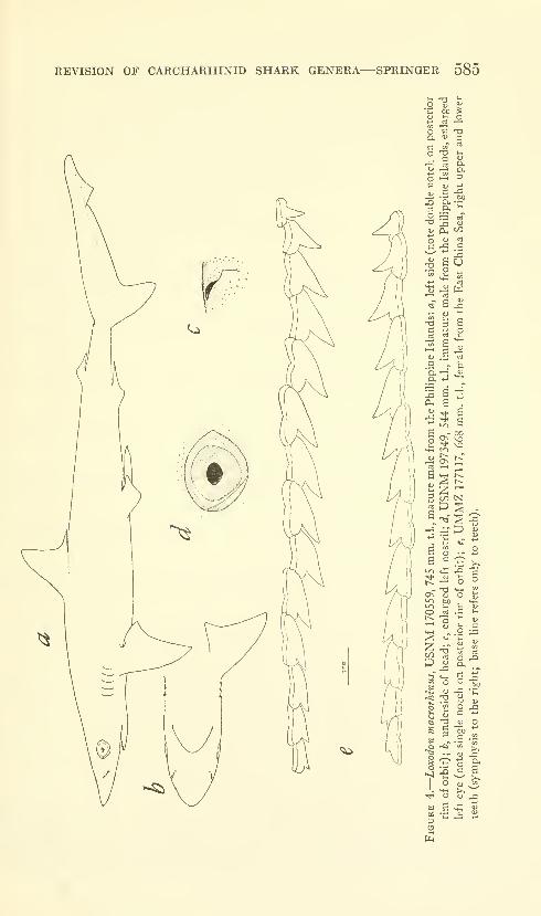
REVISION OF CARCHARHINID SHARK GENERA—SPRINGER 585
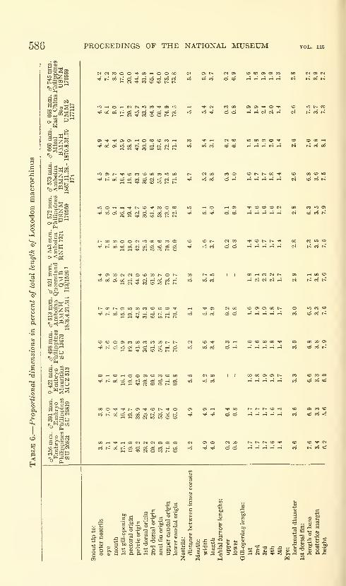
586 PROCEEDINGS OF THE NATIONAL MUSEUM
a Qwia-agg
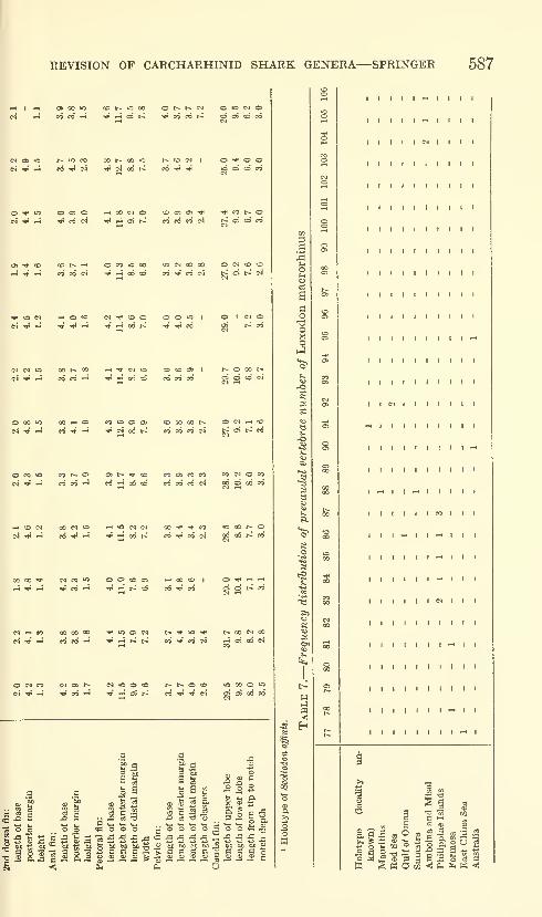
REVISION OF CARCHARHINID SHARK GENERA—SPRINGER 587
O t* t" c^
^ CO CO t^
o »o w oCO O <^ CO
(N Oi O
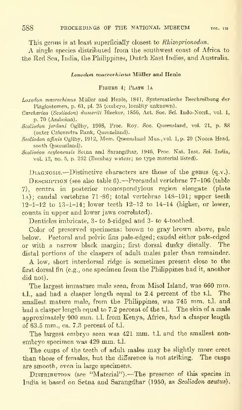
588 PROCEEDINGS OF THE NATIONAL MUSEUM vol. 115
This genus is at least superficially closest to Rhizoprionodon.
A single species distributed from the southwest coast of Africa to
the Red Sea, India, the Philippines, Dutch East Indies, and Australia.
Loxodon macrorhinus Miiller and Henle
Figure 4; Plate 1a
Loxodon macrorhinus Miiller and Henle, 1841, Systematische Beschreibung der
Plagiostomen, p. 61, pi. 25 (embryo, locality unknown).
Carcharias (Scoliodon) dumerili Bleeker, 1856, Act. Soc. Sci. Indo-Neerl., vol. I,
p. 70 (Amboina).
Scoliodon jordani Ogilby, 1908, Proc. Roy. Soc. Queensland, vol. 21, p. 88
(outer Caloundra Bank, Queensland).
Scoliodon affinis Ogilby, 1912, Mem. Queensland Mus.,vol. l,p. 29 (Noosa Head,
south Queensland).
Scoliodon ceylonensis Setna and Sarangdhar, 1946, Proc. Nat. Inst. Sci. India,
vol. 12, no. 5, p. 252 (Bombay waters; no type material listed).
Diagnosis.—Distinctive characters are those of the genus (q.v.).
Description (see also table 6).—Precaudal vertebrae 77-106 (table
7), centra in posterior monospondylous region elongate (plate
1a); caudal vertebrae 71-86; total vertebrae 148-191; upper teeth
12-1-12 to 13-1-14; lower teeth 12-12 to 14-14 (higher, or lower,
counts in upper and lower jaws correlated).
Denticles imbricate, 3- to 5-ridged and 3- to 4-toothed.
Color of preserved specimens: brown to gray brown above, pale
below. Pectoral and pelvic fins pale-edged; caudal either pale-edged
or with a narrow black margin; first dorsal dusky distally. The
distal portions of the claspers of adult males paler than remainder.
A low, short interdorsal ridge is sometimes present close to the
first dorsal fin (e.g., one specimen from the PhiHppines had it, another
did not).
The largest immature male seen, from Misol Island, was 660 mm.t.l., and had a clasper length equal to 2.4 percent of the t.l. The
smallest mature male, from the PhiHppines, was 745 mm. t.l. and
had a clasper length equal to 7.2 percent of the t.l. The skin of a male
approximately 900 mm. t.l. from Kenya, Africa, had a clasper length
of 63.5 mm., ca, 7.3 percent of t.l.
The largest embryo seen was 421 mm. t.l. and the smallest non-
embryo specimen was 429 mm. t.l.
The cusps of the teeth of adult males may be slightly more erect
than those of females, but the difference is not striking. The cusps
are smooth, even in large specimens.
Distribution (see "Material").—The presence of this species in
India is based on Setna and Sarangdhar (1950, as Scoliodon acutus).

REVISION OF CARCHARHINID SHARK GENERA—SPRINGER 589
Its apparent absence from Oceania is in accord with the distributions
of the other species discussed in the present study.
The species is quite common in Phihppine waters. I saw large
numbers of small specimens in the Manila fish market during April
1962. Its relative abundance elsewhere is unknown although Setna
and Sarangdhar (1950) report it as uncommon in Indian waters.
Wheeler (1959), who gave an excellent description of Loxodon,
reported the species common at Zanzibar. His longest specimen was
905 mm. An 850 mm. female contained two embryos. One specimen
was caught in a trammel net at a depth of about four fathoms.
NoMENCLATURAL DISCUSSION.—The typcs of the nominal forms,
except Scoliodon ceylonensis Setna and Sarangdhar and S. jordani
Ogilby, were examined and apparently are the same species. Theholotype of S. jordani was reported by Ogilby (1916) as having been
destroyed. The short outward-directed upper labial groove and the
appressed pectorals extending only to beneath the origin of the first
dorsal fin of S. jordani indicate that it was probably a Loxodon. Notype was designated for S. ceylonensis but the tooth counts and the
nature of the labial folds as given in the original description place it in
Loxodon. Setna and Sarangdhar (1950), without comment, placed
their species in synonymy with Carcharias acutus Riippell; however,
the figures and description they give are clearly those of a Loxodon,
and, as such, confirm my suspicions concerning the original description.
Material.—no locality: ISZZ 4479 (1: ca. 315 mm., embryo,
holotype of Loxodon macrorhinus) . kenya: Shimoni, DIRU 15A
(1: ca. 900, head and skin). Mauritius island: MCZ 513 (1: 421,
embryo). Seychelles islands: BMNH 1869.5.14.15 (2: ca. 315-ca.
335, embryos), red sea: NFIS 926 (1: ca. 345, embryo), NMV[no number] (1: 858). egypt: Kosseir, SMNS 1787 (1: ca. 375,
embryo), gulf of oman: Muscat, BMNH 1892.1.16.9-10 (1: 734).
SUMATRA: Padang, NMV [no number] (1: 691). misol island:
BMNH 1870.8.31.70 (or .79?) (1: 660). amboina island: BMNH1858.4.21.511 (1: 513), BMNH 1867.11.28.174 (1: 573, male, listed as
type of Carcharias dumerili Bleeker, but Bleeker described the species
from a 553 mm. female), RNH 7371 (1: 543, holotype of Carcharias
dumerili). Australia: Queensland, QMB [number missing] (1: 797,
this specimen was with a group of sharks, aU from Queensland; tags
had corroded); Noosa Head, QMB 113/1526 (1: 521, holotype of
Scoliodon affinis). Philippine islands: Oriental Negros, Dumaguete,
SU 26821 (1: 356, embryo), SU 26819 (1: 391, embryo), SU 13670
(1: 498); Limbones Cove, USNM 170560 (1: 572); off Luzon Point
(1: 745); Manila area, USNM 197349 (3: 500-544). Formosa:
Thape, ISZZ 6964 (2: 429-443). japan: East China Sea, UMMZ177117 (1: 668).
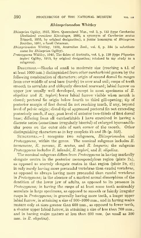
590 PROCEEDINGS OF THE NATIONAL MUSEUM vol. lu
Rhizoprionodon Whitley
Rhizoprion Ogilby, 1915, Mem. Queensland Mus., vol. 3, p. 132 (type Carcharias
(Scoliodon) crenidens Klunzinger, 1880, a synonym of Carcharias acuius
Riippell, 1835, by original designation), a junior homonym of Rhizoprion
Jourdan, 1861, a fossil Cetacean.
Rhizoprionodon Whitley, 1929, Australian Zool., vol. 5, p. 354 (a substitute
name for Rhizoprion Ogilby).
Protozygaena Whitley, 1940, The fishes of Australia, vol. 1, p. 110 (type Physodon
taylori Ogilby, 1915, by original designation; retained in my study as a
subgenus).
Diagnosis.—Sharks of small to moderate size (reaching a t.l, of
at least 1000 mm.) distinguished from other carcharhinid genera by the
following combination of characters: origin of second dorsal fin ranges
from over middle of anal base (rarely) to over anal axil; cusps of teeth
smooth to serrulate and obliquely directed rearward; labial furrow on
upper jaw usually well developed, except in most specimens of R.
oligolinx and R. taylori; lower labial furrow visible when mouth is
closed; pectoral fin origin below fourth to third gill-opening; tip of
posterior margin of first dorsal fin not reaching much, if any, beyond
level of pelvic origin ; distal tip of appressed pectoral fin not extending
posteriorly much, if any, past level of anterior two-thirds of first dorsal
base; differing from all carcharhinids I have examined in having a
discrete series (sometimes irregularly biserial) of enlarged hyomandib-
ular pores on the outer side of each corner of the mouth. Other
distinguishing characters as in key couplets lb and 2b (p. 568).
Subgenera.—I recognize two subgenera, Rhizoprionodon and
Protozygaena, within the genus. The nominal subgenus includes R.
terraenovae, R. porosus, R. acutus, and R. longurio; the subgenus
Protozygaena includes R. lalandei, R. taylori, and R. oligolinx.
The nominal subgenus differs from Protozygaena in having markedly
elongate centra in the posterior monospondylous region (plate 2a),
as opposed to scarcely elongate centra in that region (plate 2b, c);
in only rarely having more precaudal vertebrae than caudal vertebrae,
as opposed to always having more precaudal than caudal vertebrae
in Protozygaena; in the absence of a marked sexual dimorphism of the
dentition of the lower jaw of adults, as opposed to its presence in
Protozygaena; in having the cusps of at least some teeth noticeably
serrulate in large specimens, as opposed to smooth or faintly irregular
cusps in Protozygaena; in generally having more teeth, a longer upper
labial furrow, in attaining a size of 900-1000 mm., and in having males
mature only at sizes greater than 600 mm., as opposed to fewer teeth,
a shorter upper labial furrow, in attaining a size of less than 700 mm.,and in having males mature at less than 600 mm. (as small as 380
mm. in R. oligolinx).

REVISION OF CARCHARHINID SHARK GENERA—SPRINGER 591
^ CC 1-H
r1 rt O !N
« <N « ^
>-4 •»• 00 rH rt
C<1 O O O CO
rH 00 M •* 1
^ o «** c^ »-<
O t^ W C^ CO
M O O W Oft
00 * >-H i-c
rt CO
i-H t^ W
J: 2 g S -S .S •-
ft^' a^ fti ft; 05 0^" Q^
724-334—64 3
s a -sg o aam .2-
S
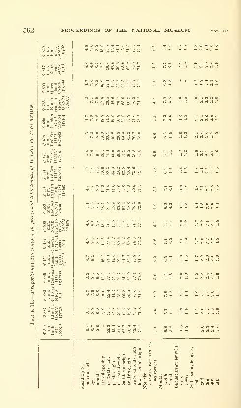
592 PROCEEDINGS OF THE NATIONAL MUSEUM
oisi
Pi
= S £ C Z o
fi o
>^ 2 o 'g 2 :» t;
o 2sSoq oOcB
2
'bOiJt)'-'
00 • 03;— <; oto p '5^ '7 "-I
CO ' J5i ^ '••'o
fc _• r 2 °^ ->
« e a _ C3 £3
•^Po C5£J
z:
o a ;:: 1-^ 2i
C^ i-H Oi 00 « CO l^ O ri (M <M
i-^QOooa>ooooooa>o*o
«D00t^«-i'^Oa>(N<M00O'^t-^oocxi'-i'^oicooieoco
O'^oo'oeooiC^io'^cC''-'
TPCOt^'HC^OOOO'^OOCOOw5o6o6o6rH4oocooico^
!;000«OCDOOOO'*00-^oocJoO'-^ccaJrociC'^c^
(^OOOiO-^CMCOWOOOMTt^o6o6oic^icDC-:J5Q'^^^0
CSr-cCOC^t-COOOOO" C^ OS^ oi "^ o\ io
(N»OCOClOiOOt>-'^0'^00^ ^ ^^ ,_, (N -^ CJ CD »o r* t-
^t^o6oa<>J^oOQo6cO'-H
rtOOOOOiiOGOOC^OtCNOOioo6o>o6c^iooc^o6cC'-H
C4 M- CO CO CO r- I-
•S.S
q .s
bjo .f^
•^CD t>-I^ OOO^OCO
coos GOCO 05<-i»0^0>
00 lO II *C OS "-I C^ flO
;d -^ P-; ,-1 c4 CN .-4
c:ic o;io oOi-ircMoo
coco CDiO »-4iOCOCOi-H
00^ OOC^ »-t"^O0COO3
t>--^t* t--CO C».-«i-HrtCO
CI'*' COiC OOi-tCOC^OO
^_, 00-^ OOi-^COOOCO
coco COcO ^OOOO'^
Oi ^ OW (^C^'^COQO
,—,05 00^ t>-oc^c^oo
OS*-" osco t'-oco-^oa
lO^O OO 05(N'*C'»CD
OS o o o ^.-H cs « c4 ^
Oi <-< C« O CO
-H (TJ CS C^ t^
I o CO «^ oc4 csi c^ ci
3^S « ^ s-
^" o ^^-Ss ^
35 S 1-1 CS CC «}< "O
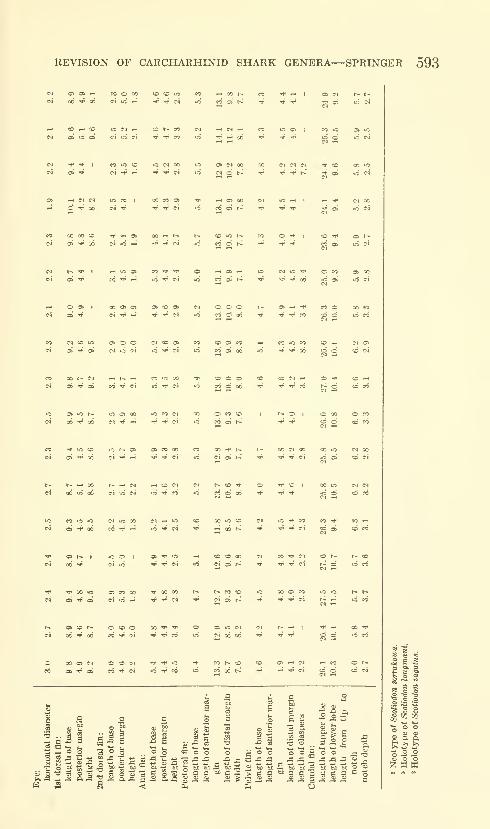
REVISION OF CARCHARHINID SHARK GENERA—SPRINGER 593
Ci 06*^06 c4iC--4 '^•^C^ IC CCGSt^
w COWCD OCIf-t COI^ffO (M f-IC^^ ec ICClC4 oiiOOS ci»Oci TjJrJ-« 10 ^—^00 -^ -flP-^
M Tf ^ ^ -^ r-
^ CD
i" o>
^ C * 00
<o 10 t^
C-1 »o •-*
CO -^
M h- * I w O 1-1 0> -H
ui « cri t-i
-H O 0> o> » o>
W rr -H
01 o oc^ io c*i
CO 0> CO -1
« o> 06 »o
CO ^CO oO) 1-1
00 I- CJ 1 (^ w
o ai 00
oi ^ ^O 5«5 CO
CO oi t^
O 00
CO o
t^^oo r^^c^ i-icoc^061006 C^OC^ lO-'J'fO
O f CO I 00 wto cfCI ^
1-1 O T»" C-l
00 >o CO
-H 06 r-
CO CO 00
c^ o> t^
»o CI
06 00
00 >o
i-j ci
CI O)
CD CI
O CO
CO CO
J- >0 CD
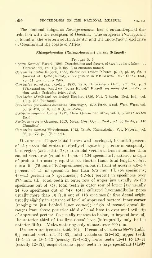
594 PROCEEDINGS OF THE NATIONAL MUSEUM vol. 115
The nominal subgenus Rhizoprionodon has a cu"Cumtropical dis-
tribution with the exception of Oceania. The subgenus Protosygaena
is found in the western south Atlantic and the Indo-Pacific exclusive
of Oceania and the coasts of Africa.
Rhisoprionodon (Rhizoprionodon) aciitus (Riippell)
Figures 5, 6
"Sorra Kowah" Russell, 1803, Descriptions and figures of two hundred fishes . . .
Coromandel, vol. l,p. 9, fig. 15 (a common name).
Carcharias acutus Riippell, 1835, Fische des rothen Meeres, p. 65, pi. 18, fig. 4
(market at Djetta; lectotype designation in Klausewdtz, 1960, Senck. Biol.,
vol. 41, nos. 5, 6, p. 292).
Carcharias sorrakowa Bleeker, 1853, Verh. Bataviaasch Gen., vol. 25, p. 9
(Vizagapatam, based on "Sorra Kowah" Russell; see nomenclatural discus-
sion under Scoliodon laticaudus).
Carcharias (Scoliodon) walbeehmi Bleeker, 1856, Nat. Tijdschr. Ned. Ind., vol.
10, p. 253 (Bintang).
Carcharias (Scoliodon) crenidens Klunzinger, 1879, Sitzb. Akad. Wiss. Wien, vol.
80, p. 426, pi. 8, fig. 3 (Queensland).
Scoliodon longmani Ogilby, 1912, Mem. Queensland Mus., vol. 1, p. 30 (Moreton
Bay).
Scoliodon vagatus Garman, 1913, Mem. Mus. Comp. Zool., vol. 36 (text), p. 116
(Zanzibar),
Carcharias eumeces Pietschmann, 1913, Jahrb. Nassauischen Ver. Naturk., vol.
66, p. 172, p. 1 (Bibundi).
Diagnosis.—Upper labial furrow well developed, 1.4 to 2.0 percent
of t.l. ; precaudal centra markedly elongate in posterior monospondy-
lous region (as in plate 2a);precaudal vertebrae less in number than
caudal vertebrae (equal in 1 out of 124 specimens) ; anterior margin
of pectoral fin usually equal to, or shorter than, total length of first
dorsal fin (79 out of 102 specimens); snout in front of nostrils 4.6-5.4
percent of t.l. in specimens less than 575 mm. t.l. (54 specimens;
4.0-4.5 percent in 8 specimens); 4.2-5.1 percent in specimens over
575 mm. t.l.; total teeth in outer row of upper jaw usually 25 (63
specimens out of 75); total teeth in outer row of lower jaw usually
24 (66 specimens out of 74); total enlarged hyomandibular pores
usually more than 16 (110 out of 118 specimens); first dorsal origin
usually slightly in advance of level of appressed pectoral inner corner
(ranging to just behind inner corner); origin of second dorsal fin
ranges from above posterior third of anal base to over anal axil; tip
of appressed pectoral fin usually reaches to below, or beyond level of,
the anterior third of the first dorsal base (infrequently only to the
anterior fifth). Males maturing only at sizes over 600 mm.Description (see also table 10).—Precaudal vertebrae 55-79 (table
9); caudal vertebrae 64-83; total vertebrae 121-162; upper teeth
11-1-11 to 13-1-13 (usually 12-1-12); lower teeth 11-11 to 13-13
(usually 12-12); cusps of some upper teeth in large specimens faintly
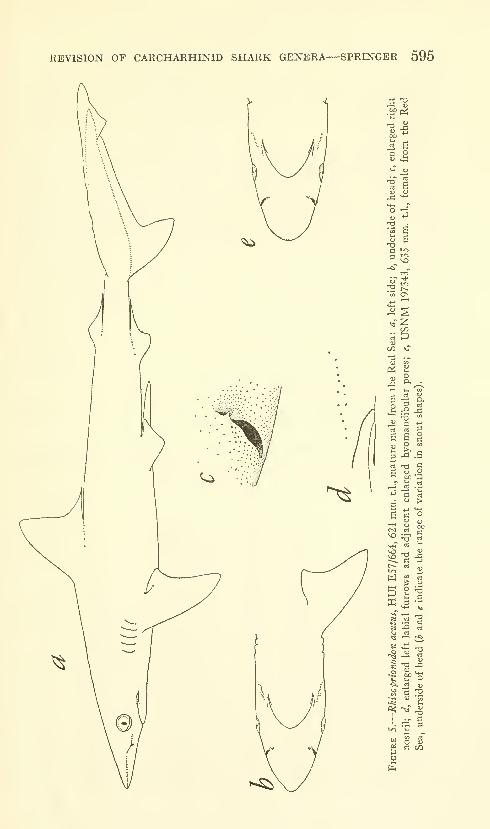
REVISION OF CARCHARHINID SHARK GENERA—SPRINGER 595
.£f P^
a o
73. t~~CO OS
Pi
t« ni gsagt. p "
e« -a SS iJ .2
W
H o
K .3
"3 IS
•2 w)
fn

596 PROCEEDINGS OF THE NATIONAL MUSEUM vol. 115
to moderately serrulate; cusps of lower teeth adumbrating this con-
dition; dentition of mature males and females similar; sometimes
cusps of teeth of mature males slightly more erect than those of
females.
Denticles imbricate, 3-ridged and 3-toothed in young, 3- to 5-ridged
and 3-toothed or with irregular posterior margin in adults.
Color of preserved specimens quite variable. Body gray, purpHsh
gray, gray brown or buff brown above, pale below. Pectorals dark
with a pale distal edge; pelvics and anal pale or dusky; upper caudal
lobe usually with a dark edging in young; edging present or absent
in adults; lower caudal lobe without a dark edging; other fins uni-
formly dark.
A low and poorly developed interdorsal ridge present or absent.
Both precaudal pits present, the upper better developed.
Growth changes: There is a general tendency for distance from
snout tip to outer nostrils, eye, mouth, and pectoral origin to decrease
in percent of t.l. with increased t.l. ; eye diameter, second dorsal base,
and caudal notch depth decrease similarly. Distance from snout
tip to second dorsal origin, anal origin, upper and lower caudal lobe
origins tend to increase in percent of t.l. with increased t.l.
In large specimens there is a tendency for the axis of the upper
caudal lobe to become raised in relation to the horizontal axis of the
body.
The smallest mature male examined was 624 mm. t.l. from Eritrea.
In general, males apparently mature at smaller sizes in the Red Sea
than elsewhere, as seen in following tabulation:
Locality
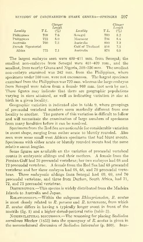
REVISION OF CARCHARHUSriD SHARK GENERA—SPRINGER 597
ClasperLength
Locality T.L. (%) Locality T.L.

598 PROCEEDINGS OF THE NATIONAL MUSEUM vol. 115
much as Bleeker's name is based on a Russell (1803) species for which
no type material is available, I here designate SU 30502, a 368 mm.immature male, as neotype of Scoliodon sorrakowa (label data with
the specimen is as follows: "Scoliodon walbeehmi, ]\Iadras Presidency,
India. Coll. Madras Fisheries Dept., 1923, Herre 1934 Expeditions.
Vizagapatam, Research. Madras, 1933")- The neotype is from the
same general locality as the holotype and it conforms to Russell's
figure and description except that it is approximately 9 mm. longer
than the holotype.
I have examined all the type material of the nominal forms placed
into the synonymy of R. acvtus and find no reason for maintaining
any as distinct from that species.
Material.^—madeira: ISZZ 7593 (1: 415 mm.). Senegal:
MNHN A8005 (1: ca. 435); Goree, IFAN [no number] (2: 409-411,
embryos), IFAN [no number] (1 : 789), ZSZM 6288 (1 : 401) ; St. Louis,
NMV [no number] (1 : 490) ; Dakar, IRSN 8.395 (1 : 696) : Joal, USNM196156 (1: 354, embryo). French guinea: Sobane, USNM 196155
(1: 409); Konkoure, Mundung, ISH 296/59 (2: 426-435): TamaraIsland, IRSN 6.907 (1: 516). Liberia: Kru Station, USNM 179719
(1: 387); Bushrod Island Beach, USNM 179718 (1: 678). Ghana:
Elmina, Ashantee, USNM 42247 (1: ca. 310), USNM 42212 (1: ca.
320): Prampram, BMNH 1939.7.12.1 (1: ca. 770): Accra, BMNH1930.3.24.1 (1: 609). Nigeria: Lagos, BMNH 1937.4.19.1-2 (2:
308-352), BMNH 1936.8.20.1 (1: 477). Cameroon: Bibundi, SMW931 (1: ca. 495, holotype of Carcharlas eumeces), NMV [no number]
(1: ca. 488). French equatorial africa: 29 miles south of CapeLopez, MRAC 80253 (1: ca. 856), MRAC 80253 (1: 775); 11 miles
south of Cape Lopez, IRSN 8.391 (1: 823). angola: 20 miles west
northwest of Moanda, IRSN 8.393 (1 : 445) ; 12 miles west of Moanda,
MRAC 80252 (1: 466); south of Safaire, MRAC 80172 (1: 356); 8
miles west of Rio Cuanza, IRSN 8.390 (1 : ca. 810). union of south
AFRICA: Durban, ANSP 73239 (1: 692), UND 82A-C (3 embryos).
PORTUGUESE EAST AFRICA: Dclagoa Bay, ANSP 55297 (1 : 355). Mozam-
bique: Beira, DIRU 12 (1: 489). Madagascar: east coast, ZSZM7379 (1 : ca. 378) : Tamatave, NMV [no number] (1 : 510). Zanzibar:
MCZ 401 (1: 817, holotype of Scoliodon vagatus), MCZ 24 (1 : ca. 610).
GULF OF ADEN: Aden Harbor, RNH 12384 (2: 310-331). red sea:
USNM 47603 (1: 553). eritrea: HUI E57/660 (1: 730, and three
embryos), HUI E57/658 (1 : 649), HUI E57/662 (1 : 646), HUI E57/661
(1: 669), HUI E57/659 (1: 721), HUI E57/663 (1: 678), HUI E57/666
(1: 445), HUI E57/664 (1: 624); near Massawa, USNM 197343 (1:
635); Hanfilah, Hasein Island [Dahlak Archipelago], NMV [no num-ber] (1: 761). SAUDI ARABIA: Djetta [Jidda], NFIS 2783 (1: ca. 440,
lectotype of Carcharias acutus, stuffed), NFIS 761 (2: 442-448).

REVISION OF CARCHARHINID SHARK GENERA—SPRINGER 599
3 ^
1-4-»

600 PROCEEDINGS OF THE NATIONAL MUSEUM vol. 115
SUEZ: NMV [no number] (1: 560). gulf of oman: Muscat, BMNH1892.1.16.9-10 (1: 706). Persian gulf: Ras el Mustaf, UZMKCN4 (1 : 510) ; Jabrin, UZMK CN3 (1 : 500) ; Tarut Bay, Zaal Island,
USNM 148103 (1: 658), USNM 148104 (1: 690, and two embryos).
INDIA: Malabar, BMNH 1889.2.1.4167-9 (1: 302); Calicut, SU41986, in part (1: 371); Krusdai Island, Gulf of Manar, SU 41985
(1: 378); Madras, NFIS 4027 (1: 365), RNH 8575 (1: 341); Madras
Presidency, SU 30502 (1 : 368, neotype of Scoliodon sorrakowa);
Pondicherry, MNHN 946, in part (1: 448); 50 miles from Ceylon,
NMV [no number], in part (1: 338). Andaman islands: BMNH1870.6.14.21 (1: 360). Thailand: NMV [no nmnber] (2: 335-362);
Bangkok, UZMK P0521 (1: 404): 13°09-13' N. and 100°52-55' E.,
3-4 miles offshore, GVF 1557 (1: ca. 840); 12°30-40' N. and 101°00-
25' E., GVF 1565 (2: 816-882). malaya: Singapore, NMV [no num-ber] (1: 317), BMNH [no number] (1: 360). bintang island:
BMNH 1867.11.28.191 (1: 440, female, holotype of Carcharias
{Scoliodon) walbeehmi; no locality listed with specimen, but Bleeker
described the species from a female, 453 mm., from Bintang); Rio,
RNH 7368 (1: 421, female listed as holotype of C. (S.) walbeehmi,
but I consider the difference in lengths too great to recognize this
specimen as holotype). Sumatra: Padang, NMV [no number]
(1: ca. 790, and three embryos), macassar: BMNH 1872.3.12.3
(1: 794). batjan [bachan] island: ISZZ 7674 (1: 321). aru
islands: near Meriri, NFIS 4026 (1: 450). Australia: Northern
Territory: Little Lagoon, northwest end of Groote Eylandt, Gulf of
Carpentaria, USNM 174076 (1: 500); Bay of Yirrkalla, Northwest
of Cape Arnhem, USNM 174077 (1: 785); Queensland: SMNS 2449
(1: ca. 575, holotype of Carcharias crenidens); Townsville, QMB16190 (1: 672); Cape Cleveland, QMB 17034 (1: 874); Salamander
Rocks, QMB 17136 (1 : 440) ; Lindeman Island, AMS IA6159 (1 : 760),
AMS IA6681 (1: ca. 205, embryo); Brisbane, USNM 176744 (1: 800);
Moreton Bay, QMB 112/292 (1: 455, holotype of Scoliodon longmani),
AMS 112621 (1: 337); Moreton Island, DHMB 368 (1: 520); Decep-
tion Bay, QMB 18241 (1: 368), 18240 (1: 342). Philippines: MCZ484 (1: 284): Sitankai, SU 13725 (1: 686); south Tumindao Lagoon,
USNM 151235 (1: 716); Mindanao, SU 13152 (1: 722); Panay:
Iloilo, SU 14454 (1: ca. 410); Capiz, SU 26862 (1: ca. 280); Unisan,
Tayabas, SU 26820 (1: ca. 298); Luzon: Cavite, SU 9639 (2; 271-
342); Orion, Bataan Province, ANSP 52798 (1: 481), ANSP 86371
(1: 242); Manila, UZMK 385 (1: 358), UZMK 386 (1: 277), SU20592 (3: 28.3-296); Manila Bay: SU 26818 (3: 283-291), CNHM46987 (1: 335), CNHM 46993 (1: 350), UWS 8568 (1: 299), SU29621 (4: 258-304); La Monja Island, USNM 151234 (1: 709);
ManHa, fish market, USNM 151236 (2: 470-519), USNM 151238
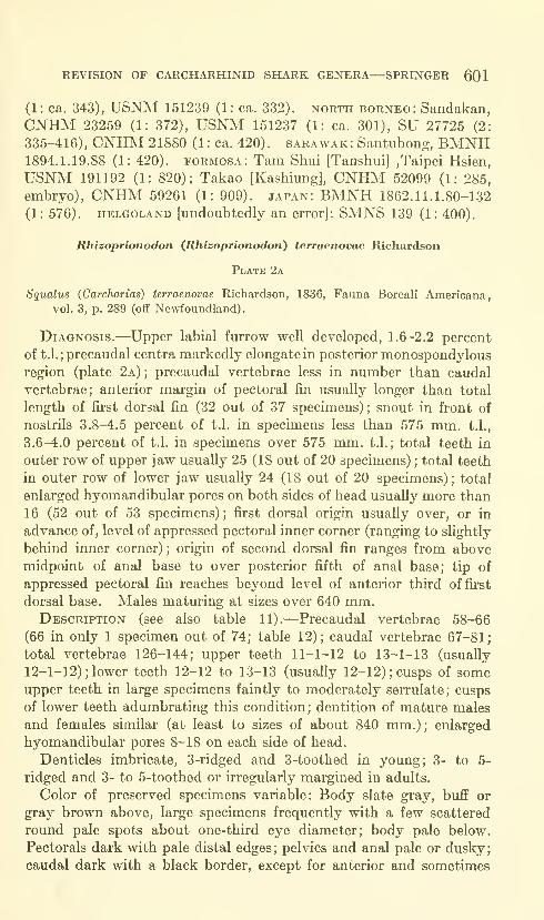
REVISION OF CARCHARHINID SHARK GENERA—SPRINGER 601
(1: ca. 343), USNM 151239 (1: ca. 332). north Borneo: Sandakan,
CNHM 23259 (1: 372), USNM 151237 (1: ca. 301), SU 27725 (2:
335-416), CNHM 21880 (1 : ca. 420). Sarawak: Santubong, BMNH1894.1.19.88 (1: 420). Formosa: Tam Shui [Tanshui] ,Taipei Hsien,
USNM 191192 (1: 820); Takao [Kashiung], CNHM 52099 (1: 285,
embryo), CNHM 59261 (1: 909). japan: BMNH 1862.11.1.80-132
(1: 576). HELGOLAND [undoubtedly an error]: SMNS 139 (1: 400).
Rhizoprionodon (Rhizoprionodon) terraenovae Richardson
Plate 2a
Squalus (Carcharias) terraenovae Richardson, 1836, Fauna Boreali Americana,vol. 3, p. 289 (oCF Newfoundland).
Diagnosis.—Upper labial furrow well developed, 1.6-2.2 percent
of t.l.; precaudal centra markedly elongate in posterior monospondylousregion (plate 2a)
;precaudal vertebrae less in number than caudal
vertebrae; anterior margin of pectoral fin usually longer than total
length of first dorsal fin (32 out of 37 specimens); snout in front of
nostrils 3.8-4.5 percent of t.l. in specimens less than 575 mm. t.l.,
3.6-4.0 percent of t.l. in specimens over 575 mm. t.l.; total teeth in
outer row of upper jaw usually 25 (18 out of 20 specimens) ; total teeth
in outer row of lower jaw usually 24 (18 out of 20 specimens) ; total
enlarged hyomandibular pores on both sides of head usually more than
16 (52 out of 53 specimens) ; first dorsal origin usually over, or in
advance of, level of appressed pectoral inner corner (ranging to slightly
behind inner corner) ; origin of second dorsal fin ranges from abovemidpoint of anal base to over posterior fifth of anal base; tip of
appressed pectoral fin reaches beyond level of anterior third offii'st
dorsal base. Males maturing at sizes over 640 mm.Description (see also table 11).—Precaudal vertebrae 58-66
(66 in only 1 specimen out of 74; table 12); caudal vertebrae 67-81;
total vertebrae 126-144; upper teeth 11-1-12 to 13-1-13 (usually
12-1-12) ; lower teeth 12-12 to 13-13 (usually 12-12); cusps of someupper teeth in large specimens faintly to moderately serrulate; cusps
of lower teeth adumbrating this condition; dentition of mature males
and females similar (at least to sizes of about 840 mm.); enlarged
hyomandibular pores 8-18 on each side of head.
Denticles imbricate, 3-ridged and 3-toothed in young; 3- to 5-
ridged and 3- to 5-toothed or irregularly margined in adults.
Color of preserved specimens variable: Body slate gray, buff or
gray brown above, large specimens frequently with a few scattered
round pale spots about one-third eye diameter; body pale below.
Pectorals dark with pale distal edges; pelvies and anal pale or dusky;
caudal dark with a black border, except for anterior and sometimes
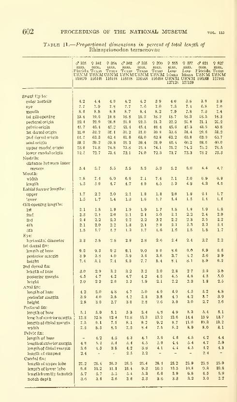
602 PROCEEDINGS OF THE NATIONAL MUSEUM
Table 11.
—
Proportional dimensions in percent of total length ofRhizoprionodon terraenovae
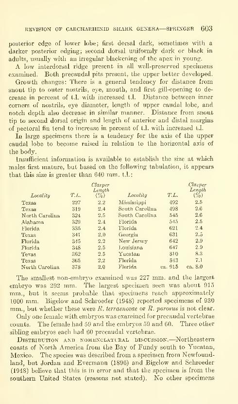
REVISION OF CARCHARHINID SHARK GENERA—SPRINGER 603
posterior edge of lower lobe; first dorsal dark, sometimes with a
darker posterior edging; second dorsal uniformly dark or black in
adults, usually with an irregular blackening of the apex in young.
A low interdorsal ridge present in all well-preserved specimens
examined. Both precaudal pits present, the upper better developed.
Growth changes: There is a general tendency for distance from
snout tip to outer nostrils, eye, mouth, and first gill-opening to de-
crease in percent of t.l. with increased t.l. Distance between inner
corners of nostrils, eye diameter, length of upper caudal lobe, and
notch depth also decrease in similar manner. Distance from snout
tip to second dorsal origin and length of anterior and distal margins
of pectoral fin tend to increase in percent of t.l, with increased t.l.
In large specimens there is a tendency for the axis of the upper
caudal lobe to become raised in relation to the horizontal axis of
the body,
InsuflBcient information is available to estabhsh the size at which
males first mature, but based on the following tabulation, it appears
that this size is greater than 640 mm. t.l.
:
Locality
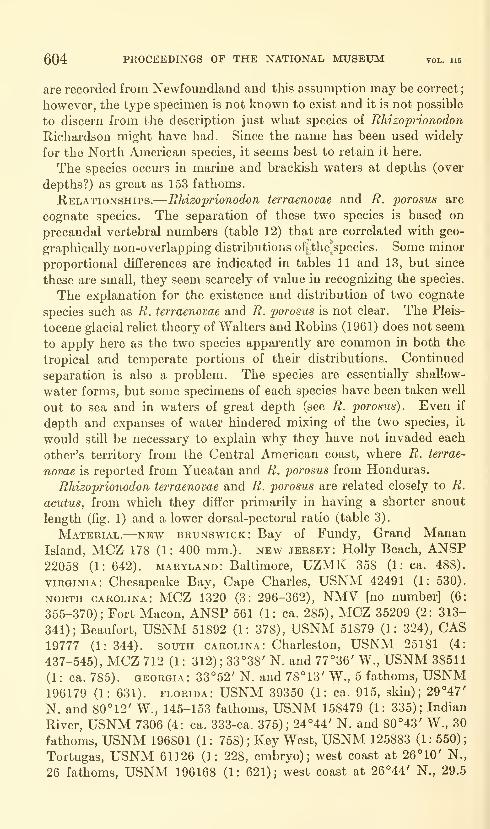
604 PROCEEDINGS OF THE NATIONAL MUSEUM vol. 115
are recorded from Newfoundland and this assumption may be correct
;
however, the type specimen is not known to exist and it is not possible
to discern from the description just what species of Rhizoprionodon
Richardson might have had. Since the name has been used widely
for the North American species, it seems best to retain it here.
The species occurs in marine and brackish waters at depths (over
depths?) as great as 153 fathoms.
Relationships.—Rhizoprionodon terraenovae and R. porosus are
cognate species. The separation of these two species is based on
precaudal vertebral numbers (table 12) that are correlated with geo-
graphically non-overlapping distributions ofithe*species. Some minor
proportional differences are indicated in tables 11 and 13, but since
these are small, they seem scarcely of value in recognizing the species.
The explanation for the existence and distribution of two cognate
species such as R. terraenovae and R. porosus is not clear. The Pleis-
tocene glacial rehct theory of Walters and Robins (1961) does not seem
to apply here as the two species apparently are common in both the
tropical and temperate portions of their distributions. Continued
separation is also a problem. The species are essentially shallow-
water forms, but some specimens of each species have been taken well
out to sea and in waters of great depth (see R. porosus). Even if
depth and expanses of water hindered mixing of the two species, it
would still be necessary to explain why they have not invaded each
other's territory from the Central American coast, where R. terrae-
novae is reported from Yucatan and R. porosus from Honduras.
Rhizoprionodon terraenovae and R. porosus are related closely to R.
acutus, from which they differ primarily in having a shorter snout
length (fig. 1) and a lower dorsal-pectoral ratio (table 3).
Material.—new Brunswick: Bay of Fundy, Grand MananIsland, MCZ 178 (1: 400 mm.), new jersey: Holly Beach, ANSP22058 (1: 642). Maryland: Baltimore, UZMK 358 (1: ca. 488).
VIRGINIA: Chesapeake Bay, Cape Charles, USNM 42491 (1: 530).
NORTH Carolina: MCZ 1320 (3: 296-362), NMV [no number] (6:
355-370); Fort Macon, ANSP 561 (1: ca. 285), MCZ 35209 (2: 313-
341); Beaufort, USNM 51892 (1: 378), USNM 51879 (1: 324), CAS19777 (1: 344). south Carolina: Charleston, USNM 25181 (4:
437-545), MCZ 712 (1 : 312) ;33°38' N. and 77°36' W., USNM 38511
(1: ca. 785). Georgia: 33°52' N. and 78°13' W., 5 fathoms, USNM196179 (1: 631). Florida: USNM 39350 (1: ca. 915, skin); 29°47'
N. and 80°12' W., 145-153 fathoms, USNM 158479 (1: 335); Indian
River, USNM 7306 (4: ca. 333-ca. 375); 24°44' N. and 80°43' W., 30
fathoms, USNM 196801 (1: 758); Key West, USNM 125883 (1: 550);
Tortugas, USNM 61126 (1: 228, embryo); west coast at 26°10' N.,
26 fathoms, USNM 196168 (1: 621); west coast at 26°44' N., 29.5
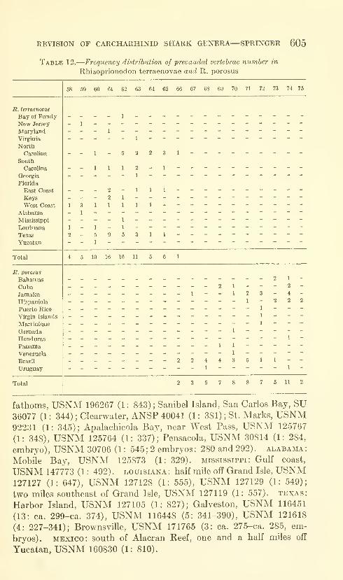
REVISION OF CARCHARHINID SHARK GENERA—SPRINGER 605
Table 12.
—
Frequency distribution of precaudal vertebrae number in
Rhizoprionodon terraenovae and R. porosus
R. terraenovae
Bay of FundyNew Jersey
Maryland
Virginia
North
Carolina
South
Carolina
Georgia
Florida
East Coast
KeysWest Coast
AlabamaMississippi
Louisiana
Texas
Yucatan
58 59 60 61 62 63 64 65 66 67 68 69 70 71 72 73 74 75
1
1
1 1
3 1
1
1 3
- 1
1
Total 10 16 16
R. porosus
BahamasCubaJamaica
Hispaniola
Puerto Rico
Virgin Islands
Martinique
Grenada
HondurasPanamaVenezuela
Brazil
Uruguay
1
1 2
1
2 1
2
4
2 2
2 2 4 4 3 5 1 1 -
_ - 1 _ - _ - - 1
Total 2 3 5 7 7 6 11 2
fathoms, USNM 196267 (1 : 843) ; Sanibel Island, San Carlos Bay, SU36077 (1 : 344) ; Clearwater, ANSP 40041 (1 : 381) ; St. Marks, USNM92231 (1: 345); Apalachicola Bay, near West Pass, USNM 125767
(1: 348), USNM 125764 (1: 337); Pensacola, USNM 30814 (1: 284,
embryo), USNM 30706 (1 : 545; 2 embryos: 280 and 292). Alabama:
Mobile Bay, USNM 125873 (1: 329). Mississippi: Gulf coast,
USNM 147773 (1 : 492) . Louisiana : half mile off Grand Isle, USNM127127 (1: 647), USNM 127128 (1: 555), USNM 127129 (1: 549);
two miles southeast of Grand Isle, USNM 127119 (1: 557). texas:
Harbor Island, USNM 127105 (1: 827); Galveston, USNM 116451
(13: ca. 299-ca. 374), USNM 116448 (5: 341-390), USNM 121618
(4: 227-341); Brownsville, USNM 171765 (3: ca. 275-ca. 285, em-
bryos). MEXICO: south of Alacran Reef, one and a half miles off
Yucatan, USNM 160830 (1: 810).
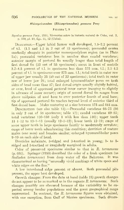
606 PROCEEDINGS OF THE NATIONAL MUSEUM vol. us
Rhizoprionodon (Rhizoprionodon) porosus Poey
FiGUKES 7, 8
Squalus porosus Poey, 1861, Memorias sobre la historia natural de Cuba, vol. 2,
p. 339, pi. 19, figs. 11, 12 (Cuba).
Diagnosis.—Upper labial furrow well developed, 1.8-2.3 percent
of t.l. (1.3 and 1.5 in 2 out of 19 specimens); precaudal centra
markedly elongate in posterior monospondylous region (as in Plate
2a);precaudal vertebrae less in number than caudal vertebrae
;
anterior margin of pectoral fin usually longer than total length of
first dorsal fin (32 out of 35 specimens) ; snout in front of nostrils
3.7-4.5 percent of t.l. in specimens less than 575 mm. t.l.; 3.3-4.3
percent of t.l. in specimens over 575 mm. t.l.; total teeth in outer row
of upper jaw usually 25 (19 out of 22 specimens) ; total teeth in outer
row of lower jaw 24; total enlarged hyomandibular pores on both
sides of head more than 17; first dorsal origin usually slightly behind,
or over, level of appressed pectoral inner corner (ranging to slightly
in advance of inner corner) ; origin of second dorsal fin ranges from
above midpoint of anal base to over posterior fourth of anal base;
tip of appressed pectoral fin reaches beyond level of anterior third of
first dorsal base. Males maturing at a size between 575 and 644 mm.Description (see also table 13).—Precaudal vertebrae 66-75 (66
in only 2 out of 58 specimens; table 12); caudal vertebrae 69-85;
total vertebrae 136-159 (only 1 with less than 140); upper teeth
11-1-12 to 13-1-13 (usually 12-1-12); lower teeth 12-12; cusps of
some upper teeth in large specimens faintlj^ to moderately serrulate;
cusps of lower teeth adumbrating this condition; dentition of mature
males (one seen) and females similar; enlarged hyomandibular pores
9-19 on each side of head.
Denticles imbricate, 3-ridged and 3-toothed in young; 3- to 5-
ridged and 3-toothed or irregularly margined in adults.
Color of preserved specimens similar to that in R. terraenovae
(p. 601). Springer (1950) described the color of a fresh specimen (as
Scoliodon terraenovae) from deep water off the Bahamas. It was
characterized as having "unusually vivid markings of white spots and
white edges on the fins."
A low interdorsal ridge present or absent. Both precaudal pits
present, the upper best developed.
Growth changes: From the data at hand (table 13) growth changes
do not appear to be as marked as in the cognate R. terraenovae. Such
changes possibly are obscured because of the variability to be ex-
pected among insular populations and the great geographical range
represented. In contrast, the R. terraenovae figures were obtained,
with one exception, from GuK of Mexico specimens. Such dimen-

REVISION OF CARCHARHINID SHARK GENERA—SPRINGER 607
Table 13.
—
Proportional dimensions in percent of total length of
Rhizoprionodon porosus
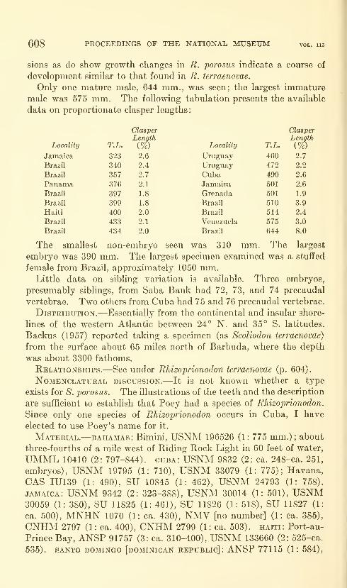
608 PROCEEDINGS OF THE NATIONAL MUSEUM
sions as do show growth changes in R. porosus indicate a course of
development similar to that found in R. terraenovae.
Only one mature male, 644 mm., was seen; the largest immature
male was 575 mm. The following tabulation presents the available
data on proportionate clasper lengths
:
Locality
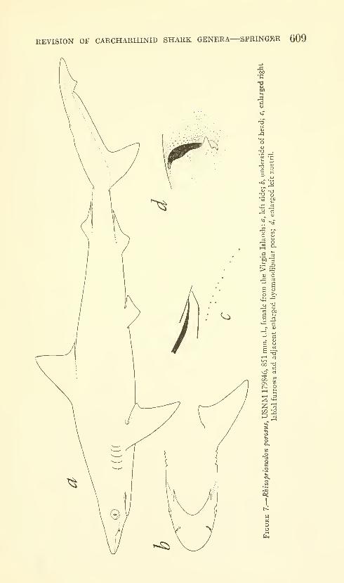
REVISION OF CARCHARHINID SHARK GENERA—SPRINGER 609
•--
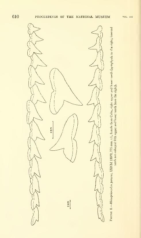
610 PROCEEDINGS OF THE NATIONAL MUSEUM
u %
£ 9-
. *
as c
o<
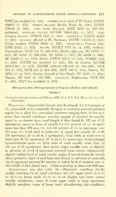
REVISION OF CARCHARHINID SHARK GENERA—SPRINGER 611
NMV [no number] (1 : 410). Puerto rico: west of El Mario, USNM196613 (1: 472). virgin islands: Booby Rock, St. John, USNM179846 (1: 851). saba bank [island]: RNH 9270 (3: 380-390,
embryos). Grenada island: BMNH 1904.6.24.1 (1: 501). Mar-
tinique island: MNHN 1143 (1: 444). Honduras: USNM 44470
(1 : 303) . PANAMA : off reef at Ft. Sherman, AMNH 1 1413B (1 : 333)
;
Col6n, market, USNM 79324 (1: 376). Venezuela: Caracas Bay,
RNH 23321 (1: 575). brazil: MNHN 3467 (1: ca. 1050, stuffed);
Pernambuco, MCZ 714 (2: 340-376); Recife, high sea, SU 52747 (1:
343), SU 52857 (2: 390-434), SU 52858 (1: 397), SU 52859 (1: 366),
SU 52860 (1: ca. 482); Bahia, USNM 43357 (1: 510), MNHN 1144
(1: 644), BMNH [no number] (1: 432); Rio de Janeiro, BMNH1923.7.30.1 (1: 415), BMNH 1903.6.9.130-132 (2: 488-540), MCZ720 (1: 357), MCZ 432 (2: 413-476), MCZ 160 (1: 514); Marica, SU52748 (1 : ca. 388) ; Santos, Httoral of Sao Paulo, SU 52861 (1 : 433)
;
Maceio, SU 8446 (2: 370-399). Uruguay: Maldonado, MCZ 525
(1: 460), NMV [no number] (1: 472).
Rhizoprionodon {Rhizoprionodon) longurio (Jordan and Gilbert)
Figure 9
Carcharias longurio Jordan and Gilbert, 1882, Proc. U.S. Nat. Mus., vol. 5, p. 106
(Mazatldn).
Diagnosis.—Upper labial furrow well developed, 2.1-2.6 percent of
t.l.;precaudal centra markedly elongate in posterior monospondylous
region (as in plate 2a); precaudal vertebrae ranging from 12 less to 8
more than caudal vertebrae; anterior margin of pectoral fin usually
equal to, or shorter than, total length of first dorsal fin (27 out of 32
specimens); snout in front of nostrils 5.1-6.0 percent of t.l. in speci-
mens less than 575 mm. t.l., 4.5-5.0 percent of t.l. in specimens over
575 mm. t.l.; total teeth in outer row of upper jaw usually 27 or 29
(29 specimens; 28 or 30 in 3 specimens); total teeth in outer row of
lower jaw 26 (21 specimens; 27 or 28 in 11 specimens); total enlarged
hyomandibular pores on both sides of head usually more than 16
(32 out of 33 specimens) ; first dorsal origin usually over, or slightly
in advance of, level of appressed pectoral inner corner, infrequently
slightly behind level of inner corner; origin of second dorsal fin occurs
above posterior thkd of anal base and always in advance of anal axil;
tip of appressed pectoral fin reaches to below level of anterior one- to
two-thirds of first dorsal base. Males maturing at sizes over 600 mm.Description (see also table 14).—Precaudal vertebrae 68-86;
caudal vertebrae 73-85; total vertebrae 146-167; upper teeth 13-1-13
to 15-1-14; lower teeth 13-13 to 14-14 (higher and lower counts
usually correlated) ; cusps of some upper teeth in large specimens
slightly serrulate; cusps of lower teeth adumbrating this condition;
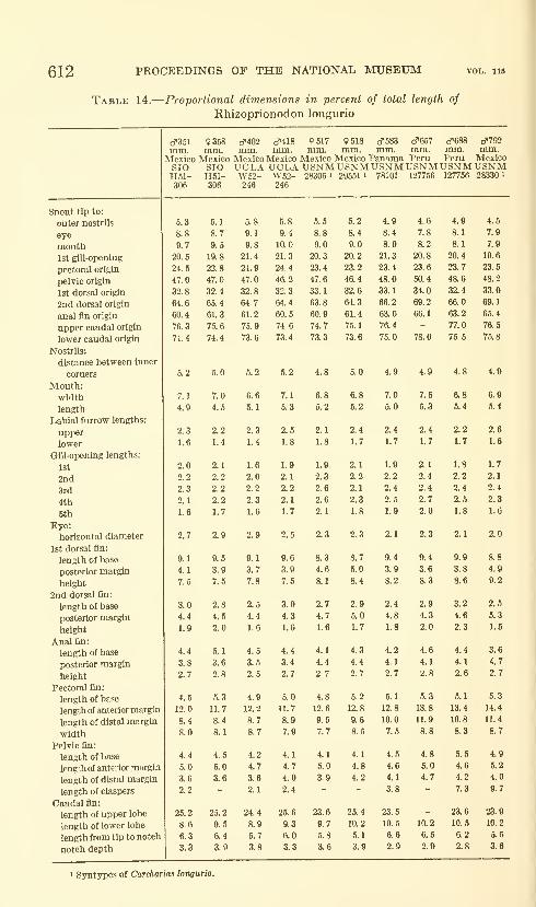
612 PROCEEDINGS OF THE NATIONAL MUSEUM
Table 14.
—
Proportional dimensions in percent of total length of
Rhizoprionodon longurio
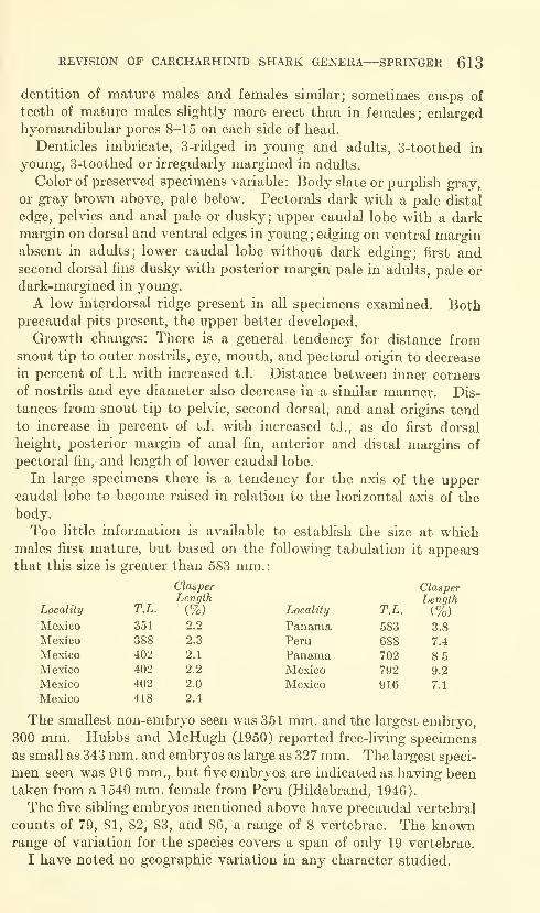
REVISION OF CARCHARHINID SHARK GENERA—SPRINGER 613
dentition of mature males and females similar; sometimes cusps of
teeth of mature males slightly more erect than in females; enlarged
hyomandibular pores 8-15 on each side of head.
Denticles imbricate, 3-ridged in young and adults, 3-toothed in
young, 3-toothed or irregularly margined in adults.
Color of preserved specimens variable: Body slate or purplish gray,
or gray brown above, pale below. Pectorals dark with a pale distal
edge, pelvies and anal pale or dusky; upper caudal lobe with a darkmargin on dorsal and ventral edges in young ; edging on ventral marginabsent in adults; lower caudal lobe without dark edging; first andsecond dorsal fins dusky with posterior margin pale in adults, pale or
dark-margined in young.
A low interdorsal ridge present in all specimens examined. Bothprecaudal pits present, the upper better developed.
Growth changes: There is a general tendency for distance fromsnout tip to outer nostrils, eye, mouth, and pectoral origin to decrease
in percent of t.l. with increased t.l. Distance between inner corners
of nostrils and eye diameter also decrease in a similar manner. Dis-
tances from snout tip to pelvic, second dorsal, and anal origins tend
to increase in percent of t.l. with increased t.l., as do first dorsal
height, posterior margin of anal fin, anterior and distal margins of
pectoral fin, and length of lower caudal lobe.
In large specimens there is a tendency for the axis of the uppercaudal lobe to become raised in relation to the horizontal axis of the
body.
Too little information is available to establish the size at whichmales fu'st mature, but based on the following tabulation it appears
that this size is greater than 583 mm.:
Locality
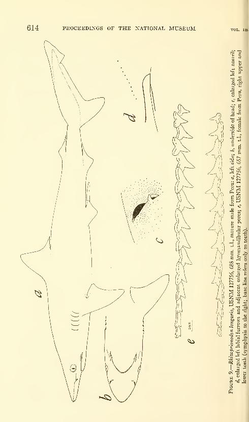
614 PROCEEDINGS OF THE NATIONAL MUSEUM
ID

REVISION OF CARCHARHINID SHARK GENERA—SPRINGER 615
Distribution.—^Eastern Pacific Ocean from Peru to southern
California, from waters as deep as 15 fathoms.
Relationships.—Within the subgenus Rhizoprionodon, R. longurio
forms a separate offshoot from the remaining three species, differing
mainly in its high tooth counts, long snout, and upper labial furrow
lengths.
Material.—Mexico: Baja California: San Juanico Bay, USNM196122 (1: 388 mm.), USNM 190594 (2: 402-403), UCLA W52-246(12: 385-466), SIO H51-306 (2: 351-358); Concepcion Bay, USNM46852 (1: 916); beach one-half mile south of San Felipe, USNM190590(1: 385);Sonora: Salinas Bay, SU 17329 (2: 398-460) ; Mazat-
lan, USNM 28306 (1: 517, syntype of Carcharias longurio), USNM28330 (1: 792, syntype of Carcharias longurio), USNM 29551 (1:518,
syntype of Carcharias longurio), SU 11594 (1: ca. 685). panama:
NMV [no number] (1 : 525) ; 1-3 miles southeast of Isla Taboga, 2-4
miles off Rio Pacora, UCLA W53-273 (4: 301-349); Panama City,
USNM 78101 (1: 583); Panama City, fish market, USNM 79291
(1 : 702). PERU: Gulf of Guayaquil: 81°13' W. and 4°13' S., USNM127756 (2: 657-688); off Mt. Organos, Cabo Blanco, USNM 127776
(5: 282-300, embryos from 1540 mm. female}.
Rhizoprionodon (Protozygaena) lalandei Valenciennes
Figures 10, 11; Plate 2b
Carcharias (ScoUodon) lalandii Valenciennes in Miiller and Henle, 1841, Sys-
tematische Bescbreibung der Plagiostomen, p. 30 (Rio de Janeiro, Martinique,
Guadeloupe).
ScoUodon intermedins Garman, 1913, Mem. Mus. Comp. Zool., vol. 36 (text),
p. 115 (Philippines, East Indies).
Diagnosis.—Upper labial furrow well developed, 1.4-2.1 percent
of t.l.;precaudal centra slightly elongate in posterior monospondylous
region (plate 2 b); precaudal vertebrae greater in number than caudal
vertebrae; anterior margin of pectoral fin shorter than total length
of first dorsal fin (equal to, in 1 out of 33 specimens) ; snout in front
of nostrils 4.4-5.1 percent of t.l. in specimens less than 575 mm.t.L, 4.4-4.7 percent of t.l. in specimens over 575 mm. t.l.; total teeth
in outer row of upper jaw 25; total teeth in outer row of lower jaw
24 (23 in 2 out of 21 specimens); total enlarged hyomandibular pores
on both sides of head usually more than 16 (25 out of 26 specimens);
first dorsal origin usually over level of appressed pectoral inner corner,
ranging to just behind inner corner; origin of second dorsal fin usually
over anal axil but may occur as far forward as above posterior fourth
of anal base; tip of appressed pectoral fin reaches to below level of
anterior third or less of first dorsal base. Males matming at about
500 mm.Description (see table 15).—Precaudal vertebrae 79-90 (only
4 specimens out of 45 with less than 85); caudal vertebrae 67-79;

616 PROCEEDINGS OF THE NATIONAL MUSEUM
Table 15.
—
Proportional dimensions in percent of total length of
Rhizoprionodon lalandei
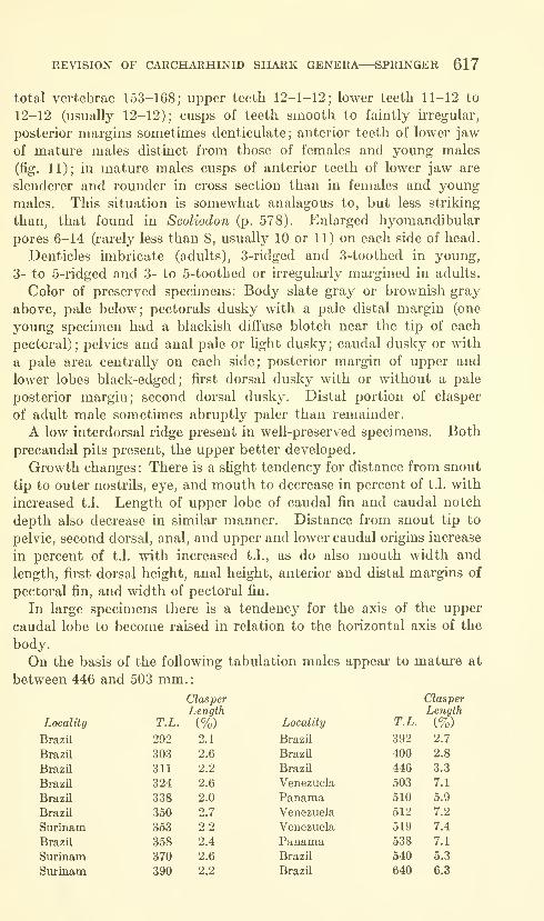
REVISION OF CARCHARHINID SHARK GENERA—SPRINGER 617
total vertebrae 153-168; upper teeth 12-1-12; lower teeth 11-12 to
12-12 (usually 12-12); cusps of teeth smooth to faintly irregular,
posterior margins sometimes denticulate; anterior teeth of lower jaw
of mature males distinct from those of females and young males
(fig. 11); in mature males cusps of anterior teeth of lower jaw are
slenderer and rounder in cross section than in females and young
males. This situation is somewhat analagous to, but less striking
than, that found in Scoliodon (p. 578). Enlarged hyomandibular
pores 6-14 (rarely less than 8, usually 10 or 11) on each side of head.
Denticles imbricate (adults), 3-ridged and 3-toothed in young,
3- to 5-ridged and 3- to 5-toothed or kregularly margined in adults.
Color of preserved specimens: Body slate gray or brownish gray
above, pale below; pectorals dusky with a pale distal margin (one
young specimen had a blackish diffuse blotch near the tip of each
pectoral); pelvics and anal pale or light dusky; caudal dusky or with
a pale area centrally on each side; posterior margin of upper and
lower lobes black-edged; first dorsal dusky with or without a pale
posterior margin; second dorsal dusky. Distal portion of clasper
of adult male sometimes abruptly paler than remainder.
A low interdorsal ridge present in well-preserved specimens. Both
precaudal pits present, the upper better developed.
Growth changes : There is a slight tendency for distance from snout
tip to outer nostrils, eye, and mouth to decrease in percent of t.l. with
increased t.l. Length of upper lobe of caudal fin and caudal notch
depth also decrease in similar manner. Distance from snout tip to
pelvic, second dorsal, anal, and upper and lower caudal origins increase
in percent of t.l. with increased t.l., as do also mouth width and
length, first dorsal height, anal height, anterior and distal margins of
pectoral fin, and width of pectoral fin.
In large specimens there is a tendency for the axis of the upper
caudal lobe to become raised in relation to the horizontal axis of the
body.
On the basis of the following tabulation males appear to mature at
between 446 and 503 mm.
:
Locality
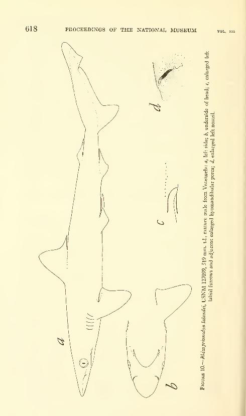
618 PROCEEDINGS OF THE NATIONAL MUSEUM
'e 'B
O J^
iT
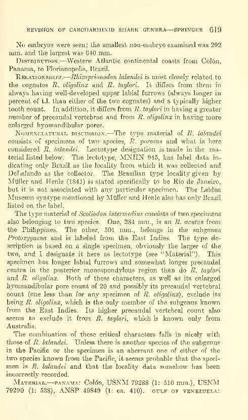
REVISION OF CARCHARHINID SHARK GENERA—SPRINGER 619
No embryos were seen; the smallest non-embryo examined was 292
mm. and the largest was 640 mm.Distribution.—Western Atlantic continental coasts from Colon,
Panama, to Florianopolis, Brazil.
Relationships.—Rhizoprionodon lalandei is most closely related to
the cognates R. oligolinx and R. taylori. It differs from them in
always having well-developed upper labial furrows (always longer in
percent of t.l. than either of the two cognates) and a typically higher
tooth count. In addition, it differs from R. taylori in having a greater
number of precaudal vertebrae and from R. oligolinx in having moreenlarged hyomandibular pores.
NoMENCLATURAL DISCUSSION.—-The tvpc material of R. lalandei
consists of specimens of two species, R. porosus and what is here
considered R. lalandei. Lectotype designation is made in the ma-terial listed below. The lectotype, MNHN 945, has label data in-
dicating only Brazil as the locality from which it was collected andDeLalande as the collector. The Brazilian type locality given byMiiller and Henle (1841) is stated specifically to be Rio de Janeiro,
but it is not associated with any particular specimen. The Leiden
Museum syntj^pe mentioned by Muller and Henle also has only Brazil
listed on the label.
The type material of Scoliodon intermedins consists of two specimens
also belonging to two species. One, 284 mm., is an R. acutus from
the Philippines. The other, 501 mm., belongs in the subgenus
Protozygaena and is labeled from the East Indies. The type de-
scription is based on a single specimen, obviously the larger of the
two, and I designate it here as lectotype (see "Material")- This
specimen has longer labial furrows and somewhat longer precaudal
centra in the posterior monospondylous region than do R. taylori
and R. oligolinx. Both of these characters, as well as its enlarged
hyomandibular pore count of 20 and possibly its precaudal vertebral
count (one less than for any specimen of R. oligolinx), exclude its
being R. oligolinx, which is the only member of the subgenus knownfrom the East Indies. Its higher precaudal vertebral count also
seems to exclude it from R. taylori, which is known only from
Australia.
The combination of these critical characters falls in nicely with
those of R. lalandei. Unless there is another species of the subgenus
in the Pacific or the specimen is an aberrant one of either of the
two species known from the Pacific, it seems probable that the speci-
men is R. lalandei and that the locality data somehow has been
incorrectly recorded.
Material.—PANAMA: Col6n, USNM 79288 (1: 510 mm.), USNM79290 (1: 538), ANSP 49849 (1: ca. 410). gulf of Venezuela:
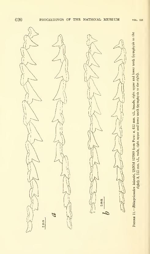
620 PROCEEDINGS OF THE NATIONAL MUSEUM
^ -^
6 U
3
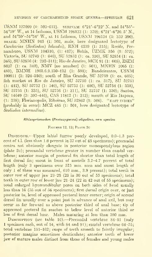
REVISION OF CARCHARHINID SHARK GENERA—SPRINGER 621
USNM 127099 (4: 503-612). Surinam: 6°24'-6°22' N. and 54°55'-
54°59' W., at 14 fathoms, USNM 196133 (1: 370); 6°21'-6°20. 5' N.,
and 54°59'-54°54' W., at 14 fathoms, USNM 196134 (3: 352-390).
brazil: MNHN 945 (1: 303, male, here designated lectotype of
Carcharias (Scoliodon) lalandei), RNH 4339 (1: 315); Recife, Per-
nambuco, USNM 104310, (1: 417); Bahia, UZMK 356 (1: 572);
Victoria, SU 52749 (1: 640), SU 52853 (1: ca. 330), SU 52854 (1: ca.
365), SU 52856 (4: 292-311); Rio de Janeiro, MCZ 91 (1: 605),ZSZM8037 (1: ca. 340), NMV [no nmnber] (1: 501), MNHN 1065 (1:
489), BMNH 1903.6.9.130-132 (1: 591); Bandeirantes, USNM100811 (3: 324-350); south of Ilha Grande, SU 52759 (1: ca. 605);
fish market at Rio de Janeiro, SU 52750 (1: ca. 317), SU 52751
(1: 412), SU 52752 (1: 540), SU 52753 (1: 400), SU 52754 (1: 338),
SU 52755 (1: 335), SU 52756 (1: 311), SU 52757 (1: 330); Santos,
SU 14049 (2: 392-446), CAS 11812 (1: 313); mouth of Rio Itapoca,
(1: 358); Florianopohs, Ribeirao, SU 52862 (1: 506). "east indies"
[probably in error]: MCZ 485 (1: 501, here designated lectotype of
Scoliodon intermedins) .
Rhizoprionodon (Protozygaena) oligolinx, new species
Figures 12, 13; Plate 2c
Diagnosis.—Upper labial furrow poorly developed, 0.0-1.3 per-
cent of t.l. (less than 1.0 percent in 37 out of 45 specimens);precaudal
centra not obviously elongate in posterior monospondylous region
(plate 2c); precaudal vertebrae greater in number than caudal ver-
tebrae; anterior margin of pectoral fin shorter than total length of
first dorsal fin; snout in front of nostrils 3.7-4.7 percent of total
length (only 3 specimens over 575 mm. seen and snout length of
only 1 of these was measured, 610 mm., 3.9 percent); total teeth in
outer row of upper jaw 23-25 (23 in 30 out of 55 specimens); total
teeth in outer row of lower jaw 21-24 (22 in 42 out of 55 specimens);
total enlarged hyomandibular pores on both sides of head usually
less than 16 (55 out of 56 specimens); first dorsal origin over, or just
posterior to, level of appressed pectoral inner corner ; origin of second
dorsal fin usually over a point just in advance of anal axil, but mayoccur as far forward as above posterior third of anal base; tip of
appressed pectoral fin reaches to below level of anterior third or
less of first dorsal base. Males maturing at less than 380 mm.Description (see table 16).—Precaudal vertebrae 84-91 (only
1 specimen each, out of 54, with 84 and 91); caudal vertebrae 64-75;
total vertebrae 151-162; cusps of teeth smooth to faintly irregular;
posterior margins sometimes denticulate; anterior teeth of lower
jaw of mature males distinct from those of females and young males
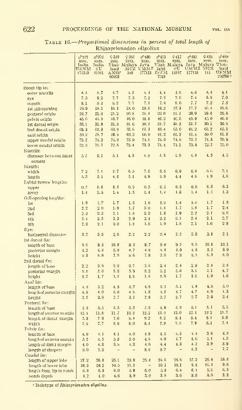
622 PROCEEDINGS OF THE NATIONAL MUSEUM
Table 16.
—
Proportional dimensions in percent of total length of
Rhizoprionodon oligolinx

REVISION OF CARCHARHINID SHARK GENERA—SPRINGER 623
(fig. 13); in mature males the cusps of tlie anterior teeth of the
lower jaw (and sometimes upper) are slenderer and rounder in cross
section than in females and young males. This situation is some-
what analogous, but less striking than that found in Scoliodon (p. 578).
Enlarged hyomandibular pores 3-8 (usually 4-7) on each side of head.
Denticles imbricate, 3-ridged and 3-toothed in young, 3- to 5-
ridged and 3- to 5-toothed or irregularly margined in adults.
Color of preserved specimens: Body slate gray or brownish
gray above, pale below; pectorals dusky with a pale margin; pelvics
and anal pale or light dusky; caudal uniformly dusky or with a pale
area centrally on each side; margins of upper caudal lobe black to
dusky-edged; first dorsal with or without a pale or faintly dusky
posterior margin; second dorsal dusky. Distal portion of clasper of
adult male sometimes abruptly paler than remainder.
Interdorsal ridge, when present, very faint. Both precaudal
pits present, the upper better developed.
Growth changes: There is a slight tendency for distance from
snout tip to eye, mouth, first gill-opening, and pectoral origin to
decrease in percent of t.l. with increased t.l. Eye diameter and
caudal notch depth decrease similarly. There is a slight tendency
for snout tip to anal origin and upper and lower caudal lobe origins
to increase in percent of t.l with increased t.l.
In large specimens there is a tendency for the axis of the upper
caudal lobe to become raised in relation to the horizontal axis of the
body.
The following tabulation indicates that males have mature claspers
at between 287 and 380 mm.; however, in at least one male, 387 mm.,
with mature claspers, dentition was still sexually undifTerentiated.
Locality
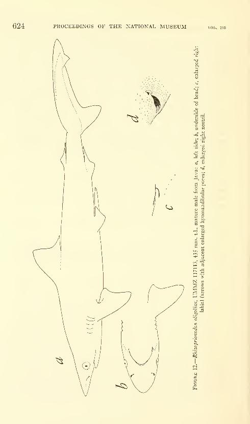
624 PROCEEDINGS OF THE NATIONAL MUSEUM
a^
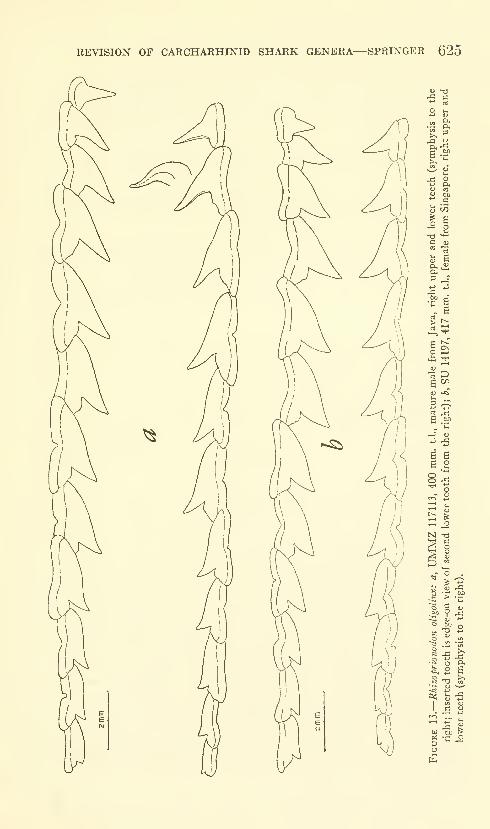
REVISION OF CARCHARHINID SHARK GENERA—SPRINGER 625
"-^
•fl C
J OJ
5 CO
fe a
j3
> ^I—i^
o 2
1-1 Vi"
6 S
^ o
o
•S
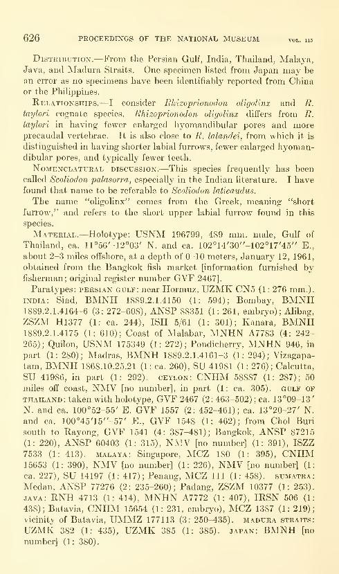
626 PROCEEDINGS OF THE NATIONAL MUSEUM vol. 115
DisTEiBUTiON.—From the Persian Gulf, India, Thailand, Malaya,Java, and Madura Straits. One specimen listed from Japan may be
an error as no specimens have been identifiably reported from Chinaor the Philippines.
Relationships.—I consider Ehizoprionodon oligolinx and R.
taylori cognate species. Ehizoprionodon oligolinx differs from R.
taylori in having fewer enlarged hyomandibular pores and moreprecaudal vertebrae. It is also close to R. lalandei, from which it is
distinguished in having shorter labial furrows, fewer enlarged hyoman-dibular pores, and typically fewer teeth.
NoMENCLATURAL DISCUSSION.—This species frequently has been
called Scoliodon palasorra, especially in the Indian literature. I havefound that name to be referable to Scoliodon laticaudus.
The name "oligolinx" comes from the Greek, meaning "short
furrow," and refers to the short upper labial furrow found in this
species.
Material.—Holot^'pe: USNM 196799, 489 mm. male. Gulf of
Thailand, ca. 11°56'-12°03' N. and ca. 102°14'30"-102°17'45" E.,
about 2-3 miles offshore, at a depth of 0-10 meters, January 12, 1961,
obtained from the Bangkok fish market [information furnished byfisherman; original register nmnber GVF 2467].
Paratypes: Persian gulf: near Hormuz, UZMK CN5 (1 : 276 mm.).
INDIA: Sind, BMNH 1889.2.1.4150 (1: 594); Bombay, BMNH1889.2.1.4164-6 (3: 272-608), ANSP 88351 (1: 261, embryo); Alibag,
ZSZM H1377 (1: ca. 244), ISH 5/61 (1: 301); Kanara, BMNH1889.2.1.4175 (1: 610); Coast of Malabar, MNHN A7783 (4: 242-
265); Quilon, USNM 175349 (1: 272); Pondicherry, MNHN 946, in
part (1: 280); Madras, BMNH 1889.2.1.4161-3 (1: 294); Vizagapa-
tam, BMNH 1868.10.25.21 (1: ca. 260), SU 41981 (1: 276); Calcutta,
SU 41986, in part (1: 292). ceylon: CNHM 58887 (1: 287); 50
miles off coast, NMV [no number], in part (1: ca. 305). gulf of
THAILAND: taken with holotype, GVF 2467 (2: 463-502) ; ca. 13°09-13'
N. and ca. 100°52-55' E. GVF 1557 (2: 452-461); ca. 13°20-27' N.
and ca. 100°45'15"-57' E., GVF 1548 (1: 462); from Choi Buri
south to Rayong, GVF 1541 (4: 387-481); Bangkok, ANSP 87215
(1: 220), ANSP 60403 (1: 315), NMV [no number] (1: 391), ISZZ7533 (1: 413). malaya: Singapore, MCZ 180 (1: 395), CNHM15653 (1: 390), NMV [no number] (1: 226), NMV [no number] (1
ca. 227), SU 14197 (1: 417); Penang, MCZ 111 (1: 458). SumatraMedan, ANSP 77276 (2: 235-260); Padang, ZSZM 10377 (1: 253)
JAVA: RNH 4713 (1: 414), MNHN A7772 (1: 407), IRSN 506 (1
438); Batavia, CNHM 15654 (1: 231, embryo), MCZ 1387 (1: 219)
vicinity of Batavia, UMMZ 177113 (3: 250-435). madura straits
UZMK 382 (1: 435), UZMK 385 (1: 385). japan: BMNH [no
number] (1: 380).

REVISION OF CARCHARHINID SHARK GENERA—SPRINGER 627
Additional material : east indian archipelago: BMNH [no num-
ber] (1 : 430) ; USNM 40029 (1 : 355; this specimen is listed in the cata-
log as questionably from Queensland, Australia; I believe it did not
come from Australia).
Rhizoprionodon (Protozygaena) taylori (Ogilby)
Figure 14
Physodon taylori Ogilby, 1915, Mem. Queensland Mus., vol. 3, p. 117 (Towns-
ville, north Queensland).
Diagnosis.—(Not until this study was in an advanced stage was it
realized that R. taylori and R. oligolinx were separate species; the
description that follows is deficient because most of the material
examined was not available to me during the writing.) Upper labial
furrow poorly developed, 0.7-1.1 percent of t.l.; precaudal centra not
obviously elongate in posterior monospondylous region (as in plate
2c);precaudal vertebrae greater in number than caudal vertebrae
;
anterior margin of pectoral fin shorter than total length of first dorsal
fin; snout in front of nostrils 4.0-5.0 percent of t.l. in specimens less
than 575 mm. t.l. (5 specimens measured), 4.2 percent of t.l. in speci-
mens over 575 mm. t.l. (2 specimens measured) ; total teeth in outer
row of upper jaw 24-25 (24 in 8 out of 9 specimens); total teeth in
outer row of lower jaw 21-23 (22 in 8 out of 9 specimens); total en-
larged hyomandibular pores on both sides of head usually more than
16 (6 out of 8 specimens) ; first dorsal fin origin over level of appressed
pectoral inner corner; origin of second dorsal fin ranges from over
posterior fourth to over posterior sixth of anal base; tip of appresssed
pectoral fin reaches to below level of anterior third or less of first
dorsal base. Only two males seen, ca. 310 and 407 mm. t.l., both
immature; clasper length in latter 4.8 percent of t.l.
Description (see table 17).—Precaudal vertebrae 73-80; caudal
vertebrae 62-70; total vertebrae 135-149; cusps of teeth smooth to
faintly irregular; posterior bases sometimes denticulate; enlarged
hyomandibular pores 7-1 1 on each side of head.
Denticles imbricate, 3- to 5-ridged and 3-toothed or irregularly
margined (based only on USNM 174075).
Color of preserved specimen (USNM 174075): Body brownish gray
above, pale below. Pectorals darker than body color with a pale
distal margin; pelvics and anal somewhat lighter than pectorals;
caudal paler than body, without black edging on upper lobe; pale
posterior edge on lower lobe; first and second dorsal fins about same
color as body dorsally.
Interdorsal ridge present or absent. Both precaudal pits present,
the upper better developed.
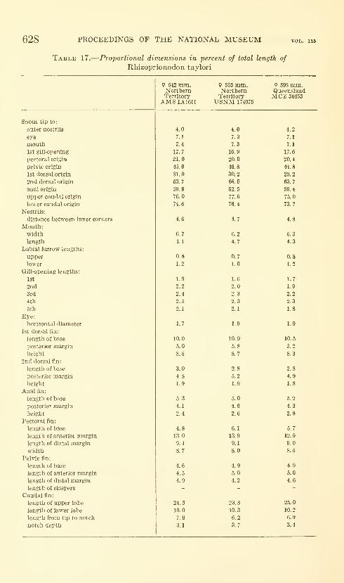
628 PROCEEDINGS OF THE NATIONAL MUSEUM
Table 17.
—
Proportional dimensions in percent of total length of
Rliizoprionodon taylori
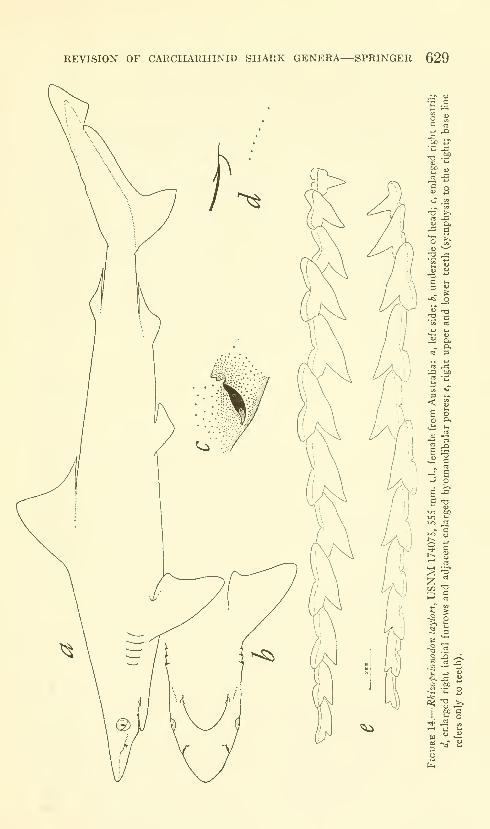
REVISION OF CARCHARHINID SHARK GENERA—SPRINGER 629
'u
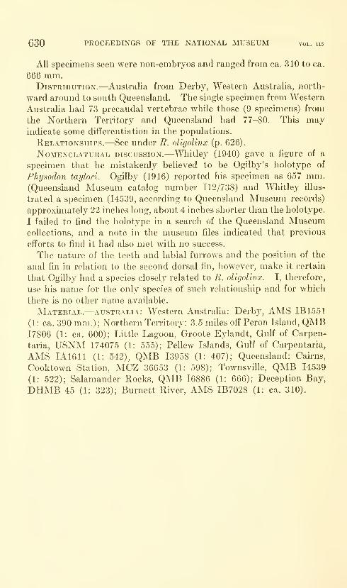
630 PROCEEDINGS OF THE NATIONAL MUSEUM vol. us
All specimens seen were non-embryos and ranged from ca. 310 to ca.
666 mm.Distribution.—Australia from Derby, Western Australia, north-
ward around to south Queensland. The single specimen from Western
Australia had 73 precaudal vertebrae while those (9 specimens) from
the Northern Territory and Queensland had 77-80. This mayindicate some differentiation in the populations.
Relationships.—See under R. oligolinx (p. 626).
NoMENCLATURAL DISCUSSION.—Wliitlcy (1940) gave a figure of a
specimen that he mistakenly beheved to be Ogilby's holotype of
Physodon taylori. Ogilby (1916) reported his specimen as 657 mm.(Queensland Museum catalog number 112/738) and Wliitley illus-
trated a specimen (14539, according to Queensland Museum records)
approximately 22 inches long, about 4 inches shorter than the holotype.
I failed to find the holotype in a search of the Queensland Museumcollections, and a note in the museum files indicated that previous
efforts to find it had also met with no success.
The nature of the teeth and labial furrows and the position of the
anal fin in relation to the second dorsal fin, however, make it certain
that Ogilby had a species closely related to R. oligolinx. I, therefore,
use his name for the only species of such relationship and for which
there is no other name available.
Material.—Australia: Western Australia: Derby, AMS IB1551
(1: ca. 390 mm.); Northern Territory: 3.5 miles off Peron Island, QMB17806 (1: ca. 600); Little Lagoon, Groote Eylandt, Gulf of Carpen-
taria, USNM 174075 (1: 555); Pellew Islands, Gulf of Carpentaria,
AMS IA1611 (1: 542), QMB 13958 (1: 407); Queensland: Cairns,
Cooktown Station, MCZ 36653 (1: 598); Townsville, QMB 14539
(1: 522); Salamander Rocks, QMB 16886 (1: 666); Deception Bay,
DHMB 45 (1: 323); Burnett River, AMS IB7028 (1: ca. 310).
Literature Cited
Backus, Richard H.
1957. Notes on western Atlantic sharks. Copeia, 1957, no. 3, pp. 246-248.
BiGELOw, Henry B., and Schroeder, W. C.
1948. Sharks, in Fishes of the western North Atlantic, part 1. Mem.Sears Found. Mar. Res., no. 1, pp. 59-546.
1957. A study of the sharks of the suborder Squaloidea. Bull. Mus. Comp.
Zool., vol. 117, no. 1, pp. 1-150.
Bleekeb, Pieter1852. Bijdrage tot de kennis der Plagiostomen van den Indischen archipel.
Verb. Bataviaasch Gen., vol. 24, pp. 1-92.
1853. Bijdrage tot de kennis der Troskieuwige visschen van den Indischen
archipel. Verh. Bataviaasch Gen., vol. 25, pp. 1-30.
1878. Quatrieme memoire sur la faune ichthyologique de la Nouvelle-
Guinee. Arch. Neerl. Sci. Nat., vol. 13, pp. 35-66.
1879. Sur quelques especes inedites ou peu connues de poissons de Chine
appartenant au Museum de Hambourg. Verh. Akad. Amsterdam,
vol. 18, pp. 1-17.
Cuvier, G. L. F. C. D.
1829. Le Regno animal, 2nd ed., vol. 2, pp. 1-406.
Fowler, Henry W.1936. The marine fishes of West Africa. Bull. Amer. Mus. Nat. Hist.,
vol. 70, pt. 1, pp. 1-605.
Garrick, John A. F.
1980. Studies on New Zealand elasmobranchii, part 11: Squaloids of the
genera Deania, EUnopterus, Oxynotus, and Dalatias in New Zealand
waters. Trans. Roy. Soc. New Zealand, vol. 88, no. 3, pp. 489-517.
Gill, Theodore N.
1862. Analytical synopsis of the order of Squali; and revision of the nomen-
clature of the genera. Ann. Lye. Nat. Hist. New York, vol. 8, pp.
367-413.
Gunther, Albert1870. Catalogue of the fishes in the British Museum, vol. 8, pp. 1-549.
Heath, Gordon W.1960. The anatomy and growth pattern of the claspers and siphon sacs of
the spiny dogfish, Squalus acanthias Linnaeus, and the smooth
dogfish, Mustelus canis Mitchill. Unpubl. doct. dissert., Cornell
Univ., 366 pp.
Hildebrand, Samuel F.
1946. A descriptive catalog of the shore fishes of Peru. U.S. Nat. Mus.,
Bull. 189, pp. 1-530.
HuBBs, Carl L., and McHugh, J. L.
1950. Pacific sharpnose shark (Scoliodon longurio) in California and Baja
California. California Fish and Game, vol. 36, no. 1, pp. 7-11.
Klausewitz, Wolfgang1960. Die Typen und Typoide des Naturmuseums Senckenberg, 23: Pisces,
Chondrichthyes, Elasmobranchii. Senck. Biol., vol. 41, nos. 6/6,
pp. 289-296.
631
632 PROCEEDINGS OF THE NATIONAL MUSEUM vol. us
MtJLLER, Johannes, and Hewle, F. G. J.
1841. Systematische Beschreibung der Plagiostomen, pp. 1-204, 60 pis.
Ogilby, J, Douglas1916. Checklist of the cephalochordates, selachians, and fishes of Queens-
land. Mem. Queensland Mus., vol. 5, pp. 70-98.
Russell, Patrick1803. Descriptions and figures of two hundred fishes collected at Vizagapa-
tam on the coast of Coromandel, vol. 1, pp. 1-78, 100 pis.
Setna, S. B., and Sarangdhar, P, N.
1949. Descriptions, bionomics and development of Scoliodon sorrakowah
(Cuvier). Rec. Indian Mus., vol. 46, no. 1, pp. 25-54.
1950. A contribution to the systematics of Scoliodon acutus (Riippell),
Hemipristis elongahis (Klunzinger) and Torpedo zugniayeri Engle-
hart. Rec. Indian Mus., vol. 47, no. 1, pp. 125-134.
Springer, Stewart1950. A revision of North American sharks allied to the genus Carcharhinus.
Amer. Mus. Nov., no. 1451, pp. 1-13.
Thillayampalam, E. M.1929. Scoliodon (the common shark of the Indian seas). Indian Zool.
Mem., no. 2, pp. 1-116.
Walters, Vladimir, and Robins, C. Richard1961. A new toadfish (Batrachoididae) considered to be a glacial relict in
the West Indies. Amer. Mus. Nov., no. 2047, pp. 1-24.
Wheeler, John F. G.
1959. Sharks of the western Indian Ocean, 1: Loxodon macrorhinus M. andH. East African Agric. Journ., vol. 25, no. 2, pp. 106-109.
Whitley, Gilbert P.
1940. The fishes of Australia, part 1: The sharks, rays, devil-fish, and other
primitive fishes of Australia and New Zealand, pp. 1-280.
U.S. GOVERNMENT PRINTrNG 0FFICE:I96<

INDEX*
(New genera, species, etc., are printed in iialics. Page numbers of principal entries
also in italics.)
Ablabesmyia, subg., 86, 87, 103Acacia, 260Acalvpta, 331, 332, 334 (key)
barberi, 332, 334, 340, 342cooleyi, 334, 337duryi, 334, 335, 336, 337
lilHani's, 331, 334, 341, 342madelinae, 338mera, 340mniophila, 331, 334, 337modcsta, 341nvctalis, 331, 334, 339ovata, 335, 341saundersi, 334, 335, 336, 337sautcri, 336, 337thomsonii, 331, 334, 33Svanduzeei, 33.5, 342vandykei, 335, 343
Acalypta (Hemiptera: Tingidae), Ameri-can species of the lacebug genus,331-345
acantliias, Squalus, 566Acanthosoma, 226Acanthurus dussumieri, 164, 178, 233
guttatus, 171, 178mata, 233nigroris, 211olivaceous, 178, 179, 203, 233, 356triostegus sandvicensis, 137, 149,
156, 164, 178, 179, 203, 211, 212,233, 356
xanthopterus, 211
aclines, Anatopynia, 104, 105 (fig.), 109
Acocoelidia, 274unipuncta, 274
acutus, Carcharias, 589, 590, 594, 598Rhizoprionodon, 569 (table), 570
(table), 571, 580, 590, 591 (table),
592 (table), 595 (figs.), 599 (figs.),
604, 619Rhizoprionodon (Rhizoprionodon),594
Adele, 292Adiantum tenerum, 26Aeaea, 290, 293, 297, 298 (key), 300, 301,
304dulcedo, 298, 300, 301, 304, 323
(fig.), 327 (fig.)
Aeaea—Continuedextensa, 297, 298, 299, 300, 306,
324 (fig.), 327 (fig.)
juvantis, 299, 300, 301, 303, 304, 305,306, 307, 323 (fig.)
ostryaeella, 297, 298, 299, 300, 302,304, 305, 319 (fig.), 320 (fig.), 322(fig.), .326 (fig.)
purpuriella, 315quadricristatella, 303quadricustatella, 298, 299, 300, 303,
328 (fig.)
rhynchosiae, 297, 298, 299, 300, 305,324 (fig.), 326 (fig.)
risor, 299, 300, 304, 307, 324 (fig.)
sagayia, 299, 300, 305, 324 (fig.)
stellans, 300stipator, 293, 298, 299, 300, 303,
323 (fig.), 326 (fig.)
venatrix, 299, 300, 301, 323 (fig.)
327 (fig.)
venifica, 298, 299, 300, 301, 323(fig.)
victor, 298, 299, 300, 304, 322 (fig.),
Aeaea sp. a, 307, 327 (fig.)
Aeaea sp. b, 307, 327 (fig.)
Aeaea sp. c, 307, 328 (fig.), 329 (fig.)
Aeaea sp. d, 290, 308, 327 (fig.)
Aeaea sp. e, 308, 328 (fig.)
Aeaea sp. f, 308, 328 (fig.)
Aeaea, unnamed sp., 307aequalis, Alpheus, 447, 451, 455
Betaeus, 447"aequalis," Betaeus, 432"aequimanus," Betaeus, 432aesiocopia, Gonioterma, 383, 384, 387
(fig.), 388 (fig.)
Stenoma, 384affinis, Scoliodon, 588, 589alascensis, Anatopynia, 109alba, Pentaneura, 89albilus, Stichaster, 43albitalus, Procladius, 122albula, Stepganasterias, 33 (map), 43albulus, Asteracanthion, 43Alebion glaber, 377algens, Anatopynia, 109algosa, Gonioterma, 383, 385, 387 (fig.),
388 (fig.)
Stenoma, 385
*No. 3492, Emilio Estrada, Betty J. Meggers, and Clifford Evans, The Jambell Culture of South Coastal
Ecuador, pp. 483-558, is indexed separately on pp. 649-654.
633
748-138—65-
634 PROCEEDINGS OF THE NATIONAL MUSEUM
Alpheidae, 431, 434.
Alpheus, 432aequalis, 447, 451, 455(Betaeus) aequalis, 447harfordi, 432, 447loiigidactylus, 441
Altica ludoviciana, 19occiden talis, 15 (fig.)) 19oleracea, 19
Alticinae (Coleoptera), Notes on newand old species of, from theWest Indies, 9-30
Amaurogramma, 297, 298extensa, 297, 306quadricristatella, 303
americana, Pentaneura, 89Amorpha fruticosa, 290amorphella, Walshia, 290Amphicarpa bracteata, 310Amphicarpa sp., 290Amphipods, Seven new, from the west
coast of North America withnotes on some unusual species,391-430
Anatopynia, 109 (key)aclines, I04, 105 (fig.), 109(Macropelopia) aclines, IO4, 105
(fig.)
alaskeusis, 109algens, 109brunnea, 109decolorata, 109discolor, 110dyari, 106, 110(Psectrotanypus) dyari, 106eumorpha, 105 (fig.), 107, 110(Psectrotanypus) eumorpha, 105
(fig-), 107fastuosa, 109florens, 109guttularis, 109hirtipennis, 106, 109johnsoni, 110marginella, 104, 109miripes, 109suhmarginella, 103, 105 (fig.), 109(Anatopynia) suhmarginella, 103,
105 (fig.)
venusta, 108, 109(Psectrotanypus) venusta, 108
Anatopynia, subg., 103, 105 (fig.), 109angulatum, Sagum, 232angusticollis, Longitarsus, 14Anisogammarus confervicolus, 4^3, 424
(fig.), 425 (fig.)
(Eogammarus) confervicolus, 423schmitti, 4^0
anuulatus, Sphaeroides, 178Anonyx, 392
nugax, 392Anthosomatidae, 225, 232Antispila, 292antleri, Cocoelidia, 271, 284 (fig.)
Neocoehdiana, 271
Anuretes, 187, 194brevis, 194heckeli 194menehune, 195, 197 (fig.), 201 (fig.),
202 (table)
renalis, 188serratus, 188, 190 (fig.), 193 (fig.),
194 (table), 199, 202aphrogramma, Gonioterraa, 383
Stenoma, 384Aphthona, 11, 12
crucifera, 9, 10, 13 (fig.)
inornata, 11, 13 (fig.)
insularis, 11, 13 (fig.)
lamprocyanea, 11, \% (fig.)
lepta, 10, 13 (fig.)
maculipenuis, 10apicalis, Pentaneura, 93Aprionodon isodon, 574apua, Mycteroperca venenosa, 178, 347,
358Aradidae, 245, 246Aradidae, Notes on, in the U.S. National
Museum, III: Subfamily Meziri-nae (Hemiptera), 245-258
Arctiidae, 47Arctostaphylos, 260argentea, Sphyraena, 178argicerauna, Gonioterma, 383, 384, 387
(fig.)
Stenoma, 384Arictus, 249arietinus, Podonomus, 127Ascalenia, 292
ceanothiella, 292asphaltina, Haltica, 21
Nesaecredida, 15 (fig.), 21, 22assimilis, Haliotis, 450astera, Cernotina, 476Asteracanthion albulus, 43
groenlandicus, 44linckii, 43polaris, 44
Asterias crispata, 36gunneri, 43militaris, 38papposa, 38stellionura, 43vulgaris, 31, 35
Asterina tumida tuberculata, 36, 37, 38Asteroidea of the Blue Dolphin Ex-
peditions to Labrador, 31-46atypicus, Longitarsus, 13 (fig.), 16Aulostomus chinensis, 363aurantia, Megacoelidia, 267
bairdi, Paramphithoe, 413Balistoides viridescens, 363barbatulus, Procladius, 119 (fig.), 121barberi, Acalvpta, 332, 334, 340, 342
Mezira, 256Pentaneura, 90, 91 (fig.), 93Tanypus, 90
basalis, Systena, 15 (fig.), 18bathyphilia, Prodiamesa, 129
baxteri, Etmopterus, 3
INDEX 635
beUa, Chinaia, 264, 281 (fig.)
bellator, Xiqilliba, 268, 283 (fig.)
bellus, Procladius, 126Taiiypus, 126
Betaeus, 431, 432, 434 (key), 449aequalis, 447"aequalis," 432"aequimanus," 432ensenadensis, 431, 432, 434, 436
(taljle), U5, 462 (fig.), 463 (fig.)
equalis, 432equimanus, 432, 447_gracilis, 432, 434, 437 (table), 453,
464 (fig.), 465 (fig.), 466 (fig.)
harfordi, 431, 432, 433, 434, 437(table), 447, 451, 453, 456, 457,458, 464 (fig.), 465 (fig.), 466(fig.)
harrimani, 431, 433, 434, 435, 436(table), 441, 444, 447, 461 (fig.),
462 (fig.), 463 (fig.)
longidactylus, 431, 432, 433, 434,436 (table), 4U, 447, 462 (fig.),
463 (fig.)
macginitieae, 432, 434, 437 (table),
451, 464 (fig.), 465 (fig.), 466(fig.)
setosus, 432, 434, 437 (table), 455,464 (fig.), 465 (fig.), 466 (fig.)
truncatus, 435Betaeus, new sp., 445Betaeus, subg., 447"Betaeus harfordi," 458Betaeus, Shrimps of the genus, on the
Pacific Coast of North Americawith descriptions of three newspecies, 431-466
bifasciata, Pentaneura, 93
bifurcatus, Dentigryps, 203, 205 (fig.),
209 (fig.), 210 (table), 347, 356(table), 357 (fig.), 366, 368, 369(tables), 370 (tables), 371 (fig.),
372, 373, 374 (table), 375 (fig.),
376 (table), 377, 378Biza, 259, 262, 274
craspa, 280 (fig.), 287 (fig.)
crocea, 274Blake, Doris H., Notes on new and old
species of Alticinae (Coleoptera)from the West Indies, 9-30
Bodianus diplotaenia, 178
bonito, Caligus, 148, 149
Boreochlus persimilis, 127, 131 (fig.)
borneensis, Carcharhinus, 560bourquini, Gonionota, 70, 71 (fig.), 74, 76
brachyptera, Placogenis, 245, 246Brachyrhynchus, 249
rugiventris, 250bracteata, Amphicarpa, 310branicki, Pseudodiamesa, 128, 129
brasiliensis, Pseudoepitrix, 17 (fig.), 24brevis, Anuretes, 194brooksi, Pentaneura, 102
brunnea, Anatopynia, 109
bryophanes, Gonioterma, 383, 385, 387(fig.)
Stenoma, 385bungei, Halirages, 4O6, 407 (fig.)
Cacao, 414lacteus, 414sanguineus, 414
caenothiella, Periploca, 295calamus, Peniculus, 137, 233, 235 (fig.)
californica, Pterygophora, 458californicus, Paralichthys, 178
Rhamnus, 313californiensis, Callianassa, 440, 447CaUgidae, 141 (fig.), 194, 377, 378Cahgoida, 232, 348Caligus, 138, 14I, 171, 177
bonito, 148, 149constrictus, 141, 157, 158, 159 (fig.),
160, 161, 162, 103, 171flexispina, I49, 151 (fig.), 154 (fig.),
156 (table)
hala, 142, 143 (fig.), 147 (fig.), 148(table)
kalumai, 171, 173 (fig.), 177 (table)kuroshio, 148, 149ligatus, 164, 165 (fig.), 170 (fig.),
171 (table)
longicervicis, 171oviceps, 165, 177, 178praetextus, 171punctatus, 177, 178quadratus, 171randalli, 156, 159 (fig.), 162 (fig.),
163 (table)
sphyraenae, 171Callianassa californiensis, 440, 447Calliopiidae, 406canicula, Scyliorhinus, 5canis, Mustelus, 566Caranx melampygus, 351carcharhinid shark genera Scoliodon,
Loxodon, and Rhizoprionodon,A revision of the, 559-632
Carcharhinidae, 559, 573, 574Carcharhinus, 560
borneensis, 560longimanus, 2oxyrhynchus, 574porosus, 560temmincki, 574
Carcharias, 580acutus, 589, 590, 594, 598crenidens, 600(Scoliodon) crenidens, 590, 594dumerili, 589(Scoliodon) dumerili, 588eumeces, 594, 598(Scoliodon) lalandei, 621(Scoliodon) lalandii, 615(Scoliodon) laticaudus, 574, 577,
578, 579, 580longurio, 611, 615(Scoliodon) macrorhynchus, 573,574 579 583
(Physodon) miilleri, 573, 574
636 PROCEEDINGS OF THE NATIONAL MUSEUM
Carcharias—Continuedpalasorra, 574, 579, 590sorrakowah, 579, 580, 594(Scoliodon) walbeehmi, 594, 600
Carcharias, subg., 601Carcharius palasorra, 580
sorrah kowah, 580Caridea, 431carinata, Tingis, 332carinaius, Tanypus, 105 (fig.), HO, 121
carioca, Mezira, 247 (fig.), 265, 256carnea, Pentaneura, 88, 89, 93Caronx melampygus, 351Ceanothiella, Ascalenia, 292
Stagmatophora, 292Ceanothus sp., 290cerifera, Myrica, 10
Cernotiua, 468, 470, 476astera, 476
ceylonensis, Scoliodon, 588, 589Chaetocnema, 23
cyanoptera, 15 (fig.), 23plicipennis, 23
Chaetodon quadrimaculatus, 178cham-pioni, Mezira, 251Chinaia, 259, 260, 261, 264
bella, 264, 281 (fig.)
lepida, 287 (fig.)
chinensis, Aulostomus, 363Chironomid midges of California, II:
Tanypodinae, Podonominae, andDiamesinae, 85-136
Chironomus nivorundus, 130
chlanidotus, Longitarsus, 13 (fig.), / .4, 16chloreus, Procladius, 122, 134chlorina, Gonioterma, SS3, 384, 387
(fig.), 388 (fig.)
Stenoma, 383Cholotis, 292
semnostola, 291, 292choreus, Tanypus, 134christiernana, Plialaena tortrix, 61
Chrysoclista, 312
Clirysopeleia, 293, 297, SI4ostryaeella, 302purpuriella, 290, 314, 315, 319 (fig.),
320 (fig.), 322 (fig.), 326 (fig.),
329 (fig.)
quadricristatella, 303chrysophrys, Lernanthropus, 232
chrysos, Pentaneura, 70, 91 (fig.)
cinereus, Polycentropus, 47S, 479 (fig.)
Cinyphus, 246saileri, 246, 247 (fig.)
squalidus, 249citrinalis, Hypercallia, 61
clara, Stenocoelidia, 263Clarke, J. F. Gates, Neotropical micro-
lepidoptera, III: Restriction of
Gonionata melobaphes Walsing-ham with descriptions of newspecies (Lepidoptera : Oecophori-dae), 61-84
clarkei, Tichocoelidia, 270Clemensia, 48
Cocoelidia, 261, 271antleri, 271, 284 (fig.)
Cocoelidia, subg., 271Coelana, 261, 269 (key)
drakei, 269, 287 (fig.)
modesta, 269, 270, 282 (fig.)
Coelana, subg., 269Coelella, 262, 273
distincta, 274, 285 (fig.)
venosa, 274Coelella, subg., 273Coelidiana, 259, 262, 269, 272, 274, 275
coronata, 275croceata, 275, 286 (fig.)
defila, 272coenosus, Pleuronichthys, 440Coleophoridae, 290Coleoptera, 9colombiana, Xenocoelidia, 266comosa, Pentaneura, 93, 97 (fig.), 102Conchapelopia, 89, 103confervicola, Mara, 423
Melita, 423confervicolus, Anisogammarus, 4^3, 424
(fig.), 425 (fig.)
Gammarus, 423constrictus, Caligus, 141, 157, 158, 159
(fig.), 160, 161, 162, 163, 171contrasta, Gonionota, 64, 65 (fig.), 70cooleyi, Acalypta, 334, 337Copepoda, 348Copepods (Crustacea), Caligoid, of the
Hawaiian Islands: Parasitic onfishes of the family Acanthuridae,137-244
Coptotelia, 62vexillata, 73
cora, Diarhabdosia, 56Rhabdatomis, 48, 49, 50, 51, 56Rhabdatomis cora, 57
CoreHdiana rubrolineata, 275, 286 (fig.)
undata, 275unipuncta, 275, 286 (fig.)
cornigera, Sympleustes, 4O8, 409 (fig.)
coroides, Diarhabdosia, 57Rhabdatomis cora, 67
coronata, Coelidiana, 275Neocoelidia, 275
corrugata, Hahotis, 450, 451contsca, Neoploca, 294, 320 (fig.), 321
(fig.), 325 (fig.), 329 (fig.)
Cosmopterigidae, 291, 292 (key)
costaUmai, Mezira, 247 (fig.), 253costatus, Nesippus, 211, 214 (fig.), 217
(fig.), 222 (fig.), 224 (table)
cracherodii, Haliotis, 450Crambidia laudamia, 53
pusa, 59Crangonidae, 431craspa, Biza, 280 (fig.), 287 (fig.)
crassus, Lepeophtheirus, 370crenidens, Carcharias, 600
Carcharias (Scoliodon), 590, 594
crenulata, Neocoelidia, 264, 280 (fig.)
Syrrhoe, 404, 405Crepidodera, 21

INDEX 637
Crepidoderini, 21cribraria, Leptasterias, 44Cribrella pectinata, 39, 40, 41
sanguinolenta scabrior, 41crispata, Astcrias, 36crispatus, Ctenodiscus, 33 (map), 36cristata, Goniouota, 81 (fig.), 83, 84
Metopa, 396, 397 (fig.)
crocea, Biza, 274croceata, Coelidiana, 275, 286 (fig.)
Neocoelidia, 275crucifera, Aphthona, 9, 10, 13 (fig.)
Ctenochaetus strigosus, 212, 233Ctenodiscus crispatus, 33 (map), 36culiciformis, Procladius, 122, 123, 124,
126, 131 (fig.), 134Tipula, 134
cupraea, Hermaeophaga, 20curtus, Dcntigryps, 203, 204, 205 (fig.),
207, 208, 209, 210, 347, 348, 350,358, 359 (fig.), 360 (fig.), 361 (fig.),
362 (table), 364, 366, 368, 369(table), 370 (tables), 371 (fig.),
372, 373, 374 (table), 375 (fig.),
376 (tables), 377, 378, 379curvata, Micropholis, 20cyanoptera, Chaetocnema, 15 (fig.), 23Cyclopoida, 232cylindricus, Longitarsus, 12, 13 (fig.), 16Cynocephalus, 573Cyrnellus, 469, 470, 471
fraternus, 468 (fig.), A69Cyrnus fraterna, 469
pallidus, 471, 475, 476Cystioecetes nimbosus, 312, 313
daedala, Sorhagenia, 313, 314, 321 (fig.),
326 (fig.)
dalli, Kyska, 392, 393 (fig.)
Daulocoris, 249Deania, 578decolorata, Anatopynia, 109defila, Coelidiana, 272
Nelidina, 272, 284 (fig.)
Deltocephalinae, 259, 260Deltocoelidia, 261, 265
maldonadoi, 265denticulatus, Procladius, 123, 124, 125
(fig.)
Dentigryps, 203, 347, 348 (table), 349(fig.), 350 (table), 371 (figs.), 376(table), 377 (kev)
hijurcatus, 203, 205 (fig.), 209 (fig.),
210 (table), 347, 356 (table), 357(figs.), 366, 368, 369 (tables), 370(tables), 371 (fig.), 372, 373, 374(table), 375 (fig.), 376 (table),377 378
curtus, 203, 204, 205 (fig.), 207, 208,209, 210, 347, 348, 350, 358, 3.59
(fig.), 360 (fig.), 361 (fig.), 362(taVjle), 364, 366, 368, 369 (table),
370 (tables), 371 (fig.l, 372, 373,374 (table), 375 (fig.), 376(tables), 377, 37S, 379
Dentigrvps—Continuedlilus, 362, 364 (fig), 365 (fig.), 366
(table), 367 (fig.), 368, 369(tables), 370 (tables), 371 (fig.),
372, 373, 374 (table), 375 (fig.),
376 (table), 377, 378, 379ulua, 35 1, 352 (table), 353 (fig.),
354 (fig.), 355 (fig.), 366, 368,369 (tables), 370 (tables), 371(fig.), 372, 373, 374 (table), 375(fig.), 376 (table), 377, 378, 379
Dentigryps (Crustacea: Caligoida), Thecaligid copepod genus, 347-380
derzhavini, Neopleustes, 413determinata, Gonionota, 67, 68, 69 (fig.)»
70, 76Diamesa fulva, 139
nivorunda, 130waltli, 130
Diamesinae, 85, 128Diarhabdosia, 47, 4S
coi"a, 58coroides, 57laudamia, 53mandana, 51melinda, 52minima, 48pueblae, 53
diastena, Pseudodiamesa, 128, 131 (fig.)
Diodon holocanthus, 212Diphyllonotus, 246diplotaenia, Bodianus, 178discolor, Anatopynia, 110dissimulatus, Lepeophtheirus, 178, 180
(fig.), 183 (fig.), 186 (table)
dissita, Gonionota, 67, 70distincta, Coelella, 274, 285 (fig.)
Neocoelidia, 273Stilipes, 414, 415 (fig.)
dognini, Rhabdatomis, 48, 49, 50, 53, 58dorsisignata, Salvina, 271, 283 (fig.)
Tettigonia, 270Drake, Carl J., and Lattin, John D.,
American species of the lacebuggenus Acalypta (Hemiptera:Tingidae), 331-345
drakei, Coelana, 269, 287 (fig.)
Drakella, 332ovata, 335saundersi, 335
Drakella [ovata], 335draudti, Rhabdatomis, 49, 54, 55dubia, Metopelloides, 4OO, 401 (fig.)
Duckworth, W. Donald, A new Brazil-ian m.oth of the genus Gonio-terma with notes on relatedspecies (Lepidoptera: Stenomi-dae), 381-390
dulcedo, Aeaea, 298, 300, 301, 304, 323(fig.), 327 (fig.)
dumerili, Carcharias, 589Carcharias (Scoliodon), 588
duryi, Acalypta, 334, 335, 336, 337Dusius, 249dussumieri, Acanthurus, 184, 178, 233
638 PROCEEDINGS OF THE NATIONAL MUSEUM
dyari, Anatopvnia, 106, 110Tanypus,'l06
Dysgamus, 377
Echiniphimedia hodgsoni, 420echinophora, Ushakoviella, 4^'^, 419
(fig.)
Ectroproceros, 292Elachista rhamniella, 312endeca, Solaster, 31, 32, 33 (map)ensenadensis, Betaeus, 431, 432, 434,
436 (table), US, 462 (fig.), 463(fig.)
Eogammarus, subg., 423Epinephalus guaza, 178
labriformis, 178morio, 178
Epinephelus fuscoguttatus, 363equalis, Betaeus, 432equimanus, Betaeus, 432, 447erransella, Perimede, 290Eschinaster eschrichtii, 39eschrichti, Henrieia, 33 (map), 35, 40
(fig.), 42Henrieia eschrichti, 39
eschrichtii, Eschinaster, 39esmarki, Metopa, 400Etmopterus baxteri, 3eumeces, Carcharias, 594, 598eumorpha, Anatopvnia, 105 (fig.), i07,
110eurvcephala, Mezira, 254Eurycoelidia, subg., 262, 263Euryphoridae, 377Eustixis minima, 47, 48excavata, Gonionota, 7S, 79 (fig.), 82Exoceras, 25, 26
facialis, 17 (fig.), S5heikertingeri, 17 (fig.), 26hispaniola, 26hoffmani, 26hottensis, 26jamaicensis, 26punctatissima, 26suffriani, 17 (fig.), 26tetraspilota, 26
expansus, Norion, 137, 225, 2S6, 228(fig.), 230 (fig.)
exquistta, Gonioterma, 382, 383, 384, 387(fig.), 388 (fig.)
extensa, Aeaea, 297, 298, 299, 300, 306,324 (fig.), 327 (fig.)
Amaurogramma, 297, 306Rhabdatomis, 49, 66, 59
extima, Gonionota, 75 (fig.), 76
facialis, Exoceras, 17 (fig.), 25fasseli, Rhabdatomis, 49, 6S, 59, 60fastigata, Gonioterma, 383, 386
Stenoma, 385fastuosa, Anatopynia, 109
Fenestrella, 332ovata, 335
Field, William D., Moths of the genusRhabdatomis Dyar (Arctiidae:
Lithosiinae), 47-60
fimhriata, Gonionota, 80, 81 (fig.), 83Pentaneura, 102
Fistularia petimba, 356flaveola, Pentaneura, 102flavescens, Zebrasoma, 178, 179flavida, Psychomyia, 478, 479 (fig.)
flavifrons, Pentaneura, 89flavus, Nyctiophylax, 471flexispina, Caligus, 149, 151 (fig.), 154
(fig.), 156 (table)
Flint, Oliver S., Jr., Notes on someNearctic Psychomyiidae withspecial reference to their larvae(Trichoptera), 467-481
florens, Anatopynia, 109fluminalis, Pentaneura, 88, 91 (fig.)
fragilis, Pentaneura, 93franciscana, Strongylocentrotus, 453fraterna, Cyrnus, 469fraternus, Cyrnellus, 468 (fig.), 469
Nyctiophylax, 469freemam, Procladius, 122, 124, 125
(fig.), 126fruticosa, Amorpha, 290fulgens, Haliotis, 449, 450fulva, Diamesa, 129fuscodorsata, Neocoelidia, 263, 264, 280
(fig.)
Stenodoelidia, 263Tettigonia, 263
fuscoguttatus, Epinephelus, 363futilis, Pentaneura, 93
Gadus macrocephalus, 178Galeichthys guatamelensis, 178Galeus melanostomus, 5
Gammaridae, 420Gammarus confervicolus, 423Garrick, J. A. F., Additional information
on the morphology of an emxbryowhale shark, 1-8
Genus A, 467, 471, 475Genus B, 467, 471G€7ius C, 469, 471, 476, 477 (fig.)
glaber, Alebion, 377glauca, Prionace, 137
glaucus, Squalus, 573, 579
gleditschiaeella, Periploca, 295
Gleditsia sp., 290
Gloiopotes ornatus, 377gnaphalodes, Tournefortia, 14
goniistii, Lepeophtheirus, 378
goniodes, Pentaneura, 89, 90, 91 (fig.)
Gonionota, 62bourquini, 70, 71 (fig.), 74, 76contrasta, 64, 65 (fig.), 70
cristata, 81 (fig.), 82, 84determinata, 67, 68, 69 (fig.), 70, 76dissita, 67, 70excavata, 78, 79 (fig.), 82extima, 75 (fig.), 76fimhriata, 80, 81 (fig.), 83hyptiotes, 80, 81 (fig.)
incalescens, 77, 79 (fig.)
incontigua, 70, 74, 75 (fig.)
INDEX 639
Gonionota—Continuedmelobaphes, 61, 62, 63 (fig.), 66, 70,
74, 76, 77notoclontella, 62periphereia, 77, 79 (fig.)
rhacina, 80vexillata, 61, 66, 73
Gonionota melobaphes Walsingham,Restriction of, with descriptions
of new species (Lepidoptera:Oecophoridae), Neotropical mi-crolepidoptera. III, 61-84
"Goniota incisa," 81Gonioterma, A new Brazilian moth of
the genus, with notes on relatedspecies (Lepidoptera: Steno-midae), 381-390
Gonioterma, 381, 382, 383aesiocopia, 383, S84, 387 (fig.), 388
(fig.)
algosa, 383, S85, 387 (fig.), 388(fig.)
aphrogramma, 383argicerauna, 383, 38^, 387 (fig.)
brvophanes, 383, 385, 387 (fig.)
chiorina, 383, 384, 387 (fig.), 388(fig.)
exquisita, 382, 383, 384, 387(fig.), 388 (fig.)
fastigata, 383, 385gracilis, Betaeus, 432, 434, 437 (table),
453, 464 (fig.), 465 (fig.), 466(fig.)
Grainger, E. H., Asteroidea of the BlueDolphin ExDeditions to Labra-dor, 30-46
grisea, Pentaneura, 103Scalypta, 341
grodhausi, Tanypus, 115 (fig.), 116, 121groenlandica, Leptasterias, 31, 33 (map),
35, 44groenlandicus, Asteracanthion, 44guatamelensis, Galeichthys, 178guaza, Epinephalus, 178guianensis, Mezira, 256gumia, Synploca, 290, 295, 320 (fig.),
322 (fig.), 325 (fig.), 329 (fig.)
gunneri, Asterias, 43guttatus, Acanthurus, 170, 178guttularis, Anatopynia, 109guttulata, Hypsopsetta, 178Gymnosporangium sp., 290
Haematomis, 48Haliotis, 433, 450
assimilis, 450corrugata, 450, 451cracherodii, 450fulgens, 449, 450kamtchatkana, 450rufescens, 450sorenseni, 450wallalensis, 450
Halirages bungei, 406, 407 (fig.)
Haltica asphaltina, 21occidentalis, 19
harfordi, Alpheus, 432, 447Betaeus, 431, 432, 433, 434, 437
(table), 447, 451, 453, 456, 457,464 (fig.), 465 (fig.), 466 (fig,)
"harfordi, Betaeus," 458harrimani, Betaeus, 431, 433, 434, 435,
436 (table), 441, 444, 447, 461(fig.), 462 (fig.), 463 (fig.)
Hart, Josephine F. L., Shrimps of thegenus Betaeus on the Pacific
Coast of North America withdescriptions of three new species,431-466
hastatus, Lepeophtheirus, 378heckeli, Anuretes, 194Heikei'tingerelia, 27, 28heikertingeri, Exoceras, 17 (fig.), 26Heliotropum indicum, 18Hemilactica portoricensis, 15 (fig.), 20Henricia eschrichti, 33 (map), 35, 40
(fig.), 42eschrichti eschrichti, 39sanguinolenta, 31, 33 (map), 39, 41scobrior, 33 (map), 36, 41, 42 (fig.)
Hermaeophaga cupraea, 20jamaicensis, 15 (fig.), 19
hexacanthus, Naso, 142, 149, 164, 178,
188, 195, 202, 203, 226, 233, 356hirtipennis, Anatopynia, 106, 109liispaniola, Exoceras, 26hispaniolae, Pseudoepitrix, 25, 26Hodges, Ronald W., A review of the
North American moths of thefamily Walshiidae (Lepidoptera:Gelechioidea), 289-330
hodgsoni, Ecliiniphimedia, 420Iioffmani, Exoceras, 26
Homoschema, 23Pseudoepitrix, 25, 26
holocanthus, Diodon, 212Homoschema hoffmani, 23
jamaicense, 23xanihocyaneum, 15 (fig.), 22
hottensis, Exoceras, 26Pseudoepitrix, 25, 26
Hyas sp., 404Hydrangea sp., 290Hypercallia, 61, 62
citrinalis, 61incalescens, 77melobaphes, 62, 66, 70, 73, 74, 78melobaphes vexillata, 73vexillata, 73
Hyperia, 417Hypsopsetta guttulata, 178hyptiotes, Gonionota, 80, 81 (fig.)
imperialis, Tanypus, 112, 115 (fig.), 120,
121impuncticoUis, Longitarsus, 14incalescens, Gonionota, 77, 79 (fig.)
Hypercallia, 77incisa, Goniota, 81incontigua, Gonionota, 70, 74, 75 (fig.)
indecisa, Pentaneura, 102
indicum, Heliotropum, 18
640 PROCEEDINGS OF THE NATIONAL MUSEUM
inflata, Neococlidia, 266Xeuocoelidia, 266, 281 (fig.)
iuornata, Aphthona, 11, 13 (fig.)
insularis, Aphthona, 11, 13 (fig.)
iutermedius, Longitarsus, 14Scoliodon, 615, 619, 621
invalida, Metopa, 404imjoensis, Pentaneura, 97 (fig.), 98, 102Iphiraedia pugettensis, 410, 413ips, Tozzita, 267, 282 (fig.)
isodon, Aprionodon, 574Ithome, 289, 290, 292, 293, 294, 316, 317
jamaicense, Homoschenia, 23jamaicensis, Exoceras, 26
Hermaeophaga, 15 (fig.), 19Pseudoepitrix, 25, 26
Jassinae, 259johnsoni, Anatopjaiia, 110jordani, Scoliodon, 588, 589Juniperus sp., 290Jussiaea sp., 19juvantis, Aeaea, 299, 300, 301, 303, 304,
305, 306, 307, 323 (fig.)
kala, Caligus, 142, 143 (fig.), 147 (fig.),
148 (table)
kalumai, Caligus, 171, 173 (fig.), 177(table)
kamtcliatkana, Haliotis, 450kiefferi, Podonomus, 127knahi, Rhabdatomis, 49, 55Kormilev, Nicholas A., Notes on
Aradidae in the U.S. NationalMuseum, III: Subfamily Meziri-nae (Hemiptera), 245-258
"Kowah, Sorra," 579, 594Ki-amer, James P., A generic revision of
the leafhopper subfamily Neo-coelidiinae (Homoptera; Cicadel-lidae), 259-288
kuroshio, Caligus, 148, 149Kyska, 391
dalli, 392, 393 (fig.)
Labridae, 212labriformis, Epinephalus, 178Labroides, 137lactea, Stilipes, 414, 417lacteus. Cacao, 414Lactoplirys trigonus, 178laeta, Periploca, 290lalandei, Carcharias (Scoliodon), 621
Rhizoprionodon, 569 (table), 570(table), 571. 578, 590, 591, 616(table), 617 (table), 618 (figs.),
620 (figs.), 626Rhizom'ionodon (Protozygaena)
,
615"
lalandii, Carcharias (Scoliodon), 615Laminaria, 458lamprocyanea, Aphthona, 11, 13 (fig.)
lata, Mezira, 257laticaudus, Carcharias (Scoliodon), 574,
577, 578, 579, 580
laticaudus, Carcharias (Scoliodon)—Con.Scoliodon, 560, 561, 568, 569
(table), 570 (table), 574, 576-577(table), 578, 580 (table), 581(figs.), 582 (figs.), 584, 594, 597,626
latipes, Sympleustes, 408Lattin, John D.—see Drake, Carl J.
laudamia, Crambidia, 53Diarhabdosia, 53Lithosia, 47, 53Rhabdatomis, 49, 50, 53, 54, 55, 56,
58, 60Laverna quinquicristatella, 310Lavernidae, 291Leguminosae, 290Lepeophtheirus, 138, 178, 188, 194, 195,
378, 379crassus, 370dissimulatus, 17S, 180 (fig.), 183
(fig.), 186 (table)
goniistii, 378hastatus, 378lichiae, 379molae, 379spinifer, 351, 378, 379
lepida, Chinaia, 287 (fig.)
Lepidoptera, 61lepta, Aphthona, 10, IS (fig.)
Leptasterias cribraria, 44groenlandica, 31, 33 (map), 35, 44littoralis, 31, 32, 33 (map), 35polavis, 31, 33 (map), 44
Lernaeenicus sp., 137Lernaeoceridae, 232Lernanthropus, 226, 229, 232
chrj^sophrys, 232pagelli, 232
Lespedeza sp., 290, 310, 311Lewis, Alan G., Caligoid copepods
(Crustacea) of the HawaiianIslands: Parasitic on fishes of
the family Acanthuridae, 137-244Lewis, Alan G., The caligid copepod
genus Dentigryps (Crustacea:Caligoida), 347-380
lichiae, Lepeophtheirus, 379ligatiis, CaUgus, 164, 165 (fig.), 170 (fig.),
171 (table)
lillianis, Acalypta, 331, 334, S4I, 342lincki, Urasterias, 32, 33 (map), 34, 43linckii, Asteracanthion, 43literatus, Naso, 226Lithocolletis robinella, 315Lithosia laudamia, 47, 53
peruviana, 54, 59pusa, 53
Lithosiinae, 47littoralis, Leptasterias, 31, 32, 33 (map),
35litiis, Dentigryps, 362, 364 (fig.), 365
(fig.), 366 (table), 367 (fig.), 368,369 (tables), 370 (tables), 371(figs.), 372, 373, 374 (table), 375(fig.), 376 (table), 377, 378, 379
longicervicis, Caligus, 171

INDEX 641
longicornis, Pseudoepitrix, 25, 26longidactylus, Alpheus, 441
Betaeus, 431, 432, 433, 434, 43G(table), Ul, 447, 462 (fig.), 463(fig.)
longifrons, Syrrhoe, 404, 405 (fig.)
longimanus, Carcharhinus, 2Longitarsus, 14, IS
angusticoUis, 14atypicus, 13 (fig.), 16chlanidotus, 13 (fig.), 14, 16cylindricus, 12, 13 (fig.), 16impuncticoUis, 14intennedius, 14oakleiji, 13 (fig.), 16ooptfrus, 14subtilis, 15 (fig.), 18
Longitarsus sp., 18longmani, Scoliodon, 593, 594longurio, Carcharias, 611, 615
Rhizoprionodon, 569 (table), 570(table), 571, 590, 501, 612 (table),
613 (table), 614 (figs.)
Rhizoprionodon (Rhizoprionodon)
,
611Loxodon, 559, 560, 567, 568 (key),
578 5S3 589macro'rhinus, 500, 568, 569 (table),
570 (table), 583, 585 (figs.), 586(table) , 587 (table) , 588
ludoviciana, Altiea, 19luteonotata, Mezira, 247 (fig.), S50Lvpe, 469, 480lyra, Pentaneura, 93, 96, 97 (fig.), 102Lysianassidae, 391
macginiiieae , Betaeus, 432, 434, 437(table), 451, 464 (fig.), 465 (fig.),
466 (fig.)
macrocephalus, Gadus, 178Macropelopia, subg., 104, 105 (fig.), 109macrorhinus, Loxodon, 560, 568, 569
(tablR), 570 (table), 583, 585(fip^s.), 586 (figs.), 587 (table),
5SSmacrorhvnchus, Carcharias (Scoliodon),
573, 574, 579, 583maculipennis, Aphthona, 10madelinae, Acalypta, 338maldonadoi, Deltocoelidia, 265mallochi, Pentaneura, 86
Tanypus, 86mandana, Diarhabdosia, 51
Rhabdatomis, 48, 49, 50, 51Mara confervicola, 423marginella, Anatopynia, 104, 109marmorata, Pentaneura, 93mata, Acanthurus, 233Megacoelidia, 261, S66, 267 (key)
aurantia, 267splendida, 266, 267
melampygus, Caronx, 351melanostomus, Galeus, 5
melinda, Diarhabdosis, 52Rhabdatomis, 48, 49, 5?
Melita confervicola, 423
melobaphes, Gouionota, 61, 62, 63 (fig.),
66, 70, 74, 76, 77Hypercallia, 62, 66, 70, 73, 74, 78
menehune, Anuretes, 195, 197 (fig.),
201 (fig.), 202 (table)
mera, Acalypta, 340Merluccius productus, 178Mesometopa, 400
sinuata, 398, 399 (fig.)
Metopa, 395, 404cristata, 396, 397 (fig.)
esmarki, 400iuvalida, 404pacifi.ca, 402stelleri, 394, 395 (fig.)
Metopella, 404pacifica, 402
Metopelloides duhia, 400, 401 (fig.)
shoemakeri, 402mexicana, Mezira, 252mexico, Xiphocentrou, 467Mezira, 246, 249
barbari, 256carioca, 247 (fig.), 255, 256championi, 251costalimai, 247 (fig.), 253eurycephala, 254guianensis, 256lata, 257luteonotata, 247 (fig.), 250mexicana, 252paralata, 257regularis, 256rugiventris, 250, 251saltensis, 255
Mezirinae, 246, 249Microlepidoptera, 381microlepidoptera. Neotropical III, 61-84Micropholis curvata, 20militaris, Asterias, 38
Pteraster, 33 (map), 38minima, Diarhabdosia, 48
Eustixis, 47, 48minuticaudae, Peniculus, 237miripes, Anatopynia, 109miscecolorella, Walshia, 290, 293mniophila, Acalypta, 331, 334, 337modesta, Acalypta, 341
Coelana, 269, 270, 282 (fig.)
Neocoelidia, 2G9mola, Mola, 379Mola mola, 379molae, Lepeophtheirus, 379Mompha punctiferella, 316, 317Momphidae, 291, 292 (key)
Monanthia, 332monilis, Pentaneura, 86
Pentaneura (Ablabcsmvia), 86Tipula, 86
Monodiamesa, subsp., 129
morio, Epinephalus, 178
miilleri, Carcharias (Physodon), 573, 574Physodon, 577, 678, 583
Mustelus canis, 566norrisi, 3
642 PROCEEDINGS OF THE NATIONAL MUSEUM
Mycteroperca tigris, 358venenosa, 358venenosa apua, 178, 347, 358
Myrica cerifera, 10Myripristis sp., 356
Naso, 149, 232hexacanthus, 142, 149, 164, 178,
188, 195, 202, 203, 226, 233, 356literatus, 226unicornis, 195, 233
nebulifer, Paralabrax, 178Nelidina, 262, 272 (key)
defila, 272, 284 (fig.)
taeniola, 272, 287 (fig.)
Nelidina, subg., 272Neocoelidia, 259, 261, 262
coronata, 275crenulata, 261^, 280 (fig.)
croceata, 275distincta, 273fuscodorsata, 263, 264, 280 (fig.)
inflata, 266modesta, 269obscura, 273, 285 (fig.)
pulchella, 262, 279 (fig.)
rubrolineata, 274tuberculata, 262, 278 (fig.)
tumidifrons, 262, 278 (fig.)
verecunda, 26J^
virgata, 262, 279 (fig.)
Neocoelidiana, 262, 271, 273antleri, 271obscura, 273
Neocoelidnnae, 259, 260, 261 (key)
Neocoelidnnae (Homoptera: Cicadel-lidae), A generic revision of theleafhopper subfamily, 259-288
Neopleustes derzhavini, 413Neo-ploca, 293, 294
corusca, 294, 320 ffig.), 321 (fig.),
325 (fig.), 329 (fig.)
ncopunchfennts, Tanvpus, 113, 118, 119(fig.), 121
nephophvlus, Nyctiophylax, 471, 472,
AU, 475 ffig.)
Nepotula, 293, 296, 319 (fig.)
secura, 296, 319 (fig.), 321 (fig.),
329 (fig.)
Nesaecreprda, 21asphaltina, 15 (fig.), 21, 22rufomarginata, 15 (fig.), ^^
Nesippus, 211costatus, 211, 2U (fig.l, 217 (fig.),
222 (fig.), 224 (table)
Neureclipsis, 469nigra, Periploca, 290nigroris, Acanthurus, 211Nilotanypus, 103nimbosa, Sorhagenia, 290, 312, 313, 314,
321 (fig.), 326 (fig.), 329 (fig.)
nimbosus, Cystioecetes, 312, 313nivorunda, Diamesa, ISOnivorundus, Chironomus, 130nomada, Psychomyia, 478, 479 (fig.)
Psychomyiella, 478
norena, Pentaneura, 93Norion, 225
expansus, 137, 225, 226, 228 (fig.),
230 (fig.)
norrisi, Mustelus, 3notodontella, Gonionota, 62nuhila, Stilbosis, 309, 311, 322 (fig.), 325
(fig.)
nugax, Anonyx, 392nyctalis, Acalypta, 331, 334, 339Nyctiophvlax, 467, 468, 470, 471 (key),
476, 479flavus, 471fraternus, 469nephophilus, 471, 472, 474, 475 (fig.)
species A, 475 (fig.)
uncus, 475vestitus, 467, 471, 472, 473 (fig.),
474
oakleyi, Longitarsus, 13 (fig.), 16Obithovie, 291, 292, 293, 316, 319 (fig.)
punctiferella, 290, 317, 319 (fig.),
320 (fig.), 321 (fig.), 325 (fig.),
329 (fig.)
obscura, Neocoelidia, 273, 285 (fig.)
Neocoelidiana, 273occidentalis, Altica, 15 (fig.), 19
Haltica, 19Oecophoridae, 61, 290, 291okoboji, Pentaneura, 90oleracea, Altica, 19oligolinx, Rhizoprionodon, 560, 569
(table), 570 (table), 571, 578,580, 590, 591, 619, 622 (table),
623 (table), 624 (figs.), 625 (figs.),
626, 627, 630Rhizoprionodon (Protozygzena),
621olivaceous, Acanthurus, 178, 179, 203,
233, 356oopterus, Longitarsus, 14Orectolobidae, 4ornata, Pentaneura, 93ornatus, Gloiopotes, 377Oroessa, 249Orthocladiinae, 85Orthosteira, 332Orthostira [sic], 332
thomsoni, 338Ostrya virginiana, 290, 302ostrvaeella, Aeaea, 297, 298, 299, 300,
302, 304, 305, 319 (fig.), 320(fig.), 322 (fig.), 326 (fig.)
Chrj'sopeleia, 302ovata, Acalvpta, 335, 341
Drakelia, 335Fenestrella, 335
[ovata], Drakelia, 335oviceps, Caligus, 156, 177, 178oxyrhynchus, Carcharhinus, 574
Pachycheles rudis, 458pacifica, Metopa, 402
Metopella, 402Proboloides, 402, 403 (fig.)
pagelli, Lernantliropus, 232
INDEX 643
"Pala Sorra," 574, 579palasorra, Carcharias, 574, 579, 580
Carcharius (?), 580Scoliodon, 580, 626
pallidus (?), Cyrnus, 471, 475pallidus, Cyrnus, 476Pandaridae, 211Pandarus satyrus, 137
smithii, 137Pandarus sp., 137papposa, Asterias, 38papposus, Solaster, 31, 33 (map), 35, SSParacoelidea, 259, 262, 263
tuberculata, 262Paralabrax nebulifer, 178paralata, Mezira, 257Paralichthys californicus, 178Paramphithoe bairdi, 413Paramphithoidae, 417Parapleustes pugettensis, 4^0, 412 (fig.)
parastellatus, Tanypus, 113, 115 (fig.),
121particornella, Walshia, 292, 293pectinata, Cribrella, 39, 40, 41peleensis, Tanypus, 87
Pentaneura, 87Peniculus, 232
calamus, 137, 233, 235 (fig.)
minuticaudae, 237Pentaneura, 89, 90 ,93, 102, 103
alba, 89americana, 89apicalis, 93barberi, 90, 91 (fig.), 93(Pentaneura) barberi, 90, 91 (fig.)
bifasciata, 93brooksi, 102carnea, 88, 89, 93chrysos, 70, 91 (fig.)
(Pentaneura) chrysos, 70, 91 (fig.)
co7nosa, 95, 97 (fig.), 102(Pentaneura) comosa, 93, 97 (fig.),
102fimbriata, 102flaveoia, 102flavifrons, 89fluminalis, 88, 91 (fig.)
(Pentaneura) fluminalis, 88, 91(fig.)
fragilis, 93futilis, 93goniodes, 89, 90, 91 (fig.)
(Pentaneura) goniodes, 89, 91 (fig.)
grisea, 103(Pentaneura) Group E, 102 (key)indecisa, 102inyoensis, 97 (fig.), 98, 102(Pentaneura) inyoensis, 97 (fig.), 98lyra, 94, 96, 97 (fig.), 102(Pentaneura) lyra, 96, 97 (fig.), 102mallochi, 86(Ablabesmyia) mallochi, 86marmorata, 93monilis, 86(Ablabesmyia) monilis, 86norena, 93
Pentaneura—Continuedokoboji, 90ornata, 93peleensis, 87(Ablabesmyia) peleensis, 57pilosella, 97 (fig.), 100, 102(Pentaneura) pilosella, 97 (fig.),
100planensis, 102pulehripennis, 93rurika, 89sequoiaensis, 97 (fig.), 99, 102(Pentaneura) sequoiaensis, 97 (fig.),
99, 102sinuosa, 93smithae, 97 (fig.), 100, 102(Pentaneura) smithae, 97 (fig.), 100,
102thryptica, 95, 97 (fig.), 102(Pentaneura) thryptica, 96, 97 (fig.),
102vitellina, 89, 90
Pentaneura, subg., 70, 88, 89, 90, 91 (fig.),
93. 95, 96, 97 (fig.), 98, 99, 100,102 (key), 103
Perimede, 289, 292, 293erransella, 290
periphereia, Gonionota, 77, 79 (fig.)
Periploca, 289, 290, 291, 292, 293, 294caenothiella, 295gleditschiaeella, 295laeta, 290nigra, 290
persimilis, Boreochlus, 127, 131 (fig.)
Trichotanypus, 127pertinax, Pseudodiamesa, 129peruviana, Lithosia, 54, 59
Rhabdatomis, 49, 58, 69petimba, Fistularia, 356Phalaena tortrix christiernana, 61Phyllospadix sp., 458Phylocentropus, 469Ph3'sodon miilleri, 577, 578, 583
taylori, 590, 627Physodon, subg., 573, 574, 578pilosella, Pentaneura, 97 (fig.), 100, 102pilosellus, Tanvpus, 100Pinus, 260Placogenis, 246
brachyptera, 245, 246Plagiostoma, 579planensis, Pentaneura, 102Pleuronichthvs coenosus, 440Pleustidae, 408plicipennis, Chaetocnema, 23Podonomhiae, 85, 127Podonomus arietinus, 127
kiefferi, 127Podonomus sp., 1^7polaris, Asteracanthion, 44
Leptasterias, 31, 33 (map), 44pohdima, Sidfava, 17 (fig.), 37, 29Polycentropodinae, 469Polycentropus, 469, 470, 478
cinereus, 478, 479 (fig.)
vestitus, 472
644 PROCEEDINGS OF THE NATIONAL MUSEUM
Polygonaceae, 290polytomus, Procladius, 122Pomacentridae, 212Poraniomorpha tumida, 32, 33 (map),
34, 36porosus, Carcharhinus, 560
Rhizoprionodon, 569 (table), 570(table), 571, 590, 591, 597, 603,604, 605 (table), 608 (table), 609(figs.), 610 (figs.), 619
Rhizoprionodon (Rhizoprionodon)
,
606Squalus, 606
portoricensis, Hemilactica, 15 (fig.), 20praetextus, Caligus, 171Prionace glauca, 137Proboloides, 404
pacifica, 40£, 403 (fig.)
Prochola, 292Procladius albitalus, 122
barbatulus, 119 (fig.), 1'21
bellus, imchoreus, 122, 134culiciformis, 122, 123, 124, 126,
131 (fig.), 134denticulatus, 123, 1^4, 125 (fig.)
freeraani, 122, 124, 125 (fig.), 126polytomus, 122sag'ittahs, 122
Prodiamesa bathyphilia, 129(Monodiamesa) bathyphilia, 129
Prodiamesa sp., 129Prodiamesa (Monodiamesa) sp., 129productus, Merluccius, 178Protenthes punctipennis, 110, 118Protozygaena, 590Protozygaena, subg., 590, 615, 619, 621,
627PsectrotanyDUS, subg., 105 (fig.), 106,
107, iOS, 109pseudo-acacia, Robinia, 290, 315Pseudodiamesa branicki, 12S, 129
(Pseudodiamesa) branicki, 128diastena, 128, 131 (fig.)
(Pseudodiamesa) diastena, 128, 131(fig.)
pertinax, 129Pseudodiamesa, subg., 128, 131 (fig.)
Pseudoepitrix, 9, 24, 25, 26hrasiliensis, 17 (fig.), 24hispaniolae, 25, 26hoffmani, 25, 26hottensis, 25, 26jamaicensis, 25, 26longicornis, 25, 26punctatissima, 25, 26nigosa, 17 (fig.), 24suffriani, 25, 26tetraspilota, 25, 26trinitatis, 25, 26
Psychomyiidae, 407, 4(^9 (key)
Psychorayiidae, Notes on some Nearc-tic, with special reference totheir larvae (Trichoptera), 467-481
Psychomiinae, 469
Psychomyia, 469, 478 (key)flavida, 478, 479 (fig.)
nomada, 478, 479 (fig.)
Psychomyiella nomada, 478Pteraster militaris, 33 (map), 38
pulvillus, 33 (map), 36, 39Pterygophora californica, 458pueblae, Diarhabdosia, 53
Rhabdatomis, 48, 53pugettensis, Iphimedia, 410, 413
Parapleustes, 410, 412 (fig.)
Upogebia, 440, 447pulchella, Neocoelidia, 262, 279 (fig.)
pulchripennis, Pentaneura, 93pulvillus, Pteraster, 33 (map), 36, 89punctatissima, Exoceras, 26
Pseudoepitrix, 25, 26Sidfaya 17 (fig.), 28
punctatus, Cahgus, 177, 178punctiferella, Mompha, 316, 317
Obithome, 290, 317, 319 (fig.), 320(fig.), 321 (fig.), 325 (fig.), 329(fig.)
punctipennis, Protenthes, 110, 118Tanypus, 111, 112, 118, 131 (fig.),
132purpuratus, Strongylocentrotus, 453purpuriella, Aeaea, 315
Chrvsopeleia, 290, 314, 315, 319(lig.), 320 (fig.), 326 (fig.), 329(fig.)
purshiana, Rhamnus, 313pusa, Crambidia, 59
Lithosia, 53Rhabdatomis, 48, 49, 50, 58, 59
quadratus, Caligus, 171quadricristatella, Aeaea, 303
Amaurogramma, 303Chrysopeieia, 303
quadricustatella, Aeaea, 298, 299, 300,303, 328 (fig.)
quadridens, Sympleustes, 410quadrimaculatus, Chaetodon, 178Quercus stellata, 305Quercus sp., 290quinque-cristatella, Stilbosis, 310quinquicristatella, Laverna, 310
randalli, Cahgus, 156, 159 (fig.), 162(fig.), 163 (table)
regularis, Mezira, 256renalis, Anuretes, 188Rhabdatomis, 47, 48 (kev), 58
cora, 48, 49, 50, 51, 56cora cora, 57cora coroides, 57dognini, 48, 49, 50, 53draudti, 49, 54, 55, 58extensa, 49, 56, 59fasseli, 49, 55, 59, 60knabi, 49, 55laudamia, 49, 50, 53, 54, 55, 56, 58,
60mandana, 48, 49, 50, 51melinda, 48, 49, 52
INDEX 645
Rhabdatomis—Continuedperuviana, 49, 58, 59pueblae, 48, 53pusa, 48, 49, 50, 58, 59zaba, 47, 48, 49, 50
Rhabdatomis Dyar (Arctiidae: Litho-siinae). Moths of the genus, 47-60
rhacina, Gonionota, 80rhamniella, Elachista, 312
Sorhageuia, 290, 291, 312, 319 (fig.),
320 (fig.)
Rhamnus californieus, 313purshiaua, 313
Rhamnus sp., 290, 312, 313Rhincodon typus, 1
Rhizoprion, 590Rhizoprionodon, 559, 560, 567, 568
(key), 571, 572 (fig.), 578, 580,584, 588, 590, 591 (table), 604, 608
acutus, 569 (table), 570 (table), 571,
580, 590, 591 (table), 592 (table),
595 (figs.), 599 (figs.), 604, 619(Rhizoprionodon) acutus, 594lalandei, 569 (table), 570 (table),
571, 578, 590, 591, 616 (table),
617 (table), 618 (figs.), 620 (figs.),
626(Protozvgaena), lalandei, 615longurio, 569 (table), 570 (table),
571, 590, 591, 612 (table), 613(table), 614 (figs.)
(Rhizoprionodon) longurio, 611oligolinx, 560, 569 (table), 570
(table), 571, 578, 580, 590, 591,619, 622 (table), 623 (table), 624(figs.), 625 (figs.), 626, 627, 630
(Protozygaena) oligolinx, 621porosus, 569 (table), 570 (table),
571, 590, 591, 597, 603, 604, 605(table), 608 (table), 609 (fig.),
610 (figs.), 619(Rhizoprionodon) porosus, 606taylorl, 560, 561, 569 (table), 570
(table), 571, 578, 590, 591, 619,626, 628 (table), 629 (fig.)
(Protozygaena) taylori, 627terraenovae, 561, 569 (table), 570
(table), 571, 590, 591, 597, 602(table), 603, 604, 605 (table),
606, 607 (table), 608(Rhizoprionodon) terraenovae, 601
Rhizoprionodon, subg., 590, 594, 597,601, 606, 611, 615
Rhus, 260Rhynchosia tomentosa, 290, 306rhynchosiae, Aeaea, 297, 298, 299, 300,
305, 324 (fig.), 326 (fig.)
risor, Aeaea, 299, 300, SO4, 307, 324(fig.)
robinella, Lithocolletis, 315Robinia pseudo-acacia, 290, 315Robinia sp., 290robusta, Santaremia, 249rubroHneata, Corelidiana, 275, 286 (fig.)
Neocoelidia, 274
rudis, Pachycheles, 458rufescens, Haliotis, 450rufomarginata, Nesaecrepida, 15 (fig.), 22rugiventris, Brachvrliynchus, 250
Mezira, 250, 251rugosa, Pseudoepitrix, 17 (fig.), 24rurika, Pentaneura, 89
sagana, Aeaea, 299, 300, 305, 324 (fig.)
sagittalis, Procladius, 122Sagum, 226, 232
angulatum, 232saileri, Cinyphus, 246, 247 (fig.)
saltensis, Mezira, 255Salvina, 259, 261, 270Salvina dorsisignata, 271, 283 (fig.)
sandvicensis, Acanthurus triostegus, 137,149, 156, 164, 178, 179, 2U3, 211,212, 233, 356
sanguineus. Cacao, 414Stilipes, 414, 417
sanguinolenta, Henricia, 31, 33 (map),39, 41
Santaremia, 249robusta, 249
satyrus, Pandarus, 137saundersi, Acalypta, 334, 335, 336, 337
Drakella, 335sauteri, Acalypta, 336, 337scabrior, Cribrella sanguinolenta, 41Scaeosophidae, 291scalpi'um, Xesui'us, 188Scarus sp., 212schmitii, Anisogammarus, ^^0,^421 (fig.)
scobrior, Henricia, 33 (map), 36, 4I, 42(fig.)
Scoliodon, 559, 560, 568 (key), 573, 584,617, 623
affinis, 588, 589ceylonensis, 588, 589intermedins, 615, 619, 621jordani, 588, 589laticaudus, 560, 561, 568, 569
(table), 570 (table), 574, 576-577(table), 578, 580 (table), 581(fig.), 582 (fig.), 584, 594, 597,626
longmani, 593, 594palasorra, 580, 626sorrakowa, 578, 579, 593, 597, 598,
600terraenovae, 606vagatus, 593, 594, 598walbeehmi, 598
"Scoliodon" sp., 584Scoliodon, subg., 573, 574, 577, 578, 579,
580, 583, 588, 590, 594, 615, 621Scyliorhinus canicula, 5
secura, Nepotula, 296, 321 (fig.), 329(fig.)
semnostola, Cholotis, 291, 292sequoiaensis, Pentaneura, 97 (fig.), 99,
102
serratus, Anuretes, 188, 190 (fig.), 193(fig.), 194 (table), 199, 202
646 PROCEEDINGS OF THE NATIONAL MUSEUM
setosus, Betaeus, 432, 434, 437 (table),
465, 464 (fig.), 465 (fig.), 466 (fig.)
Shoemaker, Clarence R., Seven newamphipods from the west coast
of North America with notes onsome unusual species, 391
shoemakeri, Metopelloides, 402Sidfaya, 26
polutima, 17 (fig.), 27, 29pundatissima, 17 (fig.), 28
sinuata, Mesometopa, 398, 399 (fig.)
sinuosa, Pentaneura, 93smithae, Pentaneura, 97 (fig.), 100, 102smithii, Pandarus, 137Solaster endeca, 31, 32, 33 (map)
papposus, 31, 33 (map), 35, 38tumida, 36
Somateria voniger, 402sorenseni, Haliotis, 450Sorhagenia, 293, 312 (key)
daedala, 313, 314, 321 (fig.), 326(fig.)
nimbosa, 290, 312, 313, 314, 321(fig.), 326 (fig.), 329 (fig.)
rhamniella, 290, 291, 312, 319(fig.), 320 (fig.)
"Sorra, Pala," 574, 579"Sorra Kowah," 579, 594sorrah kowah, Carcharius (?), 580sorrakowa, Scoliodon, 578, 579, 593,
597, 598, 600sorrakowah, Carcharias, 579, 580, 594Sphaeralcea, 260Sphaeroides annulatus, 178Sphyraena argentea, 178sphyraenae, Caligus, 171Sphyrnidae, 560spinifer, Lepeophtheirus, 351, 378, 379splendida, Megacoelidia, 266, 267Springer, Victor G., A revision of the
carcharhinid shark genera Sco-liodon, Loxodon, and Rhizo-prionodon, 559-632
squalidus, Cinyphus, 249Squalus acanthias, 566
glaucus, 573, 579porosus, 606(Carcharias) terraenovae, 601
Stagmatophora ceanothiella, 292stellans, Aeaea, 300steUaris, Scyliorhmus, 5
stellata, Quercus, 305stellatus, Tanypus, 114, 115 (ng.), 121stellcri, Metopa, 394, 395 (fig.)
stellionura, Asterias, 43
Stenocoehdia, 262, 263clara, 263fuscodorsata, 263verecunda, 264virgata, 262
Stenoma, 381aesiocopia, 384algosa, 385aphrogramma, 384argicerauna, 384
Stenoma—Continuedbryophanes, 385chlorina, 383fastigata, 385
Stenopleustes, 408Stenothoidae, 394Stephanasterias albula, 33 (map), 43Stichaster albilus, 43Stilbosis, 293, 308, 309 (key)
nuUla, 309, 311, 322 (fig.), 325 (fig.)
quinque-cristatella, 310tesquatella, 310tesquella, 290, 291, 293, 309, 310,
312, 319 (fig.), 320 (fig.), 321(fig.), 325 (fig.), 329 (fig.)
Stilipedidae, 414Stilipes, 414
distincta, 414, 415 (fig.)
lactea, 414, 417sanguineus, 414, 417
stipator, Aeaea, 293, 298, 299, 300, 303,323 (fig.), 326 (fig.)
Strigocoris, 249strigosus, Ctenochaetus, 212, 233Strongylocentrotus franciscana, 453
purpuratus, 453Sublette, James E., Chironojnid midges
of California, II: Tanypodinae,Podonominae, and Diamesinae,85-136
submarginella, Anatopynia, 103, 105(fig.), 109
subtilis, Longitarsus, 15 (fig.), 18suflfriani, Exoceras, 17 (fig.), 26
Pseudoepitrix, 25, 26"Sympleustcs," 410Sympleustes cornigera, 403, 409 (fig.)
latipes, 408quadi'idens, 410
Synploca, 294, 295gumia, 290, 295, 320 (fig.), 322
(fig.), 325 (fig.), 329 (fig.)
Syrrhoe crenulata, 404, 405longifrons, 404, 405 (fig.)
Systena basalis, 15 (fig.), 18
taeniola, Nelidina, 272, 287 (fig.)
Talara, 48Tanypodinae, 85, 86
Tanypus, 112, 120 (key)
barberi, 90bellus, 126carinatus, 105 (fig.), 110, 121choreus, 134dyari, 106grodhausi, 115 (fig.), 116, 121imperialis, 112, 115 (fig.), 120, 121mallochi, 86neopunctipennis, 113, 118, 119 (fig.),
121parastellatus, 113, 115 (fig.), 121peleensis, 87pilosellus, 100punctipennis. 111, 112, 118, 131
(fig.), 132
INDEX 647
Tanvpus—Continuedstellatus, 114, 115 (fig.), 121vcnustus, 108
Taxodium sp., 290taylori, Physodon, 590, 627
Rhizoprionodon, 660, 561, 569(table), 570 (table), 571, 578,
590, 591, 619, 626, 628 (table),
629 (figs.)
Rhizoprionodon (Protozygaena),627
teminincki, Carcharhinus, 574tenerum, Adiantum, 26terraenovae, Rhizoprionodon, 561, 569
(table), 570 (table), 571, 590,
591, 597, 602 (table), 603, 604,605 (table), 606, 607 (table), 608
Rhizoprionodon (Rhizoprionodon),601
Scoliodon, 606Squalus (Carcharia), 601
tesquatella, Stilbosis, 312tesquella, Stilbosis, 290, 291, 293, 309,
310, 312, 319 (fig.), 320 (fig.),
321 (fig.), 325 (fig.), 329 (fig.)
tetraspilota, Exoceras, 26Pseudoepitrix, 25, 26
Pettigonia dorsisignata, 270fuscodorsata, 263verecunda, 264
Phienemannimyia, 88, 90, 98, 103thomsoni, Orthostira, 338thomsonii, Acalypta, 331, 334, S38thryptica, Pentaneura, 95, 97 (fig.), 102Tichocoelidia, 261, 270
clarkei, 270tigris, Mvcteroperca, 358Tineidae,' 290Tingis carinata, 332Tinodcs, 469, 479 (fig.), 480Tipula culiciformis, 134
monilis, 86Tirouidae, 404, 414tomentosa, Rhynchosia, 290, 306Touruefortia gnaphalodes, 14Tozzita, 261, 267
ips, 267, 282 (fig.)
Trichotanypus persimilis, 127trigonus, Lactophrys, 178trinitatis, Pseudoepitrix, 25, 26truncatus, Betaeus, 435tuberculata, Asterina tumida, 36, 37, 38
Neocoelidia, 262, 278 (fig.)
Paracoelidea, 262Tucca, 231tumida, Poraniomorpha, 32, 33 (map),
34, 36Solaster, 36
tumidifrons, Neocoelidia, 262, 278 (fig.)
Typhlocybinae, 260typus, Rhincodon, 1
Ulmus sp., 290ulua, Dentigryps, 351, 352 (table), 353
(fig.), 354 (fig.), 355 (fig.), 366,^
ulua, Dentigryps—Continued368, 369, 370 (tables), 371 (figs.),
372, 373, 374 (table), 375 (fig.),
376 (table), 377, 378, 379uncus, Nyctiophylax, 475undata, Corelidiana, 275unicornis, Naso, 195, 233uiiipuncta, Acocoehdia, 274
CoreUdiana, 275, 286 (fig.)
Upogebia, 440, 444, 447pugettensis, 440, 447
Urasterias lincki, 32, 33 (map), 34, 43Ushakoviella echiaophora, 417, 419 (fig.)
vagatus, Scoliodon, 593, 594, 598vanduzeei, Acalypta, 335, 342vandykei, Acalypta, 335, 343venatrix, Aeaea, 299, 300, 301, 323 (fig.)
venenosa, Mycteroperca, 358venifica, Aeaea, 298, 299, 300, 301, 323
(fig.)
venosa, Coelella, 274venusta, Anatopynia, lOS, 109venustus, Tanypus, 108verecunda, Neocoelidia, 264
Stenocoelidia, 264Tettigonia, 264
vestitus, Nyctiophylax, 467, 471, 472,473 (fig.), 474
Polycentropus, 473vexillata, Coptotelia, 73
Gonionota, 61, 66, 73Hypercallia, 73Hypercallia melobaphes, 73
victor, Aeaea, 298, 299, 300, 304, 322(fig.), 327 (fig.)
virgata, Neocoelidia, 262, 279 (fig.)
Stenocoelidia, 262virginiaua, Ostrya, 290, 302viridescens, Balistoides, 363vitellina, Pentaneura, 89, 90voniger, Somateria, 402vulgaris, Asterias, 31, 35
walbeehmi, Carcharias (Scoliodon), 594,600
Scoliodon, 598wallalensis, Haliotis, 450Walshia, 289, 292, 293, 296
amorphella, 290miscecolorella, 290, 293particornella, 292, 293
V/alshiidae, 289, 291, 292 (key), 293(key), 329 (figs.)
Walshiidae (Lepidoptera: Gelechioidea),A review of the North Americanmoths of the family, 289-330
waltli, Diamesa, 130whale shark. Additional information on
the morphology of an embryo,1-8

648 PROCEEDINGS OF THE NATIONAL MUSEUM
xanthocyaneum, Homoschema, 15 (fig.))
22xanthopterus, Acanthurus, 211Xenocoelidia, 261, 265, 266 (key)
colombiana, 266inflata, 266, 281 (fig.)
youngi, 266, 281 (fig.)
Xesurus scalprum, 188Xiphocentron, 469
mexico, 467
Xiqilliba, 261, 268bellator, 268, 283 (fig.)
youngi, Xenocoelidia, 265, 281 (fig.)
zaba, Rhabdatomis, 47, 48, 49, 50Zanclidae, 212Zebrasoma flavescens, 178, 179Ziziphinus actutus, 37

Index for pages 483-558
The Jambeli Culture of South Coastal Ecuador
Emilio Estrada, Betty J. Meggers, and Clifford Evans
Adornos, from JambeK Incised, 523(figs.)
Agriculture, lack of, 540American Philosophical Society, grant
from, 484Amulets, anthropomorphic, 494, 495
(figs.), 496 (figs.)
Anomalocardia suhimhricata, 489Appendix, 548-558Archeological complexes, geographical
distribution of, 543 (map)Artifacts, 491, 539, 544
pottery, 502-507shell, 491, 492 (figs.), 493 (figs.), 494
(figs.), 495 (figs.), 490 (figs.), 497stone, 497-502
Atacames, 542 (chart)
Atlatl hook, 493, 495 (fig.), 497, 540Ax money, copper, 544, 545Azuay, 543 (map)
Bahfa area, 544Rahla culture, 506, 540 (table), 542
(chart), 543 (map), 544Bahfa Phase, 507Bark beater, stone, 489, 497, 539, 540,
544Bark beaters, frequency of, 558 (table)
Basalt, 501Base form, annular, 511, 515 (fig.)
annular pedestal, 515 (fig.)
lioUow polypod, 515 (fig.)
rounded or slightly flattened, 511solid polypod, 515 (fig.)
Base forms, 511-515 (figs.), 558 (table)
frequency of, 558 (table)
Bead blanks, 493, 494 (fig.)
Beads, 493, 494 (figs.), 506partly red or orange, 493pottery, 506shell, 502, 506stone, 501 (figs.)
white, 493Bird ornament, 495 (fig.), 496-497Body flanges, 517 (figs.)
Body forms, 507, 508-509 (figs.), 512(figs.), 513 (figs.), 514 (figs.)
Bolivar, 543 (map)Burial site (Site P-4), 490Bushnell, G. H. S., 493, 500, 541, 546
Campo Alegro No. 1 (P-2), 491Campo Alegro on Pund Island, 489
748-138—65 3
Canal de Jambeli, 485 (map), 487 (map)Canal del Morro, 484, 485 (map), 486
(map)Cangrejito, Logarto Salitrc (G-L-30),
491, 497Carbon- 14 dates, 540, 544Cattle ranches, abandonment of, 484Cerro Morro, 486 (map)Chirije, 542 (chart)
Chiveria (0-3), 506, 507Chorrera Period, 542 (chart)
Christian Era, 485Chronological position and affiliations,
540-546Chronological sequence on Ecuadorian
coast, 542 (chart)
Clams, 489Clay, hard lumpy, 490
sterile black, 490sticky, 490
Cobblestone, gneiss, 497quartzite, 497
Conch shell, carved, 493 (figs.)
Conglomerate, coarse-grained, 497fine, 501
Cotopaxi, 543 (map)Culture complexes, tabulation, 540Cut 1, 490, 497, 500, 501, 506, 538
(fold-in fig.), 551 (table), 552(table)
Daule culture, 542 (chart), 543 (map)Disks, pottery, 507
Earplugs, 506, 507 (fig.)
Ecuador, southern coast, 485 (map)Ecuadorian coast, 401, 540, 541, 544El Morro, Ecuador, 483, 489El Morro Salitre, Guayas Province, 491
(list)
El Oro coast, 489, 542 (chart)
El Oro Province, 484, 485, 488 (map),489 543 (map)
El Oro Province Sites, 490 (li.st), 539Embarcadero (0-5), 490, 497Esmeraldas subdivision, 542 (chart),
543 (map)Estero Chiveria No. 1 (0-3), 490Estero Chiveria No. 2 (0-4), 490Estero del Morro, 486 (map)Estero Embarcadero, 490Estrada, Emilio, 483, 484, 489, 490, 491,
506, 541, 544, 546
649
650 PROCEEDINGS OF THE NATIONAL MUSEUM
Estrada, Emilio, Betty J. Meggers, andClifford Evans, The Jambell Cul-ture of South Coastal Ecuador,483
Estrada, Emilio, Meggers, Betty, andEvans, Clifford, explorations by,
483, 537Evans, Cliflford, and Meggers, Betty J.,
544Evans, Clifford, Meggers, Betty, and
Estrada, Emilio, 507
Figurine, anthropomorphic, 502Figurine arm, pottery, 490Figurine fragments, 639Figurine head, 506, 507 (fig.), 544Figurines, 491, 502, 503 (figs.), 504
(figs.), 505 (fig.), 506 (figs.), 507(figs.), 540
frequency of, 558 (table)
imported, 505, 506 (figs.)
La Plata Sentado, 506pottery, 491, 502, 503 (figs.), 504
(figs.), 505 (figs.), 506 (figs.), 507(figs.), 540, 541, 544
Fishbones, 490Formative Period, 538, 540, 542 (chart)
Gabbro, gray-green, 497Garbanzal, Peru, 524, 544, 545Geographical distribution and site char-
acteristics, 484Geological Subdivisions, 542 (chart)
Guangala culture, 493, 540 (table), 541,
542 (chart), 543 (map), 544, 546Guangala Phase, 493, 506 (figs.), 537,
546, 548 (table), 549 (table), 550(table), 551 (table), 552 (table),
553 (table)
Guangala Sites, 541, 544Guayaquil, 483, 485 (map)Guayas coast, 542 (chart)
northern, 541southern, 486 (map), 487
Guayas mainland, 489Guayas Province, 484, 489, 491 (list),
537, 543 (map)Gulf of Guayaquil, 484, 485 (map),
538, 541, 543 (map), 546
Hammerstones, quartzite, 497, 498,
499 (figs.), 500 (figs.)
Headdress, helmet, 506, 507 (fig.)
Helmet lieaddress, 506, 507 (fig.)
Hill, Judith, typist, 484Human skeletal remains, 490
lacking, 490
Institute of Andean Research, Project
J, 484Integration Period, 542 (chart), 544,
546Ishida, Fiichiro, and others, 544, 545
Jam'a-Coaque culture, 542 (chart), 543(map)
Jambelf culture, 484, 487, 489, 490,491, 502, 507, 538, 539, 540(table), 541, 542 (chart), 543(map), 544, 545
Jambell Culture of South CoastalEcuador, The, 483, 543 (map)
Jambelf Islands, 483, 485 (map)Jambelf Phase examples, 492 (fig.),
500 (figs.), 503 (figs.), 504 (figs.),
505 (figs.), 506 (figs.), 508-509(figs.)
Jambelf Phase Sites, 483, 484, 485(map), 486 (map), 487 (map),488 (map), 489, 490 (list), 537,538 (fold-in fig.), 539, 540, 544,
553 (table), 558 (table)
Jambelf Sites, group A (with pottery),
490 (list), 491 (list)
Jambelf Sites, group B (without pot-
tery), 490, 491 (list)
Jars, globular, 544Jer6nimo (P-13), 491
Knife or saw, sandstone, 497
La Libertad, 493La Manguita (G-86), 489, 491La Plata Sentado figurines, 506, 544Lagarto, Ecuador, 483Lagarto salitre (Guayas Province), 48G
(map), 4S9, 491 (list)
Las Huacas (0-6), 486, 490, 492, 493,
494, 496, 497, 502Level, surface, 549 (table), 550 (table),
551 (table), 553 (table)
Level O-IO, 538 (fold-in fig.), 539 (fold-
in fig.), 548 (table), 553 (table),
554 (table), 556 (table), 558(table)
Level O-20, 501, 538 (fold-in fig.), 539(fold-in fig.), 548 (table), 551(table), 554 (table), 555 (table),
556 (table), 557 (table), 558(table)
Level 10-20, 538 (fold-in fig.), 539(fold-in fig.), 548 (table), 553(table), 554 (table), 556 (table),
558 (table)
Level 20-30, 506, 538 (fold-in fig.),
539 (fold-in fig.), 548 (table), 553(table), 554 (table), 556 (table),
558 (table)
Level 20-40, 490, 538 (fold-in fig.), 539(fold-in fig.), 548 (table), 551(table), 554 (table), 555 (table),
556 (table), 557 (table), 558(table)
Level 30-40, 538 (fold-in fig.), 539 (fold-
in fig.), 548 (table), 553 (table),
554 (table), 556 (table), 558(table)
Level 40-60, 490, 538 (fold-in fig.),
539 (fold-in fig.), 548 (table),
551 (table), 554 (table), 555(table), 556 (table), 557 (table),
558 (table)
INDEX 651
Level 60-80, 489, 497, 538 (fold-in fig.),
539 (fold-in fig.), 548 (table), 552(table), 555 (table), 557 (table),
558 (table)
Level 80-100, 489, 500, 501, 538 (fold-in
fig.), 539 (fold-in fig.), 549(table), 555 (table), 557 (table),
558 (table)
Level 100-120, 538 (fold-in fig.), 539(fold-in fig.), 549 (table), 553(table), 555 (table), 557 (table),
558 (table)
Level 120-140, 490, 497, 538 (fold-in
fig.), 539 (fold-in fig.), 549(table), 555 (table), 557 (table),
558 (table)
Level 140-160, 538 (fold-in fig.), 539(fold-in fig.), 549 (table), 555(table), 557 (table)
Lewis, George Robert, drawings by, 484Literature cited, 547Los Chalacos (P-12), 491Los Rios, 543 (map)
Machala, 485 (map)Machalilla culture, 540, 542 (chart)
Manabi Subdivision, 542 (chart), 543(map)
Mangrove Islands, extent of, 485 (map)Mangrove swamps, 483, 484, 486, 487
(map), 488 (map), 489, 491, 539Manos, stone, 497, 498 (fig.), 541Manteiio culture, 537, 542 (chart), 545Manteiio occupation, 538, 546Manteno Phase, 537, 548 (table), 549
(table), 550 (table), 551 (table),
552 (table)
Material cultures, tabulation, 540Meggers, Betty J., see Estrada, Meg-
gers, and Evans, 483Metates, stone, 497, 541Mica tempering, 544Milagro culture, 542 (chart), 545Molina, Walter, 483Morro salitre, 486 (map)Museo Victor Emilio Estrada, 483
National Science Foundation, grantfrom, 484
Neck flanges, 517 (figs.)
Objects, bone, 491miscellaneous, 491wood, 491
Obsidian dates, 540, 544Oliva peruviana, 493Ornaments, 491, 501-502
bird, 495 (fig.), 496shell, 492 (figs.), 540stone, 540
Osirea columbiensis, 486, 489Oyster, mangrove iOstrea columbiensis),
486, 489
Paint, black, 526, 527red, 505, 506, 518, 519, 520, 523,
524, 526, 527, 529, 531, 532, 533,534, 538, 541
white, 503, 518, 527, 533, 534, 536Parrot figure, 495 (fig.), 496-497Pendants, plain, 496
shell, 492 (fig.), 540Penrose Fund, grant from, 484Peruvian Border, 484, 485Pesqueria salitre, 486 (map)Pichincha, 543 (map)Pigment, red, 497, 500, 503Playas, 484, 485 (map)Playas-Posorja area, 484, 485 (map),
490, 539Pongal, 485 (map)Posorja, Ecuador, 483, 485 (map), 489Post holes, traces of, 490Pottery, 483
Ayalan Ordinario, see Ayalan Plain,516
Ayalan Plain, 506, 515, 516, 538(fold-in fig.), 548 (table), 549(table), 550 (table), 551 (table),552 (table)
Barcelona Linea Oscura, see Bar-celona Painted Dark Line, 537,541
Barcelona Painted Dark Line, 537,541, 553 (table)
bases, see also base form, 539Chorrera Iridescent, 537, 541, 553
(table)
Chorrera Iridiscente, sec ChorreraIridescent, 537, 541
decorations, 517, 524, 528-529, 531,533, 535
Guangala Bicolor, 541Guangala Brunido, see GuangalaBurnished Line, 537, 541
Guangala Burnished Line, 537, 541,553 (table)
Guangala Finger Painted, 537, 541,553 (table)
Guangala Phase type, 537, 538(fold-in fig.), 553 (table)
Guangala Pintado con Dedo, seeGuangala Finger Painted, 537,541
Guangala Polished Red, 553 (table)Guangala White-on-Red, 541Incised, 553 (table)
Incised and punctate decoration,504
Jambeli Blanco Banado, see JambeliWhite Wash, 536
Jambeli Blanco Pintado, see Jam-bell White Painted, 535
Jambeli Blanco Sobre Rojo Pulido,see Jambeli White-on-Red, 532
Jambeli Incised, 516-517 (fig.),
518 (figs.), 523 (figs.), 538 (fold-infig.), 544, 548 (table), 549 (table),
550 (table), 551 (table), 552(table)
652 PROCEEDINGS OF THE NATIONAL MUSEUM
Pottery—ContinuedJambeli Incised with impressed
rings on flange, 521 (figs.), 522(figs.)
Jambeli Incised with openwork, 519(fig.), 520 (figs.)
Jambeli Inciso, see Jambeli Incised,
516Jambeli Negative, 511, 526 (figs.),
527 (figs.), 538 (fold-in fig.), 540,
544, 548 (table), 549 (table), 550(tabic), 551 (table), 552 (table)
Jambeli Negativo, see JambeliNegative, 524
Jambeli Ordinario, see JambeliPlain 525
Jambeli Plain, 515, 516, 525, 538(fold-in fig.), 548 (table), 549(table), 550 (table), 551 (table),
552 (table)
Jambeli Polished Red, 511, 524,
530, 538 (fold-in fig.), 544, 548(table), 549 (table), 550 (table),
551 (table), 552 (table)
Jambeli Punctate, 528 (figs.), 529(figs.), 538 (fold-in fig.), 548(table), 549 (table), 550 (table),
551 (table), 552 (table)
Jambeli Punteado, see JambeliPunctate, 528
Jambeli Raspado con Concha, see
Jambeli Shell Scraped, 531Jambeli Red Banded, 511, 515, 530,
538 (fold-in fig.), 540, 548 (table),
549 (table), 550 (table), 551(table), 552 (table)
Jambeli Red Wash, 531, 538 (fold-in
fig.), 548 (table), 549 (table), 550(table), 551 (table), 552 (table)
Jambeli Rojo Baiiado, see JambeliRed Wash, 531
Jambeli Rojo en Bandas, see
Jambeli Red Banded, 530Jambeli Rojo Pulido, see Jambeli
Polished Red, 530Jambeli Shell Scraped, 531, 538
(fold-in fig.), 548 (table), 549(table), 550 (table), 551 (table),
552 (table)
Jambeli type, 484, 490, 503 (figs.),
504 (figs.), 506Jambeli white and red variety, 534
(figs.), 535Jambeli white-on-red, 490, 504, 511,
515, 530, 532, 533 (figs.), 534(figs.), 538 (fold-in fig.), 540, 541,
544, 548 (table), 549 (table), 550(table), 551 (table), 552 (table)
Jambeli White Painted, 535, 536(figs.), 538 (fold-in fig.), 544, 548(table), 549 (table), 550 (table),
551 (table), 552 (table)
Jambeli White Wash, 536, 538(fold-in fig.), 548 (table), 549(table), 550 (table), 551 (table),
552 (table)
Pottery—ContinuedLa Libertad Bicolor, 541La Libertad Tricolor, 541legs, 539Manteilo Phase type, 537-538Plain, 553 (table)
Playas Engraved, 537Playas Grabado, see Playas En-
graved, 537Playas Gray Polished, 537Playas Gris Pulido, see Playas Gray
Polished, 537Posorja Polished Plain, 537, 538
(fold-in fig.), 548 (table), 549(table), 550 (table), 551 (table),
552 (table)
Posorja Pulido, see Posorja PolishedPlain, 537
rim embellishments, 517 (figs.)
rim shapes, frequency of, 554-557(table)
spouts, 539Pottery type descriptions, 516-537Pottery types, 507-515, 548 (tabid, 549
(table), 550 (table), 551 (table),
552 (table), 553 (table)
frequency of, 548 (table), 549ftable), 550 (table), 551 (table),
552 (table)
percentage, 548 (table), 549 (table),
550 (table), 551 (table), 552(table), 553 (table), 554 (table)
unclassified, 538, 548 (table), 549(table), 550 (table), 551 (table),
552 (table)
Puna Island, Guayas Province, 483, 484,485 rmap),'486 (map), 487 (map),489, 491 (list), 539
Quartzitc, 497
Radiocarbon Laboratory, Dept. of
Physics and Chemistry, Gaku-shuin University, 544
Rain, scarcity of, 484Regional Development Period, 491, 537,
540, 541, 542 (chart)
Regional Developmental cultures, 540Pirn embeUishments, 517 (figs.)
Rim forms, analysis of, 539Rim lugs, 517 (figs.)
Rim shapes, 554-557 (table)
frequency of, 554-557 (table)
Rims, undulating, 517 (figs.)
Rio Tumbes, 544Rock, broken, 490
fire-burnt, 487Rubin, Mcver, and Alexander, Corrinnc,
544^
Salitre San Miguel, 489Salitres, 483, 484, 487, 489, 490Sample G-605, 545Sand, tempering, 538, 544Sandstone, 497Schist, chlorite, 501
INDEX 653
Seriated Sequence, implications of, 538-540 (fold-in fig.)
Serpentine, 501Shale, 501SIicll container, 540, 541Shell middens, 486, 489, 490Shell objects, miscellaneous, 491Shell refuse, 490Shellfish, 489, 540Shells, compacted, 487, 489, 540
conch, 493 (figs.)
cone, 492perforated complete, 492 (figs.), 493
(figs.)
white, 495Slierds, 483, 486, 489, 490, 507, 518
(figs.), 519 (figs.), 520 (figs.), 521(figs.), 522 (figs.), 526 (figs.), 527(figs.), 528 (figs.), 529 (figs.), 533(figs.), 534 (figs.), 536 (figs.), 539,545
Applique Nubbins, 538base, 507, 511, 515 (figs.)
body, 507, 508-509 (figs.), 512(figs.), 513 (figs.), 514 (figs.)
Trade, 537-538, 540, 541Unclassified decorated, 538worked, 506-507
Site characteristics, 484Site G-84 (Posorja), 483, 489, 491, 506,
538 (fold-in fig.), 539 (fold-in
fig.), 541, 548 (table), 553 (table).
554 (table), 556 (table), 558(table)
Site G-86 (La Manguita), 483, 486, 489,491, 497, 501, 538 (fold-in fig.),
539 (fold-in fig.), 548 (table),
549 (table), 553 (table), 555(table), 557 (table), 558 (table)
Site G-109, 491, 538 (fold-in fig.), 539(fold-in fig.), 549 (table), 554(table), 556 (table), 558 (table)
Site G-L-2, 491, 538 (fold-in fig.), 539(fold-in fig.), 549 (table), 554(table), 556 (table), 558 (table)
Site G-L-3, 491, 538 (fold-in fig.), 539(fold-in fig.), 549 (table), 554(table), 556 (table), 558 (table)
Site G-L-7, 491, 538 (fold-in fig.), 539(fold-in fig.), 549 (table), 554(table), 556 (table), 558 (table)
Site G-L-9, 491, 550 (table)
Site G-L-12, 491, 550 (table)
Site G-L-13, 491Site G-L-16, 491Site G-L-18, 491Site G-L-20, 491Site G-L-21, 491Site G-L-27, 491, 538 (fold-in fig.), 539
(fold-in fig.), 550 (table), 554(table), 556 (table), 558 (table)
Site G-L-28, 491, 538 (fold-in fig.), 539(fold-in fig.), 550 (table), 554(table), 556 (table), 558 (table)
Site G-L-29, 491, 550 (table)
Site G-L-30 (Cangrejito), 491, 497, 537,538 (fold-in fig.), 539 (fold-infig.), 545, 550 (table), 553 (table),554 (table), 556 (table), 558(table)
Site G-L-34, 491, 550 (table)Site G-Mo-3, 491, 502, 538 (fold-in
fig.), 539 (fold-in fig.), 550(table), 554 (table), 556 (table),558 (table)
Site G-Mo-11, 491, 538 (fold-in fig.),
539 (fold-in fig.), 551 (table),554 (table), 556 (table), 558(table)
Site G-Mo-15, 491, 551 (table)Site G-Mo-16, 491Site G-Mo-17, 491, 538 (fold-in fig.),
539 (fold-in fig.), 551 (table), 554(table), 556 (table), 558 (table)
Site M-7, 544Site 0-3 (Estero Chiveria No. 1), 490,
505, 506, 507, 537, 538 (fold-infig.), 539 (fold-in fig.), 546, 551(table), 553 (table), 554 (table),556 (table), 558 (table)
Site 0-4 (Estero Chiveria No. 2), 490Site 0-5 (Embarcadero), 487, 490, 497,
500, 501, 538 (fold-in fig.), 539(fold-in fig.), 551 (table), 552(table), 554 (table), 556 (table),558 (table)
Site 0-6 (Las Huacas), 486, 490, 492,493, 494, 495, 496, 497, 500,502, 506, 507, 524, 538 (fold-infig.), 539 (fold-in fig.), 552(table), 554 (table), 556 (table),558 (table)
Site 0-7 (Tendales), 490Site P-2 (Campo Alegro No. 1), 489,
491, 538 (fold-in fig.), 539 (fold-in fig.), 552 (table), 554 (table),556 (table), 558 (table)
Site P-4, 490Site P-8, 491, 538 (fold-in fig.), 539
(fold-in fig.), 552 (table), 555(table), 557 (table), 558 (table)
Site P-9, 491, 538 (fold-in fig.), 539(fold-in fig.), 552 (table), 555(table), 557 (table), 558 (table)
Site P-12 (Los Chalacos), 491, 538(fold-in fig.), 539 (fold-in fig.),
552 (table), 554 (table), 556(table), 558 (table)
Site P-13 (Jer6nimo), 491, 552 (table)Sites, habitation, 487
Jambeh' Phase, 483, 484, 485 (map)Sites and levels, in seriated order, 554-
557 (table), 558 (table)
Sites with sherds, 486 (map)Sites without sherds, 486 (map)Spindle whorl, stone, 502, 540Spondylus shell, 491, 493, 494, 495, 496Spouts, 517 (figs.)
Stone, discoidal shaped, 500 (fig.), 501rarely used, 491worked, 501
654 PROCEEDINGS OF THE NATIONAL MUSEUM
Stone chips, 487Stones, fire-burnt, 490Strata cut, 483Swett, Jorge, 483
Tcaone culture, 542 (chart), 543 (map)Tejar culture, 540 (table), 542 (chart),
543 (map)Tendales (0-7), El Oro Province, 484,
485 (map), 490Test pit, 552 (table), 554 (table), 55G
(table)
Tomb 2, Garbanzal site, 544Tombs, shaft and chamber, 544Tumbes, 485 (map), 544
Utensils, stone, 497-501
452Valdivia culture, 489, 507, 540,(chart)
Vegetation, 486Vessel appendages, frequency of, 558
(table)
Vessel form, bowl with everted rim,
509 (fig.), 510, 539 (fold-in fig.)
carinated bowl, 508 (fig.), 510, 540compotera, 540deep bowl with expanded rim,
509 (fig.), 510
Vessel—Continuedjar with constricted neck and
everted rim, 510, 512 (figs.)
jar with exteriorly thickened rim,
511, 513 (figs.)
large compotera, 511, 514 (fig.),
539 (fold-in fig.)
large compotera with apron flange,
511, 514 (figs.)
large compotera with cut-out de-sign, 540
rounded, shallow to deep, 507, 508(fig.)
rounded jar with constricted mouth,511, 513 (figs.)
shallow bowl with beveled or up-turned rim, 508 (fig.), 510
shallow bowl with exterior flangerim, 510, 512 (fig.)
shallow bowl with flat rim, 508(fig.), 510
shallow Ijowl with interior rimthickening, 509 (fig.), 510
Vessel forms, 508-509 (figs.)
analysis of, 539 (fold-in figs.)
Vessel shapes, 540, 544, 545 (figs.)
Vessel supports, polypod or tripod, 540
Xesspe, Mejia, 524, 544
o


Related Documents