99.559 RO 000 GREAT BARRIER REEF MARINE PARK AUTHORITY RESEARCH PUBLICATION No. 64 Procedures for the Salvage and Necropsy of the Dugong (Dugong dugon) C Eros, H Marsh, R Bonde, T O'Shea, C Beck, C Recchia and K Dobbs

Welcome message from author
This document is posted to help you gain knowledge. Please leave a comment to let me know what you think about it! Share it to your friends and learn new things together.
Transcript
99.559 RO 000
GREAT BARRIER REEF MARINE PARK AUTHORITY
RESEARCH PUBLICATION No. 64
Procedures for the Salvage and Necropsy of the
Dugong (Dugong dugon)
C Eros, H Marsh, R Bonde, T O'Shea, C Beck, C Recchia and K Dobbs
JAWS C(K..)K. UNIVI 12S , I Y GREAT BARRIER REEF clenwt.-cet MARINE PARK AUTHORITY
ofik s3vola
C Eras, H Marsh School of Tropical Environment Studies and Geography
James Cook University and CRC Reef Research Centre
onde, C Beck U.S. Geological Survey
Florida Caribbean Science Centre
T O'Shea U.S. Geological Survey
Midcontinent Ecological Science Centre
C Recchia, K Dobbs Great Barrier Reef Marine Park Authority
April 2000 First Edition
A REPORT TO THE GREAT BARRIER REEF MARINE PARK AUTHORITY
0 Great Barrier Reef Marine Park Authority 2000
ISSN 1037-1508 ISBN 0 642 23088 9
Published May 2000 by the Great Barrier Reef Marine Park Authority
This work is copyright. Apart from any use as permitted under the Copyright Act 1968, no part may be reproduced by any process without prior written permission from the Great Barrier Reef Marine Park Authority. Requests and inquiries concerning reproduction and rights should be addressed to the Director, Information Support Group, Great Barrier Reef Marine Park Authority, PO Box 1379, Townsville Qld 4810.
The opinions expressed in this document are not necessarily those of the Great Barrier Reef Marine Park Authority. Accuracy in calculations, figures, tables, names, quotations, references etc. is the complete responsibility of the authors.
Authors' contact details are as follows: Carole Eros, Helene Marsh: School of Tropical Environment Studies and Geography, James Cook University, Townsville Qld 4811 and Cooperative Research Centre for Ecologically Sustainable Development of the Great Barrier Reef, James Cook University, Townsville Qld 4811; Robert Bonde, Cathy Beck: U.S. Geological Survey, Florida Caribbean Science Centre, Sirenia Project, 412 NE 16th Avenue, Room 250, Gainsville, FL 32601, USA; Thomas O'Shea: U.S. Geological Survey, Midcontinent Ecological Science Centre, 4512 McMurray Avenue, Fort Collins, CO 80525, USA; Cheri Recchia, Kirstin Dobbs: Great Barrier Reef Marine Park Authority.
National Library of Australia Cataloguing-in-Publication data:
Procedures for the salvage and necropsy of the dugong (dugong dugon).
Bibliography. ISBN 0 642 23088 9.
1. Dugong - Anatomy 2. Dugong - Autopsy I. Eros, C., 1970- . II. Great Barrier Reef Marine Park Authority (Australia). (Series : Research publication (Great Barrier Reef Marine Park Authority (Australia)) ; no. 64).
599.559
GREAT B ER REEF MARINE PARK AUTHORITY
PO Box 1 379 Townsville Qld 4810 Telephone (07) 4750 0700
CONTENTS
1 INTRC UCTION 1 1.1 Objectives 1 1.2 Purpose of Necropsies 1 1.3 Summary of Status and Life History of the Dugong 2
2 ELEMENTS OF A ST' NDING NETWORK 3 2.1 The Stranding Network in Queensland, Australia 3
2.1.1 Agencies and Organisations Involved 3 2.1.2 Reporting Procedures 4
2.2 Stranding Database 4
3 INCIDENT RESPONSE PROCEDURES 5 3.1 Safety 5 3.2 Documentation 5
3.2.1 Record Keeping 6 3.2.1.1 Datasheets 6
3.2.2 Photographs 6 3.2.2.1 Stranding Location 7 3.2.2.2 Necropsy 7
3.2.3 Measurements 8 3.3 Initial Assessment 8 3.4 Transporting a Carcass 9
4 NECROPSY TECHNIQUE 10 4.1 Summary of Dissection Process 13
4.1.1 Samples 14 4.2 External Examination and Initial Incisions 15 4.3 Gastrointestinal Tract 17 4.4 Liver and Gall Bladder 20 4.5 Pericardial Cavity, Heart, Major Blood Vessels and Mammary Glands 21
4.5.1 Blood Sample 22 4.6 Respiratory System 23 4.7 Urinary System 24 4.8 Female Reproductive System 24 4.9 Male Reproductive System 26 4.10 Head and Neck Region 27 4.11 The Skeleton 28
4.11.1Forensic Examination of Bones 41
5 SPECIMEN COLLECTION AND PRESERVATION 42 5.1 Fixatives and Preservatives 42
5.1.1 10% Neutral Buffered Formalin 42 5.1.2 Bouin's Solution 42 5.1.3 Ethyl Alcohol EtOH (100%) 42 5.1.4 Acetic Acid 43 5.1.5 Dimethyl Sulfoxide (DMSO) 43 5.1.6 Frozen Samples 43
5.2 Specimen Collection Techniques 43 5.2.1 Ingesta 43 5.2.2 Parasites 43 5.2.3 Genetics 43
111
5.2.4 Toxic Element and Organohalogen Analysis 44 5.2.5 Urine 44 5.2.6 Haemolysed Blood 45 5.2.7 Microbiology 45
6 TRANSPORTATION OF SPECIMENS 46
7 DETERMINATION OF CAUSES OF DEATH 48 7.1 Disease 48 7 2 Predation 49 7.3 Dependent Calves 49 7.4 Starvation 49 7.5 Vessel Strikes 49 7.6 Incidental Catch 50 7.7 Other Human-related Causes 52 7.8 Undetermined 52
8 GLOSSARY 53
ACKNOWLEDGMENTS 57
REFERENCES 58
FIGURES
Incisions recommended for dugong necropsy from the ventral surface 29 First incision of a dugong necropsy 29 Cross-section of tissue layers near the ventral mid-line 30 Removing the left slab of tissue layers after initial incisions 30 Exposed organs in situ following initial incisions and removal of dermal layers 31 Stomach and intestines of the dugong 31 Carcass with liver and intestines displaced outside the abdominal cavity 32 Pleural cavity showing location of major internal organs after removal of liver, stomach and intestines 32 Opening the stomach of a dugong 33 Ventral view of the dugong heart after removal of epicardium, fat, veins and nerves 33 Dorsal surface of the dugong ovary 34 The right testis and epididymis of a male dugong 35 Ventral view of the reproductive tract of the male dugong 35 Lateral view of the dugong head 36 Lateral view of the dugong skeleton 37 Recommended sites for collecting tissue samples from liver, kidney and blubber for contaminants analysis 37 Dugong carcass diagnosed as dying from bacterial peritonitis 38 Propeller strike marks on a dugong head 39 Numerous markings on the back and sides of the body are common 39 Dugong suspected of being entangled in a net 40
TABLES
Suggested list of items to be used during necropsy procedures 11 Key roles and responsibilities at dugong necropsies in Queensland, Australia 12
iv
ATTACHMENTS
Marine Wildlife Stranding and Mortality Report 63 Dugong Stranding and Necropsy Datasheet 65 Dugong Morphometrics 73
Procedures for the Salvage and Necropsy of the Dugong (Dugong dugon) by C. Eros, H. Marsh, R. Bonde, T. O'Shea, C. Beck, C. Recchia and K. Dobbs
ISBN 0 642 23088 9
The Great Barrier Reef Marine Park Authority apologises for any inconvenience caused by the following errata in this publication.
Errata
p 36 The numbers on figure 14B should be 1 cm higher than depicted.
1 INTRODUCTION
Data and specimens collected from dugong carcasses and live stranded individuals provide vital information for research and management agencies. The ability to assign a cause of death (natural and/or human induced) to a carcass assists management to identify major threats to a population in certain areas and to evaluate and adjust as necessary management measures. Data collected from dugong carcasses have contributed to research in areas such as life history (Marsh 1980, 1999; Marsh et al. 1984a, b, c); feeding biology (Marsh et al. 1982; Preen 1995); investigating the stock structure of dugongs (Tikel 1998); contaminants studies (Haynes et al. 1999); heavy metal analyses (Denton et al. 1980); parasitology (Blair 1981); and the effects of habitat change. This manual has been adapted from the 'Manual of Procedures for the Salvage and Necropsy of Carcasses of the West Indian Manatee (Trichechus manatus)' in Florida, U.S.A. (Bonde et al. 1983).
1.1 Objectives
This manual provides a detailed guide for dugong (Dugong dugon) carcass handling and necropsy procedures. It is intended to be used as a resource and training guide for anyone involved in dugong incidents, including management officers, biologists, parks and wildlife field staff, and veterinarians and pathologists who may lack dugong expertise. Because of the wide range of professionals this book is targeting, information and the use of technical terms is necessarily extensive. Section 8 provides definitions of various terms used and italicised throughout the text.
Dugong stranding and necropsy procedures in place along the east coast of Queensland, Australia (southern Great Barrier Reef region) are provided as examples throughout the text; however, this book is intended to be used as a resource guide by those who respond to stranded dugongs throughout the dugong's range. Throughout this manual, ideal dugong carcass handling and necropsy practices and procedures are described. Procedures may need to be modified in actual events depending on the location, available equipment and personnel, freshness of carcass, etc. Limited information is provided in this manual on managing live dugong stranding incidents. The Queensland Parks and Wildlife Service and Great Barrier Reef Marine Park Authority (1999) have provided information, adapted from Geraci and Lounsbury (1993), detailing options available in responding to stranded live dugongs.
1.2 Purpose of Necropsies
There is little information on the causes of morbidity and mortality in marine mammals that utilise inshore and near offshore habitats. In addition to providing biological information, necropsy (or post-mortem) examinations provide opportunities to investigate signs of natural and human-induced causes of death. For example, while diseases manifest in any species as either primary or secondary events, a finding of increased secondary diseases within a species could warrant an investigation of a possible common environmental determinant (B. Hill pers. comm. 2000). The main objectives of dugong stranding and necropsy procedures described here are (in descending order of priority):
To gather the best possible information to identify cause of death; To collect other information relevant to management agencies implementing conservation initiatives for the recovery and conservation of dugong populations; To collect basic biological information.
It should be appreciated that evidence collected during dugong stranding and subsequent necropsy evaluations may be used in a court of law. Therefore, it is imperative that these
1
incidents be documented as extensively as possible. Standardised written and photographic documentation are the most effective means of collecting the appropriate data (see Section 3.2).
1.3 Summary of Status and Life History of the Dugong
The dugong occupies a large range that spans some forty countries and includes tropical and subtropical coastal and island waters from east Africa to Vanuatu. A significant proportion of the world's dugongs is found in northern Australian waters, where most of the modern dugong research has been conducted. In Australia, the dugong's range extends from Moreton Bay in the east across the north coast to Shark Bay in the west (Marsh and Lefebvre 1994).
Australia is obligated to conserve dugongs under several international conventions and national conservation acts. The dugong is listed in The World Conservation Union Red Data Book of Threatened Species as 'Vulnerable to Extinction' (International Union for the Conservation of Nature 1996). At the national level, dugongs are included as a 'Listed Migratory Species' under the Commonwealth Environmental Protection and Biodiversity Conservation Act 1999, in view of its status under the Bonn Convention as a 'Listed Marine Species'. In Australia, the dugong is listed as 'Vulnerable' under Queensland's Nature Conservation Act 1992, as 'Protected' under the Northern Territory's Territory Parks and Wildlife Conservation Act and New South Wales' National Parks and Wildlife Act, and as 'Specially Protected' under Western Australia's Wildlife Conservation Act 1950. One of the World Heritage values of the Great Barrier Reef Region is that it 'provides major feeding grounds for large populations of the endangered species Dugong dugon' (Great Barrier Reef Marine Park Authority [GBRMPA] 1981).
Dugongs can live for about 70 years. Dugongs of both sexes that are less than 2.2 m in length are thought to be immature, whereas those greater than 2.5 m in length are mature. Those between 2.2 m and 2.5 m can be either immature or mature (Marsh et al. 1984c). Dugongs breed very slowly and females usually start having young when they are about 10 to 17 years old. Pregnancy lasts about 13 months and dugongs only have one calf at a time. Calves will stay with their mothers for 18 months or more. Female dugongs usually wait at least three years before they breed again (Marsh 1995). Population simulations indicate that even with the most optimistic combinations of life-history parameters (e.g. low natural mortality and no human-induced mortality), a dugong population is unlikely to increase more than 5% per year (Marsh 1999).
2
2 ELEMENTS OF A STRANDING NETWORK
The gathering of good information from a live stranding or carcass incident requires an organised systematic response including early detection and reporting followed by rapid effective action (e.g. efficient search, recovery and transport techniques). Ideally a stranding network should include:
A mechanism or system for allowing quick reporting of live stranded, sick, injured or dead animals, such as a telephone 'hotline';
An emergency response team to respond to reports of stranded live or dead animals; Organised and standardised data collection and reporting procedures; Logistic support and equipment for retrieval and transport of live animals or carcasses (when appropriate);
A facility for medical treatment and rehabilitation in the case of live strandings; A facility for the effective necropsy of dead animals by trained personnel; A centralised and institutionalised facility for the permanent storage of data, photographs and specimen material (e.g. state or national museums).
The establishment of an incident control management team to oversee the operation (including searches, salvage, necropsy, disposal and debriefs) will assist in the co-ordination of an incident. When a necropsy is warranted, it is imperative that follow-up pathology and other testing by trained personnel is arranged for each incident, and that biological and anecdotal information is collected according to a standardised protocol to allow for detection of any trends in mortality patterns (see Section 3).
The overall objectives of a stranding network should be:
To enable the wider community to report, effectively and rapidly, live strandings and mortalities. To ensure that an appropriate response is made to all reported dugong carcasses and live strandings. To maximise the number of live stranded and dead dugongs recovered so that the causes of mortality, strandings and injuries are identified. To ensure timely reporting of dugong live strandings and mortalities to all relevant parties, including the public. To enable long-term scientific studies of dugongs which provide information to improve their conservation, management and biological understanding. To increase public awareness of dugongs.
2.1 The Stranding Network in Queensland, Australia
The following is an example of the components of a stranding network in operation along the east coast of Queensland, Australia. This network encompasses a variety of government agencies (Commonwealth, State, regional offices), the public, various boating associations and private organisations.
2.1.1 Agencies and Organisations Involved
The overall coordination of dugong stranding and necropsy procedures is provided from the Queensland Parks and Wildlife Service (QPWS - State agency) office in Brisbane. However, the actual planning and operation of the stranding network is a regional responsibility. With few exceptions, such as situations where personal safety may be put at risk, QPWS staff in district offices examine and/or retrieve all reported sick, injured or dead dugongs with the primary
3
purpose of determining the cause of death and the presence, nature and extent of any disease or injury.
Reports of dugong and other marine wildlife incidents are provided mainly by the general public through a well-advertised statewide marine stranding telephone hotline. Reports are also provided by numerous other organisations and agencies. These include Air Sea Rescue, Coastwatch, Surf Lifesavers Australia, Queensland Commercial Fishermen's Organisation, Sunfish, Queensland Boating and Fisheries Patrol (QBFP), and local government councils. With the appropriate authorisation, staff of these agencies are often able to assist in carcass recovery. The QBFP plays a vital role in carcass reporting due to its responsibility for controlling shark nets and drumlines in Queensland and its expertise and time spent patrolling the inshore waters of the Queensland coast.
The Queensland statewide stranding network currently operates with the assistance of several external bodies that respond to incidents. The Queensland Department of Primary Industries veterinary laboratories in Rockhampton and Oonoonba provide expert technical assistance with necropsies and pathology. Live marine animal strandings from throughout southern and central Queensland are reported to Sea World (Gold Coast), and Underwater World (Sunshine Coast) who, with authorisation from QPWS, often respond to the strandings directly. In addition, Sea World provides staff and logistical support (including a vessel and helicopter) to QPWS for some live stranding incidents. Underwater World also assists QPWS staff in responding to and retrieval of stranded live animals. In the Townsville area, the Water Police often assist in recovering dugong carcasses, and biologists from James Cook University and the Great Barrier Reef Marine Park Authority frequently assist with necropsies. The QBFP, State Emergency Service, Surf Lifesavers Australia, and volunteers from the Australian Whale Conservation Society, local councils and the general public also provide invaluable assistance at strandings.
2.1.2 Reporting Procedures
Reports of dugong carcasses are transmitted to key State and Commonwealth government offices on an internal e-mail listserver to facilitate coordinated responses and rapid sharing of information. A public e-mail listserver is also used to inform interested persons and organisations of confirmed dugong live stranding and carcass incidents, and to provide information on causes of death when available. Joint State and Commonwealth media releases are usually issued as soon as possible after each incident.
2.2 Stranding Database
Data from each incident should be collected and reported according to a standardised protocol, and then incorporated into a statewide (or equivalent) database to allow for analyses and detection of trends. In Queensland, stranding information is incorporated into a stranding database managed and owned by QPWS. The data are entered into a searchable, relational database of stranded marine wildlife incidents. A second database is maintained with additional information relating to pathological analyses.
4
3 INCIDENT RESPONSE PROCEDURES
There are several stages involved in responding to a dugong live stranding or carcass incident: Receiving the initial report; Locating and identifying the live stranding or carcass; Assessing the condition of the animal; Deciding on appropriate action (release, retrieval, necropsy); Data collection and reporting.
For reports involving dead animals (carcass reports), specific decisions required include: The condition of the carcass; The appropriate extent of the necropsy to be conducted; Whether to conduct the necropsy on site or at another suitable location or facility; How to transport the carcass (if needed); How to dispose of the carcass after necropsy.
3.1 Safety
Contact with sick, injured or dead dugongs can pose a risk to people. These risks include injuries sustained from a live animal (e.g. animal thrashing or rolling), contraction of diseases, being cut or injured during the handling and examination of the carcass, or being adversely affected by chemicals used to preserve tissues. Appropriate precautions should be taken during all stages of handling a live or dead dugong.
The risk of humans contracting a zoonotic disease (a disease that can potentially be passed on to humans) from a dead animal is always present, especially when it is not known what the animal has died from (Bryden et al. 1998). Therefore, persons performing necropsies on dead animals should be well aware of the risks of exposing oneself to disease. It is essential that all people present wear protective clothing (gloves, boots, facemask etc.) to avoid bodily contact with any fluids from the animal (see Section 7.1). Use of disinfectant soaps after necropsy, or following exposure should be a routine procedure.
Persons involved with necropsy sampling should also be aware of the risks associated with preservation materials (see Section 5). Some preservatives are carcinogenic, others toxic and flammable. Chemicals should always be used in a well-ventilated area and care should be taken to ensure containers are tightly capped.
Once a carcass has been opened it is impossible to contain all the body fluids and tissue. For sanitary reasons, on-site necropsy should not be a regular practice and should only be done if the site is far from recreational areas, and the general public can be kept well back from the necropsy. Following the necropsy, waste tissue should be contained and immediately incinerated or buried, in compliance with local standards and ordinances, in a location where human and wildlife contact with the remains will be minimal. The work area and equipment should be scrubbed down with disinfectant detergents. Thoroughly wash and sterilise clothing as soon as possible after the necropsy. If available, it is preferable to use disposable items (e.g. disposable overalls) to reduce cross contamination during laundry processes.
3.2 Documentation
It is imperative that proper documentation is made at each stage. Data and specimens collected over time from the retrieval and subsequent necropsy of dugong carcasses need to be obtained in a standardised, systematic way to allow the records to be comparable and suitable for
5
detailed analyses. Determining the cause of death relies on the collection of relevant information from sick, injured or dead dugongs through a stranding network. QPWS estimates that a cause of death for dugongs in Queensland could be assigned to only approximately 42% of the cases in 1998, 64% in 1997 and 47% in 1996 (note, these data include some animals listed as dead from disease, QPWS pers. comm. 1999). The percentage of carcasses for which the cause of death can be determined may not increase significantly, as evidenced by the manatee salvage and necropsy program run in the United States, which operates a more comprehensive program in a cooler climate, but can not assign a cause of death to 38-69% of their cases (Marine Mammal Commission 1998). However, those carcasses for which a cause of death can be determined provide useful insights into potential impacts on the animals.
3.2.1 Record Keeping
It is important to standardise the methods of measuring carcasses, recording data and collecting specimens to facilitate comparisons over time and among different investigators. Accurate record keeping is accomplished through the consistent use of standardised data collection protocols and forms. Several types of forms can be used to report on different phases of the incident. These forms are described below and examples are provided in Attachments 1-3 (note: forms provided in this book are in use by research and management organisations in Queens)and, Australia, at dugong live stranding and carcass incidents and necropsies). Each carcass should be assigned a unique identification number to facilitate record keeping and analysis. It is important to record as much information as possible and take photographs, however the objective is to 'record' and not to 'interpret'. Preconceived ideas can affect later findings, particularly when environmental or weather conditions are difficult, the necropsy is rushed, or the carcass is not very fresh.
3.2.1.1 Datasheets
In Queensland, the Marine Wildlife Stranding and Mortality Report (Attachment 1) is used to record information at the stranding site before the carcass is moved to a necropsy facility or necropsied on site. It is vital to conduct an initial assessment at this stage prior to any transport (see Section 3.3). It is important to include several photographs at this stage (see Section 3.2.2).
The D ig Stranding and Necropsy Data Sheet (Attachment 2) is used to record information during the necropsy. This sheet includes a suggested list of specimens to be collected and space for the names of observers, the specimen number, sex, locality, date, time and cause of death in addition to the place, date and time of the carcass examination. An external description including details of skin colour, scars, ectoparasites (e.g. barnacles), injuries and any other abnormalities should also be included and accompanied with supporting photographs. Any external markings can be recorded in detail on the Dugong External Examination: Markings Data Sheet section of the Dugong Stranding and Necropsy Datasheet (Attachment 2). Included on this data sheet are spaces listing the samples taken, the method of preservation and the destination or intended recipient for each sample.
The Evaluation of Human Interaction section of the Dugong Stranding and Necropsy Data Sheet (Attachment 2) should be filled out when human interaction is suspected as a primary or contributing cause of death.
3.2.2 Photographs
Multiple photographs of external and internal features are necessary for documentation of each dugong live stranding or carcass incident. Photographs should be taken of dorsal, ventral, and both lateral aspects of each carcass, including dorsal and ventral aspects of/be tail fluke.
6
Additional detailed photographs should be taken of any unusual marks, scrapes, scars, wounds, skin lesions, or natural external features. A ruler or scale of known size should be present in every photograph. A 'data-back' which imprints the date on each photo as it is exposed is useful. A 28-mm wide-angle lens is useful for overall specimen photographs and a 50-mm lens with a macro function is ideal for close-ups. Extra batteries for camera and flash, flash attachment, and extra film should always be carried. If an appropriate camera is not available, use whatever is available or make additional notes and sketches. Each developed photo should be labelled with the specimen number, sex, species, date, location, and a brief description of the subject depicted. Photos should be labelled and stored in organised catalogues in a cool, dry place.
3.2.2.1 Stranding Location
The following photographs should be taken as a matter of standard practice during carcass retrieval:
Location Carcass in situ plus surrounding scene (from a distance while approaching) A short series of photographs as the retrievers move towards the carcass Carcass posture in the water or on the shore where found. If possible, wash off sand etc. to improve photo definition (important for subsequent investigation of possible pathology) All persons present at the scene Close-ups of any unusual marks, scrapes, scars, wounds or natural features of the animal Overall dorsal, ventral, and lateral aspects. This may be the only opportunity to thoroughly examine and photograph the back of the entire carcass if the dugong is in the water and thus can be easily rolled. External photographs taken on site are also generally superior because the carcass is in better condition than it will be by the time it reaches the necropsy facility. Close-ups of head/neck (both sides), flippers (dorsal and ventral views), tail (dorsal and ventral views) and tail stock Actions taken to move/remove animal from location, with particular attention to any resulting damage to, or marks on, the animal
3.2.2.2 Necropsy
If time is limited, the priority is to take photographs only where there is a suspicion of unusual circumstances or abnormalities. However, it is ideal to take all photographs suggested in each incident in order to facilitate subsequent re-examination and comparisons between cases.
External views before incisions External abnormalities, old and new scars Dermis layers after first incisions All organs in situ after first incisions Gastrointestinal tract including stomach, small intestine, large intestine, caecum, spleen, pancreas Transverse colon (when exposed) Liver, Gall bladder Dorsal and ventral surfaces of the lung and any unusual features Heart Urinary tract including kidneys Entire reproductive tract (male or female - including each ovary) Foetus (if present) Brain
7
3.2.3 Measurements
A series of standardised measurements should be recorded for all carcasses. The minimum measurements required are body length, tail fluke width, genital opening to anus distance and teat length (if female) (see page 1 of Attachment 2). There are several additional measurements that can be taken if specific morphometric data from a carcass are desired (e.g. for stock studies). These measurements are outlined in Attachment 3 (also see Heinsohn 1981). Straight-line measurements should not be taken over the body contours but along the side of the animal (such as body length or tail fluke width). Measurements subject to distortion (especially girths) are only accurate if taken from fresh (see Section 3.3) carcasses in which bloating has not occurred, and should not be taken on badly decomposed specimens.
Scar measurements should be recorded on the Markings Data Sheet (Attachment 2). Sketches can be made on the data sheet in appropriate places and measurements of scars can be recorded.
3.3 Initial Assessment
Detailed data should be recorded at the recovery site for each dead or live dugong examined. The officer in charge (or equivalent) should take notes as per the data sheets (Attachments 1 and 2) and include information on the animal's condition and the general characteristics of the area in which it was found. If possible, note any ongoing human activities in local waters (i.e. boat usage, fishing activities) and how regularly dugongs are seen in the area (daily, seldom, seasonally). If the original reporting source is present, ask the exact location of the animal when first seen (use a map and include latitude and longitude details or a Global Positioning System [GPS] fix if possible) and how it was originally positioned. A detailed description of any external features of the dugong is important.
Injuries should be photographed, measured (if possible), and shown on diagrams; all attached ropes, nets and other apparatus should be photographed and described in situ and collected for evidence in a possible court action, but not removed from the dugong (unless alive). Supporting photographs should also be taken (Section 3.2.2).
Carcass condition should be described on the data sheet during the initial assessment. This refers to the state of decomposition and is influenced by factors such as ambient temperature, post-mortem interval and body size. It is important that a qualified person assess the condition of the carcass. The assessment of carcass condition guides the decision as to whether or not a necropsy should be conducted, the types of samples to be taken and the subsequent pathology tests to be done. For example, bacteriology and virology (for disease diagnosis) can only be done on fresh (Categories 1-3) carcasses, while heavy metal, pesticide and DNA analyses can be done on samples collected from fairly decomposed animals. Six types of descriptions to code a carcass are used on the datasheets provided (based on categories outlined in Geraci and Lounsbury 1993):
Live when first reported but subsequently died. Carcass in good condition (fresh/edible). Carcass fair (decomposed but organs intact). Carcass poor (advanced decomposition). Mummified carcass (skin holding bones). Disarticulated bones (no soft tissue remaining).
Categories 1-2 (Fresh): Little or no bloating due to general tissue decomposition Skin not sloughed
8
Flippers not stiffened vertically Internally all organs intact with material generally suitable for histopathology
Category 3 (Moderately Decomposed): Slight bloating Some skin sloughing or stiffening of flippers All internal organs including the liver show integrity, although autolysis and decomposition may render the tissue matrix unsuitable for standard histopathology
Category 4 (Badly Decomposed) Usually bloated Missing patches of skin, with flippers stiffened vertically Internal organs, particularly the liver, show loss of integrity or complete disintegration. In some carcasses bloating may not be evident due to very advanced decomposition or release of gas through wounds
Categories 5-6 (Dried carcasses or bones) Advanced to the point where little remains of the carcass other than the skeleton or hide.
External examinations (see Section 4.2) should be conducted on all carcasses. Carcasses in the 1, 2 and 3 categories (as per the stranding data sheet, Attachments 1 and 2) should be necropsied in detail. If possible, these carcasses should be transported to a suitable facility for necropsy. After the necropsy has been completed the animal should be buried at a designated location. The location of burial should be noted on the datasheet (Attachment 1) to enable recovery if required. Carcasses in the 4, 5 and 6 categories should be examined to the extent possible. An internal examination should always be conducted on intact carcasses because carcasses that appear decomposed externally can be in relatively good condition internally.
3.4 Transporting a Carcass
If a carcass is to be transported to a necropsy facility, this should occur as soon as possible (preferably within 24 hours). The estimated time of arrival should be given to all parties involved. If possible, a carcass should be chilled during transport by placing it on ice within a body bag or suitable waterproof cover. A rolled plastic tube may also be suitable, as it can be cut to length, the body slid into the tube and a knot tied at each end; ice can then be placed amnnd/be tube, held in with a second tube of plastic. However, this may not be possible if time is limited. The carcass should be shaded from the sun before and during transport to minimise tissue decomposition. Loading and transport should be as gentle and efficient as possible to minimise any damage to the carcass which may obscure evidence indicating cause of death. It is important to carefully document any marks or other damage to the carcass caused by handling and transport. Any damage must be documented, preferably by photographs, and recorded on the datasheets.
Equipment needed to transport a carcass should include a truck and trailer equipped with a power winch fitted with a front mounted tow bar. A fully equipped vehicle should carry harnesses, mobile phone, pager, water testing kits, pathological sample kits and a complete list of contact names and telephone numbers for most marine incidents.
A necropsy may have to be performed on site if an area is inaccessible by boat, if collecting the carcass either manually or by using lifting machinery is not suitable, or the if carcass is so badly decomposed as to make transport impossible.
9
4 NECROPSY TECHNIQUE
Performing an effective necropsy requires consistent procedures, or the keeping of detailed notes and photographic records, proper equipment, proper labelling of each sample and experience. This section provides guidelines for post-mortem examination of all major organ systems in a dugong carcass. Use of these guidelines, when appropriate, should result in a thorough necropsy. Other sections provide information to aid in keeping records (Section 3.2), preserving material (Section 5), and classifying the cause of death (Section 7). Table 1 provides a list of equipment necessary for a thorough necropsy. Table 2 provides an example of the key roles and responsibilities that may be required to conduct and record a necropsy effectively.
The necropsy should be performed in an area that has restricted public access, is sheltered from rain and direct sun, and is screened from flying insects. Access to running water, as well as electricity for refrigerators, freezers, bone saws, and other equipment, is also important. Efforts should be made to contain fluids and tissue waste and to keep the work area as clean as possible. In compliance with local workplace health and safety regulations (see Section 3.1), all personnel handling dead animals should wear adequate protective clothing including surgical gloves, face mask, overalls (to cover all body surfaces) and rubber boots (Table 1). Disinfectants should be used on tools and work surfaces and care taken not to expose eyes, nose, mouth, and skin to contamination.
It is recognised that it may not be possible to adhere to the following necropsy instructions and guidelines in all situations. This text aims to provide guidelines for a detailed necropsy by a qualified professional (e.g. veterinary pathologist) in a well-equipped facility. The extent of an actual necropsy will depend on location, carcass condition and the availability and experience of personnel, equipment and other resources. Each necropsy should be adapted to the situation; however, the overriding objective during a necropsy should be to determine the cause of death. At each stage, tissues should be sampled as soon as possible after they are exposed. Samples to be collected for microbiology, histopathology and toxicology examinations should be accorded first priority to minimise any chances of further contamination. Collection of other biological information (including organ weights and morphometrics) should be secondary to collection of samples needed to determine the cause of death. It should be noted that the time since death and presence/absence of disease/dehydration might influence the weight of organs. Lists of samples to be collected at a necropsy are summarised in Section 4.1.1 and are highlighted at the beginning of each necropsy section (from Sections 4.2-4.10).
10
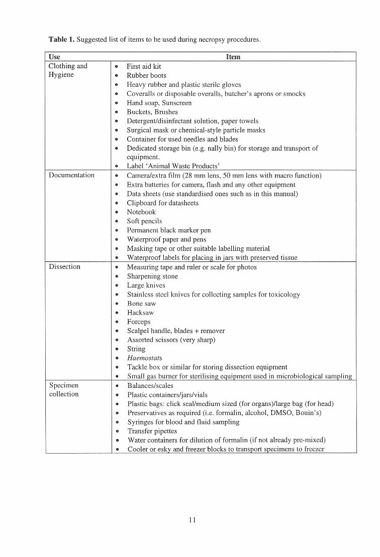
Table 1. Suggested list of items to be used during necropsy procedures.
Use Item Clothing and • First aid kit Hygiene • Rubber boots
• Heavy rubber and plastic sterile gloves • Coveralls or disposable overalls, butcher's aprons or smocks • Hand soap, Sunscreen • Buckets, Brushes • Detergent/disinfectant solution, paper towels • Surgical mask or chemical-style particle masks • Container for used needles and blades • Dedicated storage bin (e.g. nally bin) for storage and transport of
equipment. • Label 'Animal Waste Products'
Documentation • Camera/extra film (28 mm lens, 50 mm lens with macro function) • Extra batteries for camera, flash and any other equipment • Data sheets (use standardised ones such as in this manual) • Clipboard for datasheets • Notebook • Soft pencils • Permanent black marker pen • Waterproof paper and pens • Masking tape or other suitable labelling material • Waterproof labels for placing in jars with preserved tissue
Dissection • Measuring tape and ruler or scale for photos • Sharpening stone • Large knives • Stainless steel knives for collecting samples for toxicology • Bone saw • Hacksaw • Forceps • Scalpel handle, blades + remover • Assorted scissors (very sharp) • String • Haemostats • Tackle box or similar for storing dissection equipment • Small gas burner for sterilising equipment used in microbiological sampling
Specimen • Balances/scales collection • Plastic containers/jars/vials
• Plastic bags: click seal/medium sized (for organs)/large bag (for head) • Preservatives as required (i.e. formalin, alcohol, DMSO, Bouin's) • Syringes for blood and fluid sampling • Transfer pipettes • Water containers for dilution of formalin (if not already pre-mixed) • Cooler or esky and freezer blocks to transport specimens to freezer
11
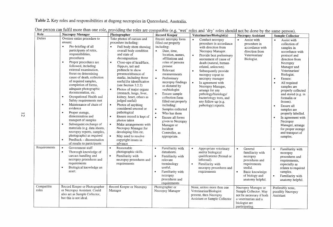
Table 2. Key roles and responsibilities at dugong necropsies in Queensland, Australia.
One person can fulfil more than one role s.„ rovidin the roles are compatible , `wet' riles and`dry' Role Necropsy Manager Photographer Record Keeper Veterinarian/Biologist(s) Necropsy Assistant Sample Collector Duties Oversee entire procedure to
ensure: Pre-briefing of all participants of roles, responsibilities, procedures Proper procedures are followed, including external examination, focus on determining cause of death, collection of required samples, completion of forms, adequate photographic documentation, etc. Occupational Health and Safety requirements met Maintenance of chain of evidence Proper storage, dissemination and transport of samples Subsequent exchange of materials (e.g. data sheets, necropsy reports, samples, photographs) as required Feedback — dissemination of results to participants
Take photos of carcass and procedure including:
Full body shots showing overall body condition and state of decomposition Close-ups of head/face, flippers, tail and peduncle to show presence/absence of marks, including those useful for identification (see Section 3.2.2) Photos of major organs (stomach, lungs, liver, kidney, heart, others as judged useful) Photos of anything considered unusual or pathological Ensure record is kept of photos taken Make arrangements with Necropsy Manager for developing film etc. May need to resolve copyright issues in advance
Ensure necropsy form filled out properly including:
Date, time, location, names, affiliations and roles of persons present Relevant measurements Preliminary necropsy findings as dictated by vet/biologist Ensure sample collection form filled out properly including: Samples collected Who has them
. Ensure all forms given to Necropsy Manager or Incident Controller, as appropriate.
Conduct necropsy procedure in accordance with direction from Necropsy Manager. Provide best preliminary assessment of cause of death (natural, human- related, unknown). Subsequently provide necropsy report to necropsy manager In agreement with Necropsy Manager, arrange for any pathology/histology/ bacteriology tests, and any follow-up (e.g. pathology) reports.
_
Assist with procedure in accordance with direction from Veterinarian/ Biologist.
Assist with collection of samples in accordance with protocol and direction from Necropsy Manager and Veterinarian/ Biologist. Ensure: All required samples are properly collected and stored (e.g. in formalin or frozen). Ensure all samples are properly labelled. In agreement with Necropsy Manager, arrange for proper storage and transport of samples.
Requirements Government staff Thorough knowledge of carcass handling and necropsy procedures and requirements Biological knowledge an asset.
Reasonable photographic skills. Familiarity with necropsy procedures and requirements
Familiarity with datasheets. Familiarity with relevant terminology useful. Familiarity with necropsy procedures and requirements
Appropriate veterinary and/or biological qualifications (formal or informal). Familiarity with necropsy procedures and requirements
General familiarity with necropsy procedures and requirements useful. Basic knowledge of biology and anatomy helpful.
Familiarity with necropsy procedures and requirements, especially as relates to required samples. Familiarity with anatomy helpful.
Compatible roles
Record Keeper or Photographer or Necropsy Assistant. Could also act as Sample Collector, but this is not ideal.
Record Keeper or Necropsy Manager
Photographer or Necropsy Manager
None, unless more than one Veterinarian/Biologist present, then Necropsy Assistant or Sample Collector
Necropsy Manager or Sample Collector. May not be necessary if both a veterinarian and a biologist are participating.
Preferably none, possibly Necropsy Assistant
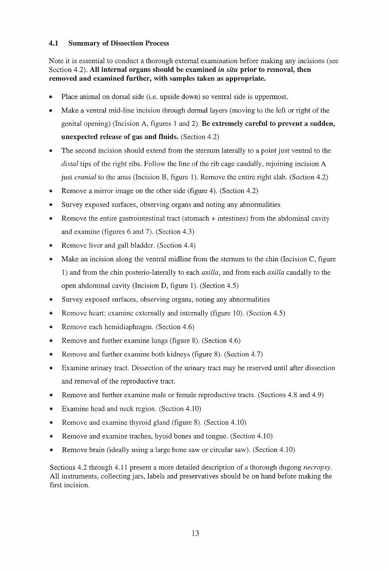
4.1 Summary of Dissection Process
Note it is essential to conduct a thorough external examination before making any incisions (see Section 4.2). All internal organs should be examined in situ prior to removal, then removed and examined further, with samples taken as appropriate.
Place animal on dorsal side (i.e. upside down) so ventral side is uppermost.
Make a ventral mid-line incision through dermal layers (moving to the left or right of the
genital opening) (Incision A, figures 1 and 2). Be extremely careful to prevent a sudden,
unexpected release of gas and fluids. (Section 4.2)
The second incision should extend from the sternum laterally to a point just ventral to the
distal tips of the right ribs. Follow the line of the rib cage caudally, rejoining incision A
just cranial to the anus (Incision B, figure 1). Remove the entire right slab. (Section 4.2)
Remove a mirror image on the other side (figure 4). (Section 4.2)
Survey exposed surfaces, observing organs and noting any abnormalities
Remove the entire gastrointestinal tract (stomach + intestines) from the abdominal cavity
and examine (figures 6 and 7). (Section 4.3)
Remove liver and gall bladder. (Section 4.4)
Make an incision along the ventral midline from the sternum to the chin (Incision C, figure
1) and from the chin posterio-laterally to each axilla, and from each axilla caudally to the
open abdominal cavity (Incision D, figure 1). (Section 4.5)
Survey exposed surfaces, observing organs, noting any abnormalities
Remove heart; examine externally and internally (figure 10). (Section 4.5)
Remove each hemidiaphragm. (Section 4.6)
Remove and further examine lungs (figure 8). (Section 4.6)
Remove and further examine both kidneys (figure 8). (Section 4.7)
Examine urinary tract. Dissection of the urinary tract may be reserved until after dissection
and removal of the reproductive tract.
Remove and further examine male or female reproductive tracts. (Sections 4.8 and 4.9)
Examine head and neck region. (Section 4.10)
Remove and examine thyroid gland (figure 8). (Section 4.10)
Remove and examine trachea, hyoid bones and tongue. (Section 4.10)
Remove brain (ideally using a large bone saw or circular saw). (Section 4.10)
Sections 4.2 through 4.11 present a more detailed description of a thorough dugong necropsy. All instruments, collecting jars, labels and preservatives should be on hand before making the first incision.
13
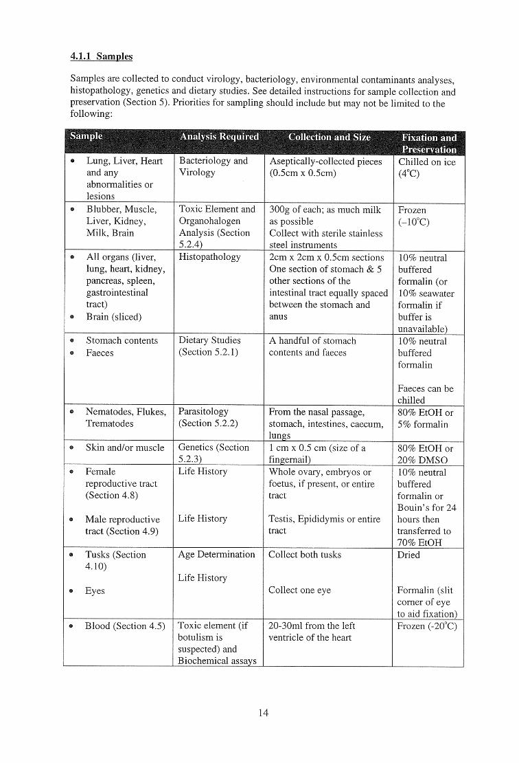
4.1.1 Samples
Samples are collected to conduct virology, bacteriology, environmental contaminants analyses, histopathology, genetics and dietary studies. See detailed instructions for sample collection and preservation (Section 5). Priorities for sampling should include but may not be limited to the following:
1ple Analysis Required
Bacteriology and Virology
Collection and Size Fixation and Preservation
Lung, Liver, Heart and any abnormalities or lesions
Aseptically-collected pieces (0.5cm x 0.5cm)
Chilled on ice (4°C)
Blubber, Muscle, Liver, Kidney, Milk, Brain
Toxic Element and Organohalogen Analysis (Section 5.2.4)
300g of each; as much milk as possible Collect with sterile stainless steel instruments
Frozen (-10°C)
All organs (liver, lung, heart, kidney, pancreas, spleen, gastrointestinal tract) Brain (sliced)
Histopathology 2cm x 2cm x 0.5cm sections One section of stomach & 5 other sections of the intestinal tract equally spaced between the stomach and anus
10% neutral buffered formalin (or 10% seawater formalin if buffer is unavailable)
Stomach contents Faeces
Dietary Studies (Section 5.2.1)
A handful of stomach contents and faeces
10% neutral buffered formalin
Faeces can be chilled
Nematodes, Flukes, Trematodes
Parasitology (Section 5.2.2)
From the nasal passage, stomach, intestines, caecum, lungs
80% EtOH or 5% formalin
Skin and/or muscle Genetics (Section 5.2.3)
1 cm x 0.5 cm (size of a fingernail)
80% EtOH or 20% DMSO
Female reproductive tract (Section 4.8)
Male reproductive tract (Section 4.9)
Life History
Life History
Whole ovary, embryos or foetus, if present, or entire tract
Testis, Epididymis or entire tract
10% neutral buffered formalin or Bouin's for 24 hours then transferred to 70% EtOH
Tusks (Section 4.10)
Eyes
Age Determination
Life History
Collect both tusks
Collect one eye
Dried
Formalin (slit corner of eye to aid fixation)
Blood (Section 4.5) Toxic element (if botulism is suspected) and Biochemical assays
20-30m1 from the left ventricle of the heart
Frozen (-20 °C)
14
.0i*tTE® Body length Length of teats
o Genital opening to anus Tail fluke width Blubber thickness
(See Attachment 2)
Seagrass from mouth (if present 1 eye Milk from the teat Skin Blubber from outermost layer Muscle Nasal flukes Any abnormalities
Organs in situ (after first incision) Wounds, scars Any abnormalities (external and internal)
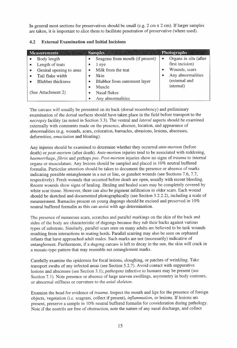
In general most sections for preservatives should be small (e.g. 2 cm x 2 cm). If larger samples are taken, it is important to slice them to facilitate penetration of preservative (where used).
4.2 External Examination and Initial Incisions
The carcass will usually be presented on its back (dorsal recumbency) and preliminary examination of the dorsal surfaces should have taken place in the field before transport to the necropsy facility (as noted in Section 3.3). The ventral and lateral aspects should be examined externally with comments made on the presence, absence, location, and appearance of abnormalities (e.g. wounds, scars, coloration, barnacles, abrasions, lesions, abscesses, deformities, emaciation and bloating).
Any injuries should be examined to determine whether they occurred ante-mortem (before death) or post-mortem (after death). Ante-mortem injuries tend to be associated with reddening, haemorrhage, fibrin and perhaps pus. Post-mortem injuries show no signs of trauma to internal organs or musculature. Any lesions should be sampled and placed in 10% neutral buffered formalin. Particular attention should be taken to document the presence or absence of marks indicating possible entanglement in a net or line, or gunshot wounds (see Sections 7.6, 7.7, respectively). Fresh wounds that occurred before death are open, usually with recent bleeding. Recent wounds show signs of healing. Healing and healed scars may be completely covered by white scar tissue. However, there can also be pigment infiltration in older scars. Each wound should be sketched and documented photographically (see Section 3.2.2.2), including a scale of measurement. Barnacles present on young dugongs should be excised and preserved in 10% neutral buffered formalin as this can assist with age determination.
The presence of numerous scars, scratches and parallel markings on the skin of the back and sides of the body are characteristic of dugongs because they rub their backs against various types of substrate. Similarly, parallel scars seen on many adults are believed to be tusk wounds resulting from interactions in mating herds. Parallel scarring may also be seen on orphaned infants that have approached adult males. Such marks are not (necessarily) indicative of entanglement. Furthermore, if a dugong carcass is left to decay in the sun, the skin will crack in a mosaic-type pattern that may resemble net entanglement marks.
Carefully examine the epidermis for focal lesions, sloughing, or patches of wrinkling. Take transport swabs of any infected areas (see Section 5.2.7). Avoid contact with suppurative lesions and abscesses (see Section 3.1); pathogens infective to humans may be present (see Section 7.1). Note presence or absence of large uneven swellings, asymmetry in body contours, or abnormal stiffness or curvature to the axial skeleton.
Examine the head for evidence of trauma. Inspect the mouth and lips for the presence of foreign objects, vegetation (i.e. seagrass, collect if present), inflammation, or lesions. If lesions are present, preserve a sample in 10% neutral buffered formalin for consideration during pathology. Note if the nostrils are free of obstruction, note the nature of any nasal discharge, and collect
15
nasal flukes (Cochleotrema indicum) in 10% formalin (see Section 5.2.2). Examine the eyes and preserve one in 10% formalin if not badly decomposed. Eyes can be collected as an alternative age determination technique. The external auditory meatus should be located for examination and as a reference point if morphometrics are required (Heinsohn 1981; Attachment 3). On some specimens the meatus may be difficult to locate. It is found at about the same distance caudal to the eye as the eye is from the tip of the snout. A slice through the dermis at the meatus will reveal a 1-2 mm diameter canal filled with a black waxy paste. Note if the mandible may be moved with ease or if it is stiff. Leave a more detailed examination of the head and neck for a later stage in the necropsy (see Section 4.10).
Examine each flipper for freedom of movement, inflammatory lesions, healed wounds or other abnormalities. Measure the lengths of both teats in females; palpate each teat working up towards the nipple, and note the presence or absence of milk, pus, blood, or other material and take samples if present. Note if both teats are of approximately equal size, or if there is any apparent shrinkage or swelling. Cut the gland to check for the presence of milk. If available, collect a sample of milk and freeze for histology and toxicology.
Examine the umbilicus for abnormalities or infections, particularly in calves. Examine the urogenital aperture for discharges or abnormalities. In recently parturient or near-term pregnant females the vaginal canal is enlarged and supple, and the examiner's protected forearm can pass through to the uterus. Females in late pregnancy will also show a bulge with a prominent curve cranial to the urogenital opening, which may exude mucus. Note the texture and characteristics of vaginal fluids, and collect samples to later check for sperm under a microscope if recent copulation is suspected. Note if semen is exuding from the external genitalia of males. Examine the anus for blockage, and note the presence or absence of faeces or other discharges, describing texture, colour and consistency. Photograph any abnormalities of the tail fluke, including a reference scale.
Carefully obtain the measurement data detailed in Section 3.2.3 and in Attachment 2. These include body length, lengths of teats, genital-opening-to-anus distance and fluke width.
Using a knife, begin the first incision (Incision A, figure 1; figure 2) making a mid-line incision through the skin, blubber and muscle (figure 3) to the anus (move to the right of the genital aperture) without penetrating the abdominal cavity. (Throughout the text of this manual right refers to the animal's right, left to the animal's left.). When cutting through the dermis and blubber layers, be extremely careful to prevent a sudden release of gas and fluids from the abdominal cavity and the digestive tract. Once the length of the incision has been made, cautiously make a small cut in the parietal peritoneum at the mid-abdomen using bandage scissors (blunt point directed internally), gradually lengthening the cut as the internal pressure is reduced. The parietal peritoneum can then be cut the length of incision A (figure 1), taking care not to nick underlying organs. Make a second large incision with a knife (Incision B, figure 1) from the sternum laterally to a point just ventral to the distal tips of the right ribs. Follow the line of the rib cage caudally, rejoining incision A just cranial to the genital aperture. Remove the entire right slab and put it to the side. Remove a mirror-image left slab by cutting down the midline just to the left of the genital aperture to a point cranial to the anus, and by making a lateral cut from the sternum to a point just ventral to the distal tips of the left ribs, proceeding caudally as in figure 1 (figure 4). The genitalia should remain with the carcass. Be careful not to disturb the underlying organs during removal of these slabs. Look for any subdermal haemorrhaging and take samples if present. Take a sample of any haematomas (if present) and preserve in 10% neutral buffered formalin to allow for ageing of the wound(s). Fresh haematomas (four days old or less) are unorganised while older haematomas show developing fibrin structure and tissue organisation.
16
Photograph all exposed organs in situ (figures 4 and 5), including a reference scale (ruler or other known scale). Even if organs appear normal, photographic documentation is essential for future reference in supporting diagnoses, particularly should legally sensitive issues arise. Take measurements of the exposed dermis and thickness of the outer and inner blubber layers at the mid-ventral and mid-lateral layer cake-like surfaces from the left slab (figure 3). Describe the quantity, colour and texture of the blubber. Blubber appearance and thickness can be an indicator of body condition and general health. Collect a sample of the outer blubber and outer muscle layers for toxicology analysis following instructions detailed in Section 5.2. Collect samples for genetic analysis (see Section 5.2). Remark on the general appearance of the abdominal cavity. Note the presence of fluids, if any, and their colours and consistencies. Remark on peculiar odours, the presence or absence of gas and ingesta, displacement of organs, ruptures, adhesions and/or haemorrhage. Examine the gastrointestinal mesenteries for discoloration or haemorrhage, and mesenteric lymph nodes for size and colour. Take transport swabs (if appropriate) and collect tissue samples for histopathology.
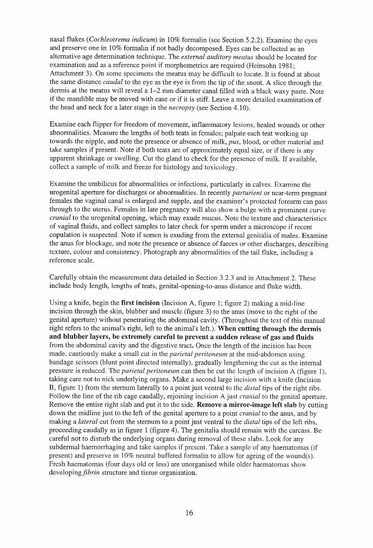
4.3 Gastrointestinal Tract
Phot , aphs
The large intestine is up to 25m long in an adult, more than twice as long as the small intestine (figure 6). The spleen is generally dark coloured and small. The stomach is a simple sac, a large muscular dilatation of the alimentary canal between the oesophagus and the small intestine (figures 6 and 7). The presence of nematodes in the stomach and cardiac gland is normal.
Stomach contents and nematodes Section of stomach and 5 sections along the intestines equally spaced between the stomach and anus Section of caecum and parasites
• Adrenal gland Spleen
• Pancreas (if carcass is fresh) Intestinal contents (including faeces) Lymph node(s) Any abnormalities
Gastrointestinal tract in situ and spread out on table. Any abnormalities
The gastrointestinal tract and associated structures of the digestive system are removed for examination following in situ inspection of the exposed serosal surfaces and mesenteries for haemorrhage, cysts, tears, abscesses or other lesions. Examine the different parts of the gastrointestinal tract and the mucosal surface for the presence of any abnormalities (e.g. discolorations, haemorrhages, cysts, lesions, fibrin strands and adhesions, obstructions, stenosis, inflation, ulcers, foreign objects, sediment etc.).
After initial incisions, the stomach is somewhat obscured by the liver lobes but when these are gently pulled aside it can be seen that the adult stomach, when distended with ingesta, bulges to
fill the anterior body cavity. The spleen is situated to the left of the median plane, adjacent to the dorsal stomach wall near its region of maximum dilation (figure 8). The cardiac gland of the stomach, which is not initially obvious externally, can be seen after the removal of the lesser omentum as a definite protuberance of the left dorsal stomach wall (figure 8).
Begin to remove the gastrointestinal tract by locating the junction of the descending colon and the rectum. Free this segment by cutting the mesentery (mesocolon), and tie the rectum off with string in two places a few centimetres apart, just dorsal to the urinary bladder. Sever the descending colon between the tied strings and start to cut the mesocolon cranially to begin removal of the entire tract. The diaphragm is on an almost horizontal plane, and each half is
17
referred to as a hemidiaphragm (figure 8). The descending mesocolon is attached to the left hemidiaphragm, near the left kidney. The mesocolon should be cut and the colour, texture, and quantity of fat described for the transverse colon and adjoining mesenteries. Tie a string at the centre of the transverse colon to mark its location for future reference.
The coiled ascending colon is attached to the parietal peritoneum at the vertebral column. Continue dissections to free the ascending colon. The caecum marks the junction of the large and small intestines (figure 6). It is located to the left of the vertebral column. Cut the peritoneum joining the dorsal surface of the caecum to the vertebral column and hemidiaphragm while lifting the caecum. Continue to apply tension and remove the loops of the ileum and jejunum. At this point the coeliac and cranial mesenteric arteries are severed. The aorta and caudal (inferior) vena cava are left attached to the hemidiaphragm.
Complete the removal of the gastrointestinal tract by cutting between the duodenum and hemidiaphragm until the pylorus of the stomach is reached. Do not cut the hemidiaphragm. Then stop and move cranially to where the oesophagus enters the stomach. Sever the oesophagus about 5 cm cranial to the stomach and continue to cut between the stomach and hemidiaphragm, dissecting through the lesser omentum, hepatic artery, and the bile duct. Clamp the bile duct with a haemostat before severing. Cut the bile duct caudal to the haemostat. The entire gastrointestinal tract may be removed from the abdominal cavity once the stomach attachments are freed. Place the tract on a flat, clean surface for later detailed examination.
After the gastrointestinal tract has been removed but prior to detailed examination, the examiner should inspect the peritoneal lining and the abdominal cavity. Note any abnormalities (areas of oedema, adhesions, abscesses, growths, ruptures of the diaphragm or body wall, or other peculiarities). If not contaminated by foreign material, note the amount (by removal with a graduated container) and characteristics (colour, consistency, presence of fibrin strands, etc.) of fluids. If clotted blood is present, measure the amount.
Locate the adrenal glands (figure 8). These are small glands found along each medial edge of the vertebral column cranial to the kidneys. They are best located by palpation of the region. If the adrenal glands cannot be located they may have been removed inadvertently with the gastrointestinal tract. Once the adrenals are located and removed, they should be examined for cysts or swellings and sliced like bread at no greater than 5-mm thickness for examination and preservation.
The gastrointestinal tract should be placed on a large clean working area so it can be spread out for examination. Detailed examination of the gastrointestinal tract can be left until last in order to avoid any possible cross contamination to the general work area, particularly when necropsies are being carried out under less than ideal conditions. Begin by cutting the jejunum and ileum free of the mesentery and carefully examine all serosal surfaces for haemorrhages. Clamp off any areas inadvertently nicked during removal. Once the serosal surfaces have been examined and described, the spleen and pancreas should be collected and the lumen and mucosa of the stomach examined.
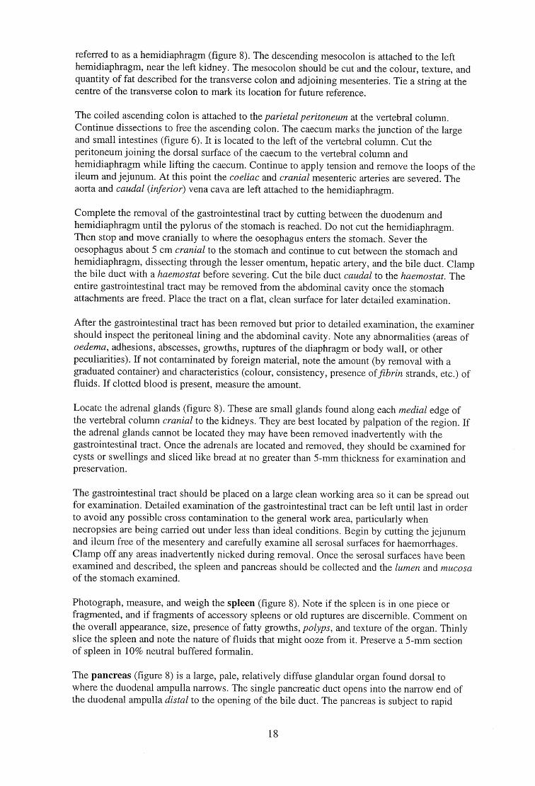
Photograph, measure, and weigh the spleen (figure 8). Note if the spleen is in one piece or fragmented, and if fragments of accessory spleens or old ruptures are discernible. Comment on the overall appearance, size, presence of fatty growths, polyps, and texture of the organ. Thinly slice the spleen and note the nature of fluids that might ooze from it. Preserve a 5-mm section of spleen in 10% neutral buffered formalin.
The pancreas (figure 8) is a large, pale, relatively diffuse glandular organ found dorsal to where the duodenal ampulla narrows. The single pancreatic duct opens into the narrow end of the duodenal ampulla distal to the opening of the bile duct. The pancreas is subject to rapid
18
decomposition. In fresh carcasses (Categories 1-3) it should be removed, photographed, measured, weighed in its entirety, examined for abnormalities and a 0.5 cm section taken and preserved in 10% neutral buffered formalin.
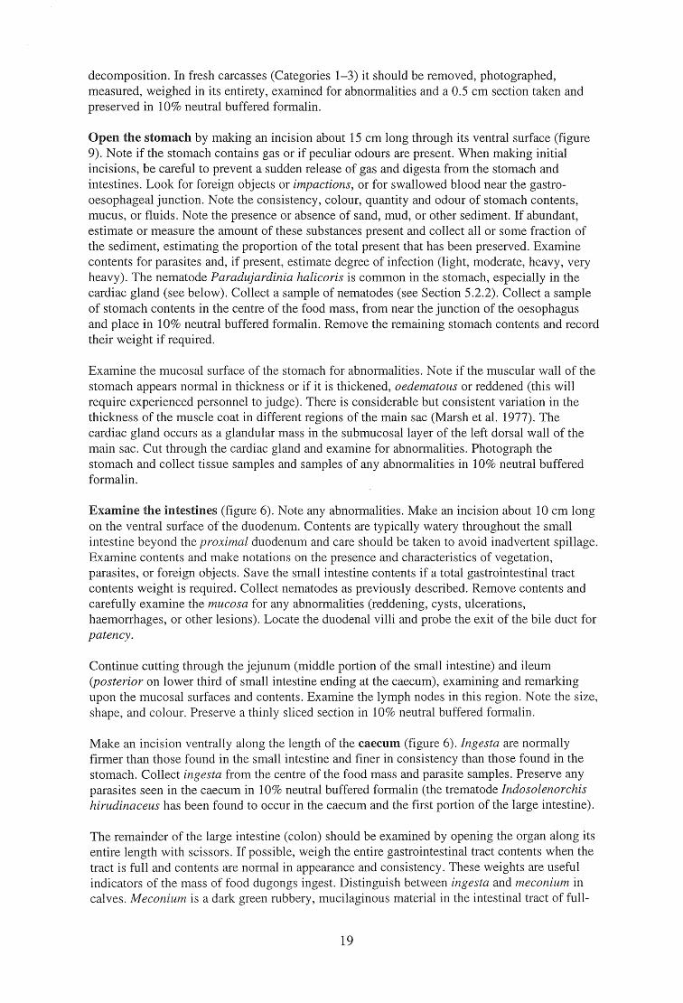
Open the stomach by making an incision about 15 cm long through its ventral surface (figure 9). Note if the stomach contains gas or if peculiar odours are present. When making initial incisions, be careful to prevent a sudden release of gas and digesta from the stomach and intestines. Look for foreign objects or impactions, or for swallowed blood near the gastro-oesophageal junction. Note the consistency, colour, quantity and odour of stomach contents, mucus, or fluids. Note the presence or absence of sand, mud, or other sediment. If abundant, estimate or measure the amount of these substances present and collect all or some fraction of the sediment, estimating the proportion of the total present that has been preserved. Examine contents for parasites and, if present, estimate degree of infection (light, moderate, heavy, very heavy). The nematode Paradujardinia halicoris is common in the stomach, especially in the cardiac gland (see below). Collect a sample of nematodes (see Section 5.2.2). Collect a sample of stomach contents in the centre of the food mass, from near the junction of the oesophagus and place in 10% neutral buffered formalin. Remove the remaining stomach contents and record their weight if required.
Examine the mucosal surface of the stomach for abnormalities. Note if the muscular wall of the stomach appears normal in thickness or if it is thickened, oedematous or reddened (this will require experienced personnel to judge). There is considerable but consistent variation in the thickness of the muscle coat in different regions of the main sac (Marsh et al. 1977). The cardiac gland occurs as a glandular mass in the submucosal layer of the left dorsal wall of the main sac. Cut through the cardiac gland and examine for abnormalities. Photograph the stomach and collect tissue samples and samples of any abnormalities in 10% neutral buffered formalin.
Examine the intestines (figure 6). Note any abnormalities. Make an incision about 10 cm long on the ventral surface of the duodenum. Contents are typically watery throughout the small intestine beyond the proximal duodenum and care should be taken to avoid inadvertent spillage. Examine contents and make notations on the presence and characteristics of vegetation, parasites, or foreign objects. Save the small intestine contents if a total gastrointestinal tract contents weight is required. Collect nematodes as previously described. Remove contents and carefully examine the mucosa for any abnormalities (reddening, cysts, ulcerations, haemorrhages, or other lesions). Locate the duodenal villi and probe the exit of the bile duct for patency.
Continue cutting through the jejunum (middle portion of the small intestine) and ileum (posterior on lower third of small intestine ending at the caecum), examining and remarking upon the mucosal surfaces and contents. Examine the lymph nodes in this region. Note the size, shape, and colour. Preserve a thinly sliced section in 10% neutral buffered formalin.
Make an incision ventrally along the length of the caecum (figure 6). Ingesta are normally firmer than those found in the small intestine and finer in consistency than those found in the stomach. Collect ingesta from the centre of the food mass and parasite samples. Preserve any parasites seen in the caecum in 10% neutral buffered formalin (the trematode Indosolenorchis hirudinaceus has been found to occur in the caecum and the first portion of the large intestine).
The remainder of the large intestine (colon) should be examined by opening the organ along its entire length with scissors. If possible, weigh the entire gastrointestinal tract contents when the tract is full and contents are normal in appearance and consistency. These weights are useful indicators of the mass of food dugongs ingest. Distinguish between ingesta and meconium in calves. Meconium is a dark green rubbery, mucilaginous material in the intestinal tract of full-
19
term foetuses and neonates. Take transport swabs if enteritis is suspected. Collect tissue samples for histopathology in 10% neutral buffered formalin. Collect parasites, noting location in intestines, and give an estimate of the degree of infection (light, moderate, heavy, very heavy), proportion collected, and approximate total present. Collect a sample of ingesta in 10% neutral buffered formalin from the mid-region of the large intestine.
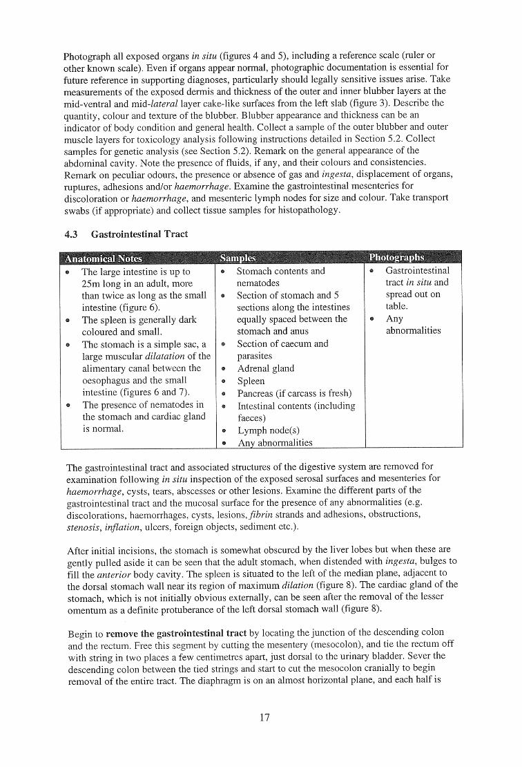
4.4 Liver and Gall Bladder
graphs
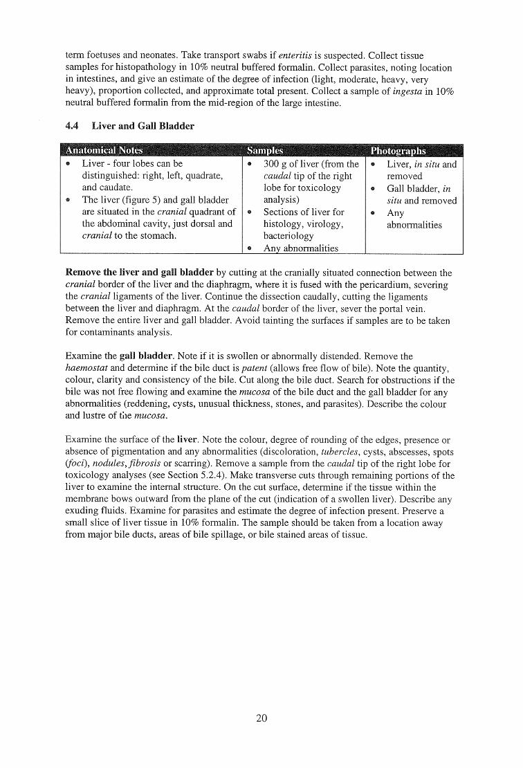
Liver - four lobes can be distinguished: right, left, quadrate, and caudate. The liver (figure 5) and gall bladder are situated in the cranial quadrant of the abdominal cavity, just dorsal and cranial to the stomach.
300 g of liver (from the caudal tip of the right lobe for toxicology analysis) Sections of liver for histology, virology, bacteriology Any abnormalities
Liver, in situ and removed Gall bladder, in situ and removed Any abnormalities
Remove the liver and gall bladder by cutting at the cranially situated connection between the cranial border of the liver and the diaphragm, where it is fused with the pericardium, severing the cranial ligaments of the liver. Continue the dissection caudally, cutting the ligaments between the liver and diaphragm. At the caudal border of the liver, sever the portal vein. Remove the entire liver and gall bladder. Avoid tainting the surfaces if samples are to be taken for contaminants analysis.
Examine the gall bladder. Note if it is swollen or abnormally distended. Remove the haemostat and determine if the bile duct is patent (allows free flow of bile). Note the quantity, colour, clarity and consistency of the bile. Cut along the bile duct. Search for obstructions if the bile was not free flowing and examine the mucosa of the bile duct and the gall bladder for any abnormalities (reddening, cysts, unusual thickness, stones, and parasites). Describe the colour and lustre of the mucosa.
Examine the surface of the liver. Note the colour, degree of rounding of the edges, presence or absence of pigmentation and any abnormalities (discoloration, tubercles, cysts, abscesses, spots (foci), nodules, fibrosis or scarring). Remove a sample from the caudal tip of the right lobe for toxicology analyses (see Section 5.2.4). Make transverse cuts through remaining portions of the liver to examine the internal structure. On the cut surface, determine if the tissue within the membrane bows outward from the plane of the cut (indication of a swollen liver). Describe any exuding fluids. Examine for parasites and estimate the degree of infection present. Preserve a small slice of liver tissue in 10% formalin. The sample should be taken from a location away from major bile ducts, areas of bile spillage, or bile stained areas of tissue.
20
4.5 Pericardial Cavity, Heart, Major Blood Vessels and Mammary Glands
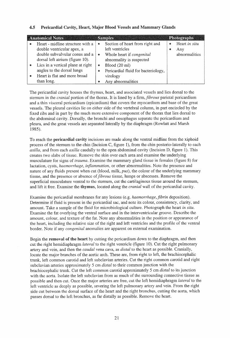
Heart - midline structure with a double ventricular apex, a double subvalvular conus and a dorsal left atrium (figure 10). Lies in a vertical plane at right angles to the dorsal lungs Heart is flat and more broad than long.
Section of heart from right and left ventricles Whole heart if congenital abnormality is suspected Blood (20 ml) Pericardial fluid for bacteriology, virology Any abnormalities
Heart in situ Any abnormalities
The pericardial cavity houses the thymus, heart, and associated vessels and lies dorsal to the sternum in the cranial portion of the thorax. It is lined by a firm, fibrous parietal pericardium and a thin visceral pericardium (epicardium) that covers the myocardium and base of the great vessels. The pleural cavities lie on either side of the vertebral column, in part encircled by the fixed ribs and in part by the much more extensive component of the thorax that lies dorsal to the abdominal cavity. Dorsally, the bronchi and oesophagus separate the pericardium and pleura, and the great vessels are separated laterally by the diaphragm (Rowlatt and Marsh 1985).
To reach the pericardial cavity incisions are made along the ventral midline from the xiphoid process of the sternum to the chin (Incision C, figure 1), from the chin posterio-laterally to each axilla, and from each axilla caudally to the open abdominal cavity (Incision D, figure 1). This creates two slabs of tissue. Remove the skin over each area and examine the underlying musculature for signs of trauma. Examine the mammary gland tissue in females (figure 8) for lactation, cysts, haemorrhage, inflammation, or other abnormalities. Note the presence and nature of any fluids present when cut (blood, milk, pus), the colour of the underlying mammary tissue, and the presence or absence of fibrous tissue, lumps or abscesses. Remove the superficial musculature ventral to the sternum, cut the cartilaginous tissue around the sternum, and lift it free. Examine the thymus, located along the cranial wall of the pericardial cavity.
Examine the pericardial membranes for any lesions (e.g. haemorrhage, fibrin deposition). Determine if fluid is present in the pericardial sac, and note its colour, consistency, clarity, and amount. Take a sample of the fluid for microbiological culture. Photograph the heart in situ. Examine the fat overlying the ventral surface and in the interventricular groove. Describe the amount, colour, and texture of the fat. Note any abnormalities in the position or appearance of the heart, including the relative size of the right and left ventricles and the profile of the ventral border. Note if any congenital anomalies are apparent on external examination.
Begin the removal of the heart by cutting the pericardium down to the diaphragm, and then cut the right hemidiaphragm lateral to the right ventricle (figure 10). Cut the right pulmonary artery and vein, and then the caudal vena cava, as distal to the heart as possible. Cranially, locate the major branches of the aortic arch. These are, from right to left, the brachiocephalic trunk, left common carotid and left subclavian arteries. Cut the right common carotid and right subclavian arteries approximately 5 cm distal to their common junction with the brachiocephalic trunk. Cut the left common carotid approximately 5 cm distal to its junction with the aorta. Isolate the left subclavian from as much of the surrounding connective tissue as possible and then cut. Once the major arteries are free, cut the left hemidiaphragm lateral to the left ventricle as deeply as possible, severing the left pulmonary artery and vein. From the right side cut between the dorsal surface of the heart and the right bronchus, cutting the aorta, which passes dorsal to the left bronchus, as far distally as possible. Remove the heart.
21
Examine the heart externally. Note if the muscular wall of the heart is firm or flabby, if either of the ventricles show abnormally rounded bulging (dilatation), or if there is any evidence of hypertrophy. Examine the external surface for the presence of scars, abscesses, haemorrhage, or other unusual features. Note the presence of any clear vesicular gelatinous material adhering to the internal lining of the heart (this is also occasionally visible on the exterior surface). The presence of this material is a condition called cachexia (`water fat'), and is an indicator of severe starvation in dugongs (an internal examination of the heart will also reveal this condition).
Examine the heart internally by cutting through the ventral surface of the right atrium to the right ventricle. A sample of blood (minimum 20 ml) from the heart should be frozen for toxicology, contaminants analysis, and if botulism is suspected. Examine the endocardium, chordae tendineae, and papillary muscles for inflammation, scars, tears, haemorrhage, plaque, or other abnormalities. Examine the dorsal and ventral cusps and the smaller medial and lateral cusps of the right atrioventricular valve for inflammation, thickness, hardening, growths, or other abnormalities. Continue the incision from the right ventricle through the pulmonary trunk, examining the three semilunar cusps of the pulmonary valve. Turn the heart over and from the dorsal aspect make a new incision from the left atrium to the left ventricle, examining the left atrioventricular valve and interior as on the right side. Make a third incision in the dorsal side of the heart from the left ventricle through the ascending aorta. Examine the three semilunar cusps of the aortic orifice for growths, hardness, wear, holes, and other features. Examine the wall of the aorta and the coronary arteries for plaque build-up, emboli or thrombi, noting colour, size, thickness, and texture. Note if the interventricular and interatrial septa are complete. Examine the heart for evidence of coarctation or aneurysms. Note if blood is present in the left ventricle, and whether it is clotted.
Note the colour of blood and the sheen or lustre of the internal lining of the heart. Note the presence or absence of chicken fat clots, or if there is no evidence of clotting. Post-mortem clots can be distinguished from thrombi in that they are uniform in colour, smooth and shiny, uniform in texture, and unattached but moulded to the vessel in which they are formed. Ante-mortem thrombi are often a layered mixture of red and grey, friable, dull, roughened, stringy, and attached to the walls of blood vessels.
The heart should also be examined for evidence of congenital anomalies, particularly in young animals. Examine the ductus arteriosus between the pulmonary artery and the aorta with a probe to determine if it is patent or has closed, as is normal in larger, older animals, remaining as the ligamentum arteriosum. Examine the pulmonary artery for stenosis or constriction. Take tissue samples for histology including samples of any suspected abnormalities.
Major blood vessels should be inspected routinely during examination of the organ systems they supply. In calves, particular attention should be paid to the umbilical vessels for necrosis or abscesses.
4.5.1 Blood Sample
Whenever possible collect 20 ml of blood in a sterile tube for hormonal and other biochemical assays. Blood will usually have to be obtained with a syringe from the left ventricle as soon as possible after death. Allow it to clot in a cool place for several hours (if you are working on a beach, placing the tube at an angle in the sand in a shady place is fine). Remove the serum with a Pasteur pipette. If possible, centrifuge the serum (keep the supernatant) before freezing. It should keep indefinitely at -20°C.
With a fresh (Categories 1-3) carcass, blood samples can provide valuable data on blood chemistry, haematology and pathology.
22
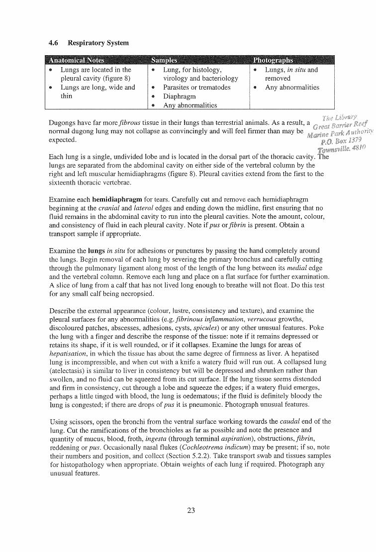
Anatomical Notes Lungs are located in the pleural cavity (figure 8) Lungs are long, wide and thin
iT Lung, for histology, virology and bacteriology Parasites or trematodes Diaphragm Any abnormalities
Lungs, in situ and removed Any abnormalities
4.6 Respiratory System
Dugongs have far more fibrous tissue in their lungs than terrestrial animals. As a result, a normal dugong lung may not collapse as convincingly and will feel firmer than may be expected.
Each lung is a single, undivided lobe and is located in the dorsal part of the thoracic cavity. The lungs are separated from the abdominal cavity on either side of the vertebral column by the right and left muscular hemidiaphragms (figure 8). Pleural cavities extend from the first to the sixteenth thoracic vertebrae.
Examine each hemidiaphragm for tears. Carefully cut and remove each hemidiaphragm beginning at the cranial and lateral edges and ending down the midline, first ensuring that no fluid remains in the abdominal cavity to run into the pleural cavities. Note the amount, colour, and consistency of fluid in each pleural cavity. Note if pus or fibrin is present. Obtain a transport sample if appropriate.
Examine the lungs in situ for adhesions or punctures by passing the hand completely around the lungs. Begin removal of each lung by severing the primary bronchus and carefully cutting through the pulmonary ligament along most of the length of the lung between its medial edge and the vertebral column. Remove each lung and place on a flat surface for further examination. A slice of lung from a calf that has not lived long enough to breathe will not float. Do this test for any small calf being necropsied.
Describe the external appearance (colour, lustre, consistency and texture), and examine the pleural surfaces for any abnormalities (e.g. fibrinous inflammation, verrucous growths, discoloured patches, abscesses, adhesions, cysts, spicules) or any other unusual features. Poke the lung with a finger and describe the response of the tissue: note if it remains depressed or retains its shape, if it is well rounded, or if it collapses. Examine the lungs for areas of hepatisation, in which the tissue has about the same degree of firmness as liver. A hepatised lung is incompressible, and when cut with a knife a watery fluid will run out. A collapsed lung (atelectasis) is similar to liver in consistency but will be depressed and shrunken rather than swollen, and no fluid can be squeezed from its cut surface. If the lung tissue seems distended and firm in consistency, cut through a lobe and squeeze the edges; if a watery fluid emerges, perhaps a little tinged with blood, the lung is oedematous; if the fluid is definitely bloody the lung is congested; if there are drops of pus it is pneumonic. Photograph unusual features.
Using scissors, open the bronchi from the ventral surface working towards the caudal end of the lung. Cut the ramifications of the bronchioles as far as possible and note the presence and quantity of mucus, blood, froth, ingesta (through terminal aspiration), obstructions, fibrin, reddening or pus. Occasionally nasal flukes (Cochleotrema indicum) may be present; if so, note their numbers and position, and collect (Section 5.2.2). Take transport swab and tissues samples for histopathology when appropriate. Obtain weights of each lung if required. Photograph any unusual features.
23
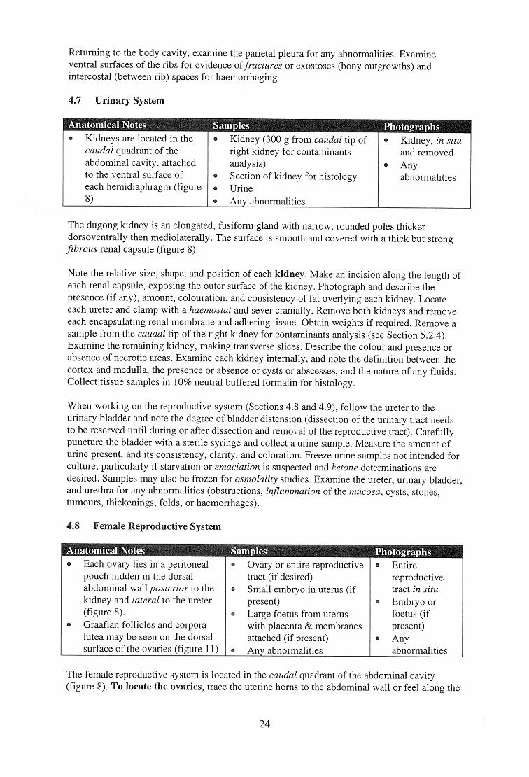
Anatomical Notes Kidneys are located in the caudal quadrant of the abdominal cavity, attached to the ventral surface of each hemidiaphragm (figure 8)
Kidney (300 g from caudal tip of right kidney for contaminants analysis) Section of kidney for histology Urine Any abnormalities
Kidney, in situ and removed Any abnormalities
Each ovary lies in a peritoneal pouch hidden in the dorsal abdominal wall posterior to the kidney and lateral to the ureter (figure 8). Graafian follicles and corpora lutea may be seen on the dorsal surface of the ovaries (figure 11)
Ovary or entire reproductive tract (if desired) Small embryo in uterus (if present) Large foetus from uterus with placenta & membranes attached (if present) Any abnormalities
Entire reproductive tract in situ Embryo or foetus (if present) Any abnormalities
Returning to the body cavity, examine the parietal pleura for any abnormalities. Examine ventral surfaces of the ribs for evidence offractures or exostoses (bony outgrowths) and intercostal (between rib) spaces for haemorrhaging.
4.7 Urinary System
The dugong kidney is an elongated, fusiform gland with narrow, rounded poles thicker dorsoventrally then mediolaterally. The surface is smooth and covered with a thick but strong fibrous renal capsule (figure 8).
Note the relative size, shape, and position of each kidney. Make an incision along the length of each renal capsule, exposing the outer surface of the kidney. Photograph and describe the presence (if any), amount, colouration, and consistency of fat overlying each kidney. Locate each ureter and clamp with a haemostat and sever cranially. Remove both kidneys and remove each encapsulating renal membrane and adhering tissue. Obtain weights if required. Remove a sample from the caudal tip of the right kidney for contaminants analysis (see Section 5.2.4). Examine the remaining kidney, making transverse slices. Describe the colour and presence or absence of necrotic areas. Examine each kidney internally, and note the definition between the cortex and medulla, the presence or absence of cysts or abscesses, and the nature of any fluids. Collect tissue samples in 10% neutral buffered formalin for histology.
When working on the reproductive system (Sections 4.8 and 4.9), follow the ureter to the urinary bladder and note the degree of bladder distension (dissection of the urinary tract needs to be reserved until during or after dissection and removal of the reproductive tract). Carefully puncture the bladder with a sterile syringe and collect a urine sample. Measure the amount of urine present, and its consistency, clarity, and coloration. Freeze urine samples not intended for culture, particularly if starvation or emaciation is suspected and ketone determinations are desired. Samples may also be frozen for osmolality studies. Examine the ureter, urinary bladder, and urethra for any abnoinialities (obstructions, inflammation of the mucosa, cysts, stones, tumours, thickenings, folds, or haemorrhages).
4.8 Female Reproductive System
The female reproductive system is located in the caudal quadrant of the abdominal cavity (figure 8). To locate the ovaries, trace the uterine horns to the abdominal wall or feel along the
24
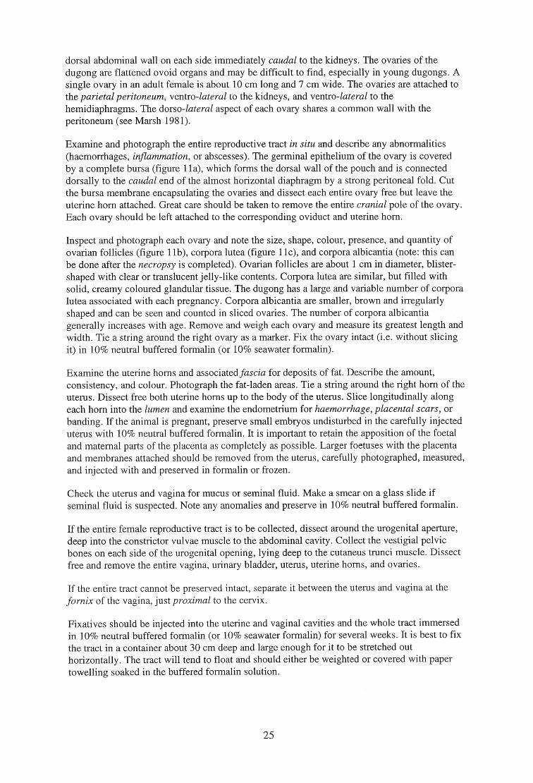
dorsal abdominal wall on each side immediately caudal to the kidneys. The ovaries of the dugong are flattened ovoid organs and may be difficult to find, especially in young dugongs. A single ovary in an adult female is about 10 cm long and 7 cm wide. The ovaries are attached to the parietal peritoneum, ventro-lateral to the kidneys, and ventro-lateral to the hemidiaphragms. The dorso-lateral aspect of each ovary shares a common wall with the peritoneum (see Marsh 1981).
Examine and photograph the entire reproductive tract in situ and describe any abnormalities (haemorrhages, inflammation, or abscesses). The germinal epithelium of the ovary is covered by a complete bursa (figure 11a), which forms the dorsal wall of the pouch and is connected dorsally to the caudal end of the almost horizontal diaphragm by a strong peritoneal fold. Cut the bursa membrane encapsulating the ovaries and dissect each entire ovary free but leave the uterine horn attached. Great care should be taken to remove the entire cranial pole of the ovary. Each ovary should be left attached to the corresponding oviduct and uterine horn.
Inspect and photograph each ovary and note the size, shape, colour, presence, and quantity of ovarian follicles (figure 11b), corpora lutea (figure 11c), and corpora albicantia (note: this can be done after the necropsy is completed). Ovarian follicles are about 1 cm in diameter, blister-shaped with clear or translucent jelly-like contents. Corpora lutea are similar, but filled with solid, creamy coloured glandular tissue. The dugong has a large and variable number of corpora lutea associated with each pregnancy. Corpora albicantia are smaller, brown and irregularly shaped and can be seen and counted in sliced ovaries. The number of corpora albicantia generally increases with age. Remove and weigh each ovary and measure its greatest length and width. Tie a string around the right ovary as a marker. Fix the ovary intact (i.e. without slicing it) in 10% neutral buffered formalin (or 10% seawater formalin).
Examine the uterine horns and associated fascia for deposits of fat. Describe the amount, consistency, and colour. Photograph the fat-laden areas. Tie a string around the right horn of the uterus. Dissect free both uterine horns up to the body of the uterus. Slice longitudinally along each horn into the lumen and examine the endometrium for haemorrhage, placental scars, or banding. If the animal is pregnant, preserve small embryos undisturbed in the carefully injected uterus with 10% neutral buffered formalin. It is important to retain the apposition of the foetal and maternal parts of the placenta as completely as possible. Larger foetuses with the placenta and membranes attached should be removed from the uterus, carefully photographed, measured, and injected with and preserved in formalin or frozen.
Check the uterus and vagina for mucus or seminal fluid. Make a smear on a glass slide if seminal fluid is suspected. Note any anomalies and preserve in 10% neutral buffered formalin.
If the entire female reproductive tract is to be collected, dissect around the urogenital aperture, deep into the constrictor vulvae muscle to the abdominal cavity. Collect the vestigial pelvic bones on each side of the urogenital opening, lying deep to the cutaneus trunci muscle. Dissect free and remove the entire vagina, urinary bladder, uterus, uterine horns, and ovaries.
If the entire tract cannot be preserved intact, separate it between the uterus and vagina at the fornix of the vagina, just proximal to the cervix.
Fixatives should be injected into the uterine and vaginal cavities and the whole tract immersed in 10% neutral buffered formalin (or 10% seawater formalin) for several weeks. It is best to fix the tract in a container about 30 cm deep and large enough for it to be stretched out horizontally. The tract will tend to float and should either be weighted or covered with paper towelling soaked in the buffered formalin solution.
25
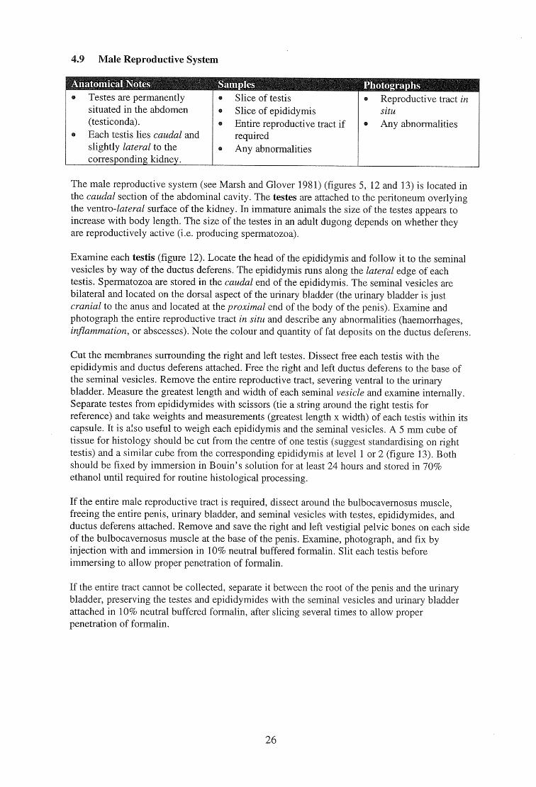
Testes are permanently situated in the abdomen (testiconda). Each testis lies caudal and slightly lateral to the corresponding kidney.
Slice of testis Slice of epididymis Entire reproductive tract if required Any abnormalities
Reproductive tract in situ Any abnormalities
4.9 Male Reproductive System
Anatomical Notes Samples Photographs
The male reproductive system (see Marsh and Glover 1981) (figures 5, 12 and 13) is located in the caudal section of the abdominal cavity. The testes are attached to the peritoneum overlying the ventro-lateral surface of the kidney. In immature animals the size of the testes appears to increase with body length. The size of the testes in an adult dugong depends on whether they are reproductively active (i.e. producing spermatozoa).
Examine each testis (figure 12). Locate the head of the epididymis and follow it to the seminal vesicles by way of the ductus deferens. The epididymis runs along the lateral edge of each testis. Spermatozoa are stored in the caudal end of the epididymis. The seminal vesicles are bilateral and located on the dorsal aspect of the urinary bladder (the urinary bladder is just cranial to the anus and located at the proximal end of the body of the penis). Examine and photograph the entire reproductive tract in situ and describe any abnormalities (haemorrhages, inflammation, or abscesses). Note the colour and quantity of fat deposits on the ductus deferens.
Cut the membranes surrounding the right and left testes. Dissect free each testis with the epididymis and ductus deferens attached. Free the right and left ductus deferens to the base of the seminal vesicles. Remove the entire reproductive tract, severing ventral to the urinary bladder. Measure the greatest length and width of each seminal vesicle and examine internally. Separate testes from epididymides with scissors (tie a string around the right testis for reference) and take weights and measurements (greatest length x width) of each testis within its capsule. It is also useful to weigh each epididymis and the seminal vesicles. A 5 mm cube of tissue for histology should be cut from the centre of one testis (suggest standardising on right testis) and a similar cube from the corresponding epididymis at level 1 or 2 (figure 13). Both should be fixed by immersion in Bouin's solution for at least 24 hours and stored in 70% ethanol until required for routine histological processing.
If the entire male reproductive tract is required, dissect around the bulbocavernosus muscle, freeing the entire penis, urinary bladder, and seminal vesicles with testes, epididymides, and ductus deferens attached. Remove and save the right and left vestigial pelvic bones on each side of the bulbocavernosus muscle at the base of the penis. Examine, photograph, and fix by injection with and immersion in 10% neutral buffered formalin. Slit each testis before immersing to allow proper penetration of formalin.
If the entire tract cannot be collected, separate it between the root of the penis and the urinary bladder, preserving the testes and epididymides with the seminal vesicles and urinary bladder attached in 10% neutral buffered formalin, after slicing several times to allow proper penetration of formalin.
26
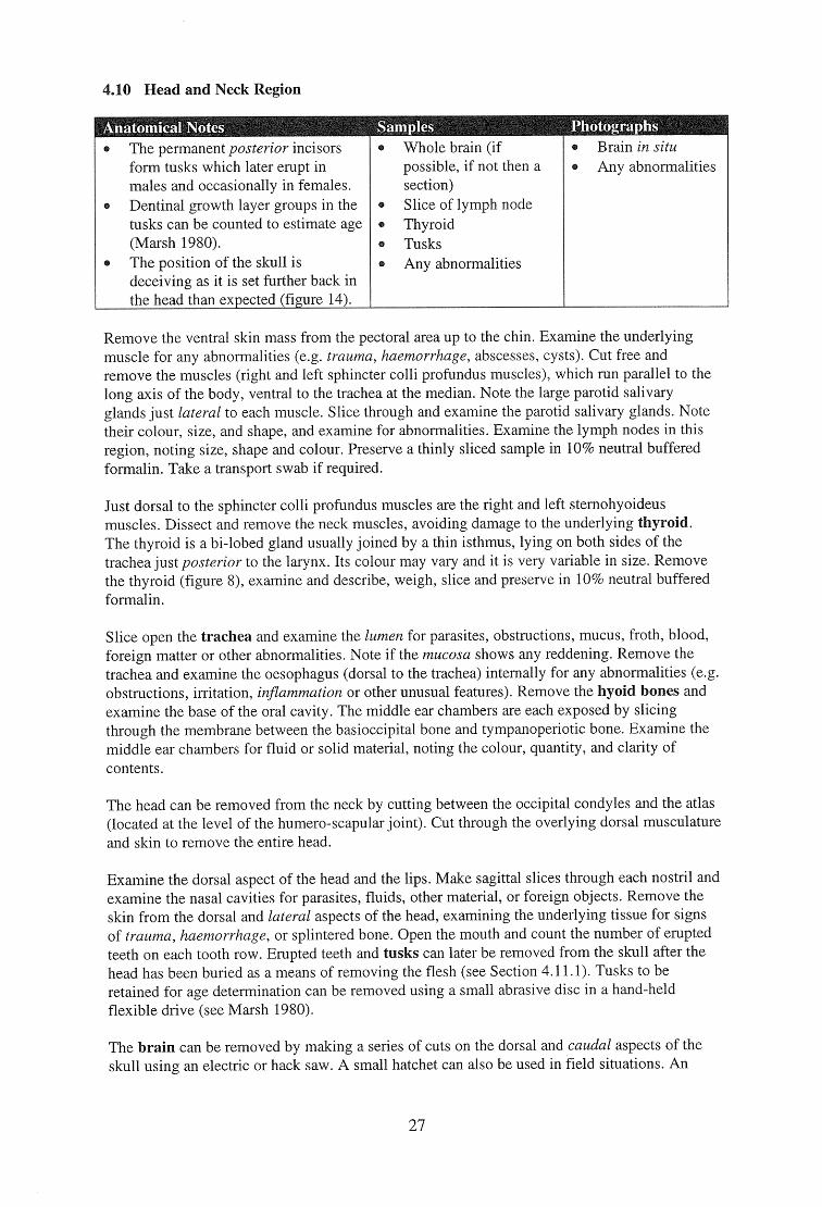
4.10 Head and Neck Region
Anatomical Notes amples Photographs
The permanent posterior incisors form tusks which later erupt in males and occasionally in females. Dentinal growth layer groups in the tusks can be counted to estimate age (Marsh 1980). The position of the skull is deceiving as it is set further back in the head than expected (figure 14).
Whole brain (if possible, if not then a section) Slice of lymph node Thyroid Tusks Any abnormalities
Brain in situ Any abnormalities
Remove the ventral skin mass from the pectoral area up to the chin. Examine the underlying muscle for any abnormalities (e.g. trauma, haemorrhage, abscesses, cysts). Cut free and remove the muscles (right and left sphincter colli profundus muscles), which run parallel to the long axis of the body, ventral to the trachea at the median. Note the large parotid salivary glands just lateral to each muscle. Slice through and examine the parotid salivary glands. Note their colour, size, and shape, and examine for abnormalities. Examine the lymph nodes in this region, noting size, shape and colour. Preserve a thinly sliced sample in 10% neutral buffered formalin. Take a transport swab if required.
Just dorsal to the sphincter colli profundus muscles are the right and left sternohyoideus muscles. Dissect and remove the neck muscles, avoiding damage to the underlying thyroid. The thyroid is a bi-lobed gland usually joined by a thin isthmus, lying on both sides of the trachea just posterior to the larynx. Its colour may vary and it is very variable in size. Remove the thyroid (figure 8), examine and describe, weigh, slice and preserve in 10% neutral buffered formalin.
Slice open the trachea and examine the lumen for parasites, obstructions, mucus, froth, blood, foreign matter or other abnormalities. Note if the mucosa shows any reddening. Remove the trachea and examine the oesophagus (dorsal to the trachea) internally for any abnormalities (e.g. obstructions, irritation, inflammation or other unusual features). Remove the hyoid bones and examine the base of the oral cavity. The middle ear chambers are each exposed by slicing through the membrane between the basioccipital bone and tympanoperiotic bone. Examine the middle ear chambers for fluid or solid material, noting the colour, quantity, and clarity of contents.
The head can be removed from the neck by cutting between the occipital condyles and the atlas (located at the level of the humero-scapular joint). Cut through the overlying dorsal musculature and skin to remove the entire head.
Examine the dorsal aspect of the head and the lips. Make sagittal slices through each nostril and examine the nasal cavities for parasites, fluids, other material, or foreign objects. Remove the skin from the dorsal and lateral aspects of the head, examining the underlying tissue for signs of trauma, haemorrhage, or splintered bone. Open the mouth and count the number of erupted teeth on each tooth row. Erupted teeth and tusks can later be removed from the skull after the head has been buried as a means of removing the flesh (see Section 4.11.1). Tusks to be retained for age determination can be removed using a small abrasive disc in a hand-held flexible drive (see Marsh 1980).
The brain can be removed by making a series of cuts on the dorsal and caudal aspects of the skull using an electric or hack saw. A small hatchet can also be used in field situations. An
27
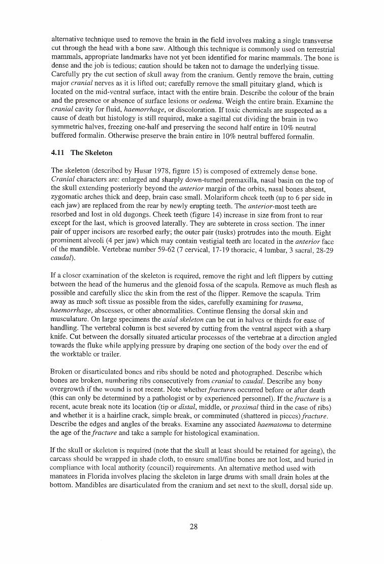
alternative technique used to remove the brain in the field involves making a single transverse cut through the head with a bone saw. Although this technique is commonly used on terrestrial mammals, appropriate landmarks have not yet been identified for marine mammals. The bone is dense and the job is tedious; caution should be taken not to damage the underlying tissue. Carefully pry the cut section of skull away from the cranium. Gently remove the brain, cutting major cranial nerves as it is lifted out; carefully remove the small pituitary gland, which is located on the mid-ventral surface, intact with the entire brain. Describe the colour of the brain and the presence or absence of surface lesions or oedema. Weigh the entire brain. Examine the cranial cavity for fluid, haemorrhage, or discoloration. If toxic chemicals are suspected as a cause of death but histology is still required, make a sagittal cut dividing the brain in two symmetric halves, freezing one-half and preserving the second half entire in 10% neutral buffered formalin. Otherwise preserve the brain entire in 10% neutral buffered formalin.
4.11 The Skeleton
The skeleton (described by Husar 1978, figure 15) is composed of extremely dense bone. Cranial characters are: enlarged and sharply down-turned premaxilla, nasal basin on the top of the skull extending posteriorly beyond the anterior margin of the orbits, nasal bones absent, zygomatic arches thick and deep, brain case small. Molariform cheek teeth (up to 6 per side in each jaw) are replaced from the rear by newly erupting teeth. The anterior-most teeth are resorbed and lost in old dugongs. Cheek teeth (figure 14) increase in size from front to rear except for the last, which is grooved laterally. They are subterete in cross section. The inner pair of upper incisors are resorbed early; the outer pair (tusks) protrudes into the mouth. Eight prominent alveoli (4 per jaw) which may contain vestigial teeth are located in the anterior face of the mandible. Vertebrae number 59-62 (7 cervical, 17-19 thoracic, 4 lumbar, 3 sacral, 28-29 caudal).
If a closer examination of the skeleton is required, remove the right and left flippers by cutting between the head of the humerus and the glenoid fossa of the scapula. Remove as much flesh as possible and carefully slice the skin from the rest of the flipper. Remove the scapula. Trim away as much soft tissue as possible from the sides, carefully examining for trauma, haemorrhage, abscesses, or other abnormalities. Continue flensing the dorsal skin and musculature. On large specimens the axial skeleton can be cut in halves or thirds for ease of handling. The vertebral column is best severed by cutting from the ventral aspect with a sharp knife. Cut between the dorsally situated articular processes of the vertebrae at a direction angled towards the fluke while applying pressure by draping one section of the body over the end of the worktable or trailer.
Broken or disarticulated bones and ribs should be noted and photographed. Describe which bones are broken, numbering ribs consecutively from cranial to caudal. Describe any bony overgrowth if the wound is not recent. Note whether fractures occurred before or after death (this can only be determined by a pathologist or by experienced personnel). If the fracture is a recent, acute break note its location (tip or distal, middle, or proximal third in the case of ribs) and whether it is a hairline crack, simple break, or comminuted (shattered in pieces) fracture. Describe the edges and angles of the breaks. Examine any associated haematoma to determine the age of the fracture and take a sample for histological examination.
If the skull or skeleton is required (note that the skull at least should be retained for ageing), the carcass should be wrapped in shade cloth, to ensure small/fine bones are not lost, and buried in compliance with local authority (council) requirements. An alternative method used with manatees in Florida involves placing the skeleton in large drums with small drain holes at the bottom. Mandibles are disarticulated from the cranium and set next to the skull, dorsal side up.
28
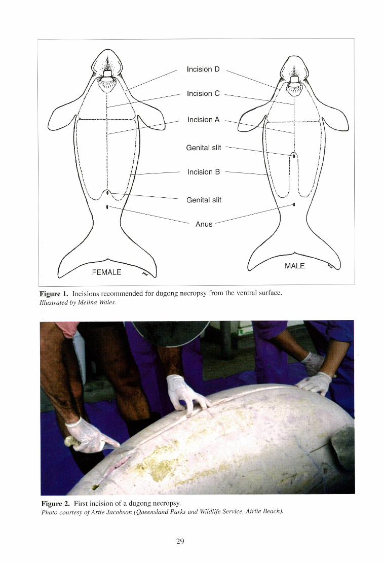
Arfr
MALE
Incision D
Incision C
Incision A
Genital slit
Incision B
Genital slit
Anus
Figure 1. Incisions recommended for dugong necropsy from the ventral surface. Illustrated by Melina Wales.
Figure 2. First incision of a dugong necropsy. Photo courtesy of Artie Jacobson (Queensland Parks and Wildlife Service, Airlie Beach).
29
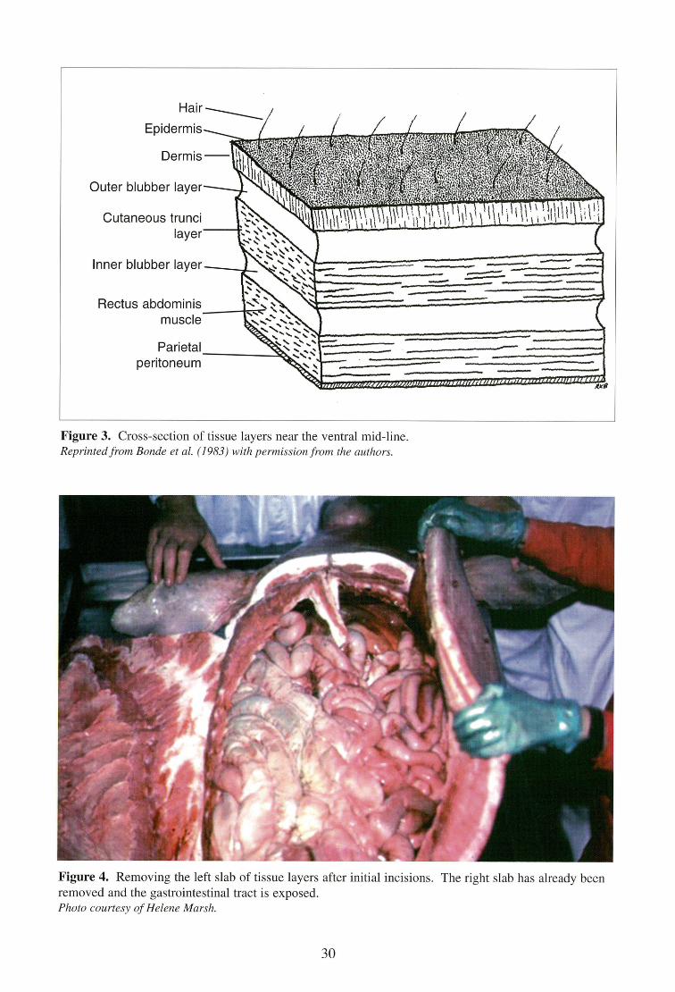
"rlyttliffizzecerwIte/17 ,606011.111illiorow. 0111111"flicsozioricssmooresmalfirtliflif.
Hair
Epidermis
Dermis
Outer blubber layer
Cutaneous trunci layer
Inner blubber layer
Rectus abdominis muscle
Parietal peritoneum
Figure 3. Cross-section of tissue layers near the ventral mid-line. Reprinted from Bonde et al. (1983) with permission from the authors.
Figure 4. Removing the left slab of tissue layers after initial incisions. The right slab has already been removed and the gastrointestinal tract is exposed. Photo courtesy of Helene Marsh.
30
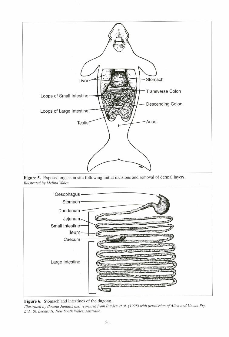
Oesophagus
Stomach
Duodenum
Jejunum
Small Intestine
Ileum
Caecum
Large Intestine
„vol. OrTIMOVikak# 144g14 r*Li+4016SWINOWilik".46110160 ,
----,LAWANOVViigrageMarrtiVi •
AIWA
ww,1#-Ai letewqvaiwoviW
Ve4tilifftWiswilm wo
143%"00.9opewowieowo.
le""1"51"1"46S"1""ta" '?"4"14 '‘tovomi -.,sobeetolsoitomogrOwo*MW
sii,44RiAtemogtAws4moawstow
,
Y.
Figure 5. Exposed organs in situ following initial incisions and removal of dermal layers. Illustrated by Melina Wales
Figure 6. Stomach and intestines of the dugong. Illustrated by Bozena Jantulik and reprinted from Bryden et al. (1998) with permission of Allen and Unwin Pty. Ltd., St. Leonards, New South Wales, Australia.
31
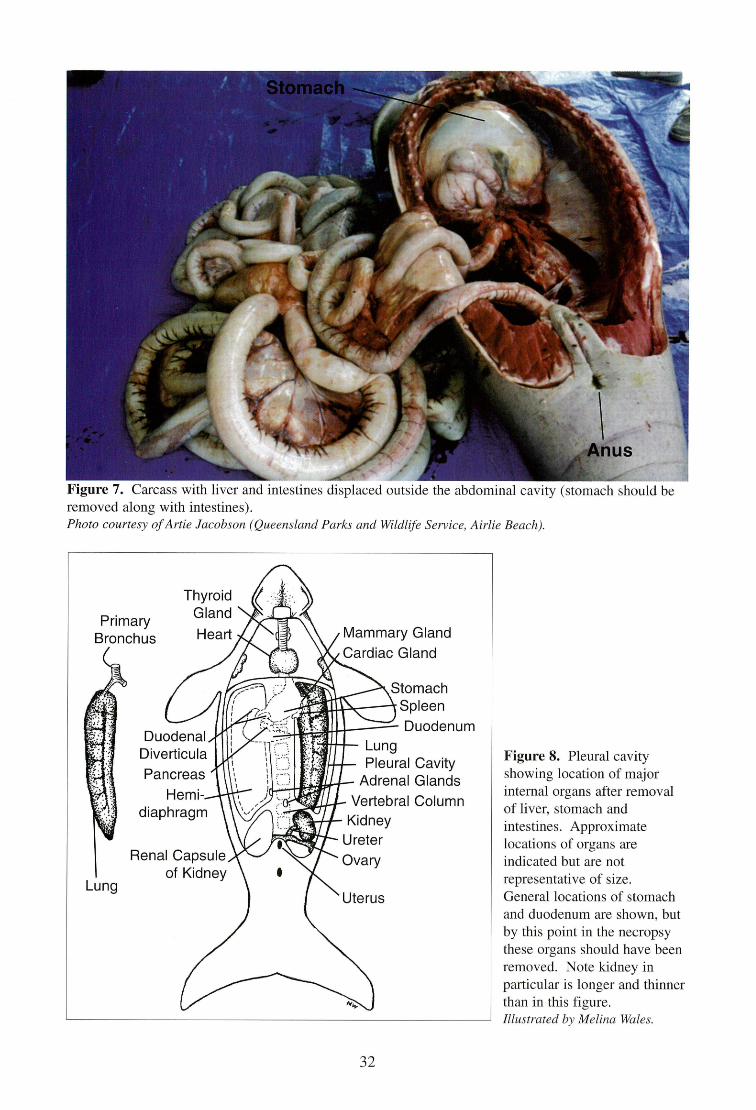
Primary Bronchus
Thyroid Gland
Heart Mammary Gland Cardiac Gland
Stomach Spleen
vial Duodenum Lung Pleural Cavity
Adrenal Glands Vertebral Column Kidney
Ureter Ovary
Uterus
Duodenal Diverticula Pancreas
Hemi-diaphragm
Renal Capsule of Kidney
Lung
AN,
Figure 7. Carcass with liver and intestines displaced outside the abdominal cavity (stomach should be removed along with intestines). Photo courtesy of Artie Jacobson (Queensland Parks and Wildlife Service, Airlie Beach).
Figure 8. Pleural cavity showing location of major internal organs after removal of liver, stomach and intestines. Approximate locations of organs are indicated but are not representative of size. General locations of stomach and duodenum are shown, but by this point in the necropsy these organs should have been removed. Note kidney in particular is longer and thinner than in this figure. Illustrated by Melina Wales.
32
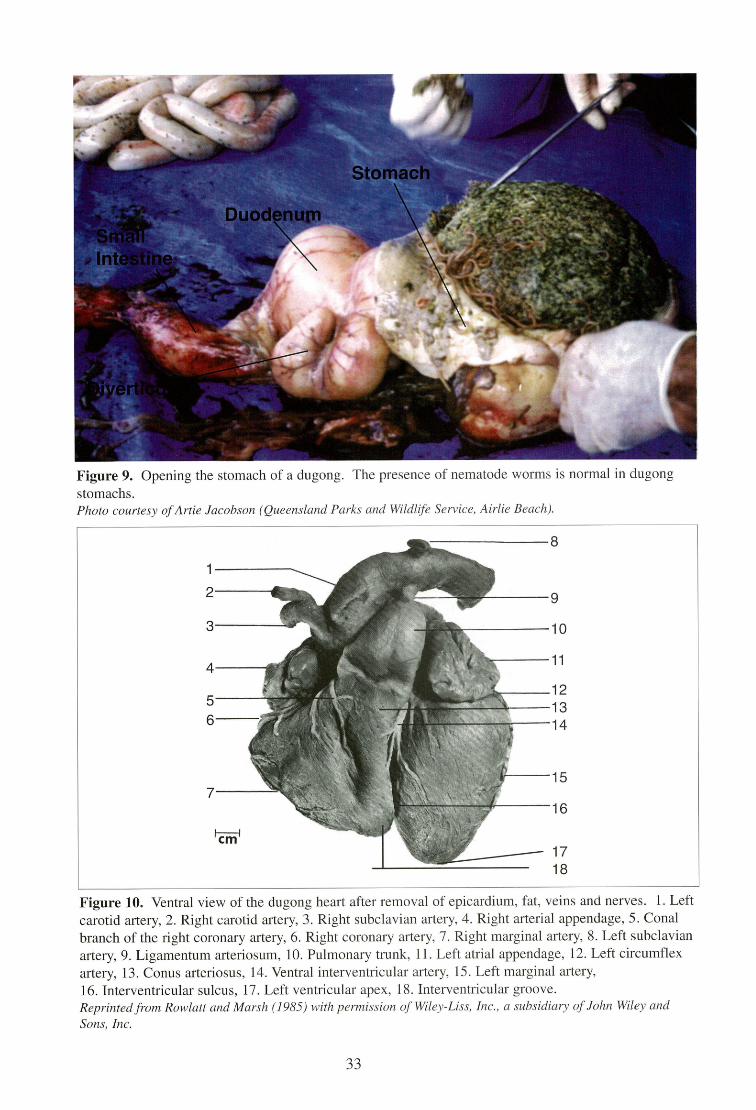
Figure 9. Opening the stomach of a dugong. The presence of nematode worms is normal in dugong stomachs. Photo courtesy of Artie Jacobson (Queensland Parks and Wildlife Service, Airlie Beach).
Figure 10. Ventral view of the dugong heart after removal of epicardium, fat, veins and nerves. 1. Left carotid artery, 2. Right carotid artery, 3. Right subclavian artery, 4. Right arterial appendage, 5. Conal branch of the right coronary artery, 6. Right coronary artery, 7. Right marginal artery, 8. Left subclavian artery, 9. Ligamentum arteriosum, 10. Pulmonary trunk, 11. Left atrial appendage, 12. Left circumflex artery, 13. Conus arteriosus, 14. Ventral interventricular artery, 15. Left marginal artery, 16. Interventricular sulcus, 17. Left ventricular apex, 18. Interventricular groove. Reprinted from Rowlatt and Marsh (1985) with permission of Wiley-Liss, Inc., a subsidiary of John Wiley and Sons, Inc.
33
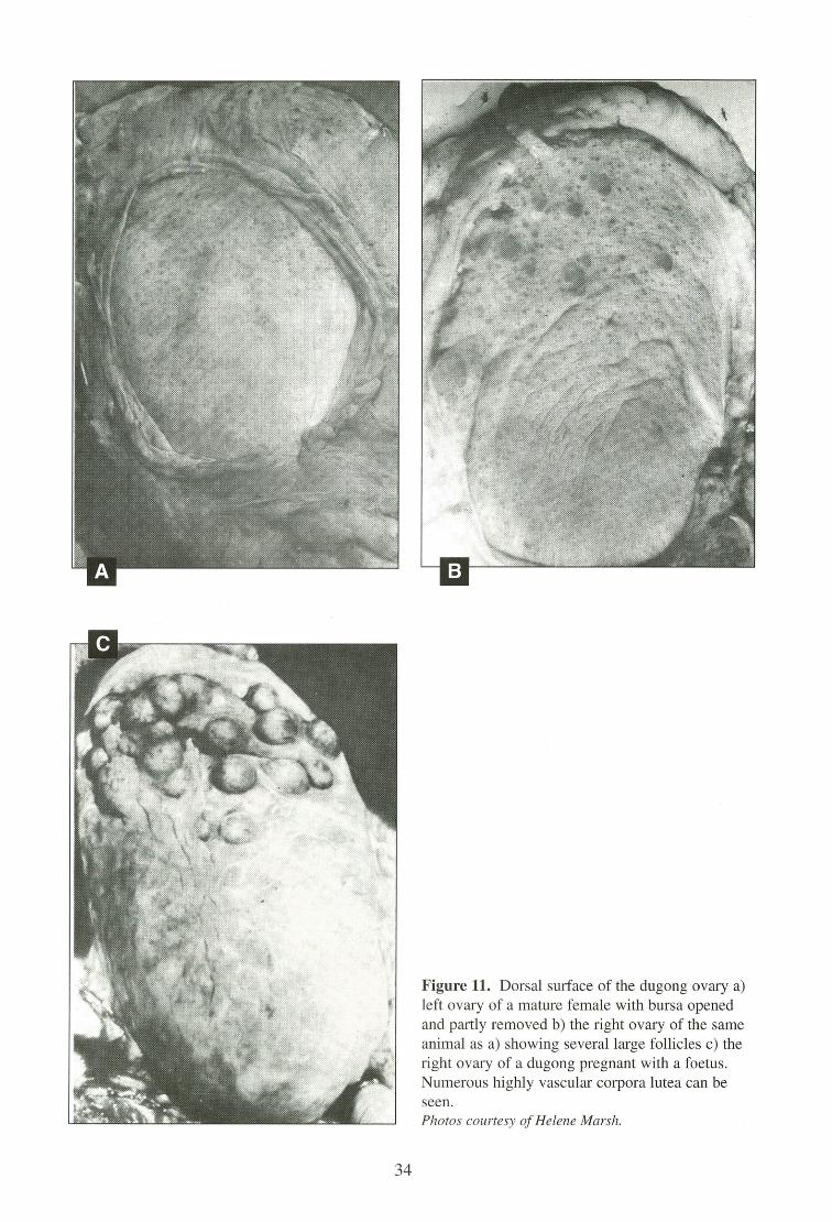
Figure 11. Dorsal surface of the dugong ovary a) left ovary of a mature female with bursa opened and partly removed b) the right ovary of the same animal as a) showing several large follicles c) the right ovary of a dugong pregnant with a foetus. Numerous highly vascular corpora lutea can be seen. Photos courtesy of Helene Marsh.
34
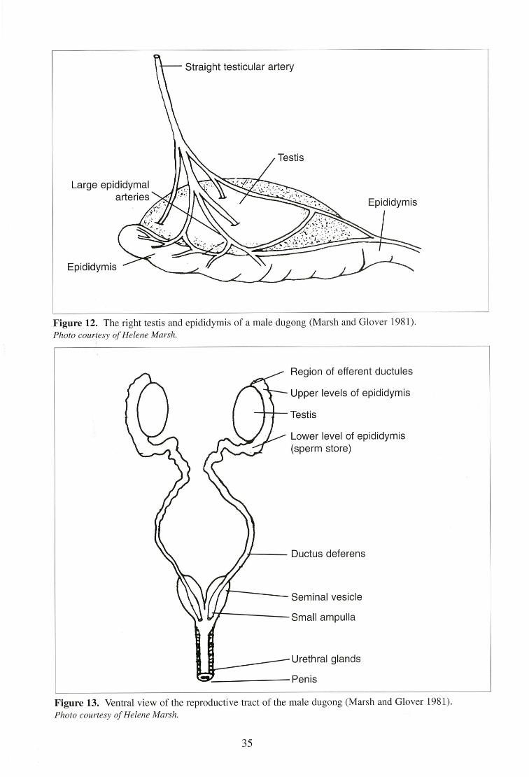
Figure 12. The right testis and epididymis of a male dugong (Marsh and Glover 1981). Photo courtesy of Helene Marsh.
Region of efferent ductules
Upper levels of epididymis
Testis
Lower level of epididymis (sperm store)
Ductus deferens
Seminal vesicle
Small ampulla
Urethral glands
Penis
Figure 13. Ventral view of the reproductive tract of the male dugong (Marsh and Glover 1981). Photo courtesy of Helene Marsh.
35
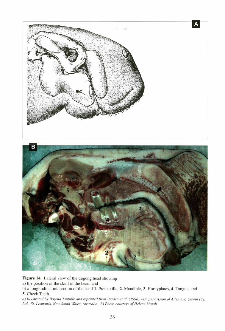
A
Figure 14. Lateral view of the dugong head showing the position of the skull in the head. and a longitudinal midsection of the head 1. Premaxilla, 2. Mandible, 3. Hornyplates, 4. Tongue, and
5. Cheek Teeth. a) Illustrated by Bozena Jantulik and reprinted from Bryden et al. (1998) with permission of Allen and Unwin Pty. Ltd., St. Leonards, New South Wales, Australia. b) Photo courtesy of Helene Marsh.
36
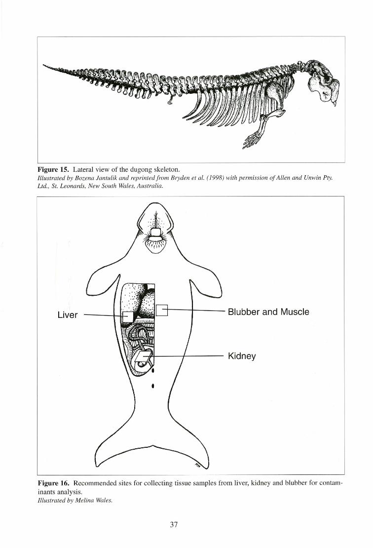
Figure 15. Lateral view of the dugong skeleton. Illustrated by Bozena Jantulik and reprinted from Bryden et al. (1998) with permission of Allen and Unwin Pty. Ltd., St. Leonards, New South Wales, Australia.
Figure 16. Recommended sites for collecting tissue samples from liver, kidney and blubber for contam-inants analysis. Illustrated by Melina Wales.
37
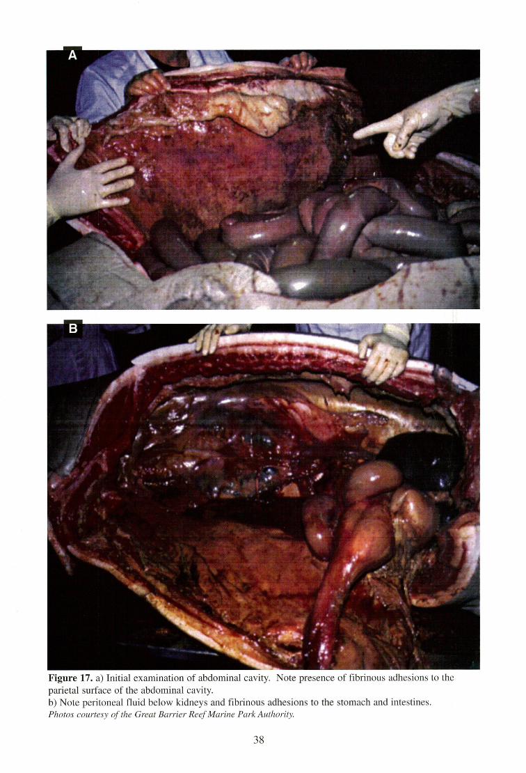
Figure 17. a) Initial examination of abdominal cavity. Note presence of fibrinous adhesions to the parietal surface of the abdominal cavity. b) Note peritoneal fluid below kidneys and fibrinous adhesions to the stomach and intestines. Photos courtesy of the Great Barrier Reef Marine Park Authority.
38
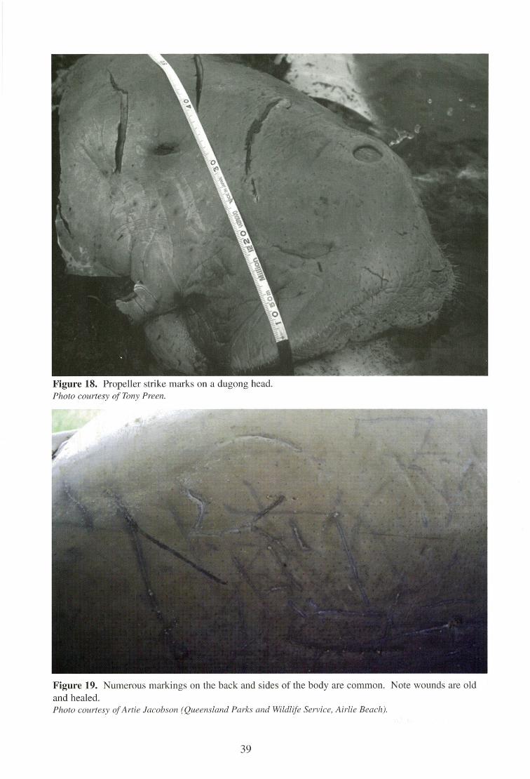
Figure 18. Propeller strike marks on a dugong head. Photo courtesy of Tony Preen.
Figure 19. Numerous markings on the back and sides of the body are common. Note wounds are old and healed. Photo courtesy of Artie Jacobson (Queensland Parks and Wildlife Service, Airlie Beach).
39
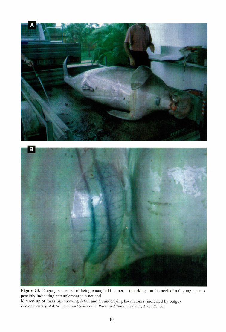
Figure 20. Dugong suspected of being entangled in a net. a) markings on the neck of a dugong carcass possibly indicating entanglement in a net and b) close up of markings showing detail and an underlying haematoma (indicated by bulge). Photos courtesy of Artie Jacobson (Queensland Parks and Wildlife Service, Airlie Beach).
40
Flippers are placed flat on the bottom and far enough apart so that carpal bones and digits of separate limbs do not intermingle as the remaining flesh disintegrates. Label the skull and the outside of each barrel with a tag marked with the correct field number of the specimen. The drum is then covered with a securely fastened hardware cloth or chicken wire lid. Care should be taken to prevent smaller bones from being lost or separated from the specimen.
4.11.1 Forensic Examination of Bones
Bones should be examined for evidence of fractures and knife cuts, and the skull should be retained for age determination. Because sirenian bone is very dense, marks made by a sharp knife are very characteristic and can be indicative of entanglement or poaching activities. The marks made on the surface of the bone by a sharp knife are usually narrow and shallow. If soft tissue is present, opened edges likely will be cut evenly. Edges of soft tissue torn due to internal gas pressure, predation, and other post-mortem changes, are usually irregular. When poaching is suspected, the examiner should look for straight-line cuts through the surrounding soft tissues and periosteum, and narrow, generally shallow, cuts on the underlying bone. As with all forms of acute trauma, ante-mortem haemorrhage should be noted if present, and photographs taken. Ante-mortem injuries tend to be associated with reddening, haemorrhage, fibrin and perhaps pus. Post-mortem injuries show no signs of trauma to internal organs or musculature. The fractured bone and associated tissues should be retained for subsequent histological examination.
41
5 SPECIMEN COLLECTION AND PRESERVATION
This section outlines the use of fixatives and preservatives used in necropsy procedures and some specialised collection techniques. Tissue samples should be routinely collected from all organ systems for histopathology if a carcass is fresh (Categories 1-3). Particular attention should be given to collecting and examining samples from areas with gross lesions. Histological samples should include the interface between normal and diseased tissue. Neutral buffered formalin (10% neutral buffered seawater will do the regular buffer is unavailable) is a suitable fixative for general purposes; other solutions may be required for more specialised collection techniques and analyses as discussed in Section 5.2. Crushing, stretching, scraping, or otherwise damaging specimens should be avoided. All samples should be secured in durable, leak-proof containers with appropriate tags as well as internal and external labels.
5.1 Fixatives and Preservatives
5.1.1 10% Neutral Buffered Formalin
The most widely used fixative for wet tissue samples is 10% neutral buffered formalin. For histopathology, samples should be collected from relatively fresh (Categories 1-3) carcasses and cut into small pieces (2 cm x 2 cm x 0.5 cm) and placed in at least 10 volumes of formalin per volume of sample. Larger samples and entire organs may also be preserved in formalin, but these should be cut adequately or infused with formalin using a needle and syringe to insure maximum penetration of formalin into tissue. Do not collect large samples that can not be adequately fixed. Fixation should be complete in 1 to 2 weeks. Samples must be checked routinely during storage to avoid loss of fluid. 10% neutral buffered formalin can be prepared as follows:
Formalin (37% Formaldehyde)
100 ml Distilled or tap water
900 ml Disodium hydrogen phosphate Na, HPO 4 2H20
6.5 g
Sodium dihydrogen phosphate Na, H 2 1304 4.5 g
Note: Formalin should not be handled without gloves or inhaled. Always use in a well-ventilated area and ensure containers are tightly capped (see Section 3.1).
5.1.2 Bouin's Solution
Used for preserving gonad samples. Fixation occurs in less than 24 hours. Samples fixed in Bouin's fluid should be transferred to 10% neutral buffered formalin or 70% ethanol (ethyl alcohol) after 24 hours. Bouin's fluid can be prepared as follows:
Saturated aqueous picric acid 750 ml Formalin 250 ml Glacial Acetic Acid 50 ml
Note: Dry picric acid is explosive. Use extreme care with storage and handling (see Section 3 1).
5.1.3 Ethyl Alcohol EtOH (100%)
Used for preserving nematodes. Formalin (5%) may also be used but is less satisfactory than alcohol.
42
5.1.4 Acetic Acid
Used for killing certain parasites (see Section 5.2.2).
5.1.5 Dimethyl Sulfoxide (DMSO)
Used to preserve tissue for genetic analyses. Tissue should be preserved immediately in tubes containing 20% DMSO in saturated NaC1 solution.
A note on DMSO: DMSO is to be used with caution as it is toxic and mildly flammable. It does not require declaration for transport on aircraft, as it is not toxic or flammable enough to require a United Nations chemical code. However, it is hazardous to your health and should not be ingested. If DMSO comes in contact with your skin it should be immediately washed off with soap and water (see Section 3.1).
5.1.6 Frozen Samples
Samples collected for genetic, contaminants and pesticide analyses should be frozen as soon as possible.
5.2 Specimen Collection Techniques
5.2.1 Ingesta
Stomach contents collected for dietary studies can be preserved in 10% neutral buffered or seawater formalin or in 80% EtOH. About 100 ml of material should be collected from the mouth (if present) and from each region of the digestive tract (including stomach, duodenum, mid-small intestine, caecum, and mid-large intestine) and diluted with an equal volume of preservative. Do not freeze ingesta samples — the freezing of seagrass and algal fragments will burst the cell walls and turn the sample mushy, making seagrass identification much more difficult.
5.2.2 Parasites
Parasites need to be killed then relaxed and preserved. Nematodes are best killed by dropping them into glacial acetic acid for one minute, then transferring them to 80% alcohol for storage. Nematodes tend to straighten out when placed in acetic acid, thus making them easier to work with. If acetic acid is not available they may be killed with hot 80% alcohol and then stored in 70-80% alcohol. Formalin (5%) may be used for the preservation of nematodes, but is less satisfactory than alcohol. Flukes need more careful treatment. All flukes may be killed and relaxed using hot (80°C) water. Larger wolins should then be placed on a piece of moist filter paper in a petri dish. This is to render them as flat as possible without compressing them, and is best done with the aid of a dissecting microscope and a pair of needles to move the worms. Another piece of filter paper may then be placed on top of the worms and 10% formalin added, drop-by-drop, until the filter paper is damp. They become sufficiently fixed after about one hour, and can then be removed to a vial of 10% formalin without causing them to curl up again. See Blair (1981) for a description of parasites found in dugongs.
5.2.3 Genetics
The most favoured tissue for genetic analysis from a fresh (Categories 1-3) carcass is gonad, liver or muscle (see Rainey 1981). If the carcass is not fresh, skin (the grey epidermal layer) and/or muscle is the most favoured. Collect a 1 cm x 0.5 cm sample and preserve either frozen
43
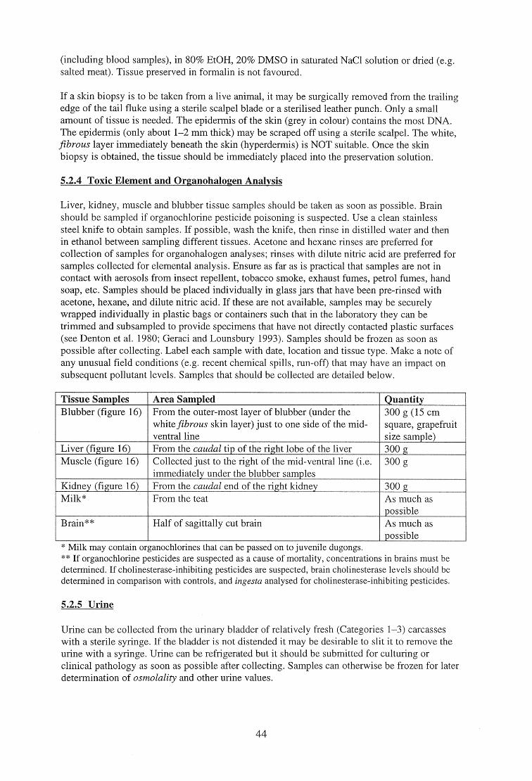
(including blood samples), in 80% EtOH, 20% DMSO in saturated NaC1 solution or dried (e.g. salted meat). Tissue preserved in foinialin is not favoured.
If a skin biopsy is to be taken from a live animal, it may be surgically removed from the trailing edge of the tail fluke using a sterile scalpel blade or a sterilised leather punch. Only a small amount of tissue is needed. The epidermis of the skin (grey in colour) contains the most DNA. The epidermis (only about 1-2 mm thick) may be scraped off using a sterile scalpel. The white, fibrous layer immediately beneath the skin (hyperdermis) is NOT suitable. Once the skin biopsy is obtained, the tissue should be immediately placed into the preservation solution.
5.2.4 Toxic Element and Organohalogen Analysis
Liver, kidney, muscle and blubber tissue samples should be taken as soon as possible. Brain should be sampled if organochlorine pesticide poisoning is suspected. Use a clean stainless steel knife to obtain samples. If possible, wash the knife, then rinse in distilled water and then in ethanol between sampling different tissues. Acetone and hexane rinses are preferred for collection of samples for organohalogen analyses; rinses with dilute nitric acid are preferred for samples collected for elemental analysis. Ensure as far as is practical that samples are not in contact with aerosols from insect repellent, tobacco smoke, exhaust fumes, petrol fumes, hand soap, etc. Samples should be placed individually in glass jars that have been pre-rinsed with acetone, hexane, and dilute nitric acid. If these are not available, samples may be securely wrapped individually in plastic bags or containers such that in the laboratory they can be trimmed and subsampled to provide specimens that have not directly contacted plastic surfaces (see Denton et al. 1980; Geraci and Lounsbury 1993). Samples should be frozen as soon as possible after collecting. Label each sample with date, location and tissue type. Make a note of any unusual field conditions (e.g. recent chemical spills, run-off) that may have an impact on subsequent pollutant levels. Samples that should be collected are detailed below.
Tissue Samples Area Sampled Quantity Blubber (figure 16) From the outer-most layer of blubber (under the
white fibrous skin layer) just to one side of the mid- ventral line
300 g (15 cm square, grapefruit size sample)
Liver (figure 16) From the caudal tip of the right lobe of the liver 300 g Muscle (figure 16) Collected just to the right of the mid-ventral line (i.e.
immediately under the blubber samples 300 g
Kidney (figure 16) From the caudal end of the right kidney 300 g Milk* From the teat As much as
possible Brain** Half of sagittally cut brain As much as
possible * Milk may contain organochlorines that can be passed on to juvenile dugongs. ** If organochlorine pesticides are suspected as a cause of mortality, concentrations in brains must be determined. If cholinesterase-inhibiting pesticides are suspected, brain cholinesterase levels should be determined in comparison with controls, and ingesta analysed for cholinesterase-inhibiting pesticides.
5.2.5 Urine
Urine can be collected from the urinary bladder of relatively fresh (Categories 1-3) carcasses with a sterile syringe. If the bladder is not distended it may be desirable to slit it to remove the urine with a syringe. Urine can be refrigerated but it should be submitted for culturing or clinical pathology as soon as possible after collecting. Samples can otherwise be frozen for later determination of osmolality and other urine values.
44
5.2.6 Haemolysed Blood
Haemolysed blood from very fresh carcasses (Categories 1-3) can be collected from the heart and stored frozen.
5.2.7 Microbiology
Sampling for microbiological testing may only be possible when a qualified professional (e.g. veterinarian, pathologist and/or trained microbiology technicians) is conducting the necropsy and when the tissues are in suitable condition (usually Categories 1-3). Transport swabs should be removed from their sterile wrapping, brushed against the lesion or fluid requiring sampling, and replaced in the media tube as swiftly as possible to avoid contamination. The tube should then be labelled, chilled, and submitted to a microbiology laboratory within 72 hours. Separate swabs should be taken from areas where the presence of pathogens is suspected. Specimens may also be collected using rat-toothed forceps and scissors (from sterile packets) and placed in sterile plastic vials on ice. Sampling of the pleural surface of the lungs, bronchi, stomach mucosa, perineal fluid, fluid in the pericardial sac, brain surfaces, abscesses, or infected areas is recommended for relatively fresh (Categories 1-3) carcasses.
45
6 T NSPORTATION OF SPECIMENS
All samples to be transported should be packaged in heavy-duty containers and should comply with the appropriate local protocol. It is important that all agencies involved in sending preserved samples are aware of the local regulations. Specimens sent in bottles should be sealed with tape and enclosed in sealed plastic bags. Specimens can also be sent wrapped; place tissues in paper towels moistened with 10% formalin and then place wrapped sample in a sealed bottle or in two sealed plastic bags (see Webb 1998). Place samples in a strong insulated container or cardboard box. Addresses on the transport box should be clearly marked. The inside of the container should contain a duplicate address and information specifying the material enclosed and nature of the shipment. Enclose a copy of the data sheet to provide pertinent information on the carcass.
Frozen samples must be shipped in very sturdy ice chests that will not break in transit. Use ice bricks to ensure samples stay cold. For long shipments, a substantial quantity of dry ice should be included. Three kilograms of dry ice lasts approximately 72 hours. Arrangements must be made with the receiver prior to shipping so that someone will be present to promptly handle the package and properly deal with the contents at the destination. Courier service and airfreight are acceptable. In the latter case, choose flights with a minimum number of connections, preferably at cooler times of the day. Inform the recipient by telephone of the identification number at the time of shipping in case the shipment is lost and requires tracing. In Australia, dry ice is considered dangerous goods and requires shipment in a specific foam container and accompanying paperwork filled out by a Dangerous Goods Officer. Therefore, prior to sending specimens in dry ice, the sender needs to liaise with a Dangerous Goods Officer in order to obtain instructions on packaging and paperwork.
In Queensland, Australia, the Department of Primary Industries Rockhampton Veterinary Laboratory supplies a specimen pack used for shipping diagnostic specimens' by road transit (i.e. by bus). The pack is designed to provide three waterproof layers around the specimens being shipped. The pack consists of a wax impregnated cardboard box, which contains a square gusseted plastic liner bag of the same design and density plastic as that used in export meat boxes. The specimens, preserved in appropriate jars, are packed into the plastic bag lined wax box, surrounded with sufficient absorbent material (i.e. clean cotton wool/wadding, shredded or crumpled newspaper) to absorb leakage. Several cold bricks should be included. The contents of the specimen pack are designed to provide workers with sufficient material for specimen collection and include:
• • • •
2 large seal-able plastic bags 6 medium plastic jars 4 large plastic jars 5 x 70 ml sterile jars
• • •
20 x 20 ml bottles 6 scalpel blades (No. 5 ice bricks
22)
Bone specimens can be wrapped in protective paper or plastic and packed in styrofoam chips or an equivalent alternative.
International shipment of samples must comply with the local requirements of the Convention on International Trade in Endangered Species of Wild Fauna and Flora (CITES). In Australia, the Wildlife Protection (Regulation of Exports and Imports) Act 1982 is the legislative basis for conservation-orientated controls on the export and import of wildlife and wildlife products.
Diagnostic specimens are any animal material including, but not limited to, excreta, secreta, blood and its components, tissue and tissue fluids, being shipped for purposes of diagnosis, but excluding live infected animals.
46
Controls under this Act apply to transactions undertaken by museums, zoos, scientific institutions, commercial organisations, tourists, migrants and the general public. The Act controls the export of most Australian native animals and plants and fulfils Australia's legislative requirements as a signatory to CITES.
47
7 DETERMINATION OF CAUSES OF DEATH
Careful observations of stranded marine mammals can document evidence of natural mortality, such as disease and predation, as well as human-induced trauma, such as entanglement in fishing gear, vessel collisions, gunshot wounds and knife marks. It is important to determine if injuries occurred before or after death (see Section 4.2).
7.1 Disease
Dugongs are susceptible to a wide range of diseases, some of them infectious or parasitic. These include pneumonia, pancreatitis and dermatitis. A species of Cryptosporidium, a small apicomplexan protozoan inhabiting the respiratory and gastrointestinal tracts of a wide range of vertebrates, was found in the small intestine of a dugong (Hill et al. 1997). Wild dugongs support a range of parasites, including at least 19 species of trematodes and one species of nematode internally, and a barnacle and a copepod externally. Dugongs may also carry a range of other diseases documented in marine mammals such as leptospirosis, lobomycosis, cryptococcosus, blastomycosis, caliciviruses, salmonellosis, morbillivirus, toxoplasmosis, tuberculosis and hepatitis (Bryden et al. 1998, Smith et al. 1978). Elliot et al. (1981) describes a fatal case of salmonellosis. Diagnosis of disease generally requires corroboration by qualified professional pathologists with supporting evidence from histopathology and microbiology. Samples should be sent to pathologists and other specialists for verification.
Instruments, surfaces, clothing etc. should be properly disinfected after the necropsy is completed and the carcass disposed. Care should be taken to avoid cross-contamination of clothing during cleaning (see Section 3.1).
The following is an example of a dugong diagnosed as dying from a bacterial peritonitis infection:
A subadult male dugong, estimated to be about eight to nine years old and which measured 235 cm in length was reported floating offshore of Cardwell in northern Queensland, Australia. The carcass was in reasonably fresh condition, estimated to have been dead less than 24 hours. The Queensland Parks and Wildlife Service recovered the carcass, which was transported to James Cook University for a necropsy by a Queensland Department of Primary Industries veterinarian.
External examination An examination of the external surfaces of the carcass did not reveal any unusual markings or indications of disease processes, except that bloody fluid was draining from both nares. There was a light to moderate covering of barnacles on the dorsal surface of the animal. There was no evidence of shark bites; both flippers and the tail were intact.
Internal examination (figure 17) When the carcass was opened for internal examination, a substantial subcutaneous fat layer was present. The abdominal cavity was full of a copious amount of peritoneal fluid that was red/green in colour and of a foul odour. A large amount offibrin was adhered to the viscera, especially to a small section of the small intestine, holding it to the dorsal body wall. Faecal material in the large intestine was hardened indicating the animal may have been dehydrated prior to death.
The stomach was full of seagrass and a few nematodes were present within the cardiac gland. A large amount of blood was present in the pericardial sac and within the pleural cavity. An area of haemorrhage was noted at the points of attachment of the spleen to the visceral surface of the stomach.
48
Tissue samples and swabs of major organs and areas of excess fluidlhaemorrhage were taken for histology, virology, and bacteriology.
Cause of Death Severe bacterial peritonitis with haemorrhagic septicaemia, as evidenced from post-mortem, histological examination and bacterial isolation. The bacterial peritonitis and haemorrhagic septicaemia were judged to have been caused by a toxin producing bacterium, Clostridium sporogenes which is presumed to have entered the abdominal cavity via an intestinal perforation, caused by the penetration of an unidentified helminth parasite, as seen on post mortem and histological examination.
7.2 Predation
There is little information about non-human predation on dugongs. There is evidence of dugongs being preyed upon by killer whales in Shark Bay in Western Australia (Bryden et al. 1998). Specimens bearing large scars, which indicate they have experienced and survived attacks by large sharks, are occasionally sighted. There are two accounts of shark attacks on dugong calves in northern Australia and dugong remains are commonly found in the guts of tiger sharks in Shark Bay (N. Gales, pers comm. 1999). Observers in aircraft have photographed dugongs in the jaws of crocodiles off Cape York Peninsula on at least two occasions; however, it was not known whether the dugongs died before or after the attack (Bryden et al. 1998).
7.3 Dependent Calves
Calves which die at or soon after birth can be distinguished by: (1) the presence of meconium in the digestive tract (but no milk or vegetation), (2) an unhealed (a non-involuted) umbilicus, and clotted blood in the umbilical artery and (3) the horny plates in the mouth are unstained by seagrass. Morphological abnormalities or birth defects may contribute to an early death, particularly deformities of the heart. Signs of starvation (see below) or anorexia are sometimes encountered in small calves, indicating prolonged separation from mothers.
7.4 Starvation
Carcasses show emaciation (dorsal prosis evident), and fat undergoes serous atrophy, leaving a clear or jell-like appearance to remaining deposits, which are generally reduced in starving animals. This is most prominent around the ventral surfaces and interventricular groove of the heart. The presence of any clear vesicular gelatinous material adhering to the internal lining of the heart (this is also occasionally visible on the exterior surface) denotes a condition called cachexia (`water fat'). This is an indication of severe starvation in dugongs (an internal examination of the heart will also reveal this condition). The gastrointestinal tract may be empty or contain unusual material including algae, dead seagrass rhizomes and anoxic sediment. The gall bladder may be distended with bright yellow bile. Animals in this condition may also have infections. These may include multiple purulent abscesses in the dermis, musculature, umbilical vessels, and kidneys.
7.5 Vessel Strikes
Although there are few records of dugong deaths due to vessel strikes in Australian waters, the QPWS have recorded seven dugong carcasses with propeller cuts. Vessel strikes are a major cause of mortality for Florida manatees (Ackerman et al. 1995) and the rise of vessel traffic in the dugong's range is increasing the likelihood of strikes. Areas of particular risk occur where there are extensive shallow areas used by regionally important populations of dugongs that are close to recreational or commercial boating facilities (e.g. Moreton Bay, near Brisbane and Missionary Bay, near Cardwell, Queensland).
49
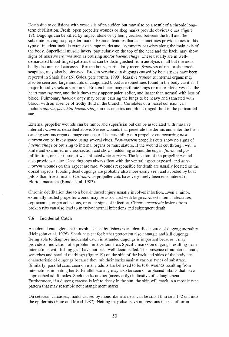
Death due to collisions with vessels is often sudden but may also be a result of a chronic long-term debilitation. Fresh, open propeller wounds or skeg marks provide obvious clues (figure 18). Dugongs can be killed by impact alone or by being crushed between the hull and the substrate leaving no propeller marks. External features that can sometimes provide clues to this type of incident include extensive scrape marks and asymmetry or twists along the main axis of the body. Superficial muscle layers, particularly on the top of the head and the back, may show signs of massive trauma such as bruising and/or haemorrhage. These usually are in well-demarcated blood-tinged patterns that can be distinguished from autolysis in all but the most badly decomposed carcasses. Broken bones, particularly recent fractures of ribs or shattered scapulae, may also be observed. Broken vertebrae in dugongs caused by boat strikes have been reported in Shark Bay (N. Gales, pers comm. 1999). Massive trauma to internal organs may also be seen and large amounts of coagulated blood are sometimes found in the body cavities if major blood vessels are ruptured. Broken bones may perforate lungs or major blood vessels, the heart may rupture, and the kidneys may appear paler, softer, and larger than normal with loss of blood. Pulmonary haemorrhage may occur, causing the lungs to be heavy and saturated with blood, with an absence of frothy fluid in the bronchi. Correlates of a vessel collision can include anuria, petechial haemorrhage in mesenteries and blood-tinged fluid in the pericardial sac.
External propeller wounds can be minor and superficial but can be associated with massive internal trauma as described above. Severe wounds that penetrate the dermis and enter the flesh causing serious organ damage can occur. The possibility of a propeller cut occurring post-mortem can be investigated using several clues. Post-mortem propeller cuts show no signs of haemorrhage or bruising to internal organs or musculature. If the wound is cut through with a knife and examined in cross-section and shows reddening around the edges,fibrin and pus infiltration, or scar tissue, it was inflicted ante-mortem. The location of the propeller wound also provides a clue. Dead dugongs always float with the ventral aspect exposed, and ante-mortem wounds on this aspect are rare. Wounds responsible for death are usually located on the dorsal aspects. Floating dead dugongs are probably also more easily seen and avoided by boat pilots than live animals. Post-mortem propeller cuts have very rarely been encountered in Florida manavtes (Bonde et al. 1983).
Chronic debilitation due to a boat-induced injury usually involves infection. Even a minor, externally healed propeller wound may be associated with large purulent internal abscesses, septicaemia, organ adhesions, or other signs of infection. Chronic osteolytic lesions from broken ribs can also lead to massive internal infections and subsequent death.
7.6 Incidental Catch
Accidental entanglement in mesh nets set by fishers is an identified source of dugong mortality (Heinsohn et al. 1976). Shark nets set for bather protection also entangle and kill dugongs. Being able to diagnose incidental catch in stranded dugongs is important because it may provide an indication of a problem in a certain area. Specific marks on dugongs resulting from interactions with fishing gear have not been well documented. The presence of numerous scars, scratches and parallel markings (figure 19) on the skin of the back and sides of the body are characteristic of dugongs because they rub their backs against various types of substrate. Similarly, parallel scars seen on many adults are believed to be tusk wounds resulting from interactions in mating herds. Parallel scarring may also be seen on orphaned infants that have approached adult males. Such marks are not (necessarily) indicative of entanglement. Furthermore, if a dugong carcass is left to decay in the sun, the skin will crack in a mosaic type pattern that may resemble net entanglement marks.
On cetacean carcasses, marks caused by monofilament nets, can be small thin cuts 1-2 cm into the epidermis (Hare and Mead 1987). Netting may also leave impressions instead of, or in
50
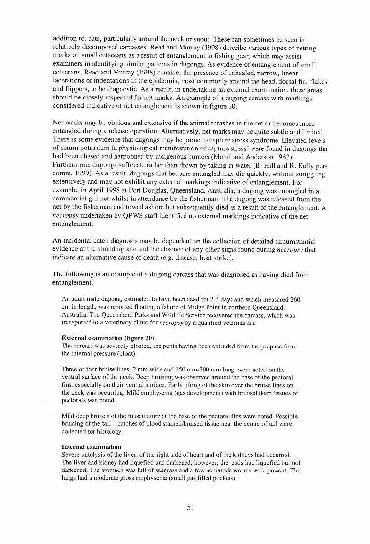
addition to, cuts, particularly around the neck or snout. These can sometimes be seen in relatively decomposed carcasses. Read and Murray (1998) describe various types of netting marks on small cetaceans as a result of entanglement in fishing gear, which may assist examiners in identifying similar patterns in dugongs. As evidence of entanglement of small cetaceans, Read and Murray (1998) consider the presence of unhealed, narrow, linear lacerations or indentations in the epidermis, most commonly around the head, dorsal fin, flukes and flippers, to be diagnostic. As a result, in undertaking an external examination, these areas should be closely inspected for net marks. An example of a dugong carcass with markings considered indicative of net entanglement is shown in figure 20.
Net marks may be obvious and extensive if the animal thrashes in the net or becomes more entangled during a release operation. Alternatively, net marks may be quite subtle and limited. There is some evidence that dugongs may be prone to capture stress syndrome. Elevated levels of serum potassium (a physiological manifestation of capture stress) were found in dugongs that had been chased and harpooned by indigenous hunters (Marsh and Anderson 1983). Furthermore, dugongs suffocate rather than drown by taking in water (B. Hill and R. Kelly pers comm. 1999). As a result, dugongs that become entangled may die quickly, without struggling extensively and may not exhibit any external markings indicative of entanglement. For example, in April 1998 at Port Douglas, Queensland, Australia, a dugong was entangled in a commercial gill net whilst in attendance by the fisherman. The dugong was released from the net by the fisherman and towed ashore but subsequently died as a result of the entanglement. A necropsy undertaken by QPWS staff identified no external markings indicative of the net entanglement.
An incidental catch diagnosis may be dependent on the collection of detailed circumstantial evidence at the stranding site and the absence of any other signs found during necropsy that indicate an alternative cause of death (e.g. disease, boat strike).
The following is an example of a dugong carcass that was diagnosed as having died from entanglement:
An adult male dugong, estimated to have been dead for 2-3 days and which measured 260 cm in length, was reported floating offshore of Midge Point in northern Queensland, Australia. The Queensland Parks and Wildlife Service recovered the carcass, which was transported to a veterinary clinic for necropsy by a qualified veterinarian.
External examination (figure 20) The carcass was severely bloated, the penis having been extruded from the prepuce from the internal pressure (bloat).
Three or four bruise lines, 2 mm wide and 150 mm-200 mm long, were noted on the ventral surface of the neck. Deep bruising was observed around the base of the pectoral fins, especially on their ventral surface. Early lifting of the skin over the bruise lines on the neck was occurring. Mild emphysema (gas development) with bruised deep tissues of pectorals was noted.
Mild deep bruises of the musculature at the base of the pectoral fins were noted. Possible bruising of the tail — patches of blood stained/bruised tissue near the centre of tail were collected for histology.
Internal examination Severe autolysis of the liver, of the right side of heart and of the kidneys had occurred. The liver and kidney had liquefied and darkened; however, the testis had liquefied but not darkened. The stomach was full of seagrass and a few nematode worms were present. The lungs had a moderate gross emphysema (small gas filled pockets).
51
Samples were taken for histopathology. There were no signs of disease or infection within the animal.
Cause of Death The bruising around the neck and pectorals is consistent with entanglement with some type of rope. The air embolisms on the tissue surface of the right lung are consistent with suffocation/drowning.
Given these findings, and the lack of any other obvious causes of death, this animal is judged to have died as a result of entanglement (i.e. suffocation). The excessive degenerative change of the liver, kidney and heart may be the result of a struggle at the time of death, raising the animal's internal body temperature while depleting the intracellular energy reserves.
7.7 Other Human-related Causes
Gunshot wounds leave small entry holes on the external surface, which must be searched for carefully during necropsy. Buckshot or other foreign objects resulting from non-lethal vandalism may be encountered while removing skin or flesh, for example, in the snout region. Radiography of suspect areas will help locate foreign objects. If bullets are found, they should be saved for law enforcement personnel.
Dugongs that have been killed for meat usually have large pieces of flesh missing from the carcass, often from the abdomen.
There is little information available on contaminant levels in dugong and any subsequent effects of pollutants such as potential reproductive failure and reduced immune system capability. Concentrations of polychlorinated dibenzo-p-dioxins in the tissues of three dugongs from the Great Barrier Reef were higher than reported for other marine mammals (Haynes et al. 1999) but the significance of this to dugong ecology and physiology is unknown.
Underwater explosions are difficult to diagnose from a cause-of-death standpoint because the trauma inflicted could merely be enough to cause the animal to lose consciousness. However, more severe injuries may include massive trauma and internal injury.
7.8 Undetermined
Cases can be classified as undetermined if no cause of death is apparent following necropsy and subsequent histopathology.
52
8 GLOSSARY'
Abscess - a localised collection of pus in a cavity formed by disintegration of tissues.
Adhesion - the stable joining of parts to one another, usually abnormally.
Aneurysm - a sac formed by localised dilation of a blood vessel.
Anorexia - loss of appetite for food.
Anterior - in front of (toward the head end of the body).
Ante-mortem - before death.
Anuria - absence of urine in bladder ndicating possible suppression of urine formation by the
kidney. Aspiration - the act of inhaling.
Atrophy - a wasting away, causing a reduction in size.
Axial Skeleton - the skeleton of the head and trunk.
Axilla - the armpit, in dugongs the area between the flipper and the thorax wall.
Cachexia - profound and marked state of constitutional disorder; general ill health and
malnutrition.
Caudal - pertaining to or towards the tail.
Chicken fat clot - a greyish-yellow blood clot, due to the settling of red blood cells before
clotting. Coarctation - stricture or narrowing.
Coeliac - pertaining to the abdomen.
Congenital - present at or existing from the time of birth.
Cranial - pertaining to or towards the head.
Cyst - a closed bladder-like sac formed in animal tissue, usually containing fluid or a semi-solid
material.
Dilatation - a condition of being expanded or stretched beyond normal dimensions.
Distal - remote; farther from any point of reference.
Emaciation - excessively thin, wasted condition of the body.
Embolus (plural Emboli) - undissolved material carried by the blood flow and impacted in some
part of the blood vascular system as thrombi or fragments of
thrombi, tissue fragments, clumps of bacteria, protozoan parasites,
fat globules or gas bubbles.
'Definitions adapted from The American Heritage Dictionary of the English Language (Morris, W. ed. 1976, Houghton Mifflin Company, Boston, Mass, 1550 pp.), Dorland's Pocket Medical Dictionary (Friel, J. P. ed. 1977, 22nd ed., W. B. Saunders Company, Philadelphia, Penn, 741 pp.); Veterinary Pathology (Smith, H. A., Jones, T. C. and Hunt, R. D. 1972, 4th ed. Lea and Febiger, Philadelphia, Penn, 1521 pp.); and Bailliere's Comprehensive Veterinary Dictionary (Blood, D. C. and Studdert, V. P. 1988, Bailliere's Tindall, Sydney, NSW, Australia, 1123 pp).
53
Enteritis - inflammation of the intestine.
External auditory meatus - external opening of the ear.
Fascia - a sheet or band of fibrous tissue.
Fibrin - a dull white stringy material formed by the coagulation of fibrinogen.
Fibrinous - pertaining to or of the nature of fibrin.
Fibrosis - a formation of fibrous tissue.
Fibrous - composed of or containing fibres.
Foci - small discrete points or areas.
Fornix - an archlike structure or space.
Fracture - the breaking of a part, especially a bone.
Friable - easily pulverised or crumbled.
Haemorrhage - the escape of blood from vessels; bleeding.
Haematoma - a bruise or collection of blood in a tissue.
Haemostat - an instrument or agent used to compress or trap bleeding of blood vessels to stop
haemorrhage.
Hepatisation - transformation into a firm mass, liver-like in texture.
Hypertrophy - increase or enlargement of an organ or part, due to an increase in the size of its
constituent cells.
Impaction - being wedged in firmly.
Inferior - situated below, or directed downward.
Inflammation - a localised protective tissue response elicited by injury or destruction of tissues,
which serves to destroy, dilute, or wall off both the injurious agent and the
injured tissue.
Inflation - distention or the act of distending, with air, gas or fluid.
Ingesta - material taken into the body by the mouth.
Ketone - any compound containing the carbonyl group, CO, and having hydrocarbon groups
attached to the carbonyl carbon.
Lateral - denoting a position farther from the medial plane or midline; side.
Lesion - a pathologic or traumatic discontinuity of tissue.
Lumen - a cavity or channel within a tube or organ.
Meconium - dark green mucilaginous material in the intestine of the foetus.
Medial - situated towards the midline.
Mucosa - mucus-producing membrane; this includes the tissue lining the lumen of the
gastrointestinal tract and the urinary bladder.
Necropsy - examination of a body after death.
Necrosis - death of individual cells or groups of cells.
54
Nodules - a small node that is solid and can be detected by touch.
Oedema - an abnormal accumulation of fluid in a body cavity or in the intercellular spaces of a
tissue or organ, usually leading to swelling.
Oedematous - characterised or pertaining to oedema.
Osmolality - the concentration of a solution in terms of osmoles of solutes per kilogram of
solvent.
Osteolytic - dissolution of bone.
Parietal peritoneum - a serous membrane lining the walls of the visceral cavity.
Parturient - giving birth or pertaining to birth.
Patent - open, unobstructed, or not closed.
Patency - state of being open, i.e. not being blocked or obstructed.
Pathogen - any disease-producing agent or microorganism.
Peduncle - the narrow area between the body and the tail of the dugong.
Petechia (plural Petechiae)- a minute red spot due to the escape of a small amount of blood.
Placental scars - conspicuous purple bands in the uterine endometrium.
Plaque - a small, flat, rounded formation or area, as a deposit of fibrous matter in the wall of a
blood vessel or localised patch of skin disease.
Pneumonia - inflammation of the lungs with exudate and consolidation.
Polyp - a growth or mass protruding from a mucous membrane.
Posterior - directed towards or situated at the back.
Post-mortem - performed or occurring after death.
Proximal - nearest to the point of reference.
Purulent - containing or forming pus.
Pus - a protein-rich liquid inflammation product made up of cells (leukocytes), a thin fluid
(liquor puris) and cellular debris.
Rupture - tearing of tissue.
Serosa - serum-producing membrane; outermost surface or wall of the gastrointestinal tract.
Serous - pertaining to or resembling serum; usually a watery fluid.
Spicule - a sharp needle-like body.
Stenosis - narrowing or contraction of a body passage or opening.
Thrombus (plural Thrombi) - a fibrinous clot which forms in and obstructs a blood vessel, or
which forms in one of the heart's chambers.
Trauma - a wound or injury.
Tubercle - small rounded projection or small firm nodule or swelling.
Tumour - swelling; a new growth of tissue.
Ulcer - a local defect produced by sloughing of necrotic inflammatory tissue.
55
Vascular - pertaining to blood supply.
Verrucous - rough, warty.
Vesicle - a small bladder or sac containing fluid; a small blister.
ACKNOWLEDGEMENTS
Many people have assisted in the preparation of this manual. We would like to thank the many reviewers who commented on the draft manuscript including those from the Queensland Parks and Wildlife Service (Ian Bell, Grahame Byron, Dr Sandy Clague, Dr Don Cook, Artie Jacobson, Dr Jeff Miller, John Olds, Paul O'Neill, Dave Orgill, Dr Mark Read, David Savage, Michael Short, Cathy Skippington, Kirstin Wortel); Marine Parks Management Coordination Unit (Malcolm Turner); the Great Barrier Reef Marine Park Authority (Ken Anthony, David Haynes, Tony Stokes); the Queensland Department of Primary Industries (Dr Bruce Hill, Dr John Norton, Dr Rachel Webb); Veterinarians (Dr David Jaimson, Dr Tim Annand); the Commonwealth Scientific and Industrial Research Organisation (Hamish Malcolm); the University of Queensland (Dr Janet Lanyon - Department of Zoology, Dr Roger Kelly - School of Veterinary Science); the Department of Conservation and Land Management in Western Australia (Dr Nick Gales); Seaworld (Dr. Wendy Blanshard) and James Cook University (Dr George Heinsohn).
This project was funded by the Great Barrier Reef Marine Park Authority, James Cook University and the Cooperative Research Centre (CRC) for the Reef Research.
57
REFERENCES
Ackerman, B. B., Wright, S. D., Bonde, R. K., Odell, D. K. & Banowetz, D. J. 1995, 'Trends and patterns in mortality of manatees in Florida, 1974-1992', pp. 223-258 in Population Biology of the Florida Manatee (Trichechus manatus latirostris), eds T. J. O'Shea, B. B. Ackerman & H. F. Percival, National Biological Service, Information and Technology Report 1.
Blair, D. 1981, `Helminth parasites of the dugong, their collection and preservation', pp. 275- 285 in The Dugong: Proceedings of a seminar/workshop held at James Cook University 8-13 May 1979, ed. H. Marsh, James Cook University, Townsville, Queensland, Australia.
Bonde, R. K., O'Shea, T. J. & Beck, C. A. 1983. Manual of Procedures for the Salvage and Necropsy of Carcasses of the West Indian Manatee (Trichechus manatus), National Technical Information Service, Springfield, VA. Doc. No. PB83-255273,175 pp.
Bryden, M., Marsh, H. & Shaughnessy, P. 1998, Dugongs, Whales, Dolphins and Seals. A Guide to the Sea Mammals of Australasia, Allen & Unwin: St. Leonards NSW.
Denton, G. R., Marsh, H., Heinsohn, G. E. & Burdon-Jones, C. 1980, 'The unusual metal status of the dugong (Dugong dugon)' , Marine Biology, 57: 201-219.
Elliot, H., Thomas, A., Ladds, P. N. & Heinsohn, G. E. 1981, 'A fatal case of salmonellosis in a dugong', Journal of Wildlife Diseases, 17: 203-208.
Geraci, J. R. & Lounsbury, V. J. 1993, Marine Mammals Ashore: A Field Guide for Strandings, Texas A&M University Sea Grant College Program: Texas. TAMU-SG-93-601, 305 pp. (Updated in 1998 and available on CD-ROM from the National Aquarium, 501 E. Pratt St., Baltimore, MD 21202, USA.)
Great Barrier Reef Marine Park Authority 1981, Nomination of the Great Barrier Reef by the Commonwealth of Australia for inclusion in the World Heritage List, Unpublished report to the World Heritage Committee, UNESCO, 14 pp.
Hare, M. P. & Mead, J. G. 1987, Handbook for Determination of Adverse Human-marine Mamma Interactions from Necropsies, Marine Mammal Program, National Museum of Natural History, Smithsonian Institution, Washington, D.C., NWAFC Processed Report 87-06,35 pp.
Haynes, D., Muller, J. & McLauchlan, M. S. 1999, 'Polychlorinated dibenzo-p-dioxins and dibenzofurans in Great Barrier Reef (Australia) dugongs (Dugong dugon)' , Chemosphere, 38: 255-262.
Heinsohn, G. E. 1981, 'Methods of taking measurements, other data and specimen material from dugong carcasses', pp. 228-238 in The Dugong: Proceedings of a seminar/workshop held at James Cook University 8-13 May 1979, ed. H. Marsh, James Cook University, Townsville, Queensland, Australia.
Heinsohn, G. E., Marsh, H. & Spain, A. V. 1976, 'Extreme risk of mortality to dugongs (Mammalia: Sirenia) from netting operations', Australian Journal of Wildlife Research, 3: 117— 121.
Hill, B. D., Fraser, I. R. & Prior, H. C. 1997, `Cryptosporidium infection in a dugong (Dugong dugon)' , Australian Veterinary Journal, 75(9): 670-671.
Husar, S. 1978, 'Dugong dugon', Mammalian Species, 88: 1-7.
58
International Union for the Conservation of Nature 1996, IUCN Red List of Threatened Animals, eds J. Baillie & B. Groombride, IUCN, Gland, Switzerland.
Marsh, H. 1980, 'Age determination of the dugong (Dugong dugon (Muller)) in Northern Australia and its biological implications', Report of the International Whaling Commission (Special Issue), 3: 181-201.
Marsh, H. 1981, 'Preliminary description of the reproductive organs of the female dugong and suggested methods of specimen collection as part of a carcass salvage program', pp. 248-260 in The Dugong: Proceedings of a seminar/workshop held at James Cook University 8-13 May 1979, ed. H. Marsh, James Cook University, Townsville, Queensland, Australia.
Marsh, H. 1995, 'The life history, pattern of breeding and population dynamics of the dugong,' pp. 75-83 in Population Biology of the Florida Manatee, eds T. J. O'Shea, B. B. Ackerman & H. F. Percival, U. S. Department of the Interior, National Biological Service, Information and Technology Report 1, Washington, D.C.
Marsh, H. 1999, 'Reproduction in sirenians', pp. 243-256 in 'Reproduction in marine mammals', I. L. Boyd, C. Lockyer & H. D. Marsh, pp. 219-286 in Biology of Marine Mammals, eds J. E. Reynolds, III & S. A. Rommel, Smithsonian Institution Press, Washington D.C.
Marsh, H. & Anderson, P. K. 1983, 'Probable susceptibility of dugongs to capture stress', Biological Conservation, 25: 1-3.
Marsh, H. & Glover, T. D. 1981, 'A preliminary description of the reproductive organs of the male dugong and suggested methods of specimen collection', pp. 261-274 in The Dugong: Proceedings of a seminar/workshop held at James Cook University 8-13 May 1979, ed. H. Marsh, James Cook University, Townsville, Queensland, Australia.
Marsh, H. & Lefebvre, L. W. 1994, `Sirenian status and conservation efforts', Aquatic Mammals, 20: 155-170.
Marsh, H., Heinsohn, G. E. & Spain, A. V. 1977, 'The stomach and duodenal diverticula of the Dugong (Dugong dugon)' , pp. 271-295 in Functional Anatomy of Marine Mammals, ed. R .J. Harrison, Academic Press, London.
Marsh, H., Channells, P. W., Heinsohn, G. E. & Morissey, J. 1982, 'Analysis of stomach contents of dugongs from Queensland', Australian Wildlife Research, 9: 55-67.
Marsh, H., Heinsohn, G. E. & Channells, P. W. 1984a, 'Changes in the ovaries and uterus of the dugong, Dugong dugon (Sirenia: Dugongidae) with age and reproductive activity', Australian Journal of Zoology, 32: 743-766.
Marsh, H., Heinsohn, G. E. & Glover, T. D. 1984b, 'Changes in the male reproductive organs of the dugong, Dugong dugon (Sirenia: Dugongidae) with age and reproductive activity', Australian Journal of Zoology, 32: 721-742.
Marsh, H., Heinsohn, G. E. & Marsh, L. M. 1984c, 'Breeding cycle, life history and population dynamics of the dugong, Dugong dugon (Sirenia: Dugongidae)', Australian Journal of Zoology, 32: 767-785.
59
Preen, A. R. 1995, 'Diet of dugongs: are they omnivores?', Journal of Mammalogy, 76: 163— 171.
Queensland Parks and Wildlife Service & Great Barrier Reef Marine Park Authority 1999, A review of Queensland's dugong stranding and carcass response procedures, QPWS, Brisbane, Queensland, Australia, 33 pp. + attachments.
Rainey, W. E. 1981, 'Procedure for collection of dugong tissues for electrophoresis', pp. 240— 247 in The Dugong: Proceedings of a seminar/workshop held at James Cook University, 8-13 May 1979, ed. H. Marsh, James Cook University, Townsville, Queensland, Australia.
Read, A.J. & Murray, K. T. 1998, Gross evidence of human-induced mortality in small cetaceans, NOAA Technical Memorandum (in prep.), 21 pp.
Rowlatt, U. & Marsh, H. 1985, 'The heart of the dugong (Dugong dugon) and the West Indian Manatee (Trichechus manatus) (Sirenia)', Journal of Morphology, 186: 95-105.
Smith, A. W., Vedrus, N. A., Akers, T. G. & Gilmartin, W. E. 1978, 'Hazards of disease transfer from marine mammals to land mammals: review and recent findings', Journal of the American Veterinary Medical Association, 173(9): 1131-1133.
Tikel, D. 1998, Using a Genetic Approach to Optimise Dugong (Dugong dugon) Conservation Management, PhD Thesis, James Cook University, Townsville, Queensland, Australia, 277 pp.
Webb, R. 0. 1998, Cultured Aquatic Animals. Preparation for Veterinary Laboratory Examination (Second Edition), Queensland Department of Primary Industries, Oonoonba Veterinary Laboratory, Townsville, Queensland, Australia.
60

ATTACHMENTS
61
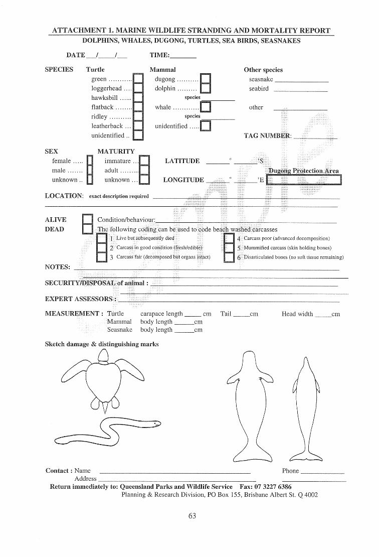
ATTACHMENT 1. MAKINE WILDLIFE STRANDING AND MORTALITY REPORT DOLPHINS, WHALES, DUGONG, TURTLES, SEA BIRDS, SEASNAKES
DATE TIME:
SPECIES Turtle green
loggerhead
hawksbill
flatback
ridley
leatherback
unidentified
Mammal Other species
dugong seasnake
dolphin El seabird species
whale other species
unidentified
TAG NUMBER:
SEX MATURITY female immature ... LATITUDE male adult
unknown unknown ... LONGITUDE
-wag, Protection i-L-ea
LOCATION: exact description required
ALIVE
DEAD
Condition/behaviour:
The following coding can he used to code beach washed carcasses
1 Live but subsequently died
2 Carcass in good condition (fresh/edible)
3 Carcass fair (decomposed but organs intact)
4 Carcass poor (advanced decomposition)
5 Mummified carcass (skin holding bones)
6 Disarticulated bones (no soft tissue remaining)
NOTES:
SECURITY/DISPOSAL of ar,ini :
EXPERT ASSESSORS :
MEASUREMENT: Turtle carapace length cm Tail cm Head width cm
Mammal body length cm Seasnake body length cm
Sketch damage & distinguishing marks
Contact : Name Address
Phone
Return immediately to: Queensland Parks and Wildlife Service Fax: 07 3227 6386 Planning & Research Division, PO Box 155, Brisbane Albert St. Q 4002
63
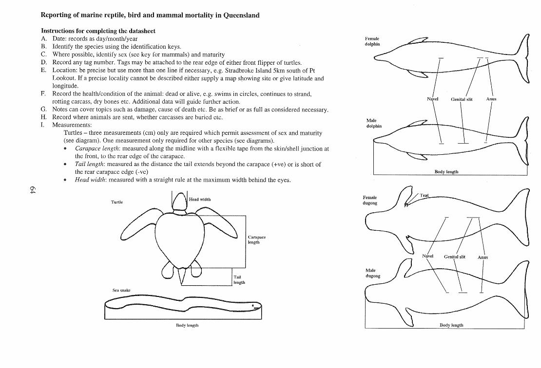
N ye Genita slit Anus
Teat
Reporting of marine reptile, bird and mammal mortality in Queensland
Instructions for completing the datasheet Date: records as day/month/year Female
dolphin Identify the species using the identification keys. Where possible, identify sex (see key for mammals) and maturity Record any tag number. Tags may be attached to the rear edge of either front flipper of turtles. Location: be precise but use more than one line if necessary, e.g. Stradbroke Island 5km south of Pt Lookout. If a precise locality cannot be described either supply a map showing site or give latitude and longitude. Record the health/condition of the animal: dead or alive, e.g. swims in circles, continues to strand, rotting carcass, dry bones etc. Additional data will guide further action. Notes can cover topics such as damage, cause of death etc. Be as brief or as full as considered necessary. Record where animals are sent, whether carcasses are buried etc.
Male Measurements: dolphin
Turtles — three measurements (cm) only are required which permit assessment of sex and maturity (see diagram). One measurement only required for other species (see diagrams).
Carapace length: measured along the midline with a flexible tape from the skin/shell junction at the front, to the rear edge of the carapace. Tail length: measured as the distance the tail extends beyond the carapace (+ve) or is short of the rear carapace edge (-ye) Head width: measured with a straight rule at the maximum width behind the eyes.
Carapace ength
Sea snake
Body length
Female dugong
Navel nital slit Anus
Male dugong
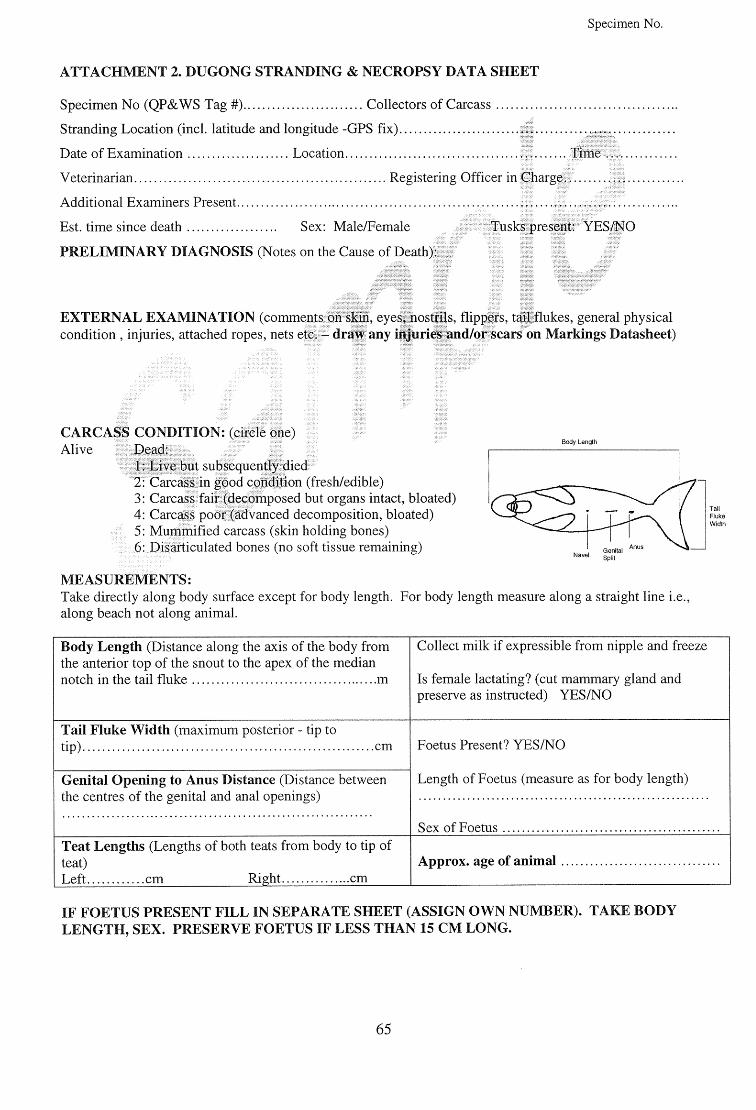
Body Length
Tail Fluke Width
Specimen No.
ATTACHMENT 2. DUGONG STRANDING & NECROPSY DATA SHEET
Specimen No (QP&WS Tag #). Collectors of Carcass
Stranding Location (incl. latitude and longitude -GPS fix)
Date of Examination Location Time
Veterinarian Registering Officer in Charge
Additional Examiners Present
Est. time since death Sex: Male/Female Tusks present YES/NO
PRELIMINARY DIAGNOSIS (Notes on the Cause of Death):
EXTERNAL EXAMINATION (comments on skin, eyes, nostrils, flippers, tail flukes, general physical condition , injuries, attached ropes, nets etc. — draw any injuries and/or scars on Markings Datasheet)
CARCASS CONDITION: (circle one) Alive Dead:
I :.Live•but subsequently died • Carcass in good condition (fresh/edible) Carcass fair (decomposed but organs intact, bloated) Carcass poor (advanced decomposition, bloated) Mummified carcass (skin holding bones) Disarticulated bones (no soft tissue remaining)
MEASUREMENTS: Take directly along body surface except for body length. For body length measure along a straight line i.e., along beach not along animal.
Body Length (Distance along the axis of the body from the anterior top of the snout to the apex of the median notch in the tail fluke . m
Collect milk if expressible from nipple and freeze
Is female lactating? (cut mammary gland and preserve as instructed) YES/NO
Tail Fluke Width (maximum posterior - tip to tip) cm Foetus Present? YES/NO
Length of Foetus (measure as for body length)
Sex of Foetus
Genital Opening to Anus Distance (Distance between the centres of the genital and anal openings)
Teat Lengths (Lengths of both teats from body to tip of teat) Left cm Right . ..cm
Approx. age of animal
IF FOETUS PRESENT FILL IN SEPARATE SHEET (ASSIGN OWN NUMBER). TAKE BODY LENGTH, SEX. PRESERVE FOETUS IF LESS THAN 15 CM LONG.
65
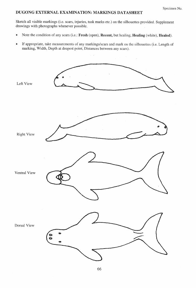
Specimen No. DUGONG EXTERNAL EXAMINATION: MARKINGS DATASHEET
Sketch all visible markings (i.e. scars, injuries, tusk marks etc.) on the silhouettes provided. Supplement drawings with photographs whenever possible.
Note the condition of any scars (i.e.: Fresh (open), Recent, but healing, Healing (white), Healed).
If appropriate, take measurements of any markings/scars and mark on the silhouettes (i.e. Length of marking, Width, Depth at deepest point, Distances between any scars).
Left View
Right View
Ventral View
Dorsal View
66
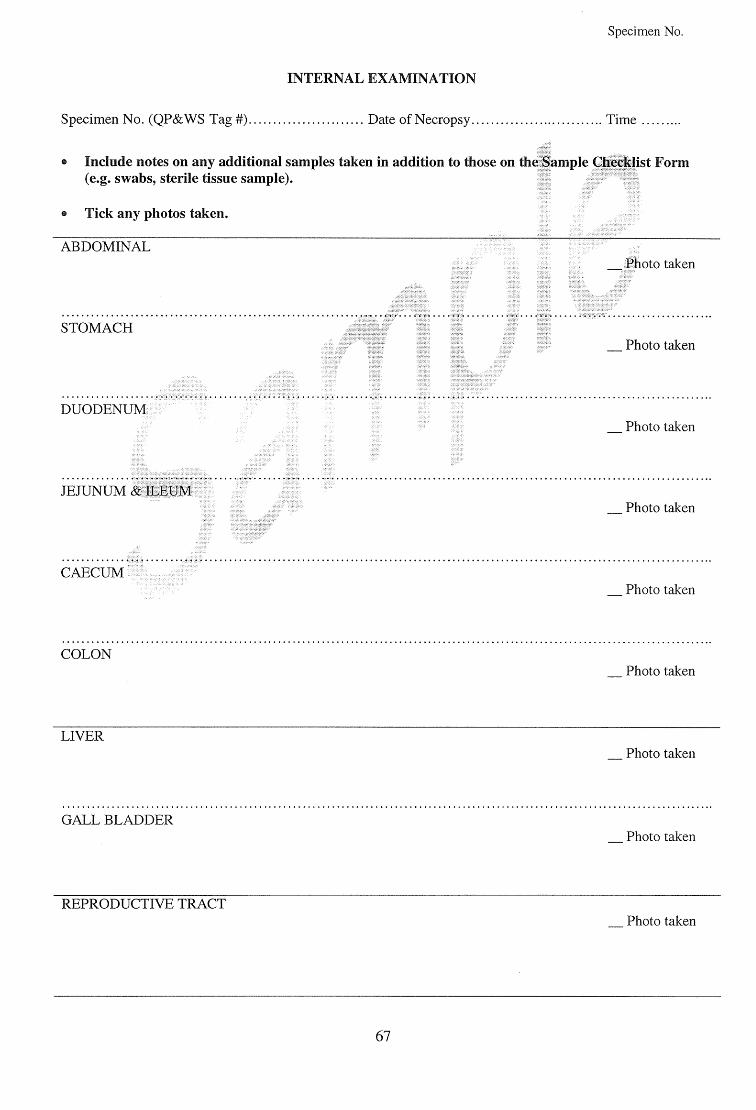
Specimen No.
INTERNAL EXAMINATION
Specimen No. (QP&WS Tag #) Date of Necropsy Time
Include notes on any additional samples taken in addition to those on the Sample Checklist Form (e.g. swabs, sterile tissue sample).
Tick any photos taken.
ABDOMINAL Photo taken
STOMACH Photo taken
DUODENUM ___ Photo taken
JEJUNUM & ILEUM Photo taken
CAECUM Photo taken
COLON ___ Photo taken
LIVER Photo taken
GALL BLADDER Photo taken
REPRODUCTIVE TRACT Photo taken
67
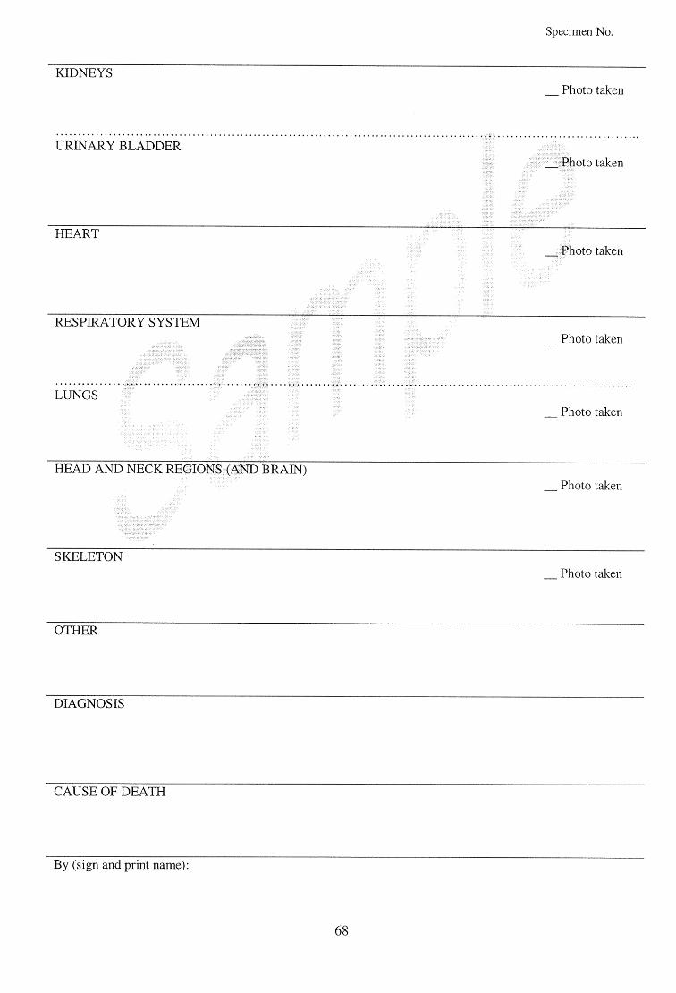
Specimen No.
KIDNEYS Photo taken
URINARY BLADDER Photo taken
HEART Photo taken
RESPIRATORY SYSTEM Photo taken
LUNGS ____ Photo taken
HEAD AND NECK REGIONS (AND BRAIN) Photo taken
SKELETON Photo taken
OTHER
DIAGNOSIS
CAUSE OF DEATH
By (sign and print name):
68
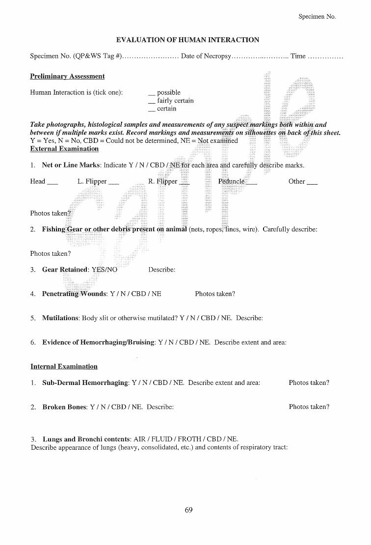
Specimen No.
EVALUATION OF HUMAN INTE • CTION
Specimen No. (QP&WS Tag #) Date of Necropsy Time
Preliminary Assessment
Human Interaction is (tick one): possible fairly certain certain
Take photographs, histological samples and measurements of any suspect markings both within and between if multiple marks exist. Record markings and measurements on silhouettes on back of this sheet. Y = Yes, N = No, CBD = Could not be determined, NE = Not examined External Examination
. Net or Line Marks: Indicate Y / N / CBD / NE for each area and carefully describe marks.
Head
L. Flipper R. Flipper Peduncle Other
Photos taken?
Fishing Gear or other debris present on animal (nets, ropes, lines, wire). Carefully describe:
Photos taken?
Gear Retained: YES/NO Describe:
Penetrating Wounds: Y / N / CBD / NE Photos taken?
Mutilations: Body slit or otherwise mutilated? Y / N / CBD / NE. Describe:
Evidence of Hemorrhaging/Bruising: Y / N / CBD / NE. Describe extent and area:
Internal Examination
Sub-Dermal Hemorrhaging: Y / N / CBD / NE. Describe extent and area:
Photos taken?
Broken Bones: Y / N / CBD / NE. Describe:
Photos taken?
Lungs and Bronchi contents: AIR / FLUID / FROTH / CBD / NE. Describe appearance of lungs (heavy, consolidated, etc.) and contents of respiratory tract:
69
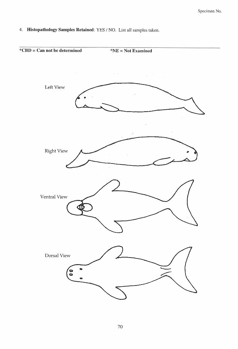
Ventral View
Dorsal View
Specimen No.
4. Histopathology Samples Retained: YES / NO. List all samples taken.
*CBD = Can not be determined *NE = Not Examined
Left View
70
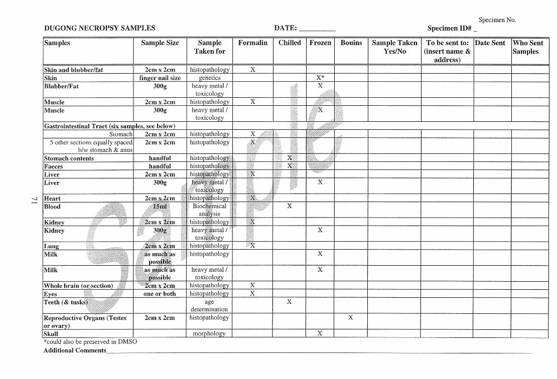
Specimen No. DUGONG NECROPSY SAMPLES
DATE:
Specimen ID# _
Samples Sample Size Sample Taken for
Formalin Chilled Frozen Bouins Sample Taken Yes/No
To be sent to: (insert name &
_ address)
Date Sent Who Sent Samples
Skin and blubber/fat 2cm x 2cm histopathology X
Skin finger nail size aenetics t, X* Blubber/Fat 300g heavy metal /
toxicology X
Muscle 2cm x 2cm histopathology X Muscle 300g heavy metal /
toxicology Gastrointestinal Tract (six samples, see below)
Stomach 2cm x 2cm histopathology X 5 other sections equally spaced
b/w stomach & anus 2cm x 2cm histopathology
Stomach contents handful histopathology Faeces handful histopathology Liver 2cm x 2cm histopathology Liver 300g heavy metal /
toxicology X
Heart 2cm x 2cm histopathology Blood 15ml Biochemical
analysis X
Kidney 2cm x 2cm histopathology Kidney 300g heavy metal /
toxicology X
Lung 2cm x 2cm histopathology X Milk as much as
possible.
histopathology X
_ Milk as much as
possible heavy metal /
toxicology X
. Whole brain (or section) 2cm x 2cm histopathology X Eyes one or both histopathology X
Teeth (& tusks) age determination _
X
— Reproductive Organs (Testes or ovary)
2cm x 2cm histopathology
_
X
Skull morphology X *could also be preserved in DMSO Additional Comments
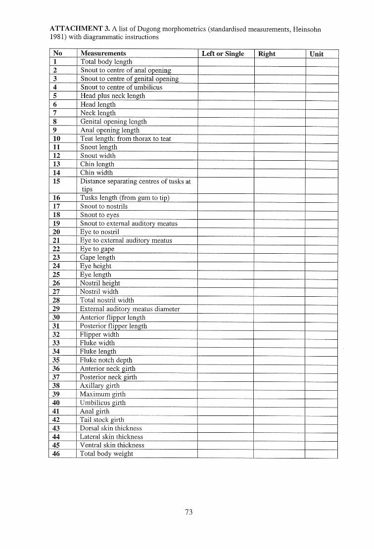
ATTACHMENT 3. A list of Dugong morphometrics (standardised measurements, Heinsohn 1981) with diagrammatic instructions
No Measurements Left or Single Right Unit 1 Total body length
Snout to centre of anal opening 2 3 Snout to centre of genital opening 4 Snout to centre of umbilicus 5 Head plus neck length 6 Head length 7 Neck length 8 Genital opening length 9 Anal opening length 10 Teat length: from thorax to teat 11 Snout length 12 Snout width 13 Chin length 14 Chin width 15 Distance separating centres of tusks at
tips 16 Tusks length (from gum to tip) 17 Snout to nostrils 18 Snout to eyes 19 Snout to external auditory meatus 20 Eye to nostril 21 Eye to external auditory meatus 22 Eye to gape 23 Gape length 24 Eye height 25 Eye length 26 Nostril height 27 Nostril width 28 Total nostril width 29 External auditory meatus diameter 30 Anterior flipper length 31 Posterior flipper length 32 Flipper width 33 Fluke width 34 Fluke length 35 Fluke notch depth 36 Anterior neck girth 37 Posterior neck girth 38 Axillary girth 39 Maximum girth 40 Umbilicus girth 41 Anal girth 42 Tail stock girth 43 Dorsal skin thickness 44 Lateral skin thickness 45 Ventral skin thickness 46 Total body weight
73
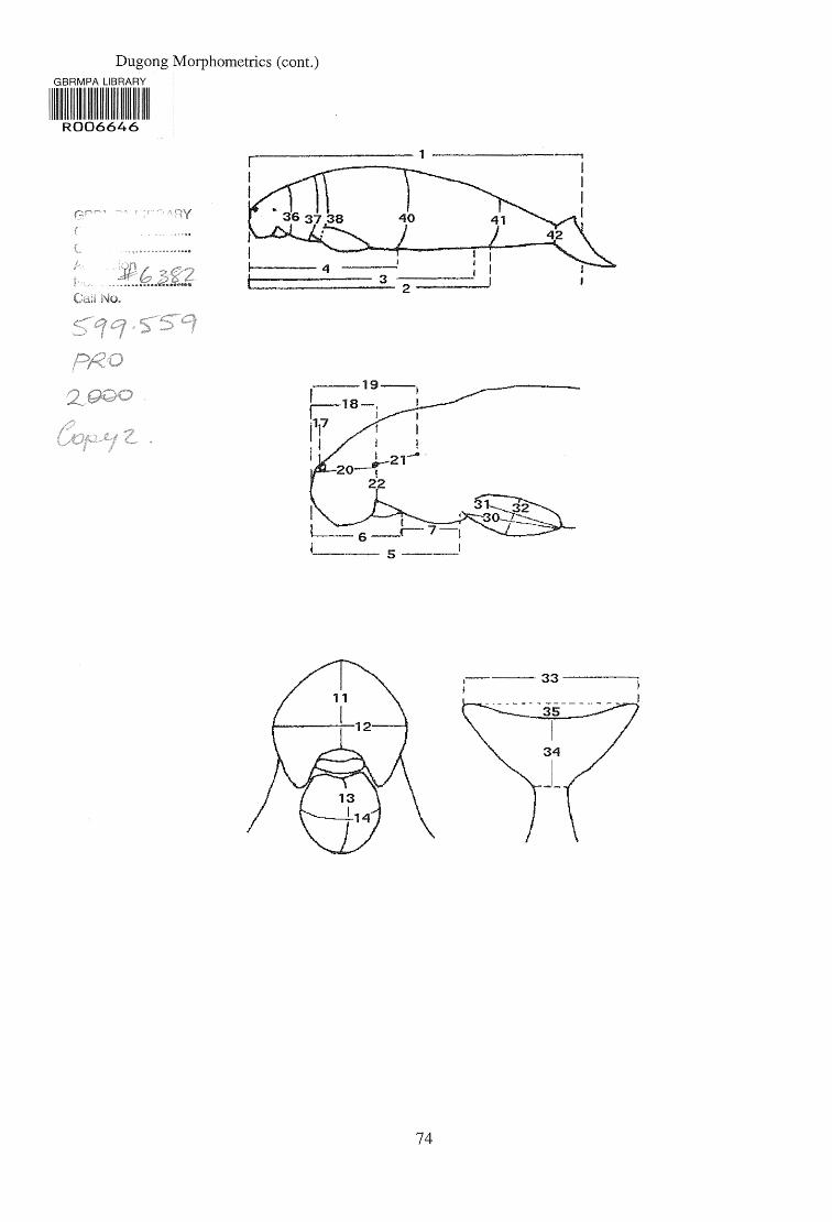
3 2
Dugong Morphometrics (cont.) GBRMPA LIBRARY
111 11 01111 1111 11 R006646
5
74
Related Documents