Chemistry & Biology Article Probing the Pore Drug Binding Site of Microtubules with Fluorescent Taxanes: Evidence of Two Binding Poses Isabel Barasoain, 1 Ana M. Garcı´a-Carril, 1 Ruth Matesanz, 1 Giorgio Maccari, 2 Chiara Trigili, 1 Mattia Mori, 2 Jing-Zhe Shi, 3 Wei-Shuo Fang, 3 Jose ´ M. Andreu, 1 Maurizio Botta, 2 and J. Fernando Dı´az 1, * 1 Centro de Investigaciones Biolo ´ gicas, Consejo Superior de Investigaciones Cientı ´ficas, Ramiro de Maeztu 9, 28040 Madrid, Spain 2 Department of Pharmaceutical and Chemical Technology, Faculty of Pharmacy, University of Siena, I-53100 Siena, Italy 3 Institute of Materia Medica, Chinese Academy of Medical Sciences, 1 Xian Nong Tan Street, Beijing 100050, China *Correspondence: [email protected] DOI 10.1016/j.chembiol.2010.02.006 SUMMARY The pore site in microtubules has been studied with the use of Hexaflutax, a fluorescent probe derived from paclitaxel. The compound is active in cells with similar effects to paclitaxel, indicating that the pore may be a target to microtubule stabilizing agents. While other taxanes bind microtubules in a monophasic way, thus indicating a single type of sites, Hexaflutax association is biphasic. Analysis of the phases indicates that two different binding sites are detected, reflecting two different modes of binding, which could arise from different arrange- ments of the taxane or fluorescein moieties in the pore. Association of the 4-4-20 antifluorescein monoclonal antibody-Hexaflutax complex to micro- tubules remains biphasic, thus indicating that the two phases observed arise from two different poses of the taxane moiety. INTRODUCTION The clinical success of paclitaxel and docetaxel has triggered the search for compounds with a similar mechanism of action but without their inconveniences (low solubility and develop- ment of resistances). This has resulted in the discovery of many compounds with very different chemical structures, epo- thilones, discodermolides, dyctiostatins, eleutherobin, sarco- dyctins, cyclostreptin, laulimalide, and peloruside. These compounds bind to at least three different binding sites. Lauli- malide and peloruside reversibly compete among them for a binding site whose location is yet unknown (Pryor et al., 2002; Gaitanos et al., 2004) but not with taxanes, epothilones, discodermolides, dyctiostatins, and eleutherobin, which revers- ibly compete among them for binding to microtubules (Buey et al., 2005). It has been proven that taxanes and epothilones bind to a site in the luminal face of microtubules (Nogales et al., 1998; Nettles et al., 2004), whereas cyclostreptin, which irreversibly competes with taxane and ‘‘taxane binding site’’ drugs, shares its binding between two locations, the same luminal binding site described for taxanes and epothilones and a site in the external surface of microtubules located in the type I pore (Diaz et al., 2003; Buey et al., 2007), making binding to both sites mutually exclusive. Taxanes can not directly bind to the internal luminal site; however, they bind very fast to preformed microtubules (Diaz et al., 2003, 2000), thus they have to transiently bind to an easily accessible binding site in their way to the luminal site. Since binding of cyclostreptin to microtubules completely inhibits paclitaxel binding (Buey et al., 2007), the external site of cyclo- streptin can be assigned as the initial external binding site for taxanes. It is not yet known to which one of the sites discodermolides, dyctiostatins, eleutherobin, and sarcodyctins bind: only to the luminal, only to the external, or to both. However, it is easy to observe NMR TR-Noesy of discodermolide and dyctiostatin bound to microtubules (Canales et al., 2008), which indicates a fast kinetic rate of the release step, pointing toward necessary binding to a site different from the slow dissociating luminal site, which could be the pore site. It is possible as well to observe TR-Noesy of docetaxel bound to microtubules (Matesanz et al., 2008), which suggests that taxane dissociation proceeds similarly. Although it is relatively straightforward to measure and model the interactions of the taxanes and taxane-site binding compounds with the inner luminal binding site (Matesanz et al., 2008; Snyder et al., 2001), almost nothing is known about the nature of the external binding site, just its location in the type I pore and one of the amino acids (Thr220) that is labeled by cyclo- streptin on its way to the inner site (Buey et al., 2007). The reason for this is that the equilibrium methods normally used to charac- terize MSA-microtubule interactions (Buey et al., 2005; Li et al., 2000) cannot provide information about the transient binding to the external site or easily distinguish between binding to the external or the luminal site. The possible conformations of the external binding site have been recently studied using molecular modeling techniques. The models propose different interactions: in the model of Freedman et al. (2009) the binding site involves both protofila- ments of the pore, with the taxane core bound to both b subunits and the side chain bound to the a subunit involved. In the models of Magnani et al. (2009) only one protofilament is involved in the interaction, with the taxane core bound to the b subunit and the side chain bound the a subunit. A feasible way to study the interactions of ligands with tran- sient sites like the microtubule pore is the kinetic approach Chemistry & Biology 17, 243–253, March 26, 2010 ª2010 Elsevier Ltd All rights reserved 243

Welcome message from author
This document is posted to help you gain knowledge. Please leave a comment to let me know what you think about it! Share it to your friends and learn new things together.
Transcript
Chemistry & Biology
Article
Probing the Pore Drug Binding Site ofMicrotubules with Fluorescent Taxanes:Evidence of Two Binding PosesIsabel Barasoain,1 Ana M. Garcıa-Carril,1 Ruth Matesanz,1 Giorgio Maccari,2 Chiara Trigili,1 Mattia Mori,2 Jing-Zhe Shi,3
Wei-Shuo Fang,3 Jose M. Andreu,1 Maurizio Botta,2 and J. Fernando Dıaz1,*1Centro de Investigaciones Biologicas, Consejo Superior de Investigaciones Cientıficas, Ramiro de Maeztu 9, 28040 Madrid, Spain2Department of Pharmaceutical and Chemical Technology, Faculty of Pharmacy, University of Siena, I-53100 Siena, Italy3Institute of Materia Medica, Chinese Academy of Medical Sciences, 1 Xian Nong Tan Street, Beijing 100050, China
*Correspondence: [email protected]
DOI 10.1016/j.chembiol.2010.02.006
SUMMARY
The pore site in microtubules has been studied withthe use of Hexaflutax, a fluorescent probe derivedfrom paclitaxel. The compound is active in cellswith similar effects to paclitaxel, indicating that thepore may be a target to microtubule stabilizingagents. While other taxanes bind microtubules ina monophasic way, thus indicating a single type ofsites, Hexaflutax association is biphasic. Analysisof the phases indicates that two different bindingsites are detected, reflecting two different modes ofbinding, which could arise from different arrange-ments of the taxane or fluorescein moieties in thepore. Association of the 4-4-20 antifluoresceinmonoclonal antibody-Hexaflutax complex to micro-tubules remains biphasic, thus indicating that thetwo phases observed arise from two different posesof the taxane moiety.
INTRODUCTION
The clinical success of paclitaxel and docetaxel has triggered
the search for compounds with a similar mechanism of action
but without their inconveniences (low solubility and develop-
ment of resistances). This has resulted in the discovery of
many compounds with very different chemical structures, epo-
thilones, discodermolides, dyctiostatins, eleutherobin, sarco-
dyctins, cyclostreptin, laulimalide, and peloruside. These
compounds bind to at least three different binding sites. Lauli-
malide and peloruside reversibly compete among them for
a binding site whose location is yet unknown (Pryor et al.,
2002; Gaitanos et al., 2004) but not with taxanes, epothilones,
discodermolides, dyctiostatins, and eleutherobin, which revers-
ibly compete among them for binding to microtubules (Buey
et al., 2005). It has been proven that taxanes and epothilones
bind to a site in the luminal face of microtubules (Nogales
et al., 1998; Nettles et al., 2004), whereas cyclostreptin, which
irreversibly competes with taxane and ‘‘taxane binding site’’
drugs, shares its binding between two locations, the same
luminal binding site described for taxanes and epothilones and
a site in the external surface of microtubules located in the
Chemistry & Biology 17, 2
type I pore (Diaz et al., 2003; Buey et al., 2007), making binding
to both sites mutually exclusive.
Taxanes can not directly bind to the internal luminal site;
however, they bind very fast to preformed microtubules (Diaz
et al., 2003, 2000), thus they have to transiently bind to an easily
accessible binding site in their way to the luminal site. Since
binding of cyclostreptin to microtubules completely inhibits
paclitaxel binding (Buey et al., 2007), the external site of cyclo-
streptin can be assigned as the initial external binding site for
taxanes.
It is not yet known to which one of the sites discodermolides,
dyctiostatins, eleutherobin, and sarcodyctins bind: only to the
luminal, only to the external, or to both. However, it is easy to
observe NMR TR-Noesy of discodermolide and dyctiostatin
bound to microtubules (Canales et al., 2008), which indicates
a fast kinetic rate of the release step, pointing toward necessary
binding to a site different from the slow dissociating luminal
site, which could be the pore site. It is possible as well to observe
TR-Noesy of docetaxel bound to microtubules (Matesanz et al.,
2008), which suggests that taxane dissociation proceeds
similarly.
Although it is relatively straightforward to measure and model
the interactions of the taxanes and taxane-site binding
compounds with the inner luminal binding site (Matesanz et al.,
2008; Snyder et al., 2001), almost nothing is known about the
nature of the external binding site, just its location in the type I
pore and one of the amino acids (Thr220) that is labeled by cyclo-
streptin on its way to the inner site (Buey et al., 2007). The reason
for this is that the equilibrium methods normally used to charac-
terize MSA-microtubule interactions (Buey et al., 2005; Li et al.,
2000) cannot provide information about the transient binding to
the external site or easily distinguish between binding to the
external or the luminal site.
The possible conformations of the external binding site have
been recently studied using molecular modeling techniques.
The models propose different interactions: in the model of
Freedman et al. (2009) the binding site involves both protofila-
ments of the pore, with the taxane core bound to both b subunits
and the side chain bound to the a subunit involved. In the models
of Magnani et al. (2009) only one protofilament is involved in the
interaction, with the taxane core bound to the b subunit and the
side chain bound the a subunit.
A feasible way to study the interactions of ligands with tran-
sient sites like the microtubule pore is the kinetic approach
43–253, March 26, 2010 ª2010 Elsevier Ltd All rights reserved 243
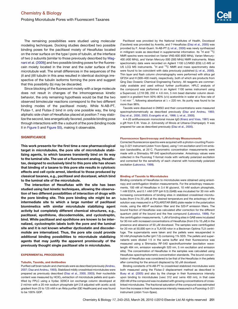
Figure 1. Chemical Structures of the Compounds Used and Effect on Cellular Microtubules
(A–H) Effect of Hexaflutax as compared to Flutax1 and paclitaxel on microtubule network (A, C, E, and G) and nucleus morphology (B, D, F, and H). A549 cells were
incubated for 24 hr with DMSO (A and B), 200 nM paclitaxel, 1 mM Flutax-1 (E and F), or 5 mM Hexaflutax (G and H). Microtubules were immunostained with
a-tubulin monoclonal antibodies and DNA was stained with Hoechst 33342. Insets are mitotic spindles from the same preparation. The scale bar represents
10 mm. All panels and insets have the same magnification.
Chemistry & Biology
Probing Microtubule Pores with Fluorescent Taxanes
(Diaz et al., 2003, 2000). The kinetics of binding and dissociation
of ligands to microtubules should provide information about the
interaction of the compound with the pore site. In this work the
interaction of taxanes with microtubules has been characterized
using two probes labeled at different parts of the molecule
(Figure 1), C7 in the north face and the C13 side chain, which
has a large contribution to the energy of binding (Matesanz
et al., 2008). The first probe is Hexaflutax (Diaz et al., 2005),
a fluorescent taxane derivative tailored to have a separation
between the fluorescein and the taxane moieties allowing
binding of a monoclonal antibody [4-4-20 (Kranz and Voss,
1981)] directed against the fluorescein moiety, as long as the tax-
ane moiety is bound to an external site, but not if the taxane is
bound to the internal site. It has been proven that this compound
either bound or not to the antibody remains at the external site
when bound to microtubules (Diaz et al., 2005). The other
compound studied was 30-N-m-aminobenzamido-30-N-deben-
zamidopaclitaxel (N-AB-PT) (Li et al., 2000), a taxane derivative
that carries a fluorescent aminobenzamido group at the C13
side chain. The results indicate that both fluorescent taxanes
bind to the pore site with an affinity of the order of micromolar.
While N-AB-PT binds in a single type of site similar to C7 fluores-
cent-labeled paclitaxel analogs, i.e., Flutax-1 and Flutax-2, the
presence of the long aliphatic chain in Hexaflutax allows
a second possibility of binding of the compounds to this site,
which results in the observation of a biphasic binding kinetics.
244 Chemistry & Biology 17, 243–253, March 26, 2010 ª2010 Elsevie
RESULTS
Paclitaxel-like Cellular Effects of HexaflutaxIn previous work (Diaz et al., 2005) we had found that Hexaflutax
binds to the external site of microtubules. Thus we wanted to
characterize its cellular effects in tumor cells to detect any
possible differential effect between binding to the pore and the
luminal sites. First, cytotoxicity in A2780 and A2780AD as
compared to paclitaxel and the two other fluorescent taxanes,
Flutax-1 and Flutax-2, was determined. Hexaflutax is less active
with IC50 in A2780 cells of 2.3 mM as compared with the other
fluorescein-labeled compounds, Flutax-1 and -2 (IC50 0.26 mM
and 0.8 mM, respectively), all fluorescent taxanes being signifi-
cantly less active than paclitaxel (IC50 1.1 nM). The three fluores-
cent taxanes are inactive against P-glycoprotein-overexpressing
A2780AD cells at the highest concentration (20 mM) tested (IC50
paclitaxel 1.1 mM).
We also studied the effect of Hexaflutax on cellular microtu-
bules. Treatment of A549 cells for 24 hr with either paclitaxel
(200 nM), Flutax-1 (1 mM), or Hexaflutax (5 mM) gave rise to
the characteristic cytoplasmic microtubule bundles as well
as to aberrant mitosis (monopolar spindles) and to micro-
nucleated cells (Figure 1), as expected from the microtubule-
stabilizing agent activity observed for the ligand. Hexaflutax is
able to in vitro induce tubulin assembly in conditions in which
tubulin itself is unable to assemble, i.e., in 10 mM phosphate
r Ltd All rights reserved
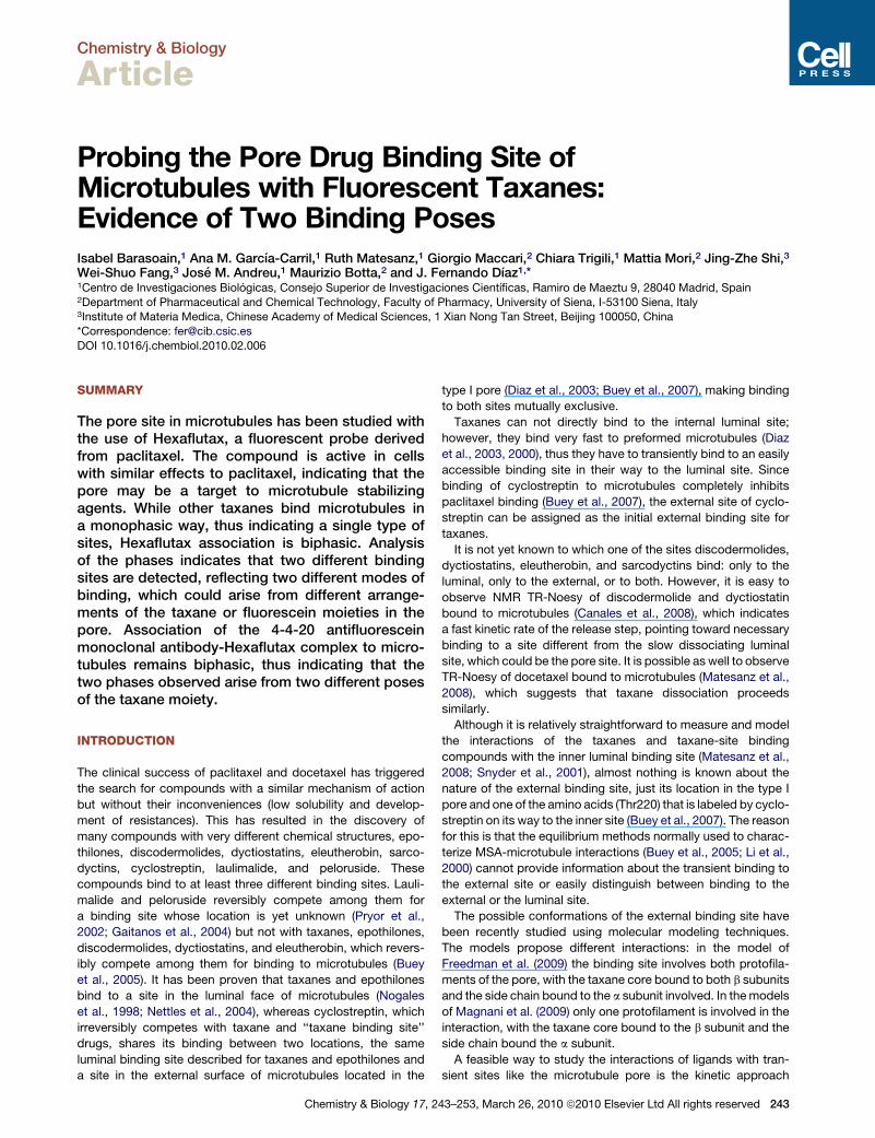
Figure 2. Effect of Taxanes in Morphology and Cell Cycle of A549 Cells
(A–D) Morphology of A549 cells after treatment with paclitaxel, Flutax-1, and Hexaflutax. A549 cells were incubated for 20 hr with either DMSO (A), 20 nM Taxol
(B), 2 mM Flutax-1 (C), and 4 mM Hexaflutax (D).
(E–L) Effect on A549 and A2780 cell cycle of Hexaflutax as compared with Taxol and Flutax-1. A549 cells were incubated for 20 hr with DMSO (E), 20 nM Taxol (F),
2 mM Flutax-1 (G), and 4 mM Hexaflutax (H) and A2780 cells with DMSO (I), 30 nM Taxol (J), 2 mM Flutax-1 (K), and 10 mM Hexaflutax (L).
Chemistry & Biology
Probing Microtubule Pores with Fluorescent Taxanes
buffer, 1 mM EDTA, 6 mM MgCl2, and 0.1 mM GTP (pH 7.0)
(Evangelio, 1999).
We next studied whether Hexaflutax was able to accumulate
cells in the G2+M phase of the cell cycle as paclitaxel and the
Chemistry & Biology 17, 2
other two fluorescent derivatives do. A549 lung carcinoma cells
as well as A2780 ovarian carcinoma cells were incubated for
20 hr in the presence of these three drugs and their cell
morphology was examined by DIC microscopy (Figures 2A–2D)
43–253, March 26, 2010 ª2010 Elsevier Ltd All rights reserved 245
Chemistry & Biology
Probing Microtubule Pores with Fluorescent Taxanes
before processing cells for the cell cycle experiment. Most A549-
treated cells and A2780 cells (data not shown) were rounded
mitotic cells as compared to the control cells that were spread
epithelial-like adherent cells with few mitotic cells. 20 nM Taxol,
2 mM Flutax-1, and 4 mM Hexaflutax and 30 nM Taxol, 2 mM
Flutax-1, and 10 mM Hexaflutax accumulate cells in the G2+M
phase of the cell cycle in both A549 and A2780 cells, respectively
(Figures 2E–2L), demonstrating that the binding of taxanes to
the pore and to the lumenal sites produce the same biological
effect in cells.
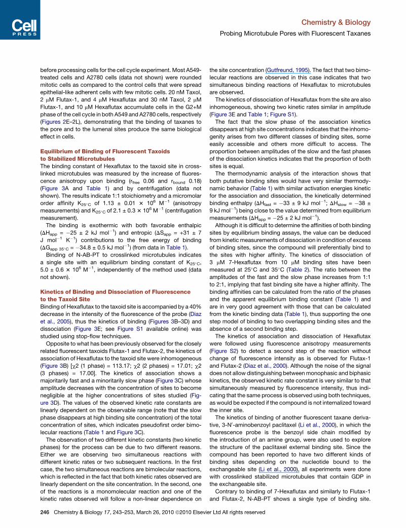
Equilibrium of Binding of Fluorescent Taxoidsto Stabilized MicrotubulesThe binding constant of Hexaflutax to the taxoid site in cross-
linked microtubules was measured by the increase of fluores-
cence anisotropy upon binding (rfree 0.06 and rbound 0.18)
(Figure 3A and Table 1) and by centrifugation (data not
shown). The results indicate 1:1 stoichiometry and a micromolar
order affinity K25�C of 1.13 ± 0.01 3 106 M�1 (anisotropy
measurements) and K25�C of 2.1 ± 0.3 3 106 M�1 (centrifugation
measurement).
The binding is exothermic with both favorable enthalpic
(DHapp = �25 ± 2 kJ mol�1) and entropic (DSapp = +31 ± 7
J mol�1 K�1) contributions to the free energy of binding
(DGapp 35�C = �34.8 ± 0.5 kJ mol�1) (from data in Table 1).
Binding of N-AB-PT to crosslinked microtubules indicates
a single site with an equilibrium binding constant of K25�C,
5.0 ± 0.6 3 106 M�1, independently of the method used (data
not shown).
Kinetics of Binding and Dissociation of Fluorescenceto the Taxoid SiteBinding of Hexaflutax to the taxoid site is accompanied by a 40%
decrease in the intensity of the fluorescence of the probe (Diaz
et al., 2005), thus the kinetics of binding (Figures 3B–3D) and
dissociation (Figure 3E; see Figure S1 available online) was
studied using stop-flow techniques.
Opposite to what has been previously observed for the closely
related fluorescent taxoids Flutax-1 and Flutax-2, the kinetics of
association of Hexaflutax to the taxoid site were inhomogeneous
(Figure 3B) [c2 (1 phase) = 113.17; c2 (2 phases) = 17.01; c2
(3 phases) = 17.00]. The kinetics of association shows a
majoritarily fast and a minoritarily slow phase (Figure 3C) whose
amplitude decreases with the concentration of sites to become
negligible at the higher concentrations of sites studied (Fig-
ure 3D). The values of the observed kinetic rate constants are
linearly dependent on the observable range (note that the slow
phase disappears at high binding site concentration) of the total
concentration of sites, which indicates pseudofirst order bimo-
lecular reactions (Table 1 and Figure 3C).
The observation of two different kinetic constants (two kinetic
phases) for the process can be due to two different reasons.
Either we are observing two simultaneous reactions with
different kinetic rates or two subsequent reactions. In the first
case, the two simultaneous reactions are bimolecular reactions,
which is reflected in the fact that both kinetic rates observed are
linearly dependent on the site concentration. In the second, one
of the reactions is a monomolecular reaction and one of the
kinetic rates observed will follow a non-linear dependence on
246 Chemistry & Biology 17, 243–253, March 26, 2010 ª2010 Elsevie
the site concentration (Gutfreund, 1995). The fact that two bimo-
lecular reactions are observed in this case indicates that two
simultaneous binding reactions of Hexaflutax to microtubules
are observed.
The kinetics of dissociation of Hexaflutax from the site are also
inhomogeneous, showing two kinetic rates similar in amplitude
(Figure 3E and Table 1; Figure S1).
The fact that the slow phase of the association kinetics
disappears at high site concentrations indicates that the inhomo-
genity arises from two different classes of binding sites, some
easily accessible and others more difficult to access. The
proportion between amplitudes of the slow and the fast phases
of the dissociation kinetics indicates that the proportion of both
sites is equal.
The thermodynamic analysis of the interaction shows that
both putative binding sites would have very similar thermody-
namic behavior (Table 1) with similar activation energies kinetic
for the association and dissociation, the kinetically determined
binding enthalpy (DHfast = �33 ± 9 kJ mol�1; DHslow = �38 ±
9 kJ mol�1) being close to the value determined from equilibrium
measurements (DHapp = �25 ± 2 kJ mol�1).
Although it is difficult to determine the affinities of both binding
sites by equilibrium binding assays, the value can be deduced
from kinetic measurements of dissociation in condition of excess
of binding sites, since the compound will preferentially bind to
the sites with higher affinity. The kinetics of dissociation of
3 mM 7-Hexaflutax from 10 mM binding sites have been
measured at 25�C and 35�C (Table 2). The ratio between the
amplitudes of the fast and the slow phase increases from 1:1
to 2:1, implying that fast binding site have a higher affinity. The
binding affinities can be calculated from the ratio of the phases
and the apparent equilibrium binding constant (Table 1) and
are in very good agreement with those that can be calculated
from the kinetic binding data (Table 1), thus supporting the one
step model of binding to two overlapping binding sites and the
absence of a second binding step.
The kinetics of association and dissociation of Hexaflutax
were followed using fluorescence anisotropy measurements
(Figure S2) to detect a second step of the reaction without
change of fluorescence intensity as is observed for Flutax-1
and Flutax-2 (Diaz et al., 2000). Although the noise of the signal
does not allow distinguishing between monophasic and biphasic
kinetics, the observed kinetic rate constant is very similar to that
simultaneously measured by fluorescence intensity, thus indi-
cating that the same process is observed using both techniques,
as would be expected if the compound is not internalized toward
the inner site.
The kinetics of binding of another fluorescent taxane deriva-
tive, 3-N0-aminobenzoyl paclitaxel (Li et al., 2000), in which the
fluorescence probe is the benzoyl side chain modified by
the introduction of an amine group, were also used to explore
the structure of the paclitaxel external binding site. Since the
compound has been reported to have two different kinds of
binding sites depending on the nucleotide bound to the
exchangeable site (Li et al., 2000), all experiments were done
with crosslinked stabilized microtubules that contain GDP in
the exchangeable site.
Contrary to binding of 7-Hexaflutax and similarly to Flutax-1
and Flutax-2, N-AB-PT shows a single type of binding site.
r Ltd All rights reserved
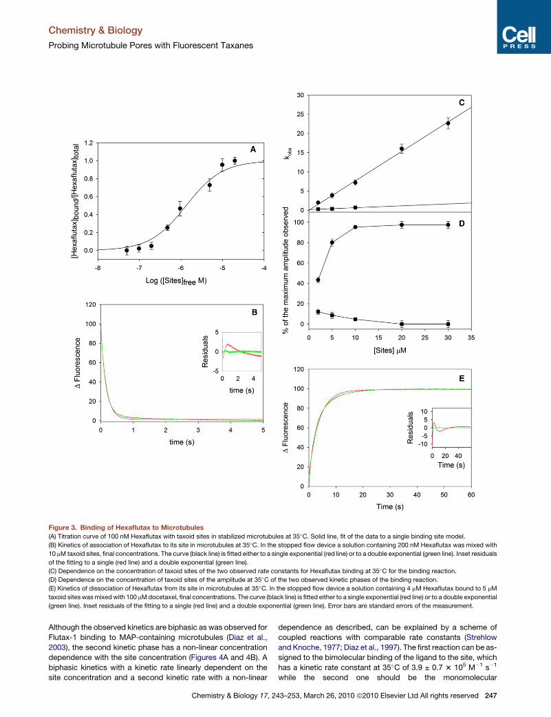
Figure 3. Binding of Hexaflutax to Microtubules(A) Titration curve of 100 nM Hexaflutax with taxoid sites in stabilized microtubules at 35�C. Solid line, fit of the data to a single binding site model.
(B) Kinetics of association of Hexaflutax to its site in microtubules at 35�C. In the stopped flow device a solution containing 200 nM Hexaflutax was mixed with
10 mM taxoid sites, final concentrations. The curve (black line) is fitted either to a single exponential (red line) or to a double exponential (green line). Inset residuals
of the fitting to a single (red line) and a double exponential (green line).
(C) Dependence on the concentration of taxoid sites of the two observed rate constants for Hexaflutax binding at 35�C for the binding reaction.
(D) Dependence on the concentration of taxoid sites of the amplitude at 35�C of the two observed kinetic phases of the binding reaction.
(E) Kinetics of dissociation of Hexaflutax from its site in microtubules at 35�C. In the stopped flow device a solution containing 4 mM Hexaflutax bound to 5 mM
taxoid sites was mixed with 100 mM docetaxel, final concentrations. The curve (black line) is fitted either to a single exponential (red line) or to a double exponential
(green line). Inset residuals of the fitting to a single (red line) and a double exponential (green line). Error bars are standard errors of the measurement.
Chemistry & Biology
Probing Microtubule Pores with Fluorescent Taxanes
Although the observed kinetics are biphasic as was observed for
Flutax-1 binding to MAP-containing microtubules (Diaz et al.,
2003), the second kinetic phase has a non-linear concentration
dependence with the site concentration (Figures 4A and 4B). A
biphasic kinetics with a kinetic rate linearly dependent on the
site concentration and a second kinetic rate with a non-linear
Chemistry & Biology 17, 2
dependence as described, can be explained by a scheme of
coupled reactions with comparable rate constants (Strehlow
and Knoche, 1977; Diaz et al., 1997). The first reaction can be as-
signed to the bimolecular binding of the ligand to the site, which
has a kinetic rate constant at 35�C of 3.9 ± 0.7 3 105 M�1 s�1
while the second one should be the monomolecular
43–253, March 26, 2010 ª2010 Elsevier Ltd All rights reserved 247
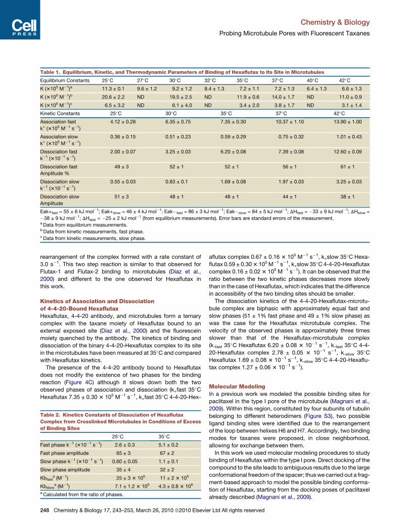
Table 1. Equilibrium, Kinetic, and Thermodynamic Parameters of Binding of Hexaflutax to Its Site in Microtubules
Equilibrium Constants 25�C 27�C 30�C 32�C 35�C 37�C 40�C 42�C
K (3105 M�1)a 11.3 ± 0.1 9.6 ± 1.2 9.2 ± 1.2 8.4 ± 1.3 7.2 ± 1.1 7.2 ± 1.3 6.4 ± 1.3 6.6 ± 1.3
K (3105 M�1)b 20.6 ± 2.2 ND 19.5 ± 2.5 ND 11.9 ± 0.6 14.0 ± 1.7 ND 11.0 ± 0.9
K (3105 M�1)c 6.5 ± 3.2 ND 6.1 ± 4.0 ND 3.4 ± 2.0 3.8 ± 1.7 ND 3.1 ± 1.4
Kinetic Constants 25�C 30�C 35�C 37�C 42�C
Association fast
k+ (3105 M�1 s�1)
4.12 ± 0.28 6.35 ± 0.75 7.35 ± 0.30 10.37 ± 1.10 13.90 ± 1.00
Association slow
k+ (3105 M�1 s�1)
0.36 ± 0.15 0.51 ± 0.23 0.59 ± 0.29 0.75 ± 0.32 1.01 ± 0.43
Dissociation fast
k�1 (310�1 s�1)
2.00 ± 0.07 3.25 ± 0.03 6.20 ± 0.08 7.39 ± 0.08 12.60 ± 0.09
Dissociation fast
Amplitude %
49 ± 3 52 ± 1 52 ± 1 56 ± 1 61 ± 1
Dissociation slow
k�1 (310�1 s�1)
0.55 ± 0.03 0.83 ± 0.1 1.69 ± 0.08 1.97 ± 0.03 3.25 ± 0.03
Dissociation slow
Amplitude
51 ± 3 48 ± 1 48 ± 1 44 ± 1 38 ± 1
Eak+fast = 55 ± 6 kJ mol�1; Eak+slow = 46 ± 4 kJ mol�1; Eak�fast = 86 ± 3 kJ mol�1; Eak�slow = 84 ± 5 kJ mol�1; DHfast = �33 ± 9 kJ mol�1; DHslow =
�38 ± 9 kJ mol�1; DHapp = �25 ± 2 kJ mol�1 (from equilibrium measurements). Error bars are standard errors of the measurement.a Data from equilibrium measurements.b Data from kinetic measurements, fast phase.c Data from kinetic measurements, slow phase.
Chemistry & Biology
Probing Microtubule Pores with Fluorescent Taxanes
rearrangement of the complex formed with a rate constant of
3.0 s�1. This two step reaction is similar to that observed for
Flutax-1 and Flutax-2 binding to microtubules (Diaz et al.,
2000) and different to the one observed for Hexaflutax in
this work.
Kinetics of Association and Dissociationof 4-4-20-Bound HexaflutaxHexaflutax, 4-4-20 antibody, and microtubules form a ternary
complex with the taxane moiety of Hexaflutax bound to an
external exposed site (Diaz et al., 2000) and the fluorescein
moiety quenched by the antibody. The kinetics of binding and
dissociation of the binary 4-4-20-Hexaflutax complex to its site
in the microtubules have been measured at 35�C and compared
with Hexaflutax kinetics.
The presence of the 4-4-20 antibody bound to Hexaflutax
does not modify the existence of two phases for the binding
reaction (Figure 4C) although it slows down both the two
observed phases of association and dissociation (k+fast 35�C
Hexaflutax 7.35 ± 0.30 3 105 M�1 s�1, k+fast 35�C 4-4-20-Hex-
Table 2. Kinetics Constants of Dissociation of Hexaflutax
Complex from Crosslinked Microtubules in Conditions of Excess
of Binding Sites
25�C 35�C
Fast phase k�1 (310�1 s�1) 2.6 ± 0.3 5.1 ± 0.2
Fast phase amplitude 65 ± 3 67 ± 2
Slow phase k�1 (310�1 s�1) 0.60 ± 0.05 1.1 ± 0.1
Slow phase amplitude 35 ± 4 32 ± 2
Kbfasta (M�1) 25 ± 3 3 105 11 ± 2 3 105
Kbslowa (M�1) 7.1 ± 1.2 3 105 4.3 ± 0.8 3 105
a Calculated from the ratio of phases.
248 Chemistry & Biology 17, 243–253, March 26, 2010 ª2010 Elsevie
aflutax complex 0.67 ± 0.16 3 105 M�1 s�1, k+slow 35�C Hexa-
flutax 0.59 ± 0.30 3 105 M�1 s�1, k+slow 35�C 4-4-20-Hexaflutax
complex 0.16 ± 0.02 3 105 M�1 s�1). It can be observed that the
ratio between the two kinetic phases decreases more slowly
than in the case of Hexaflutax, which indicates that the difference
in accessibility of the two binding sites should be smaller.
The dissociation kinetics of the 4-4-20-Hexaflutax-microtu-
bule complex are biphasic with approximately equal fast and
slow phases (51 ± 1% fast phase and 49 ± 1% slow phase) as
was the case for the Hexaflutax microtubule complex. The
velocity of the observed phases is approximately three times
slower than that of the Hexaflutax-microtubule complex
(k-fast 35�C Hexaflutax 6.20 ± 0.08 3 10�1 s�1, k-fast 35�C 4-4-
20-Hexaflutax complex 2.78 ± 0.05 3 10�1 s�1, k-slow 35�C
Hexaflutax 1.69 ± 0.08 3 10�1 s�1, k-slow 35�C 4-4-20-Hexaflu-
tax complex 1.27 ± 0.06 3 10�1 s�1).
Molecular ModelingIn a previous work we modeled the possible binding sites for
paclitaxel in the type I pore of the microtubule (Magnani et al.,
2009). Within this region, constituted by four subunits of tubulin
belonging to different heterodimers (Figure S3), two possible
ligand binding sites were identified due to the rearrangement
of the loop between helixes H6 and H7. Accordingly, two binding
modes for taxanes were proposed, in close neighborhood,
allowing for exchange between them.
In this work we used molecular modeling procedures to study
binding of Hexaflutax within the type I pore. Direct docking of the
compound to the site leads to ambiguous results due to the large
conformational freedom of the spacer; thus we carried out a frag-
ment-based approach to model the possible binding conforma-
tion of Hexaflutax, starting from the docking poses of paclitaxel
already described (Magnani et al., 2009).
r Ltd All rights reserved
Figure 4. Binding of N-AB-PT and 4-4-20-Hexaflutax Complex to Microtubules
(A and B) Kinetics of dependence on the concentration of taxoid sites of the fast (A) and slow (B) observed rate constants for N-AB-PT binding to crosslinked
stabilized microtubules at 35�C.
(C) Comparison between the kinetics of association of Hexaflutax and the 4-4-20-Hexaflutax complex to its site in microtubules at 35�C. In the stopped flow
device, a solution containing 500 nM Hexaflutax (solid line; average of two shots) or 500 nM Hexaflutax and 750 nM 4-4-20 antibody (dashed line; average of
ten shots) was mixed with 10 mM taxoid sites, final concentrations.
(D) Kinetics of dissociation of Hexaflutax from its site in microtubules at 35�C. In the stopped flow device a solution containing 2 mM Hexaflutax (solid line; average
of two shots) or 2 mM Hexaflutax plus 2.5 mM 4-4-20 (dashed line; average of ten shots) bound to 2.5 mM taxoid sites was mixed with 100 mM docetaxel, final
concentrations. Note that the curves are scaled to 100 since the fluorescence of the free Hexaflutax is five times larger than that of the 4-4-20 Hexaflutax complex.
Chemistry & Biology
Probing Microtubule Pores with Fluorescent Taxanes
The first tested hypothesis was that the inhomogenity
observed of the binding was due to possible differences between
the most common isotypes of human b-tubulin, namely, bI and
bIII. Thus, we modeled paclitaxel on each subtype obtaining
a total of four complexes (two possible binding modes and two
isotypes). The two binding modes previously found were kept
unchanged, which was expected due to the highly conserved
sequences of the binding site among isotype bI and bIII.
Then, in order to study the possibility that the inhomogenity
observed arises from different binding poses of the fluorescein
moiety in the binding site, a preliminary mapping of the whole
pore region with only the fluorescent moiety of Hexaflutax mole-
cule was performed by means of the blind docking approach
(Hetenyi and van der Spoel, 2002). In a preliminary investigation,
carried out toward the free protein, we found two possible
binding modes. The most representative binding mode for the
fluorescent moiety of Hexaflutax was found in the inner wall of
microtubules, between two b subunits. No differences between
the bI and bIII isotypes were found in this region. A second alter-
native binding site for the fluorescent moiety of Hexaflutax was
found in the outer wall of microtubules.
The docking of the fluorescein moiety in the binding site was
then repeated toward the four paclitaxel/tubulin complexes
previously determined, keeping the paclitaxel molecule frozen
Chemistry & Biology 17, 2
in the binding conformations already described. The binding
modes for the fluorescent moiety were approximately the
same as obtained without paclitaxel in the pore site. Two large
clusters of conformations were found, collecting a significant
proportion of the total conformations docked. The highly popu-
lated clusters were generally at the lowest free energy of binding
(Figure S4A) and were representative for the interaction of the
fluorescent moiety in the inner wall of the type I pore. On the
contrary, lower populated clusters described the interaction
with the outer side of the pore.
The well defined inner binding cavity used by fluorescein is
constituted by residues Lys216, Thr218, and Gly277 of subunit
b1 and Arg77, Pro87, Asp88, and Phe90 of subunit b2; a differ-
ence in amino acid composition between bI and bIII isotypes
can be found in the loop H6-H7, changing Thr218 to Ala218.
This difference, however, does not affect the ligand binding
conformation. On the contrary, the outer side of the pore lacks
a defined interaction groove that results in a large scattering of
binding poses for the fluorescent moiety.
Interestingly, the analysis of the docking-based binding pose
calculated for the fluorescent moiety toward the paclitaxel/
tubulin complexes shows, in all cases, a close proximity between
the two moieties allowing a direct linkage that could reproduce
the whole Hexaflutax by means of this fragment-based strategy.
43–253, March 26, 2010 ª2010 Elsevier Ltd All rights reserved 249

Figure 5. Docking-Based Binding Modes of Hexaflutax in the
Binding Site
These conformations were obtained by a fragment-based approach starting
from two conformations of the taxane moiety (binding mode 1 and binding
mode 2) toward isotypes bI and bIII of tubulin. The two top panels show the
bI complexes, whereas the bottom panels show the bIII complexes, all viewed
from the inside of the microtubule. a and b subunits are colored in cyan and
yellow, respectively; the hexaflutax molecule is shown in green sticks. The
residues mostly involved in the interaction with the fluorescent moiety of hex-
aflutax are highlighted by red surfaces.
Chemistry & Biology
Probing Microtubule Pores with Fluorescent Taxanes
In order to do so the linker between paclitaxel and the fluores-
cent moiety was docked in the ternary complexes (paclitaxel,
fluorescein, and tubulin) calculated in the previous step, after
defining two anchor positions for covalent bonding on each
substructure (Figure S4B). A slight energy minimization process
was then performed for all the complexes to relax the linker
within the whole Hexaflutax, resulting in the models presented
in Figure 5 when the ternary complex used was that with the fluo-
rescein placed in the inner wall of the microtubule and those pre-
sented in Figure S5 when the ternary complex used was that with
the fluorescein placed in the outer wall of the microtubule.
However, it should be pointed out that the contribution of the
fluorescein moiety to the free energy of binding should be very
low, because the equilibrium binding constant of the 4-4-20 anti-
body-Hexaflutax to microtubules is three times higher than those
of Hexaflutax (Diaz et al., 2005) (although an exact calculation of
its contribution cannot be done since the close proximity of
tubulin and antibody residues will result in unspecific interac-
tions). Thus, it is very likely that the fluorescein moiety is distrib-
uted between the unbound state and the inner and outer bound
positions as indicated by the fact that microtubule-bound Hexa-
flutax fluorescence is rapidly quenched by the antibody.
Free energies of binding of Hexaflutax with the fluorescein
moiety placed in the two different possibilities were further calcu-
lated with the local search algorithm implemented on AutoDock4.
A mean difference of 13.96 KJ/mol between internal and external
binding modes was found (Table S1). This difference was prob-
ably due to the most favorable Van der Waals interactions ex-
ploited by Hexaflutax in the unique inner site, with respect to
the scattered conformations observed in the outer region.
DISCUSSION
Hexaflutax is a fluorescent probe of the pore site of microtu-
bules, which is not internalized to the luminal site. The compound
is cytotoxic with similar cellular effects to the other taxanes
studied, indicating that compounds binding to the external site
have the same microtubule stabilizing activity and they kill cells
through the blocking of microtubule dynamics. Thus the external
binding site can be the target of specifically designed antitumor
drugs. This compound has 35 times less affinity than the
compound with a shorter spacer between the taxane and the
fluorescein group (Flutax-1) (Diaz et al., 2000), suggesting
a free energy of around �9 kJ/mol for the internalization of the
taxanes.
Hexaflutax Binds Inhomogeneously to the Pore Siteof MicrotubulesThe main difference observed between the binding of Hexaflutax
to microtubules and that of other taxanes, Flutax-1 and Flutax-2,
is the existence of biphasic behavior in both the association and
dissociation of each compound from/to its site. This biphasic
behavior arises from the interaction of the compound with the
external site and not from any possible simultaneous binding
with both the internal and the external sites, since this is also
observed when any possible interaction with the internal site is
blocked with an antibody.
Kinetic analysis indicates that the inhomogenity arises from
two kinds of binding sites, which may be proposed to be: (a)
250 Chemistry & Biology 17, 243–253, March 26, 2010 ª2010 Elsevie
different overlapping binding sites (either from the paclitaxel or
the fluorescein moiety), (b) sites formed by different tubulin iso-
forms, or (c) two different pores of microtubules. However,
hypothesis c would be very unlikely since the observed stoichi-
ometry is very close to 1. A fourth, alternative hypothesis would
be (d) the existence of two different types of binding sites in
microtubules, a high affinity one in GTP tubulin and a low affinity
one in GDP-bound tubulin as previously described by Li et al.
(2000) for N-AB-PT. This possibility can be discarded because
with the stabilized microtubules used, which have GDP bound
to the exchangeable nucleotide site, we can only observe a single
type of binding site with binding affinity of the order of 107 M�1 for
N-AB-PT.
It can then be assumed that the inhomogenity of Hexaflutax
arises from either two binding modes to the same site (with
one of the modes not allowed in the case of Flutax-1 and Flu-
tax-2) or from binding to two different tubulin isoforms, with
binding parameters similar for Flutax-1 and Flutax-2, but not
for Hexaflutax. Since the kinetic constant of the fast phase
from association is very similar in the case of all compounds
studied (k+1 35�Flutax-1 6.12 ± 0.22 3 105 M�1 s�1, Flutax-2
5.66 ± 0.50 3 105 M�1 s�1, N-AB-PT 3.9 ± 0.7 3 105 M�1 s�1,
Hexaflutax 7.35 ± 0.30 3 105 M�1 s�1) and the taxane moiety
of Hexaflutax and Flutax-1 and Flutax-2 are identical, it is
straightforward to assign this fast phase of association to the
interaction that Flutax-1 and Flutax-2 have in their association.
r Ltd All rights reserved
Chemistry & Biology
Probing Microtubule Pores with Fluorescent Taxanes
The remaining possibilities were studied using molecular
modeling techniques. Docking studies described two possible
binding poses for the paclitaxel moiety of Hexaflutax located
on the inner surface on the type I pore of tubulin, at the interface
of two b subunits [similar to those previously described by Mag-
nani et al. (2009)] and two possible binding poses for the fluores-
cein moiety located in the inner and the outer surface of the
microtubule wall. The small differences in the sequences of the
bI and bIII tubulin in this area resulted in identical dockings irre-
spective of the tubulin isoforms forming the pore and suggest
that this possibility (b) may be discarded.
Since blocking of the fluorescent moiety with a large molecule
does not result in changes of the inhomogeneous kinetic
behavior, the only remaining hypothesis would be that the two
observed bimolecular reactions correspond to the two different
binding modes of the paclitaxel moiety. While N-AB-PT,
Flutax-1, and Flutax-2 bind in only one possible way, the long
aliphatic side chain of Hexaflutax placed at position 7 may stabi-
lize the second, less energetically favored, possible binding pose
through interactions with the a subunit of the pore (binding mode
II in Figure 5 and Figure S5), making it observable.
SIGNIFICANCE
This work presents for the first time a new pharmacological
target in microtubules, the pore site of microtubule stabi-
lizing agents, to which taxanes transiently bind on its way
to the luminal site. The use of a fluorescent analog, Hexaflu-
tax, designed to exclusively bind to this pore site has shown
that binding of a taxane to this pore site results in cytotoxic
effects and cell cycle arrest, identical to those produced by
classical taxanes, e.g., paclitaxel and docetaxel, which bind
to the luminal site of the microtubule.
The interaction of Hexaflutax with the site has been
studied using fast kinetic techniques, allowing the observa-
tion of two different possibilities of interaction of drugs with
the pore binding site. This pore binding site might be an
intermediate site to which a large number of paclitaxel
biomimetics with similar microtubule stabilizing agent
activity but completely different chemical structures, e.g.,
paclitaxel, epothilone, discodermolide, and cyclostreptin,
bind. While paclitaxel and epothilone are known to be inter-
nalized, cyclostreptin labels both the pore and the luminal
site and it is not known whether dyctiostatin and discoder-
molide are internalized. Thus, the pore site could provide
additional binding possibilities to microtubule stabilizing
agents that may justify the apparent promiscuity of the
previously thought single paclitaxel site in microtubules.
EXPERIMENTAL PROCEDURES
Tubulin, Taxoids, and Antibodies
Purified calf brain tubulin and chemicals were as described previously (Andreu,
2007; Diaz and Andreu, 1993). Stabilized mildly crosslinked microtubules were
prepared as previously described (Diaz et al., 2000, 2003); their nucleotide
content was measured by HClO4 extraction of microtubule pellets and quan-
tified by FPLC using a Vydac 320IC4 ion exchange column developed at
2 ml/min with a 20 min sodium phosphate (pH 2.8 adjusted with acetic acid)
gradient from 25 to 125 mM in an Akta purifier (GE Healthcare) and was found
to be 100% GDP.
Chemistry & Biology 17, 2
Paclitaxel was provided by the National Institutes of Health, Docetaxel
(Taxotere) was provided by Aventis, and 7-Hexaflutax (Diaz et al., 2005) was
provided by F. Amat-Guerri. N-AB-PT (Li et al., 2000) was newly synthesized
in milligram scale as described in supplemental information. Its 1H and 13C
NMR spectra were recorded on Varian VNS-600 (600 MHz), Varian Mercury-
400 (400 MHz), and Varian Mercury-300 (300 MHz) NMR instruments. Mass
spectrometry data were recorded on Agilent 1100 LC/MSD (ESI) LC-MS or
US LCQ MS instruments. 1H and 13C NMR and mass spectrometry data
were found to be coincident with data previously published (Li et al., 2000).
Thin layer and flash column chromatography were performed with silica gel
GF254 and H (300–400 mesh), respectively, both of which are products from
Qing Dao Oceanic Chemical Engineering Factory. All reagents are commer-
cially available and used without further purification. HPLC analysis of
the compound was performed in an Agilent 1100 series instrument using
a Supercosil, LC18 DB, 250 3 4.6 mm, 5 mm bead diameter column devel-
oped in a gradient from 50%–80% (v/v) acetonitrile in water at a flow rate of
1 ml min�1, following absorbance at l = 220 nm. Its purity was found to be
more than 98%.
Taxoids were dissolved in DMSO and their concentrations were measured
spectrophotometrically as described previously (Diaz and Andreu, 1993;
Diaz et al., 2000, 2003; Evangelio et al., 1998; Li et al., 2000).
4-4-20 antifluorescein monoclonal mouse IgG (Kranz and Voss, 1981) was
a gift from E.W. Voss Jr. (University of Illinois at Urbana-Champaign). It was
prepared for use as described previously (Diaz et al., 2005).
Fluorescence Spectroscopy and Anisotropy Measurements
Corrected fluorescence spectra were acquired with a photon-counting Fluoro-
log-3-221 instrument (Jobin Yvon-Spex), using 1 nm excitation and 5 nm emis-
sion bandwidths, at 25�C. Fluorometric concentration measurements were
made with a Shimadzu RF-540 spectrofluorometer. Anisotropy values were
collected in the Fluorolog T-format mode with vertically polarized excitation
and corrected for the sensitivity of each channel with horizontally polarized
excitation (Lakowicz, 1999).
Binding of Taxoids to Microtubules
Binding constants of Hexaflutax to microtubules were obtained using anisot-
ropy and centrifugation titration measurements. For the anisotropy measure-
ments, 100 nM of Hexaflutax in 3.4 M glycerol, 10 mM sodium phosphate,
1 mM EGTA, and 0.1 mM GTP (pH 6.5) (GAB) was incubated for 30 min with
increasing concentrations of binding sites in stabilized crosslinked microtu-
bules (from 0 to 20 mM) at the desired temperature and the anisotropy of the
solution was measured in a POLARSTAR BMG plate reader in the polarization
mode, using the 480-P excitation filter and the 520-P emission filters. The
bound fraction of Hexaflutax was calculated after correction for the different
quantum yield of the bound and the free compound (Lakowicz, 1999). For
the centrifugation measurements, 1 mM of binding sites in GAB were incubated
for 30 min with increased concentrations of Hexaflutax (from 0 to 10 mM) in the
presence and absence of 50 mM docetaxel. The samples were centrifugated
for 20 min at 50,000 rpm in a TLA100 rotor in a Beckman Optima TLX centri-
fuge. The supernatants were taken and the pellets were resuspended in
50 mM phosphate buffer (pH 7.0) containing 1% SDS. The pellets and super-
natants were diluted 1:5 in the same buffer and their fluorescence was
measured using a Shimadzu RF-540 spectrofluorimeter (excitation wave-
length 484 nm, emission wavelength 520 nm, 5 nm excitation and emission
slits). The concentration of Hexaflutax in the samples was calculated using
Hexaflutax spectrophotometric concentration standards. The bound concen-
tration of Hexaflutax was considered to be that of the Hexaflutax in the pellets
after correcting for the amount displaced by 50 mM docetaxel.
Binding constants of N-AB-PT to crosslinked stabilized microtubules were
both measured using the Flutax-2 displacement method as described in
Buey et al. (2005) and also by the change in their fluorescence intensity
upon binding to microtubules (lexc 312 and lems 450 nm), in that case
200 nM of the compound was incubated with growing concentrations of cross-
linked microtubules. The fractional saturation of the compound was estimated
from the increase in their fluorescence intensity measured in a Fluorolog-3-221
instrument (Jobin Yvon-Spex).
43–253, March 26, 2010 ª2010 Elsevier Ltd All rights reserved 251
Chemistry & Biology
Probing Microtubule Pores with Fluorescent Taxanes
Kinetics of Binding of Taxanes to Microtubules
The kinetics of binding and dissociation of taxanes to stabilized microtubules
was measured by the change of intensity of fluorescence or the change in
anisotropy of the fluorescence using a Bio-Logic SF300S stopped flow device
equipped with a fluorescence detection system with an excitation wavelength
of 495 nm and a filter with a cut-off of 520 nm in the emission pathway for Hex-
aflutax and the 4-4-20-Hexaflutax complex and with an excitation wavelength
of 312 nm and a filter with a cut-off of 380 nm in the emission pathway for
N-AB-PT paclitaxel, with polarizers in the optical pathways when the anisot-
ropy change was measured. Appropriate photo bleaching controls were
done. The fitting of the kinetic curves was done with a non-linear least-squares
fitting program based on the Marquardt algorithm (Bevington, 1969).
Cell Biology Studies
Human A549 non-small lung carcinoma cells and human ovarian carcinomas
A2780 and A2780AD (MDR overexpressing P-glycoprotein) were cultured as
previously described (Buey et al., 2007).
Indirect immunofluorescence and cell cycle analysis was performed as
described before (Buey et al., 2005). Cytotoxicity assays were performed
with the MTT assay modified as described in Yang et al. (2007).
Homology Modeling
Sequences of human bI and bIII isotypes of tubulin were retrieved from the
UniProt online database (UniProt-Consortium, 2009). Three dimensional
models were thus generated from the 1JFF structure through homology
modeling approaches by means of the Modeller9v5 python library (Sali and
Blundell, 1993; Martı-Renom et al., 2000; Fiser et al., 2000; Eswar et al., 2006)
according to the method already described to model the 3D structure of tubulin
(sus scrofa sequence) (Magnani et al., 2009). A very_fastalgorithm was used due
to the high similarity showed by the selected human sequences (86%).
Each model was then refined by means of the ‘‘Protein Preparation Wizard’’
protocol of the Maestro suite (Mohamadi et al., 1990), selecting the most prob-
able state at pH 7.0 ± 2 for protein residues. The position of each atom of the
protein was relaxed through an energy minimization step carried out on the
basis of the OPLS2005 force field (Jorgensen and Tirado-Rives, 1988; Kamin-
ski et al., 2001), allowing for atom movements up to root mean square devia-
tion displacement below 0.3 A.
Docking Studies
All docking studies were performed with the AutoDock4 programs (v4.2.1)
(Morris et al., 1998). Interaction grids were calculated for all the atom types
of selected ligands with AutoGrid4 (v4.2.1) and were centered within the center
of the so-called type I pore constituted by subunit a4, a3, b1, and b2
(Figure S3). Potential grid maps were of 25 3 25 3 35 A dimensions, with
a point spacing of 0.375 A. Possible conformations of global energy minimum
for tested molecules were generated toward the receptor surface, using
a Lamarckian genetic algorithm (LGA), whereas free energies of binding
were further calculated using the local search algorithm.
All LGA parameters were kept at the default values with the only exception
being the maximum number of energy evaluations that was increased to 5 M,
whereas the initial population for each generation step was enhanced to
250 individuals and the number of docking runs was set to 250.
For local search methods we performed 100 runs for each calculation, start-
ing with an initial population constituted by 150 individuals. Coordinates of the
ligand mass center, torsional dihedrals, and initial orientation were retained as
those obtained by the docking results of the LGA methods.
Docking poses were clustered using a root mean square deviation tolerance
of 2.0 A.
Energy Minimization
All docking results were minimized with the MacroModel (Mohamadi et al.,
1990) application of the Maestro Suite, using the AMBER* force field (Ferguson
and Kollman, 1991; McDonald and Still, 1992) in implicit water solvent. The
convergence criteria was set to the energy gradient of 0.01 kJ/Amol or
a maximum of 5000 iterations. An energy minimization step was further per-
formed for each ligand and for all protein residues included within 3 A from
each ligand’s atom, whereas an additional shell of residues (5 A from the first
shell) was restrained with a force constant of 200 kJ$A�2$mol�1.
252 Chemistry & Biology 17, 243–253, March 26, 2010 ª2010 Elsevie
SUPPLEMENTAL INFORMATION
Supplemental Information includes five figures and one table can be found
with this article online at doi:10.1016/j.chembiol.2010.02.006.
ACKNOWLEDGMENTS
We wish to thank S. Wang, M. Seisdedos, and P. Lastres for their technical
assistance; F. Amat-Guerri for 7-Hexaflutax; E.W. Voss Jr. for the 4-4-20 anti-
fluorescein monoclonal mouse IgG; and Rhone Poulenc Rorer Aventis for
Docetaxel. We also would like to thank Matadero Municipal Vicente de Lucas
de Segovia for providing the calf brains for tubulin purification. This work was
supported in part by grant BIO2007-61336 from the Ministry of Science and
Innovation to J.F.D., BIPPED-CM from Comunidad de Madrid to J.F.D. and
J.M.A., and grant MOST No. 2006DFA31490 to W.S.F.
Received: November 30, 2009
Revised: January 26, 2010
Accepted: February 11, 2010
Published: March 25, 2010
REFERENCES
Andreu, J.M. (2007). Tubulin purification. In Methods in Molecular Medicine,
J. Zhou, ed. (Totowa, NJ: Humana Press Inc.), pp. 17–28.
Bevington, P.R. (1969). Data reduction and error analysis for the physical
sciences (New York: McGraw-Hill Book Co.).
Buey, R.M., Barasoain, I., Jackson, E., Meyer, A., Giannakakou, P., Paterson,
I., Mooberry, S., Andreu, J.M., and Diaz, J.F. (2005). Microtubule interactions
with chemically diverse stabilizing agents: thermodynamics of binding to the
paclitaxel site predicts cytotoxicity. Chem. Biol. 12, 1269–1279.
Buey, R.M., Calvo, E., Barasoain, I., Pineda, O., Edler, M.C., Matesanz, R.,
Cerezo, G., Vanderwal, C.D., Day, B.W., Sorensen, E.J., et al. (2007). Cyclo-
streptin binds covalently to microtubule pores and lumenal taxoid binding
sites. Nat. Chem. Biol. 3, 117–125.
Canales, A., Matesanz, R., Gardner, N.M., Andreu, J.M., Paterson, I., Diaz,
J.F., and Jimenez-Barbero, J. (2008). The bound conformation of microtu-
bule-stabilizing agents: NMR insights into the bioactive 3D structure of disco-
dermolide and dictyostatin. Chemistry 14, 7557–7569.
Diaz, J.F., and Andreu, J.M. (1993). Assembly of purified GDP-tubulin into
microtubules induced by taxol and taxotere: reversibility, ligand stoichiometry,
and competition. Biochemistry 32, 2747–2755.
Diaz, J.F., Sillen, A., and Engelborghs, Y. (1997). Equilibrium and kinetic study
of the conformational transition toward the active state of p21Ha-ras, induced
by the binding of BeF3- to the GDP-bound state, in the absence of GTPase-
activating proteins. J. Biol. Chem. 272, 23138–23143.
Diaz, J.F., Strobe, R., Engelborghs, Y., Souto, A.A., and Andreu, J.M. (2000).
Molecular recognition of taxol by microtubules. Kinetics and thermodynamics
of binding of fluorescent taxol derivatives to an exposed site. J. Biol. Chem.
275, 26265–26276.
Diaz, J.F., Barasoain, I., and Andreu, J.M. (2003). Fast kinetics of Taxol binding
to microtubules. Effects of solution variables and microtubule-associated
proteins. J. Biol. Chem. 278, 8407–8419.
Diaz, J.F., Barasoain, I., Souto, A.A., Amat-Guerri, F., and Andreu, J.M. (2005).
Macromolecular accessibility of fluorescent taxoids bound at a paclitaxel
binding site in the microtubule surface. J. Biol. Chem. 280, 3928–3937.
Eswar, N., Webb, B., Marti-Renom, M.A., Madhusudhan, M.S., Eramian, D.,
Shen, M.-Y., Pieper, U., and Sali, A. (2006). Comparative protein structure
modeling using Modeller. Curr. Protoc. Bioinformatics, Chapter 5, Unit 5.6.
Evangelio, J.A. (1999). Interaction of microtubules with fluorescent taxanes.
PhD thesis, Universidad Complutense de Madrid, Madrid, Spain.
Evangelio, J.A., Abal, M., Barasoain, I., Souto, A.A., Lillo, M.P., Acuna, A.U.,
Amat-Guerri, F., and Andreu, J.M. (1998). Fluorescent taxoids as probes of
the microtubule cytoskeleton. Cell Motil. Cytoskeleton 39, 73–90.
r Ltd All rights reserved

Chemistry & Biology
Probing Microtubule Pores with Fluorescent Taxanes
Ferguson, D.M., and Kollman, P.A. (1991). Can the Lennard-Jones 6-12 func-
tion replace the 10-12 form in molecular mechanics calculations? J. Comput.
Chem. 12, 620–626.
Fiser, A., Do, R.K.G., and Sali, A. (2000). Modeling of loops in protein struc-
tures. Protein Sci. 9, 1753–1773.
Freedman, H., Huzil, J.T., Luchko, T., Luduena, R.F., and Tuszynski, J.A.
(2009). Identification and characterization of an intermediate taxol binding
site within microtubule nanopores and a mechanism for tubulin isotype binding
selectivity. J. Chem. Inf. Model. 49, 424–436.
Gaitanos, T.N., Buey, R.M., Diaz, J.F., Northcote, P.T., Teesdale-Spittle, P.,
Andreu, J.M., and Miller, J.H. (2004). Peloruside A does not bind to the taxoid
site on beta-tubulin and retains its activity in multidrug-resistant cell lines.
Cancer Res. 64, 5063–5067.
Gutfreund, H. (1995). Kinetics for the Life Sciences (Cambridge: Cambridge
University Press).
Hetenyi, C., and van der Spoel, D. (2002). Efficient docking of peptides
to proteins without prior knowledge of the binding site. Protein Sci. 11,
1729–1737.
Jorgensen, W.L., and Tirado-Rives, J. (1988). The OPLS potential functions for
proteins—energy minimizations for crystals of cyclic-peptides and crambin.
J. Am. Chem. Soc. 110, 1657–1666.
Kaminski, G.A., Friesner, R.A., Tirado-Rives, J., and Jorgensen, W.L. (2001).
Evaluation and reparametrization of the OPLS-AA force field for proteins via
comparison with accurate quantum chemical calculations on peptides.
J. Phys. Chem. B 105, 6474–6487.
Kranz, D.M., and Voss, E.W., Jr. (1981). Partial elucidation of an anti-hapten
repertoire in BALB/c mice: comparative characterization of several mono-
clonal anti-fluorescyl antibodies. Mol. Immunol. 18, 889–898.
Lakowicz, J. (1999). Principles of Fluorescence Spectroscopy (New York:
Kluwer Academic/Plenum Publishers).
Li, Y., Edsall, R., Jr., Jagtap, P.G., Kingston, D.G., and Bane, S. (2000). Equi-
librium studies of a fluorescent paclitaxel derivative binding to microtubules.
Biochemistry 39, 616–623.
Magnani, M., Maccari, G., Andreu, J.M., Diaz, J.F., and Botta, M. (2009).
Possible binding site for paclitaxel at microtubule pores. FEBS J. 276,
2701–2712.
Martı-Renom, M.A., Stuart, A.C., Fiser, A., Sanchez, R., Melo, F., and Sali, A.
(2000). Comparative protein structure modeling of genes and genomes. Annu.
Rev. Biophys. Biomol. Struct. 29, 291–325.
Chemistry & Biology 17, 2
Matesanz, R., Barasoain, I., Yang, C., Wang, L., Li, X., De Ines, C., Coderch, C.,
Gago, F., Jimenez-Barbero, J., Andreu, J.M., et al. (2008). Optimization of
taxane binding to microtubules. Binding affinity decomposition and incre-
mental construction of a high-affinity analogue of paclitaxel. Chem. Biol. 15,
573–585.
McDonald, D.Q., and Still, W.C. (1992). Amber* torsional parameters for the
peptide backbone. Tetrahedron Lett. 33, 7743–7746.
Mohamadi, F., Richards, N.G.J., Guida, W.C., Liskamp, R., Lipton, M., Cau-
field, C., Chang, G., Hendrickson, T., and Still, W.C. (1990). MacroModel an
integrated software system for modeling organic and bioorganic molecules
using molecular mechanics. J. Comput. Chem. 11, 440–467.
Morris, G.M., Goodsell, D.S., Halliday, R.S., Huey, R., Hart, W.E., Belew, R.K.,
and Olson, A.J. (1998). Automated docking using a Lamarckian genetic algo-
rithm and an empirical binding free energy function. J. Comput. Chem. 19,
1639–1662.
Nettles, J.H., Li, H., Cornett, B., Krahn, J.M., Snyder, J.P., and Downing, K.H.
(2004). The binding mode of epothilone A on alpha,beta-tubulin by electron
crystallography. Science 305, 866–869.
Nogales, E., Wolf, S.G., and Downing, K.H. (1998). Structure of the alpha beta
tubulin dimer by electron crystallography. Nature 391, 199–203.
Pryor, D.E., O’Brate, A., Bilcer, G., Diaz, J.F., Wang, Y., Kabaki, M., Jung, M.K.,
Andreu, J.M., Ghosh, A.K., Giannakakou, P., et al. (2002). The microtubule
stabilizing agent laulimalide does not bind in the taxoid site, kills cells resistant
to paclitaxel and epothilones, and may not require its epoxide moiety for
activity. Biochemistry 41, 9109–9115.
Sali, A., and Blundell, T.L. (1993). Comparative protein modeling by satisfac-
tion of spatial restraints. J. Mol. Biol. 234, 779–815.
Snyder, J.P., Nettles, J.H., Cornett, B., Downing, K.H., and Nogales, E. (2001).
The binding conformation of Taxol in beta-tubulin: a model based on electron
crystallographic density. Proc. Natl. Acad. Sci. USA 98, 5312–5316.
Strehlow, H., and Knoche, W. (1977). Fundamentals of Chemical Relaxation
(Weinheim, Germany: Verlag Chemie), pp. 101–105.
UniProt-Consortium. (2009). Universal Protein Resource (UniProt) 2009.
Nucleic Acids Res. 37, D169–D174.
Yang, C.G., Barasoain, I., Li, X., Matesanz, R., Liu, R., Sharom, F.J., Diaz, J.F.,
and Fang, W.S. (2007). Overcoming tumor drug resistance with high-affinity
taxanes: a SAR study of C2-modified 7-acyl-10-deacetyl cephalomannines.
ChemMedChem 2, 691–701.
43–253, March 26, 2010 ª2010 Elsevier Ltd All rights reserved 253
Related Documents