Probing the Periplasmic-open State of Lactose Permease in Response to Sugar Binding and Proton Translocation Pushkar Y. Pendse 1 , Bernard R. Brooks 2 , and Jeffery B. Klauda 1,2,* 1 Department of Chemical and Biomolecular Engineering, University of Maryland, College Park, MD 20742 2 Laboratory of Computational Biology, National Institutes of Health, Bethesda, MD 20892-9314 Abstract Based on the crystal structure of lactose permease (LacY) open to the cytoplasm, a hybrid molecular simulation approach with self-guided Langevin dynamics (SGLD) is used to describe conformational changes that lead to a periplasmic-open state. This hybrid approach consists of implicit (IM) and explicit (EX) membrane simulations and requires SGLD to enhance protein motions during the IM simulations. The pore radius of the lumen increases by 3.5 Å on the periplasmic side and decreases by 2.5 Å on the cytoplasmic side (relative to the crystal structure), which suggest a lumen that is fully open to the periplasm to allow for extracellular sugar transport and closed to the cytoplasm. Based on our simulations, the mechanism that triggers this conformational change to the periplasmic-open state is the protonation Glu269 and binding of the disaccharide. Then, helix packing is destabilized by breaking of several side chains involved in hydrogen bonding (Asn245, Ser41, Glu374, Lys42 and Gln242). For the periplasmic-open conformations obtained from our simulations, helix-helix distances agree well with experimental measurements using double electron-electron resonance, fluorescence resonance energy transfer, and varying sized cross-linkers. The periplasmic-open conformations are also in compliance with various substrate accessibility/reactivity measurements that indicate an opening of the protein lumen on the periplasmic side on sugar binding. The comparison with these measurements suggests a possible incomplete closure of the cytoplasmic half in our simulations. However, the closure is sufficient to prevent the disaccharide from transporting to the cytoplasm, which is in accordance with the well-established alternating access model. Ser53, Gln60, and Phe354 are determined to be important in sugar transport during the periplasmic-open stage of the sugar transport cycle and the sugar is found to undergo an orientational change in order to escape the protein lumen. Keywords Major Facilitator Superfamily; Secondary Active Transporters; Protein Folding; Enhanced Conformational Sampling; Membrane Protein © 2010 Elsevier Ltd. All rights reserved. * To whom correspondence should be addressed: [email protected], Phone: (301) 405-1320, Fax: (301) 314-9126. Publisher's Disclaimer: This is a PDF file of an unedited manuscript that has been accepted for publication. As a service to our customers we are providing this early version of the manuscript. The manuscript will undergo copyediting, typesetting, and review of the resulting proof before it is published in its final citable form. Please note that during the production process errors may be discovered which could affect the content, and all legal disclaimers that apply to the journal pertain. NIH Public Access Author Manuscript J Mol Biol. Author manuscript; available in PMC 2011 December 3. Published in final edited form as: J Mol Biol. 2010 December 3; 404(3): 506–521. doi:10.1016/j.jmb.2010.09.045. NIH-PA Author Manuscript NIH-PA Author Manuscript NIH-PA Author Manuscript

Welcome message from author
This document is posted to help you gain knowledge. Please leave a comment to let me know what you think about it! Share it to your friends and learn new things together.
Transcript
Probing the Periplasmic-open State of Lactose Permease inResponse to Sugar Binding and Proton Translocation
Pushkar Y. Pendse1, Bernard R. Brooks2, and Jeffery B. Klauda1,2,*
1Department of Chemical and Biomolecular Engineering, University of Maryland, College Park,MD 207422Laboratory of Computational Biology, National Institutes of Health, Bethesda, MD 20892-9314
AbstractBased on the crystal structure of lactose permease (LacY) open to the cytoplasm, a hybridmolecular simulation approach with self-guided Langevin dynamics (SGLD) is used to describeconformational changes that lead to a periplasmic-open state. This hybrid approach consists ofimplicit (IM) and explicit (EX) membrane simulations and requires SGLD to enhance proteinmotions during the IM simulations. The pore radius of the lumen increases by 3.5 Å on theperiplasmic side and decreases by 2.5 Å on the cytoplasmic side (relative to the crystal structure),which suggest a lumen that is fully open to the periplasm to allow for extracellular sugar transportand closed to the cytoplasm. Based on our simulations, the mechanism that triggers thisconformational change to the periplasmic-open state is the protonation Glu269 and binding of thedisaccharide. Then, helix packing is destabilized by breaking of several side chains involved inhydrogen bonding (Asn245, Ser41, Glu374, Lys42 and Gln242). For the periplasmic-openconformations obtained from our simulations, helix-helix distances agree well with experimentalmeasurements using double electron-electron resonance, fluorescence resonance energy transfer,and varying sized cross-linkers. The periplasmic-open conformations are also in compliance withvarious substrate accessibility/reactivity measurements that indicate an opening of the proteinlumen on the periplasmic side on sugar binding. The comparison with these measurementssuggests a possible incomplete closure of the cytoplasmic half in our simulations. However, theclosure is sufficient to prevent the disaccharide from transporting to the cytoplasm, which is inaccordance with the well-established alternating access model. Ser53, Gln60, and Phe354 aredetermined to be important in sugar transport during the periplasmic-open stage of the sugartransport cycle and the sugar is found to undergo an orientational change in order to escape theprotein lumen.
KeywordsMajor Facilitator Superfamily; Secondary Active Transporters; Protein Folding; EnhancedConformational Sampling; Membrane Protein
© 2010 Elsevier Ltd. All rights reserved.*To whom correspondence should be addressed: [email protected], Phone: (301) 405-1320, Fax: (301) 314-9126.Publisher's Disclaimer: This is a PDF file of an unedited manuscript that has been accepted for publication. As a service to ourcustomers we are providing this early version of the manuscript. The manuscript will undergo copyediting, typesetting, and review ofthe resulting proof before it is published in its final citable form. Please note that during the production process errors may bediscovered which could affect the content, and all legal disclaimers that apply to the journal pertain.
NIH Public AccessAuthor ManuscriptJ Mol Biol. Author manuscript; available in PMC 2011 December 3.
Published in final edited form as:J Mol Biol. 2010 December 3; 404(3): 506–521. doi:10.1016/j.jmb.2010.09.045.
NIH
-PA Author Manuscript
NIH
-PA Author Manuscript
NIH
-PA Author Manuscript

INTRODUCTIONIn biological cells, one of the most important functions of membranes is to control themolecular traffic in and out of the cell. This transport is primarily controlled bytransmembrane proteins and these can either be classified as primary or secondarytransporters. Primary transporters directly utilize energy, such as, adenosine triphosphate orlight, to induce conformational changes in proteins and transport small molecules “uphill”.1Secondary active transporters couple the chemical gradient or movement of ions, such as H+
or Na+, with the movement of the solute2 and are expressed in all species from single-celledorganisms to mammals.3 The Major Facilitator Superfamily (MFS) is an important class ofsecondary active membrane transporters. Members of MFS can be found in almost all typesof biota, from unicellular organisms to humans,4 and are very diverse in terms of substratetransport. The molecules transported by MFS range from simple disaccharides to complexdrug molecules.4
One of the most extensively studied MFS proteins, lactose permease (LacY) of Escherichiacoli, uses the free energy released from the from downhill translocation of protons across theplasma membrane to carry out stoichiometric symport of galactosides against aconcentration gradient.5; 6 In this active transport, the direction of proton electrochemicalgradient (ΔµH+) determines the direction of galactoside transport. In the absence of ΔµH+,LacY uses the free energy released from the downhill translocation of a galactoside togenerate ΔµH+.5; 6; 7
LacY consists of twelve transmembrane α-helices that form two pseudo-symmetric domainsand a hydrophilic cavity that exists between these two domains.5 It shares this structuralmotif with other MFS proteins, i.e., glycerol 3-phosphate transporter (GlpT)8 and EmrD9,suggesting that it can be used as a structural and functional model for this superfamily ofproteins. The x-ray crystal structure of LacY was first measured with a C154G mutant thatwas arrested in the cytoplasmic-open state5 and later the structure of the wild type protein10was determined (also open to the cytoplasm). There is a negligible Cα root mean squareddeviation (RMSD) of 1.2 Å between these two structures and both have a tightly packedperiplasmic half preventing movement of the substrate. The pseudo two domain symmetry(Figure S1) suggests that these domains could rotate and form the outward-facing structureof LacY, but the exact details are unknown.11; 12 Although attempts have been made tocrystallize the periplasmic-open state, these have not been successful and suggest that at theconditions for crystallization the cytoplasmic-open state is the most stable or that with thelowest free energy.10 The crystal structures of two other MFS proteins (GlpT8 and EmrD9)are each in states with a tightly packed and closed periplasmic half.
Since atomic-level descriptions of the periplasmic-open state of LacY have beenunsuccessful, various other experimental techniques have been used to indirectly measurehow LacY structurally changes to allow for sugar transport. Double electron-electronresonance (DEER)13 and single molecule fluorescence resonance energy transfer (FRET)12experiments that use labeled residue pairs to measure interhelical distances indicated adecrease in cytoplasmic interhelical distances and increase in periplasmic interhelicaldistances for the wild-type LacY (LacY-wt) compared to the inward-facing state. Sitedirected alkylation11; 14 experiments that measure changes in reactivity for single-Cysreplacement mutants for nearly all the 417 residues in LacY indicated an increase in thereactivity on the periplasmic side upon sugar binding. Thiol cross-linking15 experimentsalso showed that long and flexible cross-linking agents exhibit sugar transport, while notransport activity was observed for short cross-linking agents. All these experiments indicatea mechanism that involves significant opening and closing of the hydrophilic cavity oneither sides of the membrane.
Pendse et al. Page 2
J Mol Biol. Author manuscript; available in PMC 2011 December 3.
NIH
-PA Author Manuscript
NIH
-PA Author Manuscript
NIH
-PA Author Manuscript
The mechanism for the sugar/proton transport by LacY based on the experimental findingsis shown as a simple kinetic scheme similar to Guan and Kaback6 (Figure 1). The protoncoupled influx of a sugar consists of the following six steps: (1) LacY in the outward-facingstate is protonated at Glu269 or the proton is shared between Glu269 and His322 (2) sugarenters LacY from periplasmic side (3) proton is transferred to Glu325 through His322 andthe protein undergoes conformational change to the inward-facing state (4) sugar is thenreleased on the cytoplasmic side (5) proton is released on the cytoplasmic side (6) LacYtransitions back to the outward-facing state. According to this mechanism, LacY undergoesa global conformational change from a periplasmic-open to a cytoplasmic-open state onsugar binding with the same binding site available for sugar in both these states.6 Althoughthis mechanism explains transitions between the inward- and outward-facing states of LacY,it lacks structural details of the proposed outward-facing state as well as the transitionsbetween the inward- and outward-facing states (step 3 in Fig. 1). A different model for thesugar transport by LacY states that the specific interplay between the above mentionedresidues, Glu269, His322 and Glu325, may not be a requirement for the sugar transport, butseveral residues including Asp240, Glu269, Arg302, Lys319, His322 and Glu325 facilitatesugar transport without essentially being directly involved in proton/sugar binding.16
Several simulation studies were performed on LacY and other MFS proteins to throw lighton the transport mechanism. These focused on helix dynamics and stability,17 substratebinding,18; 19; 20; 21 and substrate transport pathway.19; 21; 22 Yin et al.19 were the firstto report any structural changes in LacY using a simulation approach. They carried outmolecular dynamics (MD) simulations of LacY embedded in a fully hydrated lipid bilayer tostudy the conformational changes in LacY on translocation of a proton from Glu325 toGlu269. In the Glu269 protonated state, slight closing was observed in the cytoplasmic halfduring the 10-ns simulation. Similar cytoplasmic closure was observed during the 50-nssimulations by Holyoake et al.22 with Glu325 protonated. This suggests that closing of thecytoplasmic half is not dependent on the protonation state of LacY. Steered moleculardynamics (SMD) simulations by Jensen et al.23 in which sugar was pulled through theprotein lumen resulted in small periplasmic conformational changes. Although transport ofthe disaccharide from the binding pocket to the periplasm was observed, the periplasmicpore did not open in a manner consistent with experimental studies.12; 13; 15 This indicatesthat the observed structural changes could be an artifact of the SMD approach, which maynot allow enough time for the protein to respond to the substrate being pulled through thelumen. Recent MD simulations on an MFS protein, GlpT, also resulted in partial closure ofthe cytoplasmic half while no significant periplasmic changes were observed.24
The focus of this work is to understand the atomic-level details of the outward-facing stateof LacY and the mechanism for structural changes between the inward- and outward-facingstates (Fig. 1, step 3). A two-step hybrid simulation approach is used (see methods). In thefirst step, self guided Langevin dynamics (SGLD)25 simulations are performed with LacYin an implicit bilayer to enhance the conformational changes that MD cannot attain. SGLDuses local averaged momentum, which is averaged over the neighboring conformationalspace, to guide the conformational transition.25 The local averaging time and the guidingfactor determine the low frequency motions to be enhanced and the strength of guiding,respectively.25 Previously, SGLD has been shown to significantly improve the ability tosearch protein conformations without the loss of accuracy in peptide folding25 and variousconformational changes in aqueous proteins.26; 27 This method has been tested extensivelyin small molecule and peptide folding simulations with the density of states, energies(kinetic and potential), and energy fluctuations all unaltered compared to long unguidedsimulations. A reasonable choice in SGLD parameters maintains this stability whilesignificantly enhancing conformational sampling.25 In the second step, MD simulations ofstructures screened from the first step are carried out in a fully explicit and hydrated lipid
Pendse et al. Page 3
J Mol Biol. Author manuscript; available in PMC 2011 December 3.
NIH
-PA Author Manuscript
NIH
-PA Author Manuscript
NIH
-PA Author Manuscript
bilayer. The binding of the disaccharide is studied with a focus on changes associated withperiplasmic-open conformations of LacY. Residues and interhelical interactions that areimportant during protein conformational changes to a periplasmic-open state are alsodetermined. We conclude by suggesting mutations of specific residues to aid in crystallizethe outward-facing state and describe a mechanism for this conformational change.
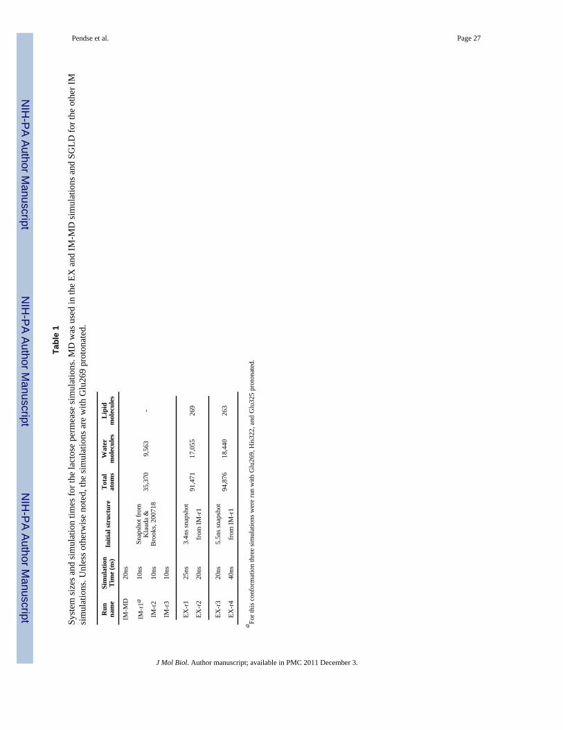
RESULTSThis section is divided in three subsections. The first subsection explains the rationalebehind the use of this hybrid simulation approach (IM-EX) for obtaining structural changesin LacY. The details of this approach are given in the Methods section, but thenomenclature, system size and simulation times are in Table 1. Structural changes aredescribed by means of Root Mean Square Deviation (RMSD) from 1PV7 crystal structureand pore radius analysis. Structural information about the proposed outward-facing LacY isalso described in this section, such as helix-helix distances, domain angle changes, andstructure stabilizing residue-residue interactions. In the second subsection, the data obtainedfrom the simulations is compared with the results from FRET,12 DEER,13 cross-linking,15and substrate accessibility experiments.14 The final subsection describes the binding of asugar molecule in the inward- and outward-facing states.
Structural changes from the IM-EX simulation approachImplicit bilayer simulations—Initially, a MD implicit membrane simulation and aprevious explicit simulation18 were compared to verify that the implicit membrane did notinfluence the protein structure and that of the water in the lumen. (Unless otherwise notedIM simulations are with Glu269 protonated.) Since the implicit membrane only acts on theprotein, waters that are in the protein lumen are not directly influenced by this implicitmembrane. If there is no significant conformational change in the protein compared toprevious explicit simulations,18 then the hydration of the protein lumen should be similarfor the EX and IM simulations. As described below, the IM-MD simulations resulted in nosignificant change in protein structure and can be compared with previous EX simulations.The water profile is nearly identical between the EX and IM-MD simulations (Figure 2),which suggests a negligible influence on waters in the protein lumen.
Conformational changes on the periplasmic half of LacY in the IM-MD simulation werenegligible compared to previous EX simulations and the x-ray crystal structure. Thedistances between helices at the membrane interface (Figure S2) only showed slight changesfrom the x-ray structure and consistent with fluctuations seen in previous EX simulations.18The pore radius at the periplasmic constriction (Figure 3) did not change from the x-raycrystal structure, and for the cytoplasmic half, it decreased by ~2 Å which is consistent withprevious simulations.18; 19 Multiple simulations will be required to determine if the IM-MD simulations result in the conformational changes similar to EX simulations, which is notthe focus of present study. The details of the helix-helix distance and pore radiuscalculations are given in the Methods.
Since MD was not successful in obtaining conformational changes in the periplasmic half ofLacY, SGLD was used to enhance conformational sampling. Since we are starting from astate open to the cytoplasm, the mechanism6 in Figure 1 suggest that proton translocationfrom Glu325 to Glu269 facilitates this structural change. Therefore, there are two controls inour simulations that should not result in periplasmic conformational changes if protonationof Glu269 triggers such a change, i.e., IM-r1 simulations with Glu325 and His322protonated. In addition, simulations were carried out with apo-LacY (without sugar) tounderstand the effect of sugar binding on conformational changes11; 13 and a likely thirdcontrol.
Pendse et al. Page 4
J Mol Biol. Author manuscript; available in PMC 2011 December 3.
NIH
-PA Author Manuscript
NIH
-PA Author Manuscript
NIH
-PA Author Manuscript
All simulations resulted in a significant closure of the cytoplasmic half but only thesimulation with Glu269 protonated resulted in opening on the periplasmic side (Figure S3).The cytoplasmic closure was observed to be independent of the protonation states ofGlu269, His322 and Glu325 which is in accordance with the findings by Holyoake et al.22All SGLD simulations with Glu269 protonated resulted in conformational changes in theperiplasmic half of LacY (Figures 3, S2, and S4) indicating that protonation of Glu269 andbinding of sugar triggers opening of LacY on the periplasmic side.
The remaining presentation of the results will be based on simulations with Glu269protonated and a disaccharide bound because these were the only to show periplasmicconformational changes. IM-r1 resulted in the largest conformational change with anincrease in pore radius ~3 Å (Figure 3, bottom). This transition occurred between 4–6 ns andresulted in a large increase in helix-helix distances between the C- and N-terminus domains(Figure S4). For IM-r2 and -r3, the opening of the periplasmic half is reduced compared toIM-r1. Consistent for all these runs is that H-V/VI and H-IX/X remain packed with onlyminor changes in helix-helix distances.
The cytoplasmic half of the protein remained partially closed with a pore radius of ~2.25 Åfor each run except IM-r3. Similar to the periplasmic half, this involved a decrease in helix-helix distances between the C- and N-terminus domains of LacY (Figure S2 and S4). Thelargest change from the x-ray crystal structure involved H-V. Overall, H-IV and VIII had thelargest RMSD for the runs with SGLD and the disaccharide, i.e., 2.1±0.2 and 1.9±0.4 Å,respectively. Moreover, the C-terminus domain of LacY had a larger RMSD (5.4±0.5 Å)compared to the N-terminus domain (3.3±0.3 Å).
Based on the results above, significant conformational changes in LacY exist for the cyto-and periplasmic halves of the protein with the SGLD implicit membrane simulations withGlu269 protonated and a disaccharide bound. Since the implicit membrane was used toenhance simulation time scales, two protein conformations (Figure S5) were selected andinserted into an explicit POPE membrane to obtain the final conformations of LacY. Theprotein is simulated in an explicit membrane to prevent any possible artifacts due to theimplicit membrane by obtaining structures in a more realistic environment (see Methods).These protein conformations were obtained from the 3.4 and 5.5 ns snapshots of the IM-r1simulation and represent an intermediate and periplasmic-open state based on the lumenradii, respectively. These were chosen carefully based on the comparison with the distancesmeasured from the DEER experiments13 to see if initial states in the explicit membrane areimportant to obtain final protein conformations open to the periplasm.
Explicit membrane simulations—The EX-r1 and EX-r2 simulations were carried outstarting with the intermediate state and EX-r3 and EX-r4 were carried out starting with theperiplasmic-open state. Protein backbone RMSDs from 1PV7 were calculated over thecourse of the trajectories. For EX-r4, RMSD of the N- and C-terminal domains were 4.1±0.2and 5.1±0.2 Å, respectively, which is consistent with the IM simulations in which the C-terminal domain shows more structural deviation from the crystal structure than the N-terminal domain. Locally, H-IV with Glu126 and H-VIII with the protonated Glu269showed the maximum structural drift with average RMSDs of 1.5±0.1 Å and 1.9±0.2 Å,respectively.
All the simulation runs showed a decrease in the pore radius on the cytoplasmic side by ~2Å from the crystal structure (Figure 4, top), which is similar to the implicit simulations. Thepore radius increase on the periplasmic side was 1–1.5 and 2.5–3 Å for EX-r1 and -r4,respectively. The average pore radius profiles for EX-r3 and EX-r4 were compared with theprofile of the periplasmic-open state screened from IM-r1 (Figure 4, bottom). EX-r4 showed
Pendse et al. Page 5
J Mol Biol. Author manuscript; available in PMC 2011 December 3.
NIH
-PA Author Manuscript
NIH
-PA Author Manuscript
NIH
-PA Author Manuscript

an increase of 1–1.2 Å in the pore radius while EX-r3 showed a decrease of ~1 Å from theperiplasmic-open snapshot structure. Similarly, EX-r1 showed opening beyond theintermediate snapshot while EX-r2 showed closing (data not shown). In the inward-facingstate of LacY (1PV7), the pore radius in the tightly closed periplasmic half is ~1 Å. For EX-r1 and EX-r4, the pore radius at the narrowest section in the cytoplasmic half is ~1.5 and ~2Å, respectively. For these runs, the cytoplasmic side is not as tightly packed as theperiplasmic side in the inward-facing crystal structure, but is sufficient to prevent thedisaccharide, which has a minimum span of ~7 Å, to transport to the cytoplasm. Only watercan transport from the cytoplasm to the protein lumen in this state and this occurs rarely forthis nearly closed cytoplasmic structure.
On the cytoplasmic side, H-V showed the largest change in separation for all EX runs(Figure 5) and is consistent with the IM simulations. For EX-r1 and EX-r4, several helicesshowed significant increases for the periplasmic half of the protein (H-II/III/IV/VI/VII/XI).For EX-r2 and EX-r3, no significant change in the helix-helix separation was observed onthe periplasmic side. For these runs, H-IV was the only helix to show some increase inseparation with respect to the helices in C-terminal domain.
To summarize, for EX-r1 and EX-r4, the protein underwent major conformational changesbeyond IM-r1 resulting in a significant periplasmic opening (Figure 6, left). The proteinlumen closed on the cytoplasmic side (Figure 6, right) and blocked the access of sugar onthe cytoplasmic side. This is in accordance with the well-established alternating accessmodel,11 which states that the sugar binding site is accessible only from the periplasm in theoutward-facing state of LacY.
Description of the transitions to the Outward-facing State of LacY—A fulldescription of the of transition between the open states of this protein (Fig. 1, step 3) is leftfor the discussion section but important structural and stabilizing interactions are brieflypresented in this section. The pore radius profiles clearly indicate that two out of fourexplicit simulation runs (EX-r1 and -r4) resulted in opening of the protein structure on theperiplasmic side (movie in supplementary material), while the other runs lacked anysignificant periplasmic opening. Figure 7 (top) shows the separation transmembrane helicesof the periplasmic half (z>0) and opening of the protein lumen for EX-r4. Although theconformational changes of LacY involve a mechanism more complex than rigid-bodyrotation of the two domains,6 the angle between the principal axis vector of the entiredomain obtained from simulations and that from the crystal structure is calculated toquantify the pseudo-rotation of the domain. The principal axes of the N- and C-terminaldomain are calculated based on the backbone atoms of the first six and the last six helices,respectively. For EX-r1, principal axis vectors of the N- and C-terminal domains made11.3±1.6° and 11.4±1.6° angles with the corresponding crystal structure vectors. For EX-r4,the N-terminal domain rotated by ~11°, whereas the C-terminal domain rotated by ~20°from the crystal structure. The domains rotate in directions opposite to each other so that theprotein lumen closes on the cytoplasmic side and opens on the periplasmic side. PDB of theoutward-facing model of LacY obtained from EX-r4 is included in the supplementarymaterials.
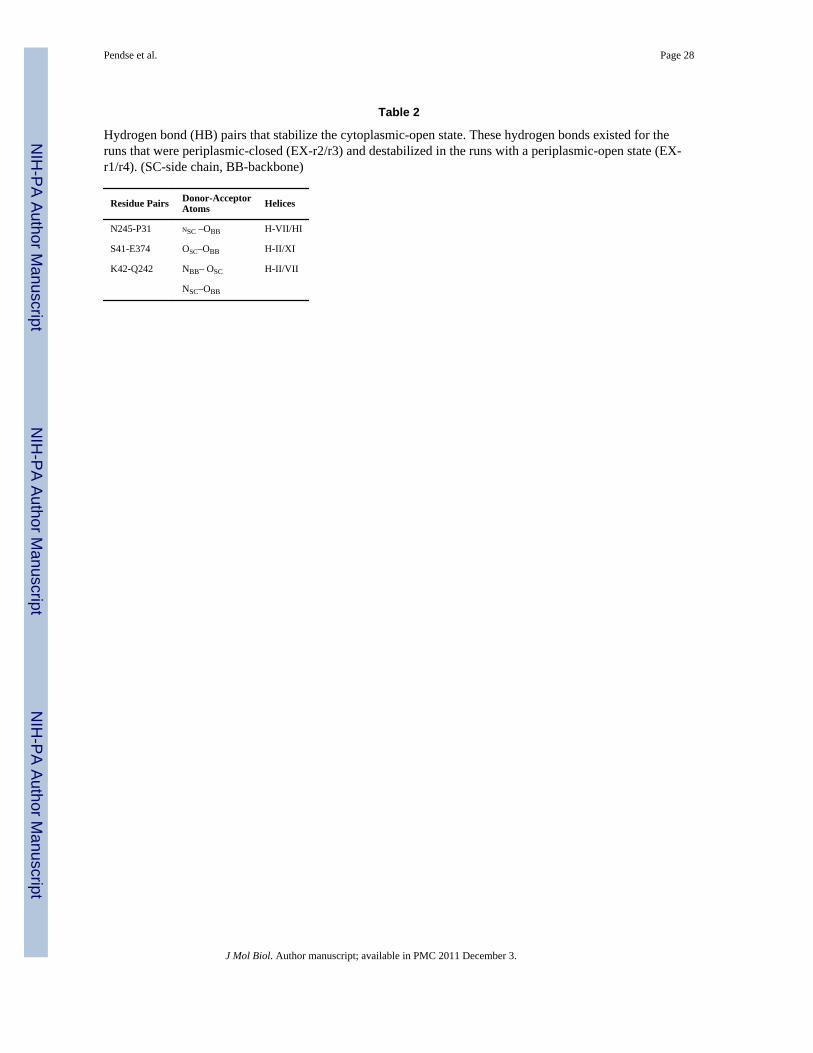
Packing of some helices on the periplasmic side was stabilized by hydrogen bondinteractions, which are listed in the Table 2. These interdomain hydrogen bonds are brokenin EX-r1 and EX-r4 resulting in the opening of LacY on the periplasmic side. The backboneoxygen of Pro31 was involved in hydrogen bonding in EX-r2 and EX-r3 indicating theexistence of close tertiary packing between H-I/H-VII near the periplasmic end. The otherresidue involved in hydrogen binding with Pro31, Asn245, has been shown to play animportant role in gating the periplasmic pathway of LacY.28
Pendse et al. Page 6
J Mol Biol. Author manuscript; available in PMC 2011 December 3.
NIH
-PA Author Manuscript
NIH
-PA Author Manuscript
NIH
-PA Author Manuscript

Comparison with the experimental dataSince no periplasmic-open structure exists for this or other MFS proteins a detailedcomparison with our simulations is needed to justify the above described periplasmicopening and cytoplasmic closing. Although atomic-level detail is not available, lower-resolution experimental techniques will be compared with our simulations, i.e., FRET,12DEER,13 cross-linking,15 and site-directed alkylation experiments.11; 14
DEER and FRET Experiments—Previous DEER experiments were carried out on Cysmutants of LacY labeled with (1-oxyl-2,2,5,5-tetramethylpyrroline-3-methyl)-methanethiosulfonate (MTSL). The distances between the spin labels measured in theexperiments were compared with the corresponding Cα-Cα distances calculated from thesimulations. The correspondence between the two sets of distances is not one to one.However, the comparison throws light on cytoplasmic and periplasmic structural changesand distance changes between cytoplasmic- and periplasmic-open states may cancel out theeffect of this bulky spin label if the orientation remains constant. The Cα-Cα distancechanges from the cytoplasmic-open (x-ray structure) to the periplasmic-open state (DEER-based) are compared with the LacY simulations in Figure 8 and Table S1. The valuesreported are based on Gaussian fits to distance distributions of the pairs (Figure S6).
On the periplasmic side, the distances increased significantly from IM-r1 for EX-r1 (TableS1) and EX-r4 (Figure 8). For EX-r4, increase in the distances from IM-r1 was 3–5 Å. Thepairs Val105/Thr310 (H-IV/H-IX) and Ile164/Ser375 (H-V/H-XI) showed the maximumseparation from the x-ray crystal structure by 12–13 Å. These two distances matchconsiderably well with the DEER experiments. The Ile164/Thr310 distance (H-V/H-X)increased over the IM-r1 run by ~3 Å and ~7 Å from the cytoplasmic-open state but wasslightly lower than experiment. For EX-r2 and EX-r3, the distances remained the same ordecreased from IM-r1 for all periplasmic pairs.
On the cytoplasmic side, the distance between the pairs decreased for EX-r1 and EX-r4compared to IM-r1. For EX-r4, this decrease was 2–5 Å for all the pairs. The distancesSer136/Gln340 and Asn137/Gln340 (both H-IV/H-X) showed the maximum decrease of 10Å from the crystal structure. The distance change for Arg73/Ser401 (H-III/H-XII) was 5 Åwhich was considerably smaller than the experimental value of 21 Å, but all other distancesfrom EX-r4 compared favorably with the experimental data. For EX-r3, most distancesincreased from IM-r1, but a few show no significant change or a slight decrease (Figure 8).
These results also qualitatively comply with the findings from the FRET experiments inwhich the cytoplasmic ends of H-III and H-XII move closer while the periplasmic ends ofH-V and H-XI move apart upon sugar binding.12
Cross-linking Experiments—Three paired double-Cys mutants (I40C/N245C, T45C/N245C and I32C/N245C) located at the interface of the N- and C-terminal domains near theperiplasmic end were used in previous cross-linking experiments.15 Homobifunctional thiolcross-linking reagents of different lengths and flexibilities were used to test the influence ofcross-linking on the transport activity of a disaccharide, TDG. For the I40C/N245C double-Cys mutant, transport activity of sugar was almost entirely blocked with cross-linkingreagents of length less than ~15 Å. With the flexible reagents MTS-14-O4-MTS (~17 Å)and MTS-17-O5-MTS (~22 Å), full transport activity of the disaccharide was observed.These cross-linkers suggest that the periplasmic opening of the protein should be between 15– 17 Å for the outward-facing state.
To determine the size of periplasmic opening, Cβ-Cβ distances between the three residuepairs (I40/N245, T45/N245 and I32/N245) were calculated from our simulations. The
Pendse et al. Page 7
J Mol Biol. Author manuscript; available in PMC 2011 December 3.
NIH
-PA Author Manuscript
NIH
-PA Author Manuscript
NIH
-PA Author Manuscript
experimentally-based periplasmic open distances are the spacer arm distances of the cross-linking reagent and not the distance between Cβ atoms. The distance between the Cβ atomswill be higher than the spacer arm distance and the equilibrium bond distance between Cβ-Sof cysteine (1.8 Å) is used as an offset. Therefore, Cβ-Cβ distances between the residue pairsshould be between 18.6 and 20.6 Å. The results (Table 3) show that for EX-r1 and EX-r4,the separations for the residue pairs I32/N245, T45/N245 and I32/N245 agree favorably withthe size of the opening indicated by the cross-linking experiments. These cross-linkingexperiments also indicate that the inter-thiol distance between I32C/N245C is close to ~6 Åin absence of the ligand, which is similar to Cβ-Cβ distance for the inward-facing crystalstructure (1PV7).5 For EX-r2 and EX-r3 (periplasmic-closed runs), the mean values for Cβ-Cβ distance between I32/N245 are 5.8 Å and 6.5 Å, respectively. Also, the mean Cβ-Cβdistances between the other two pairs are close to the crystal structure.
Accessibility and Reactivity Experiments—Site directed alkylation of single-Cysmutants of LacY at various positions with small membrane-permeant alkylating reagents, N-ethylmaleimide (NEM)11; 29; 30; 31; 32; 33; 34 and tetra-methylrhodamine-5-maleimide(TMRM),14 was previously measured to investigate the conformational changes in LacYupon sugar binding by quantifying the accessibility and/or reactivity at those residues. Thereactivity of a Cys residue can be limited by close contacts between transmembrane helicesin its surroundings and a change in the reactivity on sugar binding indicates an alteration inthe environment at that position in the sequence.11; 14 Sugar binding was shown to result inan increase in the reactivity of Cys-mutants that predominantly lie in the periplasmic halfwhile a decrease in the reactivity was observed at positions predominantly lying in thecytoplasmic half. For the protein conformations obtained from our simulations, the contactsurface areas were calculated for the residues at these positions as a measure of the closecontacts (see Methods). These areas were compared with the contact surface areas of therespective residues in the crystal structure to assess the change in environment of theseresidues (Table S2). As the experiments were performed on the Cys-mutants, the bulky sidechain atoms beyond the Cβ were neglected when calculating the surface areas.
On the periplasmic side, most (85%) of the residue positions that had an increase in Cysreactivity11; 14 also had an increase in the contact surface area for the EX-r1 and -r4 (TableS2). On the cytoplasmic side, some residues that had a decreased Cys reactivity did notshow a decrease in the contact surface area. This may be a result of incomplete cytoplasmicclosure observed during the simulations.
Site specific labeling with methanethiosulfonate ethylsulfonate (MTSES), a smallhydrophilic, membrane-impermeant thiol reagent was used to determine the change in wateraccessibility at various positions on sugar binding.29; 30; 31; 32; 33; 34; 35 The positionsthat showed a change in water accessibility were determined. To compare with the MTSESaccessibility studies, water accessible surface areas of various residues were calculated fromour simulations and were compared with the corresponding surface area values for 1PV7(Table 4). For EX-r1 and EX-r4, there is near perfect agreement for the positions thatmeasured an increase or decrease in accessibility on sugar binding.
Sugar bindingSince conformational changes of the protein are coupled to sugar binding (Figure 1),11; 13in the final section of the result we briefly describe sugar binding. Although these results arebased on several explicit membrane simulations, more detailed and comprehensive studieson binding are needed to obtain a complete picture of sugar binding in the periplasmic openstate and presented here may not be the entire story. The interactions between thedisaccharide, ββ-(Galp)2, with LacY are similar for all EX runs. The disaccharide binding
Pendse et al. Page 8
J Mol Biol. Author manuscript; available in PMC 2011 December 3.
NIH
-PA Author Manuscript
NIH
-PA Author Manuscript
NIH
-PA Author Manuscript

pocket in EX-r1 and -r4 (outward-facing LacY) is similar to the IM simulations. Thedisaccharide formed hydrogen bonds with Glu126, Arg144, Asp237, Glu269, Lys358,Phe118, Asn119, Asp240, His322 and Tyr350, which is consistent with our previousdisaccharide binding studies.18 Separate from these well-known sugar-protein interactions,the disaccharide formed hydrogen bonds with Gln60 and Phe354 (Figure S7). Both theseresidues have been shown to be important in sugar transport.29; 32; 36; 37 For EX-r1, ββ-(Galp)2 escaped the binding pocket during the last 5 ns of simulation. It migrated ~7 Åtowards the periplasm after the protein started opening on the periplasmic side and formedhydrogen bonds with Ser53 and Asn119 in this binding conformation (Figure S8) with theimportance of the former in the transport cycle confirmed by experiments.32 Similarly forEx-r2, where LacY started opening on the cytoplasmic side, the disaccharide escaped thebinding pocket and migrated towards the cytoplasm. The disaccharide was found to beundergoing a change in its orientation in order to escape on either sides of the membrane. Inthe binding pocket, the two sugar rings are aligned nearly parallel to the membrane, whereasthe rings become perpendicular to the membrane when the disaccharide starts to escape theprotein lumen (Figure S9) as observed by Jensen et al.23 Despite the differences in sugarbinding when comparing the cytoplasmic- and periplasmic-open states, our simulationsagree with the alternating access model11 in which the same binding site is believed to beaccessible to the sugar molecules in inward and outward-facing states. Moreover, in theoutward-facing state, the protein lumen is closed on the cytoplasmic side preventing thesugar from escaping to the cytoplasm.
DISCUSSIONLacY undergoes widespread conformational changes during substrate transport based onvarious biochemical experiments.6 Previous MD simulations have resulted in significantconformational changes on the cytoplasmic side compared to the crystal structure. Adecrease in pore radius was observed in the cytoplasmic half for LacY with Glu269protonated in a 10-ns simulation19 and with Glu325 protonated in 50-ns simulation. Ourprevious EX simulations with Glu325 protonated18 also resulted in similar decrease in thecytoplasmic pore radius by ~2 Å. However, negligible pore radius changes in theperiplasmic half were observed during these simulations.
Several control simulations were performed to verify that our methods open the protein onthe periplasmic side only when a sugar is bound and the proper protonation of a titratableresidue. Only simulations with Glu269 protonated and a sugar bound resulted inconformational changes in the periplasmic half. Simulations without the sugar (apo), Glu325protonated, or His322 protonated, all maintained a conformation prevent sugar transport tothe periplasm. Although this was expected for simulations without the sugar or Glu325protonated (Figure 1),6 our simulations suggest that protonation of His322 does not trigger aconformational change.
All IM simulations with Glu269 protonated and a sugar bound resulted in greaterperiplasmic conformational changes in LacY compared to previous EX simulations becauseof enhanced sampling. The structural changes were enhanced by the use of SGLD instead ofMD and an implicit bilayer. The enhancement alone appears to allow for structural changesand sugar binding is a trigger for protein structural changes because LacY-apo remainedclosed on the periplasmic side. Structures were screened from the IM simulations andinserted into an explicit bilayer so that a more realistic environment can be used to obtain afinal set of conformations (Figure 7, bottom). Essentially, it was our aim to use the implicitmembrane simulations with SGLD to overcome a transition state or energy barrier betweenthe two open states in a reasonable amount of time. If the screened structures lie in themetastable transition state region in the free energy profile, then there would be an equal
Pendse et al. Page 9
J Mol Biol. Author manuscript; available in PMC 2011 December 3.
NIH
-PA Author Manuscript
NIH
-PA Author Manuscript
NIH
-PA Author Manuscript
probability to go back to the inward-facing state or further open to the outward-facing stateassuming a single transitional barrier. For each of the screened structures, one simulationresulted in a fully periplasmic-open state, while another started closing on the periplasmicside and opening on the cytoplasmic side. Certainly more runs would be required todetermine if the screened structures are at or near a transition state, but these results doindicate the ability of this approach to lead to a periplasmic-open state and also return to aconformation closed to the periplasm.
The pore radius profile of LacY’s lumen (Figures 2 and 3) and helix-helix separationdistance maps (Figure 5, S2, and S3) clearly demonstrate a protein structural change. Aninteresting point to be noted is the average pore radius profile for EX-r4 (Figure 4, top). Thecytoplasmic half of this profile resembles greatly with the periplasmic half of the pore radiusprofile of the crystal structure and vice versa. Besides the similarities in the two halves, thetwo profiles are analogous in the sugar binding region (~z =0 Å).
The helix-helix maps (Figures 5 and S3) indicate that on the periplasmic side structurechanges involve significant intradomain as well as interdomain helix movements, whereasonly the interdomain displacements are dominant on the cytoplasmic side. The periplasmicconformational changes indicate a more complex mechanism involving significantintradomain changes to yield a final outward-facing state. However, if the initial and finalstructure of LacY is only considered, then the rigid body rotation of LacY is consistent withthe helix map. Therefore, intradomain changes are important as the protein changes betweenopen states, but in the end these collapse back to similar helix packing. More specifically, H-V showed the largest change in the separation on the periplasmic and cytoplasmic sides. TheC154G mutant of LacY, which is trapped in an inward-facing conformation, has Gly154 (H-V) at the domain interface. Gly154 is believed to cause tighter packing of H-V near theinterface hence hindering the movement of H-V which was found to be significant duringthe conformational transitions from our simulations.
The Cα-Cα distances calculated from EX-r1 and EX-r4 agree favorably with the DEERexperiments (Figure 8 and Table S1).13 On the periplasmic side, the distance changes forpairs V105/T210 and I164/S375 are in agreement with the experimental values. On thecytoplasmic side, the distance changes differ from some of the experimental valuesindicating a possible partial incomplete cytoplasmic closure. However, the cytoplasmic halfis closed to the extent that it prevents and transport of sugar to the cytoplasm (Figure 6,right). The largest discrepancy between the simulations and the experiments is for thecytoplasmic pair R73/S401 suggesting that H-II/XII is the last helix pair to close. Moreover,the distances calculated from the simulations are Cα-Cα distances while those measured inthe experiments are the distances between the spin labels (MTSL). The approximate distancebetween the label and the Cα atom is 8 Å, which may result in the maximum deviation of ±16 Å. For example, the distance changes reported from the DEER experiments differconsiderably for the pairs S136/S401 and N137/S401 even though there is little distanceseparating the two residues (Ser136 and Asn137). Similar difference is present between thepairs S136/Q340 and N137/Q340. It was suggested that these striking differences may be aresult of significant rotation of the helices including winding and unwinding at the ends.13No such conformational changes were observed during our simulations. The distancescalculated from EX-r4 are close to the mean of the DEER measurements for theS136(N137)/Q340 and S136(N137)/S401 pairs. This suggests that the orientation of the spinlabel may play a role in the DEER measurements. To explicitly investigate the effect of spinlabels, simulations with labeled residue pairs will be required and are planned as futurework.
Pendse et al. Page 10
J Mol Biol. Author manuscript; available in PMC 2011 December 3.
NIH
-PA Author Manuscript
NIH
-PA Author Manuscript
NIH
-PA Author Manuscript
In addition to DEER measurements, our simulations are in excellent agreement with theamount of the periplasmic opening measured using cross-linking experiments15 (Table 3)and site-directed alkylation with alkylating agents of various sizes.38 Based on these helixdistance measurements, it can be concluded that the periplasmic opening observed from EX-r1 and -r4 closely matches the outward-facing state.
The periplasmic opening of LacY obtained from our simulations is also in compliance withNEM,11 TMRM,14 and MTSES29; 30; 31; 32; 33; 34 labeling experiments. 85% ofresidues that had a significant increase in NEM and TMRM activity on the periplasmic sideshowed an increase contact surface area in EX-r1 and -r4. On the cytoplasmic side, theclosure obtained from our simulations may not be complete thus preventing the expecteddecrease in the surface area values for some residues, especially those on H-III and -IX(Table S2). However, we suggest that the steric effect of sugar binding at Gln60 may be areason behind the reduced NEM32 and TMRM14 reactivity observed with the Cysreplacement at that position. Although the comparisons with DEER and NEM/TMRMaccessibility studies indicate a possible incomplete cytoplasmic closure for our outward-facing LacY model, the pore radius profiles indicate that the closure is sufficient to block theaccess of sugar to the cytoplasm as per the alternating access model.11 Inaccessibility of thedisaccharide from the cytoplasmic end in the outward-facing state can be clearly seen inFigure 6 (right) where the protein lumen is closed on the cytoplasmic side.
The interdomain hydrogen bonds N245/P31, S41/E374 and K42/Q242 are destabilized inEX-r1 and -r4 while they are maintained in EX-r2 and -r3 (Table 2). Moreover, thesehydrogen bonds stabilized the helix-packing in previous EX simulations18 as well as in theinitial parts of our IM simulations. These interdomain hydrogen bond interactions appear tobe crucial in stabilizing the inward-facing state. Side chains of Asn245, Ser41, Glu374,Lys42 and Gln242 are involved in stabilizing the inward-facing structure and mutations inthese residues may destabilize the inward-facing state. Moreover, breaking of thesehydrogen bonding interactions in the outward-facing state explains the increase in NEM11and TMRM14 labeling at residues 31, 42 and 245 on sugar binding. In addition, any changein the interactions between H-VII and the surrounding helices at residue 242 was found toinhibit the transport activity which is consistent with our findings.39
The findings from our simulations are summarized below to give mechanistic insights intothe sugar transport cycle and conformational changes of LacY from the outward to inwardfacing state (Fig. 1, step 3). This is also shown as a cartoon in Figure 9.
a. Binding of a disaccharide and translocation of proton to Glu269 is necessary totrigger the conformational transformation of LacY. The interdomain hydrogenbonds N245/P31, S41/E374 and K42/Q242 stabilize the inward-facing structure ofLacY. In the binding pocket, two rings of the disaccharide are oriented nearlyparallel to the membrane.
b. In the intermediate state, the disaccharide lies in the binding pocket but is weaklybound to the protein at Glu126, Arg144 and Glu269. The structure stabilizinginterdomain hydrogen interactions are broken. The salt bridges E126/R144 andD237/K358 remain intact during the transition
c. The disaccharide remains in the binding pocket during the conformational changewhich is in accordance with the alternating access model11 but undergoes anorientational change such that the two rings are nearly perpendicular to themembrane. It interacts with Ser56, Gln60, Asn119, Tyr350, Phe354, Cys355, andGln359 in this binding conformation.
Pendse et al. Page 11
J Mol Biol. Author manuscript; available in PMC 2011 December 3.
NIH
-PA Author Manuscript
NIH
-PA Author Manuscript
NIH
-PA Author Manuscript

In summary, we were able to obtain significant structural changes on the periplasmic as wellas the cytoplasmic side of LacY. The periplasmic changes resulted in the opening of theprotein lumen to outside the cell. The outward-facing state from our simulations agreesfavorably with a multitude of experimental data. The inward-facing state is stabilized bycertain interhelical and interdomain interactions and destabilizing these interactions mayhelp crystallize the outward-facing state. Our simulations are in compliance with thealternating access model11 for the sugar binding but some additional residues that areimportant in sugar binding are determined. The importance of these residues in sugarbinding and conformational change was confirmed by experiments. Ultimately, this IM-EXmethod offers a possible route to obtain structures of other secondary active transportersbased on crystal structures open to the periplasm or cytoplasm in an efficient manner.However, a more systematic study on better characterized proteins will be needed to furthertest the accuracy and limitations of this method.
METHODSThe CHARMM simulation package40; 41 was used with a simulation time step of 2 fs. TheCHARMM family of force fields are used to describe the atomic interactions of the lipids,42; 43 protein (C22-CMAP),44; 45; 46 and disaccharide.47 The TIP3P water model wasused (the version consistent with CHARMM parameters48; 49). Long range electrostaticinteractions were calculated with the particle-mesh Ewald (PME)50 method. The screeningparameter (κ) was set at 0.45 Å−1 and a fast Fourier transform grid density of ~1 Å−1.Lennard-Jones interactions were switched smoothly to zero between 8–10 Å.40 TheSHAKE algorithm51 was used to constrain hydrogen atoms. Extended system formalism isused to maintain the pressure with a barostat52; 53 and the temperature constant (EXsimulations only) with the Nosé-Hoover method54 at 310.15 K. Glu269 protonated for mostsimulations, which has been suggested to be the preferred protonated state open to theperiplasm7; 19, but other protonated states were also used (see below). All simulationcoordinates were saved every picosecond for analysis purposes.
A new two step hybrid simulation method, referred to here as IM-EX (implicit-explicitmembrane), was used to probe conformational changes in LacY because fully explicitmembrane MD simulations are limited to timescales approaching 1 µs.55 For the first step(IM) in our hybrid simulation procedure, an implicit membrane is used reducecomputational demand and enhance conformational changes of the protein. GeneralizedBorn solvation models would be the ideal choice for an implicit membrane, such as thosedeveloped by Feig et al.56; 57; 58 However, our benchmark simulations with LacY and theheterogeneous dielectric generalized Born method57 required simulation times that wereequal to or greater than a fully explicit bilayer. Instead, an external hydrophobic potentialbased on the method of Edholm and Jähnig59 is used to represent the lipid/water interface.This potential is assumed to vary exponentially across the membrane surfaces,
where the membrane interface is at ±z0 (assumed to be 15Å), the decay length (λ) is 2, and hiis the hydrophobic energy of atoms taken from reference.59 An explicit water interface isincluded in all implicit lipid membrane simulations (Figure S1). The lumen of LacY is filledwith water18; 19; 22 and not including explicit water may result in an unphysical closing ofthe cytoplasmic opening. Consequently, the implicit membrane only acts on the protein. At
Pendse et al. Page 12
J Mol Biol. Author manuscript; available in PMC 2011 December 3.
NIH
-PA Author Manuscript
NIH
-PA Author Manuscript
NIH
-PA Author Manuscript

the membrane interface, a thermodynamically stable vapor-liquid interface is formed whichprevents water from flooding into the membrane region.
SGLD is used to further enhance protein motions.25 This method has been shown toenhance low-frequency motions and conformational searching of proteins.25; 26; 27 Thefollowing SGLD parameters are used in the implicit simulations: local averaging time tL=0.25 ps, guiding temperature of 1 K, and a friction factor γ = 1 ps−1. The local averagingtime enhances all motions with periods larger than tL, while the guiding temperature limitsthe perturbation to the system. As mentioned in the introduction, this method maintains thecorrect ensemble averages and fluctuations.25
The implicit bilayer simulations (IM) were performed with three residues (Glu269, His322and Glu325) separately protonated because of their involvement in the proton translocationmechanism. The initial protein structures for the IM simulations are from certain snapshotsof previous explicit membrane (EX) simulations.18 The 5-ns snapshot of EX-wt/ββ-(Galp)2(run 2) in Klauda and Brooks18 was used for the initial conformations of IM-r1 (run 1 withSGLD), IM-apo (simulation without sugar but using SGLD), IM-322H (simulations withHis322 protonated), IM-325H (simulations with Glu325 protonated) and IM-MD (simulationwith MD instead of SGLD). The 5-ns snapshot was chosen because it has a slightconformational change in the periplasmic half of LacY compared to the x-ray crystalstructure.5 A single disaccharide, ββ-(Galp)2, is used for all but the apo simulation becausesugar binding is known to induce structural changes toward the outward-facing state.13 Twodifferent protein coordinates were used as initial structures for the IM-r2 and IM-r3 runs.IM-r2 used a protein snapshot from the EX-wt/ββ-(Galp)2 (run 2) in reference 18 at 12 ns(some conformational change in the periplasmic half) and IM-r3 used the final coordinate ofthis previous run (no changes in the periplasmic half). Four IM simulations with SGLD wererun for 10 ns each and the IM-MD simulation was run for 20 ns. Each simulation consistedof 35,370 total atoms with 9,563 water molecules.
For the second step (EX), two independent simulations were run for each of the twoscreened conformations from the previous IM step with different initial velocities. All EXsimulations were run with a time step of 2 fs. An explicit palmitoyloleoylphosphati-dylethonolamine (POPE) bilayer was used to model the plasma membrane, which is knownto be important for the function and topology of LacY.60; 61; 62 Simulations were carriedout in the NPAT ensemble at physiological temperature of 310.15 K and an experimentallysuggested area of 65.2 Å2/ lipid.63 The details about the simulation time and system size foreach run are given in Table 1. The P21 periodic boundary condition64 was used whichallows for the redistribution of lipids between the leaflets, which is important for anysignificant structural change in LacY. For EX-r1, the average number of lipids in theperiplasmic leaflet reduced from 142.0±1.7 in the first ns to 133.1±1.9 in the last ns.
Pore radius analysis calculations were done using the program HOLE65 based on 0.5-nsblocks. The distances between all the helices on the periplasmic and cytoplasmic halves ofthe protein are used as a measure of protein structural change and helix packing. Helix-helixdistance maps were generated by calculating average distances between the helix centers onperiplasmic and cytoplasmic sides every nanosecond. The helix centers are calculated basedon the center of mass of backbone atoms between z=10±1 and −10±1 Å for periplasmic andcytoplasmic sides respectively, as used previously.22 The molecular surface areas arecalculated using Lee and Richards algorithm66 that uses a sphere of radius 1.6 Å to probethe molecular surface. Visual molecular dynamics (VMD) was used to make all molecularfigures.67
Pendse et al. Page 13
J Mol Biol. Author manuscript; available in PMC 2011 December 3.
NIH
-PA Author Manuscript
NIH
-PA Author Manuscript
NIH
-PA Author Manuscript

Supplementary MaterialRefer to Web version on PubMed Central for supplementary material.
AcknowledgmentsThis research was supported in part by University of Maryland startup funds (J.B.K and P.Y.P.) and the IntramuralResearch Program of the NIH, National Heart, Lung and Blood Institute (B.R.B.). Simulations were run on theHigh Performance Computational Cluster (HPCC) at the University of Maryland. We also thank Ron Kaback andVladamir Kosho at UCLA for supplying us with the raw experimental DEER data to compare with our simulations.
References1. Alberts, B.; Bray, D.; Johnson, A.; Lewis, J.; Raff, M.; Roberts, K.; Walter, P. Essential Cell
Biology: An Introduction to the Molecular Biology of the Cell. New York: Barland Publishing, Inc;1998.
2. Krishnamurthy H, Piscitelli CL, Gouaux E. Unlocking the molecular secrets of sodium-coupledtransporters. Nature. 2009; 459:347–355. [PubMed: 19458710]
3. Sobczak I, Lolkema JS. Structural and mechanistic diversity of secondary transporters. CurrentOpinion in Microbiology. 2005; 8:161–167. [PubMed: 15802247]
4. Milton H, Saier J. Families of transmembrane sugar transport proteins. Molecular Microbiology.2000; 35:699–710. [PubMed: 10692148]
5. Abramson J, Smirnova I, Kasho V, Verner G, Kaback HR, Iwata S. Structure and mechanism of thelactose permease of Escherichia coli. Science. 2003; 301:610–615. [PubMed: 12893935]
6. Guan L, Kaback HR. Lessons from Lactose Permease. Annual Review of Biophysics andBiomolecular Structure. 2006; 35:67–91.
7. Abramson J, Iwata S, Kaback HR. Lactose permease as a paradigm for membrane transport proteins- (Review). Molecular Membrane Biology. 2004; 21:227–236. [PubMed: 15371012]
8. Huang YF, Lemieux JM, Song MA, Wang DN. Structure and mechanism of the glycerol-3-phosphate transporter from Escherichia coli. Science. 2003; 301:616–620. [PubMed: 12893936]
9. Yin Y, He X, Szewczyk P, Nguyen T, Chang G. Structure of the multidrug transporter EmrD fromEscherichia coli. Science. 2006; 312:741–744. [PubMed: 16675700]
10. Guan L, Mirza O, Verner G, Iwata S, Kaback HR. Structural determination of wild-type lactosepermease. Proceedings of the National Academy of Sciences. 2007; 104:15294–15298.
11. Kaback HR, Dunten R, Frillingos S, Venkatesan P, Kwaw I, Zhang W, Ermolova N. Site-directedalkylation and the alternating access model for LacY. Proceedings of the National Academy ofSciences of the United States of America. 2007; 104:491–494. [PubMed: 17172438]
12. Majumdar DS, Smirnova I, Kasho V, Nir E, Kong XX, Weiss S, Kaback HR. Single-molecularFRET reveals sugar-induced conformational dynamics in LacY. Proceedings from the NationalAcademdy of Science. 2007; 104:12640–12645.
13. Smirnova I, Kasho V, Choe J-Y, Altenbach C, Hubbell WL, Kaback HR. Sugar binding induces anoutward facing conformation of LacY. Proceedings of the National Academy of Sciences. 2007;104:16504–16509.
14. Nie Y, Ermolova N, Kaback HR. Site-directed Alkylation of LacY: Effect of the ProtonElectrochemical Gradient. Journal of Molecular Biology. 2007; 374:356–364. [PubMed:17920075]
15. Zhou Y, Guan L, Freites JA, Kaback HR. Opening and closing of the periplasmic gate in lactosepermease. Proceedings from the National Academdy of Science. 2008; 105:3774–3778.
16. Franco PJ, Matzke EA, Johnson JL, Wiczer BM, Brooker RJ. A suppressor analysis of residuesinvolved in cation transport in the lactose permease: Identification of a coupling sensor. Journal ofMembrane Biology. 2006; 211:101–113. [PubMed: 16988863]
17. Bennett M, D’Rozario R, Sansom MSP, Yeagle PL. Asymmetric stability among thetransmembrane helices of lactose permease. Biochemistry. 2006; 45:8088–8095. [PubMed:16800633]
Pendse et al. Page 14
J Mol Biol. Author manuscript; available in PMC 2011 December 3.
NIH
-PA Author Manuscript
NIH
-PA Author Manuscript
NIH
-PA Author Manuscript

18. Klauda JB, Brooks BR. Sugar binding in lactose permease: Anomeric state of a disaccharideinfluences binding structure. Journal of Molecular Biology. 2007; 367:1523–1534. [PubMed:17320103]
19. Yin Y, Jensen MO, Tajkhorshid E, Schulten K. Sugar binding and protein conformational changesin lactose permease. Biophysical Journal. 2006; 91:3972–3985. [PubMed: 16963502]
20. Law CJ, Almqvist J, Bernstein A, Goetz RM, Huang Y, Soudant C, Laaksonen A, Hovmöller S,Wang D-N. Salt-bridge Dynamics Control Substrate-induced Conformational Change in theMembrane Transporter GlpT. Journal of Molecular Biology. 2008; 378:826–837.
21. Law CJ, Enkavi G, Wang D-N, Tajkhorshid E. Structural Basis of Substrate Selectivity in theGlycerol-3-Phosphate: Phosphate Antiporter GlpT. 2009; 97:1346–1353.
22. Holyoake J, Sansom MSP. Conformational change in an MFS protein: MD simulations of LacY.Structure. 2007; 15:873–884. [PubMed: 17637346]
23. Jensen MO, Yin Y, Tajkhorshid E, Schulten K. Sugar Transport across Lactose Permease Probedby Steered Molecular Dynamics. Biophysical Journal. 2007; 93:92–102. [PubMed: 17434947]
24. Enkavi G, Tajkhorshid E. Simulation of Spontaneous Substrate Binding Revealing the BindingPathway and Mechanism and Initial Conformational Response of GlpT. Biochemistry. 2010;49:1105–1114. [PubMed: 20058936]
25. Wu XW, Brooks BR. Self-guided Langevin dynamics simulation method. Chemical PhysicsLetters. 2003; 381:512–518.
26. Damjanovic A, Wu X, Garcia-Moreno EB, Brooks BR. Backbone Relaxation Coupled to theIonization of Internal Groups in Proteins: A Self-Guided Langevin Dynamics Study. BiophysicalJournal. 2008; 95:4091–4101. [PubMed: 18641078]
27. Damjanovic A, García-Moreno EB, Brooks BR. Self-guided Langevin dynamics study ofregulatory interactions in NtrC. Proteins: Structure, Function, and Bioinformatics. 2009; 76:1007–1019.
28. Zhou Y, Nie Y, Kaback HR. Residues Gating the Periplasmic Pathway of LacY. Journal ofMolecular Biology. 2009; 394:219–225. [PubMed: 19781551]
29. Ermolova NVMR, Kaback HR. Site-Directed Alkylation of Cysteine Replacements in the LactosePermease of Escherichia coli: Helices I, III, VI and XI. Biochemistry. 2006; 45:4182–4189.[PubMed: 16566592]
30. Frillingos S, Kaback HR. The Role of Helix VIII in the Lactose Permease of Escherichia coli: II.Site-Directed Sulfhydryl Modification. Protein Science. 1997; 6:438–443. [PubMed: 9041647]
31. Kwaw I, Zen K, Hu Y, Kaback HR. Site-Directed Sulfhydryl Labeling of the Lactose Permease ofEscherichia coli: Helices IV and V That Contain the Major Determinants for Substrate Binding.Biochemistry. 2001; 40:10491–10499. [PubMed: 11523990]
32. Venkatesan P, Hu Y, Kaback HR. Site-Directed Sulfhydryl Labeling of the Lactose Permease ofEscherichia coli: Helix X. Biochemistry. 2000; 39:10656–10661. [PubMed: 10978149]
33. Venkatesan P, Kwaw I, Hu Y, Kaback HR. Site-Directed Sulfhydryl Labeling of the LactosePermease of Escherichia coli: Helix VII. Biochemistry. 2000; 39:10641–10648. [PubMed:10978147]
34. Zhang W, Hu Y, Kaback HR. Site-Directed Sulfhydryl Labeling of Helix IX in the LactosePermease of Escherichia coli. Biochemistry. 2003; 42:4904–4908. [PubMed: 12718531]
35. Venkatesan P, Liu Z, Hu Y, Kaback HR. Site-Directed Sulfhydryl Labeling of the LactosePermease of Escherichia coli: N-Ethylmaleimide-Sensitive Face of Helix II. Biochemistry. 2000;39:10649–10655. [PubMed: 10978148]
36. Green AL, Anderson EJ, Brooker RJ. A Revised Model for the Structure and Function of theLactose Permease. The journal of Biological Chemistry. 2000; 275:23240–23246. [PubMed:10807929]
37. Green AL, Hrodey HA, Brooker RJ. Evidence for Structural Symmetry and Functional Asymmetryin the Lactose Permease of Escherichia coli. Biochemistry. 2003; 42:11226–11233. [PubMed:14503872]
38. Nie Y, Sabetfard FE, Kaback HR. The Cys154-->Gly mutation in LacY causes constitutiveopening of the hydrophilic periplasmic pathway. Journal of Molecular Biology. 2008; 379:695–703. [PubMed: 18485365]
Pendse et al. Page 15
J Mol Biol. Author manuscript; available in PMC 2011 December 3.
NIH
-PA Author Manuscript
NIH
-PA Author Manuscript
NIH
-PA Author Manuscript
39. Nie Y, Zhou Y, Kaback HR. Clogging the Periplasmic Pathway in LacY. Biochemistry. 2009;48:738–743. [PubMed: 19128028]
40. Brooks BR, Bruccoleri RE, Olafson BD, States DJ, Swaminathan S, Karplus M. CHARMM - aProgram for Macromolecular Energy, Minimization, and Dynamics Calculations. Journal ofComputational Chemistry. 1983; 4:187–217.
41. Brooks BRCL, Brooks IAD, Mackerell J, Nilsson L, Petrella RJ, Roux B, Won Y, Archontis G,Bartels C, Boresch S, Caflisch A, Caves L, Cui Q, Dinner AR, Feig M, Fischer S, Gao J,Hodoscek M, Im W, Kuczera K, Lazaridis T, Ma J, Ovchinnikov V, Paci E, Pastor RW, Post CB,Pu JZ, Schaefer M, Tidor B, Venable RM, Woodcock HL, Wu X, Yang W, York DM, Karplus M.CHARMM: The biomolecular simulation program. Journal of Computational Chemistry. 2009;30:1545–1614. [PubMed: 19444816]
42. Klauda JB, Brooks BR, MacKerell AD Jr, Venable RM, Pastor RW. An Ab Initio Study on theTorsional Surface of Alkanes and its Effect on Molecular Simulations of Alkanes and a DPPCBilayer. Journal of Physical Chemistry B. 2005; 109:5300–5311.
43. Klauda JB, Pastor RW, Brooks BR. Adjacent gauche stabilization in linear alkanes: Implicationsfor polymer models and conformational analysis. Journal of Physical Chemistry B. 2005;109:15684–15686.
44. Buck M, Bouguet-Bonnet S, Pastor RW, MacKerell AD. Importance of the CMAP correction tothe CHARMM22 protein force field: Dynamics of hen lysozyme. Biophysical Journal. 2006;90:L36–L38. [PubMed: 16361340]
45. Feig M, MacKerell AD Jr, Brooks CL. Force field influence on the observation of π-helical proteinstructures in molecular dynamics simulations. Journal of Physical Chemistry B. 2003; 107:2831–2836.
46. MacKerell AD, Feig M, Brooks CL. Improved treatment of the protein backbone in empirical forcefields. Journal of the American Chemical Society. 2004; 126:698–699. [PubMed: 14733527]
47. Kuttel M, Brady JW, Naidoo KJ. Carbohydrate solution simulations: Producing a force field withexperimentally consistent primary alcohol rotational frequencies and populations. Journal ofComputational Chemistry. 2002; 23:1236–1243. [PubMed: 12210149]
48. Durell SR, Brooks BR, Bennaim A. Solvent-Induced Forces between Two Hydrophilic Groups.Journal of Physical Chemistry. 1994; 98:2198–2202.
49. Jorgensen WL, Chandrasekhar J, Madura JD, Impey RW, Klein ML. Comparison of SimplePotential Functions for Simulating Liquid Water. Journal of Chemical Physics. 1983; 79:926–935.
50. Darden T, York D, Pedersen L. Particle Mesh Ewald - an NLog(N) Method for Ewald Sums inLarge Systems. Journal of Chemical Physics. 1993; 98:10089–10092.
51. Ryckaert JP, Ciccotti G, Berendsen HJC. Numerical Integration of the Cartesian Equations ofMotion of a System with Contraints: Molecular Dynamics of n-alkanes. Journal of ComputationalPhysics. 1977; 23:327–341.
52. Nosé S, Klein ML. A Study of Solid and Liquid Carbon Tetrafluoride Using the Constant PressureMolecular-Dynamics Technique. Journal of Chemical Physics. 1983; 78:6928–6939.
53. Andersen HC. Molecular-Dynamics Simulations at Constant Pressure and/or Temperature. Journalof Chemical Physics. 1980; 72:2384–2393.
54. Hoover WG. Canonical dynamics: Equilibrium phase-space distributions. Physical Review A.1985; 31:1695–1697. [PubMed: 9895674]
55. Khalili-Araghi F, Gumbart J, Wen P-C, Sotomayor M, Tajkhorshid E, Schulten K. Moleculardynamics simulations of membrane channels and transporters. Current Opinion in StructuralBiology. 2009; 19:128–137. [PubMed: 19345092]
56. Im W, Feig M, Brooks CL. An implicit membrane generalized born theory for the study ofstructure, stability, and interactions of membrane proteins. Biophysical Journal. 2003; 85:2900–2918. [PubMed: 14581194]
57. Tanizaki S, Feig M. A generalized Born formalism for heterogeneous dielectric environments:Application to the implicit modeling of biological membranes. Journal of Chemical Physics. 2005;122
58. Tanizaki S, Feig M. Molecular dynamics simulations of large integral membrane proteins with animplicit membrane model. Journal of Physical Chemistry B. 2006; 110:548–556.
Pendse et al. Page 16
J Mol Biol. Author manuscript; available in PMC 2011 December 3.
NIH
-PA Author Manuscript
NIH
-PA Author Manuscript
NIH
-PA Author Manuscript

59. Edholm O, Jahnig F. The Structure of A Membrane-Spanning Polypeptide Studied by Molecular-Dynamics. Biophysical Chemistry. 1988; 30:279–292. [PubMed: 3207847]
60. Xie J, Bogdanov M, Heacock P, Dowhan W. Phosphatidylethanolamine andmonoglucosyldiacylglycerol are interchangeable in supporting topogenesis and function of thepolytopic membrane protein lactose permease. Journal of Biological Chemistry. 2006; 281:19172–19178. [PubMed: 16698795]
61. Wang XY, Bogdanov M, Dowhan W. Topology of polytopic membrane protein subdomains isdictated by membrane phospholipid composition. EMBO Journal. 2002; 21:5673–5681. [PubMed:12411485]
62. Bogdanov M, Heacock PN, Dowhan W. A polytopic membrane protein displays a reversibletopology dependent on membrane lipid composition. EMBO Journal. 2002; 21:2107–2116.[PubMed: 11980707]
63. Shaikh SR, Brzustowicz MR, Gustafson N, Stillwell W, Wassall SR. Monounsaturated PE does notphase-separate from the lipid raft molecules sphingomyelin and cholesterol: Role forpolyunsaturation? Biochemistry. 2002; 41:10593–10602. [PubMed: 12186543]
64. Dolan EA, Venable RM, Pastor RW, Brooks BR. Simulations of membranes and other interfacialsystems using P21 and pc periodic boundary conditions. Biophysical Journal. 2002; 82:2317–2325. [PubMed: 11964222]
65. Smart OS, Goodfellow JM, Wallace BA. The Pore Dimensions of Gramicidin A. BiophysicalJournal. 1999; 65:2455. [PubMed: 7508762]
66. Lee B, Richards FM. The interpretation of protein structures: Estimation of static accessibility.Journal of Molecular Biology. 1971; 55:379–400. [PubMed: 5551392]
67. Humphrey W, Dalke A, Schulten K. VMD: Visual molecular dynamics. Journal of MolecularGraphics. 1996; 14:33–38. [PubMed: 8744570]
Pendse et al. Page 17
J Mol Biol. Author manuscript; available in PMC 2011 December 3.
NIH
-PA Author Manuscript
NIH
-PA Author Manuscript
NIH
-PA Author Manuscript
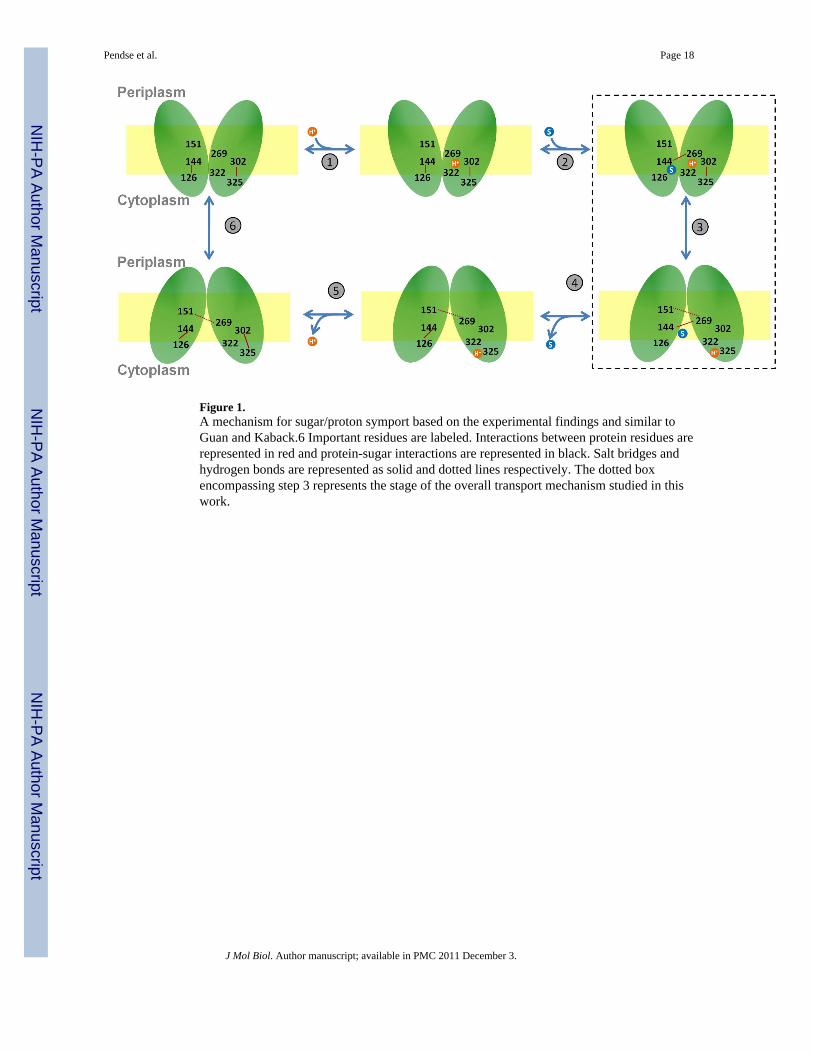
Figure 1.A mechanism for sugar/proton symport based on the experimental findings and similar toGuan and Kaback.6 Important residues are labeled. Interactions between protein residues arerepresented in red and protein-sugar interactions are represented in black. Salt bridges andhydrogen bonds are represented as solid and dotted lines respectively. The dotted boxencompassing step 3 represents the stage of the overall transport mechanism studied in thiswork.
Pendse et al. Page 18
J Mol Biol. Author manuscript; available in PMC 2011 December 3.
NIH
-PA Author Manuscript
NIH
-PA Author Manuscript
NIH
-PA Author Manuscript

Figure 2.The hydration of LacY’s lumen for the average of six simulations in an explicit POPEmembrane (POPE)18 and the implicit membrane simulation with MD (Implicit). Thenumber of water molecules within bins of 1 Å is reported with the periplasmic half aspositive z.
Pendse et al. Page 19
J Mol Biol. Author manuscript; available in PMC 2011 December 3.
NIH
-PA Author Manuscript
NIH
-PA Author Manuscript
NIH
-PA Author Manuscript

Figure 3.The pore radius profile of LacY’s lumen is reported for the implicit membrane simulations,where the periplasmic half is for z>0. The top panel is a comparison of several IMsimulations with the x-ray structure pore radius. The pore radius profiles reported in the toppanel are calculated based on the averages over the entire simulation described in Table 1.The bottom panel is for IM-r1 for the average over the simulation of 10 ns and averages ofthe 5 and 6 one nanosecond blocks.
Pendse et al. Page 20
J Mol Biol. Author manuscript; available in PMC 2011 December 3.
NIH
-PA Author Manuscript
NIH
-PA Author Manuscript
NIH
-PA Author Manuscript
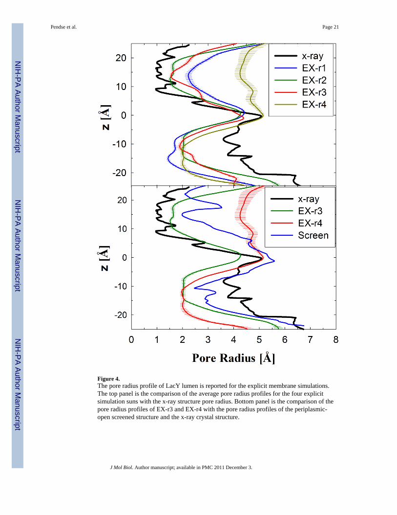
Figure 4.The pore radius profile of LacY lumen is reported for the explicit membrane simulations.The top panel is the comparison of the average pore radius profiles for the four explicitsimulation suns with the x-ray structure pore radius. Bottom panel is the comparison of thepore radius profiles of EX-r3 and EX-r4 with the pore radius profiles of the periplasmic-open screened structure and the x-ray crystal structure.
Pendse et al. Page 21
J Mol Biol. Author manuscript; available in PMC 2011 December 3.
NIH
-PA Author Manuscript
NIH
-PA Author Manuscript
NIH
-PA Author Manuscript

Figure 5.Average helix center distance deviation from the x-ray crystal structure (1PV7) for (left)first and (right) last nanosecond from the trajectory of EX-r4. All helix-helix pairs (# in rowsand columns) are shown with the distances below the diagonal corresponding to thecytoplasmic half and periplasmic above. The legend or colorbar is shown on the bottom andis in Å.
Pendse et al. Page 22
J Mol Biol. Author manuscript; available in PMC 2011 December 3.
NIH
-PA Author Manuscript
NIH
-PA Author Manuscript
NIH
-PA Author Manuscript

Figure 6.The outward-facing structure of LacY from EX-r4. (left) Snapshot of LacY embedded in afully hydrated lipid bilayer. (right) The cytoplasmic halves of the transmembrane helices (z< 0 from the center of mass (COM) of the protein) viewed from the cytoplasmic end in thedirection perpendicular to the membrane. The residues on the helices forming the lumen arerepresented as molecular surfaces based on the van der waals radii of the individual atoms.The color-coding of the helices is shown in right.
Pendse et al. Page 23
J Mol Biol. Author manuscript; available in PMC 2011 December 3.
NIH
-PA Author Manuscript
NIH
-PA Author Manuscript
NIH
-PA Author Manuscript

Figure 7.The periplasmic halves of the transmembrane helices (z > 0 from the COM of the protein)viewed from the periplasmic end in the direction perpendicular to membrane in which thesolid helices indicate the periplasmic-open structure from EX-r4. Transparent helicesrepresent (top) the crystal structure (1PV7) and (bottom) the structure from EX-r1.
Pendse et al. Page 24
J Mol Biol. Author manuscript; available in PMC 2011 December 3.
NIH
-PA Author Manuscript
NIH
-PA Author Manuscript
NIH
-PA Author Manuscript

Figure 8.The Cα-Cα difference in distance between residue pairs relative to the x-ray crystal structure.Negative values indicate that the helix-helix distance has decreased. The distance ismeasured as the mean of the Gaussian peak fit to the distance distributions (see Figure S6 asan example).
Pendse et al. Page 25
J Mol Biol. Author manuscript; available in PMC 2011 December 3.
NIH
-PA Author Manuscript
NIH
-PA Author Manuscript
NIH
-PA Author Manuscript
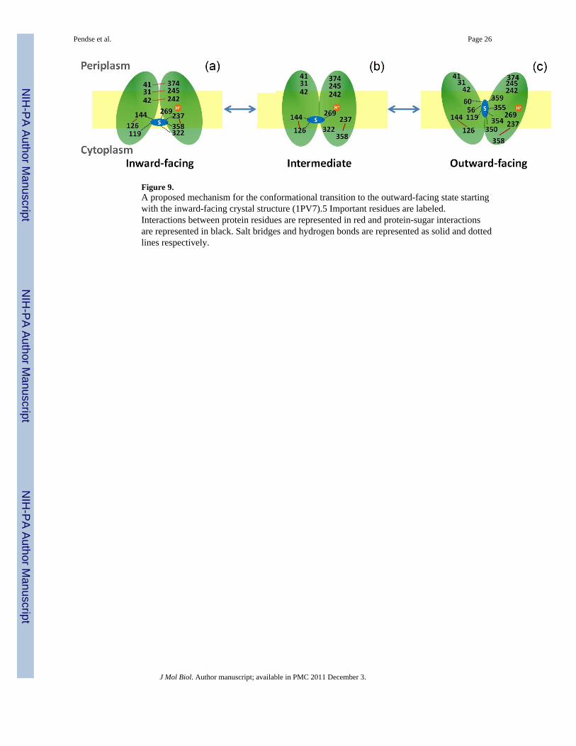
Figure 9.A proposed mechanism for the conformational transition to the outward-facing state startingwith the inward-facing crystal structure (1PV7).5 Important residues are labeled.Interactions between protein residues are represented in red and protein-sugar interactionsare represented in black. Salt bridges and hydrogen bonds are represented as solid and dottedlines respectively.
Pendse et al. Page 26
J Mol Biol. Author manuscript; available in PMC 2011 December 3.
NIH
-PA Author Manuscript
NIH
-PA Author Manuscript
NIH
-PA Author Manuscript
NIH
-PA Author Manuscript
NIH
-PA Author Manuscript
NIH
-PA Author Manuscript
Pendse et al. Page 27
Tabl
e 1
Syst
em si
zes a
nd si
mul
atio
n tim
es fo
r the
lact
ose
perm
ease
sim
ulat
ions
. MD
was
use
d in
the
EX a
nd IM
-MD
sim
ulat
ions
and
SG
LD fo
r the
oth
er IM
sim
ulat
ions
. Unl
ess o
ther
wis
e no
ted,
the
sim
ulat
ions
are
with
Glu
269
prot
onat
ed.
Run
nam
eSi
mul
atio
nT
ime
(ns)
Initi
al st
ruct
ure
Tot
alat
oms
Wat
erm
olec
ules
Lip
idm
olec
ules
IM-M
D20
ns
Snap
shot
from
Kla
uda
&B
rook
s, 20
0718
35,3
709,
563
-IM
-r1a
10ns
IM-r
210
ns
IM-r
310
ns
EX-r
125
ns3.
4ns s
naps
hot
91,4
7117
,055
269
EX-r
220
nsfr
om IM
-r1
EX-r
320
ns5.
5ns s
naps
hot
94,8
7618
,440
263
EX-r
440
nsfr
om IM
-r1
a For t
his c
onfo
rmat
ion
thre
e si
mul
atio
ns w
ere
run
with
Glu
269,
His
322,
and
Glu
325
prot
onat
ed.
J Mol Biol. Author manuscript; available in PMC 2011 December 3.
NIH
-PA Author Manuscript
NIH
-PA Author Manuscript
NIH
-PA Author Manuscript
Pendse et al. Page 28
Table 2
Hydrogen bond (HB) pairs that stabilize the cytoplasmic-open state. These hydrogen bonds existed for theruns that were periplasmic-closed (EX-r2/r3) and destabilized in the runs with a periplasmic-open state (EX-r1/r4). (SC-side chain, BB-backbone)
Residue Pairs Donor-AcceptorAtoms Helices
N245-P31 NSC –OBB H-VII/HI
S41-E374 OSC–OBB H-II/XI
K42-Q242 NBB– OSC H-II/VII
NSC–OBB
J Mol Biol. Author manuscript; available in PMC 2011 December 3.

NIH
-PA Author Manuscript
NIH
-PA Author Manuscript
NIH
-PA Author Manuscript
Pendse et al. Page 29
Tabl
e 3
Mea
n Cβ-
Cβ
dist
ance
s bet
wee
n re
sidu
es (Å
) use
d in
the
doub
le-C
ys m
utan
t exp
erim
enta
l stu
dies
.15
The
sugg
este
d Cβ-
Cβ
dist
ance
in th
e ou
twar
d-fa
cing
stat
e fr
om c
ross
-link
ing
expe
rimen
ts a
re 1
8.6–
20.6
Å.
Res
idue
Cal
cula
ted
mea
n di
stan
ces
1PV
7
Pair
sE
X-r
1E
X-r
2E
X-r
3E
X-r
4
I40-
N24
520
.510
.412
.121
.28.
9
T45-
N24
517
.08.
19.
717
.78.
1
I32-
N24
517
.25.
86.
518
.36.
9
J Mol Biol. Author manuscript; available in PMC 2011 December 3.

NIH
-PA Author Manuscript
NIH
-PA Author Manuscript
NIH
-PA Author Manuscript
Pendse et al. Page 30
Tabl
e 4
Cha
nges
in w
ater
acc
essi
ble
surf
ace
area
s com
pare
d to
the
crys
tal s
truct
ure
(1PV
7) fo
r pos
ition
s tha
t sho
wed
cha
nge
in M
TSES
acc
essi
bilit
y on
suga
rbi
ndin
g.
Incr
ease
d su
rfac
e ar
eaco
mpa
red
to 1
PV7
Dec
reas
ed su
rfac
e ar
eaco
mpa
red
to 1
PV7
Incr
ease
dD
ecre
ased
MT
SES
MT
SES
EX
-r1
EX
-r4
EX
-r1
EX
-r4
acce
ssib
ility
acce
ssib
ility
18, 3
2, 3
8,42
–44,
241
-24
6, 2
48,
249,
265
,32
7
18, 3
2, 3
8,42
–44,
241
-24
6, 2
48,
249,
326
,26
5, 3
27
60,3
2660
18, 3
2, 3
8, 4
2-44
, 241
–246
,24
8, 2
49, 2
65,
327
60, 3
26
J Mol Biol. Author manuscript; available in PMC 2011 December 3.
Related Documents