Probing the a-Helical Structural Stability of Stapled p53 Peptides: Molecular Dynamics Simulations and Analysis Zuojun Guo 1 , Udayan Mohanty 1 , Justin Noehre 2 , Tomi K. Sawyer 2 , Woody Sherman 3 and Goran Krilov 1,* 1 Department of Chemistry, Boston College, 2609 Beacon Street, Chestnut Hill, MA 02467, USA 2 Aileron Therapeutics, Inc., 840 Memorial Drive, Cambridge, MA 02139, USA 3 Schrodinger, Inc., 120 W 45th Street, 17th Fl., New York, NY 10036, USA *Corresponding author: Goran Krilov, [email protected] Reactivation of the p53 cell apoptosis pathway through inhibition of the p53-hDM2 interaction is a viable approach to suppress tumor growth in many human cancers and stabilization of the heli- cal structure of synthetic p53 analogs via a hydro- carbon cross-link (staple) has been found to lead to increased potency and inhibition of protein– protein binding (J. Am. Chem. Soc. 129: 5298). However, details of the structure and dynamic sta- bility of the stapled peptides are not well under- stood. Here, we use extensive all-atom molecular dynamics simulations to study a series of stapled a-helical peptides over a range of temperatures in solution. The peptides are found to exhibit sub- stantial variations in predicted a-helical propensi- ties that are in good agreement with the experimental observations. In addition, we find significant variation in local structural flexibility of the peptides with the position of the linker, which appears to be more closely related to the observed differences in activity than the absolute a-helical stability. These simulations provide new insights into the design of a-helical stapled pep- tides and the development of potent inhibitors of a-helical protein–protein interfaces. Key words: circular dichroism, drug design, hDM2, molecular dynamic simulations, p53, protein–protein interfaces, stapled peptide, a-helicity Received 2 December 2009, revised 19 January 2010 and accepted for publication 24 January 2010 A renaissance of peptide drug discovery has emerged over the past several years. In particular, the identification of synthetically 'sta- pled' a-helical peptides having promising pharmacokinetic, meta- bolic stability, and cell-penetrating properties has sparked tremendous interest in their development for a plethora of thera- peutic targets that have otherwise been deemed 'undruggable' by more conventional small-molecule strategies (1). To date, numerous examples of stapled peptides for varying therapeutic targets have been described, including p53 (2), BID BH3 (3,4), BAD BH3 (5), NOTCH (6), and HIV-1 capsid (7). Both single-turn (i +4 fi i ) and double-turn (i +7 fi i ) stapling chemistries are represented in these studies. Synthetic p53 peptides incorporating double-turn stapling chemistry have been previously described (2) and have provided insight to the evolution of a prototype series of cell-penetrating and in vitro biologically effective lead compounds. This series of peptides (Table 1) serves as the basis for the computational work pre- sented here. These stapled p53 peptides have incorporated C a -methyl-amino acids having terminal olefin alkyl side chains of different lengths and chirality, such as (S)-CH 2 ) 3 -CH=CH 2 and (R)-CH 2 ) 6 -CH=CH 2 , which upon metathesis form an all-carbon macrocycle via an olefin linkage (see compound 8, Scheme 1). The structure–activity relationships of p53-stapled peptides 1–10 involve further amino acid modifications to decrease negative charge (i.e., Asp and Glu replacement by Asn and Gln, respec- tively) and facilitate cell penetration as well as point mutations to avoid nuclear export and ubiquitination (i.e., Lys replacement by Arg). In contrast, three key hydrophobic amino acids deemed criti- cal for E3 ubiquitin ligase (MDM2) binding (i.e., Leu, Trp, and Phe as highlighted in stapled p53 peptide analogs 1–10) were con- served throughout the series. Importantly, the relationships between the structure and dynamic stability of stapled peptides are not well understood. Recently, an all-atom Monte Carlo folding simulation study comparing unmodified peptides derived from RNase A and BID BH3 with varying i +4 fi i and i +7 fi i stapling chemistry has been reported (8). Another investigation (9) led to the parameterization of a minimal, dynamic model reproducing the structural stability Editor’s invited manuscript to celebrate the 4th Anniversary of Chemical Biology & Drug Design. 348 Chem Biol Drug Des 2010; 75: 348–359 Research Article ª 2010 John Wiley & Sons A/S doi: 10.1111/j.1747-0285.2010.00951.x

Welcome message from author
This document is posted to help you gain knowledge. Please leave a comment to let me know what you think about it! Share it to your friends and learn new things together.
Transcript

Probing the a-Helical Structural Stability ofStapled p53 Peptides: Molecular DynamicsSimulations and Analysis
Zuojun Guo1, Udayan Mohanty1, JustinNoehre2, Tomi K. Sawyer2, WoodySherman3 and Goran Krilov1,*
1Department of Chemistry, Boston College, 2609 Beacon Street,Chestnut Hill, MA 02467, USA2Aileron Therapeutics, Inc., 840 Memorial Drive, Cambridge, MA02139, USA3Schrodinger, Inc., 120 W 45th Street, 17th Fl., New York, NY10036, USA*Corresponding author: Goran Krilov, [email protected]
Reactivation of the p53 cell apoptosis pathwaythrough inhibition of the p53-hDM2 interaction isa viable approach to suppress tumor growth inmany human cancers and stabilization of the heli-cal structure of synthetic p53 analogs via a hydro-carbon cross-link (staple) has been found to leadto increased potency and inhibition of protein–protein binding (J. Am. Chem. Soc. 129: 5298).However, details of the structure and dynamic sta-bility of the stapled peptides are not well under-stood. Here, we use extensive all-atom moleculardynamics simulations to study a series of stapleda-helical peptides over a range of temperatures insolution. The peptides are found to exhibit sub-stantial variations in predicted a-helical propensi-ties that are in good agreement with theexperimental observations. In addition, we findsignificant variation in local structural flexibilityof the peptides with the position of the linker,which appears to be more closely related to theobserved differences in activity than the absolutea-helical stability. These simulations provide newinsights into the design of a-helical stapled pep-tides and the development of potent inhibitors ofa-helical protein–protein interfaces.
Key words: circular dichroism, drug design, hDM2, moleculardynamic simulations, p53, protein–protein interfaces, stapled peptide,a-helicity
Received 2 December 2009, revised 19 January 2010 and accepted forpublication 24 January 2010
A renaissance of peptide drug discovery has emerged over the pastseveral years. In particular, the identification of synthetically 'sta-pled' a-helical peptides having promising pharmacokinetic, meta-bolic stability, and cell-penetrating properties has sparkedtremendous interest in their development for a plethora of thera-peutic targets that have otherwise been deemed 'undruggable' bymore conventional small-molecule strategies (1). To date, numerousexamples of stapled peptides for varying therapeutic targets havebeen described, including p53 (2), BID BH3 (3,4), BAD BH3 (5),NOTCH (6), and HIV-1 capsid (7). Both single-turn (i + 4 fi i ) anddouble-turn (i + 7 fi i ) stapling chemistries are represented inthese studies.
Synthetic p53 peptides incorporating double-turn stapling chemistryhave been previously described (2) and have provided insight tothe evolution of a prototype series of cell-penetrating and in vitrobiologically effective lead compounds. This series of peptides(Table 1) serves as the basis for the computational work pre-sented here. These stapled p53 peptides have incorporatedCa-methyl-amino acids having terminal olefin alkyl side chains ofdifferent lengths and chirality, such as (S)-CH2)3-CH=CH2 and(R)-CH2)6-CH=CH2, which upon metathesis form an all-carbonmacrocycle via an olefin linkage (see compound 8, Scheme 1).The structure–activity relationships of p53-stapled peptides 1–10
involve further amino acid modifications to decrease negativecharge (i.e., Asp and Glu replacement by Asn and Gln, respec-tively) and facilitate cell penetration as well as point mutations toavoid nuclear export and ubiquitination (i.e., Lys replacement byArg). In contrast, three key hydrophobic amino acids deemed criti-cal for E3 ubiquitin ligase (MDM2) binding (i.e., Leu, Trp, and Pheas highlighted in stapled p53 peptide analogs 1–10) were con-served throughout the series.
Importantly, the relationships between the structure and dynamicstability of stapled peptides are not well understood. Recently,an all-atom Monte Carlo folding simulation study comparingunmodified peptides derived from RNase A and BID BH3 withvarying i + 4 fi i and i + 7 fi i stapling chemistry has beenreported (8). Another investigation (9) led to the parameterizationof a minimal, dynamic model reproducing the structural stability
Editor’s invited manuscript to celebrate the 4th Anniversary of Chemical Biology & Drug Design.
348
Chem Biol Drug Des 2010; 75: 348–359
Research Article
ª 2010 John Wiley & Sons A/S
doi: 10.1111/j.1747-0285.2010.00951.x
profile of a series of BH3-stapled peptides as a function of thecross-linking moiety.
Here, we use extensive molecular dynamics simulations to study aseries of stapled a-helical peptides over a range of temperatures insolution. The peptides are found to exhibit substantial variations in
predicted a-helicities that are in good agreement with the experi-mental values (2). In addition, we find significant variation in localstructural flexibility of the stapled peptides with the position of thelinker, which appears to be more closely related to the observeddifferences in activity than the absolute a-helical stability. Thesesimulations provide new insights into the design of a-helical
Table 1: The sequences of sta-pled a-helical p53 peptide analogs1–10
Peptide Sequence(a,b,c)
WT Ac L S Q E T F S D L W K L L P E N NH2
NH2
NH2
NH2
NH2
NH2
NH2
NH2
NH2
NH2
NH2
1 Ac L S Q E T F S D X W K L L P E X
2 Ac L S Q E X F S D L W K X L P E N
3 Ac L S Q X T F S D L W X L L P E N
4 Ac L S Q E T F X D L W K L L X E N
5 Ac L S Q E T F X N L W K L L X Q N
6 Ac L S Q Q T F X N L W R L L X Q N
7 Ac Q S Q Q T F X N L W K L L X Q N
8 Ac Q S Q Q T F X N L W R L L X Q N
9 Ac Q S Q Q T A X N L W R L L X Q N
10* Ac Q S Q Q T F X N L W R L L X Q N
aX and X refer to Scheme 1 relative to the full chemical structure of peptide 8 (specifically, the two novelCa-methyl-amino acids that are macrocyclized by olefin metathesis).bPeptide 10* is a linear peptide (specifically, X refers to Ca-methyl-amino acid with (S)-CH2)3-CH=CH2 side chain,and X refers to for Ca-methyl-amino acid with (R)-CH2)6-CH=CH2 side chain).cKey hydrophobic amino acids are highlighted in blue.
Scheme 1: Structures of stapled p53 analog peptide 8 and linear analog peptide 10. A cis-olefin linkage is depicted for peptide 8 rela-tive to synthesis and structural characterization by NMR and CD analysis at Aileron Therapeutics and will be described separately from thispublication.
Probing the a-Helical Structural Stability of Stapled p53 Peptides
Chem Biol Drug Des 2010; 75: 348–359 349
stapled peptides and the development of potent inhibitors of a-heli-cal protein–protein interfaces.
Materials and Methods
Initial structuresAll-atom structures of the 16-residue wild-type p53 peptide and thestapled analogs 1–10 were constructed using the Maestro packagein an a-helical configuration. The olefin bridge was added manu-ally, with the double bond placed in a cis configuration, as experi-mentally observed (based on synthesis and structuralcharacterization by NMR and CD studies at Aileron Therapeuticsand will be reported elsewhere). Protonation states were assignedto ionizable residues according to the pKa based on pH = 7.0 usingthe Protein Preparation workflow in Maestro and the Epik module(10). Acetyl and carboxamide moieties were incorporated at the N-and C-terminii of the peptide, respectively. Each peptide was thenplaced in a cubic cell, with size adjusted to maintain a minimumdistance of 10 � to the cell boundary, and soaked with a pre-equili-brated box of water using the System Builder module of the Des-mond package (11,12). All overlapping solvent molecules wereremoved and an appropriate number of counter ions were added tomaintain charge neutrality.
Molecular dynamics simulationsAll molecular dynamics (MD) simulations were performed using theDesmond package (11,12). The OPLS 2005 force field (13,14) wasused to model all peptide interactions, and the TIP3P model (15)was used for water. The particle-mesh Ewald method (16) (PME)was used to calculate long-range electrostatic interactions with agrid spacing of 0.8 �. Van der Waals and short range electrostaticinteractions were smoothly truncated at 9.0 �. Nose–Hoover ther-mostats (17) were utilized to maintain the constant simulation tem-perature and the Martina–Tobias–Klein method (18) was used tocontrol the pressure. The equations of motion were integrated usingthe multistep RESPA integrator (19) with an inner time step of2.0 fs for bonded interactions and non-bonded interactions withinthe short range cutoff. An outer time step of 6.0 fs was used fornon-bonded interactions beyond the cutoff. Periodic boundary condi-tions were applied throughout.
The system was equilibrated with the default protocol provided inDesmond, which consists of a series of restrained minimizationsand molecular dynamics simulations designed to slowly relax thesystem, while not deviating substantially from the initial proteinco-ordinates. In short, two rounds of steepest descent minimizationwere performed with a maximum of 2000 steps and a harmonicrestraint of 50 kcal ⁄ mol per �2 on all solute atoms. Next, a seriesof four molecular dynamics simulations were performed. The firstsimulation was run for 12 ps at a temperature of 10�K in the NVT(constant number of particles, volume, and temperature) ensemblewith solute heavy atoms restrained with force constant of 50 kcal ⁄mol per �2. Next, another 12 ps simulation was performed at 10�Kwith the same harmonic restraints, this time in the NPT (constantnumber of particles, pressure, and temperature) ensemble. A 24-pssimulation followed with the temperature raised to 300 K in the
NPT ensemble and the force constant retained. Finally, a 24-ps sim-ulation was performed at 300 K in the NPT ensemble with allrestraints removed. The above-mentioned default equilibration wasfollowed by a 5000-ps NPT simulation to equilibrate the system. A10-ns NPT production simulation was then run and configurationswere saved in 4-ps intervals.
Additional simulations were run at high temperature and the finalconfiguration was subjected to the above-mentioned protocol priorto the start of the 300-K production runs. For the high-temperaturesimulations, an initial set-up and equilibration period was performedas described earlier except for the final production stage. Each pep-tide was simulated at 500�K, which should provide sufficient kineticenergy to allow crossing of the relevant barriers separating theenergy minima. A number of configurations (10–20) were selectedfrom each high-temperature trajectory and used as a starting pointfor the 10-ns production simulations at 300 K described earlier. Intotal, 100–200 ns of sampling was performed for each peptide.
Replica exchange molecular dynamicssimulationsThe replica exchange molecular dynamics (REMD) approach (20)involves evolving a number of copies of the system in parallel atdifferent temperatures and periodically exchanging the configura-tions between trajectories i and j with the probabilitywij = exp()D), where D ” (bi ) bj) (Ej ) Ei), and b ” 1 ⁄ kBT. Thisinsures proper canonical sampling at all temperatures, with high-temperature simulations facilitating barrier crossings and lowtemperature simulations to explore local free energy minima. Thesystems were prepared and relaxed as described earlier. In eachcase, 64 replicas of the system were evolved in parallel for 15 nsat constant NVT, with temperatures evenly spaced between 300and 600 K in approximately 4-K intervals. After the first 100 ps,replica exchanges between each pair of nearest neighbor trajecto-ries were attempted every 12 ps to equilibrate the system. Follow-ing a 5-ns period, the configurations were saved in 1-ps intervalsover the final 10 ns of each simulation, providing an aggregate640 ns of sampling for each system. The exchange acceptance ran-ged from 22% to 55%, and convergence was confirmed by compar-ing ensemble averages computed separately over the fist and lasthalf of the production period.
Results and Discussion
We have performed extensive all-atom explicit solvent moleculardynamics simulations on a series of 10 stapled p53 peptide analogspreviously synthesized and studied by Bernal et al. (2), as well asthe wild-type peptide. The goal of this study was to determine howand to what extent the introduction of hydrocarbon cross-links atvarious locations in the sequence modifies the equilibrium confor-mational population of the peptides in solution. In particular, a crys-tal structure of the amino-terminal domain of MDM2 protein boundto the transactivation domain of p53, which the stapled peptidesseek to mimic, shows the latter to be in a mostly a-helical confor-mation when bound to the receptor, allowing three hydrophobicamino acids on the hydrophobic side of the helix to adopt extended
Guo et al.
350 Chem Biol Drug Des 2010; 75: 348–359

side-chain conformations and pack with each other (21,22). Circulardichroism (CD) measurements of free p53 in aqueous solution showa positive correlation between the degree of a-helical content andbinding affinity of the peptides for hMDM2 (2). However, the a-heli-cal content was found to vary widely across the series and someof the peptides with slightly decreased helical content exhibitedsignificant activity in the low to mid nanomolar range, suggesting amore complex relationship between the peptide structures and bind-ing activity. Also, intimately related to the intrinsic helical proper-ties of the stapled p53 peptides is the Ca-methyl modification ofthe two amino acids that undergo metathesis to form the macro-cycle. It is well-known that such Ca-methyl modification of linearpeptides contributes to helicity (23–27), albeit such modificationshave not been shown to confer the unique cell-penetrating proper-ties of stapled peptides.
Structural distributionOne of the challenges of molecular dynamics of biomolecules isfully exploring the energetic landscape of the system. Biomole-cules have complex potential energy profiles and simulationsoften get trapped for long periods of time in local minimabecause of the high energy barriers that may exist. In additionto not observing certain structures that may be biologically rele-vant, inadequate sampling also prevents recovering the correctthermodynamic distribution that is needed to correctly predict theenergetics of the system. To improve sampling of the peptide, a
series of simulations were performed at high temperature (500 K)and snapshots were taken for multiple simulations at room tem-perature (300 K), as described in the Materials and Methods sec-tion. Because the active domain of p53 adopts a largely helicalstructure when bound to the MDM2 receptor, peptides that arenot helical in solution would have to incur a conformational pen-alty to adopt the helical bound state conformation and thereforeare hypothesized to be less active. Furthermore, the degree ofhelical content in the unbound state should correlate with bind-ing activity if the conformational strain energy in the boundstate is a limiting factor in the binding process. To better under-stand the structural profile of the peptides in the unbound state,we studied the helical content of the stapled peptides insolution.
There are numerous ways to define helical structure of the peptidechain. For example, proteins residues are typically classified as heli-cal if their backbone dihedral angles / and w lie in a narrowregion of the Ramachandran diagram, with / between )95� and)35�, and w between )15� and )70� (28). For a-helices, whichare the predominant helical form in proteins, the sum of the / andw angles on adjacent residues is �)105�. This confines the di-hedrals to a narrow strip with a slope close to )1 around an ideala-helix geometry characterized by (/,w) = ()60�,)45�). For compari-son, the sums of the above-mentioned angles were about )75� forthe p-helix and )130� for the 310-helix (28). Helical conformations
a b c d
e f g
i j k
h
Figure 1: The contour plots of the conformational distribution of the p53 peptide analogs as a function of the number of backbonei + 4 fi i a-helical hydrogen bonds and the percent a-helical content, for (a) 1, (b) 2, (c) 3, (d) 4, (e) 5, (f) 6, (g) 7, (h) 8, (i) 9, (j) 10, (k) wildtype. Brighter color indicates higher intensity.
Probing the a-Helical Structural Stability of Stapled p53 Peptides
Chem Biol Drug Des 2010; 75: 348–359 351

are also recognized by a specific pattern of hydrogen bonds formedbetween the backbone acceptor carbonyl (C=O) of residue i and thebackbone amine (NH) of another residue, including i + 3 (310-helix),i + 4 (a-helix), or i + 5 (p-helix). These definitions were used toanalyze the conformational ensembles of stapled p53 peptideanalogs.
In Figure 1, we plot the conformational population distribution as afunction of percent a-helical content defined as the ratio of thenumber of helical residues to the total number of residues in thechain, and the number of a-helical (i + 4 fi i ) hydrogen bonds. Thea-helical nature of individual residues was assigned by the STRIDEprogram (29). The existence of hydrogen bonds was the determinedvia a geometric criterion requiring a minimum donor (D)-to-(A)acceptor distance of 3.5 � and a maximum A–H–D (acceptor–H–donor) angle of 30�. Interestingly, the helical population is predictedto be quite low for most peptides. In particular, 1, 2, 3, 8, and theWT p53 show a single pronounced peak at <5% helicity and 0-1native hydrogen bonds, indicating minimal a-helical population.Compounds 4 and 5 each show two additional pronounced peaks,including a broad maximum centered at 30% helicity and 2 hydro-gen bonds, and 50% helicity and 3 hydrogen bonds, respectively.These peaks are present to a lesser extent in 6, and a small peakat 30% helicity and 1 hydrogen bond is observed for 7, 9, and theunstapled peptide 10.
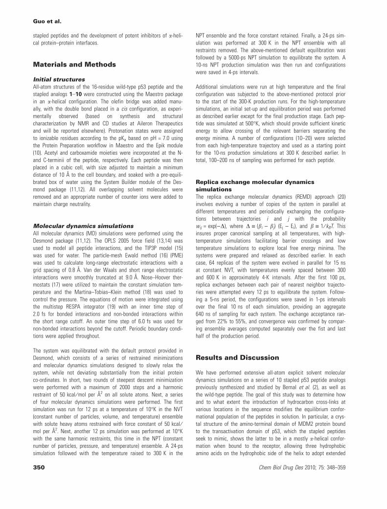
The average percent helicity for the entire population as calcu-lated by STRIDE is given in Table 2. These are significantly lowerthan the helical content reported by Bernal et al. (10), which wasestimated from CD measurements. One possibility for such a dis-crepancy may be because of a distortion of the helical structureaway from the ideal a-helix geometry, indicating that the helix cri-teria used in the calculations mentioned earlier may be too strin-gent for direct comparison to experimental CD spectra. Toinvestigate this further, we studied the distribution of the (/,w)dihedrals for the p53 WT and ten stapled peptide analogs, asshown in Figure 2.
All distributions show a strong peak in the range / = [)95,)50�]and w = [)50�,)10�], which is generally in the allowed a-helix
region of the Ramachandran diagram. Furthermore, these peaksdisplay an oblong shape and lie roughly along a line with a slopeof -1 in the (/,w) plane, indicative of a periodic helical structure.The peptides known experimentally to have weak helical propen-sity, including 1, 2, 3, and WT show significant populations inthe two peaks located in extended b-sheet region (the upper leftregion of the Ramachandran plot), while strongly helical peptidessuch as 4 and 8, which were found to be highly biologicallyeffective (2), show most of the populations around the a-helicalregion. Still, the peaks are significantly broader than expected foran a-helix, indicating a distortion of the helical structure fromideal values.
In Table 3, we report the percentage of residues whose dihedral(/,w) pairs were found to lie within a 30� band between(/,w) = ()95�, )15�) and ()35�, )70�), characteristic of an a-helix.The percentages are generally higher than those reported bySTRIDE, particularly for the weakly helical peptides and more in linewith experimentally measured CD results. This supports the viewthat the default helical criteria in STRIDE is too stringent for directcomparison with CD spectra and that the helical structures adoptedby the peptides are somewhat distorted from their ideal values. Wealso report the total number of backbone hydrogen bonds, whichincludes acceptor C=O to H–N donor pairings such as those fromthe 310-helices, p-helices, and b-hairpin turns, in addition to thea-helical backbone hydrogen bonds. The significantly larger valuesindicate a considerable contribution from the non-a-helical struc-tural motifs to the equilibrium ensemble.
Next, we examine the conformational distribution of the stapledpeptides as a function of temperature. This is useful because thedegree of persistence of structural features in the peptide across arange of temperature provides insights into the stability of second-ary structure and the energetic profile of the transition from struc-tured to unstructured states. In Figure 3, we plot the averagea-helicity computed by STRIDE as a function of temperature. Theconformations of the peptide were harvested from separate 10-nsconstant NPT simulations, following a 5-ns equilibration periodperformed at each temperature, ranging from 300 to 500 K (seeMaterials and Methods for details).
Table 2: Average helicity andnumber of a-helical hydrogenbonds from MD simulations at300 K
Peptide Sequence Helicity α-helical H-bonds
WT Ac L S Q E T F S D L W K L L P E N NH2 2.0% 0.17
1 Ac L S Q E T F S D X W K L L P E X NH2 4.0% 0.44
2 Ac L S Q E X F S D L W K X L P E N NH2 0.0050% 0.17
3 Ac L S Q X T F S D L W X L L P E N NH2 1.8% 0.26
4 Ac L S Q E T F X D L W K L L X E N NH2 38% 2.0
5 Ac L S Q E T F X N L W K L L X Q N NH2 45% 2.5
6 Ac L S Q Q T F X N L W R L L X Q N NH2 26% 1.6
7 Ac Q S Q Q T F X N L W K L L X Q N NH2 15% 1.0
8 Ac Q S Q Q T F X N L W R L L X Q N NH2 22% 1.3
9 Ac Q S Q Q T A X N L W R L L X Q N NH2 12% 0.85
10 Ac Q S Q Q T F X N L W R L L X Q N NH2 11% 0.72
Guo et al.
352 Chem Biol Drug Des 2010; 75: 348–359
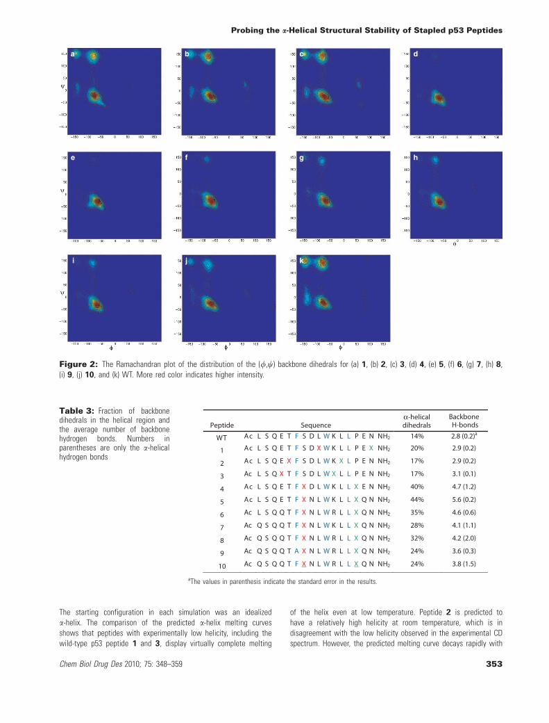
The starting configuration in each simulation was an idealizeda-helix. The comparison of the predicted a-helix melting curvesshows that peptides with experimentally low helicity, including thewild-type p53 peptide 1 and 3, display virtually complete melting
of the helix even at low temperature. Peptide 2 is predicted tohave a relatively high helicity at room temperature, which is indisagreement with the low helicity observed in the experimental CDspectrum. However, the predicted melting curve decays rapidly with
Table 3: Fraction of backbonedihedrals in the helical region andthe average number of backbonehydrogen bonds. Numbers inparentheses are only the a-helicalhydrogen bonds
Peptide Sequenceα-helicaldihedrals
Backbone H-bonds
WT Ac L S Q E T F S D L W K L L P E N NH2 14% 2.8 (0.2)a
1 Ac L S Q E T F S D X W K L L P E X NH2 20% 2.9 (0.2)
2 Ac L S Q E X F S D L W K X L P E N NH2 17% 2.9 (0.2)
3 Ac L S Q X T F S D L W X L L P E N NH2 17% 3.1 (0.1)
4 Ac L S Q E T F X D L W K L L X E N NH2 40% 4.7 (1.2)
5 Ac L S Q E T F X N L W K L L X Q N NH2 44% 5.6 (0.2)
6 Ac L S Q Q T F X N L W R L L X Q N NH2 35% 4.6 (0.6)
7 Ac Q S Q Q T F X N L W K L L X Q N NH2 28% 4.1 (1.1)
8 Ac Q S Q Q T F X N L W R L L X Q N NH2 32% 4.2 (2.0)
9 Ac Q S Q Q T A X N L W R L L X Q N NH2 24% 3.6 (0.3)
10 Ac Q S Q Q T F X N L W R L L X Q N NH2 24% 3.8 (1.5)
aThe values in parenthesis indicate the standard error in the results.
a b c d
e f g h
i j k
Figure 2: The Ramachandran plot of the distribution of the (/,w) backbone dihedrals for (a) 1, (b) 2, (c) 3, (d) 4, (e) 5, (f) 6, (g) 7, (h) 8,(i) 9, (j) 10, and (k) WT. More red color indicates higher intensity.
Probing the a-Helical Structural Stability of Stapled p53 Peptides
Chem Biol Drug Des 2010; 75: 348–359 353

temperature, indicating a co-operative unfolding process with a highenergy barrier to initiate. Longer simulations at room temperaturemay yield different helicity predictions if this is a low frequencyevent that is not sampled statistically significantly with the currentprotocol. On the other hand, peptide 1 displays a flat curve, indicat-ing that a low proportion of a-helical structure is maintained acrossa broad temperature range. This suggests a more stable, althoughnot necessarily a-helical, structure.
Among the first group of peptides, 4 exhibits the highest helicity atroom temperature (�50%), in agreement with experiment, andmaintains this profile up to 450 K. The second series of peptides, 5
through 8, demonstrate similar helical content at 300 K, in therange of 40–60%, with similar melting curves. In contrast withexperimental data, 6, which displays significantly weaker CD signal,exhibits a melting curve similar to the rest of the peptides in theseries. Compounds 9 and 10 display the fastest decay of helicalstructure, in good agreement with experiment.
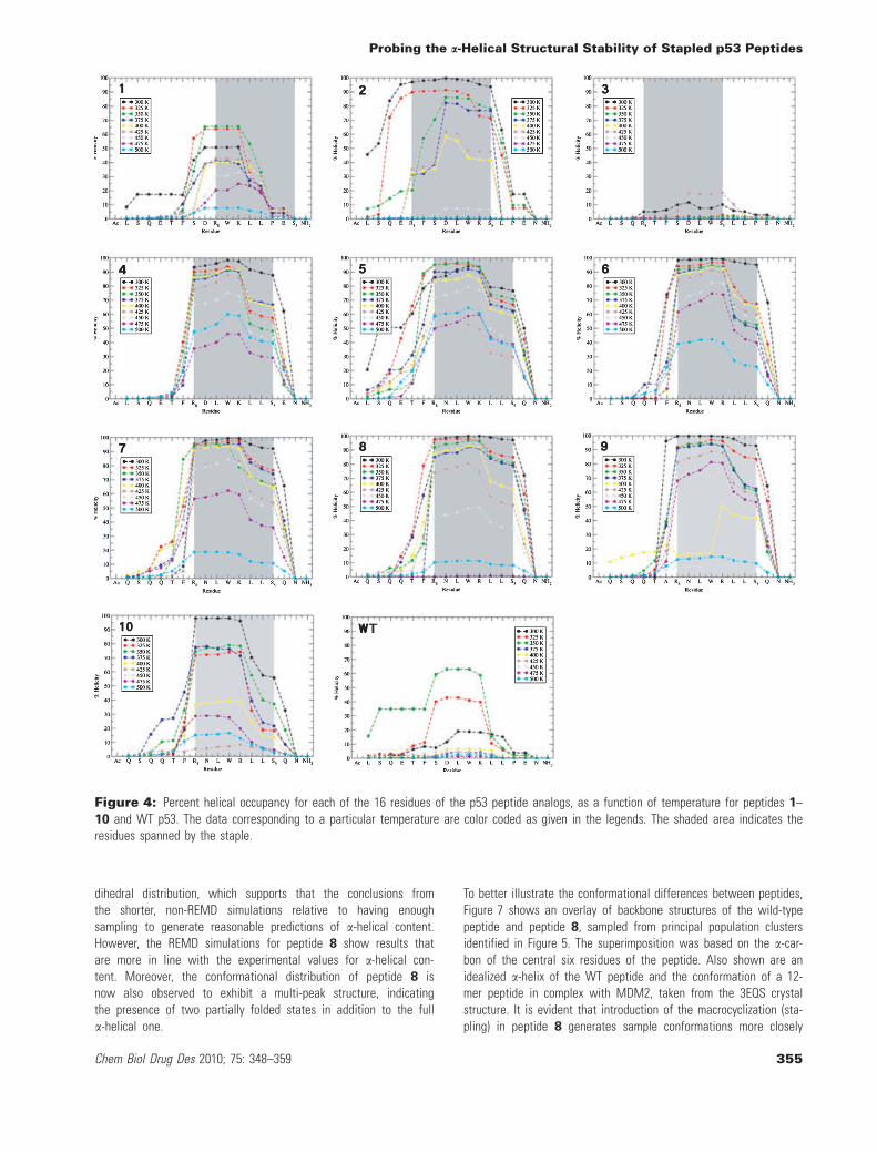
To further investigate the effect of the olefin linker on the second-ary structure of the peptides, Figure 4 shows the per-residue
percent a-helicity, as computed by STRIDE, for the eleven stapledpeptides at a series of temperatures. Several conclusions can bedrawn from the plot. First, the per-residue helical profiles decay ata rate consistent with the average melting curves shown in Fig-ure 3. However, helical content is not distributed evenly across theamino acid residues in the peptide. In each case, the region of thepeptide with the highest helical content corresponds to the portionof the sequence spanned by the linker, although in some cases,such as 1 at 300 K, 9 at 400 K, and 10 at 425 K, helicity is foundto be more evenly distributed over the sequence. Moreover, as thetemperature increases, this region consistently maintains proportion-ately more helical content than the rest of the sequence. Hence,the linker is clearly predicted to induce helical structure formationbut the effect is primarily local. A similar effect was observed byKutchukian et al. (8) in their Monte Carlo study of the conforma-tional distribution of stapled peptides derived from RNase A usinga knowledge-based pseudo-potential, with the authors attributingthe increased helix propensity of the spanned region to the stabil-ization of the folded state relative to the denatured conformationensemble. This may explain the significant variations in the potencyof the stapled peptides with linkers spanning different portions ofthe sequence and the lack of direct correlation between the experi-mental CD spectra and the peptide activity. Interestingly, Kutchukianet al. found that in case of a different sequence of stapled peptidesderived from BID BH3, the spanned region is not predominantlyhelical, and a different, partially folded state is stabilized, indicatingthat the specific amino acid sequence of the peptide plays a signifi-cant role in determining the preference for helical conformations inconjunction with the location and length of the linker. We haveobserved such behavior in the helical propensity of 9, which is one-half relative to 8, even though the only difference is the replace-ment of a phenylalanine at position 8 with alanine, while the posi-tion of the staple and the spanned segment of the sequence isunaltered. Similarly, we observe that in case of peptides with moststable helices such as 8 and 4, the helical region extends well pastthe segment spanned by the linker indicating a more comprehensivestabilization. This parallels the findings of Hamacher et al. (9) who,using a simplified Go model of the BH3 peptide, uncovered evi-dence of global dynamic stabilization of the cross-linked peptidehelical structure in addition to the local stabilization imparted bythe linker.
Finally, to explore whether the above-mentioned findings wouldbe maintained with substantially more sampling, a subset of thepeptides were subjected to an aggregate 640 ns of replicaexchange molecular dynamics (REMD), as described in the Mate-rials and Methods section. The computational resources requiredto run these simulations limited the number of peptides thatcould be run via REMD. Figures 5–8 show the helical conforma-tional distribution, Ramachandran plots, per-residue helicity andthe helix melting curves obtained from REMD simulations of pep-tides 4, 8, and WT, where the former two were selected forfurther study as they displayed a high degree of helicity by CDas well as high biological activity (2). In addition, peptide 8
showed a significantly lower helicity than observed experimen-tally in our constant temperature simulations. A comparison ofFigures 5 and 6 with Figures 1 and 2 revealed both similaritiesand differences. There is consistency in the overall backbone
Figure 3: The helix melting curves showing the percent helicalcontent as a function of temperature for (top) 1, 2, 3, 4, and thewild type; (bottom) 5, 6, 7, 8, 9, 10. The color coding is indicatedin the legends.
Guo et al.
354 Chem Biol Drug Des 2010; 75: 348–359
dihedral distribution, which supports that the conclusions fromthe shorter, non-REMD simulations relative to having enoughsampling to generate reasonable predictions of a-helical content.However, the REMD simulations for peptide 8 show results thatare more in line with the experimental values for a-helical con-tent. Moreover, the conformational distribution of peptide 8 isnow also observed to exhibit a multi-peak structure, indicatingthe presence of two partially folded states in addition to the fulla-helical one.
To better illustrate the conformational differences between peptides,Figure 7 shows an overlay of backbone structures of the wild-typepeptide and peptide 8, sampled from principal population clustersidentified in Figure 5. The superimposition was based on the a-car-bon of the central six residues of the peptide. Also shown are anidealized a-helix of the WT peptide and the conformation of a 12-mer peptide in complex with MDM2, taken from the 3EQS crystalstructure. It is evident that introduction of the macrocyclization (sta-pling) in peptide 8 generates sample conformations more closely
Figure 4: Percent helical occupancy for each of the 16 residues of the p53 peptide analogs, as a function of temperature for peptides 1–10 and WT p53. The data corresponding to a particular temperature are color coded as given in the legends. The shaded area indicates theresidues spanned by the staple.
Probing the a-Helical Structural Stability of Stapled p53 Peptides
Chem Biol Drug Des 2010; 75: 348–359 355

resembling an ideal helix (red), whereas the WT visits a plethora ofrandom coil structures (gray), only infrequently visiting the partiallyfolded helix observed in MDM2 complex crystal structure.
The melting curves in Figure 8 and the per-residue a-helicityshown in Figure 9 for the REMD simulations are qualitatively
similar to the data from shorter simulations, but quantitativelymore in line with experimental results. This suggests that forqualitative understanding of a-helical content and relative helicalcontent between peptides, it is sufficient to run shorter simula-tions. However, for quantitative predictions or comparisons withexperiments, it is necessary to run long simulations, potentially
A B
D E
C
Figure 7: Backbone structures of peptide 8 sampled from the high helicity (A) and medium helicity (B) population cluster, and those ofthe WT p53 peptide (C), shown in comparison with the idealized p53 helix (D) and the conformation of p53 in crystal structure of the complexwith MDM2 (E).
Figure 5: The contour plots of the conformational distribution of the WT p53 (left), 8 (middle), and 4 (right) peptides, as a function of thenumber of backbone i fi i + 4 a-helical hydrogen bonds and the percent a-helical content; computed from the replica exchange moleculardynamics (REMD) data. The Ramachandran plot of the distirbution of the (/,w) backbone dihedrals.
Figure 6: The Ramachandran plot of the distribution of the (/,w) backbone dihedrals for the wild-type p53 (left), 8 (middle), and 4 (right)peptides, computed from the replica exchange molecular dynamics (REMD) data.
Guo et al.
356 Chem Biol Drug Des 2010; 75: 348–359

with enhanced sampling, as in the REMD simulations performedin this work. Animations of the REMD simulations are providedas Supporting Information. Movie S1 shows the wild-type p53peptide and stapled p53 peptide 8. Movie S2 shows the keyhydrophobic side chains of stapled p53 peptide 8 that are impor-tant to its biological properties.
Conclusions and future directions
We have used extensive all-atom molecular dynamics simulationsto study the structural distribution of a number of cross-linked ana-logs of the 16-residue transactivation domain of the p53 protein.The overall propensity to form a-helical structures was found tovary widely across the p53 peptide analog series and to dependboth on the relative position of the macrocyclization (stapling) inthe sequence as well as the specific amino acid substitutions. Ingeneral, all stapled p53 peptides except for 2 were found to havea higher a-helical content than the WT p53 peptide, which is inagreement with experimental CD measurements (2). The relative a-
helical propensity of most peptides was found to be in good quali-tative agreement with experimental measurements, although ourvalues are generally lower than those reported by Bernal et al. (2).This is partly because helical structures formed by the stapled pep-tides are somewhat distorted from the ideal a-helix geometry, andthus sometimes missed by the helix recognition algorithm inSTRIDE. The latter is evident from the Ramachandran plots thatshow significant, but somewhat broader peaks in the a-helix regionof the diagram. Computing the fraction of residues with (/,W) dihe-drals within narrowly defined a-helical parameters gives helicitiesthat are closer to the values estimated from CD measurements.REMD simulations performed for several peptides show thatimproved sampling results in predicted helicities that are very closeto experimental values.
Interestingly, simulations of peptides 5 and 6 predict the helicalcontent to be somewhat higher than suggested by experiments(2), as seen in Figure 1 and Table 2. While these peptides sharethe same position of the cross-link as 4, they differ by substitu-tion of several acidic residues with their amide counterparts,altering the total charge of the peptide. It is possible that thealtered interactions with the specific solvent medium used in CDexperiments reduce the helical propensity, which is not fully cap-tured by our simulations. We also found that for several pep-tides, including 4, 5, 6, and 8, multiple partially folded statesare populated at room temperature. Stabilization of such 'decoystates' by the staples was also observed by Kutchukian et al.(8), who pointed out that removal of these states may lead tomore stable a-helical structures. This is particularly evident inthe case of peptide 8, where the most populated state is thepartially folded state with mean helicity of 55% (Figure 5).Removal or destabilization of this state would likely lead to apopulation shift to a nearly fully helical state at mean helicity75%.
The helical propensities were not distributed equally over the entiresequence. In most cases, the residues with the highest degree ofhelicity were found to be those spanned by the staple. In a fewcases, particularly for 5 and 8, significant helical content wasobserved in several residues outside of the linked region, while oth-ers with the identically positioned cross-link such as 4 do not share
Figure 9: Percent helical occupancy for each of the 16 residues of WT (left), 8 (middle), and 4 (right) peptide analogs as a function oftemperature, computed from the replica exchange molecular dynamics (REMD) data. The profiles corresponding to a particular temperatureare color coded as shown in the legends. The shaded area indicates the residues spanned by the staple.
Figure 8: The helix melting curves computed from the replicaexchange molecular dynamics (REMD) data, showing the percenthelical content as a function of temperature for the wild-type p53peptide, 8, and 4.
Probing the a-Helical Structural Stability of Stapled p53 Peptides
Chem Biol Drug Des 2010; 75: 348–359 357

this trait. On the other hand, a distinct shoulder is observed in theper-residue helicity distribution for all peptides containing the LLmotif in the spanned region. This suggests that, while helix forma-tion is primarily because of local stabilization of helical geometryby the rigid linker, helical propensity is also strongly modulated bythe details of the amino acid sequence of the peptide itself, andthis should be taken into account relative to drug design a bioactivemolecule. An existing question is to what extent the helical propen-sity is affected by the nature of the linker – its relative flexibility orrigidity, and conformational preference. We plan to investigate thisrelationship between the sequence-driven and linker-induced helicalpropensity in future studies. Finally, no attempt was made to corre-late the properties of the peptides in the solution simulations withbinding activity to MDM2 or to simulate the peptides in the boundstate. Such work constitutes our ongoing desire to both understandand design stapled peptides having enhanced binding affinity totheir respective therapeutic targets.
Acknowledgments
We would like to thank Dr Carl Elkin for helpful comments onthe manuscript and Noeris Salam for making the PyMOL movies inthe Supporting Information. This work was partially funded by theGuggenheim Fellowship awarded to U.M.
References
1. Walensky L.D., Verdine G.L. (2007) The challenge of druggingundruggable targets in cancer: lessons learned from targetingBCL-2 family members. Clin Cancer Res;13:7264–7270.
2. Bernal F., Tyler A.F., Korsmeyer S.J., Walensky L.D., Verdine G.L.(2007) Reactivation of the p53 tumor suppressor pathway by astapled p53 peptide. J Am Chem Soc;129:2456–2457.
3. Walensky L.D., Kung A.L., Escher I., Malia T.J., Barbuto S.,Wright R.D., Wagner G., Verdine G.L., Korsmeyer S.J. (2004)Activation of apoptosis in vivo by a hydrocarbon-stapled BH3helix. Science;305:1466–1470.
4. Walensky L.D., Pitter K., Morash J., Oh K.J., Barbuto S., Fisher J.,Smith E., Verdine G.L., Korsmeyer S.J. (2006) A stapled BID BH3helix directly binds and activates BAX. Mol Cell;24:199–210.
5. Danial N.N., Walensky L.D., Zhang C.Y., Choi C.S., Fisher J.K.,Molina A.J., Datta S.R. et al. (2008) Dual role of proapoptoticBAD in insulin secretion and b cell survival. Nat Med;14:144–153.
6. Moellering R.E., Cornejo M., Davis T.N., Del Bianco C., AsterJ.C., Blacklow S.C., Kung A.L., Gilliland D.G., Verdine G.L., Brad-ner J.E. (2009) Direct inhibition of the NOTCH transcription fac-tor complex. Nature;462:182–188.
7. Bhattacharya S., Zhang H., Debnath A.K., Cowburn D. (2008)Solution structure of a hydrocarbon stapled peptide inhibitor incomplex with monomeric C-terminal domain of HIV-1 capsid.J Biol Chem;283:16274–16278.
8. Kutchukian P.S., Yang J.S., Verdine G.L., Shakhnovich E.I. (2009)All-atom model for stabilization of a-helical structure in peptidesby hydrocarbon staples. J Am Chem Soc;131:4622–4627.
9. Hamacher K., H�bsch A., McCammon J.A. (2006) A minimalmodel for stabilization of biomolecules by hydrocarbon cross-linking. J Chem Phys;124:164907 (1–8)
10. Shelley J.C., Cholleti A., Frye L.L., Greenwood J.R., Timlin M.R.,Uchimaya M. (2007) Epik: a software program for pKa predictionand protonation state generation for drug-like molecules. J Comp-Aided Mol Des;21:681–691.
11. Bowers K.J., Chow E., Xu H., Dror R.O., Eastwood M.P., Greger-sen B.A., Klepeis J.L., Kolossvary I., Moraes M.A., SacerdotiF.D., Salmon J.K., Shan Y., Shaw D.E. (2006) Scalable algorithmsfor molecular dynamics simulations on commodity clusters. InSC '06. Proceedings of the 2006 ACM ⁄ IEEE conference onSupercomputing. New York, USA.
12. Desmond v2.2, Schrçdinger, Inc.: Portland, OR.13. Jorgensen W.L., Maxwell D.S., Tirado-Rives J. (1996) Develop-
ment and testing of the OPLS all-atom force field on conforma-tional energetics and properties of organic liquids. J Am ChemSoc;118:11225–11236.
14. Kaminski G.A., Friesner R.A., Tirado-Rives J., Jorgensen W.L.(2001) Evaluation and reparametrization of the OPLS-AA forcefield for proteins via comparison with accurate quantum chemi-cal calculations on peptides. J Phys Chem B;105:6474–6487.
15. Jorgensen W.L., Chandrasekhar J., Madura J.D., Impey R.W.,Klein M.L. (1983) Comparison of simple potential functions forsimulating liquid water. J Chem Phys;79:926–935.
16. Essmann U., Perera L., Berkowitz M.L., Darden T., Lee H., Peder-sen L.G. (1995) A smooth particle mesh Ewald method. J ChemPhys;103:8577–8593.
17. Hoover W.G. (1985) Canonical dynamics: equilibrium phase-spacedistributions. Phys Rev A;31:1695–1697.
18. Martyna G.J., Tobias D.J., Klein M.L. (1994) Constant pressuremolecular dynamics algorithms. J Chem Phys;101:4177–4189.
19. Humphreys D.D., Friesner R.A., Berne B.J. (1994) A multiple-time-step molecular dynamics algorithm for macromolecules.J Phys Chem;98:6885–6892.
20. Sugita Y., Okamoto Y. (1999) Replica-exchange molecular dynam-ics method for protein folding. Chem Phys Lett;314:141–151.
21. Kussie P.H., Gorina S., Marechal V., Elenbaas B., Moreau J.,Levine A.J., Pavletich N.P. (1996) Structure of the MDM2 onco-protein bound to the p53 tumor suppressor transactivationdomain. Science;274:948–953.
22. Frishman D., Argos P. (1995) Knowledge-based protein secondarystructure assignment. Proteins;23:566–579.
23. Formaggio F., Pegoraro S., Crisma M., Valle G., Toniolo C., Pr�ci-goux G., Boesten W.H., Schoemaker H.E., Kamphuis J. (1993)Reverse relationship between a-carbon chirality and helixhandedness in (a-Me)Phe peptides. J Biomol Struct Dyn;10:919–931.
24. Kubelka J., Silva R.A., Keiderling T.A. (2002) Discriminationbetween peptide 3(10)- and a-helices. Theoretical analysis ofthe impact of a-methyl substitution on experimental spectra.J Am Chem Soc;124:3532–5332.
25. Toniolo C., Formaggio F., Tognon S., Broxterman Q.B., Kaptein B.,Huang R., Setnicka V., Keiderling T.A., McColl I.H., Hecht L.,Barron LD. (2004) The complete chirospectroscopic signature ofthe peptide 3(10)-helix in aqueous solution. Biopolymers;75:32–45.
Guo et al.
358 Chem Biol Drug Des 2010; 75: 348–359

26. Moretto A., Crisma M., Kaptein B., Broxterman Q.B., Toniolo C.(2006) N-methylation of Na-acylated, fully Ca-methylated, linear,folded peptides: synthetic and conformational aspects. Biopoly-mers;84:553–565.
27. De Poli M., Moretto A., Crisma M., Peggion C., Formaggio F.,Kaptein B., Broxterman Q.B., Toniolo C. (2009) Is the backboneconformation of Ca-methyl proline restricted to a single region?Chemistry;15:8015–8025.
28. Lesk A. (2001) Introduction to Protein Architecture. Oxford, UK:Oxford University Press.
29. Toniolo C., Crisma M., Bonora G.M., Klajc B., Lelj F., Grimaldi P.,Rosa A. et al. (1991) Peptides from chiral Ca,a-disubstituted gly-cines. Synthesis and characterization, conformational energycomputations and solution conformational analysis of Ca-methyl,Ca–isopropylglycine [(a-Me)Val)] derivatives and model peptdies.Int J Pept Protein Res;38:242–252.
Supporting Information
Additional supporting information may be found in the online ver-sion of this article.
Movie S1. Replica exchange molecular dynamics on p53 wild-type peptide and stapled p53 peptide 8.
Movie S2. Replica exchange molecular dynamics on stapled p53peptide 8 showing key hydrophobic side chains that are importantto its biological properties.
Please note: Wiley-Blackwell is not responsible for the content orfunctionality of any supporting materials supplied by the authors.Any queries (other than missing material) should be directed to thecorresponding author for the article.
Probing the a-Helical Structural Stability of Stapled p53 Peptides
Chem Biol Drug Des 2010; 75: 348–359 359
Related Documents