Published Ahead of Print 2 March 2007. 2007, 189(9):3556. DOI: 10.1128/JB.01826-06. J. Bacteriol. and Samir Bejar Moez Rhimi, Michel Juy, Nushin Aghajari, Richard Haser Mutagenesis l-Arabinose Isomerase by Site-Directed US100 Bacillus stearothermophilus and Substrate Affinity in the Thermoactive Probing the Essential Catalytic Residues http://jb.asm.org/content/189/9/3556 Updated information and services can be found at: These include: REFERENCES http://jb.asm.org/content/189/9/3556#ref-list-1 at: This article cites 31 articles, 12 of which can be accessed free CONTENT ALERTS more» articles cite this article), Receive: RSS Feeds, eTOCs, free email alerts (when new http://journals.asm.org/site/misc/reprints.xhtml Information about commercial reprint orders: http://journals.asm.org/site/subscriptions/ To subscribe to to another ASM Journal go to: on March 30, 2014 by guest http://jb.asm.org/ Downloaded from on March 30, 2014 by guest http://jb.asm.org/ Downloaded from

Welcome message from author
This document is posted to help you gain knowledge. Please leave a comment to let me know what you think about it! Share it to your friends and learn new things together.
Transcript
Published Ahead of Print 2 March 2007. 2007, 189(9):3556. DOI: 10.1128/JB.01826-06. J. Bacteriol.
and Samir BejarMoez Rhimi, Michel Juy, Nushin Aghajari, Richard Haser Mutagenesis l-Arabinose Isomerase by Site-Directed
US100Bacillus stearothermophilusand Substrate Affinity in the Thermoactive Probing the Essential Catalytic Residues
http://jb.asm.org/content/189/9/3556Updated information and services can be found at:
These include:
REFERENCEShttp://jb.asm.org/content/189/9/3556#ref-list-1at:
This article cites 31 articles, 12 of which can be accessed free
CONTENT ALERTS more»articles cite this article),
Receive: RSS Feeds, eTOCs, free email alerts (when new
http://journals.asm.org/site/misc/reprints.xhtmlInformation about commercial reprint orders: http://journals.asm.org/site/subscriptions/To subscribe to to another ASM Journal go to:
on March 30, 2014 by guest
http://jb.asm.org/
Dow
nloaded from
on March 30, 2014 by guest
http://jb.asm.org/
Dow
nloaded from
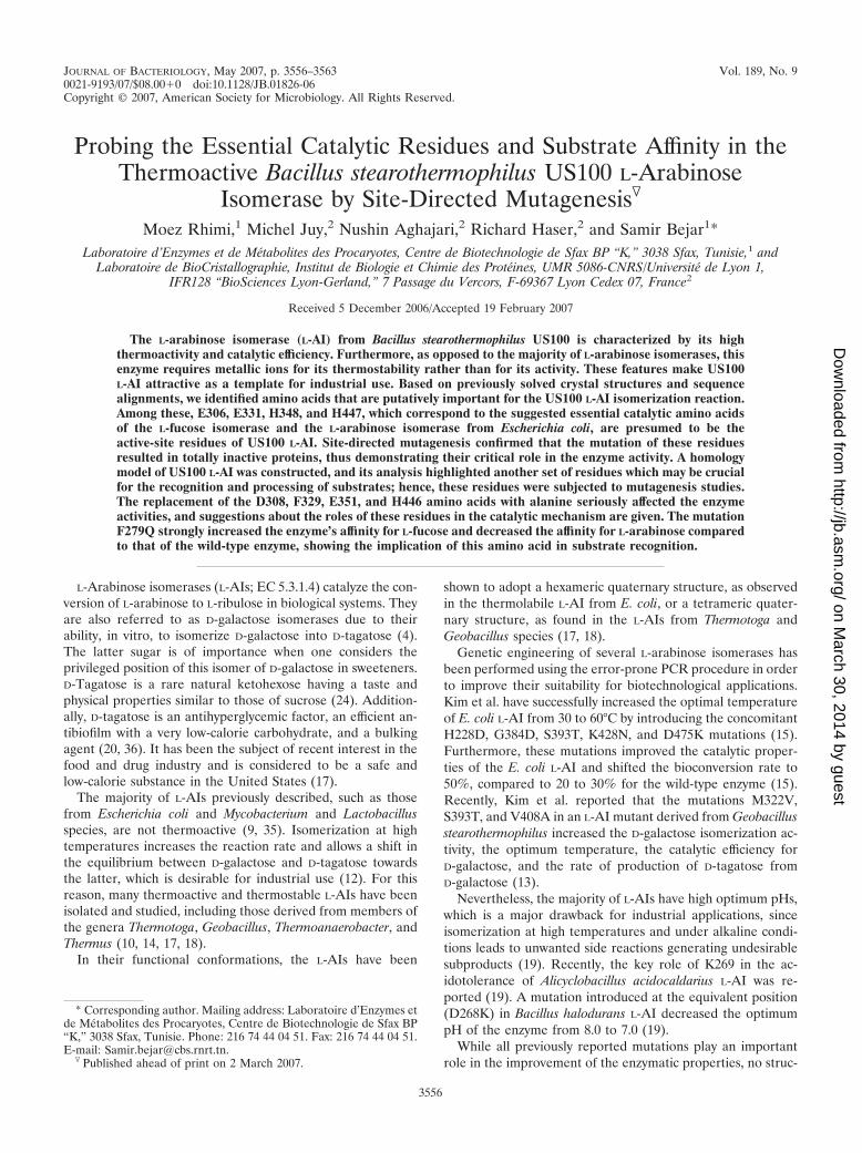
JOURNAL OF BACTERIOLOGY, May 2007, p. 3556–3563 Vol. 189, No. 90021-9193/07/$08.00�0 doi:10.1128/JB.01826-06Copyright © 2007, American Society for Microbiology. All Rights Reserved.
Probing the Essential Catalytic Residues and Substrate Affinity in theThermoactive Bacillus stearothermophilus US100 L-Arabinose
Isomerase by Site-Directed Mutagenesis�
Moez Rhimi,1 Michel Juy,2 Nushin Aghajari,2 Richard Haser,2 and Samir Bejar1*Laboratoire d’Enzymes et de Metabolites des Procaryotes, Centre de Biotechnologie de Sfax BP “K,” 3038 Sfax, Tunisie,1 and
Laboratoire de BioCristallographie, Institut de Biologie et Chimie des Proteines, UMR 5086-CNRS/Universite de Lyon 1,IFR128 “BioSciences Lyon-Gerland,” 7 Passage du Vercors, F-69367 Lyon Cedex 07, France2
Received 5 December 2006/Accepted 19 February 2007
The L-arabinose isomerase (L-AI) from Bacillus stearothermophilus US100 is characterized by its highthermoactivity and catalytic efficiency. Furthermore, as opposed to the majority of L-arabinose isomerases, thisenzyme requires metallic ions for its thermostability rather than for its activity. These features make US100L-AI attractive as a template for industrial use. Based on previously solved crystal structures and sequencealignments, we identified amino acids that are putatively important for the US100 L-AI isomerization reaction.Among these, E306, E331, H348, and H447, which correspond to the suggested essential catalytic amino acidsof the L-fucose isomerase and the L-arabinose isomerase from Escherichia coli, are presumed to be theactive-site residues of US100 L-AI. Site-directed mutagenesis confirmed that the mutation of these residuesresulted in totally inactive proteins, thus demonstrating their critical role in the enzyme activity. A homologymodel of US100 L-AI was constructed, and its analysis highlighted another set of residues which may be crucialfor the recognition and processing of substrates; hence, these residues were subjected to mutagenesis studies.The replacement of the D308, F329, E351, and H446 amino acids with alanine seriously affected the enzymeactivities, and suggestions about the roles of these residues in the catalytic mechanism are given. The mutationF279Q strongly increased the enzyme’s affinity for L-fucose and decreased the affinity for L-arabinose comparedto that of the wild-type enzyme, showing the implication of this amino acid in substrate recognition.
L-Arabinose isomerases (L-AIs; EC 5.3.1.4) catalyze the con-version of L-arabinose to L-ribulose in biological systems. Theyare also referred to as D-galactose isomerases due to theirability, in vitro, to isomerize D-galactose into D-tagatose (4).The latter sugar is of importance when one considers theprivileged position of this isomer of D-galactose in sweeteners.D-Tagatose is a rare natural ketohexose having a taste andphysical properties similar to those of sucrose (24). Addition-ally, D-tagatose is an antihyperglycemic factor, an efficient an-tibiofilm with a very low-calorie carbohydrate, and a bulkingagent (20, 36). It has been the subject of recent interest in thefood and drug industry and is considered to be a safe andlow-calorie substance in the United States (17).
The majority of L-AIs previously described, such as thosefrom Escherichia coli and Mycobacterium and Lactobacillusspecies, are not thermoactive (9, 35). Isomerization at hightemperatures increases the reaction rate and allows a shift inthe equilibrium between D-galactose and D-tagatose towardsthe latter, which is desirable for industrial use (12). For thisreason, many thermoactive and thermostable L-AIs have beenisolated and studied, including those derived from members ofthe genera Thermotoga, Geobacillus, Thermoanaerobacter, andThermus (10, 14, 17, 18).
In their functional conformations, the L-AIs have been
shown to adopt a hexameric quaternary structure, as observedin the thermolabile L-AI from E. coli, or a tetrameric quater-nary structure, as found in the L-AIs from Thermotoga andGeobacillus species (17, 18).
Genetic engineering of several L-arabinose isomerases hasbeen performed using the error-prone PCR procedure in orderto improve their suitability for biotechnological applications.Kim et al. have successfully increased the optimal temperatureof E. coli L-AI from 30 to 60°C by introducing the concomitantH228D, G384D, S393T, K428N, and D475K mutations (15).Furthermore, these mutations improved the catalytic proper-ties of the E. coli L-AI and shifted the bioconversion rate to50%, compared to 20 to 30% for the wild-type enzyme (15).Recently, Kim et al. reported that the mutations M322V,S393T, and V408A in an L-AI mutant derived from Geobacillusstearothermophilus increased the D-galactose isomerization ac-tivity, the optimum temperature, the catalytic efficiency forD-galactose, and the rate of production of D-tagatose fromD-galactose (13).
Nevertheless, the majority of L-AIs have high optimum pHs,which is a major drawback for industrial applications, sinceisomerization at high temperatures and under alkaline condi-tions leads to unwanted side reactions generating undesirablesubproducts (19). Recently, the key role of K269 in the ac-idotolerance of Alicyclobacillus acidocaldarius L-AI was re-ported (19). A mutation introduced at the equivalent position(D268K) in Bacillus halodurans L-AI decreased the optimumpH of the enzyme from 8.0 to 7.0 (19).
While all previously reported mutations play an importantrole in the improvement of the enzymatic properties, no struc-
* Corresponding author. Mailing address: Laboratoire d’Enzymes etde Metabolites des Procaryotes, Centre de Biotechnologie de Sfax BP“K,” 3038 Sfax, Tunisie. Phone: 216 74 44 04 51. Fax: 216 74 44 04 51.E-mail: [email protected].
� Published ahead of print on 2 March 2007.
3556
on March 30, 2014 by guest
http://jb.asm.org/
Dow
nloaded from
tural explanation was provided in previous studies due to theabsence of a three-dimensional (3D) structure of an L-AI. Veryrecently, the 3D structure of E. coli L-AI was determined (23).However, a detailed analysis of the structure-function relation-ships of this isomerase was not reported. In addition, no in-formation was given concerning the isomerization mechanismand the amino acids implicated, except for residues E306 andE333, which correspond to the catalytic residues identified inE. coli L-fucose isomerase (L-FI), also known as D-arabinoseisomerase (31).
We have previously described the cloning, overexpression,purification, and characterization of a thermostable L-arabi-nose isomerase isolated from the thermophilic Bacillus stearo-thermophilus US100 strain. The purified enzyme is a homotet-ramer with a molecular mass of 56 kDa for each monomer(28). US100 L-AI has an optimum temperature of about 80°Cand an optimum pH between 7.5 and 8.0 and differs fromearlier reported L-AIs in its behavior towards metallic ions(28).
Here we report the identification of the essential catalyticamino acids implicated in the isomerization reaction of US100L-AI by using site-directed mutagenesis and 3D structure ho-mology modeling. In addition, the enzyme’s affinity featureswere investigated.
MATERIALS AND METHODS
Bacterial strains, plasmids, and media. E. coli HB101 (F� hsdS20 ara-1recA13 proA12 lacY1 galK2 rpsL20 mtl-1 xyl-5) was used as an Ara� mutant hoststrain and also for the purification of the recombinant L-AIs. The plasmid pMR5containing the wild-type US100 araA gene (28) was used for the production ofthe wild-type US100 L-AI protein. pMR12, pMR13, pMR14, pMR15, pMR16,pMR17, pMR18, pMR19, and pMR20 are the recombinant plasmids carrying themutated US100 araA gene (Table 1). The culture of different E. coli strainsharboring wild-type and mutated genes was done in Luria-Bertani medium (30)supplemented, when necessary, with ampicillin (100 �g/ml) and IPTG (isopro-pyl-�-D-thiogalactopyranoside; 160 �g/ml).
DNA manipulation and PCR. The preparation of plasmid DNA, digestion withrestriction endonucleases, and separation of fragments by agarose gel electro-phoresis were performed as described by Sambrook et al. (30). PCRs werecarried out with a Gene Amp PCR System 9700 (Applied Biosystems). Theamplification reaction mixtures (100 �l) contained Pfu (Pfu DNA polymerase)amplification buffer, 10 mM (NH4)2SO4, 10 pmol of each primer, 100 ng of DNAtemplate, and 2 U of Pfu enzyme (Appligene). The cycling parameters were 94°Cfor 5 min followed by 40 cycles at 94°C for 30 s, 55°C for 60 s, and 72°C for 120 s.
Construction of L-arabinose mutant enzymes. L-Arabinose isomerase mutantenzymes were generated using the US100 L-AI wild-type coding sequence inplasmid pMR5 as the template. Mutations were introduced by site-directed PCRmutagenesis. Thus, two nonmutagenic external primers, F-araA (5�-GTGAACGGGGAGGAGCAATG-3�) and R-araA (5�-GAAATCTTACCGCCCCCGCC-3�), and two partial complementary internal primers containing the desiredmutation were designed (Table 1).
For each mutation, two separate PCRs were done and the resulting fragmentswere amplified using pMR5 as the template with the corresponding primers foreach reaction. The resulting two PCR fragments were extracted separately, andthen a third amplification was carried out with a mixture containing these frag-ments in the presence of the external primers. The PCR products were purifiedusing the GFX PCR DNA and gel band purification kit by following the instruc-tions of the manufacturer (Amersham Biosciences).
The purified mutated DNA fragments were cloned into a pUT57 vector,previously linearized with the SmaI restriction enzyme, and the E. coli HB101strain was transformed with the vector. Transformants were plated on Luria-Bertani medium supplemented with ampicillin (100 �g/ml) and IPTG (160 �g/ml). The obtained colonies were analyzed by restriction, and mutations wereconfirmed by DNA sequencing using an automated DNA sequencer (AppliedBiosystems). Obtained mutations are listed in Table 1.
Preparation of crude extracts and protein purification. Cells were harvestedby centrifugation at 7,500 � g for 10 min, and the pellets were suspended in 100
mM HEPES buffer supplemented with 1 mM MnSO4 and 0.2 mM CoCl2. Thencell suspensions were incubated for 2 h on ice in the presence of 5 mg oflysozyme/ml, 100 mM phenylmethylsulfonyl fluoride, and 2 �g of pepstatin A/ml.Cell disruption was carried out by sonication at 4°C for 6 min (pulsations of 3 sat 90 A) by using a Vibra-Cell 72405 sonicator, and cell debris was removed bycentrifugation (30,000 � g for 30 min at 4°C).
For protein purification, crude cell extract from each strain was heated (70°Cfor 30 min) and centrifuged at 30,000 � g for 30 min at 4°C. Proteins wereprecipitated between 50 and 80% acetone saturation, suspended in 100 mMHEPES buffer (pH 7.5), concentrated, and desalted in centrifugal microconcen-trators (Amicon, Inc.) with a 30-kDa membrane cutoff. Purification to homoge-neity was achieved by fast-performance liquid chromatography using a UNO-Q12anion exchange column equilibrated with 100 mM HEPES buffer (pH 7.5). Theproteins were eluted at a flow rate of 3 ml/min by using a linear NaCl gradientranging from 0 to 1 M in the same HEPES buffer. Pooled fractions displayingL-AI activity were desalted and concentrated.
Protein quantification and electrophoresis. Protein concentrations were de-termined using Bradford’s method with bovine serum albumin as the standard(3). The purified enzyme samples were allowed to migrate in 12% sodiumdodecyl sulfate-polyacrylamide gel electrophoresis (SDS-PAGE) gels accordingto the method of Laemmli (16). Protein bands were visualized by Coomassiebrilliant blue R-250 (Bio-Rad) staining.
Flame atomic absorption spectrometry analysis. For sample preparation, eachmineralized sample was employed at a final concentration of 10 mg/ml. A Perkin-Elmer Analyst 200 atomic absorption spectrophotometer (Norwalk, CT),equipped with a deuterium lamp background correction system, was used formetal binding quantification. Manganese hollow cathode lamps (wavelength,279.5 nm; slit, 0.2 nm; Perkin-Elmer) were used as the primary radiation source.Analytical measurements were based on the average absorbance under the con-ditions recommended by the manufacturer.
Protein identification. Extracted wild-type and mutant proteins were sepa-rated by SDS-PAGE. After staining with Coomassie blue Biosafe (Bio-Rad),bands corresponding to the expected molecular masses were excised from the geland washed in acetonitrile–50 mM ammonium hydrogen carbonate (vol/vol).Thereafter, samples were stirred for 15 min and vacuum dried for 30 min, and 0.5mg of trypsin (Promega) in 25 ml of ammonium hydrogen carbonate (50 mM)was added. Samples were digested for 16 h at 37°C. For matrix-assisted laserdesorption ionization–time of flight (MALDI-TOF) analyses, 2 �l of trifluoro-acetic acid (5%) was added to stop the trypsinolysis reaction. Peptide mixtureswere analyzed using a cyano-4-hydroxycinamic matrix prepared at 5 mg/ml in50% acetonitrile containing 0.1% trifluoroacetic acid. The trypsin autolytic pep-tides (842.5 and 2,211.1 Da) were used as internal calibrators. Peptides withmasses in the range of 700 to 4,000 Da were selected.
Peptides were analyzed using a Voyager DE STR MALDI-TOF mass spec-trometer (Applied Biosystems). Recorded tandem mass spectrometry spectrawere compared to theoretical fragmentations of a trypsinolyzed US100 AraAprotein (accession number CAI29261) via the Sequest algorithm incorporated inthe BioWorks software (version 2; Thermo Finnigan).
Enzyme assays and kinetic parameter determination. L-Arabinose isomeraseactivity was measured by determining the amount of L-ribulose or L-fuculoseformed. Under standard conditions, the reaction mixture contained 0.2 mMCoCl2, 1 mM MnSO4, 50 �l of the enzyme preparation at a suitable dilution, 5mM L-arabinose (or L-fucose), and 100 mM HEPES buffer (pH 7.5) in a finalvolume of 1 ml. The reaction mixture was incubated at 80°C for 1 and 10 min for
TABLE 1. Oligonucleotides used for site-directed mutagenesis
Desiredmutation Primer sequencea Plasmid
E306A GATTTGGCGGCGCAGGCGACTGG pMR12D308A CGGCGAAGGCGCCTGGAAAACGG pMR16F279Q CTTTACGACGACGCAAGAAGATTTGCA pMR20F329A AAAGGAACATCGGCCATGGAAGACTA pMR18E331A ACATCGTTCATGGCAGACTACACG pMR13H348A CTTGGCGCGGTTATGCTCGAAGTA pMR15E351A GCGCATATGCTCGCAGTATGCCCGACG pMR17H446A AGCCGGCGGAGCGGCTCACACATGCTT pMR19H447A GGCGGAGCGCATGCCACATGCTTC pMR14
a Nucleotide sequences corresponding to the mutated amino acids are under-lined.
VOL. 189, 2007 US100 L-AI CATALYTIC RESIDUES AND SUBSTRATE AFFINITY 3557
on March 30, 2014 by guest
http://jb.asm.org/
Dow
nloaded from
L-arabinose and L-fucose, respectively, followed by cooling of the samples on iceto stop the reaction. The amount of L-ribulose or L-fuculose generated wasdetermined by the cysteine carbazole sulfuric acid method, and the absorbanceat 560 nm was measured (6).
For determination of the kinetic parameters, assays were carried out understandard conditions by using 1 to 800 mM substrate (L-arabinose or L-fucose).
One unit of L-arabinose isomerase activity was defined as the amount ofenzyme catalyzing the formation of 1 �mol of ketosugar per min under theabove-specified conditions.
Amino acid sequence analysis and homology modeling. Sequence analysis andmultiple alignments were done using the programs BLAST and CLUSTALW (1,34). The secondary structure of the protein was predicted using the DSSPprogram (11), and rendering of the alignment including the superimposition ofsecondary structures was performed with the program ESPript (8). The 3Dhomology model of US100 L-AI was generated using the Geno3D server (5), andthe superposition of 3D structures was performed with the “rigid” option inthe graphics software TURBO-FRODO (29). Images were generated usingTURBO-FRODO, and the surface rendering was done with UCSF Chimera(http://www.cgl.ucsf.edu/chimera).
RESULTS AND DISCUSSION
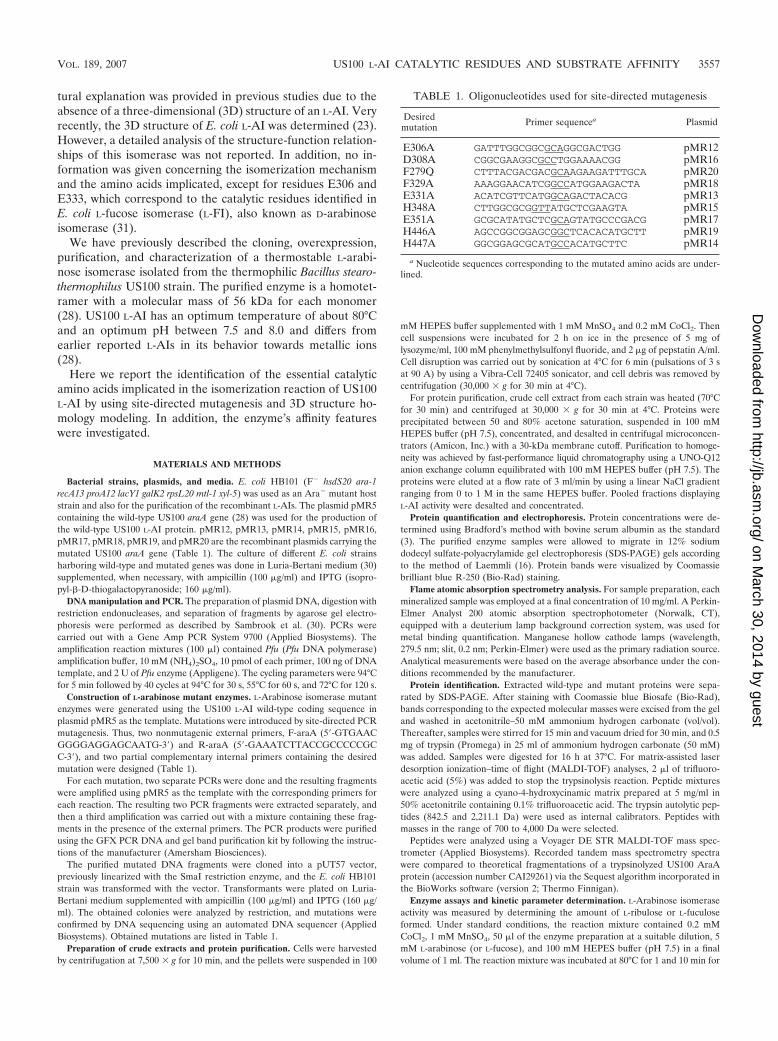
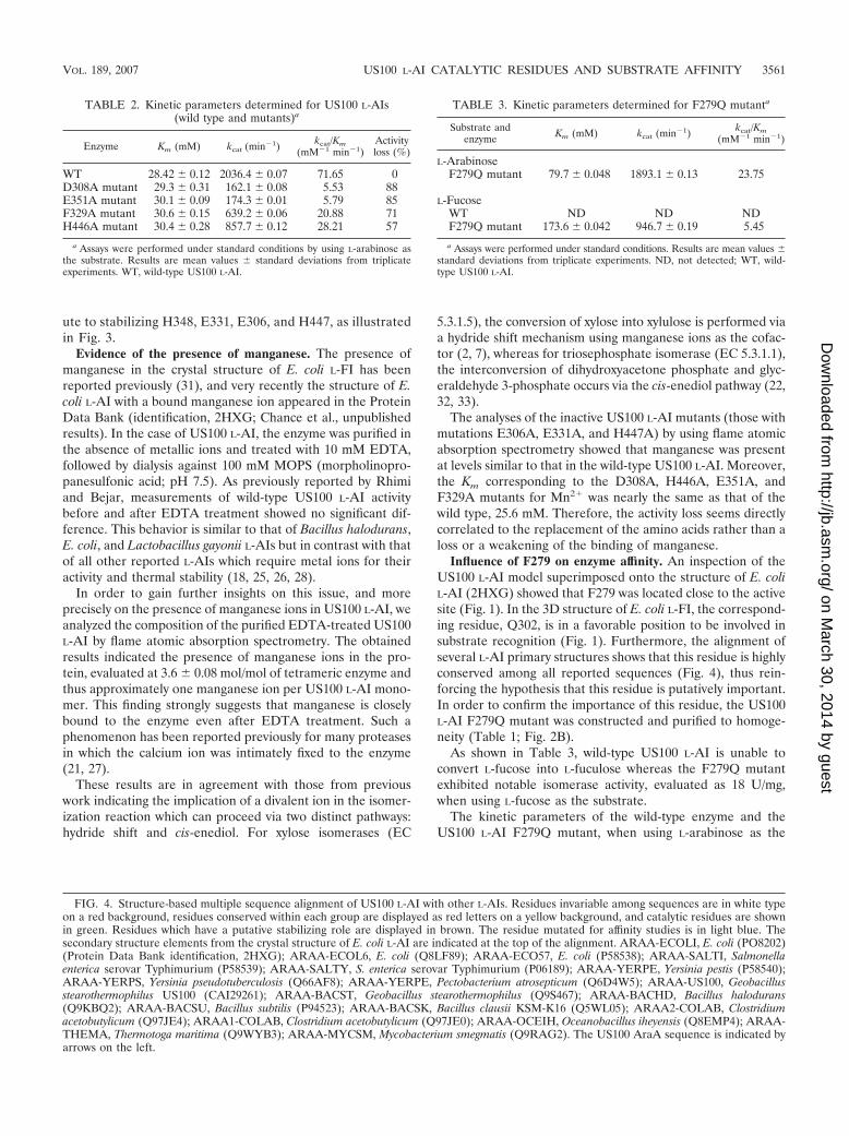
Structural analysis of the US100 L-AI active site and com-parison with that of E. coli L-FI. Until now, no detailed analysisof the isomerization mechanism and the catalytic residues im-plicated in the reaction in the L-AI family has been reported.This prompted us to undertake the identification of crucialcatalytic residues by using all available data. The 3D crystalstructure of the L-fucose isomerase from E. coli, also known asD-arabinose isomerase (Protein Data Bank identification,1FUI), was solved previously (31). In that previous work, thecatalytic residues were reported to be E337, D361, and H528and to interact with a manganese ion (Mn2�). Furthermore,the 3D structure of the hexameric E. coli L-AI (Protein DataBank identification, 2AJT) which displays 60% sequence iden-tity to the US100 L-AI was solved previously (23). The authorsof the study reported that this crystal structure was manganesedepleted. More recently, coordinates for the 3D structure ofthe same E. coli L-AI but this time with a bound Mn2� ion weredeposited in the Protein Data Bank (identification, 2HXG;M. R. Chance, W. Zhu, and B. A. Manjasetty, unpublishedresults). After the superimposition of the two E. coli L-AIstructures, 2AJT (Mn2� depleted) and 2HXG (containingMn2�), an inspection of the active-site region showed thatresidues H350 and H450 displayed similar orientations in thetwo structures whereas the orientations of E306 and E333differed slightly due to the presence in 2HXG of Mn2� insteadof a putative water molecule as in the published structure of2AJT (23).
Taking advantage of this newly solved structure and usingmolecular-modeling techniques, we have constructed a 3Dmodel of US100 L-AI. The generated model has a root meansquare deviation of 1.5 Å from that of E. coli L-AI (2HXG)when C-� atoms are superimposed. As shown in Fig. 1, thepresumed essential catalytic residues of E. coli L-AI as well asthose of US100 L-AI (described hereafter) were perfectly su-perimposed onto those of E. coli L-FI. By analogy with E. coliL-FI catalytic residues E337, D361, and H528, residues E306,E331, and H447 from the US100 L-AI enzyme were deduced tobe the essential catalytic amino acids. In order to confirm thisobservation, site-directed mutagenesis was employed to re-place these amino acids with alanine.
Monitoring of the US100 L-AI activity by using crude pro-
tein extracts demonstrated that whereas an appreciable activityof the wild-type US100 L-AI of 46 U/mg was detected, nomeasurable activity of the E306A, E331A, and H447A mutantswas detected. Note that the activity assays were performed atthe optimal temperature (80°C) as well as at 50°C, at which theenzyme is completely thermostable.
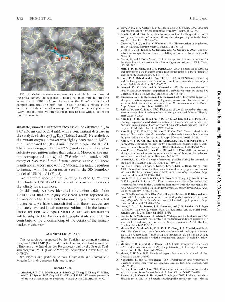
With the aim of proving that these mutated proteins wereinactive but efficiently expressed, the crude protein was ana-lyzed by SDS-PAGE. As shown in Fig. 2A, all mutant enzymeswere successfully overexpressed, with bands displaying a mo-lecular mass of 56 kDa as expected for a monomer of US100L-AI (28). Furthermore, an analysis of these bands by massspectrometry (results not shown) confirmed the desired muta-tions.
One may thus conclude that the loss of activity of the re-ported mutants stems from the role of the corresponding mu-tated residues as catalytic residues in the isomerization reac-tion.
Other crucial catalytic residues of US100 L-AI. On the basisof the US100 L-AI model, an analysis of the active-site regionindicated additional amino acids which could be implicated inthe catalytic mechanism. As illustrated in Fig. 3, H348, which ishighly conserved among all reported L-AIs (Fig. 4), is locatedin a position where it can interact with the manganese ionand/or the substrate. These observations incited us to mutateH348 into an alanine. The expression of the US100 L-AI
FIG. 1. Superposition of the three catalytic residues from E. coli L-AI(Protein Data Bank identification, 2HXG; in dark blue) onto E. coliL-FI (Protein Data Bank identification, 1FUI; in red) and the US100L-AI model (yellow). L-Fucitol (FOC), shown in light blue, interactswith the Mn2� ion (brown spheres). Note also the location of F279 inthe US100 L-AI model and its corresponding residue, Q302, in thethree-dimensional structure of E. coli L-FI.
3558 RHIMI ET AL. J. BACTERIOL.
on March 30, 2014 by guest
http://jb.asm.org/
Dow
nloaded from
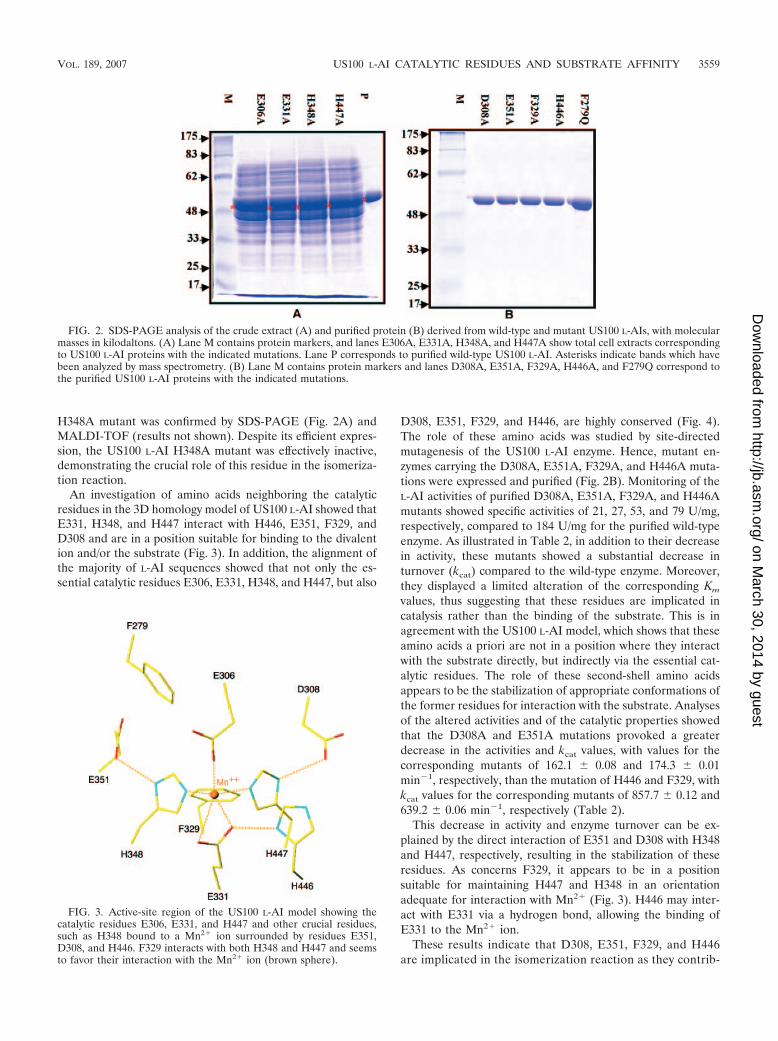
H348A mutant was confirmed by SDS-PAGE (Fig. 2A) andMALDI-TOF (results not shown). Despite its efficient expres-sion, the US100 L-AI H348A mutant was effectively inactive,demonstrating the crucial role of this residue in the isomeriza-tion reaction.
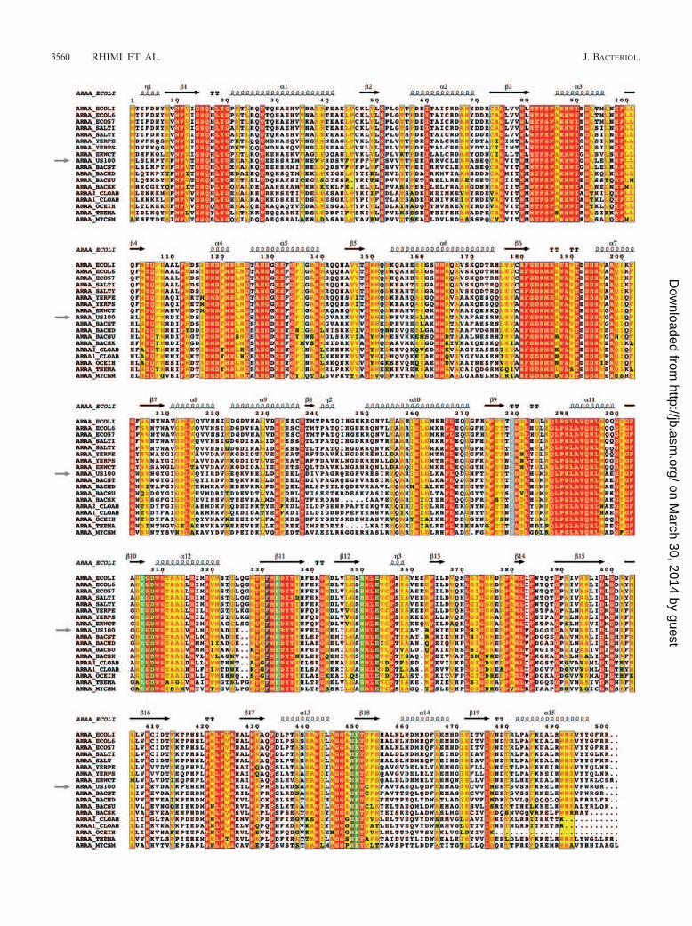
An investigation of amino acids neighboring the catalyticresidues in the 3D homology model of US100 L-AI showed thatE331, H348, and H447 interact with H446, E351, F329, andD308 and are in a position suitable for binding to the divalention and/or the substrate (Fig. 3). In addition, the alignment ofthe majority of L-AI sequences showed that not only the es-sential catalytic residues E306, E331, H348, and H447, but also
D308, E351, F329, and H446, are highly conserved (Fig. 4).The role of these amino acids was studied by site-directedmutagenesis of the US100 L-AI enzyme. Hence, mutant en-zymes carrying the D308A, E351A, F329A, and H446A muta-tions were expressed and purified (Fig. 2B). Monitoring of theL-AI activities of purified D308A, E351A, F329A, and H446Amutants showed specific activities of 21, 27, 53, and 79 U/mg,respectively, compared to 184 U/mg for the purified wild-typeenzyme. As illustrated in Table 2, in addition to their decreasein activity, these mutants showed a substantial decrease inturnover (kcat) compared to the wild-type enzyme. Moreover,they displayed a limited alteration of the corresponding Km
values, thus suggesting that these residues are implicated incatalysis rather than the binding of the substrate. This is inagreement with the US100 L-AI model, which shows that theseamino acids a priori are not in a position where they interactwith the substrate directly, but indirectly via the essential cat-alytic residues. The role of these second-shell amino acidsappears to be the stabilization of appropriate conformations ofthe former residues for interaction with the substrate. Analysesof the altered activities and of the catalytic properties showedthat the D308A and E351A mutations provoked a greaterdecrease in the activities and kcat values, with values for thecorresponding mutants of 162.1 � 0.08 and 174.3 � 0.01min�1, respectively, than the mutation of H446 and F329, withkcat values for the corresponding mutants of 857.7 � 0.12 and639.2 � 0.06 min�1, respectively (Table 2).
This decrease in activity and enzyme turnover can be ex-plained by the direct interaction of E351 and D308 with H348and H447, respectively, resulting in the stabilization of theseresidues. As concerns F329, it appears to be in a positionsuitable for maintaining H447 and H348 in an orientationadequate for interaction with Mn2� (Fig. 3). H446 may inter-act with E331 via a hydrogen bond, allowing the binding ofE331 to the Mn2� ion.
These results indicate that D308, E351, F329, and H446are implicated in the isomerization reaction as they contrib-
FIG. 2. SDS-PAGE analysis of the crude extract (A) and purified protein (B) derived from wild-type and mutant US100 L-AIs, with molecularmasses in kilodaltons. (A) Lane M contains protein markers, and lanes E306A, E331A, H348A, and H447A show total cell extracts correspondingto US100 L-AI proteins with the indicated mutations. Lane P corresponds to purified wild-type US100 L-AI. Asterisks indicate bands which havebeen analyzed by mass spectrometry. (B) Lane M contains protein markers and lanes D308A, E351A, F329A, H446A, and F279Q correspond tothe purified US100 L-AI proteins with the indicated mutations.
FIG. 3. Active-site region of the US100 L-AI model showing thecatalytic residues E306, E331, and H447 and other crucial residues,such as H348 bound to a Mn2� ion surrounded by residues E351,D308, and H446. F329 interacts with both H348 and H447 and seemsto favor their interaction with the Mn2� ion (brown sphere).
VOL. 189, 2007 US100 L-AI CATALYTIC RESIDUES AND SUBSTRATE AFFINITY 3559
on March 30, 2014 by guest
http://jb.asm.org/
Dow
nloaded from
3560 RHIMI ET AL. J. BACTERIOL.
on March 30, 2014 by guest
http://jb.asm.org/
Dow
nloaded from
ute to stabilizing H348, E331, E306, and H447, as illustratedin Fig. 3.
Evidence of the presence of manganese. The presence ofmanganese in the crystal structure of E. coli L-FI has beenreported previously (31), and very recently the structure of E.coli L-AI with a bound manganese ion appeared in the ProteinData Bank (identification, 2HXG; Chance et al., unpublishedresults). In the case of US100 L-AI, the enzyme was purified inthe absence of metallic ions and treated with 10 mM EDTA,followed by dialysis against 100 mM MOPS (morpholinopro-panesulfonic acid; pH 7.5). As previously reported by Rhimiand Bejar, measurements of wild-type US100 L-AI activitybefore and after EDTA treatment showed no significant dif-ference. This behavior is similar to that of Bacillus halodurans,E. coli, and Lactobacillus gayonii L-AIs but in contrast with thatof all other reported L-AIs which require metal ions for theiractivity and thermal stability (18, 25, 26, 28).
In order to gain further insights on this issue, and moreprecisely on the presence of manganese ions in US100 L-AI, weanalyzed the composition of the purified EDTA-treated US100L-AI by flame atomic absorption spectrometry. The obtainedresults indicated the presence of manganese ions in the pro-tein, evaluated at 3.6 � 0.08 mol/mol of tetrameric enzyme andthus approximately one manganese ion per US100 L-AI mono-mer. This finding strongly suggests that manganese is closelybound to the enzyme even after EDTA treatment. Such aphenomenon has been reported previously for many proteasesin which the calcium ion was intimately fixed to the enzyme(21, 27).
These results are in agreement with those from previouswork indicating the implication of a divalent ion in the isomer-ization reaction which can proceed via two distinct pathways:hydride shift and cis-enediol. For xylose isomerases (EC
5.3.1.5), the conversion of xylose into xylulose is performed viaa hydride shift mechanism using manganese ions as the cofac-tor (2, 7), whereas for triosephosphate isomerase (EC 5.3.1.1),the interconversion of dihydroxyacetone phosphate and glyc-eraldehyde 3-phosphate occurs via the cis-enediol pathway (22,32, 33).
The analyses of the inactive US100 L-AI mutants (those withmutations E306A, E331A, and H447A) by using flame atomicabsorption spectrometry showed that manganese was presentat levels similar to that in the wild-type US100 L-AI. Moreover,the Km corresponding to the D308A, H446A, E351A, andF329A mutants for Mn2� was nearly the same as that of thewild type, 25.6 mM. Therefore, the activity loss seems directlycorrelated to the replacement of the amino acids rather than aloss or a weakening of the binding of manganese.
Influence of F279 on enzyme affinity. An inspection of theUS100 L-AI model superimposed onto the structure of E. coliL-AI (2HXG) showed that F279 was located close to the activesite (Fig. 1). In the 3D structure of E. coli L-FI, the correspond-ing residue, Q302, is in a favorable position to be involved insubstrate recognition (Fig. 1). Furthermore, the alignment ofseveral L-AI primary structures shows that this residue is highlyconserved among all reported sequences (Fig. 4), thus rein-forcing the hypothesis that this residue is putatively important.In order to confirm the importance of this residue, the US100L-AI F279Q mutant was constructed and purified to homoge-neity (Table 1; Fig. 2B).
As shown in Table 3, wild-type US100 L-AI is unable toconvert L-fucose into L-fuculose whereas the F279Q mutantexhibited notable isomerase activity, evaluated as 18 U/mg,when using L-fucose as the substrate.
The kinetic parameters of the wild-type enzyme and theUS100 L-AI F279Q mutant, when using L-arabinose as the
FIG. 4. Structure-based multiple sequence alignment of US100 L-AI with other L-AIs. Residues invariable among sequences are in white typeon a red background, residues conserved within each group are displayed as red letters on a yellow background, and catalytic residues are shownin green. Residues which have a putative stabilizing role are displayed in brown. The residue mutated for affinity studies is in light blue. Thesecondary structure elements from the crystal structure of E. coli L-AI are indicated at the top of the alignment. ARAA-ECOLI, E. coli (PO8202)(Protein Data Bank identification, 2HXG); ARAA-ECOL6, E. coli (Q8LF89); ARAA-ECO57, E. coli (P58538); ARAA-SALTI, Salmonellaenterica serovar Typhimurium (P58539); ARAA-SALTY, S. enterica serovar Typhimurium (P06189); ARAA-YERPE, Yersinia pestis (P58540);ARAA-YERPS, Yersinia pseudotuberculosis (Q66AF8); ARAA-YERPE, Pectobacterium atrosepticum (Q6D4W5); ARAA-US100, Geobacillusstearothermophilus US100 (CAI29261); ARAA-BACST, Geobacillus stearothermophilus (Q9S467); ARAA-BACHD, Bacillus halodurans(Q9KBQ2); ARAA-BACSU, Bacillus subtilis (P94523); ARAA-BACSK, Bacillus clausii KSM-K16 (Q5WL05); ARAA2-COLAB, Clostridiumacetobutylicum (Q97JE4); ARAA1-COLAB, Clostridium acetobutylicum (Q97JE0); ARAA-OCEIH, Oceanobacillus iheyensis (Q8EMP4); ARAA-THEMA, Thermotoga maritima (Q9WYB3); ARAA-MYCSM, Mycobacterium smegmatis (Q9RAG2). The US100 AraA sequence is indicated byarrows on the left.
TABLE 2. Kinetic parameters determined for US100 L-AIs(wild type and mutants)a
Enzyme Km (mM) kcat (min�1) kcat/Km(mM�1 min�1)
Activityloss (%)
WT 28.42 � 0.12 2036.4 � 0.07 71.65 0D308A mutant 29.3 � 0.31 162.1 � 0.08 5.53 88E351A mutant 30.1 � 0.09 174.3 � 0.01 5.79 85F329A mutant 30.6 � 0.15 639.2 � 0.06 20.88 71H446A mutant 30.4 � 0.28 857.7 � 0.12 28.21 57
a Assays were performed under standard conditions by using L-arabinose asthe substrate. Results are mean values � standard deviations from triplicateexperiments. WT, wild-type US100 L-AI.
TABLE 3. Kinetic parameters determined for F279Q mutanta
Substrate andenzyme Km (mM) kcat (min�1) kcat/Km
(mM�1 min�1)
L-ArabinoseF279Q mutant 79.7 � 0.048 1893.1 � 0.13 23.75
L-FucoseWT ND ND NDF279Q mutant 173.6 � 0.042 946.7 � 0.19 5.45
a Assays were performed under standard conditions. Results are mean values �standard deviations from triplicate experiments. ND, not detected; WT, wild-type US100 L-AI.
VOL. 189, 2007 US100 L-AI CATALYTIC RESIDUES AND SUBSTRATE AFFINITY 3561
on March 30, 2014 by guest
http://jb.asm.org/
Dow
nloaded from
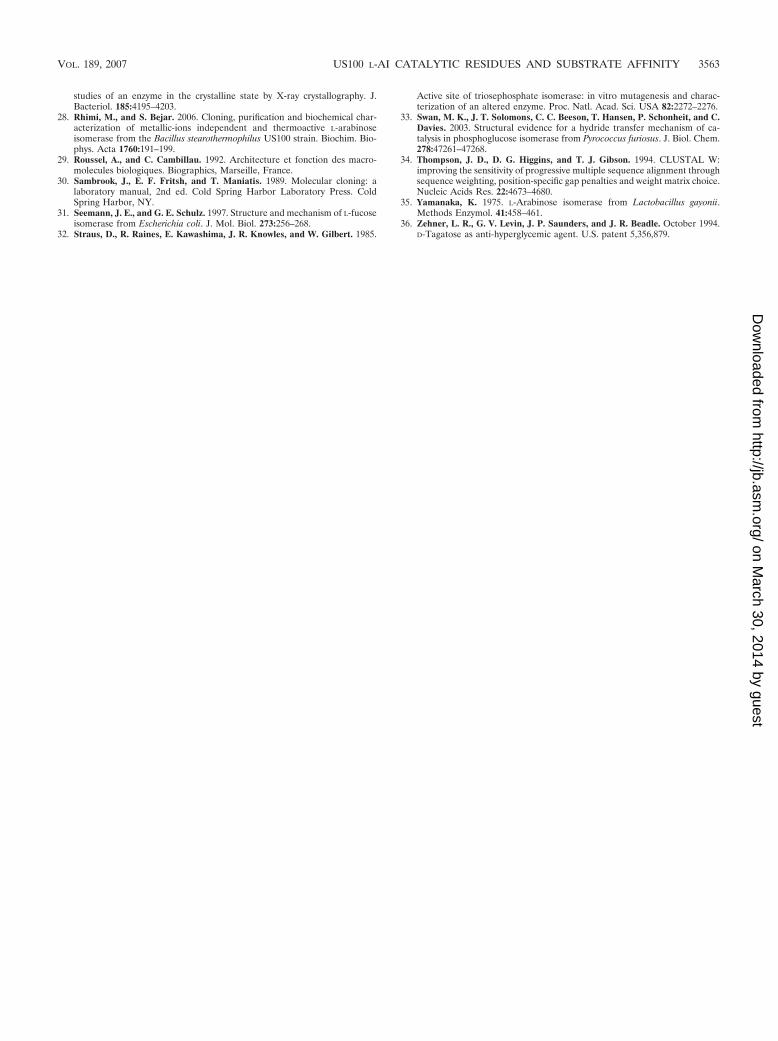
substrate, showed a significant increase of the estimated Km to79.7 mM instead of 28.4 mM, with a concomitant decrease inthe catalytic efficiency (kcat/Km) (Tables 2 and 3). Nevertheless,the mutant enzyme turnover was slightly decreased to 1,893.1min�1 compared to 2,036.4 min�1 for wild-type US100 L-AI.These results suggest that the F279Q mutation is implicated insubstrate recognition rather than catalysis. Moreover, the mu-tant corresponded to a Km of 173.6 mM and a catalytic effi-ciency of 5.45 mM�1 min�1 with L-fucose (Table 3). Theseresults are in accordance with F279 being in a suitable positionto interact with the substrate, as seen in the 3D homologymodel of US100 L-AI (Fig. 5).
We therefore conclude that mutating F279 to Q279 shiftsthe affinity of US100 L-AI in favor of L-fucose and decreasesthe affinity for L-arabinose.
In this study, we have identified nine amino acids of theUS100 L-AI that are highly conserved within reported se-quences of L-AIs. Using molecular modeling and site-directedmutagenesis, we have demonstrated that these residues areintimately involved in substrate recognition and in the isomer-ization reaction. Wild-type US100 L-AI and selected mutantswill be subjected to X-ray crystallography studies in order tocontribute to the understanding of the details of the isomer-ization mechanism.
ACKNOWLEDGMENTS
This research was supported by the Tunisian government contractprogram CBS-LEMP (Centre de Biotechnologie de Sfax-Laboratoired’Enzymes et Metabolites des Procaryotes) and by the French-Tuni-sian program CMCU (Comite Mixte de Cooperation Universitaire, no.04/0905).
We express our gratitude to Neji Gharsallah and EmmanuelleMaguin for their generous help and support.
REFERENCES
1. Altschul, S. F., T. L. Madden, A. A. Schaffer, J. Zhang, Z. Zhang, W. Miller,and D. J. Lipman. 1997. Gapped BLAST and PSI-BLAST: a new generationof protein database search programs. Nucleic Acids Res. 25:3389–3402.
2. Blow, D. M., C. A. Collyer, J. D. Goldberg, and O. S. Smart. 1992. Structureand mechanism of D-xylose isomerase. Faraday Discuss., p. 67–73.
3. Bradford, M. M. 1976. A rapid and sensitive method for the quantification ofmicrogram quantities of protein utilizing the principle of protein-dye bind-ing. Anal. Biochem. 72:248–254.
4. Cheetam, P. S. J., and A. N. Woottom. 1993. Bioconversion of D-galactoseinto D-tagatose. Enzyme Microb. Technol. 15:105–108.
5. Combet, C., M. Jambon, G. Deleage, and C. Geourjon. 2002. Geno3D:automatic comparative molecular modelling of protein. Bioinformatics 18:213–214.
6. Dische, Z., and E. Borenfreund. 1951. A new spectrophotometric method forthe detection and determination of keto sugars and trioses. J. Biol. Chem.192:583–587.
7. Fenn, T. D., D. Ringe, and G. A. Petsko. 2004. Xylose isomerase in substrateand inhibitor michaelis states: atomic resolution studies of a metal-mediatedhydride shift. Biochemistry 43:6464–6474.
8. Gouet, P., X. Robert, and E. Courcelle. 2003. ESPript/ENDscript: extractingand rendering sequence and 3D information from atomic structures of pro-teins. Nucleic Acids Res. 31:3320–3323.
9. Izumori, K., Y. Ueda, and K. Yamanaka. 1978. Pentose metabolism inMycobacterium smegmatis: comparison of L-arabinose isomerases induced byL-arabinose and D-galactose. J. Bacteriol. 133:413–414.
10. Jorgensen, F., O. C. Hansen, and P. Stougaard. 2004. Enzymatic conversionof D-galactose to D-tagatose: heterologous expression and characterisation ofa thermostable L-arabinose isomerase from Thermoanaerobacter mathranii.Appl. Microbiol. Biotechnol. 64:816–822.
11. Kabsch, W., and C. Sander. 1983. Dictionary of protein secondary structure:pattern recognition of hydrogen-bonded and geometrical features. Biopoly-mers 22:2577–2673.
12. Kim, B. C., Y. H. Lee, H. S. Lee, D. W. Lee, E. A. Choe, and Y. R. Pyun. 2002.Cloning, expression and characterization of L-arabinose isomerase fromThermotoga neapolitana: bioconversion of D-galactose to D-tagatose using theenzyme. FEMS Microbiol. Lett. 212:121–126.
13. Kim, H. J., J. H. Kim, H. J. Oh, and D. K. Oh. 2006. Characterization of amutated Geobacillus stearothermophilus L-arabinose isomerase that increasesthe production rate of D-tagatose. J. Appl. Microbiol. 101:213–221.
14. Kim, J. W., Y. W. Kim, H. J. Roh, H. Y. Kim, J. H. Cha, K. H. Park, and. C. S.Park. 2003. Production of tagatose by a recombinant thermostable L-arabi-nose isomerase from Thermus sp. IM6501. Biotechnol. Lett. 25:963–967.
15. Kim, P., S. H. Yoon, M. J. Seo, D. K. Oh, and J. H. Choi. 2001. Improvementof tagatose conversion rate by genetic evolution of thermostable galactoseisomerase. Biotechnol. Appl. Biochem. 34:99–102.
16. Laemmli, U. K. 1970. Cleavage of structural proteins during the assembly ofthe head of bacteriophage T4. Nature 227:680–685.
17. Lee, D., H. Jang, E. Choe, B. Kim, S. Lee, S. Kim, Y. Hong, and Y. Pyun.2004. Characterization of a thermostable L-arabinose (D-galactose) isomer-ase from the hyperthermophilic eubacterium Thermotoga maritima. Appl.Environ. Microbiol. 70:1397–1404.
18. Lee, D. W., E. A. Choe, S. B. Kim, S. H. Eom, Y. H. Hong, S. J. Lee, H. S. Lee,D. Y. Lee, and Y. R. Pyun. 2005. Distinct metal dependence for catalytic andstructural functions in the L-arabinose isomerases from the mesophilic Ba-cillus halodurans and the thermophilic Geobacillus stearothermophilus. Arch.Biochem. Biophys. 434:333–343.
19. Lee, S. J., D. W. Lee, E. A. Choe, Y. H. Hong, S. B. Kim, B. C. Kim, and Y. R.Pyun. 2005. Characterization of a thermoacidophilic L-arabinose isomerasefrom Alicyclobacillus acidocaldarius: role of Lys-269 in pH optimum. Appl.Environ. Microbiol. 71:7888–7896.
20. Levin, G. V., L. R. Zehner, J. P. Saunders, and J. R. Beadle. 1995. Sugarsubstitutes: their energy values, bulk characteristics, and potential healthbenefits. Am. J. Clin. Nutr. 62:1161S–1168S.
21. Lin, S. J., E. Yoshimura, H. Sakai, T. Wakagi, and H. Matsuzawa. 1999.Weakly bound calcium ions involved in the thermostability of aqualysin I, aheat-stable subtilisin-type protease of Thermus aquaticus YT-1. Biochim.Biophys. Acta 1433:132–138.
22. Mande, S. C., V. Mainfroid, K. H. Kalk, K. Goraj, J. A. Martial, and W. G.Hol. 1994. Crystal structure of recombinant human triosephosphate isomer-ase at 2.8 A resolution. Triosephosphate isomerase-related human geneticdisorders and comparison with the trypanosomal enzyme. Protein Sci. 3:810–821.
23. Manjasetty, B. A., and M. R. Chance. 2006. Crystal structure of Escherichiacoli L-arabinose isomerase (ECAI), the putative target of biological tagatoseproduction. J. Mol. Biol. 360:297–309.
24. Mazur, A. W. May 1989. Functional sugar substitutes with reduced calories.European patent 341062.
25. Nakamatu, T., and K. Yamanaka. 1969. Crystallization and properties ofL-arabinose isomerase from Lactobacillus gayonii. Biochim. Biophys. Acta178:156–165.
26. Patrick, J. W., and N. Lee. 1968. Purification and properties of an L-arabi-nose isomerase from Escherichia coli. J. Biol. Chem. 243:4312–4318.
27. Ravaud, S., P. Gouet, R. Haser, and N. Aghajari. 2003. Probing the role ofdivalent metal ions in a bacterial psychrophilic metalloprotease: binding
FIG. 5. Molecular surface representation of US100 L-AI, aroundthe active center. The substrate L-fucitol has been modeled into theactive site of US100 L-AI on the basis of the E. coli L-FI–L-fucitolcomplex structure. The Mn2� ion located near the substrate in theactive site is shown as a brown sphere. F279 has been replaced byQ279, and the putative interaction of this residue with L-fucitol (inblue) is presented.
3562 RHIMI ET AL. J. BACTERIOL.
on March 30, 2014 by guest
http://jb.asm.org/
Dow
nloaded from
studies of an enzyme in the crystalline state by X-ray crystallography. J.Bacteriol. 185:4195–4203.
28. Rhimi, M., and S. Bejar. 2006. Cloning, purification and biochemical char-acterization of metallic-ions independent and thermoactive L-arabinoseisomerase from the Bacillus stearothermophilus US100 strain. Biochim. Bio-phys. Acta 1760:191–199.
29. Roussel, A., and C. Cambillau. 1992. Architecture et fonction des macro-molecules biologiques. Biographics, Marseille, France.
30. Sambrook, J., E. F. Fritsh, and T. Maniatis. 1989. Molecular cloning: alaboratory manual, 2nd ed. Cold Spring Harbor Laboratory Press. ColdSpring Harbor, NY.
31. Seemann, J. E., and G. E. Schulz. 1997. Structure and mechanism of L-fucoseisomerase from Escherichia coli. J. Mol. Biol. 273:256–268.
32. Straus, D., R. Raines, E. Kawashima, J. R. Knowles, and W. Gilbert. 1985.
Active site of triosephosphate isomerase: in vitro mutagenesis and charac-terization of an altered enzyme. Proc. Natl. Acad. Sci. USA 82:2272–2276.
33. Swan, M. K., J. T. Solomons, C. C. Beeson, T. Hansen, P. Schonheit, and C.Davies. 2003. Structural evidence for a hydride transfer mechanism of ca-talysis in phosphoglucose isomerase from Pyrococcus furiosus. J. Biol. Chem.278:47261–47268.
34. Thompson, J. D., D. G. Higgins, and T. J. Gibson. 1994. CLUSTAL W:improving the sensitivity of progressive multiple sequence alignment throughsequence weighting, position-specific gap penalties and weight matrix choice.Nucleic Acids Res. 22:4673–4680.
35. Yamanaka, K. 1975. L-Arabinose isomerase from Lactobacillus gayonii.Methods Enzymol. 41:458–461.
36. Zehner, L. R., G. V. Levin, J. P. Saunders, and J. R. Beadle. October 1994.D-Tagatose as anti-hyperglycemic agent. U.S. patent 5,356,879.
VOL. 189, 2007 US100 L-AI CATALYTIC RESIDUES AND SUBSTRATE AFFINITY 3563
on March 30, 2014 by guest
http://jb.asm.org/
Dow
nloaded from
Related Documents