PRKDC mutations associated with immunodeficiency, granuloma, and autoimmune regulator–dependent autoimmunity Anne-Laure Mathieu, PhD, a,b,c,d Estelle Verronese, BS, e Gillian I. Rice, PhD, f Fanny Fouyssac, MD, g Yves Bertrand, MD, PhD, h Capucine Picard, MD, PhD, i Marie Chansel, MSc, j Jolan E. Walter, MD, PhD, k Luigi D. Notarangelo, MD, l Manish J. Butte, MD, PhD, m Kari Christine Nadeau, MD, PhD, m Krisztian Csomos, PhD, k David J. Chen, PhD, n Karin Chen, MD, o Ana Delgado, BS, e Chantal Rigal, BS, e Christine Bardin, BS, e Catharina Schuetz, MD, PhD, p Despina Moshous, MD, PhD, j H elo € ıse Reumaux, MD, q Franc ¸ ois Plenat, MD, PhD, r Alice Phan, MD, PhD, v Marie-Th er ese Zabot, PharmD, PhD, t Brigitte Balme, MD, s S ebastien Viel, PharmD, a,b,c,d,u Jacques Bienvenu, PharmD, PhD, a,b,c,d,u Pierre Cochat, MD, PhD, v Mirjam van der Burg, PhD, w Christophe Caux, PhD, e E. Helen Kemp, BSc, PhD, x Isabelle Rouvet, PharmD, PhD, t Christophe Malcus, PharmD, PhD, y Jean-Francois M eritet, PharmD, PhD, z Annick Lim, MSc, aa Yanick J. Crow, MD, PhD, f Nicole Fabien, PharmD, PhD, u Christine M en etrier-Caux, PhD, e Jean-Pierre De Villartay, PhD, j Thierry Walzer, PhD, a,b,c,d and Alexandre Belot, MD, PhD a,b,c,d,v Lyon, Nancy, Paris, and Lille, France, Manchesterand Sheffield, United Kingdom, Boston, Mass, Stanford, Calif, Dallas, Tex, Salt Lake City, Utah, Ulm, Germany, and Rotterdam, The Netherlands Background: PRKDC encodes for DNA-dependent protein kinase catalytic subunit (DNA-PKcs), a kinase that forms part of a complex (DNA-dependent protein kinase [DNA-PK]) crucial for DNA double-strand break repair and V(D)J recombination. In mice DNA-PK also interacts with the transcription factor autoimmune regulator (AIRE) to promote central T-cell tolerance. From a CIRI, International Center for Infectiology Research, Universit e de Lyon; b Inserm U1111, Lyon; c Ecole Normale Sup erieure de Lyon; d CNRS, UMR5308, Lyon; e Universit e de Lyon, INSERM U1052, Centre de Recherche en Canc erologie de Lyon, Plateforme d’Innovation en Immuno-monitoring et Immunoth erapie, Centre L eon B erard, and in the framework of the LABEX DevWeCan (ANR-10-LABX- 0061) of University de Lyon, within the program ‘‘Investissements d’Avenir’’ (ANR-11-IDEX-0007) operated by the French National Research Agency (ANR), Lyon; f Manchester Centre for Genomic Medicine, Institute of Human Development, Faculty of Medical and Human Sciences, Manchester Academic Health Centre; g the Pediatric Haematology and Oncology Department, Children Hospital–CHU NANCY Vandoeuvre les Nancy; h Institut d’H ematologie et d’Oncologie P ediatrique (Hospices Civils de Lyon), Universit e Claude Bernard Lyon I, Lyon; i Study Center for Primary Immunodeficiencies, Assistance Publique-H^ opitaux de Paris, Necker Hospital, Labo- ratory of Human Genetics of Infectious Diseases, Necker Branch, INSERM U1163, Sorbonne Paris Cit e, Paris Descartes University, Imagine Institute, Paris Descartes University, Paris; j INSERM UMR 1163, Laboratoire Dynamique du G enome et Syst eme Immunitaire Universit e Paris Descartes–Sorbonne Paris Cit e, Imagine Insti- tute, Paris; k Pediatric Allergy & Immunology and the Center for Immunology and In- flammatory Diseases, Massachusetts General Hospital, Harvard Medical School, Boston; l the Division of Immunology, Boston Children’s Hospital and Harvard Med- ical School, Boston; m the Department of Pediatrics, Division of Immunology, Allergy and Rheumatology, Stanford University; n the Division of Molecular Radiation Biology Department of Radiation Oncology, University of Texas Southwestern Med- ical Center, Dallas; o the Department of Pediatrics, Division of Allergy, Immunology & Rheumatology, University of Utah School of Medicine, Salt Lake City; p the Depart- ment of Pediatrics and Adolescent Medicine, University Medical Center Ulm; q the Pe- diatric Rheumatology and Emergency Unit, Jeanne de Flandre Hospital, Lille; r the Pathology Department, H emato-Oncologie P ediatrique, CHU Nancy; s the Pathology Department, Centre Hospitalier Lyon Sud, Hospices Civils de Lyon; t the Biotech- nology Department, Hospices Civils de Lyon; u the Immunobiology Department, Hos- pices Civils de Lyon, Centre Hospitalier Lyon Sud, Lyon; v Pediatric Rheumatology, Nephrology and Dermatology Department and EPICIME Hospices Civils de Lyon and Universit e Claude-Bernard Lyon 1, Lyon; w the Department of Immunology, Eras- mus MC, University Medical Center Rotterdam; x the Department of Human Meta- bolism, University of Sheffield; y the Cell Immunology Department, H^ opital Edouard Herriot, Hospices Civils de Lyon, Lyon; z the Virology Unit, Hopital Cochin, Assistance Publique-H^ opitaux de Paris; and aa Immunoscope Group, Immunology Department, Institut Pasteur, Paris. Supported by Hospices Civils de Lyon, Soci et e Francaise de Rhumatologie, INSERM and ANR (ANR-14-CE14-0026-03) (to A.B.), as well as National Institutes of Health grant CA162804 (to D.J.C.), CPRIT RP110465 (to D.J.C.), and LYRIC grant INCa_4664 (to C.C.). T.W.’s laboratory is supported by European Research Council (ERC-Stg 281025), Institut National de la Sant e et de la Recherche M edicale (IN- SERM), Centre National de la Recherche Scientifique (CNRS), Universit e Claude Bernard Lyon 1, ENS de Lyon. Y.J.C. acknowledges the European Research Council (GA 309449). Disclosure of potential conflict of interest: This study was supported by Hospices Civils de Lyon and INSERM, as well as National Institutes of Health (NIH) grant CA162804; CPRIT RP110465. G. I. Rice’s institution has received funding from the European Research Council (GA309449: Fellowship to Y.J.C.). J. E. Walter and his institution have received funding from the NIH and National Institute of Allergy and Infectious Diseases (NIAID) (5K08AI103035). L. D. Notarangelo is employed at Boston Chil- dren’s Hospital and is on the Board of Scientific Counselors of the NIAID and NIH, has received or has grants pending from the NIH and the March of Dimes, and receives royalties from UpToDate. M. Butte’s institution has received funding from the NIH (R01 GM110482). H. Reumaux has received compensation for travel and other meeting-related expenses from the European League Against Rheumatism (EULAR). P. Cochat has received compensation for supplying expert testimony from Novartis, as well as for travel and other meeting-related expenses from Raptor Pharmaceuticals. M. van der Burg’s institution received funding from ZonMW Vidi grant 91712323. Y. J. Crow has received funding from the European Research Council (GA 309449: Fellow- ship to Y.J.C.) and from the European Union’s Seventh Framework Programme (FP7/ 2007-2013). J.-P. De Villartay’s institution has received grants from INCa (PLBIO11- 151) and Ligue National contre le Cancer. T. Walzer’s institution has received funding from the European Research Council (ERC), and has received support for travel for this study or other purposes from ERC and his laboratory is supported by Agence Na- tionale de la Recherche, the European Research Council (ERC-Stg 281025), Institut National de la Sant e et de la Recherche M edicale (INSERM), Centre National de la Recherche Scientifique (CNRS), Universit e Claude Bernard Lyon 1, and ENS de Lyon. A. Belot’s institution has received funding from the Institut national de la sant e et de la recherche m edicale, the French Society of Rheumatology, and Hospices Civils de Lyon; he has received compensation for delivering expert testimony from Pfizer, as well as compensation for travel and other meeting-related expenses from Novartis; and he has received or has grants pending from Advances in Neuroblastoma Research 2014. The rest of the authors declare that they have no other relevant conflicts of interest. Received for publication April 28, 2014; revised December 28, 2014; accepted for pub- lication January 6, 2015. Corresponding author: Alexandre Belot, MD, PhD, Service de N ephrologie, Rhumatolo- gie, Dermatologie P ediatriques, H^ opital Femme M ere Enfant, Hospices Civils de Lyon, 57 Bd Pinel, 69677 Bron Cedex, France. E-mail: [email protected]. 0091-6749/$36.00 Ó 2015 American Academy of Allergy, Asthma & Immunology http://dx.doi.org/10.1016/j.jaci.2015.01.040 1

Welcome message from author
This document is posted to help you gain knowledge. Please leave a comment to let me know what you think about it! Share it to your friends and learn new things together.
Transcript

PRKDC mutations associated with immunodeficiency,granuloma, and autoimmune regulator–dependentautoimmunity
Anne-Laure Mathieu, PhD,a,b,c,d Estelle Verronese, BS,e Gillian I. Rice, PhD,f Fanny Fouyssac, MD,g
Yves Bertrand, MD, PhD,h Capucine Picard, MD, PhD,i Marie Chansel, MSc,j Jolan E. Walter, MD, PhD,k
Luigi D. Notarangelo, MD,l Manish J. Butte, MD, PhD,m Kari Christine Nadeau, MD, PhD,m Krisztian Csomos, PhD,k
David J. Chen, PhD,n Karin Chen, MD,o Ana Delgado, BS,e Chantal Rigal, BS,e Christine Bardin, BS,e
Catharina Schuetz, MD, PhD,p Despina Moshous, MD, PhD,j H�elo€ıse Reumaux, MD,q Francois Plenat, MD, PhD,r
Alice Phan, MD, PhD,v Marie-Th�er�ese Zabot, PharmD, PhD,t Brigitte Balme, MD,s S�ebastien Viel, PharmD,a,b,c,d,u
Jacques Bienvenu, PharmD, PhD,a,b,c,d,u Pierre Cochat, MD, PhD,v Mirjam van der Burg, PhD,w Christophe Caux, PhD,e
E. Helen Kemp, BSc, PhD,x Isabelle Rouvet, PharmD, PhD,t Christophe Malcus, PharmD, PhD,y
Jean-Francois M�eritet, PharmD, PhD,z Annick Lim, MSc,aa Yanick J. Crow, MD, PhD,f Nicole Fabien, PharmD, PhD,u
Christine M�en�etrier-Caux, PhD,e Jean-Pierre De Villartay, PhD,j Thierry Walzer, PhD,a,b,c,d and
Alexandre Belot, MD, PhDa,b,c,d,v Lyon, Nancy, Paris, and Lille, France, Manchester and Sheffield, United Kingdom,
Boston, Mass, Stanford, Calif, Dallas, Tex, Salt Lake City, Utah, Ulm, Germany, and Rotterdam, The Netherlands
Background: PRKDC encodes for DNA-dependent protein kinasecatalytic subunit (DNA-PKcs), a kinase that forms part of acomplex (DNA-dependent protein kinase [DNA-PK]) crucial for
From aCIRI, International Center for Infectiology Research, Universit�e de Lyon; bInserm
U1111, Lyon; cEcole Normale Sup�erieure de Lyon; dCNRS, UMR5308, Lyon;eUniversit�e de Lyon, INSERM U1052, Centre de Recherche en Canc�erologie de
Lyon, Plateforme d’Innovation en Immuno-monitoring et Immunoth�erapie, Centre
L�eon B�erard, and in the framework of the LABEX DevWeCan (ANR-10-LABX-
0061) of University de Lyon, within the program ‘‘Investissements d’Avenir’’
(ANR-11-IDEX-0007) operated by the French National Research Agency (ANR),
Lyon; fManchester Centre for Genomic Medicine, Institute of Human Development,
Faculty of Medical and Human Sciences, Manchester Academic Health Centre; gthe
Pediatric Haematology and Oncology Department, Children Hospital–CHU NANCY
Vandoeuvre les Nancy; hInstitut d’H�ematologie et d’Oncologie P�ediatrique (Hospices
Civils de Lyon), Universit�e Claude Bernard Lyon I, Lyon; iStudy Center for Primary
Immunodeficiencies, Assistance Publique-Hopitaux de Paris, Necker Hospital, Labo-
ratory of Human Genetics of Infectious Diseases, Necker Branch, INSERM U1163,
Sorbonne Paris Cit�e, Paris Descartes University, Imagine Institute, Paris Descartes
University, Paris; jINSERM UMR 1163, Laboratoire Dynamique du G�enome et
Syst�eme Immunitaire Universit�e Paris Descartes–Sorbonne Paris Cit�e, Imagine Insti-
tute, Paris; kPediatric Allergy & Immunology and the Center for Immunology and In-
flammatory Diseases, Massachusetts General Hospital, Harvard Medical School,
Boston; lthe Division of Immunology, Boston Children’s Hospital and Harvard Med-
ical School, Boston; mthe Department of Pediatrics, Division of Immunology, Allergy
and Rheumatology, Stanford University; nthe Division of Molecular Radiation
Biology Department of Radiation Oncology, University of Texas Southwestern Med-
ical Center, Dallas; othe Department of Pediatrics, Division of Allergy, Immunology&
Rheumatology, University of Utah School of Medicine, Salt Lake City; pthe Depart-
ment of Pediatrics and Adolescent Medicine, UniversityMedical Center Ulm; qthe Pe-
diatric Rheumatology and Emergency Unit, Jeanne de Flandre Hospital, Lille; rthe
Pathology Department, H�emato-Oncologie P�ediatrique, CHU Nancy; sthe Pathology
Department, Centre Hospitalier Lyon Sud, Hospices Civils de Lyon; tthe Biotech-
nology Department, Hospices Civils de Lyon; uthe Immunobiology Department, Hos-
pices Civils de Lyon, Centre Hospitalier Lyon Sud, Lyon; vPediatric Rheumatology,
Nephrology and Dermatology Department and EPICIME Hospices Civils de Lyon
and Universit�e Claude-Bernard Lyon 1, Lyon; wthe Department of Immunology, Eras-
mus MC, University Medical Center Rotterdam; xthe Department of Human Meta-
bolism, University of Sheffield; ythe Cell Immunology Department, Hopital
Edouard Herriot, Hospices Civils de Lyon, Lyon; zthe Virology Unit, Hopital Cochin,
Assistance Publique-Hopitaux de Paris; and aaImmunoscope Group, Immunology
Department, Institut Pasteur, Paris.
Supported by Hospices Civils de Lyon, Soci�et�e Francaise de Rhumatologie, INSERM
and ANR (ANR-14-CE14-0026-03) (to A.B.), as well as National Institutes of Health
grant CA162804 (to D.J.C.), CPRIT RP110465 (to D.J.C.), and LYRIC grant
INCa_4664 (to C.C.). T.W.’s laboratory is supported by European Research Council
DNA double-strand break repair and V(D)J recombination. Inmice DNA-PK also interacts with the transcription factorautoimmune regulator (AIRE) to promote central T-cell tolerance.
(ERC-Stg 281025), Institut National de la Sant�e et de la Recherche M�edicale (IN-
SERM), Centre National de la Recherche Scientifique (CNRS), Universit�e Claude
Bernard Lyon 1, ENS de Lyon. Y.J.C. acknowledges the European Research Council
(GA 309449).
Disclosure of potential conflict of interest: This study was supported by Hospices Civils
de Lyon and INSERM, as well as National Institutes of Health (NIH) grant CA162804;
CPRIT RP110465. G. I. Rice’s institution has received funding from the European
Research Council (GA309449: Fellowship to Y.J.C.). J. E. Walter and his institution
have received funding from the NIH and National Institute of Allergy and Infectious
Diseases (NIAID) (5K08AI103035). L. D. Notarangelo is employed at Boston Chil-
dren’s Hospital and is on the Board of Scientific Counselors of the NIAID and NIH,
has received or has grants pending from the NIH and theMarch of Dimes, and receives
royalties from UpToDate. M. Butte’s institution has received funding from the NIH
(R01 GM110482). H. Reumaux has received compensation for travel and other
meeting-related expenses from the European League Against Rheumatism (EULAR).
P. Cochat has received compensation for supplying expert testimony from Novartis, as
well as for travel and other meeting-related expenses fromRaptor Pharmaceuticals. M.
van der Burg’s institution received funding from ZonMW Vidi grant 91712323. Y. J.
Crow has received funding from the European Research Council (GA 309449: Fellow-
ship to Y.J.C.) and from the European Union’s Seventh Framework Programme (FP7/
2007-2013). J.-P. De Villartay’s institution has received grants from INCa (PLBIO11-
151) and Ligue National contre le Cancer. T. Walzer’s institution has received funding
from the European Research Council (ERC), and has received support for travel for
this study or other purposes from ERC and his laboratory is supported by Agence Na-
tionale de la Recherche, the European Research Council (ERC-Stg 281025), Institut
National de la Sant�e et de la Recherche M�edicale (INSERM), Centre National de la
Recherche Scientifique (CNRS), Universit�e Claude Bernard Lyon 1, and ENS de
Lyon. A. Belot’s institution has received funding from the Institut national de la sant�e
et de la recherche m�edicale, the French Society of Rheumatology, and Hospices Civils
de Lyon; he has received compensation for delivering expert testimony from Pfizer, as
well as compensation for travel and other meeting-related expenses fromNovartis; and
he has received or has grants pending from Advances in Neuroblastoma Research
2014. The rest of the authors declare that they have no other relevant conflicts of
interest.
Received for publication April 28, 2014; revised December 28, 2014; accepted for pub-
lication January 6, 2015.
Corresponding author: Alexandre Belot, MD, PhD, Service de N�ephrologie, Rhumatolo-
gie, Dermatologie P�ediatriques, Hopital Femme M�ere Enfant, Hospices Civils de
Lyon, 57 Bd Pinel, 69677 Bron Cedex, France. E-mail: [email protected].
0091-6749/$36.00
� 2015 American Academy of Allergy, Asthma & Immunology
http://dx.doi.org/10.1016/j.jaci.2015.01.040
1

Abbreviations used
AIRE: Autoimmune regulator
ANA: Antinuclear autoantibody
APECED: Autoimmune polyendocrinopathy, candidiasis, and
ectodermal dystrophy
BAFF: B cell–activating factor
BCR: B-cell receptor
BMT: Bone marrow transplantation
CaSR: Calcium-sensing receptor
CID: Combined immunodeficiency
DCLRE1C: DNA cross-link repair 1C
DNA-PK: DNA-dependent protein kinase
DNA-PKcs: DNA-dependent protein kinase catalytic subunit
DSB: Double-strand break
LIG4: DNA ligase IV
mTEC: Medullary thymic epithelial cell
NK: Natural killer
OS: Omenn syndrome
PMA: Phorbol 12-myristate 13-acetate
RAG: Recombination-activating gene
SCID: Severe combined immunodeficiency
J ALLERGY CLIN IMMUNOL
nnn 2015
2 MATHIEU ET AL
Objective: We sought to understand the causes of aninflammatory disease with granuloma and autoimmunityassociated with decreasing T- and B-cell counts over time thathad been diagnosed in 2 unrelated patients.Methods: Genetic, molecular, and functional analyses wereperformed to characterize an inflammatory disease evocative ofa combined immunodeficiency.Results: We identified PRKDC mutations in both patients. Thesepatients exhibited a defect in DNA double-strand break repairand V(D)J recombination. Whole-blood mRNA analysisrevealed a strong interferon signature. On activation, memoryT cells displayed a skewed cytokine response typical of TH2 andTH1 but not TH17. Moreover, mutated DNA-PKcs did notpromote AIRE-dependent transcription of peripheral tissueantigens in vitro. The latter defect correlated in vivo withproduction of anti–calcium-sensing receptor autoantibodies,which are typically found in AIRE-deficient patients. Inaddition, 9 months after bone marrow transplantation, patient1 had Hashimoto thyroiditis, suggesting that organ-specificautoimmunity might be linked to nonhematopoietic cells, suchas AIRE-expressing thymic epithelial cells.Conclusion: Deficiency of DNA-PKcs, a key AIRE partner, canpresent as an inflammatory disease with organ-specificautoimmunity, suggesting a role for DNA-PKcs in regulatingautoimmune responses and maintaining AIRE-dependenttolerance in human subjects. (J Allergy Clin Immunol2015;nnn:nnn-nnn.)
Key words: Autoimmune regulator, tolerance, DNA-dependentprotein kinase catalytic subunit, PRKDC, autoimmunity, VDJrecombination, severe combined immunodeficiency, recombination-activating gene
Severe combined immunodeficiency (SCID) is the mostprofound phenotype among the primary immunodeficiencies,occurring secondary to mutations in genes affecting lymphocytedevelopment or function.1 Children with SCID present with highsusceptibility to bacterial, viral, and fungal infections in earlyinfancy and are subject to opportunistic infections frequentlyassociated with protracted diarrhea and failure to thrive. In theabsence of hematopoietic stem cell transplantation or genetherapy, the disease is usually lethal within the first year of life.
More than 20 different genetic deficiencies have been reportedwith an SCID phenotype, including genes involved in V(D)Jrecombination (ie, antigen receptor gene rearrangement[recombination-activating gene 1 [RAG1], RAG2, DNA cross-link repair 1C [DCLRE1C], PRKDC, DNA ligase IV [LIG4],and Cernunnos]).2 Typically, patients with SCID have a severereduction or absence of circulating T cells, and depending onthe causative gene, B cells and natural killer (NK) cells mightalso appear in low numbers or be absent.
Whereas null mutations of SCID-related genes cause typical(classical) SCID, hypomorphic mutations are associatedwith residual T-cell differentiation and function and result inatypical (leaky) SCID.2,3 One such well-described phenotype isOmenn syndrome (OS), comprising early-onset generalizederythroderma (before age 1 year), failure to thrive, diarrhea,hepatosplenomegaly, eosinophilia, and increased IgE levels.4 Inmost cases of OS, there is residual function of proteins involvedin V(D)J recombination.4,5 Later-onset milder forms of primaryimmunodeficiencies can also occur and are characterized by
recurrent infections, autoimmunity, and granuloma.6-8 Interest-ingly, patients with atypical SCID with a TlowBlow phenotypewere demonstrated to be more prone to immune dysregulation,including development of granuloma, autoimmune cytopenia,and inflammatory bowel disease.2
In the context of OS or TlowBlow SCID (ie, SCID withautoimmunity and granuloma), distinct mechanismsmight jointlycontribute to the disruption of immune tolerance. First, a break inboth central and peripheral B-cell tolerance has been identified insome cases. As an example, in a homozygous RAG1 S723Cmouse model, V(D)J recombination activity is reduced but notabrogated and is associated with autoantibody production andexpansion of immunoglobulin-secreting cells.9 In this model theefficiency of B-cell receptor (BCR) editing, a mechanismallowing rearrangement of the BCR to reduce its autoreactivespecificity, is decreased, and the serum level of B cell–activatingfactor (BAFF; a key cytokine involved in activation and survivalof B cells) is markedly increased.9
Second, impaired intrathymic T-cell maturation has beenidentified. The autoimmune regulator (AIRE) protein is atranscriptional factor expressed in medullary thymic epithelialcells (mTECs), playing a critical role in central T-cell tolerance.AIRE induces ectopic expression of autoantigens in mTECs anddrives the negative selection of autoreactive T cells, although theprecise molecular mechanisms are still unclear.10,11 AIREdeficiency leads to the autoimmune polyendocrinopathy, candidi-asis, and ectodermal dystrophy (APECED) syndrome11 and isassociated with production of various autoantibodies, includinganti–calcium-sensing receptor (CaSR) antibodies in one third ofpatients.12 AIRE expression and development of mTECs aredependent on the presence of positively selected T cells.13-15
A decrease in T-cell production might account for low AIREexpression in the thymus.16 In patients with OS, AIRE mRNAand protein levels are decreased in patients’ thymus cells andPBMCs, leading to the suggestion of an impairment incentral tolerance.17 However, no evidence for AIRE-relatedautoantibodies has been found thus far in these patients.

J ALLERGY CLIN IMMUNOL
VOLUME nnn, NUMBER nn
MATHIEU ET AL 3
PRKDC encodes DNA-dependent protein kinase catalyticsubunit (DNA-PKcs), which is active when in a heterotrimericcomplex (DNA-dependent protein kinase [DNA-PK]) with Kuproteins 70 and 80 and in interaction with DNA or RNA.18 Themain function of DNA-PK is to recognize double-strand DNAbreaks and to catalyze a repair process known as nonhomologousend joining. In a similar way DNA-PK is crucial for V(D)Jrecombination in developing T and B cells. Concordantly,DNA-PKcs or Ku-deficient mice are severely immunodeficient,with increased radiosensitivity and susceptibility to tumordevelopment.19,20 In addition to its role in DNA recombination,DNA-PK has been recently identified in mice as part of amultiprotein complex required for AIRE-dependent expressionof peripheral tissue antigens in mTECs, a process necessary forthe establishment of central tolerance.21
Previously, 2 unrelated patients with typical SCID wereidentified, both with mutations in PRKDC.22,23 The patientspresented with a T2B2NK1 phenotype, failure to thrive, andchronic infections during the first year of life. One of them alsodemonstrated growth failure, microcephaly, and seizures.22
Here we describe 2 unrelated patients with PRKDC mutationspresenting with immunodeficiency and autoimmunity. Bothpatients had granulomas and a variety of autoantibodies. Inaddition to an oligoclonal T-cell repertoire, these 2 patientsexhibited a progressive T- and B-cell deficiency and immunedysregulation with a shift to TH1 and TH2, but not TH17,lymphocytes on activation. We show that PRKDC mutations areresponsible for a defect of AIRE transcriptional activity in vitroand associated with APECED-related autoantibody production.
METHODSDetails of the materials and methods used in this study are provided in the
Methods section in this article’s Online Repository at www.jacionline.org.
RESULTS
Clinical features of 2 patients with combined
immunodeficiencyThis male patient 1 (Pt1) was born to a consanguineous couple
of Turkish background (Fig 1, A). A younger nonidentical twinbrother died in the first year of life with a diagnosis of aspirationpneumonia, chronic diarrhea, and failure to thrive. At birth, Pt1was unremarkable with normal weight and occipitofrontal headcircumference. He was given a diagnosis of persistent asthma inthe first 2 years of life and experienced occasional ear infections.At the age of 6 years, he presented with acute arthritisinvolving the left elbow and right knee with positive antinuclearautoantibody (ANA) levels (Fig 1, B), which is indicative ofoligoarticular juvenile idiopathic arthritis. He responded well tointra-articular steroid injections andmethotrexate. However, afterpoor methotrexate tolerance (recurrent pneumopathy), thistreatment was discontinued. At the 8 years of age, he was givena diagnosis of bronchiectasis, splenic granuloma (Fig 1, C), andskin granuloma (Fig 1, D and E). Periodic acid–Schiff andZiehl-Neelsen staining revealed no pathogens (data not shown).PCR assays for Mycobacteria species and 16s RNA werenegative, and a diagnosis of sarcoidosis was suggested. InitialT- and B-cell counts were normal, with increased serumimmunoglobulin levels (Table I). Over time, immunoglobulinsubclass assessment revealed a deficiency in IgA, IgG2, and
IgG4. A decrease in T- and B-cell numbers was also observed,whereas NK cells remained within the normal range. Strikingly,memory phenotype CD41CD45RO1 T cells represented morethan 90% of circulating CD4 T cells, and CD41CD45RA1T cellswere decreased to less than 5%. Immunoglobulin subclassanalysis revealed a deficiency in IgA, IgG2, and IgG4, whereastotal IgG levels were increased. Maternal engraftment of T cellswas ruled out by using PCR (data not shown), and a diagnosisof combined immunodeficiency (CID) with autoimmunity andgranuloma was made.
After myeloablative conditioning with fludarabine andbusulfan, Pt1 underwent bone marrow transplantation (BMT)from his HLA-identical mother, which resulted in reconstitutionwith 100% donor chimerism of the myeloid, B-cell, and T-cellcompartments. Nine months after transplantation, his skingranuloma had disappeared. However, concomitant with donorT-cell expansion, symptomatic Hashimoto thyroiditis appearedwith fatigue, sensitivity to cold, low cardiac rate, and increasedthyroid-stimulating hormone, positive anti-thyroid peroxidase(>300 IU/mL), and anti-thyroglobulin (>3000 IU/mL) antibodylevels. Thyroxin substitution was started.
An unrelated female patient (Pt2) was born to a nonconsangui-neous couple of Turkish background (Fig 1, F). Familial historyrevealed 4 deaths in the first 2 years of life attributed toprematurity (II.4 and II.5) or sepsis (II.6 and II.7). She presentedwith a history of recurrent lung infections. Bronchiectasis wasrecognized at the age of 5 years, and splenic granulomatouslesions were identified at 9 years of age on an abdominalcomputed tomographic scan (Fig 1,G). Splenic biopsy specimensrevealed well-circumscribed caseating epithelioid granulomaswith negative special stains for microorganisms (Fig 1, H). Atthe age of 20 years, she had symmetric polyarthritis (elbow andknee) sensitive to nonsteroidal anti-inflammatory drugs.Immunologic assessment revealed low numbers of circulatingT and B cells and normal T-cell proliferation with mitogen andantigens (Table I). IgG levels were increased to greater than 22g/L, with a monoclonal gammopathy (IgG2k2), even thoughcirculating B cells were undetectable. NK cell counts werenormal, defining TlowB2NK1 CID. ANAs were also detected inthe serum of Pt2.
PRKDC mutations are present in both patients with
CIDMutations in RAG1 and RAG2 were excluded by means of
Sanger sequencing. A homozygous locus encompassing PRKDCwas identified on chromosome 8 by using single nucleotidepolymorphism analysis in Pt1 and predicting an autosomalrecessive pattern of inheritance. Sanger sequencing identified ac.9185T>G (p.Leu3062Arg) homozygous missense variant anda homozygous c.6340delGAG (pGly2113del) deletion inPRKDCin this patient (Fig 1, I). The glycine deletion has an allelefrequency of 1% (rs79703138) and has previously beendetermined as likely nonpathogenic.23 Conversely, thec9185T>G homozygous variant was predicted as pathogenic onthe basis of species conservation, the output of pathogenicityprediction packages (SIFT: deleterious/score, 0; POLYPHEN:probably damaging/score, 1), and their absence from the dbSNPand 1000 Genomes project databases and 13,006 controlchromosomes typed for this allele on the Exome Variant Server.One previous report demonstrated that the c9185T>G variant

FIG 1. Clinical features of patients with PRKDC mutations. Pt1: A, family tree; B, positive antinuclear
antibody levels; C, computed tomographic scan showing nodular lung lesions with infiltrate (left panel)and spleen granulomatous lesions (right panel); D, skin granulomatous lesion of the limb (left panel) andelbow (right panel); and E, epithelioid noncaseating granulomas from skin biopsy specimens. Pt 2: F, family
tree; G, hepatomegaly with splenic granulomatosis (computed tomographic scan); H, caseating
granulomas from spleen biopsy specimens; and I, schematic representation of the DNA-PKcs protein and
the mutations identified in Pt1 and Pt2.
J ALLERGY CLIN IMMUNOL
nnn 2015
4 MATHIEU ET AL
results in decreased Artemis activation, leading to impairedcoding joint formation and a defect in recombination.23 Theparents of Pt1 were heterozygous for the 2 variants. Sequencingof the unrelated patient Pt2 revealed the same 2 variants in thehomozygous state.
Patients’ fibroblasts are defective in DNA
double-strand break repairPreviously, PRKDC mutations have been associated with a
defect in DNA double-strand break (DSB) repair.22,23 We firstassessed cell survival after 7 days of treatment with theradiomimetic drug phleomycin. Primary fibroblasts of bothpatients displayed a dose-dependent defect in survival comparedwith control fibroblasts. Their survival was similar to that ofradiosensitive CERNUNNOS-deficient cells at a dose of 400ng/mL phleomycin (Fig 2, A). Therefore we analyzed the DSB
repair defect in more detail by counting foci of the DSB marker53BP1 after irradiation in Pt2’s fibroblasts. 53BP1 foci remainedincreased at 24 hours compared with the control (Fig 2, B, and seeFig E1 in this article’s Online Repository at www.jacionline.org).Thus we confirmed earlier findings that cells with PRKDCmutations result in defective DNA DSB repair.23
The T-cell receptor Vb repertoire is limited in
patients with PRKDC mutationsDNA-PKcs is mandatory for V(D)J recombination in mice, and
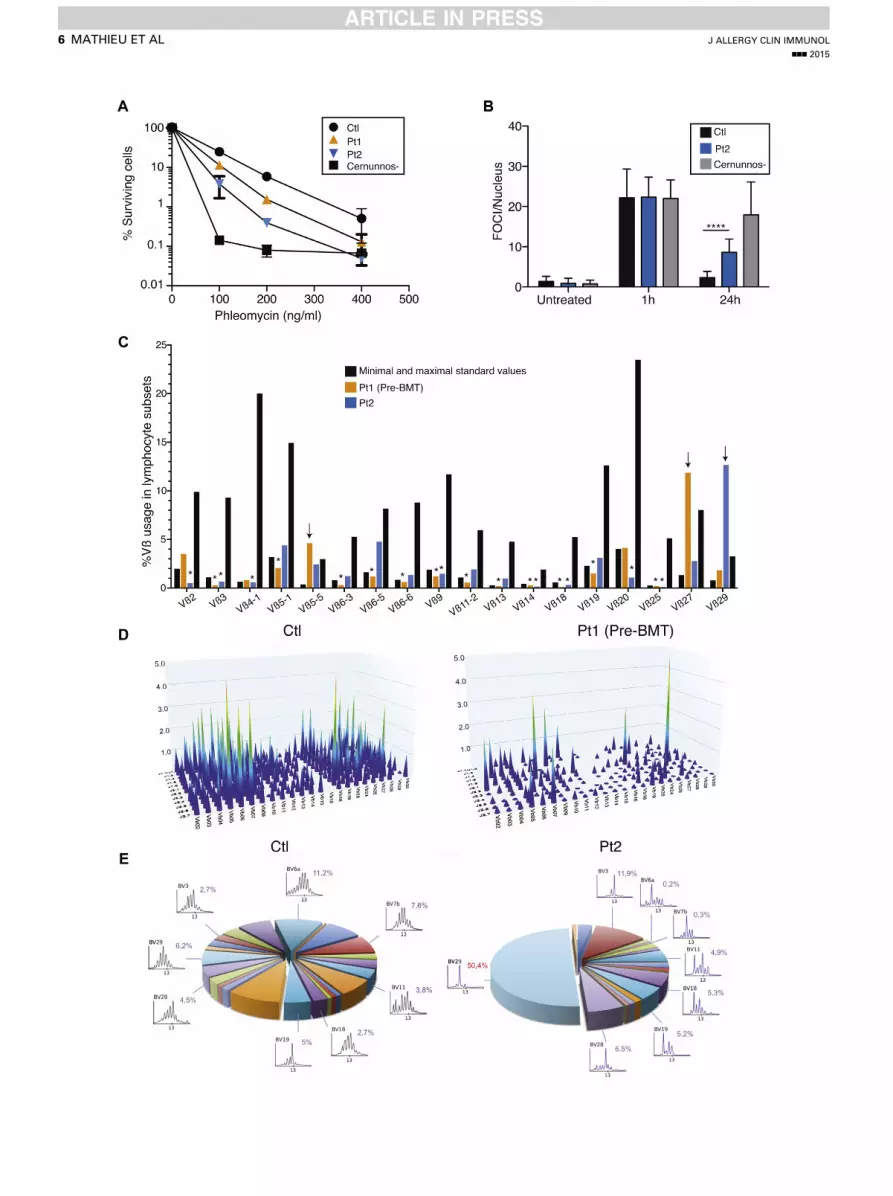
the 2 previously described patients with PRKDC mutationsdemonstrated a complete lack of T and B cells.22,23 By contrast,the 2 patients reported here presented with circulating T and Bcells. Therefore we investigated the T-cell Vb repertoire in bothpatients using flow cytometry (18 Vb families with anomaliesin Pt1 and Pt2; Fig 2, C) and quantitative PCR (276 combinations

TABLE I. Clinicobiological assessment of Pt1 and Pt2
Patient 1 (before BMT) Patient 2
6 y
Normal
values/mL (%) 8 y 10 y 11 y
Normal
values/mL (%) 14 y 15 y
Normal
values/mL 18 y 20 y
Normal
values/mL
Lymphocyte count/mL (%) 7324 (21) 2100-6450 (33-62) 774 (11) 1264 (18) 668 (13) 1440-5340 (29-56) 699 (15) 467 (12) 1400-3300 890 (32) 637 (14) 1000-4000
T-cell population/mL (%)
CD31 cells 3589 (49) 1350-3900 (56-77) 387 (50) 670 (53) 334 (50) 1060-3370 (59-79) 433 (62) 289 (62) 1000-2200 623 (70) 376 (59) 700-2100
CD31CD41 cells 1245 (17) 740-2450 (27-49) 186 (24) 265 (21) 154 (23) 420-1930 (29-50) 269 (38) 165 (35) 530-1300 380 (42) 278 (44) 530-1300
CD31CD81 cells 1978 (27) 500-1610 (19-34) 194 (25) 392 (31) 180 (27) 410-138 (20-34) 91 (13) 90 (19) 390-920 168 (19) 49 (7) 330-920
Naive CD4 T cells
(CD41CD45RA1)
50 (4) 100-800 (66-77) 2 (1) 5 (2) 3 (2) 100-800 (55-67) 0 0 23-770 0 4 (1) 23-770
Memory CD4 T cells
(CD41CD45RO1)
1145 (92) 150-850 (NA) 171 (92) 252 (95) 137 (89) 150-850 269 (62) 182 (63) 240-700 423 (68) 263 (70) 240-700
Double-negative
CD31CD42CD82
cells
1 (0.2) 9-78 (0.3-1.8) 17 (2) 14 (3) 9-78
(0.3-1.8)
19 (3) 56 (9) 7-74
(0.3-1.8)
Regulatory CD31
CD251CD1272 cells
9 (3.4) 7 (4.8) 44-60 (4-10)
TCRab cells 311 (93.1) 150-850 (80-98)
TCRgd cells 22 (6.5) 20-350 (2-14)
Natural killer (CD161CD561
CD32) cells/mL (%)
3442 (47) 150-800 (6-19) 364 (47) 569 (45) 307 (46) 145-600 (6-21) 231 (33) 149 (32) 70-480 196 (22) 229 (36) 90-600
Dendritic cells 0.73 0.624-0.853 0.75 0.624-0.853
Monocytes
Classical monocytes
(CD141CD162)
3.64 472-780 1.44 472-780
Inflammatory
monocytes
(CD141CD161)
828.61 18-44 187.19 18-44
Nonclassical
monocytes
(CD142CD161)
88.19 15-39 35.08 15-39
B cells/mL (%)
CD191 147 (2) 370-1570 (15-34) 8 (1) 13 (1) 7 (1) 230-1130 (10-27) 0 0 110-570
Immunoglobulin (g/L)
IgG 14.6 14.1 6.55-12.17 22 6.55-12.17
IgG18.8 11.5 4.23-10.6
IgG2<0.02 0.2 0.76-3.55
IgG31.159 1.131 0.17-1.73
IgG4<0.003 <0.003 0.016-1.15
IgA <0.06 <0.06 0.51-1.63 <0.06 0.75-2.38
IgM 1.29 1.44 0.57-1.62 0.88 0.71-2.02
IgE <4.88 310-6 <366 310-6 <4.88 310-6 <366 310-6
NA, Not applicable; TCR, T-cell receptor.
J ALLERGY CLIN IMMUNOL
VOLUME nnn, NUMBER nn
MATHIEU ET AL 5
Vb-J, IMGT nomenclature, in Pt1; Fig 2, D) at different timepoints. Molecular PCR analysis of TRBV-J combinationsrevealed a poorly diversified peripheral T-cell repertoire inPt1 (Fig 2, D), together with commonly overrepresentedrearrangements, such as TRB V27-J2.3, V19-J2.1, V07-J2.1,V10-J1.4, V07-J2.7, V02-J2.2, V03-J1.3, and V05-J2.7. In Pt2Vb291 T cells were clonally expanded, whereas most other Vbfamilies in both patients were underrepresented. These data areconsistent with limited V(D)J recombination in both patients.We used Immunoscope technology to analyze the T-cellrepertoire of Pt2 (Fig 2, E). The latter experiment showed a majoroligoclonal expansion of Vb29 T cells.
Patients with PRKDC mutations have increased
BAFF levels and numbers of inflammatory
monocytes and memory T cells expressing TH1 and
TH2, but not TH17, cytokines on activationLimited data are available on the distribution and function of
residual T cells in patients with CID with autoimmunity
and granuloma. TH1 cytokines, TH17 cytokines, or both are sup-posed to promote granuloma formation.24 In both patients we firstexplored the expression of and response to cytokines in the blood.The expression of 6 interferon-stimulated genes (IFI27, IFI44L,IFIT1, ISG15, RSAD2, and SIGLEC1) was strongly increased inboth patients, thus indicating potent in vivo interferon induction(Fig 3, A). In addition, the expression of BAFF, TNF-a, andIFN-g mRNA in both patients was upregulated in whole bloodcells, similar to that seen in patients with IFIH1 mutations, arecently identified cause of interferon-driven autoimmunity (Fig3, B).25 In contrast, IL-17A and IL-6 transcripts remained withinthe normal range (see Fig E2,A, in this article’s Online Repositoryat www.jacionline.org). Consistent with these quantitative PCRdata, high BAFF and TNF-a levels were detected in the sera ofPt1 and Pt2 compared with those in control subjects (Fig 3, C).After BMT, BAFF levels decreased in Pt1 to those of a patientwith a hypomorphic mutation in Artemis (see Fig E2, B). IFN-glevels were normal in the patient’s sera. Type I interferon activity,as measured by using a viral cytopathic assay, was also found tobe normal in the serum of Pt2 (data not shown).
J ALLERGY CLIN IMMUNOL
nnn 2015
6 MATHIEU ET AL

=
FIG 3. Cytokine expression in whole blood and on activation in T cells. A and B, RT-PCR of a panel of 6
interferon-stimulated genes (mean 6 SD; Fig 3, A) and RT-PCR of a panel of 3 cytokines (Fig 3, B) in whole
blood. C, BAFF, IFN-g, and TNF-a measurements in sera of control subjects and Pt1 and Pt2. D-H, Cytokine
expression in both CD4 and CD8 positive and CD45RA negative T cells as measured by using fluorescence-
activated cell sorting in pediatric or adult control subjects (pCtl or aCtl, respectively) and Pt1 and Pt2.
J ALLERGY CLIN IMMUNOL
VOLUME nnn, NUMBER nn
MATHIEU ET AL 7
Next, we analyzed the frequency of a wide range of immunecell subsets in whole blood using flow cytometry and assessedintracellular expression of several cytokines in lymphocyteson stimulation with phorbol 12-myristate 13-acetate (PMA)/ionomycin (Fig 3, D-H, and Table I). The percentage of NK anddendritic cells appeared normal in both patients. An increasednumber of inflammatory CD141CD161 monocytes was detectedin patients compared with age-matched control subjects. CD41
and CD81 naive T-cell counts were dramatically reduced inpatients, which is in line with a low thymic output. Interestingly,
FIG 2. DSB repair defect and T-cell receptor oligocl
fibroblasts from a control subject, a Cernunnos-defic
foci (FOCI)/nucleus after irradiation in fibroblasts from
subject. ****P < .0001. C, Quantitative analysis of Vb d
(asterisks) or overrepresentation (arrows). D, Three-dimin Pt1. E, Immunoscope of Vb repertoire for the contro
most CD41CD45RA1T cells, which are routinely referred to asnaive T cells, were actually memory T cells with no CCR7expression, defining the T effector memory with RA1 (TEMRA)subset (data not shown). Similarly, patients’ CD41CD391
regulatory T cells did not express CD45RA, suggesting adefect in the thymic development of natural regulatory T cells(see Fig E2, C). On stimulation with PMA and ionomycin(5 hours’ treatment), the production of TH1-related (IFN-gand TNF-a), TH2-related (IL-4 and IL-13), or TH17-related(IL-17A) cytokines by blood T-cell subsets was measured by
onal repertoire. A, Clonogenic survival assays in
ient subject, Pt1, and Pt2. B, Numbers of g-53BP1
a control subject, Pt2, and a Cernunnos-deficient
istribution in CD31 T cells. Vb underrepresentation
ensional graph of immune combinatorial diversity
l subject and Pt2.
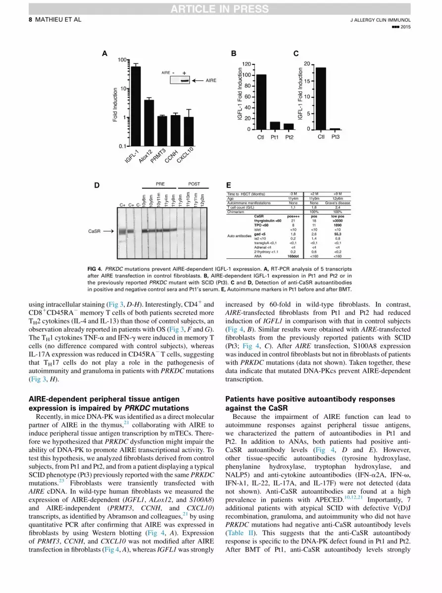
FIG 4. PRKDC mutations prevent AIRE-dependent IGFL-1 expression. A, RT-PCR analysis of 5 transcripts
after AIRE transfection in control fibroblasts. B, AIRE-dependent IGFL-1 expression in Pt1 and Pt2 or in
the previously reported PRKDC mutant with SCID (Pt3). C and D, Detection of anti-CaSR autoantibodies
in positive and negative control sera and Pt1’s serum. E, Autoimmune markers in Pt1 before and after BMT.
J ALLERGY CLIN IMMUNOL
nnn 2015
8 MATHIEU ET AL
using intracellular staining (Fig 3,D-H). Interestingly, CD41 andCD81CD45RA2 memory T cells of both patients secreted moreTH2 cytokines (IL-4 and IL-13) than those of control subjects, anobservation already reported in patients with OS (Fig 3, F andG).The TH1 cytokines TNF-a and IFN-g were induced in memory Tcells (no difference compared with control subjects), whereasIL-17A expression was reduced in CD45RA2T cells, suggestingthat TH17 cells do not play a role in the pathogenesis ofautoimmunity and granuloma in patients with PRKDCmutations(Fig 3, H).
AIRE-dependent peripheral tissue antigen
expression is impaired by PRKDC mutationsRecently, in mice DNA-PKwas identified as a direct molecular
partner of AIRE in the thymus,21 collaborating with AIRE toinduce peripheral tissue antigen transcription by mTECs. There-fore we hypothesized that PRKDC dysfunction might impair theability of DNA-PK to promote AIRE transcriptional activity. Totest this hypothesis, we analyzed fibroblasts derived from controlsubjects, from Pt1 and Pt2, and from a patient displaying a typicalSCID phenotype (Pt3) previously reported with the same PRKDCmutations.23 Fibroblasts were transiently transfected withAIRE cDNA. In wild-type human fibroblasts we measured theexpression of AIRE-dependent (IGFL1, ALox12, and S100A8)and AIRE-independent (PRMT3, CCNH, and CXCL10)transcripts, as identified by Abramson and colleagues,21 by usingquantitative PCR after confirming that AIRE was expressed infibroblasts by using Western blotting (Fig 4, A). Expressionof PRMT3, CCNH, and CXCL10 was not modified after AIREtransfection in fibroblasts (Fig 4, A), whereas IGFL1was strongly
increased by 60-fold in wild-type fibroblasts. In contrast,AIRE-transfected fibroblasts from Pt1 and Pt2 had reducedinduction of IGFL1 in comparison with that in control subjects(Fig 4, B). Similar results were obtained with AIRE-transfectedfibroblasts from the previously reported patients with SCID(Pt3; Fig 4, C). After AIRE transfection, S100A8 expressionwas induced in control fibroblasts but not in fibroblasts of patientswith PRKDC mutations (data not shown). Taken together, thesedata indicate that mutated DNA-PKcs prevent AIRE-dependenttranscription.
Patients have positive autoantibody responses
against the CaSRBecause the impairment of AIRE function can lead to
autoimmune responses against peripheral tissue antigens,we characterized the pattern of autoantibodies in Pt1 andPt2. In addition to ANAs, both patients had positive anti-CaSR autoantibody levels (Fig 4, D and E). However,other tissue-specific autoantibodies (tyrosine hydroxylase,phenylanine hydroxylase, tryptophan hydroxylase, andNALP5) and anti-cytokine autoantibodies (IFN-a2A, IFN-v,IFN-l1, IL-22, IL-17A, and IL-17F) were not detected (datanot shown). Anti-CaSR autoantibodies are found at a highprevalence in patients with APECED.10,12,21 Importantly, 7additional patients with atypical SCID with defective V(D)Jrecombination, granuloma, and autoimmunity who did not havePRKDC mutations had negative anti-CaSR autoantibody levels(Table II). This suggests that the anti-CaSR autoantibodyresponse is specific to the DNA-PK defect found in Pt1 and Pt2.After BMT of Pt1, anti-CaSR autoantibody levels strongly

TABLE II. Anti-CaSR assessment in patients with V(D)J recombination deficiency
Patient Mutation Clinical history
Serum related
to HSCT
Age at time
of sample
CaSR
antibody
Pt1 PRKDC p.Leu3062Arg; p.Leu3062Arg Arthritis, skin granuloma, recurrent infections Before Pos (46.4)
After Pos (3.26)
Pt2 PRKDC p.Leu3062Arg; p.Leu3062Arg Arthritis, spleen granuloma, recurrent infections Before Pos (28.1)
Artemis DCLRE1C p.Leu70del; p.0 (del exons 1–4) No severe infection, cutaneous granuloma Before <6 y Neg
RAG case 1 RAG1 p.Arg474Cys; p.Lys983AsnfsX9 Autoimmune cytopenia, severe infections Before 2 y NegAfter >29 mo Neg
RAG case 2 RAG1 p.Arg474Cys; p.Lys983Asn Autoimmune cytopenia, severe infections Before >8 y Neg
RAG case 3 RAG1 p.Arg841Gln; p.Phe974Leu Idiopathic T-cell lymphopenia, sister with
severe autoimmunity
Before <3 mo Neg
RAG case 4 RAG1 p.His612Arg; pHis612Arg Evans syndrome, skin granuloma, splenomegaly,
severe infections
After 10 y Neg
RAG case 5 RAG1 p.Arg314Trp; p.Arg507Trp/Arg737His Skin granuloma, EBV lymphoma, no severe
infections
Before <6 y Neg
RAG case 6 RAG2 p.Phe62Le; p.Phe62Leu Autoantibodies, T-cell lymphopenia, severe
infections
Before 29 y Neg
J ALLERGY CLIN IMMUNOL
VOLUME nnn, NUMBER nn
MATHIEU ET AL 9
decreased, although immunoblotting demonstrated slightpersistent positivity (Fig 4, D), and Hashimoto thyroiditisoccurred 9 months after BMT with the appearance of donorT and B cells (Fig 4, E).
DISCUSSIONExtending the phenotype of patients with PRKDC mutations,
we report here the cases of 2 unrelated patients withmanifestations of CID with immunodeficiency, granuloma, andautoimmunity caused by a homozygous p.Leu3062Arg mutationin PRKDC. Recurrent infections were documented in both cases,but their clinical history did not reveal opportunistic infections.Inflammatory manifestations were prominent in Pt1, who wasinitially given a diagnosis of ANA-positive oligoarticular juvenileidiopathic arthritis. He subsequently had skin granuloma and lunginvolvement, which was considered indicative of sarcoidosis. Pt2was given a diagnosis of a CID at the age of 9 years. There was noobvious neurological involvement in either patient. Pt2 had 2healthy children.
Both patients carried the same homozygous pathogenicp.Leu3062Arg variant as the first patient published with amutation in PRKDC.23 All 3 patients shared a common Turkishancestry, suggesting a founder effect for these variants. The first2 patients reported with PRKDC mutations presented with atypical SCID phenotype, including recurrent candidiasis andlower pulmonary infections. Of note, 1 sibling of Pt1 and 4siblings of Pt2 died early in life from recurrent infections. Wedid not have access to DNA for these subjects, but we speculatethat their deaths were related to the same primary immunodefi-ciency being responsible for typical SCID. The range of clinicalphenotypes strongly suggests that additional factors contributeto the overall severity of the disease. Stochastic events or othermodifying genes might affect the overall manifestation of thisdeficiency. This is reminiscent of the situation already describedfor T2B2 SCID and OS within the same family harboring thesame RAG1 mutation.26
The hallmark of classical OS is an expansion of autoreactive Tcells with an HLA-DR1CD45RO1 phenotype and an oligoclonalT-cell repertoire.4 In patients with OS, the response of activated Tcells is skewed toward a TH2 type and is associated with increased
secretion of IL-4 and IL-5, which is responsible for an increasedproduction of IgE and eosinophilia, respectively.27 Here, in the 2patients with PRKDCmutations, production of TH2 cytokines wasalso slightly increased in memory T cells on stimulation. TH1 andTH17 cells are recognized to play pivotal roles in autoimmunityand granuloma formation in patients with various inflammatoryconditions, such as Crohn disease, juvenile idiopathic arthritis,and sarcoidosis.28-30 Both protein and mRNA levels of the TH1cytokines IFN-g and TNF-a were increased in serum and wholeblood cell transcripts as well as on stimulation of memory T cellsisolated from both patients with CID (similar to levels in controlsubjects). However, on activation, TH17 cytokine levels werereduced in memory T cells. We hypothesize that TH1 and TH2cytokines can drive immunopathology in patients with CID withgranuloma and autoimmunity. Unexpectedly, however, neitherIFN-g nor type-I interferon activitywasmeasureable in Pt2’s serum,whereas whole blood cells displayed a strong interferon signature.This signature might be acquired after intratissular activation ofT cells or might be secondary to chronic activation of innatepathways in response to environmental antigens.31 Furthermore,BAFF levels were increased in both DNA-PKcs–deficientand ARTEMIS-deficient patients, as reported in patients withhypomorphic RAG,9 and might promote autoreactive B-cellsurvival. Interestingly, BAFF is produced by activated monocytesand also promotes TH1-associated inflammation in mice. BAFFand TH1 cytokines might synergize in patients with CID and leadto autoimmunity.32,33
In the context of a recombination defect, there are severaloverlapping mechanisms, as detailed in the introduction,which might lead to autoimmunity. ANA1 arthritis has beenidentified in other patients with immunodeficiency34,35 andmight be related to the T- and B-cell developmental defect,impaired BCR editing, repertoire disruption, and immune cellcytokine overproduction (eg, TNF-a and BAFF in Pt1 andPt2). We think that in the context of PRKDC mutation, oneadditional mechanism should be considered: direct impairmentof AIRE-dependent tissue-specific antigen expression couldpromote APECED-like manifestations. The relevance ofinteractions between AIRE, which plays a critical role in thenegative selection of autoreactive T cells,10,11 and DNA-PKwas previously tested by using NOD.CB17-Prkdcscid mice, which

J ALLERGY CLIN IMMUNOL
nnn 2015
10 MATHIEU ET AL
have an inactive form of DNA-PK.21 These mice lack mature thy-mocytes and therefore mTECs, which depend on thymocytecrosstalk for maturation.23-26 Mice were reconstituted withwild-type bone marrow to generate chimeric animals with wild-type lymphocytes and DNA-PK mutant mTECs to circumventthis issue. Reconstituted NOD.CB17-Prkdcscid mice, but not con-trol mice, demonstrated reduced transcription ofAIRE-dependentgenes in mTECs, despite similar levels of AIRE expression.The mice also had late-onset autoimmunity characterized byautoantibody production. Overall, the results indicated thatDNA-PK expression in mTECs is crucial to AIRE function.21
In the context of PRKDC mutations, we have shown that AIREtranscriptional activity is directly impaired, leading to a decreasein (eg, IGFL-1), or absence of (eg, S100A8) expression ofAIRE-dependent peripheral tissue antigens. These findings areassociated with in vivo production of anti-CaSR autoantibodies,which were absent in 5 patients with atypical SCID withhypomorphic RAG mutations and 1 with DLCRE1C (ARTEMIS)mutation with granuloma and autoimmunity. Moreover, Pt1 hadHashimoto thyroiditis 9 months after transplantation, which is arare event in the course of BMT but can be sometimes observed.Positivity of glutamic acid decarboxylase (GAD) antibodies isalso uncommon. These organ-specific autoantibodies suggeststhatmTECs,which are not replaced onBMT, promoted autoimmu-nity mediated by newly emigrant naive T cells. Anti-CaSRautoantibody levels were also slightly positive after BMT in thesame patient, which might also reflect the persistence of centraltolerance defect. Two other patients have been reported withPRKDC mutations and an SCID phenotype with a completeabsence of T and B cells but no autoimmune manifestations. Weacknowledge also that the organ-specific autoimmunity observedin our patients could be unrelated to the PRKDC mutation. Onecan also speculate that few circulating T cells, B cells, or both aremandatory for the development of such autoimmune features. AfterBMTof the previously reported patientwith SCID (Pt3), the last ex-amination did not reveal autoimmune signs. This suggests that thepenetrance ofAIRE-associated autoimmunity could be incomplete.
In summary, PRKDC mutations in human subjects can mimicinflammatory disease with autoimmunity and granuloma. In thepatients with CID described here, increased cytokine expressionmight contribute to autoimmunity and granuloma. In addition,PRKDC hypomorphic mutations prevent AIRE function.Organ-specific autoimmunity might result from the defectiveinteraction of mutated DNA-PKcs and AIRE in the thymus, andanti-CaSR might serve as potential biomarker of this condition.
We thank the patients for their participation in this study. We also thank
Nadia Plantier and ImmunID Technologies for providing the 3-dimensional
immune repertoire analysis.
Key messages
d DNA-PKcs is a multifunctional protein involved in AIRE-dependent transcription in the thymus.
d PRKDC mutations can lead to a broad range of diseases,from SCIDs to milder immunodeficiency with granuloma-tous and autoimmune manifestations and positive anti-tissue autoantibody levels.
REFERENCES
1. Fischer A. Human primary immunodeficiency diseases. Immunity 2007;27:835-45.
2. Felgentreff K, Perez-Becker R, Speckmann C, Schwarz K, Kalwak K, Markelj G,
et al. Clinical and immunological manifestations of patients with atypical severe
combined immunodeficiency. Clin Immunol 2011;141:73-82.
3. Shearer WT, Dunn E, Notarangelo LD, Dvorak CC, Puck JM, Logan BR, et al.
Establishing diagnostic criteria for severe combined immunodeficiency
disease (SCID), leaky SCID, and Omenn syndrome: the Primary Immune
Deficiency Treatment Consortium experience. J Allergy Clin Immunol 2014;133:
1092-8.
4. Villa A, Notarangelo LD, Roifman CM. Omenn syndrome: inflammation in leaky
severe combined immunodeficiency. J Allergy Clin Immunol 2008;122:1082-6.
5. De Villartay JP. V(D)J recombination deficiencies. Adv ExpMed Biol 2009;650:46-58.
6. Schuetz C, Huck K, Gudowius S, Megahed M, Feyen O, Hubner B, et al.
An immunodeficiency disease with RAG mutations and granulomas. N Engl J
Med 2008;358:2030-8.
7. Grunebaum E, Bates A, Roifman CM. Omenn syndrome is associated with
mutations in DNA ligase IV. J Allergy Clin Immunol 2008;122:1219-20.
8. Ege M, Ma Y, Manfras B, Kalwak K, Lu H, Lieber MR, et al. Omenn syndrome
due to ARTEMIS mutations. Blood 2005;105:4179-86.
9. Walter JE, Rucci F, Patrizi L, Recher M, Regenass S, Paganini T, et al. Expansion
of immunoglobulin-secreting cells and defects in B cell tolerance in
Rag-dependent immunodeficiency. J Exp Med 2010;207:1541-54.
10. Anderson MS, Su MA. Aire and T cell Development. Curr Opin Immunol 2011;23:
198-206.
11. Mathis D, Benoist C. Aire. Annu Rev Immunol 2009;27:287-312.
12. Gavalas NG, Kemp EH, Krohn KJ, Brown EM, Watson PF, Weetman AP. The
calcium-sensing receptor is a target of autoantibodies in patients with autoimmune
polyendocrine syndrome type 1. J Clin Endocrinol Metab 2007;92:2107-14.
13. Irla M, Hugues S, Gill J, Nitta T, Hikosaka Y, Williams IR, et al. Autoantigen-
specific interactions with CD41 thymocytes control mature medullary thymic
epithelial cell cellularity. Immunity 2008;29:451-63.
14. Surh CD, Ernst B, Sprent J. Growth of epithelial cells in the thymic medulla is
under the control of mature T cells. J Exp Med 1992;176:611-6.
15. Poliani P, Vermi W, Facchetti F. Thymus microenvironment in human primary
immunodeficiency diseases. Curr Opin Allergy Clin Immunol 2009;9:489-95.
16. Poliani PL, Facchetti F, Ravanini M, Gennery AR, Villa A, Roifman CM, et al.
Early defects in human T-cell development severely affect distribution and
maturation of thymic stromal cells: possible implications for the pathophysiology
of Omenn syndrome. Blood 2009;114:105-8.
17. Cavadini P, Vermi W, Facchetti F, Fontana S, Nagafuchi S, Mazzolari E, et al.
AIRE deficiency in thymus of 2 patients with Omenn syndrome. J Clin Invest
2005;115:728-32.
18. Zhang S, Schlott B, G€orlach M, Grosse F. DNA-dependent protein kinase
(DNA-PK) phosphorylates nuclear DNA helicase II/RNA helicase A and hnRNP
proteins in an RNA-dependent manner. Nucleic Acids Res 2004;32:1-10.
19. Blunt T, Gell D, Fox M, Taccioli GE, Lehmann AR, Jackson SP, et al. Identification
of a nonsense mutation in the carboxyl-terminal region of DNA-dependent protein
kinase catalytic subunit in the SCID mouse. Proc Natl Acad Sci U S A 1996;93:
10285-90.
20. Gu Y, Seidl KJ, Rathbun GA, Zhu C, Manis JP, van der Stoep N, et al. Growth
retardation and leaky SCID phenotype of Ku70-deficient mice. Immunity 1997;
7:653-65.
21. Abramson J, Giraud M, Benoist C, Mathis D. Aire’s partners in the molecular
control of immunological tolerance. Cell 2010;140:123-35.
22. Woodbine L, Neal JA, Sasi NK, Shimada M, Deem K, Coleman H, et al. PRKDC
mutations in a SCID patient with profound neurological abnormalities. J Clin
Invest 2013;123:2969-80.
23. Van der Burg M, Ijspeert H, Verkaik NS, Turul T, Wiegant WW, Morotomi-Yano
K, et al. A DNA-PKcs mutation in a radiosensitive T-B- SCID patient inhibits
Artemis activation and nonhomologous end-joining. J Clin Invest 2009;119:91-8.
24. Torrado E, Cooper AM. IL-17 and Th17 cells in tuberculosis. Cytokine Growth
Factor Rev 2010;21:455-62.
25. Rice GI, del Toro Duany Y, Jenkinson EM, Forte GM, Anderson BH, Ariaudo G,
et al. Gain-of-function mutations in IFIH1 cause a spectrum of human disease
phenotypes associated with upregulated type I interferon signaling. Nat Genet
2014;46:503-9.
26. De Saint-Basile G, Le Deist F, de Villartay JP, Cerf-Bensussan N, Journet O, Brousse
N, et al. Restricted heterogeneity of T lymphocytes in combined immunodeficiency
with hypereosinophilia (Omenn’s syndrome). J Clin Invest 1991;87:1352-9.
27. Schanden�e L, Ferster A, Mascart-Lemone F, Crusiaux A, G�erard C, Marchant A,
et al. T helper type 2-like cells and therapeutic effects of interferon-gamma in

J ALLERGY CLIN IMMUNOL
VOLUME nnn, NUMBER nn
MATHIEU ET AL 11
combined immunodeficiency with hypereosinophilia (Omenn’s syndrome). Eur J
Immunol 1993;23:56-60.
28. Miossec P. Interleukin-17 and Th17 cells: from adult to juvenile arthritis–now it is
serious! Arthritis Rheum 2011;63:2168-71.
29. H€oltt€a V, Klemetti P, Sipponen T, Westerholm-Ormio M, Kociubinski G, Salo H,
et al. IL-23/IL-17 immunity as a hallmark of Crohn’s disease. Inflamm Bowel
Dis 2008;14:1175-84.
30. Ten Berge B, Paats MS, Bergen IM, van den Blink B, Hoogsteden HC, Lambrecht
BN, et al. Increased IL-17A expression in granulomas and in circulating memory
T cells in sarcoidosis. Rheumatology (Oxford) 2012;51:37-46.
31. Park J, Munagala I, Xu H, Blankenship D, Maffucci P, Chaussabel D, et al.
Interferon signature in the blood in inflammatory common variable immune
deficiency. PLoS One 2013;8:e74893.
32. Scapini P, Hu Y, Chu CL, Migone TS, Defranco AL, Cassatella MA, et al.
Myeloid cells, BAFF, and IFN-gamma establish an inflammatory loop that
exacerbates autoimmunity in Lyn-deficient mice. J Exp Med 2010;207:1757-73.
33. Sutherland AP, Ng LG, Fletcher CA, Shum B, Newton RA, Grey ST, et al. BAFF
augments certain Th1-associated inflammatory responses. J Immunol 2005;174:
5537-44.
34. Romberg N, Chamberlain N, Saadoun D, Gentile M, Kinnunen T, Ng YS, et al.
CVID-associated TACI mutations affect autoreactive B cell selection and
activation. J Clin Invest 2013;123:4283-93.
35. Andr�es E, Limbach FX, Kurtz JE, Kurtz-Illig V, Schaeverbeke T, Pflumio F, et al.
Primary humoral immunodeficiency (late-onset common variable immuno-
deficiency) with antinuclear antibodies and selective immunoglobulin deficiency.
Am J Med 2001;111:489-91.

METHODS
Immunohistochemical analysesMorphological studies of skin and spleen biopsy specimens were
carried out on 5-mm-thick, formalin-fixed, paraffin-embedded samples with
hematoxylin and eosin staining.
Clonogenic survival assaysFibroblasts (104) from a healthy control subject, a Cernunnos-deficient
patient, or Pt1 and Pt2 were plated in duplicates on day 0 in culture medium.
Increasing doses of the radiomimetic drug phleomycin (InvivoGen, Toulouse,
France) was added, and cells were cultured for 7 days. We measured cell
numbers by using flow cytometry and analyzed the percentage of surviving
cells as the ratio of treated/untreated cells.
Analysis of 53BP1 foci after ionizing radiationEarly passaged primary fibroblasts were cultured on cover slips and
X-irradiated (5 Gy). One or 24 hours after irradiation, cells were washed with
PBS, fixed with 4% paraformaldehyde for 15 minutes, and incubated for 20
minutes with PBS/0.1 mol/L glycine. Cells were then permeabilized in 0.5%
Triton X-100 in PBS for 15 minutes. After each step, cover slips were rinsed 3
times with PBS. Thereafter, cells were incubated for 30 minutes with
PBS-BSA 1% and labeled (30 minutes) with primary antibodies (Anti-53BP1;
Novus Biological, Littleton, Colo). Then cells were washed with PBS/BSA
1% solution and incubated (30 minutes) with secondary antibodies (Alexa
Fluor 488 goat F[Ab9]2; Molecular Probes, Eugen, Ore). Slides were stained
for 5minutes with 0.1mg/mL 49-6-diamidino-2-phenylindole dihydrochloride
and mounted in FluorSave (Calbiochem, Nottingham, United Kingdom).
Slides were analyzed by using epifluorescence microscopy (Axioplan; Zeiss,
Oberkochen, Germany). Images were processed for quantification with
ImageJ software.
T-cell receptor Vb repertoire analysisT-cell repertoire diversity was measured with the Human ImmunTraCkeR
test (ImmunID Technologies), a technology based on genomic DNA
Multi-N-plex quantitative PCR technology.E1 Human T-cell receptor Vb-J
rearrangements were documented according to IMGT nomenclature
(www.imgt.org). Phenotypic analysis of the T-cell Vb repertoire was
performed on whole blood samples by using the IOTest Beta Mark kit
(Beckman Coulter, Villepinte, France) containing 24 mAbs identifying
approximately 70% of the T-cell repertoire. Whole blood cells were stained
with PECy5-conjugated CD3, PECy7-conjugated CD4, and each combination
of 3 fluorescein isothiocyanate–, phycoerythrin-, and fluorescein
isothiocyanate/phycoerythrin-conjugated anti-Vb mAbs in 8 sample tubes.
Whole blood samples were automatically lysed with the IMMUNOPREP
Reagent System (Beckman Coulter), washed, and fixed in 0.5% formaldehyde
in PBS. T cells (0.5 to 1 3 104) were acquired on a Cytomics FC500 flow
cytometer, and data were analyzed with CXP analysis software. All reagents
and instruments were purchased from Beckman Coulter. Lymphocytes were
first gated according to forward-scatter/side-scatter parameters, and then
CD31, CD41, and CD31CD42 cells were selected. The proportion of each
Vb family was compared with the minimum and maximum of each reference
value obtained from IOTest Beta Mark kit–related data to evaluate expanded
or restricted Vb families. Expansions or restrictions were defined respectively
for values greater than the maximum or less than the minimum reference
values of the corresponding family.
Immunoscope quantitative repertoireThe methods have been described elsewhere.E2 Briefly, the total RNA
Miniprep Kit (Qiagen, Courtaboeuf, France) was used, and cDNA was
synthesized with SuperScript II Reverse Transcriptase (Invitrogen by Life
Technologies, Carlsbad, Calif). PCR reactions were carried out by combining
a reverse primer and a specific fluorophore-labeled probe for the constant
region (MGB–TaqMan probe) with 1 of 24 primers covering the different
TRBV chains. The different human TRBV germline genes can be clustered
in 24 families according to their level of homology (IMGT nomenclature).
Real-time PCR reactions were subsequently carried out with a final
concentration of 400 nmol/L of each oligonucleotide primer, 200 nmol/L of
the fluorogenic probe, and FastStart Universal Probe Master Rox (Roche,
Mannheim, Germany). Thermal cycling conditions comprised TaqDNA
Polymerase activation at 958c for 10 minutes and then subjected to 40 cycles
of denaturation at 958c for 15 seconds, annealing, and extension at 608c for 1minutes. For all these reactions, real-time quantitative PCR was then
performed on an ABI-7300 system (Applied Biosystems by Life
Technologies, Carlsbad, Calif). The relative use of each TRBV family was
calculated according to the following formula:
UðBVyÞ5 +x5 24
x5 1
2ðCtðxÞ2CtðyÞÞ;
where Ct(x) is the fluorescent threshold cycle number measured for the BVy
family.
For Immunoscope profiles, products were then subjected to run-off
reactions with a nested fluorescent primer specific for the constant region
for a total of 3 cycles. The fluorescent products were separated and
analyzed with an ABI-PRISM 3730 DNA analyzer. The size and intensity
of each band were analyzed with Immunoscope software,E3 which has been
adapted to the capillary sequencer. Fluorescence intensities were plotted in
arbitrary units on the y-axis, and CDR3 lengths (in amino acids) were plotted
on the x-axis.
Measurement of whole blood cytokine expressionBlood was collected into PAXgene tubes (PreAnalytix, Hombrechtikon,
Switzerland) and, after being kept at room temperature for between 2 and 72
hours, was frozen at 2208C until extraction. Total RNA was extracted
from whole blood with a PAXgene (PreAnalytix. Hombrechtikon,
Switzerland) RNA isolation kit. RNA concentration was assessed with a
spectrophotometer (FLUOstar Omega; Labtech, Ortenberg, Germany).
Quantitative RT-PCR analysis was performed with the TaqMan Universal
PCR Master Mix (Applied Biosystems, Paisley, United Kingdom) and a
cDNA derived from 40 ng of total RNA. TaqMan probes were used for IFI27
(Hs01086370_m1), IFI44L (Hs00199115_m1), IFIT1 (Hs00356631_g1),
ISG15 (Hs00192713_m1), RSAD2 (Hs01057264_m1), SIGLEC1 (Hs00
988063_m1), IL1B (Hs01555410_m1), IL6 (Hs00985639_m1), TNFA
(Hs99999043_m1), IFNG (Hs00989291_m1), IL17A (Hs00174383_m1),
and TNFSF13 (BAFF; Hs00198106_m1). The relative abundance of each
target transcript was normalized to the expression level of HPRT1
(Hs03929096_g1) and 18S (Hs999999001_s1) and assessed with the Applied
Biosystems StepOne software (version 2.1) and DataAssist software (version
3.01). For each of the 6 probes, individual (patient and control) data were
expressed relative to a single calibrator (control C25). The relative
quantification for each transcript is equal to 22DCt (ie, the normalized fold
change relative to a control).
BAFF ELISABAFF levels were measured in serum samples with a Human BAFF/BLyS/
TNFSF13B Quantikine ELISA Kit (R&D Systems, Minneapolis, Minn),
according to the manufacturer’s protocol. The BAFF concentration was
recorded in picograms per milliliter.
TNF-a, IFN-g, and type I interferonIFN-g and TNF-a levels were measured with an ELISA kits (Life
Technologies), according to the manufacturer’s instructions. IFN-g and
TNF-a concentrations were recorded as international units per milliliter and
picograms per milliliter, respectively. IFN-a is titrated by using the cytopathic
effect inhibition assay, as previously described.E4 In this antiviral assay about
1 U/mL interferon is the quantity necessary to produce a cytopathic effect of
50%. Interferon activity is measured on bovine MDBK cells with vesicular
J ALLERGY CLIN IMMUNOL
nnn 2015
11.e1 MATHIEU ET AL

stomatitis virus.E5 Units are determined with respect to the World Health
Organization international standard for Hu-IFN-Alpha 2b (code 95/566)
provided by the National Institute for Biological Standards and Control
(Pestka 1986).
Phenotypic analysis on whole blood cellsBriefly, 0.5 to 1 3 106 whole blood cells were stained with a mixture of
fluorochrome-labeled antibodies for each panel for 20 minutes at 48C and
then washed twice in staining buffer (PBS, 2% FBS, and 1 mmol/L EDTA).
Erythrocytes were lysed at room temperature for 10 minutes in the dark with
BD Pharm Lyse buffer 1X (BD Biosciences, Le Pont de Claix, France).
Cells were resuspended in 300 mL of staining buffer in the presence of 2 mL
of 49-6-diamidino-2-phenylindole dihydrochloride (2 mg/mL) to exclude
dead cells, and all events were acquired on a FACSCanto II flow
cytometer (BD Biosciences). Results were analyzed with FlowJo software,
version 9.6.4 (TreeStar, Ashland, Ore). Monocytes were identified as
classical (CD141CD162), activated (CD141CD161), or nonclassical
(CD142CD161). CD4 T cells were identified as naive (CD31CD41
CD45RA1CCR71), central memory (TCM; CD31CD41CD45RA2CCR71),
effector memory RA1 (CD31CD41CD45RA1CCR72), or effector memory
(TEM; CD31CD41CD45RA2CCR72). CD8 T cells were identified as
naive (CD31CD42CD45RA1CCR71), central memory (TCM; CD31CD42
CD45RA2CCR71), effector memory RA1 (TEMRA; CD31CD42
CD45RA1CCR72), and CD8 effector memory (TEM; CD31CD42CD45
RA2CCR72).
Assessment of cytokine production in activated
whole blood cellsBriefly, 900 mL of heparinized whole blood was incubated at 378C in a 5%
CO2 humidified atmosphere for 5 hours in the presence or absence of PMA
(50 ng; Sigma-Aldrich, Saint Quentin Fallavier, France) and ionomycin
(1 ng/mL, Sigma-Aldrich) together with a protein transport inhibitor
(GolgiPlug, 10 mg/mL, BD Biosciences). At the end of stimulation, erythro-
cytes were lysed at room temperaturewith BDPharmLyse buffer.White blood
cells were washed in staining buffer and stained with the corresponding
surface antibodies panel. After washing in PBS, cells were fixed with
formaldehyde (Sigma-Aldrich) at 2% for 20 minutes at 48C, washed twice
in staining buffer, and stored overnight at 48C. Cells were then permeabilized
in staining buffer supplemented with 0.5% saponin and stained for 20 minutes
at 48Cwith the corresponding intracytoplasmic anti-cytokine antibodies (IL-2,
IL-4, IL-13, IL-17, IFN-g, and TNF-a). Cells were resuspended in 600 mL of
staining buffer, and all events were acquired on a flow cytometer fitted with 4
lasers (violet, blue, yellow, and red; LSRII Fortessa for functional analyses BD
biosciences). Results were analyzed with FlowJo software, version 9.6.4, and
cytokine secretion by different cell subsets defined by the gating strategy was
evaluated by creation of Boolean gates.
Fibroblast transfection and immunoblotting to
detect AIRE expressionPrimary fibroblasts were cultured from a skin biopsy sample from Pt1, Pt2,
or a control subject in Dulbecco modified Eagle medium supplemented with
10%FBS, 2mmol/Lglutamine, 10mmol/LHEPES, and40mg/mLgentamicin
(Life Technologies, Courtaboeuf, France). Fibroblasts were transfected with
either pMax-GFP (Lonza, Basel, Switzerland) vector or TrueORF gold vector
coding for Myc-DDK-tagged ORF of human AIRE transcript variant AIRE-1
(OriGene Technologies, Rockville, Md) by using the jetPEI reagent (Polyplus
Transfection, Illkirch, France). After 24 hours, fibroblasts were lysed in NP-40
lysis buffer (20 mmol/L Tris/HCl [pH 7.4], 150 mmol/L NaCl, 2 mmol/L
EDTA, and 1% NP-40 [Sigma-Aldrich]) containing protease inhibitors for 30
minutes at 48C. Supernatants were collected after 10 minutes of centrifugation
at 16,000g and 48C, and protein content was quantified with the mBCA quan-
tification kit (Thermo Fisher Scientific Biosciences, Villebon sur Yvette,
France). Protein extracts (50 mg) were analyzed by using Western blotting
with anti-AIRE antibody (Abnova, Taipei, Taiwan), anti-rabbit IgG antibody
conjugated to horseradish peroxidase (1:10,000, Sigma-Aldrich), and a BM
Chemiluminescence Blotting Substrate Kit (Roche).
Measurement of gene expression in AIRE-
transfected fibroblastsFibroblasts from patients or control subjects were cultured and transfected
as above. After 24 hours, total RNA was extracted with TRIzol reagent,
according to the manufacturer’s instructions (Life Sciences). Quality and
absence of genomic DNA contamination were assessed with a Bioanalyzer
(Agilent, Massy, France). We used a high-capacity RNA-to-cDNA kit
(Applied Biosystems) to generate cDNA for RT-PCR. PCR was carried out
with a SybrGreen-based kit (FastStart Universal SYBR Green Master; Roche,
Basel, Switzerland) on a StepOne plus instrument (Applied Biosystems).
Primers were designed by using the Roche Web site (Universal ProbeLibrary
Assay Design Center) as follows: IGFL1, forward 59-GGCTGCATCGTAGCTGTCTT-39 and reverse 59-GCATCAGGTAAGGAGTCATGG-39; Alox12,forward 59-CTGAAGATGGAGCCCAATG-39 and reverse 59-ACAGTGTTGGGGTTGGAGAG-39; PRMT3, 59-CAGGGTCGTGTTCTCTACGG-39 andreverse 59-TTTCCTTTCAAGGCTTCACCT-39; CCNH, forward 59-ATGATTACGTCTCAAAGAAATCCA-39 and reverse 59-CTACCAGGTCGTCATCAGTCC-39; CXCL10, forward 59-GAAAGCAGTTAGCAAGGAAAGGT-39 and reverse 59-GACATATACTCCATGTAGGGAAGTGA-39; and OAZ1,
forward 59-GGATAAACCCAGCGCCAC-39 and reverse 59-TACAGCAGTGGAGGGAGACC-39.
Detection of autoantibodies against CaSRThe immunoblotting method for detecting anti-CaSR autoantibodies
is described elsewhere.E6 Briefly, 20-mg samples of the Escherichia
coli–expressed CaSR extracellular domain (amino acid residues 1-603;
SWISS-PROT no. P41180) were separated by means of SDS-PAGE and
transferred to nitrocellulose membranes. Membranes were used in standard
immunoblotting experiments with patient or control sera (1:100 dilution),
anti-human IgG antibody conjugated to horseradish peroxidase (1:2000
dilution, Sigma-Aldrich), and a BM Chemiluminescence Blotting Substrate
Kit (Roche). In addition, anti-CaSR autoantibodies were detected in some
patients’ serum samples by using a CaSR immunoprecipitation assay, as
detailed previously.E7
Detection of ANAsThe ANA detection method has been described elsewhere.E8 Briefly,
detection was performed by using an indirect immunofluorescence
technique with HEp2 cells. Sera diluted at 1:160 were incubated on HEp2
cells (Bio-Rad, Marnes la Coquette, France) for 30 minutes at room
temperature. After 3 washes in PBS, pH 7.4, the slides were incubated
with a goat anti-human IgG (F[ab9]2) fluorescein isothiocyanate conjugated
to fluorescein isothiocyanate (diluted at 1:100; Bio-Rad Laboratories,
Hercules, Calif) for 30 minutes at room temperature. The slides were
examined with an Olympus fluorescence microscope (Olympus, Center
Valley, PA). A classical titration of each ANA positive at a titer of 1:160
was performed by means of serial 1-in-2 dilution until a dilution of
1:1280 was reached. A titer equal to or greater than 1:160 was interpreted
as a positive result.
Statistical analysesDifferences in numbers of foci were analyzed by using the nonparametric
Mann-Whitney U test (1-tailed; P < .05 was considered significant) in the
GraphPad Prism program (GraphPad Software, La Jolla, Calif).
Study approvalThe study was approved by the Medical Ethics Committee of Sud Est III
(Lyon, France) and carried out in accordance with the Declaration of Helsinki
principles. All patients provided written informed consent for inclusion of
their details and samples in the study.
J ALLERGY CLIN IMMUNOL
VOLUME nnn, NUMBER nn
MATHIEU ET AL 11.e2

REFERENCES
E1. ManuelM, Tredan O, Bachelot T, ClapissonG, Courtier A, Parmentier G, et al. Lym-
phopenia combined with low TCR diversity (divpenia) predicts poor overall survival
in metastatic breast cancer patients. Oncoimmunology 2012;1:432-40.
E2. Lim A, Baron V, Ferradini L, Bonneville M, Kourilsky P, Pannetier C.
Combination of MHC-peptide multimer-based T cell sorting with the
Immunoscope permits sensitive ex vivo quantitation and follow-up of human
CD8+ T cell immune responses. J Immunol Methods 2002;261:177-94.
E3. Pannetier C, Delassus S, Darche S, Saucier C, Kourilsky P. Quantitative titration
of nucleic acids by enzymatic amplification reactions run to saturation. Nucleic
Acids Res 1993;21:577-83.
E4. Rubinstein S, Familletti PC, Pestka S. Convenient assay for interferons. J Virol
1981;37:755-8.
E5. Familletti PC, Rubinstein S, Pestka S. A convenient and rapid
cytopathic effect inhibition assay for interferon. Methods Enzymol 1981;78:
387-94.
E6. Mayer A, Ploix C, Orgiazzi J, Desbos A, Moreira A, Vidal H, et al.
Calcium-sensing receptor autoantibodies are relevant markers of acquired
hypoparathyroidism. J Clin Endocrinol Metab 2004;89:4484-8.
E7. Gavalas NG, Kemp EH, Krohn KJ, Brown EM, Watson PF, Weetman AP.
The calcium-sensing receptor is a target of autoantibodies in patients with
autoimmune polyendocrine syndrome type 1. J Clin Endocrinol Metab 2007;
92:2107-14.
E8. Guyomard S, Salles G, Coudurier M, Rousset H, Coiffier B, Bienvenu J, et al.
Prevalence and pattern of antinuclear autoantibodies in 347 patients with
non-Hodgkin’s lymphoma. Br J Haematol 2003;123:90-9.
J ALLERGY CLIN IMMUNOL
nnn 2015
11.e3 MATHIEU ET AL
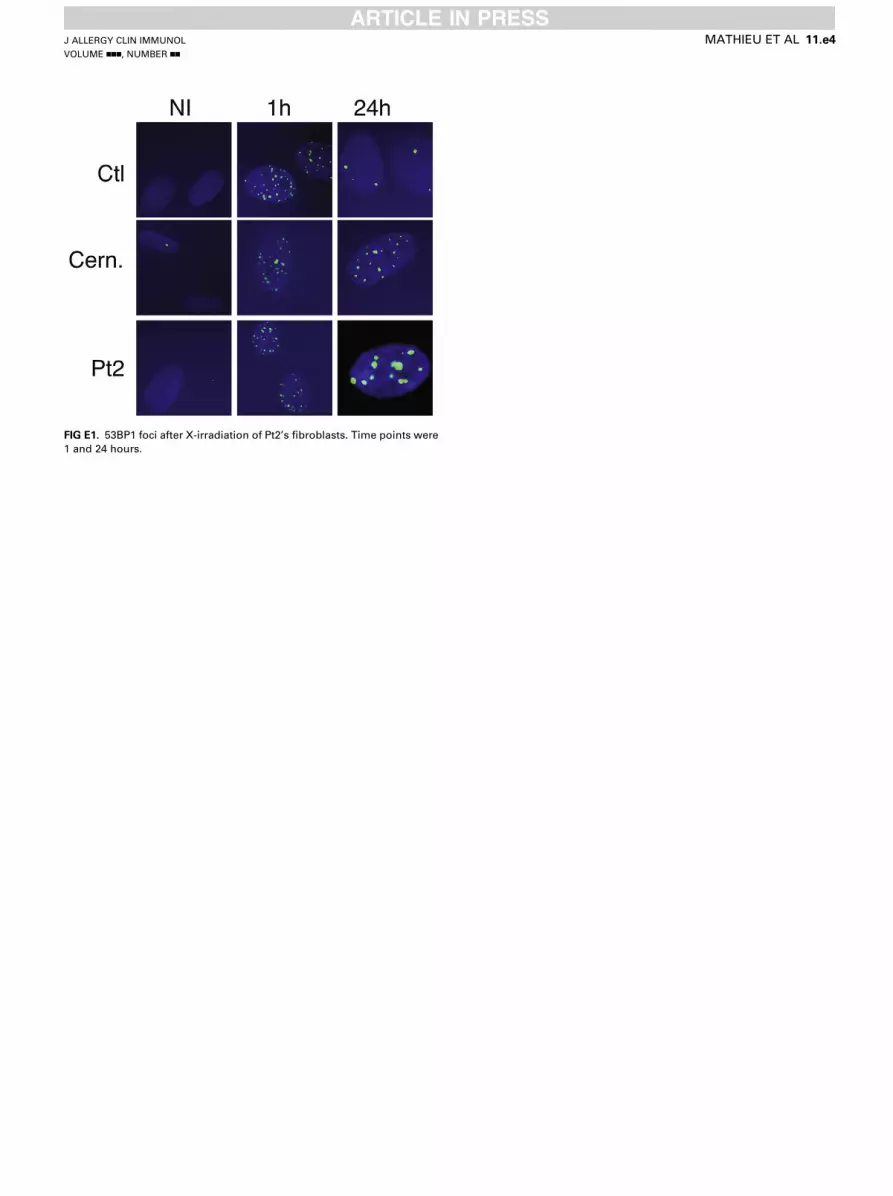
FIG E1. 53BP1 foci after X-irradiation of Pt2’s fibroblasts. Time points were
1 and 24 hours.
J ALLERGY CLIN IMMUNOL
VOLUME nnn, NUMBER nn
MATHIEU ET AL 11.e4

FIG E2. A, Quantitative RT-PCR of a panel of 3 cytokines in whole
blood measured in Pt1 (before BMT) and Pt2 and 29 control subjects
(mean 6 SD). B, BAFF measurement in sera of Pt1 (before and after BMT)
and Pt2, one Artemis-deficient patient, and 6 control subjects by using
ELISA. C, CD39 expression in regulatory T cells (CD41CD251CD127low) in
Pt1 (before BMT) and Pt2 compared with that in age-matched control
subjects. pCtl, Pediatric control subject; aCtl, adult control subject.
J ALLERGY CLIN IMMUNOL
nnn 2015
11.e5 MATHIEU ET AL
Related Documents