AD_________________ Award Number: W81XWH-08-1-0036 TITLE: CD24 as a Potential Therapeutic Target in Prostate Cancer PRINCIPAL INVESTIGATOR: Pan Zheng, M.D., Ph.D. CONTRACTING ORGANIZATION: University of Michigan Ann Arbor, MI 48109 REPORT DATE: 2009 TYPE OF REPORT: Annual PREPARED FOR: U.S. Army Medical Research and Materiel Command Fort Detrick, Maryland 21702-5012 DISTRIBUTION STATEMENT: Approved for public release; Distribution unlimited The views, opinions and/or findings contained in this report are those of the author(s) and should not be construed as an official Department of the Army position, policy or decision unless so designated by other documentation.

Welcome message from author
This document is posted to help you gain knowledge. Please leave a comment to let me know what you think about it! Share it to your friends and learn new things together.
Transcript

AD_________________ Award Number: W81XWH-08-1-0036 TITLE: CD24 as a Potential Therapeutic Target in Prostate Cancer PRINCIPAL INVESTIGATOR: Pan Zheng, M.D., Ph.D.
CONTRACTING ORGANIZATION: University of Michigan Ann Arbor, MI 48109 REPORT DATE: April 2009 TYPE OF REPORT: Annual PREPARED FOR: U.S. Army Medical Research and Materiel Command Fort Detrick, Maryland 21702-5012 DISTRIBUTION STATEMENT: Approved for public release;
Distribution unlimited The views, opinions and/or findings contained in this report are those of the author(s) and should not be construed as an official Department of the Army position, policy or decision unless so designated by other documentation.
REPORT DOCUMENTATION PAGE Form Approved
OMB No. 0704-0188 Public reporting burden for this collection of information is estimated to average 1 hour per response, including the time for reviewing instructions, searching existing data sources, gathering and maintaining the data needed, and completing and reviewing this collection of information. Send comments regarding this burden estimate or any other aspect of this collection of information, including suggestions for reducing this burden to Department of Defense, Washington Headquarters Services, Directorate for Information Operations and Reports (0704-0188), 1215 Jefferson Davis Highway, Suite 1204, Arlington, VA 22202-4302. Respondents should be aware that notwithstanding any other provision of law, no person shall be subject to any penalty for failing to comply with a collection of information if it does not display a currently valid OMB control number. PLEASE DO NOT RETURN YOUR FORM TO THE ABOVE ADDRESS. 1. REPORT DATE (DD-MM-YYYY) 0 -0 -2009
2. REPORT TYPE Annual report
3. DATES COVERED (From - To) Apr 2008 - 2009
4. TITLE AND SUBTITLE CD24 as a Potential Therapeutic Target in Prostate Cancer
5a. CONTRACT NUMBER W81XWH-08-1-0036
5b. GRANT NUMBER
5c. PROGRAM ELEMENT NUMBER
6. AUTHOR(S) Pan Zheng, MD, PhD ([email protected])
5d. PROJECT NUMBER
5e. TASK NUMBER
5f. WORK UNIT NUMBER
7. PERFORMING ORGANIZATION NAME(S) AND ADDRESS(ES)
8. PERFORMING ORGANIZATION REPORT NUMBER
University of Michigan BSRB 1810 109 Zina Pitcher Pl Ann Arbor, MI 48109
9. SPONSORING / MONITORING AGENCY NAME(S) AND ADDRESS(ES) 10. SPONSOR/MONITOR’S ACRONYM(S) U.S. Army Medical Research and Materiel Command
Fort Detrick, 11. SPONSOR/MONITOR’S REPORT Maryland 21702-5012 NUMBER(S) 12. DISTRIBUTION / AVAILABILITY STATEMENT Approved for public release; Distribution unlimited
13. SUPPLEMENTARY NOTES
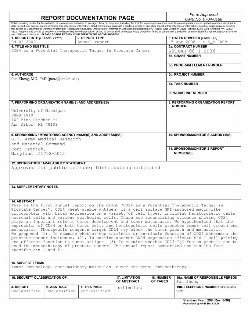
14. ABSTRACT This is the first annual report on the grant “CD24 as a Potential Therapeutic Target in Prostate Cancer”. CD24 (heat-stable antigen) is a cell surface GPI-anchored mucin-like glycoprotein with broad expression on a variety of cell types, including hematopoietic cells, neuronal cells and various epithelial cells. There are accumulating evidence showing CD24 plays an important role in tumor development and tumor metastasis. We hypothesized that the expression of CD24 on both tumor cells and hematopoietic cells promotes tumor cell growth and metastasis. Therapeutic reagents target CD24 may block the tumor growth and metastasis. We proposed (1). To examine whether the intrinsic or extrinsic function of CD24 determine the prostate cancer incidence. (2). To examine whether CD24 expression affects the T cell priming and effector function to tumor antigen. (3) To examine whether CD24-IgG fusion protein can be used in immunotherapy of prostate cancer. The annual report summarized the results from specific aim 1 and 2.
15. SUBJECT TERMS Tumor immunology, costimulatory molecules, tumor antigens, immunotherapy.
16. SECURITY CLASSIFICATION OF:
17. LIMITATION OF ABSTRACT
18. NUMBER OF PAGES
19a. NAME OF RESPONSIBLE PERSON Pan Zheng
a. REPORT Unclassified
b. ABSTRACT Unclassified
c. THIS PAGE Unclassified
unlimited
19b. TELEPHONE NUMBER (include area code)
Standard Form 298 (Rev. 8-98) Prescribed by ANSI Std. Z39.18
panz
Text Box
28
Annual Report Principal Investigator: Pan Zheng, MD, PhD
Table of Contents Introduction…………………………………………………………….…………......4 Body…………………………………………………………………………………….5-7 Key Research Accomplishments………………………………………….………8 Reportable Outcomes……………………………………………………………….9 Conclusions…………………………………………………………………………..10 References……………………………………………………………………………11 Appendices……………………………………………………………………………
Annual Report Principal Investigator: Pan Zheng, MD, PhD
4
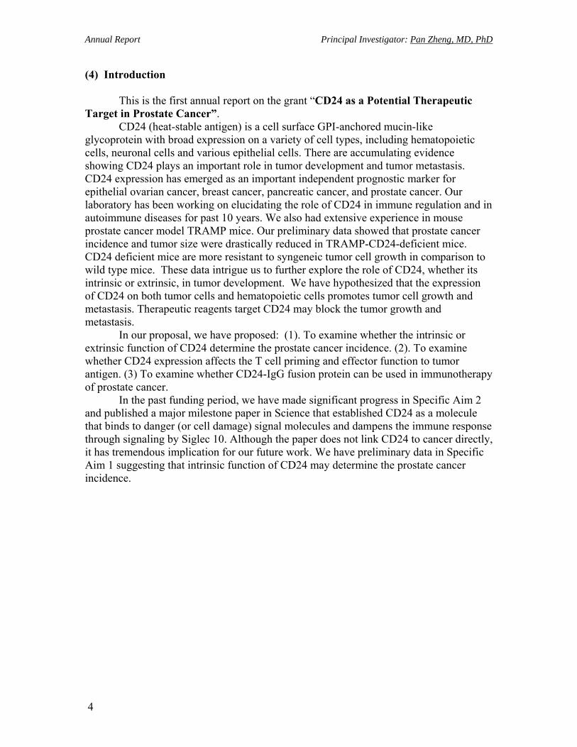
(4) Introduction This is the first annual report on the grant “CD24 as a Potential Therapeutic Target in Prostate Cancer”. CD24 (heat-stable antigen) is a cell surface GPI-anchored mucin-like glycoprotein with broad expression on a variety of cell types, including hematopoietic cells, neuronal cells and various epithelial cells. There are accumulating evidence showing CD24 plays an important role in tumor development and tumor metastasis. CD24 expression has emerged as an important independent prognostic marker for epithelial ovarian cancer, breast cancer, pancreatic cancer, and prostate cancer. Our laboratory has been working on elucidating the role of CD24 in immune regulation and in autoimmune diseases for past 10 years. We also had extensive experience in mouse prostate cancer model TRAMP mice. Our preliminary data showed that prostate cancer incidence and tumor size were drastically reduced in TRAMP-CD24-deficient mice. CD24 deficient mice are more resistant to syngeneic tumor cell growth in comparison to wild type mice. These data intrigue us to further explore the role of CD24, whether its intrinsic or extrinsic, in tumor development. We have hypothesized that the expression of CD24 on both tumor cells and hematopoietic cells promotes tumor cell growth and metastasis. Therapeutic reagents target CD24 may block the tumor growth and metastasis. In our proposal, we have proposed: (1). To examine whether the intrinsic or extrinsic function of CD24 determine the prostate cancer incidence. (2). To examine whether CD24 expression affects the T cell priming and effector function to tumor antigen. (3) To examine whether CD24-IgG fusion protein can be used in immunotherapy of prostate cancer. In the past funding period, we have made significant progress in Specific Aim 2 and published a major milestone paper in Science that established CD24 as a molecule that binds to danger (or cell damage) signal molecules and dampens the immune response through signaling by Siglec 10. Although the paper does not link CD24 to cancer directly, it has tremendous implication for our future work. We have preliminary data in Specific Aim 1 suggesting that intrinsic function of CD24 may determine the prostate cancer incidence.
Annual Report Principal Investigator: Pan Zheng, MD, PhD
5
(5) Body of Annual Report STATEMENT OF WORK (SOW) PC073392. CD24 as a potential therapeutic target in prostate cancer Pan Zheng Task I. To examine whether the cancer-cell intrinsic or extrinsic function of CD24 determines the prostate cancer incidence and/or progression. (Month 1-36). (In Progress).
a. To generate TRAMP mice with CD24 deficient background. (Month 1-6). (Finished).
b. To generate four different groups of bone marrow chimera mice (Month 7-18). (In Progress) 6 week-old TRAMP WT or TRAMP CD24-/- mice will receive 1000 Rad of irradiation. 5x10⁶/mouse of T-depleted bone marrow cells from either WT or CD24-/- mice will be used to reconstitute the irradiated mice. The four groups of chimera mice WT>WT TRAMP (group I), WT>CD24-/-TRAMP (group II), CD24-/->WT TRAMP (group III), CD24-/->CD24-/- TRAMP (group IV) mice will be generated. In group I mice, all cell types will have CD24 gene. In group II, the bone marrow derived cells will have CD24 gene, while all the other tissues and cells including prostate will be CD24 deficient. In group III, the bone marrow derived cells will be CD24 deficient, while other cell types will have CD24 gene. In group IV, all cell types will be CD24 deficient.
c. To monitor tumor growth in chimera mice by MRI (Month 9-36). (In progress) We have established collaboration with Dr. Brian Ross in University of Michigan to use MRI to measure the growth of prostate cancer in the mouse TRAMP model. We will measure the prostate size determine prostate tumor size and to record the progression of prostate cancer. The tumor volume will be used to calculate the log transformation of tumor volume. The tumor growth over time was analyzed using the StataR XTGEE (cross-sectional generalized estimating equations) model. A significant difference will be based on a P<0.05.
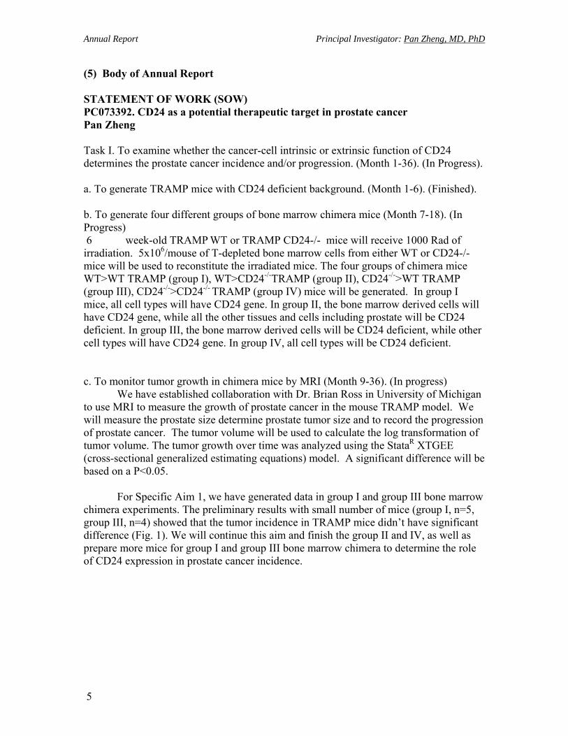
For Specific Aim 1, we have generated data in group I and group III bone marrow chimera experiments. The preliminary results with small number of mice (group I, n=5, group III, n=4) showed that the tumor incidence in TRAMP mice didn’t have significant difference (Fig. 1). We will continue this aim and finish the group II and IV, as well as prepare more mice for group I and group III bone marrow chimera to determine the role of CD24 expression in prostate cancer incidence.
Annual Report Principal Investigator: Pan Zheng, MD, PhD
6
A. B.
Fig. 1. CD24 expression in bone marrow derived cells does not significantly affect the prostate cancer incidence (Preliminary result). A. MRI images of 30 weeks old TRAMP mice. The TRAMP mice were lethally irradiated (1000 rad) at 8 weeks and reconstituted with 5x10⁶ T-depleted bone marrow cells from either WT or CD24-deficient mice. B. Prostate sizes as measured by MRI. P=0.48.
BMT to TRAMP
0
400
800
1200
1600
mm
3
CD24KB6
CD24KOB6
Annual Report Principal Investigator: Pan Zheng, MD, PhD
7
Task II. To examine the role of CD24 in the priming and effector function of cancer-reactive T cells (Month 1-36). (In Progress). a. To breed the TCR-I transgenic mice with CD45.1 congenic mice (Month 1-12). We have order the TCR-I transgenic mice from Jackson Laboratory. We expect the mice will become available in three months as they are kept as cryopreserved embryos in Jackson Laboratory. We will amplify the colony and breed with CD45.1 congenic mice to facilitate the tracking of CD45.1 T cells after they are adoptive transferred to TRAMP mice which carry the CD45.2 marker. (Currently in breeding). b. To perform the adoptive transfer experiments (Month 13-36). We will purify T cells from TCR-I transgenic mice and label the cells with CFSE. These T cells will adoptive transferred to the BM chimera mice generated in Task I in various time points correlated with tumor development. c. To determine the T cell priming and effector functions (Month 14-36). d. To examine the role of CD24 expression in tumor susceptibility to immunotherapy by CTL (Month 18-36). We were hindered by prohibition of any breeding due to a serological test showing the rooms that housing our colonies were infected by MHV in August 2008. To effectively eliminate the viral infection, all breeding was stopped until the serological test showing two quarterly negative results. However, we make significant progress in study the function and binding partners of CD24 molecule and published an important paper in Science. (Appendix 1). Task 3. Immunotherapy targeting the CD24 molecule (Month 19-36). (Not started yet). a. To generate B-cell deficient TRAMP mice (Month 1-12). We have obtained the μMT mice from Jackson Laboratories and have initiated the breeding. We expect that the mice will be available for use early 2008. b. To use CD24Ig to prevent prostate cancer in TRAMP mice (Month 7-36). c. To use CD24Ig to treat prostate cancer in TRAMP mice (Month 7-36).

Annual Report Principal Investigator: Pan Zheng, MD, PhD
8
(6) Key Research Accomplishments
• We published a major milestone paper in Science that established CD24 as a molecule that binds to danger (or cell damage) signal molecules and dampens the immune response through signaling by Siglec 10.
• We have preliminary data in Specific Aim 1 suggesting that intrinsic function of CD24 may determine the prostate cancer incidence.

Annual Report Principal Investigator: Pan Zheng, MD, PhD
9
• (7) Reportable Outcomes: Manuscripts: 1. Chen GY, Tang J, Zheng P, Liu Y. 2009. CD24 and Siglect-10 selectively repress
tissue damage induced immune responses. Science 323: 1722.
Annual Report Principal Investigator: Pan Zheng, MD, PhD
10
(8) Conclusions: CD24 is a very interesting molecule. There are accumulating evidence showing
CD24 plays an important role in tumor development and tumor metastasis. CD24 expression has emerged as an important independent prognostic marker for epithelial ovarian cancer, breast cancer, pancreatic cancer, and prostate cancer. Our laboratory has been working on the role of CD24 in immune regulation and in autoimmune diseases for past 10 years. We also had extensive experience in mouse prostate cancer model TRAMP mice. We found that if the TRAMP mice don’t have any CD24, then the mice developed much less prostate cancer. These data intrigue us to further explore the role of CD24 in prostate cancer development.
Our working hypothesis is that the expression of CD24 on both tumor cells and white blood cells promotes tumor cell growth. We can develop therapeutic reagents target CD24 to block the tumor growth.
The initial preliminary study showed that CD24 expression in bone marrow derived cells did not appear to play a significant role in prostate cancer development. We are going to perform more experiment to confirm or to dispute this preliminary result.
We have made major breakthrough in identify CD24-Siglec 10 signaling pathway in regulating damage induced immune response. The abstract on the Science paper is presented here: Patten recognition receptors, which recognize pathogens or components of injured cells (danger), trigger activation of the innate immune system. Whether and how the host distinguishes between danger- versus pathogen-associated molecular patterns remains unresolved. We report that CD24-deficient mice exhibit increased susceptibility to danger- but not pathogen-associated molecular patterns. CD24 associates with high mobility group box 1, heat shock protein 70, and heat shock protein 90; negatively regulates their stimulatory activity; and inhibits nuclear factor kB (NF-kB) activation. This occurs at least in part through CD24 association with Siglec-10 in humans or Siglec-G in mice. Our results reveal that the CD24–Siglec G pathway protects the host against a lethal response to pathological cell death and discriminates danger- versus pathogen-associated molecular patterns.

Annual Report Principal Investigator: Pan Zheng, MD, PhD
11
(9) References: None.
DOI: 10.1126/science.1168988 , 1722 (2009); 323Science
et al.Guo-Yun Chen,Damage�Induced Immune ResponsesCD24 and Siglec-10 Selectively Repress Tissue
www.sciencemag.org (this information is current as of March 30, 2009 ):The following resources related to this article are available online at
http://www.sciencemag.org/cgi/content/full/323/5922/1722version of this article at:
including high-resolution figures, can be found in the onlineUpdated information and services,
http://www.sciencemag.org/cgi/content/full/1168988/DC1 can be found at: Supporting Online Material
http://www.sciencemag.org/cgi/content/full/323/5922/1722#otherarticles, 14 of which can be accessed for free: cites 28 articlesThis article
http://www.sciencemag.org/cgi/collection/immunologyImmunology
: subject collectionsThis article appears in the following
http://www.sciencemag.org/about/permissions.dtl in whole or in part can be found at: this article
permission to reproduce of this article or about obtaining reprintsInformation about obtaining
registered trademark of AAAS. is aScience2009 by the American Association for the Advancement of Science; all rights reserved. The title
CopyrightAmerican Association for the Advancement of Science, 1200 New York Avenue NW, Washington, DC 20005. (print ISSN 0036-8075; online ISSN 1095-9203) is published weekly, except the last week in December, by theScience
on
Mar
ch 3
0, 2
009
ww
w.s
cien
cem
ag.o
rgD
ownl
oade
d fr
om
24. M. A. Schumacher, R. G. Brennan, Mol. Microbiol. 45,885 (2002).
25. Y.-J. Chen et al., Proc. Natl. Acad. Sci. U.S.A. 104, 18999(2007).
26. E. E. Zheleznova, P. N. Markham, A. A. Neyfakh,R. G. Brennan, Cell 96, 353 (1999).
27. R. E. Watkins et al., Science 292, 2329 (2001).28. S. Murakami, R. Nakashima, E. Yamashita, A. Yamaguchi,
Nature 419, 587 (2002).29. A. B. Shapiro, V. Ling, Eur. J. Biochem. 250, 122 (1997).30. R. Callaghan, G. Berridge, D. R. Ferry, C. F. Higgins,
Biochim. Biophys. Acta 1328, 109 (1997).31. I. Bosch, K. Dunussi-Joannopoulos, R. L. Wu, S. T. Furlong,
J. Croop, Biochemistry 36, 5685 (1997).
32. J. Dong, G. Yang, H. S. McHaourab, Science 308, 1023(2005).
33. M. Chami et al., J. Mol. Biol. 315, 1075 (2002).34. M. Hofacker et al., J. Biol. Chem. 282, 3951 (2007).35. G. Tombline, A. Muharemagi, L. B. White, A. E. Senior,
Biochemistry 44, 12879 (2005).36. We thank D. C. Rees, I. Wilson, R. H. Spencer,
M. B. Stowell, A. Senior, A. Frost, V. M. Unger, C. D. Stout,and P. Wright. Y. Weng was supported by a scholarshipfrom P. R. China. We thank Stanford Synchrotron RadiationLightsource, Advanced Light Source, and Advanced PhotonSource. This work was supported by grants from the Army(W81XWH-05-1-0316), NIH (GM61905, GM078914, andGM073197), the Beckman Foundation, the Skaggs
Chemical Biology Foundation, Jasper L. and Jack DentonWilson Foundation, the Southwest Cancer and TreatmentCenter, and the Norton B. Gilula Fellowship. Coordinatesand structure factors deposited to the Protein DataBank(PDB accession codes 3G5U, 3G60, and 3G61).
Supporting Online Materialwww.sciencemag.org/cgi/content/full/323/5922/1718/DC1Materials and MethodsFigs. S1 to S20Tables S1 to S3References
19 November 2008; accepted 17 February 200910.1126/science.1168750
CD24 and Siglec-10 SelectivelyRepress Tissue Damage–InducedImmune ResponsesGuo-Yun Chen,1 Jie Tang,4 Pan Zheng,1,2* Yang Liu1,3*
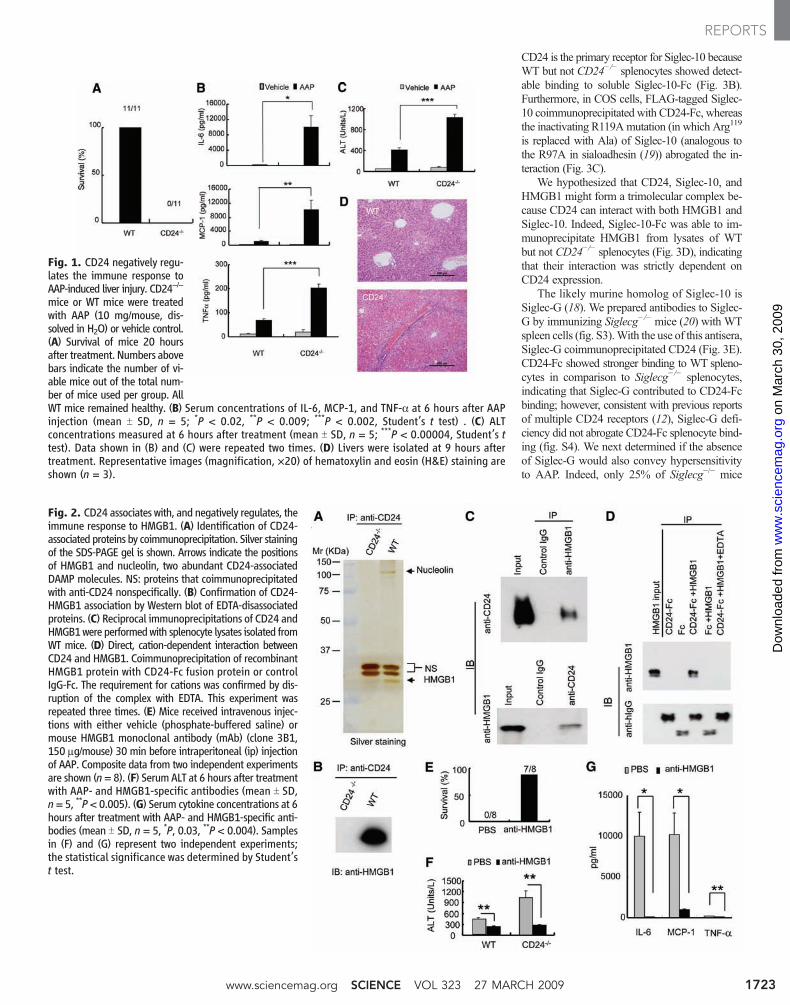
Patten recognition receptors, which recognize pathogens or components of injured cells (danger),trigger activation of the innate immune system. Whether and how the host distinguishes betweendanger- versus pathogen-associated molecular patterns remains unresolved. We report thatCD24-deficient mice exhibit increased susceptibility to danger- but not pathogen-associatedmolecular patterns. CD24 associates with high mobility group box 1, heat shock protein 70,and heat shock protein 90; negatively regulates their stimulatory activity; and inhibits nuclearfactor kB (NF-kB) activation. This occurs at least in part through CD24 association with Siglec-10in humans or Siglec-G in mice. Our results reveal that the CD24–Siglec G pathway protects thehost against a lethal response to pathological cell death and discriminates danger- versuspathogen-associated molecular patterns.
Pathogen-associated molecular patterns(PAMPs) interact with Toll-like receptors(TLRs) on innate immune cells to initiate pro-
tective immune responses (1–3). Danger-associatedmolecular patterns (DAMPs) (4), which are intra-cellular components such as high mobility groupbox 1 (HMGB1), heat shock protein 70 (HSP70),heat shock protein 90 (HSP90), and cellular RNAreleased during cellular injury, also induce TLR-dependent inflammatory responses (5–8). Whetherthe host is able to discriminate between DAMPsand PAMPs is not clear.
We used an acetaminophen (AAP)-inducedliver necrosis model (9) to identify genes that reg-ulate the innate immune response resulting fromtissue injury. A sublethal dose of AAP (10 mg/mouse), which is tolerated by wild-type (WT)mice,caused rapid death of CD24-deficient (CD24−/−)mice within 20 hours (Fig. 1A). We then testedwhether CD24 regulated the inflammatory responseto AAP-induced liver injury because CD24 is
expressed on liver oval cells and hematopoeiticcells, but not on hepatocytes (10). Indeed, wedetected a massive increase in the inflammatorycytokines interleukin-6 (IL-6),monocyte chemotacticprotein–1 (MCP-1), and tumor necrosis factor–a(TNF-a) after AAP treatment (Fig. 1B). This was ac-companied by increased amounts of serum alaninetransaminase (ALT), which is indicative of liverdamage (Fig. 1C), and liver hemorrhage and necro-sis (Fig. 1D). These observations revealed thatCD24 protects against AAP-induced hepatoxicity,most likely by regulating the inflammatory response.
CD24 is a small glycosylphosphoinositol-anchored protein that is able to provide costimu-latory signals to T cells and has been implicatedin the development of autoimmune disease (11–15).We set out to identify proteins that associate withCD24 because none of the known CD24 ligandsprovided insight into its protective effect in ourliver injurymodel.We focused on proteins whoseinteractions can be disrupted by the cation che-lator EDTA, because more than 90% of the massof CD24 is estimated to be derived from glyco-sylation (12) and because protein-polysaccharideinteractions largely depend on cations. Briefly, weimmunoprecipitated CD24 and its associated pro-teins from lysates ofmouse splenocytes. The proteinseluted by EDTAwere subjected to high-throughputmass spectrometry analysis and SDS–polyacrylamidegel electrophoresis (PAGE). HMGB1, a prototypical
DAMPmolecule that activates the immune responsefollowing tissue damage (16), was among the mostprominent proteins that we identified (Fig. 2A andtable S1). HMGB1 coimmunoprecipitated withCD24 and this interaction was specific (Fig. 2Band C). A recombinant CD24-Fc fusion proteinspecifically coimmunoprecipitated recombinantHMGB1, demonstrating that the interaction be-tween CD24 and HMGB1 was direct (Fig. 2D).
To determine whether the hypersensitivity toAAP observed inCD24−/−mice was the result ofan enhanced immune response to HMGB1, we in-jected AAP-treated mice with antibodies to HMGB1(fig. S1). In one representative experiment, block-ade of HMGB1 rescued 87.5% of the mice thatreceived AAP (Fig. 2E). Treated mice exhibiteddecreased ALT abundance, indicating reduced he-patocyte destruction (Fig. 2F). The production ofIL-6, MCP-1, and TNF-a was also greatly reduced(Fig. 2G). Thus, CD24 protects against AAP-induced lethal hepatoxicity by dampening the im-mune response against HMGB1.
HMGB1 can be divided into two domains: aninhibitory A box and a stimulatory B box (17).To determine whether CD24 inhibits HMGB1 bybinding to the inhibitory A box, we produceddeletionmutants lacking either theA box or the Bbox. CD24-Fc immunoprecipitated full-lengthHMGB1 and the box B–containing mutant, butnot the box A–containing mutant (fig. S2). Thus,inhibition of HMGB1 by CD24 does not requiredirect interaction with box A.
CD24 has no known mechanism for signaltransduction. To understand howCD24 negativelyregulatesHMGB1,we searched for a potentialCD24receptor that may transduce signals downstream ofCD24.Wewere particularly interested in sialic acid–binding immunoglobulin (Ig)-like lectins (Siglecs),which are cell surface receptors of the immunoglob-ulin superfamily that recognize sialic acid–containingproteins (18). Siglecs are primarily expressed bycells of hematopoietic origin (18).Most Siglecs areconsidered to be negative regulators of the immunesystem because they contain one or more cytosolicimmune receptor tyrosine-based inhibitory motifs(ITIMs) (18). To determine whether CD24 interactswith Siglecs, we incubated splenocytes on platescoated with the recombinant extracellular domainsof ITIM-containing Siglec-5, -7, -10 or -11. Siglec-10, but not Siglecs -5, -7, or -11, bound to CD24(Fig. 3A). Flow cytometric analysis indicated that
1Division of Immunotherapy, Department of Surgery, Uni-versity of Michigan School of Medicine, Ann Arbor, MI 48109,USA. 2Department of Pathology, University of Michigan Schoolof Medicine, Ann Arbor, MI 48109, USA. 3Department ofInternal Medicine, University of Michigan School of Medicine,Ann Arbor, MI 48109, USA. 4Institute of Biophysics, ChineseAcademy of Science, Beijing, China.
*To whom correspondence should be addressed. E-mail:[email protected] (Y.L.), [email protected] (P.Z.)
27 MARCH 2009 VOL 323 SCIENCE www.sciencemag.org1722
REPORTS
on
Mar
ch 3
0, 2
009
ww
w.s
cien
cem
ag.o
rgD
ownl
oade
d fr
om
CD24 is the primary receptor for Siglec-10 becauseWT but not CD24−/− splenocytes showed detect-able binding to soluble Siglec-10-Fc (Fig. 3B).Furthermore, in COS cells, FLAG-tagged Siglec-10 coimmunoprecipitated with CD24-Fc, whereasthe inactivating R119Amutation (in which Arg119
is replaced with Ala) of Siglec-10 (analogous tothe R97A in sialoadhesin (19)) abrogated the in-teraction (Fig. 3C).
We hypothesized that CD24, Siglec-10, andHMGB1 might form a trimolecular complex be-cause CD24 can interact with both HMGB1 andSiglec-10. Indeed, Siglec-10-Fc was able to im-munoprecipitate HMGB1 from lysates of WTbut not CD24−/− splenocytes (Fig. 3D), indicatingthat their interaction was strictly dependent onCD24 expression.
The likely murine homolog of Siglec-10 isSiglec-G (18). We prepared antibodies to Siglec-G by immunizing Siglecg−/− mice (20) with WTspleen cells (fig. S3).With the use of this antisera,Siglec-G coimmunoprecipitated CD24 (Fig. 3E).CD24-Fc showed stronger binding to WT spleno-cytes in comparison to Siglecg−/− splenocytes,indicating that Siglec-G contributed to CD24-Fcbinding; however, consistent with previous reportsof multiple CD24 receptors (12), Siglec-G defi-ciency did not abrogate CD24-Fc splenocyte bind-ing (fig. S4). We next determined if the absenceof Siglec-G would also convey hypersensitivityto AAP. Indeed, only 25% of Siglecg−/− mice
Fig. 1. CD24 negatively regu-lates the immune response toAAP-induced liver injury. CD24−/−
mice or WT mice were treatedwith AAP (10 mg/mouse, dis-solved in H2O) or vehicle control.(A) Survival of mice 20 hoursafter treatment. Numbers abovebars indicate the number of vi-able mice out of the total num-ber of mice used per group. AllWT mice remained healthy. (B) Serum concentrations of IL-6, MCP-1, and TNF-a at 6 hours after AAPinjection (mean T SD, n = 5; *P < 0.02, **P < 0.009; ***P < 0.002, Student’s t test) . (C) ALTconcentrations measured at 6 hours after treatment (mean T SD, n = 5; ***P < 0.00004, Student’s ttest). Data shown in (B) and (C) were repeated two times. (D) Livers were isolated at 9 hours aftertreatment. Representative images (magnification, ×20) of hematoxylin and eosin (H&E) staining areshown (n = 3).
Fig. 2. CD24 associates with, and negatively regulates, theimmune response to HMGB1. (A) Identification of CD24-associated proteins by coimmunoprecipitation. Silver stainingof the SDS-PAGE gel is shown. Arrows indicate the positionsof HMGB1 and nucleolin, two abundant CD24-associatedDAMP molecules. NS: proteins that coimmunoprecipitatedwith anti-CD24 nonspecifically. (B) Confirmation of CD24-HMGB1 association by Western blot of EDTA-disassociatedproteins. (C) Reciprocal immunoprecipitations of CD24 andHMGB1were performedwith splenocyte lysates isolated fromWT mice. (D) Direct, cation-dependent interaction betweenCD24 and HMGB1. Coimmunoprecipitation of recombinantHMGB1 protein with CD24-Fc fusion protein or controlIgG-Fc. The requirement for cations was confirmed by dis-ruption of the complex with EDTA. This experiment wasrepeated three times. (E) Mice received intravenous injec-tions with either vehicle (phosphate-buffered saline) ormouse HMGB1 monoclonal antibody (mAb) (clone 3B1,150 mg/mouse) 30 min before intraperitoneal (ip) injectionof AAP. Composite data from two independent experimentsare shown (n= 8). (F) Serum ALT at 6 hours after treatmentwith AAP- and HMGB1-specific antibodies (mean T SD,n= 5, **P< 0.005). (G) Serum cytokine concentrations at 6hours after treatment with AAP- and HMGB1-specific anti-bodies (mean T SD, n = 5, *P, 0.03, **P < 0.004). Samplesin (F) and (G) represent two independent experiments;the statistical significance was determined by Student’st test.
www.sciencemag.org SCIENCE VOL 323 27 MARCH 2009 1723
REPORTS
on
Mar
ch 3
0, 2
009
ww
w.s
cien
cem
ag.o
rgD
ownl
oade
d fr
om
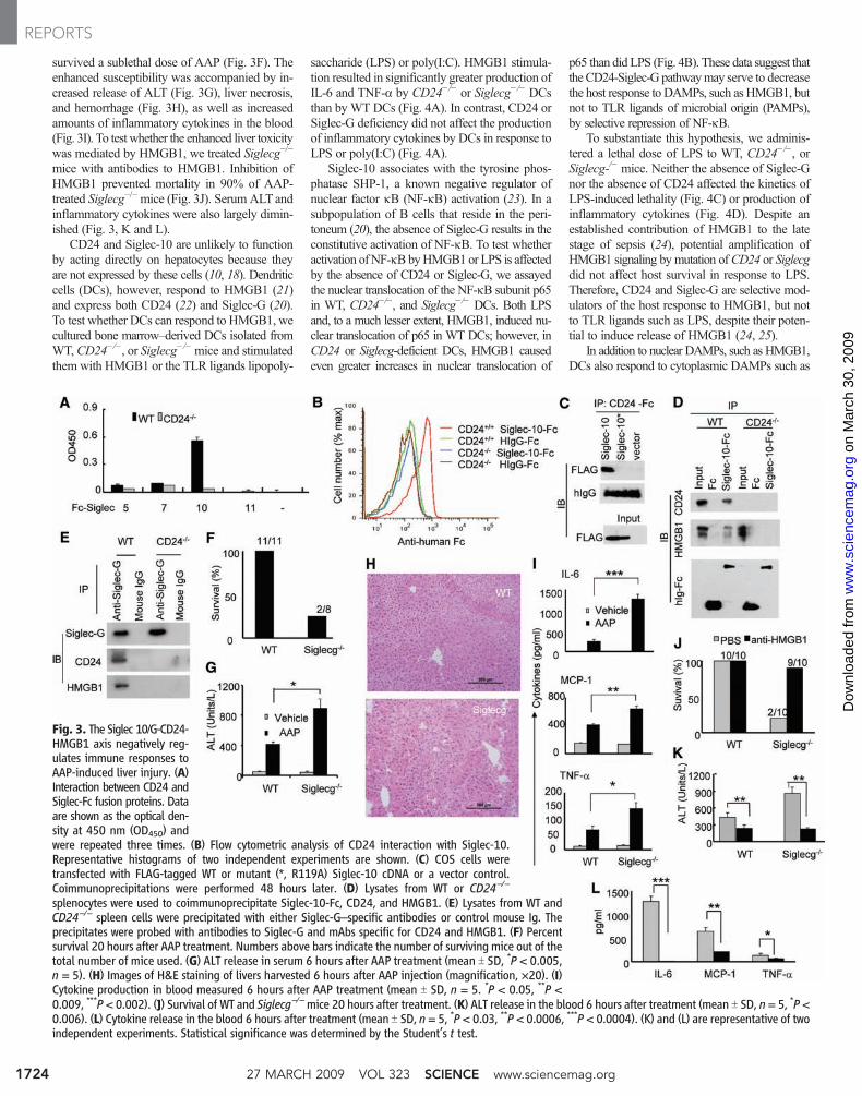
survived a sublethal dose of AAP (Fig. 3F). Theenhanced susceptibility was accompanied by in-creased release of ALT (Fig. 3G), liver necrosis,and hemorrhage (Fig. 3H), as well as increasedamounts of inflammatory cytokines in the blood(Fig. 3I). To test whether the enhanced liver toxicitywas mediated by HMGB1, we treated Siglecg−/−
mice with antibodies to HMGB1. Inhibition ofHMGB1 prevented mortality in 90% of AAP-treated Siglecg−/−mice (Fig. 3J). Serum ALTandinflammatory cytokines were also largely dimin-ished (Fig. 3, K and L).
CD24 and Siglec-10 are unlikely to functionby acting directly on hepatocytes because theyare not expressed by these cells (10, 18). Dendriticcells (DCs), however, respond to HMGB1 (21)and express both CD24 (22) and Siglec-G (20).To test whether DCs can respond to HMGB1, wecultured bone marrow–derived DCs isolated fromWT, CD24−/−, or Siglecg−/−mice and stimulatedthem with HMGB1 or the TLR ligands lipopoly-
saccharide (LPS) or poly(I:C). HMGB1 stimula-tion resulted in significantly greater production ofIL-6 and TNF-a by CD24−/− or Siglecg−/− DCsthan by WT DCs (Fig. 4A). In contrast, CD24 orSiglec-G deficiency did not affect the productionof inflammatory cytokines by DCs in response toLPS or poly(I:C) (Fig. 4A).
Siglec-10 associates with the tyrosine phos-phatase SHP-1, a known negative regulator ofnuclear factor kB (NF-kΒ) activation (23). In asubpopulation of B cells that reside in the peri-toneum (20), the absence of Siglec-G results in theconstitutive activation of NF-kΒ. To test whetheractivation ofNF-kΒ byHMGB1 or LPS is affectedby the absence of CD24 or Siglec-G, we assayedthe nuclear translocation of the NF-kΒ subunit p65in WT, CD24−/−, and Siglecg−/− DCs. Both LPSand, to a much lesser extent, HMGB1, induced nu-clear translocation of p65 in WT DCs; however, inCD24 or Siglecg-deficient DCs, HMGB1 causedeven greater increases in nuclear translocation of
p65 than did LPS (Fig. 4B). These data suggest thatthe CD24-Siglec-G pathwaymay serve to decreasethe host response to DAMPs, such as HMGB1, butnot to TLR ligands of microbial origin (PAMPs),by selective repression of NF-kΒ.
To substantiate this hypothesis, we adminis-tered a lethal dose of LPS to WT, CD24−/−, orSiglecg-/− mice. Neither the absence of Siglec-Gnor the absence of CD24 affected the kinetics ofLPS-induced lethality (Fig. 4C) or production ofinflammatory cytokines (Fig. 4D). Despite anestablished contribution of HMGB1 to the latestage of sepsis (24), potential amplification ofHMGB1 signaling by mutation ofCD24 or Siglecgdid not affect host survival in response to LPS.Therefore, CD24 and Siglec-G are selective mod-ulators of the host response to HMGB1, but notto TLR ligands such as LPS, despite their poten-tial to induce release of HMGB1 (24, 25).
In addition to nuclear DAMPs, such as HMGB1,DCs also respond to cytoplasmic DAMPs such as
Fig. 3. The Siglec 10/G-CD24-HMGB1 axis negatively reg-ulates immune responses toAAP-induced liver injury. (A)Interaction between CD24 andSiglec-Fc fusion proteins. Dataare shown as the optical den-sity at 450 nm (OD450) andwere repeated three times. (B) Flow cytometric analysis of CD24 interaction with Siglec-10.Representative histograms of two independent experiments are shown. (C) COS cells weretransfected with FLAG-tagged WT or mutant (*, R119A) Siglec-10 cDNA or a vector control.Coimmunoprecipitations were performed 48 hours later. (D) Lysates from WT or CD24−/−
splenocytes were used to coimmunoprecipitate Siglec-10-Fc, CD24, and HMGB1. (E) Lysates from WT andCD24−/− spleen cells were precipitated with either Siglec-G–specific antibodies or control mouse Ig. Theprecipitates were probed with antibodies to Siglec-G and mAbs specific for CD24 and HMGB1. (F) Percentsurvival 20 hours after AAP treatment. Numbers above bars indicate the number of surviving mice out of thetotal number of mice used. (G) ALT release in serum 6 hours after AAP treatment (mean T SD, *P < 0.005,n = 5). (H) Images of H&E staining of livers harvested 6 hours after AAP injection (magnification, ×20). (I)Cytokine production in blood measured 6 hours after AAP treatment (mean T SD, n = 5. *P < 0.05, **P <0.009, ***P < 0.002). (J) Survival of WT and Siglecg−/−mice 20 hours after treatment. (K) ALT release in the blood 6 hours after treatment (mean T SD, n= 5, *P <0.006). (L) Cytokine release in the blood 6 hours after treatment (mean T SD, n = 5, *P < 0.03, **P < 0.0006, ***P < 0.0004). (K) and (L) are representative of twoindependent experiments. Statistical significance was determined by the Student’s t test.
27 MARCH 2009 VOL 323 SCIENCE www.sciencemag.org1724
REPORTS
on
Mar
ch 3
0, 2
009
ww
w.s
cien
cem
ag.o
rgD
ownl
oade
d fr
om
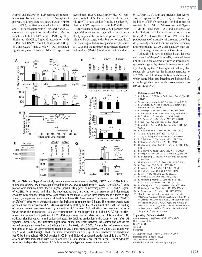
HSP70 and HSP90 by TLR-dependent mecha-nisms (6). To determine if the CD24-Siglec-Gpathway also regulates host responses to HSP70and HSP90, we first evaluated whether HSP70and HSP90 associate with CD24 and Siglec-G.Coimmunoprecipitations revealed that CD24 as-sociates with both HSP70 and HSP90 (Fig. 4E).Similar to HMGB1, Siglec-G association withHSP70 and HSP90 was CD24 dependent (Fig.4F), and CD24−/− and Siglecg−/− DCs producedsignificantly more IL-6 and TNF-a in response to
recombinant HSP70 and HSP90 (Fig. 4G) com-pared to WT DCs. These data reveal a criticalrole for CD24 and Siglec-G in the negative reg-ulation of DC response to multiple DAMPs.
Our results suggest that CD24 partners withSiglec-10 in humans or Siglec-G in mice to neg-atively regulate the immune response to proteinsreleased by damaged cells, but not to ligands ofmicrobial origin. Pattern recognition receptors suchas TLRs and the receptor of advanced glycationend products (RAGE)mediate activation induced
by DAMP (7, 8). Our data indicate that repres-sion of response to HMGB1 may be achieved byinhibition of NF-kΒ activation. Inhibitionmay bemediated by SHP-1. SHP-1 associates with Siglec-10 via its ITIM motif (26), and deficiency ofeither Siglec-G or SHP-1 enhances NF-kΒ activa-tion (20, 23). Given the role of HMGB1 in thepathogenesis of a number of diseases, includingdrug toxicity (9) and liver and cardiac ischemiaand reperfusion (27, 28), this pathway may un-cover new targets for disease intervention.
Although it is well established that the hostcan recognize “danger” induced by damaged tissue(4), it is unclear whether or how an immune re-sponses triggered by tissue damage is regulated.By identifying the CD24-Siglec-G pathway thatselectively suppresses the immune response toDAMPs, our data demonstrate a mechanism bywhich tissue injury and infection are distinguished,even though they both use the evolutionarily con-served TLRs (5–8).
References and Notes1. C. A. Janeway, Cold Spring Harb. Symp. Quant. Biol. 54,
1 (1989).2. Y. Liu, C. A. Janeway Jr., Int. Immunol. 3, 323 (1991).3. R. Medzhitov, P. Preston-Hurlburt, C. A. Janeway Jr.,
Nature 388, 394 (1997).4. P. Matzinger, Annu. Rev. Immunol. 12, 991 (1994).5. K. A. Cavassani et al., J. Exp. Med. 205, 2609 (2008).6. D. G. Millar et al., Nat. Med. 9, 1469 (2003).7. J. S. Park et al., J. Biol. Chem. 279, 7370 (2004).8. J. Tian et al., Nat. Immunol. 8, 487 (2007).9. P. Scaffidi, T. Misteli, M. E. Bianchi, Nature 418, 191
(2002).10. S. A. Ochsner et al., Stem Cells 25, 2476 (2007).11. Y. Liu et al., J. Exp. Med. 175, 437 (1992).12. Y. Liu, P. Zheng, Trends Immunol. 28, 315 (2007).13. X. F. Bai et al., J. Clin. Invest. 105, 1227 (2000).14. L. Wang et al., PLoS Genet. 3, e49 (2007).15. Q. Zhou et al., Proc. Natl. Acad. Sci. U.S.A. 100, 15041
(2003).16. H. E. Harris, A. Raucci, EMBO Rep. 7, 774 (2006).17. H. Yang et al., Proc. Natl. Acad. Sci. U.S.A. 101, 296 (2004).18. P. R. Crocker, J. C. Paulson, A. Varki, Nat. Rev. Immunol.
7, 255 (2007).19. M. Vinson et al., J. Biol. Chem. 271, 9267 (1996).20. C. Ding et al., PLoS One 2, e997 (2007).21. L. Apetoh et al., Nat. Med. 13, 1050 (2007).22. O. Li et al., J. Exp. Med. 203, 1713 (2006).23. L. I. Pao et al., Immunity 27, 35 (2007).24. H. Wang et al., Science 285, 248 (1999).25. E. Abraham, J. Arcaroli, A. Carmody, H. Wang,
K. J. Tracey, J. Immunol. 165, 2950 (2000).26. G. Whitney et al., Eur. J. Biochem. 268, 6083 (2001).27. M. Andrassy et al., Circulation 117, 3216 (2008).28. A. Tsung et al., J. Exp. Med. 201, 1135 (2005).29. This study was supported by grants from the NIH
(AI064350, CA58033, and CA112001), U.S. departmentof Defense (W81XWH-08-1-0036), and Natural ScienceFoundation of China (30640420558) and Ministry ofScience and Technology of China (2006CB910901). Theauthors have no conflicts of interest. We dedicate thisstudy to Dr. Charles A. Janeway Jr.
Supporting Online Materialwww.sciencemag.org/cgi/content/full/1168988/DC1Materials and MethodsFigs. S1 to S4Table S1References
25 November 2008; accepted 26 February 2009Published online 5 March 2009;10.1126/science.1168988Include this information when citing this paper.
Fig. 4. CD24 and Siglec-G negatively regulate immune responses to HMGB1, HSP70, and HSP90, but notto LPS and poly(I:C). (A) Production of cytokines by DCs. DCs cultured from WT, CD24−/−, or Siglecg−/− bonemarrow were stimulated with LPS (100 ng/ml), poly(I:C) (10 mg/ml), or increasing doses (5, 10, and 20 mg/ml)of HMGB1 for 6 hours, and then the supernatants were analyzed for the presence of inflammatorycytokines with cytokine beads array. Data represent the mean T SD for three independent cultures of DCsin each genotype and were repeated at least three times. (B) Bone marrow DCs isolated from WT, CD24−/−,or Siglecg−/− mice were stimulated under the indicated conditions for 6 hours. The nuclear lysates wereprepared and the activation of NF-kB was assessed by blotting for the p65 subunit of NF-kB. The loadingof nuclear protein was determined by amounts of Sp1 protein. Fold induction over medium control isshown below the immunoblots. Data are representative of two independent experiments. (C) Age-matchedmale mice received ip injections of LPS (450 mg/mouse). Kaplan Meier survival plots are shown. Nostatistical significance was found by log-rank tests. (D) Cytokine production in the serum 4 hours after LPSinjection (mean T SD; the statistical significance of the differences between the control and one of thetreated groups was determined by Student’s t test. *P < 0.03, **P < 0.002). The numbers of mice used werethe same as in (C). (E) Coimmunoprecipitation of CD24 and Hsp70 and Hsp90. (F) Siglec-G associates withHsp70 and Hsp90 through CD24. The same precipitates used in Fig. 3E were analyzed for Hsp70 andHsp90 by immunoblot. (G) Deficiencies in CD24 and Siglec-G enhanced production of IL-6 and TNF-aat 6 hours after stimulation with HSP70 and HSP90. Data shown represent the mean T SD of cytokinesfrom four independent isolates of DCs from each genotype and were repeated twice.
www.sciencemag.org SCIENCE VOL 323 27 MARCH 2009 1725
REPORTS
on
Mar
ch 3
0, 2
009
ww
w.s
cien
cem
ag.o
rgD
ownl
oade
d fr
om
www.sciencemag.org/cgi/content/full/1168988/DC1
Supporting Online Material for
CD24 and Siglec-10 Selectively Repress Tissue Damage–Induced Immune Responses
Guo-Yun Chen, Jie Tang, Pan Zheng,* Yang Liu*
*To whom correspondence should be addressed. E-mail: [email protected] (Y.L.),
[email protected] (P.Z.)
Published 5 March 2009 on Science Express DOI: 10.1126/science.1168988
This PDF file includes:
Materials and Methods Figs. S1 to S4 Table S1 References

Supplemental Information.
Materials and Methods
Reagents Recombinant proteins consisting of human IgG Fc and extracellular
domains of SIglec 5, 7, 10 and 11 were purchased from R&D Systems. Horse-
radish perioxidase conjugated anti-mouse, or anti-rabbit secondary-step
reagents, as well as anti-p65 and anti-sp1 were purchased from Santa Cruz
Biotechnology. Anti-FLAG M2 affinity gel, anti-FLAG mAb, acetaminophen
(AAP) and lipopolysaccharide (LPS, from E. coli 055:B5) were purchased from
Sigma (St Louis, MO). The composition CD24Fc have been described 1, the
product is obtained from OncoImmune, Inc. (Columbus, OH). Human HSP70,
HSP90 and anti-mouse Hsp70, Hsp90 antibodies were purchased from
Biovision, Inc. (Mountain View, CA). The anti-HMGB-1 antibodies 3E8 and 3B1
were described in supplemental information.
cDNAs encoding either full-length or specifically truncated human HMGB-
1 and N-FLAG-tagged WT or mutant (119R>A) Siglec10 were cloned into
expression vector pCMV-Tag 2B (Sigma). All constructs were verified by DNA
sequencing. For purification of FLAG-tagged HMGB-1, the full-length HMGB-1
expression vector was transfected into TSA cells, the lysates were used as
source to purify recombinant HMGB-1 according to a reported procedure 2.
Experimental animals Mice with targeted mutations of CD24 and Siglecg were
produced from ES cells of C57BL/6 origin as have been described 3, 4. Age- and
sex-matched wild type C57BL/6 mice were used as controls. All mice were used
at 6-8 weeks of age. All procedures involving mice have been approved by the
University of Michigan Animal Care and Use Committee.

Mouse pathology For ALT measurements, blood was collected at given time
points. Serum was isolated by centrifugation of clotted blood at 12,000 x g for 10
min at room temperature and then sent to Animal Diagnostic Laboratory of
Animal Research Facility, University of Michigan (Ann Arbor, USA) for
determining ALT activity. For histology, liver was removed and immediately fixed
in 4% formaldehyde-PBS solution, embedded in paraffin, sectioned at 5 μm, and
stained with hematoxylin and eosin. Serum cytokines were determined using
mouse cytokine bead array designed for inflammatory cytokines (Cat. No
552364, BD Biosciences).
Flow cytometric analysis for SIglec10 ligands Spleen cells from WT or
CD24-/- mice were washed in buffer A (150 mM NaCl, 3 mM MnCl2, 1 mM CaCl2,
1 mM MgCl2, 25mM Tris, pH 7.6, 2% BSA), and incubated for 1 hour at 37 °C
with 1 μg of Siglec-10-Fc or Fc control. The bound receptors were detected with
PE conjugated anti-human IgG-Fc and analyzed on a BD LSII.
Immunoprecipitation and immunoblotting Cell lysates were prepared in the
buffer B (1 % Triton X-100, 150 mM NaCl, 3 mM MnCl2, 1 mM CaCl2, 1 mM
MgCl2, 25 mM Tris, pH 7.6) and protease inhibitors (1 μg/ml leupeptin, 1 μg/ml
aprotinin and 1 mM phenylmethylsulfonyl fluoride). Samples were pre-cleared
with 60 μl of protein A-conjugated agarose beads (Upstate, Lake Placid, NY) for
2 h at 4°C or 37°C with rotation, and then incubated with corresponding
antibodies (anti-CD24 mAbs M1/69 and 20C9, 10 μg/ml; anti-HMGB-1, 2 μg/ml;
anti-HSP70 and HSP90 antibodies, 3 μg/ml). The beads were washed four
times with buffer B and re-suspended in SDS sample buffer for Western blot

analyses with given antibodies (0.5 μg/ml). The anti-Siglec-G antisera were
used at 1:100 dilution.
Mass spectrometry After gel concentration, the protein samples were
submitted to Taplin Spectrometry Facility at Harvard Medical School for high
throughput analysis.
Statistics The differences in cytokine proteins and ALT activities were
analyzed by Student’s t test. The differences in survival rates were analyzed by
Kaplan-Meier survival analysis with log-rank test.
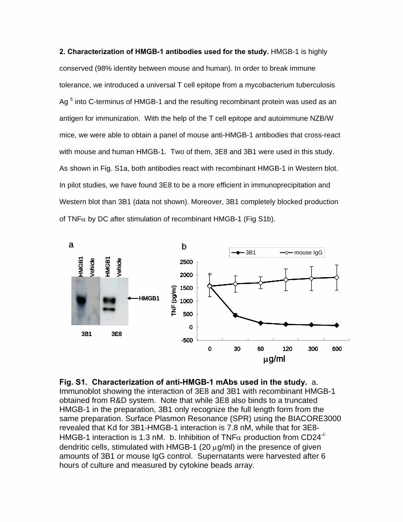
2. Characterization of HMGB-1 antibodies used for the study. HMGB-1 is highly
conserved (98% identity between mouse and human). In order to break immune
tolerance, we introduced a universal T cell epitope from a mycobacterium tuberculosis
Ag 5 into C-terminus of HMGB-1 and the resulting recombinant protein was used as an
antigen for immunization. With the help of the T cell epitope and autoimmune NZB/W
mice, we were able to obtain a panel of mouse anti-HMGB-1 antibodies that cross-react
with mouse and human HMGB-1. Two of them, 3E8 and 3B1 were used in this study.
As shown in Fig. S1a, both antibodies react with recombinant HMGB-1 in Western blot.
In pilot studies, we have found 3E8 to be a more efficient in immunoprecipitation and
Western blot than 3B1 (data not shown). Moreover, 3B1 completely blocked production
of TNFα by DC after stimulation of recombinant HMGB-1 (Fig S1b).
-500
0
500
1000
1500
2000
2500
0 30 60 120 300 600
TNF
(pg/
ml)
3B1 mouse IgG
μg/ml
3B1 3E8
HM
GB
1
Veh
icle
HM
GB
1
Veh
icle
HMGB1
a b
-500
0
500
1000
1500
2000
2500
0 30 60 120 300 600
TNF
(pg/
ml)
3B1 mouse IgG
μg/ml
3B1 3E8
HM
GB
1
Veh
icle
HM
GB
1
Veh
icle
HMGB1
-500
0
500
1000
1500
2000
2500
0 30 60 120 300 600
TNF
(pg/
ml)
3B1 mouse IgG
-500
0
500
1000
1500
2000
2500
0 30 60 120 300 600
TNF
(pg/
ml)
3B1 mouse IgG
μg/ml
3B1 3E8
HM
GB
1
Veh
icle
HM
GB
1
Veh
icle
HMGB1
3B1 3E8
HM
GB
1
Veh
icle
HM
GB
1
Veh
icle
HMGB1
a b Fig. S1. Characterization of anti-HMGB-1 mAbs used in the study. a. Immunoblot showing the interaction of 3E8 and 3B1 with recombinant HMGB-1 obtained from R&D system. Note that while 3E8 also binds to a truncated HMGB-1 in the preparation, 3B1 only recognize the full length form from the same preparation. Surface Plasmon Resonance (SPR) using the BIACORE3000 revealed that Kd for 3B1-HMGB-1 interaction is 7.8 nM, while that for 3E8-HMGB-1 interaction is 1.3 nM. b. Inhibition of TNFα production from CD24-/-
dendritic cells, stimulated with HMGB-1 (20 μg/ml) in the presence of given amounts of 3B1 or mouse IgG control. Supernatants were harvested after 6 hours of culture and measured by cytokine beads array.
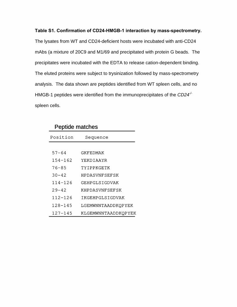
Table S1. Confirmation of CD24-HMGB-1 interaction by mass-spectrometry.
The lysates from WT and CD24-deficient hosts were incubated with anti-CD24
mAbs (a mixture of 20C9 and M1/69 and precipitated with protein G beads. The
precipitates were incubated with the EDTA to release cation-dependent binding.
The eluted proteins were subject to trysinization followed by mass-spectrometry
analysis. The data shown are peptides identified from WT spleen cells, and no
HMGB-1 peptides were identified from the immunoprecipitates of the CD24-/-
spleen cells.
30-42 HPDASVNFSEFSK
Position Sequence
76-85 TYIPPKGETK
114-126 GEHPGLSIGDVAK
29-42 KHPDASVNFSEFSK
112-126 IKGEHPGLSIGDVAK
128-145 LGEMWNNTAADDKQPYEK
57-64 GKFEDMAK
127-145 KLGEMWNNTAADDKQPYEK
154-162 YEKDIAAYR
Peptide matches
30-42 HPDASVNFSEFSK
Position Sequence
76-85 TYIPPKGETK
114-126 GEHPGLSIGDVAK
29-42 KHPDASVNFSEFSK
112-126 IKGEHPGLSIGDVAK
128-145 LGEMWNNTAADDKQPYEK
57-64 GKFEDMAK
127-145 KLGEMWNNTAADDKQPYEK
154-162 YEKDIAAYR
Peptide matches
A box B box +Acidic tail
FBCA
IP: CD24 Fc
anti-hIgG
Input
anti-FLAG
F BC A vect
or
anti-FLAG
IB
1 8990
215
A box B box +Acidic tail
FBCA
IP: CD24 Fc
anti-hIgG
Input
anti-FLAG
F BC A vect
or
anti-FLAG
IB
1 8990
215
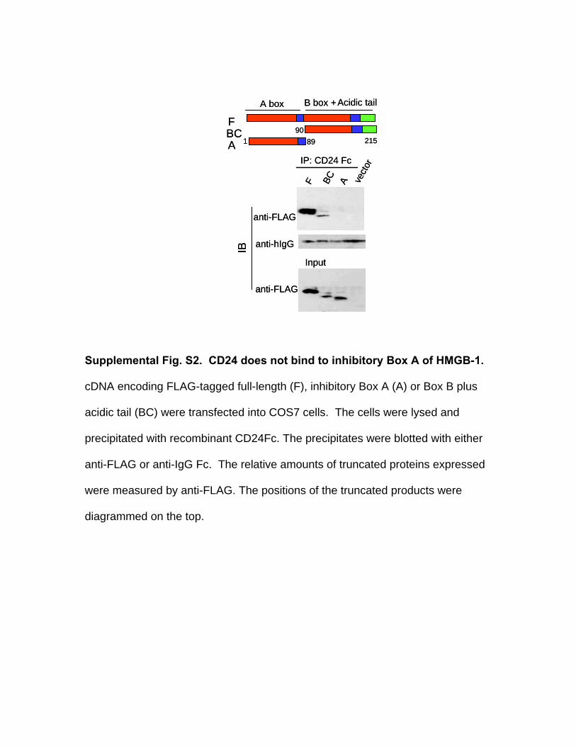
Supplemental Fig. S2. CD24 does not bind to inhibitory Box A of HMGB-1.
cDNA encoding FLAG-tagged full-length (F), inhibitory Box A (A) or Box B plus
acidic tail (BC) were transfected into COS7 cells. The cells were lysed and
precipitated with recombinant CD24Fc. The precipitates were blotted with either
anti-FLAG or anti-IgG Fc. The relative amounts of truncated proteins expressed
were measured by anti-FLAG. The positions of the truncated products were
diagrammed on the top.
WT
Sig
lecg
-/-
181.8
115.582.264.2
48.8
A B
PE-anti-mouse IgG
Cel
l num
ber (
max
%) WTSiglecg-/-
WT
Sig
lecg
-/-
181.8
115.582.264.2
48.8
A B
PE-anti-mouse IgG
Cel
l num
ber (
max
%)
WT
Sig
lecg
-/-
181.8
115.582.264.2
48.8
A B
PE-anti-mouse IgG
Cel
l num
ber (
max
%) WTSiglecg-/-
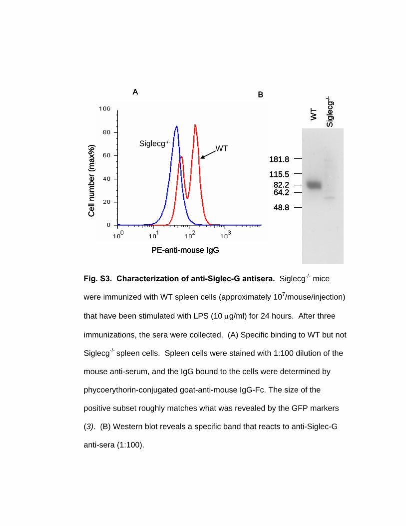
Fig. S3. Characterization of anti-Siglec-G antisera. Siglecg-/- mice
were immunized with WT spleen cells (approximately 107/mouse/injection)
that have been stimulated with LPS (10 μg/ml) for 24 hours. After three
immunizations, the sera were collected. (A) Specific binding to WT but not
Siglecg-/- spleen cells. Spleen cells were stained with 1:100 dilution of the
mouse anti-serum, and the IgG bound to the cells were determined by
phycoerythorin-conjugated goat-anti-mouse IgG-Fc. The size of the
positive subset roughly matches what was revealed by the GFP markers
(3). (B) Western blot reveals a specific band that reacts to anti-Siglec-G
anti-sera (1:100).
WT/CD24-Fc
Siglecg-/-/CD24-Fc
Siglecg-/-/Fc
WT/Fc
PE-Streptavidin
Cel
l num
ber (
% m
ax) WT/CD24-Fc
Siglecg-/-/CD24-Fc
Siglecg-/-/Fc
WT/Fc
PE-Streptavidin
Cel
l num
ber (
% m
ax)
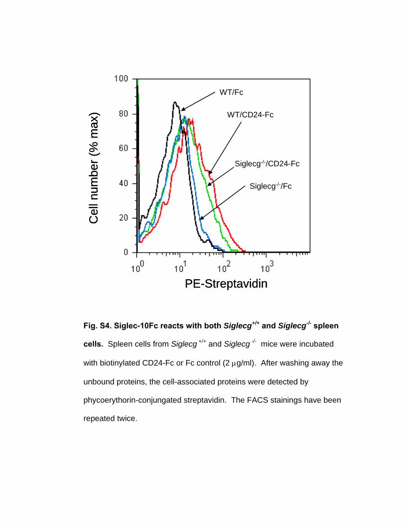
Fig. S4. Siglec-10Fc reacts with both Siglecg+/+ and Siglecg-/- spleen
cells. Spleen cells from Siglecg +/+ and Siglecg -/- mice were incubated
with biotinylated CD24-Fc or Fc control (2 μg/ml). After washing away the
unbound proteins, the cell-associated proteins were detected by
phycoerythorin-conjungated streptavidin. The FACS stainings have been
repeated twice.

References 1. Bai, X.F. et al. The heat-stable antigen determines pathogenicity of self-reactive T
cells in experimental autoimmune encephalomyelitis. J. Clin. Invest. 105, 1227-1232 (2000).
2. Sha, Y., Zmijewski, J., Xu, Z. & Abraham, E. HMGB-1 develops enhanced proinflammatory activity by binding to cytokines. J. Immunol. 180, 2531-2537 (2008).
3. Ding, C. et al. Siglecg limits the size of B1a B cell lineage by down-regulating NFkappaB activation. PLoS ONE 2, e997 (2007).
4. Nielsen, P.J. et al. Altered erythrocytes and a leaky block in B-cell development in CD24/HSA-deficient mice. Blood 89, 1058-1067 (1997).
5. Lohnas, G.L., Roberts, S.F., Pilon, A. & Tramontano, A. Epitope-specific antibody and suppression of autoantibody responses against a hybrid self protein. J Immunol 161, 6518-6525 (1998).

delivers its genome by ejection/injection
(rather than by disassembly of its capsid).
Most of these pressurized viruses are likely to
be bacteriophages, which generally do not
enter their host cell but rather inject their
genome upon binding to the outer cell mem-
brane (see the figure, panel A). The genome
involved must be dsDNA, because single-
stranded nucleic acid is easily compressible
and hence does not get sufficiently pressur-
ized upon being confined.
It is thus no coincidence that most bacter-
ial viruses have dsDNA genomes. In contrast,
plant and animal viruses, whose capsids gen-
erally enter the cytoplasm of their host cells
and then disassemble, mostly have ssRNA
genomes. On the other hand, a mammalian
dsDNA virus such as herpes, whose capsid
enters the cytoplasm of its host cell by pass-
ing through the outer cell membrane, must
still inject its genome into the nucleus upon
binding to a nuclear pore complex (see the
figure, panel B). The pressure in its capsid
should be comparable to those in bacterio-
phages. The motor protein that packages its
genome is thus expected to exert forces as
high as tens of piconewtons.
In many phage life cycles, the freshly repli-
cated, not-yet-packaged DNA genomes are
linked together in a polymer. High-resolution
cryoelectron microscopy studies on phage
P22 (11) have revealed the configuration of
the packaged DNA and shown how the motor
protein complex acts as a pressure sensor when
a certain density of DNA is achieved. At this
point, packaging stops and the DNA is cut.
As always, new understanding raises new
questions. For example, because the host cell
cytoplasm has an osmotic pressure of several
atmospheres, phage ejection stops when the
capsid pressure drops to a few atmospheres;
what drives delivery of the rest of the genome?
In some cases, it is transcription of the genes
that have already been delivered; in others, it
may involve the influx of water through the
phage to accommodate the growth of the host
bacterium (12).
State-of-the-art biophysical studies will
help to elucidate these and other issues, such as
how capsids can withstand pressures on the
order of 50 atmospheres. Notable among these
studies are the reconstitution of bacteriophage
λ “from scratch” (13), the probing of elastic
properties of individual viruses by atomic
force microscopy (14), the observation of
genome ejection from single viruses by fluo-
rescence microscopy (15), and the simulation
of protein capsid assembly, as well as single-
molecule manipulations and high-resolution
cryoelectron microscopy. Much as phages
played key roles in the development of molec-
ular biology and the genetic engineering revo-
lution, pressurized viruses are likely to be cen-
tral to the emerging field of physical virology.
References and Notes
1. A. Evilevitch, L. Lavelle, C. M. Knobler, E. Raspaud, W. M.
Gelbart, Proc. Natl. Acad. Sci. U.S.A. 100, 9292 (2003).
2. P. Grayson et al., Virology 348, 430 (2006).
3. S. Tzlil, J. Kindt, W. M. Gelbart, A. Ben-Shaul, Biophys. J.
84, 1616 (2003).
4. D. E. Smith et al., Nature 413, 748 (2001).
5. D. N. Fuller et al., J. Mol. Biol. 373, 1113 (2007).
6. D. N. Fuller D. M. Raymer, V. Kottadiel, V. B. Rao, D. E.
Smith, Proc. Natl. Acad. Sci. U.S.A. 104, 16868 (2008).
7. D. Raoult et al., Science 306, 1344 (2004); published
online 14 October 2004 (10.1126/science.1101485).
8. W. C. Earnshaw, S. C. Harrison, Nature 268, 598 (1977).
9. D. C. Rau, V. A. Parsegian, Biophys. J. 61, 246 (1992).
10. J. Kindt , S. Tzlil, A. Ben-Shaul, W. M. Gelbart, Proc. Natl.
Acad. Sci. U.S.A. 98, 13671 (2001).
11. G. C. Lander et al., Science 312, 1791 (2006); published
online 17 May 2006 (10.1126/science.1127981).
12. P. Grayson, I. J. Molineux, Curr. Opin. Microbiol. 10, 401
(2007).
13. H. Gaussier, Q. Yang, C. E. Catalano, J. Mol. Biol. 357,
1154 (2006).
14. J.-P. Michel et al., Proc. Natl. Acad. Sci. U.S.A. 103, 6184
(2006).
15. S. Mangenot, M. Hochrein, J. Radler, L. Letellier, Curr.
Biol. 15, 430 (2005).
16. Supported by NSF grant CHE-0714411.
10.1126/science.1170645
www.sciencemag.org SCIENCE VOL 323 27 MARCH 2009 1683
PERSPECTIVES
Every organism faces a bewildering
array of threats, pathogens foremost
among them. But how does the
immune system distinguish between an infec-
tion and trauma? Both elicit similar inflam-
matory immune responses. On page 1722 of
this issue, Chen et al. (1) explain why and how
the immune system responds appropriately in
either scenario.
All mammals have an impressive arsenal
of molecules and cells specialized to fight
pathogens. Adaptive immunity, in the form
of antibody production by B cells and the
instruction of “killer” or cytotoxic T cells, is a
critical component of the body’s defenses.
However, this is only a second line of defense
that selectively recognizes microbes attacking
for a second time. Without a first line of
defense—innate immunity—mammals would
succumb to pathogens still unrecognized by B
and T cells.
The broad outlines of our current under-
standing were first sketched 20 years ago by
Charles Janeway (2), starting from the idea
that the immune system cannot recognize
pathogens individually, because the informa-
tion required is huge and would rapidly
become obsolete. Pathogens continually
evolve, confounding the ability of the immune
system to recognize them. Instead, immune
cells recognize broad molecular patterns
rather than detailed features of specific
pathogens. Such pathogen-associated molec-
ular patterns (PAMPs) comprise molecular
structures that are found in microbes but not in
host tissues. Moreover, PAMPs are essential
for the survival or the pathogenicity of
microbes; thus, they cannot simply do away
with PAMPs to evade recognition by the
immune system.
Toll-like receptor 4 (TLR4) was the first
receptor to be identified that recognizes
PAMPs (3). It recognizes lipopolysaccharide,
a component of the outer membrane of Gram-
negative bacteria. As the name implies, it is
related to Toll, a receptor involved in pathogen
recognition in the fly Drosophila melano-
gaster. Thus, Toll-like receptors are evolution-
arily ancient, although their number in mam-
mals has grown to about one dozen.
When cells of the innate immune system—
such as macrophages, mast cells, natural killer
cells, and dendritic cells—encounter a PAMP,
they secrete cytokines and chemokines, solu-
ble molecules that signal “danger” to other
cells. The most immediate response is inflam-
mation at the site of infection, and the recruit-
ment of additional immune cells, including
neutrophils. Importantly, neutrophils “shoot at
first sight,” releasing reactive oxygen species
and proteases, thereby causing extensive col-
lateral damage to the host tissue. Usually, the
intruding pathogens are eliminated, and the
adaptive immune system “remembers” their
identity in case the organism is reinfected by
The immune system relies on specific
signaling molecules to dampen its response
to injury while maintaining the capacity
to fight infection.Dangers In and OutMarco E. Bianchi and Angelo A. Manfredi
IMMUNOLOGY
San Raffaele University, Faculty of Medicine, and SanRaffaele Scientific Institute, via Olgettina 58, 20132Milano, Italy. E-mail: [email protected]
Published by AAAS
on
Mar
ch 3
0, 2
009
ww
w.s
cien
cem
ag.o
rgD
ownl
oade
d fr
om
1684
CR
ED
IT: C
. B
ICK
EL/S
CIE
NC
E
PERSPECTIVES
the same pathogens; eventually, the
tissue is reconstructed and healed.
Inflammation, therefore, is a dam-
aging but essential response, and
becomes a problem only when it is
excessive, or persists (chronic
inflammation).
This picture of pathogen infec-
tion is complicated by the fact that
physical trauma (such as a wound
or a broken bone) causes many of
the same effects as invading
pathogens, including inflamma-
tion. Indeed, pathogens are thought
to be initially recognized by the
immune system precisely because
they cause tissue damage (4). How,
then, does the immune system rec-
ognize tissue damage? Damage-
associated molecular patterns
(DAMPs) were postulated as the
counterparts to PAMPs, with the
important distinction that DAMPs
should be endogenous—the body’s
own molecules—just as the PAMPs
should be pathogen-borne and thus exoge-
nous. For example, the molecule high mobil-
ity group box 1 (HMGB1) fits the hypotheti-
cal description of an endogenous danger sig-
nal (5) and instructs adaptive immunity in
ways similar to those elicited by exogenous
danger signals (6). HMGB1 is a component of
chromatin, the DNA-protein complex that
makes up chromosomes, and thus normally
resides in the cell nucleus. The release of
HMGB1 by cells that have died as a result of
tissue trauma signals “danger” to neighboring
cells and to the immune system. Importantly,
HMGB1 is also recognized by the pattern
recognition receptors TLR2, TLR4, and
TLR9, as well as by the receptor of advanced
glycation endproducts, another “danger”
receptor (7–9). Thus, trauma and pathogens
(DAMPs and PAMPs) engage the same
immune cell receptors, neatly explaining why
they elicit the same inflammatory responses,
although the molecular details are still largely
unknown.
Yet, some outcomes must be different
between the two scenarios. Chen et al. ex-
plored how these partly different outcomes
occur by inducing liver necrosis in mice with
an excess of acetaminophen. This treatment
causes the release of HMGB1, and thus leads
to inflammation in the absence of any patho-
gen. The authors found that mice lacking
CD24, a membrane protein expressed by
immune and stem cells, developed an inflam-
matory response that was more powerful and
lethal than that in wild-type mice. In fact,
HMGB1 was found to directly associate with
CD24 and Siglec-G, a member of the sialic
acid–binding immunoglobulin-like lectin
family. Mice lacking Siglec-G also were sen-
sitive to inflammation due to acetaminophen-
induced liver necrosis. CD24 does not contain
a cytosolic domain, and signals through
Siglec-G, which contains an immune receptor
tyrosine-based inhibitory motif (ITIM).
ITIMs are cytosolic domains that reduce acti-
vation of nuclear factor κB (NF-κB), which is
a transcription factor activated by both
DAMPs (10) and PAMPs (11) and is essential
for many aspects of the inflammatory re-
sponse, including the secretion of cytokines
and chemokines. The CD24-Siglec complex
also recognizes heat shock proteins, another
class of endogenous danger signals, but does
not respond to lipopolysaccharide or poly-
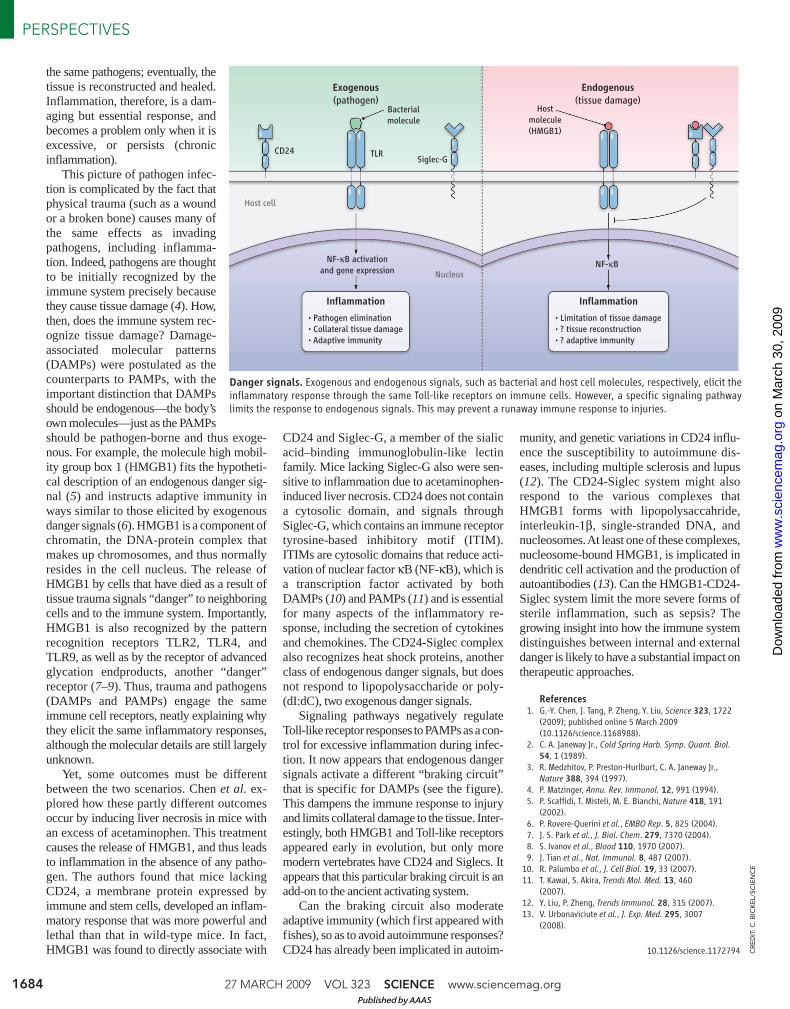
(dI:dC), two exogenous danger signals.
Signaling pathways negatively regulate
Toll-like receptor responses to PAMPs as a con-
trol for excessive inflammation during infec-
tion. It now appears that endogenous danger
signals activate a different “braking circuit”
that is specific for DAMPs (see the figure).
This dampens the immune response to injury
and limits collateral damage to the tissue. Inter-
estingly, both HMGB1 and Toll-like receptors
appeared early in evolution, but only more
modern vertebrates have CD24 and Siglecs. It
appears that this particular braking circuit is an
add-on to the ancient activating system.
Can the braking circuit also moderate
adaptive immunity (which first appeared with
fishes), so as to avoid autoimmune responses?
CD24 has already been implicated in autoim-
munity, and genetic variations in CD24 influ-
ence the susceptibility to autoimmune dis-
eases, including multiple sclerosis and lupus
(12). The CD24-Siglec system might also
respond to the various complexes that
HMGB1 forms with lipopolysaccahride,
interleukin-1β, single-stranded DNA, and
nucleosomes. At least one of these complexes,
nucleosome-bound HMGB1, is implicated in
dendritic cell activation and the production of
autoantibodies (13). Can the HMGB1-CD24-
Siglec system limit the more severe forms of
sterile inflammation, such as sepsis? The
growing insight into how the immune system
distinguishes between internal and external
danger is likely to have a substantial impact on
therapeutic approaches.
References1. G.-Y. Chen, J. Tang, P. Zheng, Y. Liu, Science 323, 1722
(2009); published online 5 March 2009
(10.1126/science.1168988).
2. C. A. Janeway Jr., Cold Spring Harb. Symp. Quant. Biol.
54, 1 (1989).
3. R. Medzhitov, P. Preston-Hurlburt, C. A. Janeway Jr.,
Nature 388, 394 (1997).
4. P. Matzinger, Annu. Rev. Immunol. 12, 991 (1994).
5. P. Scaffidi, T. Misteli, M. E. Bianchi, Nature 418, 191
(2002).
6. P. Rovere-Querini et al., EMBO Rep. 5, 825 (2004).
7. J. S. Park et al., J. Biol. Chem. 279, 7370 (2004).
8. S. Ivanov et al., Blood 110, 1970 (2007).
9. J. Tian et al., Nat. Immunol. 8, 487 (2007).
10. R. Palumbo et al., J. Cell Biol. 19, 33 (2007).
11. T. Kawai, S. Akira, Trends Mol. Med. 13, 460
(2007).
12. Y. Liu, P. Zheng, Trends Immunol. 28, 315 (2007).
13. V. Urbonaviciute et al., J. Exp. Med. 295, 3007
(2008).
10.1126/science.1172794
Exogenous
(pathogen)
Endogenous
(tissue damage)
CD24Siglec-G
TLR
Bacterial
molecule
Host
molecule
(HMGB1)
Inflammation
Pathogen elimination
Collateral tissue damage
Adaptive immunity
Inflammation
Limitation of tissue damage
? tissue reconstruction
? adaptive immunity
NF-κB activation
and gene expression Nucleus
Host cell
NF-κB
Danger signals. Exogenous and endogenous signals, such as bacterial and host cell molecules, respectively, elicit theinflammatory response through the same Toll-like receptors on immune cells. However, a specific signaling pathwaylimits the response to endogenous signals. This may prevent a runaway immune response to injuries.
27 MARCH 2009 VOL 323 SCIENCE www.sciencemag.org
Published by AAAS
on
Mar
ch 3
0, 2
009
ww
w.s
cien
cem
ag.o
rgD
ownl
oade
d fr
om
Related Documents