AD_________________ Award Number: DAMD17-03-1-0522 TITLE: Constitutive Activation of NF-Κb in Prostate Carcinoma Cells through a Positive Feedback Loop: Implication of Inducible IKK-Related Kinase (Ikki) PRINCIPAL INVESTIGATOR: Irina Budunova, M.D., Ph.D. CONTRACTING ORGANIZATION: Northwestern University Chicago, IL 60611 REPORT DATE: August 2007 TYPE OF REPORT: Final PREPARED FOR: U.S. Army Medical Research and Materiel Command Fort Detrick, Maryland 21702-5012 DISTRIBUTION STATEMENT: Approved for Public Release; Distribution Unlimited The views, opinions and/or findings contained in this report are those of the author(s) and should not be construed as an official Department of the Army position, policy or decision unless so designated by other documentation.

Welcome message from author
This document is posted to help you gain knowledge. Please leave a comment to let me know what you think about it! Share it to your friends and learn new things together.
Transcript

AD_________________ Award Number: DAMD17-03-1-0522 TITLE: Constitutive Activation of NF-Κb in Prostate Carcinoma Cells through a Positive Feedback Loop: Implication of Inducible IKK-Related Kinase (Ikki) PRINCIPAL INVESTIGATOR: Irina Budunova, M.D., Ph.D. CONTRACTING ORGANIZATION: Northwestern University
Chicago, IL 60611 REPORT DATE: August 2007 TYPE OF REPORT: Final PREPARED FOR: U.S. Army Medical Research and Materiel Command Fort Detrick, Maryland 21702-5012 DISTRIBUTION STATEMENT: Approved for Public Release; Distribution Unlimited The views, opinions and/or findings contained in this report are those of the author(s) and should not be construed as an official Department of the Army position, policy or decision unless so designated by other documentation.

REPORT DOCUMENTATION PAGE Form Approved
OMB No. 0704-0188 Public reporting burden for this collection of information is estimated to average 1 hour per response, including the time for reviewing instructions, searching existing data sources, gathering and maintaining the data needed, and completing and reviewing this collection of information. Send comments regarding this burden estimate or any other aspect of this collection of information, including suggestions for reducing this burden to Department of Defense, Washington Headquarters Services, Directorate for Information Operations and Reports (0704-0188), 1215 Jefferson Davis Highway, Suite 1204, Arlington, VA 22202-4302. Respondents should be aware that notwithstanding any other provision of law, no person shall be subject to any penalty for failing to comply with a collection of information if it does not display a currently valid OMB control number. PLEASE DO NOT RETURN YOUR FORM TO THE ABOVE ADDRESS. 1. REPORT DATE (DD-MM-YYYY) 01-08-2007
2. REPORT TYPEFinal
3. DATES COVERED (From - To)1 AUG 2003 - 31 JUL 2007
4. TITLE AND SUBTITLE
5a. CONTRACT NUMBER
Constitutive Activation of NF-Κb in Prostate Carcinoma Cells through a Positive Feedback Loop: Implication of Inducible IKK-Related Kinase (Ikki)
5b. GRANT NUMBER DAMD17-03-1-0522
5c. PROGRAM ELEMENT NUMBER
6. AUTHOR(S) Irina Budunova, M.D., Ph.D.
5d. PROJECT NUMBER
5e. TASK NUMBER
E-Mail: [email protected] 5f. WORK UNIT NUMBER
7. PERFORMING ORGANIZATION NAME(S) AND ADDRESS(ES)
8. PERFORMING ORGANIZATION REPORT NUMBER
Northwestern University Chicago, IL 60611
9. SPONSORING / MONITORING AGENCY NAME(S) AND ADDRESS(ES) 10. SPONSOR/MONITOR’S ACRONYM(S) U.S. Army Medical Research and Materiel Command
Fort Detrick, Maryland 21702-5012 11. SPONSOR/MONITOR’S REPORT NUMBER(S) 12. DISTRIBUTION / AVAILABILITY STATEMENT Approved for Public Release; Distribution Unlimited
13. SUPPLEMENTARY NOTES
14. ABSTRACT The overall goal of this project is to understand the role of inducible inflammation-related kinase IKKi in constitutive activation of anti-apoptotic transcription factor NF-κB prostate carcinoma (PC) cells. We found that IKKi is expressed in highly malignant androgen-independent PC cells lines and in epithelial cells in benign and malignant prostate lesions. Our data provide experimental evidence that IKKi could be involved in the regulation of activity of major anti-apoptotic factor NF-κB in PC cells through a positive feedback loop. Our results also suggest that IKKi may play an important role during the transition to hormone refractory stage of PC growth via its positive effect on the nuclear translocation and activity of androgen receptor in PC cells. Taking into consideration the newly recognized association between prostate inflammation and increased risk of PC development, we extended our studies towards cross-talk between pro-inflammatory signaling mediated by IKKi and NF-kB and anti-inflammatory signaling mediated by glucocorticoid receptor (GR) in PC cells. We showed that GR functions as a tumor suppressor in prostate cells, and that inhibition of transcription factors involved in proliferation and transformation in PC cells, including NF-kB, is the major molecular mechanism of GR anti-tumor activity. As IKKi specific inhibitors are still not available, we screened several novel classes of NF-kB inhibitors for their growth-inhibitory and anti-apoptotic effects in PC cells. The results of our studies have been presented at the local and national meetings, five manuscripts have been published, one is under revision, and two are under preparation.
15. SUBJECT TERMS No subject terms provided.
16. SECURITY CLASSIFICATION OF:
17. LIMITATION OF ABSTRACT
18. NUMBER OF PAGES
19a. NAME OF RESPONSIBLE PERSON USAMRMC
a. REPORT U
b. ABSTRACT U
c. THIS PAGE U
UU
52
19b. TELEPHONE NUMBER (include area code)
Standard Form 298 (Rev. 8-98) Prescribed by ANSI Std. Z39.18
Budunova, I.
3
Table of Contents
Annual Report for FY04………………………………………………………………4
Final Report for FY01-FY04
Introduction…………………………………………………………….………….... 8
Body…………………………………………………………………………………… 8
Key Research Accomplishments………………………………………….……..10
Reportable Outcomes………………………………………………………………. 12
Conclusions………………………………………………………………………….. 14
List of personnel receiving pay from the research effort……………………14
Collaborations, promotions …………………………………………………………14
Training of postdoctoral fellows……………………………………………… ….15
References…………………………………………………………………………… 16 Supplemental figures………………………………………………………………..17 Appendices…………………………………………………………………………… 30-

Budunova, I.
4
Report for FY04.
Introduction
The overall goal of this project is to understand the role of inducible IKK-related kinase IKKi in constitutive activation of anti-apoptotic transcription factor NF-κB prostate carcinoma (PC) cells. During FY04 (no cost extension) the major direction of our work was to complete the evaluation of the biological effects of IKKi overexpression in PC cells stably infected with lentiviruses harboring w.t. IKKi, and the important role of IKKi in the transition of prostate cells to androgen-independent growth. The results of our studies in 2006-2007 have been presented at the national meetings, two manuscripts are published, one is under revision, and two are under preparation. The following describes the progress made in this year. Body
During FY04 we specifically focused on the experiments pertinent to our most important findings made in the previous years suggesting that IKKi may represent a link between inflammation and androgen receptor signaling. We continued to characterize the effect of IKKi in PC cells on basal and inducible NF-kB activity, growth, tumorigenicity and AR function.
We found that that IKKi expression in LNCaP and PC3 cells resulted in increased basal NF-kB activity measured in Luciferase assay (Fig. 4B – cells transiently transfected with w.t. IKKi, Fig. 6.A1 and 6.A2 – cells stably infected with IKKi lentivirus). IKKi-expressing cells were more sensitive to NF-kB inducers such as TNF-a, TPA, EGF, LPS, and especially IL-1 (Fig. 6.A1 and 6.A2). In addition to the experiments with exogenous kappaB reporter, we evaluated the effect of IKKi on the expression of endogenous NF-kappaB-responsive gene IkBa using RT-PCR analysis, and found that IkBa expression was induced more effectively in LNCaP-IKKi cells (Fig. 6.B1).
We confirmed that induction of IkBa phosphorylation (at Ser32/36) and p65 phosphorylation at Ser536 was increased in LNCaP-IKKi cells treated with such inducers as TNF-a and LPS (Fig.6. C1 and C2). It is well known that IkBa undergoes proteasomal degradation after phosphorylation at Ser32/36 (Ref). Thus, the effect of IKKi on basal and inducible IkBa phosphorylation was more augmented when degradation of phosphorylated IkB-a was blocked by proteasome inhibitor MG132 (Fig. 6.C1, far right lanes).
We showed that IKKi expression results in increases PC growth and tumorigenicity. Using LNCaP and PC3 clones co-expressing w.t. IKKi (Fig. 5C) and yellow fluorescent protein (YFP) to track live cells and to measure the actual number of cells/well by fluorescent plate-reader, we showed that IKKi significantly increased PC cells growth in monolayer (Fig. 5.A1. and A2). Empty-vector-expressing cells (LNCaP-V and PC3-V) were used as control. Importantly, IKKi-expressing LNCaP cells were also characterized by the increased tumorigenicity assessed by anchorage-independent growth (colony formation assay in soft agar, Fig.5. B).
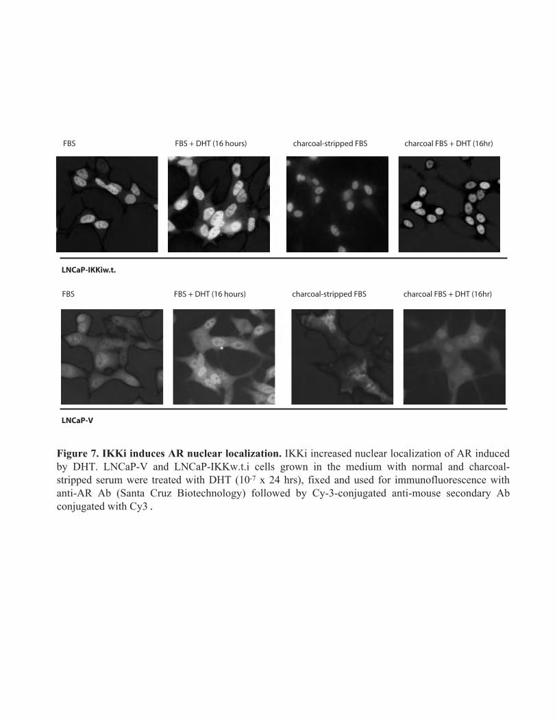
We further showed that IKKi overexpression resulted in the partial nuclear localization of endogenous androgen receptor (AR) in LNCaP cells (Fig. 7). It correlated with the increased transcriptional activity of AR in LNCaP-IKKi cells growing in the normal cell culture medium (standard fetal bovine serum) and in the medium with the decreased androgen level (charcoal-stripped serum) especially when cells were activated by androgen DHT (Fig. 8 B). The results of Luciferase assay with ARE reporter have been further extended using RT-PCR analysis of the expression of androgen-dependent endogenous gene PSA (Fig. 8.C). As shown in Fig. 8.C IKKi-expressing cells were more responsive to DHT stimulation which is reflected by significantly early increase in PSA expression in comparison to cells infected with empty virus.
Most importantly, LNCaP-IKKi cells became partly androgen-independent: they grew better than LNCaP-vector transfected cells in the medium with charcoal-stripped serum. This correlated with the

Budunova, I.
5
spontaneous translocation/accumulation of AR in the PC nuclei (Fig. 8A). Overall, our results suggest an important role of IKKi in the transition of prostate cells to
androgen-independent growth. We are currently testing the hypothesis that IKKi may affect the level of AR phosphorylation which in turn can result in spontaneous nuclear AR translocation. For this we are using Western blotting with antibodies against phosphorylated AR.
Taking into consideration the newly recognized association of prostate inflammation and prostate cancer that offers one of the greatest opportunities for preventing malignant conversion (Platz and De Marzo, 2004,De Marzo et al, 2007) we continued to study the cross-talk between pro-inflammatory signaling mediated by IKKi and NF-kB and anti-inflammatory signaling mediated by glucocorticoid receptor (GR) in PC cells. In FY02 –FY03 we found that glucocoticoids inhibit IKKi function; we also showed that GR acts as a tumor suppressor in prostate cells. In FY04 we started to work with a unique compound (CpdA) that acts as a non-steroidal ligand of both AR and GR (Fig. 9 and data not shown). Similar to steroid hormones, CpdA induces nuclear translocation of both receptors in prostate cells. Despite of this, CpdA inhibits DNA-binding and transactivation potential of AR (data not shown). In addition, CpdA inhibits GR-mediated transactivation but induces GR trans-repression via inhibition of transcription factors, first of all, NF-kB (Fig. 9). CpdA strongly inhibits growth and induces caspase-dependent apoptosis in highly malignant PC cells in AR/GR-dependent manner (Fig. 10 and 11). Overall, our data suggest that CpdA is a unique dual-target steroid receptor modulator that has a high potential for PC therapy.
Key Research Accomplishments FY04
The increased IKKi expression in different PC cells stably infected with IKKi-lentivirus results in
increased basal and inducible NF-kB activity.
IKKi overexpression results in increased growth and tumorigenicity of PC cells.
Androgen receptor was partially translocated to the nucleus and constitutively activated in IKKi-
expressing PC cells.
LNCaP-IKKi cells became partially androgen-independent and could sustain the androgen
ablation.
These results suggest that IKKi plays an important role during the transition to hormone
refractory stage of PC growth.
Compound A, a non-steroidal modulator of glucocorticoid receptor that inhibits NF-kB function,
inhibits growth and viability of highly malignant prostate cancer cells.

Budunova, I.
6
Reportable Outcomes FY04
Manuscripts:
1. Yemelyanov A., Czwornog J., Chebotaev D., Karseladze A., Kulevitch E., Yang X., Budunova I. Tumor suppressor effect of glucocorticoid receptor in prostate. Oncogene, 2007, 26:1885-1896.
2. T. Nelius, S. Filleur, A. Yemeyanov, I. Budunova, E. Shroff , Y. Mirochnik, A. Aurora, D. Veliceasa, W. Xiao, Z. Wang, and O.V. Volpert Androgen receptor targets NFkB and TSP1 to suppress prostate tumor growth in vivo. Int J Cancer. 2007, 121(5):999-1008. 3. Yemelyanov A., Czwornog J., Joshi S., Gera L., Budunova I. Compound A, a novel phyto-
modulator of steroid hormone receptors, as a candidate for prostate cancer therapy. Submitted. 4. Yemelyanov A., Kobzeva V., Budunova I. Role of IKKi in prostate cancer: A link between
inflammation and androgen receptor signaling. Manuscript is under preparation.
5. Gasparian A., Yemelyanov A., Chebotaev D., Kisseljov F., and I. Budunova. Targeting NF-κB in prostate carcinoma cells: comparative analysis of proteasome and IKK inhibitors. Manuscript is under preparation.
Abstracts presented at the national meetings: 1. Yemelyanov A., Kobzeva V., Budunova I. Role of IKKi in prostate cancer: A link between
inflammation and androgen receptor signaling. Proceedings of AACR, 2007 (abstract # LBA-9158).
2. Budunova I. , Yemelyanov A., Gasparian A. Role of IKKs and transcription factor NF-kB in
prostate tumorigenesis. P. IMPACT DOD meeting, 2007, Atlanta.
Seminars presented by P.I. Compound A, a novel phyto-modulator of steroid hormone receptors, as a candidate for prostate cancer therapy. Tumor cell biology seminars. R. Lurie Cancer Center. NU. April, 2007. Non-steroidal modulators of steroid hormone receptors as candidates for prostate cancer therapy. Children’s Memorial Research Center, Chicago. May , 2007
Conclusions for FY04. Our data provide experimental evidence that IKKi could be involved in the regulation of NF-kB
activity in PC cells through a positive feedback loop. For example, NF-kB was constitutively activated in
PC cells stably infected with w.t. IKK-expressing lentivirus. IKKi –infected cells were more responsive to
different NF-kB inducers. IKKi is highly expressed in androgen-independent malignant PC cell lines. The

Budunova, I.
7
introduction of w.t. IKKi into androgen-dependent LNCaP prostate cells significantly increased their
growth and tumorigenicity, and protected these cells from the induced apoptosis. Remarkably, IKKi
expression in LNCaP cells resulted in nuclear translocation and increased transcription potential of
androgen receptor (AR). Moreover, LNCaP-IKKi cells were more resistant to androgen ablation than
parental LNCaP cells. Those findings suggest that IKKi may play an important role in the development of
hormone refractory phase of PC growth.

Budunova, I.
8
Final Report for the entire funding period
Introduction
The overall goal of this project is to understand the role of inducible IKK-related kinase IKKi in constitutive activation of anti-apoptotic transcription factor NF-κB prostate carcinoma (PC) cells. The recent literature data published during the funded research period clearly indicated that the expression of this novel upstream IkappaB kinase strongly depends on inflammatory cytokines. It also became clear that there is a causative link between prostate inflammation and increased risk of PC development. Thus, we extended our research towards studies of cross-talk between pro-inflammatory signaling mediated by IKKi and NF-kB and anti-inflammatory signaling mediated by glucocorticoid receptor (GR) in PC cells. Unfortunately, small inhibitors of IKKi remain to be developed. Thus, we searched for other effective strategies of NF-kB blockage in PC cells, and tested growth inhibitory and pro-apoptotic potential of several novel compounds including highly specific IKKβ inhibitors, proteasomal inhibitors as well as dissociated ligands of GR that inhibit NF-kB.
Body:
Task 1. To define whether IKKi is an essential part of a positive feedback regulation of NF-κκκκB in PC cells.
We found that IKKi is expressed only in highly malignant, androgen-independent PC cells DU145 and PC3 (Fig. 4 A1). We also showed that IKKi is highly inducible in PC cells by NF-κB activators such as IL-1α and TNF-α (Fig. 4.A2). TPA appeared to be less active as IKKi inducer (data not shown). Sensitivity to IKKi induction correlated well with sensitivity of specific cell line to NF-κB induction by different inducers (Gasparian et al, 2003, abstract # 4). Consistent with this, down-regulation of NF-κB activity by proteasome inhibitors and IKK inhibitor PS1145 attenuated induction of IKKi expression by NF-κB inducers ( Yemelyanov et al., 2003. Abstract #3).
Using transient transfections of PC cells with w.t. IKKi (kindly provided by Dr. Mercurio, Signal Pharmaceuticals, Inc., San Diego, CA) and kinase inactive IKKi mutant, K38A (kindly provided by Dr. Maniatis, Harvard Medical School, Cambridge,MA) we confirmed that IKKi plays an important role in the maintenance of NF-kB basal activity in PC cells (Fig. 4B).
We also performed stable transfection of PC cells with high constitutive IKKi expression with IKKi d.n. construct and LNCaP cells that do not express endogenous IKKi with w.t. IKKi. Unfortunately, most of the selected clones lost the transgene expression during the passaging.
Thus, in our next cycle of experiments we generated several lentiviruses harboring empty vector, IKKi-FLAG, d.n. IKKi-FLAG, and generated stably infected PC3 and LNCaP cell lines with different IKKi status co-infected with YFP-expressing lentivirus. As shown in Fig. 5C, PC cells infected with IKKi w.t. lentiviruses, stably expressed transgenic IKKi tagged with FLAG. We tested the sensitivity of those IKKi-expressing cells to different NF-kB inducers, and showed that IKKi-expressing cells were overall more sensitive to NF-kB induction (Fig. 6A). They also had higher basal NF-kB activity than cells infected with empty virus (LNCaP-V and PC3-V respectively, Fig. 6A). In addition to the experiments with exogenous kappaB reporter, we evaluated the effect of IKKi on the expression of endogenous NF-kappaB-responsive gene IkBa using RT-PCR analysis, and found that IkBa expression was induced more effectively in LNCaP-IKKi cells than in LNCaP-V cells (Fig. 6.B1).
We also found that induction of IkBa phosphorylation (at Ser32/36) and p65 phosphorylation at Ser536 was increased in LNCaP-IKKi cells treated with such inducers as TNF-a and LPS (Fig.6. C1 and C2). It is well known that IkBa undergoes proteasomal degradation after phosphorylation at Ser32/36 (Ref). Thus, the effect of IKKi on basal and inducible IkBa phosphorylation was more augmented when

Budunova, I.
9
degradation of phosphorylated IkB-a was blocked by proteasome inhibitor MG132 (Fig. 6.C1, far right lanes).
Overall, these data provide the experimental evidence that IKKi could be involved in the regulation of NF-κB activity in PC cells through a positive feedback loop. Task 2. To study the expression, subcellular localization and interaction of IKKi and its
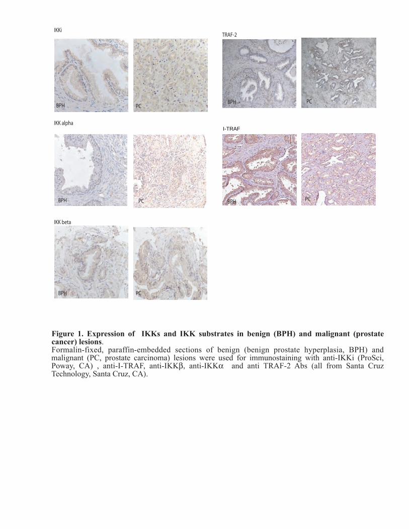
target proteins IKKβ, I-TRAF, and TRAF2 in PC cell lines and PC tumors.
Using prostate samples provided by NU prostate SPORE tissue core we performed immunostaining of more than 60 formalin-fixed paraffin-embedded samples of BPH and PCs using multiple antibodies against IKKi (four different Abs from Imgenix, Santa Cruz., Active Motif, Pro-Sci). We also performed immunostaining with different antibodies to reveal localization of potential IKKi target proteins such as IkBa (we used anti-phospho-IkBa Ab from Cell Signaling), IKKb (two different Abs from Santa Cruz and Imgenix, and Ab against phospho-IKKb from Cell Signaling), TRAF2 and I-TRAF (both from Santa Cruz) in benign and malignant prostate lesions. The staining was reviewed and scored by pathologists at Pathology Core at R. Lurie Cancer Center, who have an extensive experience in quantitative analysis of PC marker expression.
Western blot analysis of the IKKi Ab specificity revealed that the best anti-IKKi antibody was a monoclonal Ab from ProSci that gave only one specific band on Western blots when we used protein extracts from control and treated PC cells (Fig. 2 ). Thus, we analyzed IKKi localization using immunostaining of prostate tissues only with this antibody. The analysis of IKKi staining in prostate tissue samples indicated that IKKi was more intensively expressed in prostate glands than in prostate stromal fibroblasts. There was no significant difference between IKKi staining intensity in BPH and PC lesions. We also have not revealed correlation between IKKi expression and PC grade. When we used ProSci monoclonal antibody, we have not confirmed our previous finding that IKKi has preferential nuclear localization in epithelial cells in PCs (Fig. 2).
The quality of double staining for IKKi and its potential substrates on paraffin prostate sections was not satisfactory. Thus, we used thin serial sections for the analysis of target protein co-localization with IKKi.
There were no significant changes in the expression of IKKb, IKKa, I-TRAF and TRAF-2 in PC in comparison to BPH (Fig. 1). The immunostaining using antibody against phosphorylated IKB-a (Cell Signaling) was not successful (we could not detect reliable signal after immunostaining). The results of immunostaining with anti-phospho-IKKbeta Ab were published by our group recently (Yemelyanov et al., Oncogene, 2006). Overall analyses of expression have not revealed the significant correlation between IKKi and its potential target localization in PCs. Task 3. To study the mechanisms of nuclear transport of IKKi and effect of IKKi localization
on its function.
Careful analyses of subcellular localization of IKKi in prostate samples and PC cell cultures in vitro using ProSci monoclonal Ab that was the most specific according to Western blot analysis (Fig. 2) have not confirmed our initial finding that IKKi has predominantly nuclear localization. As shown in Fig. 3, both endogenous IKKi expressed in PC3 cells and transgenic IKKi expressed in LNCaP-IKKi cells were mostly localized in the cytoplasm of cells. Due to these findings, we have changed the plan of our research as it was not feasible to study the mechanisms of nuclear localization of IKKi.
As a result, the major focus of our research in FY03 and FY04 was shifted towards biological role of IKKi in prostate cells. We found that IKKi gives growth advantage to PC cells and revealed the

Budunova, I.
10
potential involvement of IKKi in the development of hormone refractory stage of PC growth via activation of androgen receptor.
Taking into consideration the newly recognized association of prostate inflammation and prostate cancer that offers one of the greatest opportunities for preventing malignant conversion we continued to study the cross-talk between pro-inflammatory signaling mediated by IKKi and NF-kB and anti-inflammatory signaling mediated by glucocorticoid receptor (GR) in PC cells. We showed that GR functions as a tumor suppressor in prostate cells, and that glucocorticoids and non-steroidal modulators of GR have a strong potential for the treatment of PC patients.
As small inhibitors of IKKi remain to be developed, we searched for other effective strategies of NF-kB blockage in PC cells, and tested growth inhibitory and pro-apoptotic potential of several novel compounds including highly specific IKKβ inhibitors, proteasomal inhibitors as well as dissociated ligands of GR that inhibit NF-kB. We found that proteasomal inhibitors are much more potent than IKK inhibitors in terms of induction of apoptosis in PC cells (Gasparian et al., 2006, abstract # 9).
The detailed findings are described in FY04 report (see above, p. 4 and 5).
Key Research Accomplishments for the entire funding period
Technical achievements:
We have developed technical protocols for : optimal PC cell stable infection by lentiviruses; optimal regimens of selection of transfected cells; enrichment of cells co-infected with YFP by FACS. Evaluation of growth curves for YFP-infected PC cells using fluorescent plate
reader.
We provided consultations on the lentiviral infection of epithelial cells and post-infection selection for numerous researchers at Northwestern University.
We have generated several lentiviruses harboring empty vector, IKKi-FLAG, d.n. IKKi-FLAG,
and generated stably infected PC3 and LNCaP cell lines with different IKKi status co-infected with YFP-expressing lentivirus.
Research findings:
We showed that novel inflammation-related upstream IkappaB kinase IKKi is expressed only in highly malignant androgen-independent PC cells lines.
We found that IKKi is expressed in glandular component of prostate samples including BPH and
PC.
We obtained experimental evidence that IKKi could be involved in the regulation of NF-�B activity in PC cells through a positive feedback loop:
IKKi is highly inducible in PC cells in NF-kB-dependent fashion. IKKi expression on
mRNA and protein levels is increased by NF-kB activators such as IL-1a and TNF-a; and is blocked by NF-kB inhibitors such as proteasome (PS431 and MG132) and IKKbeta (PS1145) inhibitors.

Budunova, I.
11
Transient transfection of PC cell lines with w.t. IKKi resulted in activation of kB.Luciferase reporter, whereas IKKi dominant negative (d.n.) mutant K38A suppressed basal NF-kB activity in those cells.
Different PC cells stably infected with IKKi-lentivirus had increased basal and inducible NF-kB activity.
Blockage of IKKi function by transfection of PC3 cells with IKKi d.n. mutant resulted in the decrease of constitutive expression of endogenous κB-responsive genes IκB-α and IL6.
We found that IKKi gives growth advantage to PC cells:
w.t. IKKi increased LNCaP and PC cell growth in monolayer; w.t. IKKi increased tumorigenicity of LNCaP cells assessed in colony forming assay.
Our results suggest that IKKi plays an important role during the transition to hormone refractory
stage of PC growth:
Androgen receptor (AR) was partially translocated to the nucleus and constitutively activated in IKKi-expressing LNCaP cells even in androgen –depleted medium;
IKKi increased transactivation potential of AR in prostate carcinoma cells;
LNCaP-IKKi cells became partially androgen-independent and could sustain the androgen ablation.
Taking into consideration the newly recognized association between prostate inflammation and
increased risk of PC development, we extended our studies towards cross-talk between pro-inflammatory signaling mediated by IKKi and NF-kB and anti-inflammatory signaling mediated by glucocorticoid receptor (GR) in PC cells.
We showed that glucocorticoids inhibit IKKi expression in PC cells.
We showed that GR functions as a tumor suppressor in prostate cells:
We found that the expression of glucocorticoid receptor (GR) was dramatically decreased
in @ 80% of prostate carcinomas; GR inhibited multiple transcriptional factors involved in proliferation and transformation
in PC cells, including NF-kB; GR decreased expression and inhibited activity of the MAP-kinases (MAPKs) including
p38, JNK/SAPK, Mek1/2 and Erk1/2 in PC cells; Activated GR signaling resulted in strong inhibition of PC cell growth and normalization
of PC cell phenotype assessed by anchorage-independent growth and expression of PC markers.
Compound A, a novel non-steroidal modulator of glucocorticoid receptor that inhibits NF-kB function, inhibited growth and induced apoptosis of highly malignant prostate cancer cells in GR-dependent manner.

Budunova, I.
12
Summary of the Reportable Outcomes for the entire funding period:
Manuscripts:
1. Yemelyanov A., Czwornog J., Chebotaev D., and Budunova I. New methods for gene transfer: advantages of lentivirus-mediated gene transduction. R. Lurie Comprehensive Cancer Center Journal, v.X, No1, p. 21-26. 2. Yemelyanov A., Gasparian A., Lindholm P., Dang L., Pierce J., F. Kisseljov, A. Karseladze, Budunova I. Effect of IKK inhibitor PS1145 on NF-kappaB function, proliferation, apoptosis, and invasion activity in prostate carcinoma cells. Oncogene, 2006, 25(3):387-98. 3. Yemelyanov A., Czwornog J., Chebotaev D., Karseladze A., Kulevitch E., Yang X., Budunova I. Tumor suppressor effect of glucocorticoid receptor in prostate. Oncogene, 2007, 26:1885-1896. 4. Nelius T., S. Filleur, A. Yemeyanov, I. Budunova, E. Shroff , Y. Mirochnik, A. Aurora, D. Veliceasa, W. Xiao, Z. Wang, and O.V. Volpert Androgen receptor targets NFkB and TSP1 to suppress prostate tumor growth in vivo. Int J Cancer. 2007, 121(5):999-1008. 5. Yemelyanov A., Czwornog J., Joshi S., Gera L., Budunova I. Compound A, a novel phyto- modulator of steroid hormone receptors, as a candidate for prostate cancer therapy. In revision. 6. Yemelyanov A., Kobzeva V., Budunova I. Role of IKKi in prostate cancer: A link between inflammation and androgen receptor signaling. Manuscript is under preparation. 7. Gasparian A., Yemelyanov A., Chebotaev D., Kisseljov F., and I. Budunova. Targeting NF-κB in prostate carcinoma cells: comparative analysis of proteasome and IKK inhibitors. Manuscript is under preparation.
Abstracts presented at the local and national meetings: 1. Yemelyanov A., Yao Y, and Budunova I. Possible role of IKKi in the constitutive activation of NF-κB in prostate carcinoma cells. Keystone Symposium: NF-kB: biology and pathology. January11-16, 2004, Snowbird, Utah, p. 101. 2. Budunova I. , Yemelyanov A., Gasparian A., Dang L., Pierce J. Effect of IKK-beta specific
inhibitor PS1145 on NF-kappaB activity and apoptosis in prostate carcinoma cell lines. Proceedings of AACR 45, 2004 (abstract # 4572).
3. Yemelyanov, A., Yao, Y.J, and Budunova, I. IKKi is a component of the positive feedback loop
involved in the constitutive activation of NF-kB in prostate carcinoma cells. Proceedings of AACR 44: 852, 2003.
4. Gasparian, A. V., Yao, Y. J., Slaga T.J. and Budunova, I. V. High sensitivity of prostate carcinoma
cell lines to NF-kB induction. Proceedings of AACR, 44: 1451, 2003.
5. Budunova I. , Yemelyanov A., Gasparian A., Dang L., Pierce J. Effect of IKK-beta specific inhibitor PS1145 on NF-kappaB activity and apoptosis in prostate carcinoma cell lines. Proceedings of AACR 45, 2004 (abstract # 4572).

Budunova, I.
13
6. Yemelyanov A., Czwornong J., Chebotaev D., Karseladze A., Yang X., Budunova I. Expression and function of glucocorticoid receptor in prostate carcinomas and PC cells. The Chicago Signal Trunsduction Symposium, May 2005, Chicago, IL.
7. Yemelyanov A., Czwornong J., Chebotaev D., Karseladze A., Yang X., Budunova I. Expression and
function of glucocorticoid receptor in prostate carcinomas and PC cells. Keystone Symposium: Hormonal regulation of tumorigenesis. February 20-25, 2005, Monterey, CA, p. 43.
8. Yemelyanov A., Czwornong J., Chebotaev D., Karseladze A., Yang X., Budunova I. Expression and
function of glucocorticoid receptor in prostate carcinomas and PC cells. The Chicago Signal Trunsduction Symposium, May 2005, Chicago, IL.
9. Gasparian A., Gasparian N., A. Yemelyanov, D. Chebotaev, F. Kisseljov, and I. Budunova. Targeting
NF-κB in prostate carcinoma cells: comparative analysis of proteasome and IKK inhibitors. Keystone Symposium: NF-kB: 20 years on the road from biochemistry to pathology. March 23-28, 2006, Banff, Alberta, Canada, p. 53.
10. Yemelyanov, A. Gasparian, P. Lindholm, L. Dang, F. Kisseljov, A. Karseladze, and I. Budunova.
Effects of IKK inhibitor PS1145 on NF-κB function, proliferation, apoptosis and invasion activity in prostate carcinoma cells. 28, 2006, Banff, Alberta, Canada, p. 63.
11. Yemelyanov A., Czwornong J., Chebotaev D., Karseladze A., Yang X., Budunova I. Decreased
expression of glucocorticoid receptor in prostate carcinomas and its anti-tumorigenic activity in PC cells in vitro. Proceedings of AACR 47, 2006 (abstract # 5335).
12. Yemelyanov A., Kobzeva V., Budunova I. Role of IKKi in prostate cancer: A link between
inflammation and androgen receptor signaling. Proceedings of AACR, 2007 (abstract # LBA-9158). 13. Budunova I. , Yemelyanov A., Gasparian A. Role of IKKs and transcription factor NF-kB in
prostate tumorigenesis. P. 23. IMPACT DOD meeting, 2007, Atlanta.
Seminars presented by P.I. 1. Constitutive activation of NF-kB in prostate carcinoma cells: possible role of feedback loop involving IKKi. Department of Urology seminar program, Feinberg School of Medicine, Northwestern University, Chicago, September, 2003.
2. Effect of NF-kB inhibitor PS1145 and glucocorticoids on prostate carcinoma cells. Prostate SPORE, R. Lurie Cancer Center, Northwestern University, Chicago, IL, September 2004. 3. Targeting NF-kB transcription factor and IKK kinases in prostate carcinoma cells. The University of Auckland, School of Medicine-Auckland Cancer Society Research Center, Auckland , New Zealand , November 2004 4. Targeting the transcription factor NF-kB and up-stream kinases for intervention of prostate and skin cancer. Ludwig Institute for Cancer Research and Royal Melbourne Hospital, Melbourne, Australia, November 2004. 5. Constitutively active NF-κB transcription factor and IKKb kinase in human prostate carcinoma cells as

Budunova, I.
14
a possible targets for intervention. Epithelial group seminar series. R.Lurie Cancer Center. Northwestern University. December, 2004. 6. Invited oral presentation. Expression and function of glucocorticoid receptor in prostate carcinomas and PC cells. Keystone Symposium:Hormonal regulation of tumorigenesis. February 2005, Monterey, CA, p. 43. 7. Compound A, a novel phyto-modulator of steroid hormone receptors, as a candidate for prostate cancer therapy. Tumor cell biology seminars. R. Lurie Cancer Center. NU. April, 2007. 8. Non-steroidal modulators of steroid hormone receptors as candidates for prostate cancer therapy. Children’s Memorial Research Center, Chicago. May , 2007
Conclusions the entire funding period:
We found that novel inflammation-related upstream IkappaB kinase IKKi is expressed only in highly malignant androgen-independent PC cells lines. IKKi is also expressed in epithelial cells in benign and malignant prostate lesions. Our data provide experimental evidence that IKKi could be involved in the regulation of activity of major anti-apoptotic factor NF-κB in PC cells through a positive feedback loop. Our results also suggest that IKKi may play an important role during the transition to hormone refractory stage of PC growth via its positive effect on the nuclear translocation and activity of androgen receptor in PC cells. Taking into consideration the newly recognized association between prostate inflammation and increased risk of PC development, we extended our studies towards cross-talk between pro-inflammatory signaling mediated by IKKi and NF-kB and anti-inflammatory signaling mediated by glucocorticoid receptor (GR) in PC cells. We showed that GR functions as a tumor suppressor in prostate cells, and that inhibition of transcription factors involved in proliferation and transformation in PC cells, including NF-kB, is the major molecular mechanism of GR anti-tumor activity. Finally, we discovered that Compound A, a novel non-steroidal modulator of glucocorticoid receptor that inhibits NF-kB function, inhibited growth and induced apoptosis of highly malignant prostate cancer cells in GR-dependent manner.
List of personnel receiving pay from the research effort for the entire funding period:
1. Irina Budunova, M.D., Ph.D., P.I.
2. Alexander Yemelyanov, M.D., Ph.D., program investigator
3. Vera Kobzeva, Ph.D., program investigator
4. Chebotaev Dmitry, Ph.D., program investigator
5. Czwornog Jennifer, Research Assistant
Collaborations for the entire funding period:
In 2003 P.I. became a member of R. Lurie Comprehensive Cancer Center/ Northwestern University Prostate SPORE. In frames of prostate SPORE P.I. is collaborating with Dr. O. Volpert , an Associate professor at the Department of Urology at Northwestern University to study the effect of NF-kB blockage on the function of androgen receptor. This collaboration is reflected in the publication Nelius et al., 2007.

Budunova, I.
15
P.I. also started the collaboration with the pathologist Dr. X. Yang, a Professor at the Department of Pathology at Northwestern to study the expression of IKKs and steroid hormone receptors in PCs. The results of collaboration are reflected in the manuscript Yemelyanov et al., 2007.
P.I. continues collaboration with Dr. A. Karseladze, Chair of the Department of Molecular Pathology at N. Blokhin Cancer Research Center (Moscow, Russia) to study the expression of NF-kB and IKKs in PCs. The results of collaboration are reflected in two manuscripts Yemelyanov et al., 2006, Yemelyanov et al., 2007.
Further, P.I. initiated collaboration with Dr. P. Lindholm, an Associate Professor at the Department of Pathology at Northwestern University to study the effect of IKK inhibition on PC cell invasion. The results of this collaboration are included in the manuscript published by Yemelyanov et al.,2006.
During funding period (06.2003-06.2007) P.I., Dr. Irina Budunova has expanded her work towards the search for most effective strategies of NF-kB blockage in PC cells. Her group tested several novel compounds including highly specific IKKβ inhibitor PS1145, proteasomal inhibitor PS341/Velcade (both in collaboration with Millenium Pharmaceuticals Inc. , Cambridge, MA), as well as dissociated ligands of glucocorticoid receptor that inhibit NF-kB via stimulation of negative protein/protein interaction between activated GR and p65, including AL438 (in collaboration with Ligand Pharmaceuticals, San Diego, CA).
Promotions:
The key researcher on this grant, Dr. A. Yemelyanov was promoted to the position of Assistant Professor at the Department of Dermatology, School of Medicine at Northwestern University (Chicago, IL).
Additional funding obtained/applied based on the work supported by award:
In 2004 P.I. initiated studies to evaluate the combined effect of glucocorticoids and IKK inhibitors on PC cell growth. This research “Combinational targeting of NF-kB transcription factor as a novel strategy for apoptosis induction and prostate carcinoma treatment” is supported by developmental project award (to Budunova IV) from Northwestern University Prostate SPORE 5 P50 CA090386-04 (P.I. C. Lee). Looking for the combinational therapeutic approaches to block NF-kB in prostate P.I. became interested in steroidal and non-steroidal ligands of glucocorticoid receptor that potently inhibit NF-kB through negative interaction on protein-protein level. We also showed that glucocorticoids inhibit the expression of IKKi on PC cells. In 2007 P.I. submitted grants “Role of glucocorticoid receptor in prostate tumorigenesis: from experimental studies to clinical applications” and “Compound A, a novel phyto-modulator of steroid hormone receptors, as a candidate for prostate cancer therapy” to NIH and DOD. In 2007 proposals have not been funded; grants will be resubmitted after revision to NIH and DOD prostate program in 2008.
Training of postdoctoral fellows:
1. Dr. Dmitry Chebotaev, 2003- 2006 , Department of Dermatology, NU, Chicago, IL. Currently – Senior Researcher at the Applied Biosystems, Moscow, Russia 2. Dr. Vera Kobzeva, 2005-2006, Department of Dermatology, NU, Chicago, IL. Currently – Program Investigator, Institute of Carcinogenesis, Blokhin Cancer Center, Moscow, Russia.

Budunova, I.
16
References: 1. Peters RT, Liao SM, Maniatis T. IKKepsilon is part of a novel PMA-inducible IkappaB kinase
complex. Mol Cell. 5: 513-522, 2000. 2. Shimada, T. Kawai, T., Kiyoshi, T., Matsumoto M., Inoue, J., Tatsumi, Y., Kanamura, A., and
Akira S. IKK-i, a novel lipopolysaccharide-inducible kinase that is related to IkB kinases. Intenational Immunology. 11: 1357-1362, 1999.
3. Greten FR, Karin M. The IKK/NF-kappaB activation pathway-a target for prevention and treatment of cancer. Cancer Lett. 206(2):193-199, 2004.
4. De MarzoAM, Meeker AK, Zha S., Luo J., Nakayama M., Isaacs WB, and Nelson WG. Human prostate cancer precursors and pathobiology. Urology, 55-62, 2003.
5. Platz E.A., Del Marzo AM. Epidemiology of inflammation and prostate cancer. Journal of Urology. S36-40, 2004.
6. Adli M, Baldwin AS. IKK-i/IKKe controls constitutive, cancer cell-associated NF-B activity via regulation of Ser-536 p65/RelA phosphorylation. J Biol Chem. July 2006, e-publication.
7. Haverkamp J, Charbonneau B, Ratliff TL. Prostate inflammation and its potential impact on prostate cancer: A current review J Cell Biochem. 2007 Oct 22; [Epub ahead of print]
8. De Marzo AM, Nakai Y, Nelson WG. Inflammation, atrophy, and prostate carcinogenesis. Urol Oncol. 2007 , 25(5):398-400.
9. Eddy SF, Guo S, Demicco EG, Romieu-Mourez R, Landesman-Bollag E, Seldin DC, Sonenshein GE. Inducible IkappaB kinase/IkappaB kinase epsilon expression is induced by CK2 and promotes aberrant nuclear factor-kappaB activation in breast cancer cells. Cancer Res. 2005; 65(24):11375-11383.
10. De Bosscher K, Vanden Berghe W, Beck IM, Van Molle W, Hennuyer N, Hapgood J, Libert C, Staels B, Louw A, Haegeman G.A fully dissociated compound of plant origin for inflammatory gene repression. Proc Natl Acad Sci U S A. 2005 Nov 1;102(44):15827-32. Epub 2005 Oct 21.
Appendices:
1. Yemelyanov A., Czwornog J., Chebotaev D., Karseladze A., Kulevitch E., Yang X., Budunova I. Tumor suppressor effect of glucocorticoid receptor in prostate. Oncogene, 2007, 26:1885-1896.
2. T. Nelius, S. Filleur, A. Yemeyanov, I. Budunova, E. Shroff , Y. Mirochnik, A. Aurora, D. Veliceasa, W. Xiao, Z. Wang, and O.V. Volpert Androgen receptor targets NFkB and TSP1 to suppress prostate tumor growth in vivo. Int J Cancer. 2007, 121(5):999-1008.
3. Yemelyanov A., Kobzeva V., Budunova I. Role of IKKi in prostate cancer: A link between inflammation and androgen receptor signaling. Proceedings of AACR, 2007 (abstract # LBA-9158).
4.Budunova I. , Yemelyanov A., Gasparian A. Role of IKKs and transcription factor NF-kB in prostate tumorigenesis. P. IMPACT DOD meeting, 2007, Atlanta.

Budunova, I.
17
Supplemental figures:
Figure 1. Expression and localization of IKKs and their substrates in benign (BPH) and malignant (PC)
lesions.
Figure 2. Comparison of IKKi antibodies : application for Western blotting and immunostaining.
Figure 3. Preferential cytoplasm localization of IKKi
Figure 4. IKKi is involved in constitutive activation of NF-kB in PC cells through a positive feedback loop. Figure 5. IKKi overexpression results in the increased proliferation and tumorigenicity of PC cells. Figure 6. Effect of IKKi on NF-kB status in PC cells. Figure 7. IKKi overexpression results in increased nuclear localization of AR Figure 8. IKKi overexpression results in increased AR function and PC cell resistance to androgen ablation. Figure 9. Effect of CpdA on GR function in PC cells. Figure 10. CpdA inhibits growth of PC cells. Figure 11. CpdA induces apoptosis of PC cells.
IKK alpha
IKK beta
TRAF-2IKKi
Figure 1. Expression of IKKs and IKK substrates in benign (BPH) and malignant (prostate cancer) lesions.Formalin-fixed, paraffin-embedded sections of benign (benign prostate hyperplasia, BPH) and malignant (PC, prostate carcinoma) lesions were used for immunostaining with anti-IKKi (ProSci, Poway, CA) , anti-I-TRAF, anti-IKKβ, anti-IKKα and anti TRAF-2 Abs (all from Santa Cruz Technology, Santa Cruz, CA).
BPH PC
BPH
BPH
BPH
BPHPC
PC
PC
PC
I-TRAF

PC3 cells
IKKi110
82
65
con
tro
l
TNF-
α
IL-1
12 h
PC3 cells
IKKi110
82
65co
ntr
ol
TNF-
α
IL-1
12 h
IKKi (anti-IKKi Ab from ProScie Inc)
BPH PC
IKKi (anti-IKKi Ab from Santa Cruz Inc)
BPH PC
NS
NS
A
B
Figure 2. Comparison of IKKi antibodies: application for Western blotting and immunostaining. Formalin-fixed, paraffin-embedded sections of benign (benign prostate hyperplasia , BPH) and malignant (PC, prostate carcinoma ) lesions were used for immunostaining with two different anti-IKKi antibodies: A - from Santa Cruz Biotech (Santa Cruz, CA) and B - from ProSci laboratories (Poway, CA). PC3 cells that expressed endogenous IKKi , were treated with NF-κB inducers TNF-α (10 nM) and IL-1 (1 M), whole-cell proteins were extracted as described in Yemelyanov et al., 2006. Note: only anti-IKKi polyclonal Ab from Santa Cruz Inc that gave multiple bands on Western blots detected significant amount of IKKi in the nuclei of prostate cells in PC. NS - non-specific protein bands.

PC-3 LNCaP-IKKiwt LNCaP-IKKidn
IKKi (anti-IKKi Ab from ProSci Inc)
IKKi
con
tro
l
IL-1 con
tro
l
IL-1
nuclearfraction
cytosol fraction
Figure 3. Preferential cytoplasmic localization of IKKi. A - Cell were fixed with formalin, permebealized with methanol : acetone mix (1:1), blocked with 10% goat serum and stained with anti-IKKi antibodies (ProSci, Poway, CA) and secondary Abs conjugated with Cy3 (Jackson Immunolabs, (West Grove, PA). B - Nuclear and cytosol protein fractions were isolated from LNCaP cells expressing endogenous IKKi w.t. and analyzed by Western blotting using anti-IKKi Abs (ProSci, Poway, CA). Note: Endogenous and lentivirus-expressed exogenous IKKi were localized at most to the cell cytoplasm. Stimulation of the cells with IL-1 induced very little IKKi nuclear translocation.
A B

control
LNCaP
TNFα TNFα TNFα
6 h 12 h 6 h 12 h 6 h 12 hcontrol control
PC3
IKKi
DU145
IKKα
IKKβ
IKKi
PrPC
LNC
aP
CL-
1
PC3
DU
145
6 hr 12 hr
PC3 cells
control
IL-1
TNFαIKKi
IKKi
LNCaP cells
IL-1
6 hr 12 hr
TNFα
control
IKKi
IKKi
A.1.Northern Blot
A.3. Western Blot
A.2. Northern Blot
2
10
DU145PC3 LNCaP CL1
1
Luci
fera
se a
ctiv
ity
(% t
o c
on
tro
l)
no effect
4
6
8
IKKi w.t.IKKi d.n. (K38A)
B. Luciferase assay: 3x-kappaB-Luc.
Figure 4. IKKi is involved in constitutive activation of NF-κB in prostate carcinoma cells through a positive feedback loop. A1. Expression of IKKα, IKKβ and IKKi in PC cell lines. Northern blot analysis of IKKα, IKKβ and IKKi expression in prostate cells. Note: IKKi is expressed only in highly malignant PC3 and DU145 cells. A2 and A3. Induction of IKKi in prostate cells by cytokines. Prostate cells were treated with TNFα (10 ng/ml), and IL-1 (1 µg/ml), RNA and whole cell proteins were isolated and used for Northern and Western blot analyses to evaluate IKKi expression. B. Effect of IKKi w.t. and d.n. IKKi mutant on NF-κB activity in PC cells. Prostate cells were cotransfected with 5x-κB-Luciferase reporter (FL), Renila luciferase (RL) under minimal promoter, and either IKKi w.t. or IKKi d.n mutant (kindly provided by Dr. T. Maniatis, Harvard University, Harvard, MA). Luciferase activity was measured by dual luciferase assay. FL activity was normalized against RL activity to equilize for transfection efficacy. Note: IKKi w.t. induced NF-κB activity and IKKi d.n. mutant K38A inhibited NF-κB activity in PC cells.

LNCaP-YFP-IKKiw.t.LNCaP-YFP
0
10
20
30
40
50
60
70
80LNCaP-IKKi w.t. (YFP)
LNCaP-V (YFP)
cell
amou
nt x
105
1 2 4 6 8 10 12 14 16 180
3
6
9
12
15
PC3-IKKi w.t. (YFP)
PC3-V (YFP)
2 4 6 8 10
cell
amou
nt x
106
days of cell growthdays of cell growth
IKKi
PC3
LNC
aP
PC3
LNC
aPparental IKKi-lentivirus
C. Western Blot
A.1. Proliferation in cell monolayer
B. Colony formation Assay (0.7% agar)
A.2. Proliferation in cell monolayer
12
Figure 5. IKKi overexpression results in the increased proliferation and tumorigenicity of PC cells. Generation of IKKi w.t. cells. PC3 and LNCaP cells were stably infected with lentivirus expressing human IKKi cDNA (kindly provided by Dr. T. Maniatis, Harvard University, Harvard, MA). For easier tracking, the additional cell lines were co-infected with IKKi and yellow fluorescent protein (YFP) lentiviruses. Control cell lines were infected with either the empty lentivirus (LNCaP-V, PC3-V) or the lentivirus expressing YFP (LNCaP-YFP; PC3-YFP). A. Effect of IKKi on proliferation of prostate cells. Number of IKKi- and empty virus infected was calculated by hemocytometer. B. Effect of IKKi on anchorage independent growth of LNCaP-YFP cells. LNCaP-IKKi-YFP and LNCaP-V-YFP cells were grown in 0.7% soft agar for 3 weeks. Images were obtained by AxioVert 40 CFL inverted microscope equipped with a fluorescent digital camera (Zeiss). C. Western blot analysis of IKKi expression in transfected prostate cell lines.

A.1. Luciferase reporter assay: 5x-κB-Luc.
0
20
40
60
80
100
120
140
PC3-IKKi w.t.
PC3-V
basalactivity
TPA LPSEGFTNFα IL-1
No
rmal
ized
luci
fera
se s
ign
al
basalactivity
TNFα TPA EGF IL-1
0
10
20
30
40
50
No
rmal
ized
luci
fera
se s
ign
al
LNCaP-IKKi w.t.
LNCaP-V
A.2. Luciferase reporter assay: 5x-κB-Luc.
con
tro
l
0.5 h 6 h 16 h con
tro
l
0.5 h 6 h 16 h
IL-1 IL-1
IκBα
LNCaP-VLNCaP-IKKi w.t.
IKKi
GAPDH
con
tro
l
LPS TNFαTNFα /MG132
LPS /MG132 MG132
PC3-V
PC3-IKKiw.t.
p-IκBα (Ser32/36)
p-IκBα (Ser32/36)
B.1. Northern blotC.1. Western Blot
con
tro
l
TNFα/LPS (15 min)MG132 (2 hrs)
IL-1 IL-6 TNFα EGF
p-NF-κB (Ser 536)
p-NF-κB (Ser 536)
LNCaP-V
LNCaP-IKKiw.t.
C.2. Western Blot
Figure 6. Effect of IKKi on NF-kB status in prostate cells. A. IKKi increased basal and inducible activity of NF-κB. Cells were transiently transfected with 5x-κB.Luciferase reporter (FL) kindly provided by Dr. W. Greene (UCSF, San Francisco, CA) and with Renilla Luciferase (RL) under minimal promoter. Cells were treated with TNFa (10 ng/ml), IL-1 (1 µg/ml) and EGF (100 ng/ml), LPS (1 µg/ml ) and TPA (10 µg/ml) for 24 hrs, and Luciferase activity was measured by dual Luciferase assay. FL activity was normalized against RL activity to equalize for transfection efficacy. Note: in both PC cell lines IKKi most significantly increased NF-κB induction by IL-1. B. IKKi increased expression of endogenous κB-dependent genes. Cells were treated with IL-1 (1 µg/ml), total RNA was isolated by TRI reagent (Molecular Research Center, Inc., Cincinnati, OH) and subjected to Northern blotting. Membranes were probed for NF-κB regulated genes: IκBa and IKKi, and for GAPDH as a control for RNA loading. Note: in both PC cell lines IKKi significantly increased the expression of κB-dependent genes. C. IKKi significantly increased phosphorylation of IκBa (Ser32/Ser36) and p65 (Ser536). To better assess phosphorylation, cells were pre-treated with proteasomal inhibitor MG132 (x2 hr), and treated with NF-kB inducers. Whole cell protein extracts were used for Western blot analysis with anti-phospho-Ser32/Ser36 IκBa and anti-phospho-Ser536 p65 Abs (Cell Signaling, Beverly, MA).
LNCaP-IKKiw.t.
FBS FBS + DHT (16 hours) charcoal-stripped FBS charcoal FBS + DHT (16hr)
LNCaP-V
FBS FBS + DHT (16 hours) charcoal-stripped FBS charcoal FBS + DHT (16hr)
Figure 7. IKKi induces AR nuclear localization. IKKi increased nuclear localization of AR induced by DHT. LNCaP-V and LNCaP-IKKw.t.i cells grown in the medium with normal and charcoal-stripped serum were treated with DHT (10-7 x 24 hrs), fixed and used for immunofluorescence with anti-AR Ab (Santa Cruz Biotechnology) followed by Cy-3-conjugated anti-mouse secondary Ab conjugated with Cy3 .

con
tro
l
0.5 h 4 h 16 h con
tro
l
DHT0.5 h 4 h 16 h
PSA
GAPDH
LNCaP-V LNCaP-IKKi w.t.
C. RT-PCR of PSA expression
Figure 8. IKKi increased AR function and PC cell resistance to androgen ablation (B-D). A. Increased resistance of LNCaP-IKKi cells to androgen ablation. LNCaP-IKKi and LNCaP-V cells were grown in the medium with normal and charcoal-stripped serum. Cell number was calculated by hemocytometer. B. Increased basal and DHT-induced activity of AR in LNCaP-IKKi cells. LNCaP-V and LNCaP-AR cells were transfected with MMTV.Luciferase reporter (FL) (ARE.Luc) and with Renilla Luciferase (RL) under minimal promoter. Cells were treated with DHT (10-7 x 36 hrs) in the medium with normal or charcoal-stripped serum. Luciferase activity was measured by dual Luciferase assay. FL activity was normalized against RL activity to equalize for transfection efficacy. C. Increased PSA expression in LNCaP-IKKiw.t. cells. LNCaP-V and LNCaP-IKKiw.t. cells grown in the medium with normal and charcoal-stripped serum were treated with DHT (10-7 0.5-16 hrs). Total RNA was isolated and used for RT-PCR analysis of PSA expression.
0
5
10
15
20
30
35
40
cell
amou
nt x
104
2 4 6 8 10 12 14 16 18 20
days of cell growth
LNCaP-IKKi w.t. (YFP)
LNCaP-V (YFP)
A. Proliferation in cell monolayer.
LNCaP-IKKi w.t.AR immunostaining
LNCaP-VAR immunostaining
B.1. Luciferase reporter assay: ARE-Luc.
LNCaP-IKKi w.t.
LNCaP-V
0
5
10
15
20
25
30
35
40
45
50
basalactivity
DHT
0.001 µM 0.1 µM
No
rmal
ized
luci
fera
se s
ign
al
Standard FBS with androgens
LNCaP-IKKi w.t.
LNCaP-V
0.0
0.5
1.0
1.5
2.0
2.5
3.0
3.5
4.0
basalactivity
DHT
0.001 µM 0.1 µM
No
rmal
ized
luci
fera
se s
ign
al
Adnrogen-deprived FBS
C.2. Luciferase reporter assay: ARE-Luc.

E
Figure 9. Effect of CpdA on GR function in PC cells. B, C. CpdA activated GR nuclear translocation. Cells were treated with CpdA, glucocorticoid FA, and anti-glucocorticoid RU486; B – immunofluorescence; C - Western blotting, nuclear proteins. HDAC-1 and b-tubulin were used as protein loading controls. D. CpdA decreased GR-DNA binding. EMSA, nuclear proteins. LNCaP-GR cells were treated with 0.01% DMSO (control), Dex (10-7 M) and CpdA (10-5
M) for 4 hrs. A, E. CpdA effect on GR function. PC cells weretransiently transfected with TAT.Luc (A) or κB.Luc (E), and control Renilla Luciferase reporters, treated with Dex (10-6 M) and CpdA (10-5 M) for 24 hrs. For activation of NF-κB cells were co-transfected with CMV.IKKβ plasmid (in E). Reporter activity was assessed by dual Luciferase assay, and presented as factor of change. Note: CpdA induces GR nuclear translocation; inhibits DNA binding and GR transactivation, but induces GR transrepression in Luciferase assay.
0.0
1.0
2.0
3.09.5
10.0
10 1 0.1 10 1 0.1
CpdA (µM) FA + CpdA(µM)
RU486
RU486 + FA
FA
norm
aliz
edH
RE
-luc.
sig
nal
GR-DNAcomplex
cold probedilutions
cont
rol
Cpd
A
FA FA +
Cpd
A
1:5 1:10 1:20
FA
FA
HREmut
control CpdAFA
cont
rol
Cpd
A
FA
GR
HDAC-1
GR
β-tubulin
nuclearextract
whole celllysate
NF-κB-luciferase (normalized)
LNCaP-GRPC3DU145
control CpdA CpdA +IKKβ
IKKβ
40
30
20
10
0
A B
C D

Figure 10. Highly malignant PC cells are sensitive to the growth inhibitory effect of CpdA. A, B. Non-transformed prostate cells PWR-1E and PC cells LNCaP, DU145 and PC3 were treated with 0.01% DMSO (control) or CpdA (5x10⁻⁶ M) for 1-12 days. Cell number/well was determined by counting, and the absolute number of cells/well is presented as mean +/- S.D for each experimental group (three wells/group). Note: high sensitivity of androgen-independent PC3 and DU145 cells to CpdA (Fig. 5A).

PARP
cleavedPARP
Con
trol
TNFα
Cpd
A (2
day
s)
Cpd
A (2
day
s) /
TNFα
DE
X (2
day
s)
DE
X (
2 da
ys)/
TN
Fα
Con
trol
DEXCpd
A
DEXCpd
A
DEXCpd
ADEXC
pdA
2 days 4 days 6 days 8 days
DU145
A. Spontaneous apoptosis by CpdA ( Western blotting)
DU145
PC3
PARP
cleavedPARP
PARP
cleavedPARP
B. Sensitization to TNFα-induced apoptosis by CpdA (Western blotting)
Figure 11. CpdA induces apoptosis in prostate cells. A. PC3 and DU145 cells were treated for 1-8 days with DMSO (control), CpdA (2x10⁻⁶ M) and Dexamethasone (10-6 M). Nuclear cell extracts were analyzed for PARP cleavage by Western blotting using anti-PARP antibody (Cell Signaling, Danvers, MA). B. To study prostate cell sensitization to apoptosis by CpdA we used TNFα (10 ng/ml x 16 hrs). Note: CpdA induced apoptosis after 6-8 day treatment and sensitized cells to TNF αααα-induced apoptosis after 2 day treatment.

ORIGINAL ARTICLE
Tumor suppressor activity of glucocorticoid receptor in the prostate
A Yemelyanov1, J Czwornog1, D Chebotaev1, A Karseladze2, E Kulevitch2, X Yang3
and I Budunova1
1Department of Dermatology, Feinberg Medical School, Northwestern University, Chicago, IL, USA; 2Department of Pathology, NNBlokhin Cancer Research Center, RAMS, Moscow, Russia and 3Department of Pathology, Feinberg Medical School, NorthwesternUniversity, Chicago, IL, USA
Glucocorticoids are extensively used in combinationchemotherapy of advanced prostate cancer (PC). Littleis known, however, about the status of the glucocorticoidreceptor (GR) in PC. We evaluated over 200 prostatesamples and determined that GR expression was stronglydecreased or absent in 70–85% of PC. Similar to PCtumors, some PC cell lines, including LNCaP, also lackGR. To understand the role of GR, we reconstituted itsexpression in LNCaP cells using lentiviral approach.Treatment of LNCaP-GR cells with the glucocorticoidsstrongly inhibited proliferation in the monolayer culturesand blocked anchorage-independent growth. This wasaccompanied by upregulation of p21 and p27, down-regulation of cyclin D1 expression and c-Myc phospho-rylation. Importantly, the activation of GR resulted innormalized expression of PC markers hepsin, AMACR,and maspin. On the signaling level, GR decreasedexpression and inhibited activity of the MAP-kinases(MAPKs) including p38, JNK/SAPK, Mek1/2 and Erk1/2.We also found that activation of GR inhibited activityof numerous transcription factors (TF) including AP-1,SRF, NF-jB, p53, ATF-2, CEBPa, Ets-1, Elk-1, STAT1and others, many of which are regulated via MAPKcascade. The structural analysis of hepsin and AMACRpromoters provided the mechanistic rationale for PCmarker downregulation by glucocorticoids via inhibitionof specific TFs. Our data suggest that GR functions asa tumor suppressor in prostate, and inhibits multiplesignaling pathways and transcriptional factors involved inproliferation and transformation.Oncogene (2007) 26, 1885–1896. doi:10.1038/sj.onc.1209991;published online 2 October 2006
Keywords: prostate carcinoma; PIN; glucocorticoidreceptor; PC marker; transcription factor; MAPKs
Introduction
Glucocorticoid hormones regulate proliferative, inflam-matory and immune responses. For years, glucocorti-coids have been extensively used for the treatment ofhormone refractory prostate cancer (HRPC), and thecombination of paclitaxel and dexamethasone remains astandard treatment for HRPC patients in the US andother countries (reviewed by Fakih et al., 2002).Glucocorticoids have also been used as the ‘standard’therapy arm in several randomized phase II–III clinicaltrials for the combination therapy of HRPC (Fakihet al., 2002; Koutsilieris et al., 2002).
The cellular response to glucocorticoids is mediatedthrough a highly specific glucocorticoid receptor (GR).In the absence of glucocorticoids, GR is sequestered inthe cytoplasm by chaperone proteins. Following ligandbinding, the GR dissociates from the chaperones andforms homodimers, which enter the nucleus. Thereare two major mechanisms of gene regulation by GR(De Bosscher et al., 2003; Necela and Cidlowski, 2004).The direct positive transcriptional regulation (transacti-vation) occurs via binding of the GR homodimer topalindromic promoter DNA sequences called glucocor-ticoid-response elements. The indirect regulation ismediated via crosstalk with other transcription factors(TFs), including activator protein 1 (AP-1), nuclearfactor kappa-B (NF-kB), signal transducer and activa-tor of transcription (STAT)-5, mothers against DPPhomolog 3 (SMAD3), etc. (De Bosscher et al., 2003;Necela and Cidlowski, 2004). Most of such GR–TFinteractions repress the activity of partner TFs and theirtarget genes (transrepression). Recently, the additionalmechanism of indirect gene regulation by GR has beendiscovered where GR blocks mitogen-activated proteinkinases (MAPKs) (Kassel et al., 2001; Bruna et al.,2003). Indirect, DNA-independent mechanisms of GRgene regulation appear to be critical for the anti-inflammatory effects (Schacke et al., 2002), whereastheir role in the growth inhibition by glucocorticoids hasnever been addressed.
Although the clinical effect of glucocorticoids inHRCP patients is well known, the objective responseshave been found only in 20–25% of patients (Fakihet al., 2002). The limited effect of glucocorticoids inprostate carcinoma (PC) patients implies the changes in
Received 16 June 2006; revised 27 July 2006; accepted 28 July 2006;published online 2 October 2006
Correspondence: Dr I Budunova, Feinberg Medical School, Depart-ment of Dermatology, Northwestern University, Ward Building 9-332,303 East Chicago Avenue, Chicago, IL 60611, USA.E-mail: [email protected]
Oncogene (2007) 26, 1885–1896& 2007 Nature Publishing Group All rights reserved 0950-9232/07 $30.00
www.nature.com/onc
GR expression, function and/or availability of GRtargets in PC cells. Indeed, we and others showed thatdifferent types of tumor cells lose their sensitivity togrowth inhibition and apoptosis by glucocorticoidseither because of the loss of GR expression or becauseof the abnormal GR function (Ray, 1996; Budunovaet al., 1997; Greenstein et al., 2002). These observationssuggest that intact GR signaling is crucial for the growthcontrol of lymphoid and epithelial cells and that in sometissues GR may act as a tumor suppressor.
Despite the use of glucocorticoids in the standardcombinational therapies of PC patients, the informationregarding GR expression in PCs is surprisingly limitedand conflicting (Mohler et al., 1996; Nishimura et al.,2001). To our knowledge, GR expression in earlyprostate lesions such as intraepithelial neoplasia (PIN)has never been evaluated. Furthermore, GR function inthe prostate cells and its role in PC have never beenstudied, even though the growth inhibitory effect ofglucocorticoids in GR-positive human and rat prostatecells has been reported (Nishimura et al., 2001). Theseprevious studies chiefly attribute growth inhibitoryeffect of glucocorticoids to the inhibition of NF-kBTF (Nishimura et al., 2001).
Here, we for the first time present the comprehensiveanalysis of GR expression changes in the course ofprostate tumorigenesis, and determine the effect ofactivated GR signaling on proliferation and the main-tenance of transformed phenotype by PC cells.
Results
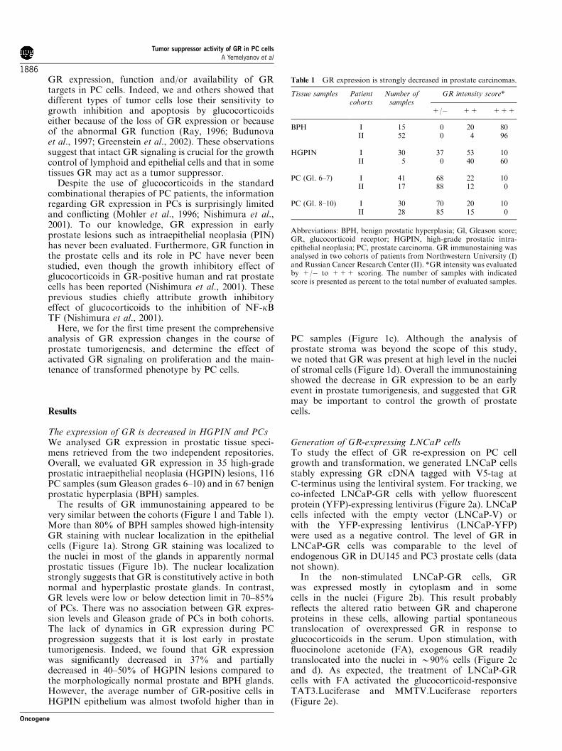
The expression of GR is decreased in HGPIN and PCsWe analysed GR expression in prostatic tissue speci-mens retrieved from the two independent repositories.Overall, we evaluated GR expression in 35 high-gradeprostatic intraepithelial neoplasia (HGPIN) lesions, 116PC samples (sum Gleason grades 6–10) and in 67 benignprostatic hyperplasia (BPH) samples.
The results of GR immunostaining appeared to bevery similar between the cohorts (Figure 1 and Table 1).More than 80% of BPH samples showed high-intensityGR staining with nuclear localization in the epithelialcells (Figure 1a). Strong GR staining was localized tothe nuclei in most of the glands in apparently normalprostatic tissues (Figure 1b). The nuclear localizationstrongly suggests that GR is constitutively active in bothnormal and hyperplastic prostate glands. In contrast,GR levels were low or below detection limit in 70–85%of PCs. There was no association between GR expres-sion levels and Gleason grade of PCs in both cohorts.The lack of dynamics in GR expression during PCprogression suggests that it is lost early in prostatetumorigenesis. Indeed, we found that GR expressionwas significantly decreased in 37% and partiallydecreased in 40–50% of HGPIN lesions compared tothe morphologically normal prostate and BPH glands.However, the average number of GR-positive cells inHGPIN epithelium was almost twofold higher than in
PC samples (Figure 1c). Although the analysis ofprostate stroma was beyond the scope of this study,we noted that GR was present at high level in the nucleiof stromal cells (Figure 1d). Overall the immunostainingshowed the decrease in GR expression to be an earlyevent in prostate tumorigenesis, and suggested that GRmay be important to control the growth of prostatecells.
Generation of GR-expressing LNCaP cellsTo study the effect of GR re-expression on PC cellgrowth and transformation, we generated LNCaP cellsstably expressing GR cDNA tagged with V5-tag atC-terminus using the lentiviral system. For tracking, weco-infected LNCaP-GR cells with yellow fluorescentprotein (YFP)-expressing lentivirus (Figure 2a). LNCaPcells infected with the empty vector (LNCaP-V) orwith the YFP-expressing lentivirus (LNCaP-YFP)were used as a negative control. The level of GR inLNCaP-GR cells was comparable to the level ofendogenous GR in DU145 and PC3 prostate cells (datanot shown).
In the non-stimulated LNCaP-GR cells, GRwas expressed mostly in cytoplasm and in somecells in the nuclei (Figure 2b). This result probablyreflects the altered ratio between GR and chaperoneproteins in these cells, allowing partial spontaneoustranslocation of overexpressed GR in response toglucocorticoids in the serum. Upon stimulation, withfluocinolone acetonide (FA), exogenous GR readilytranslocated into the nuclei in B90% cells (Figure 2cand d). As expected, the treatment of LNCaP-GRcells with FA activated the glucocorticoid-responsiveTAT3.Luciferase and MMTV.Luciferase reporters(Figure 2e).
Table 1 GR expression is strongly decreased in prostate carcinomas.
Tissue samples Patientcohorts
Number ofsamples
GR intensity score*
+/� ++ +++
BPH I 15 0 20 80II 52 0 4 96
HGPIN I 30 37 53 10II 5 0 40 60
PC (Gl. 6–7) I 41 68 22 10II 17 88 12 0
PC (Gl. 8–10) I 30 70 20 10II 28 85 15 0
Abbreviations: BPH, benign prostatic hyperplasia; Gl, Gleason score;GR, glucocorticoid receptor; HGPIN, high-grade prostatic intra-epithelial neoplasia; PC, prostate carcinoma. GR immunostaining wasanalysed in two cohorts of patients from Northwestern University (I)and Russian Cancer Research Center (II). *GR intensity was evaluatedby +/� to +++ scoring. The number of samples with indicatedscore is presented as percent to the total number of evaluated samples.
Tumor suppressor activity of GR in PC cellsA Yemelyanov et al
1886
Oncogene

GR signaling blocked proliferation and anchorage-independent growth but did not induce apoptosis inLNCaP-GR cellsWe then studied the effect of restored GR signaling onLNCaP growth in monolayer and in soft agar. We tookadvantage of YFP expression in the LNCaP-GR-YFPcells to measure the actual number of cells/well.Glucocorticoid treatment of LNCaP-GR-YFP cellsresulted in a strong growth inhibition (Figure 3a),whereas producing no significant effect on controlLNCaP-V and LNCaP-YFP cells (data not shown).
On molecular level, the decreased proliferation wasaccompanied by upregulation of cyclin-dependentkinase inhibitors p21 and p27, decreased expression of
cyclin D1 and proliferation marker Ki67, and a lowerc-Myc phosphorylation (Figure 3c and d). Interestingly,the expression of p21 was increased in LNCaP-GR cellsin comparison to LNCaP-V cells even without hormonetreatment. This may be due to GR partial spontaneousnuclear translocation described above.
To assess the transformation levels in vitro, wemeasured anchorage-independent growth in soft agar.Even without FA, both the number and the size ofthe colonies formed by LNCaP-GR-YFP cells weredecreased compared to the LNCaP-YFP control (datanot shown). Upon glucocorticoid treatment, colonyformation by LNCaP-GR-YFP cells was drasticallydecreased (Figure 3b).
GFP-tracker GR-V5 tag
LNCaP-YFP-GR
a
d e
0
4080
120
160
200
FA
FL/
Ren
illa
ratio
TAT-Luc. MMTV-Luc.Reporter
+ +- -
GR-V5 tag
FA (24 hrs)+-
118
85
kDa LNCaP-GR
HDAC1
LNCaP-GR
GR-V5 tagb
LNCaP-GR + FA
GR-V5 tagc
50 µ
m
50 µ
m
50 µ
m
Figure 2 Characterization of GR in LNCaP-GR cells. (a) Monitoring of live LNCaP-GR cells. LNCaP-GR-V5 cells were co-infectedwith YFP-expressing lentivirus and stained with Ab against V5. (b–d) Glucocorticoid-induced nuclear translocation of GR. LNCaP-GR-V5 cells treated with vehicle (b) or FA, 10�7 M� 24 h (c) were stained with Ab against V5. (d) Western blot analysis of GR innuclear protein extracts from control and FA-treated LNCaP-GR cells. (e) GR activity in dual Luciferase assay. FA-treated(10�7M� 24 h) LNCaP-GR cells were transiently transfected with TAT- and MMTV-FL reporters, and RL reference reporter.FL activity was normalized against RL activity to equalize for transfection efficacy. The results of one representative experiment(three wells/experimental group) are shown as mean7s.d.
Figure 1 Expression of GR in BPH, PC and HGPIN. Immunolocalization of GR in paraffin sections of prostate tissues. (a) BPH;(b) PC (Gleason score 7); (c) HGPIN and (d) prostate stroma. Note: Low GR expression in PC (B1) and high GR expression inapparently normal prostate (B2) combined with positive nuclear GR staining in prostate stromal cells (d).
Tumor suppressor activity of GR in PC cellsA Yemelyanov et al
1887
Oncogene

As epidermal growth factor (EGF) signaling isimportant for PC growth and transition to the HRPCstage and triggers PC cell proliferation in vitro(Mimeault et al., 2003), we chose EGF as physiologi-cally relevant stimulus to assess the GR effect on theinduced PC cell growth. The recombinant EGF sig-nificantly augmented the growth of LNCaP-V andLNCaP-YFP cells (data not shown) as well asLNCaP-GR-YFP cells, both in monolayer and in softagar (Figure 3a and b). The EGF effect was stronglyinhibited by FA in LNCaP-GR-YFP cells (Figure 3aand b), but not in control cells (data not shown). Thus,activated GR strongly suppressed proliferation andanchorage-independent growth. This inhibitory effectwas not attenuated by EGF, a well-known mitogenimplicated in the progression of PC.
In some cell types including lymphocytes, glucocorti-coid treatment may cause apoptosis (Bourcier et al.,2000; Greenstein et al., 2002). As shown in Figure 3a,FA significantly reduced the number of LNCaP cells ondays 6–12 of the treatment. However, the analysis ofthe poly-(ADP-ribose) polypeptide (PARP) cleavage,
mitochondrial potential and caspase activity in LNCaP-GR cells treated with FA did not reveal significantproapoptotic effect of glucocorticoids in PC cells (datanot shown).
GR activation normalized the expression of PC markersTo further evaluate the effect of GR signaling, weinvestigated several early and medium/late PC markerswhose expression typically changes during prostatetumorigenesis. For the profiling, we selected maspinthat is usually downregulated in PCs, hepsin, whichis upregulated in PCs and alpha-methylacyl-CoAracemase (AMACR) whose expression increases early,in both HGPIN and PC lesions (Chen et al., 2003;Ananthanarayanan et al., 2005). Western blot analysisand semiquantitative reverse transcriptase–polymerasechain reaction (RT–PCR) showed that in LNCaP-GRcells hepsin and AMACR were downregulated, whereastumor suppressor maspin was upregulated upon FAtreatment (Figure 4). Interestingly, the expression of PCmarker genes was partially normalized in LNCaP-GR
Control
EGF
FA
EGF/FA
LNCaP-GR
LNCaP-V
p-c-MycKi670
20406080
100%
pos
itive
cel
ls
Ki67 DAPI
p-c-Myc DAPI
LNCaP-V + FA
p-c-Myc
Ki67 DAPI
DAPI
LNCaP-GR + FA
Cylin D1
β-tubulin
LNC
aP-G
R
LNC
aP-V
p21
p27
p-c-Myc
FA (3 days)
+--
0
30
60
90
120
150
1 d 3 d 6 d 10 d 12 d
cell
amou
nt x
105
EGFFA
FA + EGFcontrol (puncate)
treatment with FA (10-7 M)
c.1
500 µm
50 µ
M c.2
50 µ
M
500 µm
500 µm 500 µm
Figure 3 Inhibition of proliferation and anchorage-independent growth of LNCaP-GR-YFP cells by glucocorticoid. (a) Effect of FAon LNCaP-GR-YFP cell growth in monolayer. Number of LNCaP-GR-YFP cells treated with vehicle, FA (10�7M), EGF (5ng/ml) orEGFþFA was measured by YFP fluorescence using a plate reader. The results of one representative experiment (three wells/experimental group) are shown as mean7s.d. (b) Effect of FA on anchorage-independent growth of LNCaP-GR-YFP cells. LNCaP-GR-YFP cells were grown in 0.6% soft agar for 2 weeks in the presence of FA (10�7M), EGF (5ng/ml), EGFþFA or vehicle (0.1%ethanol). (c) Immunocytochemical analysis of Ki67 proliferation marker and phosphorylated form of c-Myc. LNCaP-V and LNCaP-GR cells were treated with 10�7M FA for 72h. (c1) The immunocytochemistry results were quantitated as a percent of the positivelystained cells to all cells (DAPI) in the field of view of microscope. Totally 500 cells were evaluated in each group. (d) Western blotanalysis of cell cycle-related proteins. The expression of cyclin D1, p21, p27 and phosphorylation of c-Myc was evaluated by Westernblotting in whole-cell protein extracts from LNCaP-YFP and LNCaP-GR-YFP cells treated with vehicle (�) and 10�7M FA� 72 h (þ ).
Tumor suppressor activity of GR in PC cellsA Yemelyanov et al
1888
Oncogene

cells even without FA treatment. This could beattributed to the partial nuclear localization of GR inthe untreated LNCaP-GR cells (see Figure 2). Insummary, we conclude that the restoration of GRsignaling resulted in overall normalization of PC cellphenotype.
GR activation blocked MAPK activity in LNCaP cellsThe inhibition of MAPKs is an important regulatorymechanism by GR (Kassel et al., 2001; Bruna et al.,2003; Necela and Cidlowski, 2004). Therefore, weexamined the GR effect on the basal and inducibleactivity of MAPKs dual-specificity mitogen-activatedprotein kinase 1 and 2 (Mek1/2), extracellular signal-regulated kinase 1 and 2 (Erk1/2), c-Jun NH2-terminalkinase (JNK)/stress-activated protein kinase (SAPK)and p38 using Western blot analysis with antibodies(Abs) specific for the active, phosphorylated forms ofthe respective kinases.
The levels of MAPK expression and phosphor-ylation were not affected by glucocorticoid FAin vector transfected LNCaP cells resistant to thegrowth-inhibitory effect of glucocorticoids (data notshown). In contrast, the LNCaP-GR cells had muchlower basal levels of phosphorylated forms of Mek1/2,p38 and JNK/SAPK (Figure 5a), again likely reflectingpartial GR activation discussed above. FA treatmentcaused dramatic time-dependent decrease of MAPKphosphorylation in LNCaP-GR cells. The level of
phospho-MAPKs phosphorylation was decreased bythe second day of treatment and further diminishedduring 3–6 day course of FA treatment (Figure 5a). Therelatively slow inhibition of MAPK phosphorylation byglucocorticoids is in line with the previous findings(Kassel et al., 2001; Greenberg et al., 2002).
Interestingly, our experiments revealed that glucocor-ticoids also reduced the total amount of MAPKproteins. Mek1/2, p38 and SAPK/JNK protein levelsdecreased after 24 h FA treatment, and remained at thislevel thereafter (Figure 5a). Semiquantitative RT–PCRanalysis of Mek1, Mek2, Erk1, Erk2, p38 and JNK/SAPK has not revealed significant inhibition at themRNA level (data not shown). Therefore, glucocorti-coid treatment may have affected either translation orstability of MAPK proteins.
MAPK cascade is activated by growth factorsincluding EGF, cytokines and stress (reviewed byMaroni et al., 2004). We investigated possible GR effecton induced MAPK phosphorylation using the inducersof specific MAPKs (Maroni et al., 2004). We used EGFfor Mek1/2 and Erk1/2 activation, and interleukin(IL)-1 or tumor necrosis factora (TNF)a for JNK/SAPKand p38 activation. IL-1 and EGF activated the corres-ponding MAPKs in LNCaP-V control cells within5–15min of treatment (Figure 5b and data not shown).In LNCaP-GR cells, the effect of studied inducers onMek1/2 and SAPK/JNK was preserved, but the effecton Erk1/2 and p38 phosphorylation was either weak orabsent even without hormone treatment (Figure 5b). FA
Maspin
Hepsin
AMACR
GAPDH
β-tu
bulin
norm
aliz
ed
FA (3 days)
LNC
aP-V
LNC
aP-G
R
+- -
a b
-
LNC
aP-V
+-
LNC
aP-G
R
01234
01234
1 3
1 3
1 3
1 2
2
2
2 2 2
2
2
2
2
3
1 3
1 3
01234
1 3
GA
PD
Hno
rmal
ized
0
1
2
3
1 3
01234
1 3
01234
1 3
1 2
2
2
2
3
1 3
1 3
1 3
Maspin
Hepsin
AMACR
FA (3days)
β-tubulin
Figure 4 Effect of glucocorticoid FA on the expression of PC markers in LNCaP-GR cells. (a) Semiquantitative RT–PCR analysis ofthe PC markers expression. Total RNA from LNCaP-V and LNCaP-GR cells treated with vehicle (control) or with 10�7 M FA for 72 hwas subjected to two-step RT–PCR. The amount of PCR products was measured and normalized to the amount of GAPDH PCRproduct. Quantitative data are presented as the ratio of GAPDH-normalized amount of PCR product in FA-treated vs vehicle-treatedcells. (b) Western blot analysis of the PC markers expression. Protein expression was analysed in whole-cell protein extracts fromLNCaP-V and LNCaP-GR cells treated with 10�7 M FA for 72. Signals were quantified as described in Materials and methods.
Tumor suppressor activity of GR in PC cellsA Yemelyanov et al
1889
Oncogene

pretreatment decreased the effects of EGF and IL-1 onMAPK activation even further (Figure 5b). Similarly,p38 and SAPK/JNK phosphorylation upon TNFatreatment was also inhibited in LNCaP-GR cellspretreated with glucocorticoids (data not shown).
The negative effect of glucocorticoids on Erk1/2 andp38 phosphorylation is known to be associated withincreased expression of MAPK phosphatase 1 (MKP1)(Kassel et al., 2001). We found that the expression ofMKP1 protein was indeed increased in LNCaP-GR cellstreated with FA (Figure 5c).
Overall our studies strongly suggest that GR tightlyregulates both constitutive and inducible activity ofmultiple MAPKs in PC cells.
GR regulated TFs in PC cellsOne important mechanism of gene regulation by GRinvolves its interaction with other TFs. We utilized novel
protein–DNA array technology for simultaneous assess-ment of the DNA-binding activity of multiple TFs. Theeffect of GR on TFs was evaluated in LNCaP-GR cellsafter 3 days of FA treatment, the time point whenMAPKs were strongly inhibited, but the effect on cellgrowth was modest. Vehicle-treated LNCaP-V cellswere used as an additional control to evaluate cons-titutive DNA binding. Only reproducible X2-foldchanges in DNA binding were pursued. We identifiedmultiple TFs affected by activated GR in LNCaP cells(Figure 6a). Interestingly, B85% of those TFs weredownregulated. GR-activated interferon g activation siterecognized by STAT3, and orphan nuclear receptor a.Conversely, GR inhibited several TFs known to interactdirectly with GR and involved in transrepression suchas AP-1, NF-kB, CAAT/enhancer binding protein(C/EBP)a, activating transcription factor (ATF)-2(CREB-BP1), p53 and SMAD3 (see references inFigure 6a). GR also blocked several TFs recognized as
1 2 3 6- -
p-Mek1/2(ser217/221)
p-p38(Thr180/Tyr182)
p-SAPK/JNK
p-Mek1/2(ser217/221)
p-Erk1/2(Thr202/Tyr204)
cont
rol
EG
F
cont
rol
FA EG
F
EG
F+
FA
LNCaP-V LNCaP-GR
Mek1/2
Erk1/2
FA (days)
Mek1/2
actin
p-p38(Thr180/Tyr182)
p38
p-SAPK/JNK(Thr183/Tyr185)
SAPK/JNK(Thr183/Tyr185)
p38
SAPK/JNK
cont
rol
IL-1
cont
rol
FA IL-1
IL-1
+FA
actin
LNCaP-V LNCaP-GR
FA (3 days)EGF (15 min.)
FA (3 days)IL-1 (15min.)
MKP1
a
c
b
Figure 5 Inhibition of MAPK expression and activity by glucocorticoid in LNCaP-GR cells. MAPK expression and phosphorylationwere analysed by Western blotting in whole-cell proteins extracts isolated from LNCaP-GR and LNCaP-V cells treated as indicatedbelow. (a) Effect of FA on the constitutive MAPKs expression and activity. LNCaP-GR cells were treated with vehicle or 10�7 M FAfor indicated time. (b) Effect of FA on the inducible MAPKs activity. LNCaP-GR and LNCaP-V cells were pretreated with 10�7 M FAfor 3 days and MAPKs activity was induced by 15min treatment with EGF (100 ng/ml); IL-1 (10 nM) or TNFa (10 ng/ml). (c) Effect ofFA on MKP1. LNCaP-GR cells were treated with vehicle or 10�7 M FA for indicated time.
Tumor suppressor activity of GR in PC cellsA Yemelyanov et al
1890
Oncogene

MAPK substrates/targets such as AP-1, SRF, Ets-1,Elk-1, STAT1, C/EBPa, GATA4, ATF2, nuclear factorof activated T cell (NFAT)-c, PAX6 and EGR1 (seereferences in Figure 6a of Supplementary material #1).
To validate the results of promoter array and toinvestigate the functional consequences of GR-induced
changes in TF-DNA binding, we employed reporterassays. Reporter constructs for NF-kB, Ets-1, Elk-1,SRF, STAT1/ISRE, STAT1 and NFATc factors withLuciferase under promoters containing the appropriatebinding sites (similar or identical to ones in TF array)were transfected into LNCaP-GR cells (Figure 6b). We
TF name
Down-Regulated TF
FRA-1/JUNAP-1SRF
1
5.23.52.5
+++
SAPK/JNKErk1/2
3
Ets-1Elk-1
6.27.5
n/an/a
Erk1/2, p38SAPK/JNK
STAT1/ISRE 3.1 + Erk1/2
C/EBPα 2.6 + Erk1/2
GATA4 3.7 n/a Erk1/2
ATF2 3.6 +Erk1/2, p38,JNK1/2
2
SMAD3 2.2 + p38
NF-κB 3.6 + multiple
p53 3.7 + n/a
EGR1 2.8 n/a Erk1/2SAPK/JNK
PAX6 3.4 n/a Erk1/2, p38
NFAT-c 2.8 + Erk1/2SAPK/JNK
Up-Regulated TF
STAT3/GAS 2.3 +Erk1/2SAPK/JNK
RORα 3.1 n/a n/a
Ref
De Bosscher et al., 2003; Adcock, 2001; Necela andCidlowski, 2004; Pulvere et al., 1991; Derijard et al.,1994; Inostroza et al., 2005; Karaganni et al.,1994
Bebied et al., 2003; Foulds et al., 2004; Ricote et al.,2006; Koul et al., 2004
De Bosscher et al., 2003; Adcock, 2001; Necela andCidlowski, 2004;, Xuan et al., 2005
Rudiger et al., 2002; Prusty et al., 2002
Liang et al., 2001
van Dam et al., 1995; Morton et al., 2004;Almlof et al., 1998
FL/
Ren
illa
ratio
NF-κB Ets-1 Elk-1 SRF
FAcontrol
STAT1(ISRE)
STAT1 NFATc0.0
0.5
1.0
1.5
2.0
2.5
3.0
3.5
STAT3
Adcock, 2001; Song et al.,1999; Hayes et al., 2003
De Bosscher et al., 2003; Schacke et al., 2002;Adcock, 2001; Necela and Cidlowski, 2004
Sengupta and Wasylyk, 2001
Rolli-Derikinderen et al., 2003
Mikkola et al., 1999
Adcock, 2001; Porter et al., 2000; Lerner et al., 2003
De Bosscher et al., 2003; Adcock, 2001; Necela andCidlowski, 2004; Lerner et al., 2003
a
b
Figure 6 Analysis of glucocorticoid effect on TF activity in LNCaP-GR cells. (a) Analysis of multiple TF basal activity using Protein-DNA array. Nuclear protein extracts from vehicle-treated and FA-treated (10�7 M FA� 72 h) LNCaP-GR cells were used to analyseactivity of multiple TFs (Protein-DNA interaction array, Panomics Inc., Fremont, CA, USA). (1) TF-DNA-binding change (folds ofdecrease or increase of DNA binding in FA-treated cells compared to control); (2) TF is a known partner for GR (þ ) and (3) TF is aknown down-stream target for indicated MAPK. N/A – literature data are not available. See complete references in Supplementarymaterial #1. (b) Validation of the Protein-DNA array. The array data were validated by transient transfections of LNCaP-GR cellswith corresponding Luciferase reporter vectors and RL reporter. Cells were treated with vehicle or FA (10�7M� 24 h). The transfectiondata are presented as described in Figure 2e. The results of one representative experiment (three wells/experimental group) are shownas mean7s.d.
Tumor suppressor activity of GR in PC cellsA Yemelyanov et al
1891
Oncogene

were able to confirm transrepression of all studied TFs,with the exception of NFATc.
To link our data on TF regulation by GR to theregulation of PC markers by GR/glucocorticoids, wescreened promoter sequences of AMACR, hepsin andmaspin (between positions �2000 and þ 1 from thetranscription start sites) for the appropriate binding sitesusing online Transcription Element Search System(TESS) (Schug and Overton, 2005). We found thatpromoters of hepsin and AMACR contained bindingsites for TFs inhibited by GR including C/EBP-a, Ets-1,Elk-1, NFATc, SRF and GATA4 (Figure 7).
Discussion
Despite the wide use of glucocorticoids for PCtreatment, the changes of GR expression duringprostate tumorigenesis and its role in the prostate cellsremain unknown. Here, we developed a comprehensivepicture of GR expression during prostate tumorigenesis.We found that GR expression was decreased or absentin 70–85% of PCs compared to apparently normalprostate or BPH. We also revealed that the decrease inGR expression occurs early in prostate tumorigenesis, atthe stage of HGPIN. The early loss of GR expression inprostate tumorigenesis resembles changes reported forestrogen receptor b, an inhibitor of prostate growth(Fixemer et al., 2003). In contrast, the expression ofother steroid hormone receptors either remains stable,like AR, or is increased, like estrogen receptor a and
progesterone receptor (Fixemer et al., 2003; Torlakovicet al., 2005). These results combined with our in vitrodata discussed below strongly suggest a tumor sup-pressor role for GR in the prostate. Remarkably, the lossof GR was specific only for the epithelial compartmentof PCs. In benign and malignant prostate specimensalike, the stromal cells showed predominant nuclearlocalization of GR (Figure 1 and Mohler et al., 1996),suggesting an important role of GR specifically inprostate epithelium. It will be important to understandthe molecular mechanisms that underlie the decrease ofGR expression in PC cells. Even though the regulationof GR expression has not been well studied, the recentdata indicate that DNA methylation is one of themechanisms of epigenetic regulation of GR expression(Weaver et al., 2005).
We evaluated the effects of restored GR signaling inLNCaP cells lacking endogenous GR on proliferation,differentiation and transformation. The inhibition ofLNCaP-GR cell growth by FA correlated with thedecreased Ki67 and Cyclin D1 expression, the increasedexpression of cell cycle inhibitors p21Cip1 and p27Kip1, andthe decreased c-Myc phosphorylation. Cell cycle-relatedproteins affected by glucocorticoids in LNCaP-GR cellsare highly relevant to PC: cell cycle aberrations in PCare frequently linked to increased expression of cyclinD1 and other G1 phase cyclins, decrease in cell cycleinhibitor p27Kip1, amplification and activation of c-Myc(reviewed by Quinn et al., 2005).
To evaluate the effect of GR signaling on LNCaP-GRcell differentiation, we analysed intermediate/late PCmarkers hepsin and maspin, and the early PC marker
AMACR
+1-2000 bp -1000 -500-1500
HepsinEts-1/Elk-1
C/EBPαElk-1
C/EBPα
c-Myc
Ets-1
NFAT-c
C/EBPα
CREB/ATF
C/EBPα
CREB/ATF
Ets-1/Elk-1
AP-1/c-Fos/c-Jun
AP-1/c-Fos/c-Jun
AP-1/c-Fos/c-JunSRF
C/EBPαC/EBPαNFAT-c
GATA4
Hepsin
TF Position Sequence
c-Ets-1 -1883 CCAGGAAG-1425 GCTTCCGGC
Elk-1 -1882 CAGGAAG-1169 GCAGGAACTG
C/EBPα -1580 TGGGGCAAG-1296 AGCAAAC-797 CCCTCCTC-402 CCCTCCTC
CREB -402 AGGnnnTACGCCAATF -596 TGAGGTCAGGG
NFATc -1383 TTTCC
c-Myc -1419 CGCCCAGGTGGCC
AMACR
TF Position Sequence
c-Ets-1 -1112 GCAGGAAGCA
Elk-1 -1113 CAGGAAG
C/EBPα -1323 ACTGGAAAT-1619 TTTGCAAAT
NFAT-c -794 TTTTTCTCTT
GATA4 -883 AGATAT
AP-1/ -545 TGAGTTAc-Fos/ -1642 GTGACTAAc-Jun -1491 TGACTAA
SRF -1203 CCAAT
a
b
Figure 7 In silico analysis of putative TF binding sites in the promoters of PC markers. (a) Map of predicted TF binding sites ofhepsin and AMACR promoters. 50 Upstream promoter sequences relative to the transcription start sites between positions �2000 andþ 1 were analysed by online TESS. (b) Position and sequences of the predicted TF-DNA binding sites in PC marker promoters.
Tumor suppressor activity of GR in PC cellsA Yemelyanov et al
1892
Oncogene

AMACR, all currently introduced for routine PCdiagnostics (Dhanasekaran et al., 2001; Chen et al.,2003; Jiang and Woda, 2004). All these markers areimportant for PC growth, angiogenesis and metastases.AMACR and hepsin are protumorigenic (Zha et al.,2003; Klezovitch et al., 2004), whereas maspin is anestablished tumor suppressor in different types ofepithelium, including prostate. Maspin blocks angioge-nesis, growth and invasion by PC cell in vitro and in vivo(reviewed by Schaefer and Zhang, 2003). We showedthat GR-mediated signaling promotes differentiatedstate in PC cells where potential oncogenes hepsin andAMACR are downregulated and the expression oftumor suppressor maspin is increased. We also showeddramatic decrease of transformation reflected by the lossof anchorage-independent growth. In summary, we forthe first time demonstrated overall normalization of PCcell phenotype by GR signaling.
Finally, we performed fine dissection of the mechan-isms underlying GR antitumor activity in PC cells. Oneimportant mode of gene regulation by GR, transrepres-sion, is in most cases mediated by direct interactionbetween GR and other TFs or by the crosstalk betweenGR and other signaling pathways, especially MAPKs(Kassel et al., 2001; Schacke et al., 2002; Bruna et al.,2003; De Bosscher et al., 2003; Necela and Cidlowski,2004).
MAPK-mediated signaling is crucial for proliferationand survival of tumor cells (Greenberg et al., 2002;Ricote et al., 2006). Although changes in activation ofspecific MAPKs during prostate tumorigenesis arecomplex, nuclear expression of activated Erk and p38and the level of phosphorylation of their targets Elk-1and ATF-2 are frequently increased in PCs (Ricoteet al., 2006; Gioeli et al., 2006). We found that activationof GR signaling blocked the activity of four majorMAPKs: p38, JNK/SAPK, Mek1/2 and Erk1/2. Thesedata are in line with the observations that glucocorti-coids suppress MAPKs activity in some cell types. Oneof the mechanisms of GR inhibition of MAPKs involvesincreased expression of MKP1 (Kassel et al., 2001), aprimary glucocorticoid-responsive gene (Wu et al.,2004). We also found the increased MPK1 expressionin GR-positive cells treated with glucocorticoids. Ourexperiments also revealed an additional level of MAPKregulation by glucocorticoids, via post-transcriptionaldecrease of the total MAPK protein amount. Overallour results indicate that MAPKs are tightly regulated byGR/glucocorticoids in prostate cells.
Further, we showed that among numerous TFs whoseactivity was altered by GR in LNCaP cells, more than85% were downregulated upon GR activation. Many ofthose, including AP-1, SRF, Ets-1, Elk-1, STAT1/ISRE,ATF2, C/EBPa, GATA4, EGR1 and PAX6 arerecognized MAPK targets (references in Figure 6a andSupplementary material #1). Thus, their downregula-tion is an obvious consequence of the MAPK blockadeby glucocorticoids. However, without further studies,we cannot rule out other mechanisms including thediminished expression of those TFs or their directinteraction with GR. Importantly, TFs repressed by GR
contribute to the different steps of prostate tumorige-nesis (references in Figure 6a and Supplementarymaterial #1), and may control the expression of PCmarkers and differentiation of PC cells (Peterziel et al.,1999; Grossmann et al., 2001).
In summary, our results suggest that GR signaling hasan antitumor effect in prostate cells, and that gluco-corticoid treatment of patients at early stages of prostatetumor development such as HGPIN, when PC cells stillexpress GR, may result in the inhibition of PC growthand normalization of PC cell phenotype. In the future, itwill be important to extend our studies, and to evaluatethe GR expression in prostate at the stage of HRPC andin PC metastatic lesions. In any case, the changes in GRexpression should be taken into consideration to designthe optimal time regimens for PC patient treatment withthese steroid hormones and to enhance the clinicalbenefit of glucocorticoid therapy.
Materials and methods
Cell cultures and treatmentsLNCaP cells (American Tissue Culture Collection, Rockville,MD, USA) were cultured in RPMI1640 medium (Gibco BRLLife Technologies, Rockville, MD, USA) with 10% FBS(HyClone, Logan, UT, USA), sodium pyruvate (10mM), N-2-hydroxyethylpiperazine-N0-2-ethanesulfonic acid (10mM) andantibiotics (Gibco BRL Life Technologies, Rockville, MD,USA) (referred thereafter as complete medium). The cells weretreated with 10�9–10�6M FA (Sigma, Saint Louis, MO, USA),TNFa (10 ng/ml), IL-1 (1 mg/ml) and EGF (1–100 ng/ml) (allfrom BioSource Inc., Camarillo, CA, USA) where indicated.
Generation of LNCaP-GR cellsTo generate LNCaP cells stably expressing rat GR cDNA(kindly provided by Dr M Beato, Philipps-Universitat,Marburg, Germany) tagged with V5 at the C-terminus, weused lentiviral system (Invitrogen Corp., Carlsbad, CA, USA).For selection of GR-positive clones, 6mg/ml blasticidin wasapplied. For easier tracking, a second line of LNCaP cells wasco-infected with CMV.GR-V5tag and YFP lentiviruses, andthe cells containing YFP were selected by sorting. Control celllines were established by infecting LNCaP cells with either theempty lentivirus (LNCaP-V) or the YFP-expressing lentivirus(LNCaP-YFP).
Western blot analysisWhole-cell protein extracts were prepared using radio-immunoprecipitation assay buffer as described elsewhere(Rosenberg, 1996), resolved by sodium dodecyl sulfate–polyacrylamide gel electrophoresis on 10% gels, transferredto nitrocellulose membranes (BioRad, Hercules, CA, USA),incubated with primary Abs (see Supplementary material #2)followed by peroxidase-conjugated anti-rabbit or anti-mouseimmunoglobulin (Ig)G secondary Abs (Cell Signaling Tech-nology, Beverly, MA, USA) and ECL reagent (AmershamPharmacia Biotech, Sweden) for the band visualization. Toverify equal loading and adequate transfer, the membraneswere probed with anti-actin and/or anti-b-tubulin Abs (SantaCruz Biotechnology, Pasadena, CA, USA). To quantify thesignals, images were scanned and digitized using ImageJsoftware (NIH, Bethesda, MD, USA).
Tumor suppressor activity of GR in PC cellsA Yemelyanov et al
1893
Oncogene

Transient transfections and Luciferase assayPC cells at 70% confluence were transfected with reporterLuciferase plasmids (see the list of reporter plasmids in theSupplementary material #3) in 24-well plates using Effectenereagent (Qiagen Inc., Valencia, CA, USA). Each well totallycontained 0.2mg of the plasmid DNA. All experimental andcontrol groups contained at least three wells. The cells wereharvested 36 h after transfection and Luciferase activity wasmeasured using commercial Luciferase assay (Promega Corp.,San Luis Obispo, CA, USA) on a TD20/20 Turner lumin-ometer (Turner Design Instruments, Sunnyvale, CA, USA).When necessary, the cells were pretreated with 10�9–10�6 M FAor vehicle (0.1% ethanol) for 2 days before transfections. Thetransfection efficacy was normalized using Renilla Luciferase(RL) under minimal promoter (Promega, Madison, WI, USA)to equalize for the transfections efficiency.
RT–PCRA two-step RT–PCR reaction using reverse transcriptasemurine leukemia virus-RT, random primers and PCR-Super-mix (both from Invitrogen Corp., Carlsbad, CA, USA) withappropriate PCR primers was performed using total RNAisolated by the RNAeasy kit (Qiagen Inc., Valencia, CA,USA). The PCR primers (see the primer sequences in theSupplementary material #4) were designed using the Primer-Bank database (http://pga.mgh.harvard.edu/primerbank/),RTPrimerDB Real Time PCR Primer and Probe Database(http://medgen.ugent.be/rtprimerdb/index.php) and VectorNTI software (Invitrogen Corp., Carlsbad, CA, USA).PCR products were run on 1.5% agarose gels, the actual
amount of PCR product was measured by Agilent 2001Bioanalyzer and normalized to the amount of glyceraldehyde-3-phosphate dehydrogenase (GAPDH) PCR product. Thequantitative data are presented as the ratio of GAPDH-normalized amount of PCR product in FA-treated vs vehicle-treated cells.
Proliferation assayThe proliferation was measured by direct cell counts, or forthe YFP-expressing cells fluorescence was measured bya Victor plate reader (Perkin-Elmer, Boston, MA, USA)at 436 nm excitation, and 480 nm emission wavelengths. Forboth tests, the cells plated at 104 cells/well onto 12-wellplates were cultured in complete media with 6 mg/ml blasticidinin the presence of FA, EGF or vehicle (0.1% ethanol) for 1–12days. Each experimental and control group consisted of threewells.
Colony formation assay in soft agarThe modification of previously described standard assay(Li and Johnson, 1998) was used. Briefly, the cellswere trypsinized, washed in complete medium, resuspendedin the medium with 0.6% agar and plated over the pre-formedagar underlayers (1% agar in complete medium) in 12-wellplates (104 cells in 350ml/well). After 2 and 4 weeks, thecolonies were analysed using Zeiss fluorescent invertedmicroscope AxioVert. Each experimental and control groupconsisted of six wells.
Immunostaining of prostate tissues and cell culturesDetails of tissue collection procedure, immunostaining of cellcultures and tissue samples, and morphological evaluation aredescribed in Supplementary material #5. Tissues were obtainedfrom two cohorts of consented untreated patients (aged40–82 years) by TURP (transurethral prostatic resection) orradical prostatectomy. Immunostaining of paraffin-embedded
sections of formalin-fixed prostate samples was performedusing primary mouse monoclonal anti-GR Abs (Novocastra,Norwell, MA, USA) followed by secondary anti-mouse IgG-reagent provided in the diaminobenzidine chromogen-basedEnvisionþ System-HRP kit (DakoCytomation, Carpinteria,CA, USA) and counterstained in Mayer’s hematoxylin. Thenumber of prostate epithelial cells with nuclear GR signal wasevaluated by þ to þ þ þ scoring.
Immunostaining of cell cultures was performed on sterilecoverslips. Cells were fixed, permeabilized and incubated withprimary Abs (overnight at 41C) followed by anti-rabbit donkeyfluorescein isothiocyanate-conjugated and/or anti-mouse don-key Cy-3-conjugated secondary Abs (both from JacksonImmunoresearch, West Grove, PA, USA) and application of4,6-diamidino-2-phenylindole (DAPI) (Vector LaboratoriesInc., Burlingame, CA, USA) to identify the nuclei.
Transcription factor protein/DNA arraysTo simultaneously evaluate the activity of multiple TFs, weused Combo-Array version of TranSignal protein/DNAinteraction array (Panomics Inc., Fremont, CA, USA) con-taining probes for binding sites for over 300 TFs (for detaileddescription see Jiang et al., 2004 and Supplementary material#6). The experiment was repeated three times. The differencesin signal between FA- and vehicle-treated samples X2 wereconsidered statistically and biologically significant if they wererevealed in all three experiments.
In silico analysis of TF binding sitesIn silico promoter analysis of hepsin, maspin and AMACRwas performed using 50 upstream promoter sequences betweenpositions �2000 and þ 1 from the transcription start sites. Theonline Transcription Element Searching System TESS wasused (Schug and Overton, 2005). Only TF binding sitesdisplaying no variability from canonical sequences wereselected for the analysis.
Statistical analysisAll experiments were repeated at least three times. Mean ands.d. values were calculated using Microsoft Excel software andcompared using paired Student’s t-test. A P-value of o0.05was considered statistically significant.
Abbreviations
AMACR, alpha-methylacyl-CoA racemase; AP-1, activatorprotein 1; BPH, benign prostatic hyperplasia; Erk1/2, extra-cellular signal-regulated kinase 1 and 2; FA, fluocinoloneacetonide; HGPIN, high-grade prostatic intraepithelial neo-plasia; HRPC, hormone refractory prostate carcinoma;MAPKs, mitogen-activated protein kinases; Mek1/2, dualspecificity mitogen-activated protein kinase 1 and 2; NF-kB,nuclear factor kappa-B; PC, prostate carcinoma; PIN, pros-tatic intraepithelial neoplasia; SAPK/JNK, stress-activatedprotein kinase/c-Jun-N-terminal kinase; TF, transcriptionfactor; YFP, yellow fluorescent protein.
Acknowledgements
This work was supported by the DOD prostate grantDAMD17-03-1-0522 and the Northwestern University Pros-tate SPORE Developmental Project (to IB). We thank Drs KRYamamoto (University of California, San Francisco, CA,USA), WC Greene (Gladstone Institute for Virology and
Tumor suppressor activity of GR in PC cellsA Yemelyanov et al
1894
Oncogene

Immunology, University of California, San Francisco, CA,USA), DJ Klumpp, (Northwestern University, Chicago IL,USA), M Beato, (Philipps-Universitat, Marburg, Germany)
for their generous gift of plasmids. We are grateful toDrs P Stern, J Pelling and O Volpert (Northwestern University,Chicago, IL, USA) for fruitful discussions of our work.
References
Ananthanarayanan V, Deaton RJ, Yang XJ, Pins MR,Gann PH. (2005). Alpha-methylacyl-CoA Racemase(AMACR) expression in normal prostatic glands andhigh-grade prostatic intraepithelial neoplasia (HGPIN):association with diagnosis of prostate cancer. Prostate 63:341–346.
Bourcier T, Forgez P, Borderie V, Scheer S, Rostene W,Laroche L. (2000). Regulation of human corneal epithelialcell proliferation and apoptosis by dexamethasone. InvestOphthalmol Vis Sci 41: 4133–4141.
Bruna A, Nicolas M, Munoz A, Kyriakis JM, Caelles C.(2003). Glucocorticoid receptor–JNK interaction mediatesinhibition of the JNK pathway by glucocorticoids. EMBO J22: 6035–6044.
Budunova IV, Carbajal S, Kang H, Viaje A, Slaga TJ. (1997).Altered glucocorticoid receptor expression and functionduring mouse skin carcinogenesis. Mol Carcinogen 18:177–185.
Chen Z, Fan Z, McNeal JE, Nolley R, Caldwell MC,Mahadevappa M et al. (2003). Hepsin and maspin areinversely expressed in laser capture microdissectionedprostate cancer. J Urol 169: 1316–1319.
De Bosscher K, Vanden Berghe W, Haegeman G. (2003). Theinterplay between the glucocorticoid receptor and nuclearfactor-kappaB or activator protein-1: molecular mechanismsfor gene repression. Endocr Rev 24: 488–522.
Dhanasekaran SM, Barrette TR, Ghosh D, Shah R,Varambally S, Kurachi K et al. (2001). Delineationof prognostic biomarkers in prostate cancer. Nature 412:822–826.
Fakih M, Johnson CS, Trump DL. (2002). Glucocorticoidsand treatment of prostate cancer: a preclinical and clinicalreview. Urology 60: 553–561.
Fixemer T, Remberger K, Bonkhoff H. (2003). Differentialexpression of the estrogen receptor beta (ERbeta) in humanprostate tissue, premalignant changes, and in primary,metastatic, and recurrent prostatic adenocarcinoma.Prostate 54: 79–87.
Gioeli D, Black BE, Gordon V, Spencer A, Kesler CT, Eblen STet al. (2006). Stress kinase signaling regulates androgenreceptor phosphorylation, transcription, and localization.Mol Endocrinol 20: 503–515.
Greenberg AK, Hu J, Basu S, Hay J, Reibman J, Yie TA et al.(2002). Glucocorticoids inhibit lung cancer cell growththrough both the extracellular signal-related kinase pathwayand cell cycle regulators. Am J Respir Cell Mol Biol 27:320–328.
Greenstein S, Ghias K, Krett NL, Rosen ST. (2002).Mechanisms of glucocorticoid-mediated apoptosis in hema-tological malignancies. Clin Cancer Res 8: 1681–1694.
Grossmann ME, Huang H, Tindall DJ. (2001). Androgenreceptor signaling in androgen-refractory prostate cancer.J Natl Cancer Inst 93: 1687–1697.
Jiang X, Norman M, Roth L, Li X. (2004). Protein-DNAarray-based identification of transcription factor activitiesregulated by interaction with the glucocorticoid receptor.J Biol Chem 279: 38480–38485.
Jiang Z, Woda BA. (2004). Diagnostic utility of alpha-methylacyl CoA racemase (P504S) on prostate needlebiopsy. Adv Anat Pathol 11: 316–321.
Kassel O, Sancono A, Kratzschmar J, Kreft B, Stassen M,Cato AC. (2001). Glucocorticoids inhibit MAP kinase viaincreased expression and decreased degradation of MKP-1.EMBO J 20: 7108–7116.
Klezovitch O, Chevillet J, Mirosevich J, Roberts RL,Matusik RJ, Vasioukhin V. (2004). Hepsin promotes prostatecancer progression and metastasis. Cancer Cell 6: 185–195.
Koutsilieris M, Mitsiades C, Dimopoulos T, Vacalicos J,Lambou T, Tsintavis A et al. (2002). Combination ofdexamethasone and a somatostatin analogue in the treat-ment of advanced prostate cancer. Expert Opin Invest Drugs11: 283–293.
Li CL, Johnson GR. (1998). Clonal cultures in vitro forhematopoietic cells using semisolid agar medium. In: Celis JS(ed). Cell Biology: A Laboratory Handbook 2nd edn, vol. 1.Academic Press: San-Diego (CA), pp 172–177.
Maroni PD, Koul S, Meacham RB, Koul HK. (2004).Mitogen activated protein kinase signal transduction path-ways in the prostate. Cell Commun Signal 2: 5–12.
Mimeault M, Pommery N, Henichart JP. (2003). Newadvances on prostate carcinogenesis and therapies: involve-ment of EGF–EGFR transduction system. Growth Factors21: 1–14.
Mohler JL, Chen Y, Hamil K, Hall SH, Cidlowski JA,Wilson EM et al. (1996). Androgen and glucocorticoidreceptors in the stroma and epithelium of prostatichyperplasia and carcinoma. Clin Cancer Res 2: 889–895.
Necela BM, Cidlowski JA. (2004). Mechanisms of glucocorti-coid receptor action in noninflammatory and inflammatorycells. Proc Am Thorac Soc 1: 239–246.
Nishimura K, Nonomura N, Satoh E, Harada Y,Nakayama M, Tokizane T et al. (2001). Potential mechanismfor the effects of dexamethasone on growth of androgen-independent prostate cancer. J Natl Cancer Inst 93: 1739–1746.
Peterziel H, Mink S, Schonert A, Becker M, Klocker H, CatoAC. (1999). Rapid signalling by androgen receptor inprostate cancer cells. Oncogene 18: 6322–6329.
Quinn DI, Henshall SM, Sutherland RL. (2005). Molecularmarkers of prostate cancer outcome. Eur J Cancer 41:858–887.
Ray DW. (1996). Molecular mechanisms of glucocorticoidresistance. J Endocrinol 149: 1–5.
Ricote M, Garcia-Tunon I, Bethencourt F, Fraile B, Onsurbe P,Paniagua R et al. (2006). The p38 transduction pathway inprostatic neoplasia. J Pathol 208: 401–407.
Rosenberg I. (1996). Protein Analysis and Purification: Bench-top Techniques. Birkhauser: Boston, MA, pp 103–109.
Schacke H, Docke WD, Asadullah K. (2002). Mechanismsinvolved in the side effects of glucocorticoids. PharmacolTher 96: 23–43.
Schaefer JS, Zhang M. (2003). Role of maspin in tumormetastasis and angiogenesis. Curr Mol Med 3: 653–658.
Schug J, Overton CG. (2005). TESS: Transcription ElementSearch Software on the WWW. Eur J Cancer 41: 858–887(http://www.cbil.upenn.edu/cgi-bin/tess/tess).
Torlakovic E, Lilleby W, Berner A, Torlakovic G, Chibbar R,Furre T et al. (2005). Differential expression of steroidreceptors in prostate tissues before and after radiationtherapy for prostatic adenocarcinoma. Int J Cancer 117:381–386.
Tumor suppressor activity of GR in PC cellsA Yemelyanov et al
1895
Oncogene

Weaver I, Champagne F, Brown S, Dymov S, Sharma S,Meaney M et al. (2005). Reversal of maternal programmingof stress responses in adult offspring through methylsupplementation: altering epigenetic marking later in life.J Neurosci 25: 11045–11054.
Wu W, Chaudhuri S, Brickley DR, Pang D, Karrison T, ConzenSD. (2004). Microarray analysis reveals glucocorticoid-regulated
survival genes that are associated with inhibition ofapoptosis in breast epithelial cells. Cancer Res 64:1757–1764.
Zha S, Ferdinandusse S, Denis S, Wanders RJ, Ewing CM,Luo J et al. (2003). Alpha-methylacyl-CoA racemase as anandrogen-independent growth modifier in prostate cancer.Cancer Res 63: 7365–7376.
Supplementary Information accompanies the paper on the Oncogene website (http://www.nature.com/onc).
Tumor suppressor activity of GR in PC cellsA Yemelyanov et al
1896
Oncogene

Androgen receptor targets NFjB and TSP1 to suppress prostate tumor
growth in vivo
Thomas Nelius1, Stephanie Filleur1, Alexander Yemelyanov2, Irina Budunova2, E. Shroff3, Yelena Mirochnik4,Arin Aurora4, Dorina Veliceasa4, Wuhan Xiao5, Zhou Wang6 and Olga V. Volpert4*
1Department of Urology, Texas Tech University Health Sciences Center, Texas Tech University, Lubbock, TX2Department of Dermatology, Northwestern University Feinberg School of Medicine, Chicago, IL3Department of Pulmonary Medicine, Northwestern University Feinberg School of Medicine, Chicago, IL4Department of Urology, Northwestern University Feinberg School of Medicine, Chicago, IL5Institute of Hydrobiology, Chinese Academy of Sciences, Wuhan, China6Department of Urology and Pittsburgh Cancer Institute, University of Pittsburgh, Pittsburgh, PA
The androgen role in the maintenance of prostate epithelium issubject to conflicting opinions. While androgen ablation drives theregression of normal and cancerous prostate, testosterone maycause both proliferation and apoptosis. Several investigators notedecreased proliferation and stronger response to chemotherapy ofthe prostate cancer cells stably expressing androgen receptor(AR), however no mechanistic explanation was offered. In this pa-per we demonstrate in vivo anti-tumor effect of the AR on prostatecancer growth and identify its molecular mediators. We analyzedthe effect of AR on the tumorigenicity of prostate cancer cells.Unexpectedly, the AR-expressing cells formed tumors in malemice at a much lower rate than the AR-negative controls. More-over, the AR-expressing tumors showed decreased vascularity andmassive apoptosis. AR expression lowered the angiogenic potentialof cancer cells, by increasing secretion of an anti-angiogenic pro-tein, thrombospondin-1. AR activation caused a decrease in RelA,a subunit of the pro-survival transcription factor NFjB, reducedits nuclear localization and transcriptional activity. This, in turn,diminished the expression of its anti-apoptotic targets, Bcl-2 andIL-6. Increased apoptosis within AR-expressing tumors was likelydue to the NFjB suppression, since it was restricted to the cellslacking nuclear (active) NFjB. Thus we for the first time identifiedcombined decrease of NFjB and increased TSP1 as molecularevents underlying the AR anti-tumor activity in vivo. Our dataindicate that intermittent androgen ablation is preferable to con-tinuous withdrawal, a standard treatment for early-stage prostatecancer.' 2007 Wiley-Liss, Inc.
Key words: prostate cancer; androgen receptor; NFjB; angio-genesis; apoptosis
Androgen withdrawal, common treatment for prostate cancer(PrCa), frequently leads to androgen independence.1,2 Androgenbinding to AR facilitates AR dimerization and binding to theandrogen response element (ARE) CGTACAnnnTGTTCT andtranscription. In addition, AR mediates nongenomic androgeneffects, intracellular calcium flux and kinase activation.3 In andro-gen-independent cell lines, AR may cause cell growth in theabsence of ligand.4 Unlawful AR activation can occur without ste-roids via surface receptors, like HER-2,5 or by growth factors, likeinterleukin-6, oncostatin-M or bombesin.6,7 AR gene amplificationcan also lead to increased transcriptional activity.8 PTEN, a tumorsuppressor, along with membrane protein caveolin dampen ARactivity.9,10 Thus AR can be active even in low androgen environ-ment. AR mutations cluster in an area that defines AR proteininteractions,11–13 they are rare in local disease14–16 but frequent inmetastases where they enable binding with estradiols, glucocorti-coids and anti-androgens11,17 (reviewed in Ref. 17).
The role of androgens in cell survival and proliferation remainscontroversial. In androgen-sensitive LNCaP cells, physiologic lev-els of dihydroxytestosterone (DHT) fail to induce prostate-specificgenes but enhance growth, possibly via Rb phosphorylation,18 orvia CDK2, CDK4 and p16 genes19; moreover AR blocking agentsinhibit proliferation.20,21 Blocking AR with antisense oligonucleo-
tides, ribozymes, or Hsp90 hampers PrCa expansion.11 At thesame time, androgen may halt cell cycle via p27,18 and facilitatesdifferentiation,22 AR expression in null PC-3 cells causes growtharrest, apoptosis and decreased invasion,23–30 and in DU145 cells,growth arrest and differentiation.31 Moreover, AR activation bymitogentic androgen doses sensitizes prostate cancer cells to thecytotoxic insult by taxanes.32
High microvascular density (MVD) in PrCa marks poor progno-ses and metastases.33 Testosterone stimulates endothelial prolifer-ation and vascular regrowth (angiogenesis) after castration, how-ever these may be secondary, due to hypoxia.34,35 In culture,androgens stimulate angiogenic factors via HIF-1.36 The loss ofangiogenesis inhibitors in PrCa has been demonstrated,33,37 how-ever direct androgen suppression was only shown for pigment epi-thelial-derived factor (PEDF).37 Conversely, thrombospondin-1(TSP1) is decreased or lost in hormone refractory disease.38
NFjB transcription factor is highly active in PrCa due to hyper-active regulatory IjB kinase complex.39 NFjB promotes prolifer-ation and inhibits apoptosis via c-myc, cyclin D, IL-6 and Bcl-2,or by suppressing Bax.40 Noteworthy, in PrCa AR status inverselycorrelates with NFjB activity.25,41,42
We analyzed how inducible AR affects the tumorigenicity ofAR-null PC-3 cells. Unexpectedly, the AR(1) PC-3 cells becameless tumorigenic on ambient testosterone background. Moreover,AR(1) tumors displayed low MVD and massive apoptosis. Thediminished angiogenesis was due to elevated TSP1, whileincreased apoptosis may be due to dramatically decreased NFjBactivity. AR expression lowered NFjB RelA, mRNA and protein,and reduced RelA activity and nuclear localization. This, in turn,dramatically decreased pro-survival Bcl-2 and IL-6. Thus we haveshown the anti-tumor activity of AR in vivo and identified some ofits mediators.
Material and methods
Cells
Bovine adrenal capillary endothelial cells (BAMVEC) weregrown in MCDB131 (Sigma) with supplements (BioWhittacker).PC-3 were maintained in RPMI1640 (Invitrogen), 10% FBS and1% Penicillin/Streptomycin. PC-3 cells expressing tetracycline
Grant sponsor: NIH; Grant number: 1R01 HL077471-01; Grant sponsor:CDA, Northwestern University Prostate SPORE; Grant number: 5P50CA90386; Grant sponsor: DOD PCRP; Grant number: DAMD17-03-1-0522; Grant sponsor: PPA, Northwestern University Prostate SPORE;Grant number: 5P50 CA90386.*Correspondence to: Department of Urology, Northwestern University
Feinberg School of Medicine, 303 East Chicago Ave., Chicago, IL 60611.Fax: 312-908-7275. E-mail: [email protected] 8 January 2007; Accepted after revision 22 March 2007DOI 10.1002/ijc.22802Published online 8 May 2007 in Wiley InterScience (www.interscience.
wiley.com).
Int. J. Cancer: 121, 999–1008 (2007)' 2007 Wiley-Liss, Inc.
Publication of the International Union Against Cancer
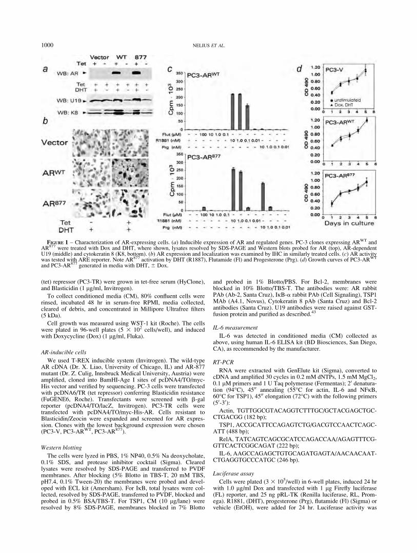
(tet) repressor (PC3-TR) were grown in tet-free serum (HyClone),and Blasticidin (1 lg/ml, Invitrogen).
To collect conditioned media (CM), 80% confluent cells wererinsed, incubated 48 hr in serum-free RPMI, media collected,cleared of debris, and concentrated in Millipore Ultrafree filters(5 kDa).
Cell growth was measured using WST-1 kit (Roche). The cellswere plated in 96-well plates (5 3 102 cells/well), and inducedwith Doxycycline (Dox) (1 lg/ml, Fluka).
AR-inducible cells
We used T-REX inducible system (Invitrogen). The wild-typeAR cDNA (Dr. X. Liao, University of Chicago, IL) and AR-877mutant (Dr. Z. Culig, Innsbruck Medical University, Austria) wereamplified, cloned into BamHI-Age I sites of pcDNA4/TO/myc-His vector and verified by sequencing. PC-3 cells were transfectedwith pcDNA6/TR (tet repressor) conferring Blasticidin resistance(FuGENE6, Roche). Transfectants were screened with b-galreporter (pcDNA4/TO/lacZ, Invitrogen). PC3-TR cells weretransfected with pcDNA4/TO/myc-His-AR. Cells resistant toBlasticidin/Zeocin were expanded and screened for AR expres-sion. Clones with the lowest background expression were chosen(PC3-V, PC3-ARWT, PC3-AR877).
Western blotting
The cells were lyzed in PBS, 1% NP40, 0.5% Na deoxycholate,0.1% SDS, and protease inhibitor cocktail (Sigma). Clearedlysates were resolved by SDS-PAGE and transferred to PVDFmembranes. After blocking (5% Blotto in TBS-T, 20 mM TBS,pH7.4, 0.1% Tween-20) the membranes were probed and devel-oped with ECL kit (Amersham). For IjB, total lysates were col-lected, resolved by SDS-PAGE, transferred to PVDF, blocked andprobed in 0.5% BSA/TBS-T. For TSP1, CM (10 lg/lane) wereresolved by 8% SDS-PAGE, membranes blocked in 7% Blotto
and probed in 1% Blotto/PBS. For Bcl-2, membranes wereblocked in 10% Blotto/TBS-T. The antibodies were: AR rabbitPAb (Ab-2, Santa Cruz), IjB-a rabbit PAb (Cell Signaling), TSP1MAb (A4.1, Novus), Cytokeratin 8 pAb (Santa Cruz) and Bcl-2antibodies (Santa Cruz). U19 antibodies were raised against GST-fusion protein and purified as described.43
IL-6 measurement
IL-6 was detected in conditioned media (CM) collected asabove, using human IL-6 ELISA kit (BD Biosciences, San Diego,CA), as recommended by the manufacturer.
RT-PCR
RNA were extracted with GenElute kit (Sigma), converted tocDNA and amplified 30 cycles in 0.2 mM dNTPs, 1.5 mM MgCl2,0.1 lM primers and 1 U Taq polymerase (Fermentas); 20 denatura-tion (94�C), 4500 annealing (55�C for actin, IL-6 and NFjB,60�C for TSP1), 4500 elongation (72�C) with the following primers(50-30):Actin, TGTTGGCGTACAGGTCTTTGC/GCTACGAGCTGC-
CTGACGG (182 bp);
TSP1, ACCGCATTCCAGAGTCTG/GACGTCCAACTCAGC-ATT (488 bp);
RelA, TATCAGTCAGCGCATCCAGACCAA/AGAGTTTCG-GTTCACTCGGCAGAT (222 bp);
IL-6, AAGCCAGAGCTGTGCAGATGAGTA/AACAACAAT-CTGAGGTGCCCATGC (246 bp).
Luciferase assay
Cells were plated (3 3 105/well) in 6-well plates, induced 24 hrwith 1.0 lg/ml Dox and transfected with 1 lg Firefly luciferase(FL) reporter, and 25 ng pRL-TK (Renilla luciferase, RL, Prom-ega). R1881, (DHT), progesterone (Prg), flutamide (Fl) (Sigma) orvehicle (EtOH), were added for 24 hr. Luciferase activity was
FIGURE 1 – Characterization of AR-expressing cells. (a) Inducible expression of AR and regulated genes. PC-3 clones expressing ARWT andAR877 were treated with Dox and DHT, where shown, lysates resolved by SDS-PAGE and Western blots probed for AR (top), AR-dependentU19 (middle) and cytokeratin 8 (K8, bottom). (b) AR expression and localization was examined by IHC in similarly treated cells. (c) AR activitywas tested with ARE reporter. Note AR877 activation by DHT (R1887), Flutamide (Fl) and Progesterone (Prg). (d) Growth curves of PC3-ARWT
and PC3-AR877 generated in media with DHT, 6 Dox.
1000 NELIUS ET AL.
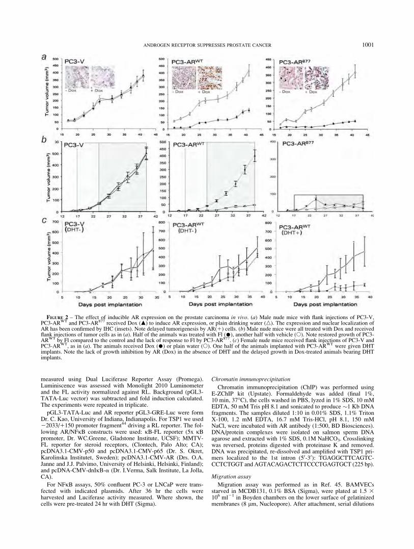
measured using Dual Luciferase Reporter Assay (Promega).Luminiscence was assessed with Monolight 2010 Luminometerand the FL activity normalized against RL. Background (pGL3-TATA-Luc vector) was subtracted and fold induction calculated.The experiments were repeated in triplicate.
pGL3-TATA-Luc and AR reporter pGL3-GRE-Luc were formDr. C. Kao, University of Indiana, Indianapolis. For TSP1 we used22033/1150 promoter fragment44 driving a RL reporter. The fol-lowing AR/NFjB constructs were used: jB-FL reporter (5x jBpromoter, Dr. WC.Greene, Gladstone Institute, UCSF); MMTV-FL reporter for steroid receptors, (Clontech, Palo Alto; CA);pcDNA3.1-CMV-p50 and pcDNA3.1-CMV-p65 (Dr. S. Okret,Karolinska Institutet, Sweden); pcDNA3.1-CMV-AR (Drs. O.A.Janne and J.J. Palvimo, University of Helsinki, Helsinki, Finland);and pcDNA-CMV-dnIjB-a (Dr. I.Verma, Salk Institute, La Jolla,CA).
For NFjB assays, 50% confluent PC-3 or LNCaP were trans-fected with indicated plasmids. After 36 hr the cells wereharvested and Luciferase activity measured. Where shown, thecells were pre-treated 24 hr with DHT (Sigma).
Chromatin immunoprecipitation
Chromatin immunoprecipitation (ChIP) was performed usingE-ZChIP kit (Upstate). Formaldehyde was added (final 1%,10 min, 37�C), the cells washed in PBS, lyzed in 1% SDS, 10 mMEDTA, 50 mM Tris pH 8.1 and sonicated to produce �1 Kb DNAfragments. The samples diluted 1:10 in 0.01% SDS, 1.1% TritonX-100, 1.2 mM EDTA, 16.7 mM Tris-HCl, pH 8.1, 150 mMNaCl, were incubated with AR antibody (1:500, BD Biosciences).DNA/protein complexes were isolated on salmon sperm DNAagarose and extracted with 1% SDS, 0.1M NaHCO3. Crosslinkingwas reversed, proteins digested with proteinase K and removed.DNA was precipitated, re-dissolved and amplified with TSP1 pri-mers localized to the 1st intron (50-30): TGAGGCTTCAGTC-CCTCTGGT and AGTACAGACTCTTCCCTGAGTGCT (225 bp).
Migration assay
Migration assay was performed as in Ref. 45. BAMVECsstarved in MCDB131, 0.1% BSA (Sigma), were plated at 1.5 3106 ml21 in Boyden chambers on the lower surface of gelatinizedmembranes (8 lm, Nucleopore). After attachment, serial dilutions
FIGURE 2 – The effect of inducible AR expression on the prostate carcinoma in vivo. (a) Male nude mice with flank injections of PC3-V,PC3-ARWT and PC3-AR877 received Dox (m) to induce AR expression, or plain drinking water (n). The expression and nuclear localization ofAR has been confirmed by IHC (insets). Note delayed tumorigenesis by AR(1) cells. (b) Male nude mice were all treated with Dox and receivedflank injections of tumor cells as in (a). Half of the animals was treated with Fl (�), another half with vehicle (s). Note restored growth of PC3-ARWT by Fl compared to the control and the lack of response to Fl by PC3-AR877. (c) Female nude mice received flank injections of PC3-V andPC3-ARWT, as in (a). The animals received Dox (�) or plain water (s). One half of the animals implanted with PC3-ARWT were given DHTimplants. Note the lack of growth inhibition by AR (Dox) in the absence of DHT and the delayed growth in Dox-treated animals bearing DHTimplants.
1001ANDROGEN RECEPTOR SUPPRESSES PROSTATE CANCER

of CM from PC3-V, PC3-ARWT or PC3-AR877 were placed in topwells for 4 hr. Background migration (BSA) was subtracted andthe data presented as percent maximal migration (10 ng/mlbFGF). All samples were tested in quadruplicate.
Tumorigenicity assay
PC3-V, PC3-ARWT or PC3-AR877 cells were injected s.c. inhindquarters of athymic male mice (nu/nu, National Cancer Insti-tute, 4–6 weeks), 106 cells/site, 5 animals/group, 2 sites/animal.To induce AR expression, Dox (1mg/ml) was given in drinkingwater. The tumors were measured every 3 days and the volumescalculated as length 3 width2 3 0.52. The experiment wasrepeated using the same numbers of female athymic mice 6 DHTpellets. The pellets were generated in the lab as described inRef. 43. Flutamide (Fl) (40 mg/kg, Sigma) was given daily p.o. Atthe endpoint tumors were removed, snap-frozen or fixed in 4%formaldehyde. The animals were handled following the NationalInstitute of Health guidelines, protocols approved by NorthwesternUniversity Animal Care and Use Committee.
Immunostaining
Five micrometer cryosections were fixed in cold acetone, 1:1acetone/chloroform and acetone (10 min ea), rinsed in PBS,blocked with Avidin-Biotin Blocking kit, mouse Ig (Vector) andincubated 30 min with rat CD31 (1:125, PharMingen) and mouseTSP1 antibodies (1:100, Neomarkers). The slides were washed inPBS and incubated 15 min with donkey anti-rat RhodamineXantibodies (1:200, Jackson Immunoresearch) and biotinylated
anti-mouse antibodies (1:200, Vector). Slides were developedwith FITC-conjugated Avidin D (20 lg/ml, Vector). Biotinylatedanti-rabbit antibodies were applied in blocking solution (1:200, 30min) and followed by 1 lg/ml Streptavidin-Cy5 (Jackson Immu-noresearch). To visualize apoptosis, the sections were evaluatedby TUNEL (ApopTag kit, Serologicals).
For AR, 5 lm sections were deparaffinized, rehydrated, washed,antigen retrieved 15 min at 20–25 psi, 100�C in citric buffer pH6.0 and 20 min at room temperature. Endogenous peroxidase wasinhibited with blocking solution (Dako) and AR antibody added(30 min, N-20, Santa Cruz, 1:200) followed by HRP-conjugatedanti-rabbit antibodies (30 min). Slides were developed with diami-nobenzidine and counterstained with hematoxylin. Nonimmunerabbit serum served as negative control.
For NFjB, the sections after antigen retrieval were blockedwith 20% goat serum in PBS, and incubated with mouse mAb forhuman p65/RelA (Cell Signaling), followed by fluorescent goatanti-mouse Ab (Jackson Immunoresearch). Representative ex-periments of 4 are shown.
Image quantification
Fluorescent images were obtained using Nikon fluorescentmicroscope (Diaphot 200) and converted to digital files usingMetaMorph software. The same software was used to measure flu-orescence intensity and compare the values to DAPI counterstainused as background. CD31-positive structures (MVD) werecounted in 10 403 fields using MetaMorph software. Apoptotic
FIGURE 3 – AR-expressing tumors showed increased apoptosis and decreased MVD. (a) Paraffinized tumor sections were stained with H&E(left) and snap-frozen sections (right) for endothelial marker CD31 (red) and apoptosis (TUNEL, green). To confirm AR functional activity, thesame sections were stained for U19 (blue). (b) Quantitative analysis of MVD. (c) Endothelial cell apoptosis calculated as percent of TUNEL-positive of total CD31-positive structures (merge, yellow). (d) Tumor cell apoptosis (total TUNEL-positive cells minus TUNEL positive endo-thelial cells). All measurements were performed with MetaMorph software.
1002 NELIUS ET AL.

cells were quantified in 10 random fields using MetaMorphsoftware.
Statistical analysis
Mean and standard error values were calculated and comparedusing paired Student’s t test and ANOVA. p values < 0.05 wereconsidered significant.
Results
AR induction reduced tumorigenicity
We generated PC-3 cells inducibly expressing wild-type AR(PC3-ARWT) and promiscuous AR T877A46 (PC3-AR877)(Fig. 1a). Induced AR levels were comparable to LNCaP cells(not shown). The AR axis was restored and two AR-dependentgenes, U1943 and cytokeratin 847 robustly induced upon AR acti-vation (Fig. 1a). Both wild-type and mutant AR became nuclear inthe presence of DHT (Fig. 1a) and induced transcription of theARE-luciferase reporter (Fig. 1c). AR877 was also activated byFlutamide and progesterone (Fig. 1c). The expression and nuclear
localization of AR were induced in vivo in the PC-3 cellsimplanted in male mice upon Dox treatment (Fig. 2a, insets).However AR re-expression failed to enhance PC-3 growth inresponse to DHT (Fig. 1d). Moreover, AR(1) cells were lesstumorigenic in male mice in the presence of Dox (Fig. 2a). Whenwe used oral Flutamide to block endogenous testosterone, PC3-ARWT regained tumorigenicity while PC3-AR877 did not (Fig.2b), suggesting that weak activation by Fl was sufficient to sup-press tumor growth. Finally, PC3-ARWT cell formed tumors inDox-treated female mice, obviously lacking endogenous testoster-one, but not when they received DHT implants, underscoring therepression by androgen (Fig. 2c).
AR(1) PC-3 tumors had lower MVD and higher apoptosis rate
We measured MVD in the AR(1) and AR(2) PC-3 tumors.PC3-ARWT and PC3-AR877 tumors in Dox-treated animals had2.2–2.6 times lower MVD (p < 0.01) than untreated controls, orthe AR(2) controls (Figs. 3a and 3b). TUNEL showed more endo-thelial and nonendothelial apoptotic cells in the AR(1) PC-3tumors (Figs. 3a, 3c and 3d). AR remained functional in thesetumors: its localization was predominantly nuclear in Dox treated
FIGURE 4 – AR decreased angiogenesis via angioinhibitory TSP1. (a) RT-PCR detection of TSP1 mRNA in PC3-V, PC3-ARWT and PC3-AR.877 Note decreased TSP1 mRNA by Fl in PC3-ARWT but not AR.877 (b) Western blot of Conditioned Media from PC3-V, PC3-ARWT andPC3-AR877 6 Dox. All cells were DHT-stimulated. (c) The induction of TSP1-Luc reporter in AR(1) and control cells. (d) ChIP of the putativeARE in the 1st intron of TSP1 gene. IC: isotype control. PCR control: cloned TSP1 promoter amplified with the same primers. DNA input isshown. (e) Endothelial cell chemotaxis with the CM from PC3-V, PC3-ARWT and PC3-AR877. The cells were treated with Dox and stimulatedwith DHT, where indicated and the CM collected and tested at increasing concentrations with the TSP1 neutralizing antibody (�) or with iso-type control antibody (IgA) (s). Note the that CM from AR(1) cells are less potent at inducing endothelial cell chemotaxis, and that TSP1 neu-tralizing antibody, but not isotype control improves migration (arrowheads). (f) Immunostaining of the PC3-V, PC3-ARWT and PC3-AR877
tumors in control and Dox-treated male mice. Cryosections were stained for CD31 (red) and TSP1 (green).
1003ANDROGEN RECEPTOR SUPPRESSES PROSTATE CANCER

males (Fig. 2a) and, AR responsive protein, U19 was stronglyupregulated (Fig. 3a). Thus restoring AR axis in the androgen-insensitive cells delayed tumor progression, lowered MVD andincreased apoptosis.
AR activation upregulated angioinhibitory TSP1
Seeking AR-dependent changes affecting MVD, we investi-gated angiogenic mediators in AR(1) and AR(2) cells. Threepro-angiogenic cytokines, VEGF, bFGF and IL-8, previouslyidentified in PrCa,36,48–53 remained unaltered. We were unable todetect changes in VEGF mRNA or protein using quantitativeRT-PCR, ELISA, or immunostaining (data not shown). TSP1 is acritical angiogenesis inhibitor, whose expression is significantlylower in cancerous compared to the normal prostate51,54; an indexintegrating TSP1 with angiogenesis independently predicts sur-vival.52 In our model, TSP1 was low in parental PC-3 and PC3-Vcells. In PC3-ARWT and PC3-AR877, TSP1 mRNA and secretedprotein became high upon Dox/DHT stimulation (Figs. 4a and4b). In PC3-ARWT Dox/DHT increased activity of the luciferasereporter containing 22033/1150 TSP1 promoter fragment44
(Fig. 4c). Moreover, ChIP demonstrated AR binding to the TSP1promoter (Fig. 4d).
TSP1 suppressed angiogenesis in AR(1) cells
The migration of endothelial cells up the gradient of angiogenicfactors is an important component of angiogenesis and an indica-tor of angiogenic activity of a given cell line.55 The majority ofnatural inhibitors block endothelial cell chemotaxis induced byVEGF or by bFGF. To determine if TSP1 was responsible for thedecrease of angiogenesis in AR(1) tumors, we examined endothe-lial cell chemotaxis to CM from the PC3-V and PC3-AR. PC3-VCM induced migration, with or without Dox and/or DHT, withEC50 5 2.4 lg/ml. CM from nonstimulated PC3-ARWT and PC3-AR877 were also angiogenic, with similar EC50 (1.9–2.2 lg/ml),and not significantly altered by TSP1 antibodies (Fig. 4e). How-ever, CM from PC3-ARWT and PC3-AR877 stimulated to expressAR and activated with DHT became less angiogenic (EC50 > 10lg/ml). This lower angiogenic activity was due to TSP1, sinceTSP1 neutralizing antibody restored angiogenic activity (Fig. 4e).IHC showed MVD reduction in AR(1) tumors, paralleled by a
FIGURE 5 – The interference between AR and NFjB. (a) Reporter assays in cells transfected with the combination of NFjB-Luc, ARE-Luc,AR or p65/p50. pcDNA-neo was used to equalize the total DNA input. DHT (black bars) or control vehicle (ethanol: gray bars), where added,where indicated. Note decreased NFjB activity in the PC3-AR and increased AR activity in the presence of dnIjBa (NFjB superrepressor). (b)PC3-ARWT cells were treated with Dox or Dox/DHT, nuclear extracts collected and analyzed by EMSA. Cold probe (Comp) or IKK inhibitor,BMS345543 (10 lM, BMS) added where indicated. (c) Total IjB-a in Dox/DHT stimulated cells. PC3-V -ARWT and -AR877 cells were treatedfor 24 hours with vehicle ethanol or DHT, as indicated, and IjB-a detected in total cell lysates by Western blot. The blot was re-probed for b-tubulin to assess loading (lower panel). (d) RT-PCR of p65 (RelA) mRNA in similarly treated cells. Note a decrease upon AR activation. (e)PC3-ARWT cells were grown on coverslips, treated as indicated and stained for AR (top) or NFjB (bottom). Note the lack of nuclear NFjBupon DHT stimulation. (f, g) IL-6 levels in Dox/DHT stimulated cells. The cells were treated as in (c), total RNA collected and RT-PCR per-formed with primers for IL-6 (F). C1, no cDNA; C2, no primers. (g) IL-6 detected by ELISA, in the media conditioned by similarly treated cells.(h) Western blot for Bcl-2.
1004 NELIUS ET AL.

dramatic increase in TSP1 (Fig. 4f), pointing to a similar course ofevents in vivo.
AR activation lowered NFjB levels and activity
Seeking reasons for the decreased viability/increased apoptosisin AR(1) tumors we investigated NFjB status of AR(1) and (2)PC-3 populations. Constitutive NFjB activation and subsequentBcl-2 increase mark hormone refractory PrCa.42,56,57 Conversely,AR and NFjB counteract in transcription assays.58 Indeed, re-porter assays showed high basal NFjB activity in PC-3 cells,which was decreased upon transient transfection with ARWT anddiminished further by DHT (Fig. 5a, left). Moreover, ARE-Luc re-porter activity, moderate in PC3 transfected with ARWT, doubledin DHT-treated cells when NFjB was blocked with dnIjB-a(Fig. 5a, center). Conversely in AR-sensitive LNCaP p50/p65 dra-matically reduced ARE-Luc transactivation, with or without DHT(Fig. 5a, right). EMSA showed that DHT significantly reducedNFjB DNA binding in PC3-AR cells (Fig. 5b). NFjB is chieflyregulated via cytoplasmic retention by IjB-a. However, the IjB-a levels in the AR(1) cells showed only modest increase, afterDox treatment (Fig. 5c) Unexpectedly, DHT significantlydecreased the RelA mRNA in PC3-AR but not in PC3-V cells(Fig. 5d).
In addition, in PC3-ARWT and AR877, DHT lowered nuclearp65/RelA (Fig. 5e and data not shown). Nuclear localization ofAR and p65 were mutually exclusive: in PC3-ARWT, AR was pre-dominantly cytoplasmic in the absence of DHT, while p65 wasmostly nuclear. Conversely, in DHT-treated cells AR was predom-inantly nuclear, while p65 became cytoplasmic (Fig. 5e).
AR blocked pro-survival NFjB targets
DHT severely decreased the two NFjB targets, IL-6, as wasmeasured at mRNA level and secreted protein (Figs. 5f and 5g),and Bcl2 (Fig. 5h). Both proteins are capable of increasing cellsurvival.
AR diminished nuclear NFjB and increasedapoptosis in vivo
The decrease in active NFjB remained true in vivo. WhileAR(2) tumors showed NFjB staining in the cytoplasm andnuclei, in AR(1) tumors RelA resided mainly in the cytoplasm(Fig. 6a). Similar to the in vitro results, RelA immunoreactivitywas much weaker in AR(1) tumors (Figs. 6a and 6c). Higher inci-dence of nuclear NFjB was accompanied by low apoptosis rates,while in Dox-treated male mice AR(1) tumors showed lessnuclear NFjB and higher apoptosis (Figs. 6b and 6d).
Discussion
Current in vivo models include tumor grafting in syngeneic orimmune compromised animals, or autochthonous tumors in genet-ically manipulated mice. The differences in structure, physiologyand cancer progression in mouse and human prostate59 make itessential to complement the findings from genetically altered micewith those from xenografted tumors. Indeed, stroma and thesmooth muscle are major structural and functional components inhuman, but not in mouse prostate. Lobular structure is seen in themouse but not in human prostate, while mice have no transitionalzone, prostatic urethra and capsule.59 Most importantly, prostatecancer does not occur spontaneously in wild-type mice; the major-
FIGURE 6 – AR effect on NFjB nuclear localization and apoptosis in vivo. (a, c) Immunostaining of AR(1) (PC3-AR) and control (PC3-V)tumors for p65 (RelA). The mice were given drinking water 6 Dox, the resultant tumors stained for RelA (NFjB, red). (a) Representativeimages of the stained sections. Note nuclear RelA (solid arrowheads) in the absence of Dox and the lack of nuclear staining in Dox-treatedtumors (empty arrowheads). (c) Immunofluorescence intensity was measured using MetaMorph software in a minimum of 12 random fields on 3independent sections. (b, d) Inverse correlation between nuclear NFjB localization and apoptosis. Sections of Dox-treated or untreated (control)AR(1) tumors were stained for RelA (NFjB, red). Apoptosis was visualized by TUNEL (green). (b) Representative images of the stainedsections. Upper panels show merged images. Empty arrowheads indicate cells lacking nuclear RelA. (d) TUNEL-positive cells were quantifiedin 6 random fields, 3 independent sections.
1005ANDROGEN RECEPTOR SUPPRESSES PROSTATE CANCER
ity of mouse models are driven by SV-40 large and small T viraloncogenes. Other suspect oncogenes and tumor suppressors yieldintraepithelial neoplasia (PIN) but not PrCa.60 Only three geneshave been found critical for prostate carcinogenesis in mice: anoncogenic IGF-1 and cMyc, and a tumor suppressor PTEN.61,62
Surprisingly, AR failed as prostate-specific oncogene in trans-genic models, its overexpression yields PIN but no invasive carci-noma.63 In another study, wild-type and promiscuous AR mutantT857A (T877A analogue) fail to induce PIN in young animalssuggesting that ligand driven AR activation does not induce epi-thelial hyperproliferation in the whole prostate.64 Thus androgenrole in PrCa is not unequivocal. In normal prostate it likey main-tains homeostasis of proliferation vs. apoptosis, while androgenablation changes AR targets from apoptotic to survival/prolifera-tion. Interestingly, studies form Liao and coworkers demonstratethat AR-positive LNCaP cells, conditioned by long-term androgenwithdrawal become hypersensitive to androgen and could be sup-pressed by androgen in vivo65 and identify decreased cMyc andincreased Bax as responsible genes.66 Moreover. LNCaP sublinesrendered androgen-independent, could be suppressed by androgenand then reversed to androgen-dependent phenotype.67
According to Greenberg and coworkers, transgenes encodingeither AR-WT or AR-T857A, a mouse analog of human T877Amutant, did not cause prostate cancer in mice.64 Consistent withtheir data, we showed that inducible wild-type and T877A ARfailed to expedite tumor progression in a subcutaneous xenograftmodel, but instead caused dramatic delay in tumor progression,decreased MVD and increased apoptosis. Interestingly, thesechanges occurred predominantly in vivo. Other investigatorsobserved decreased proliferation upon re-expression of AR,26
however in our hands, AR(1) and (2) cells in vitro grew at thesame rate. This difference may be due to the use of inducible ARexpression, while stable transfectans may have acquired additionalchanges due to the constitutive AR overexpression. The moleculareffects of AR expression/activation in PC-3 cells were twofold:decreased activation of NFjB, a pro-survival transcription factorin prostate epithelium,40,56,57 and decreased overall angiogenicactivity due to increased angioinhibitory TSP1, which translatedinto AR-dependent decrease of tumor MVD. The inverse correla-tion between TSP1 levels and prostate cancer progression and vas-cularization has been previously shown,33,51,52,54 however TSP1induction by AR has not been demonstrated.
The crosstalk between AR and NFjB has been previouslyshown in vitro, where NFjB inactivation resulted in higher apo-ptosis rates.25,68 However, others indicate that AR also mayincrease NFjB activity.69,70 Despite NFjB blockade, AR expres-
sion failed to increase apoptosis in vitro. Increased tumor apopto-sis in vivo suggests that NFjB deactivation lowered the survivalof AR(1) cells under stress. This is consistent with potentiatedresponse to genotoxic stress by AR.24 In our system AR(1) cellslow in NFjB activity, become apoptotic in response to hypoxiadue to insufficient angiogenesis. Conversely, AR(2) cells remainresistant. In addition, NFjB may contribute to the angiogenicproperties of prostate epithelium by increasing NOS and cycloox-ygenase-271: its inactivation would further reduce tumor MVD.
It is widely accepted that functional AR is expressed in a largeportion of advanced prostate cancers. However the majority of ARpathway genes (HERPUD1, STK39, DHCR24, and SOCS2) aresuppressed in metastatic prostate cancer,72 underscoring the factthat many of the AR targets counter cancer progression.
Our study indicates that both wild-type AR and AR with alteredligand specificity, lack the ability to transform prostate epithelium.Conversely, Greenberg and coworkers identified carcinogenic ARmutations in the transactivation domain.73 Interestingly, somaticmutations associated with male infertility are in the DNA and lin-gand binding domains and the hinge, while �40% cancer-associ-ated mutations are in the transactivation domain, where they affectcofactor interactions. Although >80% AR point mutations havebeen identified in cancer specimens, their functional consequencesare not verified, except for a few isolated cases. Combined data byGreenberg’s group64 and our’s suggest that while AR maintainsinteractions with proper coactivators and corepressors, it continuesto control homeostatic proliferation, apoptosis, and angiogenesis.One possible explanation is the release of AR control over NFjBactivity: once disrupted, NFjB activation, in turn, favors increasedsurvival, dampens stress responses and favors tumor progression.The mechanism of AR interference with NFjB remains unclear:although weak AR/NFjB interaction was observed in vitro,74 theresult has never been reproduced. Other investigators suggestcompetitive binding to adjacent cis-regulatory elements on theDNA.75 We observed modest IjB-a increase in the AR(1) cells,however higher RelA mRNA and protein levels are more likely toplay a role. Indeed, DHT stimulation on the AR(1) tumors pro-duced the decrease in general NFjB immunoreactivity.
Our results suggest that persistent androgen ablation promotesthe progression to androgen independent phenotype and indicatepossible benefits of the treatment where androgen application andablation are used in succession or intermittently.
Acknowledgements
We thank Dr. Wang and Dr. Levenson for helpful discussion.
References
1. Scott WW, Menon M, Walsh PC. Hormonal therapy of prostatic can-cer. Cancer 1980;45 (7 Suppl):1929–36.
2. Denis L, Murphy GP. Overview of phase III trials on combined andro-gen treatment in patients with metastatic prostate cancer. Cancer1993;72 (12 Suppl):3888–95.
3. Heinlein CA, Chang C. The roles of androgen receptors and andro-gen-binding proteins in nongenomic androgen actions. Mol Endocri-nol 2002;16:2181–7.
4. Zegarra-Moro OL, Schmidt LJ, Huang H, Tindall DJ. Disruption ofandrogen receptor function inhibits proliferation of androgen-refrac-tory prostate cancer cells. Cancer Res 2002;62:1008–13.
5. Craft N, Shostak Y, Carey M, Sawyers CL. A mechanism for hor-mone-independent prostate cancer through modulation of androgenreceptor signaling by the HER-2/neu tyrosine kinase. Nat Med1999;5:280–5.
6. Smith PC, Hobisch A, Lin DL, Culig Z, Keller ET. Interleukin-6 andprostate cancer progression. Cytokine Growth Factor Rev 2001;12:33–40.
7. Godoy-Tundidor S, Hobisch A, Pfeil K, Bartsch G, Culig Z. Acquisi-tion of agonistic properties of nonsteroidal antiandrogens after treat-ment with oncostatin M in prostate cancer cells. Clin Cancer Res2002;8:2356–61.
8. Koivisto P, Visakorpi T, Kallioniemi OP. Androgen receptor geneamplification: a novel molecular mechanism for endocrine therapy
resistance in human prostate cancer. Scand J Clin Lab Invest 1996;226:57–63.
9. Nan B, Snabboon T, Unni E, Yuan XJ, Whang YE, Marcelli M. ThePTEN tumor suppressor is a negative modulator of androgen receptortranscriptional activity. J Mol Endocrinol 2003;31:169–83.
10. Lu ML, Schneider MC, Zheng Y, Zhang X, Richie JP. Caveolin-1interacts with androgen receptor. A positive modulator of androgenreceptor mediated transactivation. J Biol Chem 2001;276:13442–51.
11. Culig Z, Klocker H, Bartsch G, Steiner H, Hobisch A. Androgenreceptors in prostate cancer. J Urol 2003;170 (4 Part 1):1363–9.
12. Hirawat S, Budman DR, Kreis W. The androgen receptor: structure,mutations, and antiandrogens. Cancer Invest 2003;21:400–17.
13. Huang H, Tindall DJ. The role of the androgen receptor in prostatecancer. Crit Rev Eukaryot Gene Expr 2002;12:193–207.
14. Culig Z, Klocker H, Eberle J, Kaspar F, Hobisch A, Cronauer MV,Bartsch G. DNA sequence of the androgen receptor in prostatic tumorcell lines and tissue specimens assessed by means of the polymerasechain reaction. Prostate 1993;22:11–22.
15. Tilley WD, Buchanan G, Hickey TE, Bentel JM. Mutations in theandrogen receptor gene are associated with progression of humanprostate cancer to androgen independence. Clin Cancer Res 1996;2:277–85.
16. Hyytinen ER, Haapala K, Thompson J, Lappalainen I, Roiha M,Rantala I, Helin HJ, Janne OA, Vihinen M, Palvimo JJ, Koivisto PA.
1006 NELIUS ET AL.

Pattern of somatic androgen receptor gene mutations in patients withhormone-refractory prostate cancer. Lab Invest 2002;82:1591–8.
17. Marcelli M, Ittmann M, Mariani S, Sutherland R, Nigam R, MurthyL, Zhao Y, DiConcini D, Puxeddu E, Esen A, Eastham J, Weigel NL,et al. Androgen receptor mutations in prostate cancer. Cancer Res2000;60:944–9.
18. Kokontis JM, Hay N, Liao S. Progression of LNCaP prostate tumorcells during androgen deprivation: hormone-independent growth,repression of proliferation by androgen, and role for p27Kip1 inandrogen-induced cell cycle arrest. Mol Endocrinol 1998;12:941–53.
19. Lu S, Tsai SY, Tsai MJ. Regulation of androgen-dependent prostaticcancer cell growth: androgen regulation of CDK2, CDK4, and CKIp16 genes. Cancer Res 1997;57:4511–6.
20. Culig Z, Hobisch A, Herold M, Hittmair A, Thurnher M, Eder IE,Cronauer MV, Rieser C, Ramoner R, Bartsch G, Klocker H, Konwa-linka G. Interleukin 1b mediates the modulatory effects of mono-cytes on LNCaP human prostate cancer cells. Br J Cancer 1998;78:1004–11.
21. Mitchell SH, Zhu W, Young CY. Resveratrol inhibits the expressionand function of the androgen receptor in LNCaP prostate cancer cells.Cancer Res 1999;59:5892–5.
22. Berger R, Febbo PG, Majumder PK, Zhao JJ, Mukherjee S, SignorettiS, Campbell KT, Sellers WR, Roberts TM, Loda M, Golub TR, HahnWC. Androgen-induced differentiation and tumorigenicity of humanprostate epithelial cells. Cancer Res 2004;64:8867–75.
23. Whitacre DC, Chauhan S, Davis T, Gordon D, Cress AE, MiesfeldRL. Androgen induction of in vitro prostate cell differentiation. CellGrowth Differ 2002;13:1–11.
24. Heisler LE, Evangelou A, Lew AM, Trachtenberg J, Elsholtz HP,Brown TJ. Androgen-dependent cell cycle arrest and apoptotic deathin PC-3 prostatic cell cultures expressing a full-length human andro-gen receptor. Mol Cell Endocrinol 1997;126:59–73.
25. Altuwaijri S, Lin HK, Chuang KH, Lin WJ, Yeh S, Hanchett LA,Rahman MM, Kang HY, Tsai MY, Zhang Y, Yang L, Chang C.Interruption of nuclear factor jB signaling by the androgen receptorfacilitates 12-O-tetradecanoylphorbolacetate-induced apoptosis inandrogen-sensitive prostate cancer LNCaP cells. Cancer Res 2003;63:7106–12.
26. Yuan S, Trachtenberg J, Mills GB, Brown TJ, Xu F, Keating A.Androgen-induced inhibition of cell proliferation in an androgen-insensitive prostate cancer cell line (PC-3) transfected with a humanandrogen receptor complementary DNA. Cancer Res 1993;53:1304–11.
27. Lu S, Liu M, Epner DE, Tsai SY, Tsai MJ. Androgen regulation ofthe cyclin-dependent kinase inhibitor p21 gene through an androgenresponse element in the proximal promoter. Mol Endocrinol 1999;13:376–84.
28. Grierson AJ, Shaw CE, Miller CC. Androgen induced cell death inSHSY5Y neuroblastoma cells expressing wild-type and spinal bulbarmuscular atrophy mutant androgen receptors. Biochim Biophys Acta2001;1536:13–20.
29. Lin HK, Yeh S, Kang HY, Chang C. Akt suppresses androgen-induced apoptosis by phosphorylating and inhibiting androgen recep-tor. Proc Natl Acad Sci USA 2001;98:7200–5.
30. Fu M, Wang C, Wang J, Zhang X, Sakamaki T, Yeung YG, Chang C,Hopp T, Fuqua SA, Jaffray E, Hay RT, Palvimo JJ, et al. Androgenreceptor acetylation governs trans activation and MEKK1-induced ap-optosis without affecting in vitro sumoylation and trans-repressionfunction. Mol Cell Biol 2002;22:3373–88.
31. Sigala S, Tognazzi N, Rizzetti MC, Faraoni I, Missale C, BonmassarE, Spano P. Nerve growth factor induces the re-expression of func-tional androgen receptors and p75(NGFR) in the androgen-insensitiveprostate cancer cell line DU145. Eur J Endocrinol 2002;147:407–15.
32. Hess-Wilson JK, Daly HK, Zagorski WA, Montville CP, KnudsenKE. Mitogenic action of the androgen receptor sensitizes prostate can-cer cells to taxane-based cytotoxic insult. Cancer Res 2006;66:11998–2008.
33. van Moorselaar RJ, Voest EE. Angiogenesis in prostate cancer: itsrole in disease progression and possible therapeutic approaches. MolCell Endocrinol 2002;197:239–50.
34. Franck-Lissbrant I, Haggstrom S, Damber JE, Bergh A. Testosteronestimulates angiogenesis and vascular regrowth in the ventral prostatein castrated adult rats. Endocrinology 1998;139:451–6.
35. Lissbrant IF, Lissbrant E, Persson A, Damber JE, Bergh A. Endothe-lial cell proliferation in male reproductive organs of adult rat is highand regulated by testicular factors. Biol Reprod 2003;68:1107–11.
36. Mabjeesh NJ, Willard MT, Frederickson CE, Zhong H, Simons JW.Androgens stimulate hypoxia-inducible factor 1 activation via auto-crine loop of tyrosine kinase receptor/phosphatidylinositol 30-kinase/protein kinase B in prostate cancer cells. Clin Cancer Res 2003;9:2416–25.
37. Doll JA, Stellmach VM, Bouck NP, Bergh AR, Lee C, Abramson LP,Cornwell ML, Pins MR, Borensztajn J, Crawford SE. Pigment epithe-
lium-derived factor regulates the vasculature and mass of the prostateand pancreas. Nat Med 2003;9:774–80.
38. Colombel M, Filleur S, Fournier P, Merle C, Guglielmi J, Courtin A,Degeorges A, Serre CM, Bouvier R, Clezardin P, Cabon F. Andro-gens repress the expression of the angiogenesis inhibitor thrombo-spondin-1 in normal and neoplastic prostate. Cancer Res 2005;65:300–8.
39. Gasparian AV, Yao YJ, Kowalczyk D, Lyakh LA, Karseladze A,Slaga TJ, Budunova IV. The role of IKK in constitutive activation ofNF-jB transcription factor in prostate carcinoma cells. J Cell Sci2002;115 (Part 1):141–51.
40. Ghosh S, Karin M. Missing pieces in the NF-jB puzzle. Cell 2002;109:S81–S96.
41. Supakar PC, Jung MH, Song CS, Chatterjee B, Roy AK. Nuclear fac-tor j B functions as a negative regulator for the rat androgen receptorgene and NF-j B activity increases during the age-dependent desensi-tization of the liver. J Biol Chem 1995;270:837–42.
42. Shukla S, Maclennan GT, Marengo SR, Resnick MI, Gupta S. Consti-tutive activation of P I3 K-Akt and NF-jB during prostate cancerprogression in autochthonous transgenic mouse model. Prostate2005;64:224–39.
43. Xiao W, Zhang Q, Jiang F, Pins M, Kozlowski JM, Wang Z. Suppres-sion of prostate tumor growth by U19, a novel testosterone-regulatedapoptosis inducer. Cancer Res 2003;63:4698–704.
44. Framson P, Bornstein P. A serum response element and a binding sitefor NF-Y mediate the serum response of the human thrombospondin 1gene. J Biol Chem 1993;268:4989–96.
45. Good DJ, Polverini PJ, Rastinejad F, Le Beau MM, Lemons RS,Frazier WA, Bouck NP. A tumor suppressor-dependent inhibitor ofangiogenesis is immunologically and functionally indistinguishablefrom a fragment of thrombospondin. Proc Natl Acad Sci USA1990;87: 6624–8.
46. Culig Z, Hobisch A, Bartsch G, Klocker H. Androgen receptor—anupdate of mechanisms of action in prostate cancer. Urol Res 2000;28:211–9.
47. Foster BA, Gingrich JR, Kwon ED, Madias C, Greenberg NM. Char-acterization of prostatic epithelial cell lines derived from transgenicadenocarcinoma of the mouse prostate (TRAMP) model. Cancer Res1997;57:3325–30.
48. Ferrer FA, Miller LJ, Andrawis RI, Kurtzman SH, Albertsen PC,Laudone VP, Kreutzer DL. Angiogenesis and prostate cancer: in vivoand in vitro expression of angiogenesis factors by prostate cancercells. Urology 1998;51:161–7.
49. Strohmeyer D, Rossing C, Bauerfeind A, Kaufmann O, Schlechte H,Bartsch G, Loening S. Vascular endothelial growth factor and its cor-relation with angiogenesis and p53 expression in prostate cancer.Prostate 2000;45:216–24.
50. Meyer GE, Yu E, Siegal JA, Petteway JC, Blumenstein BA, BrawerMK. Serum basic fibroblast growth factor in men with and withoutprostate carcinoma. Cancer 1995;76:2304–11.
51. Doll JA, Reiher FK, Crawford SE, Pins MR, Campbell SC, BouckNP. Thrombospondin-1, vascular endothelial growth factor and fibro-blast growth factor-2 are key functional regulators of angiogenesis inthe prostate. Prostate 2001;49:293–305.
52. Kwak C, Jin RJ, Lee C, Park MS, Lee SE. Thrombospondin-1, vascu-lar endothelial growth factor expression and their relationship withp53 status in prostate cancer and benign prostatic hyperplasia. BJUInt 2002;89:303–9.
53. Burchardt M, Burchardt T, Chen MW, Hayek OR, Knight C, ShabsighA, de L. Taille A, Buttyan R. Vascular endothelial growth factor-Aexpression in the rat ventral prostate gland and the early effects ofcastration. Prostate 2000;43:184–94.
54. Mehta R, Kyshtoobayeva A, Kurosaki T, Small EJ, Kim H, Stroup R,McLaren CE, Li KT, Fruehauf JP. Independent association of angio-genesis index with outcome in prostate cancer. Clin Cancer Res2001;7:81–8.
55. Polverini PJ, Bouck NP, Rastinejad F. Assay and purification of natu-rally occurring inhibitor of angiogenesis. Methods Enzymol 1991;198:440–50.
56. Sweeney C, Li L, Shanmugam R, Bhat-Nakshatri P, Jayapraka-san V, Baldridge LA, Gardner T, Smith M, Nakshatri H,Cheng L. Nuclear factor-jB is constitutively activated in pros-tate cancer in vitro and is overexpressed in prostatic intraepi-thelial neoplasia and adenocarcinoma of the prostate. Clin Can-cer Res 2004;10:5501–7.
57. Fradet V, Lessard L, Begin LR, Karakiewicz P, Masson AM, Saad F.Nuclear factor-jB nuclear localization is predictive of biochemicalrecurrence in patients with positive margin prostate cancer. ClinCancer Res 2004;10:8460–4.
58. Keller ET, Chang C, Ershler WB. Inhibition of NFjB activity throughmaintenance of IjBa levels contributes to dihydrotestosterone-medi-ated repression of the interleukin-6 promoter. J Biol Chem 1996;271:26267–75.
1007ANDROGEN RECEPTOR SUPPRESSES PROSTATE CANCER

59. Roy-Burman P, Wu H, Powell WC, Hagenkord J, Cohen MB.Genetically defined mouse models that mimic natural aspects ofhuman prostate cancer development. Endocr Relat Cancer 2004;11:225–54.
60. Kasper S. Survey of genetically engineered mouse models for prostatecancer: analyzing the molecular basis of prostate cancer development,progression, and metastasis. J Cell Biochem 2005;94:279–97.
61. DiGiovanni J, Kiguchi K, Frijhoff A, Wilker E, Bol DK, Beltran L,Moats S, Ramirez A, Jorcano J, Conti C. Deregulated expressionof insulin-like growth factor 1 in prostate epithelium leads to neo-plasia in transgenic mice. Proc Natl Acad Sci USA 2000;97:3455–60.
62. Kwabi-Addo B, Giri D, Schmidt K, Podsypanina K, Parsons R,Greenberg N, Ittmann M. Haploinsufficiency of the Pten tumorsuppressor gene promotes prostate cancer progression. Proc NatlAcad Sci USA 2001;98:11563–8.
63. Stanbrough M, Leav I, Kwan PW, Bubley GJ, Balk SP. Prostaticintraepithelial neoplasia in mice expressing an androgen receptortransgene in prostate epithelium. Proc Natl Acad Sci USA 2001;98:10823–8.
64. Han G, Buchanan G, Ittmann M, Harris JM, Yu X, Demayo FJ, TilleyW, Greenberg NM. Mutation of the androgen receptor causes onco-genic transformation of the prostate. Proc Natl Acad Sci USA 2005;102:1151–6.
65. Umekita Y, Hiipakka RA, Kokontis JM, Liao S. Human prostatetumor growth in athymic mice: inhibition by androgens and stimula-tion by finasteride. Proc Natl Acad Sci USA 1996;93:11802–7.
66. Lin Y, Kokontis J, Tang F, Godfrey B, Liao S, Lin A, Chen Y, XiangJ. Androgen and its receptor promote Bax-mediated apoptosis. MolCell Biol 2006;26:1908–16.
67. Chuu CP, Hiipakka RA, Fukuchi J, Kokontis JM, Liao S. Androgencauses growth suppression and reversion of androgen-independentprostate cancer xenografts to an androgen-stimulated phenotype inathymic mice. Cancer Res 2005;65:2082–4.
68. Norata GD, Tibolla G, Seccomandi PM, Poletti A, Catapano AL.Dihydrotestosterone decreases tumor necrosis factor-alpha and lipo-polysaccharide-induced inflammatory response in human endothelialcells. J Clin Endocrinol Metab 2006;91:546–54.
69. Death AK, McGrath KC, Sader MA, Nakhla S, Jessup W,Handelsman DJ, Celermajer DS. Dihydrotestosterone promotes vas-cular cell adhesion molecule-1 expression in male human endothelialcells via a nuclear factor-jB-dependent pathway. Endocrinology2004;145:1889–97.
70. Lee SO, Lou W, Nadiminty N, Lin X, Gao AC. Requirement for NF-(j)B in interleukin-4-induced androgen receptor activation in prostatecancer cells. Prostate 2005;64:160–7.
71. Chiarugi V, Magnelli L, Chiarugi A, Gallo O. Hypoxia inducespivotal tumor angiogenesis control factors including p53, vascularendothelial growth factor and the NFjB-dependent inducible nitricoxide synthase and cyclooxygenase-2. J Cancer Res Clin Oncol 1999;125:525–8.
72. Hendriksen PJ, Dits NF, Kokame K, Veldhoven A, van WeerdenWM, Bangma CH, Trapman J, Jenster G. Evolution of the androgenreceptor pathway during progression of prostate cancer. Cancer Res2006;66:5012–20.
73. Han G, Foster BA, Mistry S, Buchanan G, Harris JM, Tilley WD,Greenberg NM. Hormone status selects for spontaneous somaticandrogen receptor variants that demonstrate specific ligand and cofac-tor dependent activities in autochthonous prostate cancer. J BiolChem 2001;276:11204–13.
74. Palvimo JJ, Reinikainen P, Ikonen T, Kallio PJ, Moilanen A, JanneOA. Mutual transcriptional interference between RelA and androgenreceptor. J Biol Chem 1996;271:24151–6.
75. Cinar B, Yeung F, Konaka H, Mayo MW, Freeman MR, Zhau HE,Chung LW. Identification of a negative regulatory cis-element in theenhancer core region of the prostate-specific antigen promoter:implications for intersection of androgen receptor and nuclear factor-jBsignalling in prostate cancer cells. Biochem J 2004;379 (Part 2): 421–31.
1008 NELIUS ET AL.

Appendix 3. Yemelyanov A., Kobzeva V., Budunova I. Role of IKKi in prostate cancer: A link between inflammation and androgen receptor signaling. Proceedings of AACR, 2007 (abstract # LBA-9158).
Our recent data and data by others indicate that anti-apoptotic, pro-tumorigenic factor NF-kB is constitutively activated in androgen-independent prostate carcinoma (PC) cell lines and in prostate tumors. The important step in NF-kB activation is the phosphorylation of IkBa inhibitor proteins by IKK kinases (IKK) and IKK-related kinases IKKi/e and TBK1/NAK. IKKi is highly inducible kinase whose expression is known to be activated by numerous pro-inflammatory cytokines. We found that IKKi is expressed in androgen-independent PC cells (PC3 and DU145) with high level of constitutively active NF-kB but not in androgen-dependent PC cell lines (LNCaP and MDA PCa 2b) and primary prostate epithelial cells. Immunostaining revealed that IKKi is well expressed in BPH and PCs. Next, our data provide the evidence that IKKi could be involved in the regulation of NF-kB activity in PC cells through a positive feedback loop. Indeed, the treatment of PC cells with NF-kB inducers results in a rapid induction of IKKi. On the other hand, transient transfection of different PC cells with wild type (w.t.) IKKi results in activation of NF-kB. To further study the IKKi function in PC cells we generated PC3 and LNCaP cells stably expressing w.t. IKKi and dominant negative (d.n.) K38A mutant of IKKi using infection with corresponding lentiviruses. In both cell lines IKKi d.n. had no effect on cell morphology, growth, and tumorigenicity. In contrast, PC cells infected with w.t. IKKi displayed significant increase in growth in monolayer and in soft agar in comparison to cells infected with empty virus. As expected, stable overexpression of w.t. IKKi in PC cells resulted in NF-kB activation that correlated with increased level of phosphorylation of both IkBa (Ser36/32) and p65/RelA (Ser536). Unexpectedly expression of w.t. IKKi in LNCaP cells resulted in the increased expression of androgen receptor (AR) on both mRNA and protein level, and the accumulation of AR protein in the nucleus. We further demonstrated the increased basal activity of AR in LNCaP-IKKi w.t. cells, and the enhanced responsiveness to low doses of androgens. Experiments with androgen-deprived serum demonstrated that growth of LNCaP-IKKi w.t. cells was less dependent on the androgens than LNCaP-V cells. The mechanisms of regulation of AR expression and function by IKKi are currently under study. In conclusion, the revealed control of AR-mediated signaling by IKKi may represent an important regulatory link between the inflammation and tumorigenesis in prostate.

Appendix 4. ROLE OF IKKs AND TRANSCRIPTION FACTOR NF-kB IN PROSTATE TUMORIGENESIS Irina Budunova, Alexander Yemelyanov and Alexander Gasparian (current address: Cleveland Biolabs, Cleveland, OH, 44106). IMPACT DOD meeting, 2007, Atlanta. P. 118.
One of the contributing factors to high mortality rate from prostate cancer (PC) is the extreme resistance of PC cells to apoptosis induced by radio- and chemotherapy. Thus, the specific induction of apoptosis is an important target for PC treatment. One of the central anti-apoptotic pathways in cells is mediated by NF-κB transcription factor. NF-κB activation requires degradation of IκB inhibitory proteins which involves several steps including IκB phosphorylation by IκB kinases IKKα/IKKβ. Novel family of IKK-related kinases including inducible IKKi/ε activated by numerous pro-inflammatory cytokines, also could phosphorylate IkB and function further upstream in the NF-κB signaling pathway.
Using funds from DOD Prostate Cancer Research Programs (New Investigator award DAMD17-01-1-0015 and Idea Development award DAMD17-03-1-0522), we determined the status and function of NF-κB in PC cells and PC tumors. We found that NF-κB is constitutively activated in human androgen-independent PC cell lines and in human PCs (Gasparian et al., 2002). We also discovered that the level of IκBα phosphorylation and the rate of IκBα degradation are increased in androgen-independent malignant PC cells in part due to the high level of constitutive IKKα/IKKβ activity. Recently we found that IKKα/β are phosphorylated in PC samples suggesting that IKK kinases are indeed constitutively activated in prostate tumors (Yemelyanov et al., 2006).
Further, we discovered that IKKi is highly expressed only in androgen-independent PC cells with constitutively active NF-κB, and could be involved in the regulation of NF-κB activity in PC cells through a positive feedback loop (Yemelyanov et al., 2004). The forced expression of exogenous IKKi in LNCaP cells that lack this kinase, resulted in significant increase in NF-kB activity, and increased PC cell growth and tumorigenicity.
This work has been expanded towards the search for most effective strategies of NF-kB blockage in PC cells. We tested several novel compounds including highly specific IKKβ inhibitor PS1145, proteasomal inhibitor PS341/Velcade (both in collaboration with Millenium Pharmaceuticals Inc. , Cambridge, MA), as well as dissociated ligands of glucocorticoid receptor that inhibit NF-kB via stimulation of negative protein/protein interaction between activated GR and p65: AL438 (in collaboration with Ligand Pharmaceuticals, San Diego, CA) and Compound A (P.I. initiated research). We determined that IKKβ inhibitor PS1145 induced apoptosis in PC cells, inhibited their growth, and strongly inhibited their invasion (Yemelyanov et al., 2006). We also found that dissociated ligands of GR are highly cytostatic and cytotoxic in malignant PC cells (Yemelyanov et al., submitted). Finally, we showed that Selenium, an effective preventive agent for PC, inhibited NF-κB activity in PC cells, and that NF-κB blockage made PC cells more sensitive to Se-induced apoptosis (Gasparian et al., 2002).
Related Documents