Journal of Case Reports and Images in Oncology, Vol. 7, 2021. ISSN: 2582-1318 J Case Rep Images Oncology 2021;7:100081Z10DF2021. www.ijcrioncology.com Ferreira et al. 1 Primary malignant giant-cell tumor of bone: Case report of a rare clinical condition with an atypical clinical course David Ferreira, André Spranger, Paulo Almeida, Dolores Presa, José Portela, Joaquim Soares do Brito ABSTRACT Introduction: A primary malignant giant-cell tumor of bone (PMGCT) is an extremely rare disease and usually presents with a better prognosis than its secondary counterpart does (secondary malignant giant-cell tumor of bone or SMGCT). Case Report: We present a case of a highly atypical PMGCT of the proximal left tibia, with extremely fast progression, pathological fracture, fungation, and patient death. Conclusion: This clinical case is an example of the most extreme and aggressive biological behavior that could arise with a PMGCT. These usually have a better prognosis than most, among malignant giant-cell tumors. This case highlights the need for a low threshold of suspicion for malignant transformation when giant-cell tumors have an unusual presentation. Keywords: Diagnosis, Malignant giant-cell tumor, Management, Outcomes How to cite this article Ferreira D, Spranger A, Almeida P, Presa D, Portela J, do Brito JS. Primary malignant giant-cell tumor of bone: Case report of a rare clinical condition with an atypical clinical course. J Case Rep Images Oncology 2021;7:100081Z10DF2021. David Ferreira 1 , MD, André Spranger 1 , MD, Paulo Almeida 1 , MD, Dolores Presa 2 , MD, José Portela 1 , MD, Joaquim Soares do Brito 1 , MD, MSc, FEBOT Affiliations: 1 Orthopedics Department, Centro Hospitalar Lis- boa Norte, Lisbon, Portugal; 2 Pathology Department, Centro Hospitalar Lisboa Norte, Lisbon, Portugal. Corresponding Author: Joaquim Soares do Brito, MD, MSc, FE- BOT, Centro Hospitalar Lisboa Norte, EPE. Avenida Professor Egas Moniz, 1649-035 Lisbon, Portugal; Email: joaquimsoares- [email protected] Received: 28 January 2021 Accepted: 15 March 2021 Published: 23 April 2021 CASE REPORT PEER REVIEWED | OPEN ACCESS Article ID: 100081Z10DF2021 ********* doi: 10.5348/100081Z10DF2021CR INTRODUCTION Giant-cell tumor of bone (GCT) is a well-known but rare benign bone lesion [1–3]. Despite its benign biological nature, GCT can develop significant local aggressiveness, can be misdiagnosed as bone sarcoma, and can even generate lung metastases in two to three percent of cases [4]. Additionally, on exceptional occasions, a typical benign giant-cell tumor of bone can undergo malignant transformation, becoming a malignant giant-cell tumor of bone (MGCT) [4, 5]. Most often, a malignant giant-cell tumor of bone arises secondarily (SMGCT) after treatments such as surgery or radiotherapy [6, 7]. Nonetheless, primary malignancy (PMGCT) can also occur. In plain radiographic studies, PMGCT has the typical appearance of a giant-cell tumor and in most cases is impossible to distinguish from them. On the other hand, SMGCT usually has a more malignant appearance on plain films [6]. Likewise, the clinical presentations of a PMGCT or SMGCT could be comparable to that of classic giant-cell tumors. The pathology presents with areas of high grade sarcoma next to areas of benign giant cell tumor, which makes proper diagnosis difficult [6, 7]. In the largest study describing the epidemiology of malignancy in GCT in the United States, the average five-year survival rate was 84.2% and the mean survival time was 11 years and 11 months [8]. Other studies have supported these findings—a high survival rate is expected in patients with malignant giant-cell tumors of bone [9]. Nonetheless, despite those reported findings, a good outcome is not always the case. Herein we present an atypical clinical case of an unusually aggressive primary malignant giant-cell tumor of the proximal left tibia, due to extremely fast progression this caused a pathological fracture, fungation, and eventually, the patient’s death.

Welcome message from author
This document is posted to help you gain knowledge. Please leave a comment to let me know what you think about it! Share it to your friends and learn new things together.
Transcript

Journal of Case Reports and Images in Oncology, Vol. 7, 2021. ISSN: 2582-1318
J Case Rep Images Oncology 2021;7:100081Z10DF2021. www.ijcrioncology.com
Ferreira et al. 1
CASE REPORT OPEN ACCESS
Primary malignant giant-cell tumor of bone: Case report of a rare clinical condition with an atypical clinical course
David Ferreira, André Spranger, Paulo Almeida, Dolores Presa, José Portela, Joaquim Soares do Brito
ABSTRACT
Introduction: A primary malignant giant-cell tumor of bone (PMGCT) is an extremely rare disease and usually presents with a better prognosis than its secondary counterpart does (secondary malignant giant-cell tumor of bone or SMGCT).
Case Report: We present a case of a highly atypical PMGCT of the proximal left tibia, with extremely fast progression, pathological fracture, fungation, and patient death.
Conclusion: This clinical case is an example of the most extreme and aggressive biological behavior that could arise with a PMGCT. These usually have a better prognosis than most, among malignant giant-cell tumors. This case highlights the need for a low threshold of suspicion for malignant transformation when giant-cell tumors have an unusual presentation.
Keywords: Diagnosis, Malignant giant-cell tumor, Management, Outcomes
How to cite this article
Ferreira D, Spranger A, Almeida P, Presa D, Portela J, do Brito JS. Primary malignant giant-cell tumor of bone: Case report of a rare clinical condition with an atypical clinical course. J Case Rep Images Oncology 2021;7:100081Z10DF2021.
David Ferreira1, MD, André Spranger1, MD, Paulo Almeida1, MD, Dolores Presa2, MD, José Portela1, MD, Joaquim Soares do Brito1, MD, MSc, FEBOTAffiliations: 1Orthopedics Department, Centro Hospitalar Lis-boa Norte, Lisbon, Portugal; 2Pathology Department, Centro Hospitalar Lisboa Norte, Lisbon, Portugal.Corresponding Author: Joaquim Soares do Brito, MD, MSc, FE-BOT, Centro Hospitalar Lisboa Norte, EPE. Avenida Professor Egas Moniz, 1649-035 Lisbon, Portugal; Email: [email protected]
Received: 28 January 2021Accepted: 15 March 2021Published: 23 April 2021
CASE REPORT PEER REVIEWED | OPEN ACCESS
Article ID: 100081Z10DF2021
*********
doi: 10.5348/100081Z10DF2021CR
INTRODUCTION
Giant-cell tumor of bone (GCT) is a well-known but rare benign bone lesion [1–3]. Despite its benign biological nature, GCT can develop significant local aggressiveness, can be misdiagnosed as bone sarcoma, and can even generate lung metastases in two to three percent of cases [4]. Additionally, on exceptional occasions, a typical benign giant-cell tumor of bone can undergo malignant transformation, becoming a malignant giant-cell tumor of bone (MGCT) [4, 5].
Most often, a malignant giant-cell tumor of bone arises secondarily (SMGCT) after treatments such as surgery or radiotherapy [6, 7]. Nonetheless, primary malignancy (PMGCT) can also occur. In plain radiographic studies, PMGCT has the typical appearance of a giant-cell tumor and in most cases is impossible to distinguish from them. On the other hand, SMGCT usually has a more malignant appearance on plain films [6]. Likewise, the clinical presentations of a PMGCT or SMGCT could be comparable to that of classic giant-cell tumors. The pathology presents with areas of high grade sarcoma next to areas of benign giant cell tumor, which makes proper diagnosis difficult [6, 7].
In the largest study describing the epidemiology of malignancy in GCT in the United States, the average five-year survival rate was 84.2% and the mean survival time was 11 years and 11 months [8]. Other studies have supported these findings—a high survival rate is expected in patients with malignant giant-cell tumors of bone [9]. Nonetheless, despite those reported findings, a good outcome is not always the case. Herein we present an atypical clinical case of an unusually aggressive primary malignant giant-cell tumor of the proximal left tibia, due to extremely fast progression this caused a pathological fracture, fungation, and eventually, the patient’s death.

Journal of Case Reports and Images in Oncology, Vol. 7, 2021. ISSN: 2582-1318
J Case Rep Images Oncology 2021;7:100081Z10DF2021. www.ijcrioncology.com
Ferreira et al. 2
CASE REPORT
A 55-year-old woman was referred to our practice after three months of unrelenting left knee pain without a history of trauma. She described the pain as sharp, intense, and mechanically related. The pain was aggravated by activity, relieved with rest, and was managed with acetaminophen and non-steroidal anti-inflammatory drugs with fair relief. The medical and family histories were unremarkable. She had no other associated symptoms.
Physical examination of the left knee showed no deformities, no leg-length discrepancy, and no abnormality of joint fluid or knee instability. The patient presented with no muscle atrophy, but mild edema around the knee could be noted. Tenderness was present over the proximal tibial metaphysis but no vascular or neurologic abnormalities were apparent. Anteroposterior and lateral radiographs showed a lytic metaphyseal lesion involving the proximal aspect of the left tibia (Figure 1A and B). The computed tomography (CT) scan confirmed a wide osteolytic lesion, with images of mineralization inside, cortical rupture, and some soft tissue involvement. The bone scan showed an isolated active lesion suggesting a benign but active bone tumor in the proximal metaphysis of the left tibia.
Imaging-guided percutaneous biopsy failed to identify any abnormality in the histology sample. Nonetheless, the patient returned to the outpatient clinic due to increasing pain in the knee requiring walking aids for ambulation. At that time we decided to perform an open biposy to ensure enough sample to allow a proper histological diagnosis. The second biopsy reported the presence of fusiform cells and osteoclast-like giant multinuclear cells, without pleomorphism or mitosis, which allowed the final diagnosis of giant-cell tumor of bone. A surgical procedure was proposed for resection and arthroplasty reconstruction, which the patient declined.
After another two months the patient returned to the outpatient clinic due to excruciating pain despite opioid therapy (implemented by the pain management team). The physical examination showed an important finding of knee swelling, with a voluminous palpable mass in the supra-patellar recess and a fixed knee deformity in flexion. The new radiographs revealed a pathological fracture in the proximal left tibia (Figure 2). In this setting and due to the dissociation between histology and biological behavior of the tumor we decided to perform a new open biopsy and a new magnetic resonance imaging (MRI) to assess the soft tissue involvement.
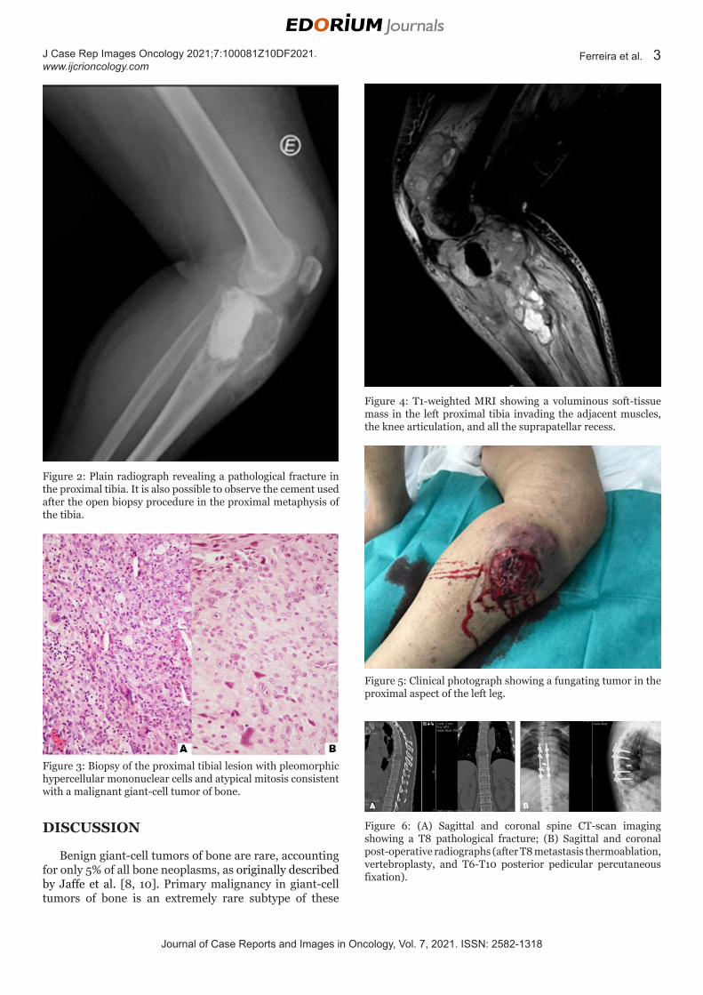
The new biopsy revealed some areas of typical histological findings for a giant-cell tumor, but there were other areas of pleomorphic hypercellular mononuclear cells with a high number of atypical mitoses. The final diagnosis for these findings was of a malignant giant-cell tumor of bone (Figure 3). The MRI showed a volumous soft tissue mass in the left proximal tibia invading the adjacent muscles, the knee articulation and all the
suprapatelar recess (Figure 4). During follow-up after the new biopsy, we also documented the breakdown of the surgical incision with progressive fungation of a soft tissue tumoral mass complicated by bacterial superinfection, which needed antibiotic therapy (Figure 5). A new CT scan and bone scan additionally showed several lung, vertebral, right iliac, and right femoral metastases. After discussion in a multidisciplinary meeting the decision was for above-knee amputation, chemotherapy, and additional radiotherapy for symptomatic bone lesions.
During the immediate post-operative period after the above-knee amputation (with histology showing free margins but tumoral vascular invasion), the patient developed acute intense dorsal pain. No neurological causes were found. The spine CT scan showed a T8 pathological fracture (Figure 6A). Because of this, the patient was returned to the operating theatre for thermoablation of the T8 metastasis, vertebroplasty, and T6-T10 posterior pedicular percutaneous fixation (Figure 6B), followed by local radiotherapy (30 Gy). The transpedicular biopsy confirmed metastasis from the malignant giant-cell tumor of the left tibia. In addition to surgery, the patient underwent chemotherapy with doxorubicin and cisplatin, nonetheless the disease kept progressing as shown on the follow-up CT and bone scan.
Shortly after chemotherapy the patient presented with right iliac pain. On the CT and bone scan there were already new metastatic masses occupying the right iliac bone and sacrum with probable sacral root involvement. Palliative radiotherapy was recommended again, but the patient sustained a right femoral pathologic fracture (Figure 7A) requiring return to the operating theatre for a long cephalomedullary nail (Figure 7B). Radiotherapy was also offered for the right iliac mass and all of the right femur (total 20 Gy), and a second-line chemotherapy cycle was proposed. Unfortunately, only 16 months after initial evaluation in the outpatient clinic for left knee pain, the patient suffered rapid clinical deterioration and eventually died.
Figure 1: (A) Anteroposterior radiograph showing a lytic metaphyseal lesion involving the proximal aspect of the left tibia; (B) Lateral radiograph showing the same lytic metaphyseal lesion involving the proximal aspect of the left tibia.
Journal of Case Reports and Images in Oncology, Vol. 7, 2021. ISSN: 2582-1318
J Case Rep Images Oncology 2021;7:100081Z10DF2021. www.ijcrioncology.com
Ferreira et al. 3
Figure 2: Plain radiograph revealing a pathological fracture in the proximal tibia. It is also possible to observe the cement used after the open biopsy procedure in the proximal metaphysis of the tibia.
Figure 3: Biopsy of the proximal tibial lesion with pleomorphic hypercellular mononuclear cells and atypical mitosis consistent with a malignant giant-cell tumor of bone.
Figure 4: T1-weighted MRI showing a voluminous soft-tissue mass in the left proximal tibia invading the adjacent muscles, the knee articulation, and all the suprapatellar recess.
Figure 5: Clinical photograph showing a fungating tumor in the proximal aspect of the left leg.
Figure 6: (A) Sagittal and coronal spine CT-scan imaging showing a T8 pathological fracture; (B) Sagittal and coronal post-operative radiographs (after T8 metastasis thermoablation, vertebroplasty, and T6-T10 posterior pedicular percutaneous fixation).
DISCUSSION
Benign giant-cell tumors of bone are rare, accounting for only 5% of all bone neoplasms, as originally described by Jaffe et al. [8, 10]. Primary malignancy in giant-cell tumors of bone is an extremely rare subtype of these

Journal of Case Reports and Images in Oncology, Vol. 7, 2021. ISSN: 2582-1318
J Case Rep Images Oncology 2021;7:100081Z10DF2021. www.ijcrioncology.com
Ferreira et al. 4
tumors, which can arise in 1–2% of all reported cases [5]. In fact, Beebe-Dimmer et al. estimated in their recent population study an annual incidence of 1.6 per 10,000,000 persons per year in the United States, which confirms the rarity of these neoplasms [8].
The literature concerning malignancies in giant-cell tumors is often confusing due to the lack of clear definitions. In 1970, Dahlin et al. considered as MGCT any malignant tumor with histologic evidence of benign counterpart features or in material previously removed from the same area [7]. If the tumor presented with malignant stromal cells throughout, Dahlin considered it should be classified as another malignant tumor with a different proper classification, despite its containing multinucleated benign cells, as does a giant cell tumor [7, 11]. Furthermore, the presence of metastasis at the onset of GCT should not be held as an automatic hallmark for malignization of the tumor, but instead as an unusual feature for a borderline benign tumor [12–15]. To clarify the study of MGCT it is crucial to use clear definitions and separate primary from secondary malignant giant cell tumor of bone: PMGCT is a lesion in which there are areas of synchronous high-grade sarcoma next to areas of benign typical GCT; SMGCT is a metachronous high-grade sarcomatous growth which arises in a previously benign GCT that has been treated by either surgery or radiotherapy [5, 6]. In this setting, and as reported by Bertoni et al., PMGCT is much rarer than SMGCT [6].
Like classic GCT, the malignant version of the disease preferentially invades the ends of long bones including the distal femur, proximal tibia, and distal tibia [4–7, 16–18]. Most often, the clinical presentation for a PMGCT is nonspecific, with pain and swelling as common symptoms [6]. Additionally, radiologic findings of PMGCT are very similar to those of GCT, which makes it extremely difficult to distinguish malignancies from benign tumors, even when the former has metastases or soft tissue invasion. We should not forget that typical GCT has the capacity to promote aggressive bone destruction, local invasion, and metastases [1, 19, 20].
While GCT is typically associated with a favorable prognosis, the long-term prognosis for malignant transformation seems to be poor—Bertoni et al. reported a high mortality rate, specially with SMGCT [6]. Anract et al. also reported a poor prognosis for MGCTB with a five-year survival rate of only 50%, despite the combination of surgery and chemotherapy [18]. Nonetheless, more recent reports from Beebe-Dimmer et al. showed a mean survival time of 11 years and 11 months, with a five-year relative survival of 84.2%. Domovitov et al. also found a 16% mortality rate for patients with MGCT; both studies suggesting a low-grade malignancy [9]. In this setting, and due to unclear and sometimes contradictory reports in the literature, Gong et al. considered that the prognosis of MGCTB was still indefinite, mainly because of its rarity and the short follow-up [5].
Considering all MGCT, PMGCT seems to have a better prognosis compared with SMGCT as pointed out by Gong et al. [5]. Nascimento et al. also found a better outcome for PMGCT, but Anract et al. observed equally poor outcomes when comparing between PMGCTB and SMGCTB [17, 18]. In the report by Bertoni et al., and despite the poor outcomes for all MGCT (most patients died due to lung metastatic disease), SMGCT again had a much worse prognosis, highlighting what seems to be a less aggressive nature for PMGCT [6].
There are some risk factors associated with poor outcomes regarding MGCT. Beebe-Dimmer et al. found in their study a relationship between older age and a more advanced-stage disease, with an increased risk for death [8]. More specifically, for each five years of increased age at diagnosis, the risk of death increased by 41%. For patients with distant metastases detected at the time of diagnosis, the risk of death was 5.2 times higher compared to those diagnosed with a tumor confined to bone [8].
Herein we have reported on a 55-year-old woman where the PMGCT diagnosis was difficult and required three biopsies (one imaging percutaneous biopsy without malignancy and two surgical biopsies, the second of which finally allowed the proper diagnosis). When the diagnosis for PMGCT was made, the patient had already sustained a pathological fracture and presented with a fungated lesion due to an extremely fast-growing tumor. Furthermore, the disease had already metastasized to the lungs and bone as identified in the CT and bone scan. Despite surgical treatment, chemotherapy, and radiotherapy the disease kept progressing and the patient died less than one year after diagnosis.
CONCLUSION
This clinical case delineates a very rare and aggressive presentation even for a malignant giant-cell tumor. This is an example of the most extreme aggressive biological behavior that could arise with a PMGCT, which is usually associated with a better prognosis. Indeed, this report
Figure 7: (A) Radiograph showing a right femoral diaphysis pathological fracture; (B) Radiograph of right femoral diaphysis pathological fracture after osteosynthesis with a long cephalomedullary nail.

Journal of Case Reports and Images in Oncology, Vol. 7, 2021. ISSN: 2582-1318
J Case Rep Images Oncology 2021;7:100081Z10DF2021. www.ijcrioncology.com
Ferreira et al. 5
highlights the need for a low threshold of suspicion for malignancy in any case of GCT with non-usual manifestation and a dissociation between histology and clinical behavior. We can also conclude that PMGCT is a confusing disease, since its clinical presentation and initial radiologic findings here were nonspecific and similar to any GCTB, but it evolved into a fulminant and shocking outcome. Even histological examination, the gold standard for diagnosis, had serious difficulties delivering a final result. Unfortunately, the delay in diagnosing such an unusual and aggressive PMGCT prevented a better and more efficient management of the disease.
REFERENCES
1. Mendenhall WM, Zlotecki RA, Scarborough MT, Gibbs CP, Mendenhall NP. Am J Clin Oncol 2006;29(1):96–9.
2. Werner M. Giant cell tumour of bone: Morphological, biological and histogenetical aspects. Int Orthop 2006;30(6):484–9.
3. Dahlin DC. Caldwell lecture. Giant cell tumor of bone: Highlights of 407 cases. AJR Am J Roentgenol 1985;144(5):955–60.
4. Alberghini M, Kliskey K, Krenacs T, Picci P, Kindblom L, Forsyth R, Athanasou NA. Morphological and immunophenotypic features of primary and metastatic giant cell tumour of bone. Virchows Archiv 2010;456(1):97–103.
5. Gong L, Liu W, Sun X, Sajdik C, Tian X, Niu X, Huang X. Histological and clinical characteristics of malignant giant cell tumor of bone. Virchows Archiv 2012;460(3):327–34.
6. Bertoni F, Bacchini P, Staals EL. Malignancy in giant cell tumor of bone. Cancer 2003;97(10);2520–9.
7. Dahlin DC, Cupps RE, Johnson EW Jr. Giant-cell tumor: A study of 195 cases. Cancer 1970;25(5):1061–70.
8. Beebe-Dimmer JL, Cetin K, Fryzek JP, Schuetze SM, Schwartz K. The epidemiology of malignant giant cell tumors of bone: An analysis of data from the Surveillance, Epidemiology and End Results Program (1975–2004). Rare Tumors 2009;1(2):e52.
9. Domovitov SV, Healey JH. Primary malignant giant-cell tumor of bone has high survival rate. Ann Surg Oncol 2010;17(3):694–701.
10. Jaffe HL, Lichtenstein L, Portis RB. Giant cell tumor of bone. Its pathologic appearance, grading, supposed variants and treatment. Arch Pathol 1940;30(3):993–1031.
11. Troup JB, Dahli DC, Coventry MB. The significance of giant cells in osteogenic sarcoma. Do they indicate a relationship between osteogenic sarcoma and giant cell tumor of bone? Proc Staff Meet Mayo Clin 1960;35:179–86.
12. Pan P, Dahlin DC, Lipscomb PR, Bernatz PE. “Benign” giant cell tumor of the radius with pulmonary metastasis. Mayo Clin Proc 1964;39:344–9.
13. Bertoni F, Present D, Enneking WS. Giant-cell tumour of bone with pulmonary metastases. J Bone Joint Surg Am 1985;67(6):890–900.
14. Bertoni F, Present D, Sudanese A, Bacchini P, Campanacci M. Giant-cell tumour of bone with pulmonary metastases. Six case reports and a review of the literature. Clin Orthop Relat Res 1988;(237):275–85.
15. Maloney WJ, Vaughan LM, Jones HH, Ross J, Nagel DA. Benign metastasing giant cell tumour of bone: Report of three cases and review of the literature. Clin Orthop 1989;(243):208–215.
16. Hutter RV, Worcester JN Jr, Francis KC, Foote FW Jr, Stewart FW. Benign and malignant giant cell tumors of bone. A clinicopathological analysis of the natural history of the disease. Cancer 1962;15:653–690.
17. Nascimento AG, Huvos AG, Marcove RC. Primary malignant giant cell tumor of bone: A study of eight cases and review of the literature. Cancer 1979;44(4):1393–402.
18. Anract P, De Pinieux G, Cottias P, Pouillart P, Forest M, Tomeno B. Malignant giant-cell tumours of bone. Clinico-pathological types and prognosis: A review of 29 cases. Int Orthop 1998;22(1):19–26.
19. Enneking WF. A system of staging musculoskeletal neoplasms. Clin Orthop Relat Res 1986;(204):9–24.
20. Campanacci M, Baldini N, Boriani S, Sudanese A. Giant-cell tumor of bone. J Bone Joint Surg Am 1987;69(1):106–14.
*********
Author ContributionsDavid Ferreira – Conception of the work, Drafting the work, Revising the work critically for important intellectual content, Final approval of the version to be published, Agree to be accountable for all aspects of the work in ensuring that questions related to the accuracy or integrity of any part of the work are appropriately investigated and resolved
André Spranger – Conception of the work, Interpretation of data, Revising the work critically for important intellectual content, Final approval of the version to be published, Agree to be accountable for all aspects of the work in ensuring that questions related to the accuracy or integrity of any part of the work are appropriately investigated and resolved
Paulo Almeida – Conception of the work, Analysis of data, Revising the work critically for important intellectual content, Final approval of the version to be published, Agree to be accountable for all aspects of the work in ensuring that questions related to the accuracy or integrity of any part of the work are appropriately investigated and resolved
Dolores Presa – Acquisition of data, Revising the work critically for important intellectual content, Final approval of the version to be published, Agree to be accountable for all aspects of the work in ensuring that questions related to the accuracy or integrity of any part of the work are appropriately investigated and resolved
José Portela – Conception of the work, Analysis of data, Interpretation of data, Revising the work critically for

Journal of Case Reports and Images in Oncology, Vol. 7, 2021. ISSN: 2582-1318
J Case Rep Images Oncology 2021;7:100081Z10DF2021. www.ijcrioncology.com
Ferreira et al. 6
important intellectual content, Final approval of the version to be published, Agree to be accountable for all aspects of the work in ensuring that questions related to the accuracy or integrity of any part of the work are appropriately investigated and resolved
Joaquim Soares do Brito – Conception of the work, Design of the work, Analysis of data, Drafting the work, Revising the work critically for important intellectual content, Final approval of the version to be published, Agree to be accountable for all aspects of the work in ensuring that questions related to the accuracy or integrity of any part of the work are appropriately investigated and resolved
Guarantor of SubmissionThe corresponding author is the guarantor of submission.
Source of SupportNone.
Consent StatementWritten informed consent was obtained from the patient for publication of this article.
Conflict of InterestAuthors declare no conflict of interest.
Data AvailabilityAll relevant data are within the paper and its Supporting Information files.
Copyright© 2021 David Ferreira et al. This article is distributed under the terms of Creative Commons Attribution License which permits unrestricted use, distribution and reproduction in any medium provided the original author(s) and original publisher are properly credited. Please see the copyright policy on the journal website for more information.
Access full text article onother devices
Access PDF of article onother devices

Related Documents