Supplementary information Comparative proteomic study of Arabidopsis mutants mpk4 and mpk6 Tomáš Takáč 1 , Pavol Vadovič 1 , Tibor Pechan 2 , Ivan Luptovčiak 1 , Olga Šamajová 1 , Jozef Šamaj 1* 1 Centre of the Region Haná for Biotechnological and Agricultural Research, Faculty of Science, Palacký University, Šlechtitelů 27, 783 71 Olomouc, Czech Republic 2 Institute for Genomics, Biocomputing & Biotechnology, Mississippi State University, MS 39762, USA

Welcome message from author
This document is posted to help you gain knowledge. Please leave a comment to let me know what you think about it! Share it to your friends and learn new things together.
Transcript

Supplementary information
Comparative proteomic study of Arabidopsis mutants mpk4 and mpk6 Tomáš Takáč1, Pavol Vadovič1, Tibor Pechan2, Ivan Luptovčiak1, Olga Šamajová1, Jozef Šamaj1*
1Centre of the Region Haná for Biotechnological and Agricultural Research, Faculty of Science, Palacký University, Šlechtitelů 27, 783 71 Olomouc, Czech Republic 2 Institute for Genomics, Biocomputing & Biotechnology, Mississippi State University, MS 39762, USA

B
Methylation, methionine synthesis
Protein synthesis
ER body formation
Glycolysis
Floral organ development
A Ascorbate cycle
Metabolism
Protein folding
Figure S1 Interaction networks in mpk4 mutant differential root proteome as predicted by STRING software for proteins with decreased (A) and increased (B) abundance in the mutant compared to wild type.

Methionine synthesis and methylation
Cytoskeleton
Protein synthesis
Figure S2 Interaction networks in mpk6 mutant differential root proteome as predicted by STRING software for proteins with decreased (A) and increased (B) abundance in the mutant compared to wild type.
A
B
Aquaporins
Protein synthesis

Figure S3. Comparison of gene onthology annotations (at 3rd level of onthology) of all proteins identified in mpk4 and mpk6 mutant roots according to biological process. Relative protein numbers of proteins per annotation are presented in the graph. Venn diagram shows the number of GO annotations assigned solely to mpk6 (red field) and mpk4 mutant root proteome (blue field) as well as the number of GO annotations present in both mutant proteomes (overlap).
0 10 20 30 40 50
catabolic process
nitrogen compound metabolic process
cellular component organization
establishment of localization
single-multicellular organism process
regulation of biological process
anatomical structure development
response to biotic stimulus
cellular component biogenesis
single-organism developmental process
regulation of biological quality
reproductive process
response to endogenous stimulus
single organism signaling
immune response
methylation
macromolecule localization
response to external stimulus
death
circadian rhythm
regulation of molecular function
multicellular organism reproduction
interspecies interaction between organisms
multi-multicellular organism process
cellular process involved in reproduction
detection of stimulus
mpk6
mpk4
0 0
mpk6 mpk4
26

Figure S4. Comparison of gene onthology annotations (at 6th level of onthology) of all proteins identified in mpk4 and mpk6 mutant roots according to molecular function. Relative protein numbers of proteins per annotation are presented in the graph. Venn diagram shows the number of GO annotations assigned solely to mpk6 (red field) and mpk4 mutant root proteome (blue field) as well as the number of GO annotations present in both mutant proteomes (overlap).
0 5 10 15 20 25
transition metal ion binding
ribonucleotide binding
purine ribonucleoside binding
purine nucleotide binding
pyrophosphatase activity
cation transmembrane transporter activity
amino acid binding
calcium ion binding
translation factor activity, nucleic acid binding
P-P-bond-hydrolysis-driven transmembrane transporter activity
anion transmembrane transporter activity
protein kinase activity
glucosidase activity
carboxylesterase activity
protein disulfide isomerase activity
3-isopropylmalate dehydrogenase activity
glyceraldehyde-3-phosphate dehydrogenase (NAD+) activity
S-adenosylmethionine-dependent methyltransferase activity
ribonuclease activity
nucleobase-containing compound kinase activity
gated channel activity
substrate-specific channel activity
aconitate hydratase activity
endopeptidase activity
phosphogluconate dehydrogenase activity
1-aminocyclopropane-1-carboxylate oxidase activity
adenosylhomocysteinase activity
isocitrate dehydrogenase activity
succinate-CoA ligase activity
acetyltransferase activity
relative number of proteins in annotation
mpk6mpk4
3 0
mpk6 mpk4
27
0 20 40 60 80
cytosol
Golgi apparatus
nucleus
plastid
mitochondrion
vacuole
ribosome
intracellular organelle lumen
organelle envelope
endoplasmic reticulum
microbody
cytoskeleton
organelle subcompartment
photosynthetic membrane
relative number of proteins in annotation
mpk4
mpk6
Figure S5. Comparison of gene onthology annotations (at 8th level of onthology) of all proteins identified in mpk4 and mpk6 mutant roots (in 3 out of 4 biological replicates) according to cellular component. Relative protein numbers of proteins per annotation are presented in the graph. Venn diagram shows the number of GO annotations assigned solely to mpk6 (red field) and mpk4 mutant root proteome (blue field) as well as the number of GO annotations present in both mutant proteomes (overlap).
0 0
mpk6 mpk4
14
Accession Coverage # Peptides MW [kDa] calc. pI Score Description Intensity sum (average Col0)
Intensity sum (average mpk4 ) P -value fold
gi334185401 34.34 17 32.1 5.44 82.86 JA-responsive protein 1 44159838 28287813 0.023 0.64gi18413214 29.59 9 18.8 8.27 61.76 nucleoside diphosphate kinase 1 37876742 23032132 0.045 0.61
gi15224648 30 6 11 4.88 43.39 membrane-associated progesterone binding protein 2 45080293 16934108 0.001 0.38
gi18379240 46.36 24 17.5 5.73 142.14 MLP-like protein 328 133320704 46183710 0.001 0.35gi18379244 49.01 23 17.6 5.55 128.66 MLP-like protein 329 92297424 23811471 0.001 0.26gi334186408 17.5 10 37.4 9.6 55.92 L-ascorbate peroxidase S 16727486 1908328 0.022 0.11gi30696930 21.37 18 52.1 7.75 122.47 Monodehydroascorbate reductase 45865373 10864237 0.001 0.24gi30686836 25.1 17 29.4 5.47 75.07 dehydrin ERD10 24023126 7804980 0.02 0.32gi30690396 31.25 41 38.8 6.67 258.53 Peroxidase family protein 168956530 70979878 0.016 0.42gi30690772 34.22 29 29.2 8.5 153.92 glutathione S-transferase phi 8 99466734 49415219 0.001 0.5gi145323784 45.78 34 27.5 6.29 181.81 L-ascorbate peroxidase 1 78213410 18975297 0.041 0.24gi15237716 13.42 5 33.8 6.1 30.87 TOMV RNA binding protein 12474685 2640969 0.001 0.21gi15232058 13.4 6 34.9 8.95 25.84 peroxidase 27 4017186 - - unique in WTgi18401423 11.93 1 12.2 5.68 14 heat stable protein 1 2733535 - - unique in WTgi145334501 5.84 1 27 5.63 22.57 binding partner of acd11 1 4012129 - - unique in WT
gi334188446 16.92 18 63.3 5.87 104.79 TCP-1/cpn60 chaperonin family protein 44038838 10240942 0.007 0.23gi15230534 23.54 34 71.1 5.25 208.69 heat shock protein 70-4 113395522 - - unique in WTgi334185190 26.81 44 71.1 5.7 229.3 heat shock protein 70-3 144132985 82821161 0.008 0.57gi240255880 53.49 11 9.3 4.48 61.9 heat shock factor binding protein 21432992 - - unique in WTgi30691626 14.37 19 73 5.62 147.99 heat shock protein 70-1 37202366 11969206 0.015 0.32gi15236211 43.02 19 18.4 8.69 168.3 rotamase cyclophilin 5 99205263 49647915 0.004 0.5gi30684617 29.17 11 15.4 5.55 64.37 40S ribosomal protein S12-1 31597439 4316075 0.004 0.14gi15234781 60.47 19 18.4 7.81 130.6 peptidyl-prolyl cis-trans isomerase CYP1 84563207 45953462 0.005 0.54gi30696056 14 25 93.8 6.25 161.39 elongation factor EF-2 36188952 2767139 0.001 0.08
gi15237054 30.87 19 26 6.4 176.65 V-type proton ATPase subunit E1 579193317 6277232 0.015 0.11
gi15233538 43.51 15 14 5.2 73 profilin 2 49367881 12542005 0.004 0.25gi15224838 43.51 11 14.3 4.82 72.27 profilin 1 28135567 10004823 0.020 0.36
gi79558700 17.16 6 19 6.79 32.4 rhodanese-like domain-containing protein 8990279 - - unique in WT
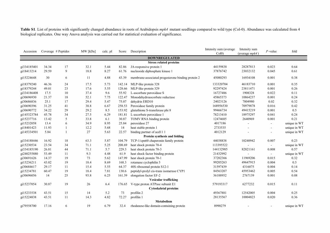
Table S1. List of proteins with significantly changed abundance in roots of Arabidopsis mpk4 mutant seedlings compared to wild type (Col-0). Abundance was calculated from 4 biological replicates. One way Anova analysis was carried out for statistical evaluation of significance.
DOWNREGULATEDStress related proteins
Protein synthesis and folding
Vesicular trafficking
Cytoskeletal proteins
Metabolism
gi334186360 25.58 4 14 5.26 22.15 peptide methionine sulfoxide reductase B5 7918550 - - unique in WTgi30695409 4.27 1 40.9 6.5 23.45 acetoacetyl-CoA thiolase 2 4097356 - - unique in WT
gi18423437 8.88 1 19.2 4.77 3.67 probable NADH dehydrogenase [ubiquinone] 1 alpha subcomplex subunit 5 14759169 3307172 0.018 0.22
gi15225353 8.31 7 45.3 6.71 88.83 Succinyl-CoA ligase [GDP-forming] subunit beta 24356937 8969895 0.021 0.37gi15219721 38.86 32 35.5 6.55 225.34 malate dehydrogenase 146565660 60763875 0.016 0.41gi238479568 9.82 3 18.5 4.78 16.32 thiocyanate methyltransferase 1 6653891 2836376 0.017 0.43gi145329204 22.88 13 32.3 7.49 75.34 triosephosphate isomerase 26467098 11954894 0.041 0.45gi15225798 24.68 14 48.5 8.34 87.2 3-ketoacyl-CoA thiolase 2 22599512 10566258 0.028 0.47gi15219886 15.18 10 21.2 4.34 45.56 cytochrome C oxidase 6B 27322231 14556289 0.009 0.53
gi15236568 14.57 8 17.5 6.34 43.47 polyketide cyclase/dehydrase and lipid transport superfamily protein 38732855 10718416 0.018 0.28
gi79313339 20.76 5 25.5 5.2 26.99 alpha/beta-Hydrolases superfamily protein 8443513 2694350 0.024 0.32gi186502696 24.42 27 54.5 5.92 129.1 aminopeptidase family protein 39638087 24594163 0.035 0.62gi30687411 16.99 5 39.7 7.53 44.11 Dihydrolipoamide succinyltransferase 9641136 - - unique in WT
gi18400212 4.45 2 58.4 7.65 31.36 dihydrolipoamide acetyltransferase, long form protein 11109967 - - unique in WT
gi15240454 3.66 4 50.1 9.14 25.37 dihydrolipoyllysine-residue succinyltransferase component of 2-oxoglutarate dehydrogenase complex 1 728437
- - unique in WT
gi15239843 24.7 21 35.7 6.79 146.81 malate dehydrogenase 94027894 - - unique in WTgi15237998 7.88 4 21.5 6.7 22.47 ATP synthase subunit delta' 6292429 - - unique in WTgi15237679 14.29 5 12 6.4 32.07 peptidyl-prolyl isomerase FKBP12 7765998 - - unique in WTgi15233349 6.46 13 98.1 6.4 78.55 aconitate hydratase 1 7324322 - - unique in WTgi15226573 4.78 7 65.5 6.28 43.64 Ferredoxin--nitrite reductase 3798383 - - unique in WTgi15231715 50.56 43 38.5 6.46 272.67 fructose-bisphosphate aldolase 166335365.1 117852344.5 0.025 0.71
Signalinggi18417863 13.6 5 30.2 4.81 36.66 14-3-3-like protein GF14 upsilon 3761124 - - unique in WT
Proteolysisgi22331076 6.44 2 81.8 6.77 30.68 Subtilase family protein 3949056 - - unique in WT
gi15220874 27.55 13 22.1 8.78 63.97 kunitz type trypsin and protease inhibitor domain-containing protein 13269998 - - unique in WT
gi145325425 20.25 6 25.9 4.75 59.01 proteasome subunit alpha type-5-A 6712319 2157322 0.023 0.32gi15219345 16.27 9 45.5 4.82 59.27 metacaspase 4 14305198 - - unique in WT
Unknown functiongi240254562 3 6 148.5 4.75 81.64 uncharacterized protein 12034279 - - unique in WTgi18402503 33.54 4 16.9 5.3 33.78 uncharacterized protein 4479077 - - unique in WT
Cell wall compositiongi145324054 6.3 5 33.9 10.7 42.42 arabinogalactan protein 31 5011771 - - unique in WT
Translationgi30682607 1.6 3 141.9 6.6 25.44 mRNA decapping complex VCS 122872 - - unique in WT
Scaffolding proteingi15225899 13.68 3 21 8.54 18.85 Remorin family protein 7158948 - - unique in WT

gi15227257 22.46 10 35.4 8.9 81.77 ATP synthase subunit gamma 3880740 18269069 0.002 4.71
gi15218090 16.1 8 54.4 6.33 80.45 putative mitochondrial-processing peptidase subunit alpha-1 3945991 51746913 0.038 2.31
gi15240765 16.67 5 29.6 8.85 64.56 voltage dependent anion channel 2 4835579 11676030 0.005 2.41gi79587736 18.2 10 31.9 5.27 55.37 glyoxalase I homolog 5433689 38446250 0.001 7.08gi79327847 31.7 26 41.9 6 125.33 isopropylmalate dehydrogenase 1 21088922 50750377 0.007 2.41
gi15229231 50 55 36.9 7.12 308.86 glyceraldehyde-3-phosphate dehydrogenase C subunit 1 141154062 370726048 0.004 2.63
gi145334507 23.1 57 84.3 6.51 317.79 5-methyltetrahydropteroyltriglutamate--homocysteine methyltransferase 43824674 120126136 0.025 2.74
gi15235213 24.71 13 29.1 5.29 89.73 putative caffeoyl-CoA O-methyltransferase 16286350 37972542 0.027 2.33gi30699430 34.66 27 42.1 5.68 203.37 phosphoglycerate kinase 83143041 131826906 0.016 1.59
gi79313257 25.2 24 80.7 4.7 221.13 DNA topoisomerase-like protein 44648919 74541493 0.001 1.67gi15239697 24.62 8 13.6 10.15 39.65 histone H2A 8672749 19871188 0.021 2.29
gi15220216 34.7 20 36.2 5.38 159.85 annexin 1 18417212 81194961 0.013 4.41gi240255920 34.3 23 37.7 6.84 144.83 putative beta-1,3-endoglucanase 14524276 86520500 0.004 5.96gi18414298 22.7 13 47.5 5.31 107.75 monodehydroascorbate reductase (NADH) 9202420 56147858 0.046 6.1gi15235401 38.21 23 24.1 6.35 146.9 glutathione S-transferase F2 41864796 118071075 0.021 2.82gi18411929 12.79 2 25.6 6.4 65.94 glutathione S-transferase TAU 19 4073923 12470006 0.013 3.06gi79607896 31.91 60 72.4 5.5 368.81 jacalin-like lectin domain-containing protein 102024107 373005179 0.001 3.66
gi15236981 17.91 2 15.5 10.4 28.17 60S ribosomal protein L14-2 4460163 32625377 0.001 7.31gi30694819 13.95 4 19.8 9.92 34.55 60S ribosomal protein L11-2 2227646 21059651 0.005 9.45gi145332383 13.43 6 45.7 6.28 42.16 translational initiation factor 4A-1 5401363 15502069 0.043 2.87gi18410311 12.56 6 23.8 7.18 59.49 kunitz trypsin inhibitor 1 3790257 13873594 0.001 3.66gi15232603 25 14 34.1 5.7 104.3 60S acidic ribosomal protein P0-2 3050007 14057122 0.044 4.61gi15233740 15.67 20 94.1 5.3 141.95 HSP90-like protein GRP94 9673127 48278582 0.000 4.99gi15233268 34 13 27.5 7.11 64.98 20S proteasome alpha-3 subunit 2774847 15100795 0.012 5.44
gi18404748 28.76 26 43.1 8.7 164.62 GDSL esterase/lipase 18672028 102368760 0.005 5.48
gi15232845 25.42 28 59.1 6.79 183.49 probable mitochondrial-processing peptidase subunit beta 3945991 51746913 0.001 13.11
gi186513287 3.78 3 49 7.72 18.56 argininosuccinate synthase 1422378 5166958 0.041 3.63gi145333043 14.77 10 35.5 5.16 61 adenosylhomocysteinase 892125 22523603 0.015 25.25
gi15229809 5.88 8 21.6 9.41 26.53 uncharacterized protein 10940894 28197085 0.047 2.58gi22328599 18.39 3 9.5 4.97 24.38 RNA recognition motif-containing protein 4138353 unique in mpk4gi186478702 3.71 3 43.9 7.61 17.48 catalase 3 - 9288542 - unique in mpk4
AminoAcid metabolism
Unknown
UPREGULATEDMetabolism
Nucleic acid binding
Stress related
Protein synthesis, processing and folding
Lipid metabolism

gi18421006 30.77 19 39.8 9.22 106.73 TRAF-like family protein - 77036438 - unique in mpk4gi18410982 9.38 4 17.8 4.54 18.16 S-phase kinase-associated protein 1 - 2756551 - unique in mpk4gi18406229 13.89 15 45 5.82 76.81 TRAF-like protein - 2756551 - unique in mpk4gi15241316 33.78 17 25 10.32 87.57 40S ribosomal protein S8-1 - 55838806 - unique in mpk4gi15229631 15.7 3 16.9 10.78 36.15 60S ribosomal protein L26-1 - 16600695 - unique in mpk4gi15226503 6.61 3 26.9 7.2 16.1 short-chain dehydrogenase reductase 3a - 8057351 - unique in mpk4

Accession Coverage # Peptides MW [kDa] calc. pI Score Description Intensity sum (average Col0)
Intensity sum (average mpk6 ) Fold change P value
Stress related proteinsgi15228041 35.3 4 41.8 6.51 47.67 aquaporin TIP1-1 12761819 672496 0.05 1.02E-05gi15231569 32.38 7 41.8 5.6 42.67 aquaporin TIP1-2 10500652 813310 0.08 0.005gi18414298 29.49 8 47.5 4.97 56.54 monodehydroascorbate reductase (NADH) 55562447 1493058 0.03 0.01
Cytoskeletal proteinsgi15233538 41.66 7 14 5.2 46.79 profilin 2 94136176 19684596 0.21 0.049
Metabolismgi15220770 41.79 1 36.2 5.12 44.348 1-aminocyclopropane-1-carboxylate oxidase 2 4871669 462263 0.09 0.032gi15221156 32.66 5 61.4 6.1 49.41 pyrophosphate--fructose-6-phosphate 1-phosphotransferase 31023724 2581092 0.08 0.01gi15240628 41.86 2 41.72 9.86 40.65 delta subunit of Mt ATP synthase 39349332 8045791 0.2 0.03gi15241592 30.25 10 36.1 8.39 70.64 Succinyl-CoA ligase [GDP-forming] subunit alpha-1 85150756 18785954 0.22 0.01gi18403295 41.8 8 55.2 8.43 61.33 gamma-aminobutyrate transaminase POP2 32812295 4583895 0.14 0.015
Protein synthesis and foldinggi30691988 41.73 1 37.7 8.18 41426 chaperone protein dnaJ 3 31023724 2581092 0.14 0.022gi15226231 48.7 15 41.7 4.61 86.42 60S acidic ribosomal protein P2-1 142985326 58068438 0.41 0.021gi15240317 41.95 6 60.3 5.47 56.61 TCP-1/cpn60 chaperonin family protein 7454995 1014447 0.14 0.03gi18394184 27.55 5 41.86 7.69 45.84 chaperonin 10 150262741 13474315 0.09 0.01
Cell Wall proteinsgi145324054 45.87 8 33.9 10.7 53.18 arabinogalactan protein 31 38160494 24382141 0.64 0.03
Nucleic acid bindinggi15239697 40 6 41803 4.22 37.45 histone H2A 45987241 19931344 0.43 0.02
gi145333041 22.48 5 41.7 6.55 41.877 glycine-rich RNA-binding protein 2 82983541 11330246 0.14 0.029
Stress responsegi334185190 24.35 22 71.1 5.7 202.97 heat shock protein 70-3 72663041 229242810 3.15 0.047gi15219623 18.32 22 59.7 7.21 162.51 beta-glucosidase 22 21211934 241821111 11.4 0.017gi145334555 18.65 8 36.9 8.18 66.61 TRAF-like family protein 28420192 72189838 2.54 0.029gi18421006 20.51 7 39.8 9.62 49.14 TRAF-like family protein - 45109989 - unique in mpk6gi15233320 13.6 1 25 5.42 34.18 aquaporin TIP2-1 - 291261 - unique in mpk6gi238479502 46296 4 50.4 4.93 38.64 jacalin-related lectin 22 - 21204913 - unique in mpk6
Protein synthesis and foldinggi30687350 42.6 9 41.6 4.56 44.45 Elongation factor 1-beta 2 35745455 86882077 2.43 0.007gi145332383 34.08 15 45.7 6.28 99.12 translational initiation factor 4A-1 6463817 78329209 12.12 0.005gi15226212 19.83 3 41.6 9.94 31.96 60S ribosomal protein L7-2 9171521 27849796 3.04 0.033gi30691619 23.13 4 41.6 4.56 26.64 elongation factor 1B beta - 12710359 - unique in mpk6gi15231024 41.6 1 59.2 6.3 41.833 T-complex protein 1 subunit alpha - 6989583 - unique in mpk6
Protein degradationgi15230005 28.67 2 47.5 5.3 24.69 regulatory particle triple-A ATPase 5A - 8410216 - unique in mpk6
Cytoskeletal proteinsgi15241179 19.78 11 49.6 5.1 93.14 tubulin alpha-5 27700598 80220285 2.9 0.053
Table S2. List of proteins with significantly changed abundance in roots of Arabidopsis mpk6 mutant seedlings compared to wild type (Col-0). Abundance was calculated from 4 biological replicates. One way Anova analysis was carried out for statistical evaluation of significance.
DOWNREGULATED
UPREGULATED

gi15242516 36.34 16 41.7 5.49 127.72 actin 7 23306738 135297066 5.81 0.048gi30697298 39.52 8 41.6 5.19 47.96 actin depolymerizing factor 3 13497389 38089510 2.82 0.031gi30683070 28.57 13 47.2 8.9 103.84 tubulin alpha-6 chain 22867734 114613659 5.01 0.022gi15222075 23.62 12 41.8 5.58 106.86 actin 8 - 114837357 - unique in mpk6gi15227503 43.07 3 42.1 5.33 46.37 Actin-like ATPase superfamily protein - 26913061 - unique in mpk6
Metabolismgi15222848 52.96 54 36.9 7.18 373.32 glyceraldehyde 3-phosphate dehydrogenase 262612712 595627944 2.27 0.018gi15229522 35.05 18 53.1 5.74 180.69 adenosylhomocysteinase 2 72720355 115940281 1.56 0.037gi15236375 41.78 20 51.7 7.23 116.93 serine hydroxymethyltransferase 4 49262480 177373784 3.61 0.007gi186505986 25.38 4 42.5 6.18 60.44 S-adenosylmethionine synthase 3 7853307 50065528 6.36 0.002gi334186086 23.62 9 63.8 6.8 61.95 ketol-acid reductoisomerase 9186766 31621802 3.44 0.005gi26557005 25.44 23 54.9 6.23 175.1 ATPase subunit 1 81707979 154980113 1.9 0.05
gi15231059 24. 53 1 47.5 8.32 18.32 acyl-coenzyme A oxidase 4 - 3900946 - unique in mpk6gi145332819 46.5 3 51.9 6.7 30.92 pyruvate kinase - 8527319 - unique in mpk6
Lipid metabolismgi15232671 35.09 2 91.8 5.87 56.92 phospholipase D alpha 1 - 5325577 - unique in mpk6gi15221970 27.39 1 98 5.52 41.69 lipoxygenase 1 - 5459943
Cell divisiongi15232776 41.7 11 89.3 5.25 70.08 cell division control protein 48-A - 40140792 - unique in mpk6
Aminoacid metabolismgi15233111 46.08 3 39.9 8.54 41.28 cysteine synthase C1 - 11878185 - unique in mpk6
Signallinggi18396845 44.16 2 41.7 4.81 41.863 14-3-3 family protein - 10738972 - unique in mpk6gi15220941 35.85 1 35.7 7.71 35.27 WD-40 repeat ArcA-like protein (Rack1A) - 8125602 - unique in mpk6
Protein transportgi18391349 16.35 3 41.8 4.15 32.082 vacuolar calcium-binding protein-like protein - 9683235 - unique in mpk6
Unknown function
gi145333007 31.35 2 41.6 4.42 24.38 Nascent polypeptide-associated complex subunit alpha-likeprotein 4 - 9187629 - unique in mpk6
gi15235944 17.98 1 41.8 8.69 41.771 uncharacterized protein - 32134464 - unique in mpk6

category mpk4 mutant mpk6 mutant
total number of proteins with significantly changed abundance 65 32
upregulated ≥ 5 fold 9 5
upregulated 1.5- 5 fold 22 11
downregulated 1.5- 5 fold 30 7
downregulated ≥ 5 fold 4 9
detected uniquely in one sample (control/mutant) 25/8 0/19
Table S3. Classification of proteins according changes in their abundances in the mpk4 and mpk6 mutants, as compared to the Col-0 wild type.

Accession Protein name fold change in mpk4 fold change in mpk6
gi145324054 arabinogalactan protein 31 unique in wt 0.64
gi15233538 profilin 2 0.25 0.21
gi15239697 histone H2A 2.29 0.43
gi18414298 monodehydroascorbate reductase (NADH) 6.1 0.26
gi334185190 heat shock protein 70-3 0.57 3.15
gi145334555 TRAF-like family protein unique in mpk4 2.54
gi15222848 glyceraldehyde 3-phosphate dehydrogenase 2.71 2.27
gi145332383 translational initiation factor 4A-1 2.87 12.11
Table S4. Proteins detected as differentially abundant in both mpk4 and mpk6 mutants.

Position Code Kinase Peptide Score Cutoff >gi|145324054|ref|NP_001077616.1| arabinogalactan protein 31 (unique in wild type)
33 T CMGC/MAPK EVNHKTQTPSLAPAP 43.355 35.046 70 S CMGC/MAPK HPHPPAKSPVKPPVK 35.092 35.046 81 S CMGC/MAPK PPVKAPVSPPAKPPV 39.59 35.046
113 S CMGC/MAPK PPVKPPVSPPAKPPV 37.553 35.046 165 S CMGC/MAPK APVKPPVSPPTKPPV 41.472 35.046 173 T CMGC/MAPK PPTKPPVTPPVYPPK 35.063 35.046
>gi|30696056|ref|NP_849818.1| elongation factor EF-2-like protein LOS1 (fold change 0.08) 764 T CMGC/MAPK EEMQRPGTPLYNIKA 35.552 35.046 834 T CMGC/MAPK KGLKEAMTPLSEFED 38.873 35.046
>gi|145325425|ref|NP_001077717.1| proteasome subunit alpha type-5-A (fold change 0.32) 56 S CMGC/MAPK AVEKRITSPLLEPSS 36.624 35.046
>gi|30691626|ref|NP_195504.2| heat shock protein 70-1 (fold change 0.32) 669 T CMGC/MAPK GTSGTEQTPEAEFEE 42.453 35.046
>gi|334185190|ref|NP_001189847.1| heat shock protein 70-3 (fold change 0.57) 403 T CMGC/MAPK DLLLLDVTPLSLGLE 48.055 35.046
>gi|15218090|ref|NP_175610.1| putative mitochondrial-processing peptidase subunit alpha-1 (fold change 2.31) 38 T CMGC/MAPK VAETSSSTPAYLSWL 37.379 35.046 94 T CMGC/MAPK LKIASETTPNPAASI 40.124 35.046
228 S CMGC/MAPK GYSGPLASPLYAPES 46.132 35.046 >gi|15240765|ref|NP_201551.1| voltage dependent anion channel 2 (fold change 2.41)
137 S CMGC/MAPK DITATLGSPVISFGA 40.513 35.046 >gi|15221019|ref|NP_175802.1| GDSL esterase/lipase (fold change 5.48)
32 S CMGC/MAPK ITVAGQNSPVVALFT 36.626 35.046 82 T CMGC/MAPK KFSDGLITPDFLAKF 36.566 35.046
371 T CMGC/MAPK REFWVPPTPATVHAS 39.119 35.046 >gi|186513287|ref|NP_001119047.1| argininosuccinate synthase (fold change 3.63)
331 S CMGC/MAPK ALNGKALSPATLLAE 47.712 35.046 >gi|15232603|ref|NP_187531.1| 60S acidic ribosomal protein P0-2 (fold change 4.61)
158 T CMGC/MAPK KGTVEIITPVELIKQ 35.106 35.046 202 S CMGC/MAPK YDNGSVFSPEVLDLT 43.1 35.046
>gi|15232845|ref|NP_186858.1| probable mitochondrial-processing peptidase subunit beta (fold change 13.11) 40 S CMGC/MAPK DSVPASASPTALSPP 37.688 35.046 45 S CMGC/MAPK SASPTALSPPPPHLM 42.61 35.046
>gi|15226573|ref|NP_179164.1| Ferredoxin--nitrite reductase (unique in wild type) 10 S CMGC/MAPK SFSLTFTSPLLPSSS 50.472 35.046
>gi|15233349|ref|NP_195308.1| aconitate hydratase 1 (unique in wild type) 304 S CMGC/MAPK RATIANMSPEYGATM 36.127 35.046 659 S CMGC/MAPK YFKGMTMSPPGPHGV 42.372 35.046
>gi|18400212|ref|NP_566470.1| dihydrolipoamide acetyltransferase. long form protein (unique in wild type) 80 S CMGC/MAPK TTSTKLSSPMAGPKL 35.217 35.046
120 S CMGC/MAPK EIGMPSLSPTMTEGN 41.945 35.046 >gi|22331076|ref|NP_566473.2| Subtilase family protein (unique in wild type)
21 S CMGC/MAPK PLLLCFFSPSSSSSD 37.946 35.046 63 S CMGC/MAPK LLRSLPSSPQPATLL 57.857 35.046 85 S CMGC/MAPK HGFSARLSPIQTAAL 45.139 35.046
114 T CMGC/MAPK REIHTTHTPAFLGFS 41.702 35.046 484 S CMGC/MAPK LGTLIGPSPPSPRVA 35.062 35.046 487 S CMGC/MAPK LIGPSPPSPRVAAFS 44.914 35.046 713 S CMGC/MAPK ANVEIDVSPSKLAFS 37.94 35.046
>gi|240254562|ref|NP_565741.4| uncharacterized protein (unique in wild type) 1299 T CMGC/MAPK SSSGNVTTPTQTAST 47.724 35.046
>gi|30682607|ref|NP_850576.1| mRNA decapping complex VCS (unique in wild type) 28 S CMGC/MAPK PGISAQPSPVTQQQQ 40.154 35.046 63 S CMGC/MAPK TPPLNLQSPRSNHNP 37.132 35.046
625 S CMGC/MAPK TLPQLPLSPRLSSKL 59.13 35.046
Table S5. List of differentially regulated proteins in mpk4 mutant roots containing MAPK-specific phosphorylation site as predicted by GPS 3.0 software.

Instances (Matched Sequence)
Cell Compartme
nt Pattern Probabilit
y
arabinogalactan protein 31
KFNRSLVAV 180-188 cell wall1 [KR]{0,2}[KR].{0,2}[KR].{2,4}[I
LVM].[ILVF] 4.324e-03 KKLGKSTVVV 285-294 KLGKSTVVV 286-294
elongation factor EF-2 KRLAKSDPMVV 509-519 cytoplasm6 [KR]{0,2}[KR].{0,2}[KR].{2,4}[ILVM].[ILVF] 4.32E-03 RLAKSDPMVV 510-519
proteasome subunit alpha type-5-A KTKEGVVLAV 41-50
peroxisome, cytoplasm
(prediction)
[KR]{0,2}[KR].{0,2}[KR].{2,4}[ILVM].[ILVF] 4.32E-03
heat shock protein 70-1 KRSDNIDL 289-296 cytosol7 [KR]{0,2}[KR].{0,2}[KR].{2,4}[ILVM].[ILVF] 4.324e-03 KKQLIDL 573-579
heat shock protein 70-3 RARFEELNI 305-313 nucleus, cytoplasm2
[KR]{0,2}[KR].{0,2}[KR].{2,4}[ILVM].[ILVF] 4.324e-03 RIPKVQQLLV 348-357
putative mitochondrial-processing peptidase subunit alpha-1
RKMKVEI 196-202 mitochondria [KR]{0,2}[KR].{0,2}[KR].{2,4}[ILVM].[ILVF] 4.324e-03
voltage dependent anion channel 2 KHPRFGLSLAL 264-274
mitochondria plasma
membrane8
[KR]{0,2}[KR].{0,2}[KR].{2,4}[ILVM].[ILVF] 4.324e-03
GDSL esterase/lipase KFMKIPLAI 88-96 extracellular (prediction)
[KR]{0,2}[KR].{0,2}[KR].{2,4}[ILVM].[ILVF] 4.324e-03
argininosuccinate synthase RGKLKKVVL 93-101
plastid9 [KR]{0,2}[KR].{0,2}[KR].{2,4}[ILVM].[ILVF] 4.324e-03 KKHNVPVPV 255-263
KKDMYMMSV 293-301 KLYKGSVSV 393-401
60S acidic ribosomal protein P0-2
RKGLRGDSVVL 44-54 ribosome [KR]{0,2}[KR].{0,2}[KR].{2,4}[I
LVM].[ILVF] 4.324e-03 KGLRGDSVVL 45-54 KINKGTVEI 148-156
probable mitochondrial-processing peptidase subunit beta
RRSQRRLFL 11-19 mitochondria [KR]{0,2}[KR].{0,2}[KR].{2,4}[I
LVM].[ILVF] 4.324e-03 RINRERDVIL 210-219
Ferredoxin--nitrite reductase
KPKRSVLV 19-26
plastid [KR]{0,2}[KR].{0,2}[KR].{2,4}[ILVM].[ILVF] 4.324e-03
KIEREPMKL 74-82 KSSKDDIDVRL 103-113 RKWNVCV 246-252 KDGRFGFNLLV 274-284 KRCEEAIPL 291-299 RQKTRMMWL 328-336 KKGVRVTELVPL 554-565 KGVRVTELVPL 555-565
aconitate hydratase 1 RIDKLPYSIRI 35-45 cytosol, mitochondria
10
[KR]{0,2}[KR].{0,2}[KR].{2,4}[ILVM].[ILVF] 4.324e-03 KRPHDRVPL 378-386
KKACDLGL 458-465 dihydrolipoamide acetyltransferase, long form protein
RRDHAVAV 20-27 mitochondria11
[KR]{0,2}[KR].{0,2}[KR].{2,4}[ILVM].[ILVF] 4.324e-03
Subtilase family protein RRHPSVISV 93-101 extracellular (prediction)
[KR]{0,2}[KR].{0,2}[KR].{2,4}[ILVM].[ILVF] 4.324e-03
uncharacterized protein KSRDIDLSF 1264-1272 nucleus (prediction)
[KR]{0,2}[KR].{0,2}[KR].{2,4}[ILVM].[ILVF] 4.324e-03
mRNA decapping complex VCS
RKAQPLVVL 352-360 cytoplasmic foci12
[KR]{0,2}[KR].{0,2}[KR].{2,4}[ILVM].[ILVF] 4.324e-03 KESKRLEVAL 918-927
KRLEVAL 921-927
Table S6. List of differentially regulated proteins in mpk4 mutant roots containing MAPK-docking site as predicted by The Eukaryotic Linear Motif resource for Functional Sites in Proteins (http://elm.eu.org/).
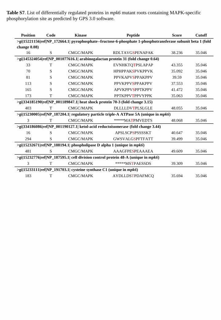
Position Code Kinase Peptide Score Cutoff >gi|15221156|ref|NP_172664.1| pyrophosphate--fructose-6-phosphate 1-phosphotransferase subunit beta 1 (fold change 0.08)
16 S CMGC/MAPK RDLTAVGSPENAPAK 38.236 35.046 >gi|145324054|ref|NP_001077616.1| arabinogalactan protein 31 (fold change 0.64)
33 T CMGC/MAPK EVNHKTQTPSLAPAP 43.355 35.046 70 S CMGC/MAPK HPHPPAKSPVKPPVK 35.092 35.046 81 S CMGC/MAPK PPVKAPVSPPAKPPV 39.59 35.046
113 S CMGC/MAPK PPVKPPVSPPAKPPV 37.553 35.046 165 S CMGC/MAPK APVKPPVSPPTKPPV 41.472 35.046 173 T CMGC/MAPK PPTKPPVTPPVYPPK 35.063 35.046
>gi|334185190|ref|NP_001189847.1| heat shock protein 70-3 (fold change 3.15) 403 T CMGC/MAPK DLLLLDVTPLSLGLE 48.055 35.046
>gi|15230005|ref|NP_187204.1| regulatory particle triple-A ATPase 5A (unique in mpk6) 3 T CMGC/MAPK *****MATPMVEDTS 48.068 35.046
>gi|334186086|ref|NP_001190127.1| ketol-acid reductoisomerase (fold change 3.44) 16 S CMGC/MAPK APSLSCPSPSSSSKT 40.647 35.046
294 S CMGC/MAPK GWSVALGSPFTFATT 39.499 35.046 >gi|15232671|ref|NP_188194.1| phospholipase D alpha 1 (unique in mpk6)
481 S CMGC/MAPK AAAGFPESPEAAAEA 49.609 35.046 >gi|15232776|ref|NP_187595.1| cell division control protein 48-A (unique in mpk6)
3 T CMGC/MAPK *****MSTPAESSDS 39.309 35.046 >gi|15233111|ref|NP_191703.1| cysteine synthase C1 (unique in mpk6)
183 T CMGC/MAPK AYDLLDSTPDAFMCQ 35.694 35.046
Table S7. List of differentially regulated proteins in mpk6 mutant roots containing MAPK-specific phosphorylation site as predicted by GPS 3.0 software.

Protein Instances (Matched Sequence) Cell
Compartment Pattern Probability
pyrophosphate--fructose-6-phosphate 1-phosphotransferase subunit beta 1
KKAMVEL 512-518 plastid,
cytoplasm (prediction)
[KR]{0,2}[KR].{0,2}[KR].{2,4}[ILVM].[ILVF] 4.324e-03
arabinogalactan protein 31 KFNRSLVAV 180-188
cel wall1 [KR]{0,2}[KR].{0,2}[KR].{2,4}[ILVM].[ILVF] 4.324e-03 KKLGKSTVVV 285-294
KLGKSTVVV 286-294
heat shock protein 70-3 RARFEELNI 305-313 nucleus,
cytoplasm2 [KR]{0,2}[KR].{0,2}[KR
].{2,4}[ILVM].[ILVF] 4.324e-03 RIPKVQQLLV 348-357
regulatory particle triple-A ATPase 5A
RKGKCVVL 102-109 proteasome [KR]{0,2}[KR].{0,2}[KR
].{2,4}[ILVM].[ILVF] 4.324e-03 KERFEKLGV 194-202
ketol-acid reductoisomerase KKEKVSL 88-94 plastid (prediction)
[KR]{0,2}[KR].{0,2}[KR].{2,4}[ILVM].[ILVF] 4.324e-03
phospholipase D alpha 1
RRPKPGGDVTI 244-254
plasma membrane3 cytoplasm,
nucleus (prediction)
[KR]{0,2}[KR].{0,2}[KR].{2,4}[ILVM].[ILVF] 4.324e-03
RPKPGGDVTI 245-254 KKKASEGVRV 259-268 KKASEGVRV 260-268 KLRDLSDIII 439-448 RRAKDFIYV 511-519 RAKDFIYV 512-519 KGEKFRVYVVV 561-571 KFRVYVVV 564-571
cell division control protein 48-A
RKKSPNRLVV 23-32
nucleus, cytoplasm4
[KR]{0,2}[KR].{0,2}[KR].{2,4}[ILVM].[ILVF] 4.324e-03
KKSPNRLVV 24-32 KVVRSNLRVRL 90-100 RVRLGDVISV 97-106 RPVRKGDLFL 148-157 RKGDLFL 151-157 KSRAHVIV 339-346 RRFGRFDREIDI 361-372 RFGRFDREIDI 362-372 RFDREIDIGV 365-374 KNMKLAEDVDL 389-399 REKMDVIDL 427-435 RPGRLDQLIYI 639-649
cysteine synthase C1 KRDASLLI 50-57 plastids,
cytosol, mitochondria5
[KR]{0,2}[KR].{0,2}[KR].{2,4}[ILVM].[ILVF] 4.324e-03 KSKNPNVKI 238-246
KGKLIVTI 331-338
Table S8. List of differentially regulated proteins in mpk6 mutant roots containing MAPK-docking site as predicted by The Eukaryotic Linear Motif resource for Functional Sites in Proteins (http://elm.eu.org/).

Position Code Kinase Peptide Score Cutoff 68 S CMGC/MAPK NMNSTSSSPSSSSSS 47.048 14.896
223 S CMGC/MAPK SSTCSAASPLSSSSD 41.254 14.896 87 S CMGC/MAPK SQVISFGSPDTKTNP 22.791 14.896 67 S CAMK/CAMKL NNMNSTSSSPSSSSS 17.138 6.355 98 S Other/PEK KTNPVETSLNFSNQV 14.818 6.12 31 T Other/PEK DFPICGETNTNPGSE 13.682 6.12 5 S CK1 ***MDDSSFMDLMID 11.75 4.352
229 S TK ASPLSSSSDEVSIFK 11.667 8.142 228 S CMGC/CK2 AASPLSSSSDEVSIF 11.467 7.389
4 S Other/NEK ****MDDSSFMDLMI 11.321 4.166 58 T CAMK/CAMKL RPTKQMKTNNNMNST 11.304 6.355
213 S CK1/CK1 VYLDDDSSSYSSTCS 11.015 4.847 84 S Other/WEE SRTSQVISFGSPDTK 11 3.917
227 S CMGC/CK2 SAASPLSSSSDEVSI 10.945 7.389 92 T Other/PEK FGSPDTKTNPVETSL 10.364 6.12 79 T Other/Haspin SSSSGSRTSQVISFG 10 7
Table S9. Prediction of potential kinase responsible for phosphorylation of NAI1 (At1g52410) in Arabidopsis thaliana as predicted by GPS 3.0 software.
Elm Name Instances
Positions Cell Compartment Pattern Probability (Matched Sequence)
DOC_MAPK_1 RKDFGQILI 596-604 [A]
nucleus, cytosol [KR]{0,2}[KR].{0,2}[KR].{2,4}[ILVM].[ILVF]
4.32E-03
Table S10. Examination of the presence of MAPK docking site in the amino acid sequence of NAI1 (At1g52410) as predicted by Eukaryotic Linear Motif (ELM) resource (http://elm.eu.org/index.html).

Supplementary References 1. Liu, C. & Mehdy, M. C. A nonclassical arabinogalactan protein gene highly expressed in vascular tissues, AGP31, is transcriptionally repressed by methyl jasmonic acid in Arabidopsis. Plant Physiol. 145, 863–874 (2007).
2. Dufresne, P. J. et al. Heat shock 70 protein interaction with Turnip mosaic virus RNA-dependent RNA polymerase within virus-induced membrane vesicles. Virology 374, 217–227 (2008).
3. Gookin, T. E. & Assmann, S. M. Significant reduction of BiFC non-specific assembly facilitates in planta assessment of heterotrimeric G-protein interactors. Plant J. 80, 553–567 (2014).
4. Park, S., Rancour, D. M. & Bednarek, S. Y. In planta analysis of the cell cycle-dependent localization of AtCDC48A and its critical roles in cell division, expansion, and differentiation. Plant Physiol. 148, 246–258 (2008).
5. Heeg, C. et al. Analysis of the Arabidopsis O-acetylserine(thiol)lyase gene family demonstrates compartment-specific differences in the regulation of cysteine synthesis. Plant Cell 20, 168–185 (2008).
6. Guo, Y., Xiong, L., Ishitani, M. & Zhu, J.-K. An Arabidopsis mutation in translation elongation factor 2 causes superinduction of CBF/DREB1 transcription factor genes but blocks the induction of their downstream targets under low temperatures. Proc. Natl. Acad. Sci. U. S. A. 99, 7786–7791 (2002).
7. Sung, D. Y., Vierling, E. & Guy, C. L. Comprehensive Expression Profile Analysis of the Arabidopsis Hsp70 Gene Family. Plant Physiol. 126, 789–800 (2001).
8. Robert, N. et al. Voltage-dependent-anion-channels (VDACs) in Arabidopsis have a dual localization in the cell but show a distinct role in mitochondria. Plant Mol. Biol. 78, 431–446 (2012).
9. Winter, G., Todd, C. D., Trovato, M., Forlani, G. & Funck, D. Physiological implications of arginine metabolism in plants. Front. Plant Sci. 6, 534 (2015).
10. Hooks, M. A. et al. Selective induction and subcellular distribution of ACONITASE 3 reveal the importance of cytosolic citrate metabolism during lipid mobilization in Arabidopsis. Biochem. J. 463, 309–317 (2014).
11. Taylor, N. L., Heazlewood, J. L., Day, D. A. & Millar, A. H. Lipoic acid-dependent oxidative catabolism of alpha-keto acids in mitochondria provides evidence for branched-chain amino acid catabolism in Arabidopsis. Plant Physiol. 134, 838–848 (2004).
12. Xu, J., Yang, J.-Y., Niu, Q.-W. & Chua, N.-H. Arabidopsis DCP2, DCP1, and VARICOSE Form a Decapping Complex Required for Postembryonic Development. Plant Cell 18, 3386–3398 (2006).
Related Documents