Prevalence, species composition, genetic variation and pathogenicity of clover rot (Sclerotinia trifoliorum) and Fusarium spp. in red clover in Finland T. Yli-Mattila & G. Kalko & A. Hannukkala & S. Paavanen-Huhtala & K. Hakala Received: 11 February 2008 / Accepted: 2 July 2009 # KNPV 2009 Abstract The species composition of a total of 173 red clover root fungal isolates from red clover roots from two established organic fields, a field in a transitional phase to organic and from two conventional fields was investigated based on morphology and molecular meth- ods. Fusarium avenaceum was the most common Fusarium species overall but it occurrred less frequently in older organic fields. Gliocladium spp., Trichoderma spp. and Rhizoctonia spp. isolates were more common in the established organic clover fields, which had been under organic management for more than ten years and in one conventional field, than in a field still in the transitional phase. The taxonomical status of certain Fusarium, Alternaria and Sclerotinia isolates difficult to identify by morphological traits alone could be con- firmed by species-specific primers and by comparing their ITS (internal transcribed spacer region) sequences to known sequences. The fingerprinting patterns of RAPD-PCR products could be used for the identifica- tion of fungal isolates and for studying the genetic variation among the isolates. Only one of the Fusarium isolates originating from apparently healthy red clover roots was clearly pathogenic to germinated red clover seedlings. In detached leaf experiments, the cvs Jokioinen and Ilte were more susceptible to one of the Sclerotinia trifoliorum isolates than cvs Betty and Bjursele, while all of them were equally susceptible to two other S. trifoliorum isolates. In further greenhouse experiments with intact plants it was possible to slow down the development of clover rot to some extent by means of one of the biological agents tested (Bacillus subtilis 10-VIZR, commercial name Alirin B), and almost completely by chemical control. Keywords Biological control . ITS sequences . Organic farming . RAPD-PCR . Resistance . Root rot Introduction Red clover (Trifolium pratense) is the main perennial leguminous fodder crop with symbiotic Rhizobium bacteria for nitrogen fixation in Finland. Practically Eur J Plant Pathol DOI 10.1007/s10658-009-9516-1 T. Yli-Mattila (*) : S. Paavanen-Huhtala Laboratory of Plant Physiology and Molecular Biology, University of Turku, FIN-20014 Turku, Finland e-mail: [email protected] G. Kalko Laboratory of Microbiological Plant Protection, All-Russia Research Institute for Plant Protection, Shosse Podbelskogo, 3, St.-Petersburg 196608, Russia A. Hannukkala Plant Production Research/Plant Protection, MTT Agrifood Research Finland, FIN-31600 Jokioinen, Finland K. Hakala Plant Production Research, MTT Agrifood Research Finland, FIN-31600 Jokioinen, Finland

Welcome message from author
This document is posted to help you gain knowledge. Please leave a comment to let me know what you think about it! Share it to your friends and learn new things together.
Transcript
Prevalence, species composition, genetic variationand pathogenicity of clover rot (Sclerotinia trifoliorum)and Fusarium spp. in red clover in Finland
T. Yli-Mattila & G. Kalko & A. Hannukkala &
S. Paavanen-Huhtala & K. Hakala
Received: 11 February 2008 /Accepted: 2 July 2009# KNPV 2009
Abstract The species composition of a total of 173 redclover root fungal isolates from red clover roots from twoestablished organic fields, a field in a transitional phaseto organic and from two conventional fields wasinvestigated based on morphology and molecular meth-ods. Fusarium avenaceum was the most commonFusarium species overall but it occurrred less frequentlyin older organic fields. Gliocladium spp., Trichodermaspp. and Rhizoctonia spp. isolates were more commonin the established organic clover fields, which had beenunder organic management for more than ten years andin one conventional field, than in a field still in the
transitional phase. The taxonomical status of certainFusarium, Alternaria and Sclerotinia isolates difficult toidentify by morphological traits alone could be con-firmed by species-specific primers and by comparingtheir ITS (internal transcribed spacer region) sequencesto known sequences. The fingerprinting patterns ofRAPD-PCR products could be used for the identifica-tion of fungal isolates and for studying the geneticvariation among the isolates. Only one of the Fusariumisolates originating from apparently healthy red cloverroots was clearly pathogenic to germinated red cloverseedlings. In detached leaf experiments, the cvsJokioinen and Ilte were more susceptible to one of theSclerotinia trifoliorum isolates than cvs Betty andBjursele, while all of them were equally susceptible totwo other S. trifoliorum isolates. In further greenhouseexperiments with intact plants it was possible to slowdown the development of clover rot to some extent bymeans of one of the biological agents tested (Bacillussubtilis 10-VIZR, commercial name Alirin B), andalmost completely by chemical control.
Keywords Biological control . ITS sequences .
Organic farming . RAPD-PCR . Resistance . Root rot
Introduction
Red clover (Trifolium pratense) is the main perennialleguminous fodder crop with symbiotic Rhizobiumbacteria for nitrogen fixation in Finland. Practically
Eur J Plant PatholDOI 10.1007/s10658-009-9516-1
T. Yli-Mattila (*) : S. Paavanen-HuhtalaLaboratory of Plant Physiology and Molecular Biology,University of Turku,FIN-20014 Turku, Finlande-mail: [email protected]
G. KalkoLaboratory of Microbiological Plant Protection,All-Russia Research Institute for Plant Protection,Shosse Podbelskogo, 3,St.-Petersburg 196608, Russia
A. HannukkalaPlant Production Research/Plant Protection,MTT Agrifood Research Finland,FIN-31600 Jokioinen, Finland
K. HakalaPlant Production Research,MTT Agrifood Research Finland,FIN-31600 Jokioinen, Finland
all crop rotations in organic agriculture include redclover, as good yields of cereals can only be obtainedwhen they are grown after an efficient nitrogen fixer.The soil structure is also improved by the large rootbiomass of red clover. Red clover is often alsoincluded in crop rotations in conventional farmingbecause of these benefits. Besides the beneficialeffects on soil structure and fertility, red clover is animportant forage plant for cattle (Bertilsson andMurphy 2003).
Organic agriculture has become increasingly im-portant in Finland during the last 10–20 years.Organic farming produces only 2% of Finland's totalgrain yield, but as much as 11% of pea, 10% of ryeand 3–4% of hay and silage yield. In contrast, organicmilk production in 2007 constituted only approxi-mately 1% (27×106l) of total milk production inFinland (www.matilda.fi). One of the main problemsin organic agriculture is the low persistence of redclover in the ley. The main reason for this is the pooroverwintering and thus non-perenniality of red clover,caused mainly by frost and different plant diseases.The causal agent of clover rot, Sclerotinia trifoliorum(also known as Sclerotinia crown and stem rot), iseconomically one of the most important pathogens ofyoung red clover stands in Europe and North America(Ylimäki 1969a; Willetts and Wong 1980; Saloniemi1993; Öhberg 2008). Clover rot (Fig. 1) maycompletely destroy large patches in red clover fieldsduring winter. Root rots, mainly caused by Fusariumspecies cause severe injuries in older red clover crops.
In Finland, the severity of clover rot declined in the1980’s compared to the 1950’s and 1960’s (Ylimäki1969a; Huokuna 1985), probably due to the decreasein red clover cultivation following the increased useof nitrogen fertilisers. In organic farms where legu-
minous crops are grown very frequently in the cropsequence, however, there is the risk that the impor-tance of clover rot will again increase. In 1960’sPCNB (pentachloronitrobenzene, quintozene) prod-ucts were successfully applied in clover rot control(Ylimäki 1969a, b). These environmentally question-able compounds have been banned in conventionalproduction for decades and chemical control is out ofquestion in organic agriculture. No biocontrol agentsare currently registered for clover rot control inFinland. The management of clover rot is completelybased on cultural practices and cultivar resistance.Pure stands of red clover are extremely vulnerable toclover rot attacks and therefore only mixtures of redclover and gramineous species are recommended inproduction (Huokuna 1985). Some progress in im-proving genetic resistance especially in late-floweringcultivars has been recently achieved (Öhberg 2008).
Sclerotia of S. trifoliorum can survive in the soilfor several years (Ylimäki 1969a; Öhberg 2008), butunder field conditions they are attacked and degradedby a number of mycoparasites that can penetrate therind (Bolton et al. 2006). During the summer, S.trifoliorum is dormant in sclerotia. In autumn thesclerotia germinate and produce apothecia. Highhumidity promotes further infection between lateautumn and early spring, resulting in the systemicgrowth of mycelium in the infected plant and othersaround it.
In the closely related S. sclerotiorum the sclerotiagerminate and produce apothecia during the summer.Sclerotinia sclerotiorum can penetrate the cuticle of thehost plant using enzymes or appressoria and throughstomata (Bolton et al. 2006). According to Guimaraesand Stotz (2004) oxalic acid produced by S. sclerotio-rum during infection may open the stomata for the
Fig. 1 a. Field plot,completely destroyed bySclerotinia trifoliorum incultivar trials in easternFinland (Sotkamo), spring2004. b. Damage andsclerotia (shown by redarrows) caused bySclerotinia trifoliorum inthe same field plot as inFig. 1a (photos by JukkaKemppainen)
Eur J Plant Pathol
invading hyphae. No detailed information on theseprocesses is available for S. trifoliorum.
Many Fusarium species, along with other soil-borne plant pathogenic fungi, can cause root rot in redclover under certain environmental conditions, andmay gradually reduce the number of living red cloverplants in the field (Ylimäki 1967; Rufelt 1986; Lager2002). Most of these fungi are minor pathogens, butin the presence of disease-enhancing abiotic factors orplant stress, caused for instance by frequent cutting orharvesting, the disease may be severe (Rufelt 1986;Lager 2002). While clover rot usually causes damageduring the winter in regions with a long lasting andthick snow-cover, root rot causes injuries throughoutthe growing season (Ylimäki 1967; 1969a). Thedamping-off form of root rot may cause severedamage to seedlings, while root rot of older plantscauses a slow decline of clover in the field.
In the early stages, symptoms of root rot infectionmay be limited to the cortical region, but later theinternal vascular cylinder is damaged and finallydestroyed. As a result, the growth and metabolismof the plant are disturbed and the plant slowlydeclines. Root rot occurs at all times of the year, butthe damage is usually greatest in the spring soon aftersnow-melt, when the clover plants are weakened bythe winter (Ylimäki 1967).
Since the morphological identification of fungalspecies and strains is often difficult, new molecularmethods, based on DNA sequences, have beendeveloped. The advantages of these new methodsare their rapidity, sensitivity and cost-effectiveness(Janse 1995). RAPD (random amplified polymorphicDNA)-PCR is one of the fingerprinting methods,which can be used for screening differences andsimilarities between fungal isolates without previousknowledge about their DNA sequences. Whenspecies-specific markers are found, the PCR productscan be sequenced and more specific SCAR (sequencecharacterised amplified region) primers can bedesigned (e.g. Paavanen-Huhtala et al. 2000).
This research was part of an extensive organicfarming research programme (2003–2005) gatheringtogether all knowledge of Finnish organic farmingand aimed at promoting organic farming in Finland.The programme was financed by the Ministry ofAgriculture and Forestry, together with MTT Agri-food Research Finland. The present study is a part ofa subproject Red clover efficiently into organically
produced milk (www.mtt.fi/research/projects). Thisproject aimed at improving the profitability of organicmilk production by increasing the efficiency of redclover cultivation and milk production. The pathogenstudies reported here were part of the study todetermine ideal variety types and clover-grass mix-tures for best winter survival and persistence of redclover in leys.
The main purpose of this paper was to determinethe major species associated with organic andconventional red clover, their genetic variation andpathogenicity to red clover. For this purpose, fungalisolates were recovered from root samples of organic,transitional and conventional red clover fields andidentified using different morphological and molecu-lar techniques, and pathogenicity tests were per-formed. The DNA sequences were also used forstudying the relationships between S. trifoliorumisolates compared to known Sclerotinia DNA sequen-ces. In addition, differences in S. trifoliorum resis-tance among four red clover cultivars andpathogenicity among three S. trifoliorum isolateswere studied in detached leaf experiments. Finally,the inhibitory effects of various fungal and bacterialbiological control agents on S. trifoliorum infectionby one isolate were studied on the red clover cv.Bjursele in greenhouse experiments.
Materials and methods
Fungal isolates and their morphological identification
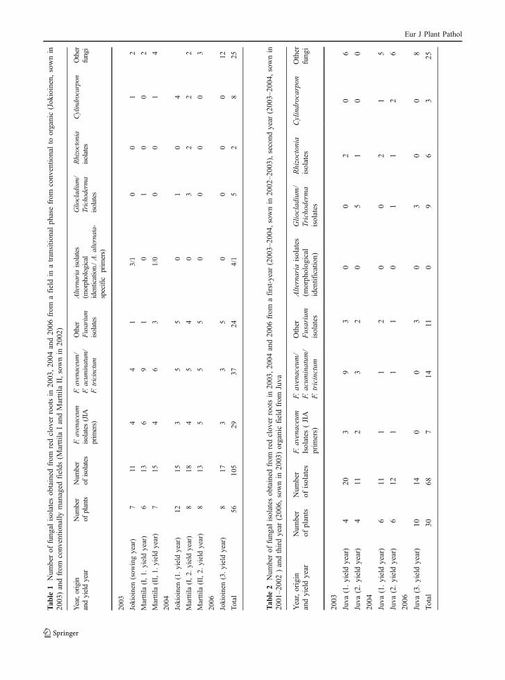
Symptomless red clover plants were collected from amixed red clover-grass field in a transitional phasefrom conventional to organic (sown in 2003) atJokioinen (60°48′ N, 23°29′ E) (south-western Fin-land, 7 in plants 2003 and 12 plants in 2004), fromtwo conventional fields (VT and JP) at Marttila(60°63′ N, 23°00′ E) (south-western Finland, 13 plantsin 2003 and 16 plants in 2004) used for red clover seedproduction of cv. Betty (sown in 2002) (Table 1), andfrom two mixed red-clover-grass organic fields (sownin 2001 and 2002) at Juva (61°53′ N, 27°51′ E)(south-eastern Finland, 8 plants in 2003 and 12 plantsin 2004) in September 2003 and July 2004 (Table 2).The fields at Juva have been under organic manage-ment since 1986. From a third mixed red-clover-grassresearch field at Sotkamo (647′ N, 28°23′ E) (transi-
Eur J Plant Pathol
Tab
le1
Num
berof
fung
alisolates
obtained
from
redclov
errootsin
2003
,20
04and20
06from
afieldin
atransitio
nalph
asefrom
conv
entio
nalto
organic(Jok
ioinen,sownin
2003
)andfrom
conv
entio
nally
managed
fields
(Marttila
IandMarttila
II,sownin
2002
)
Year,origin
andyieldyear
Num
ber
ofplants
Num
ber
ofisolates
F.avenaceum
isolates
(JIA
primers)
F.avenaceum/
F.acum
inatum
/F.
tricinctum
Other
Fusarium
isolates
Alternaria
isolates
(morphological
identication./A.alternata-
specific
primers)
Gliocladium/
Trichoderm
aisolates
Rhizoctonia
isolates
Cylindrocarpon
Other
fungi
2003
Jokioinen(sow
ingyear)
711
44
13/1
00
12
Marttila
(I,1.
yieldyear)
613
69
10
10
02
Marttila
(II,1.
yieldyear)
715
46
31/0
00
14
2004
Jokioinen(1.yieldyear)
1215
35
50
10
4
Marttila
(I,2.
yieldyear)
818
45
40
32
22
Marttila
(II,2.
yieldyear)
813
55
50
00
03
2006
Jokioinen(3.yieldyear)
817
33
50
00
012
Total
5610
529
3724
4/1
52
825
Tab
le2
Num
berof
fung
alisolates
obtained
from
redclov
errootsin
2003
,20
04and20
06from
afirst-year
(200
3–20
04,sownin
2002–2
003),second
year
(200
3–20
04,sownin
2001
–2002)andthirdyear
(2006,
sownin
2003)organicfieldfrom
Juva
Year,origin
andyieldyear
Num
ber
ofplants
Num
ber
ofisolates
F.avenaceum
Isolates
(JIA
prim
ers)
F.avenaceum/
F.acum
inatum
/F.
tricinctum
Other
Fusarium
isolates
Alternaria
isolates
(morph
olog
ical
identification)
Glio
clad
ium/
Tricho
derm
aisolates
Rhizocton
iaisolates
Cylindrocarpo
nOther
fung
i
2003
Juva
(1.yieldyear)
420
39
30
02
06
Juva
(2.yieldyear)
411
23
20
51
00
2004
Juva
(1.yieldyear)
611
11
20
02
15
Juva
(2.yieldyear)
612
11
10
11
26
2006
Juva
(3.yieldyear)
1014
00
30
30
08
Total
3068
714
110
96
325
Eur J Plant Pathol
tional field) in northeastern Finland only sclerotia fromdead plants were collected in 2004. Additionalsclerotia were obtained from a conventional field atRovaniemi in northern Finland in 2003. In addition,dead or diseased red clover plants grown in the samefields at Jokioinen (8 plants, Table 1) and Juva (10plants, Table 2) were collected in May 2006.
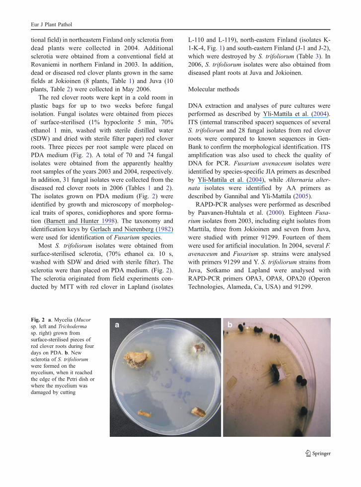
The red clover roots were kept in a cold room inplastic bags for up to two weeks before fungalisolation. Fungal isolates were obtained from piecesof surface-sterilised (1% hypoclorite 5 min, 70%ethanol 1 min, washed with sterile distilled water(SDW) and dried with sterile filter paper) red cloverroots. Three pieces per root sample were placed onPDA medium (Fig. 2). A total of 70 and 74 fungalisolates were obtained from the apparently healthyroot samples of the years 2003 and 2004, respectively.In addition, 31 fungal isolates were collected from thediseased red clover roots in 2006 (Tables 1 and 2).The isolates grown on PDA medium (Fig. 2) wereidentified by growth and microscopy of morpholog-ical traits of spores, conidiophores and spore forma-tion (Barnett and Hunter 1998). The taxonomy andidentification keys by Gerlach and Nierenberg (1982)were used for identification of Fusarium species.
Most S. trifoliorum isolates were obtained fromsurface-sterilised sclerotia, (70% ethanol ca. 10 s,washed with SDW and dried with sterile filter). Thesclerotia were than placed on PDA medium. (Fig. 2).The sclerotia originated from field experiments con-ducted by MTT with red clover in Lapland (isolates
L-110 and L-119), north-eastern Finland (isolates K-1-K-4, Fig. 1) and south-eastern Finland (J-1 and J-2),which were destroyed by S. trifoliorum (Table 3). In2006, S. trifoliorum isolates were also obtained fromdiseased plant roots at Juva and Jokioinen.
Molecular methods
DNA extraction and analyses of pure cultures wereperformed as described by Yli-Mattila et al. (2004).ITS (internal transcribed spacer) sequences of severalS. trifoliorum and 28 fungal isolates from red cloverroots were compared to known sequences in Gen-Bank to confirm the morphological identification. ITSamplification was also used to check the quality ofDNA for PCR. Fusarium avenaceum isolates wereidentified by species-specific JIA primers as describedby Yli-Mattila et al. (2004), while Alternaria alter-nata isolates were identified by AA primers asdescribed by Gannibal and Yli-Mattila (2005).
RAPD-PCR analyses were performed as describedby Paavanen-Huhtala et al. (2000). Eighteen Fusa-rium isolates from 2003, including eight isolates fromMarttila, three from Jokioinen and seven from Juva,were studied with primer 91299. Fourteen of themwere used for artificial inoculation. In 2004, several F.avenaceum and Fusarium sp. strains were analysedwith primers 91299 and Y. S. trifoliorum strains fromJuva, Sotkamo and Lapland were analysed withRAPD-PCR primers OPA3, OPA8, OPA20 (OperonTechnologies, Alameda, Ca, USA) and 91299.
Fig. 2 a. Mycelia (Mucorsp. left and Trichodermasp. right) grown fromsurface-sterilised pieces ofred clover roots during fourdays on PDA. b. Newsclerotia of S. trifoliorumwere formed on themycelium, when it reachedthe edge of the Petri dish orwhere the mycelium wasdamaged by cutting
Eur J Plant Pathol
Artificial inoculation with Fusarium conidia
The pathogenicity of Fusarium isolates from symp-tomless plants was determined in laboratory tests oninoculated seeds and eventually on the seedlings ofcvs Jokioinen and Bjursele. Seven isolates (four F.avenaceum isolates, two F. oxysporum isolates andone F. culmorum isolate) were studied on bothcultivars and seven additional isolates were studiedon cv. Jokioinen only.
Seeds were surfaced-sterilised with 0.1% AgNO3
for 1 min, washed with SDW and inoculated with aconidial suspension of 0.3–1.5×10⁶ conidia ml−1.Inocula were prepared by washing the spores from thesurfaces of 2–3 week old Fusarium cultures growingin Petri dishes on a nutrient-rich medium (potatodextrose agar, PDA) or on a low-nutrient medium(Synthetischer Nährstoffarmer Agar, SNA). The den-sities of conidia were estimated with a haemocytom-eter. The seeds were inoculated by soaking in aconidial suspension for 10 min and then lightly driedon sterile filter paper or sterile absorbent cotton andplaced in sterile Petri dishes onto moistened filterpaper. The experiments were performed in 4 repli-cates, with 50 seeds per (Petri dish) replicate.
The seeds were incubated in the Petri dishes underconstant light (80–90 μmol photons m−2 s−1) at 24°C.After 2 days of incubation, the germination of the seedswas observed, and after 2 weeks the severity of diseasesymptoms on the seedlings was evaluated using a scalewith classes: 0 = healthy root, 1 = <10% of the rootsurface affected by brown lesions, 2 = 10–25% of theroot surface covered by brown lesions, 3 = 25–50% ofthe root surface covered by brown lesions, 4=>50% ofthe root surface covered by brown lesions or plant dead.
Plant material
The field experiments at Jokioinen, Juva and Sot-kamo, from which the fungal samples were collected
were part of a larger research project of MTTAgrifood Research Finland dealing with organic redclover.
Four different cultivars of red clover (Trifoliumpratense) were compared in the study. Jokioinen is adiploid cultivar of Finnish origin (Boreal), which isrecommended for southern Finland. Bjursele is aSwedish (Svalöf Weibull) diploid cultivar, which israther winter-hardy and recommended for the wholeof Finland. Betty and Ilte are tetraploids of Swedish(Svalöf Weibull) and Estonian (Jõgeva Plant BreedingInstitute) origin, respectively. Betty originates fromBjursele; it is even more winter-hardy, and isrecommended for the whole of Finland. Ilte, thoughless winter-hardy than Betty, has a capacity for veryhigh yields, and is recommended for southern Finland(Kangas et al. 2005). For the leaf tests with all fourclover cultivars, plants were grown in the greenhousein standard potting mixture, and the leaves of twomonth-old plants were used for tests. The commercialstandard potting mixture (Kekkilä, Finland) wasbased on peat, mixed with 20% of the total volumeof mineral soil (33% coarse sand, 33% fine sand and33% sandy loam). The mixture was fertilised with1.2 kg m−3 of N-P-K fertiliser (6–12–24) and with6 kg m−3 of Mg containing (5%) ground limestone.The pH of the mixture was 6.2.
For the whole-plant tests, Bjursele clover plantswere sown on a standard potting mixture and firstgrown in a growth room with a 12/12 h day (40–55 μmol photons m−2s−1 )/night regime at about 20–22ºC. When the plants were three weeks-old, theywere potted in 8×8 cm pots and transferred to twogrowth chambers with a 12/12 h day/night regime(12 h light of 190–270 μmol photons m−2s−1, 20°C /12 h in darkness, 10°C. After two weeks growth theplants were transferred to a sterilised growth roomwith a 12/12 h day (40–55 μmol photons m−2s−1)/night regime at 15°C, where they were treated withbiological agents within two days of the transfer.
Strains Geographical origin (latitude and longitude) Year of isolation
L-119, L-110 Rovaniemi (6630′N, 25°44′ E) 2003
J-2, J-1 Juva (61°53′ N, 27°51′ E) 2004
J-6 Juva (61°53′ N, 27°51′ E) 2006
K-1, K2, K3, K4 Sotkamo (64°7′ N, 28°23′ E) 2004
Jo-14 Jokioinen (60°48′ N, 23°29′ E) 2006
Table 3 Sclerotiniatrifoliorum strains usedin the work
Eur J Plant Pathol
Preparation of S. trifoliorum inoculum
The mycelial inoculum was prepared according to themodified method of Rhodes et al. (1989) and Marumet al. (1994). Three mycelial balls with a diameter ofabout 2–3 cm (grown from three mycelial plugs inone flask during 7 days at 15°C on a rotary shaker in250 ml of PD broth) were added to 250 ml of waterand blended in a Waring Blender for about 5–10 s.The mycelium for leaf inoculum was separated fromthe water by centrifugation and then blended in thesame Waring Blender for 2 min in 200 ml of SDW.Three drops of Tween 80 were added to every 100 mlof inoculum.
Inoculation of detached clover leaveswith Sclerotinia trifoliorum isolates
Detached leaves of cvs Bjursele, Jokioinen, Betty andIlte were used for evaluation of the pathogenicity ofthree isolates, L-119, K-1 and J-2, of S. trifoliorum.The clover leaves (about two months-old) wereplaced in Petri dishes on the surface of 0.5% wateragar with 50 mgl−1 benzimidazole (modified fromDelclos et al. 1997, who used ascospore inoculum).The experiments were performed in 4 replicates, with6–12 leaflets per (Petri dish) replicate.
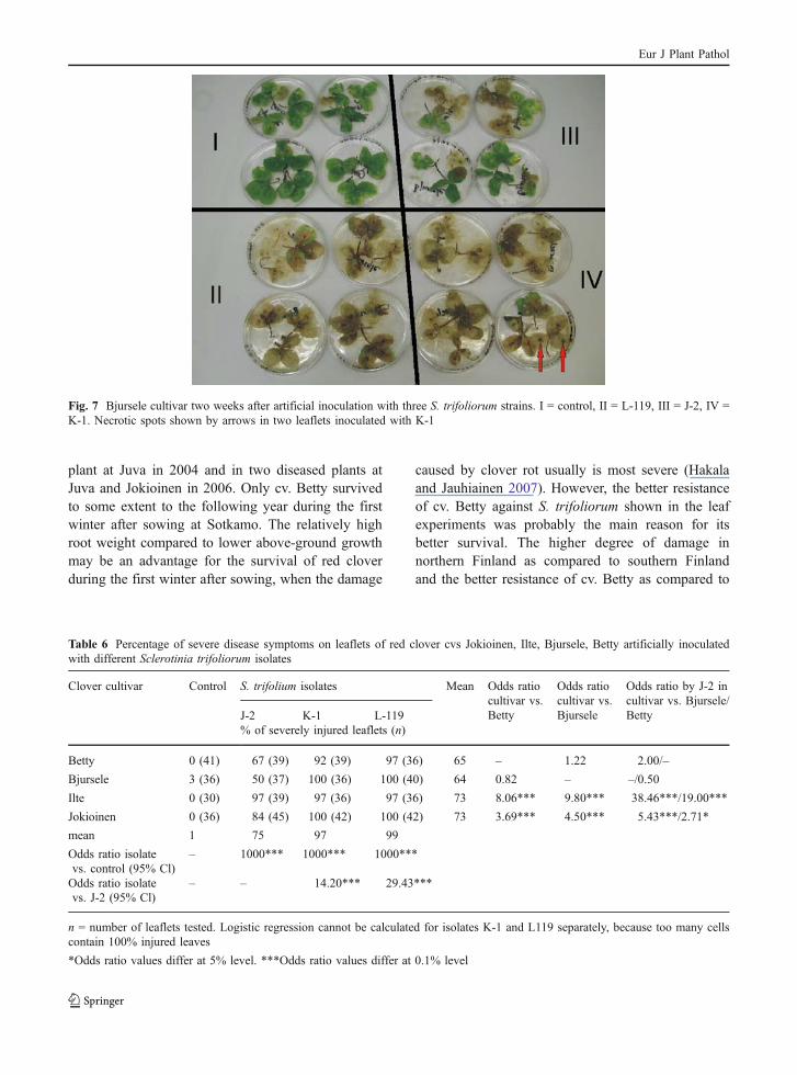
The clover leaflets were inoculated with 10 μldrops of inoculum (optical density of 1.3–1.7 atwavelength 750 nm), which were placed in the middleof each leaflet (Efimova 1985). The controls weremoistened with 10 μl drops of water (with addedTween 80). The Petri dishes with the inoculated cloverleaves were then incubated at 15°C in 12 h day-length(light intensity 40–55 μmol photons m−2s−1) for twoweeks. Disease symptoms were evaluated using a scalewith classes: 0 = healthy leaf, 1 = slight necrosis, 2 =moderate necrosis, 3 = severe necrosis, 4 = leaf dead.
Evaluation of antagonistic activity on intact plants
The effect of commercially available antagonists onS. trifoliorum on potted intact five week-old cloverplants was studied in an experiment with inoculationof the cv. Bjursele by the aggressive (based on leafexperiments) S. trifoliorum isolate L-119.
Biological agents and one fungicide (Rovral, Bayer,active agent iprodion, sold by Kemira GrowHow inFinland) were sprayed one day before S. trifoliorum
inoculation as follows: Mycostop (Streptomyces gri-seoviridis, Verdera Ltd, Finland) and Alirin B (Bacillussubtilis 10-VIZR, Russia, Novikova et al. 2003),0.6 ml/plant of 0.5% solution; Prestop and Gliomix(Gliocladium catenulatum, Verdera Ltd, Finland),0.6 ml/plant of 2% solution, 1.2×10⁶ cfu/plant; andRovral, 0.6 ml/plant of a solution of 165 mg/100 ml asa chemical control. All the preparates were mixed withdeionised water 1–1.5 h before use. The spore amountwas 1.5×10⁸/plant in Alirin B and 1.5×10⁶/plant inMycostop. The inoculated controls were treated withdeionised water (also 0.6 ml/plant). The experimentswere performed in three replicates, with five plants perreplicate placed on a tray.
The day after spraying with biological agents andfungicide, the clover plants were sprayed withmycelial inoculum of isolate L-119. The opticaldensity of mycelial fragment suspension was 1.05(750 nm). The amount sprayed was ca. 1 ml per plant.Control plants (water control) were sprayed withwater mixed with Tween 80 (3 drops per 100 ml ofwater). After inoculation, a plastic top was placedover the replicate trays to keep the plants at 100%relative humidity, and the resulting chambers (fiveplants per chamber) were incubated at 15°C in 12 hday-length (light intensity 40–55 μmol photons m−2s−1)for four weeks. The effect of the inoculation (inoculatedcontrol) was compared to the control plants sprayedwith water (water control). Disease development wasrated two and four weeks after the use of biologicalagents by using the same classes as for clover leaves.
The biological effectiveness (BEDSI) of biologicaland chemical agents was calculated by the formula:
BEDSI ¼ DSIc � DSIð Þ=DSIc
DSI disease severity index in samples treated bybiological agent
DSIc disease severity index in inoculated controlwithout biological control.
The disease severity was simply taken as the meanof the five classes assessed. The classes were notlinear but more like exponential scaling with 0, 0–10,10–25, 25–50 and 50–100 %. Thus, the classes 0–4were converted in the mean percentage values (0, 5,17.5, 37.5 and 75) before calculating the mean andcomparing the biological effectiveness in Table 7.
Eur J Plant Pathol
Statistical analysis
The effect of the S. trifoliorum strain on the frequencyof severely injuried leaves in different red clovercultivars was analysed using logistic regressionanalysis as described by Lehtinen et al. (2007). Toachieve binomial distribution the original diseaseratings 0–2 were reclassified as 0, healthy leavesand mild symptoms, and ratings 3–4 as 1, severesymptoms. In logistic regression the response variableis the probability that an event will occur (e.g.prevalence of severe Sclerotinia symptoms), hencethe response variable is constrained between 0 and 1.The ‘odds’ of an event are defined as the probabilityof the outcome event occurring divided by theprobability of the event not occurring. The ‘oddsratio’ is one set of odds divided by another. An oddsratio of 1 indicates that the event under study isequally likely in both values of the predictor. An oddsratio >1 indicates that the event is more likely in thefirst value, whilst an odds ratio <1 indicates that theevent is less likely in the first value. The data wereanalysed in three steps. First, odds ratios werecalculated for the whole data. The untreated controldiffered so much from all the isolates that compar-isons between different isolates could not be shown.Therefore, in the second step the untreated controlwas omitted from the analysis and other isolates were
compared to isolate J-2 and other cultivars werecompared to cvs Betty and Bjursele. In the third step,differences among isolates were studied separately foreach of the four cultivars.
Results
Fungal species composition in red clover roots basedon morphological characteristics, species-specificprimers and ITS sequence analyses
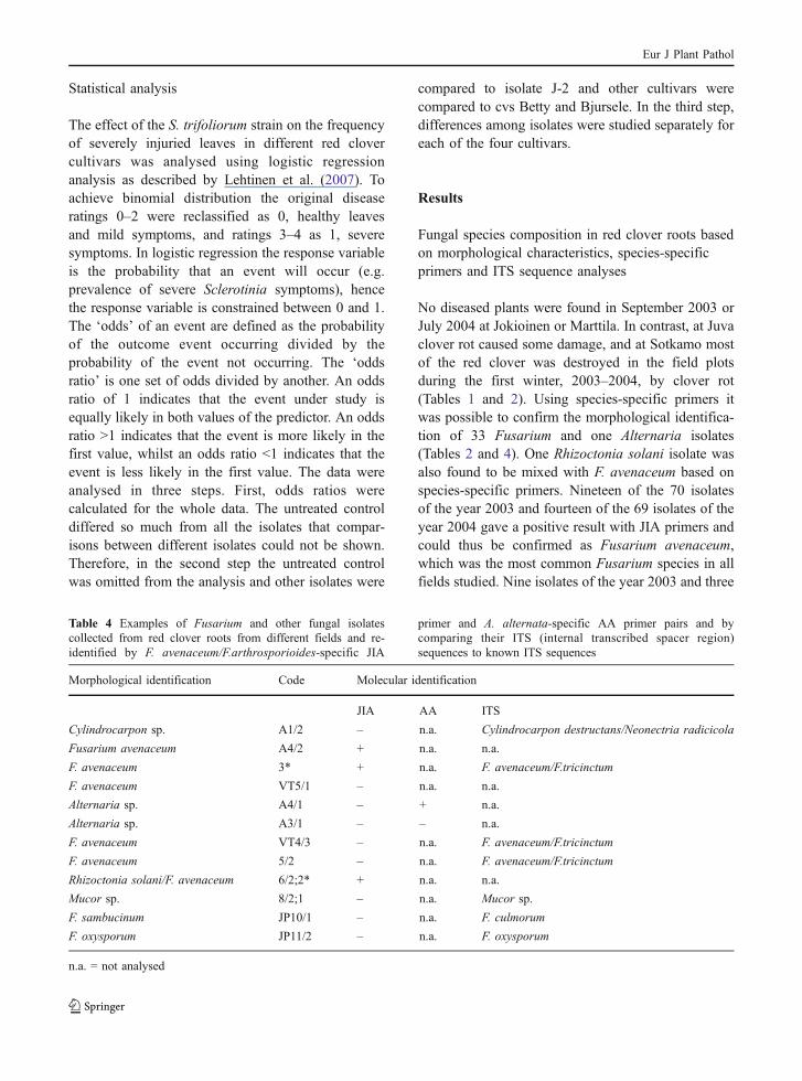
No diseased plants were found in September 2003 orJuly 2004 at Jokioinen or Marttila. In contrast, at Juvaclover rot caused some damage, and at Sotkamo mostof the red clover was destroyed in the field plotsduring the first winter, 2003–2004, by clover rot(Tables 1 and 2). Using species-specific primers itwas possible to confirm the morphological identifica-tion of 33 Fusarium and one Alternaria isolates(Tables 2 and 4). One Rhizoctonia solani isolate wasalso found to be mixed with F. avenaceum based onspecies-specific primers. Nineteen of the 70 isolatesof the year 2003 and fourteen of the 69 isolates of theyear 2004 gave a positive result with JIA primers andcould thus be confirmed as Fusarium avenaceum,which was the most common Fusarium species in allfields studied. Nine isolates of the year 2003 and three
Table 4 Examples of Fusarium and other fungal isolatescollected from red clover roots from different fields and re-identified by F. avenaceum/F.arthrosporioides-specific JIA
primer and A. alternata-specific AA primer pairs and bycomparing their ITS (internal transcribed spacer region)sequences to known ITS sequences
Morphological identification Code Molecular identification
JIA AA ITS
Cylindrocarpon sp. A1/2 – n.a. Cylindrocarpon destructans/Neonectria radicicola
Fusarium avenaceum A4/2 + n.a. n.a.
F. avenaceum 3* + n.a. F. avenaceum/F.tricinctum
F. avenaceum VT5/1 – n.a. n.a.
Alternaria sp. A4/1 – + n.a.
Alternaria sp. A3/1 – – n.a.
F. avenaceum VT4/3 – n.a. F. avenaceum/F.tricinctum
F. avenaceum 5/2 – n.a. F. avenaceum/F.tricinctum
Rhizoctonia solani/F. avenaceum 6/2;2* + n.a. n.a.
Mucor sp. 8/2;1 – n.a. Mucor sp.
F. sambucinum JP10/1 – n.a. F. culmorum
F. oxysporum JP11/2 – n.a. F. oxysporum
n.a. = not analysed
Eur J Plant Pathol
isolates of the year 2004, morphologically identifiedas F. avenaceum, gave a negative result with JIAprimers. Fusarium culmorum was only found in theconventional fields, while R. solani, Gliocladium spp.and Trichoderma spp. were most common in theorganic fields at Juva; Cylindrocarpon spp. weremore common in 2004 than in 2003 (Tables 1 and 2).There were no clear differences in the number ofspecies in seemingly healthy red clover roots betweendifferent fields in 2003–2004.
In 2006, Fusarium fungi were found in six of thediseased red clover plants at Jokioinen and threeFusarium isolates could be identified as F. avenaceumwith JIA primers. S. trifoliorum was found in one redclover plant at Jokioinen and one at Juva (Table 3).Fusarium, Mucor, Gliocladium, Trichoderma andPhoma fungi and unidentified oomycetes were alsofound at Juva in 2006 (Tables 1 and 2). It wasinteresting that the S. trifoliorum isolate at Juva wasisolated together with a Gliocladium isolate from adiseased red clover plant, which was growing close toseveral dead red clover plants. Isolates A4/2 and A4/3were F. avenaceum, while isolate 6/2;2 was a mixtureof two species (Table 4). The morphological identifi-cation of isolate 5/2 could not be confirmed byspecies-specific primers, but according to the ITSsequence it was either F. avenaceum or closely relatedF. tricinctum.
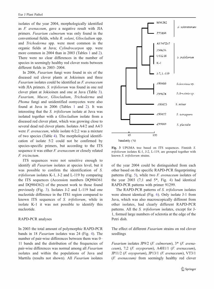
ITS sequences were not sensitive enough toidentify all Fusarium isolates at species level, but itwas possible to confirm the identification of S.trifoliorum isolates K-1, J-2 and L-119 by comparingthe ITS sequences (Accession numbers DQ904361and DQ904362) of the present work to those foundpreviously (Fig. 3). Isolates J-2 and L-119 had onenucleotide difference in the ITS1 region compared toknown ITS sequences of S. trifoliorum, while inisolate K-1 it was not possible to identify thisnucleotide.
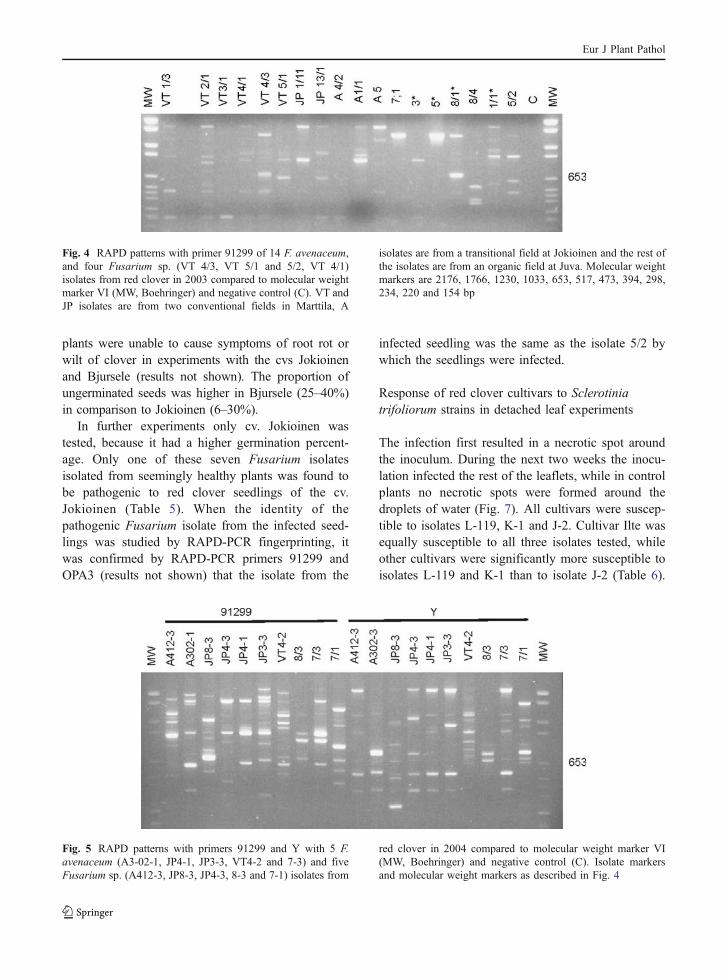
RAPD-PCR analyses
In 2003 the total amount of polymorphic RAPD-PCRbands in 18 Fusarium isolates was 24 (Fig. 4). Thenumber of pair-wise differences between them was 0–11 bands and the distribution of the frequencies ofpair-wise differences was normal among all Fusariumisolates and within the populations of Juva andMarttila (results not shown). All Fusarium isolates
of the year 2004 could be distinguished from eachother based on the specific RAPD-PCR fingerprintingpatterns (Fig. 5), while two F. avenaceum isolates ofthe year 2003 (7;1 and 5*, Fig. 4) had identicalRAPD-PCR patterns with primer 91299.
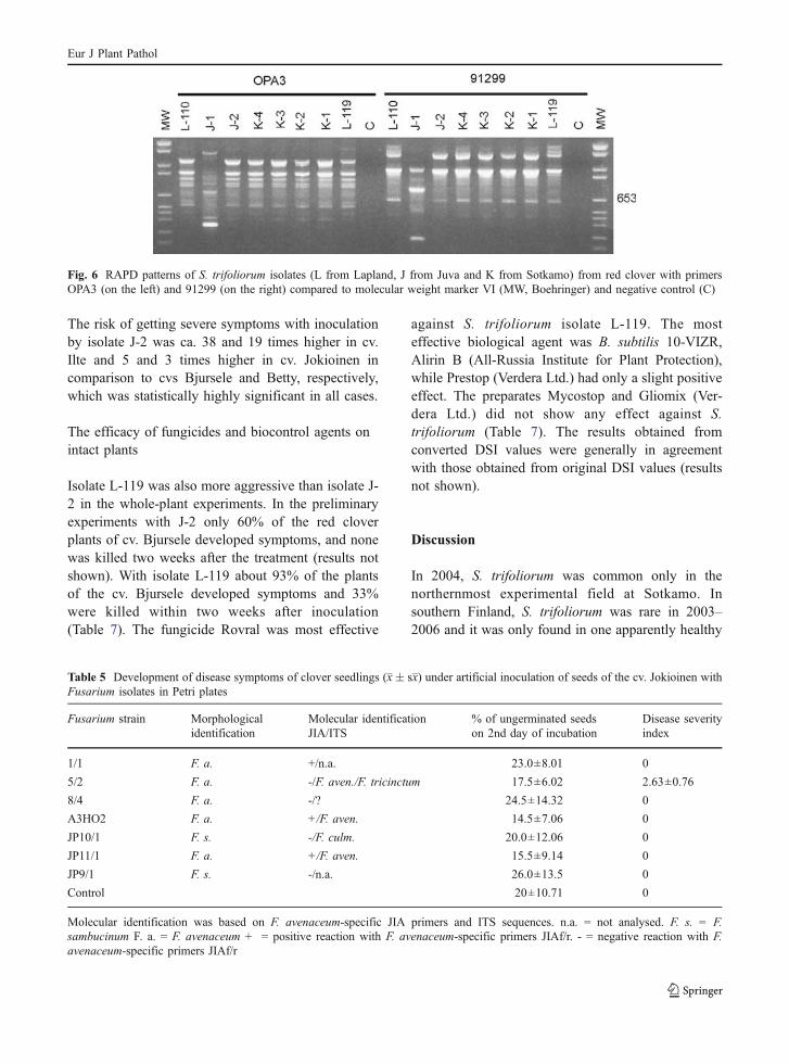
The RAPD-PCR patterns of S. trifoliorum isolateswere almost identical (Fig. 6). Only isolate J-1 fromJuva, which was also macroscopically different fromother isolates, had clearly different RAPD-PCRpatterns. All the S. trifoliorum isolates, except for J-1, formed large numbers of sclerotia at the edge of thePetri dish.
The effect of different Fusarium strains on red cloverseedlings
Fusarium isolates JP9/2 (F. culmorum), 3* (F. avena-ceum), 7;2 (F. oxysporum), A4H1/1 (F. avenaceum),JP11/2 (F. oxysporum), JP13/1 (F. avenaceum), VT3/1(F. avenaceum) from seemingly healthy red clover
Fig. 3 UPGMA tree based on ITS sequences. Finnish S.trifoliorum isolates K-1, J-2, L-119, are grouped together withknown S. trifoliorum strains.
Eur J Plant Pathol
plants were unable to cause symptoms of root rot orwilt of clover in experiments with the cvs Jokioinenand Bjursele (results not shown). The proportion ofungerminated seeds was higher in Bjursele (25–40%)in comparison to Jokioinen (6–30%).
In further experiments only cv. Jokioinen wastested, because it had a higher germination percent-age. Only one of these seven Fusarium isolatesisolated from seemingly healthy plants was found tobe pathogenic to red clover seedlings of the cv.Jokioinen (Table 5). When the identity of thepathogenic Fusarium isolate from the infected seed-lings was studied by RAPD-PCR fingerprinting, itwas confirmed by RAPD-PCR primers 91299 andOPA3 (results not shown) that the isolate from the
infected seedling was the same as the isolate 5/2 bywhich the seedlings were infected.
Response of red clover cultivars to Sclerotiniatrifoliorum strains in detached leaf experiments
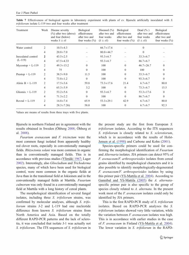
The infection first resulted in a necrotic spot aroundthe inoculum. During the next two weeks the inocu-lation infected the rest of the leaflets, while in controlplants no necrotic spots were formed around thedroplets of water (Fig. 7). All cultivars were suscep-tible to isolates L-119, K-1 and J-2. Cultivar Ilte wasequally susceptible to all three isolates tested, whileother cultivars were significantly more susceptible toisolates L-119 and K-1 than to isolate J-2 (Table 6).
Fig. 4 RAPD patterns with primer 91299 of 14 F. avenaceum,and four Fusarium sp. (VT 4/3, VT 5/1 and 5/2, VT 4/1)isolates from red clover in 2003 compared to molecular weightmarker VI (MW, Boehringer) and negative control (C). VT andJP isolates are from two conventional fields in Marttila, A
isolates are from a transitional field at Jokioinen and the rest ofthe isolates are from an organic field at Juva. Molecular weightmarkers are 2176, 1766, 1230, 1033, 653, 517, 473, 394, 298,234, 220 and 154 bp
Fig. 5 RAPD patterns with primers 91299 and Y with 5 F.avenaceum (A3-02-1, JP4-1, JP3-3, VT4-2 and 7-3) and fiveFusarium sp. (A412-3, JP8-3, JP4-3, 8-3 and 7-1) isolates from
red clover in 2004 compared to molecular weight marker VI(MW, Boehringer) and negative control (C). Isolate markersand molecular weight markers as described in Fig. 4
Eur J Plant Pathol
The risk of getting severe symptoms with inoculationby isolate J-2 was ca. 38 and 19 times higher in cv.Ilte and 5 and 3 times higher in cv. Jokioinen incomparison to cvs Bjursele and Betty, respectively,which was statistically highly significant in all cases.
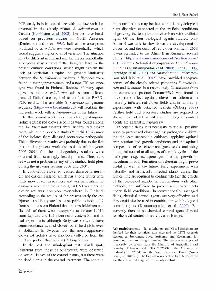
The efficacy of fungicides and biocontrol agents onintact plants
Isolate L-119 was also more aggressive than isolate J-2 in the whole-plant experiments. In the preliminaryexperiments with J-2 only 60% of the red cloverplants of cv. Bjursele developed symptoms, and nonewas killed two weeks after the treatment (results notshown). With isolate L-119 about 93% of the plantsof the cv. Bjursele developed symptoms and 33%were killed within two weeks after inoculation(Table 7). The fungicide Rovral was most effective
against S. trifoliorum isolate L-119. The mosteffective biological agent was B. subtilis 10-VIZR,Alirin B (All-Russia Institute for Plant Protection),while Prestop (Verdera Ltd.) had only a slight positiveeffect. The preparates Mycostop and Gliomix (Ver-dera Ltd.) did not show any effect against S.trifoliorum (Table 7). The results obtained fromconverted DSI values were generally in agreementwith those obtained from original DSI values (resultsnot shown).
Discussion
In 2004, S. trifoliorum was common only in thenorthernmost experimental field at Sotkamo. Insouthern Finland, S. trifoliorum was rare in 2003–2006 and it was only found in one apparently healthy
Fig. 6 RAPD patterns of S. trifoliorum isolates (L from Lapland, J from Juva and K from Sotkamo) from red clover with primersOPA3 (on the left) and 91299 (on the right) compared to molecular weight marker VI (MW, Boehringer) and negative control (C)
Table 5 Development of disease symptoms of clover seedlings (x� sx) under artificial inoculation of seeds of the cv. Jokioinen withFusarium isolates in Petri plates
Fusarium strain Morphologicalidentification
Molecular identificationJIA/ITS
% of ungerminated seedson 2nd day of incubation
Disease severityindex
1/1 F. a. +/n.a. 23.0±8.01 0
5/2 F. a. -/F. aven./F. tricinctum 17.5±6.02 2.63±0.76
8/4 F. a. -/? 24.5±14.32 0
A3HO2 F. a. +/F. aven. 14.5±7.06 0
JP10/1 F. s. -/F. culm. 20.0±12.06 0
JP11/1 F. a. +/F. aven. 15.5±9.14 0
JP9/1 F. s. -/n.a. 26.0±13.5 0
Control 20±10.71 0
Molecular identification was based on F. avenaceum-specific JIA primers and ITS sequences. n.a. = not analysed. F. s. = F.sambucinum F. a. = F. avenaceum + = positive reaction with F. avenaceum-specific primers JIAf/r. - = negative reaction with F.avenaceum-specific primers JIAf/r
Eur J Plant Pathol
plant at Juva in 2004 and in two diseased plants atJuva and Jokioinen in 2006. Only cv. Betty survivedto some extent to the following year during the firstwinter after sowing at Sotkamo. The relatively highroot weight compared to lower above-ground growthmay be an advantage for the survival of red cloverduring the first winter after sowing, when the damage
caused by clover rot usually is most severe (Hakalaand Jauhiainen 2007). However, the better resistanceof cv. Betty against S. trifoliorum shown in the leafexperiments was probably the main reason for itsbetter survival. The higher degree of damage innorthern Finland as compared to southern Finlandand the better resistance of cv. Betty as compared to
Fig. 7 Bjursele cultivar two weeks after artificial inoculation with three S. trifoliorum strains. I = control, II = L-119, III = J-2, IV =K-1. Necrotic spots shown by arrows in two leaflets inoculated with K-1
Table 6 Percentage of severe disease symptoms on leaflets of red clover cvs Jokioinen, Ilte, Bjursele, Betty artificially inoculatedwith different Sclerotinia trifoliorum isolates
Clover cultivar Control S. trifolium isolates Mean Odds ratiocultivar vs.Betty
Odds ratiocultivar vs.Bjursele
Odds ratio by J-2 incultivar vs. Bjursele/BettyJ-2 K-1 L-119
% of severely injured leaflets (n)
Betty 0 (41) 67 (39) 92 (39) 97 (36) 65 – 1.22 2.00/–
Bjursele 3 (36) 50 (37) 100 (36) 100 (40) 64 0.82 – –/0.50
Ilte 0 (30) 97 (39) 97 (36) 97 (36) 73 8.06*** 9.80*** 38.46***/19.00***
Jokioinen 0 (36) 84 (45) 100 (42) 100 (42) 73 3.69*** 4.50*** 5.43***/2.71*
mean 1 75 97 99
Odds ratio isolatevs. control (95% Cl)
– 1000*** 1000*** 1000***
Odds ratio isolatevs. J-2 (95% Cl)
– – 14.20*** 29.43***
n = number of leaflets tested. Logistic regression cannot be calculated for isolates K-1 and L119 separately, because too many cellscontain 100% injured leaves
*Odds ratio values differ at 5% level. ***Odds ratio values differ at 0.1% level
Eur J Plant Pathol
Bjursele in northern Finland are in agreement with theresults obtained in Sweden (Öhberg 2008; Öhberg etal. 2008).
Fusarium avenaceum and F. tricinctum were themost common fungi isolated from apparently healthyred clover roots, especially in conventionally managedfields. Rhizoctonia solani was more common in organicthan in conventionally managed fields. This is inaccordance with previous studies (Ylimäki 1967; Lager2002). Interestingly, also Gliocladium and Trichodermaspecies, many of which have been used for biologicalcontrol, were more common in the organic fields atJuva than in the transitional field at Jokioinen and in theconventionally managed fields at Marttila. Fusariumculmorum was only found in a conventionally managedfield at Marttila with a long history of cereal plants.
The morphological identification of several fungalisolates, including three S. trifoliorum strains, wasconfirmed by molecular analyses, although S. trifo-liorum strains J-2 and L-119 had one nucleotidedifference from known S. trifoliorum strains fromNorth America and Asia. Based on the totallydifferent RAPD-PCR patterns and the lack of sclero-tia, it was concluded that isolate J-1 was actually notS. trifoliorum. The ITS sequences of S. trifoliorum in
the present study are the first from European S.trifoliorum isolates. According to the ITS sequencesS. trifoliorum is closely related to S. sclerotiorum,which is in accordance with the results of Holst-Jensen et al. (1998) and Carbone and Kohn (2001).
Species-specific primers could be used for con-firming the morphological identification of Fusariumand Alternaria isolates. JIA primers can detect 95% ofF. avenaceum/F. arthrosporioides isolates from cerealgrains identified by morphological characters and it isalso possible to identify morphologically-degeneratedF. avenaceum/F. arthrosporioides isolates by usingthis primer pair (Yli-Mattila et al. 2004). According toGannibal and Yli-Mattila (2005) the A. alternata-specific primer pair is also specific to the group ofspecies closely related to A. alternata. In the presentwork most of the F. avenaceum isolates could only beidentified by JIA primers.
This is the first RAPD-PCR study of S. trifoliorumisolates. Based on RAPD-PCR analyses the S.trifoliorum isolates showed very little variation, whilethe variation between F. avenaceum isolates was high.This is in accordance with earlier studies in the caseof F. avenaceum in Finland (Yli-Mattila et al. 2004).The lower variation in S. trifoliorum in the RAPD-
Table 7 Effectiveness of biological agents in laboratory experiment with plants of cv. Bjursele artificially inoculated with S.trifoliorum isolate L-119 two and four weeks after treatment
Treatment Week Disease severity(%) after two (above)and four (below)weeks x� sx
Biologicaleffectivenessafter two andfour weeks (%)
Diseased (%)after two andfour weeks(x� sx)
Biologicaleffectivenessafter two andfour weeks (%)
Dead (% )after two andfour weeks(x� sx)
Biologicaleffectivenessafter two andfour weeks (%)
Water control 2 10.5±4.3 – 66.7±17.6 – 0 –
4 20.8±7.0 – 80.0±44.7 – 0 –
Inoculated control(L-119)
2 43.5±2.5 – 93.3±6.7 – 33.3±6.7 –
4 67.5±4.33 – 93.3±6.7 – 86.7±6.7 –
Mycostop + L-119 2 49.3±13.2 0 100 0 46.7±26.7 0
4 75 0 100 0 100 0
Prestop + L-119 2 38.5±9.0 11.5 100 0 33.3±6.7 0
4 73.8±1.2 0 100 0 93.3±6.7 0
Alirin B + L-119 2 17.5±3.6 59.8 73.3±17.6 21.4 6.7±6.7 80.0
4 65.3±5.9 3.2 100 0 73.3±6.7 15.5
Gliomix + L-119 2 53.2±5.6 0 93.3±6.7 0 53.3±17.6 0
4 71.3±2.2 0 100 0 86.7±6.7 0
Rovral + L-119 2 14.8±7.4 65.9 53.3±29.1 42.9 6.7±6.7 80.0
4 28.3±7.26) 58.0 100 0 6.7±6.7 92.3
Values are means of results from three trays with five plants.
Eur J Plant Pathol
PCR analysis is in accordance with the low variationobtained in the closely related S. sclerotiorum inCanada (Hambleton et al. 2002). On the other hand,based on previous studies in North America(Renhström and Free 1993), half of the ascosporesproduced by S. trifoliorum were heterothallic, whichwould suggest a higher level of variation. The situationmay be different in Finland and the bigger homothallicascospores may survive better here, at least in thepresent climatic conditions, which might explain thelack of variation. Despite the genetic similaritybetween the S. trifoliorum isolates, differences werefound in their aggressiveness, and a new ITS sequencetype was found in Finland. Because of many openquestions, more S. trifoliorum isolates from differentparts of Finland are required to confirm the RAPD-PCR results. The available S. sclerotiorum genomesequence (http://www.broad.mit.edu) will facilitate themolecular work with S. trifololiorum in the future.
In the present work only one clearly pathogenicisolate against red clover seedlings was found amongthe 14 Fusarium isolates from healthy red cloverroots, while in a previous study (Ylimäki 1967) mostof the isolates from diseased roots were pathogenic.This difference in results was probably due to the factthat in the present work the isolates of the years2003–2004 for the pathogenicity studies wereobtained from seemingly healthy plants. Thus, rootrot was not a problem in any of the studied field plotsduring the growing seasons 2003 and 2004.
In 2003–2005 clover rot caused damage in north-ern and eastern Finland, which has a long winter withthick snow cover. In southern and western Finland nodamages were reported, although 40–50 years earlierclover rot was common everywhere in Finland.According to the results of the present study the cvsBjursele and Betty are less susceptible to isolate J-2from south-eastern Finland than the cvs Jokioinen andIlte. All of them were susceptible to isolates L-119from Lapland and K-1 from north-eastern Finland inleaf experiments, although Betty was shown to havesome resistance against clover rot in field plots evenat Sotkamo. In Sweden too, the most aggressiveclover rot isolates have also been collected from thenorthern part of the country (Öhberg 2008).
In the leaf and whole-plant tests small spots(different from those of Sclerotinia) were observedon several leaves of the control plants, but there wereno dead plants in the control treatment. The spots in
the control plants may be due to abiotic physiologicalplant disorders connected to the artificial conditionsof growing the test plants in chambers with artificiallight. Of the four biological agents studied, onlyAlirin B was able to slow down the development ofclover rot and the death of red clover plants. In 2008it was permitted to use Alirin B in Russia in severalplants (http://www.mcx.ru/documents/section/show/4016.89.htm). Sclerotial mycoparasites Coniothyriumminitans (Diamantopoulou et al. 2000; Li et al. 2006,Partridge et al. 2006) and Sporidesmium sclerotivo-rum (del Rio et al. 2002) have provided adequatecontrol of the closely related pathogens S. sclerotio-rum and S. minor. In a recent study C. minitans fromthe commercial product Contans®WG was found tohave some effect against clover rot sclerotia innaturally infected red clover fields and in laboratoryexperiments with detached leaflets (Öhberg 2008)Further field and laboratory studies are required toshow, how effective different biological controlagents are against S. trifoliorum.
In organic fields it is necessary to use all possibleways to protect red clover against pathogens: cultivat-ing the least susceptible cultivars, applying optimalcrop rotation and growth conditions and the optimalcomposition of red clover and grass seeds, and usingbiological control at all stages of the life cycles of thepathogens (e.g. ascospore germination, growth ofmycelium in soil, formation of sclerotia) might proveuseful as well in the future. Field experiments withnaturally and artificially infected plants during thewinter time are required to confirm whether the effectsof the biological agents, in combination with othermethods, are sufficient to protect red clover plantsunder field conditions. In conventionally managedfields, chemical control agents are very effective, andthey could also be used in combination with biologicalcontrol agents (Diamantopoulou et al. 2000). Butcurrently there is no chemical control agent allowedfor chemical control in red clover in Europe.
Acknowledgements Taina Lahtinen and Nina Painilainen arethanked for their technical assistance and the MTT researchstations at Jokioinen, Juva, Sotkamo and Rovaniemi forproviding plant and fungal samples. The study was supportedfinancially by grants from the Ministry of Agriculture andForestry of Finland (No. 3481/502/2002), the Academy ofFinland (No. 52104) and the Nordic Research Board (Nord-Forsk, no. 040291). The English was checked by Ellen Valle ofthe Department of English, University of Turku.
Eur J Plant Pathol
References
Barnett, H.L., & Hunter B.B. (1998). Illustrated genera ofImperfect Fungi. Fourth edition. The American Phyto-pathological Society, St, Paul, Minnesota, USA 218 p.
Bertilsson, J., & Murphy, M. (2003). Effects of feeding cloversilages on feed intake, milk production and digestion indairy cows. Grass and Forage Science, 58, 309–322.
Bolton, M. D., Thomma, B. P. H. J., & Nelson, B. D. (2006).Pathogen profile of Sclerotinia sclerotiorum (Lib.) deBary: biology and molecular traits of a cosmopolitanpathogen. Molecular Plant Pathology, 7, 1–16.
Carbone, I., & Kohn, L. M. (2001). A microbial population –species interface: nested cladistic and coalescent inferencewith multilocus data. Molecular Ecology, 10, 947–964.
Delclos, B., Mousset-Declas, C., & Raynal, G. (1997). A simplemethod for the evaluation of red clover (Trifolium pratense L.)resistance to Sclerotinia trifoliorum. Euphytica, 93, 173–179.
Del Rio, L. E., Martinson, C. A., & Yang, X. B. (2002).Biological control of Sclerotinia stem rot of soybean withSporidesmium sclerotivorum. Plant Disease, 86, 999–1004.
Diamantopoulou, A., Litkei, J., Skopa, C., & Christias, C.(2000). Effect of inhibitors of sclerotium formation on thesclerotial mycoparasite Coniothyrium minitans and its hostSclerotinia sclerotiorum. Mycological Research, 104,1449–1452.
Efimova G.G. (1985). Difference of isolates of casual agents ofclover root rot. Zaschita rastenij No 5, 26 (in Russian).
Gannibal, Ph B, & Yli-Mattila, T. (2005). Cultural and moleculardifferentiation of small-spored Alternaria species associatedwith poaceae. Mycology and Phytopathology, 39, 13–23.
Gerlach, W., & Nierenberg, H. I. (1982). The Genus Fusarium – apictorial atlas. Mitteilungen aus der Biologischen Bundesan-stalt für Land-Forstwirtschaft Berlin-Dahlem, 209, 1–406.
Guimaraes, R. L., & Stotz, H. U. (2004). Oxalate production bySclerotinia sclerotiorum regulates guard cells duringinfection. Plant Physiology, 136, 3701–3711.
Hakala, K., & Jauhiainen, L. (2007). Yield and nitrogen concentra-tion of above- and below-ground biomasses of red clovercultivars in pure stands and inmixtures with three grass speciesin northern Europe. Grass and Forage Science, 62, 312–321.
Hambleton, S., Walker, C., & Kohn, L. M. (2002). Clonallineages of Sclerotinia sclerotiorum previously knownfrom other crops predominate in 1999–2000 samples fromOntario and Quebec soybean. Canadian Journal of PlantPathology, 24, 309–315.
Holst-Jensen, A., Vaage, M., & Schumacher, T. (1998). Anapproximation to the phylogeny of Sclerotinia and relatedgenera. Nordic Journal of Botany, 18, 705–719.
Huokuna E. (1985). Apilan punahomeen esiintymisestä. Maa-talouden Tutkimuskeskus, tiedote 22/85 (in Finnish).
Janse, J. D. (1995). New methods of diagnosis in plant pathology –perspectives and pitfalls. EPPO Bulletin, 25, 5–17.
Kangas A., Laine A., Niskanen M., Salo Y., Vuorinen M.,Jauhiainen L. & Nikander H. (2005). Virallisten lajikeko-keiden tulokset 1997–2004. Results of official varietytrials. MTT:n selvityksiä, 83.
Lager J. (2002). Soil-borne diseases in intensive legumecropping. SLU. Unit of Plant Pathology and BiologicalControl. Dissertation. Agraria 362.
Lehtinen, A., Hannukkala, A., Rantanen, T., & Jauhiainen, L. (2007).Phenotypic and genetic variation in Finnish potato-late blightpopulations, 1997–2000. Plant Pathology, 56, 480–491.
Li, G. Q., Hyang, H. C., Miao, H. J., Erickson, R. S., Jiang, D. H.,& Wiao, Y. N. (2006). Biological control of sclerotiniadiseases of rapeseed by aerial applications of the mycopar-asite Coniothyrium minitans. European Journal of PlantPathology, 114, 345–355.
Marum, P., Smith, R. R., & Grau, C. R. (1994). Developmentof procedures to identify red clover resistant to Sclerotiniatrifoliorum. Euphytica, 77, 257–261.
Novikova, I. I., Litvinenko, A. I., Boikova, I. V., Yaroshenko,V. A., & Kalko, G. V. (2003). Biological activity of newmicrobiological preparations alirins B and S designed forplant protection against diseases. I. Biological activity ofalirins against diseases of vegetable crops and potato.Mycology and Phytopathology, 37, 92–98. (In Russian).
Öhberg, H. (2008) Studies of the persixtence of red clovercultivars in Sweden with particular reference to Sclerotiniatrifoliorum. Acta Universitatis Agriculturae Suecial. Doc-toral Thesis No. 2008:8, SLU, Umeå, Sweden.
Öhberg, H., Ruth, P., & Bång, U. (2008). Differential responses ofred clover cultivars to Sclerotinia trifoliorum under diversenatural climatic conditions. Plant Pathology, 57, 459–466.
Paavanen-Huhtala, S., Avikainen, H., & Yli-Mattila, T. (2000).Development of strain-specific primers for Gliocladiumcatenulatum strain used in biological control. EuropeanJournal of Plant Pathology, 106, 187–198.
Partridge, D. E., Sutton, T. B., Jordan, D. L., & Curtis, V. L.(2006). Management of Sclerotinia blight of peanut withthe biological control agent Coniothyrium minitans. PlantDisease, 90, 957–963.
Rehnström, A. L., & Free, S. J. (1993). A simple method forthe mating of Sclerotinia trifoliorum. Experimental My-cology, 17, 236–239.
Rhodes L.H., Anderson T.H., Pierson P.E. & Myers D.K.(1989). Field evaluation of Sclerotinia crown and stem rotin six alfalfa cultivars. (Paper presented at the Proc. 21stCentral Alfalfa Imp. Conf. 13–14).
Rufelt S. (1986). Studies on Fusarium root rot of red clover(Trifolium pretense L.) and the potential for its control.Swedish University of Agricultural Sciences. Department ofPlant and Forest Protection, Uppsala. Dissertation, 10.
Saloniemi I. (1993). Coevolution in predator-pray systems andplant-pathogen interactions. Dissertation, University ofOregon, Eugene.
Willetts, H. J., &Wong, J. A.-C. (1980). The Biology of Sclerotiniasclerotiorum, S. trifoliorum, and S. minor with emphasis ofspecific nomenclature. The Botanical Review, 46, 102–165.
Ylimäki, A. (1967). Root rot as a cause of red clover decline inleys in Finland. Annales Agriculturae Fenniae, 6, 1–59.
Ylimäki, A. (1969a). Clover rot as a cause of poor overwintering ofclover in Finland. Journal of the Scientific Agricultural Societyof Finland, 41, 222–242. (in Finnish with English summary).
Ylimäki, A. (1969b). Fungicidal effects of some chemicals onSclerotinia trifoliorum Erikss. Journal of the ScientificAgricultural Society of Finland, 41, 243–250.
Yli-Mattila, T., Paavanen-Huhtala, S., Parikka, P., Konstantinova, P.,& Gagkaeva, T. (2004). Molecular and morphologicaldiversity of Fusarium species in Finland and northwesternRussia. European Journal of Plant Pathology, 110, 573–585.
Eur J Plant Pathol
Related Documents