PREVALENCE OF MALARIA AND SELECTEDARBOVIRAL INFECTIONSIN PATIENTS PRESENTING WITH UNDIAGNOSED FEBRILE ILLNESS IN RUSINGA ISLAND, KENYA KIPANGA PURITY NGINA A Research Thesis Submitted to the Graduate School in Partial Fulfillment for the Requirements of the Award of Master of Science Degree in Biochemistry of Egerton University EGERTON UNIVERSITY APRIL, 2014

Welcome message from author
This document is posted to help you gain knowledge. Please leave a comment to let me know what you think about it! Share it to your friends and learn new things together.
Transcript
PREVALENCE OF MALARIA AND SELECTEDARBOVIRAL INFECTIONSIN
PATIENTS PRESENTING WITH UNDIAGNOSED FEBRILE ILLNESS IN
RUSINGA ISLAND, KENYA
KIPANGA PURITY NGINA
A Research Thesis Submitted to the Graduate School in Partial Fulfillment for the
Requirements of the Award of Master of Science Degree in Biochemistry of Egerton
University
EGERTON UNIVERSITY
APRIL, 2014
ii
DECLARATION AND RECOMMENDATION
DECLARATION
I declare that this thesis is my original work and has not been submitted for award in any
institution of learning to the best of my knowledge.
Signature: Date:
Ms. Purity N. Kipanga
RECOMMENDATION
We confirm that this thesis has our approval to be presented for examination as per the Egerton
University regulations.
Signature: Date:
Dr. P.O. Mireji
Egerton University
Signature: Date:
Dr. JandouweVillinger
International Center for Insect Physiology and Ecology (ICIPE)

iii
COPYRIGHT
© 2014, Purity NginaKipanga
No part of this project report should be reproduced or transmitted in any form by mechanical
means including photocopying, recording or any information storage or retrieval without
permission in writing from the author or Egerton University on behalf of the author.
All rights reserved.

iv
DEDICATION
To my loving parents Mr. and Mrs. Kipanga, my siblings, Augustine Muthiani and Phillip
Mutukuwhose prayers and support have propelled me this far.
v
ACKNOWLEDGEMENT
I thank the Almighty God for His grace and goodness, for His protection and sustenance this far.
I greatly thank my supervisors Drs. JandouweVillinger and Paul Mireji for their quality
supervision and mentorship during the project period. Their insightful advice and guidance made
it possible for me to complete the project on time. I equally acknowledge Dr. Daniel Masiga for
having given me the opportunity to work at the Emerging Infectious Lab and interact with great
men and women who made my stay pleasurable and fulfilling. Finally, many thanks go to Mr.
David Omondi, Mrs. Yvonne Ukamakaand Mr. Thomas Ogaowho assisted in defense
preparation and editing ofthis work.
vi
ABSTRACT
Onsetof uncomplicated malaria is characterized by fever, headache, joint pains, myalgia and
lack of appetite. These non-specific signs and symptoms also presentin patients with arthropod
borne viral (arboviral) infections complicate differential diagnoses. The lack of diagnosticsthat
can detectarboviral infections in Kenyan public hospitals coupled with malaria diagnostic tools
incapable of detecting low Plasmodiumparasitemia,has led to diagnosis based on clinical
symptoms only,favouring malaria diagnosis at the expense of arboviral infections
detection.Investigations were conducted to detectPlasmodiumparasites undetected by
microscopy and rapid diagnostic tests (RDTs) and determine Sindbis and Bunyamwera viruses
neutralizing antibodies among undiagnosed febrile ill patients in Rusinga Island. Human blood
and serum samples (n=92) were collected from patients without malaria (as confirmed by
microscopy and RDTs) fromTom MboyaHospital in the island. The blood samples were
screened for Plasmodium parasites by nested PCR coupled to high resolution melting analysis
(nPCR-HRM), and serum samples screened for neutralizing antibodies by plaque reduction
neutralization test (PRNT). Association between risk factors and exposure to infections was
determined by Chi square and Logistic multivariate analyses. Plasmodium parasites were
detected in 36 (39.1%) of the 92 patients. Out of these 36 patients with Plasmodium infections,
only 16 (44.4%) were correctly treated with antimalarial medication with the rest being treated
with antibiotics, antihelminthes and amoebicides. Conversely, a majority of non-malaria febrile
patients (n=32) were treated with antimalarial medication. Plasmodium falciparum was the
major malaria-causing parasite detected in Rusinga Island (29 out of 36).Individuals involved in
outdoor activities (farmers and fishermen) were 2.24-2.43 folds more likely to get malaria
infections than those involved in indoor-based (teachers/students)occupations.Neutralizing
antibodies against Sindbis virus were detected in five (5.4%) patients, three of whom had
malaria co-infection. No antibodies against Bunyamwera virus were detected. Theseresults
demonstrate limitations of differential diagnostics of febrile illness in rural malaria endemic
settings that undermineproper acute febrile illness managementand patient care. The under-
appreciation of arboviral infections is of great concern, in a country where active arbovirus
circulation has been demonstrated, resulting in poor health outcomes for non-malaria febrile
patients. This study highlights the need for improved diagnostics deployable in rural malaria
endemic settings to counter the increasing challenges of low parasitemia malaria and non-
malaria undiagnosed acute febrile illnesses.
vii
TABLE OF CONTENTS
DECLARATION AND RECOMMENDATION .......................................................................... ii
COPYRIGHT ................................................................................................................................ iii
DEDICATION ...............................................................................................................................iv
ACKNOWLEDGEMENT .............................................................................................................. v
ABSTRACT ...................................................................................................................................vi
TABLE OF CONTENTS ............................................................................................................. vii
LIST OF TABLES .........................................................................................................................ix
LIST OF FIGURES ........................................................................................................................ x
LIST OF PLATES .........................................................................................................................xi
LIST OF ABBREVIATIONS ...................................................................................................... xii
CHAPTER ONE ............................................................................................................................. 1
INTRODUCTION .......................................................................................................................... 1
1.1 Background information ....................................................................................................... 1
1.2 Statement of the problem ...................................................................................................... 2
1.3 Objectives .............................................................................................................................. 3
1.3.1 General objective ............................................................................................................ 3
1.3.2 Specific objectives .......................................................................................................... 3
1.4 Hypotheses ............................................................................................................................ 3
1.5 Justification ........................................................................................................................... 3
CHAPTER TWO ............................................................................................................................ 5
LITERATURE REVIEW ............................................................................................................... 5
2.1 Malaria diagnosis .................................................................................................................. 5
2.1.1 Microscopy in diagnosis of malaria................................................................................ 5
2.1.2 Rapid diagnostic test (RDT) approaches in detection of malaria ................................... 5
2.1.3 Molecular based techniques for malaria detection ......................................................... 6
2.2 High resolution melting analysis ........................................................................................... 7
2.3 Malaria over-diagnosis .......................................................................................................... 8
2.4 Arbovirus transmission ......................................................................................................... 9
2.4.1 Sindbis virus ................................................................................................................. 11
2.4.2 Bunyamwera virus ........................................................................................................ 12
2.5 Neutralizing antibodies ....................................................................................................... 13

viii
CHAPTER THREE ...................................................................................................................... 14
MATERIALS AND METHODS .................................................................................................. 14
3.1 Study site ............................................................................................................................. 14
3.2 Study design ........................................................................................................................ 15
3.2.1 Human blood sampling ................................................................................................. 15
3.2.2 Extraction of total DNA from blood............................................................................. 16
3.2.3 Detection of Plasmodium parasites by nested PCR-HRM (nPCR-HRM) ................... 16
3.2.4 Virus isolation............................................................................................................... 17
3.2.5 Virus isolates confirmation by passage ........................................................................ 17
3.2.6 Virus Confirmation by PCR ......................................................................................... 17
3.2.7 Determination of viral titer (Plaque Assay) .................................................................. 18
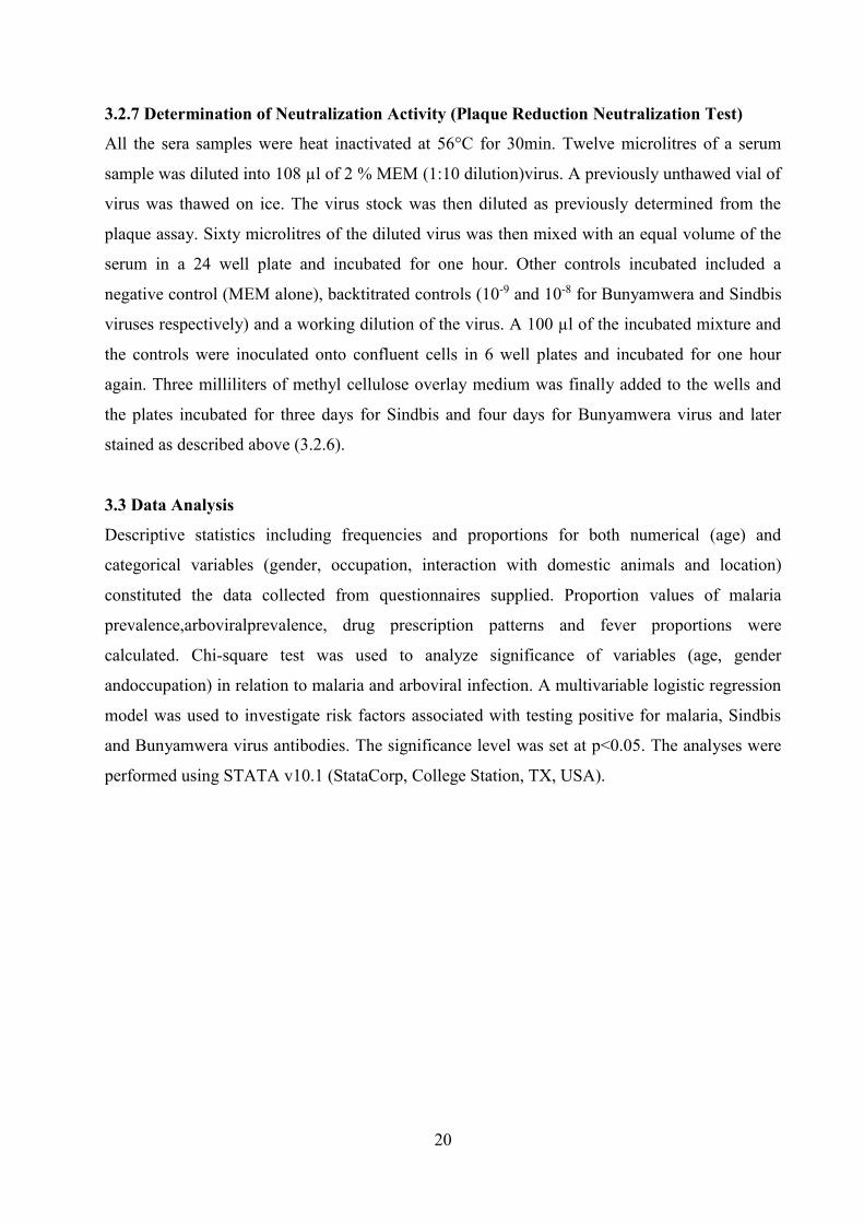
3.2.7 Determination of Neutralization Activity (Plaque Reduction Neutralization Test) ..... 20
3.3 Data Analysis ...................................................................................................................... 20
CHAPTER FOUR ......................................................................................................................... 21
RESULTS ..................................................................................................................................... 21
4.1 Characteristics of study subjects ......................................................................................... 21
4.2 Malaria prevalence among the undiagnosed febrile patients .............................................. 21
4.3 Prevalence of arboviral infections in Rusinga Island .......................................................... 24
CHAPTER FIVE .......................................................................................................................... 27
DISCUSSION ............................................................................................................................... 27
CHAPTER SIX ............................................................................................................................. 31
CONCLUSION AND RECOMMENDATION ............................................................................ 31
6.1 Conclusion ........................................................................................................................... 31
6.2 Recommendations ............................................................................................................... 31
REFERENCES ............................................................................................................................. 32
APPENDICES .............................................................................................................................. 42
ix
LIST OF TABLES
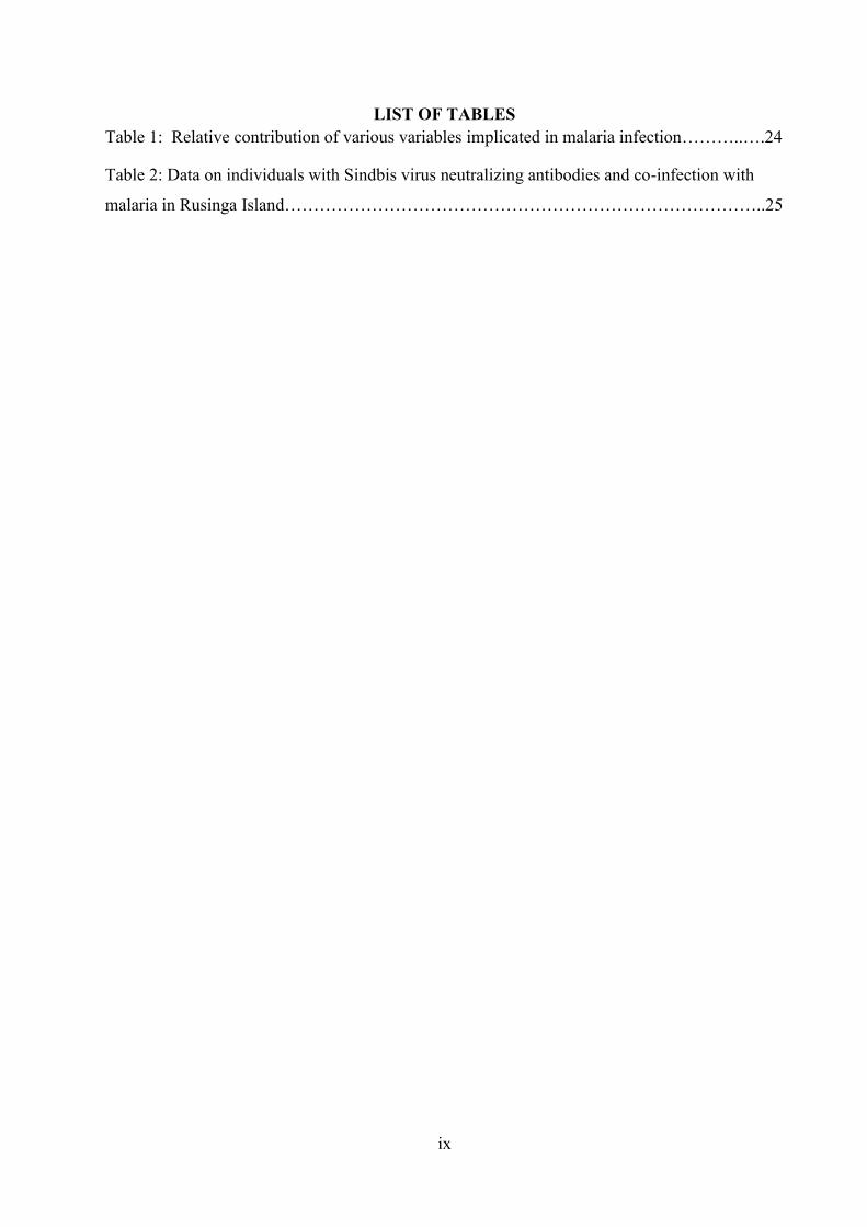
Table 1: Relative contribution of various variables implicated in malaria infection………..….24
Table 2: Data on individuals with Sindbis virus neutralizing antibodies and co-infection with
malaria in Rusinga Island………………………………………………………………………..25
x
LIST OF FIGURES
Figure 1: Normalized HRM curve………………………………………………………………..8
Figure 2: Map showing the location of Rusinga Island on Lake Victoria………………………14
Figure 3: Plaque assay for Bunyamwera virus showing virus dilution and corresponding number
of plaques formed; number of plaques reduce with increase in virus
dilution………………………………………………………………………………...19
Figure 4: Malaria prevalence rates and abundance of Plasmodium species in Rusinga Island...21
Figure 5: Distinct melting profiles of P.malariae, P. falciparum and P. ovale found in human
blood samples collected from Tom Mboya hospital, Rusinga Island………………...22
Figure6: Prescription patterns of antimalarial medication and other treatments such as
antibiotics, antihelminthes and amoebicides administered to patients with malaria and
without malaria...............................................................................................................23
Figure 7: Causes of fever include malaria and other undetermined non-malaria infections…….24
Figure 8: A pie chart illustrating the acute febrile illnessstatus in Rusinga Island……………..25

xi
LIST OF PLATES
Plate 1:Plaque reduction in wells 28, 29 and 30 due to the action of neutralizing
antibodies………………………………………………………………………………………..26
xii
LIST OF ABBREVIATIONS
Arbovirus Arthropod borne virus
CHIKV Chikungunya virus
CPE Cytopathic effects
DDSR Division of disease surveillance and response
EIP Extrinsic incubation period
HRM High resolution melting
ICIPE International Center for Insect Physiology and Ecology
MM Maintenance media
NMAUF Non malarial acute undifferentiated fevers
PCR Polymerase chain reaction
PFU Plaque forming unit
PRNT Plaque reduction neutralization test
RDTs Rapid diagnostic tests
SSA Sub-Saharan Africa
WHO World Health Organization
1
CHAPTER ONE
INTRODUCTION
1.1 Background information
Febrile illnesses are characterized by a sudden onset of fever, which in addition to joint pains,
vomiting, myalgia and headache havebeen readily taken to infer uncomplicated malaria in sub-
Saharan Africa (SSA) where malaria is endemic(WHO, 2010a; Lubell et al., 2008; Perkins and
Bell, 2008; Amexo et al., 2004).Other multipleand potentially deadly diseases characterized by
similar symptoms include typhoid, arthropod borne viral (arboviral) infections, leptospirosis,
meningococcal meningitis and lower respiratory infections(mainly in children) like
pneumonia(Gwer et al., 2007; Chandramohan et al., 2002; Bojang et al., 2000), complicating
differential diagnoses. Clinicians often follow clinical algorithms to diagnose malaria, which
though sensitive, have low specificity, especially in malaria endemic areas (WHO,
2010a;Chandramohan et al., 2002; Barat et al., 1999).Tendencies of over-diagnosis and over-
treatment of malaria even by qualified doctors are common in Africa, (Olaleye et al., 1998),
leading to wastage of antimalarial drugs, deaths from unknown illnesses, increased perception of
anti-malarial drug resistance, presentation of undesirable side effects and economic burden to
the poor (Amexo et al., 2004).
Although many governments in SSA have embraced laboratory diagnosis of malaria using
microscopy or rapid diagnostic test kits (RDTs) as recommended by the World Health
Organization (WHO), misdiagnosis of malaria still continues, hampering the true prevalence of
malaria from being established (Barat et al., 1999). The misdiagnosis has further been attributed
to untrained personnel without the needed expertise in microscopy usage, poor servicing and
quality control for microscopes (Hanscheid, 2003). Additionally, RDTs may be of poor quality
due to lack of appropriate storage conditions or low quality manufacturing standards (WHO,
2010b). The unreliability of standard malaria screening protocols permits many clinicians to
prescribe anti-malarial medication even in the presence of laboratory reports indicative of
absence of malaria (Chinkhumba et al., 2010; Petti et al., 2006; Hanscheid, 2003). An
unambiguous, sensitive and reliable technique is necessary to sufficiently decompose sources of
illnesses, especially malaria and arboviral infections with similar and often overlapping
presentations. This can potentially be achieved by evaluating existing PCR based contemporary
techniques (Nicastri et al., 2009).
2
The lack of follow-ups on the real causes of fever in SSA is indeed wanting (Joshi et al., 2008;
Reyburn et al., 2004). Though numerous studies on bacterial caused infections that range from
typhoid, pneumonia, meningitis, have been carried out both in Kenya and other countries in SSA
to explain the cause of non-malaria febrile illness in patients and especially in children (Nadjm
et al., 2010; Berkley et al., 2005a; Berkley et al., 2005b; Parent du Châtelet et al., 2004;
O'Dempsey et al., 1993), little has been done to ascertain the role of arboviruses in causing fever
in non-malaria febrile patients in Kenya. Not much is known about the etiology of non-malaria
fevers in SSA (Hawkes et al., 2009; Perkins and Bell, 2008), especially after bacterial infections
have been ruled out. Determining the causes of these fevers is equally challenging due to
resource limitations in hospitals in developing countries. In research centres, there is little
surveillance on arboviruses during inter-epidemic periods with much publicity during epidemics
and epizootics.
The Malaria Atlas Project (Hay and Snow, 2006) and the Ministry of Health in Kenya (2010)
have categorized the western part of the country around the lake Victoria region as a malaria
endemic region. The altitude and presence of water throughout the year are key factors that favor
the expansive growth of the mosquito population. The lake offers a permanent proliferation site
for different species of mosquitoes while the warm temperatures are important for facilitating
the mosquito breeding cycle. These different species of mosquitoes are responsible for
transmitting not only malaria, but also arboviruses. Previous studies in other areas of the western
part of Kenya have shown prevalence of Chikungunya virus (42%-59%), Rift Valley fever (1-
19%) and (4%-9%) West Nile virus (Mease et al., 2011; Sutherland et al., 2011; LaBeaud et al.,
2007). It is thus important to determine whether there are other viruses that play any significant
role in causing febrile illnesses in Rusinga Island.
1.2 Statement of the problem
There isgrowing concernover the increase in numbers of undiagnosed febrile patients in Rusinga
Island,western Kenya. Malaria and arboviral infections are common among patients in the island.
Unfortunately, both diseases have similar and often overlapping clinical symptoms. Existing
diagnostic tools such as microscopy and RDTs are predominantly biased towards detection of
malaria, often resulting in over-diagnosis of malaria at the expense of arboviral infections.
Additionally,the sensitivity and specificity of microscopy and RDT techniques to detect minimal
Plasmodiumparasitemia that seeds subsequent explosion of parasite populations is inadequate.
3
There is need to develop new tools that can efficiently differentiate malaria and arboviral
infections, and improvemalarial detection efficiency.
1.3 Objectives
1.3.1 General objective
To determine malariaand arboviral infection rates among patients presenting with undiagnosed
febrile illness in Rusinga Island, Kenya.
1.3.2 Specific objectives
1. To detect Plasmodium infections undetected by microscopy or RDTs using PCR, in patients
presenting with undiagnosed febrile illnesses in Rusinga Island, western Kenya.
2. To determine seroprevalence of Sindbis and Bunyamweraviruses neutralizing antibodies in
patients presentingwith undiagnosed febrile illnesses in Rusinga Island, western Kenya.
1.4 Hypotheses
1. Plasmodium infections undetected by microscopy or RDTs cannot be detected by PCR in
patients presenting with undiagnosed febrile illnesses in Rusinga Island.
2. Sindbis and Bunyamweraviruses neutralizing antibodies are not prevalent in serum
samplesof patients presenting with undiagnosed febrile illnesses in Rusinga Island.
1.5 Justification
Malaria has been over-diagnosed in clinics in Rusinga Island, Mbita constituency, leading to
neglect of arboviral and other febrile related infections. Primary causative reason for the over-
diagnosis is overlapping clinical presentations of the two diseases, and relative over-investment
by the government and community in diagnosis and treatment of malaria while neglecting
arboviral diagnostics. In this respect, most cases presenting febrile related manifestations have by
default been subjected to anti-malaria medication, even when microscopy and RDT examination
of blood sampled from the patients have not detected malaria causative agents. The febrile related
manifestations may be due to infection by Plasmodium undetectable by the classical microcopy
and RDT techniques, or arboviral infection. The actual state of infections can be established by
interrogating the samples for Plasmodium infection using more specific and sensitive molecular
tools. Tools such as nestedPCR(nPCR) followed by high resolution melting analysis (HRM) and
assessment of seroprevalence of Sindbis and Bunyamwera viruses neutralizing antibodies in the
samples using established techniques were used. These approaches were interrogated in this study

4
to provide insight on the prevalence of the two ailments among patients visiting Tom Mboya
hospital in Rusinga Island, western Kenya.
5
CHAPTER TWO
LITERATURE REVIEW
2.1 Malaria diagnosis
The WHO recommends that apart from clinically diagnosing malaria using the symptomatic
approach, parasitological tests should be performed to confirm the presence of parasitemia.
Microscopy and RDTs are the two parasitological tests recommended for use (WHO, 2010a).
Before this, the use of clinical algorithms was widespread as the way to diagnose malaria in many
malaria struck regions of Africa (Chandramohan et al., 2002). Kenya is among many other
countries in SSA that adopted this policy to better manage malaria within its borders (Kenyan
Ministry of Health, 2010).
2.1.1 Microscopy in diagnosis of malaria
Microscopic screening of Giemsa-stained thick and thin blood smears is the standard tool for
parasitological diagnosis of malaria in majority of hospitals worldwide (Njama-Meya et al.,
2007;Mangold et al., 2005; Milne et al., 1994). Microscopy has its use in malaria diagnosis,
speciation of malaria parasites, parasite quantitation, ability to assess response to antimalarial
treatment and identification of other causes of fever (WHO, 2010a). Even with the ability to
perform such functions, microscopy has been faulted in several occasions (Salwa et al., 2009;
Johnston et al., 2006; Mangold et al., 2005; Milne et al., 1994). Incorrect diagnosis and even
incorrect species identification undermine the realization of the primary goal in malaria control
and case management which is to reduce morbidity,progression to severe disease and mortality
(Kenyan Ministry of Health, 2010;Njama-Meya et al., 2007). Poorly trained laboratory staff,
poorly maintained microscopes that give poor results, unavailability of good quality reagents,
poor supervision, quality control and inability to detect low parasitemia belowabout 100 parasites
per microlitreare some of the downfalls of microscopy(Salwa et al., 2009; Amexo et al., 2004;
Hanscheid, 2003; Trampuz et al., 2003; Moody, 2002). According to Zurovacet al (2006), they
found out that the sensitivity and specificity of these microscopes was 68.6% and 61.5%
respectively.
2.1.2 Rapid diagnostic test (RDT) approaches in detection of malaria
These methods utilize an immune-chromatographic technique to detect parasite specific antigens
such as the specific histidine-rich protein 2 on Plasmodium falciparum and Plasmodiumaldolase
or lactate dehydrogenase to detect the other Plasmodium species (Moody, 2002). As reported by
Fançony et al (2013), RDTs can be used in community based malaria cross-sectional studies as
6
they favour malaria detection in the absence of expert microscopists. A similar study by Salwaet
al(2009), observed that RDTs had slightly better sensitivity and specificity compared to
microscopy as they can be used for validation/confirmation of microscopic diagnosis.
Additionally, a study by Nicastriet al (2009) further showed that RDTs were able to detect an
additional three samples as being malaria positive which had been missed by microscopy. There
is however conflicting information observed by Chinkhumbaet al (2010) that microscopy was
able to pick 44 samples that were negative by RDT, 59% (26) of which had > 5,000 parasites per
microlitre. Moody (2002), also states that a negative result by RDT cannot be taken as such until
confirmed by microscopy. He further states that the sensitivity of these RDTs below
100parasites/µl is usually low. In areas where laboratory microscopic services are not available or
of poor quality, such as outside of formal health systems, RDTs usually come in handy as a cost
effective and alternative means of malaria diagnosis that is easy to use with no intensive training
required. However, they too have up to a certain level of accuracy, especially compromised by
low parasitemia that normally ends up giving false negative results (Baiden et al., 2011;
Ishengoma et al., 2011). Furthermore, they cannot be used to give information on malaria
parasite density and follow up on treatments to check if the disease is clearing as they pick up all
antigens of living or dead parasites (Kenyan Ministry of Health, 2010). Also, according to WHO
(2010b), quality assurance is a challenging issue especially due to humidity and temperature
inconsistencies during transportation and storage.
2.1.3 Molecular based techniques for malaria detection
Polymerase Chain Reaction (PCR),a molecular based technique, is capable of amplifying nucleic
acid molecules millions of times; producing multiple copies and is a more reliable technique
compared to microscopy and RDTs. Though not used in Kenyan hospitals for clinical
management of diseases (Kenyan Ministry of Health, 2010), it is for sure the best diagnostic tool
to use in malaria detection, giving true positive or negative results with minimal inconsistencies.
This technique has several variations in terms of primers, nucleic acid extraction procedures,
resulting in different experimental assays (Bass et al., 2008, Boonma et al., 2007, Oyedeji et al.,
2007). Numerous studies have compared results from microscopyand RDT diagnosis of malaria
with those from PCR on the same and the results are greatly refined and more accurate from the
latter (Harris et al., 2010, Johnston et al., 2006). A study carried out in Tanzania by Nicastri and
colleagues (2009), evaluated blood samples for the presence of malaria using microscopy, RDTs
and nested PCR, the latter being used as the reference gold standard. The results showed that the
7
PCR was able to detect false positives by microscopy (12 out of 32) and an additional 5 malaria
cases that had been missed by both microscopy and RDT. The nested PCR also gave a higher
parasite density than that reported by microscopy. Plasmodium detection of mixed infections and
even very low parasitemia is possible by PCR, not overlooking the fact that species
differentiation and parasitic quantitation is accurate and precise. Conventional PCRs that are
often labour-intensive, requiring post- PCRprocesses such as gel electrophoresis and having to
work with the carcinogenic ethidium bromide, have long turnaround times and are greatly
susceptible to contamination. The nested PCR with melting curve analysis addresses these
limitations and those of RDTs and microscopy (Mangold et al., 2005) though relatively
expensive.
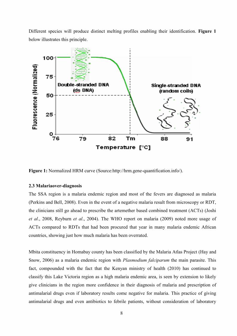
2.2 High resolution melting analysis
High resolution melting (HRM) analysis characterizes double-stranded DNA samples based on
their dissociation (melting)behavior. It is a novel, homogeneous, close-tube, post-PCR method
that enables individuals to analyze genetic variations (Single Nucleotide Polymorphisms (SNPs),
mutations, species identification, epigenetic studies for analysis of DNA methylation status) in
PCR amplicons. It is similar to classical melting curve analysis, but due to small increments in
temperature (0.008-0.2 ̊C), far more information on the melting behavior of a particular sample
can be studied. Samples can be discriminated according tosequence, length, GC content, or strand
complementarity,down to single base-pair changes.HRM analysis can only be performed on
instruments thathave HRM hardware and software installed. Data is acquiredusing specialized
HRM sources and detectors (Rotor-gene Q ® user manual, 2012).
Fluorescence decreases as DNA intercalating dye is released during thermal induced double
stranded (ds) DNA dissociation. The sharp decrease in fluorescence marks the greatest rate of
change in fluorescence and at its midpoint; the melting temperature (Tm) of a particular DNA
sample is established.In terms of cost-benefit analysis, HRManalysis provides accurate results
and savings on probe andlabel costs compared to other methods.
As the temperature increases, the double stranded DNA molecule containing intercalated dye
such as Evagreen dye™ dissociatesuniquely into single stranded DNA.
8
Different species will produce distinct melting profiles enabling their identification. Figure 1
below illustrates this principle.
Figure 1: Normalized HRM curve (Source:http://hrm.gene-quantification.info/).
2.3 Malariaover-diagnosis
The SSA region is a malaria endemic region and most of the fevers are diagnosed as malaria
(Perkins and Bell, 2008). Even in the event of a negative malaria result from microscopy or RDT,
the clinicians still go ahead to prescribe the artemether based combined treatment (ACTs) (Joshi
et al., 2008, Reyburn et al., 2004). The WHO report on malaria (2009) noted more usage of
ACTs compared to RDTs that had been procured that year in many malaria endemic African
countries, showing just how much malaria has been overrated.
Mbita constituency in Homabay county has been classified by the Malaria Atlas Project (Hay and
Snow, 2006) as a malaria endemic region with Plasmodium falciparum the main parasite. This
fact, compounded with the fact that the Kenyan ministry of health (2010) has continued to
classify this Lake Victoria region as a high malaria endemic area, is seen by extension to likely
give clinicians in the region more confidence in their diagnosis of malaria and prescription of
antimalarial drugs even if laboratory results come negative for malaria. This practice of giving
antimalarial drugs and even antibiotics to febrile patients, without consideration of laboratory
9
results or further examination is wide spread not only in Kenya (Zurovac et al., 2008), but also in
the rest of SSA region. A study carried out in Tanzania showed that although a total of 201 slides
tested negative for malaria, 22% (44) of these patients were treated with antimalarial drugs alone,
34% (68) with antibiotics alone, 26% (52) on a combination of the two and 18% (37) left the
centers with no medication (Reyburn et al., 2006).
There is evidence to show that malaria transmissions and fevers due to malaria are generally on
the decrease in Kenya and even in other highly endemic areas of the SSA region due to the
numerous mosquito eradication programmes(Griffin et al., 2010; WHO, 2010b; WHO, 2009;
Ceesay et al., 2008; O'Meara et al., 2008;WHO, 2008; Okiro et al., 2007).However, the practice
of administering antimalarial drugs indiscriminatelycauses neglect ofother febrile illnesses which
are on the increase and may be fatal (D'Acremont et al., 2009). A study carried out in Tanzania in
2004 (Reyburnet al., 2004), confirmed that there were more mortalities in non-malaria febrile
patients than in malaria confirmed patients, indicating the fact that the other non-malarial acute
undifferentiated fevers (NMAUF), especially the viral ones have been greatly ignored (Joshi et
al., 2008). Moreover, the team found that about half (43%) of the deceased patients (non-malaria)
were not treated with antibiotics while at the hospital and though they did not know the cause of
the deaths, they suggested that the intervention of antibiotics might have helped avoid or reduce
the mortalities (Reyburn et al., 2004).
Non-malarial acute undifferentiated fevers (NMAUF) refer to febrile illnesses with no indication
of an organ-specific disease after diagnosis of malaria has been excluded. In developing
countries, such acute undifferentiated fevers include those caused by arboviruses that depend on
arthropod vectors for their transmission (Joshi et al., 2008). In Ecuador, a study that was carried
out to determine the causes of fever found out that arboviral infections such as dengue and yellow
fever contributed to fever though not as much as leptospirosis and malaria (Manock et al., 2009).
2.4Arbovirustransmission
Arboviruses are Arthropod borne virusesthat are biologically transmitted by hematophagous
(blood feeding) arthropods. Arboviral replication is characterized by a biological cycle in the
arthropod vector and vertebrate host. The arthropods become infected after a blood meal from a
viremic vertebrate and remain infectious for the rest of their lives. The virus is amplified in these
arthropods during an incubation period that results in viral replication in the arthropods salivary
glands. Later, the virus is transmitted to non-immune vertebrate hosts during feeding by the
10
arthropod (Weaver and Reisen, 2010, Sang and Dunster, 2001). Arboviruses circulate in wild
animals and cause disease to humans and/ or domestic animals which in some cases are
incidental/dead-end hosts that produce viremias inadequate to cause arthropod infections after
spillover transmissions occur (Weaver and Reisen, 2010).
The transmission of the arboviruses by these vectors can either be vertical or horizontal. In
vertical transmission, the arboviruses are transmitted to the arthropod progeny transovarially
whereas in horizontal transmission, there may be either oral transmission by a competent vector
to a vertebrate host or sexually, whereby the female transmits the arbovirus to the male during
mating (Weaver and Reisen, 2010). While most of the documented arboviruses cause zoonoses,
about 50% of these viruses (about 100 out of the 535 that infect humans) are transmitted by
mosquitoes (Sang and Dunster, 2001). Three families of arboviruses that cause great concern to
public health are Togaviridae, Bunyaviridae and Flaviviridae. The infections by these arboviruses
range from mild febrile illnesses that are self-limiting and last for a short duration, to more severe
encephalitis and hemorrhagic fevers that are fatal. Sang and Dunster (2001) noted in their study
that especially in malaria endemic regions, majority of arboviral caused infections remain
undiagnosed and that their effect to public health has been greatly underestimated. Factors such
as non-specific symptoms seen in arboviral infections, lack of specialized diagnostic services and
active surveillance systems were largely sited as the main reasons for misdiagnosis of these
infections. Similar observations were made by LaBaeud and colleagues (2011).
In Kenya, there has been evidence of arboviral activity detected in human serum and even in
mosquitoes from different parts of the country such as the coastal region, the north-eastern
region, the Rift valley and even the western regions(LaBeaud et al., 2011, Mease et al., 2011,
Sutherland et al., 2011, LaBeaud et al., 2008, Woods et al., 2002, Reiter et al., 1998, Morrill et
al., 1991, Johnson et al., 1983, Bowen et al., 1973) . In 1992/1993, Yellow fever outbreak was
reported in Kerio Valley in Rift valley Province, Chikungunya virus outbreak was reported in
Lamu Island in the coastal region in 2004, and two Rift valley fever outbreaks were reported in
the North-Eastern parts of the country in 1997 and 2006/2007. Similarly,entomologicstudies on
arboviral circulation in mosquitoes have also been done (LaBeaudet al., 2011, Miller et al.,
2000). These studies have been able to link specific species of mosquitoes as being responsible
for transmission of specific arboviruses in different geographical regions of the
country.Culexquinquefasciatus was identified as one of the mosquito vectors responsible of
transmitting both Rift valley fever and West Nile viruses (LaBeaudet al., 2011).
11
Sang and Dunster (2001) attribute the emergence and re-emergence of arboviruses in Kenya to
five variables namely: The vector, the virus, humans, the wild vertebrate host and finally to
environmental factors. The presence of a water body either due to heavy rainfall that causes
flooding, a natural feature such as a lake, still water in boats, a water container that is not closed,
tyres that collect water or even gutters around the homestead that remain with water, provide a
breeding ground for the arthropods that rely on water for their larval stages to thrive. Warm
temperatures have equally been established to favour the activity of arthropods. With the
increasing reality of global warming, there has been increased distribution of these arthropods
especially the flying ones as they venture into new warm territories that were cold previously.
Global warming has also been documented to cause a reduction in the extrinsic incubation period
(EIP) of these arboviruses. Extrinsic incubation period refers to the period of time between when
a vector ingests the arbovirus in the blood meal and when it transmits the arbovirus to a
vertebrate host. Thus, with this period being shortened, there is increase probability of the vector
transmitting the virus multiple times in its lifetime. Commerce and migration of humans, import
of livestock from other regions has also been implicated in the arboviral threat witnessed in the
country. Virus mutation can result in the changing of a previously preferred vector for another
vector that is competent enough to transmit the virus. An example of a change in vector
preference was seen in the Reunion Islands during the 2005/2006 Chikungunya virus (CHIKV)
outbreak where the known CHIKV vector Aedesaegyptiwas absent or in scarce numbers while
Aedesalbopictus was noted to be the principal CHIKV vector (Tsetsarkin et al., 2007).
2.4.1 Sindbisvirus
Sindbis virus is an enveloped single stranded virus of positive polarity that is transmitted to
humans by the bite of an infected mosquito (Culex species). Their genomic RNA of about11.7kb
nucleotides encodes four non-structural proteins, a capsid and two envelope proteins (Strauss et
al., 1984). A member of the Western equine encephalitis complex, the Sindbis virus belongs to
the Togaviridae family and shares the alphavirus genus position with other viruses such as
Chikungunya, Semliki Forest virus, Onyong’nyong’ virus, Ross River virus, Venezuelan equine
encephalitis and Eastern equine encephalitis viruses. The virus was first isolated in 1952 in
Sindbis health district near Cairo, Egypt from pools of ornithophilicCulexunivittatus and
Culexpipiens mosquitoes (Taylor et al., 1955). Infection with the virus is characterized by rash,
arthralgia and fever. Although the symptoms last for a short duration (less than a week) and
12
recovery is complete, some patients still suffer recurrent joint swelling and tenderness for
months (Tesh, 1982).
In nature, the virus is maintained by vertebrate hosts (birds) and invertebrate vectors
(mosquitoes). Sindbis virus is prevalent in South and East Africa, Egypt, Israel, Phillipines and
parts of Australia. Different regions have different names for the disease caused by Sindbis
virus. In Sweden it is called Ockelbo, Pogosta in Finland, Karelian fever in Russia and Babanki
virus in much of SSA. It was only in 2004 that the causative agent of Pogosta disease was
isolated in Finland directly from human isolates and confirmed to be Sindbis virus(Kurkela et
al., 2004). Passerines and tetraonid birds have been greatly implicated in transmitting the virus
to different geographical regions over long distances (Jöst et al., 2010, Kurkela et al., 2008).
A systematic mosquito surveillance study carried out in Kenya between 2007-2012, established
the circulation of Sindbis virus in Culex mosquitoes found around large water bodies in Kisumu
associated with the Lake Victoria water basin and Naivasha associated with Lake
Naivasha(Ochieng et al., 2013). Important to note is that this virus was not found in the arid and
semi-arid areas of Kenya, where most of the arbovirus diversity and abundance was recorded.
This was attributed to the fact that migratory birds which are the vertebrate hosts of the virus,
usually swam around these water points during their stop over breeding seasons (Ochieng et al.,
2013). As already established, the virus is of public health importance and it would be further
interesting to see whether there is a correlation between seroprevalence in human subjects and
the indication of it being present in mosquitoes from this region.
2.4.2 Bunyamwera virus
Bunyamwera virus is an enveloped, segmented, single stranded virus of negative polarity. It is a
member of the family Bunyaviridae and genus Orthobunyavirus. Its genome and that of the
other members of this family consist of three linear genomic RNA segments: Small (S),Medium
(M) and Large (L) segments. They encode for six proteins in total: The nucleocapsid protein
(N), a non-structural protein (NS), three envelope gycoproteins and a viral RNA dependent RNA
polymerase.
Due to the presence of the three segments, reassortments frequently occur and lead to an
increase in the number of members in this family. Other viruses in this family that are of public
health and agricultural importance include Ngari virus; a reassortment of Bunyamwera virus
with the M segment of Batai virus (Brieseet al., 2006), Rift Valley Fever virus and Crimean-
13
Congo Hemorrhagic Fever (Gerrard et al., 2004). Bunyamwera virus was first isolated from
Aedesmosquitoes that were caught in Semliki forest in Uganda (Smithburn et al., 1946). It is
transmitted to humans by infected mosquitoes and very likely ticks (Lwande et al., 2013) that
have fed on infected vertebrate blood. Infection by the virus results to a mild febrile illness
characterized by headache, fever, joint and back pain, rash and mild involvement of the central
nervous system. Serological evidence of the infection has also been largely reported in SSA, but
most infections go unrecognized (LeDuc and Porterfield, 2005). Outbreaks have also occurred in
North America, South America, Africa(Gerrard et al., 2004), and Europe. More recently in
Kenya, a five year surveillance study on mosquitoes also indicated circulation of the virus in
Garissa and Magadi(Ochieng et al., 2013) .
2.5 Neutralizing antibodies
According to biology online, neutralizing antibodies refer to antibodies that are capable of
keeping an infectious agent, usually a virus, from infecting a cell by blocking the cell’s receptors
or neutralizing the virus’s biological effect by interfering with its receptors. This results in the
inactivation of the virus such that it is no longer able to infect and replicate in cell cultures or
animals(WHO, 2007). Several studies have shown that detecting the presence of a particular
arbovirus is possible by serologically identifying neutralizing antibodies in the sera of study
subjects (LaBeaud et al., 2008, Buckley et al., 2003). When a virus infects a cell within a fixed
cell monolayer, it produces a viral plaque which is formed due to cell lysis. The lysed cell then
spreads the infection to adjacent cells where the infection-to-lysis cycle is repeated. The infected
cell area thus creates a plaque; an area of infection surrounded by uninfected cells (Kaufmann
and Kabelitz, 2002). It is assumed that one plaque is representative of a single virus particle.
Therefore, when serum containing neutralizing antibodies against a specific virus is mixed and
incubated with a predetermined virus dilution, it is thus expected that the number of viral plaques
that were formed initially in the absence of the serum will now be reduced due to the presence of
these neutralizing antibodies. This principle is the basis of plaque reduction neutralization test
(PRNT) also referred to as serum dilution neutralization test.
14
CHAPTER THREE
MATERIALS AND METHODS
3.1 Study site
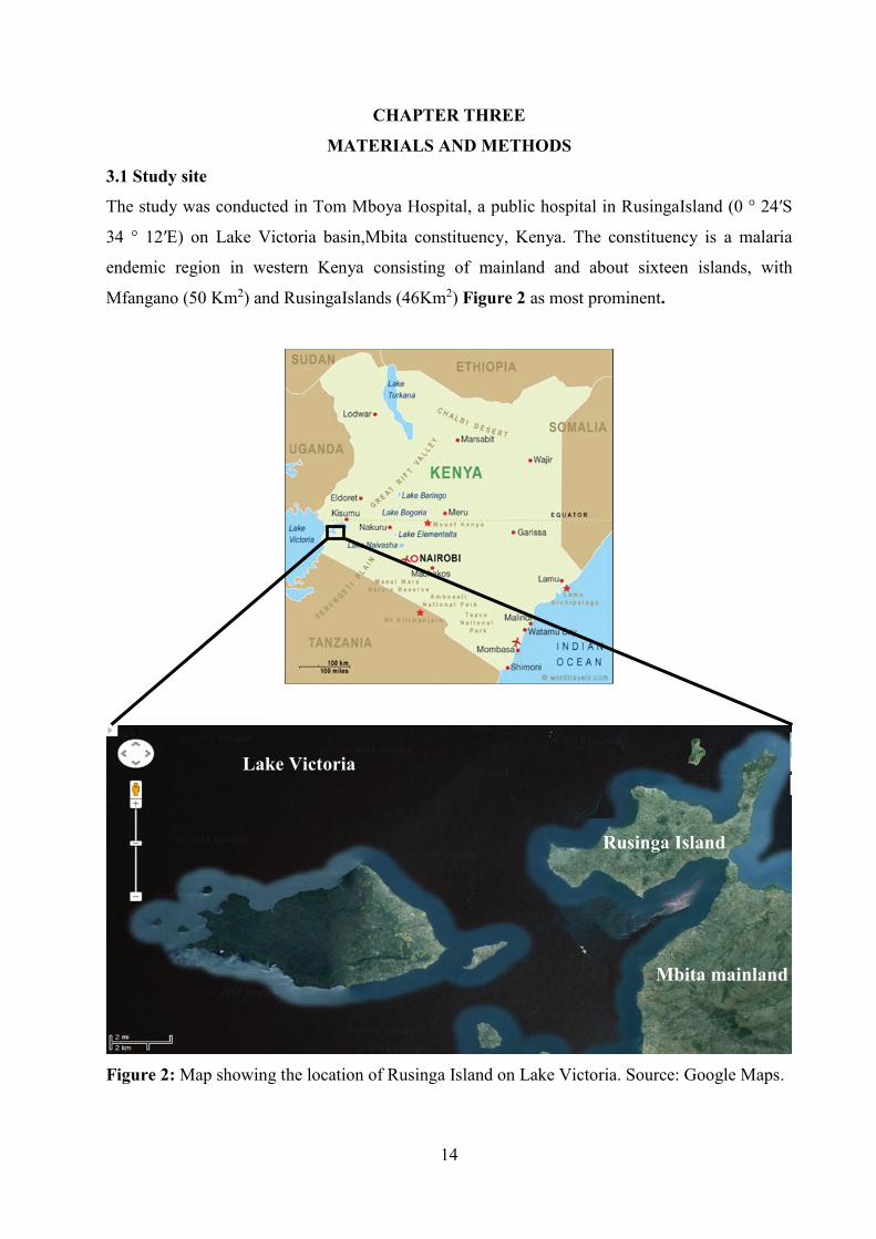
The study was conducted in Tom Mboya Hospital, a public hospital in RusingaIsland (0 ° 24′S
34 ° 12′E) on Lake Victoria basin,Mbita constituency, Kenya. The constituency is a malaria
endemic region in western Kenya consisting of mainland and about sixteen islands, with
Mfangano (50 Km2) and RusingaIslands (46Km2) Figure 2 as most prominent.
Figure 2: Map showing the location of Rusinga Island on Lake Victoria. Source: Google Maps.
Mbita mainland
Rusinga Island
Lake Victoria
15
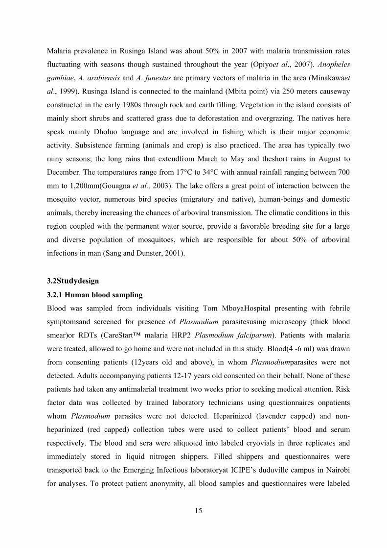
Malaria prevalence in Rusinga Island was about 50% in 2007 with malaria transmission rates
fluctuating with seasons though sustained throughout the year (Opiyoet al., 2007). Anopheles
gambiae, A. arabiensis and A. funestus are primary vectors of malaria in the area (Minakawaet
al., 1999). Rusinga Island is connected to the mainland (Mbita point) via 250 meters causeway
constructed in the early 1980s through rock and earth filling. Vegetation in the island consists of
mainly short shrubs and scattered grass due to deforestation and overgrazing. The natives here
speak mainly Dholuo language and are involved in fishing which is their major economic
activity. Subsistence farming (animals and crop) is also practiced. The area has typically two
rainy seasons; the long rains that extendfrom March to May and theshort rains in August to
December. The temperatures range from 17°C to 34°C with annual rainfall ranging between 700
mm to 1,200mm(Gouagna et al., 2003). The lake offers a great point of interaction between the
mosquito vector, numerous bird species (migratory and native), human-beings and domestic
animals, thereby increasing the chances of arboviral transmission. The climatic conditions in this
region coupled with the permanent water source, provide a favorable breeding site for a large
and diverse population of mosquitoes, which are responsible for about 50% of arboviral
infections in man (Sang and Dunster, 2001).
3.2Studydesign
3.2.1 Human blood sampling
Blood was sampled from individuals visiting Tom MboyaHospital presenting with febrile
symptomsand screened for presence of Plasmodium parasitesusing microscopy (thick blood
smear)or RDTs (CareStart™ malaria HRP2 Plasmodium falciparum). Patients with malaria
were treated, allowed to go home and were not included in this study. Blood(4 -6 ml) was drawn
from consenting patients (12years old and above), in whom Plasmodiumparasites were not
detected. Adults accompanying patients 12-17 years old consented on their behalf. None of these
patients had taken any antimalarial treatment two weeks prior to seeking medical attention. Risk
factor data was collected by trained laboratory technicians using questionnaires onpatients
whom Plasmodium parasites were not detected. Heparinized (lavender capped) and non-
heparinized (red capped) collection tubes were used to collect patients’ blood and serum
respectively. The blood and sera were aliquoted into labeled cryovials in three replicates and
immediately stored in liquid nitrogen shippers. Filled shippers and questionnaires were
transported back to the Emerging Infectious laboratoryat ICIPE’s duduville campus in Nairobi
for analyses. To protect patient anonymity, all blood samples and questionnaires were labeled
16
with barcode identifiers. The samples were collected between May 28, 2012 and Feb 28,
2013and the process facilitated by staff in the Kenyan Ministry of Public Health and Sanitation
and Division of Disease Surveillance and Response (DDSR) of the Government of Kenya.
Ethicalclearance to work on human samples was provided byKEMRI’s National Ethical Review
Board, seeappendix 1.
3.2.2 Extraction of total DNA from blood
Total DNA in blood samples was extracted as described by Kawasaki (1990) with few
modifications. Briefly, 50 µl of human blood was aliquoted from a labeled 2 ml cryovial tube
and placed into its respective labeled 1.5 ml eppendorf tube. 0.5 ml of Tris-EDTA (TE) buffer
pH 7.5 was then added and spun for five minutes at 13,000 relative centrifugal force (rcf) at 4°C.
The resultant supernatant was discarded, pellet re-suspended in TE buffer and vortexed. The
procedure was repeated three times. The final pellet was re-suspended in 100 µl of K buffer (see
appendix 2),vortexed and incubated at 55°C for an hour. The extracted DNA was incubated at
95°C for 10 minutes to inactivate proteinase K, and then stored at -20°C until when required.
3.2.3 Detection of Plasmodium parasitesby nested PCR-HRM (nPCR-HRM)
Plasmodium DNA in the total extracted DNA was amplified using nested PCR. For the primary
amplification step, a forward primer (PL 1459 out F) CTG GTT AAT TCC GAT AAC and a
reverse primer (PL 1706 out R)TAA ACT TCC TTG TGT TAG AC were used. Similarly, a
second pair of primers described elsewhere (Mangoldet al., 2005) was used for the secondary
amplification reaction. These primers targeted the 18S rRNA gene marker. Hot Firepol® HRM
mix kit (Solis BioDyne, Estonia) was used for the two amplification processes. Optimal DNA
amplification for each of the two reaction steps was carried out in a 10µl final reaction volume
that consisted of 1µl DNA template, 2µl HRM mix, 0.5µl of 0.5µM of both primers and 6µl
nuclease free PCR water. The PCR thermal conditions consisted of an initial denaturation at
95°C for 5 minutes, 45 cycles of denaturation at 94°C for 20 seconds, decreasing annealing
temperatures from 65°C-50°C for 25 seconds (cycles 1-5), 50°C for 40 seconds (cycles 6-10),
50°C for 50 seconds (cycles 11-45), and extension at 72°C for 30 seconds. A final extension of
72°C for 3 minutes was included before HRM analysis.Upon completion, the amplification
process then transitioned into the melting phase (HRM) in the same closed tube system yielding
distinct melting profiles. These profiles were indicative of various Plasmodium species present
in the samples. The set of conditions for HRM included a stepwise temperature increase of
0.2°C/sec from 75°C to 90°C, with fluorescence acquisition at each temperature transition. The
17
Rotorgene Q® machine(QIAGEN, Germany)facilitated both the amplification process and HRM
analysis. Plasmodium falciparum infected blood and PCR water were used as positive and
negative controls respectively in the PCR process. The other Plasmodium species that could not
be detected using the HRM platform due to lack of positive controls, were deduced by
sequencing at Macrogen, Korea.Using Geneious software (6.1.5 version, Biomatters; Kearseet
al. 2012), the resultant chromatograms were trimmed and curated sequences were aligned with
known Plasmodia sequences obtained from GenBank (Accession numbers: AF145336 and
AB489195 for P. malariae, AB182489 and AB182493 for P. ovale and JQ627152 and
JF681166 for P. falciparum), see appendix 3.
3.2.4Virusisolation
Clean Vero cell lines (from the kidney of green African monkey:Chlorocebussabaeus) were
propagated and maintained in T25 and T75 culture flasks. To establish viral stocks of Sindbis
and Bunyamwera viruses, confluent cells were trypsinized and plated onto a twenty-four well
plate. After two days, the cells were confluent and 50 µl of suspected mosquito field samples of
Bunyamwera and Sindbisviruses that had been homogenized were plated onto these wells. The
plates were incubated for one hour at 37°C in 5% CO2then 1 ml of maintenance media was
added. The plate was returned to the incubator and presence of cytopathic effects (CPE)
observed as from the next day until when the CPE were spotted. The infected cells with the
media were then harvested, placed into cryovials and frozen at -80 °C.
3.2.5Virus isolates confirmation by passage
200µl of the infected cells that had been harvested from the original inoculation were re-
inoculated into confluent T-25 flasks and monitored until the CPE was reproduced. The flasks
were then frozen at -80°C for a day then thawed and the contents transferred into 15ml
centrifuge tubes. The tubes were centrifuged at 2500-3000rpm for 10min, the supernatant
collected and aliquoted into cryovials of 1 ml each then stored at -80°C.
3.2.6 Virus Confirmation by PCR
To confirm that the CPE observed was actually due to the presence of the intended viruses, 250
µl of each of the harvested viruses was put into a sterile cryovial and RNA extracted using the
Trizol (Invitrogen, Carlsbad, CA, USA) extraction method following manufacturer’s
instructions. About 5 µlof the extracted RNA was converted into complementary DNA
(cDNA)through reverse transcriptase PCR. Here, the final reaction volume of 10µl consisted of
18
5 µl RNA template, 0.5 µl water, 2 µl transcriptase®buffer, 1µl dNTPs and random hexamer
primer, 0.25 µl RNase inhibitor and reverse transcriptase from the Roche kit (Mannheim,
Germany).The forty-five minute reaction conditions were 25 °C for 10 min, 55°C for 30 min and
85°C for 5 min. This was followed by real time PCR and HRM (section 3.2.3) using CTG CTA
ACA CCA GCA GTA CTT TTG AC (OrthoBun F1) and TGG AGG GTA AGA CCA TCG
TCA GGA ACT G (OrthoBun R1) forward and reverse primers for Bunyavirus and TGG CGC
TAT GAT GAA ATC TGG AAT GTT (Vir 2052 F) and TAC GAT GTT GTC GTC GCC GAT
GAA (Vir 2052 R) forward and reverse primers for Sindbis virus. The resultant melting profiles
of the samples were compared with the melting profiles of Sindbis and Bunyamwera virus
controls, a positive sample being one with similar profile to the controls. Absence of similar
profiles meant that the entire process from cell culture to molecular work had to be repeated.
3.2.7 Determination of viral titer (Plaque Assay)
Plaque assay was used to determine known amounts of virus (viral titer) that produced
recommended plaque forming units per well. To achieve about 800pfu/ml (80plaques in a well
when 100µl of virus is added per well), tenfold dilutions of each virus were prepared as
described at (www.bdbiosciences.com › Resources › Baculovirus Protein Expression). Briefly,
fifty microlitres of a viral stock (e.g. Sindbis) was added onto 450 µl 2% MEM (Sigma Aldrich,
St. Louis, USA) over a tenfold dilution series upto 10-10. A hundred microlitre of these dilutions
was then plated onto their respective confluent Vero cells in 6 well plates. After one hour
incubation at 37°C, 3 ml of methyl cellulose overlay medium was added to each well and
incubated at 37°C, 5% CO2 for threedays for Sindbis virus and four days for Bunyamwera virus.
After the overlay was removed, 10 % formaldehyde was added to the wells and placed under
UV for 30 min for fixing and to inactivate the virus.The plates were placed under slow running
tap water to remove the formaldehyde and stained immediately with 0.5 % methyl violet dye
then washed off and left overnight to dry.
The plaques were then counted and plaque forming units per ml (pfu/ml) calculated using the
formula:
Plaque forming units per ml = number ofplaques/volume of diluted virus per well
19
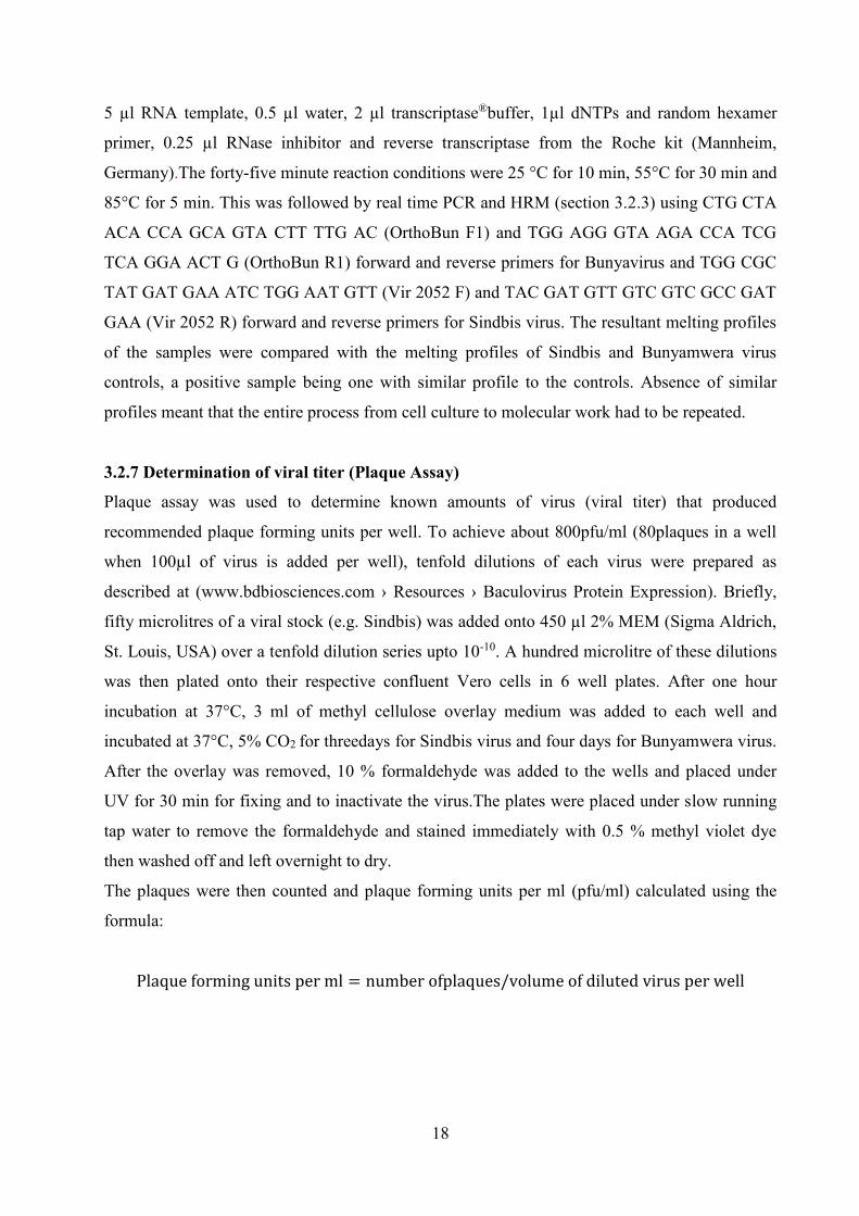
As shown in Figure 3, the numbers of plaques reducewith increase in virus dilution. This
facilitates the selection of the well (and hence the dilution) to be used for the next assay which is
plaque reduction neutralization test (PRNT).
Figure 3:Plaque assay for Bunyamwera virus showing virus dilution and corresponding number
of plaques formed; number of plaques reduce with increase in virus dilution.
For Bunyamwera and Sindbis viruses, 10-8 dilution and 10-7 dilutions respectively produced the
desired number of plaques and thus were used for the plaque reduction neutralization procedure.
20
3.2.7 Determination of Neutralization Activity (Plaque Reduction Neutralization Test)
All the sera samples were heat inactivated at 56°C for 30min. Twelve microlitres of a serum
sample was diluted into 108 µl of 2 % MEM (1:10 dilution)virus. A previously unthawed vial of
virus was thawed on ice. The virus stock was then diluted as previously determined from the
plaque assay. Sixty microlitres of the diluted virus was then mixed with an equal volume of the
serum in a 24 well plate and incubated for one hour. Other controls incubated included a
negative control (MEM alone), backtitrated controls (10-9 and 10-8 for Bunyamwera and Sindbis
viruses respectively) and a working dilution of the virus. A 100 µl of the incubated mixture and
the controls were inoculated onto confluent cells in 6 well plates and incubated for one hour
again. Three milliliters of methyl cellulose overlay medium was finally added to the wells and
the plates incubated for three days for Sindbis and four days for Bunyamwera virus and later
stained as described above (3.2.6).
3.3 Data Analysis
Descriptive statistics including frequencies and proportions for both numerical (age) and
categorical variables (gender, occupation, interaction with domestic animals and location)
constituted the data collected from questionnaires supplied. Proportion values of malaria
prevalence,arboviralprevalence, drug prescription patterns and fever proportions were
calculated. Chi-square test was used to analyze significance of variables (age, gender
andoccupation) in relation to malaria and arboviral infection. A multivariable logistic regression
model was used to investigate risk factors associated with testing positive for malaria, Sindbis
and Bunyamwera virus antibodies. The significance level was set at p<0.05. The analyses were
performed using STATA v10.1 (StataCorp, College Station, TX, USA).
21
CHAPTER FOUR
RESULTS
4.1 Characteristics of study subjects The study comprised 92 subjects aged between 12 and 70 years, with a mean age of 34 years.
There were 45 females and 45 males, with the gender of two individuals not reported.Mean body
parameters of these patients such as body temperature and body weight were 37.7 ºC and 59.23
Kg respectively, with 66 of them having fever. The subjects were recruited from Tom Mboya
clinic between May 28, 2012 and Feb 28, 2013.
4.2 Malaria prevalence amongthe undiagnosed febrile patients
Prevalence of malaria in Rusinga Island and a summary of the Plasmodium species present in
the island are illustrated in figure 4.
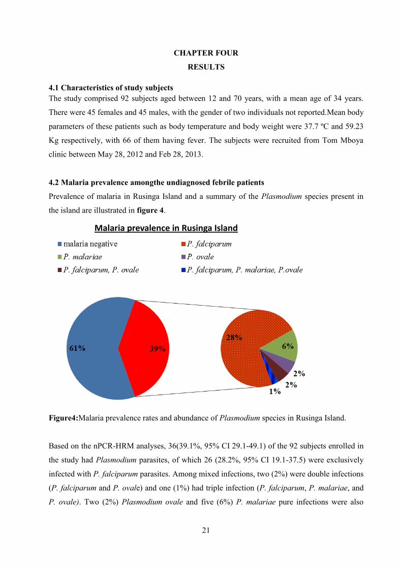
Figure4:Malaria prevalence rates and abundance of Plasmodium species in Rusinga Island.
Based on the nPCR-HRM analyses, 36(39.1%, 95% CI 29.1-49.1) of the 92 subjects enrolled in
the study had Plasmodium parasites, of which 26 (28.2%, 95% CI 19.1-37.5) were exclusively
infected with P. falciparum parasites. Among mixed infections, two (2%) were double infections
(P. falciparum and P. ovale) and one (1%) had triple infection (P. falciparum, P. malariae, and
P. ovale). Two (2%) Plasmodium ovale and five (6%) P. malariae pure infections were also
Malaria prevalence in Rusinga Island
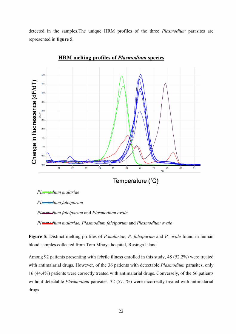
22
detected in the samples.The unique HRM profiles of the three Plasmodium parasites are
represented in figure 5.
Figure 5: Distinct melting profiles of P.malariae, P. falciparum and P. ovale found in human
blood samples collected from Tom Mboya hospital, Rusinga Island.
Among 92 patients presenting with febrile illness enrolled in this study, 48 (52.2%) were treated
with antimalarial drugs. However, of the 36 patients with detectable Plasmodium parasites, only
16 (44.4%) patients were correctly treated with antimalarial drugs. Conversely, of the 56 patients
without detectable Plasmodium parasites, 32 (57.1%) were incorrectly treated with antimalarial
drugs.
HRM melting profiles of Plasmodium species
Plasmodium malariae
Plasmodium falciparum
Plasmodium falciparum and Plasmodium ovale
Plasmodium malariae, Plasmodium falciparum and Plasmodium ovale
23
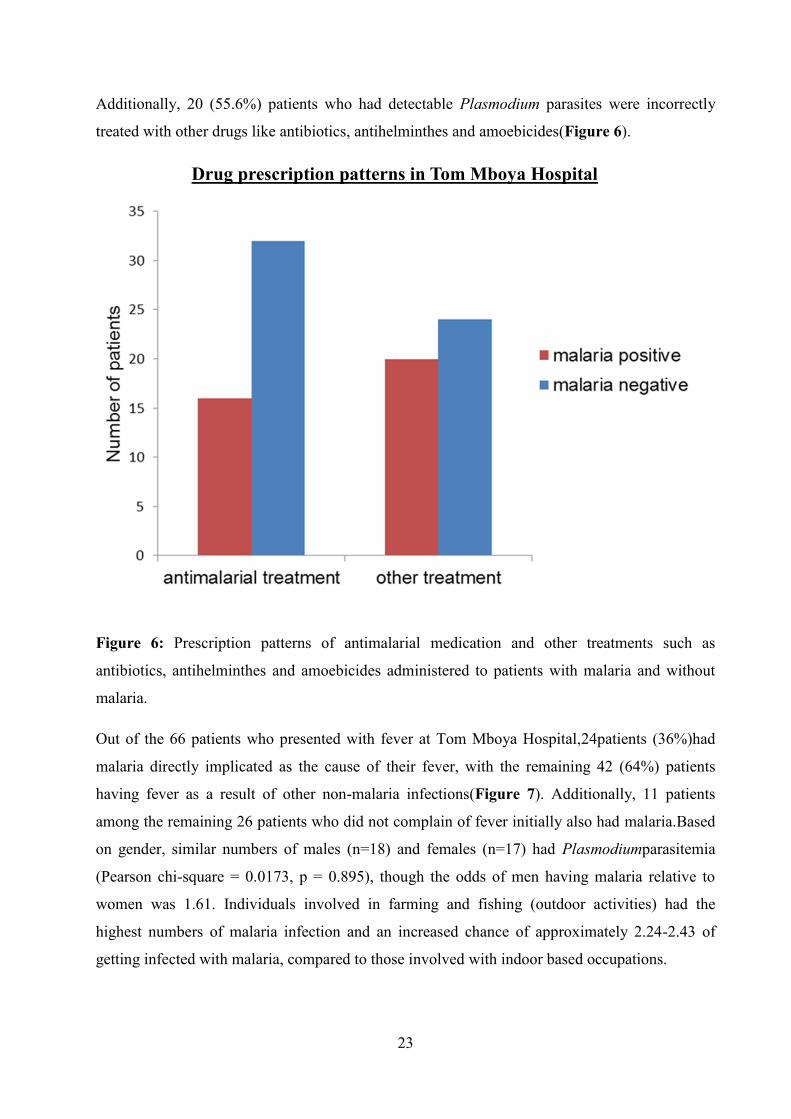
Additionally, 20 (55.6%) patients who had detectable Plasmodium parasites were incorrectly
treated with other drugs like antibiotics, antihelminthes and amoebicides(Figure 6).
Drug prescription patterns in Tom Mboya Hospital
Figure 6: Prescription patterns of antimalarial medication and other treatments such as
antibiotics, antihelminthes and amoebicides administered to patients with malaria and without
malaria.
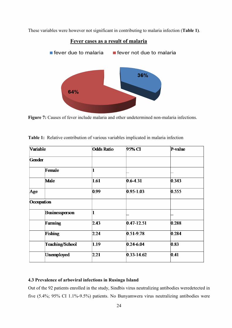
Out of the 66 patients who presented with fever at Tom Mboya Hospital,24patients (36%)had
malaria directly implicated as the cause of their fever, with the remaining 42 (64%) patients
having fever as a result of other non-malaria infections(Figure 7). Additionally, 11 patients
among the remaining 26 patients who did not complain of fever initially also had malaria.Based
on gender, similar numbers of males (n=18) and females (n=17) had Plasmodiumparasitemia
(Pearson chi-square = 0.0173, p = 0.895), though the odds of men having malaria relative to
women was 1.61. Individuals involved in farming and fishing (outdoor activities) had the
highest numbers of malaria infection and an increased chance of approximately 2.24-2.43 of
getting infected with malaria, compared to those involved with indoor based occupations.
24
These variables were however not significant in contributing to malaria infection (Table 1).
Fever cases as a result of malaria
Figure 7: Causes of fever include malaria and other undetermined non-malaria infections.
Table 1: Relative contribution of various variables implicated in malaria infection
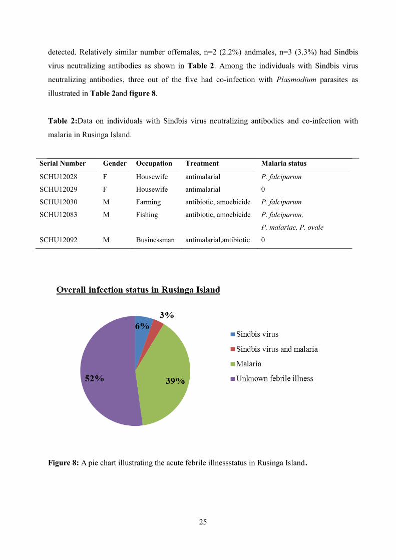
4.3 Prevalence of arboviral infections in Rusinga Island
Out of the 92 patients enrolled in the study, Sindbis virus neutralizing antibodies weredetected in
five (5.4%; 95% CI 1.1%-9.5%) patients. No Bunyamwera virus neutralizing antibodies were
25
detected. Relatively similar number offemales, n=2 (2.2%) andmales, n=3 (3.3%) had Sindbis
virus neutralizing antibodies as shown in Table 2. Among the individuals with Sindbis virus
neutralizing antibodies, three out of the five had co-infection with Plasmodium parasites as
illustrated in Table 2and figure 8.
Table 2:Data on individuals with Sindbis virus neutralizing antibodies and co-infection with
malaria in Rusinga Island.
Serial Number Gender Occupation Treatment Malaria status
SCHU12028 F Housewife antimalarial P. falciparum
SCHU12029 F Housewife antimalarial 0
SCHU12030 M Farming antibiotic, amoebicide P. falciparum
SCHU12083 M Fishing antibiotic, amoebicide P. falciparum,
P. malariae, P. ovale
SCHU12092 M Businessman antimalarial,antibiotic 0
Figure 8: A pie chart illustrating the acute febrile illnessstatus in Rusinga Island.
26
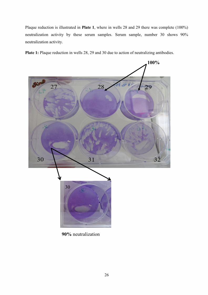
Plaque reduction is illustrated in Plate 1, where in wells 28 and 29 there was complete (100%)
neutralization activity by these serum samples. Serum sample, number 30 shows 90%
neutralization activity.
Plate 1: Plaque reduction in wells 28, 29 and 30 due to action of neutralizing antibodies.
90% neutralization
100%
30
27
CHAPTER FIVE
DISCUSSION
A sensitive and specific diagnostic tool that is capable of precise detection of low
Plasmodiumparasitemia is a desirable asset in any health facility. From the results, 39.1%
malaria prevalence was recorded from 36 patients in Tom MboyaHospital,Rusinga Island.
Ironically, this value constituted individualswho had been previously diagnosed as not having
malaria by microscopy and RDTs,butin whom Plasmodium parasites were later detected by
nPCR-HRM technique. The inability of microscopy and RDTs to detect low
Plasmodiumparasitemia of below 100parasites/µl (Trampuz et al., 2003; Moody, 2002) was
noted as the most probable reason. This is because even the nPCR-HRM technique that is highly
sensitive yielded only small amounts of fluorescence from the amplified samples, indicating the
low amounts of parasitemia in the samples. By combining the high sensitivity of nested PCR
with real-time species differentiating HRM analysis, the reporting of true malaria prevalence for
epidemiological studies can be synergistically enhanced. nPCR-HRM enhances low-parasitemia
malaria diagnosis and can potentially surmount the deficiencies of microscopy and RDT based
results in determining malaria parasitemia, and evaluating epidemiology of the disease.Several
studies carried out in the Kenyan highlands of Nandi and Kisii and also in Iran noted increased
detection of Plasmodiumparasites by nested PCR that had been initially missed by microscopy
(Zoghiet al., 2012; Wangaiet al., 2011). Though microscopy is regarded as the standard
diagnostic tool for malaria detection in SSA, complementary use of molecular based methods is
important especially in rural endemic areas. This will enable clinicians rule out malaria in
differential diagnoses and prevent the over-use of antimalarial medication and the effects that
come with it.
A major medical implication evident from the results is that febrile illness misdiagnosis and
misadministration of antimalarial drugs can be significant in malaria endemic settings, which
result in a myriad of downstream issues. They include improper treatment and patient care,
chronic suffering of patients, drug wastageand development of parasite resistance to these drugs
(Amexoet al., 2004). Due to the inability of microscopy and RDTs to detect low
Plasmodiumparasitemia (Okellet al., 2009),92 febrile patients were diagnosed as not having
malaria but were still given different medications on the basis of clinical symptoms alonedespite
negative microscopy and/or RDTs results for malaria(Roucheret al., 2012; Chinkhumbaet al.,
2010; Chandler et al., 2008).Due to limited diagnostic platforms for diagnosis of other non-
28
malaria febrile illnesses and the low sensitivity and specificity of the present malaria screening
protocols, clinicians are limited to heuristic methods of clinical diagnoses(Chandramohanet al.,
2002), mostly having a bias towards malaria diagnosis and treatment. This is despite the fact that
there are other potential differential diagnoses for febrile illness, including arthropod borne viral
(arboviral) infections (Crump et al., 2013; Hertz et al., 2012; Joshi et al., 2008).The drug
prescriptions have inadvertently contributed to parasite resistance to anti-malarials (Na-
Bangchangand Karbwang,2013) and mortalities from other undiagnosed illnesses (Reyburnet
al., 2006).Only 16 (44.4%)patients with low-parasitemiamalaria were correctly treated with
antimalarial drugsat Tom MboyaHospital,with a majority of the antimalarial drugs being
prescribed to 32 (57.1%) patients in whom Plasmodiumparasitemiawas not detected. The
scenario of over-prescription of antimalarial drugs in non-malaria febrile patients and under-
prescription of antimalarial drugs in low-parasitemia malaria patients was noted.It is therefore
necessary to establish diagnostic techniques that detect low-parasitemia to clearly discriminate
malaria related febrile clinical symptoms from those related to other differential illnesses, and
improve case management of febrile illness.
As earlier stated, fever used to be synonymous to malaria (Perkins and Lubell, 2008;
Chandramohanet al., 2002), however,this perception has changed over time due to
implementation of proper policies that encourage parasitological testing before drug prescription
(WHO, 2010a).In this study, malaria was cited as the cause of fever in only 24 (36%) of the 66
febrile patients who initially presented to the hospital with fever. Moreover, we also found out
that of the remaining 26 patients that did not present with fever initially, 11 patients did in fact
have Plasmodium infections. Fever is thus a poor indicator of malaria.These results are similar
to a systematic review carried out by D’Acremont and associates (2010) that reviewed 39
journal papers carried out in the past 20 years in Africa on malaria related fevers. This large-
scale review showed that there had been a reduction in the number of fevers related to malaria
over time; in that, only about a fifth of all fevers were found to be directly as a result of malaria.
This was attributed to a change in diagnosis from clinical to laboratory based.
Nearly all malaria cases (29 out of 36, 80.6%) were caused by P. falciparum, the most
dangerous malaria causing parasite. These results show that P. falciparum is still the main
species responsible for malaria in this malaria endemic region as earlier established (Kenyan
Ministry of Health, 2010; Hay and Snow, 2006). Additionally, the melting curve profiles for the
29
three Plasmodium species were similar to those by Mangold and colleagues (2005). We however
improved the sensitivity of the assay by adopting the advantages of nested PCR.
Table 1 shows that individuals involved in fishing and farming (outdoor) activities were more
susceptible to being infected with malaria compared to those who work indoors such as
shopkeepers, housewives, carpenters, teachers and students. This can be attributed to the fact
that while outdoors and especially in areas that have water (artificially made or naturally
present) which are breeding sites for mosquitoes, chances of getting bitten are increased
especially during dawn and dusk (Imbahaleet al., 2011). Fishermen usually spend nights out
fishing and hence are even more prone to being bitten by mosquitoes. This may also explain
further the 1.61 increased chance of men getting malaria infection as opposed to women because
fishing is mainly carried out by men (Ukoroije and Abowei, 2012).
The estimated seroprevalence of Sindbis virus neutralizing antibodies was 5.4%(n=5) of whom 3
had co-infection with malaria. The value (5.4%) is indicative of exposure to Sindbis virus,
resulting in the production of antibodies specific to the virus.The primary reservoir hosts
implicated in maintaining the virus are the birds. The abundant supply of fish provided by the
lake ensures a constant presence of both migratory and native birds which use Rusinga Island as
their nesting place, bringing the infection nearer to the humans. A study carried out in Finland
noted a relatively similar prevalence rate in humans of 5.2% between the years 1999-2003
(Kurkellaet al., 2008). Increased interaction between reservoirs, humans and mosquito vectors
(Hall et al., 2012) could be responsible for the relatively higher numbers of Sindbis infection in
men than women in this study. During 2007-2012 period, Ochieng and colleagues (2013)
reported the presence of Sindbis and Sindbis-like viruses in Culex and Culiseta mosquitoes from
Kisumu; a town located 73Km from Rusinga Island in the Kenyan Lake Victoria basin. In their
study, they did not find Bunyamwera virus in any of the sampled mosquitoes from this lake side
region (Ochienget al., 2013), similar to this study. However, this study clearly indicates that
Sindbisvirus is likely to contribute to febrile illness in the region.
The data collected at Tom Mboya Hospital (data not shown as further work is ongoing) showed
that no further tests were carried out to determine the cause of febrile illness once malaria was
ruled out. Febrile illnesses can have many etiologies ranging from bacterial, fungal,
mycobacterial, arboviral, protozoan, bacterial zoonoses and even viral infections. These
unknowns complicate appropriate febrile illness diagnosis and treatment.In resource limited
malaria endemic areas such as Rusinga Island, diagnostic services for detecting non-malarial
30
acute febrile illnesses are limited (Crump et al., 2013, Joshi et al., 2008). This situation leads
tonon-malaria febrile illness cases being often heuristically treated, based on clinical symptoms,
with anti-malarial drugs (Crump et al., 2013; Roucheret al., 2012, Ickeet al., 2005) despite the
stated range of unrecognized differential diagnoses (Crump et al., 2013; Hertz et al., 2012).
Prescription of antibiotics normally follows as the clinicians try to clinically diagnose the non-
specific symptoms exhibited in febrile illnesses (Reyburnet al., 2006). Arboviral infections have
been underappreciated in Kenya despite the fact that they are prevalent and of great public
health importance. This is mainly due to absence of diagnostic tools for detecting arboviruses
coupled with limited guidelines on how to manage acute febrile illnesses.Plate 1 shows evidence
that individuals in Rusinga Islandhave been exposed to Sindbis virus and that’s why they have
antibodies against the virus. The chance that there may be unnoticed but active transmission of
Sindbis virus is also greatly possible, exposing the population to harmful effects of the infection
such as arthralgia. The etiology of a huge percentage (52%) of febrile illnesses still remains
unknown in Rusinga Island. Several studies have highlighted how malaria is over-diagnosed and
treated at the expense of arboviral infections and other febrile illnesses, resulting in poor health
outcomes for patients without malaria (Crump et al., 2013; Manocket al., 2009). Therefore,
there’s need for active surveillance of a wide range of arboviruses in this population to assist in
better understanding of arbovirus epidemiology in Rusinga Island and the Lake Victoria basin.
Finally, these findings demonstrate the limitations of differential diagnostics of febrile illness in
rural malaria endemic settings that preventproper acute febrile illness management and patient
care. This causes inadvertent over-prescription of antimalarial drugs in non-malaria febrile
patients and under-prescription of antimalarial drugs in low-parasitemia malaria patients,
neglecting arboviral infections in the process. Clear and proper guidelines on the management of
non-malaria acute febrile illnesses in Kenya should be developed and implemented to guide
clinicians on differential diagnoses (Nyandigisiet al., 2011).
31
CHAPTER SIX
CONCLUSION AND RECOMMENDATION
6.1 Conclusion
The findings of this study can be concluded as follows:
1) Out of a total of 92 patients identified as not having Plasmodium parasites by
microscopy and RDTs, Plasmodium parasites were detected in 36 (39%) of them by
nPCR-HRM technique. This demonstrates the high specificity and sensitivity of our tool
over microscopy and RDTs in the detection of low Plasmodiumparasitemiain the rural
malaria endemic area of Rusinga Island. As a result of misdiagnosis, misadministration
of drugs occurred. Only 16 (44.4%) patients out of the 36 patients with malaria were
correctly treated with antimalarial medication. A majority of febrile patients (n= 32,
57.1%) without malaria were incorrectly treated with antimalarials,demonstrating the
limitations of differential diagnostics of febrile illness in the rural malaria endemic
setting of Rusinga Island.
2) Additionally, a seroprevalence of 5.4% (n=5) of Sindbis virus neutralizing antibodies
was detected in Rusinga Island. The presence of these specific Sindbis antibodies is
indicative of exposure to Sindbis virus. The likelihood of Sindbis virus being in active
circulation though unnoticed is high. No Bunyamwera virus neutralizing antibodies were
detected in this cohort (n=92). There is increased interaction between the vector for
Sindbis virus (Culex mosquitoes), the primary host (birds) and man resulting in the
transmission of the virus and hence the detection of the antibodies. The situation is
converse for Bunyamwera virus.
6.2 Recommendations
1. Sensitive diagnostic platforms for malaria diagnosis should be adopted or developed and
implemented to curb the increased cases of undetected low Plasmodiumparasitemia and
ensure rational use of antimalarial drugs.For example, adoption of the nPCR-HRM
technique to Kenyan hospitals.
2. Active human and entomological arboviralsurveillance of a wide range of
arbovirusesshould be stepped up in Rusinga Island and the Lake Victoria basin,to better
inform policy makers on arboviralepidemiology and management.
3. Development of affordable arboviral diagnostics and their use in hospitals should be
considered in Kenya. This will improve acute febrile illness management in the country
and rule out arboviral infections in differential diagnoses.
32
REFERENCES
Amexo, M., Tolhurst, R., Barnish, G. and Bates, I. (2004). Malaria misdiagnosis: effects on the
poor and vulnerable. Lancet,364: 1896-1898.
Baiden, F., Webster, J., Owusu-Agyei, S. and Chandramohan, D. (2011). Would rational use of
antibiotics be compromised in the era of test-based management of malaria? Tropical
Medicine and International Health,16: 142-144.
Barat, L., Chipipa, J., Kolczak, M. and Sukwa, T. (1999). Does the availability of blood slide
microscopy for malaria at health centers improve the management of persons with fever
in Zambia? American Journal of Tropical Medicine and Hygiene,60: 1024-10 30.
Bass, C., Nikou, D., Blagborough, A. M., Vontas, J., Sinden, R. E., Williamson, M. S. and Field,
L. M. (2008). PCR-based detection of Plasmodium in Anopheles mosquitoes: a
comparison of a new high-throughput assay with existing methods. Malaria Journal,7:
177.
Berkley, A., Lowe, S., Phil, M., Mwangi, I., Williams, T., Bauni, E., Mwarumba, S., Ngetsa, C.,
Slack, P. E., Njenga, S., Hart, C., Maitland, K., English, M., Marsh, K. and Scott, G.
(2005a). Bacteremia among Children Admitted to a Rural Hospital in Kenya. The New
England Journal of Medicine,352: 39-47.
Berkley, J. A., Maitland, K., Mwangi, I., Ngetsa, C., Mwarumba, S., Lowe, B. S., Newton, C.
R., Marsh, K., Scott, J. A. and English, M. (2005b). Use of clinical syndromes to target
antibiotic prescribing in seriously ill children in malaria endemic area: observational
study. British Medical Journal,330: 995.
Biology Online-www.biology-online.org/dictionary/Neutralizing_antibody. Accessed on 16-
July-2012.
Bojang, K. A., Obaro, S., Morison, L. A. and Greenwood, B. M. (2000). A prospective
evaluation of a clinical algorithm for the diagnosis of malaria in Gambian children.
Tropical Medicine and International Health,5: 231-236.
Boonma, P., Christensen, P. R., Suwanarusk, R., Price, R. N., Russell, B. and Lek-Uthai, U.
(2007). Comparison of three molecular methods for the detection and speciation of
Plasmodium vivax and Plasmodium falciparum. Malaria Journal,6: 124.
Bowen, E. T. W., Simpson, D. I. H., Platt, G. S., Way, H., Bright, W. F. and Day, J. (1973).
Large scale irrigation and arbovirus epidemiology, Kano plain, Kenya II. Preliminary
serological survey. Transactions of the Royal Society of Tropical Medicine and
Hygiene,67: 702-709.
33
Briese, T., Bird, B., Kapoor, V., Nichol, S. T. and Lipkin, W. I., (2006) Batai and Ngari Viruses:
M Segment Reassortment and Association with Severe Febrile Disease.Outbreaks in
East Africa.Journal of Virology,80(11): 5627-5630
Buckley, A., Dawson, A., Moss, S. R., Hinsley, S. A., Bellamy, P. E. and Gould, E. A. (2003).
Serological evidence of West Nile virus, Usutu virus and Sindbis virus infection of birds
in the UK. Journal of General Virology,84: 2807-2817.
Ceesay, S. J., Casals-Pascual, C., Erskine, J., Anya, S. E., Duah, N. O., Fulford, A. J., Sesay, S.
S., Abubakar, I., Dunyo, S., Sey, O., Palmer, A., Fofana, M., Corrah, T., Bojang, K. A.,
Whittle, H. C., Greenwood, B. M. and Conway, D. J. (2008). Changes in malaria indices
between 1999 and 2007 in The Gambia: A retrospective analysis. Lancet,372: 1545-
1554.
Chandler, C. I. R., Jones, C., Boniface, G., Juma, K., Reyburn, H. and Whitty, C. J. M. (2008).
Guidelines and mindlines: why do clinical staff over-diagnose malaria in Tanzania? A
qualitative study.Malaria Journal,7: 53.
Chandramohan, D., Jaffar, S. and Greenwood, B. (2002). Use of clinical algorithms for
diagnosing malaria. Tropical Medicine and International Health,7: 45-52.
Chinkhumba, J., Skarbinski, J., Chilima, B., Campbell, C., Ewing, V., San Joaquin, M., Sande,
J., Ali, D. andMathanga, D. (2010). Comparative field performance and adherence to test
results of four malaria rapid diagnostic tests among febrile patients more than five years
of age in Blantyre, Malawi.Malaria Journal, 9:209.
Crump, J. A., Morrissey, A. B., Nicholson, W. L., Massung, R. F., Stoddard, R. A., Galloway,
R. L., Ooi, E. E., Maro, V. P., Saganda, W., Kinabo, G. D., Muiruri, C. and Bartlett, J. A.
(2013). Etiology of severe non-malaria febrile illness in Northern Tanzania: a
prospective cohort study. PLoS Neglected Tropical Diseases,7: e2324.
D'acremont, V., Lengeler, C., Mshinda, H., Mtasiwa, D., Tanner, M. and Genton, B. (2009).
Time to move from presumptive malaria treatment to laboratory-c.onfirmed diagnosis
and treatment in African children with fever. PLoS Medicine,6: 252.
Fançony, C., Sebastião, Y. V., Pires, J. E., Gamboa, D. and Nery, S. V. (2013). Performance of
microscopy and RDTs in the context of a malaria prevalence survey in Angola: a
comparison using PCR as the gold standard. Malaria Journal, 12: 284.
Gerrard, S. R., Li, L., Barrett, A. D.and Nichol, S. T. (2004). Ngari Virus Is a Bunyamwera
Virus Reassortant That Can Be Associated with Large Outbreaks of Hemorrhagic Fever
in Africa. Journal of Virology,78:8922-8926.
http://www.ncbi.nlm.nih.gov/pubmed?term=Chinkhumba%20J%5BAuthor%5D&cauthor=true&cauthor_uid=20646312
34
Gouagna, L. C., Okech, B. A., Kabiru, E. W., Killeen, G. F., Obare, P., Ombonya, S., Bier, J. C.,
Knols, B. G., Githure, J. I. and Yan, G. (2003). Infectivity of Plasmodiumfalciparum
gametocytes in patients attending rural health centres in western Kenya. East African
Medical Journal,80: 627-634.
Griffin, J. T., Hollingsworth, T. D., Okell, L. C., Churcher, T. S., White, M., Hinsley, W.,
Bousema, T., Drakeley, C. J., Ferguson, N. M., Basanez, M. G. and Ghani, A. C. (2010).
Reducing Plasmodium falciparum malaria transmission in Africa: a model-based
evaluation of intervention strategies. PLoS Medicine,7.
Gwer, S., Newton, C. R. and Berkley, J. A. (2007). Over-diagnosis and co-morbidity of severe
malaria in African children: a guide for clinicians. American Journal of Tropical
Medicine and Hygiene,77: 6-13.
Gwer, S., Thuo, N., Idro, R., Ndiritu, M., Boga, M., Newton, C. and Kirkham, F. (2012).
Changing trends in incidence andaetiology of childhood acutenon-traumatic coma over a
period ofchanging malaria transmission in ruralcoastal Kenya: a retrospective analysis.
British Medical Journal, 2:e000475.
Hall, R. A., Blitvich, B.J., Cheryl, A.J. and Stuart, D.B. ( 2012). Advances in arbovirus
surveillance, detection and diagnosis. Journal of Biomedicine and Biotechnology,
51:2969.
Hanscheid, T. (2003). Current strategies to avoid misdiagnosis of malaria. Clinical Microbiology
and Infection,9: 497-504.
Harris, I., Sharrock, W. W., Bain, L. M., Gray, K. A., Bobogare, A., Boaz, L., Lilley, K.,
Krause, D., Vallely, A., Johnson, M. L., Gatton, M. L., Shanks, G. D. and Cheng, Q.
(2010). A large proportion of asymptomatic Plasmodium infections with low and sub-
microscopic parasite densities in the low transmission setting of Temotu Province,
Solomon Islands: challenges formalaria diagnostics in an elimination setting. Malaria
Journal,9: 254.
Hawkes, M.,Katsuva, J. P. and Masumbuko,C. K. (2009). Use and limitations of malaria rapid
diagnostic testing by community health workers in war-torn Democratic Republic of
Congo. Malaria Journal, 8: 308.
Hay, I. S. and Snow, W. R. (2006). The Malaria Atlas Project: Developing Global Maps of
Malaria Risk. PLoS Medicine,3: 473.
Hertz, J. T., Munishi, O. M., Ooi, E. E., Howe, S., Lim, W. Y., Chow, A., Morrissey, A. B.,
Bartlett, J. A., Onyango, J. J., Maro, V. P., Kinabo, G. D., Saganda, W., Gubler, D. J.
andCrump, J. A. (2012). Chikungunya and dengue fever among hospitalized febrile
35
patients in northern Tanzania. American Journalof Tropical Medicine and Hygiene,86:
171–7.
Icke, G., Davis, R. and McConnell, W. Teaching health workers malaria diagnosis. PLoS Med
2005; 2: e11.
Imbahale, S. S.,Paaijmans, K. P., Mukabana, W. R.,Lammeren, R. V.,Githeko, A. K., and
Takken, W. (2011).A longitudinal study on Anopheles mosquitolarval abundance in
distinct geographical andenvironmental settings in western Kenya.Malaria Journal, 10:
81
Ishengoma, S. D., Francis, F., Mmbando, P. B., Lusingu, P. A., Magistrado, P., Alifrangis, M.,
Theander, G. T., Bygbjerg, C. and Lemnge, M. M. (2011). Accuracy of malaria rapid
diagnostic tests in community studies and their impact on treatment of malaria in an area
with declining malaria burden in north-eastern Tanzania. Malaria Journal 10: 176.
Johnson, B. K., Ocheng, D., Gichogo, A., Okiro, M., Libondo, D., Tukei, P. M., Ho, M.,
Mugambi, M., Timms, G. L. and French, M. (1983). Antibodies against haemorrhagic
fever viruses in Kenya populations. Transactions of the Royal Society of Tropical
Medicine and Hygiene,77: 731-733.
Johnston, P. S., Pieniazek, J. N., Xayavong, V. M., Slemenda, B. S., Wilkins, P. P. and Da Silva,
J. A. (2006). PCR as a Confirmatory Technique for Laboratory Diagnosis of Malaria.
Journal of Clinical Microbiology,44: 1087-1089.
Joshi, R., Colford, J. M., Jr., Reingold, A. L. and Kalantri, S. (2008). Nonmalarial acute
undifferentiated fever in a rural hospital in central India: diagnostic uncertainty and
overtreatment with antimalarial agents. American Journal of Tropical Medicine and
Hygiene,78: 393-399.
Jöst, H., Bialonski, A., Storch, V., Günther, S., Becker, N. and Schmidt-Chanasit, J.
(2010).Isolation and Phylogenetic Analysis of Sindbis Viruses from Mosquitoes in
Germany.Journal of Clinical Microbiology,48: 1900-1903.
Kaufmann, S.H, Kabelitz, D. (2002). Methods in Microbiology:Immunology of Infection. Vol
32. Academic Press. ISBN 0-12-521532-0
Kearse, M.,Moir, R., Wilson, A., Havas, S. S., Cheung, M., Sturrock, S., Buxton, S., Cooper, A.,
Markowitz, S., Duran, C., Thierer, T., Ashton, B., Meintjes, P.and Drummond, A.
(2012). Geneious Basic: An integrated and extendable desktop software platform for the
organization and analysis of sequence data. Bioinformatics,28 (12): 1647-1649.
Kenyan Ministry of Public Health (2010).National Guidelines for the Diagnosis, Treatment and
Prevention of Malaria in Kenya, Third edition.
36
Kurkela, S., Manni, T., Vaheri, A. and Vapalahti, O. (2004). Causative Agent of Pogosta
Disease Isolated from Blood and Skin Lesions. Emerging Infectious Diseases 10.
Kurkela, S., Rätti, O., Huhtamo, E., Uzcátegui, N. Y., Nuorti§, J. P., Laakkonen, J., Manni, T.,
Helle, P., Vaheri, A. and Vapalahti, O. 2008. Sindbis Virus Infection in Resident Birds,
Migratory Birds, and Humans, Finland. Emerging Infectious Diseases.,14.
LaBeaud A. D., Ochiai Y, Peters CJ, Muchiri EM, King CH. (2007). Spectrumof Rift Valley
fever virus transmission in Kenya: insights from threedistinct regions. American Journal
of Tropical Medicine and Hygiene, 76:795–800
LaBeaud, A. D., Muchiri, E. M., Ndzovu, M., Mwanje, M. T., Muiruri, S., Peters, C. J. and
King, C. H. (2008). Interepidemic Rift Valley Fever Virus Seropositivity, Northeastern
Kenya. Emerging Infectious Diseases,14: 1240-1246.
LaBeaud, A. D., Sutherland, L. J., Muiruri, S., Muchiri, E. M., Gray, L. R., Zimmerman, P.A.,
Hise, A. G. and King, C. H. (2011). Arbovirus Prevalence in Mosquitoes, Kenya.
Emerging Infectious Diseases. .17: 233-241.
Leduc, J. W. and Porterfield, J. S. (2005). Bunyaviridae. In: Warrell, D. A., Cox, T. M., Firth, J.
D. and Benz, E. J. (eds.) Oxford Text book of Medicine. 4 ed. USA: Oxford University
Press.
Lubell, Y., Reyburn, H., Mbakilwa, H., Mwangi, R., Chonya, S., Whitty, C. J. and Mills, A.
(2008). The impact of response to the results of diagnostic tests for malaria: cost-benefit
analysis. British Medical Journal,336: 202-205.
Lwande, O. W., Lutomiah, J., Obanda, V., Gakuya, F., Mutisya, J., Mulwa, F., Michuki, G.,
Chepkorir, E., Fischer, A., Venter, M. and Sang, R. (2013). Isolation of Tick and
Mosquito-Borne Arboviruses from Ticks Sampled from Livestock and Wild Animal
Hosts in Ijara District, Kenya.Vector-Borne and Zoonotic Diseases., epub ahead of print
doi:10.1089/vbz.2012.1190.
Mangold, K. A., Manson, R. U., Koay, E. S., Stephens, L., Regner, M., Thomson, R. B., Jr.,
Peterson, L. R. and Kaul, K. L. (2005). Real-time PCR for detection and identification of
Plasmodium spp. Journal of Clinical Microbiology,43: 2435-2440.
Manock, S. R., Jacobsen, K. H., De Bravo, N. B., Russell, K. L., Negrete, M., Olson, J. G.,
Sanchez, J. L., Blair, P. J., Smalligan, R. D., Quist, B. K., Espin, J. F., Espinoza, W. R.,
Maccormick, F., Fleming, L. C. and Kochel, T. (2009). Etiology of acute
undifferentiated febrile illness in the Amazon basin of Ecuador. American Journal of
Tropical Medicine and Hygiene,81: 146-51.
37
Mease, L., Coldren, R., Musila, L., Prosser, T., Ogolla, F., Ofula, V., Schoepp, R., Rossi, C. and
Adungo, N. (2011). Seroprevalence and distribution of arboviral infections among rural
Kenyan adults: A cross-sectional study Virology Journal,8: 371.
Miller, B. R., Nasci, R. S., Godsey, M. S., Savage, H. M., Lutwama, J. J., Lanciotti, R. S. and
Peters, C. J. (2000). First field evidence for natural vertical transmission of West Nile
virus in Culex univittatus complex mosquitoes from Rift Valley province, Kenya.
American Journal of Tropical Medicine and Hygiene,62: 240-246.
Milne, L. M., Kyi, M. S., Chiodini, P. L. and Warhurst, D. C. (1994). Accuracy of routine
laboratory diagnosis of malaria in the United Kingdom. Journal of Clinical
Pathology,47: 740-742.
Minakawa, N., Mutero, C. M., Githure, J. I., Beier, J. C. and Yan, G. (1999). Spatial distribution
and habitat characterization of anopheline mosquito larvae in Western Kenya.American
Journal of Tropical Medicine and Hygiene, 61(6):1010-1016.
Moody, A. (2002). Rapid diagnostic tests for malaria parasites. Clinical Microbiology
Reviews,15(1): 66-78.
Morrill, J. C., Johnson, B. K., Hyams, C., Okoth, F., Tukei, P. M., Mugambi, M. and Woody, J.
(1991). Serological evidence of arboviral infections among humans of coastal Kenya.
The Journal of Tropical Medicine and Hygiene,94: 166-168.
Mwangangi, J. M., Mbogo, C. M., Orindi, B. O., Muturi, E. J., Midega, J. T., Nzovu, J.,
Gatakaa, H., Githure, J., Borgemeister, C., Keating, J. and Beier, J. C. (2013).Shifts in
malaria vector species composition andtransmission dynamics along the Kenyan
coastover the past 20 years. Malaria Journal,12: 13.
Na-Bangchang K and Karbwang J. (2013). Emerging artemisinin resistance in the border areas
of Thailand. Expert Review on Clinical Pharmacology,6: 307–22.
Nadjm, B., Amos, B., Mtove, G., Ostermann, J., Chonya, S., Wangai, H., Kimera, J., Msuya,
W., Mtei, F., Dekker, D., Malahiyo, Olomi, R., Crump, J., Whitty, J. M. and Reyburn, H.
(2010). WHO guidelines for antimicrobial treatment in children admitted to hospital in
an area of intense Plasmodium falciparum transmission: prospective study. British
Medical Journal,340: 1350.
Nicastri, E., Bevilacqua, N., Sane Schepisi, M., Paglia, M. G., Meschi, S., Ame, S. M.,
Mohamed, J. A., Mangi, S., Fumakule, R., Di Caro, A., Capobianchi, M. R., Kitua, A.,
Molteni, F., Racalbuto, V. and Ippolito, G. (2009). Accuracy of malaria diagnosis by
microscopy, rapid diagnostic test, and PCR methods and evidence of antimalarial
38
overprescription in non-severe febrile patients in two Tanzanian hospitals. American
Journal of Tropical Medicine and Hygiene,80: 712-717.
Njama-Meya, D., Clark, T. D., Nzarubara, B., Staedke, S., Kamya, M. R. and Dorsey, G. (2007).
Treatment of malaria restricted to laboratory-confirmed cases: a prospective cohort study
in Ugandan children. Malaria Journal,6: 7.
Nyandigisi, A., Memusi, D., Mbithi, A., Ang’wa, N., Shieshia, M., Muturi, A., Sudoi, R.,
Githinji, S., Juma, E. and Zurovac, D. (2011). Malaria Case-Management following
Change of Policy toUniversal Parasitological Diagnosis and TargetedArtemisinin-Based
Combination Therapy in Kenya. Plos One,6(9): e24781.
Ochieng, C., Lutomiah, J., Makio, A., Koka, H., Chepkorir, E., Yalwala, S., Mutisya, J., Musila,
L., Khamadi, S., Richardson, J., Bast, J., Schnabel, D., Wurapa, E. and Sang, R. (2013).
Mosquito-borne arbovirus surveillance at selected sites in diverse ecological zones of
Kenya; 2007 – 2012. Virology Journal,10: 140.
O'dempsey, T. J., Mcardle, T. F., Laurence, B. E., Lamont, A. C., Todd, J. E. and Greenwood,
B. M. (1993). Overlap in the clinical features of pneumonia and malaria in African
children. Transactions of the Royal Society of Tropical Medicine and Hygiene,87: 662-
665.
Okiro, E. A., Hay, S. I., Gikandi, P. W., Sharif, S. K., Noor, A. M., Peshu, N., Marsh, K. and
Snow, R. W. (2007). The decline in paediatric malaria admissions on the coast of Kenya.
Malaria Journal,6: 151.
Okell, L. C., Ghani, A. C., Lyons, E. and Drakeley, C. J. (2009). Submicroscopic infection in
Plasmodium falciparum-endemic populations: a systematic review and meta-analysis.
Journal of Infectious Diseases,200: 1509–1517.
Olaleye, B. O., Williams, L. A., D'alessandro, U., Weber, M. M., Mulholland, K., Okorie, C.,
Langerock, P., Bennett, S. and Greenwood, B. M. (1998). Clinical predictors of malaria
in Gambian children with fever or a history of fever. Transactions of the Royal Society of
Tropical Medicine and Hygiene,92: 300-304.
O'meara, W. P., Bejon, P., Mwangi, T. W., Okiro, E. A., Peshu, N., Snow, R. W., Newton, C. R.
and Marsh, K. (2008). Effect of a fall in malaria transmission on morbidity and mortality
in Kilifi, Kenya. Lancet,372: 1555-1562.
Opiyo, P.,Mukabana, W. R.,Kiche, I.,Mathenge, E.,Killeen, G. F. And Fillinger, U. (2007) An
exploratory study of community factors relevant for participatory malaria control on
Rusinga Island, western Kenya. Malaria Journal, 6: 48.
39
Oyedeji, S. I., Awobode, H. O., Monday, G. C., Kendjo, E., Kremsner, P. G. and Kun, J. F.
(2007). Comparison of PCR-based detection of Plasmodium falciparum infections based
on single and multicopy genes. Malaria Journal,6: 112.
Parent Du Châtelet, I., Traore, Y., Gessner, B. D., Antignac, A., Naccro, B., Njanpop-
Lafourcade, B. M., Ouedraogo, M. S., Tiendrebeogo, S. R., Varon, E. and Taha, M. K.
(2004). Bacterial meningitis in Burkina Faso: surveillance using field-based polymerase
chain reaction testing. Clinical Infectious Diseases,40: 17-25.
Perkins, M. D. and Bell, D. R. (2008). Working without a blindfold: the critical role of
diagnostics in malaria control. Malaria Journal,7(Supplement 1): S5.
Petti, C. A., Polage, C. R., Quinn, T. C., Ronald, A. R. and Sande, M. A. (2006). Laboratory
medicine in Africa: a barrier to effective health care. Clinical Infectious Diseases,42:
377-382.
Reiter, P., Cordellier, R., Ouma, J. O., Cropp, C. B., Savage, H. M., Sanders, E. J., Marfin, A.
A., Tukei, P. M., Agata, N. N., Gitau, L. G., Rapuoda, B. A. and Gubler, D. J. (1998).
First recorded outbreak of yellow fever in Kenya, 1992-1993. II. Entomologic
investigations. American Journal of Tropical Medicine and Hygiene,59: 650-656.
Reyburn, H., Mbatia, R., Drakeley, C., Carneiro, I., Mwakasungula, E., Mwerinde, O., Saganda,
K., Shao, J., Kitua, A., Olomi, R., Greenwood, B. M. and Whitty, C. J. (2004).
Overdiagnosis of malaria in patients with severe febrile illness in Tanzania: a prospective
study. British Medical Journal,329: 1212.
Reyburn, H., Ruanda, J., Mwerinde, O. and Drakeley, C. (2006). The contribution of
microscopy to targeting antimalarial treatment in a low transmission area of Tanzania.
Malaria Journal,5: 4.
Rotor-gene Q User Manual, (2012). version 2. Germany. page 18
Roucher, C., Rogier, C., Dieye-Ba, F., Sokhna, C., Tall, A. and Trape, J.F. (2012). Changing
malaria epidemiology and diagnostic criteria for Plasmodium falciparum clinical
malaria. PLoS One,7: e46188.
Sang, R. C. and Dunster, L. M. (2001). The growing threat of arbovirus transmission and
outbreaks in Kenya: a review. East African Medical Journal,78: 655-661.
Salwa, A. E., El-Feki Ael, K., Mahgoub, B. A., El-Rayah El, A. and Giha, H. A. (2009). Malaria
overdiagnosis and burden of malaria misdiagnosis in the suburbs of central Sudan:
special emphasis on artemisinin-based combination therapy era. Diagnostic
Microbiology and Infectious Disease,64: 20-26.
40
Sauerwein, R. W., Bousema, J. T.,Gouagna, L. C., Drakeley, C. J., Meutstege, A. M., Okech, B.
A.,Akim, I. N. J.,Beier, J. C. and Githure, J. I. (2004).Plasmodium falciparum
gametocyte carriage in asymptomaticchildren in western Kenya. Malaria Journal, 3: 18.
Smithburn, K. C., Haddow, A. J. and Mahapyt, A. F. (1946). A neurotropic virus isolated from
Aedes mosquitoes caught in the Semliki Forest.American Journal of Tropical Medicine
26: 189-208.
Strauss, J. H. and Strauss, E. G. (1994). The alphaviruses: gene expression, replication, and
evolution. Microbiological Reviews.,58: 491-562.
Sutherland, L. J., Cash, A. A., Huang, Y. S., Sang, R. C., Malhotra, I., Moormann, A. M., King,
C. L., Weaver, S. C., King, C. H. andLaBeaud, A. D. (2011). Serologic Evidence of
Arboviral Infections among Humans in Kenya. American Journal of Tropical Medicine
and Hygiene,85: 158-161.
Taylor, R. M., Hurlbut, H. S., Work, T. H., Kingston, J. R. and Frothingham, T. E. (1955).
Sindbis virus: a newly recognized arthropodtransmitted virus.American Journal of
Tropical Medicine and Hygiene. ,4: 844-862.
Tesh, R. B. (1982). Arthritides Caused by Mosquito-Borne Viruses. Annual Review of Medicine,
33:31-40.
Trampuz, A., Jereb, M.,Muzlovic, I., and Rajesh, M., P. (2003). Clinical review: Severe malaria.
Critical Care,7(4): 315–323.
Tsetsarkin, K. A., Vanlandingham, D. L., Mcgee, C. E. and Higgs, S. (2007). A single mutation
in chikungunya virus affects vector specificity and epidemic potential. PLoS
Pathogens,3: 201.
Ukoroije, B.R. and J.F.N. Abowei. (2012). Some Occupational Diseases in Culture Fish
eries Management and Practices Part One: Malaria and River Blindness(Onchocerciasis).
International Journal of Fishes and Aquatic Sciences,1(1): 47-63.
Wangai, L. N., Karau, M. G., Njiruh, P. N., Sabah, O., Kimani, F. T., Magoma, G. and Kiambo,
N. (2011).Sensitivity of Microscopy compared to Molecular Diagnosis of P.
falciparum: Implications On Malaria Treatment In Epidemic Areas InKenya. African
Journal of infectious diseases, 5(1): 1 – 6.
Weaver, S. C. and Reisen, W. K. (2010). Present and future arboviral threats. Antiviral
Research,85: 328-345.
Woods, W. C., Karpati, A. M., Grein, T., Mccarthy, N., Gaturuku, P., Muchiri, E., Dunster, L.,
Henderson, A., Khan, A. S., Swanepoel, R., Bonmarin, I., Martin, L., Mann, P., Smoak,
41
B. L., Ryan, M., Ksiazek, T. G., Arthur, R. R., Ndikuyeze, A., Agata, N. N., Peters, C. J.
and Force, W. H. O. H. F. T. (2002). An Outbreak of Rift Valley Fever in Northeastern
Kenya, 1997-98. Emerging Infectious Diseases. ,8: 138-144
World Health Organisation (WHO),(2007). Guidelines for Plaque reduction neutralization
testing of human antibodies to Dengue viruses. Geneva, Switzerland.
World Health Organisation(WHO),(2008). World Malaria Report.Geneva, Switzerland.
World Health Organisation(WHO),(2009). World Malaria Report. Geneva, Switzerland.
World Health Organisation(WHO),(2010a). Guidelines for the treatment of malaria. Second
edition, Geneva, Switzerland.
World Health Organisation(WHO),(2010b). World Malaria Report. Geneva, Switzerland.
Www.bdbiosciences.com › Resources › Baculovirus Protein Expression- visited on 6th July
2012.
Zhou, G., Afrane, Y. A., Dixit, A., Atieli, H. E., Lee, M. C., Wanjala, C. L., Beilhe, L. B.,
Githeko, A. K. and Yan, G. (2013). Modest additive effects of integrated vector
control measures on malaria prevalence andtransmission in western Kenya. Malaria
Journal, 12: 256.
Zoghi, S., Mehrizi, A. A., Raeisi, A., Haghdoost, A. A., Turki, H., Safari, R., Kahanali, A. A.
and Zakeri, S. (2012). Survey for asymptomatic malaria cases in low transmission
settings of Iran under elimination programme. Malaria Journal, 11: 126.
Zurovac, D., Njogu, J., Akhwale, W., Hamer, D. H., Larson, B. A. and Snow, R. W. (2008).
Effects of revised diagnostic recommendations on malaria treatment practices across age
groups in Kenya. Tropical Medicine and International Health,13: 784-787.
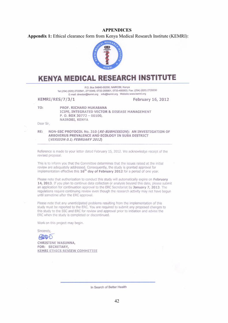
42
APPENDICES
Appendix 1: Ethical clearance form from Kenya Medical Research Institute (KEMRI):

43
Appendix2: Components of K Buffer
10mM Tris-HCl, pH 8.3
50mM KCl
1.5mM MgCl2
0.5% Tween 20
100µg/ml proteinase K (freshly dissolved enzyme)
-Proteinase K: at 20mg/ml in 10mM Tris-HCl, pH 7.5
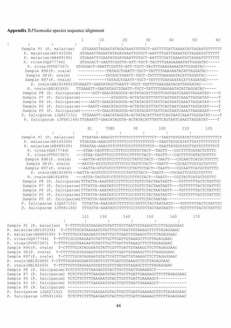
44
Appendix 3:Plasmodia species sequence alignment
1 10 20 30 40 50 60
| | | | | | |
Sample P1 (P. malariae) GTGAAATTAGAATATAGATAAATTGTGCT-AATTTTGATTAAAATATTAGAATGTTTTTT
P. malariae(AF145336) GTGAAATTAGAATATAGATAAATTGTGCT-AATTTTGATTAAAATATTAGAATGTTTTTT
P. malariae(AB489195) GTGAAATTCGAATATAGATAAATTGTGCT-AATTTCGATTAAAATATTAGAATGTTTTTT
P. vivax(GQ477744) GTGGGACT-GAATTCGGTTG-ATT-TGCT-TACTTTGAAGAAAATATTGGGATAC-----
P. vivax(PVU07367) GTGGGACT-GAATTCGGTTG-ATT-TGCT-TACTTTGAAGAAAATATTGGGATAC-----
Sample R96(P. ovale) ------------TATAGCTGAATT-TGCT-TATTTTGAAGAATACATTAGGATAC-----
Sample S8(P. ovale) ------------TATAGCTGAATT-TGCT-TATTTTGAAGAATACATTAGGATAC-----
Sample R97(P. ovale) ------------TATAGCTGAATT-TGCT-TATTTTGAAGAATACATTAGGATAC-----
P. ovale(AB182489)GTGAAATT-GAATATAGCTGAATT-TGCT-TATTTTGAAGAATACATTAGGATAC-----
P. ovale(AB182493) TTGAAATT-GAATATAGCTGAATT-TGCT-TATTTTGAAGAATATATTAGGATAC-----
Sample P8 (P. falciparum)-----ATT-GAACATAGGTA-ACTATACATTTATTCAGTAATCAAATTAGGATAT----T
Sample P7 (P. falciparum)-------------ATAGGTA-ACTATACATTTATTCAGTAATCAAATTAGGATAT----T
Sample P4 (P. falciparum)----AATT-GAACATAGGTA-ACTATACATTTATTCAGTAATCAAATTAGGATAT----T
Sample P2 (P. falciparum)---GAATT-GAACATAGGTA-ACTATACATTTATTCAGTAATCAAATTAGGATAT----T
Sample P5 (P. falciparum)-------T-GAACATAGGTA-ACTATACATTTATTCAGTAATCAAATTAAGAGAT----T
P. falciparum (JQ627152) TTGAAATT-GAACATAGGTA-ACTATACATTTATTCAGTAATCAAATTAGGATAT----T
P. falciparum (JF681166)TTGAAATT-GAACATAGGTA-ACTATACATTTATTCAGTAATCAAATTAGGATAT----T
61 7080 90 100 110 120
| | | | | | |
Sample P1 (P. malariae) TTAATAA-AAACGTTCTTTTCCCTTTTTTTCT--TAATTATGCATATTTATTCTTTTTCT
P. malariae(AF145336) TTGATAA-AAACGTTCTTTTCCCTTTTTTTCT--TAATTATGCATATTTATTTTTTTTCT
P. malariae(AB489195) TTAATAA-AAACGTTCTTTTCCCTTTTTTTCT--TAATTATGCATATTTATTCTTTTTCT
P. vivax(GQ477744) ---GTAA-CAGTTTCCCTTTCCCTTTTCTACT--TAGTT---CGCTTTTCATACTGTTTC
P. vivax(PVU07367) ---GTAA-CAGTTTCCCTTTCCCTTTTCTACT--TAGTT---CGCTTTTCATACTGTTTC
Sample R96(P. ovale) --AATTA-ATGTGTCCTTTTCCCTATTCTACT--TAATT---CGCAATTCATGCTGTTTC
Sample S8(P. ovale) --AATTA-ATGTGTCCTTTTCCCTATTCTACT--TAATT---CGCAATTCATGCTGTTTC
Sample R97(P. ovale) --AATTA-GTGTGTCCTTTTCCCTATTCTACT--TAATT---CGCAATTCATGCTGTTTC
P. ovale(AB182489)--AATTA-ATGTGTCCTTTTCCCTATTCTACT--TAATT---CGCAATTCATGCTGTTTC
P. ovale(AB182493) ---ATTA-TAGTGTCCTTTTCCCTTTTCTACT--TAATT---CGCTATTCATGCTGTTTC
Sample P8 (P. falciparum)TTTATTA-AAATATCCTTTTCCCTGTTCTACTAATAATT---TGTTTTTTACTCTATTTC
Sample P7 (P. falciparum)TTTATTA-AAATATCCTTTTCCCTGTTCTACTAATAATT---TGTTTTTTACTCTATTTC
Sample P4 (P. falciparum)TTTATTA-AAATATCCTTTTCCCTGTTCTACTAATAATT---TGTTTTTTACTCTATTTC
Sample P2 (P. falciparum)TTTATTA-AAATATCCTTTTCCCTGTTCTACTAATAATT---TGTTTTTTACTCTATTTC
Sample P5 (P. falciparum)TTTATTA-AAATATCCTTTTCCCTGTTCTACTAATAA-----------------------
P. falciparum (JQ627152) TTTATTA-AAATATCCTTTTCCCTGTTCTACTAATAATT---TGTTTTTTACTCTATTTC
P. falciparum (JF681166) TTTATTA-AAATATCCTTTTCCCTGTTCTACTAATAATT---TGTTTTTTACTCTATTTC
121 130 140 150 160 170
| | | | | |
Sample P1 (P. malariae) T-TTTTCGCATAAGAATGTATTTGCTTAATTGTAAAGCTCCTTAGAGGAAC
P. malariae(AF145336) T-CTTTTGCATAAGAATGTATTTGCTTAATTGTAAAGCTTCTTAGAGGAAC
P. malariae(AB489195) T-TTTTTGCATAAGAATGTATTTGCTTAATTGTAAAGCTTCTTAGAGGAAC
P. vivax(GQ477744) T-TTTTCGCGTAAGAATGTATTTGCTTGATTGTAAAGCTTCTTAGAGGAAC
P. vivax(PVU07367) T-TTTTCGCGTAAGAATGTATTTGCTTGATTGTAAAGCTTCTTAGAGGAAC
Sample R96(P. ovale) T-CTTTTGCATAGGAATGTATTCGTTTGATTGTAAAGCTTCTTAGAGGAAC
Sample S8(P. ovale) T-CTTTTGCATAGGAATGTATTCGTTTGATTGTAAAGCTTCTTAGAGGAAC
Sample R97(P. ovale) T-CTTTTGCATAGGAATGTATTCGTTTGATTGTAAAGCTTCTTAGAGGAAC
P. ovale(AB182489) T-CTTTTGCATAGGAATGTATTCGTTTGATTGTAAAGCTTCTTAGAGGAAC
P. ovale(AB182493) T-TTTTTGTGTAGGAATGTATTCGTTTGATTGTAAAGCTTCTTAGAGGAAC
Sample P8 (P. falciparum)TCTCTTCTTTTAAGAATGTACTTGCTTGATTGAAAAGCT------------
Sample P7 (P. falciparum) TCTCTTCTTTTAAGAATGTACTTGCTTGATTGAAAAGCTTCTTAGAGGAAC
Sample P4 (P. falciparum) TCTCTTCTTTTAAGAATGTACTTGCTTGATTGAAAAGCT------------
Sample P2 (P. falciparum) TCTCTTCTTTTAAGAATGTACTTGCTTGATTGAAAAGCT------------
Sample P5 (P. falciparum) ---------------------------------------------------
P. falciparum (JQ627152) TCTCTTCTTTTAAGAATGTACTTGCTTGATTGAAAAGCTTCTTAGAGGAAC
P. falciparum (JF681166) TCTCTTCTTTTAAGAATGTACTTGCTTGATTGAAAAGCTTCTTAGAGGAAC
Related Documents