Prevaccination genomic diversity of human papillomavirus genotype 6 (HPV 6) Boštjan J. Kocjan a , Mario Poljak a , Mojca Cimerman a , Nina Gale b , Marko Potočnik c , Željka Bogovac a , Katja Seme a, ⁎ a Institute of Microbiology and Immunology, Faculty of Medicine, University of Ljubljana, Zaloška 4, 1105 Ljubljana, Slovenia b Institute of Pathology, Faculty of Medicine, University of Ljubljana, Ljubljana, Slovenia c Department of Dermatovenereology, University Medical Centre Ljubljana, Ljubljana, Slovenia abstract article info Article history: Received 20 April 2009 Returned to author for revision 2 June 2009 Accepted 17 June 2009 Available online 12 July 2009 Keywords: Human papillomaviruses HPV HPV 6 Genomic diversity Genomic variants Genital warts Laryngeal papillomas Vaccine Slovenia Prevaccination genomic diversity of human papillomavirus genotype 6 (HPV 6) was established by sequencing 3798 bp of 77 clinically important HPV 6 isolates obtained from 45 and 32 patients with genital warts and laryngeal papillomas, respectively. By analyzing pooled L1, LCR, E6, E2, and E5 nucleotide data of an individual isolate, a total of 36 different genomic variants were identified, of which six (12 isolates), one (one isolate) and 29 (64 isolates) corresponded to HPV 6b, HPV 6a, and HPV 6vc genetic lineages, respectively. Several novel, potentially important mutations were identified. Non-prototypic HPV 6vc genomic variants were found in the majority of genital warts and laryngeal papillomas included in the study. The presence of serious HPV 6 genome sequence errors was confirmed and novel sequence errors were identified in sequence repositories. © 2009 Elsevier Inc. All rights reserved. Introduction Human papillomavirus genotype 6 (HPV 6) is one of the most important HPV genotypes. Next to HPV 11, it is the major causative agent of genital warts and laryngeal papillomas, the most frequent benign tumours in the anogenital region and lower respiratory tract, respectively (Gissmann et al., 1983; Gale et al., 1994; Brown et al., 1999; Gale, 2005; Gale and Zidar, 2006; Potočnik et al., 2007). According to the latest papillomavirus classification criteria, HPV 6 is placed in the alpha genus — species 10, together with HPV 11, HPV 13, HPV 44, and HPV 74 (de Villiers et al., 2004). Based on its clinical association mainly with benign lesions, HPV 6 is regarded as a “low- risk” HPV genotype (reviewed in Grassmann et al. (1996)). HPV 6 is one of the four primary targets of the recently introduced quad- rivalent HPV vaccine (Barr and Tamms, 2007; Garland et al., 2007). HPV 6 was discovered by Southern blot hybridization in a tissue specimen of condyloma acuminata (Gissmann and zur Hausen, 1980). The complete genome, later designated HPV 6b, was cloned in 1981 (de Villiers et al., 1981) and fully sequenced and completely characterized two years later (Schwarz et al., 1983). Molecular studies performed in the years following the discovery of this virus demonstrated that HPV 6 is polymorphic and consists of several genomic variants. The focus of these studies has been on certain portions of the HPV 6 genome, particularly the long-control region (LCR), which contains regulatory elements for viral transcription and replication, and the coding regions for L1, L2, E6, E7, and E2 proteins (Rando et al., 1986; Kasher and Roman, 1988; Farr et al., 1991; Icenogle et al., 1991; Yaegashi et al., 1993; Kitasato et al., 1994; Heinzel et al., 1995; Roman and Brown,1995; Grassmann et al., 1996; Suzuki et al., 1997; Caparros-Wanderley et al., 1999; Kovelman et al., 1999). In addition to HPV 6b, complete genomes of two closely related HPV 6 isolates, designated HPV 6a and HPV 6vc, have been cloned and fully characterized (Hofmann et al., 1995; Kovelman et al., 1999). On the basis of nucleotide sequence comparisons, HPV 6 isolates are usually grouped into prototype HPV 6b-related (prototypic) and HPV 6a/6vc- related (non-prototypic) genomic variants (Heinzel et al., 1995; Grassmann et al., 1996; Caparros-Wanderley et al., 1999; Kovelman et al., 1999). Non-prototypic HPV 6 genomic variants seem to predominate in genital warts (Gissmann et al., 1983; Rubben et al., 1992; Brown et al., 1993; Krige et al., 1997; Suzuki et al., 1997; Caparros-Wanderley et al., 1999). Knowledge about the natural genetic diversity of HPV 6 is fairly limited, since the majority of HPV 6 genomic diversity studies performed so far have only investigated a limited number of isolates, which were almost exclusively from genital warts, and studies have focused on a single HPV 6 genomic region or even its short part only. Thus, in the present study we further investigated genomic diversity Virology 391 (2009) 274–283 ⁎ Corresponding author. Fax: +386 1543 7418. E-mail address: [email protected] (K. Seme). 0042-6822/$ – see front matter © 2009 Elsevier Inc. All rights reserved. doi:10.1016/j.virol.2009.06.030 Contents lists available at ScienceDirect Virology journal homepage: www.elsevier.com/locate/yviro

Welcome message from author
This document is posted to help you gain knowledge. Please leave a comment to let me know what you think about it! Share it to your friends and learn new things together.
Transcript

Virology 391 (2009) 274–283
Contents lists available at ScienceDirect
Virology
j ourna l homepage: www.e lsev ie r.com/ locate /yv i ro
Prevaccination genomic diversity of human papillomavirus genotype 6 (HPV 6)
Boštjan J. Kocjan a, Mario Poljak a, Mojca Cimerman a, Nina Gale b, Marko Potočnik c,Željka Bogovac a, Katja Seme a,⁎a Institute of Microbiology and Immunology, Faculty of Medicine, University of Ljubljana, Zaloška 4, 1105 Ljubljana, Sloveniab Institute of Pathology, Faculty of Medicine, University of Ljubljana, Ljubljana, Sloveniac Department of Dermatovenereology, University Medical Centre Ljubljana, Ljubljana, Slovenia
⁎ Corresponding author. Fax: +386 1 543 7418.E-mail address: [email protected] (K. Seme).
0042-6822/$ – see front matter © 2009 Elsevier Inc. Adoi:10.1016/j.virol.2009.06.030
a b s t r a c t
a r t i c l e i n f oArticle history:Received 20 April 2009Returned to author for revision 2 June 2009Accepted 17 June 2009Available online 12 July 2009
Keywords:Human papillomavirusesHPVHPV 6Genomic diversityGenomic variantsGenital wartsLaryngeal papillomasVaccineSlovenia
Prevaccination genomic diversity of human papillomavirus genotype 6 (HPV 6) was established bysequencing 3798 bp of 77 clinically important HPV 6 isolates obtained from 45 and 32 patients with genitalwarts and laryngeal papillomas, respectively. By analyzing pooled L1, LCR, E6, E2, and E5 nucleotide data of anindividual isolate, a total of 36 different genomic variants were identified, of which six (12 isolates), one (oneisolate) and 29 (64 isolates) corresponded to HPV 6b, HPV 6a, and HPV 6vc genetic lineages, respectively.Several novel, potentially important mutations were identified. Non-prototypic HPV 6vc genomic variantswere found in the majority of genital warts and laryngeal papillomas included in the study. The presence ofserious HPV 6 genome sequence errors was confirmed and novel sequence errors were identified in sequencerepositories.
© 2009 Elsevier Inc. All rights reserved.
Introduction
Human papillomavirus genotype 6 (HPV 6) is one of the mostimportant HPV genotypes. Next to HPV 11, it is the major causativeagent of genital warts and laryngeal papillomas, the most frequentbenign tumours in the anogenital region and lower respiratory tract,respectively (Gissmann et al., 1983; Gale et al., 1994; Brown et al.,1999; Gale, 2005; Gale and Zidar, 2006; Potočnik et al., 2007).According to the latest papillomavirus classification criteria, HPV 6 isplaced in the alpha genus— species 10, together with HPV 11, HPV 13,HPV 44, and HPV 74 (de Villiers et al., 2004). Based on its clinicalassociation mainly with benign lesions, HPV 6 is regarded as a “low-risk” HPV genotype (reviewed in Grassmann et al. (1996)). HPV 6 isone of the four primary targets of the recently introduced quad-rivalent HPV vaccine (Barr and Tamms, 2007; Garland et al., 2007).
HPV 6 was discovered by Southern blot hybridization in a tissuespecimen of condyloma acuminata (Gissmann and zur Hausen, 1980).The complete genome, later designated HPV 6b, was cloned in 1981(de Villiers et al., 1981) and fully sequenced and completelycharacterized two years later (Schwarz et al., 1983). Molecular studiesperformed in the years following the discovery of this virusdemonstrated that HPV 6 is polymorphic and consists of several
ll rights reserved.
genomic variants. The focus of these studies has been on certainportions of the HPV 6 genome, particularly the long-control region(LCR), which contains regulatory elements for viral transcription andreplication, and the coding regions for L1, L2, E6, E7, and E2 proteins(Rando et al., 1986; Kasher and Roman,1988; Farr et al., 1991; Icenogleet al., 1991; Yaegashi et al., 1993; Kitasato et al., 1994; Heinzel et al.,1995; Roman and Brown, 1995; Grassmann et al., 1996; Suzuki et al.,1997; Caparros-Wanderley et al., 1999; Kovelman et al., 1999). Inaddition to HPV 6b, complete genomes of two closely related HPV 6isolates, designated HPV 6a and HPV 6vc, have been cloned and fullycharacterized (Hofmann et al., 1995; Kovelman et al., 1999). On thebasis of nucleotide sequence comparisons, HPV 6 isolates are usuallygrouped into prototype HPV 6b-related (prototypic) and HPV 6a/6vc-related (non-prototypic) genomic variants (Heinzel et al., 1995;Grassmann et al., 1996; Caparros-Wanderley et al., 1999; Kovelmanet al., 1999). Non-prototypic HPV 6 genomic variants seem topredominate in genital warts (Gissmann et al., 1983; Rubben et al.,1992; Brown et al., 1993; Krige et al., 1997; Suzuki et al., 1997;Caparros-Wanderley et al., 1999).
Knowledge about the natural genetic diversity of HPV 6 is fairlylimited, since the majority of HPV 6 genomic diversity studiesperformed so far have only investigated a limited number of isolates,which were almost exclusively from genital warts, and studies havefocused on a single HPV 6 genomic region or even its short part only.Thus, in the present study we further investigated genomic diversity

275B.J. Kocjan et al. / Virology 391 (2009) 274–283
of HPV 6 in the prevaccination era, in order to provide important datafor future epidemiological, functional and molecular assay develop-ment and vaccination studies. To achieve this, approximately half ofthe genome (3798 bp) of 77 clinically important HPV 6 isolates (e.g.,those causing disease) was sequenced. The nucleotide sequencealignments were used to identify HPV 6 genomic variants and toinvestigate the linkage of these variants across the L1, LCR, E6, E2, E5aand E5b genomic regions. In addition, the coding regions wereexamined in detail to determine the prevalence, extent and distribu-tion of amino acid changes that might have important biologicalproperties. The LCR genomic region was used to determine therelationship between genomic variants described in our study andthose described previously (Heinzel et al., 1995). This study, carriedout on the largest number of HPV 6 isolates to date, is believed to bethe first extensivework on the genetic diversity of the HPV 6 genotype.
Results
All 77 HPV 6 isolates included in the study were successfullyamplified and sequenced across the L1 (nt 5700–7495), LCR-E6(nt 7233–650) and E2–E5 (nt 3142–4538) genomic regions. Genomicsequences were established for 541 bp of E2 ORF (nt 3289–3829), forthe entire LCR genomic region and for the entire L1, E6, E5a, and E5bORFs. HPV 6 L1, E6, E2 and E5 genomic variants were identified usingthe prototype HPV 6b genome (GenBank accession no. X00203) as astandard for comparison and nucleotide position numbering. For thedetermination of HPV 6 LCR genomic variants, an LCR sequenceamended by inclusion of a 94 bp segment at position 7350 in theprototype HPV 6b genome (Heinzel et al., 1995) was used.
HPV 6 L1 genomic variants
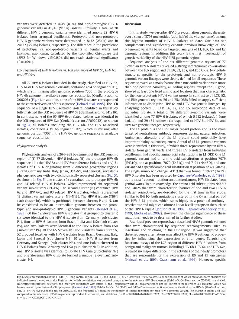
A total of fifteen L1 genomic variants were identified among 77HPV 6 isolates (Fig. 1). Prototypic and non-prototypic HPV 6 genomicvariants were determined in 12/77 (15.6%) and in 65/77 (84.4%)isolates, respectively. The prototype L1 sequencewas identified in only6/77 (7.8%) isolates. Sequence analysis of the entire L1 gene revealed25 nucleotide exchanges between the variants and the prototype HPV6 sequence. The maximum distance between the variants and theprototype sequence was 11 nucleotides (0.7% of the entire L1 ORF).Nucleotide substitutions altered the L1 amino acid sequence of oneprototypic (6-L1-3, represented by two isolates) and two non-prototypic (6-L1-13 and 6-L1-14, represented by ten and one isolate,respectively) HPV 6 variants; up to two amino acids (0.4% of the L1protein) were exchanged (Fig. 1).
HPV 6 LCR genomic variants
A total of fifteen LCR genomic variants were identified among 77HPV 6 isolates (Fig. 1). Prototypic and non-prototypic HPV 6 genomicvariants were determined in 12/77 (15.6%) and in 65/77 (84.4%)isolates, respectively. None of the isolates corresponded to theprototype LCR sequence. Mutations were observed in 39 genomicpositions: 30 single nucleotide exchanges, one 6 bp and one 1 bpdeletion, and two 20 bp, one 14 bp and one 1 bp insertions. Themaximum genomic distance between the variants and the prototypesequence was 20 mutations, thereby affecting 2.5% (20/806) of theentire LCR genomic region.
HPV 6 E6 genomic variants
A total of ten E6 genomic variants were identified among 77 HPV 6isolates (Fig. 1). Prototypic and non-prototypic HPV 6 genomicvariants were determined in 12/77 (15.6%) and in 65/77 (84.4%)isolates, respectively. None of the isolates corresponded to thereference E6 sequence. Molecular analysis of the entire E6 ORF
revealed 13 point mutations observed in 12 genomic positions. Themaximum distance between the variants and the prototype sequencewas 7 nucleotides (1.5% of the entire E6 gene). Nucleotide substitu-tions altered the E6 amino acid sequence of all non-prototypic HPV 6variants (65 isolates); up to two amino acids (1.3% of the E6 protein)were exchanged (Fig. 1).
HPV 6 E2 genomic variants
A total of nine E2 genomic variantswere identified among77HPV-6isolates (Fig. 2). Prototypic andnon-prototypicHPV6 genomic variantswere determined in 12/77 (15.6%) and in 65/77 (84.4%) isolates,respectively. The reference E2 sequence was identified in 9/77 (11.8%)isolates. Sequence analysis of the 541bp segmentof E2ORF revealed 23nucleotide exchanges between the variants and the prototype E2sequence. The maximum variant divergence from the prototypesequence was 16 nucleotides, thereby affecting 2.9% of the evaluatedE2 nucleotide sequence. Point mutations altered the E2 amino acidsequence of one prototypic (6-E2-2, represented by one isolate) and allnon-prototypic (65 isolates)HPV6 variants. As shown in Fig. 2, up to 10amino acids (5.6% of the analyzed part of the E2 protein) wereexchanged.
HPV 6 E5a and E5b genomic variants
A total of sixteen E5a genomic variants were identified among 77HPV 6 isolates (Fig. 2). Prototypic and non-prototypic HPV 6 genomicvariants were determined in 12/77 (15.6%) and in 65/77 (84.4%)isolates, respectively. The reference E5a sequence was identified in12/77 (15.6%) isolates. There were 22 point mutations observed in 19genomic positions. The maximum genomic distance between thevariants and the prototype sequencewas 14 nucleotides, amounting toa diversity of 5.1% of the entire E5a ORF. Nucleotide substitutionsaltered the E5a amino acid sequence of all non-prototypic HPV 6variants (65 isolates); up to seven amino acids (7.7% of the E5aprotein) were exchanged (Fig. 2).
A total of nine E5b genomic variants were identified among 77 HPV6 isolates (Fig. 2). Prototypic and non-prototypic HPV 6 genomicvariants were determined in 12/77 (15.6%) and in 65/77 (84.4%)isolates, respectively. The prototype E5b sequence was identified in10/77 (13.0%) isolates. Sixteen point mutations were observed in 15positions. The maximum distance between the variants and theprototype sequence was 11 nucleotides, thereby affecting 5.0% of theentire E5b ORF. Point mutations altered the E5b amino acid sequenceof all non-prototypic HPV 6 variants (65 isolates)— up to seven aminoacids (9.7% of the E5b protein) were exchanged (Fig. 2).
HPV 6 genomic variants
The nucleotide data of L1, LCR, E6, E2, and E5 genomic regions werecombined in individual isolates and HPV 6 genomic variants weredetermined. Thus, among 77 HPV 6 analyzed isolates, 36 differentgenomic variants were identified (Fig. 3). As shown in Fig. 3, strongintergenomic co-variation between LCR, late (L1), and early (E6, E2,E5a, and E5b) viral genes was observed in all analyzed isolates.Twelve (15.6%) HPV 6 isolates represented genomic variants of theprototype HPV 6 isolate (six genomic variants) while 65 (84.4%)isolates represented closely related genomic variants of HPV 6a (onegenomic variant, represented by one isolate) and HPV 6vc (29genomic variants) (Fig. 3).
Distribution of HPV 6 genomic variants in genital warts vs. laryngealpapillomas
A total of 22 different HPV 6 genomic variants were identifiedamong 45HPV 6 isolates from genital warts. Prototypic HPV 6 genomic

276 B.J. Kocjan et al. / Virology 391 (2009) 274–283
277B.J. Kocjan et al. / Virology 391 (2009) 274–283
variants were detected in 4/45 (8.9%) and non-prototypic HPV 6genomic variants in 41/45 (91.1%) isolates. Similarly, a total of 20different HPV 6 genomic variants were identified among 32 HPV 6isolates from laryngeal papillomas. Prototypic and non-prototypicHPV 6 genomic variants were determined in 8/32 (25.0%) and in24/32 (75.0%) isolates, respectively. The difference in the prevalenceof prototypic vs. non-prototypic variants in genital warts andlaryngeal papillomas, calculated by the two-tailed Chi-square test(SPSS for Windows v15.0.0.0), did not reach statistical significance(P=.1091).
LCR sequences of HPV 6 isolates vs. LCR sequences of HPV 6b, HPV 6a,and HPV 6vc
All 77 HPV 6 isolates included in the study, classified as HPV 6b,HPV 6a or HPV 6vc genomic variants, contained a 94 bp segment (D1),which is still missing after genomic position 7350 in the prototypeHPV 6b genome in available sequence repositories (GenBank acc. no.X00203) (Fig. 4). In addition, none of the HPV 6 isolates correspondedto the corrected version of this sequence (Heinzel et al., 1995). The LCRsequence of a single HPV 6a-related isolate identified in this studyfully matched the LCR sequence of HPV 6a (GenBank acc. no. L41216).In contrast, none of the 65 HPV 6vc-related isolates was identical tothe LCR sequence of HPV 6vc (GenBank acc. no. AF092932). As shownin Fig. 4, all isolates, including the HPV 6b- and HPV 6a-relatedisolates, contained a 19 bp segment (D2), which is missing aftergenomic position 7367 in the HPV 6vc genome sequence in availablesequence repositories.
Phylogenetic analysis
Phylogenetic analysis of a 264–268 bp segment of the LCR genomicregion of (i) 77 Slovenian HPV 6 isolates, (ii) the prototype HPV 6bsequence, (iii) the HPV 6a and HPV 6vc reference isolates and (iv) 31isolates of HPV 6 originating from 7 different geographic regions(Brazil, Germany, India, Italy, Japan, USA-NY, and Senegal), revealed aphylogenetic tree with two dichotomically separated clusters (Fig. 5).As shown in Fig. 5, one cluster (P) contained the prototype HPV 6band 24 related HPV 6 isolates, which represented six separatedvariant sub-clusters (P1–P6). The second cluster (N) contained HPV6a and HPV 6vc, and 83 related HPV 6 isolates, which represented13 distinct variant sub-clusters (N1–N13). The HPV 6 isolate SN6-1(sub-cluster In), which is positioned between clusters P and N, canbe considered to be an intermediate genome between the proto-typic and non-prototypic HPV 6 genomic variants (Heinzel et al.,1995). Of the 12 Slovenian HPV 6 isolates that grouped to cluster P,six were identical to the HPV 6 isolate from Germany (sub-clusterP1), four to HPV 6 isolates from Brazil, India and USA (sub-clusterP5), and two isolates were identical to the HPV 6 isolate from USA(sub-cluster P4). Of the 65 Slovenian HPV 6 isolates from cluster N,52 grouped together with HPV 6 isolates from Brazil, Germany, Italy,Japan and Senegal (sub-cluster N1), 10 with HPV 6 isolates fromGermany and Senegal (sub-cluster N6), and one isolate clustered toHPV 6 isolates from Germany and USA (sub-cluster N12). In addition,one HPV 6 isolate was identical to isolate HPV 6ma (sub-cluster N7)and one Slovenian HPV 6 isolate formed a unique (Slovenian) sub-cluster N4.
Fig. 1. Sequence variations of the L1 ORF (A), long-control region-LCR (B), and E6 ORF (C) ofindicated across the top vertically. Positions for which no variation was detected comparedNucleotide substitutions, deletions, and insertions are marked with letters, Δ, and I, respectibeen amended by inclusion of a 94 bp segment (Heinzel et al., 1995). Ref-6a, Ref-6vc, 6-LCR-4L41216) or HPV 6vc (GenBank acc. no. AF092932). The frequency (ƒ) indicates the numbercompared to the reference HPV 6b sequences is provided. Insertions (I) and deletions (D): II4=T; D1=ATGTGTGTTGTATATATGT.
Discussion
In this study, we describe HPV 6 prevaccination genomic diversityover a span of 3798 nucleotides (app. half of the viral genome), amongthe highest number of HPV 6 isolates to date. Our data setcomplements and significantly expands previous knowledge of HPV6 genomic variants based on targeted analysis of L1, LCR, E6, and E2genomic regions. In addition, this work is the first investigation ofgenetic variability of the HPV 6 E5 genomic region.
Sequence analysis of the six different genomic regions of 77Slovenian HPV 6 isolates revealed a strong intergenomic co-variationbetween the LCR region and L1, E6, E2, E5a, and E5b ORFs. Nucleotidesignatures specific for the prototypic and non-prototypic HPV 6genomic variant lineages were clearly defined for all sequences. Theseregions showed, as a main feature, fixed nucleotide variations in morethan one position. Similarly, all coding regions, except the L1 gene,showed at least one fixed amino acid location that was characteristicfor the non-prototypic HPV 6 variant group. In contrast to L1, LCR, E2,and E5b genomic regions, E6 and E5a ORFs failed to supply sufficientinformation to distinguish HPV 6a and HPV 6vc genetic lineages. Byanalyzing pooled L1, LCR, E6, E2, and E5 nucleotide data of anindividual isolate, a total of 36 different genomic variants wereidentified among 77 HPV 6 isolates, of which 6 (12 isolates), 1 (oneisolate), and 29 (64 isolates) corresponded to HPV 6b, HPV 6a, andHPV 6vc genetic lineages, respectively.
The L1 protein is the HPV major capsid protein and is the maintarget of neutralizing antibody responses during natural infection.Amino acid alterations of the L1 protein could potentially haveimportant biological consequences. A total of 15 L1 genomic variantswere identified in this study, of which three, represented by ten HPV 6isolates from genital warts and three HPV 6 isolates from laryngealpapillomas, had specific amino acid substitutions in L1 ORF. One L1genomic variant had an amino acid substitution at position 7079(E431Q), one at positions 7079 (E431Q) and 7121 (N445D), and onevariant had a specific amino acid substitution at position 7232 (P482S).The single amino acid change E431Q that was found in 10/77 (14.3%)HPV 6 isolates has been reported by Caparros-Wanderley et al. (1999)as themost frequentmutation encountered amongHPV6 isolates fromgenital warts. To our knowledge, the amino acid substitutions N445Dand P482S that were characteristic features of one and two HPV 6isolates, respectively, are described for the first time in this study.Similar to E431Q, both mutations clustered in the C-terminal part ofthe HPV 6 L1 protein, which ranks highly as a potential antibody-reactive site andmight constitute a linear B-cell epitope on the surfaceof the HPV 6 capsid (Jenison et al., 1989; Caparros-Wanderley et al.,1999; Modis et al., 2002). However, the clinical significance of thesemutations needs to be determined in further studies.
A series of previous reports have described HPV 6 genomic variantsthat were characterized by sequence rearrangements, such asinsertions and deletions, in the LCR region. It was suggested thatthese sequence alternations may affect the HPV 6 pathogenic proper-ties by influencing the expression of viral genes. Surprisingly,functional assays of the LCR region of different HPV 6 isolates frombenign andmalignant tumors, including HPV 6b, HPV 6a, and HPV 6vc,revealed no major difference in the activities of their early promotersthat are responsible for the expression of E6 and E7 oncogenes(Heinzel et al., 1995; Grassmann et al., 1996). However, specific
77 Slovenian HPV 6 isolates. Genomic positions at which mutations were observed areto the reference HPV 6b sequences (Ref-6b-O; GenBank acc. no. X00203) are shaded.
vely. The LCR sequence coded Ref-6b-H refers to the reference LCR sequence, which has⁎, and 6-E6-4⁎ indicate nucleotide sequences identical to the HPV 6a (GenBank acc. no.of isolates identified for each HPV 6 genomic variant. The change in amino acid (aa)
1=TATGTGTATATGTGTGTATA; I2=TACATTATTGTATA; I3=ATATGTTTATTGCCACTGCA;

278 B.J. Kocjan et al. / Virology 391 (2009) 274–283
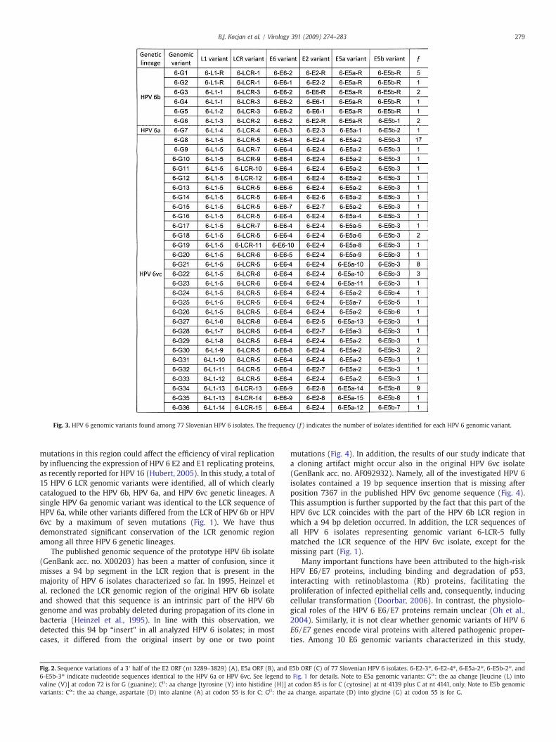
Fig. 3. HPV 6 genomic variants found among 77 Slovenian HPV 6 isolates. The frequency (ƒ) indicates the number of isolates identified for each HPV 6 genomic variant.
279B.J. Kocjan et al. / Virology 391 (2009) 274–283
mutations in this region could affect the efficiency of viral replicationby influencing the expression of HPV 6 E2 and E1 replicating proteins,as recently reported for HPV 16 (Hubert, 2005). In this study, a total of15 HPV 6 LCR genomic variants were identified, all of which clearlycatalogued to the HPV 6b, HPV 6a, and HPV 6vc genetic lineages. Asingle HPV 6a genomic variant was identical to the LCR sequence ofHPV 6a, while other variants differed from the LCR of HPV 6b or HPV6vc by a maximum of seven mutations (Fig. 1). We have thusdemonstrated significant conservation of the LCR genomic regionamong all three HPV 6 genetic lineages.
The published genomic sequence of the prototype HPV 6b isolate(GenBank acc. no. X00203) has been a matter of confusion, since itmisses a 94 bp segment in the LCR region that is present in themajority of HPV 6 isolates characterized so far. In 1995, Heinzel etal. recloned the LCR genomic region of the original HPV 6b isolateand showed that this sequence is an intrinsic part of the HPV 6bgenome and was probably deleted during propagation of its clone inbacteria (Heinzel et al., 1995). In line with this observation, wedetected this 94 bp “insert” in all analyzed HPV 6 isolates; in mostcases, it differed from the original insert by one or two point
Fig. 2. Sequence variations of a 3′ half of the E2 ORF (nt 3289–3829) (A), E5a ORF (B), and6-E5b-3⁎ indicate nucleotide sequences identical to the HPV 6a or HPV 6vc. See legend tovaline (V)] at codon 72 is for G (guanine); Cβ: aa change [tyrosine (Y) into histidine (H)] avariants: Cα: the aa change, aspartate (D) into alanine (A) at codon 55 is for C; Gβ: the a
mutations (Fig. 4). In addition, the results of our study indicate thata cloning artifact might occur also in the original HPV 6vc isolate(GenBank acc. no. AF092932). Namely, all of the investigated HPV 6isolates contained a 19 bp sequence insertion that is missing afterposition 7367 in the published HPV 6vc genome sequence (Fig. 4).This assumption is further supported by the fact that this part of theHPV 6vc LCR coincides with the part of the HPV 6b LCR region inwhich a 94 bp deletion occurred. In addition, the LCR sequences ofall HPV 6 isolates representing genomic variant 6-LCR-5 fullymatched the LCR sequence of the HPV 6vc isolate, except for themissing part (Fig. 1).
Many important functions have been attributed to the high-riskHPV E6/E7 proteins, including binding and degradation of p53,interacting with retinoblastoma (Rb) proteins, facilitating theproliferation of infected epithelial cells and, consequently, inducingcellular transformation (Doorbar, 2006). In contrast, the physiolo-gical roles of the HPV 6 E6/E7 proteins remain unclear (Oh et al.,2004). Similarly, it is not clear whether genomic variants of HPV 6E6/E7 genes encode viral proteins with altered pathogenic proper-ties. Among 10 E6 genomic variants characterized in this study,
E5b ORF (C) of 77 Slovenian HPV 6 isolates. 6-E2-3⁎, 6-E2-4⁎, 6-E5a-2⁎, 6-E5b-2⁎, andFig. 1 for details. Note to E5a genomic variants: Gα: the aa change [leucine (L) intot codon 85 is for C (cytosine) at nt 4139 plus C at nt 4141, only. Note to E5b genomica change, aspartate (D) into glycine (G) at codon 55 is for G.

Fig. 4.Multiple alignment of a 5′ part of the LCR genomic region (nt 7340–7448) of the prototype HPV 6b (GenBank acc. no. X00203), HPV 6a (GenBank acc. no. L41216), and HPV6vc (GenBank acc. no. AF092932) and HPV 6 isolates characterized in this study; for simplicity, only 18 representative HPV 6 isolates (LP22-LP129) are shown. The sequence codedHPV-6b-H refers to the original HPV 6b LCR sequence, which has been amended by inclusion of a 94 bp segment (Heinzel et al., 1995). D1: a segment of 94 bp(ATGTACTGTTATATGTATGTGTGTTGTATATATGTGTGTATATATGTGTCTGTGTGTATATGTATATGTATGTGTTGTGTATATATATGTGTGT); D2: a segment of 19 bp (ATGTGTGTTGTATATATGT).
280 B.J. Kocjan et al. / Virology 391 (2009) 274–283
eight (all non-prototypic genomic variants), harbored specificamino acid substitutions in the HPV 6 E6 gene; seven variants hada substitution at position 251 (H50Q) and one variant had a specificsubstitution at positions 156 (T19S) and 251 (H50Q). Since theamino acid substitution H50Q was also found in the majority of HPV6 isolates previously characterized (Grassmann et al., 1996), wepresume that it prevails in naturally occurring HPV 6 populations.However, this amino acid exchange clustered outside currentfunctionally characterized domains of the HPV E6 protein (reviewedin Grassmann et al., 1996); its meaning is therefore at presentunclear.
HPV E2 protein is a critical regulator of viral transcription andreplication. In a study by Kovelman et al. (1999) it has been shownthat E2 ORF is divergent between the three fully characterized HPV6 genomes: HPV 6b, HPV 6a and HPV 6vc. In addition, this studyrevealed several amino acid substitutions in the E2 protein thatwere characteristic of HPV 6a and HPV 6vc genetic lineages. Usingthe HPV 6b and HPV 6vc E2 proteins, very similar activities intranscriptional activation and repression assays in primary humankeratinocytes were observed, suggesting that natural variants of theHPV 6 E2 protein do not affect its regulatory function (Kovelman etal., 1999). In this study, we analyzed the genetic variability of a 3′part of the E2 gene that encodes for the E2 hinge and DNA-binding/dimerisation domains. Among nine E2 genomic variants identified,six (represented by 65 HPV 6 isolates) had amino acid sequencesidentical to the E2 protein of HPV 6b, HPV 6a, or HPV 6vc isolates.Additional novel amino acid substitutions were identified in oneprototypic (K351R, nt 3774) and in two HPV 6vc-related genomicvariants (G262A, nt 3507; H264Q, nt 3514 and Q273P, nt 3540). Itwould, however, be of great interest to investigate further whetheridentified natural variants of the HPV 6 E2 protein affect its key rolein controlling HPV 6 replication in conjunction with the E1 proteinand its variants.
The biological functions of the HPV E5 protein and its role in theHPV replication cycle are not well understood. Documentedfunctions include transformation and stimulation of the prolifera-tion of HPV-infected cells and induction of koilocytosis in conjunc-tion with the HPV E6 proteins (Doorbar, 2006; Krawczyk et al.,2008). In contrast to other alpha-PVs, which have one E5 ORF, HPV 6possesses two E5 genes: E5a and E5b (Schwarz et al., 1983; Hofmannet al., 1995; Kovelman et al., 1999). It has been shown previously thatboth genes are actively transcribed in laryngeal papillomas (Wardand Mounts, 1989), indicating that they may take part in productiveHPV 6 infection. We determined a total of 16 E5a genomic variants inthis study, of which eight (represented by 60 isolates) had an aminoacid sequence identical to the E5a protein of HPV 6b or HPV 6vc
isolates. Novel amino acid substitutions detected at positions 3892(E2D), 3912 (A9G), 3912 (A9G) and 4158 (Q91P), 4100 (L72V), and4139/4141 (Y85H) in the E5a ORF constituted additional character-istic features of an additional six HPV 6vc-related genomic variants.Similarly, in the remaining two HPV 6vc genomic variants, a novelHPV 6 E5a protein variant was identified; this protein differed fromthe HPV 6vc E5a protein by a single amino acid substitution in codon88 (Fig. 2, variants 6-E5a-11 and 6-E5a-12). In contrast to E5a, onlynine E5b genomic variants were identified, five of which, repre-sented by 65 isolates, had amino acid sequences identical to the E5bprotein of HPV 6b, HPV 6a, or HPV 6vc isolates (Fig. 2). Novel aminoacid exchanges identified at positions 4267 (V37I), 4304 (N49T),4322 (D55A) and 4342 (Y62H), and 4322 (D55G) in the E5b ORFwere additional characteristic features of the remaining four HPV6vc-related genomic variants. Whether the reported amino acidvariations in the HPV 6 E5a and E5b ORFs affect the biologicalfunctions of E5 proteins remains unknown.
The results of this study for the first time allowed phylogeneticcomparison of HPV 6 genomic variants found worldwide (Heinzelet al., 1995) with a relatively large number of HPV 6 isolates fromone geographically and ethnically closed cohort of patients (i.e.,Slovenian patients). This LCR based phylogenetic comparisonshowed that 76/77 (98.7%) Slovenian HPV 6 isolates were identicalto several phylogenetically diverse HPV 6 genomes identifiedpreviously in other parts of the world. Thus, in contrast to HPV16 and HPV 18, the existence of specific and geographicallyrestricted HPV 6 genomic variants seems highly unlikely.
In summary, the first extensive work on the genomic diversity ofthe HPV 6 genotype showed a substantial prevaccination genomicdiversity of this HPV genotype. A total of 36 different HPV 6 genomicvariants were identified in this study. Non-prototypic HPV 6genomic variants belonging to the HPV 6vc genetic lineage werefound in the majority of genital warts and laryngeal papillomasincluded in the study. The presence of serious sequence errors wasconfirmed and novel sequence errors were identified in sequencerepositories. We believe that our study will be helpful in futureepidemiological, functional, molecular assay development andvaccination studies.
Materials and methods
Clinical samples
A total of 77 HPV 6 isolates were included in the study. Forty-fiveisolates were obtained from the same number of male patientssuffering from exophytic genital warts on the glans of the penis, the

Fig. 5. Neighbor-joining algorithm based phylogenetic tree of HPV 6 LCR genomic variants from Slovenia (LP5-CAC116X), Brazil (AM6-1, B6-1, B6-4b, B6-5, B6-15, and B6-17),Germany (G6-2b, G6-6, G6-8, G6-20, G6-42, and G6-78), India (IN6-2, IN6-3, and IN6-6), Italy (I6-1), Japan (J6-4, J6-6, J6-8, and J6-11), USA-NY (NY6-1, NY6-11, NY6-13, NY6-16, andNY6-19), and Senegal (SN6-1, SN6-3, SN6-4b, SN6-6b, and SN6-11). Bootstrap values (%) above 50 are shown (1000 bootstrap replicates for each grouping of a tree).
281B.J. Kocjan et al. / Virology 391 (2009) 274–283
coronal sulcus or the foreskin (Potočnik et al., 2007; Poljak et al.,2009) and 32 isolates were obtained from 17 male and 15 femalepatients suffering from laryngeal papillomas.
Amplification of HPV 6 L1, LCR-E6, and E2-E5 genomic regions
The genomic diversity of all HPV 6 isolates was investigated withinthe L1, LCR, E6, E2, E5a, and E5b regions of the HPV 6 genome. The1796 bp fragment, which contained the complete L1 ORF (1503 bp)was amplified by PCR using the primer pair HPV6-L1F (5′-TGTTTTCAT-
TACAGGTTCTGGA-3′, nt 5700–5721) and HPV6-L1R (5′-TAAACACA-CATACACATTACACAAA-3′, nt 7495–7471). The 1414–1430 bp fragmentcontaining the complete LCR genomic region (806–822 bp) andcomplete E6 ORF (453 bp) was PCR amplified using the primer pairHPV6-LCRF (5′-CTGCTGTTTCCAAAGCCTCT-3′, nt 7233–7252) andHPV6-E6R (5′-CCACTTCGTCCACCTCATCT-3′, nt 650–631). The 1397 bpfragment encompassing the 3′ half of E2 ORF and the entire E5a(276 bp) and E5b (219 bp) ORFs was amplified by PCR using HPV6-E2F(5′-GGACAMTGACWCCTGGGTAAAG-3′, nt 3142–3163) and HPV6-E5R(5′-GTGTTGTGCTCCACCTTAG-3′, nt 4538–4520) primers. All primers

Table 1List of primers used for sequencing the HPV 6 L1, LCR-E6, and E2-E5a-E5b PCR products.
Genomicregion
Primer Sequence (5′–3′) Genomicpositiona
L1 HPV6-L1S CGTAAACGTATTCCCTTATTTTTT (sense) 5761–5784HPV6-FS2b CCCTGGACAGGATAAC (sense) 6190–6205HPV6-L1S1 TTCTACGGAAGGAACAAATG (sense) 6522–6541HPV6-L1S2 CAGACGTGCGATTTCCACTA (antisense) 6632–6613HPV6-FS5b CCACACGCAGTACC (sense) 6783–6796
LCR-E6 HPV6-LCRSc AATCCTATATATTTTGTGCCAGGT (sense) 7703–7726HPV6-LCRS1c TTGGCAGGATATGATGCACT (antisense) 7774–7755HPV6-LCRS2 TGGTCTATGGTCGTTGCAGA (antisense) 145–126HPV6-E6S ATAGGAGGGACCGAAAACG (sense) 26–44
E2–E5 HPV6-E2S AATGTGCTGGTTGTTCCTGC (antisense) 3933–3914HPV6-E5S GTGAGGAACAAAGGCAACAGT (sense) 3738–3758
a Nucleotide positions of primers were compared to the prototype HPV 6 genome(GenBank acc. no. X00203).
b Primers reported by Caparros-Wanderley et al. (1999).c Nucleotide positions of these primers were compared to the corrected LCR
sequence of the prototype HPV 6 (Heinzel et al., 1995).
282 B.J. Kocjan et al. / Virology 391 (2009) 274–283
used in the studywere designed according to the genomic sequences ofthe prototype HPV 6b isolate (GenBank acc. no. X00203), and HPV 6a(GenBank acc. no. L41216) and HPV 6vc (GenBank acc. no. AF092932)isolates using Primer3 (http://frodo.wi.mit.edu/) and Netprimer(http://www.premierbiosoft.com) programs. The corrected LCRsequence of prototype HPV 6b (Heinzel et al., 1995) was used fordesigning and genomic position numbering of primers located in theHPV 6 LCR genomic region.
All PCR reactions were performed in 0.2 ml reaction tubes, eachcontaining up to 200 ng of template DNA, 25 μl of High Fidelity PCRMaster (Roche Diagnostics, Manheim, Germany), 300 nm of each ofthe “gene”-specific primers and water up to 50 μl. The cyclingconditions used were 2 min at 94 °C, and 10 cycles of 10 s at 94 °C,70 s at 55 °C and 1 min at 72 °C. This was followed by additional 25cycles of 15 s at 94 °C, 30 s at 55 °C and 1 min at 72 °C (the extensionstep increased by 5 s for each successive cycle). The final extensionstep was performed at 72 °C for 7 min and the reaction mixtureswere cooled to 4 °C. All PCR amplifications were carried out on theGeneAmp® PCR instrument type 9700 (PE Applied Biosystems,Foster City, USA).
Sequencing and identification of HPV 6 genomic variants
The PCR products were analyzed on ready-to-use PCR CheckITWide Mini S-2x25 gels (Elchrom Scientific, Zurich, Switzerland)using a 500 bp DNA ladder (Roche Diagnostics) and purified with aQIAquick PCR purification kit (Qiagen, Hilden, Germany). Concen-trations of purified amplicons were estimated on a 0.8% agarose gel(A9539-Agarose, Sigma-Aldrich, St. Louis, USA) using a High DNAMass Ladder (Invitrogen, Carlsbad, USA) and set to 50 ng/μl.Sequencing of PCR products was performed at Macrogen, Ltd.(Seoul, Korea) with the same primers as those used for PCR.Additional sequencing primers that were also used are given inTable 1. HPV 6 L1, E6, E2, E5a, and E5b genomic variants wereidentified with the BioEdit Sequence Alignment Editor v7.0.5.3(North Carolina State University, Raleigh, USA), using the genome ofprototype HPV 6b isolate (GenBank acc. no. X00203) as a standardfor comparisons and nucleotide position numbering. The correctedLCR sequence of prototype HPV 6b (Heinzel et al., 1995) was usedfor determination of HPV 6 LCR genomic variants.
Phylogenetic analyses of HPV 6 variants were based on multiplealignment of LCR sequences between HPV 6 genomic positions 7676and 7939 (a segment of 264–268 bp) and performed using the Phylipprogram package (v3.65), as described previously (Heinzel et al.,1995); a neighbor-joining (NJ) method was used to construct aphylogenetic tree, which was visualized with the Treeview program(v1.6.6) of the University of Glasgow.
Nucleotide-sequence accession numbers
The HPV 6 nucleotide sequence data reported in this paper aredeposited in the DDBJ, EMBL and GenBank databases under thefollowing accession numbers: L1 sequences (FM876121–FM876165,FM897134–FM897165), LCR sequences (FM876166–FM876210,FM897166–FM897197), E6 sequences (FM875941–FM875985,FM897006–FM897037), E2 sequences (FM875986–FM876030,FM897038–FM897069), E5a sequences (FM876031–FM876075,FM897070–FM897101), and E5b sequences (FM876076–FM876120,FM897102–FM897133).
Acknowledgments
This study was partially supported by the Slovenian ResearchAgency — contract Z3-0220-0381-08.
References
Barr, E., Tamms, G., 2007. Quadrivalent human papillomavirus vaccine. Clin. Infect. Dis.45, 609–617.
Brown, D.R., Bryan, J.T., Cramer, H., Fife, K.H., 1993. Analysis of human papillomavirustypes in exophytic condylomata acuminata by hybrid capture and Southern blottechniques. J. Clin. Microbiol. 31, 2667–2673.
Brown, D.R., Schroeder, J.M., Bryan, J.T., Stoler, M.H., Fife, K.H.,1999. Detection ofmultiplehuman papillomavirus types in Condylomata acuminata lesions from otherwisehealthy and immunosuppressed patients. J. Clin. Microbiol. 37, 3316–3322.
Caparros-Wanderley, W., Savage, N., Hill-Perkins, M., Layton, G., Weber, J., Davies, D.H.,1999. Intratype sequence variation among clinical isolates of the humanpapillomavirus type 6 L1 ORF: clustering of mutations and identification of afrequent amino acid sequence variant. J. Gen. Virol. 80, 1025–1033.
de Villiers, E.M., Gissmann, L., zur Hausen, H., 1981. Molecular cloning of viral DNA fromhuman genital warts. J. Virol. 40, 932–935.
de Villiers, E.M., Fauquet, C., Broker, T.R., Bernard, H.U., zur Hausen, H., 2004.Classification of papillomaviruses. Virology 324, 17–27.
Doorbar, J., 2006. Molecular biology of human papillomavirus infection and cervicalcancer. Clin. Sci. (Lond) 110, 525–541.
Farr, A., Wang, H., Kasher, M.S., Roman, A., 1991. Relative enhancer activity andtransforming potential of authentic human papillomavirus type 6 genomes frombenign and malignant lesions. J. Gen. Virol. 72, 519–526.
Gale, N., 2005. Papilloma/papillomatosis. In: Barnes, L., Eveson, J.W., Reichart, P.,Sidransky, D. (Eds.), Pathology and Genetics of Head and Neck Tumours. IARCPress,Lyon, pp. 144–145.
Gale, N., Zidar, N., 2006. Benign and potentially malignant lesions of the squamousepithelium and squamous cell carcinoma. In: Cardesa, A., Slootweg, P.J. (Eds.),Pathology of the Head and Neck. Springer, Heidelberg, pp. 1–38.
Gale, N., Poljak, M., Kambič, V., Ferluga, D., Fischinger, J., 1994. Laryngeal papillomatosis:molecular, histopathologic, and clinical evaluation. Virchows Arch. 425, 291–295.
Garland, S.M., Hernandez-Avila, M., Wheeler, C.M., Perez, G., Harper, D.M., Leodolter, S.,Tang, G.W., Ferris, D.G., Steben, M., Bryan, J., Taddeo, F.J., Railkar, R., Esser, M.T., Sings,H.L., Nelson, M., Boslego, J., Sattler, C., Barr, E., Koutsky, L.A., Females United toUnilaterally Reduce Endo/Ectocervical Disease (FUTURE) I Investigators, 2007.Quadrivalent vaccine against human papillomavirus to prevent anogenital diseases.N. Engl. J. Med. 356, 1928–1943.
Gissmann, L., zur Hausen, H., 1980. Partial characterization of viral DNA from humangenital warts (Condylomata acuminata). Int. J. Cancer 25, 605–609.
Gissmann, L., Wolnik, L., Ikenberg, H., Koldovsky, U., Schnurch, H.G., zur Hausen, H.,1983. Human papillomavirus types 6 and 11 DNA sequences in genital and laryngealpapillomas and in some cervical cancers. Proc. Natl. Acad. Sci. U.S.A. 80, 560–563.
Grassmann, K., Wilczynski, S.P., Cook, N., Rapp, B., Iftner, T., 1996. HPV6 variants frommalignant tumors with sequence alterations in the regulatory region do notreveal differences in the activities of the oncogene promoters but do containamino acid exchanges in the E6 and E7 proteins. Virology 223, 185–197.
Heinzel, P.A., Chan, S.Y., Ho, L., O'Connor, M., Balaram, P., Campo, M.S., Fujinaga, K.,Kiviat, N., Kuypers, J., Pfister, H., Steinberg, B.M., Tay, S.K., Villa, L.L., Bernard, H.U.,1995. Variation of human papillomavirus type 6 (HPV-6) and HPV-11 genomessampled throughout the world. J. Clin. Microbiol. 33, 1746–1754.
Hofmann, K.J., Cook, J.C., Joyce, J.G., Brown, D.R., Schultz, L.D., George, H.A., Rosolowsky,M., Fife, K.H., Jansen, K.U., 1995. Sequence determination of human papillomavirustype 6a and assembly of virus-like particles in Saccharomyces cerevisiae. Virology209, 506–518.
Hubert, W.G., 2005. Variant upstream regulatory region sequences differentiallyregulate human papillomavirus type 16 DNA replication throughout the viral lifecycle. J. Virol. 79, 5914–5922.
Icenogle, J.P., Sathya, P., Miller, D.L., Tucker, R.A., Rawls,W.E., 1991. Nucleotide and aminoacid sequence variation in the L1 and E7 open reading frames of humanpapillomavirus type 6 and type 16. Virology 184, 101–107.
Jenison, S.A., Yu, X.P., Valentine, J.M., Galloway, D.A., 1989. Human antibodies react withan epitope of the human papillomavirus type 6b L1 open reading frame which isdistinct from the type-common epitope. J. Virol. 63, 809–818.

283B.J. Kocjan et al. / Virology 391 (2009) 274–283
Kasher, M.S., Roman, A., 1988. Characterization of human papillomavirus type 6bDNA isolated from an invasive squamous carcinoma of the vulva. Virology 165,225–233.
Kitasato, H., Delius, H., zur Hausen, H., Sorger, K., Rösl, F., de Villiers, E.M., 1994.Sequence rearrangements in the upstream regulatory region of human papil-lomavirus type 6: are these involved in malignant transition? J. Gen. Virol. 75,1157–1162.
Kovelman, R., Bilter, G.K., Roman, A., Brown, D.R., Barbosa, M.S., 1999. Humanpapillomavirus type 6: classification of clinical isolates and functional analysis ofE2 proteins. J. Gen. Virol. 80, 2445–2451.
Krawczyk, E., Suprynowicz, F.A., Liu, X., Dai, Y., Hartmann, D.P., Hanover, J., Schlegel, R.,2008. Koilocytosis: a cooperative interaction between the human papillomavirusE5 and E6 oncoproteins. Am. J. Pathol. 173, 682–688.
Krige, D., Mills, H.R., Berrie, E.L., Doherty, N.C., Jones, D.K., Ryan, C.A., Davies, H., Myint,S., McCance, D.J., Layton, G.T., French, T.J., 1997. Sequence variation in the early genesE1E4, E6 and E7 of human papilloma virus type 6. Virus Res. 49, 187–191.
Modis, Y., Trus, B.L., Harrison, S.C., 2002. Atomic model of the papillomavirus capsid.EMBO J. 21, 4754–4762.
Oh, S.T., Longworth, M.S., Laimins, L.A., 2004. Roles of the E6 and E7 proteins in the lifecycle of low-risk human papillomavirus type 11. J. Virol. 78, 2620–2626.
Poljak, M., Kocjan, B.J., Potočnik, M., Seme, K., 2009. Anogenital hairs represent animportant reservoir of alpha-papillomaviruses in patients with genital warts. J. Infect.Dis. 199, 1270–1274.
Potočnik, M., Kocjan, B.J., Seme, K., Poljak, M., 2007. Distribution of humanpapillomavirus (HPV) genotypes in genital warts from males in Slovenia. ActaDermatovenerol. Alp. Panonica Adriat. 16, 91–98.
Rando, R.F., Groff, D.E., Chirikjian, J.G., Lancaster, W.D., 1986. Isolation and characteriza-tion of a novel human papillomavirus type 6 DNA from an invasive vulvarcarcinoma. J. Virol. 57, 353–356.
Roman, A., Brown, D., 1995. Sequence variation in the extreme 5′ end of the humanpapillomavirus type 6a long control region. J. Infect. Dis. 171, 697–700.
Rubben, A., Beaudenon, S., Favre, M., Schmitz, W., Spelten, B., Grussendorf-Conen, E.I.,1992. Rearrangements of the upstream regulatory region of human papillomavirustype 6 can be found in both Buschke-Lowenstein tumours and in condylomataacuminata. J. Gen. Virol. 73, 3147–3153.
Schwarz, E., Dürst, M., Demankowski, C., Lattermann, O., Zech, R., Wolfsperger, E., Suhai,S., zur Hausen, H., 1983. DNA sequence and genome organization of genital humanpapillomavirus type 6b. EMBO J. 2, 2341–2348.
Suzuki, T., Tomita, Y., Nagata, H., Kono, A., Simizu, B., 1997. Nucleotide and amino acidsequence variations in the L1 open reading frame of human papillomavirus type 6.J. Med. Virol. 53, 19–24.
Ward, P., Mounts, P., 1989. Heterogeneity in mRNA of human papillomavirus type-6subtypes in respiratory tract lesions. Virology 168, 1–12.
Yaegashi, N., Xi, L., Batra, M., Galloway, D.A., 1993. Sequence and antigenic diversity intwo immunodominant regions of the L2 protein of human papillomavirus types 6and 16. J. Infect. Dis. 168, 743–747.
Related Documents