Preterm Infants – Odontological Aspects Marianne Rythén Institute of Odontology at Sahlgrenska Academy University of Gothenburg Swedish Dental Journal Supplement 224, 2012

Welcome message from author
This document is posted to help you gain knowledge. Please leave a comment to let me know what you think about it! Share it to your friends and learn new things together.
Transcript
Marianne R
ythén
Preterm
Infants – Od
onto
log
ical Asp
ects
Preterm Infants – Odontological Aspects
2012
Marianne Rythén
Institute of Odontologyat Sahlgrenska AcademyUniversity of Gothenburg
Swedish Dental Journal Supplement 224, 2012
ISBN 978-91-628-8436-9Printed by Ale Tryckteam AB, Bohus

Preterm Infants – Odontological Aspects
Marianne Rythén
Department of Pediatric Dentistry Institute of Odontology
Sahlgrenska Academy at the University of Gothenburg
Gothenburg 2012

Cover illustration: Human Concepts, EdelPix 2001, edited by Eva Almqvist.
Preterm infants – Odontological aspects © Marianne Rythén 2012 [email protected] All rights reserved. No part of this publication may be reproduced or transmitted, in any form or by any means, without written permission. Permission for reprinting the papers published was given by the publishers. Printed by Ale Tryckteam, Göteborg, Sweden 2012 Swedish Dental Journal Supplement 224, 2012 ISSN: 0348-6672 ISBN 978-91-628-8364-9 GUPEA http://hdl.handle.net/2077/28265
To Anders, Lisa and Carl

Preterm Infants – Odontological Aspects Marianne Rythén
Department of Pediatric Dentistry, Institute of Odontology Sahlgrenska Academy at the University of Gothenburg
Gothenburg, Sweden
ABSTRACT Preterm birth is associated with medical complications and treatments postnatally and disturbances in growth and development. Primary and permanent teeth develop during this postnatal period. The overall aim of the present thesis was to elucidate the effects of preterm birth and postnatal complications on oral health and the dento-alveolar development during adolescence, and to study the effects of preterm birth on caries during childhood, in a well-defined group of preterm infants. In the same group, explore the development of the primary and permanent teeth and compare the results with a matched control group and control teeth. The subjects consisted of 40(45) of 56 surviving infants, born <29 weeks of gestational age (GA), and matched healthy children born at term. The material consisted of 44 teeth from 14 of the preterm adolescents and 36 control teeth from healthy children. Clinical examinations and dental cast analysis were performed during adolescence and morbidity was noted. Retrospective information from medical and dental records was obtained. Dental enamel was analyzed in a polarized light microscopy, and scanning electron microscopy. Further, chemical analyses of enamel and dentin were performed with X-ray microanalysis. The results showed that during adolescence, more preterms had plaque and gingival inflammation, lower salivary secretion, more S. mutans and severe hypomineralization. Retrospectively, less caries was noted at six years of age, but more children had hypomineralization in the primary dentition. Angle Class II malocclusion, large over-bite and deep bite associated with medical diagnoses were frequent. Furthermore, smaller dental arch perimeters in girls, at 16 years of age, and smaller tooth size in the incisors, canines and first molars were found. The morphological findings were confirmed in the XRMA analyses. In postnatal enamel, varying degrees of porosities >5% and incremental lines were seen. Lower values of Ca and Ca/C ratio and higher values of C were found. Ca/P ratio in both enamel and dentine indicates normal hydroxyapatite in both groups. No single medical diagnosis, postnatal treatment or morbidity in adolescents could explain the findings. As a conclusion, there are indications for poor oral outcome in this group of preterm infants during adolescence, and disturbed mineralization in primary teeth. Keywords: Demineralization, enamel, enamel hypoplasia, microradiography, mineralization, neonatal line, polarized light microscopy, scanning electron microscopy, secondary ions mass spectrometry, X-ray microanalysis. Swedish Dental Journal Supplement 224, 2012 ISSN: 0348-6672 ISBN: 978-91-628-8364-9 GUPEA http://hdl.handle.net/2077/28265

i
LIST OF PAPERS This thesis is based on the following studies referred to in the text by their Roman numerals (I-IV).
I. Rythén M, Niklasson A, Hellström A, Hakeberg M, Robertson A. Risk indicators for poor oral health in adolescents born extremely preterm. Accepted for publication in Swedish Dental Journal, 2012.
II. Rythén M, Thilander B, Robertson A. Dento-alveolar characteristics in adolescents born extremely preterm (EPT). Accepted for publication in European Journal of Orthodontics, 2012. By permission of Oxford University Press on behalf of the European Orthodontic Society.
III. Rythén M, Norén JG, Sabel N, Steiniger F, Niklasson A, Hellström A, Robertson A. Morphological aspects on dental hard tissues in primary teeth from preterm infants. International Journal of Paediatric Dentistry (2008) Nov;18(6):397-406.
IV. Rythén M, Sabel N, Dietz W, Robertson A, Norén JG. Chemical aspects on dental hard tissues in primary teeth from preterm infants. European Journal of Science (2010) Aug;118(4):389-95.
The papers are reprinted with kind permission from the copyright holders.
ii
CONTENT ABBREVIATIONS ..................................................................................... V 1. INTRODUCTION .................................................................................. 1
1.1 Preterm birth ................................................................................ 1 1.1.1 Definitions ............................................................................ 1 1.1.2 Prevalence and survival ........................................................ 2 1.1.3 Neonatal morbidity and treatments ....................................... 2 1.1.4 Growth and development ...................................................... 3 1.1.5 Effects of preterm birth in school children and adolescents . 3
1.2 Oral Health and preterm birth ...................................................... 5 1.2.1 Preterm birth related to oral hygiene and
periodontal diseases .............................................................. 5 1.2.2 Preterm birth related to caries ............................................... 6
1.3 Dento-facial characteristics and preterm birth ............................. 7 1.3.1 Preterm birth related to growth and skeletal development of
the head ................................................................................. 7 1.3.2 Preterm birth related to jaw morphology and malocclusions 8 1.3.3 Preterm birth related to tooth dimension .............................. 9
1.4 Dental hard tissues in preterm infants .......................................... 9 1.4.1 Dental development .............................................................. 9 1.4.2 Morphology ........................................................................ 11 1.4.3 Chemical composition of enamel and dentin ...................... 12 1.4.4 Mineralization disturbances - etiology and frequency ........ 13 1.4.5 Preterm birth related to mineralization disturbances .......... 15
2. AIM ................................................................................................. 16 3. MATERIALS AND METHODS ............................................................. 17
3.1 Subjects (I, II) ............................................................................ 17 3.2 Tooth material (III, IV) .............................................................. 19
iii
3.3 Methods ..................................................................................... 21 3.3.1 Study design ....................................................................... 21 3.3.2 Ethical approval and considerations ................................... 21 3.3.3 Medical records and medical history .................................. 21 3.3.4 Odontological registrations (I, II) ....................................... 21 3.3.5 Dental records at 3, 6 and 9 years of age (I) ....................... 23 3.3.6 Study cast analyses (II) ....................................................... 23 3.3.7 Morphological and chemical analysis of primary teeth (III,
IV) ....................................................................................... 25 3.3.8 Statistical methods .............................................................. 32
4. RESULTS .......................................................................................... 34 4.1 Postnatal complications/treatments and health during
adolescence ................................................................................ 34 4.2 Oral health in adolescents born preterm (I) ............................... 36
4.2.1 Oral hygiene and periodontal disease ................................. 36 4.2.2 Caries .................................................................................. 36 4.2.3 Saliva and bacteria .............................................................. 37 4.2.4 Mineralization disturbances ................................................ 37
4.3 Dento-alveolar characteristics in adolescents born preterm (II) 38 4.3.1 Dental development ............................................................ 38 4.3.2 Malocclusions and dento-alveolar measurements .............. 38
4.4 Morphological and chemical findings of primary teeth in children born preterm (III, IV) ................................................... 42
4.4.1 Macroscopical findings (III) ............................................... 42 4.4.2 Postnatal health and treatments of children born preterm
contributing with teeth ........................................................ 42 4.4.3 Histo-morphological findings (POLMI) ............................. 43 4.4.4 Histo-morphological findings (SEM) ................................. 48 4.4.5 Chemical findings (XRMA) ............................................... 49
iv
4.4.6 Clinical and morphological findings and medical associations (I, III, IV) ........................................................ 52
5. DISCUSSION ..................................................................................... 54 5.1 Methodiological considerations ................................................. 54
5.1.1 Study groups and material .................................................. 54 5.1.2 Methods .............................................................................. 56
5.2 The strength and limitations ....................................................... 58 5.3 Ethical considerations ................................................................ 60 5.4 Postnatal aspects and health during adolescence ....................... 61 5.5 Aspects on dental plaque, periodontal diseases and caries ........ 62 5.6 Dento-alveolar considerations during adolescence .................... 65 5.7 Aspects on enamel and dentin defects ....................................... 67
5.7.1 Clinical aspects ................................................................... 67 5.7.2 Morphological and chemical aspects in enamel ................. 68 5.7.3 Chemical aspects of dentin ................................................. 70 5.7.4 Aspects on enamel defects and postnatal morbidity ........... 71
6. CONCLUSIONS .................................................................................. 73 7. CLINICAL IMPLICATIONS ................................................................. 75 8. ACKNOWLEDGEMENTS .................................................................... 76 9. REFERENCES .................................................................................... 78
v
ABBREVIATIONS ADHD Attention deficit hyperactivity disorder BoP Bleeding-on-probing BPD Broncho-pulmonell Dysplasia BW Birth weight BWsds Standard deviation of birth weight CTR Controls CTRT Control teeth CTRDT Decayed teeth, controls CTRDTia Decayed teeth initial approximal, controls CTRFT Filled teeth, controls DDE Developmental defects in enamel deftm Decayed extracted filled teeth in primary molars DMFT Decayed missing filled teeth DTibl Decayed teeth initial buccal/lingual EDJ Enamel-dental junction ELBW Extremely low birth weight EPT Extremely preterm GA Gestational age IVH Intra-ventricular haemorrhage LBW Low birth weight NEC Necrotic enterocolitis NNL Neonatal line POLMI Polarization microscopy PT Preterm PTT Preterm teeth PTDT Decayed teeth, preterm PTDTia Decayed teeth initial approximal, preterm PTRFT Filled teeth, preterm PVL Periventricular leucomalacia ROP Retinopathy

vi
SD Standard deviation SEM Scanning electron microscopy SGA Small for gestational age SIMS Secondary ion mass spectrometry SSL Subsurface lesion VLBW Very low birth weight VPT Very preterm WHO World Health Organization XRMA X-ray micro-analyses
Marianne Rythén
1
1. INTRODUCTION
1.1 Preterm birth In the last decade, research regarding preterm infants has resulted in a considerable number of publications discussing different complications and treatments involved with preterm birth. Research focus has moved toward children with a lower gestational age (GA). The incidence of preterm birth has been fairly constant over the years, however, advances in perinatal medicine have increased the survival rate of children born with the lowest GA and birth weight, resulting in an increase of morbidity among preterm children (1-3).
1.1.1 Definitions The nomenclature and the definition of subgroups of very preterm (VPT), extremely preterm (EPT) and extremely immature have varied. These variations in definitions can be confusing when comparing different publications. According to the International Classification of Diseases and Related Health problems, the World Health Organization (WHO) (4), preterm birth is defined as childbirth occurring at less than 37 completed weeks or 259 days of gestation. Infants born before 28 completed weeks or 196 days of gestation are considered extremely preterm.
Another classification of preterm infants used in research is according to birth weight (BW). Previously, the common classification was; low birth weight (LBW) <2500g, very low birth weight (VLBW) <1500g and extremely low birth weight (ELBW) <1000g. According to WHO (4), infants with a birth weight of 1000g - 2499g are now diagnosed as LBW and a birth weight of 999g or less is diagnosed as ELBW. However, this classification for preterm birth can be misleading as low birth weight is not necessarily associated with preterm birth.
In Studies I and II in the present thesis, the terminology EPT was used for children born with a GA<29 weeks and in Studies III and IV, the terminology VPT was used for the same children. This difference in terminology is explained by the children being labeled as VPT in
Preterm infants – Odontological aspects
2
earlier publications. In an effort to use the same definition used in more recent publications, the terminology was changed to EPT. In the present thesis, the children are presented as preterm (PT).
The cause of preterm birth is not fully understood. Causative factors associated with preterm birth are maternal and/or fetal complications such as eclampsia and intrauterine growth restriction. Most preterm births are spontaneous, caused by labor with membrane ruptures (preterm premature rupture of membranes, PPROM) or without membrane rupture, resulting from multiple causes including maternal medical disorders as infection or inflammation, vascular disease, and uterine over distension. Suggested risk factors for spontaneous preterm births have included previous preterm birth, ethnicity, psychological or social stress, smoking habits, periodontal disease and low maternal body-mass index. A genetic component has also been associated with spontaneous preterm birth (5, 6).
1.1.2 Prevalence and survival The worldwide prevalence of preterm is estimated to 9.6% of all births. The prevalence being lower in developed countries (7). The birth rate in Sweden for preterm infants with a GA below 32 weeks seems to be fairly constant (8). However, in the last decade, the rates of live births and the survival rate has increased in infants with a GA of < 25 weeks and low birth weight (3). According to the Swedish Birth Registration, 0.3% of all children born alive were born before a GA of 29 weeks during the years 1988-91 (8).
1.1.3 Neonatal morbidity and treatments Preterm birth is often associated with neonatal morbidity. The most frequent diagnoses postnatally in infants born with a GA of <27 weeks are perinatal asphyxia and respiratory disturbances resulting in chronic lung disease such as broncholpulmonary dysplasia (BPD), growth failure, patent ductus arteriosis, septicaemia, retinopathy (ROP), necrotizing enterocolitis (NEC) and intraventricular hemorrhage (IVH), and/or periventricular leukomalacia (PVL), resulting in neurological sequels and severe brain injury. Studies have shown that 50-75% of all infants born with a GA<27 weeks, survived without any
Marianne Rythén
3
major morbidities (9, 10). The preterm infants are exposed to medical interventions such as artificial ventilation, surgery, invasive treatments, and pharmacological therapies (steroids, surfactants and antibiotics). The infants have also been kept hospitalized for different periods of time. Tube feeding is essential as preterm infants have a poor nutritional sucking ability at birth (11, 12).
The changes in neonatal care during the early 1990s, seen as active treatment with surfactant factors, antinatal and postnatal steroids and assistant ventilation, have resulted in an increased survival rate, but also an increase of neonatal morbidity. Further, a change in formulas with an increase of proteins improved the outcome of growth (13, 14). The morbidity is more severe with a lower GA and BW (2, 3, 10, 15).
1.1.4 Growth and development Most of the anatomic structures are established by 20 weeks of GA. The last trimester is characterized by growth and neurological development and the accumulation of important minerals, such as phosphorous and calcium, takes place (16). The lungs are not fully developed until 33-35 weeks GA. The surfactant factor, essential for respiration, is missing. The nutritive sucking ability is not developed until 34 GA weeks (6, 12).
Being born extremely preterm is associated with low birth weight. Growth failure is common among preterm infants. Catch-up growth periods during the first year and then during childhood seem to diminish this aberration (17, 18). However, when the children reach school age and adolescence, a difference in length and weight (19) between preterm children and children born at term persists. A smaller head circumference has been noted in preterm children (19, 20).
1.1.5 Effects of preterm birth in school children and adolescents
The morbidity associated with preterm birth often affects the outcome later in life, resulting in physical, psychological and economic costs and is considered a health problem worldwide. Adolescents born with a low GA, or with ELBW, may have persistent neuro-developmental

Preterm infants – Odontological aspects
4
and growth-related sequels and show poorer physical abilities later in life, higher mean blood pressure and poorer respiratory function (21-23) . The long term morbidity did not decrease during the 1990s, but during the last decade, an improvement in neuro-development is seen among the ELBW children (15).
The frequency and severity of these morbidities are increased with a lower GA and disabilities are more often seen in boys (24, 25). Major neonatal morbidities predict for a worse outcome, but the frequency of morbidity does not increase with age (23). Chronic health conditions, including functional limitations such as cerebral palsy (5-17% vs. 1%), asthma (20% vs. 6%), and visual and hearing impairments (9-27 % vs. 1%), as well as cognitive (40-50% vs. 5%) and neuro-psychiatric impairments (10% vs. 5%), are more common among children born with a GA<26 weeks, compared to controls (24, 26, 27).
The wide range of impairments in motor functions, associated with preterm birth and low birth weight (28-32) are more related to the male sex, postnatal treatments and complications rather than GA. Impaired motor function among preterm infants is also associated with a lower IQ, academic abilities and behavioral problems (29).
The cognitive function in preterm /ELBW children and adolescents is lower and the children achieved poorer results at school compared with children born at term (28, 33, 34). In Sweden, most of the children attend the normal school system, but a higher frequency of children born before a GA of 29 weeks received special education (22, 35). The educational level among adolescents at 18 years of age and young adults born with a GA <29 weeks and/or VLBW is lower (21, 22).
Neuro-psychiatric diagnoses among extremely preterm children are more common. Autism Spectrum Disorders (36, 37), as well as Attention-Deficit/Hyperactivity Disorder (ADHD) (33, 38-40), are associated with preterm birth and the frequency of ADHD increases with a lower GA.
Eating and drinking habits are developed later in preterm children (20) and feeding disturbances have been reported (11, 41, 42). Forty-two percent of children with early feeding problems were born preterm
Marianne Rythén
5
(43) and a threefold increased risk for a later diagnosis of anorexia nervosa has been reported among preterms born with a GA of less than 33 weeks (44).
The self-perceived health status was lower but the self-related quality of life and function did not differ between ELBW/VLBW children and controls (21, 45-47). No differences in morbidity such as fever, cough and intake of antibiotics have been shown (20-22, 35).
1.2 Oral Health and preterm birth
1.2.1 Preterm birth related to oral hygiene and periodontal diseases
Inadequate oral hygiene is considered to cause gingival inflammation, calculus and increase the risk for chronic periodontitis (48). Reported risk factors, associated with periodontitis in adolescents, include the presence of subgingival calculus, dental caries and restorations, smoking habits and periodontal pathogens (49-52). The use of gingival inflammation and the presence of supragingival calculus as adequate indicators for periodontitis have been questioned (53). Normal and abnormal variations in the hormone level during puberty may increase the gingival reaction to plaque and in some children, periodontitis is a manifestation of a systemic disease (51). The frequency of periodontitis in young adults in Sweden is very low, however, plaque and gingival inflammation are frequently found in Swedish adolescents (52, 54, 55).
Oral hygiene is dependent on regular and careful tooth brushing and dental flossing. The preterm children, having an increased prevalence in disturbed motor function and perception, as well as a wide range of cognitive and behavioral impairments, may have difficulties in performing daily oral care on their own, when having reached adolescence. An increased frequency of plaque and gingival inflammation has recently been shown in 10-12 year old preterm children (56).
Preterm infants – Odontological aspects
6
1.2.2 Preterm birth related to caries Caries prevalence in the western world has decreased during the past decades and there is a skewed distribution having few children with a high caries frequency (57-59). Finding children with caries risk factors and indicators may help to further reduce this prevalence among those children. Risk factors and indicators for developing caries includes inadequate salivary flow and composition, high numbers of cariogenic bacteria, immunological and genetic factors as well as lifestyle and behavioral factors, including poor oral hygiene, poor dietary habits, the use of medications containing sugar and inappropriate feeding methods. Past caries experience is strongly associated with caries risk. Social status, poverty, parental education and insufficient fluoride exposure are indicators associated with caries (52, 60-62).
Caries development is associated with the composition and size of the biofilm, oral hygiene, the presence of carbohydrates, sugar and the host defense (63-68). The acidogenety of plaque depends on the bacterial production. The presence of Lactobacillus in plaque indicates a low pH. Still today, the most common bacteria associated with caries is mutans streptococci (63-66).
The salivary flow and flow rate is an important factor in the protection against caries. The evidence, however, of the association between salivary pH and caries is questioned (69, 70). The saliva secretion is controlled by the autonomic nervous system. Salivary flow rate is reduced when the autonomic system is activated, as in stress. Morbidity, such as asthma, affects the salivary flow (71). It is not known if preterm birth has an affect on salivary secretion, however, stress and asthma are more frequently found in preterm children (28, 72).
Children with morbidity and impaired motor function have an increased caries prevalence compared with healthy children (71, 73-75), however, behavioral disturbances did not show an increase in caries, but instead poor oral behavior (76). Most of these risk factors and indicators may be found in preterm children and the presumption of increased caries prevalence is adequate. Several studies have shown an association between LBW and prematurity and an increase of caries (77-82). Disturbed enamel mineralization, frequently found in preterm
Marianne Rythén
7
children, may predispose for caries (78, 79, 83). The immunological defense in preterm infants is known to be disturbed, which may enable early establishment of mutans streptococci (84-86). The dietary habits in preterm children with LBW have been shown in early childhood to comprise a higher sugar intake (42, 79). Further, preterm birth is more frequent in socially deprived groups (87), a strong predictor for caries (61).
The daily use of fluoridated toothpaste has the strongest evidence in the reduction of caries (88). Preterm children, with a wide range of cognitive and behavioral impairments, may not comprehend this fact when managing their own dental care. Despite these obvious risk factors and indicators, no consensus regarding preterm birth and caries was shown in a review article (89).
1.3 Dento-facial characteristics and preterm birth
1.3.1 Preterm birth related to growth and skeletal development of the head
Facial growth is a complex interaction of interstitial growth and surface apposition in a continuous remodeling process that continues into adulthood (90). The development is individual but in general, a minor increase of growth is seen in the early mixed dentition (6-8 years), and the facial pattern changes with a growth acceleration during puberty (91).
Growth failure is common among the preterm children but catch-up growth periods during infancy seem to diminish this condition (17). If preterm children follow normal growth patterns in the skeletal facial growth, is not fully understood. However, the head circumference is smaller at birth and remains smaller without catch-up growth up to the age of 11 years (19). It has been shown that the cranio-facial morphology in preterm children at 8-10 years of age differs from children born at term. They have a shorter anterior cranial base (n-s), less convex profile (n-ss-pg), shorter maxillary length (sp-pm) (92) and more malocclusion traits (93). The differences in cranio-facial
Preterm infants – Odontological aspects
8
dimensions in school children may be associated with a growth hormone deficiency (94, 95). When growth deficiency was treated with hormones, these effects were reduced (96).
There are significant changes in the dental arches during the early mixed development period, with an increase of the anterior segment. When exfoliation of primary molars takes place, the posterior segments decrease. The total arch circumference is also dependent on the increase of arch width. When all these development changes are considered, no changes in the total arch perimeter between the first permanent molars are observed between 5 and 31 years of age (97). The dento-alveolar growth is mainly genetic, but is also affected by external factors such as muscle activities associated with head positioning and parafunctions as finger sucking and mouth breathing. Compensatory growth, both in the skeletal development, but also in the dento-alveolar development, has been shown (98-102).
1.3.2 Preterm birth related to jaw morphology and malocclusions
There is no general agreement of the impact of preterm birth on dento-alveolar development (103). During the postnatal period, the preterm infants often require prolonged intubation and oral/nasal intubation resulting in asymmetry and a deepening of the palate (104-107). Palatal grooves and a deepening of the palate due to laryngoscope and orotracheal tube pressure have also been shown (108, 109). These defects may persist (105), but have also been shown to disappear with age due to the remodeling process (107, 110). However, there are discrepancies in the diagnostic methods and no uniform definitions of the findings exists (111). The effect of the narrow palate, shown as an increased frequency of cross-bites, has been shown (105) in young school children. The studies discussing cross-bites are few, the age range is widespread and confounding factors are not considered (103). More malocclusion traits among preterm infants, compared with controls, and more need of orthodontic treatments are shown (92). Regarding the sagittal and vertical relation, the results differ. The sagittal relation seen as Angle Class II was reported (112), while the Angle Class III relation in canines was more frequent in another study (113). Both deep bite (92) and open bite (114) are more frequently
Marianne Rythén
9
found in preterm children. The referred studies were performed before pubertal growth had started.
1.3.3 Preterm birth related to tooth dimension The dento-alveolar development starts intrauterine (115, 116) and the neonatal and postnatal period may affect normal tooth development.
The effects of preterm birth, morbidity and postnatal treatments during tooth development, such as mineralization defects, altered crown dimensions and morphology, and disturbances in the tooth eruption have been shown (112, 117-120).
The dimension of teeth is mainly determined genetically (121, 122). The tooth dimension in the primary and permanent dentition may be affected during the developmental stage when the matrix is secreted both prenatally and postnatally. Medical diagnoses have shown to affect the enamel dimension in prenatal, respectively, postnatal enamel (123). An association between tooth dimension and maternal health, BW, GA, gender and syndromes has been shown (118, 120, 124-130). However, there is no uniform agreement that preterm birth influences the tooth size.
1.4 Dental hard tissues in preterm infants
1.4.1 Dental development The development of the tooth is an interaction between the cells deriving from the ectoderm and mesoderm. The primary teeth are initiated in the 6-8th week of GA by an in-growth of the ectoderm into the mesenchyme, and completed by the first year. The enamel develops from the ectodermal dental lamina and differentiates through different stages (bud stage, cap stage, and bell stage) together with underlying dentin, pulp and cementum from the mesenchyme. The permanent teeth are formed in the same manner with the start at the 20th-22nd gestational week from the epithelium of the primary tooth germs and from the elongation of the dental lamina. (115, 131, 132).

Preterm infants – Odontological aspects
10
The onset of the calcification process in primary teeth starts at 14 weeks GA and continues the first year of life (131, 133, 134). In permanent teeth, the mineralization starts at 36-40 weeks GA and continues during childhood (131, 135). The duration of the crown formation of primary teeth is relatively short (one year), compared with the permanent teeth (3-5 years) (133, 135-137). The chronology of calcification of the primary teeth is as follows: Central incisors, first molars, lateral incisors, canines and second molars (131). It has been shown that the primary incisors and molars are mineralized incisally/cuspally at fullterm births (40 weeks of GA) (116, 131). Thus, enamel in children born preterm is poorly maturated at birth.
The development of the dental hard tissues starts with a complex interaction between the differentiated ectodermal epithelial cells (ameloblasts) and differentiated mesencymal cells (odontoblasts) into enamel and dentin. The enamel develops in three stages. At the secretion stage, when the enamel apposition takes place, the ameloblasts secrete a gel containing 65% water, 20% organic material and 15% hydroxyapatite by weight percent (135). During the secretion phase, the hydrophobic protein amelogenin is the main compound. In association with matrix formation, primary mineralization takes place. The development of hydroxyapatite (Ca10(PO4)6(OH)2) crystals starts with enucleation initiated by amelogenin (138). The ability of amelogenin to bind and promote hydroxyapatite crystallization is dependent on the surrounding pH (139, 140). The ameloblasts deposit the first enamel at the dentin surface beneath the future cusp tip (131, 141). The appositional growth takes place perpendicular to the enamel-dentin junction (EDJ), until the entire thickness of the enamel is formed with an increased rate from the EDJ to the surface. The rate of extension growth cervically along the EDJ toward the cervical margin is faster in deciduous teeth compared with permanent teeth (131, 133, 137).
When the enamel prisms have achieved full length, the ameloblasts transition from matrix-producing cells to resorbtion cells. Selective replacements of proteins take place by tissue fluid (transition stage). The enamel at this stage is very porous due to high water content. Finally, an additional increase of crystal growth takes place and the amount of water is reduced (142, 143). Mineralization during
Marianne Rythén
11
maturation phase starts at the surface toward the inner layer, and then heavily toward the surface (144). Thus, the last part of the crown to be mineralized is the cervical outer part. The enamel now contains 95% hydroxyapatite and 1% organic material and 4% water by weight (141). The enamel formation is a continuous process. Thus, different parts of the enamel may be at different stages (116, 136, 138, 143).
The dentin develops from odontoblasts, differentiated from mesenchymal cells next to the ameloblasts. The odontoblasts secret an organic matrix, predentin, containing Type III collagen, seen as large diameter collagen fibrils, and later densely packed Type I collagen fibrils. Within matrix vesicles, the first crystal is seen. This crystal grows rapidly, the vesicles burst and clusters of minerals form (globular calcification), forming mantel dentin. When the primary dentin is formed, the odontoblast moves away from the enamel developing cells, forming a matrix, toward the pulp center, leaving a process behind as a cell extension. These processes run in canaliculi - dental tubes. The formed matrix now contains a ground substance of proteoglycanes and densely packed collagen fibrils perpendicular to the tubes. Two types of mineralization are seen in dentin - globular and linear. Throughout the dentinogenesis, the mineralization continues in a rhythmic cycle. Secondary and tertiary dentin continuously mineralizes after the root formation is completed. The secondary dentin is laid down as a continuation of the primary dentin, while tertiary dentin is a reaction to injury by damaged odontoblasts (145, 146).
1.4.2 Morphology In normal mineralized enamel, hydroxyapatite crystals are incorporated in the prisms running from the EDJ toward the surface. The crystals are hexagonal and organized closely packed, mostly parallel to the prisms axis. The prisms are cylindrical and the shape, size and spacing are determined by the morphology of the matrix-secreting ameloblasts. They are separated by prism sheets from the interprismatic substance where interprismatic crystals are organized perpendiculary. When studying the prisms in SEM, the pattern changes depending on how the cut is made. In cross-sections, the prisms have a characteristic keyhole outline. Each prism is formed by one ameloblast
Preterm infants – Odontological aspects
12
(147, 148). The prisms run in an oblique direction vertically from the EDJ toward the cusp surface and cervically more horizontal. The prisms have a turning movement as it runs toward the surface, seen as an optical phenomenon, bands of Hunter and Schreger (138, 149).
In a polarized light microscope, lines from the EDJ to the surface are distinguished in longitudinal cuts, striae of Rhezius. They are caused by the weekly rhythm in enamel production. When accentuated, they are called incremental lines. The etiology for these lines is disturbances during enamel formation causing the prisms to change direction and with fewer crystals along the lines. In polarized light microscopy, this line appears hypomineralized. The incremental line that separates the prenatal mineralized enamel from the postnatal is denoted the neonatal line (133, 136-138, 149, 150).
Subsurface lesions, seen in polarized microscope as a hypomineralized area just beneath the surface, earlier associated with mineralization disturbances is, however, no longer associated with pathology (151).
The dentin is less mineralized than the enamel. It is built-up by a mineralized matrix of densely packed collagen fibrils in a ground substance. Dental tubules, containing the odontoblast process, extend through the entire thickness of the dentin in an S-shaped path from the EDJ to the pulp. The tubules are covered by a highly calcified matrix, the so-called peritubular dentin. In-between the tubules is the intertubular dentin where the fibrils are arranged randomly, perpendicular to the dentinal tubules. Areas of less mineralized dentin, interglubular dentin, are seen as irregular dentin often below the mantel dentin (145, 146). Long-period incremental lines, as in enamel, are also seen in the dentin, termed contour lines of Owen (152). The dentin changes over time and sclerotic dentin is seen as a response to stimulus (146).
1.4.3 Chemical composition of enamel and dentin The main component in enamel and dentin is hydroxyapatite. Enamel hydroxyapatite differs from ideal hydroxyapatite since HPO4
2-, CO32-,
Na+, F+ and other ions are incorporated into its apatite lattice. The Ca/ P ratio in hydroxyapatite is 1.67, but in enamel, this ratio varies. In
Marianne Rythén
13
primary enamel, the ratio Ca/ P is higher (about 2.0) (141). The Ca and P concentrations increase gradually during the latter enamel secretion phase, but the ratio Ca/P is fairly constant during formation (143). Incorporation of carbonate (CO3
-) in the molecule is seen, so-called “carbonated hydroxyapatite”, which changes the physico-chemical and mechanical properties of the enamel. Enamel is a well-mineralized apatite with less carbonate and magnesium ions incorporated into the apatite, compared with that of bone (141, 153). During the maturation stage, when the peak of mineral growth takes place, the crystal growth depends on the supply of calcium and phosphorous, but also on surrounding acidification (139, 140, 154). Earlier studies of normal enamel, with quantitative microradiography, have also shown lower levels of mineralization in deciduous teeth, compared with permanent teeth, and an increased concentration gradient from the EDJ to the surface (155-157).
Mature dentin contains 70% inorganic material, hydroxyapatite, 20% organic material, mainly collagen Type I and dentin sialophosphoprotein (DSPP) derived proteins (90%), and 12% water (weight percent) (145, 146, 158).
1.4.4 Mineralization disturbances - etiology and frequency Defects in the enamel structure can only occur during formation as mature enamel is acellular and avascular. The enamel defect is permanent, representing a time-dependent insult during formation.
Mineralization disturbances have been described according to phenotype and microscope morphology and etiology.
Enamel hypoplasia is a quantitative defect (159, 160) seen in the polarized light microscope as a local reduction of the enamel thickness with rounded borders, often in connection with the neonatal line (160, 161). The etiology of this defect is thought to be a disturbance of short duration in the ameloblasts in the secretion phase, associated with trauma or, when a chronological pattern is seen, a systemic illness (162). When studied in SEM, the base of the defects have irregular and rough prism ends and areas of porous enamel are seen (160, 163).
Preterm infants – Odontological aspects
14
Enamel hypomineralization is a qualitative defect seen as demarcated opacities with a variety of thickness and color, sometimes associated with secondary enamel breakdown or as diffuse opacities with no clearcut boundaries to normal enamel. The etiology to this defect is unclear and may be associated to the transition phase as well as the maturation phase. However, the duration of the disturbance is prolonged. Excessive intake of fluoride, malnutrition or chronic illness may be responsible for diffuse opacities. The demarcated opacities may be associated with trauma (162) or medical conditions over a longer period (164).
When hypomineralized enamel was studied in a polarized light microscope, an increased porosity was noted (151, 161, 165-169). When studied in SEM at high magnification, the prisms were fairly well-organized with indistinct borders to the inter-prismatic space and the surface was covered with a structureless film, both in the primary and permanent teeth. The crystallites were unorganized and loosely packed (163, 169-173). Diffuse opacities were more superficial (163). Elemental analyses of the chemical composition in hypomineralized enamel have shown a reduction in the mineral composition. The findings show fluctuating values for Ca and lower values for P compared with normal enamel (171, 174). The ratio for Ca/P was found unchanged (171, 172) or lower (174). The values for carbon were increased (171, 172, 174). Protein resorbtion during the maturation phase is important for normal enamel development (175). Proteins such as albumin, found in serum, inhibit crystal growth during maturation (176) and causes less mineralized areas.
In 1992, an Index of Dental Developmental Defects (DDE index) (177) was presented by a working group from the World Dental Federation (FDI) to enable uniform diagnostics. The index is descriptive and developmental defects in enamel are clinically divided into diffuse and demarcated opacities and hypoplasia. Each group is further subdivided according to appearance and location.
The etiology of developmental defects in enamel is not yet fully understood. The basic mechanism behind enamel aberrations is the disturbed function of ameloblasts during different stages of the amelogenesis. The defects could be genetic (Amelogenesis Imperfecta)
Marianne Rythén
15
where specific chromosomes are identified (158) giving a variety of phenotypes, or environmentally caused, either by local factors (trauma, radiation and infections), or systemic factors (intoxications, hypocalcemia/rickets, syndromes, nutritional disturbances, infections and metabolic disturbances) in the mother or child (167, 178-182).
1.4.5 Preterm birth related to mineralization disturbances Effects of preterm birth and low birth weight on enamel development have been shown. The short GA and low birth weight, as well as prenatal and postnatal morbidity (56, 180, 183-194) and treatments (105, 108, 195), have been associated with aberrations in the enamel. A high frequency of mineralization disturbances has been noted (32-78%) in the primary and permanent dentition. In the permanent dentition, as many as 96% of the preterm children were found with enamel defects (79).
Microscopic studies of enamel from preterm children have shown areas of increased porosities, hypoplasia (161) and shallow pits (196). It is not known what effects preterm birth and associated morbidity, nutritional disturbances and postnatal treatments have on the ameloblasts during different stages.
When changes in the degree of porosity are found in the primary enamel in preterm infants, it could also be assumed that chemical changes of the enamel and dentin would occur, however, there are no studies of the chemical composition in the enamel and dentin of primary teeth from preterm children. Of special interest is the incorporation of some of the major elements (i.e. C, O, P, Ca) during the mineralization of the dental hard tissue. The calcium physiology and pathology are regulated by magnesium, phosphorous, acid-base relation, parathyroid hormone (PTH) and vitamin D. During the last trimester of pregnancy, the major accumulation of calcium takes place (100-150mg/kg of the fetal weight/day) (16, 140).
Preterm infants – Odontological aspects
16
2. AIM The overall aim of the present thesis was to elucidate the effects of preterm birth and postnatal complications on oral health and dento-alveolar development during adolescence, in a well-defined group of preterm infants and, in the same group, explore the effects of preterm birth on development in primary teeth and compare the results with a matched control group and control teeth.
The specific aims were:
To investigate oral health and possible risk indicators for developing pathology in teeth and periodontal tissues in adolescents born with a GA<29 weeks and compare with matched healthy controls born at term, and relate the findings from the clinical examination to medical conditions during adolescence.
To study and compare, in the same groups, the frequency of mineralization disturbances in the primary and permanent dentition and relate the findings to postnatal morbidity and treatments.
To study and compare, in the same groups, malocclusion, dento-alveolar characteristics and tooth size.
To study the degree of mineralization and structural properties in primary teeth from preterm infants born with a GA<29 weeks and relate the findings to postnatal morbidity and treatment.
To study and compare the chemical element of Ca, P, O , C, Ca/P ratio and Ca/C ratio in enamel and dentin in primary teeth from preterm infants born with a GA<29 weeks and from children without known morbidity.
Marianne Rythén
17
3. MATERIALS AND METHODS
3.1 Subjects (I, II) The subjects (PT) consist of 45 (28 boys, 17 girls) adolescents from all survived infants (n=56), born before a gestational age (GA) of 29 weeks during 1988-1991 at the Sahlgrenska University Hospital (at the time Östra Hospital, Mölndal’s Hospital and Sahgrenska Hospital) in the city of Gothenburg, County of Västra Götaland, Sweden, to mothers who were residents of Gothenburg (Fig. 1). In the city of Gothenburg, with 430,000 inhabitants, 25,284 resident infants were born alive during these years. The fifty-six infants born alive with <29 weeks of GA in the city of Gothenburg, during the same period, consisted of 0.2% of all births in this area.
Figure 1. Flow chart describing the study population (preterm children and controls) and the study material (teeth).
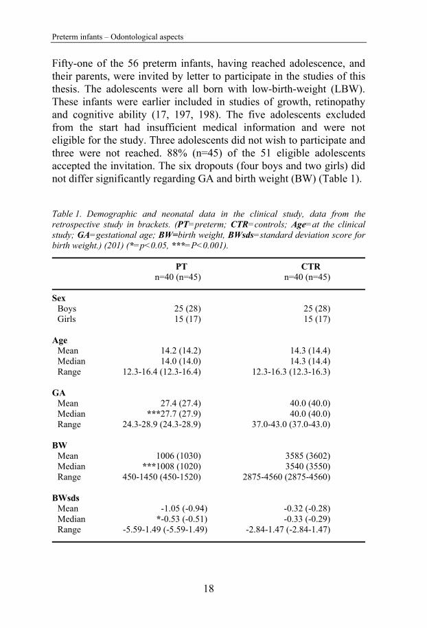
Preterm infants – Odontological aspects
18
Fifty-one of the 56 preterm infants, having reached adolescence, and their parents, were invited by letter to participate in the studies of this thesis. The adolescents were all born with low-birth-weight (LBW). These infants were earlier included in studies of growth, retinopathy and cognitive ability (17, 197, 198). The five adolescents excluded from the start had insufficient medical information and were not eligible for the study. Three adolescents did not wish to participate and three were not reached. 88% (n=45) of the 51 eligible adolescents accepted the invitation. The six dropouts (four boys and two girls) did not differ significantly regarding GA and birth weight (BW) (Table 1).
Table 1. Demographic and neonatal data in the clinical study, data from the retrospective study in brackets. (PT=preterm; CTR=controls; Age=at the clinical study; GA=gestational age; BW=birth weight, BWsds=standard deviation score for birth weight.) (201) (*=p<0.05, ***=P<0.001).
PT CTR n=40 (n=45) n=40 (n=45)
Sex
Boys 25 (28) 25 (28) Girls 15 (17) 15 (17)
Age Mean 14.2 (14.2) 14.3 (14.4) Median 14.0 (14.0) 14.3 (14.4) Range 12.3-16.4 (12.3-16.4) 12.3-16.3 (12.3-16.3)
GA Mean 27.4 (27.4) 40.0 (40.0) Median ***27.7 (27.9) 40.0 (40.0) Range 24.3-28.9 (24.3-28.9) 37.0-43.0 (37.0-43.0)
BW Mean 1006 (1030) 3585 (3602)
Median ***1008 (1020) 3540 (3550) Range 450-1450 (450-1520) 2875-4560 (2875-4560)
BWsds Mean -1.05 (-0.94) -0.32 (-0.28)
Median *-0.53 (-0.51) -0.33 (-0.29) Range -5.59-1.49 (-5.59-1.49) -2.84-1.47 (-2.84-1.47)
Marianne Rythén
19
Five of the 45 adolescents born preterm did not wish to attend the clinical examination, thus, 40 extremely preterm children (25 boys, 15 girls) participated in the clinical and dental cast studies (Fig. 1).
The controls (CTR) consisted of 45 (40) adolescents born at term and individually matched, by age, gender and catchment area of the same Public Dental Service Clinic (Fig. 1). The controls were selected by the head of the clinic at a matched Public Dental Service Clinic. The first adolescent from the patient register matching the inclusion criteria, and the parents, was invited by letter. If they declined, the following patient in the patient register was asked (Table 1).
3.2 Tooth material (III, IV) A total number of 44 earlier exfoliated primary teeth (20 incisors, 4 canines and 20 molars) were collected from 14 of the 45 adolescents born preterm, for histo-morphological studies in a polarized light microscope. The sample constituted all teeth retrieved from participating preterms. The subjects and teeth are presented in Table 2.
As control material in Study IV, thirty-six exfoliated primary teeth (27 incisors; 9 molars) were used, collected from 36 healthy children. The teeth had earlier been described in a study of elemental composition in primary teeth (202).

Preterm infants – Odontological aspects
20
Table 2. Gestational age in weeks (GA), birth weight in grams (BW) and number of collected primary teeth from the preterm children; (I=Incisor; C=Canine; M=Molar) in the different studies (ToothPOLMI=morphological study (III); ToothSEM=morphological study (III); ToothXRMA=chemical study (IV).)
Pat. GA BW Tooth Tooth Tooth POLMI SEM XRMA
1 28.14 1130 1* M 1* M 2 28.86 1370 1* I. 1*M 1* I 3 26.00 690 1* I. 1*M 1* I. 1*M 4 28.86 1015 1* I 1* I 5 25.86 695 1* I. 1*M 1* I. 1*M 6 27.86 1280 6* I. 1*C. 3*M 1* I 1*I 7 28.71 1250 3* M 1* M 1* M 8 27.43 1180 1* I 1* I 9 28.57 1450 2* M 1* M 2* M 10 28.14 1140 1* I. 1*C 1* I 1* I 11 28.14 1180 1* I. 1*M 1* I 12 27.00 1355 6* I. 2*C. 4*M 1* I 13 27.00 1000 1* M 1* M 14 28.00 1425 1* I. 2*M 1*M
Mean 27.76 1154 STD 0.98 239
Marianne Rythén
21
3.3 Methods
3.3.1 Study design Study I: Cross-sectional case-control. Study II: Cross-sectional.
Study III: Descriptive.
Study IV: Case–control.
3.3.2 Ethical approval and considerations Ethical consent was given by the Ethical Research Committee at the University of Gothenburg (Studies I, II. Dnr 262-06 and Studies III, IV. Dnr S 675-02). Informed consent was obtained from the preterm and control adolescents and their parents. Written consent from the Swedish National Board of Health and Welfare, to collect medical information from the Swedish Medical Birth Register, was retrieved.
3.3.3 Medical records and medical history The preterm infants’ neonatal and postnatal medical diagnoses and treatments were retrieved from hospital medical records and the data of the controls from the Swedish Medical Birth Registration (Studies I, II, III). A standard deviation score for birth weight (BWsds = actual birth weight – mean value of birth weight/standard deviation of respective gestational age at birth and gender) was calculated. Small for gestational age (SGA) was considered when BWsds were below -2.0 sds (201). Weight differences between birth, one month and two months of age were calculated.
At the clinical examination, medical history was obtained from the parents of the adolescents and the daily intake of medicines was noted.
3.3.4 Odontological registrations (I, II) The clinical examination was performed by a single examiner (MR) under a working light in a dental chair. Clinical photos and analogue X-ray bite-wings were taken if not provided by the adolescents´ dentists. Salivary examination and bacterial analyses were performed.

Preterm infants – Odontological aspects
20
Table 2. Gestational age in weeks (GA), birth weight in grams (BW) and number of collected primary teeth from the preterm children; (I=Incisor; C=Canine; M=Molar) in the different studies (ToothPOLMI=morphological study (III); ToothSEM=morphological study (III); ToothXRMA=chemical study (IV).)
Pat. GA BW Tooth Tooth Tooth POLMI SEM XRMA
1 28.14 1130 1* M 1* M 2 28.86 1370 1* I. 1*M 1* I 3 26.00 690 1* I. 1*M 1* I. 1*M 4 28.86 1015 1* I 1* I 5 25.86 695 1* I. 1*M 1* I. 1*M 6 27.86 1280 6* I. 1*C. 3*M 1* I 1*I 7 28.71 1250 3* M 1* M 1* M 8 27.43 1180 1* I 1* I 9 28.57 1450 2* M 2* M 2* M 10 28.14 1140 1* I. 1*C 1* I 1* I 11 28.14 1180 1* I. 1*M 1* I 12 27.00 1355 6* I. 2*C. 4*M 1* I 13 27.00 1000 1* M 1* M 14 28.00 1425 1* I. 2*M 1*M
Mean 27.76 1154 STD 0.98 239
Marianne Rythén
21
3.3 Methods
3.3.1 Study design Study I: Cross-sectional case-control. Study II: Cross-sectional.
Study III: Descriptive.
Study IV: Case–control.
3.3.2 Ethical approval and considerations Ethical consent was given by the Ethical Research Committee at the University of Gothenburg (Studies I, II. Dnr 262-06 and Studies III, IV. Dnr S 675-02). Informed consent was obtained from the preterm and control adolescents and their parents. Written consent from the Swedish National Board of Health and Welfare, to collect medical information from the Swedish Medical Birth Register, was retrieved.
3.3.3 Medical records and medical history The preterm infants’ neonatal and postnatal medical diagnoses and treatments were retrieved from hospital medical records and the data of the controls from the Swedish Medical Birth Registration (Studies I, II, III). A standard deviation score for birth weight (BWsds = actual birth weight – mean value of birth weight/standard deviation of respective gestational age at birth and gender) was calculated. Small for gestational age (SGA) was considered when BWsds were below -2.0 sds (201). Weight differences between birth, one month and two months of age were calculated.
At the clinical examination, medical history was obtained from the parents of the adolescents and the daily intake of medicines was noted.
3.3.4 Odontological registrations (I, II) The clinical examination was performed by a single examiner (MR) under a working light in a dental chair. Clinical photos and analogue X-ray bite-wings were taken if not provided by the adolescents´ dentists. Salivary examination and bacterial analyses were performed.
Preterm infants – Odontological aspects
22
Oral hygiene and periodontal status (I) Plaque, gingival health and periodontal status were registered for all incisors and first molars. As the adolescents were in different dental stages, no other teeth were used. Plaque was noted using a probe passing on the buccal and lingual surfaces along the gingival margin. Plaque was noted as general if registered both at the incisors and the molars and local, if registered either at the incisors or the molars. In all other cases, it was noted as plaque-free.
The periodontal registrations were made according to the WHO guidelines for clinical examinations (203) of incisors and molars. Bleeding on probing (BoP) and pocket depths ≥ 4mm for each site (mesial, buccal, distal and lingual) were registered. Three extremely preterm adolescents and one control did not cooperate to probing.
Caries diagnostics (I) For caries diagnostics, the DMFT (Decayed Missing Filled Teeth) index was used. A mouth mirror was used to identify manifest occlusal caries and smooth surfaces, buccally or lingually. Caries was registered in a fissure when seen as a cavity and on smooth surfaces, defined as initial when the surfaces were demineralized with the loss of translucency along the gingival margin, and as manifest when seen as a cavity. Analogue X-ray bite-wings were used for approximal caries, registered according to Shwartz et al., 1984 (204), and occlusal caries when clearly noted as radiolucency in the dentin. Missing teeth caused by caries were noted.
Intra-examination reliability for caries diagnostics on bite-wing was calculated, using 60 bite-wing radiographs from 30 randomly selected patients not involved in the study. The radiographs were diagnosed for caries according to the above mentioned criteria, twice, with a four week interval.
Dental stage (II) To investigate the relation between chronological and developmental records, the dental stage was registered according to the method described by Björk et al., 1964 (205).
Marianne Rythén
23
Salivary examination (I) For the saliva examination, stimulated saliva was collected during five minutes with the patient sitting in an upright position. The amount of saliva was measured in ml/min and registered as normal if >1ml/min (206). Dentobuff® Strip was used to measure the pH in saliva (207) and Dentocult SM Strip Mutans™, and Dentocult LB dip slides (Orion Diagnostica, Sweden) was used for identifying Streptococcus mutans (S. mutans) and Lactobacilli (208, 209). If no cultivation was seen, it was noted as no growth. At the statistical analysis for S. mutans, the groups low and medium were analyzed together.
Mineralization disturbances (I) The index of developmental defects in dental enamel (the DDE index) was used to identify enamel mineralization disturbances (177). Hypomineralized enamel (diffuse and demarcated opacities) was registered and noted as local, multiple or affecting all teeth. Hypomineralized enamel with enamel breakdown was noted separately. Enamel hypoplasia was noted as local or affecting all teeth. Fluorosis was diagnosed when confirmed with the medical history.
3.3.5 Dental records at 3, 6 and 9 years of age (I) Dental records at 3, 6 and 9 years of age, for both the preterms and the controls, were retrieved from the yearly dental records provided from the clinics in the Swedish Public Dental Service, and in five cases from private dental clinics, where the children received dental treatment. At the age of 3 years, deft (deft=decayed, extracted, filled primary teeth) was noted. At the age of 6 and 9 years, deftm (deftm=decayed, extracted, filled primary teeth, molars) and DMFT were registered. In these cases, a notation of mineralization disturbances were found in the records and this was registered.
3.3.6 Study cast analyses (II) Study casts in the central occlusion were taken, if not provided by the childrens’ dentist or orthodontist. Study cast analysis was performed for the malocclusions according to Björk et al., 1964 (205). The malocclusions were registered by the three authors together and compared with a Swedish reference material (210). When dental casts

Preterm infants – Odontological aspects
24
were missing, the malocclusion diagnoses were set by one of the examiners (MR) at the time of the clinical examination. Consideration to mesial drift by the first permanent molar was taken when sagittal diagnoses were registered. Crowding was noted when ≥4 mm in each quadrant.
Figure 2. a-b. Arch length, according to Thilander, 2009 (97). The total circumference of the dental arch, divided into right/left posterior (P2-C) and anterior segments (I2-I2), was obtained by measuring the arch perimeter to the mesial surface at the first permanent molars, thus, representing the distance (P2-P2). (P2=Second premolar; I2=Lateral incisor; C=Canine.) c-d. Arch width, according to Thilander, 2009 (97). The maxillary and mandibular intermolar width was obtained by measuring the distances between the central fossae of the corresponding first molars on the left and right side between (M1- M1). ( M1=First molar.) e. Palatal height, according to Thilander, 2009 (97). The palatal height was achieved in the mid-palatal plane, determined by measuring the perpendicular distance from the occlusal plane constructed from the permanent first molars. Through a hole in a plastic sheet, the end of the caliper was pressed to the palatal contour. The distance minus the thickness of the sheet represented palatal height.
On each study cast, arch length, width, palatal height (Figs. 2a-e) and mesio-distal tooth width was measured with a digital caliper (Cocraft®) and compared with the normal ideal (not orthodontic
Marianne Rythén
25
treated) sample (97). The measurements were performed twice at different occasions by one of the authors (MR). In order to compare the arch length, width and palatal height with the reference material, the two groups were divided according to gender and age (13 and 16 years of age) (Table 3).
Table 3. Chronological age groups (13 and 16 years old) and gender (G) (F=female; M=male). Preterm children (n=36), controls (n=39) and reference material (REF) according to Thilander, 2009 (97). Mean age (MA) and standard deviation (SD).
Preterm Controls REF G n MA SD n MA SD MA SD
13 years F 8 13.4 0.7 8 13.0 0.8 13.0 0.4 M 14 13.3 0.7 15 13.3 0.6 13.1 0.4 16 years F 6 15.7 0.4 6 15.4 0.4 15.8 0.5 M 8 15.2 0.5 10 15.5 0.6 15.8 0.5
3.3.7 Morphological and chemical analysis of primary teeth (III, IV)
All teeth were macroscopically examined for opacities and enamel hyperplasia before morphological and chemical analyses were performed. One tooth was found with enamel hypoplasia and all the teeth had marked attrition, considered normal for primary teeth when exfoliating. Before preparation, all teeth were stored in 70% ethanol for at least 24 hours.
Embedding and sectioning of teeth Since dental enamel is brittle and hard, sections of teeth for histological investigations have to be supported by an embedding media before cutting in a saw microtome. Before analyses, the specimens were, therefore, embedded in an epoxy-resin (Epofix®, Electron Microscopy Sciences, Fort Washington, PA, USA). Sagittal longitudinal sections with a thickness of approximately 100 m were prepared in a Leica SP1600 Saw Microtome (Leica Microsystems
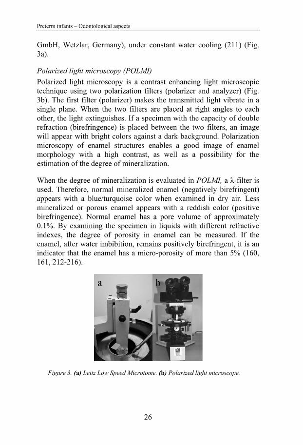
Preterm infants – Odontological aspects
26
GmbH, Wetzlar, Germany), under constant water cooling (211) (Fig. 3a).
Polarized light microscopy (POLMI) Polarized light microscopy is a contrast enhancing light microscopic technique using two polarization filters (polarizer and analyzer) (Fig. 3b). The first filter (polarizer) makes the transmitted light vibrate in a single plane. When the two filters are placed at right angles to each other, the light extinguishes. If a specimen with the capacity of double refraction (birefringence) is placed between the two filters, an image will appear with bright colors against a dark background. Polarization microscopy of enamel structures enables a good image of enamel morphology with a high contrast, as well as a possibility for the estimation of the degree of mineralization.
When the degree of mineralization is evaluated in POLMI, a λ-filter is used. Therefore, normal mineralized enamel (negatively birefringent) appears with a blue/turquoise color when examined in dry air. Less mineralized or porous enamel appears with a reddish color (positive birefringence). Normal enamel has a pore volume of approximately 0.1%. By examining the specimen in liquids with different refractive indexes, the degree of porosity in enamel can be measured. If the enamel, after water imbibition, remains positively birefringent, it is an indicator that the enamel has a micro-porosity of more than 5% (160, 161, 212-216).
Figure 3. (a) Leitz Low Speed Microtome. (b) Polarized light microscope.
Marianne Rythén
27
Scanning electron microscopy (SEM) Scanning electron microscopy is primarily used when studying the surface morphology of enamel in high magnification (Fig. 4). The microscope is used as a reversed light microscope and instead of light, electrons are used. To prevent disturbances in the electron’s path and isolate high voltage from the electron source, the electron beam and specimen are kept in vacuum. The result is an image with extensive depth of the field and a sense of three dimensional appearances. The image is produced by scanning with an electron beam with high energy in a raster scanning pattern. Electrons are reflected from the surface and transmitted to a viewing screen. The amount of reflected electrons will depend on the surface material and the angle between the electron beam and the specimen. To reinforce the effects of the reflected electrons and avoid surface charging, the surface is coated with a thin layer of gold by vapor deposition. The magnification range used is 10-30,000x.
Figure 4. Scanning electron microscope. X-ray micro analyses (XRMA) X-ray micro analyses are performed in a SEM with an attached X-ray detector and software program. Instead of reflecting electrons from the surface of the specimen, characteristic and well-defined K-α X-rays are emitted from a volume below the surface. The detector collects the X-rays and the pulses are shown on a screen where each element has its
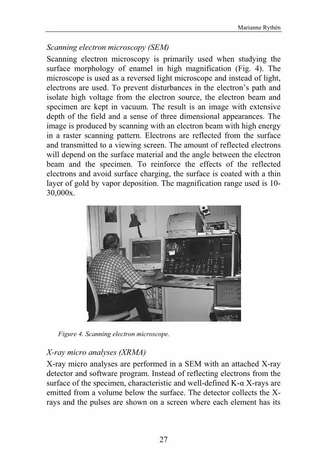
Preterm infants – Odontological aspects
28
own spectra. The pulses are transformed to a computer calculating the relative relation of weight percent between the included elements. The method can be used for chemical analyses in dental hard tissues (215). The method enables comparison between different specimens only when the circumstances are equal and the analyses are performed in the same way. In order to avoid surface charging, the specimen is coated with an electrical conducting material by vapor deposition.
POLMI analyses The enamel, in all sections, was examined in an Olympus polarizing light microscope (Olympus, Tokyo, Japan), dry in air and after water imbibition. All specimens were examined by the two authors (MR, JGN) together, whereafter, each examiner studied the sections. Finally, the findings were compared and any differences were discussed.
The appearance and location of the neonatal line (NNL), incremental lines and subsurface lesions (SSL) were registered both dry in air and after water imbibition.
The degree of mineralization, seen as porosities in the enamel, was noted by using positive and negative birefringence. The extent of the positive birefringence in the enamel, when examined dry in air and after water imbibition, was registered (Fig. 5)
For orientation within the specimens, micro-photos were taken of the central sections and used for the scanning electron microscope (SEM) analysis.
SEM analysis Four teeth, from four preterms, representing varying degrees and extent of porosity as seen in polarized microscopy, were further studied in SEM (Study III) (Table 2). For the SEM analyses, the sections were mounted on sample holders for SEM with carbon tape, etched for 30 seconds with 30% phosphoric acid and carefully rinsed with de-ionized water. The sections were coated with gold by vapor deposition. The SEM examinations were carried out in a Philips SEM 515 at 20 kV (Philips, Eindhoven, The Netherlands) and in a field emission scanning electron microscope (Gemini IMB, LEO 1530, Germany). The analyses were carried out at different locations in
Marianne Rythén
29
hypomineralized and normal enamel; close to the surface, middle enamel and close to the EDJ (Fig. 6). The morphological appearance of prisms, crystals and the unity of the structures were evaluated by the two authors together (MR, JGN).
The extent of the positive birefringence in the postnatal enamel when examined dry in air was registered in three groups:
1: When positive birefringence is noted in the surface region only. 2: When the extent is <1/2 of the enamel thickness. 3: When the extent is >1/2 of the enamel thickness.
The extent of the positive birefringence in the enamel after water imbibitions was registered as:
0-wi: When the degree of porosity is <5% in the enamel after water imbibition.
1-wi: When positive birefringence is seen only as streaks. 2-wi: When the extent is <1/2 of the enamel thickness. 3-wi: When the extent is >1/2 of the enamel thickness.
The localization of the microporous zone was registered by dividing the tooth into five groups:
A: <1/4 of the crown coronally. B: ½ of the crown coronally. C: 2/3 of the tooth coronally. D: The central part of the tooth. E: The total crown.
Figure 5. Registrations of positive and negative birefringence in enamel, when studied dry in air and after water imbibition.

Preterm infants – Odontological aspects
30
Figure 6. Areas of analyses in SEM for the four teeth examined.
The results were compared with medical data (infant respiratory distress syndrome, hematological infections, number of blood and plasma transfusions, numbers of days with artificial ventilation, and number of days with nasal oxygen with overpressure).
Analyses in XRMA Seventeen (9 incisors and 8 molars) of the 44 primary teeth from the same 14 adolescents were used for the chemical analyses. The teeth were chosen from each one of the preterm children and in three of the patients, additionally one tooth was used (Table 2).
Chemical analyses of C, O, Ca and P in the enamel and dentin were performed in Philips SEM 515 (Philips, Eindhoven, The Netherlands) equipped with an EDAX DX-4 ECON detector (EDAX Inc., Mahwah, NJ, USA). The sections from the preterm children and controls were mounted on sample holders for X-ray micro-analysis (XRMA) with carbon tape and coated with carbon by vapor deposition after being etched for 30 sec with 30% phosphoric acid and carefully rinsed with de-ionized water (Fig. 7). For all measurements, the emitted X-rays
Marianne Rythén
31
were detected during continuously fast scanning by a small window of 6.1 µm x 4.3 µm at a magnification of 650x.
Figure 7. Section of a tooth embedded in epoxy-resin, mounted on sample holder for SEM and coated with carbon.
The relative amounts of C, O, Ca and P were obtained by the EDAX DX-4 software, expressed in weight % and normalized to 100%. The software program corrected the data for carbon coating. All measurements were carried out from the surface, along a line toward the tip of the pulp, on the buccal side of the tooth starting approximately at half of the crown height, always aiming at morphologically analyzing the same area. Thus, all measurements were made in the postnatal enamel. The measurements were performed at five locations in the enamel and the dentin, respectively. The first measurement was made 10 m below the surface and followed by measurements located at ¼, ½ and ¾ of the enamel thickness and 10 m above the EDJ. The measurements in the dentin were performed in the same way, starting 10 m below the EDJ and at ¼, ½ and ¾ of the dentin thickness and 10 m above the pulp chamber (Fig. 8).
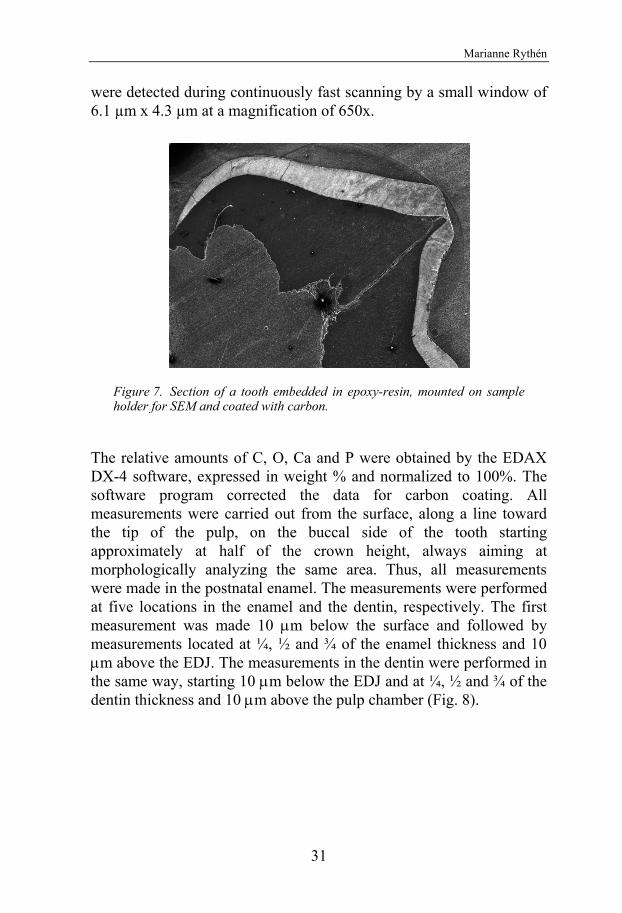
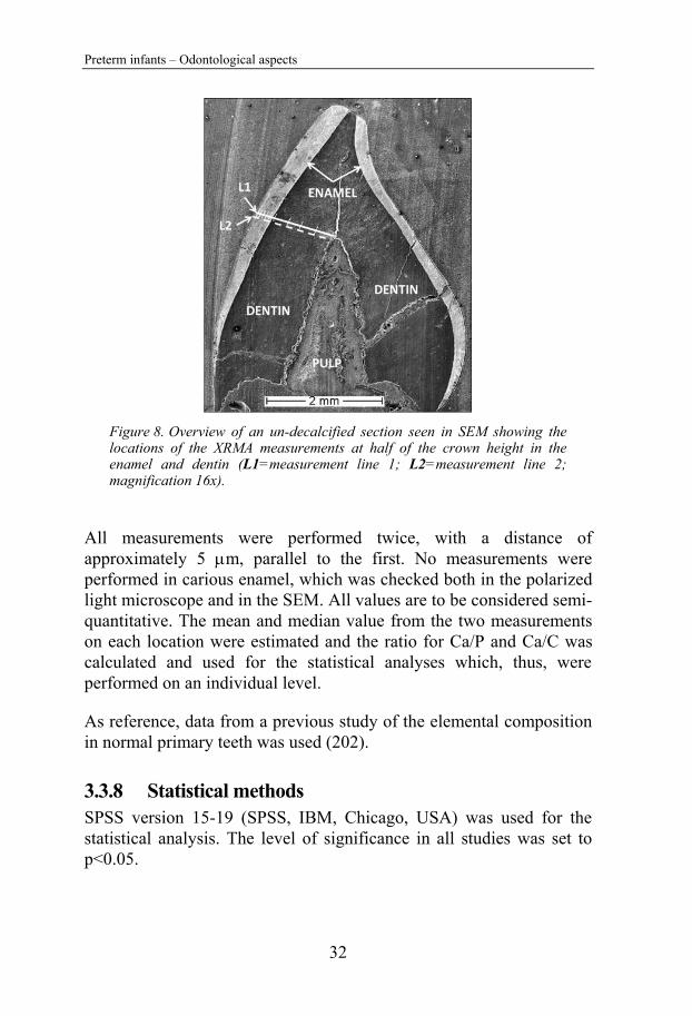
Preterm infants – Odontological aspects
32
Figure 8. Overview of an un-decalcified section seen in SEM showing the locations of the XRMA measurements at half of the crown height in the enamel and dentin (L1=measurement line 1; L2=measurement line 2; magnification 16x).
All measurements were performed twice, with a distance of approximately 5 m, parallel to the first. No measurements were performed in carious enamel, which was checked both in the polarized light microscope and in the SEM. All values are to be considered semi-quantitative. The mean and median value from the two measurements on each location were estimated and the ratio for Ca/P and Ca/C was calculated and used for the statistical analyses which, thus, were performed on an individual level.
As reference, data from a previous study of the elemental composition in normal primary teeth was used (202).
3.3.8 Statistical methods SPSS version 15-19 (SPSS, IBM, Chicago, USA) was used for the statistical analysis. The level of significance in all studies was set to p<0.05.
Marianne Rythén
33
In Study I, differences between groups were analyzed using the Chi-Square Test for nominal data, Fishers´s Exact Test for small nominal data and Mann Whitney U-test for numeric data. Correlation analyses between numeric variables were calculated using the Spearman Correlation Analyses. Non-parametric methods were used since the measured values did not have a normal distribution. Intra-observer reliability was evaluated with the Cohen’s Kappa Test.
In Study II, differences between groups were analyzed with the Chi Square Test and Fisher’s Exact Test for nominal data, and the Student`s T-test for numeric data. Correlation analyses between hospital treatments and tooth dimension were calculated using Pearson’s Correlation Analyses. Adjustments according to Bonferroni have not been performed. The Error of the method was calculated for tooth measurements according to the Dahlberg formula (217):
SE= nd 2/2 ,
where d is the difference between the two measurements and n is the number of measurements.
In Study IV, the Mann-Whitney U-test was used for comparison of the medians. Non-parametric methods were used since the measured values did not have a normal distribution. The charts shown are based on the median values.

Preterm infants – Odontological aspects
34
4. RESULTS
4.1 Postnatal complications/treatments and health during adolescence
The preterm infants were hospitalized (range 41-441 days) and treated with artificial ventilation, blood transfusions, antibiotics, artificial nutrition and in some cases, surgery (Table 4). Nine preterm infants were considered SGA. Thirty-five (thirty-nine) of forty (forty-five) infants returned to the hospital for treatments after discharge.
At the time of the clinical examination, the adolescents born preterm suffered from various medical conditions (Table 4).
Four sets of twins were found in the preterm group. No differences were observed between each sibling and the total preterm adolescents regarding oral health and risk indicators. Similarities in one set of twins were observed, seen as asthma and several teeth with hypomineralization (6/7) with severe hypomineralization (2/2). One set of twins had low salivary secretion. Further, all twins had Angle Class I relation and in one set, both siblings had crowding and dental anomalies.
None of the control infants had neonatal or postnatal medical diagnoses causing prolonged hospitalization, though six infants had signs of mild hypoxia or transient tachypnéa and one child had a hemolytic disease. No information regarding medical care after the hospital discharge was available.
According to the inclusion criteria, all controls were healthy at the time of the clinical examination with two exceptions; one had developed a nickel allergy and one had an untreated pollen allergy.
Marianne Rythén
35
Table 4. Medical data in the clinical study, data from the retrospective study in brackets. (PT=preterm; CTR=controls; BPD=bronchopulmonary dysplasia; NEC=necrotizing enterocolytis; IVH=intraventricular haemorrhage; ADHD=attention deficiency hyperactivity disorder; CP=cerebral palsy.)
PT CTR n=40 (n=45) n=40 (n=45)
Postnatal Morbidity BPD 9 (10) 0
Sepsis 17 (20) 0 NEC 4 (4) 0
Hyperbilirubinemia 18 (22) 0 IVH 4 (5) 0
Treatments Blood transfusion 36 (39) 0
Hospitalized >90days 11 (12) 0 Artificial nutrition >60days 26 (28)
Post hospital treatments within the first year 35 (39) Anamnesis at clinical examination Asthma 7 0
Heart disease 3 0 Growth deficiency 3 0 Allergy 7 2 Pierre Robins Syndrome 1 0 Turner Syndrome 1 0 Medicines (growth hormone
steroides, broncholytic sub- stances, stimulants, neuroleptica) 7 0 Dysfunctions at clinical examination ADHD 5 0
Autism 2 0 CP 2 0 Hearing, visual impairments 11 0 Perception, learning difficulties 6 0
0
Preterm infants – Odontological aspects
36
4.2 Oral health in adolescents born preterm (I)
4.2.1 Oral hygiene and periodontal disease Plaque was found in 33 preterm adolescents (23 local and 10 general) compared with 21 among the controls (18 local, 3 general, p=0.007). BoP was seen in 16 preterm adolescents compared with 7 controls (p=0.024). The number of BoP sites was also significantly higher (19 vs. 8, p=0.011). No significant difference in the frequency of periodontal pockets was found (6 PT vs. 2 CTR).
4.2.2 Caries The caries prevalence had a skewed distribution in both groups. The preterm adolescents were noted having more caries (decayed teeth=DT; decayed teeth initial approximal DTia) (PTDT=7, CTRDT=5; PTDTia=13; CTRDTia=11), but more controls had a history of caries (filled teeth=FT) (PTFT=12; CTRFT=18). DTia ≥4 was found in eight preterms compared with two controls. Ten preterm adolescents had initial caries buccally/lingually (DTibl) compared with four controls. Two preterm adolescents each had one tooth extracted due to severe hypomineralization and caries, and one control had one tooth extracted due to caries. However, there were no significant differences between the preterm adolescents and the controls in the frequency of DT, DTia, DTibl, MT or FT.
In early childhood, twice as many controls had caries in the primary dentition (decayed teeth=dt; decayed teeth molars=dtm) compared with the preterm children (3 years: PTdt=2, CTRdt=5; 6 years: PTdtm=7, CTRdtm=14). At six years of age, six controls had ≥3 decayed teeth compared with none in the preterm group. The caries frequency at six years of age was significantly higher in the controls (p=0.045). When filled teeth (filled teeth molars=ftm) were taken into account, the total difference in the caries prevalence was reduced (6 years: PTftm=8, CTRftm=5), however, there were six preterm children with only one filled tooth compared with one CTR child. No differences were found between the two groups regarding the frequency of dtm and DT at 9 years of age (9 years: PTdtm=14, CTRdtm=15; 9 years: PTDT=5, CTRDT=6). Increased prevalence of ftm, FT was found in the controls
Marianne Rythén
37
(9 years: PTftm=16, CTRftm=21; PTFT=3, CTRFT=8), and more extracted primary molars (etm) (PTetm=8, CTRetm=9; five CTR had ≥3 primary molars extracted vs. none PT), indicating a higher past caries activity among the controls, however, this was not significant.
4.2.3 Saliva and bacteria Stimulated saliva <1ml/min was found in 20 preterm adolescents compared with the seven controls (p=0.004). Most children in both groups had a normal pH value. Streptococcus mutans > 1,000,000 were found in eight preterm adolescents compared with three controls (p=0.032) and Lactobacillus >100,000 were found in five preterm adolescents and twelve controls, however, this difference was not statistically significant.
4.2.4 Mineralization disturbances The total enamel mineralization disturbances, according to the DDE index, did not differ between the two groups. Enamel hypoplasia was seen in two preterms compared with five controls. All teeth in one control had signs of hypoplasia with horizontal grooves.
Sixteen preterm adolescents were found with local (6) and multiple (8) hypomineralization, seen as demarcated opacities, compared with ten controls (5+5). Two adolescents from each group had enamel hypomineralization affecting all teeth, seen as diffuse striae, white or yellow. In the control group, one was diagnosed as having fluorosis, according to the odontological history and clinical appearance. One preterm was diagnosed with lactose intolerance and one preterm had a history of complex illness, with prolonged hospitalization and tube-feeding.
A significant difference, however, was seen in the frequency of severe hypomineralization between the preterm and control adolescents. Eleven teeth with enamel breakdown were found in seven preterm adolescents, compared to one tooth among the control adolescents (p=0.028). Further, two preterm adolescents had each one first molar extracted due to hypomineralization and caries and one was waiting for extraction.
Preterm infants – Odontological aspects
36
4.2 Oral health in adolescents born preterm (I)
4.2.1 Oral hygiene and periodontal disease Plaque was found in 33 preterm adolescents (23 local and 10 general) compared with 21 among the controls (18 local, 3 general, p=0.007). BoP was seen in 16 preterm adolescents compared with 7 controls (p=0.024). The number of BoP sites was also significantly higher (19 vs. 8, p=0.011). No significant difference in the frequency of periodontal pockets was found (6 PT vs. 2 CTR).
4.2.2 Caries The caries prevalence had a skewed distribution in both groups. The preterm adolescents were noted having more caries (decayed teeth=DT; decayed teeth initial approximal DTia) (PTDT=7, CTRDT=5; PTDTia=13; CTRDTia=11), but more controls had a history of caries (filled teeth=FT) (PTFT=12; CTRFT=18). DTia ≥4 was found in eight preterms compared with two controls. Ten preterm adolescents had initial caries buccally/lingually (DTibl) compared with four controls. Two preterm adolescents each had one tooth extracted due to severe hypomineralization and caries, and one control had one tooth extracted due to caries. However, there were no significant differences between the preterm adolescents and the controls in the frequency of DT, DTia, DTibl, MT or FT.
In early childhood, twice as many controls had caries in the primary dentition (decayed teeth=dt; decayed teeth molars=dtm) compared with the preterm children (3 years: PTdt=2, CTRdt=5; 6 years: PTdtm=7, CTRdtm=14). At six years of age, six controls had ≥3 decayed teeth compared with none in the preterm group. The caries frequency at six years of age was significantly higher in the controls (p=0.045). When filled teeth (filled teeth molars=ftm) were taken into account, the total difference in the caries prevalence was reduced (6 years: PTftm=8, CTRftm=5), however, there were six preterm children with only one filled tooth compared with one CTR child. No differences were found between the two groups regarding the frequency of dtm and DT at 9 years of age (9 years: PTdtm=14, CTRdtm=15; 9 years: PTDT=5, CTRDT=6). Increased prevalence of ftm, FT was found in the controls
Marianne Rythén
37
(9 years: PTftm=16, CTRftm=21; PTFT=3, CTRFT=8), and more extracted primary molars (etm) (PTetm=8, CTRetm=9; five CTR had ≥3 primary molars extracted vs. none PT), indicating a higher past caries activity among the controls, however, this was not significant.
4.2.3 Saliva and bacteria Stimulated saliva <1ml/min was found in 20 preterm adolescents compared with the seven controls (p=0.004). Most children in both groups had a normal pH value. Streptococcus mutans > 1,000,000 were found in eight preterm adolescents compared with three controls (p=0.032) and Lactobacillus >100,000 were found in five preterm adolescents and twelve controls, however, this difference was not statistically significant.
4.2.4 Mineralization disturbances The total enamel mineralization disturbances, according to the DDE index, did not differ between the two groups. Enamel hypoplasia was seen in two preterms compared with five controls. All teeth in one control had signs of hypoplasia with horizontal grooves.
hypomineralization, seen as demarcated opacities, compared with ten controls (5+5). Two adolescents from each group had enamel hypomineralization affecting all teeth, seen as diffuse striae, white or yellow. In the control group, one was diagnosed as having fluorosis, according to the odontological history and clinical appearance. One preterm was diagnosed with lactose intolerance and one preterm had a history of complex illness, with prolonged hospitalization and tube-feeding.
A significant difference, however, was seen in the frequency of severe hypomineralization between the preterm and control adolescents. Eleven teeth with enamel breakdown were found in seven preterm adolescents, compared to one tooth among the control adolescents (p=0.028). Further, two preterm adolescents had each one first molar extracted due to hypomineralization and caries and one was waiting for extraction.
Fourteen preterm adolescents were found with local (6) and multiple (8)

Preterm infants – Odontological aspects
38
In the primary dentition, none of the control adolescents was found to have enamel mineralization disturbances according to the dental records. Five and nine preterm children had enamel disturbances at six, respectively, nine years of age (p=0.004).
4.3 Dento-alveolar characteristics in adolescents born preterm (II)
4.3.1 Dental development No differences in the dental stage or between preterm and controls were found. Eight preterm and seven control adolescents remained in late mixed dentition (DS2M1 - DS3M1). The variations, in the late mixed and early permanent dentitions and adolescence, were normal due to individual dental development.
4.3.2 Malocclusions and dento-alveolar measurements Three or more malocclusions were twice as common among the preterm adolescents. Twice as many preterm adolescents had Angle Class II malocclusion and an overbite ≥5 mm compared with the controls. An over-jet ≥6 mm was significantly more frequent among the preterm (p=0.04) (Table 5). In ten out of twelve preterm adolescents with Angle Class II malocclusion, the diagnosis was associated with medical or functional disturbances, however, this was found in all classes (Fig. 9). Deviations of normal tooth morphology were more often seen in the preterms. This was not significant and no other differences in dental anomalies were found.
No major differences between the preterm and control groups were found in arch length, width and palatal height except for a decrease in the dental arches in the preterm girls at 16 years of age (maxillary anterior length: 28.2mm vs. 31.9mm, p= 0.039; maxillary total length: 69.5mm vs. 76.3mm, p=0.015; mandibular posterior length: 19.7mm vs. 21.6mm, p=0.004; mandibular total length: 60.3mm vs. 65.6, p=0.041; maxillary width 42.4 vs. 47.5, p= 0.028; mandibular width: 36.2mm vs. 41.4mm, p= 0.007).
Marianne Rythén
39
Figure 9. Frequency of Angle Class I, II and III, respectively, related to morbidity, neurological and neuro-psychiatric disturbances in preterm children.
A n g l e C l a s s I I I
A n g l e C l a s s I I
A n g l e C l a s s I0 5 1 0 1 5 2 0 2 5
N u m b e r o f P T c h i l d r e n
H e a l t h y
M o r b i d i t y
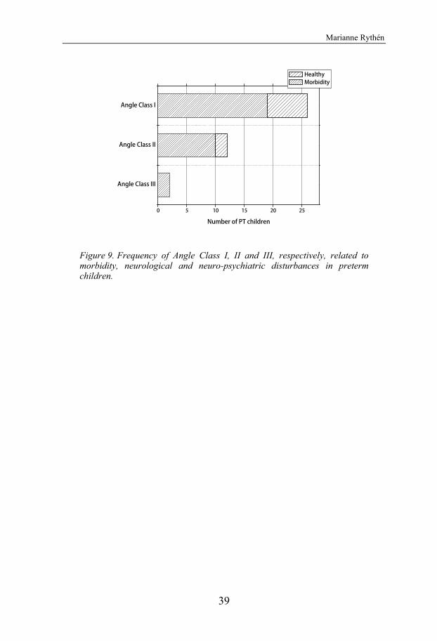
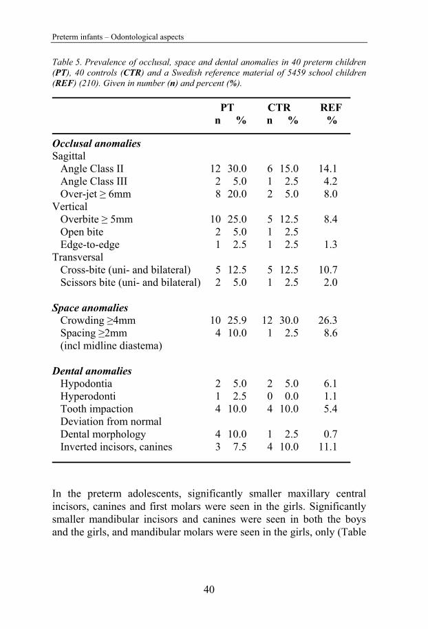
Preterm infants – Odontological aspects
40
Table 5. Prevalence of occlusal, space and dental anomalies in 40 preterm children (PT), 40 controls (CTR) and a Swedish reference material of 5459 school children (REF) (210). Given in number (n) and percent (%).
PT CTR REF n % n % %
Occlusal anomalies Sagittal
Angle Class II 12 30.0 6 15.0 14.1 Angle Class III 2 5.0 1 2.5 4.2 Over-jet ≥ 6mm 8 20.0 2 5.0 8.0
Vertical Overbite ≥ 5mm 10 25.0 5 12.5 8.4 Open bite 2 5.0 1 2.5 Edge-to-edge 1 2.5 1 2.5 1.3
Transversal Cross-bite (uni- and bilateral) 5 12.5 5 12.5 10.7 Scissors bite (uni- and bilateral) 2 5.0 1 2.5 2.0
Space anomalies Crowding ≥4mm 10 25.9 12 30.0 26.3 Spacing ≥2mm 4 10.0 1 2.5 8.6 (incl midline diastema)
Dental anomalies Hypodontia 2 5.0 2 5.0 6.1 Hyperodonti 1 2.5 0 0.0 1.1 Tooth impaction 4 10.0 4 10.0 5.4 Deviation from normal Dental morphology 4 10.0 1 2.5 0.7 Inverted incisors, canines 3 7.5 4 10.0 11.1
In the preterm adolescents, significantly smaller maxillary central incisors, canines and first molars were seen in the girls. Significantly smaller mandibular incisors and canines were seen in both the boys and the girls, and mandibular molars were seen in the girls, only (Table
Marianne Rythén
41
6). This was found especially in adolescents with prolonged postnatal hospitalization (Fig. 10).
Table 6. Mesio-distal crown diameters (mm) of permanent teeth in preterm children (PT), controls (CTR) and normal material (REF) (97), (T=tooth; G=gender; F=female; M=male) given as the mea and standard deviation (SD). (*=p<0.05, **=p<0.01; PT/CTR).
PT CTR REF T G Mean SD Mean SD Mean SD
Maxilla I1 F 1**8.2 0.45 18.9 0.55 18.5 0.52
M 18.7 0.60 19.0 0.47 19.0 0.46 I2 F 16.8 0.52 17.0 0.70 16.6 0.62 M 17.0 0.55 16.9 0.39 17.1 0.62 C F 1**7.1 0.42 17.8 0.41 17.6 0.44 M 17.9 0.36 18.1 0.33 18.3 0.52 P1 F 17.0 0.49 17.2 0.45 17.0 0.45 M 17.2 0.29 17.1 0.43 17.3 0.43 P2 F 16.7 0.39 16.8 0.36 16.7 0.49 M 16.8 0.41 16.9 0.43 16.9 0.38 M1 F **9.9 0.42 10.4 0.40 10.3 0.54 M *10.4 0.55 10.7 0.42 10.8 0.52 M2 F 19.6 0.64 10.1 0.84 19.4 0.63 M 10.1 0.57 10.3 0.56 10.0 0.62 Mandible I1 F 1*5.3 0.27 15.5 0.33 15.4 0.33
M *5.4 0.30 15.6 0.26 15.6 0.24 I2 F 1**5.7 0.34 16.1 0.39 15.9 0.32 M 1**5.9 0.40 16.2 0.39 16.1 0.27 C F 1**6.2 0.35 16.8 0.38 16.6 0.43 M *6.9 0.27 17.1 0.42 17.2 0.47 P1 F 17.0 0.54 17.2 0.41 17.0 0.42 M 17.3 0.42 17.2 0.36 17.4 0.56 P2 F 17.1 0.54 17.2 0.38 17.1 0.43 M 17.2 0.43 17.3 0.40 17.5 0.51 M1 F *10.4 0.53 10.9 0.49 10.8 0.58 M 10.9 0.86 11.0 0.53 11.3 0.62 M2 F 19.8 0.56 10.2 0.75 19.9 0.59 M 10.3 0.84 10.7 1.36 10.1 0.77
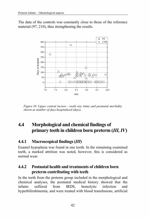
Preterm infants – Odontological aspects
42
The data of the controls was constantly close to those of the reference material (97, 210), thus strengthening the results.
Figure 10. Upper central incisor - tooth size (mm) and postnatal morbidity shown as number of days hospitalized (days).
4.4 Morphological and chemical findings of primary teeth in children born preterm (III, IV)
4.4.1 Macroscopical findings (III) Enamel hypoplasia was found in one tooth. In the remaining examined teeth, a marked attrition was noted, however, this is considered as normal wear.
4.4.2 Postnatal health and treatments of children born preterm contributing with teeth
In the teeth from the preterm group included in the morphological and chemical analyses, the postnatal medical history showed that the infants suffered from IRDS, hemolytic infection and hyperbilirubinemia, and were treated with blood transfusions, artificial
7.0 7.5 8.0 8.5 9.0 9.5 10.0
0
50
100
150
200
250
300
350
400
D
ays i
n ho
spita
l
mm
PT CTR
Marianne Rythén
43
ventilation, nasal oxygen with overpressure or oxygen alone. Three children had none of these diagnoses but were treated with blood transfusions and oxygen for other reasons. Three children had no blood transfusions and two had more than 20 occasions of transfusions. Four children had artificial ventilation and all children had received oxygen (Table 7).
4.4.3 Histo-morphological findings (POLMI) The neonatal line (NNL) was seen as a distinct incremental line, with a positive birefringence when examined both dry in air and after water imbibition. The NNL, when found, extended from the enamel dental junction (EDJ) coronal to the middle part of the enamel surface. In the preterm teeth, the NNL was located incisally (Figs. 11a-b) corresponding to the GA. The line separates prenatal enamel found in 17 teeth (15 incisors, 1 molar and 1 canine) from postnatal enamel. In 27 of the teeth (19 molars, 3 canines and 5 incisors), no neonatal line was found (Table 8).
Figure 11. (a) Un-decalcified section of a primary molar from a preterm infant, seen dry in air in polarized light with a neonatal line (NNL) and prenatal (PreN) and postnatal (PostN) porous zone (D=dentin). Original magnification ×40. (b) Un-decalcified section of the same primary molar as in (a), seen after water imbibition in polarized light with a neonatal line (NNL) and a prenatal (PreN) and a postnatal (PostN) porous zone (D=dentin). Original magnification ×40.
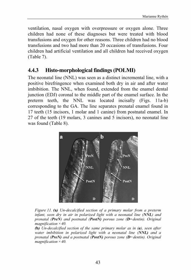
Preterm infants – Odontological aspects
42
The data of the controls was constantly close to those of the reference material (97, 210), thus strengthening the results.
Figure 10. Upper central incisor - tooth size (mm) and postnatal morbidity shown as number of days hospitalized (days).
4.4 Morphological and chemical findings of primary teeth in children born preterm (III, IV)
4.4.1 Macroscopical findings (III) Enamel hypoplasia was found in one tooth. In the remaining examined teeth, a marked attrition was noted, however, this is considered as normal wear.
4.4.2 Postnatal health and treatments of children born preterm contributing with teeth
In the teeth from the preterm group included in the morphological and chemical analyses, the postnatal medical history showed that the infants suffered from IRDS, hemolytic infection and hyperbilirubinemia, and were treated with blood transfusions, artificial
7.0 7.5 8.0 8.5 9.0 9.5 10.0
0
50
100
150
200
250
300
350
400
Day
s in
hosp
ital
mm
PT CTR
Marianne Rythén
43
ventilation, nasal oxygen with overpressure or oxygen alone. Three children had none of these diagnoses but were treated with blood transfusions and oxygen for other reasons. Three children had no blood transfusions and two had more than 20 occasions of transfusions. Four children had artificial ventilation and all children had received oxygen (Table 7).
4.4.3 Histo-morphological findings (POLMI) The neonatal line (NNL) was seen as a distinct incremental line, with a positive birefringence when examined both dry in air and after water imbibition. The NNL, when found, extended from the enamel dental junction (EDJ) coronal to the middle part of the enamel surface. In the preterm teeth, the NNL was located incisally (Figs. 11a-b) corresponding to the GA. The line separates prenatal enamel found in 17 teeth (15 incisors, 1 molar and 1 canine) from postnatal enamel. In 27 of the teeth (19 molars, 3 canines and 5 incisors), no neonatal line was found (Table 8).
Figure 11. (a) Un-decalcified section of a primary molar from a preterm infant, seen dry in air in polarized light with a neonatal line (NNL) and prenatal (PreN) and postnatal (PostN) porous zone (D=dentin). Original magnification ×40. (b) Un-decalcified section of the same primary molar as in (a), seen after water imbibition in polarized light with a neonatal line (NNL) and a prenatal (PreN) and a postnatal (PostN) porous zone (D=dentin). Original magnification ×40.
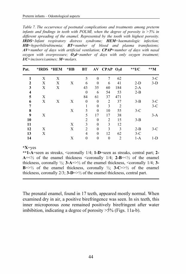
Preterm infants – Odontological aspects
44
Table 7. The occurrence of postnatal complications and treatments among preterm infants and findings in teeth with POLMI, when the degree of porosity is >5% in different spreading of the enamel. Represented by the tooth with highest porosity. IRDS=Infant respiratory distress syndrome; HEM=haematologic infection; HB=hyperbilirubinemia; BT=number of blood and plasma transfusions; AV=number of days with artificial ventilation; CPAP=number of days with nasal oxygen with overpressure; O2d=number of days with only oxygen treatment; I/C=incisors/canines; M=molars.
Pat. *IRDS *HEM *HB BT AV CPAP O2d **I/C **M
1 X X 5 0 7 62 3-C 2 X X X 6 0 6 41 2-D 3-D 3 X X 43 35 60 184 2-A 4 0 6 54 53 2-B 5 X 84 61 37 471 6 X X X 0 0 2 37 3-B 3-C 7 1 0 3 2 3-C 8 X 5 0 10 55 3-C 9 X 5 17 17 38 3-A 10 2 0 2 15 3-B 11 X 3 0 3 12 12 X X 2 0 3 3 2-B 3-C 13 X 4 0 12 62 3-C 14 X 0 0 0 2 1-A 1-D *X=yes **1-A=seen as streaks, <coronally 1/4; 1-D=seen as streaks, central part; 2-A=<½ of the enamel thickness <coronally 1/4; 2-B=<½ of the enamel thickness, coronally ½; 3-A=>½ of the enamel thickness, <coronally 1/4; 3-B=>½ of the enamel thickness, coronally ½; 3-C=>½ of the enamel thickness, coronally 2/3; 3-D=>½ of the enamel thickness, central part.
The prenatal enamel, found in 17 teeth, appeared mostly normal. When examined dry in air, a positive birefringence was seen. In six teeth, this inner microporous zone remained positively birefringent after water imbibition, indicating a degree of porosity >5% (Figs. 11a-b).
Marianne Rythén
45
Table 8. The morphological findings from the POLMI analysis and data of the preterm infants. Tooth: UI=upper incisor, LI=lower incisor, C=canine, FM=first molar, SM=second molar. NL=neonatal line: Y=present, N=not found. L=Localization of the neonatal line: 1=cervical 1/3, 2=middle 1/3, 3= incisal 1/3 4=cuspal/incisal. Pren=prenatal enamel, after water imbibition X=with >5% degree of porosity. Postn=postnatal enamel examined dry in air: N=normal, 1=positive birefringence at the enamel surface; 2=positive birefringence <½ of the enamel; 3=positive birefringence >½ of the enamel. A=<1/4 of crown; B=1/2 of crown; C=2/3 of crown; D=central; E=entire crown. IMB=postnatal enamel after water imbibition: N=normal; 0-wi=<5% degree of porosity; 1-wi=streaks of >5% degree of porosity; 2-wi=>5% degree of porosity <½ of the enamel; 3-wi=>5% degree of porosity >½ of the enamel; A=<1/4 of crown; B=1/2 of crown; C=2/3 of crown; D=central. SSL=subsurface lesion, X=present. INC=incremental lines; X=present.
Pat. nr. Tooth NL L Pren Postn Imb SSL INC
11 SM N 3C 3-wiC 12 LI Y 2 X 3D 2-wiD SM N 3E 3-wiD X 13 UI Y 3 3B 2-wiA X X SM N 3C 0-wi X 14 UI N 3B 2-wiB X 15 UI N N N FM N 2B 0-wi 16 UI Y 2 3C 1-wiD X UI Y 2 X 3E 2-wiB X C Y 4 N N X LI Y 4 N N UI N 3C 3-wiB X LI Y 4 2B 0-wi X LI Y 3 2A 2-wiA X X SM N 3C 2-wiC X X SM N 3C 3-wiB X X FM N 3C 3-wiC X 17 SM N 3C 3-wiC X SM N 3C 3-wiC X SM N 3E 3-wiC 18 UI N 3C 3-wiC 19 FM N 3B 3-wiA X X SM Y 4 3B 2-wiA 10 C N 3C 3-wiB X X UI Y 3 X 3C 0-wi X X 11 FM N 3E 0-wi X X UI Y 3 X 3E 0-wi X X 12 UI Y 3 N N UI Y 3 X 2A 0-wi X UI Y 2 3C 0-wi C N 3C 1-wiC X X FM N 3C 1-wiC X X SM N 3C 1-wiD X X SM N 3C 3-wiC X LI Y 2 N N X X LI Y 3 3B 1-wiD X UI N 3B 2-wiD C N 3B 2-wiB X X FM N 3C 3-wiC X X 13 FM N 3C 3-wiC 14 FM N 3C 1-wiD X LI Y 2 X 3A 1-wiA X X FM N 3C 1-wiD
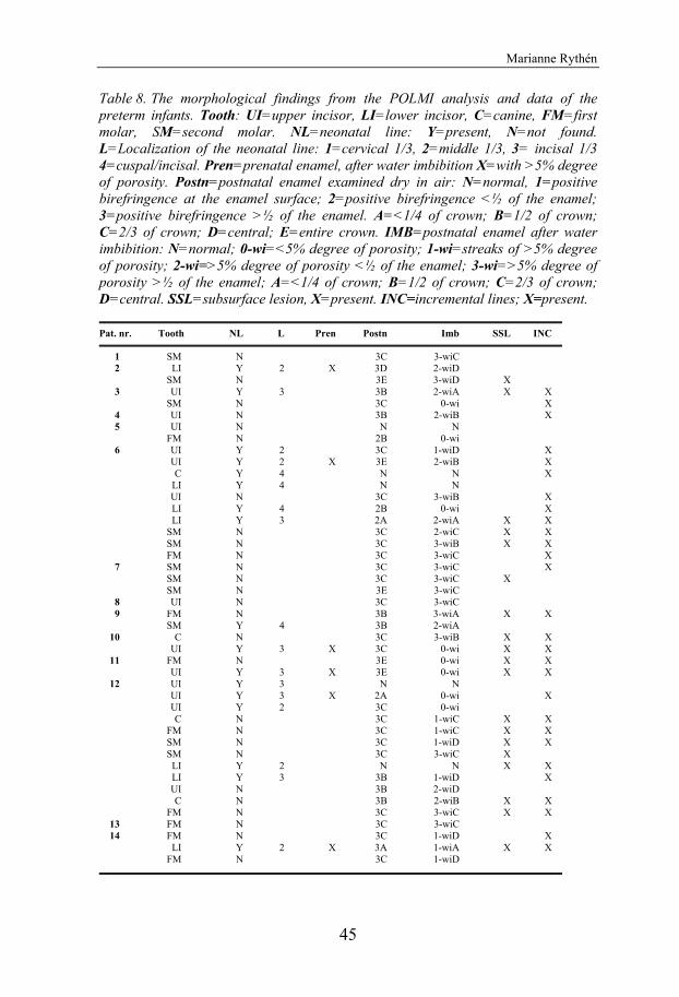
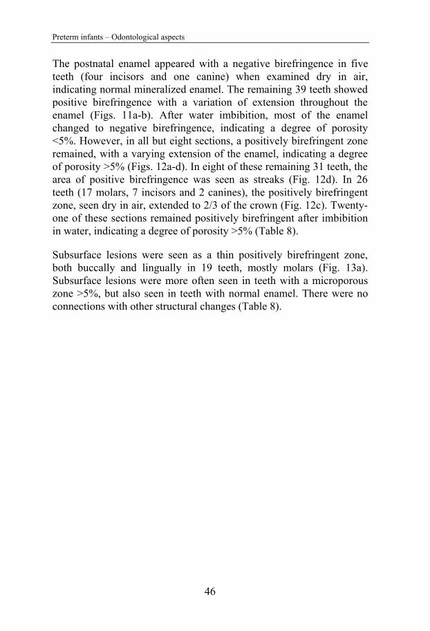
Preterm infants – Odontological aspects
46
The postnatal enamel appeared with a negative birefringence in five teeth (four incisors and one canine) when examined dry in air, indicating normal mineralized enamel. The remaining 39 teeth showed positive birefringence with a variation of extension throughout the enamel (Figs. 11a-b). After water imbibition, most of the enamel changed to negative birefringence, indicating a degree of porosity <5%. However, in all but eight sections, a positively birefringent zone remained, with a varying extension of the enamel, indicating a degree of porosity >5% (Figs. 12a-d). In eight of these remaining 31 teeth, the area of positive birefringence was seen as streaks (Fig. 12d). In 26 teeth (17 molars, 7 incisors and 2 canines), the positively birefringent zone, seen dry in air, extended to 2/3 of the crown (Fig. 12c). Twenty-one of these sections remained positively birefringent after imbibition in water, indicating a degree of porosity >5% (Table 8).
Subsurface lesions were seen as a thin positively birefringent zone, both buccally and lingually in 19 teeth, mostly molars (Fig. 13a). Subsurface lesions were more often seen in teeth with a microporous zone >5%, but also seen in teeth with normal enamel. There were no connections with other structural changes (Table 8).
Marianne Rythén
47
Figure 12. (a) Un-decalcified section of a primary molar from a preterm infant, seen dry in air in polarized light with a neonatal line (NNL) and hypomineralized postnatal enamel (PostN) (PreN=prenatal enamel; D=dentin). Original magnification ×40. (b) Un-decalcified section of a primary molar from a preterm infant, seen after water imbibition in polarized light with a neonatal line (NNL) and hypomineralized postnatal enamel (PostN) (PreN=prenatal enamel; D=dentin). Original magnification ×40. (c) Un-decalcified section of a primary molar from a preterm infant, seen dry in air in polarized light with a neonatal line (NNL) and postnatal enamel (PostN) with severe hypomineralization of the enamel (D=dentin). Original magnification ×40. (d) Un-decalcified section of a primary molar from a preterm infant, after water imbibition in polarized light with a neonatal line (NNL) and postnatal enamel (PostN) with varying degrees of hypomineralization (D=dentin). Original magnification ×40.
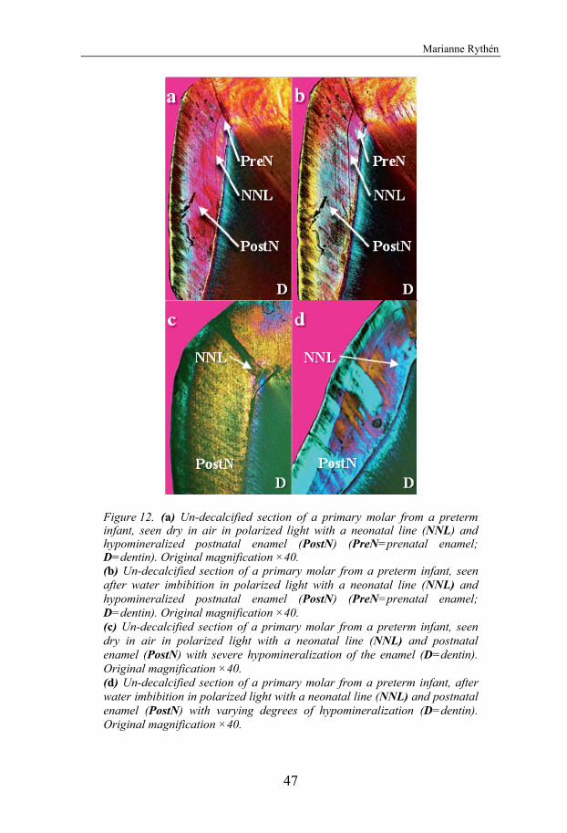
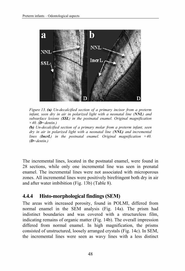
Preterm infants – Odontological aspects
48
Figure 13. (a) Un-decalcified section of a primary incisor from a preterm infant, seen dry in air in polarized light with a neonatal line (NNL) and subsurface lesions (SSL) in the postnatal enamel. Original magnification ×40. (D=dentin.) (b) Un-decalcified section of a primary molar from a preterm infant, seen dry in air in polarized light with a neonatal line (NNL) and incremental lines (IncrL) in the postnatal enamel. Original magnification ×40. (D=dentin.)
The incremental lines, located in the postnatal enamel, were found in 28 sections, while only one incremental line was seen in prenatal enamel. The incremental lines were not associated with microporous zones. All incremental lines were positively birefringent both dry in air and after water imbibition (Fig. 13b) (Table 8).
4.4.4 Histo-morphological findings (SEM) The areas with increased porosity, found in POLMI, differed from normal enamel in the SEM analysis (Fig. 14a). The prism had indistinct boundaries and was covered with a structureless film, indicating remains of organic matter (Fig. 14b). The overall impression differed from normal enamel. In high magnification, the prisms consisted of unstructured, loosely arranged crystals (Fig. 14c). In SEM, the incremental lines were seen as wavy lines with a less distinct
Marianne Rythén
49
enamel structure and often localized in hypomineralized enamel (Fig. 14d).
Figure 14. (a) Un-decalcified section of a primary incisor seen in scanning electron microscope (SEM) with normal enamel (P=enamel prism; IP=interprismatic area). Magnification ×3000; Bar=20m. (b) Un-decalcified section of a primary incisor from a preterm infant, seen in SEM with hypomineralized enamel covered with a structureless ‘film’ (P=enamel prism; IP=interprismatic area; OL=organic layer on the prisms – the structureless film). Magnification ×3000; Bar=3m. (c) Un-decalcified section of a primary incisor from a preterm infant, seen in SEM in high magnification of the porous hypomineralized enamel (P=enamel prism; IP=interprismatic area). Magnification ×30 000; Bar=1m. (d) Un-decalcified section of a primary molar from a preterm infant, seen in SEM showing the enamel structure in an area of incremental lines (ES=enamel surface; INC=incremental lines). Magnification ×3000; Bar=2m.
4.4.5 Chemical findings (XRMA) The values for the measured elements did not differ between incisors and molars, or between genders, when a statistical analysis of the medians and range was performed. Thus the values for all teeth were pooled. The results for each element and level are shown in charts as
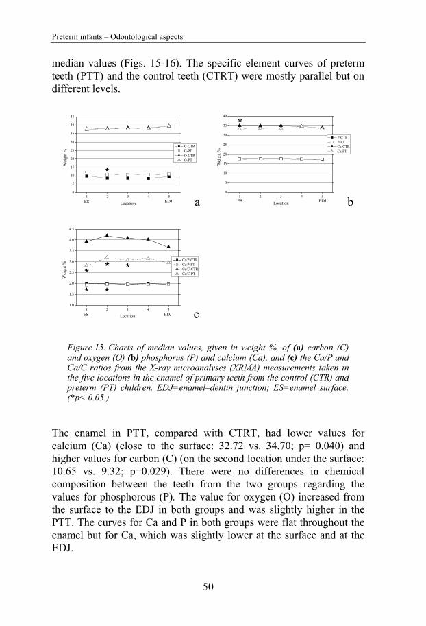
Preterm infants – Odontological aspects
50
median values (Figs. 15-16). The specific element curves of preterm teeth (PTT) and the control teeth (CTRT) were mostly parallel but on different levels.
Figure 15. Charts of median values, given in weight %, of (a) carbon (C) and oxygen (O) (b) phosphorus (P) and calcium (Ca), and (c) the Ca/P and Ca/C ratios from the X-ray microanalyses (XRMA) measurements taken in the five locations in the enamel of primary teeth from the control (CTR) and preterm (PT) children. EDJ=enamel–dentin junction; ES=enamel surface. (*p< 0.05.)
The enamel in PTT, compared with CTRT, had lower values for calcium (Ca) (close to the surface: 32.72 vs. 34.70; p= 0.040) and higher values for carbon (C) (on the second location under the surface: 10.65 vs. 9.32; p=0.029). There were no differences in chemical composition between the teeth from the two groups regarding the values for phosphorous (P). The value for oxygen (O) increased from the surface to the EDJ in both groups and was slightly higher in the PTT. The curves for Ca and P in both groups were flat throughout the enamel but for Ca, which was slightly lower at the surface and at the EDJ.
1 2 3 4 50
5
10
15
20
25
30
35
40
45
a
*
EDJES
Wei
ght %
Location
C-CTR C-PT O-CTR O-PT
1 2 3 4 50
5
10
15
20
25
30
35
40
b
*
EDJES
Wei
ght %
Location
P-CTR P-PT Ca-CTR Ca-PT
1 2 3 4 51.0
1.5
2.0
2.5
3.0
3.5
4.0
4.5
cEDJES
**
***
Wei
ght %
Location
Ca/P-CTR Ca/P-PT Ca/C-CTR Ca/C-PT
Marianne Rythén
51
In both groups, the carbon curve dropped from the surface toward the enamel bulk with a slight increase toward the EDJ. The Ca/P ratio was fairly constant, however, slightly lower in the PTT enamel, being significantly lower in the two locations below the surface (1.89 vs. 2.00, p=0.026; and 1.92 vs. 2.01, p=0.047). The Ca/C ratio was lower at the surface and increased toward the bulk with a decrease toward the EDJ in both groups. The enamel in the PTT had a significantly lower Ca/C ratio in the three locations in the outer half of the enamel (2.89 vs. 3.92, p=0.033; 3.30 vs. 4.45, p=0.016; and 3.31 vs. 4.35, p=0.038).
Figure 16. Charts of median values, given in weight %, of (a) carbon (C) and oxygen (O), (b) phosphorus (P) and calcium (Ca), and (c) Ca/P and Ca/C ratios from the X-ray microanalyses (XRMA) measurements taken in the five locations in the dentin of primary teeth from the control (CTR) and preterm (PT) children. DP=dentin–pulp junction; EDJ=enamel–dentin junction. (*p< 0.05, **p< 0.01.)
The SD was higher for the CTRT values for Ca, C and the Ca/C ratio.
1 2 3 4 50
10
20
30
40
50
60
70
aDPEDJ
*
Wei
ght %
Location
C-CTR C-PT O-CTR O-PT
1 2 3 4 50.00
1.00
2.00
3.00
4.00
5.00
6.00
7.00
bDPEDJ
**
Wei
ght %
Location
P-CTR P-PT Ca-CTR Ca-PT
1 2 3 4 50.00
0.25
0.50
0.75
1.00
1.25
1.50
1.75
2.00
cDPEDJ
**
Wei
ght %
Location
Ca/P-CTR Ca/P-PT Ca/C-CTR Ca/C-PT
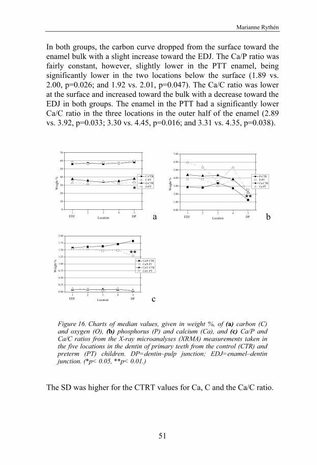
Preterm infants – Odontological aspects
52
When the measured values from the elemental analysis in the dentin from the PTT were compared with the controls, the values for Ca and P were higher among the preterms. At the dentin-pulp border, P was significantly higher in the PTT (5.82 vs. 4.67, p=0.004). The values for Ca and P decreased from the EDJ to the dentin-pulp border in both groups, however, the decrease for Ca in the PTT was larger and varied more compared with the controls. The curve for P in the controls increased in the bulk before decreasing toward the dentin-pulp border. There were no differences in the values for C between the two groups, while the values for O were lower in the dentin from the PTT, reaching significantly lower values at the dentin-pulp border (33.69 vs. 38.64, p=0.017). The curves for both C and O varied throughout the dentin for both groups with the highest values at the dentin-pulp border.
The Ca/P ratio made a marked decrease in the PTT toward the dentin-pulp border, while the ratio increased slightly in the controls (1.12 vs. 1.84, p=0.002). The Ca/C ratio decreased slightly from the EDJ toward the dentin-pulp border. No differences between the groups were seen.
The SD for the values of C, P and Ca was higher in the PTT and higher for O in the CTRT.
4.4.6 Clinical and morphological findings and medical associations (I, III, IV)
No single medical anamnesis, at the time of the examination, was associated with the clinical findings (caries, oral hygiene, gingivitis, salivary flow and S. mutans) and no single medical diagnose, postnatally, was related to the clinical enamel mineralization disturbances (Table 9). Enamel aberrations found in the POLMI examinations did not show any relation to a single neonatal or postnatal medical history found in the medical charts from the preterm infants (Table 7).
Marianne Rythén
53
Table 9. Enamel defects and postnatal morbidity (wI=Weight difference <400 gram the first month; wII=weight difference <600 gram the second month; BWs=birth weight standard deviation score <-2.00; IVH=intraventricular haemorrhage; HB=hyperbilirubinemia; BPD=bronchopulmonell dysplasia; NEC=necrotic enterocholitis; Time=>90 days at hospital; AN= artificial nutrition >60 days; No=number of teeth; Hypomin=hypomineralized enamel; Hypomin II=Severe hypomineralized enamel; Hypoplasia=enamel hypoplasia.)
wI wII BWs IVH HB BPD NEC Time AN
No 25 9 9 4 18 9 4 11 26 Hypomin 9 4 4 2 3 3 1 5 7 (16) Hypomin II 5 2 2 0 2 2 0 2 3 (7) Hypoplasia 2 1 1 0 0 0 0 0 2 (2)

Preterm infants – Odontological aspects
52
When the measured values from the elemental analysis in the dentin from the PTT were compared with the controls, the values for Ca and P were higher among the preterms. At the dentin-pulp border, P was significantly higher in the PTT (5.82 vs. 4.67, p=0.004). The values for Ca and P decreased from the EDJ to the dentin-pulp border in both groups, however, the decrease for Ca in the PTT was larger and varied more compared with the controls. The curve for P in the controls increased in the bulk before decreasing toward the dentin-pulp border. There were no differences in the values for C between the two groups, while the values for O were lower in the dentin from the PTT, reaching significantly lower values at the dentin-pulp border (33.69 vs. 38.64, p=0.017). The curves for both C and O varied throughout the dentin for both groups with the highest values at the dentin-pulp border.
The Ca/P ratio made a marked decrease in the PTT toward the dentin-pulp border, while the ratio increased slightly in the controls (1.12 vs. 1.84, p=0.002). The Ca/C ratio decreased slightly from the EDJ toward the dentin-pulp border. No differences between the groups were seen.
The SD for the values of C, P and Ca was higher in the PTT and higher for O in the CTRT.
4.4.6 Clinical and morphological findings and medical associations (I, III, IV)
No single medical anamnesis, at the time of the examination, was associated with the clinical findings (caries, oral hygiene, gingivitis, salivary flow and S. mutans) and no single medical diagnose, postnatally, was related to the clinical enamel mineralization disturbances (Table 6). Enamel aberrations found in the POLMI examinations did not show any relation to a single neonatal or postnatal medical history found in the medical charts from the preterm infants (Table 9).
Marianne Rythén
53
Table 9. Enamel defects and postnatal morbidity (wI=Weight difference <400 gram the first month; wII=weight difference <600 gram the second month; BWs=birth weight standard deviation score <-2.00; IVH=intraventricular haemorrhage; HB=hyperbilirubinemia; BPD=bronchopulmonell dysplasia; NEC=necrotic enterocholitis; Time=>90 days at hospital; AN= artificial nutrition >60 days; No=number of teeth; Hypomin=hypomineralized enamel; Hypomin II=Severe hypomineralized enamel; Hypoplasia=enamel hypoplasia.)
wI wII BWs IVH HB BPD NEC Time AN
No 25 9 9 4 18 9 4 11 26 Hypomin 9 4 4 2 3 3 1 5 7 (16) Hypomin II 5 2 2 0 2 2 0 2 3 (7) Hypoplasia 2 1 1 0 0 0 0 0 2 (2)

Preterm infants – Odontological aspects
54
5. DISCUSSION The studies in this thesis were conducted to elucidate the impact of prematurity on oral health, dento-alveolar status during adolescence, and dental development. Further, assessment was taken of the neonatal and postnatal health and treatments, and the general health during adolescence. The implication of this for the dentist is that more children with medical conditions and developmental defects, due to preterm birth, will attend the clinic. The odontological needs of these children during adolescence are not well known. The main findings in the present studies were that the preterm adolescents, in general, had no odontologically related health problems, with few exceptions in the enamel development and dento-alveolar outcome. However, there are risk indicators for an affected oral health in the future, seen as poor oral hygiene, gingival inflammation and initial caries on smooth surfaces. No single medical diagnose, postnatal treatment or the general health during adolescence was related to the findings.
5.1 Methodiological considerations
5.1.1 Study groups and material The original study group consisted of all preterm children born alive with a GA <29 weeks to mothers residing in the city of Gothenburg. The total amount of resident children born alive in this area during the years 1988-91 was 25,284. The children constituted 0.2 % of all live births at the time (8). This was lower than the total prevalence in Sweden of 0.3%. This deviation may be explained by diagnostic differences in live births.
The population in the catchment area includes different socio-economic groups and nationalities, reflected in the present study groups. The impact of different ethnicity is not discussed in this thesis, but may influence the results. However, in society today, a mix of ethnicity is common due to immigration and adoptions. To reach an eligible study size when performing research in groups with low
Marianne Rythén
55
prevalence, a mixture of nationalities is needed and also gives a better description of the situation in the society as a whole.
From the total invited group, 9% were not eligible. Thus out of 51 preterm children, 78% agreed to participate in the clinical study and 88% in the retrospective study. This might be a high rate of participation considering the years that have past and the present widespread place of residence.
More boys (25/28), compared to girls (15/17), participated in the study. This may affect the outcome as males, with a lower GA, were shown to have more morbidities postnatally and a worse outcome (25). Furthermore, the preterm children had medical diagnoses, syndromes and neurological and neuro-psychiatric sequels that affected the results. The choice in this study was to include all children and discuss the obvious effects. Excluding groups with certain diagnoses from the study would not elucidate the problem of being born preterm. The preterm group included four sets of twins. The similarity between these siblings exists, however, none of the sets were monozygotic twins, wherefore the impact on the results is considered small.
No information regarding the five children (8%) excluded from the total group of 56 was known. The eleven dropouts (six boys and 5 girls) from the clinical study did not differ from the included subjects according to GA, BW, SGA or postnatal health or treatments. Two dropouts had CP and one was known to have ADHD. Considering these facts, the dropouts may have had a small impact on the total outcome.
The controls were selected from matched clinics in the Swedish Public Dental Service, thereby taking into account social conditions. No information regarding the control dropouts was obtained. This information had been desired but was not possible to acquire. The controls were well-matched according to age (mean age in both groups differed with 0.1/0.2 years), gender, born at term, healthy and with only minor illnesses neonatally. None of the neonatal conditions caused prolonged hospitalization or symptoms later in life. The controls were chosen having no medical diagnoses or postnatal complications to minimize the bias of conflicting medical conditions
The studies in this thesis were conducted to elucidate the impact of prematurity on oral health, dento-alveolar status during adolescence, and dental development. Further, assessment was taken of the neonatal and postnatal health and treatments, and the general health during adolescence. There is an increased prevalence of surviving infants born with a lower GA and BW. The implication of this for the dentist is that more children with medical conditions and developmental defects, due to preterm birth, will attend the clinic. The odontological needs of these children during adolescence are not well known. The main findings in the present studies were that the preterm adolescents, in general, had no odontologically related health problems, with few exceptions in the enamel development and dento-alveolar outcome. However, there are risk indicators for an affected oral health in the future, seen as poor oral hygiene, gingival inflammation and initial caries on smooth surfaces. No single medical diagnose, postnatal treatment or the general health during adolescence was related to the findings.

Preterm infants – Odontological aspects
54
5. DISCUSSION The studies in this thesis were conducted to elucidate the impact of prematurity on oral health, dento-alveolar status during adolescence, and dental development. Further, assessment was taken of the neonatal and postnatal health and treatments, and the general health during adolescence. The implication of this for the dentist is that more children with medical conditions and developmental defects, due to preterm birth, will attend the clinic. The odontological needs of these children during adolescence are not well known. The main findings in the present studies were that the preterm adolescents, in general, had no odontologically related health problems, with few exceptions in the enamel development and dento-alveolar outcome. However, there are risk indicators for an affected oral health in the future, seen as poor oral hygiene, gingival inflammation and initial caries on smooth surfaces. No single medical diagnose, postnatal treatment or the general health during adolescence was related to the findings.
5.1 Methodiological considerations
5.1.1 Study groups and material The original study group consisted of all preterm children born alive with a GA <29 weeks to mothers residing in the city of Gothenburg. The total amount of resident children born alive in this area during the years 1988-91 was 25,284. The children constituted 0.2 % of all live births at the time (8). This was lower than the total prevalence in Sweden of 0.3%. This deviation may be explained by diagnostic differences in live births.
The population in the catchment area includes different socio-economic groups and nationalities, reflected in the present study groups. The impact of different ethnicity is not discussed in this thesis, but may influence the results. However, in society today, a mix of ethnicity is common due to immigration and adoptions. To reach an eligible study size when performing research in groups with low
Marianne Rythén
55
prevalence, a mixture of nationalities is needed and also gives a better description of the situation in the society as a whole.
From the total invited group, 9% were not eligible. Thus out of 51 preterm children, 78% agreed to participate in the clinical study and 88% in the retrospective study. This might be a high rate of participation considering the years that have past and the present widespread place of residence.
More boys (25/28), compared to girls (15/17), participated in the study. This may affect the outcome as males, with a lower GA, were shown to have more morbidities postnatally and a worse outcome (25). Furthermore, the preterm children had medical diagnoses, syndromes and neurological and neuro-psychiatric sequels that affected the results. The choice in this study was to include all children and discuss the obvious effects. Excluding groups with certain diagnoses from the study would not elucidate the problem of being born preterm. The preterm group included four sets of twins. The similarity between these siblings exists, however, none of the sets were monozygotic twins, wherefore the impact on the results is considered small.
No information regarding the five children (8%) excluded from the total group of 56 was known. The eleven dropouts (six boys and 5 girls) from the clinical study did not differ from the included subjects according to GA, BW, SGA or postnatal health or treatments. Two dropouts had CP and one was known to have ADHD. Considering these facts, the dropouts may have had a small impact on the total outcome.
The controls were selected from matched clinics in the Swedish Public Dental Service, thereby taking into account social conditions. No information regarding the control dropouts was obtained. This information had been desired but was not possible to acquire. The controls were well-matched according to age (mean age in both groups differed with 0.1/0.2 years), gender, born at term, healthy and with only minor illnesses neonatally. None of the neonatal conditions caused prolonged hospitalization or symptoms later in life. The controls were chosen having no medical diagnoses or postnatal complications to minimize the bias of conflicting medical conditions

Preterm infants – Odontological aspects
56
with being born preterm. The data of the controls was constantly close to that of the population-based reference materials used in Study II (97, 210), which strengthens the results in that study.
The impact of immigrant influence and low socio-economic factors associated with poor oral outcome (58, 218-220) in the present study population, was reduced by the matching and would not change the main outcome of the study.
The tooth material in Studies III and IV was derived from the included preterm children. Among the control children in the clinical study, only two contributed with teeth, thus no control material was used in Study III. The control material in Study IV was obtained from a completely different group of children without known morbidity, presented in another study (202). The number of teeth used was adequate in comparison with other similarly conducted studies (130, 161, 169, 196, 221). All teeth saved after exfoliation were collected from preterm children. Two preterm children contributed with 10 respectively 12 teeth, which constitutes almost half of the total material. This had to be taken into consideration when the results were evaluated. The four teeth studied in SEM were selected to represent different degrees of porosity in POLMI and the 17 teeth studied with XRMA were chosen to represent each child and according to the quality of the specimen.
5.1.2 Methods Study design The study design for Studies I and IV was case-control studies, where retrospective comparisons between two well-defined groups were performed. The limitation with this type of study is the retrospective part, when the results are based on medical and dental records or the medical history, and the impossible task in blinding. A cross-sectional study design was also used in Studies I and II. The evidence in this type of study is not as high as prospective studies, but may still be motivated and adds important information regarding preterm children and dental health, though care must be taken when generalizing the results in a population. Study III is a descriptive study where the results
Marianne Rythén
57
are related to information in the medical records and was confirmed in Study IV.
All registrations in the studies were performed according to well-defined criteria and techniques.
Clinical registration (I) The registrations were set by one experienced examiner. The evaluation of intra-observer reliability of the bite-wing radiograph registrations was high (p=0.81). Incisors and molars for the registration of plaque and periodontal diseases were chosen as these teeth were found in all of the children and according to the routines used in the County of Västra Götaland. In the present study, all types of enamel defects were registered according to the DDE-index, as none of the different types of defects could be ruled out as an insult due to preterm birth or postnatal complications (the control, diagnosed as fluorosis, is an exception) (162). As demarcated and diffuse opacities may both have an origin from medical conditions during the same formation phase, they were added together in the analyses.
Dental casts registration (II) The diagnoses were set together by the three authors (one orthodontist and two pedodontists) at two separate analyses and the results were compared with the population-based reference materials (97, 210). The accuracy of the registrations was confirmed by the similarity between the controls and the reference material, adding. The measurements of the dental casts were performed twice, by one of the authors (MR), and the error of the method showed a high degree of precision and accuracy. When no casts were available (four preterms and one control), the diagnoses where set at the clinical examination by one of the authors (MR) or by the responsible orthodontist. This was considered sufficiently reliable.
Polarized light microscopy and scanning electron microscopy (III) The teeth used in the studies were collected from children and had been stored for different lengths of time, in air. There was no evidence that the storage influenced the results. The examined teeth showed marked attrition incisally, making it impossible to judge the cusp tips. The specimens were brittle and easily cracked when cut, thus limiting
Dental casts registration (II)The diagnoses were set together by the three authors (one orthodontist and two pedodontists) at two separate analyses and the results were compared with the population-based reference materials (97, 210). The accuracy of the registrations was confirmed by the similarity between the controls and the reference material. The measurements of the dental casts were performed twice, by one of the authors (MR), and the error of the method showed a high degree of precision and accuracy. When no casts were available (four preterms and one control), the diagnoses where set at the clinical examination by one of the authors (MR) or by the responsible orthodontist. This was considered sufficiently reliable.
Preterm infants – Odontological aspects
58
the specimen studied in SEM and XRMA. Every cut was aimed to have the same thickness. All specimens were examined first in dry air and after water imbibition. The technique for polarized microscopy has earlier been described in the literature (161, 215, 216, 222). To minimize a subjective bias in Study III, the evaluation of POLMI analyses were performed by the author and an experienced colleague, separately, and the results were then discussed according to the chart before final judgment was made. The chart enables duplication of the study. SEM is a surface analysis technique used for studies of dental hard tissues in high magnification. The use of SEM confirms the morphological findings in POLMI.
X-ray micro analyses –XRMA (IV) The elements of interest to study in the enamel were Ca, P, C and O as these are the main consisting elements. The XRMA analyses have previously been used in enamel (171, 174, 202, 223-226). The elements studied are well-described with this method. To confirm the area analyzed, the specimen was first studied in SEM. In order to make comparisons between the teeth, the analyses were performed along a line aiming at the same location. The analyses were performed twice and the mean was calculated in order to minimize measurement error. The measurements are regarded as semi-quantitative and are expressed as weight % after normalizing the four elements to 100%. A computer software program was used for correcting the thin coating of carbon, ensuring no interference with the measurements occurred. To note is that the measurements performed are unique and can only be repeated under identical circumstances, thus comparisons with experiments performed elsewhere is not possible. Since the teeth from both preterms and controls were measured in exactly the same way and in the same instrument, they may be compared.
5.2 The strength and limitations The strength of the present studies is that the well-defined group of preterm infants, irrespective of medical disorders, with extensive information of postnatal complications and treatments, have been followed to adolescence and compared with an equally well-defined
Marianne Rythén
59
matched healthy control group. A further strength with this study was the similarity between the controls and the reference material in Study II.
Additional strength with the study was that the specimens were studied both morphologically and chemically, and subsequently, related to the detailed postnatal medical history.
The main limitations of the studies were the restricted number of preterm children available, the size of dropouts and excluded children. However, there was no evidence that the dropouts would affect the results. Considering the prevalence of preterm birth with a low GA, a nationwide collaboration is needed for larger study groups. The ideal for clinical studies is to justify the sample-size by power analyses. This was not performed due to limited availability of the study group and the inclusion criteria. The studies were motivated by the absence of studies in this field, at the time.
A limitation in Study I was the use of “no plaque”, “local plaque” and “general plaque”, instead of surface-by-surface registration, which would have proven the differences between the groups with a higher accuracy.
In Study II, the limitation was the lack of cephalometric analyses confirming the dento-alveolar diagnoses and giving a more complete picture of cranio-facial growth. However, exposing the children to radiation was not considered ethical.
Additional limitation was the lack of control teeth in Study III. An effort was made to collect teeth from the controls, however, only a few were obtained. Information regarding normal development of primary teeth was acquired from earlier studies at the department (151, 161, 168, 227).
The statistical methods used were restricted according to the study size. Accordingly, a skewed distribution was seen and non-parametric methods were used except in the dento-alveolar analyses, where normal distribution was found. Corrections with Bonferroni were not
Preterm infants – Odontological aspects
60
performed in the dento-alveolar analyses and, therefore, the results must be taken into consideration.
5.3 Ethical considerations When researching, it is of importance that ethical considerations are taken and acknowledged. Questions to answer when conducting a study are: Does the study contribute to further knowledge in the special subject? Do the involved subjects benefit from the study? Will the subjects in any way be harmed or take risks by participating? If these questions are satisfactorily answered, the study may proceed with considerations such as: Informed consent, confidentiality and honesty in the presentation of the results.
By obtaining approval from the Ethical Committee, the legal questions in the present study were fulfilled. When investigating the oral health of prematurely born children, the objective was to improve their quality of life and improve their dental care. Few previous studies exist regarding oral outcome of adolescents born preterm.
For the preterm adolescents and their parents, already burdened by morbidity, treatments, social disadvantages and earlier participation in numerous studies, considerations must be taken and acknowledged. To minimize any risk or discomfort, the examination was conducted as a yearly revision and the children’s dentists received the results. If the ordinary dentist could provide X-ray bite-wings, no additional X-ray images were taken. The study casts included in the study were, if possible, collected from the patients` dentist or orthodontist and in four cases, no casts were produced when the subjects refused to cooperate.
When accepting to participate in a study, it must be founded on informed consent, which includes comprehension of the study and free will. To fulfill these requirements for research where the autonomy is impaired might not be possible and the question arises if the study should be conducted. In the Conventions on the Rights of the Child, proclaimed by UNICEF and passed by the United Nations in 1989 (228), a child is defined when below 18 years of age, but
Marianne Rythén
61
considerations have to be taken concerning autonomy according to the children’s maturity and development. The children’s´ right to decide to informed consent is further regulated by The Swedish Ethical Review Act; 2003:460 (229) Before the study was performed, informed consent was obtained from both the adolescents and their parents. The will of the participants or parents to avoid any part of the examination was accepted. In the present study, all except one subject were able to make their own decision to participate. In this one case, the parents accepted the participation on behalf of their child.
5.4 Postnatal aspects and health during adolescence The rates of neonatal morbidities have varied over the years. BPD and sepsis increased during the years 1995 and 2004 while NEC, IVH and rethinopathy were unchanged or decreased (3). The prevalence of postnatal complications in the present study (BPD 22%; septicheamia 42%, and IVH 10%) did not differ from other studies (18). In concordance with earlier studies, invasive treatments such as mechanical ventilation and tube feeding were frequent in this study group (1, 3). Considerations must be taken to differences in diagnostics and treatments.
The children with cerebral palsy (CP), in the present study, had minor disturbances and were not dependent on means. Considering the GA and BW, the prevalence of asthma and CP was quite high (24, 26, 28, 230). Neuro-psychological impairments and a high prevalence of learning and perception difficulties among the present preterm children were in agreement with other studies regarding preterm children with a GA<26 weeks (26, 33, 34, 36, 38, 40). The PT children have previously participated on studies of growth, prevalence of visual impairments and cognitive abilities in lower ages. The results showed that compared with controls, the growth deficiency was diminished (17), visual disturbances and subnormal optical disc were more common (198) and a large variability of performance was shown (197).
Preterm infants – Odontological aspects
62
The children with Turner syndrome and Pierre Robins syndrome are included in the studies as they may have the same type of medical conditions as the rest of the group, however, considerations of the specific diagnoses must be taken in the evaluation of the results.
5.5 Aspects on dental plaque, periodontal diseases and caries
The use of risk indicators to assess caries risk is important when identify the children at risk of developing caries and periodontal diseases. Risk indicators (may include risk factors) are defined as a probable or putative risk factor, with lower evidence than risk factors confirmed in longitudinal studies, and is associated with the outcome established in cross-sectional studies (231). Caries and periodontitis have a skewed distribution in the population (53) and there is an interest to identify groups at risk for developing dental diseases.
In early childhood, the oral hygiene habits in children with low birth weight do not differ from those with normal birth weight (79, 192), indicating parental concern. The fact that the preterm children’s disability in subtle motor-function tends to increase with age (32), in combination with disturbed visual perception and visual motor- integration (232), may induce the poor oral hygiene found in this study. The results are in concordance with earlier findings in preterm children (56), and also found in children with motor-functional disturbances (74, 233). The poor oral hygiene among preterm infants may also be a consequence of the neuro-psychiatric dysfunctions and cognitive impairments found in this group and also medical disorders. Earlier findings have shown an association with poor oral behavior and hygiene and ADHD (234, 235) and long-term treated asthma (236).
The primary etiological factor for periodontal disease is the formation of plaque and the host response (48, 49, 51). A high degree of gingival inflammation (POB>25%) has a strong correlation with subgingival calculus (52) predisposing for developing periodontitis (48, 49, 51). No significant differences were found in periodontal pockets between the preterm and control adolescents. However, three times as many
Marianne Rythén
63
preterm adolescents had pockets >4mm. With a larger study size, this might have shown more significant results. No notation of subgingival calculus was presented in this study. The association between plaque and gingival inflammation and caries has been shown (52, 68, 237).
There are many risk indicators associated with caries development. However, the evidence for these indicators varies (60-62). In the present study, clinical variables such as plaque accumulation, gingival inflammation, caries experience in childhood, DMFT, bacteria in saliva, salivary flow and pH, and the frequency of mineralization disturbances were used to describe the differences between the group of preterm children and healthy children born at term, in regard to caries risk.
There is contradicting evidence for the association of preterm birth/low birth weight and caries, however, most studies on this subject showed no relation (42, 56, 77, 79, 80, 82, 89, 186, 238-244). This is in concordance with the findings in the present study, where fewer preterm children were found with fewer teeth with cavities and fillings in the primary dentition. There was an increased frequency of caries among the controls which, however, tended to decrease with age. As teenagers, there was no difference in the DMFT between the groups. However, there seemed to be an increase of initial caries among the preterm adolescents on smooth surfaces. Parental influence on oral health related issues is important in childhood. During adolescence, when the parents are taking a less part in oral health care and have less influence on dietary habits, the conditions may change in the oral cavity.
Preterm infants, as toddlers and young children, are shown to have feeding disorders and poor dietary habits (11, 41, 42, 192). These problems are not only confined to the children with underlying morbidity (245). However, in this study, this did not reflect in increased caries activity in the preterm children during childhood. Thus, the increase in smooth surface caries seen during adolescence is primarily a reflection of poor oral hygiene. This theory was not confirmed in a questionnaire performed in 12-14 year old children born preterm, where few differences in dental care or oral health issues were found compared with controls (246). However, the same author
Preterm infants – Odontological aspects
64
found an increase in the frequency of gingival inflammation at a clinical examination (56).
Today, the ecological plaque hypothesis is proposed and various bacteria in the microflora may contribute to the development of caries. The predictive value of microbiological tests is questioned (61), however, S. mutans tests are still used to predict caries (63, 64), and lactobacilli is considered a contributor but not in the induction of lesions (64). The increased amount of S. mutans found in the preterm adolescents may be associated with a high intake of carbohydrates and sugar and an indication of caries at risk.
There is insufficient evidence between the association of salivary flow, buffering capacity, pH, and caries. This lack of evidence may be multifactor (69, 70, 247). However, lowered salivary secretion is still used as an indicator for caries (69). Stimulated salivary flow was set to <1ml/min (70, 206). The lower salivary flow in the preterm adolescents was also seen in children with ADHD (234), however, no increase in dental caries was found in that study. The low salivary flow may be explained by the asthma diagnose. In a metha-analysis, asthma was doubling the risk for caries and a possible explanation for this was change in the salivary flow (75). However, in the present study, no association between asthma and low salivary secretion was found. Preterm birth and fetal growth restriction have been associated with increased sympathoadrenal activity induced by mental stress (72). It is a well-known fact that preterm children in early childhood are exposed to many painful invasive treatments resulting in changed pathways of pain (248). Situations of stress and expected painful events, when visiting the dental clinic, might result in lowered salivary secretion.
The association between increased frequency of mineralization disturbances and caries has been discussed (78, 79, 83). In this study, severe hypomineralization was found in eleven teeth in seven children. Of those, three teeth in two children were extracted or in line for extraction due to a combination of hypomineralization and caries and several of the teeth had extensive filling therapies, thus confirming the theory. In contradiction, the results in the primary dentition showed more preterm children with enamel mineralization disturbances and still no increase of caries was found.
Marianne Rythén
65
The preterm children, in early childhood and as young school children, were found to have less caries than the healthy controls. The above discussed indicators for poor oral health seem to alter the general fact that previous caries is a strong predictor for new caries (208, 249). The behavioral and cognitive impairments in preterm children may contribute to developing poor oral health as well as visual and motor dysfunctions. Studies have shown that the adolescents’ own perception of oral health varies (250). The increased levels of caries and symptoms are associated with limited oral functions, which are important for the health comprehension (251, 252). In studies, preterm children have shown a higher self-perceived health than what parents and teachers found (38, 47). This might implicate that they are in a sense more at risk to develop poor oral health as they, apart from the teenager’s perception of oral health, also have the impairment to correctly judge risk indicators. This might be reflected in an earlier study when the questionnaire did not reveal a difference in oral hygiene habits in preterms and controls, but the clinical examination showed more gingival inflammation (246).
5.6 Dento-alveolar considerations during adolescence
There is no uniform consensus in the outcome of cranio-facial and dento-alveolar development in preterm children (92, 93, 103, 113, 114, 253). This might be explained by the postnatal treatment protocol, at different delivery departments, following different regimes. Important changes in postnatal care for preterm children have occurred to reduce the effects of oral/nasal intubation and tube feeding (104, 254) and promote growth (13, 14, 255). Preterm birth may interfere with cranio-facial and dento-alveolar development due to growth disturbances in cranio-facial bones. The circumferences of the neuro-cranium in preterm children are found to be smaller (19), however, recent studies show a normalizing growth of the head over time (22, 256).
Earlier studies have shown that growth restriction results in a smaller cranial base, increased lower facial height, retrognathia and open bite (257-259). In the present study, preterm adolescents treated with
Preterm infants – Odontological aspects
66
growth hormones were represented in the groups of Angle Class I and II, indicating that treatment with growth hormones reduces the effects of growth restriction to a certain extent. The effects on dento-alveolar development caused by postnatal treatments, seen as a long flat head and narrow palates, was not confirmed in the present study nor was dento-alveolar effects caused by prolonged oral habits found. The development of the dental arches and the occlusion is a continuous process between the facial structure, monitored by genetics and environmental factors. The events that happened in early childhood affecting growth may be reduced by the catch-up growth during later childhood.
The increase in growth velocity during adolescence (91) induces changes in the cranio-facial and dento-alveolar relations resulting in dynamic changes in the occlusion, compared with that found in younger children. Therefore, this study adds new information. The main finding in the present study, opposed to earlier studies (92, 113), was a high frequency of Angle Class II in adolescents born preterm, often associated with large over-jet, thus indicating Angle Class II division 1 type. The differences in the results may depend on the inclusion criteria. In the present study, all children born preterm were included irrespective of medical diagnoses. In concordance with earlier studies, the two adolescents with cerebral palsy were found in the group with Angle Class II (260, 261). Further, two children had syndrome diagnoses associated with an Angle Class III relation and lateral open bite, respectively, Angle Class II and deep bite. Morbidities such as heart diseases, asthma and neuro-sensory and neuro-psychiatric diagnoses were found in Angle Class I and II. Thus, the impact of these diagnoses on the dento-alveolar outcome may be considered small.
The four sets of twins in the preterm group may affect the results, however, none of the sets were monozygotic twins. Similarities between twins in the present study were Angle Class I in all siblings. In one set of twins, both siblings had crowding and dental anomalies.
The increased frequency of deep bite found in the present study was in concordance with an earlier study (92) but differed from a study in an American population (114). The treatment with growth hormones may
Marianne Rythén
67
have a reduced effect on open bite (94, 257), as well as the ethnic impact (262).
The effects of preterm birth, birth weight and postnatal health on tooth dimensions, shown in earlier studies, are not unanimous (118, 120, 124, 128). In the present study, smaller mesio-distal tooth dimensions were found in incisors, canines and first molars. Further, an interesting finding was that prolonged hospitalization seemed to affect the tooth size. This finding was not supported by Fearne and Brook (128). The genetic impact on the tooth dimension may be more important than the environmental effects during tooth formation (263). There is clear evidence for changes in tooth dimension associated with chromosomal abnormalities as Down’s Syndrome, Kleinerfelter´s Syndrome and Turner Syndrome (125-127). The impact of the girl with Turner Syndrome in the present study, being mosaic, was reduced. The tooth sizes of the twins did not differ from the other preterms, however, a certain similarity was seen in the set of twins, in concordance with an earlier study (264). When correction for Bonferroni was performed, the differences disappeared, thus, conclusions should be drawn with care.
5.7 Aspects on enamel and dentin defects
5.7.1 Clinical aspects Both dentitions, being in different developing stages, may be affected by postnatal complications and treatments associated with preterm birth. The calcification starts on the cusps of the first permanent molar at one to seven weeks before birth at term (131, 135). Thus, defects found clinically may relate to episodes beyond this date. However, as these stages do not have clear cut boundaries, different parts of the tooth are found in different stages at the same time (116, 131, 170).
In the clinical study, three of four children with defects affecting all teeth were found with hypomineralization seen as white or yellow striae, one associated with a high intake of fluoride. Three preterm children and two controls each had one incisor with demarcated

Preterm infants – Odontological aspects
68
opacity, thus trauma could not be ruled out. The other defects were seen as demarcated opacities with varied extensions and locations.
In the present study, no differences were found between the two study groups regarding enamel defects in the permanent dentition, in contrast to what was found in previous studies (56, 185, 189, 190). In the study by Brogardh-Roth, molar-incisor-hypomineralization (MIH) was used as diagnose criteria, contrary to the other studies and the present study. An interesting finding in the present study was a significant difference between the groups in the frequency of severe hypomineralization seen as enamel breakdown as in MIH. In MIH, the color of the defects (yellow – brown) indicate a more profound insult involving most of the depth of the enamel (162, 265), with lower hardness values (170, 171).
MIH has earlier been associated with perinatal and postnatal problems (266), hypocalcemia (267), different diseases in childhood, medical compromised children, antibiotics (267-269), dioxins in breast milk (270) and feeding habits in toddlers (271, 272), indicating a multifactor etiology (164). Preterm children are mostly fed by formulas. In the 1990s, when the children in the present study were born, the formulas were insufficiently supplemented in proteins and calcium (14, 255).
More preterm children with enamel defects in the primary teeth were found in the retrospective clinical records compared with the controls. This is in agreement with other studies where a short GA (188, 190, 273) and developmental enamel defects in the primary dentition were shown in association with LBW (95, 105, 183, 186, 188, 191, 193, 274, 275), hypocalcemia (161, 178, 184, 187, 194, 276) and mineral deficiency (277), and different medical deficiencies associated with preterm birth (186, 188, 193, 278). In the present study of primary teeth, being retrospective, no information of location or type of enamel defects was available. However, earlier studies have shown more defects in the incisor regions incisally, and more often seen as enamel hypoplasia (79, 108, 110, 183, 186, 193-195, 276).
5.7.2 Morphological and chemical aspects in enamel The developmental defects found in clinical records in the primary dentition were also seen in the morphological and chemical analyses.
Marianne Rythén
69
Hypoplasia was only found in one tooth. Prenatal enamel was mostly seen in incisors, explained by normal development (131) due to preterm birth. According to this, the neonatal line was, when found, always seen in the incisal half of the crown, contrary to teeth from children born at term, where the neonatal line begins at the EDJ cervically (150, 161). The increase of incremental lines indicates repeated incidences of stress during the enamel formation (149, 169, 181, 279). The overall morphological appearance of the prenatal enamel did not differ from what has been described earlier (155, 161, 169, 181, 215).
The postnatal enamel, with few exceptions, showed a varying degree of increased porosity throughout the enamel, indicating a lower mineral content. This was in agreement with findings in teeth from children with low birth weight, 22q11 deletion syndrome, Ehler Danlos syndrome (161, 169, 181), and with features of fluorotic dental enamel (214).
The four teeth studied in SEM confirmed the findings in POLMI. In areas with high porosity, the prisms had unclear boundaries and were covered with a structureless film that might relate to organic components. The crystals were spaced and arranged irregularly, indicating less crystal growth. This is in concordance with earlier findings in deciduous teeth with hypomineralization (169, 280).
This change in structure, as well as the porosities found in POLMI, may indicate an increase of organic substance and a change in the mineral content. The findings in XRMA, with a significantly lower ratio of Ca/C in the bulk and outer part of the enamel, and generally lower values of Ca and P in the outer part of the enamel, suggest a normal mineralization process but with lower mineral content. The amount of C is normally decreased during the maturation phase (281). The higher values of C found in the total enamel partly reflect an increase of organic matter and explain the porosity in POLMI and the findings in SEM.
The higher content of C and lower Ca/C ratio may also be a result of a higher content of carbonate in enamel. Earlier studies have shown a decrease in carbonate concentration with increasing mineralization
Preterm infants – Odontological aspects
70
(281). Carbonate replaces either hydroxyl groups or phosphate in the hydroxyapatite lattice and rendering a more acid-labile molecule, more susceptible to caries.
The Ca/P ratio was significantly lower in the outer part of the enamel of primary teeth in preterm children. The impact of this ratio is not fully understood. In a number of studies, different values for the CA/P ratio have been found (143, 221, 225, 282), indicating normal hydroxyapatite. In enamel, the Ca/P ratio has been found to be constant throughout the development (143), even though the mineral content by weight% and volume% was lower before full maturation was accomplished. Further Ca/P ratio did not differ in hypomineralized enamel compared with normal enamel (172).
In primary teeth with increased enamel porosity, deeper caries lesions were found (283). This was not confirmed in this study where less caries was found in preterm children. However, caries is multifactorial and in adverse circumstances, this association may not be out-ruled.
5.7.3 Chemical aspects of dentin The effect of prematurity on dentin is unclear. The differences in the chemical analyses found in this study do not necessarily reflect disturbances caused by preterm birth. Most differences between the dentin in teeth from preterm children and the controls were found at the dentin-pulp border. The value of C was slightly higher close to the EDJ, which may reflect a more porous mantel dentin, but also close to the dentin-pulp border. This is in concordance with an earlier study where the mantel dentin during formation differs from circum-pulpal dentin in mineral content (284). The relative values of Ca and P fell toward the pulp and the value of P was significantly higher close to the dentin pulp border in the teeth from the preterm children. The Ca/P ratio was significantly lower. However, close to the pulp, the dentin in exfoliating teeth is subject to a number of influences as resorption and reparative dentin, wherefore conclusions concerning these differences may not be drawn (146).
Marianne Rythén
71
5.7.4 Aspects on enamel defects and postnatal morbidity The effects of mineralization disturbances in relation to medical disorders and treatments depend on the developmental stage. The secretion stage of the enamel formation has been shown to be completed around 3-4 month after normal birth in mandibular incisors and the maturation stage continues beyond 6 months (131, 134, 285). The mean formation time for cusps in primary molars are 230-400 days depending on the cusps (133). It is also shown that the enamel at the cusp remains for a longer period in the secretion phase (116).
According to earlier studies, the degree of mineralization varies during formation in different stages, from the enamel-dental junction at the cusps and outward. The last part reaching complete mineralization is the cervical part and the surface (155, 157). In fully mineralized enamel, the highest degree of mineralization is found in the outermost part and decreases gradually toward the EDJ (156, 286). Disturbances of short duration during matrix formation cause enamel hypoplasia, while prolonged disturbances in the early or late maturation are the cause of hypomineralization (144). Late disturbances will cause hypomineralization in the outermost layer (144, 162, 163). According to the developmental scheme (131, 133), the medical disturbances neonatally, such as asphyxia, neonatal distress and hypocalcemia, would subsequently affect the primary teeth in children born with <29 weeks GA, when the ameloblasts are in the secretion or transition stage, seen as hypoplasia.
Trauma in association with intubation and laryngoscope is an insult of short duration affecting the incisors during the same developing phase, thus also resulting in hypoplasia. In the present study, enamel hypoplasia was found in only one tooth. The explanation for this might be that only four infants had artificial ventilation. The effects of extended medical conditions (growth restriction, infections, respiratory deficiency and gastrointestinal problems) are more likely to affect the transition or maturation phase and the ameloblasts ability to remove organic matrix or prevent crystal growth. The effect of hypocalcemia on enamel development is discussed without uniform agreement (16, 178, 190, 276, 287), as well as the importance of pH in the immediate environment of the developing tooth (139, 140) and the inhibiting effect of albumin (176, 288). In the present study, the porosities and
Preterm infants – Odontological aspects
72
incremental lines, as well as the main differences in chemical elements between the preterm and control teeth were found in the outer part of the enamel, indicating an insult in the maturation phase of the enamel development. Only one specimen was found with hypoplasia. Most of the teeth with extensive hypomineralization were primary molars. The enamel in the first primary molars is completed at 5-6 months after birth at term, and in the second primary molars at 10-11 months postnatally (134). Thus, the insult in primary dentition occurs during an extended period of time after the hospital discharge.
Although the teeth from the preterm children were found with clinical, morphological and chemical aberrations, no condition postnatally in the present study were reflected in any major morphological aberrations in enamel in primary teeth. The fact that the number of teeth in this study was low and from few patients makes it difficult to generalize the results. This calls for future prospective studies in a larger group.
Marianne Rythén
73
6. CONCLUSIONS The main conclusions from this thesis in a group of preterm infants born with a GA<29 weeks and a matched control group are, as follows:
Adolescents born preterm had poor oral hygiene and more gingivitis and risk indicators. No significant differences in the prevalence of manifest caries or filled teeth were found. However, more initial caries, lingually and buccaly was noted.
Severe mineralization disturbances were more often found in the permanent teeth in the adolescents born preterm, compared with the controls, however, not reflected by a single medical condition or treatment postnatally. The total frequency of enamel defects did not differ between the groups.
The risk indicators constitute an increased risk for a poor adult oral outcome in extremely preterm children.
The oral health of the adolescents was not related to specific morbidity.
The adolescents born preterm have, compared with the controls, a higher frequency of Angle Class II malocclusion associated with medical and neuro-psychiatric disturbances. Preterm birth, as such, may not be the cause, but rather the association with the mentioned morbidity.
The preterm girls during adolescence have a decreased dento-alveolar length and width compared with the controls and both the preterm boys and girls have reduced tooth size in incisors, canines and first molars.
The main findings in microscopic and chemical analyses in primary teeth from preterm children with a GA of <29 weeks were:
A high frequency of enamel disturbances in the primary teeth, seen as increased porosity and incremental lines in the postnatal enamel.
Preterm infants – Odontological aspects
74
A higher relative value of C, a lower relative value of Ca and a lower ratio of Ca/C in the outer part of the enamel, compared with primary teeth from healthy children. The lower Ca/C ratio indicates more porous enamel which was confirmed in the morphological study.
In both groups, normal hydroxyapatite was found within the crystals in both the enamel and dentin, indicated by the Ca/P ratio.
The mineralization disturbances found in this study did not coincide in time with the medical trauma and conditions immediately after birth, but rather with morbidity during a prolonged period, postnatally.
Marianne Rythén
75
7. CLINICAL IMPLICATIONS Adolescents born extremely preterm with low birth
weight are an increasing group in Sweden with latent special needs.
This study has shown indicators for poor oral outcome in preterm infants during adolescence.
For the dentists meeting these adolescents, it is of importance to identify these early signals of dental health deterioration in patients without previous dental health problems.
The dentist should also be aware of an increased risk for malocclusion in extremely preterm children, especially when associated with morbidity.
An awareness of an increased risk for mineralization disturbances in the primary and permanent dentition of children born extremely preterm and in adverse circumstances may increase the caries activity.
In accordance to the above mentioned clinical implications, preterm birth should be noted when medical history is registered.

Preterm infants – Odontological aspects
76
8. ACKNOWLEDGEMENTS I would like to express my sincerest gratitude to everyone who has supported and encouraged me during my work on this thesis.
In particular I would like to thank:
Associate Professor Agneta Robertson, my supervisor, for your inspiring, never-ending support and enthusiasm when guiding me through the winding roads of science.
Professor Jörgen G. Norén, my co-supervisor, for introducing and guiding me through the microscopic world of enamel and science and for encouragement and patience.
Professor Ann Hellström, my co-supervisor for your support and valuable advice.
Professor Emeritus Birgit Thilander, my co-author, for your inspiring work and valuable knowledge of orthodontics.
Associate Professor Aimon Niklasson, my co-author and medical advisor, for sharing your knowledge of premature children and your patient assistance throughout the work.
Professor Magnus Hakeberg, my co-author for help with the statistical analysis.
My co-author Nina Sabel, for your comments and technique support.
The adolescents and their parents for participating in this study.
Grethe Jonasson and the staff at Research and Development Council of Southern Älvsborg County, for all the support and positive input.
Sandra Ståhlberg, for your accuracy at skilled proofreading.
Marianne Rythén
77
Finally and most importantly, my beloved husband Anders and my wonderful children, Lisa and Carl. Thank you for your patience, support and optimism.
The studies have been supported by grants from: The Research & Development Council in Södra Älvsborg County, The Research & Development Council in the Region of Västra Götaland, The Specialist Dental Care in Västra Götaland, The Dental Society of Gothenburg, The Foundation of Sigge Persson and Alice Nyberg for Research in Odontology.
Preterm infants – Odontological aspects
78
9. REFERENCES 1. Horbar JD, Badger GJ, Carpenter JH, Fanaroff AA, Kilpatrick
S, LaCorte M, Phibbs R, Soll RF. Trends in mortality and morbidity for very low birth weight infants, 1991-1999. Pediatrics. 2002;110:143-51.
2. Fanaroff AA, Stoll BJ, Wright LL, Carlo WA, Ehrenkranz RA, Stark AR, Bauer CR, Donovan EF, Korones SB, Laptook AR, Lemons JA, Oh W, et al. Trends in neonatal morbidity and mortality for very low birthweight infants. Am J Obstet Gynecol. 2007;196:147 e1-8.
3. Lundqvist P, Kallen K, Hallstrom I, Westas LH. Trends in outcomes for very preterm infants in the southern region of Sweden over a 10-year period. Acta Paediatr. 2009;98:648-53.
4. World Health Organization (WHO). International Classification of Diseases and Related Health Problems,10th revision, Geneva,. 2007.
5. Express Group. Fellman V, Hellstrom-Westas L, Norman M, Westgren M, Kallen K, Lagercrantz H, Marsal K, Serenius F, Wennergren M. One-year survival of extremely preterm infants after active perinatal care in Sweden. JAMA. 2009;301:2225-33.
6. Fellman V. Det för tidigt födda barnet. In: Lagerkrantz H, Hellström-Westas L, Norman M, (eds). Neonatologi: Studentlitteratur; 2008. p. 89-98.
7. Beck S, Wojdyla D, Say L, Betran AP, Merialdi M, Requejo JH, Rubens C, Menon R, Van Look PF. The worldwide incidence of preterm birth: a systematic review of maternal mortality and morbidity. Bull World Health Organ. 2010;88:31-8.
8. National Board of Health and Welfare. Statistics from the Medical Birth Registry 1973-2009. Stockholm, Sweden 2011. URL:http://192.137.163.40/epcfs/index.asp?kod=engelska Accessed December 2011.
Marianne Rythén
79
9. Lorenz JM. The outcome of extreme prematurity. Semin Perinatol. 2001;25:348-59.
10. Express Group. Incidence of and risk factors for neonatal morbidity after active perinatal care: extremely preterm infants study in Sweden. Acta Paediatr. 2010;99:978-92.
11. Thoyre SM. Feeding outcomes of extremely premature infants after neonatal care. J Obstet Gynecol Neonatal Nurs. 2007;36:366-75.
12. Delaney AL, Arvedson JC. Development of swallowing and feeding: prenatal through first year of life. Dev Disabil Res Rev. 2008;14:105-17.
13. Kuschel CA, Harding JE. Protein supplementation of human milk for promoting growth in preterm infants. Cochrane Database Syst Rev. 2000:CD000433.
14. Kuschel CA, Harding JE. Multicomponent fortified human milk for promoting growth in preterm infants. Cochrane Database Syst Rev. 2004:CD000343.
15. Wilson-Costello D, Friedman H, Minich N, Siner B, Taylor G, Schluchter M, Hack M. Improved neurodevelopmental outcomes for extremely low birth weight infants in 2000-2002. Pediatrics. 2007;119:37-45.
16. Tsang RC, Donovan EF, Steichen JJ. Calcium physiology and pathology in the neonate. Pediatr Clin North Am. 1976;23:611-26.
17. Niklasson A, Engstrom E, Hard AL, Wikland KA, Hellstrom A. Growth in very preterm children: a longitudinal study. Pediatr Res. 2003 Dec;54:899-905.
18. Finnstrom O, Olausson PO, Sedin G, Serenius F, Svenningsen N, Thiringer K, Tunell R, Wennergren M, Wesstrom G. The Swedish national prospective study on extremely low birthweight (ELBW) infants. Incidence, mortality, morbidity and survival in relation to level of care. Acta Paediatr. 1997;86:503-11.
Preterm infants – Odontological aspects
80
19. Farooqi A, Hagglof B, Sedin G, Gothefors L, Serenius F. Growth in 10- to 12-year-old children born at 23 to 25 weeks' gestation in the 1990s: a Swedish national prospective follow-up study. Pediatrics. 2006;118:e1452-65.
20. Bucher HU, Killer C, Ochsner Y, Vaihinger S, Fauchere JC. Growth, developmental milestones and health problems in the first 2 years in very preterm infants compared with term infants: a population based study. Eur J Pediatr. 2002;161:151-6.
21. Hack M. Young adult outcomes of very-low-birth-weight children. Semin Fetal Neonatal Med. 2006;11:127-37.
22. Hallin AL, Hellstrom-Westas L, Stjernqvist K. Follow-up of adolescents born extremely preterm: cognitive function and health at 18 years of age. Acta Paediatr. 2010;99:1401-6.
23. Hack M, Schluchter M, Andreias L, Margevicius S, Taylor HG, Drotar D, Cuttler L. Change in prevalence of chronic conditions between childhood and adolescence among extremely low-birth-weight children. JAMA. 2011;306:394-401.
24. Johnson S, Fawke J, Hennessy E, Rowell V, Thomas S, Wolke D, Marlow N. Neurodevelopmental disability through 11 years of age in children born before 26 weeks of gestation. Pediatrics. 2009;124:e249-57.
25. Kent AL, Wright IM, Abdel-Latif ME. Mortality and Adverse Neurologic Outcomes Are Greater in Preterm Male Infants. Pediatrics. 2012;129:124-31.
26. Farooqi A, Hagglof B, Sedin G, Gothefors L, Serenius F. Chronic conditions, functional limitations, and special health care needs in 10- to 12-year-old children born at 23 to 25 weeks' gestation in the 1990s: a Swedish national prospective follow-up study. Pediatrics. 2006;118:e1466-77.
27. Hack M, Taylor HG, Schluchter M, Andreias L, Drotar D, Klein N. Behavioral outcomes of extremely low birth weight children at age 8 years. J Dev Behav Pediatr. 2009;30:122-30.
Marianne Rythén
81
28. Hack M, Taylor HG, Drotar D, Schluchter M, Cartar L, Andreias L, Wilson-Costello D, Klein N. Chronic conditions, functional limitations, and special health care needs of school-aged children born with extremely low-birth-weight in the 1990s. JAMA. 2005;294:318-25.
29. Davis NM, Ford GW, Anderson PJ, Doyle LW. Developmental coordination disorder at 8 years of age in a regional cohort of extremely-low-birthweight or very preterm infants. Dev Med Child Neurol. 2007;49:325-30.
30. Roberts G, Anderson PJ, Davis N, De Luca C, Cheong J, Doyle LW. Developmental coordination disorder in geographic cohorts of 8-year-old children born extremely preterm or extremely low birthweight in the 1990s. Dev Med Child Neurol. 2011;53:55-60.
31. Dewey D, Creighton DE, Heath JA, Wilson BN, Anseeuw-Deeks D, Crawford SG, Sauve R. Assessment of developmental coordination disorder in children born with extremely low birth weights. Dev Neuropsychol. 2011;36:42-56.
32. de Kieviet JF, Piek JP, Aarnoudse-Moens CS, Oosterlaan J. Motor development in very preterm and very low-birth-weight children from birth to adolescence: a meta-analysis. JAMA. 2009;302:2235-42.
33. Aarnoudse-Moens CS, Weisglas-Kuperus N, van Goudoever JB, Oosterlaan J. Meta-analysis of neurobehavioral outcomes in very preterm and/or very low birth weight children. Pediatrics. 2009;124:717-28.
34. Johnson S, Wolke D, Hennessy E, Marlow N. Educational outcomes in extremely preterm children: neuropsychological correlates and predictors of attainment. Dev Neuropsychol. 2011;36:74-95.
35. Stjernqvist K, Svenningsen NW. Ten-year follow-up of children born before 29 gestational weeks: health, cognitive development, behaviour and school achievement. Acta Paediatr. 1999;88:557-62.
Preterm infants – Odontological aspects
82
36. Johnson S, Hollis C, Kochhar P, Hennessy E, Wolke D, Marlow N. Autism spectrum disorders in extremely preterm children. J Pediatr. 2010;156:525-31 e2.
37. Kuban KC, O'Shea TM, Allred EN, Tager-Flusberg H, Goldstein DJ, Leviton A. Positive screening on the Modified Checklist for Autism in Toddlers (M-CHAT) in extremely low gestational age newborns. J Pediatr. 2009;154:535-40 e1.
38. Saigal S, Pinelli J, Hoult L, Kim MM, Boyle M. Psychopathology and social competencies of adolescents who were extremely low birth weight. Pediatrics. 2003;111:969-75.
39. Carmody DP, Bendersky M, Dunn SM, DeMarco JK, Hegyi T, Hiatt M, Lewis M. Early risk, attention, and brain activation in adolescents born preterm. Child Dev. 2006;77:384-94.
40. Lindstrom K, Lindblad F, Hjern A. Preterm birth and attention-deficit/hyperactivity disorder in schoolchildren. Pediatrics. 2011;127:858-65.
41. Cerro N, Zeunert S, Simmer KN, Daniels LA. Eating behaviour of children 1.5-3.5 years born preterm: parents' perceptions. J Paediatr Child Health. 2002;38:72-8.
42. Davenport ES, Litenas C, Barbayiannis P, Williams CE. The effects of diet, breast-feeding and weaning on caries risk for pre-term and low birth weight children. Int J Paediatr Dent. 2004;14:251-9.
43. Burklow KA, McGrath AM, Valerius KS, Rudolph C. Relationship between feeding difficulties, medical complexity, and gestational age. Nutr Clin Pract. 2002;17:373-8.
44. Cnattingius S, Hultman CM, Dahl M, Sparen P. Very preterm birth, birth trauma, and the risk of anorexia nervosa among girls. Arch Gen Psychiatry. 1999;56:634-8.
45. Saigal S, Stoskopf B, Pinelli J, Streiner D, Hoult L, Paneth N, Goddeeris J. Self-perceived health-related quality of life of
Marianne Rythén
83
former extremely low birth weight infants at young adulthood. Pediatrics. 2006;118:1140-8.
46. Dinesen SJ, Greisen G. Quality of life in young adults with very low birth weight. Arch Dis Child Fetal Neonatal Ed. 2001;85:F165-9.
47. Saigal S, Feeny D, Rosenbaum P, Furlong W, Burrows E, Stoskopf B. Self-perceived health status and health-related quality of life of extremely low-birth-weight infants at adolescence. JAMA. 1996;276:453-9.
48. Mariotti A. Dental plaque-induced gingival diseases. Ann Periodontol. 1999;4:7-19.
49. Clerehugh V, Tugnait A. Periodontal diseases in children and adolescents: I. Aetiology and diagnosis. Dent Update. 2001;28:222-30, 32.
50. Van der Velden U, Abbas F, Armand S, Loos BG, Timmerman MF, Van der Weijden GA, Van Winkelhoff AJ, Winkel EG. Java project on periodontal diseases. The natural development of periodontitis: risk factors, risk predictors and risk determinants. J Clin Periodontol. 2006;33:540-8.
51. Califano JV. Position paper: periodontal diseases of children and adolescents. J Periodontol. 2003;74:1696-704.
52. Julihn A, Barr Agholme M, Modeer T. Risk factors and risk indicators in relation to incipient alveolar bone loss in Swedish 19-year-olds. Acta Odontol Scand. 2008;66:139-47.
53. Baelum V, van Palenstein Helderman W, Hugoson A, Yee R, Fejerskov O. A global perspective on changes in the burden of caries and periodontitis: implications for dentistry. J Oral Rehabil. 2007;34:872-906.
54. Ericsson JS, Abrahamsson KH, Ostberg AL, Hellstrom MK, Jonsson K, Wennstrom JL. Periodontal health status in Swedish adolescents: an epidemiological, cross-sectional study. Swed Dent J. 2009;33:131-9.
55. Abrahamsson KH, Koch G, Norderyd O, Romao C, Wennstrom JL. Periodontal conditions in a Swedish city
Preterm infants – Odontological aspects
84
population of adolescents: a cross-sectional study. Swed Dent J. 2006;30:25-34.
56. Brogardh-Roth S, Matsson L, Klingberg G. Molar-incisor hypomineralization and oral hygiene in 10- to-12-yr-old Swedish children born preterm. Eur J Oral Sci. 2011;119:33-9.
57. Hugoson A, Koch G, Helkimo AN, Lundin SA. Caries prevalence and distribution in individuals aged 3-20 years in Jonkoping, Sweden, over a 30-year period (1973-2003). Int J Paediatr Dent. 2008;18:18-26.
58. Stecksen-Blicks C, Kieri C, Nyman JE, Pilebro C, Borssen E. Caries prevalence and background factors in Swedish 4-year-old children - a 40-year perspective. Int J Paediatr Dent. 2008;18:317-24.
59. Downer MC, Drugan CS, Blinkhorn AS. Correlates of dental caries in 12-year-old children in Europe: a cross-sectional analysis. Community Dent Health. 2008;25:70-8.
60. Zero D, Fontana M, Lennon AM. Clinical applications and outcomes of using indicators of risk in caries management. J Dent Educ. 2001;65:1126-32.
61. Fontana M, Zero DT. Assessing patients' caries risk. J Am Dent Assoc. 2006;137:1231-9.
62. Selwitz RH, Ismail AI, Pitts NB. Dental caries. Lancet. 2007;369:51-9.
63. Thenisch NL, Bachmann LM, Imfeld T, Leisebach Minder T, Steurer J. Are mutans streptococci detected in preschool children a reliable predictive factor for dental caries risk? A systematic review. Caries Res. 2006;40:366-74.
64. Tanzer JM, Livingston J, Thompson AM. The microbiology of primary dental caries in humans. J Dent Educ. 2001;65:1028-37.
65. Marsh PD. Microbiology of dental plaque biofilms and their role in oral health and caries. Dent Clin North Am. 2010;54:441-54.
Marianne Rythén
85
66. Takahashi N, Nyvad B. The role of bacteria in the caries process: ecological perspectives. J Dent Res. 2011;90:294-303.
67. Gustafsson BE, Quensel CE, Lanke LS, Lundqvist C, Grahnen H, Bonow BE, Krasse B. The Vipeholm dental caries study; the effect of different levels of carbohydrate intake on caries activity in 436 individuals observed for five years. Acta Odontol Scand. 1954;11:232-64.
68. Mathiesen AT, Ogaard B, Rolla G. Oral hygiene as a variable in dental caries experience in 14-year-olds exposed to fluoride. Caries Res. 1996;30:29-33.
69. Dodds MW, Johnson DA, Yeh CK. Health benefits of saliva: a review. J Dent. 2005;33:223-33.
70. Leone CW, Oppenheim FG. Physical and chemical aspects of saliva as indicators of risk for dental caries in humans. J Dent Educ. 2001;65:1054-62.
71. Stensson M, Wendt LK, Koch G, Oldaeus G, Lingstrom P, Birkhed D. Caries Prevalence, Caries-Related Factors and Plaque pH in Adolescents with Long-Term Asthma. Caries Res. 2010;44:540-6.
72. Johansson S, Norman M, Legnevall L, Dalmaz Y, Lagercrantz H, Vanpee M. Increased catecholamines and heart rate in children with low birth weight: perinatal contributions to sympathoadrenal overactivity. J Intern Med. 2007;261:480-7.
73. Stecksen-Blicks C, Rydberg A, Nyman L, Asplund S, Svanberg C. Dental caries experience in children with congenital heart disease: a case-control study. Int J Paediatr Dent. 2004;14:94-100.
74. Engvall M, Sjogreen L, Kjellberg H, Robertson A, Sundell S, Kiliaridis S. Oral health in children and adolescents with myotonic dystrophy. Eur J Oral Sci. 2007;115:192-7.
75. Alavaikko S, Jaakkola MS, Tjaderhane L, Jaakkola JJ. Asthma and caries: a systematic review and meta-analysis. Am J Epidemiol. 2011;174:631-41.
Preterm infants – Odontological aspects
86
76. Blomqvist M, Holmberg K, Fernell E, Ek U, Dahllof G. Dental caries and oral health behavior in children with attention deficit hyperactivity disorder. Eur J Oral Sci. 2007;115:186-91.
77. Fadavi S, Punwani I, Vidyasagar D. Prevalence of dental caries in prematurely-born children. J Clinical Pediatr Dent. 1993;17:163-65.
78. Li Y, Navia JM, Bian JY. Caries experience in deciduous dentition of rural Chinese children 3-5 years old in relation to the presence or absence of enamel hypoplasia. Caries Res. 1996;30:8-15.
79. Lai PY, Seow WK, Tudehope DI, Rogers Y. Enamel hypoplasia and dental caries in very-low birthweight children: a case-controlled, longitudinal study. Pediatr Dent. 1997;19:42-9.
80. Saraiva MC, Chiga S, Bettiol H, Silva AA, Barbieri MA. Is low birthweight associated with dental caries in permanent dentition? Paediatr Perinat Epidemiol. 2007;21:49-56.
81. Rajshekar SA, Laxminarayan N. Comparison of primary dentition caries experience in pre-term low birth-weight and full-term normal birth-weight children aged one to six years. J Indian Soc Pedod Prev Dent. 2011;29:128-34.
82. Saraiva MC, Bettiol H, Barbieri MA, Silva AA. Are intrauterine growth restriction and preterm birth associated with dental caries? Community Dent Oral Epidemiol. 2007;35:364-76.
83. Jalevik B, Klingberg GA. Dental treatment, dental fear and behaviour management problems in children with severe enamel hypomineralization of their permanent first molars. Int J Paediatr Dent. 2002;12:24-32.
84. Chandra RK, Matsumura T. Developmental aspects of cell-mediated immunity and findings in low birth weight infants. Indian J Pediatr. 1980;47:103-7.
85. Alvarez JO. Nutrition, tooth development, and dental caries. Am J Clin Nutr. 1995;61:410S-6S.
Marianne Rythén
87
86. Chandra RK. Nutrition and the immune system from birth to old age. Eur J Clin Nutr. 2002;56 Suppl 3:S73-6.
87. Chapter 4. Sociodemographic and Community Factors Contributing to Preterm Birth. In: Behrman RE, Butler AS, (eds). Preterm Birth: Causes, Consequences, and Prevention. Washington DC: National Academy of Sciences; 2007.
88. SBU - The Swedish Council on Technology Assessment in Health Care. Att förebygga karies. Systemisk litteraturöversikt. Rapport nr 161. Stockholm, Sweden, 2002.
89. Burt BA, Pai S. Does low birthweight increase the risk of caries? A systematic review. J Dent Educ. 2001;65:1024-7.
90. Thilander B. Basic mechanisms in craniofacial growth. Acta Odontol Scand. 1995;53:144-51.
91. Thilander B, Persson M, Adolfsson U. Roentgen-cephalometric standards for a Swedish population. A longitudinal study between the ages of 5 and 31 years. Eur J Orthod. 2005;27:370-89.
92. Paulsson L, Soderfeldt B, Bondemark L. Malocclusion traits and orthodontic treatment needs in prematurely born children. Angle Orthod. 2008;78:786-92.
93. Paulsson L, Bondemark L. Craniofacial morphology in prematurely born children. Angle Orthod. 2009;79:276-83.
94. Pirinen S. Endocrine regulation of craniofacial growth. Acta Odontol Scand. 1995;53:179-85.
95. Oliveira-Neto LA, Melo Mde F, Franco AA, Oliveira AH, Souza AH, Valenca EH, Britto IM, Salvatori R, Aguiar-Oliveira MH. Cephalometric features in isolated growth hormone deficiency. Angle Orthod. 2011;81:578-83.
96. Funatsu M, Sato K, Mitani H. Effects of growth hormone on craniofacial growth. Angle Orthod. 2006;76:970-7.
97. Thilander B. Dentoalveolar development in subjects with normal occlusion. A longitudinal study between the ages of 5 and 31 years. Eur J Orthod. 2009;31:109-20.
Preterm infants – Odontological aspects
88
98. Linder-Aronson S. Respiratory function in relation to facial morphology and the dentition. Br J Orthod. 1979;6:59-71.
99. Moore MB, McDonald JP. A cephalometric evaluation of patients presenting with persistent digit sucking habits. Br J Orthod. 1997;24:17-23.
100. Bakor SF, Enlow DH, Pontes P, De Biase NG. Craniofacial growth variations in nasal-breathing, oral-breathing, and tracheotomized children. Am J Orthod Dentofacial Orthop. 2011;140:486-92.
101. Shapiro PA. Effects of nasal obstruction on facial development. J Allergy Clin Immunol. 1988;81:967-71.
102. Ogaard B, Larsson E, Lindsten R. The effect of sucking habits, cohort, sex, intercanine arch widths, and breast or bottle feeding on posterior crossbite in Norwegian and Swedish 3-year-old children. Am J Orthod Dentofacial Orthop. 1994;106:161-6.
103. Paulsson L, Bondemark L, Soderfeldt B. A systematic review of the consequences of premature birth on palatal morphology, dental occlusion, tooth-crown dimensions, and tooth maturity and eruption. Angle Orthod. 2004;74:269-79.
104. Ash SP, Moss JP. An investigation of the features of the pre-term infant palate and the effect of prolonged orotracheal intubation with and without protective appliances. Br J Orthod. 1987;14:253-61.
105. Kopra DE, Davis EL. Prevalence of oral defects among neonatally intubated 3- to 5- and 7- to 10-year old children. Pediatr Dent. 1991;13:349-55.
106. Macey-Dare LV, Moles DR, Evans RD, Nixon F. Long-term effect of neonatal endotracheal intubation on palatal form and symmetry in 8-11-year-old children. Eur J Orthod. 1999;21:703-10.
107. Procter AM, Lether D, Oliver RG, Cartlidge PH. Deformation of the palate in preterm infants. Arch Dis Child Fetal Neonatal Ed. 1998;78:F29-32.
Marianne Rythén
89
108. Fadavi S, Adeni S, Dziedzic K, Punwani I, Vidyasagar D. The oral effects of orotracheal intubation in prematurely born preschoolers. ASDC J Dent Child. 1992;59:420-4.
109. Erenberg A, Nowak AJ. Palatal groove formation in neonates and infants with orotracheal tubes. Am J Dis Child. 1984;138:974-5.
110. Seow KW, Tudehope DI, Brown JP, O'Callaghan M. Effect of neonatal laryngoscopy and endotracheal intubation on palatal symmetry in two- to five-year old children. Pediatr Dent. 1985;7:30-6.
111. Hohoff A, Rabe H, Ehmer U, Harms E. Palatal development of preterm and low birthweight infants compared to term infants - What do we know? Part 1: The palate of the term newborn. Head Face Med. 2005;1:8.
112. Seow WK. Effects of preterm birth on oral growth and development. Aust Dent J. 1997;42:85-91.
113. Harila-Kaera V, Gron M, Heikkinen T, Alvesalo L. Sagittal occlusal relationships and asymmetry in prematurely born children. Eur J Orthod. 2002;24:615-25.
114. Harila V, Heikkinen T, Gron M, Alvesalo L. Open bite in prematurely born children. J Dent Child (Chic). 2007;74:165-70.
115. Thesleff I, Hurmerinta K. Tissue interactions in tooth development. Differentiation. 1981;18:75-88.
116. Deutsch D, Pe'er E. Development of enamel in human fetal teeth. J Dent Res. 1982;Spec No:1543-51.
117. Norén J. Human deciduous enamel in perinatal disorders: morphological and chemical aspects [Thesis]. Gothenburg: University of Gothenburg; 1983.
118. Harila-Kaera V, Heikkinen T, Alvesalo L, Osborne RH. Permanent tooth crown dimensions in prematurely born children. Early Hum Dev. 2001;62:131-47.
Preterm infants – Odontological aspects
90
119. Viscardi RM, Romberg E, Abrams RG. Delayed primary tooth eruption in premature infants: relationship to neonatal factors. Pediatr Dent. 1994;16:23-8.
120. Seow WK, Wan A. A controlled study of the morphometric changes in the primary dentition of pre-term, very-low-birthweight children. J Dent Res. 2000;79:63-9.
121. Alvesalo L. Sex chromosomes and human growth. A dental approach. Hum Genet. 1997;101:1-5.
122. Townsend G, Bockmann M, Hughes T, Brook A. Genetic, environmental and epigenetic influences on variation in human tooth number, size and shape. Odontology. 2012;100:1-9.
123. Keinan D, Smith P, Zilberman U. Prenatal growth acceleration in maxillary deciduous canines of children with Down syndrome: histological and chemical composition study. Arch Oral Biol. 2007;52:961-6.
124. Garn SM, Osborne RH, McCabe KD. The effect of prenatal factors on crown dimensions. Am J Phys Anthropol. 1979;51:665-78.
125. Rizell S, Barrenas ML, Andlin-Sobocki A, Stecksen-Blicks C, Kjellberg H. Turner syndrome isochromosome karyotype correlates with decreased dental crown width. Eur J Orthod. 2011. Epub Feb 8.
126. Townsend GC, Alvesalo L. The size of permanent teeth in Klinefelter (47,XXY) syndrome in man. Arch Oral Biol. 1985;30:83-4.
127. Kieser J, Townsend G, Quick A. The Down syndrome patient in dental practice, part I: Pathogenesis and general and dental features. N Z Dent J. 2003;99:5-9.
128. Fearne JM, Brook AH. Small primary tooth-crown size in low birthweight children. Early Hum Dev. 1993;33:81-90.
129. Harila V, Heikkinen T, Alvesalo L. Deciduous tooth crown size in prematurely born children. Early Hum Dev. 2003;75:9-20.
Marianne Rythén
91
130. Seow WK, Young WG, Tsang AK, Daley T. A study of primary dental enamel from preterm and full-term children using light and scanning electron microscopy. Pediatr Dent. 2005;27:374-9.
131. Kraus BS JR. The human dentition before birth. Philadelphia: Lea &Febiger; 1965.
132. Nanci. A. Development of the Tooth and Its Supporting Tissues. In Nanci A (eds) Ten Cate's oral histology: Development, Structure and function. St Louis: Mosby; 2008. pp. 79-107.
133. Mahoney P. Human deciduous mandibular molar incremental enamel development. Am J Phys Anthropol. 2011;144:204-14.
134. Lunt RC, Law DB. A review of the chronology of calcification of deciduous teeth. J Am Dent Assoc. 1974;89:599-606.
135. Mahoney P. Intraspecific variation in M1 enamel development in modern humans: implications for human evolution. J Hum Evol. 2008;55:131-47.
136. Simmer JP, Papagerakis P, Smith CE, Fisher DC, Rountrey AN, Zheng L, Hu JC. Regulation of dental enamel shape and hardness. J Dent Res. 2010;89:1024-38.
137. Shellis RP. Variations in growth of the enamel crown in human teeth and a possible relationship between growth and enamel structure. Arch Oral Biol. 1984;29:697-705.
138. Nanci A. Enamel: Composition, formation, and structures. Ten Cate´s Oral Histology Development, structure and function. Mosby Elsevier; 2008.
139. Robinson C, Connell S, Brookes SJ, Kirkham J, Shore RC, Smith DA. Surface chemistry of enamel apatite during maturation in relation to pH: implications for protein removal and crystal growth. Arch Oral Biol. 2005;50:267-70.
140. Habelitz S, Denbesten PK, Marshall SJ, Marshall GW, Li W. Amelogenin control over apatite crystal growth is affected by
Preterm infants – Odontological aspects
92
the pH and degree of ionic saturation. Orthod Craniofac Res. 2005;8:232-8.
141. Simmer JP, Fincham AG. Molecular mechanisms of dental enamel formation. Crit Rev Oral Biol Med. 1995;6:84-108.
142. Robinson C, Briggs HD, Atkinson PJ, Weatherell JA. Chemical changes during formation and maturation of human deciduous enamel. Arch Oral Biol. 1981;26:1027-33.
143. Robinson C, Briggs HD, Atkinson PJ, Weatherell JA. Matrix and mineral changes in developing enamel. J Dent Res. 1979;58:871-82.
144. Suga S. Enamel hypomineralization viewed from the pattern of progressive mineralization of human and monkey developing enamel. Adv Dent Res. 1989;3:188-98.
145. Linde A, Goldberg M. Dentinogenesis. Crit Rev Oral Biol Med. 1993;4:679-728.
146. Nanci A. Dentin-pulp complex. Ten Cate´s Oral Histology Development, structure and function. Mosby Elsevier; 2008. p. 191-238.
147. Boyde A. The development of enamel structure. Proc R Soc Med. 1967;60:923-8.
148. Skobe Z. SEM evidence that one ameloblast secretes one keyhole-shaped enamel rod in monkey teeth. Eur J Oral Sci. 2006;114 Suppl 1:338-42; discussion 49-50, 82.
149. Risnes S. Growth tracks in dental enamel. J Hum Evol. 1998;35:331-50.
150. Sabel N, Johansson C, Kuhnisch J, Robertson A, Steiniger F, Noren JG, Klingberg G, Nietzsche S. Neonatal lines in the enamel of primary teeth--a morphological and scanning electron microscopic investigation. Arch Oral Biol. 2008;53:954-63.
151. Noren JG. Microscopic study of enamel defects in deciduous teeth of infants of diabetic mothers. Acta Odontol Scand. 1984;42:153-6.
Marianne Rythén
93
152. Dean MC, Scandrett AE. The relation between long-period incremental markings in dentine and daily cross-striations in enamel in human teeth. Arch Oral Biol. 1996;41:233-41.
153. Bohic S, Heymann D, Pouezat JA, Gauthier O, Daculsi G. Transmission FT-IR microspectroscopy of mineral phases in calcified tissues. C R Acad Sci III. 1998;321:865-76.
154. Smith CE, Chong DL, Bartlett JD, Margolis HC. Mineral acquisition rates in developing enamel on maxillary and mandibular incisors of rats and mice: implications to extracellular acid loading as apatite crystals mature. J Bone Miner Res. 2005;20:240-9.
155. Allan JH. Investigations into the mineralization pattern of human dental enamel. J Dent Res. 1959;38:1096-128.
156. Wilson PR, Beynon AD. Mineralization differences between human deciduous and permanent enamel measured by quantitative microradiography. Arch Oral Biol. 1989;34:85-8.
157. Suga S. Progressive mineralization pattern of developing enamel during the maturation stage. J Dent Res. 1982;Spec No:1532-42.
158. Hu JC, Simmer JP. Developmental biology and genetics of dental malformations. Orthod Craniofac Res. 2007;10:45-52.
159. Fraser D, Nikiforuk G. The etiology of enamel hypoplasia in children--a unifying concept. J Int Assoc Dent Child. 1982;13:1-11.
160. Sabel N, Klingberg G, Dietz W, Nietzsche S, Noren JG. Polarized light and scanning electron microscopic investigation of enamel hypoplasia in primary teeth. Int J Paediatr Dent. 2010;20:31-6.
161. Noren JG. Enamel structure in deciduous teeth from low-birth-weight infants. Acta Odontol Scand. 1983;41:355-62.
162. Suckling GW. Developmental defects of enamel--historical and present-day perspectives of their pathogenesis. Adv Dent Res. 1989;3:87-94.

Preterm infants – Odontological aspects
94
163. Suckling GW, Nelson DG, Patel MJ. Macroscopic and scanning electron microscopic appearance and hardness values of developmental defects in human permanent tooth enamel. Adv Dent Res. 1989;3:219-33.
164. Crombie F, Manton D, Kilpatrick N. Aetiology of molar-incisor hypomineralization: a critical review. Int J Paediatr Dent. 2009;19:73-83.
165. Thylstrup A, Fejerskov O, Mosha HJ. A polarized light and microradiographic study of enamel in human primary teeth from a high fluoride area. Arch Oral Biol. 1978;23:373-80.
166. Noren JG, Gillberg C. Mineralization disturbances in the deciduous teeth of children with so called minimal brain dysfunction. Swed Dent J. 1987;11:37-43.
167. Noren JG, Alm J. Congenital hypothyroidism and changes in the enamel of deciduous teeth. Acta Paediatr Scand. 1983;72:485-9.
168. Noren J, Magnusson BO, Grahnen H. Mineralisation defects of primary teeth in intra-uterine undernutrition. II. A histological and microradiographic study. Swed Dent J. 1978;2:67-72.
169. Klingberg G, Dietz W, Oskarsdottir S, Odelius H, Gelander L, Noren JG. Morphological appearance and chemical composition of enamel in primary teeth from patients with 22q11 deletion syndrome. Eur J Oral Sci. 2005;113:303-11.
170. Mahoney E, Ismail FS, Kilpatrick N, Swain M. Mechanical properties across hypomineralized/hypoplastic enamel of first permanent molar teeth. Eur J Oral Sci. 2004 Dec;112:497-502.
171. Fagrell TG, Dietz W, Jalevik B, Noren JG. Chemical, mechanical and morphological properties of hypomineralized enamel of permanent first molars. Acta Odontol Scand. 2010;68:215-22.
172. Mahoney EK, Rohanizadeh R, Ismail FS, Kilpatrick NM, Swain MV. Mechanical properties and microstructure of
Marianne Rythén
95
hypomineralised enamel of permanent teeth. Biomaterials. 2004;25:5091-100.
173. Jalevik B, Dietz W, Noren JG. Scanning electron micrograph analysis of hypomineralized enamel in permanent first molars. Int J Paediatr Dent. 2005;15:233-40.
174. Jalevik B, Odelius H, Dietz W, Noren J. Secondary ion mass spectrometry and X-ray microanalysis of hypomineralized enamel in human permanent first molars. Arch Oral Biol. 2001;46:239-47.
175. Robinson C, Kirkham J, Stonehouse NJ, Shore RC. Control of crystal growth during enamel maturation. Connect Tissue Res. 1989;22:139-45.
176. Garnett J, Dieppe P. The effects of serum and human albumin on calcium hydroxyapatite crystal growth. Biochem J. 1990;266:863-8.
177. A review of the developmental defects of enamel index (DDE Index). Commission on Oral Health, Research & Epidemiology. Report of an FDI Working Group. Int Dent J. 1992;42:411-26.
178. Nikiforuk G, Fraser D. The etiology of enamel hypoplasia: a unifying concept. J Pediatr. 1981;98:888-93.
179. Pindborg JJ. Aetiology of developmental enamel defects not related to fluorosis. Int Dent J. 1982;32:123-34.
180. Klingberg G, Oskarsdottir S, Johannesson EL, Noren JG. Oral manifestations in 22q11 deletion syndrome. Int J Paediatr Dent. 2002;12:14-23.
181. Klingberg G, Hagberg C, Noren JG, Nietzsche S. Aspects on dental hard tissues in primary teeth from patients with Ehlers-Danlos syndrome. Int J Paediatr Dent. 2009;19:282-90.
182. Atar M, Korperich EJ. Systemic disorders and their influence on the development of dental hard tissues: A literature review. J Dent. 2010;38:296-306.
Preterm infants – Odontological aspects
96
183. Mellander M, Noren JG, Freden H, Kjellmer I. Mineralization defects in deciduous teeth of low birthweight infants. Acta Paediatr Scand. 1982;71:727-33.
184. Seow WK. Oral complications of premature birth. Aust Dent J. 1986;31:23-9.
185. Seow WK. A study of the development of the permanent dentition in very low birthweight children. Pediatr Dent. 1996;18:379-84.
186. Fearne JM, Bryan EM, Elliman AM, Brook AH, Williams DM. Enamel defects in the primary dentition of children born weighing less than 2000 g. Br Dent J. 1990;168:433-7.
187. Funakoshi Y, Kushida Y, Hieda T. Dental observations of low birth weight infants. Pediatr Dent. 1981;3:21-5.
188. Johnsen D, Krejci C, Hack M, Fanaroff A. Distribution of enamel defects and the association with respiratory distress in very low birthweight infants. J Dent Res. 1984;63:59-64.
189. Pimlott JF, Howley TP, Nikiforuk G, Fitzhardinge PM. Enamel defects in prematurely born, low birth-weight infants. Pediatr Dent. 1985;7:218-23.
190. Aine L, Backstrom MC, Maki R, Kuusela AL, Koivisto AM, Ikonen RS, Maki M. Enamel defects in primary and permanent teeth of children born prematurely. J Oral Pathol Med. 2000;29:403-9.
191. Franco KM, Line SR, Moura-Ribeiro MV. Prenatal and neonatal variables associated with enamel hypoplasia in deciduous teeth in low birth weight preterm infants. J Appl Oral Sci. 2007;15:518-23.
192. Ferrini FR, Marba ST, Gaviao MB. Oral conditions in very low and extremely low birth weight children. J Dent Child (Chic). 2008;75:235-42.
193. Vello MA, Martinez-Costa C, Catala M, Fons J, Brines J, Guijarro-Martinez R. Prenatal and neonatal risk factors for the development of enamel defects in low birth weight children. Oral Dis. 2010;16:257-62.
Marianne Rythén
97
194. Takaoka LA, Goulart AL, Kopelman BI, Weiler RM. Enamel defects in the complete primary dentition of children born at term and preterm. Pediatr Dent. 2011;33:171-6.
195. Seow WK, Brown JP, Tudehope DI, O'Callaghan M. Developmental defects in the primary dentition of low birth-weight infants: adverse effects of laryngoscopy and prolonged endotracheal intubation. Pediatr Dent. 1984;6:28-31.
196. Seow WK, Perham S. Enamel hypoplasia in prematurely-born children: a scanning electron microscopic study. J Pedod. 1990;14:235-9.
197. Gabrielson J, Hard AL, Ek U, Svensson E, Carlsson G, Hellstrom A. Large variability in performance IQ associated with postnatal morbidity, and reduced verbal IQ among school-aged children born preterm. Acta Paediatr. 2002;91:1371-8.
198. Hellstrom A, Hard AL, Svensson E, Niklasson A. Ocular fundus abnormalities in children born before 29 weeks of gestation: a population-based study. Eye. 2000;14 (Pt 3A):324-9.
199. Hellstrom A, Hard AL, Chen Y, Niklasson A, Albertsson-Wikland K. Ocular fundus morphology in preterm children. Influence of gestational age, birth size, perinatal morbidity, and postnatal growth. Invest Ophthalmol Vis Sci. 1997;38:1184-92.
200. Hellstrom A, Hard AL, Niklasson A, Svensson E, Jacobsson B. Abnormal retinal vascularisation in preterm children as a general vascular phenomenon. Lancet. 1998;352:1827.
201. Marsal K, Persson PH, Larsen T, Lilja H, Selbing A, Sultan B. Intrauterine growth curves based on ultrasonically estimated foetal weights. Acta Paediatr. 1996;85:843-8.
202. Sabel N, Dietz W, Lundgren T, Nietzsche S, Odelius H, Rythen M, Rizell S, Robertson A, Noren JG, Klingberg G. Elemental composition of normal primary tooth enamel analyzed with XRMA and SIMS. Swed Dent J. 2009;33:75-83.

Preterm infants – Odontological aspects
98
203. World Health Organization (WHO) Oral Health Surveys. Basic Methods. 4th edn. Geneva: 1997. URL:http://new.paho. org/hq/dmdocuments/2009/OH_st_Esurv.pdf. Accessed December 2011.
204. Shwartz M, Grondahl HG, Pliskin JS, Boffa J. A longitudinal analysis from bite-wing radiographs of the rate of progression of approximal carious lesions through human dental enamel. Arch Oral Biol. 1984;29:529-36.
205. Bjoerk A, Krebs A, Solow B. A method for epidemiological registration of malocclussion. Acta Odontol Scand. 1964;22:27-41.
206. Crossner CG. Salivary flow rate in children and adolescents. Swed Dent J. 1984;8:271-6.
207. Ericson D, Bratthall D. Simplified method to estimate salivary buffer capacity. Scand J Dent Res. 1989;97:405-7.
208. Jensen B, Bratthall D. A new method for the estimation of mutans streptococci in human saliva. J Dent Res. 1989;68:468-71.
209. Birkhed D, Edwardsson S, Andersson H. Comparison among a dip-slide test (Dentocult), plate count, and Snyder test for estimating number of lactobacilli in human saliva. J Dent Res. 1981;60:1832-41.
210. Thilander B, Myrberg N. The prevalence of malocclusion in Swedish schoolchildren. Scand J Dent Res. 1973;81:12-21.
211. Noren JG, Engström C. Cutting of mineralized hard tissues with the Leitz low-speed saw microtone. Scientific and Technical Information. 1987;IX:49-52.
212. Jalevik B, Noren JG. Enamel hypomineralization of permanent first molars: a morphological study and survey of possible aetiological factors. Int J Paediatr Dent. 2000;10:278-89.
213. Gwinnett AJ. Normal enamel. I. Quantitative polarized light study. J Dent Res. 1966;45:120-7.
Marianne Rythén
99
214. Fejerskov O, Silverstone LM, Melsen B, Moller IJ. Histological features of fluorosed human dental enamel. Caries Res. 1975;9:190-210.
215. Theuns HM, Groeneveld A. Polarizing microscopy of sound enamel. Combined polarized light, microradiographic and scanning electron microscopic investigation. Caries Res. 1977;11:293-300.
216. Allan JH. The polarized light appearances of human dental enamel. J Dent Res. 1959;38:60-6.
217. Dahlberg G. Statistical Methods for Medical and Biological Students. London: Allen & Unwin; 1940. p. pp. 122-32.
218. Bratthall D, Hansel Petersson G. Cariogram--a multifactorial risk assessment model for a multifactorial disease. Community Dent Oral Epidemiol. 2005;33:256-64.
219. Kallestal C, Wall S. Socio-economic effect on caries. Incidence data among Swedish 12-14-year-olds. Community Dent Oral Epidemiol. 2002;30:108-14.
220. Julihn A, Barr Agholme M, Grindefjord M, Modeer T. Risk factors and risk indicators associated with high caries experience in Swedish 19-year-olds. Acta Odontol Scand. 2006;64:267-73.
221. Keinan D, Smith P, Zilberman U. Microstructure and chemical composition of primary teeth in children with Down syndrome and cerebral palsy. Arch Oral Biol. 2006;51:836-43.
222. Gustafson A. A morphological investigation of certain variations in the structure and mineralization of human dental enamel. Odontologisk Tidsskrift. 1959;67:365-466.
223. Montherrat-Carret L, Perrat-Mabilon B, Barbey E, Bouloc R, Boivin G, Michelet A, Magloire H. Chemical and X-ray analysis of fluoride, phosphorus, and calcium in human foetal blood and hard tissues. Arch Oral Biol. 1996;41:1169-78.
224. Debari K, Takiguchi R, Higashi S, Sasaki T, Garant PR. Correlated observations and analysis of maturation-ameloblast
Preterm infants – Odontological aspects
100
morphology and enamel mineralization. J Dent Res. 1986;65:669-72.
225. Sasaki T, Debari K, Garant PR. Ameloblast modulation and changes in the Ca, P, and S content of developing enamel matrix as revealed by SEM-EDX. J Dent Res. 1987;66:778-83.
226. Rojas-Sanchez F, Alaminos M, Campos A, Rivera H, Sanchez-Quevedo MC. Dentin in severe fluorosis: a quantitative histochemical study. J Dent Res. 2007;86:857-61.
227. Noren J, Grahnen H, Magnusson BO. Maternal diabetes and changes in the hard tissues of primary teeth. III. A histologic and microradiographic study. Acta Odontol Scand. 1978;36:127-35.
228. United Nations Comvention on the Rights of the Child from 1989: URL: http://cirp.org/library/ethics/UN-convention/. Accessed December 2011.
229. The Swedish Ethical Review Act. URL:http://www. eurecnet.org/information/sweden.html. Accessed December 2011.
230. Marlow N, Wolke D, Bracewell MA, Samara M. Neurologic and developmental disability at six years of age after extremely preterm birth. N Engl J Med. 2005;352:9-19.
231. Beck JD. Risk revisited. Community Dent Oral Epidemiol. 1998;26:220-5.
232. Geldof CJ, van Wassenaer AG, de Kieviet JF, Kok JH, Oosterlaan J. Visual perception and visual-motor integration in very preterm and/or very low birth weight children: A meta-analysis. Res Dev Disabil. 2012;33:726-36.
233. dos Santos MT, Masiero D, Simionato MR. Risk factors for dental caries in children with cerebral palsy. Spec Care Dentist. 2002;22:103-7.
234. Hidas A, Noy AF, Birman N, Shapira J, Matot I, Steinberg D, Moskovitz M. Oral health status, salivary flow rate and
Marianne Rythén
101
salivary quality in children, adolescents and young adults with ADHD. Arch Oral Biol. 2011;56:1137-41.
235. Blomqvist M, Holmberg K, Fernell E, Ek U, Dahllof G. Oral health, dental anxiety, and behavior management problems in children with attention deficit hyperactivity disorder. Eur J Oral Sci. 2006;114:385-90.
236. Stensson M, Wendt LK, Koch G, Oldaeus G, Ramberg P, Birkhed D. Oral health in young adults with long-term, controlled asthma. Acta Odontol Scand. 2011;69:158-64.
237. Kallestal C. The effect of five years' implementation of caries-preventive methods in Swedish high-risk adolescents. Caries Res. 2005;39:20-6.
238. O'Connell AC, O'Connell SM, O'Mullane E, Hoey HM. Oral health of children born small for gestational age. Ir Med J. 2010;103:275-8.
239. Nelson S, Albert JM, Lombardi G, Wishnek S, Asaad G, Kirchner HL, Singer LT. Dental caries and enamel defects in very low birth weight adolescents. Caries Res. 2010;44:509-18.
240. Shulman JD. Is there an association between low birth weight and caries in the primary dentition? Caries Res. 2005;39:161-7.
241. Gravina DB, Cruvinel VR, Azevedo TD, de Toledo OA, Bezerra AC. Prevalence of dental caries in children born prematurely or at full term. Braz Oral Res. 2006;20:353-7.
242. Ghasempour M, Ahmadpour-Kacho M, Sheikhi S. Dental caries in pre-term and low birth-weight children and related factors. J Contemp Dent Pract. 2009;10:51-8.
243. Cruvinel VR, Gravina DB, Azevedo TD, Bezerra AC, Toledo OA. Prevalence of dental caries and caries-related risk factors in premature and term children. Braz Oral Res. 2010;24:329-35.
244. Nicolau B, Marcenes W, Bartley M, Sheiham A. A life course approach to assessing causes of dental caries experience: the
Preterm infants – Odontological aspects
102
relationship between biological, behavioural, socio-economic and psychological conditions and caries in adolescents. Caries Res. 2003;37:319-26.
245. Schadler G, Suss-Burghart H, Toschke AM, von Voss H, von Kries R. Feeding disorders in ex-prematures: causes--response to therapy--long term outcome. Eur J Pediatr. 2007;166:803-8.
246. Brogardh-Roth S, Stjernqvist K, Matsson L, Klingberg G. Dental fear and anxiety and oral health behaviour in 12- to 14-year-olds born preterm. Int J Paediatr Dent. 2010;20:391-9.
247. Lenander-Lumikari M, Loimaranta V. Saliva and dental caries. Adv Dent Res. 2000;14:40-7.
248. Hermann C, Hohmeister J, Demirakca S, Zohsel K, Flor H. Long-term alteration of pain sensitivity in school-aged children with early pain experiences. Pain. 2006;125:278-85.
249. Li Y, Wang W. Predicting caries in permanent teeth from caries in primary teeth: an eight-year cohort study. J Dent Res. 2002;81:561-6.
250. Ostberg AL, Jarkman K, Lindblad U, Halling A. Adolescents' perceptions of oral health and influencing factors: a qualitative study. Acta Odontol Scand. 2002;60:167-73.
251. Baker SR, Mat A, Robinson PG. What psychosocial factors influence adolescents' oral health? J Dent Res. 2010;89:1230-5.
252. Eriksson M, Lindstrom B. Antonovsky's sense of coherence scale and its relation with quality of life: a systematic review. J Epidemiol Community Health. 2007;61:938-44.
253. Myllarniemi S. Malocclusion and childhood health. Proc Finn Dent Soc. 1972;68:286-91.
254. Morris KM, Burns YR. Reduction of craniofacial and palatal narrowing in very low birthweight infants. J Paediatr Child Health. 1994;30:518-22.
Marianne Rythén
103
255. Kuschel CA, Harding JE. Calcium and phosphorus supplementation of human milk for preterm infants. Cochrane Database Syst Rev. 2001:CD003310.
256. Westerberg AC, Henriksen C, Ellingvag A, Veierod MB, Juliusson PB, Nakstad B, Aurvag AK, Ronnestad A, Gronn M, Iversen PO, Drevon CA. First year growth among very low birth weight infants. Acta Paediatr. 2010;99:556-62.
257. Van Erum R, Mulier G, Carels C, de Zegher F. Craniofacial growth and dental maturation in short children born small for gestational age: effect of growth hormone treatment. Own observations and review of the literature. Horm Res. 1998;50:141-6.
258. Kjellberg H, Beiring M, Albertsson Wikland K. Craniofacial morphology, dental occlusion, tooth eruption, and dental maturity in boys of short stature with or without growth hormone deficiency. Eur J Oral Sci. 2000;108:359-67.
259. Spiegel RN, Sather AH, Hayles AB. Cephalometric study of children with various endocrine diseases. Am J Orthod. 1971;59:362-75.
260. Dougherty NJ. A review of cerebral palsy for the oral health professional. Dent Clin North Am. 2009;53:329-38.
261. Franklin DL, Luther F, Curzon ME. The prevalence of malocclusion in children with cerebral palsy. Eur J Orthod. 1996;18:637-43.
262. Infante PF. Malocclusion in the deciduous dentition in white, black, and Apache indian children. Angle Orthod. 1975;45:213-8.
263. Garn SM, Lewis AB, Walenga AJ. Genetic basis of the crown-size profile pattern. J Dent Res. 1968;47:1190.
264. Kabban M, Fearne J, Jovanovski V, Zou L. Tooth size and morphology in twins. Int J Paediatr Dent. 2001;11:333-9.
265. Jalevik B. Enamel hypomineralization in permanent first molars. A clinical, histo-morphological and biochemical study. Swed Dent J Suppl. 2001:1-86.
Preterm infants – Odontological aspects
104
266. Lygidakis NA, Dimou G, Marinou D. Molar-incisor-hypomineralisation (MIH). A retrospective clinical study in Greek children. II. Possible medical aetiological factors. Eur Arch Paediatr Dent. 2008;9:207-17.
267. Jalevik B, Noren JG, Klingberg G, Barregard L. Etiologic factors influencing the prevalence of demarcated opacities in permanent first molars in a group of Swedish children. Eur J Oral Sci. 2001;109:230-4.
268. Beentjes VE, Weerheijm KL, Groen HJ. [Factors involved in the etiology of hypomineralized first permanent molars]. Ned Tijdschr Tandheelkd. 2002;109:387-90.
269. Whatling R, Fearne JM. Molar incisor hypomineralization: a study of aetiological factors in a group of UK children. Int J Paediatr Dent. 2008;18:155-62.
270. Alaluusua S, Lukinmaa PL, Vartiainen T, Partanen M, Torppa J, Tuomisto J. Polychlorinated dibenzo-p-dioxins and dibenzofurans via mother's milk may cause developmental defects in the child's teeth. Environ Toxicol Pharmacol. 1996;1:193-7.
271. Fagrell TG, Ludvigsson J, Ullbro C, Lundin SA, Koch G. Aetiology of severe demarcated enamel opacities--an evaluation based on prospective medical and social data from 17,000 children. Swed Dent J. 2011;35:57-67.
272. Alaluusua S, Lukinmaa PL, Koskimies M, Pirinen S, Holtta P, Kallio M, Holttinen T, Salmenpera L. Developmental dental defects associated with long breast feeding. Eur J Oral Sci. 1996;104:493-7.
273. Grahnén H, Larsson PG. Enamel defects in the deciduous dentition of prematurely born children. Odontological Review. 1958;9:193-204.
274. Seow WK, Humphrys C, Tudehope DI. Increased prevalence of developmental dental defects in low birth-weight, prematurely born children: a controlled study. Pediatr Dent. 1987;9:221-5.
Marianne Rythén
105
275. Agenter MK, Harris EF, Blair RN. Influence of tooth crown size on malocclusion. Am J Orthod Dentofacial Orthop. 2009;136:795-804.
276. Seow WK, Brown JP, Tudehope DA, O'Callaghan M. Dental defects in the deciduous dentition of premature infants with low birth weight and neonatal rickets. Pediatr Dent. 1984;6:88-92.
277. Seow WK, Masel JP, Weir C, Tudehope DI. Mineral deficiency in the pathogenesis of enamel hypoplasia in prematurely born, very low birthweight children. Pediatr Dent. 1989;11:297-302.
278. Grahnen H, Sjolin S, Stenstrom A. Mineralization defects of primary teeth in children born pre-term. Scand J Dent Res. 1974;82:396-400.
279. Teivens A, Mornstad H, Noren JG, Gidlund E. Enamel incremental lines as recorders for disease in infancy and their relation to the diagnosis of SIDS. Forensic Sci Int. 1996;81:175-83.
280. Shore RC, Backman B, Elcock C, Brook AH, Brookes SJ, Kirkham J. The structure and composition of deciduous enamel affected by local hypoplastic autosomal dominant amelogenesis imperfecta resulting from an ENAM mutation. Cells Tissues Organs. 2010;191:301-6.
281. Sydney-Zax M, Mayer I, Deutsch D. Carbonate content in developing human and bovine enamel. J Dent Res. 1991;70:913-6.
282. Lakomaa EL, Rytomaa I. Mineral composition of enamel and dentin of primary and permanent teeth in Finland. Scand J Dent Res. 1977;85:89-95.
283. Sabel N, Robertson A, Nietzsche S, JG N. Demineralization of enamel in primary second molars related to properties of the enamel. The Scientific World Journal. 2012. In print.
284. Verdelis K, Lukashova L, Wright JT, Mendelsohn R, Peterson MG, Doty S, Boskey AL. Maturational changes in dentin mineral properties. Bone. 2007;40:1399-407.
Preterm infants – Odontological aspects
106
285. Sabel N, Klingberg G, Nietzsche S, Robertson A, Odelius H, Noren JG. Analysis of some elements in primary enamel during postnatal mineralization. Swed Dent J. 2009;33:85-95.
286. Wong FS, Anderson P, Fan H, Davis GR. X-ray microtomographic study of mineral concentration distribution in deciduous enamel. Arch Oral Biol. 2004;49:937-44.
287. Ranggard L, Noren JG, Nelson N. Clinical and histologic appearance in enamel of primary teeth in relation to neonatal blood ionized calcium values. Scand J Dent Res. 1994;102:254-9.
288. Robinson C, Brookes SJ, Kirkham J, Bonass WA, Shore RC. Crystal growth in dental enamel: the role of amelogenins and albumin. Adv Dent Res. 1996;10:173-9; discussion 9-80.

Marianne R
ythén
Preterm
Infants – Od
onto
log
ical Asp
ects
Preterm Infants – Odontological Aspects
2012
Marianne Rythén
Institute of Odontologyat Sahlgrenska AcademyUniversity of Gothenburg
Swedish Dental Journal Supplement 224, 2012
ISBN 978-91-628-8436-9Printed by Ale Tryckteam AB, Bohus
Related Documents


