www.scireproject.com Version 3.0 Pressure Ulcers Following Spinal Cord Injury MaryAnn Regan, RN BScN Robert W. Teasell, MD David Keast, MD Jo-Anne L Aubut, BA Brianne Foulon, HBA Swati Mehta, HBSc

Welcome message from author
This document is posted to help you gain knowledge. Please leave a comment to let me know what you think about it! Share it to your friends and learn new things together.
Transcript

www.scireproject.com Version 3.0
Pressure Ulcers Following Spinal
Cord Injury
MaryAnn Regan, RN BScN Robert W. Teasell, MD
David Keast, MD Jo-Anne L Aubut, BA Brianne Foulon, HBA Swati Mehta, HBSc
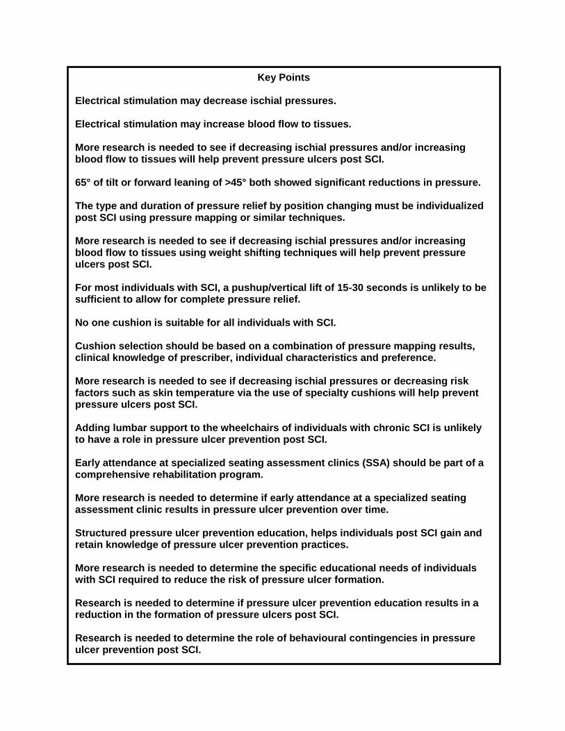
Key Points Electrical stimulation may decrease ischial pressures. Electrical stimulation may increase blood flow to tissues. More research is needed to see if decreasing ischial pressures and/or increasing blood flow to tissues will help prevent pressure ulcers post SCI. 65° of tilt or forward leaning of >45° both showed significant reductions in pressure. The type and duration of pressure relief by position changing must be individualized post SCI using pressure mapping or similar techniques. More research is needed to see if decreasing ischial pressures and/or increasing blood flow to tissues using weight shifting techniques will help prevent pressure ulcers post SCI. For most individuals with SCI, a pushup/vertical lift of 15-30 seconds is unlikely to be sufficient to allow for complete pressure relief. No one cushion is suitable for all individuals with SCI. Cushion selection should be based on a combination of pressure mapping results, clinical knowledge of prescriber, individual characteristics and preference. More research is needed to see if decreasing ischial pressures or decreasing risk factors such as skin temperature via the use of specialty cushions will help prevent pressure ulcers post SCI. Adding lumbar support to the wheelchairs of individuals with chronic SCI is unlikely to have a role in pressure ulcer prevention post SCI. Early attendance at specialized seating assessment clinics (SSA) should be part of a comprehensive rehabilitation program. More research is needed to determine if early attendance at a specialized seating assessment clinic results in pressure ulcer prevention over time. Structured pressure ulcer prevention education, helps individuals post SCI gain and retain knowledge of pressure ulcer prevention practices. More research is needed to determine the specific educational needs of individuals with SCI required to reduce the risk of pressure ulcer formation. Research is needed to determine if pressure ulcer prevention education results in a reduction in the formation of pressure ulcers post SCI. Research is needed to determine the role of behavioural contingencies in pressure ulcer prevention post SCI.
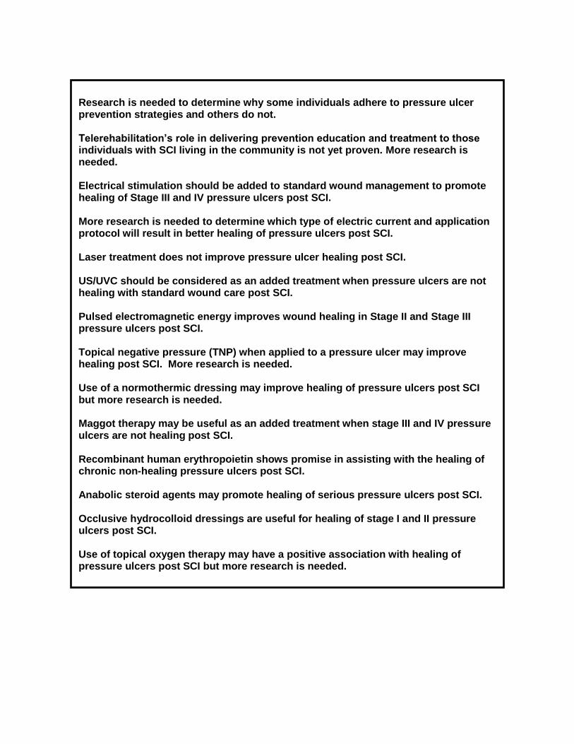
Research is needed to determine why some individuals adhere to pressure ulcer prevention strategies and others do not. Telerehabilitation’s role in delivering prevention education and treatment to those individuals with SCI living in the community is not yet proven. More research is needed. Electrical stimulation should be added to standard wound management to promote healing of Stage III and IV pressure ulcers post SCI. More research is needed to determine which type of electric current and application protocol will result in better healing of pressure ulcers post SCI. Laser treatment does not improve pressure ulcer healing post SCI. US/UVC should be considered as an added treatment when pressure ulcers are not healing with standard wound care post SCI. Pulsed electromagnetic energy improves wound healing in Stage II and Stage III pressure ulcers post SCI. Topical negative pressure (TNP) when applied to a pressure ulcer may improve healing post SCI. More research is needed. Use of a normothermic dressing may improve healing of pressure ulcers post SCI but more research is needed. Maggot therapy may be useful as an added treatment when stage III and IV pressure ulcers are not healing post SCI. Recombinant human erythropoietin shows promise in assisting with the healing of chronic non-healing pressure ulcers post SCI. Anabolic steroid agents may promote healing of serious pressure ulcers post SCI. Occlusive hydrocolloid dressings are useful for healing of stage I and II pressure ulcers post SCI. Use of topical oxygen therapy may have a positive association with healing of pressure ulcers post SCI but more research is needed.

This review has been prepared based on the scientific and professional information available in 2009. The SCIRE information (print, CD or web site www.scireproject.com) is provided for informational and educational purposes only. If you have or suspect you have a health problem, you should consult your health care provider. The SCIRE editors, contributors and supporting partners shall not be liable for any damages, claims, liabilities, costs or obligations arising from the use or misuse of this material. Regan M, Teasell RW, Keast D, Aubut JL, Foulon BL, Mehta S (2010). Pressure Ulcers Following Spinal Cord Injury. In Eng JJ, Teasell RW, Miller WC, Wolfe DL, Townson AF, Hsieh JTC, Connolly SJ, Mehta S, Sakakibara BM, editors. Spinal Cord Injury Rehabilitation Evidence. Version 3.0. www.scireproject.com We would like to acknowledge previous contributors: William B Mortenson
Table of Contents
1 Introduction ........................................................................................................................... 1 1.1 Impact of Pressure Ulcers .................................................................................................... 1 1.2 Incidence and Prevalence .................................................................................................... 1 1.3 Risk Factors ......................................................................................................................... 2 1.4 Stages (I-IV) of Pressure Ulcers ........................................................................................... 3 1.5 Prevention ............................................................................................................................ 3 1.6 Treatment ............................................................................................................................ 5
2 Prevention ............................................................................................................................. 5 2.1 Effects of Electrical Stimulation on Pressure Ulcer Prevention ............................................. 5 2.3 Pressure Relief Practices on Pressure Ulcer Prevention Post SCI ....................................... 8 2.4 Wheelchair Cushion Selection and Pressure Ulcer Prevention Post SCI .............................11 2.5 Lumbar Support Thickness on Reducing Ischial Pressures Post SCI ..................................16 2.6 The Effect of Specialized Seating Clinics on Pressure Ulcer Prevention Post SCI ..............17 2.7 Pressure Ulcer Prevention Education Post SCI ...................................................................19 2.8 Effect of Behavioural Contingencies on Pressure Ulcer Prevention Post SCI ......................21 2.9 Telerehabilitation and Pressure Ulcer Management Post SCI .............................................22
3 Treatment .............................................................................................................................24 3.1 Electrical Stimulation for Pressure Ulcer Healing Post SCI ..................................................24 3.2 Laser Treatment for Pressure Ulcer Healing Post SCI ........................................................27 3.3 Ultrasound/Ultraviolet C for Pressure Ulcer Healing Post SCI .............................................29 3.4 Effects of Non-Thermal Pulsed Electromagnetic Energy Treatment for Healing of Pressure
Ulcers Post SCI ...................................................................................................................30 3.5 Topical Negative Pressure Therapy for Pressure Ulcer Healing Post SCI ...........................31 3.6 Effects of Normothermic Dressing on Pressure Ulcer Healing Post SCI ..............................32 3.7 Recombinant Human Erythropoietin for Healing of Pressure Ulcers Post SCI .....................33 3.8 Anabolic Steroid Agents for Healing of Pressure Ulcers Post SCI .......................................34 3.9 Effectiveness of Dressings for Treatment of Pressure Ulcers Post SCI ...............................35 3.10 Maggot Therapy for Healing of Pressure Ulcers Post SCI .................................................38 3.11 Topical Oxygen for Treatment of Pressure Ulcers Post SCI ..............................................39
4 Summary ..............................................................................................................................40
References ..............................................................................................................................43

1
Pressure Ulcers Following Spinal Cord Injury
1 Introduction
1.1 Impact of Pressure Ulcers
Pressure ulcers are a serious, lifelong secondary complication of spinal cord injury (SCI) that have the potential to “interfere with physical, psychological and social well being and to impact overall quality of life” (Consortium for Spinal Cord Medicine 2000; p9). Although preventable in most situations, pressure ulcers may disrupt rehabilitation, prevent individuals with SCI from working or attending school and interfere with community reintegration. As well, the occurrence of a pressure ulcer can lead to rehospitalization often with an extended length of stay (Fuhrer et al. 1993; Krause 1998; Consortium for Spinal Cord Medicine 2000; Jones et al. 2003).
It has been estimated that pressure ulcers can account for approximately one-fourth of the cost of care for individuals with SCI. In the United States alone, it has been estimated that the cost of care for pressure ulcers is about 1.2-1.3 billion dollars annually while prevention could cost about one-tenth of this (Bogie et al. 2000; Jones et al. 2003). Because of the costs associated with treating pressure ulcers, Krause et al. (2001) state, “they have received more attention among rehabilitation and public health professionals than any other type of secondary condition associated with SCI” (p107). Despite the attention given to prevention strategies, pressure ulcers are common among individuals with SCI (Krause et al. 2001). 1.2 Incidence and Prevalence
Pressure ulcers (term used in the current document), also known as decubitius ulcers, ischemic ulcers, bed sores or skin sores, have been defined as a “localized injury to the skin and/or underlying tissue usually over a bony prominence as a result of pressure or pressure in combination with shear and/or friction” (NPUAP 2007). The primary cause of pressure ulcers is felt to be externally applied pressure for a prolonged period of time over bony prominences such as the sacrum and ischial tuberosities. This applied pressure leads to decreased blood supply to the overlying soft tissues; tissue ischemia and can ultimately lead to tissue necrosis (Lamid & Ghatit 1983; Crenshaw & Vistnes 1989; Bogie et al. 1995). DeLisa and Mikulic (1985) have noted that “the visible ulcer represents only the tip of the iceberg or the apex of the lesion” (p 210). It may take weeks before the actual size and depth of the ulcer is known. Deeper tissues such as muscle are more sensitive than skin to ischemia caused by pressure (Consortium for Spinal Cord Medicine 2000). Deep tissue injury has been added as a distinct pressure ulcer in the National Pressure Ulcer Advisory Panel’s 2007 updated pressure ulcer staging system (Black et al. 2007).
Pressure ulcer formation is a complex process that is still not clearly understood despite years of research. While the amount, duration and frequency of the applied pressure, the soft tissue’s response to loading, and the role of shear and/or friction are crucial, individual patient characteristics need to be assessed as well. Intrinsic factors such as diagnosis, history of previous tissue breakdown or surgical repair, body build, posture, muscle atrophy, nutritional status as well as magnitude and distribution of interface pressures must be considered. Extrinsic factors are also important including number of hours sitting or lying in wheelchair or bed; types of activities performed while sitting; level of functional independence; type of wheelchair, cushion and bed surface used and the support surface microenvironment; environment (climate, continence, temperature); finances; family/caregiver support; living arrangements and ease of follow up (Consortium for Spinal Cord Medicine 2000; Garber et al. 2007; Fleck & Sprigle 2007, Reger et al. 2007).

2
Annual incidence rates range from 20 – 31% and prevalence rates from 10.2 – 30% (DeLisa & Mikulic 1985, Byrne & Salzberg 1996). Chen et al. (2005) reported an increasing pressure ulcer prevalence in recent years not explained by aging, years since injury or different demographics. Risk of pressure ulcers was steady for the first 10 years and increased 15 years post injury. Fuhrer et al. (1993) noted that less extensive pressure ulcers, stages I & II, comprise about 75% of the total number of ulcers observed, leaving 25% as more severe or stage III and IV ulcers.
When a pressure ulcer is severe and not treated aggressively it can lead to further disability such as decreased mobility and loss of independence, surgical interventions, amputation, and even fatal infections (Krause 1998). It has been estimated that 7-8% of those who develop pressure ulcers will die from related complications (Richards et al. 2004). Due to the increasing life expectancy for those who sustain an SCI, the risk of developing pressure ulcers is even greater; thus making prevention a priority and a daily concern for individuals with SCI and health care providers. 1.3 Risk Factors
Prevention of pressure ulcers requires recognizing risk factors. The number of risk factors that have been associated with pressure ulcers post SCI is numerous and yet there is limited evidence that with more understanding of these risk factors a decrease in pressure ulcer incidence will occur (Salzberg et al. 1996). Risk factors that have been identified most often include: limitation in activity and mobility, injury completeness, moisture from bowel and bladder incontinence, lack of sensation, muscle atrophy, poor nutritional status and being underweight (DeLisa & Mikulic 1985; Salzberg et al. 1996; Krause et al. 2001). Studies have also found that those most likely to develop pressure ulcers are male, have lower levels of education, are unemployed and do not practice standing (Byrne & Salzberg 1996; Schryvers et al. 2000; Ash 2002; Richards et al. 2004). Other risk factors include: smoking (Lamid & Ghatit 1993; Salzberg et al. 1996; Niazi et al. 1997; Krause et al. 2001), number of comorbidities especially renal, cardiovascular, pulmonary disease and diabetes (Salzberg et al. 1996; Niazi et al. 1997; Ash 2002); residing in a nursing home/hospital (Byrne & Salzberg 1996); autonomic dysreflexia (Salzberg et al. 1996), anemia and hypoalbuminemia (DeLisa & Mikulic 1985; Scivoletto et al. 2004); spasticity and a history of previous ulcers (Vidal & Sarrias 1991; Byrne & Salzberg 1996, Guihan et al. 2008); and an increase in tissue temperature (Fisher et al. 1978); race and ethnicity (Guihan et al. 2008, Saladin and Krause,2009).
Identifying the significant risk factors associated with pressure ulcer development and being able to predict which individuals are most at risk are considered key elements of prevention. A formal assessment is required as research has shown that clinicians tend to intervene only at the highest levels of risk when an informal risk assessment is completed (Ayello & Braden 2002; AHCRP Executive Summary #3, 1992; Keast et al. 2006). Many risk assessment tools in existence were designed for the general population and for this reason their “predictive value” is imprecise in the SCI population (Consortium for Spinal Cord Medicine 2000).
A review of pressure ulcer risk assessment scales used with the SCI population was conducted by Mortensen & Miller (2008). Findings indicated that the SCIPUS (Salzberg et al. 1996) and SCIPUS-A (Salzberg et al. 1999) while developed specifically for the SCI population could not be recommended for use without further testing as they lacked reliability data and were developed and tested using the same retrospective data, limiting their validity. While the two scales showed promise, the Braden scale (Bergstrom et al. 1987) seemed to be the best tool available currently, as it is well validated. The Braden scale does require more testing with individuals with SCI.

3
1.4 Stages (I-IV) of Pressure Ulcers
“The assessment of an individual with a pressure ulcer is the basis for planning treatments, evaluating treatment effects and communicating with other caregivers” (AHCPR, Executive Summary #15 p 3). One key piece of this assessment is the staging of the pressure ulcer to classify the degree of tissue damage observed by the clinician (AHCPR, Executive summary # 15 1992). In 1989, the following staging system was recommended by the National Pressure Ulcer Advisory Panel (NPUAP 1989). As knowledge of the many factors associated with pressure ulcer formation continues to emerge, the staging system has been revised, most recently in 2007 (NPUAP 2007).
Table 1 National Pressure Ulcer Advisory Panel’s (NPUAP) updated pressure ulcer staging system (NPUAP 2007):
Stage: Description:
Deep Tissue Injury (Suspected) Stage Purple or maroon localized area of discolored intact skin or blood-filled blister due to damage of underlying soft tissue from pressure and/or shear. The area may be preceded by tissue that is painful, firm, mushy, boggy, warmer or cooler as compared to adjacent tissue.
Stage I Intact skin with non-blanchable redness of a localized area usually over a bony prominence. Darkly pigmented skin may not have visible blanching; its color may differ from the surrounding area.
Stage II Partial-thickness loss of dermis presenting as a shallow open ulcer with a red pink wound bed, without slough. May also present as an intact or open/ruptured serum-filled blister.
Stage III Full-thickness tissue loss. Subcutaneous fat may be visible but bone, tendon, or muscles are not exposed. Slough may be present but does not obscure the depth of tissue loss. May include undermining and tunneling.
Stage IV Full-thickness tissue loss with exposed bone, tendon, or muscle. Slough or eschar may be present on some parts of the wound bed. Often includes undermining and tunneling.
Unstageable Full-thickness tissue loss in which the base of the ulcer is covered by slough (yellow, tan, gray, green, or brown) and/or eschar (tan, brown, or black) in the wound bed.
Since 1989, this staging system has been used consistently in the literature and is widely used and supported (AHCPR 1992; Consortium of Spinal Cord Medicine 2000; RNAO 2002). However, authors of earlier studies have used numerous ways of documenting the severity of pressure ulcers making it challenging to draw parallels between studies.
1.5 Prevention
Preventing pressure ulcers is ultimately the best medicine and begins at the time of injury. Lifelong prevention recommendations include: examining skin daily to allow for early detection of a pressure ulcer, shifting body weight in bed and wheelchair on a regular basis independently or with assistance, keeping moisture accumulation to a minimum and cleaning and drying skin promptly after soiling, having an individually prescribed wheelchair, pressure redistribution

4
cushion and power tilt mechanism if manual pressure relief is not possible, ensuring all equipment is maintained and functioning properly, decreasing or stopping smoking and limiting alcohol intake (Consortium for Spinal Cord Medicine 2000). Krause et al. (2001) notes that effective prevention strategies require the individual with SCI to take responsibility for his/her skin care. Prevention strategies must be individualized to promote sustainable outcomes. Individuals with SCI need assistance from health care professionals to integrate realistic prevention strategies into daily schedules (Clark et al 2006). King et al. (2008) indicated that the value of preventative behavior needed to be emphasized. While in hospital, individuals with SCI need to practice skin care skills daily, know and direct their skin care program, learn to problem solve potential barriers while getting regular feedback on their performance. Support from family and the health care team is essential. As well, patients need to understand how quickly and quietly a pressure ulcer may appear and how it must be treated promptly. Other strategies suggested for education include training by peers, presenting information in a variety of methods including group learning, simulation exercises and case studies (Dunn et al. 2009).
It should be noted that outcome assessment for pressure ulcer prevention can be measured via either direct or indirect means. That is, the effectiveness of preventative interventions can be determined by direct indicators, like pressure ulcer incidence, or by indirect indicators, like ischial tuberosity (IT) pressure mapping or transcutaneous oxygen tension (PTCO2) levels. The former are preferred as they reflect definitive indications of the success (or failure) of preventative interventions. Sheppard et al. (2006) indicated that knowing one’s skin tolerance was related to intention to do pressure relief. Attendance at a seating clinic would be helpful as skin tolerance can be measured.
Whenever possible, individuals who are at risk for pressure ulcer development or who are being treated for a pressure ulcer should be referred to a registered dietitian for assessment and intervention as necessary (Keast et al. 2006). In a study by Houghton & Fraser (2008), paraplegic and tetraplegic spinal cord injured individuals living in the community with pressure ulcers (stage II to unstageable) underwent assessment that included medical and wound characteristics and screening of blood values for the presence of anemia, hydration status, glycemic control and hypoproteinemia. Study subjects with two or fewer abnormal blood values at the time of screening achieved complete wound closure following standard wound care and treatment with adjunctive therapy. Individuals who presented with greater than two abnormal blood values related to nutrition and hydration status did not achieve wound closure. The authors recommended that all individuals with pressure ulcers be screened for underlying inadequacies in nutrition and hydration and receive intervention to address these issues to promote optimal wound healing. Alexander et al. (1995) found that patients with paraplegia and a pressure ulcer had a resting energy expenditure that was hypermetabolic underscoring the need for thorough assessment and adequate nutritional support.
Recommendations for prevention or treatment of a pressure ulcer would include eating a well balanced, nutritionally complete diet with appropriate calories, proteins, micronutrients (vitamins and minerals) and fluids. The nutrition plan must be individualized based on the assessed needs (Consortium for Spinal Cord Medicine 2000; Keast et al. 2006). If a pressure ulcer is present, the plan would need to be optimized using foods, supplements and/or enteral nutrition if warranted. The individual’s weight would need to be monitored as an undesirable weight trend has been identified as an early indicator of risk (Keast et al. 2006).
There have been numerous recommendations for the prevention of pressure ulcers post SCI but it is important to consider the evidence that informs those recommendations. Potential preventative techniques found in the SCI literature that have been reviewed and will be

5
discussed in the following section include: effect of electrical stimulation on ischial pressures and blood flow, pressure relief practices, wheelchair cushion selection, effect of lumbar support thickness on ischial pressures, specialized seating clinics, pressure ulcer prevention education, behavioural contingencies, and telerehabilitation. 1.6 Treatment
Once a pressure ulcer has begun it is important to prevent if from worsening and ultimately to have it heal quickly but this is challenging. Rappl (2008) examined the metabolic and physiological changes that happen in tissue below the level of a SCI in relation to the events which take place during wound healing. The author examined that every step of wound healing is affected by the physiological changes that occur post SCI explaining why pressure ulcers may heal more slowly in individuals with a SCI. As previously stated, severe pressure ulcers can lead to further disability, surgery, amputation and death (Krause 1998). According to Chen et al. (2005) pressure ulcers are among the leading cause of unplanned rehospitalization post SCI and can contribute to longer lengths of stay and more costly treatment than other medical conditions. Once an individual has had an ulcer they are at increased risk for recurrence (Krause & Broderick 2004). Pressure ulcer treatment is more costly than prevention (Bogie et al. 2000; Jones et al. 2003). In addition to standard wound care, many adjunctive therapies are used to accelerate closure of wounds that are hard to heal. It is important to identify appropriate clients who are likely to benefit for these treatments as they are often time consuming and expensive (Houghton & Fraser 2008; Allen & Houghton 2003).
Research has looked at the effect of: electrical stimulation, laser, US/UVC, non-thermal pulsed electromagnetic energy, topical negative pressure, normothermia, recombinant human erythropoietin, anabolic steroid therapy, effectiveness of various dressings, maggot therapy and topical oxygen for healing of pressure ulcers post SCI. Each of these treatments will be discussed in subsequent sections.
2 Prevention
2.1 Effects of Electrical Stimulation on Pressure Ulcer Prevention
Electrical stimulation has been used since the 1960’s to enhance healing of various chronic wounds including pressure ulcers in both the able bodied and spinal cord injured individual (Kloth & Feeder 1988; Baker et al. 1996, Bogie et al. 2000). More recently electrical stimulation has been studied to assess its potential for pressure ulcer prevention post SCI.
Given that the primary cause of pressure ulcers is felt to be externally applied pressure over bony prominences such as ischial tuberosities (Bogie et al. 1995), researchers have studied the role of electrical stimulation in reducing ischial pressures and redistributing seating interface pressures both of which could assist with pressure ulcer prevention (Bogie et al. 2006). Prevention studies are focusing on skin vs. muscle stimulation, dynamic vs. long-term effects and surface vs. implanted devices (Levine et al. 1990; Bogie et al. 1995; 2000; 2006).
Table 2 Effects of Electrical Stimulation on Reducing Ischial Pressure Post SCI
Author Year Country Score
Research Design Total Sample Size
Methods Outcome
Population: SCI: Age = 27-47 yrs;
Gender: males = 7, females = 1; Severity 1. Overall, with chronic neuromuscular
electrical stimulation (NMES), mean

6
Author Year Country Score
Research Design Total Sample Size
Methods Outcome
Bogie & Triolo 2003 USA
Downs & Black score=13 Pre-post
N=8
of injury: AIS: A = 6, B = 2. Treatment: The exercise regimen
included 3 different stimulation patterns. Duration of exercise was varied over the 8 wk training period as the muscles became conditioned. Outcome Measures: Mean interface
pressure, mean ischial region interface pressure.
interface pressure showed no significant differences between baseline and post exercise levels.
2. Mean ischial region interface pressure had a uniform tendency to decrease post exercise assessment, p<0.01.
Lui et al. 2006b UK
Downs & Black score=13
Case Series N=5
Population: SCI: Mean age = 45 yrs;
Gender: males = 4, females = 1; Level of injury: paraplegia = 5, Severity of injury: complete = 5. Treatment: SARS implant applied
bilateral electrical stimulation for 10 seconds (frequency=20pps; pulse width range 8-800secs; amplitude of “1”). Second sacral nerve root was stimulated (S2). Outcome Measures: PP & GPP; before
and during electrical stimulation using pressure mapping.
1. There was an average 33% decrease in PP during stimulation (at rest= 148.6 mmHg; during FES =99.8mmHg; p<0.01).
2. There was also a mean 38% decrease in GPP during stimulation (at rest=54.6 mmHg; during FES= 33.8 mmHg; p<0.05).
3. An increase in pulse width resulted in lower PP. Lowest PP was attained at a stimulation pulse width range from 64-600 secs.
4. No complications were reported.
Note: AIS=ASIA Impairment Scale; GPP=Gradient Peak Pressure, FES=Functional Electrical Stimulation; NMES=Neuromuscular Electrical Stimulation; PP=Peak Pressure; SARS=Sacral Anterior Root Stimulation
Discussion
Two articles were found that examined the effects of electrical stimulation on ischial pressure. Bogie and Triolo (2003) studied changes in interface pressure distribution at the support/surface interface following 8 weeks of neuromuscular electrical stimulation (NMES) delivered via an implanted neuroprosthesis. With NMES, mean ischial regional interface pressure had a uniform tendency to decrease post exercise assessment, p<0.01.
Lui et al (2006b) studied the effects of electrical stimulation delivered via an implanted sacral anterior root stimulator (SARS) on seat interface pressure distribution. With electrical stimulation of the S2 nerve root sufficient to result in gluteal muscle contraction, there was an average decrease of 33% in peak pressure p<0.01 and a 38% decrease in gradient peak pressure p<0.05 at the ischial tuberosities of the seated participants.
While it is difficult to compare these results because one study used 8 weeks of NMES versus dynamic electrical stimulation, it does appear that electrical stimulation decreases ischial pressures. More research is needed to study the effect of long term electrical stimulation on reducing ischial pressures and whether this can be used in a clinical setting to prevent pressure ulcers post SCI.
As was stated previously, researchers are focusing on the effects of electrical stimulation, which may have a role in pressure ulcer prevention post SCI. One effect under investigation is the ability of electrical stimulation to change blood flow to skin and muscle. Bogie et al. (2006) state that with increasing interface pressures over bony prominences, regional blood flow is adversely affected. It is believed that by increasing regional blood flow, tissue health would be enhanced assisting with pressure ulcer prevention (Levine et al. 1990; Bogie et al. 1995; 2000; 2006).

7
Table 3 Electrical Stimulation for Increasing Tissue Blood Flow Post SCI
Author Year Country Score
Research Design Total Sample Size
Methods Outcome
Lui et al. 2006a UK
Downs & Black score=15 Case Series
N=6
Population: SCI: SARS study: Age = 35-
62 yrs; Gender: males = 5, females = 1; Level of injury: T3-T1; Severity of injury: complete = 6; Time since injury = 9-24 yrs. Treatment: SARS study - anterior root
stimulator (SARS) implant applied bilateral electrical stimulation to S2 nerve root for 10 seconds (frequency=20pps; pulse width range 8-800secs; amplitude of “1”). Outcome Measures: IHB; IOX before and
during electrical stimulation.
SARS study 1. IHB significantly increased during
stimulation (before stimulation, M=0.8; during stimulation, M=0.9; p=0.005).
2. IOX also increased (before stimulation, M=1.1; during stimulation, M=3.0; p=0.02).
Bogie & Triolo 2003 USA
Downs & Black score=13 Pre-post
N=8
Population: SCI: Age = 27-47 yrs;
Gender: males = 7, females = 1; Height = 1.65 to 1.88 meters; Weight = 49.89-113.40 kg; Level of injury: C5/6 to T9; Severity of injury: AIS: A = 6, B = 2. Treatment: Electrical stimulation delivered
via an implanted neuroprosthesis, which included gluteal electrodes, 8 weeks of conditioning exercises followed. Outcome Measures: Transcutaneous
Oxygen Levels (PTCO2).
1. Baseline mean unloaded tissue oxygen levels increased by 1-36% at post exercise assessment for 5/8 subjects.
2. Differences between baseline and post exercise tissue oxygen levels did not show any statistical significance.
Mawson et al. 1993 USA
Downs & Black score=10 Case Series
N=29
Population: SCI: Age = 18-57 yrs; Site of
ulcer: sacral = 7, heel = 2, other = 1; Ulcer grade: 1-4. Treatment: Study was carried out on SCI
patients lying on egg crate mattresses. Sensor was applied to the skin at approximately the second sacral segment along the midline using a two-sided airtight seal. 2 electrodes and conductive sponges, measuring 4 cm in diameter were used for administering electrical stimulation. Outcome Measures: PTCO2.
1. Experiment 1: Subsequent experiments were performed using 75 volts as no additional effect on transcutaneous oxygen tension (PTCO2) was seen when 100 volts was used.
2. Experiment 2: Compared to final baseline PTCO2 reading (mean ± SD) of 49±21mmHg, the level reached at the 30min period of high voltage pulsed galvanic stimulation (HVPGS) was 66±18 mmHg -- 35% higher (p<0.00001).
3. The level fell slightly following the first 15 minutes post stimulation period (p<0.00001).
4. Experiment 3: No change in PTCO2
with simulated HVPGS. 5. Experiment 4: No significant
differences were observed (p=0.66 in all comparisons) when experiment 2 and 4 results were compared.
Note: AIS=ASIA Impairment Scale; IHB=Cutaneous Hemagloblin; IOX= Oxygenation ; SARS=Sacral Anterior Root Stimulation
Discussion
Lui et al (2006a) administered dynamic electrical stimulation to the S2 nerve root through an implanted sacral anterior nerve root stimulator (SARS) and studied the effects on cutaneous

8
blood circulation as measured by changes in the index of Hemoglobin (IHB) and index of oxygenation (I0X). With stimulation there was a statistically significant increase in IHB p = 0.005 and I0X p = 0.02. The mechanism of how electrical stimulation altered IHB and I0X is unclear.
Bogie and Triolo (2003) administered 8 weeks of NMES to 8 subjects using gluteal electrodes. They then assessed unloaded gluteal tissue blood flow through assessment of local transcutaneous oxygen levels (PTCO2). While the results did not reach statistical significance, baseline mean unloaded tissue oxygen levels increased by 1-36% in 5/8 subjects.
Mawson et al. (1993) administered high voltage pulsed galvanic stimulation (HVPGS) to 29 SCI subjects lying supine. Baseline PTCO2 levels were compared to levels reached at the end of 30 minutes of HVPGS. The authors found PTCO2 level at the end of stimulation was 66±18 mmHg – 35% higher (p<0.00001).
While the evidence to date is promising, more research is needed to determine the effect of electrical stimulation on blood flow to tissues at risk for pressure ulcer development post SCI.
Conclusion
There is limited level 4 evidence that electrical stimulation decreases ischial pressures post SCI.
There is level 4 evidence that electrical stimulation may increase blood flow at sacral and gluteal areas post SCI.
2.3 Pressure Relief Practices on Pressure Ulcer Prevention Post SCI
Teaching individuals with spinal cord injuries to shift their weight regularly while seated is a common and intuitive recommendation for pressure ulcer prevention as it is hypothesized that this relieves pressure on at risk tissues and allows for recovery of blood flow and oxygenation (Consortium for Spinal Cord Medicine 2000; Coggrane & Rose 2003; Makhsous et al 2007a). Several techniques have been suggested depending on the physical and cognitive status of the individual and include a lateral, forward lean or vertical push up (Bogie et al 1995; Consortium for Spinal Cord Medicine 2000). When a manual weight shift cannot be performed the use of a power tilt feature has been recommended (Consortium for Spinal Cord Medicine 2000).
Table 4 Pressure Relief Practices on Preventing Ulcers Post SCI
Author Year Country Score
Research Design Total Sample Size
Methods Outcome
Spijkerman et al. 1995 Netherlands
Population: Mean age=37.7yrs; Gender:
males=15, females=3; Level of injury: C2-1. Body tilt had a significant effect on
the mean pressure, p=0.003.
Electrical stimulation may decrease ischial pressures.
Electrical stimulation may increase blood flow to tissues.
More research is needed to see if decreasing ischial pressures and/or increasing blood flow to tissues will help prevent pressure ulcers post SCI.

9
Author Year Country Score
Research Design Total Sample Size
Methods Outcome
D&B=18 Pre-Post
N=18
L2; Severity of injury: complete Treatment: Interface pressure was
assessed on SCI patients using various seat inclinations. Outcome Measures: Mean pressure
2. At seat inclination of 5, 15 and 25 overall mean pressure was 86.79, 86.90 and 82.91.
Makhsous et al. 2007a USA
Downs & Black score=14
Case Control/Repeated Measures
N=60
Population: SCI: Paraplegics (n=20):
Mean age = 35.1 yrs; Gender: males =20; Mean weight = 87.2 kg; Mean time since injury = 8.4 yrs. Tetraplegics (n=20): Mean age = 36.5 yrs; Gender: males = 15, females = 5; Mean weight = 81.8 kg; Mean time since injury = 9.2 yrs; Controls (n=20): Mean age: 39.3 yrs; Gender: males = 10, females = 10; Mean weight = 71.3 kg. Treatment: 2-1 hour sitting protocols: 1)
Dynamic protocol: alternating every 10 minutes between normal sitting (sitting upright with full seat support and no added lumbar support) and an off-loading sitting (sitting upright with position in seat section tilted down 20 degrees with pressure to IT and coccyx) configuration; 2) Wheelchair pushup protocol: one wheelchair pushup every 20 minutes, while in normal sitting configuration. Outcome Measures: Transcutaneous
partial pressure of oxygen (tcPO2) and carbon dioxide (tcPCO2).
1. In normal sitting, mean tcPO2 at IT was < 10mmHg and mean tcPCO2 was >60mmHg, for all groups. During off loading sitting configuration, IT tcPCO2 was maintained > 50mm Hg and tcPCO2 at <45 mm Hg for all groups. During pushup protocol (avg 49 sec), IT tcPO2 increased and tcPCO2 reduced only slightly.
2. With pressure release (off loading configuration) average perfusion recovery time for tcPO2 was 200-250 seconds for all groups.
3. tcPO2 perfusion recovery time was significantly shorter for control group than SCI groups, p<0.001.
Coggrave & Rose 2003 UK
Downs & Black score=14
Case Series N=50
Population: SCI: Age = 20-83 yrs;
Gender: males = 33, females = 13; Time since injury = 5 wks-50 yrs. SCI, Frankel grade A-D Treatment: Retrospective chart review. Outcome Measures: Effect of pressure
relief on transcutaneous oxygen tension (TCPO2).
1. Mean duration of pressure relief required to raise tissue oxygen to unloaded levels was 1 min 51 sec (range 42 secs-3½ mins).
2. Leaning forward with elbows or chest on knees, leaning from side to side or tilting back in wheelchair to > 65
o were all effective for pressure
relief (raising TCPO2 to unloaded levels) and more easily sustained for most individuals than a pressure relief lift.
3. Resulted in a change in practice at the seating clinic.
Henderson et al. 1994 USA
Downs & Black score=12
Case Series N=10
Population: SCI: Age = 22-67 yrs;
Gender: males = 9, females = 1; Time since injury = 1 mnth to 7 yrs. Treatment: Subjects sat upright in
wheelchair in neutral position; tipped backward at 35
o & 65
o; assisted to lean
forward (>45o from wheelchair backrest).
Pressures were measured at ischial tuberosity (IT) (point pressure) and circumscribed area around IT. Outcome Measures: Pressure levels.
1. Average pressure in the resting seated position was 189 mmHg for point pressure area and 114 mmHg for the circumscribed area.
2. When patients were in the 65o
backward tip position there was a 47% reduction in maximum point pressure and 36% reduction for the circumscribed area pressure. (p<0.05).
3. In the leaning forward position there was a 78% reduction in maximum

10
Author Year Country Score
Research Design Total Sample Size
Methods Outcome
point pressure and a 70% reduction in circumscribed area (p<0.05).
Hobson 1992 USA
Downs & Black score=10
Case Control N=22
Population: SCI: Mean Age = 40.9 yrs;
Gender: males = 10, females = 2; Mean weight = 59.8 kg; Level of injury: paraplegia = 7, tetraplegia = 5; Severity of injury: complete; Mean time since injury = 19.5 yrs; Chronicity = chronic. Able-Bodied: Mean age = 39.3 yrs; Gender: males = 6, females = 4. Treatment: Nine typical wheelchair
sitting postures. Outcome Measures: Tangentially
induced surface shear (TIS); Interface pressure.
1. Mean maximum pressure was on average 26% higher in the SCI group versus the able-bodied group.
2. Forward trunk flexion of 50° reduced the average pressure for both groups; however, SCI group encountered a 10% increase in pressure at the initial 30° of forward flexion before a reduction occurred.
3. SCI subjects had a mean peak pressure gradient that was 1.5-2.5 higher than able-bodied subjects. Maximum decrease of peak pressure gradient from a neutral position happened after the backrest reclined to 120° (18%).
4. When a sitting position change occurred, a similar shift to the anterior/posterior midline location of maximum pressure was experienced in both groups. From neutral, a forward trunk flexion at 30° and 50° produced a 2.4 and 2.7cm posterior shift. When the backrest reclined to 120°, the greatest posterior shift occurred at 6cm.
5. Maximum reduction of TIS occurred with forward trunk flexion of 50° (-133%) and full body tilt of 20° (-85%). Backward recline to 120° caused increase in TIS of 25%.
Note: IT= Ischial Tuberosity
Discussion
There are very few studies that have researched which techniques provide adequate pressure relief and how long a weight shift must be performed to unload weight-bearing areas such as the ischia.
Spijkerman et al. (1995) assessed interface pressure while individuals were tilted at 5°, 15° and 25° from horizontal. Results indicated that body tilt had a significant effect on mean pressure p=0.003. The lowest overall mean pressure (82.91mmHg) was demonstrated at 25° tilt.
Coggrave and Rose (2003), in a retrospective chart review of 46 SCI subjects seen in a seating clinic, assessed the duration of various pressure relief positions required for loaded transcutaneous oxygen tension (tCPO2) to recover to unloaded levels. Results indicated that it took approximately 2 minutes of pressure relief to raise tissue oxygen to unloaded levels for most subjects. This length of pressure relief was more easily sustained by the subjects leaning forward, side to side or having the wheelchair tipped back at > 65º compared to a pressure relief lift.

11
Similar to Coggrave and Rose (2003), Makhsous et al (2007a) demonstrated full recovery of tcPO2 with the dynamic protocol in the off loading configuration but it took > 2 minutes to achieve this result. Those individuals with paraplegia using a wheelchair pushup were only able to sustain the lift for 49 seconds leading to incomplete recovery of tissue perfusion.
Henderson et al. (1994) pressure mapped 10 SCI subjects and recorded pressures at the ischial tuberosity (IT) and a circumscribed area around the IT. The authors then pressure mapped the subjects with their wheelchairs tipped back at 35º, 65º and after the subjects were assisted into a forward leaning position >45°. Results showed that the wheelchairs tipped back at 65º and the subjects in a forward leaning position demonstrated statistically significant pressure reduction at the IT and circumscribed area (p<0.05) with the forward lean showing the greatest reduction (78% reduction at IT, 70% reduction circumscribed area).
Hobson (1992) showed that for individuals with SCI, changes in posture can reduce maximum pressures that occur while seated. Recline of the backrest to 120º, full body tilt to 20º, forward flexion to 50º and lateral bending to 15º all resulted in decreases in maximum pressures. Maximum reductions in tangentially induced shear forces (TIS) occurred with forward trunk flexion of 50º and full body tilt of 20º; backrest recline to 120º increased TIS by 25%.
The studies reviewed demonstrate that pressure relief by position change, if sustained for an appropriate length of time, results in pressure reduction and recovery of tcPO2 to unloaded levels. The type and duration of pressure relief required to achieve these results varied from individual to individual. Sustaining a pressure relief lift/pushup for the time required to allow for recovery of tcPO2 to unloaded levels (1-2 min) would be difficult for most individuals with SCI.
Conclusion
There is level 3 evidence that 1-2 minutes of pressure relief must be sustained to raise tissue oxygen to unloaded levels. There is level 4 evidence to support position changes to reduce pressure at the ischial tuberosities.
2.4 Wheelchair Cushion Selection and Pressure Ulcer Prevention Post SCI
Bogie et al (1995) stated that 47% of pressure ulcers occur at the ischial tuberosities or sacrum and are therefore more likely to have been initiated while seated. Provision of a wheelchair cushion that relieves and redistributes pressure and reduces risk of pressure ulcer formation is an important prevention recommendation. Historically, cushion design has been based on the belief that sitting interface pressure should be distributed evenly to reduce areas of high pressure underneath bony prominences (Yuen & Garrett 2001). Cushion selection can be
65° of tilt or forward leaning of >45° both showed significant reduction in pressure.
The type and duration of pressure relief by position changing must be individualized post SCI using pressure mapping or similar techniques.
More research is needed to see if decreasing ischial pressures and/or increasing blood flow to tissues using weight shifting techniques will help prevent pressure ulcers post SCI.
For most individuals with SCI, a pushup/vertical lift of 15-30 seconds is unlikely to be sufficient to allow for complete pressure relief.

12
difficult as there are numerous cushions on the market each citing specific characteristics along with various amounts of pressure reduction and redistribution that make a cushion “superior.” When assessing an individual for a cushion, factors such as the degree of pressure reduction and redistribution (Garber 1985), temperature effects (Fisher et al 1978; Seymour & Lacefield 1985); level of SCI, pressure relief abilities, transfer technique and lifestyle (Garber 1985; Markhous et al. 2007a) are typically considered. As well as a reduction in pressure ulcer risk, cushions must also promote adequate posture and stability for the individual with SCI (Sprigle et al. 1990). Seat cushions can be made from a variety of materials, can be static or dynamic (Garber 1985; Markhous et al. 2007a) and incorporated into a variety of wheelchairs. See Table 20.4. Table 5 Wheelchair Cushion Selection and Pressure Ulcer Prevention Post SCI
Author Year Country Score
Research Design Total Sample Size
Methods
Outcome
Makhsous et al. 2007b USA
Downs & Black score=18
Case Control N=60
Population: Mean age = 37 yrs; Gender:
males = 45, females = 15; Level of injury: paraplegia = 20, tetraplegia = 20, and able-bodied = 20. Treatment: Two one hour protocols. 1)
Alternative – sitting position was altered every 10 minutes between normal and WO-BPS (partially removed ischial support and lumbar support). 2) Normal – normal posture and pushups every 20 minutes. Outcome Measures: Interface pressure
on backrest and seat.
1. Those with tetraplegia had a larger contact area at the anterior portion of the cushion, as compared to the other groups.
2. The mean pressure over the whole cushion was significantly different for each group (p<0.001).
3. Tetraplegics had the highest mean pressure during the WO-BPS posture, as compared to the other groups (p<0.001).
4. The contact area of the posterior portion of the cushion and the peak interface pressure decreased in all groups, with the largest decrease in tetraplegics for the latter. The mean pressure on the anterior and middle portions of the cushion increased in all groups.
5. At the posterior portion of the seat where ischial tuberosities are usually positioned, average pressure was higher for those with paraplegia (88.9 mmHg).
6. Average push up time was 49 sec for those with paraplegia.
Burns & Betz. 1999 USA
Downs & Black score=17
Prospective controlled trial
N=16
Population: Mean age = 46 yrs; Gender:
males = 16; Level of injury: tetraplegic; Severity of injury: AIS: A = 7, B = 9. Treatment: Two static wheelchair
cushions (dry flotation and gel) upright and at 45° tilt, compared to a dynamic cushion that was composed of two air bladders (H & IT) that alternated between inflation and deflation. Outcome Measures: Interface pressure
at ischial tuberosities (IT) was assessed with Clinseat seating interface pressure sensor.
1. When compared in the high pressure condition, all cushions were significant (p<0.001), with means of 111 mmHg (dry flotation), 128 mmHg (gel), and 157 mmHg (dynamic).
2. When compared in the low pressure condition, only gel flotation (86 mmHg), and the dynamic cushion (71 mmHg), were significant (p<0.05).
3. The IT had a significantly higher mean during IT bladder inflation of the dynamic cushion than the high pressure position in the static

13
Author Year Country Score
Research Design Total Sample Size
Methods
Outcome
cushions (p<0.01), with the dry flotation having significantly lower pressure than the gel cushion (p<0.01).
4. The IT had significantly lower mean in the lower pressure position only for the dynamic cushion as compared to the gel cushion (p<0.01).
Gilsdorf et al. 1991 USA
Downs & Black score=14
Case Series N=17
Population: Paraplegics (N=6): Mean
weight = 83 kg; Tetraplegics (N=5): Mean weight = 66 kg; Able-bodied controls (N=6): Mean weight = 76 kg. Treatment: 30 minute sitting intervals, on
different surfaces [Jay cushion; ROHO cushion; hard surface (controls only)] in a wheelchair that had a forceplate attached to it. Outcome Measures: Normal & shear
seating forces; Armrest forces; Centre of mass location.
1. On Jay cushion, tetraplegics had higher amplitude lateral movements and paraplegics had more lateral zero-crossings, when compared to ROHO cushion.
2. Larger arm force variation was found in paraplegics.
3. On the ROHO cushion, all subjects had larger normal and shear forces and an anterior centre of mass.
4. Paraplegics had more variation, while tetraplegics had less, on static force factors between cushion types.
5. SCI groups had higher force measurements than control group.
6. Armrest forces applied by paraplegics were larger than those applied by tetraplegics (8-9% vs. 5%, p<0.11).
Brienza & Karg 1998 USA
Downs & Black score=14
Case Series N=12
Population: SCI: Age: 21-52 yrs; BMI =
17-32.3 kg/m2.
Treatment: Assessed forces for 3
different surfaces (flat foam, initial contour and final optimized contour) with the force sensing array (FAS) pad between the cushion and buttocks. Compared SCI to elderly subjects (No SCI). Outcome Measures: Pressure mapping,
BMI.
1. Depth values for the SCI group↑ from 37.9 mm to 52.5 mm (p<0.001).
2. A significant increase was also noted for the elderly group.
3. The mean max depth of the final contour was deeper for the SCI group (p=0.016).
4. Mean pressure values for initial and final cushions were significantly less than for flat cushions (p=0.006, p=0.003 respectively).
5. In general, mean and peak pressure were greater for the SCI subjects than for elderly subjects.
6. BMI was significantly related to peak and mean pressure values.
Seymour & Lacefield 1985 USA
Downs & Black score=13
Case Control N=20
Population: Age = 16-35 yrs; Weight =
40.6-72.5 kg; 10 cases, 10 controls. Treatment: 7 commercially available
cushions and 1 experimental cushion were evaluated for each subject. Outcome Measures: Temperature and
pressure effects for each cushion. Subjects were asked to rate each cushion as to cosmesis, handling and suitability
1. Greatest pressure was seen under the soft tissue areas of most subjects; no significant differences between the cases and controls.
2. Temperatures were lowest for gel, water and air cushions and highest for alternating pressure and foam cushions.
3. SCI group - Greatest pressure

14
Author Year Country Score
Research Design Total Sample Size
Methods
Outcome
for purchase.
under a bony area occurred most often with the Spenco cushion (90.10 mmHg); controls - it occurred most often with the Tri-pad (89.20 mmHg) indicating that these cushions did not compare favorably to others.
4. There was wide variability in pressure measurements in individual subjects (SD=12.21 mmHg). However, air filled (Bye Bye Decubiti) had the best pressure readings.
5. Cosmesis (83%) and handling (73%) were related to purchase decisions.
Garber 1985 USA
Downs & Black score=8 Case Series
N=251
Population: SCI: Gender: males = 207,
females = 44. Treatment: Assessment of pressure
distribution for 7 cushions. Outcome Measures: Seated pressure
distribution.
No statistical results reported. 1. The air filled cushion (ROHO which
was 1 of 2 used) produced the greatest pressure reduction in 51% of the subjects.
2. A foam cushion (the stainless comfy hard cushion) was effective for only 18% of the subjects even though it was the second most frequently prescribed cushion.
3. More subjects with tetraplegia received the ROHOs than subjects with paraplegia (55% vs. 45%) while more paraplegic subjects were prescribed the Jay cushion (a combination of foam and flotation materials (19% vs. 7%).
Takechi & Tokuhiro. 1998 Japan
Downs & Black score=6 N=6
Case Series
Population: Age = 18-48 yrs; Gender:
males = 6; Level of injury: paraplegia = 6; Severity of injury: complete = 6. Treatment: Five different cushions (air
cushion, contour cushion, polyurethane foam cushion, Cubicushion, silicone gel cushion). Outcome Measures: Tekscan BigMat
pressure mapping system measuring peak pressures and area of total contact.
1. If the area of contact was more widespread, the peak pressure was found to be lower.
2. The air cushion had the largest area of pressure distribution and the lowest peak pressure (257-87g/cm
2). The silicone cushion
hadthe second lowest (292-129g/cm
2) peak pressure.
Note: AIS=ASIA Impairment Scale; BMI=Body Mass Index
Discussion
Numerous authors have investigated various wheelchair cushions and seating systems to try and determine which offer the most pressure or risk factor reduction to prevent occurrence of pressure ulcers in individuals with SCI.
Makhsous et al. (2007b), in a case-control study, exposed subjects to two 1-hour protocols: alternate, where sitting posture was alternated dynamically every 10 minutes between normal (sitting upright with ischial support) and sitting upright with partially-removed ischial support and
15
lumbar support (WO-BPS), and normal (normal posture plus pushups performed every 20 minutes). These investigators found that the anterior portion of the seat cushion had a larger contact area among those with tetraplegia compared to those in the other groups. It also was determined that those with a SCI had a larger contact area in the mid portion of the seat cushion. There were significant differences between the groups when looking at the average pressure over the whole seat (p<0.001) and the total contact area on the seat cushion. With the WO-BPS posture, the average pressure for the tetraplegia group was higher than it was for the other groups (p<0.001). Most importantly, the total contact area on the posterior portion of the cushion was less for the WO-BPS posture group. As well, peak interface pressure was lower for all groups, with the greatest decrease from normal posture seen in the tetraplegia group. The average pressure increased on the anterior and middle portion of the cushion in all groups.
In the study conducted by Burns and Betz (1999), 3 wheelchair cushions were tested: dry flotation (ROHO High Profile), gel (Jay 2), and dynamic (ErgoDynamic), the last consisting of two air-filled bladders (H-bladder, IT-bladder). These were compared to each other under high pressure conditions (upright sitting or IT-bladder inflated) and low pressure conditions (seat tilted back 45° or H-bladder inflated). When analyzing the pressure placed on the ischial tuberosities, it was found that the pressure was higher during upright sitting than in the tilted back position for both the dry flotation and the gel cushion (p<0.001), with the dry flotation cushion providing more pressure relief than the gel cushion during upright sitting (112 versus 128 mmHg, p=0.01). Mean pressure with the IT-bladder-inflated cushion (157 mmHg) was greater than upright pressures for either the dry flotation or gel cushions (111 and 128 mmHg, respectively p<0.01). Most importantly, ischial tuberosity pressure for the dynamic cushion during H-bladder inflation in an upright position was comparable to the pressure for the dry flotation cushion in a tilted back position (71 versus 74 mmHg, p=0.91) and significantly less than the pressure obtained with the gel cushion (71 versus 86 mmHg, p<0.05). Brienza and Karg (1998) had subjects sit on 3 different surfaces (flat foam, initial contour and final contour). Interface pressures were measured using a pressure-sensing pad. Results indicated that when SCI subjects were compared to the elderly subjects without SCI, depth values increased and the mean maximum depth of the final contour was deeper for the SCI group, suggesting that pressure distributions for the SCI group were more sensitive to support surface characteristics than elderly subjects without SCI.
Seymour et al. (1985) evaluated 8 cushions for pressure, temperature effects and subjective factors influencing cushion purchase. While data indicated a wide variability in pressure measurements in individual subjects, the air filled cushion (Bye Bye Decubiti) had the best pressure readings. The alternating pressure and foam cushions had consistently higher temperature readings across both groups.
Gilsdorf et al (1991) studied subjects sitting on ROHO and Jay cushions. Normal force, shear force, centre of force, lateral weight shifts and amount of weight supported by armrests were studied under static and dynamic conditions. The ROHO cushion showed a tendency to carry a larger percentage of total body weight; have a more anterior centre of mass; and showed more forward shear force. There were more lateral weight shifts on the Jay cushion. Armrests supported a portion of body weight.
Garber (1985) evaluated 7 cushions based on amount of pressure reduction. The author also looked at how frequently each cushion was prescribed to subjects with quadriplegia and paraplegia. The ROHO cushion produced the greatest pressure reduction in the majority of subjects (51%) but was prescribed more often for subjects with quadriplegia vs. paraplegia (55% vs. 45%).

16
Takechi & Tokuhiro (1998) studied the seated buttock pressure distribution in six patients with paraplegia using computerized pressure mapping. Five wheelchair cushions were evaluated (air cushion, contour cushion, polyurethrane foam cushion, cubicushion, silicone gel cushion). Tests showed that if the area of contact was more widespread, the peak pressure was lower. The air cushion and the silicone cushion were found to have the lowest peak pressures. These studies demonstrate that there are individual variations inherent in those with SCI (e.g. paraplegia vs. tetraplegia). As a result the need for objective measures such as pressure mapping is needed to assist with individualizing a wheelchair cushion prescription. Objective findings together with the clinical knowledge of the prescriber, individual characteristics and the client’s subjective reports need to be considered when prescribing a wheelchair cushion to minimize pressure ulcer risk factors. None of these studies included direct evidence of pressure ulcer prevention associated with a particular cushion type.
Conclusion
There is level 3 evidence that various cushions or seating systems (e.g. dynamic versus static) are associated with potentially beneficial reduction in seating interface pressure or pressure ulcer risk factors like skin temperature.
2.5 Lumbar Support Thickness on Reducing Ischial Pressures Post SCI
Shields and Cook (1992) discussed the role spinal deformities such as kyphosis, may play in the formation of pressure ulcers in individuals with chronic SCI. In previous research with non-disabled subjects, they had demonstrated that the addition of lumbar support reduced highest seated buttock pressure and was associated with a change in pelvic tilt. If those findings were to hold true in the SCI population, the authors noted this could lead to ways to assess seated postures for appropriate pressure distribution and augment electric wheelchair seating systems to provide continuous pressure shifts.
Table 6 Lumbar Support Thickness on Reducing Ischial Pressures Post SCI
Author Year Country Score
Research Design Total Sample Size
Methods Outcome
Shields & Cook 1992 Downs & Black
score=19 Case Control
N=36
Population: Age = 21-52 yrs; Gender:
males = 20, females = 16. Treatment: All were seated onto a
pressure sensing transducer incorporated into an adjustable chair. The output was calibrated so that eight pressure intervals
1. Significantly reduced pressures were seen with greater thickness of lumbar supports (2.5-7.5 cm) for controls but not those with SCI (p<0.0001).
2. The highest-pressure areas were
No one cushion is suitable for all individuals with SCI.
Cushion selection should be based on a combination of pressure mapping results, clinical knowledge of prescriber, individual characteristics and preference.
More research is needed to see if decreasing ischial pressures or decreasing risk factors such as skin temperature via the use of specialty cushions will help
prevent pressure ulcers post SCI.

17
Author Year Country Score
Research Design Total Sample Size
Methods Outcome
were displayed. Outcome Measures: Pressure
distribution.
greater for SCI group (p<0.05) than control group for all lumbar support conditions.
3. The mean area of lowest pressure for all support conditions was significantly less for SCI groups than control group.
4. SCI group had significantly lower pelvifemoral angles than control groups in all lumbar support conditions (p<0.05).
Discussion
Shields and Cook (1992) studied 18 SCI and able-bodied subjects to test the effect of varying lumbar support thickness (0, 2.5, 5.0, 7.5 cm) on seated buttock pressures at the ischial tuberosities. With the SCI group a 2% decrease in mean high pressure was seen with the 7.5 cm lumbar support compared to a 90% reduction for the control group. With the 2.5 cm and the 5 cm lumbar support there was an increase in mean high pressure of 12% and 13% respectively compared to reductions in the control group of 25% and 80%, respectively. Surprisingly, the findings showed that the addition of lumbar support to wheelchairs had a minimal effect on reducing highest seated buttock pressure at the ischial tuberosities of subjects with chronic ≥ 3 years SCI. Given the minimal effect noted on reducing pressures at the IT, adding lumbar support to the wheelchair of those with chronic SCI is unlikely to have a role in prevention of pressure ulcers post SCI.
Conclusion
There is level 3 evidence that adding lumbar support to the wheelchair of those with chronic SCI has a negligible effect on reducing seated buttock pressures at the ischial tuberosities.
2.6 The Effect of Specialized Seating Clinics on Pressure Ulcer Prevention Post SCI
Developing the ability to maintain skin integrity and prevent pressure ulcer formation is an important component of any SCI rehabilitation program. Prevention education includes an emphasis on taking personal responsibility for maintaining healthy skin through personal care, inspection of skin, pressure relief and correct use of prescribed equipment (Bogie et al. 1995). The incorporation of seating clinics into both the inpatient and outpatient rehabilitation program has been shown to reduce the incidence of pressure ulcers and readmission rates due to pressure ulcers (Dover et al. 1992). Seating clinics not only provide education but also make recommendations for appropriate seating systems based on interface pressures, thermography and assessment of tissue viability. Verbal and visual feedback is provided to the individual with
Adding lumbar support to the wheelchairs of individuals with chronic SCI is unlikely to have a role in pressure ulcer prevention post SCI.

18
SCI and active participation is encouraged (Dover et al. 1992; Coggrave & Rose 2003; Kennedy et al. 2003).
Table 7 The Effect of Specialized Clinics on Pressure Ulcer Prevention
Author Year Country Score
Research Design Total Sample Size
Methods Outcome
Kennedy et al. 2003 UK
Downs & Black score=18
Cohort N=50
Population: SCI: Age = 16-74 yrs;
Gender: males = 37, females = 13. Treatment: Postural assessment took
place while the individual adopted their usual posture in the wheelchair. Physical alignment was documented and correct positioning of adjustable parts of the chair was checked. Any abnormal posture was then checked for correct alignment and the set-up of the seating was adjusted where required. Outcome Measures: Skin management
subscale of the NAC to assess skin management abilities.
1. Significant differences were identified between group 1 & 3 at both NAC (Needs Assessment Checklist) 1 (p<0.05) and NAC 2 (p<0.01).
2. Skin management “to be achieved” scores were significantly lower for patients who had attended specialized seating assessment clinic (SSA) before their first NAC at both time points.
3. Significant differences were also observed between the skin management “to be achieved” scores at the first & second NAC within all groups: Group 1 (p<0.0001), Group 2 (p<0.01) & Group 3 (p<0.01)
Note: NAC=Needs Assessment Checklist
Discussion
Kennedy et al. (2003) studied 50 individuals with SCI participating in a comprehensive rehabilitation program. The individuals were divided into 3 groups to determine if attendance at a specialized seating assessment clinic (SSA) would improve skin management ability as evidenced by lower “to be achieved” scores on the skin subscale of the Needs Assessment Checklist (NAC); optimal timing of attendance at the SSA was also studied. Results indicated significant differences between group 1 (attendance at SSA prior to NAC 1 (within one month of mobilization)) and group 3 (no attendance at SSA) at both NAC 1 (p<0.05) and NAC 2 (on admission to pre-discharge ward) (p<0.01). Skin management “to be achieved” scores were significantly lower for individuals who attended SSA before their first NAC at both time points. Significant differences were also observed between “to be achieved” scores at first and second NAC within all groups: Group 1(p<0.0001), Group 2 (p<0.01) and Group 3 (p<0.01). Results indicate that attendance at a SSA did improve individual’s skin management abilities and that early attendance was optimal. The results also indicate that attendance at SSA is an adjunct to the skin management abilities taught during a comprehensive rehabilitation program. More research is needed to determine if early attendance at a SSA translates into prevention of pressure ulcers over time. Conclusion
There is Level 2 evidence showing that early attendance at specialized seating assessment clinics (SSA) increases the skin management abilities of individuals post SCI.

19
2.7 Pressure Ulcer Prevention Education Post SCI
Pressure ulcer prevention education programs for individuals with SCI provide knowledge and emphasize behaviours intended to reduce the risk of pressure ulcer occurrence (Bogie 1995; Rodriguez & Garber 1994; Schubart et al. 2008). Typically this education is delivered while the individual is an inpatient at a time when they and their family are adjusting to a diagnosis of SCI and are likely suffering from information overload. Under these circumstances, the individuals’ ability to appreciate the knowledge and behaviours necessary to prevent pressure ulcers over their lifetime is compromised (Garber et al.1996; Schubart et al. 2008). With shorter lengths of stay, there is less time to deliver prevention education and fewer opportunities for reinforcement of acquired knowledge. This means that individuals with SCI are being discharged with potentially less information on pressure ulcer prevention (Garber et al.1996). As well, there is little data on the specific education needs required by individuals with SCI at risk for pressure ulcer formation (Schubart et al 2008) (See Table 20.9). Table 8 Pressure Ulcer Prevention Education Post SCI
Author Year Country Score
Research Design Total Sample Size
Methods Outcome
Rintala et al. 2008 USA
PEDro=6 RCT N=41
N(SCI)=39 N(MS)=2
Population: Age=29-78yrs; Gender:
males; Level of injury: cervical=39%, thoracic=56%; Severity of injury: complete=68% Treatment: SCI and MS patients
receiving surgical repair of a stage III or IV pressure ulcer were randomized into 3 groups: Group1: received an enhanced education and monthly structured follow-up intervention (via telephone) for 2 years after discharge; Group2: received monthly contacts (via mail) for up to 2 years after discharge to assess skin status, but no education; Group3: received minimal contact by mail every 3 months for up to 2 years after discharge to assess skin status but no education. Outcome Measures: Recurrence of
pressure ulcers or 2 years after discharge.
1. Group 1 had a significantly longer time before recurrence of pressure ulcers than other groups, p=0.002; while no sigificant difference was seen between group 2 and 3.
2. Individuals were ulcer free longer if many years had passed since their last surgery.
3. Health status had no significant effect on staying ulcer free.
4. For those with no previous ulcer surgery, persons in Group 1 were ulcer free longer than those in group 2 or 3 (19.6 vs. 10.1 or 10.3 months).
5. 1/3 of Group 1 (33.3%) had ulcer recurrence during study compared to group 2 (60%) and group 3 (90%).,
Garber et al. 2002 USA
PEDro=5 RCT N=41
N(SCI)=39; N(MS)=2
Population: Mean age = 53 yrs ; Gender:
males = 41 ; AIS: A = 28, B = 10, D = 1. MS=2; Time since injury = 17 yrs. Treatment: SCI and MS patients
receiving surgical repair of a stage III or IV pressure ulcer were randomized into an intervention group (N=20) and control
1. At discharge, both groups had an improvement on the pressure ulcer knowledge test, but more pressure ulcer knowledge was acquired within the intervention group (p<0.03).
2. At discharge, no notable differences
Early attendance at specialized seating assessment clinics should be part of a comprehensive rehabilitation program.
More research is needed to determine if early attendance at a specialized seating assessment clinic (SSA) results in pressure ulcer prevention over time.

20
Author Year Country Score
Research Design Total Sample Size
Methods Outcome
group (N=21). Intervention group: 4 1-hr enhanced education sessions dealing with management and prevention of pressure ulcers and structured follow-up (monthly telephone contact regarding skin status and use of prevention behaviours). Control group: Standard educational information given with no structured follow-up (periodic telephone contact to address skin status only). All subjects followed for 2 years after discharge or until recurrence of pelvic pressure ulcer. Outcome Measures: Demographic and
health information questionnaire; Pressure ulcer knowledge test; Health beliefs questionnaire; Multidimensional health locus of control scale.
were found on the health beliefs questionnaire and the multidimensional health locus of control scale.
3. Even though both groups remembered pressure ulcer knowledge obtained 2 yrs prior, the intervention group maintained a higher level of pressure ulcer knowledge (68%) than did the control group (60.8%) at 2 yrs post-discharge.
Note: AIS=ASIA Impairment Scale Discussion
In an RCT conducted by Garber et al. (2002), subjects in the intervention group (n=20) while an inpatient for pressure ulcer surgery were provided with four 1-hour sessions of enhanced education on the prevention and management of pressure ulcers. Information presented at the sessions included education regarding preventative strategies such as skin inspection, weight shifts/turns, nutrition and pressure redistribution surfaces for the bed and wheelchair, as well as pressure ulcer etiology. The control group (n=21) received standard education regarding preventative practices. After discharge, the groups were followed for 2 years or until recurrence of pelvic pressure ulcer. Improvement on the pressure ulcer knowledge test was noted in both groups upon discharge from hospital; however, it was significantly different between the groups (p<0.03), with those in the intervention group gaining more knowledge about preventing pressure ulcers. No significant differences were noted on the multidimensional Health Locus of Control Scale and the Health Beliefs Questionnaire between the two groups at discharge. Two years post treatment, it was noted that both groups had retained most of the knowledge they had gained during their hospitalization, but the level of knowledge retained by the control group was below that of the treatment group: 60.8% versus 68% on the pressure ulcer knowledge test. In a parallel study, Rintala et al. (2008), randomized the same subjects into three groups: Group 1 (N=20) had received the enhanced education sessions. They were followed through structured monthly telephone contact where they were questioned regarding skin status, pressure ulcer preventative behaviors and reminded of behaviors they were not using. Group 2 (N=11) were contacted monthly by mail to assess skin status only and Group 3 (n=10) were contacted every 3 months by mail to assess skin status. If those in groups 2 and 3 had not responded in 2 weeks, they were contacted by telephone. Group 1 had a significantly longer time before recurrence of pressure ulcers (19.6 months, p=0.002) while no significant difference was seen between group 2 or 3. For persons who had not had previous pressure ulcer surgery, the enhanced education and structured follow-ups extended their ulcer free time. As well, less

21
people in group 1 had a recurrence of a pressure ulcer (33.3%) versus group 2 (60%) and group 3 (90%). In summary, those individuals who received an enhanced education and structured follow-up, showed more improvement on the pressure ulcer knowledge test at discharge, retained more of this knowledge 2 years post intervention and had fewer recurrences of pressure ulcers. For those individuals who went on to have a recurrence, time to recurrence was much longer.
Conclusion
There is Level 2 evidence that providing enhanced pressure ulcer prevention education is effective at helping individuals with SCI gain and retain this knowledge. There is level 1 evidence that providing enhanced pressure ulcer education and structured follow-up is effective in reducing recurrence of pressure ulcers especially in those individuals with no previous history of pressure ulcer surgery.
2.8 Effect of Behavioural Contingencies on Pressure Ulcer Prevention Post SCI
Despite the attention given to prevention of pressure ulcers, they continue to be a common occurrence among individuals with SCI (Krause et al. 2001). For many patients admitted to hospital with a pressure ulcer it is their first time but there is a group of patients who have recurring pressure ulcers. For some of these individuals the recurrence is due to noncompliance with prevention strategies possibly related to lack of incentives to maintain healthy behaviours (Jones et al. 2003). What is not known is whether rewarding positive prevention strategies would reduce the severity of pressure ulcers or prevent them entirely, and are the results sustainable once the rewards are withdrawn? Table 9 Effect of Behavioural Contingencies on Pressure Ulcer Prevention Post SCI
Author Year Country Score
Research Design Total Sample Size
Methods Outcome
Jones et al. 2003 USA
Downs & Black score=11 Pre-post
Study 1: Initial N=8 Study 1: Final N=6 Study 2: Initial N=4
Population: Age = 25-40 yrs; Gender:
males = 6, females = 2; Level of injury paraplegia; Time since injury = 12-20 yrs. Treatment: Study 1 - Behavioural
Intervention: 3 primary components-health plan, clinic visits and financial rewards. Study 2 - Behavioural intervention: 2 treatments components were implemented (Health plan and visits) during the initial phase. Phase 2 -
Study 1:
1. Average Pressure Ulcer Scale for Healing (PUSH) decreased from baseline by an average of 10.5 points per participant.
2. 6 participants were hospitalized (not during the intervention) a total of 16 times during baseline for treatment of pressure ulcers.
3. Fewer hospitalizations were also
Structured pressure ulcer prevention education, helps individuals post SCI gain and retain knowledge of pressure ulcer prevention practices.
Research is needed to determine the specific educational needs of individuals with SCI required to reduce the risk of pressure ulcer formation.
More research is needed to determine if pressure ulcer prevention education results in a reduction of pressure ulcers post SCI.

22
Author Year Country Score
Research Design Total Sample Size
Methods Outcome
Study 2: Final N=3
which began after the patient began to experience skin problems (Included visits plus payment). Outcome Measures: Severity of
pressure sores were recorded at each level; Ulcer severity - classified using PUSH tool.
noted during the post-intervention phase compared to the baseline phase. Average monthly cost of care decreased from $6262.00/participant to $235.00 (US)
Study 2:
1. Mean PUSH scores decreased from baseline by 8.3 points (visits only) and a further 3.1 points (visits & payment phase).
2. Total number of hospitalizations decreased from 1.67 (baseline) to 0.33 (intervention and post-intervention phase).
Discussion
Results of study 1 showed average Pressure Ulcer Scale for Healing (PUSH) scores were lower by 10.5 points from baseline; no hospitalizations were required and ultimately costs during the intervention phase went from $6263.00 (US) to $235.00 (US). In the post-intervention phase, 3 subjects were able to maintain the lower PUSH scores and 3 were not. In study 2, the results were highly variable. Mean PUSH scores decreased from baseline by 8.3 points (visits only) and a further 3.1 points when payments were added. For 2 out of 3 participants PUSH scores rose again during the post-intervention phase. The mean number of hospitalizations dropped from 1.67 (baseline) to 0.33 (intervention and post-intervention).
Although this was a very small study, the data from study 1indicates that when behavioural contingencies were introduced, positive behaviours resulted. As well, this is one of the few prevention studies that did not use indirect outcome measures. For some participants results were sustainable once behavioural contingencies were withdrawn. More research is needed to determine the role of behavioural contingencies in pressure ulcer prevention post SCI.
Conclusion
There is very limited level 4 evidence to suggest that the introduction of behavioural contingencies is associated with a reduction in pressure ulcer severity and decreased health care costs.
2.9 Telerehabilitation and Pressure Ulcer Management Post SCI
“Telerehabilitation is the use of telecommunication technology to deliver rehabilitation services at a distance” (Vesmarovich et al. 1999; p 264). Telerehabilitation allows visual and verbal interaction between the individual with SCI and the health care provider. Impaired mobility and
Research is needed to determine the role of behavioural contingencies in pressure ulcer prevention post SCI.
Research is needed to determine why some individuals adhere to pressure ulcer prevention strategies and others do not.

23
distance to specialized SCI centers often make follow up care difficult for individuals with SCI (Mathewson et al. 2000; Galea et al. 2006). Telerehabilation has the potential to deliver medical rehabilitation including education, nutritional and psychosocial elements of health care at a distance facilitating continuity of care (Galea et al. 2006). Shorter lengths of stay have potentially increased the need for education post-discharge and technology can be used to continue education begun during inpatient rehabilitation including education on pressure ulcer prevention and care of ulcers if they occur. Continuation of pressure ulcer prevention education and early detection and intervention via technology may reduce the need for hospitalization related to pressure ulcers (Phillips et al 2001). The use of a videophone capable of transmitting high resolution images, and verbal interactions between nurse, patient and caregiver could mean accurate and timely assessment and treatment of wounds and improved healing (Mathewson et al 1999). In a study conducted at a mock home setting, Hill et al. (2009) found “video conferencing was better overall than the use of the telephone when assessing the detailed clinical characteristics of a pressure ulcer (p 200).” Both were found to be useful when assessing for the presence of a pressure ulcer.
Table 10 Telerehabilitation and Pressure Ulcer Management Post SCI
Author Year Country Score
Research Design Total Sample Size
Methods Outcome
Phillips et al. 1999 USA
Downs & Black score=12
Case Control Initial N=37; Final N=35
Population: Mean age = 35 yrs. Treatment: Videoconferencing was used
to assist patients in treating and monitoring pressure ulcers. Patients were divided into 3 groups: telephone, videophone, and standard care. Outcome Measures: Number of
pressure ulcers, emergency room (ER) visits, hospitalizations, doctor’s visits annually and employment rate.
1. Overall it was found that the video group reported the largest number of ulcers, followed by the standard care group and the telephone group.
2. The standard care group reported the lowest number of ER visits, hospitalizations, and health care provider visits.
3. The numbers of visits were similar for the other two groups.
4. Over half the members of each group had no hospitalizations during the study period. It was also noted that 26% of the subjects had returned to work 6 mths after injury.
Vesmarovich et al. 1999 USA
Downs & Black score=10
Case Series N=8
Population: Gender: M = 8; Age: 38-78
yrs; SCI Treatment: The outpatient nurse using
the Picasso Still Image Videophone conducted weekly telerehabiliatation visits. Subjects and family members received 30 min of education; equipment was sent home with subjects. Interviews were conducted to determine level of satisfaction Outcome Measures: Number of ulcers
healed, satisfaction.
No statistical results reported 1. Subjects were seen approx. 7 times
(range 1-18 visits). 2. 7 wound sites healed completely, 2
needed surgery. 3. Subjects and family were highly
satisfied.
Discussion

24
Vesmarovich et al. (1999) described the use of telerehabilitation delivered via a videophone system that transmitted still images and audio to treat stage III and IV ulcers. While no statistical results were reported, 7 out of 12 ulcer sites healed.
Philips et al. (1999) using the same videophone system divided SCI participants into 3 groups. The videophone group had the highest number of identified and/or reported ulcers. The annualized data for emergency room (ER) visits, hospitalizations and health care visits were similar for the video and telephone groups while hospitalizations and visits were less in the standard care group. No differences were significant at p<0.05.
Results of these two small studies fail to support the use of telerehabilitation in delivery of cost effective prevention strategies and early pressure ulcer identification and treatment. More research is needed to determine how telerehabilitation can be used to deliver and monitor compliance with pressure ulcer prevention strategies as well as its use in identification and treatment of pressure ulcers post SCI.
Conclusion
There is level 4 evidence that telerehabiliation does not make a significant difference in the prevention and treatment of pressure ulcers post SCI. More research is needed into its effectiveness for improving healing and reducing costs.
3 Treatment
3.1 Electrical Stimulation for Pressure Ulcer Healing Post SCI
The use of various forms of electrical current in augmenting tissue repair was reported as early as the 1600’s when charged goldleaf was used to prevent scarring in smallpox survivors (Kloth & Feedar 1988). The therapeutic effects of electrical stimulation for wound healing have been well documented since the 1960’s especially for wounds not responding to standard forms of treatment (Kloth & Feeder 1988; Baker et al. 1996; Bogie et al. 2000).
One theory as to why electrical stimulation is effective in promoting wound healing includes the possibility that the electrical current promotes migration of cells such as epithelial, macrophages, neutrophils and fibroblasts-galvanotaxis (Feedar et al. 1991; Baker et al. 1996; Bogie et al. 2000). Under normal circumstances there is a flow of charged particles from an uninjured area to an injured area triggering a biological repair system. The belief is that application of exogenous electrical current should be able to enhance healing in non-healing wounds by mimicking the body’s own healing system. (Carley & Wainapel 1985; Baker et al. 1996). A second theory purports that application of electric current activates cutaneous nerves and creates a centrally mediated increase in circulation to the wound thereby promoting healing (Baker et al. 1996). Despite the usage of electrical stimulation to promote wound healing, there remains a lack of clear understanding as to how it works to repair tissue (Bogie et al. 2000).
Some of the documented effects of electrical stimulation on wound healing include decreased healing time, increased collagen synthesis, increased wound tensile strength, increased rate of wound epithelialization and bactericidal effects (Kloth & Feedar 1988). Electrical stimulation has also been shown to improve tissue perfusion and reduce edema formation indirectly stimulating healing by improving oxygen delivery to tissues (Houghton & Campbell 2007). The studies on electrical stimulation for wound healing have looked at low intensity direct current, high voltage
Telerehabilitation’s role in delivering prevention education and treatment to those individuals with SCI living in the community is not yet proven. More research is needed.

25
pulsed direct current, and alternating current. The literature shows a high variability as to which protocols are the most effective for a specific patient or ulcer (Bogie et al. 2000).
The use of electrical stimulation to promote closure of pressure ulcers when combined with standard wound interventions has been recommended in both the able bodied and spinal cord injured individual. Most studies discuss the role of electrical stimulation in pressure ulcers which have failed to respond to standard treatments and electrical stimulation is seen as an adjunctive modality to standard wound treatments (Consortium of Spinal Cord Medicine 2000; Keast et al. 2006; AHCPR, Executive Summary # 15 1992). Table 11 Electrical Stimulation for Pressure Ulcer Healing Post SCI
Author Year Country Score
Research Design Total Sample Size
Methods Results
Griffin et al. 1991 USA
PEDro=7 RCT N=20
Population: Age = 10-74 yrs; Time since
injury = 4-1820 wks; Duration of ulcer = 1-116 wks. Treatment: Patients with pelvic
(sacral/coccygeal or gluteal/ischial) ulcers received high voltage pulsed direct current (HVPC) or placebo HVPC for one hour a day for 20 consecutive days. All patients received equivalent dressing changes. Wounds were mechanically debrided as necessary. “Efforts” were made to relieve pressure, but this was not described. Outcome Measures: Pressure Ulcer
Status.
1. Percentage of change decrease in the wound surface area (WSA) exhibited by the HVPC group was greater than placebo group at day 5 (p=0.03), day 15 (p=0.05) and day 20 (p=0.05).
Adegoke & Badmos 2001
Nigeria PEDro=6
RCT N=7
Population: Age = 21-60 yrs; Mean ulcer
surface area = 15.8 mm; Ulcer location: trochanter & sacrum. Treatment: Stimulation with interrupted
direct current (IDC) and nursing care or placebo IDC and nursing care; 3-45 minute treatments for 4 weeks. Outcome Measures: Surface area of
pressure ulcers.
1. Surface area of pressure ulcers of IDC group decreased by 22.2% versus 2.6% in placebo IDC group.
2. Most of the decrease in surface area occurred during the first two weeks of the study (IDC group 13.3% change 15.8%; placebo IDC group 15.1% change 1.9%).
Baker et al. 1996 USA
PEDro=4 RCT N=80
Population: Age = 17-76 yrs; Gender:
males = 66, females = 14; Severity of injury: complete, incomplete; Total number of wounds = 192. Treatment: Stimulation of A (asymmetric
biphasic), vs. B (symmetric biphasic) vs. microcurrent (MC) group originally thought to incorporate stimulation below effective level became the 3
rd treatment
group when some early therapeutic effect was noted. All remained on their stimulation protocols until their ulcers healed, the MD intervened or subject withdrew from study. Control group received sham for 4 wks then were entered into either A or B groups. Electrical stimulation treatment for all subjects consisted of 1.5 hrs of
1. No statistical differences were noted between the initial or discharge ulcer areas or in the mean healing rates among the four treatment groups.
2. Comparing the descriptive data by classifying them as good or poor healing responses failed to identify any statistically significant differences between the 2 groups.
3. When looking at the good response group, the group A protocol was most effective as compared to the MC and C protocols (p<0.05). No significant differences were found between B protocol and other treatments.
4. Those in the control group who had

26
Author Year Country Score
Research Design Total Sample Size
Methods Results
stimulation 5 days/wk. Outcome Measures: Pressure ulcer
status.
wounds healed by either protocol A or B showed that the healing rate was greater (43.3% Δ/week) than it was during the control period (9.7% Δ/week).
Karba et al. 1997 Slovenia
Downs & Black score=20
Prospective controlled trial
N=50
Population: SCI Treatment: SCI patients with pressure
ulcers of at least 500mm3 and stage 3 or
4 were divided into three groups: DC+ group receiving positive stimulation electrode overlaid on ulcer; DC+/- group received the same stimulation but two electrodes were placed on healthy skin across the wound.; SHAM group had electrodes placed on the wound but no current. Outcome Measures: Relative rate of
healing
1. The DC+ group reported the greatest relative healing rate per day (7.4%, p=0.028) while the other two groups remained at similar rates (DC+/-=4.8%, SHAM=4.2%).
Stefanovska et al. 1993 Slovenia
Downs & Black=14 Prospective Controlled
Trial N=150
Population: Not described Treatment: Currents were applied across
the wounds by a pair of self-adhesive skin electrodes. DC group (N = 18) treated with direct currents (600µA) for two hours daily. AC group (N = 82) were treated with low frequency pulsed currents for two hours daily. CO group (N = 50) received “conventional” treatment (not described) for the first month. Outcome Measures: Pressure Ulcer
Status.
1. The healing rate for the AC group (N = 42) was significantly better than the other two groups DC (N = 12), CO (N = 34) p=0.003, after excluding those with very deep, superficial or long-term wounds.
Note: AC=Low Frequency Alternate Current; DC=Direct Current; IDC=Interrupted Direct Current
Discussion
Griffin et al. (1991) showed the efficacy of high voltage pulsed direct current (HVPC) for the healing of pelvic pressure ulcers in subjects with SCI. When compared with the placebo group, the subjects healed with HVPC showed a greater percentage of change decrease in wound surface area (WSA) at day 5 (p=0.03), day 15 (p=0.05) and day 20 (p=0.05). Several studies also reported similar findings (Stefanovska et al. 1993; Baker et al.1996; Adegoke & Badmos 2001). Stefanovska et al. (1993) showed that the healing rate for wounds treated with low frequency pulsed current (AC) was significantly better than the groups treated with direct current or conventional treatment alone (p=0.003). Baker et al. (1996) showed that for ulcers that responded to any form of electrical simulation (“good responses”), asymmetric biphasic stimulation (group A) was most effective for enhanced wound healing. Wounds that were already showing healing in the control group, with the addition of either protocol A or B (symmetrical Biphasic) showed that healing rate was greater (43.3% Δ/week) when compared to control period (9.7% Δ/week). Adegoke and Badmos (2001) showed that the surface area of grade IV pelvic pressure ulcers treated with interrupted direct current (IDC) and nursing care decreased by 22.2% versus 2.6% in the placebo group. Karba et al. (1997) demonstrated that

27
when using direct current, placement of the positive stimulation electrode covering the pressure ulcer and the negative electrodes on intact skin resulted in a greater relative healing rate per day (7.4%, p=0.028) compared to when the positive and negative electrodes were both placed on intact skin on opposite sides across the wound (4.8%).
While there were differences in the type and duration of electric current applied in the 5 studies and in some cases, electrode placement, all demonstrated that when used in conjunction with standard wound management, electrical stimulation did accelerate the healing rate of pressure ulcers in patients with SCI. More research is needed to determine which type of electric current and application protocol will be most useful to enhance healing of pressure ulcers post SCI.
Conclusion
There is level I evidence from 2 RCTs to support the use of electrical stimulation to accelerate the healing rate of stage III and IV pressure ulcers when combined with standard wound management.
3.2 Laser Treatment for Pressure Ulcer Healing Post SCI
Lasers have been used in the treatment of wounds since the 1970s. Lasers are believed to exert their effects on the proliferative phase of wound healing, prompting fibroblast activity and granulation tissue formation in non-healing, chronic wounds. Currently the use of laser to promote wound closure in chronic wounds is not supported by evidence (Houghton & Campbell 2007; Consortium of Spinal Cord Medicine 2000). The two studies presented in this document support this conclusion.
Table 12 Laser Treatment for Pressure Ulcer Healing Post SCI
Author Year Country Score
Research Design Total Sample Size
Methods Outcome
Taly et al. 2004 India
PEDro=10 RCT
N=64 (ulcers) in 35 patients
Population: Age = 8-65 yrs; Gender:
males = 27, females = 8; Stage of ulcers: Stage 2 = 55, Stage 3 = 8, Stage 4 = 3. Treatment: Treatment for the
experimental group consisted of multi-wavelength light therapy (gallium-aluminum arsenide laser source) in addition to conventional treatment. For the control group, the light therapy source was held over the ulcer after switching off the beam. 14 treatments were given, 1 every other day, 3 X per week.
1. Overall no significant differences were found between the control and treatment groups. 14 ulcers healed completely in the control group compared to 18 in the treatment group (p<0.802).
2. The mean time taken to heal was 2.45 weeks in the treatment group and 1.78 in the control group (p<0.330).
3. Multi-wavelength light therapy reduced the time taken by a small
Electrical stimulation should be added to standard wound management to promote healing of Stage III and IV pressure ulcers post SCI.
More research is needed to determine which type of electric current and application protocol will result in better healing of pressure ulcers post SCI.

28
Author Year Country Score
Research Design Total Sample Size
Methods Outcome
Treatments ended when the ulcer healed or after the 14 treatment exposures. Outcome Measures: Number of ulcers
that healed.
subgroup of stage three and four ulcers to reach stage two, treatment group (N=4) 2.25 weeks; control group (N=5) 4.33 weeks. (p=0.047).
Nussbaum et al. 1994 Canada
PEDro=6 RCT
Initial N=20; Final N=16
Population: Age = 15-61 yrs. Treatment: Control group received
“standard” wound care consisting of twice daily cleansing with Hygeol (1:20) and Jelonet dressing, and avoidance of pressure on existing ulcers; Laser group received standard wound care plus laser treatment 3x weekly; US/UVC group received standard wound care with US and UVC treatments alternating over a 5 day period. Outcome Measures: Healing rate, ulcer
size.
1. US/UVC and laser treatment with US/UVC showing greater effect on wound healing than laser or control. Mean percentage of change per week in ulcer size from day 0 to complete healing for control (32.4%), US/UVC (53.5%), and laser (23.7%).
2. Several subjects showed deterioration over the study. Ulcers increased in size; (laser = 3, 62-167% change; control = 1, 58% change; US/UVC = 1, 1% change). All ulcers healed by end of study with last ulcer healed in laser group by week 20 as opposed to US/UVC group at week 6.
Note: US/UVC= Ultrasound/Ultraviolet C
Discussion
Taly et al. (2004) studied 35 subjects (64 ulcers) using multi-wavelength light therapy compared to “standard” wound care alone. Overall no significant differences were found between the two groups with regard to the number of ulcers healed and time taken to heal.
Nussbaum et al. (1994) studied 16 patients and compared standard wound care alone to standard care combined with either laser or Ultrasound/Ultraviolet C (US/UVC). Results showed that laser treatment combined with standard wound care had the least effect on wound healing compared to the control group and US/UVC group. A significant difference was found between the US/UVC and laser group with the US/UVC treatment showing the greater effect on wound healing.
Both of these studies demonstrated that laser treatment was no more effective in promoting wound healing than standard wound care alone post SCI.
Conclusion There is level 1 evidence (from two RCTs) to suggest that laser treatment has no added benefit in pressure ulcer healing post SCI than standard wound care alone.
Laser treatment does not improve pressure ulcer healing post SCI.

29
3.3 Ultrasound/Ultraviolet C for Pressure Ulcer Healing Post SCI
Houghton and Campbell (2007) note that both ultrasound (US) and ultraviolet light C (UVC) have been used in the treatment of chronic wounds. Ultrasound acts mainly at the “inflammatory stage of the wound healing cascade to stimulate the release of chemical mediators of cells which in turn produces changes in the amount and strength or integrity of the scar tissue” (p 409-410). The bactericidal effects of UVC suggest that it is indicated for the treatment of chronic infected wounds where there is much surface bacteria or where bacteria have become resistant to antibiotic therapy. The authors go on to say that there is research to support the use of UVC in the treatment of chronic infected wounds but that therapeutic US gave no added benefit when used to treat pressure ulcers. The Consortium of Spinal Cord Medicine (2000) found minimal data specific to the use of US or UVC to treat pressure ulcers in SCI. Schmuckler (2008) in a case series of 5 SCI patients with sacral pressure ulcers used low frequency, noncontact, nonthermal ultrasound (Acoutic Pressure Wound Therapy, MIST Therapy Systems) to prepare the wound bed for subsequent treatments. The author demonstrated that in 4 out of 5 wounds the therapy was effective in reducing slough and eschar, promoting granulation tissue and reducing wound area and volume. One small RCT will be discussed that combined US/UVC and compared its effects to laser and standard wound care.
Table 13 Ultrasound/Ultraviolet C for Pressure Ulcer Healing Post SCI
Author Year Country Score
Research Design Total Sample Size
Methods Outcome
Nussbaum et al. 1994 Canada
PEDro=6 RCT N=16
Population: Age = 15-61 yrs Treatment: Subjects were assigned into
one of three groups: control group received “standard” wound care consisting of twice daily cleansing with Hygeol (1:20) and Jelonet dressing, and avoidance of pressure on existing ulcers; Laser group received standard wound care plus laser treatment 3x weekly; Ultrasound/Utraviolet C (US/UVC) group received standard wound care with US and UVC treatments alternating over a 5 day period. Outcome Measures: Healing rate, ulcer
size.
1. Healing rate was not equal under all treatment conditions.
2. Paired comparisons showed the significant difference was between US/UVC and laser treatment with US/UVC showing greater effect on wound healing.
3. Mean percentage of change per week in ulcer size from day 0 to complete healing for control (32.4%), US/UVC (53.5%), and laser (23.7%)
4. Several subjects showed deterioration over the study. Ulcers increased in size; (laser = 3, 62-167% change; control = 1, 58% change; US/UVC = 1, 1% change). All ulcers healed by end of study with last ulcer healed in laser group by week 20 as opposed to US/UVC group at week 6.
Note: US/UVC= Ultrasound/Ultraviolet C
Discussion
In one small RCT (n=16) Nussbaum et al. (1994) demonstrated that when compared to standard wound care alone or laser combined with standard wound care, Ultrasound/Ultraviolet C (US/UVC) plus standard wound care showed a greater effect on wound healing in a shorter period of time. As US/UVC was alternated over 5 days and seen as one treatment, conclusions cannot be drawn as to the individual effects of US or UVC. More research is needed to study the effects of US and UVC (alone or in combination) on pressure ulcer healing post SCI.

30
Conclusion
There is level 1 evidence, from 1 small RCT, to suggest that combining US/UVC with standard wound care decreases wound healing time of pressure ulcers post SCI but no evidence to clarify whether UVC or US, used alone, have a beneficial effect.
3.4 Effects of Non-Thermal Pulsed Electromagnetic Energy Treatment for Healing of Pressure Ulcers Post SCI
Keast et al. (2006) in updating best practices recommendations for the prevention and treatment of pressure ulcers, recommends considering electromagnetic fields as one adjunctive modality for stimulating closure of chronic non-healing pressure ulcers. Electromagnetic energy is believed to act at the proliferative stage of wound healing to promote production of granulation tissue formation (Houghton & Campbell 2007). Table 14 Non-Thermal Pulsed Electromagnetic Energy for Healing of Pressure Ulcers Post SCI
Author Year Country Score
Research Design Total Sample Size
Methods Outcome
Salzberg et al. 1995 USA
PEDro=8 RCT N=30
Population: Stage II group: Age = 24-69
yrs. No data for Stage III group. Treatment: A non-thermal pulsed high
frequency, high peak power, electromagnetic energy (PEE), was delivered through a treatment head placed in light contact with the wound site and tuned to resonance in the area of the wound. Treatment was non-invasive and delivered through wound dressings for 30 minutes, twice daily for 12 weeks or until healed. The control group received 12 weeks of sham treatment. Outcome Measures: Pressure Ulcer
Status.
1. Stage II group: Treatment (N = 10), control (N = 10). After 1 week, the treatment group had a greater percentage of ulcer healed (84%) compared to the control group (40%), p=0.01 and the median size of ulcer was also smaller at one week (2.7 versus 16.5 cm
2,
p=0.015). 2. For complete healing, the treatment
group healed in a median of 13 days versus 31.5 days for the controls (p<0.001).
3. Given that there were more large ulcers (>60 cm
2) in the sham group,
data was reanalyzed for 15 subjects with ulcers <60 cm
2 with similar
results as found initially. 4. Stage III group: Treatment N = 5,
control N = 5. 3/5 healed with an average of 43 days of treatment. 0/5 healed in control group. Ulcer area decreased by average 70.6% versus 20.7% in control group.
Discussion
One RCT was found that studied the effects of electromagnetic energy on pressure ulcer healing in patients with SCI. Salzberg et al. (1995) evaluated the effects of non-thermal pulsed
US/UVC should be considered as an added treatment when pressure ulcers are not healing with standard wound care post SCI.

31
electromagnetic energy (PEE) for healing of stage II and III ulcers in patients with SCI. In the stage II treatment group (n=10), a greater proportion of ulcers healed (84%) after 1 week versus control (40%), p=0.01. For complete healing, the treatment group healed in a median 13 days versus 31.5 days for controls (p<0.001). In the stage III group, healing was also associated with PEE treatment. 3/5 ulcers healed on average within 43 days; while 0/5 healed in control group. Ulcer area decreased 70.6% versus 20.7% in control group.
Olyaee Manesh et al. (2006) in a systematic review for the Cochrane Database looked at two articles, Salzburg et al. (1995) being one of them, and found that neither study found a statistically significant difference between the healing rate of people treated with electromagnetic therapy when compared to those in the control group.
More research is needed to further our understanding of the mechanism of action of PEE and its role in pressure ulcer healing in individuals post SCI. Conclusion
There is level I evidence from one small RCT to support the efficacy of pulsed electromagnetic energy to accelerate healing of stage II and III pressure ulcers post SCI.
3.5 Topical Negative Pressure Therapy for Pressure Ulcer Healing Post SCI
Topical negative pressure therapy (TNP) distributes negative pressure (subatmospheric pressure) across an ulcer wound surface via a special dressing and can be applied continuously or intermittently. The intent of TNP is to promote wound healing and it has been used to treat a variety of acute and chronic wounds including pressure ulcers (Smith et al 2007; Argenta & Morykwas 1997). An airtight system is created using special foam, sterile tubing and canister, and an adhesive film drape (Houghton & Campbell 2007). Vacuum is applied via a suction bottle or pump (Műllner et al 1997). The negative pressure in the wound bed increases blood flow, reduces local tissue edema, decreases bacterial colonization and increases granulation tissue formation and mechanical wound closure (Smith et al 2007; Houghton & Campbell 2007; Argenta & Morykwas 1997). Table 15 Topical Negative Pressure Therapy for Pressure Ulcer Healing Post SCI
Author Year Country Score
Research Design Total Sample Size
Methods Outcome
Coggrave et al. 2002 UK
Downs & Black score=12
Case study N=7
Population: Mean age = 44.4 yrs;
Gender: males = 5, females = 2; Level of injury: paraplegia = 4, tetraplegia = 3; Location of pressure ulcer: trochanter = 3, sacrum = 4; Stage of ulcer: IV = 6. Treatment: Topical negative pressure
(TNP) applied continuously (125 mmHg), dressing changed every 4-7 days. All patients seen and assessed by dietitian; nursed on a pressure redistribution surface; turned frequently; wound
1. Within 1-2 days of treatment initiation, granulation tissue developed in all wounds.
2. Wound volume and grade decreased (33-96%) in all subjects, but rate and extent varied. Bacterial colonization was also reduced in each wound.
3. Limited dressing problems were described, although rashes and pain were reported in some.
Pulsed electromagnetic energy improves wound healing in Stage II and Stage III pressure ulcers post SCI.

32
Author Year Country Score
Research Design Total Sample Size
Methods Outcome
debrided as necessary pre-treatment. Outcome Measures: Picture & wound
swabs (every dressing change); Pressure ulcer volume (beginning and end of treatment).
4. Seal preservation in certain areas, overlapping foam on healthy skin and pressure application on bony protrusions, were reported as practical problems.
Discussion
Coggrave et al. (2002) applied topical negative pressure (TNP) continuously to pressure ulcers of seven individuals with SCI. The TNP was applied to prepare the wound for surgical closure. Treatment time varied from 11-73 days with percent decrease in wound volume varying from 33-96%. Granulation tissue was seen to develop and bacterial colonization decreased in 5 cases. Given the small sample size and variable responses, more research is needed to determine the role of TNP as a treatment for pressure ulcers post SCI. Conclusion
There is very limited level 4 evidence that topical negative pressure (TNP) improves healing of pressure ulcers post SCI.
3.6 Effects of Normothermic Dressing on Pressure Ulcer Healing Post SCI
Heat has been used for centuries because of its positive effects on wound healing (Kloth et al 2000). Heat when applied to healthy skin causes vasodilation resulting in an increase in blood flow and oxygen delivery to tissues (Rund & Sussman 2007). This has lead to a belief by some, that these effects may be beneficial for wounds such as pressure ulcers where perfusion is compromised due to pressure (Kloth et al. 2000). Normothermia is the application of controlled levels of radiant-heat energy to a wound (Consortium of Spinal Cord Medicine 2000; Kloth et al. 2000). Table 16 Effects of Normothermic Dressing in Pressure Ulcer Healing Post SCI
Author Year Country Score
Research Design Total Sample Size
Methods Outcome
Topical negative pressure (TNP) when applied to a pressure ulcer may improve healing post SCI. More research is needed.

33
Kloth et al. 2000 USA
Downs & Black score=13
Case Control N=20 (21 pressure
ulcers)
Population: Treatment group: Mean age
= 65.4 yrs; Pressure ulcer stage: III = 9, IV = 7; Number of pressure ulcers: SCI = 7, Geriatrics = 8; Control group: Mean age = 59 yrs; Pressure ulcer stage: III = 3, IV = 3; Number of pressure ulcers: SCI = 3, Geriatrics = 3. Treatment: A semiocclusive heated
dressing applied (38°C) to treatment groups’ ulcers 4.5 hrs/day, Monday to Friday, for 4 wks. Heating element delivered two 60 minute periods of warmth with 1 hr of no heat between and at the end of treatment session. Standard wound care was received by both groups. Outcome Measures: Peak daily skin
temperature; change in wound surface area.
1. Treatment group’s skin temperature increased 1.88°C inside and 1.86°C outside the pressure ulcer on average, between baseline and end of a session (p<0.05).
2. Treatment group’s pressure ulcer surface area improved significantly, when compared to the control group (60.73% & 19.24% respectively, p<0.05).
Discussion
Kloth et al. (2000) in a 4 week controlled trial of fifteen stage III and IV pressure ulcers, reported a 61% reduction in ulcer surface area for wounds treated with a normothermic dressing. In the 6 control wounds treated with standard wound care, there was a 19% reduction in ulcer surface area. Of the 21 wounds studied, 10 involved SCI patients. Conclusion
There is very limited level 3 evidence that the use of a normothermic dressing may improve healing of pressure ulcers post SCI.
3.7 Recombinant Human Erythropoietin for Healing of Pressure Ulcers Post SCI
Chronic pressure ulcers result in not only a significant negative impact on the quality of life of persons who are living with these wounds but also in extensive economic costs to the individual and the health care system. Chronic ulcers experienced by individuals with hemoglobin values less than 100 g/L may be difficult to heal because of impaired tissue oxygenation. It is important to distinguish between iron deficiency anemia and anemia of chronic disease (ACD). ACD occurs in individuals with chronic inflammatory and/or infectious processes; a chronic non-healing pressure ulcer is a chronic inflammatory condition. ACD is thought to be the result of impaired red blood cell production because of persistent elevated levels of circulating inflammatory cytokines (Spivak 2002). The endogenous hormone erythropoietin, and recombinant human erythropoietin (rHuEPO) play crucial roles in the regulation of hematopoiesis and induce red blood cell production. It has direct hemodynamic and vasoactive effects and modulates the inflammatory process, thereby potentially reversing the conditions believed to underlie chronic pressure ulcers. Treatment with rHuEPO has been shown to be effective in increasing hemoglobin values in five individuals with stage IV pressure ulcers related to ACD (Turba et al. 1992) and in the complete healing of a chronic leg ulcer in a single case report (Al-Momen 1991). Few clinical studies have been performed that investigate the value of rHuEPO in the healing of chronic wounds.
Use of a normothermic dressing may improve healing of pressure ulcers post SCI but more research is needed.
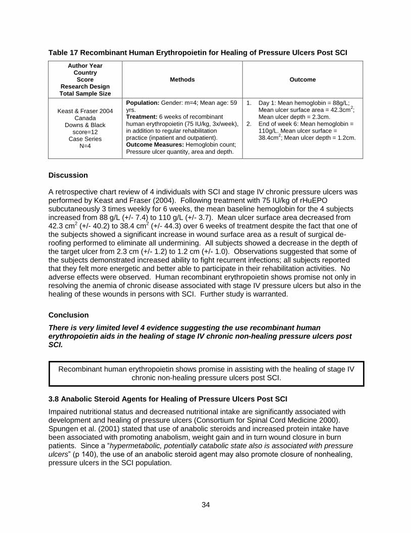
34
Table 17 Recombinant Human Erythropoietin for Healing of Pressure Ulcers Post SCI
Author Year Country Score
Research Design Total Sample Size
Methods Outcome
Keast & Fraser 2004 Canada
Downs & Black score=12
Case Series N=4
Population: Gender: m=4; Mean age: 59
yrs. Treatment: 6 weeks of recombinant
human erythropoietin (75 IU/kg, 3x/week), in addition to regular rehabilitation practice (inpatient and outpatient). Outcome Measures: Hemoglobin count;
Pressure ulcer quantity, area and depth.
1. Day 1: Mean hemoglobin = 88g/L; Mean ulcer surface area = 42.3cm
2;
Mean ulcer depth = 2.3cm. 2. End of week 6: Mean hemoglobin =
110g/L. Mean ulcer surface = 38.4cm
2; Mean ulcer depth = 1.2cm.
Discussion
A retrospective chart review of 4 individuals with SCI and stage IV chronic pressure ulcers was performed by Keast and Fraser (2004). Following treatment with 75 IU/kg of rHuEPO subcutaneously 3 times weekly for 6 weeks, the mean baseline hemoglobin for the 4 subjects increased from 88 g/L (+/- 7.4) to 110 g/L (+/- 3.7). Mean ulcer surface area decreased from 42.3 cm2 (+/- 40.2) to 38.4 cm2 (+/- 44.3) over 6 weeks of treatment despite the fact that one of the subjects showed a significant increase in wound surface area as a result of surgical de-roofing performed to eliminate all undermining. All subjects showed a decrease in the depth of the target ulcer from 2.3 cm (+/- 1.2) to 1.2 cm (+/- 1.0). Observations suggested that some of the subjects demonstrated increased ability to fight recurrent infections; all subjects reported that they felt more energetic and better able to participate in their rehabilitation activities. No adverse effects were observed. Human recombinant erythropoietin shows promise not only in resolving the anemia of chronic disease associated with stage IV pressure ulcers but also in the healing of these wounds in persons with SCI. Further study is warranted.
Conclusion
There is very limited level 4 evidence suggesting the use recombinant human erythropoietin aids in the healing of stage IV chronic non-healing pressure ulcers post SCI.
3.8 Anabolic Steroid Agents for Healing of Pressure Ulcers Post SCI
Impaired nutritional status and decreased nutritional intake are significantly associated with development and healing of pressure ulcers (Consortium for Spinal Cord Medicine 2000). Spungen et al. (2001) stated that use of anabolic steroids and increased protein intake have been associated with promoting anabolism, weight gain and in turn wound closure in burn patients. Since a “hypermetabolic, potentially catabolic state also is associated with pressure ulcers” (p 140), the use of an anabolic steroid agent may also promote closure of nonhealing, pressure ulcers in the SCI population.
Recombinant human erythropoietin shows promise in assisting with the healing of stage IV chronic non-healing pressure ulcers post SCI.

35
Table 18 Anabolic Steroid Agents for Healing of Pressure Ulcers Post SCI
Author Year Country Score
Research Design Total Sample Size
Methods Outcome
Spungen et al. 2001 USA
Downs & Black score=15
Case Series N=9
Population: Age = 24-73 yrs; Gender:
males = 9; Total number of pressure ulcers since SCI = 1 to 7. Treatment: Subjects with stage III and IV
pressure ulcers were treated with 20mg of oxandrolone daily with 20g of glutamine dissolved in orange juice. Pressure ulcer care and support surfaces remained consistent. Outcome Measures: Number of
pressure ulcers healed.
1. After oxandrolone and glutamine treatment, 8/9 subjects were completely healed, the majority within 3-6 months. 2 subjects required 12 months of treatment for complete healing.
Discussion
In a case series of nine subjects with stage III and IV pressure ulcers, Spungen et al. (2001) demonstrated complete healing in 8/9 subjects 3-12 months after daily administration of 20mg of oxandrolone. Given that this is only one small case series, more research is needed to determine the role of anabolic steroid agents (oxandrolone) for promotion of healing of stage III and IV pressure ulcers post SCI.
Conclusion
There is very limited level 4 evidence to support the use of anabolic steroid agents (oxandrolone) to promote healing of stage III and IV pressure ulcers post SCI.
3.9 Effectiveness of Dressings for Treatment of Pressure Ulcers Post SCI
Dressings are one of several interventions required to treat a wound. The appropriate choice of a dressing aids the body’s ability to heal a wound. Purposes of dressings include keeping the wound bed moist, removing excess exudate, providing a barrier against contamination and gas exchange. An appropriate dressing increases healing rate, reduces pain, and decreases infection rates while being cost effective and affordable (Broussard 2007). Due to the estimated costs associated with pressure ulcers and their treatment, various dressings used with the SCI population have been investigated. When hydrocolloid dressings are placed over a wound, the dressing absorbs the exudate and changes into a gel. The outside of the dressing allows for gas exchange and protects against outside contamination. Hydrocolloid dressings maintain a moist wound environment and support autolytic debridment. Dressings can be left in place for 3-5 days, decreasing time and costs (Heynemen et al. 2008; Consortium for Spinal Cord Medicine 2000; Houghton & Campbell 2007). Hydrocolloid dressings are typically used for stage II and III pressure ulcers (Heynemen et al. 2008).
Anabolic steroid agents may promote healing of serious pressure ulcers post SCI.

36
Hydrogel dressings act to retain moisture and rehydrate wounds, provide autolytic debridment and fill dead space. They provide minimal absorption of exudates. Hydrogel is available as a sheet or in an amorphous viscous form which requires a secondary dressing (Broussard 2007; Consortium for Spinal Cord Medicine 2000). Dressings can be left in place for 48-72 hours depending on the type of hydrogel in use (Consortium for Spinal Cord Medicine 2000; Broussard 2007). Phenytoin is an anti-epileptic medication. The healing properties of topical phenytoin were first reported over 50 years ago. Over the years, various topical preparations of phenytoin have been studied and while its exact mechanism of action is unknown, it may enhance healing by stimulation of fibroblast proliferation, promotion of collagen deposition, antibacterial activity and decreased collagenase activity (Anstead et al. 1996; Hollisaz et al. 2004; Subbanna et al. 2007). It has not been widely used because its efficacy has not been sufficiently established through controlled clinical trials (Ovington 1999; Subbanna et al. 2007). Table 19 Effectiveness of Dressings for Treatment of Pressure Ulcers Post SCI
Author Year Country Score
Research Design Total Sample Size
Methods Outcome
Subbanna et al. 2007 India
PEDro=9 RCT (double blind)
N=28
Population: Patients with stage II ulcers.
Treatment group (n=14): Mean age=34.25yrs; Gender: males=13, females=1; Control group (n=14): Mean age=31.64yrs; Gender: males=12, females=2.All subjects had stage II ulcers. Treatment: Patients were randomly
placed (double blind manner) in the treatment group which received 5mg/ml phenytoin solution soaked sterile gauge for their pressure ulcer dressing once daily for 15 days and the control group received saline soaked gauge. Outcome Measures: Pressure ulcer
scale for healing 3.0, ulcer size, ulcer volume
1. Improvement in PUSH 3.0 and ulcer size was seen in the topical phenytoin group however this difference did not reach statistical significance, p=0.261 and 0.132 respectively.
2. Reduction in ulcer volume was seen in the control group however it was not significant, p=0.777
Hollisaz et al. 2004 Iran
PEDro=7 RCT
N=83 (91 Pressure ulcers)
Population: Gender: males = 83; Level
of injury: paraplegia; Cause of injury: trauma. Stage of pressure ulcers: I = 33, II = 58. Treatment: 1 of 3 dressing groups: 1)
Simple dressing (SD) – changed twice daily; 2) Hydrocolloid dressing (HD) – changed twice weekly; 3) Adhesive plus phenytoin cream (PC) – changed once daily. Outcome Measures: Healing status of
pressure ulcer.
1. HD group had the most healing (74%) as compared to the PC group (40%, p<0.01) and the SD group (27%, p<0.005).
2. Of all the groups, the HD group also had higher rating of healing of stage I ulcers (p<0.05).
3. For stage II ulcers, HD treated ulcers healed better than SD treated ulcers (67% vs. 16%) p<0.005 but no better than PC (48%, p>0.05) treated ulcers .
4. HD also healed gluteal ulcers more completely than other groups (p<0.001); however, groups did not differ on healing of sacral ulcers.
Population: Age = 16-56 yrs; Gender:
males = 24, females = 3; Ulcer stage: I = 1. Healing rates did not differ between
groups.

37
Author Year Country Score
Research Design Total Sample Size
Methods Outcome
Kaya et al. 2005 Turkey
PEDro=4 RCT
N=27 (49 Pressure ulcers)
12, II = 34, III = 3. Treatment: Treatment group (n=15, 25
PU): an occlusive hydrogel dressing, changed every 4 days (unless contaminated). Control group (n=12, 24 PU): povidone-iodine soaked gauze changed daily. Outcome Measures: Healing rate
(cm2/day).
2. More wounds healed in the treatment group (n=21) than in the control group (n=13), p<0.04.
Whittle et al. 1996 Canada
Downs & Black score=9 Case study
N=4
Population: Mean age = 55.75 yrs;
Gender: males = 3, females = 1; Level of injury: paraplegia = 2, tetraplegia = 2; Pressure ulcer (5 ulcers) stage: II = 1, III = 3, IV = 1 Treatment: Hydrogel dressings, for
different time periods depending on the case. Outcome Measures: Pressure ulcer
improvement, measured weekly.
1. Hydrogel dressing application varied from 4 to 6 wks.
2. In all cases, pressure ulcers improved drastically, with 3 cases being completely healed.
Discussion Hollisaz et al., in a RCT involving 83 subjects, found that those in the hydrocolloid dressing (HD) group (n=28) seemed to have the greatest completion of healing regardless of ulcer location and stage (74%, p<0.005), compared to those in the phenytoin cream (PC) group (40%, n=28) or simple dressing (SD) group (27%, n=27). For stage I ulcers, those in the HD group healed faster than those in the other two groups; however, for stage II ulcers, there was no difference in healing between the HD and PC groups (67% vs 48%, p>0.05). When looking at the area of injury, gluteal ulcers also healed more completely in the HD group than in the other two, whereas the healing of sacral ulcers did not differ between the 3 groups. Subbanna et al. (2007) using a phenytoin solution (5mg/ml) found improvements in PUSH 3.0 and ulcer size when compared to normal saline but the differences did not reach statistical significance (p=0.261, p=0.132). Whittle et al. (1996) treated 5 pressure ulcers (stage II-IV) with hydrogel dressings. After approximately 4-6 weeks of treatment, 3 ulcers healed completely with the others showing a large improvement. Kaya et al. (2005) compared the effectiveness of applying an occlusive hydrogel type dressing to a poviodine-iodine soaked gauge dressing. There were no statistically significant differences in rate of healing but significantly more ulcers healed with the hydrogel dressing. Conclusion There is Level 1 evidence from a single RCT that completion of healing for stage I and II pressure ulcers is greater with an occlusive hydrocolloid dressing compared to phenytoin cream or simple dressing post SCI.

38
There is Level 2 evidence from a single, small RCT that occlusive hydrogel-type dressings heal more pressure ulcers than conservative treatment post SCI. There is level 1 evidence that topical phenytoin shows a trend towards healing of stage I and II pressure ulcers post SCI.
3.10 Maggot Therapy for Healing of Pressure Ulcers Post SCI
The beneficial effects of fly larvae have been known for centuries. The intentional use of fly larvae (maggot therapy) for the treatment of wounds was used extensively in the 1930s and 1940s but was discontinued when antimicrobials and surgical debridment were introduced. Maggot therapy (MT) was reintroduced to treat intractable wounds in the 1990s (Mumcuoglu et al. 1999). Sterilized larvae of the Phaenicia sericata species are often used for MT (Mymcuoglu et al. 1999; Sherman 2002). MT is believed to work through three processes: debridment of necrotic tissue, disinfection of the wound and promotion of tissue growth (Sherman 2002). Table 20 Maggot Therapy for Healing of Pressure Ulcers Post SCI
Author Year Country Score
Research Design Total Sample Size
Methods Outcome
Sherman et al. 1995 USA
Downs & Black score=9 Prospective Control Trial
N=8
Population: Age = 44-68 yrs; Gender:
males = 81; Level of injury: paraplegia = 7, tetraplegia = 1; Ulcer stages: III & IV. Treatment: 3-4 wks of conventional
therapy preceded maggot placement under porous sterile dressings, for 48-72 hr cycles. Sodium hypochorite, normal saline or wet-to-dry gauze dressings were applied every 8 hours in between maggot cycles. Outcome Measures: Healing of pressure
ulcer; Wound area size.
1. Maggot therapy decreased pressure ulcer surface area by 22% per week (p<0.001).
2. No adverse consequences of treatment were noted.
Discussion
In one non-RCT conducted by Sherman et al. (1995), 8 of 20 patients diagnosed with stage III and IV pressure ulcers were treated with maggot therapy. All 8 patients underwent 3 weeks of conventional treatment, followed by maggot therapy. All necrotic wounds were debrided within one week of maggot treatment and wound healing was faster among the 8 who had received maggot therapy than in the 12 who had not. Conclusion
There is Level 2 evidence from one very small study to support the use of maggot therapy as an adjunctive therapy for non-healing stage III and IV pressure ulcers post SCI.
Occlusive hydrocolloid dressings are useful for healing of stage I and II pressure ulcers post SCI.
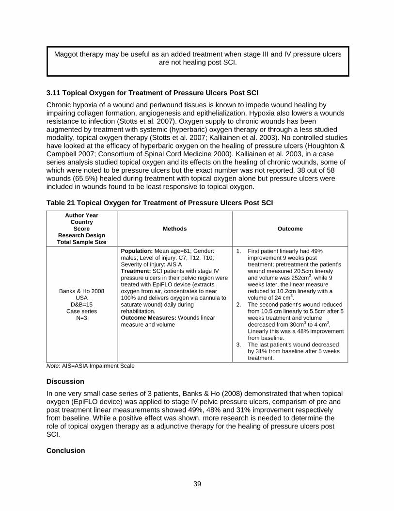
39
3.11 Topical Oxygen for Treatment of Pressure Ulcers Post SCI
Chronic hypoxia of a wound and periwound tissues is known to impede wound healing by impairing collagen formation, angiogenesis and epithelialization. Hypoxia also lowers a wounds resistance to infection (Stotts et al. 2007). Oxygen supply to chronic wounds has been augmented by treatment with systemic (hyperbaric) oxygen therapy or through a less studied modality, topical oxygen therapy (Stotts et al. 2007; Kalliainen et al. 2003). No controlled studies have looked at the efficacy of hyperbaric oxygen on the healing of pressure ulcers (Houghton & Campbell 2007; Consortium of Spinal Cord Medicine 2000). Kalliainen et al. 2003, in a case series analysis studied topical oxygen and its effects on the healing of chronic wounds, some of which were noted to be pressure ulcers but the exact number was not reported. 38 out of 58 wounds (65.5%) healed during treatment with topical oxygen alone but pressure ulcers were included in wounds found to be least responsive to topical oxygen. Table 21 Topical Oxygen for Treatment of Pressure Ulcers Post SCI
Author Year Country Score
Research Design Total Sample Size
Methods Outcome
Banks & Ho 2008 USA
D&B=15 Case series
N=3
Population: Mean age=61; Gender:
males; Level of injury: C7, T12, T10; Severity of injury: AIS A Treatment: SCI patients with stage IV
pressure ulcers in their pelvic region were treated with EpiFLO device (extracts oxygen from air, concentrates to near 100% and delivers oxygen via cannula to saturate wound) daily during rehabilitation. Outcome Measures: Wounds linear
measure and volume
1. First patient linearly had 49% improvement 9 weeks post treatment; pretreatment the patient's wound measured 20.5cm lineraly and volume was 252cm
3, while 9
weeks later, the linear measure reduced to 10.2cm linearly with a volume of 24 cm
3.
2. The second patient's wound reduced from 10.5 cm linearly to 5.5cm after 5 weeks treatment and volume decreased from 30cm
3 to 4 cm
3,
Linearly this was a 48% improvement from baseline.
3. The last patient's wound decreased by 31% from baseline after 5 weeks treatment.
Note: AIS=ASIA Impairment Scale
Discussion
In one very small case series of 3 patients, Banks & Ho (2008) demonstrated that when topical oxygen (EpiFLO device) was applied to stage IV pelvic pressure ulcers, comparism of pre and post treatment linear measurements showed 49%, 48% and 31% improvement respectively from baseline. While a positive effect was shown, more research is needed to determine the role of topical oxygen therapy as a adjunctive therapy for the healing of pressure ulcers post SCI. Conclusion
Maggot therapy may be useful as an added treatment when stage III and IV pressure ulcers are not healing post SCI.

40
There is very limited level 4 evidence that topical oxygen therapy may improve healing of pressure ulcers post SCI.
4 Summary
Numerous studies cited in this document have spoken to the fact that pressure ulcers, though largely preventable, are still a common, potentially serious lifelong secondary complication of SCI. Pressure ulcers have the potential to impact overall quality of life (Consortium for Spinal Cord Medicine 2000), disrupt rehabilitation, vocational and educational pursuits and community reintegration (Fuhrer et al. 1993; Krause 1998; Consortium for Spinal Cord Medicine 2000; Jones et al. 200), and lead to increased hospital readmission rates with longer lengths of stay (Chen 2005). Pressure ulcer prevention is more cost effective than treatment (Bogie et al. 2000; Jones et al. 2003). Despite the attention given to prevention strategies, pressure ulcers still occur.
Pressure ulcers are potentially preventable but without evidence to guide practice and education, pressure ulcers will continue to occur. Given the human and economic costs of pressure ulcer formation post SCI, more quality research needs to be done on all aspects of pressure ulcer prevention so that solid evidence is available to individuals with SCI, their families and health care providers.
There are several treatment interventions for pressure ulcers which are supported by level 1 evidence. These include: use of electrical stimulation, US/UVC and pulsed electromagnetic energy as adjunctive therapies and hydrocolloid dressings to assist with the complete healing of stage I and II pressure ulcers post SCI. Well reasoned treatment interventions supported by evidence should be incorporated into treatment plans for individuals with SCI who have pressure ulcers. Providing enhanced pressure ulcer education and structured follow-up has been shown to reduce recurrence of pressure ulcers post SCI. There is limited level 4 evidence that electrical stimulation decreases ischial pressures post SCI. There is level 4 evidence that electrical stimulation may increase blood flow at sacral and gluteal areas post SCI. There is level 3 evidence that 1-2 minutes of pressure relief must be sustained to raise tissue oxygen to unloaded levels. There is level 4 evidence to support position changes to reduce pressure at the ischial tuberosities. There is level 3 evidence that various cushions or seating systems (e.g. dynamic versus static) are associated with potentially beneficial reduction in seating interface pressure or pressure ulcer risk factors like skin temperature.
Use of topical oxygen therapy may have a positive association with healing of pressure
ulcers post SCI but more research is needed.

41
There is level 3 evidence that adding lumbar support to the wheelchair of those with chronic SCI has a negligible effect on reducing seated buttock pressures at the ischial tuberosities. There is Level 2 evidence showing that early attendance at specialized seating assessment clinics (SSA) increases the skin management abilities of individuals post SCI. There is Level 2 evidence that providing enhanced pressure ulcer prevention education is effective at helping individuals with SCI gain and retain this knowledge. There is level 1 evidence that providing enhanced pressure ulcer education and structured follow-up is effective in reducing recurrence of pressure ulcers especially in those individuals with no previous history of pressure ulcer surgery. There is very limited level 4 evidence to suggest that the introduction of behavioural contingencies is associated with a reduction in pressure ulcer severity and decreased health care costs. There is level 4 evidence that telerehabiliation does not make a significant difference in the prevention and treatment of pressure ulcers post SCI. More research is needed into its effectiveness for improving healing and reducing costs. There is level 1 evidence from 2 RCTs to support the use of electrical stimulation to accelerate the healing rate of stage III and IV pressure ulcers when combined with standard wound management. There is level 1 evidence (from two RCTs) to suggest that laser treatment has no added benefit in pressure ulcer healing post SCI than standard wound care alone. There is level 1 evidence, from 1 small RCT, to suggest that combining US/UVC with standard wound care decreases wound healing time of pressure ulcers post SCI but no evidence to clarify whether UVC or US, used alone, have a beneficial effect. There is level 1 evidence from one RCT to support the efficacy of pulsed electromagnetic energy to accelerate healing of stage II and III pressure ulcers post SCI. There is very limited level 4 evidence that topical negative pressure (TNP) improves healing of pressure ulcers post SCI. There is very limited level 3 evidence that the use of a normothermic dressing may improve healing of pressure ulcers post SCI. There is Level 2 evidence from one very small study to support the use of maggot therapy as an adjunctive therapy for non-healing stage III and IV pressure ulcers post SCI. There is very limited level 4 evidence suggesting the use recombinant human erythropoietin aids in the healing of chronic non healing pressure ulcers in post SCI.

42
There is very limited level 4 evidence to support the use of anabolic steroid agents (oxandrolone) to promote healing of stage III and IV pressure ulcers post SCI. There is Level 1 evidence from a single RCT that completion of healing for stage I and II pressure ulcers is greater with an occlusive hydrocolloid dressing compared to phenytoin cream or simple dressing post SCI. There is Level 2 evidence from a single, small RCT that occlusive hydrogel-type dressings heal more pressure ulcers than conservative treatment post SCI. There is level 1 evidence that topical phenytoin shows a trend towards healing of stage I and II pressure ulcers post SCI. There is very limited level 4 evidence that topical oxygen therapy may improve healing of pressure ulcers post SCI.

43
References Adegoke BO, Badmos KA. Acceleration of pressure ulcer healing in spinal cord injured patients
using interrupted direct current. Afr J Med Med Sci 2001;30:195-197. Agency for Health Care Quality and Research. (AHCPR) Guideline 15: Treatment of Pressure
Ulcers; 1992. www.guidelines.gov. Agency for Health Care Quality and Research (AHCPR). Guideline 3: Pressure ulcers in adults:
Prediction and Prevention; 1992. www.guidelines.gov Alexander LR, Spungen AM, Liu MH, Losada M, Bauman WA. Resting metabolic rate in
subjects with paraplegia: the effect of pressure sores. Archives of Physical Medicine & Rehabilitation 1995;76:819-22.
Al-Momen AK. Recombinant human erythropoietin induced rapid healing of a chronic leg ulcer in a patient with sickle cell disease. Acta Haematol 1991;86:46-48.
Allen J, Houghton PE. A case study for Electrical Stimulation on a stage III pressure ulcer. Wound Care Canada 2003;2:34-36.
Anstead GM, Hart LM, Fong Sunahara J, Liter ME. Phenytoin in wound healing. Annals of Pharm 1996;30:768-775.
Argenta LC, Morykwas MJ. Vacuum-assisted closure: a new method for wound control and treatment: clinical experience. Ann Plast Surg 1997;38:563-576.
Ash D. An exploration of the occurrence of pressure ulcers in a British spinal injuries unit. J Clin Nurs 2002;11:470-478.
Ayello EA, Braden B. How and why to do pressure ulcer risk assessment. Adv Skin Wound Care 2002;15:125-131.
Baker LL, Rubayi S, Villar F, Demuth SK. Effect of electrical stimulation waveform on healing of ulcers in human beings with spinal cord injury. Wound Repair Regen 1996;4:21-28.
Banks, PG, Ho, CH.. A novel topical oxygen treatment for chronic and difficult-to-heal wounds: case studies. Journal of Spinal Cord Medicine 2008; 31:297-301
Bergstrom N, Braden BJ, Laguzza A, Holman V. The Braden Scale for Predicting Pressure Sore Risk. Nurs Res 1987;36:205-210.
Black J, Baharestani M, Cuddigan J, Dorner B, Edsberg L, Langemo D, Posthauer ME, Ratliff C, Taler G. National pressure ulcer advisory panel’s updated pressure ulcer staging system. Urol Nurs. 2007;27:144-150.
Bogie KM, Nuseibeh I, Bader DL. Early progressive changes in tissue viability in the seated spinal cord injured subject. Paraplegia 1995;33:141-147.
Bogie KM, Reger SI, Levine SP, Sahgal V. Electrical stimulation for pressure sore prevention and wound healing. Assist Technol 2000;12:50-66.
Bogie KM, Triolo RJ. Effects of regular use of neuromuscular electrical stimulation on tissue health. J Rehabil Res Dev 2003;40:469-475.
Bogie KM, Wang X, Triolo RJ. Long-term prevention of pressure ulcers in high-risk patients: a single case study of the use of gluteal neuromuscular electric stimulation. Arch Phys Med Rehabil 2006;87:585-591.
Brienza DM, Karg PE. Seat cushion optimization: a comparison of interface pressure and tissue stiffness characteristics for spinal cord injured and elderly patients. Arch Phys Med Rehabil 1998;79:388-394.
Broussard CL. (2007) Dressing Decisions. In: Krasner DL, Rodeheaver GT, Sibbald RG, editors. Chronic Wound Care: A Clinical Source Book for Healthcare Professionals. Malvern, PA: HMP Communications, 249-262.
Burns SP, Betz KL. Seating pressures with conventional and dynamic wheelchair cushions in tetraplegia. Archives of Physical Medicine & Rehabilitation 1999;80:566-571.

44
Byrne DW, Salzberg CA. Major risk factors for pressure ulcers in the spinal cord disabled: a literature review. Spinal Cord 1996;34:255-263.
Carley PJ, Wainapel SF. Electrotherapy for acceleration of wound healing: low intensity direct current. Arch Phys Med Rehabil 1985;66:443-446.
Chen Y, DeVivo MJ, Jackson AB. Pressure ulcer prevalence in people with spinal cord injury: age-period-duration effects. Arch Phys Med Rehabil 2005;86:1208-1213.
Clark FA, Jackson JM, Scott MD, Carlson ME, Atkins MS, Uhles-Tanaka D et al. Data-based models of how pressure ulcers develop in daily-living contexts of adults with spinal cord injury. Arch Phys Med Rehabil 2006;87:1516-1525.
Coggrave M, West H, Leonard B. Topical negative pressure for pressure ulcer management. Br J Nurs 2002;11(6 Suppl):S29-S36.
Coggrave MJ, Rose LS. A specialist seating assessment clinic: changing pressure relief practice. Spinal Cord 2003;41:692-695.
Consortium for Spinal Cord Medicine. Pressure ulcer prevention and treatment following spinal cord injury: A clinical practice guideline for health care professionals. 1-77. 2000.
Crenshaw RP, Vistnes LM. A decade of pressure sore research: 1977-1987. J Rehabil Res Dev 1989;26:63-74.
DeLisa JA, Mikulic MA. Pressure Ulcers. What do we do if preventive management fails. Pressure Ulcers 1985;77:209-212.
Dover H, Pickard W, Swain I, Grundy D. The effectiveness of a pressure clinic in preventing pressure sores. Paraplegia 1992;30:267-272.
Dunn CA, Carlson M, Jackson JM, Clark FA. Response factors surrounding progression of pressure ulcers in community-residing adults with spinal cord injury. American Journal of Occupational Therapy 2009;63:301-9.
Feedar JA, Kloth LC, Gentzkow GD. Chronic dermal ulcer healing enhanced with monophasic pulsed electrical stimulation. Phys Ther 1991;71:639-649.
Fisher SV, Szymke TE, Apte SY, Kosiak M. Wheelchair cushion effect on skin temperature. Arch Phys Med Rehabil 1978;59:68-72.
Fleck CA, Sprigle S. (2007) Support Surfaces: Tissue Integrity, Terms, Principles, and Choice. In: Krasner DL, Rodeheaver GT, Sibbald RG, editors. Chronic Wound Care: A Clinical Source Book for Healthcare Professionals. Malvern, PA: HMP Communications, 629-640.
Fuhrer MJ, Garber SL, Rintala DH, Clearman R, Hart KA. Pressure ulcers in community-resident persons with spinal cord injury: prevalence and risk factors. Arch Phys Med Rehabil 1993;74:1172-1177.
Galea M, Tumminia J, Garback LM. Telerehabilitation in spinal cord injury persons: a novel approach. Telemed J E Health 2006;12:160-162.
Garber SL. Wheelchair cushions for spinal cord-injured individuals. Am J Occup Ther 1985;39:722-725.
Garber SL, Rintala DH, Rossi CD, Hart KA, Fuhrer MJ. Reported pressure ulcer prevention and management techniques by persons with spinal cord injury. Arch Phys Med Rehabil 1996;77:744-749.
Garber SL, Rintala DH, Holmes SA, Rodriguez GP, Friedman J. A structured educational model to improve pressure ulcer prevention knowledge in veterans with spinal cord dysfunction. J Rehabil Res Dev 2002;39:575-588.
Garber SL, Reddy NP, McLane K.M., Krouskop TA. (2007) The Role of Technology in Pressure Ulcer Prevention. In: Krasner DL, Rodeheaver GT, Sibbald RG, editors. Chronic Wound Care: A Clinical Source Book for Healthcare Professionals. Malvern, PA: HMP Communications, 653-660.
Gilsdorf P, Patterson R, Fisher S. Thirty-minute continuous sitting force measurements with different support surfaces in the spinal cord injured and able-bodied. J Rehabil Res Dev 1991;28:33-38.

45
Griffin JW, Tooms RE, Mendius RA, Clifft JK, Vander ZR, El Zeky F et al. Efficacy of high voltage pulsed current for healing of pressure ulcers in patients with spinal cord injury. Phys Ther 1991;71:433-444.
Guihan M, Smith BM, LaVela SL, Garber SL. Factors predicting pressure ulcers in veterans with spinal cord injuries. American Journal of Physical Medicine & Rehabilitation 2008;87:750-7.
Henderson JL, Price SH, Brandstater ME, Mandac BR. Efficacy of three measures to relieve pressure in seated persons with spinal cord injury. Archives of Physical Medicine & Rehabilitation 1994;75:535-539.
Heyneman A, Beele H, Vanderwee K, Defloor T. A systematic review of the use of hydrocolloids in the treatment of pressure ulcers. J Clin Nurs 2008;17:1164-1173.
Hill, M. L., Cronkite, R. C., Ota, D. T., Yao, E. C., & Kiratli, B. J. (2009). Validation of home telehealth for pressure ulcer assessment: a study in patients with spinal cord injury. Journal of Telemedicine & Telecare;15:196-202.
Hobson DA. Comparative effects of posture on pressure and shear at the body-seat interface. J Rehabil Res Dev 1992;29:21-31.
Hollisaz MT, Khedmat H, Yari F. A randomized clinical trial comparing hydrocolloid, phenytoin and simple dressings for the treatment of pressure ulcers [ISRCTN33429693]. BMC Dermatol 2004;4:18.
Houghton PE, Campbell KE. (2007) Therapeutic Modalities in the Treatment of Chronic Recalcitrant Wounds. In: Krasner DL, Rodeheaver GT, Sibbald RG, editors. Chronic Wound Care: A Clinical Source Book for Healthcare Professionals. Malvern, PA: HMP Communications, 403-415.
Houghton PE, Fraser CH. Biomarkers that predict response to adjunctive therapies in people with spinal cord injury. Proceedings of World Union of Wound Healing Societies . 2008.
Jones ML, Mathewson CS, Adkins VK, Ayllon T. Use of behavioral contingencies to promote prevention of recurrent pressure ulcers. Arch Phys Med Rehabil 2003;84:796-802.
Kalliainen LK, Gordillo GM, Schlanger R, Sen CK. Topical oxygen as an adjunct to wound healing: a clinical case series. Pathophysiology 2003; 9:81-87.
Karba, R., Semrov, D., Vodovnik, L., Benko, H., & Savrin, R. DC electrical stimulation for chronic wound healing enhancement. Part 1. Clinical study and determination of electrical field distribution in the numerical wound model. Bioelectrochemistry and Bioenergetics.1997;43:265-270.
Kaya AZ, Turani N, Akyuz M. The effectiveness of a hydrogel dressing compared with standard management of pressure ulcers. Journal of Wound Care 2005;14:42-44.
Keast DH, Fraser C. Treatment of chronic skin ulcers in individuals with anemia of chronic disease using recombinant human erythropoietin (EPO): a review of four cases. Ostomy Wound Manage 2004;50:64-70.
Keast DH, Parslow N, Houghton PE, Norton L, Fraser C. Best practice recommendations for the prevention and treatment of pressure ulcers: Update 2006. Wound Care Canada 2006;22-32.
Kennedy P, Berry C, Coggrave M, Rose L, Hamilton L. The effect of a specialist seating assessment clinic on the skin management of individuals with spinal cord injury. J Tissue Viability 2003;13:122-125.
King RB, Porter SL, Vertiz KB. Preventive skin care beliefs of people with spinal cord injury. Rehabilitation Nursing 2008;33:154-62.
Kloth LC, Feedar JA. Acceleration of wound healing with high voltage, monophasic, pulsed current. Phys Ther 1988;68:503-508.
Kloth LC, Berman JE, Dumit-Minkel S, Sutton CH, Papanek PE, Wurzel J. Effects of a normothermic dressing on pressure ulcer healing. Adv Skin Wound Care 2000;13:69-74.
Krause JS. Skin sores after spinal cord injury: relationship to life adjustment. Spinal Cord 1998;36:51-56.
46
Krause JS, Vines CL, Farley TL, Sniezek J, Coker J. An exploratory study of pressure ulcers after spinal cord injury: relationship to protective behaviors and risk factors. Arch Phys Med Rehabil 2001;82:107-113.
Krause JS, Broderick L. Patterns of recurrent pressure ulcers after spinal cord injury: identification of risk and protective factors 5 or more years after onset. Arch Phys Med Rehabil 2004;85:1257-1264.
Lamid S, El Ghatit AZ. Smoking, spasticity and pressure sores in spinal cord injured patients. Am J Phys Med 1983;62:300-306.
Levine SP, Kett RL, Cederna PS, Brooks SV. Electric muscle stimulation for pressure sore prevention: tissue shape variation. Arch Phys Med Rehabil 1990;71:210-215.
Lui QL, Nicholson GP, Knight SL, Chelvarajah R, Gall A, Middleton FRI, Ferguson-Pell MW, Craggs MD. Pressure changes under the ischial tuberosities of seated individuals during sacral nerve root stimulation. J Rehabil Res Dev 2006b;43:209-218.
Lui QL, Nicholson GP, Knight SL, Chelvarajah R, Gall A, Middleton FRI, Ferguson-Pell MW, Craggs MD. Interface pressure and cutaneous hemoglobin and oxygenation changes under ischial tuberosities during sacral nerve root stimulation in spinal cord injury. J Rehabil Res Dev 2006a;43:553-564.
Makhsous M, Rowles DM, Rymer WZ, Bankard J, Nam EK, Chen D et al. Periodically relieving ischial sitting load to decrease the risk of pressure ulcers. Arch Phys Med Rehabil 2007b;88:862-870.
Makhsous M, Priebe M, Bankard J, Rowles D, Zeigler M, Chen D et al. Measuring tissue perfusion during pressure relief maneuvers: insights into preventing pressure ulcers. J Spinal Cord Med 2007a;30:497-507.
Mathewson C, Adkins VK, Lenyoun MA, Schmidt AM, Jones ML. Using telemedicine in the treatment of pressure ulcers. Ostomy Wound Manage 1999;45:58-62.
Mathewson C, Adkins VK, Jones ML. Initial experiences with telerehabilitation and contingency management programs for the prevention and management of pressure ulceration in patients with spinal cord injuries. J Wound Ostomy Continence Nurs 2000;27:269-271.
Mawson AR, Siddiqui FH, Connolly BJ, Sharp CJ, Stewart GW, Summer WR et al. Effect of high voltage pulsed galvanic stimulation on sacral transcutaneous oxygen tension levels in the spinal cord injured. Paraplegia 1993;31:311-319.
Mortenson WB, Miller WC. A review of scales for assessing the risk of developing a pressure ulcer in individuals with SCI. Spinal Cord 2008;46:168-175.
Mullner T, Mrkonjic L, Kwasny O, Vecsei V. The use of negative pressure to promote the healing of tissue defects: a clinical trial using the vacuum sealing technique. Br J Plast Surg 1997;50:194-199.
Mumcuoglu KY, Ingber A, Gilead L, Stessman J, Friedmann R, Schulman H et al. Maggot therapy for the treatment of intractable wounds. Int J Dermatol 1999;38:623-627.
National Pressure Ulcer Advisory Panel. Pressure Ulcer Stages. NPUAP http://npuap.org/positn6.htm. 1989; Last accessed: 7-15-2005.
National Pressure Ulcer Advisory Panel. Pressure Ulcer Stages Revised by NPUAP. NPUAP http://www.npuap.org/pr2.htm. 2007; Last accessed: 9-18-2007.
Niazi ZB, Salzberg CA, Byrne DW, Viehbeck M. Recurrence of initial pressure ulcer in persons with spinal cord injuries. Adv Wound Care 1997;10:38-42.
Nursing Best Practice Guideline: Assessment and management of stage 1 to IV pressure ulcers. Toronto. RNAO 2002. www.rnao.org/bestpractices
Nussbaum EL, Biemann I, Mustard B. Comparison of ultrasound/ultraviolet-C and laser for treatment of pressure ulcers in patients with spinal cord injury. Phys Ther 1994;74:812-823.
Olyaee, M. A., Flemming, K., Cullum, N. A., & Ravaghi, H. Electromagnetic therapy for treating pressure ulcersCochrane Database of Systematic Reviews, 2001;CD002930

47
Ovington LG. Dressings and ajunctive therapies: AHCPR guidelines revisited. Ostomy Wound Manage 1999;45:94S-106S
Phillips VL, Temkin A, Vesmarovich S, Burns R, Idleman L. Using telehealth interventions to prevent pressure ulcers in newly injured spinal cord injury patients post-discharge. Results from a pilot study. Int J Technol Assess Health Care 1999;15:749-755.
Phillips VL, Vesmarovich S, Hauber R, Wiggers E, Egner A. Telehealth: reaching out to newly injured spinal cord patients. Public Health Rep 2001;116 Suppl 1:94-102.
Rappl LM. Physiological changes in tissues denervated by spinal cord injury tissues and possible effects on wound healing. International Wound Journal 2008;5:435-44.
Reger SI, Ranganathan VK, Sahgal V. Support surface interface pressure, microenvironment, and the prevalence of pressure ulcers: an analysis of the literature. Ostomy Wound Management 2007;53:50-8.
Richards JS, Waites K, Chen YY, Kogos K, Schumitt MM. The epidemiology of secondary conditions following spinal cord injury. Top Spinal Cord Inj Rehabil 2004;10:15-29.
Rintala, D. H., Garber, S. L., Friedman, J. D., & Holmes, S. A. Preventing recurrent pressure ulcers in veterans with spinal cord injury: impact of a structured education and follow-up intervention. Archives of Physical Medicine & Rehabilitation. 2008;89:1429-1441
Rodriguez GP, Garber SL. Prospective study of pressure ulcer risk in spinal cord injury patients. Paraplegia 1994;32:150-158.
Rund C, Sussman G. (2007) Nontraditional or Alternative Topical Therapies for Wound Care. In: Krasner DL, Rodeheaver GT, Sibbald RG, editors. Chronic Wound Care: A Clinical Source Book for Healthcare Professionals. Malvern, PA: HMP Communications, 287-295.
Salzberg CA, Cooper-Vastola SA, Perez F, Viehbeck MG, Byrne DW. The effects of non-thermal pulsed electromagnetic energy on wound healing of pressure ulcers in spinal cord-injured patients: a randomized, double-blind study. Ostomy Wound Manage 1995;41:42-4, 46, 48.
Saladin LK, Krause JS. Pressure ulcer prevalence and barriers to treatment after spinal cord injury: comparisons of four groups based on race-ethnicity. Neurorehabilitation 2009;24:57-66.
Salzberg CA, Byrne DW, Cayten CG, van Niewerburgh P, Murphy JG, Viehbeck M. A new pressure ulcer risk assessment scale for individuals with spinal cord injury. Am J Phys Med Rehabil 1996;75:96-104.
Schmuckler J. Acoustic pressure wound therapy to facilitate granulation tissue in sacral pressure ulcers in patients with compromised mobility: A case series. Ostomy Wound Management 2008;54:50-53.
Schryvers OI, Stranc MF, Nance PW. Surgical treatment of pressure ulcers: 20-year experience. Arch Phys Med Rehabil 2000;81:1556-1562.
Schubart JR, Hilgart M, Lyder C. Pressure ulcer prevention and management in spinal cord-injured adults: analysis of educational needs. Adv Skin Wound Care 2008;21:322-329.
Scivoletto G, Fuoco U, Morganti B, Cosentino E, Molinari M. Pressure sores and blood and serum dysmetabolism in spinal cord injury patients. Spinal Cord 2004;42:473-476.
Seymour RJ, Lacefield WE. Wheelchair cushion effect on pressure and skin temperature. Arch Phys Med Rehabil 1985;66:103-108.
Sheppard R, Kennedy P, Mackey CA. Theory of planned behaviour, skin care and pressure sores following spinal cord injury. J. Clin Psychol Med Settings. 2006;13:359-367.
Sherman RA, Wyle F, Vulpe M. Maggot therapy for treating pressure ulcers in spinal cord injury patients. J Spinal Cord Med 1995;18:71-74.
Sherman RA. Maggot versus conservative debridement therapy for the treatment of pressure ulcers. Wound Repair Regen 2002;10:208-214.
Shields RK, Cook TM. Lumbar support thickness: effect on seated buttock pressure in individuals with and without spinal cord injury. Phys Ther 1992;72:218-226.

48
Smith APS, Kieswetter K, Goodwin AL, McNulty AK. (2007) Negative Pressure Wound Therapy. In: Krasner DL, Rodeheaver GT, Sibbald RG, editors. Chronic Wound Care: A Clinical Source Book for Healthcare Professionals. Malvern, PA: HMP Communications, 271-286.
Spijkerman, D. C. M., Terburg, M., Goossens, R. H. M., & Stijnen, T. (1995). Effects of inflation pressure and posture on the body-seat interface pressure of spinal cord injured patients seated on an air-filled wheelchair cushion. Journal of Rehabilitation Sciences. 1995;8:8-12.
Spivak JL. Iron and the anemia of chronic disease. Oncology (Williston Park) 2002;16(9 Suppl 10):25-33.
Sprigle S, Chung KC, Brubaker CE. Factors affecting seat contour characteristics. J Rehabil Res Dev. 1990;27:127-134.
Spungen AM, Koehler KM, Modeste-Duncan R, Rasul M, Cytryn AS, Bauman WA. 9 clinical cases of nonhealing pressure ulcers in patients with spinal cord injury treated with an anabolic agent: a therapeutic trial. Adv Skin Wound Care 2001;14:139-144.
Stefanovska A, Vodovnik L, Benko H, Turk R. Treatment of chronic wounds by means of electric and electromagnetic fields. Part 2. Value of FES parameters for pressure sore treatment. Med Biol Eng Comput 1993;31:213-220.
Stotts NA, Wipke-Tevis DD, Hopf HW. (2007) Cofactors in impaired wound healing. In: Krasner DL, Rodeheaver GT, Sibbald RG, editors. Chronic Wound Care: A clinical Source Book for Healthcare Professionals. Malvern, PA: HMP Communications. pg 215-220.
Subbanna, P. K., Margaret Shanti, F. X., George, J., Tharion, G., Neelakantan, N., Durai, S. et al. Topical phenytoin solution for treating pressure ulcers: A prospective, randomized, double-blind clinical trial. Spinal Cord. 2007;45:739-743
Takechi, H. & Tokuhiro, A. Evaluation of wheelchair cushions by means of pressure distribution mapping. Acta Medica Okayama. 1998;52:245-254.
Taly AB, Sivaraman Nair KP, Murali T, John A. Efficacy of multiwavelength light therapy in the treatment of pressure ulcers in subjects with disorders of the spinal cord: A randomized double-blind controlled trial. Arch Phys Med Rehabil 2004;85:1657-1661.
Turba RM, Lewis VL, Green D. Pressure sore anemia: response to erythropoietin. Arch Phys Med Rehabil 1992;73:498-500.
Vesmarovich S, Walker T, Hauber RP, Temkin A, Burns R. Use of telerehabilitation to manage pressure ulcers in persons with spinal cord injuries. Adv Wound Care 1999;12:264-269.
Vidal J, Sarrias M. An analysis of the diverse factors concerned with the development of pressure sores in spinal cord injured patients. Paraplegia 1991;29:261-267.
Whittle H, Fletcher C, Hoskin A, Campbell K. Nursing management of pressure ulcers using a hydrogel dressing protocol: four case studies. Rehabil Nurs 1996;21:239-242.
Yuen HK, Garrett D. Comparison of three wheelchair cushions for effectiveness of pressure relief. Am J Occup Ther 2001;55:470-475.
Related Documents