Prof. Patrizia Querzoli Struttura Semplice di Patologia Mammaria U.O Anatomia Patologica Azienda Ospedaliero-Universitaria Ferrara ll ruolo dell’agobiopsia nella paziente candidata alla neoadiuvante [email protected] [email protected]

Welcome message from author
This document is posted to help you gain knowledge. Please leave a comment to let me know what you think about it! Share it to your friends and learn new things together.
Transcript
Prof. Patrizia QuerzoliStruttura Semplice di Patologia Mammaria
U.O Anatomia Patologica Azienda Ospedaliero-Universitaria Ferrara
ll ruolo dell’agobiopsia nella paziente candidata alla neoadiuvante
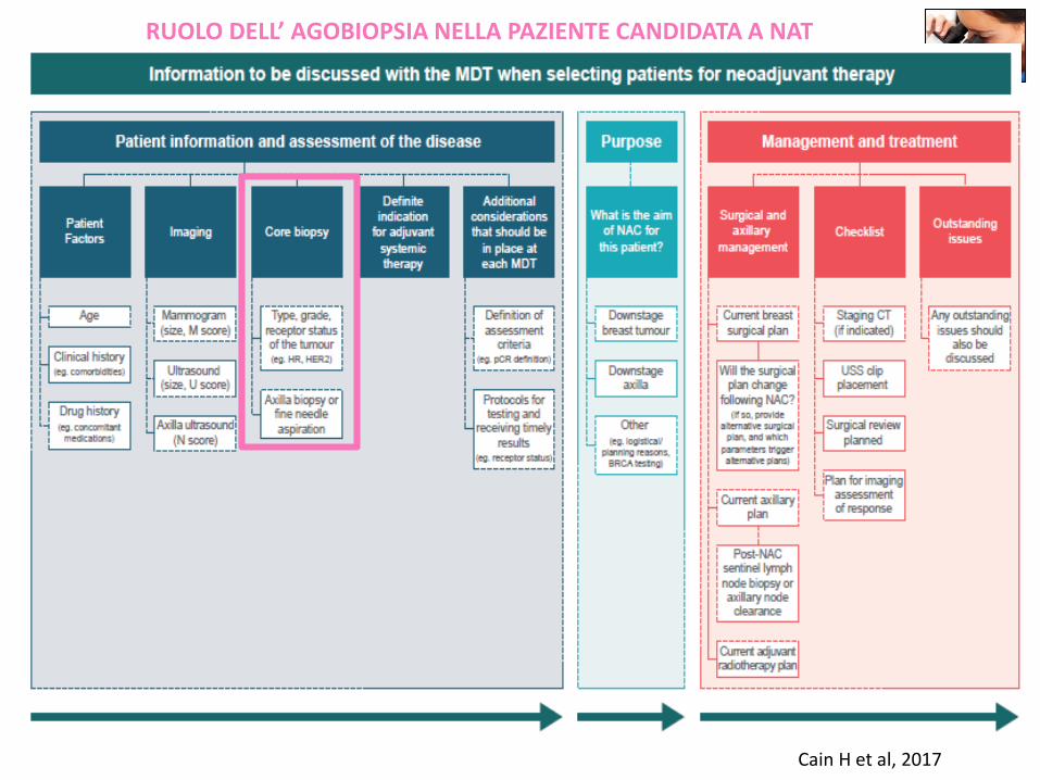
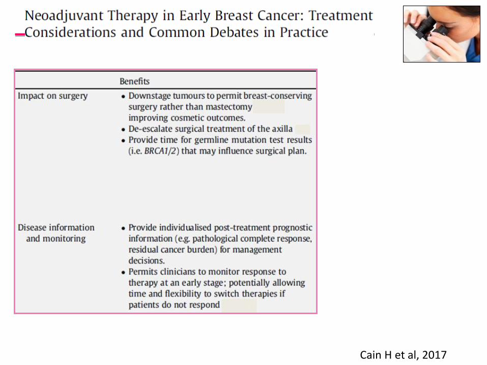
Cain H et al, 2017
RUOLO DELL’ AGOBIOPSIA NELLA PAZIENTE CANDIDATA A NAT
Cain H et al, 2017
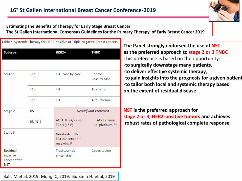
Estimating the Benefits of Therapy for Early Stage Breast CancerThe St Gallen International Consensus Guidelines for the Primary Therapy of Early Breast Cancer 2019
16° St Gallen International Breast Cancer Conference-2019
Balic M et al, 2019; Morigi C, 2019; Burstein HJ et al, 2019
The Panel strongly endorsed the use of NST as the preferred approach to stage 2 or 3 TNBCThis preference is based on the opportunity:-to surgically downstage many patients,-to deliver effective systemic therapy,-to gain insights into the prognosis for a given patient,-to tailor both local and systemic therapy based on the extent of residual disease
NST is the preferred approach forstage 2 or 3, HER2-positive tumors and achievesrobust rates of pathological complete response
Treatment should be carried out in specialised breast units/centres and provided by a multidisciplinary team specialised in breast cancer, consisting of at least medical oncologists,breast surgeons, radiation oncologists, breast radiologists, breast pathologists and breast nurses
Patient information and involvement in decision making.Information on diagnosis and treatment choice should be given repeatedly (both verbally and in writing) in a comprehensive and easily understandable form
NeoAdjuvant systemic treatmentThe decision on neoadjuvant systemic treatment should be based on the predicted sensitivity toparticular treatment types, the benefit from their use and an individual’s risk of relapse
Primary neoadjuvant systemic therapy. In locally advanced and large ‘operable’ cancers, in particular when mastectomy is required due to tumour size, PST is recommended to decrease the extent of surgery needed .In operable cases, the timing of treatment (pre- versus postoperative) has no effect on long-termoutcomes, except a possible small increase in locoregional recurrences in the PST group, but without impact on survival.In subtypes highly sensitive to ChT, such as triple-negative and HER2-positive, a neoadjuvant approach should be preferred, in tumours >2 cm.
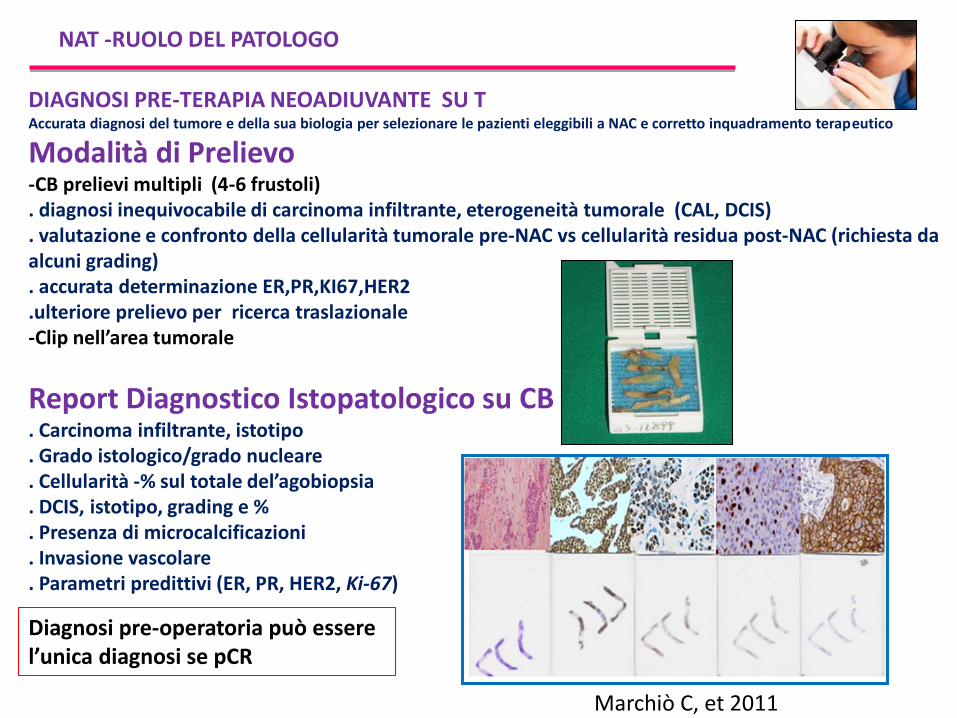
NAT -RUOLO DEL PATOLOGO
DIAGNOSI PRE-TERAPIA NEOADIUVANTE SU TAccurata diagnosi del tumore e della sua biologia per selezionare le pazienti eleggibili a NAC e corretto inquadramento terapeutico
Modalità di Prelievo -CB prelievi multipli (4-6 frustoli). diagnosi inequivocabile di carcinoma infiltrante, eterogeneità tumorale (CAL, DCIS). valutazione e confronto della cellularità tumorale pre-NAC vs cellularità residua post-NAC (richiesta da alcuni grading). accurata determinazione ER,PR,KI67,HER2.ulteriore prelievo per ricerca traslazionale-Clip nell’area tumorale
Report Diagnostico Istopatologico su CB. Carcinoma infiltrante, istotipo. Grado istologico/grado nucleare. Cellularità -% sul totale del’agobiopsia. DCIS, istotipo, grading e %. Presenza di microcalcificazioni. Invasione vascolare. Parametri predittivi (ER, PR, HER2, Ki-67)
Diagnosi pre-operatoria può essere l’unica diagnosi se pCR
Marchiò C, et 2011
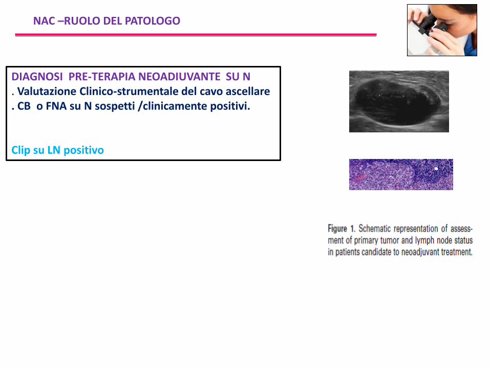
NAC –RUOLO DEL PATOLOGO
DIAGNOSI PRE-TERAPIA NEOADIUVANTE SU N. Valutazione Clinico-strumentale del cavo ascellare . CB o FNA su N sospetti /clinicamente positivi.
Clip su LN positivo
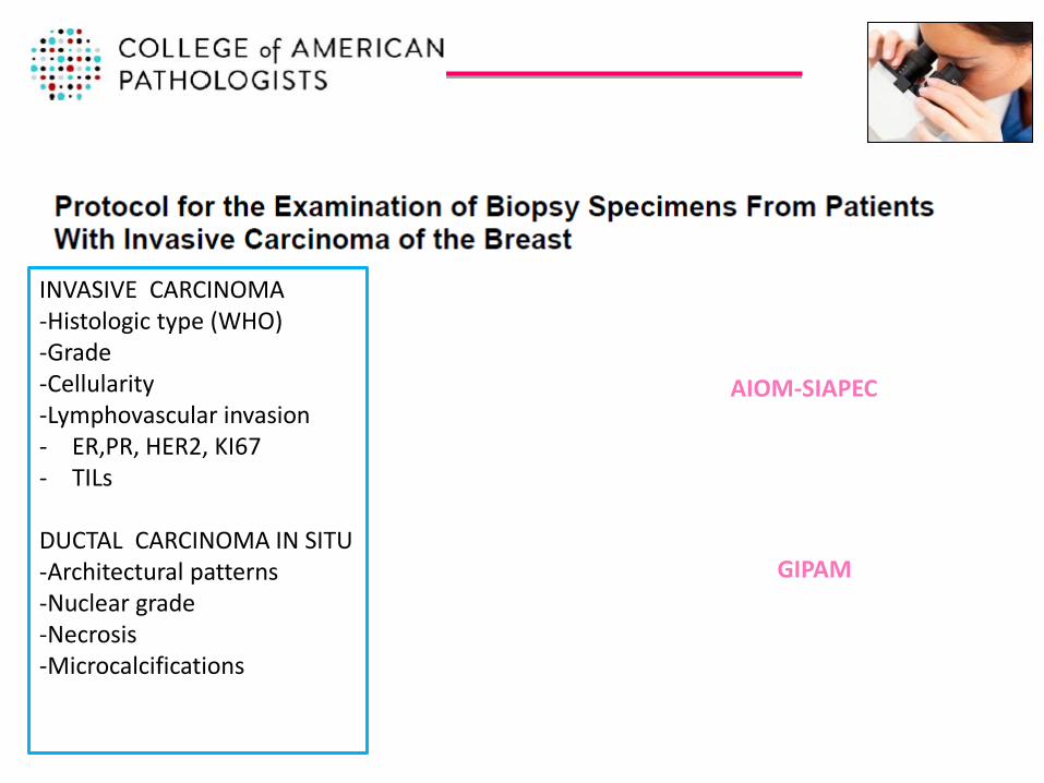
INVASIVE CARCINOMA-Histologic type (WHO)-Grade-Cellularity-Lymphovascular invasion- ER,PR, HER2, KI67- TILs
DUCTAL CARCINOMA IN SITU-Architectural patterns-Nuclear grade-Necrosis-Microcalcifications
AIOM-SIAPEC
GIPAM
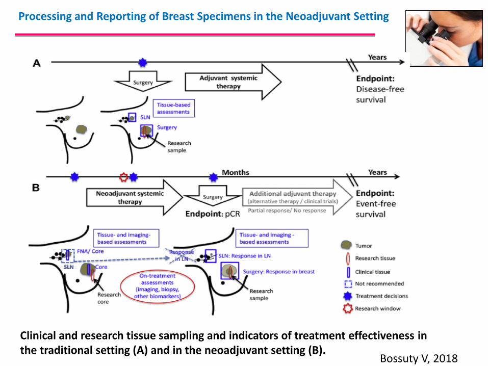
Processing and Reporting of Breast Specimens in the Neoadjuvant Setting
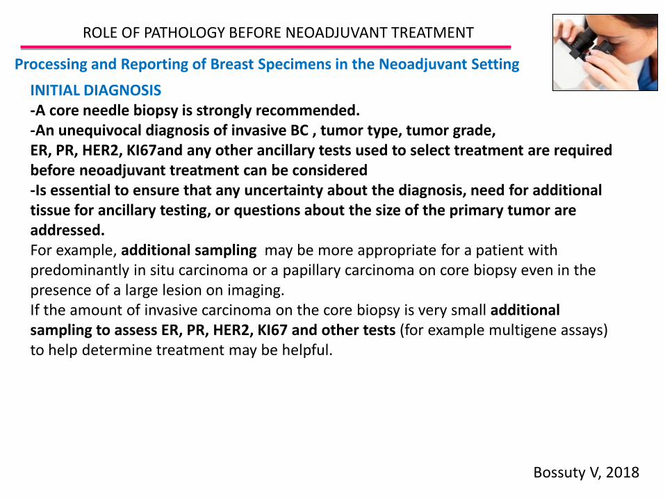
Bossuty V, 2018
ROLE OF PATHOLOGY BEFORE NEOADJUVANT TREATMENT
INITIAL DIAGNOSIS-A core needle biopsy is strongly recommended.-An unequivocal diagnosis of invasive BC , tumor type, tumor grade,ER, PR, HER2, KI67and any other ancillary tests used to select treatment are required before neoadjuvant treatment can be considered-Is essential to ensure that any uncertainty about the diagnosis, need for additional tissue for ancillary testing, or questions about the size of the primary tumor are addressed.For example, additional sampling may be more appropriate for a patient with predominantly in situ carcinoma or a papillary carcinoma on core biopsy even in the presence of a large lesion on imaging. If the amount of invasive carcinoma on the core biopsy is very small additionalsampling to assess ER, PR, HER2, KI67 and other tests (for example multigene assays) to help determine treatment may be helpful.
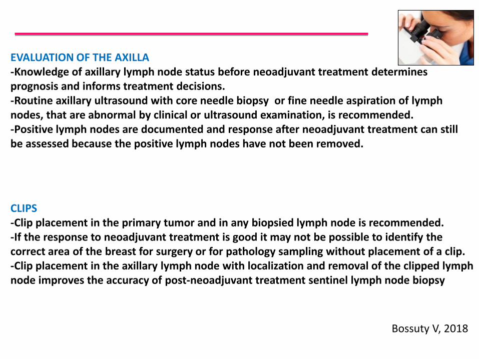
Bossuty V, 2018
EVALUATION OF THE AXILLA-Knowledge of axillary lymph node status before neoadjuvant treatment determines prognosis and informs treatment decisions.-Routine axillary ultrasound with core needle biopsy or fine needle aspiration of lymphnodes, that are abnormal by clinical or ultrasound examination, is recommended.-Positive lymph nodes are documented and response after neoadjuvant treatment can still be assessed because the positive lymph nodes have not been removed.
CLIPS-Clip placement in the primary tumor and in any biopsied lymph node is recommended. -If the response to neoadjuvant treatment is good it may not be possible to identify the correct area of the breast for surgery or for pathology sampling without placement of a clip. -Clip placement in the axillary lymph node with localization and removal of the clipped lymphnode improves the accuracy of post-neoadjuvant treatment sentinel lymph node biopsy
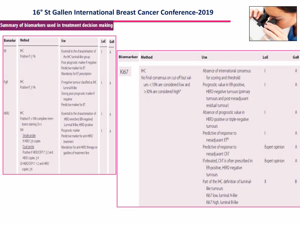
16° St Gallen International Breast Cancer Conference-2019
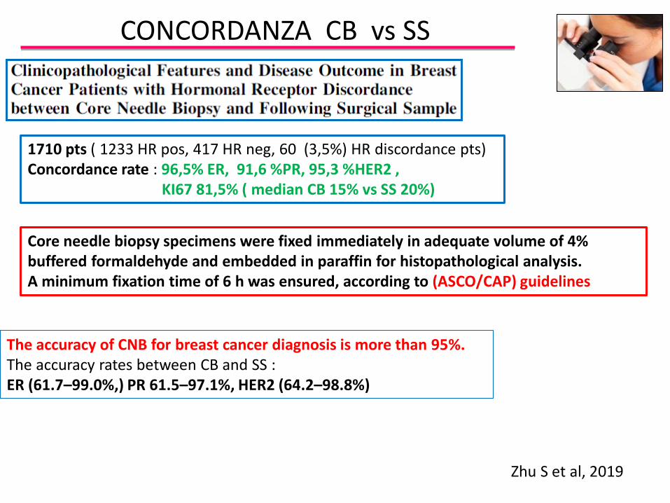
CONCORDANZA CB vs SS
Zhu S et al, 2019
1710 pts ( 1233 HR pos, 417 HR neg, 60 (3,5%) HR discordance pts)Concordance rate : 96,5% ER, 91,6 %PR, 95,3 %HER2 ,
KI67 81,5% ( median CB 15% vs SS 20%)
Core needle biopsy specimens were fixed immediately in adequate volume of 4% buffered formaldehyde and embedded in paraffin for histopathological analysis. A minimum fixation time of 6 h was ensured, according to (ASCO/CAP) guidelines
The accuracy of CNB for breast cancer diagnosis is more than 95%.The accuracy rates between CB and SS :ER (61.7–99.0%,) PR 61.5–97.1%, HER2 (64.2–98.8%)
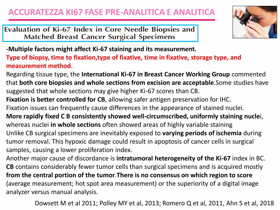
ACCURATEZZA KI67 FASE PRE-ANALITICA E ANALITICA
Dowsett M et al 2011; Polley MY et al, 2013; Romero Q et al, 2011, Ahn S et al, 2018
-Multiple factors might affect Ki-67 staining and its measurement.Type of biopsy, time to fixation,type of fixative, time in fixative, storage type, and measurement method.Regarding tissue type, the International Ki-67 in Breast Cancer Working Group commentedthat both core biopsies and whole sections from excision are acceptable.Some studies have suggested that whole sections may give higher Ki-67 scores than CB.Fixation is better controlled for CB, allowing safer antigen preservation for IHC.Fixation issues can frequently cause differences in the appearance of stained nuclei.More rapidly fixed C B consistently showed well-circumscribed, uniformly staining nuclei, whereas nuclei in whole sections often showed areas of highly variable stainingUnlike CB surgical specimens are inevitably exposed to varying periods of ischemia during tumor removal. This hypoxic damage could result in apoptosis of cancer cells in surgical samples, causing a lower proliferation index.Another major cause of discordance is intratumoral heterogeneity of the Ki-67 index in BC.CB contains considerably fewer tumor cells than surgical specimens and is acquired mostlyfrom the central portion of the tumor.There is no consensus on which region to score (average measurement; hot spot area measurement) or the superiority of a digital image analyzer versus manual analysis.
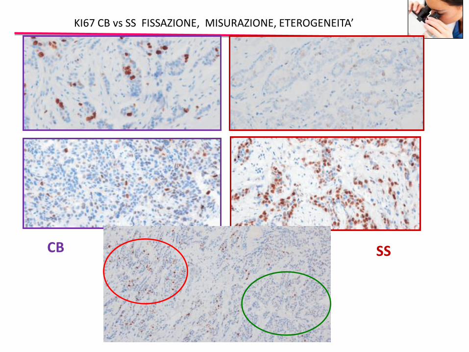
SSCB
KI67 CB vs SS FISSAZIONE, MISURAZIONE, ETEROGENEITA’
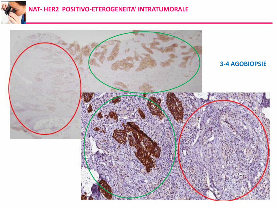
NAT- HER2 POSITIVO-ETEROGENEITA’ INTRATUMORALE
3-4 agobiopsie3-4 AGOBIOPSIE
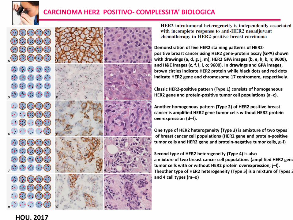
CARCINOMA HER2 POSITIVO- COMPLESSITA’ BIOLOGICA
HOU, 2017
Demonstration of five HER2 staining patterns of HER2-positive breast cancer using HER2 gene-protein assay (GPA) shownwith drawings (a, d, g, j, m), HER2 GPA images (b, e, h, k, n; 9600),and H&E images (c, f, i, l, o; 9600). In drawings and GPA images,brown circles indicate HER2 protein while black dots and red dotsindicate HER2 gene and chromosome 17 centromere, respectively.
Classic HER2-positive pattern (Type 1) consists of homogeneousHER2 gene and protein-positive tumor cell populations (a–c).
Another homogenous pattern (Type 2) of HER2 positive breastcancer is amplified HER2 gene tumor cells without HER2 proteinoverexpression (d–f).
One type of HER2 heterogeneity (Type 3) is amixture of two typesof breast cancer cell populations (HER2 gene and protein-positive
tumor cells and HER2 gene and protein-negative tumor cells, g–i)
Second type of HER2 heterogeneity (Type 4) is alsoa mixture of two breast cancer cell populations (amplified HER2 genetumor cells with or without HER2 protein overexpression, j–l). Theother type of HER2 heterogeneity (Type 5) is a mixture of Types 3and 4 cell types (m–o)
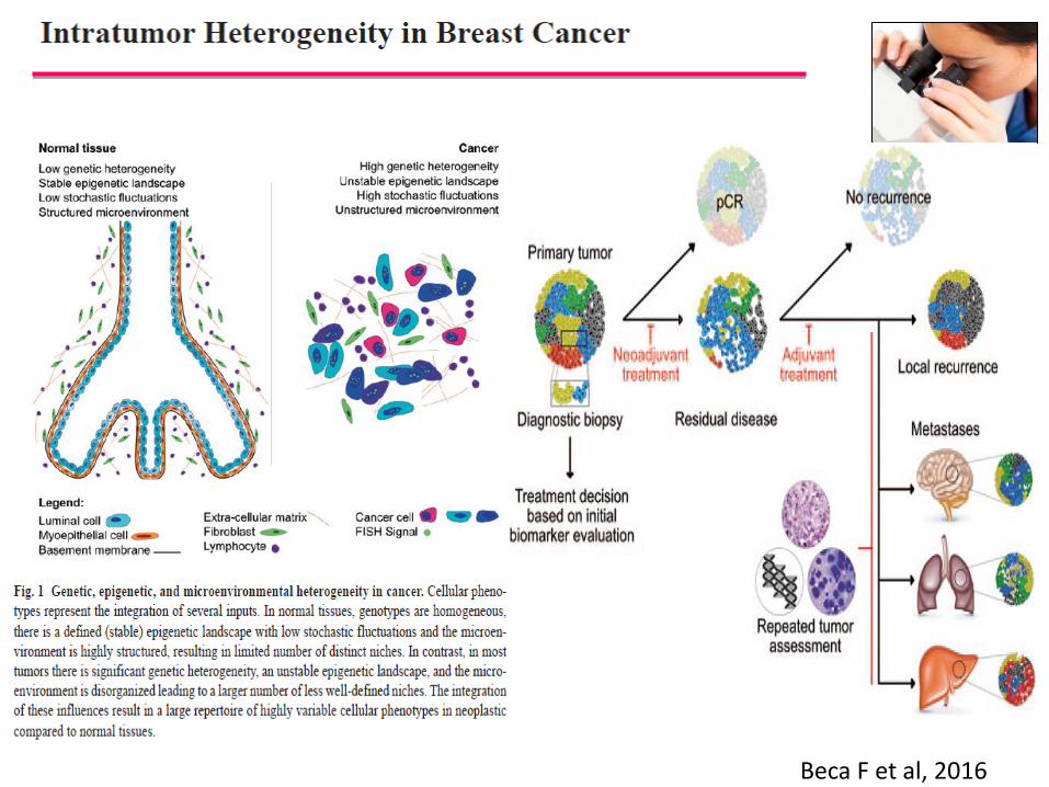
Beca F et al, 2016
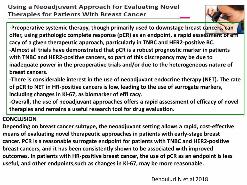
Denduluri N et al 2018
-Preoperative systemic therapy, though primarily used to downstage breast cancers, can offer, using pathologic complete response (pCR) as an endpoint, a rapid assessment of efficacy of a given therapeutic approach, particularly in TNBC and HER2-positive BC.-Almost all trials have demonstrated that pCR is a robust prognostic marker in patients with TNBC and HER2-positive cancers, so part of this discrepancy may be due to inadequate power in the preoperative trials and/or due to the heterogeneous nature of breast cancers.-There is considerable interest in the use of neoadjuvant endocrine therapy (NET). The rate of pCR to NET in HR-positive cancers is low, leading to the use of surrogate markers,including changes in Ki-67, as biomarker of effi cacy. -Overall, the use of neoadjuvant approaches offers a rapid assessment of efficacy of novel therapies and remains a useful research tool for drug evaluation.
CONCLUSIONDepending on breast cancer subtype, the neoadjuvant setting allows a rapid, cost-effective means of evaluating novel therapeutic approaches in patients with early-stage breastcancer. PCR is a reasonable surrogate endpoint for patients with TNBC and HER2-positive breast cancers, and it has been consistently shown to be associated with improvedoutcomes. In patients with HR-positive breast cancer, the use of pCR as an endpoint is less useful, and other endpoints,such as changes in Ki-67, may be more reasonable.
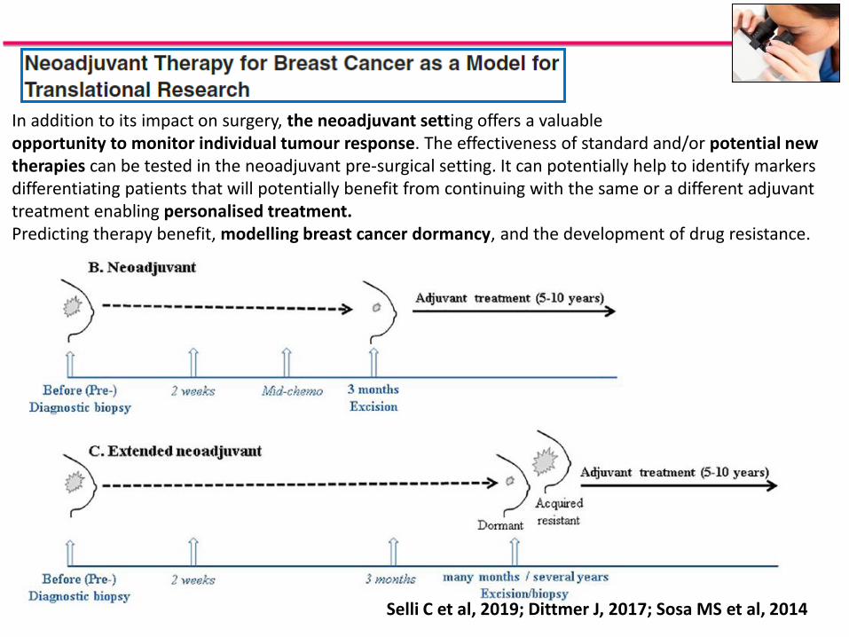
In addition to its impact on surgery, the neoadjuvant setting offers a valuableopportunity to monitor individual tumour response. The effectiveness of standard and/or potential new therapies can be tested in the neoadjuvant pre-surgical setting. It can potentially help to identify markers differentiating patients that will potentially benefit from continuing with the same or a different adjuvant treatment enabling personalised treatment. Predicting therapy benefit, modelling breast cancer dormancy, and the development of drug resistance.
Selli C et al, 2019; Dittmer J, 2017; Sosa MS et al, 2014
Bossuty V, 2018
Processing and Reporting of Breast Specimens in the Neoadjuvant Setting
Clinical and research tissue sampling and indicators of treatment effectiveness inthe traditional setting (A) and in the neoadjuvant setting (B).
Tokes T.et al 2019
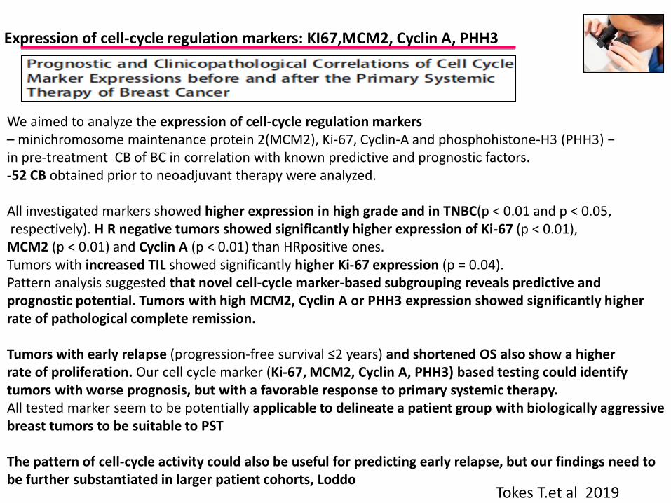
We aimed to analyze the expression of cell-cycle regulation markers – minichromosome maintenance protein 2(MCM2), Ki-67, Cyclin-A and phosphohistone-H3 (PHH3) − in pre-treatment CB of BC in correlation with known predictive and prognostic factors. -52 CB obtained prior to neoadjuvant therapy were analyzed.
All investigated markers showed higher expression in high grade and in TNBC(p < 0.01 and p < 0.05,respectively). H R negative tumors showed significantly higher expression of Ki-67 (p < 0.01), MCM2 (p < 0.01) and Cyclin A (p < 0.01) than HRpositive ones. Tumors with increased TIL showed significantly higher Ki-67 expression (p = 0.04). Pattern analysis suggested that novel cell-cycle marker-based subgrouping reveals predictive and prognostic potential. Tumors with high MCM2, Cyclin A or PHH3 expression showed significantly higher rate of pathological complete remission.
Tumors with early relapse (progression-free survival ≤2 years) and shortened OS also show a higherrate of proliferation. Our cell cycle marker (Ki-67, MCM2, Cyclin A, PHH3) based testing could identify tumors with worse prognosis, but with a favorable response to primary systemic therapy. All tested marker seem to be potentially applicable to delineate a patient group with biologically aggressive breast tumors to be suitable to PST
The pattern of cell-cycle activity could also be useful for predicting early relapse, but our findings need to be further substantiated in larger patient cohorts, Loddo
Expression of cell-cycle regulation markers: KI67,MCM2, Cyclin A, PHH3
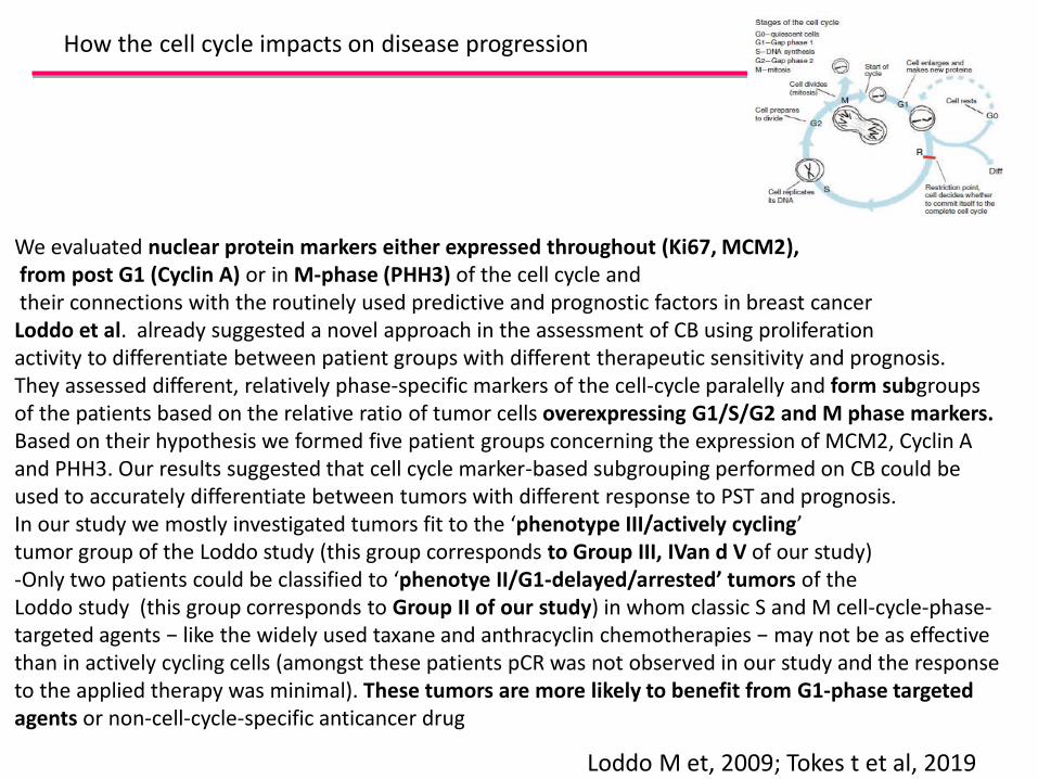
We evaluated nuclear protein markers either expressed throughout (Ki67, MCM2),from post G1 (Cyclin A) or in M-phase (PHH3) of the cell cycle andtheir connections with the routinely used predictive and prognostic factors in breast cancerLoddo et al. already suggested a novel approach in the assessment of CB using proliferationactivity to differentiate between patient groups with different therapeutic sensitivity and prognosis. They assessed different, relatively phase-specific markers of the cell-cycle paralelly and form subgroups of the patients based on the relative ratio of tumor cells overexpressing G1/S/G2 and M phase markers.Based on their hypothesis we formed five patient groups concerning the expression of MCM2, Cyclin A and PHH3. Our results suggested that cell cycle marker-based subgrouping performed on CB could be used to accurately differentiate between tumors with different response to PST and prognosis.In our study we mostly investigated tumors fit to the ‘phenotype III/actively cycling’tumor group of the Loddo study (this group corresponds to Group III, IVan d V of our study)-Only two patients could be classified to ‘phenotye II/G1-delayed/arrested’ tumors of theLoddo study (this group corresponds to Group II of our study) in whom classic S and M cell-cycle-phase-targeted agents − like the widely used taxane and anthracyclin chemotherapies − may not be as effective than in actively cycling cells (amongst these patients pCR was not observed in our study and the response to the applied therapy was minimal). These tumors are more likely to benefit from G1-phase targeted agents or non-cell-cycle-specific anticancer drug
Loddo M et, 2009; Tokes t et al, 2019
How the cell cycle impacts on disease progression
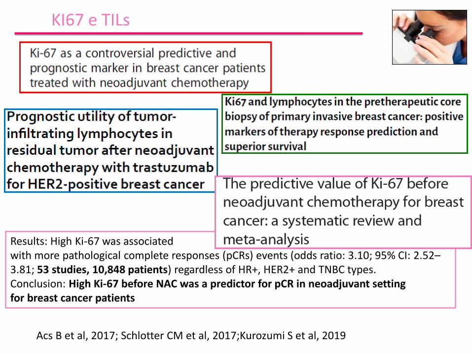
Results: High Ki-67 was associatedwith more pathological complete responses (pCRs) events (odds ratio: 3.10; 95% CI: 2.52–3.81; 53 studies, 10,848 patients) regardless of HR+, HER2+ and TNBC types.Conclusion: High Ki-67 before NAC was a predictor for pCR in neoadjuvant settingfor breast cancer patients
Acs B et al, 2017; Schlotter CM et al, 2017;Kurozumi S et al, 2019
KI67 e TILs
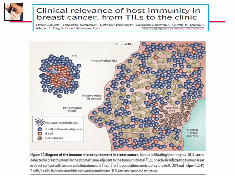
TILs
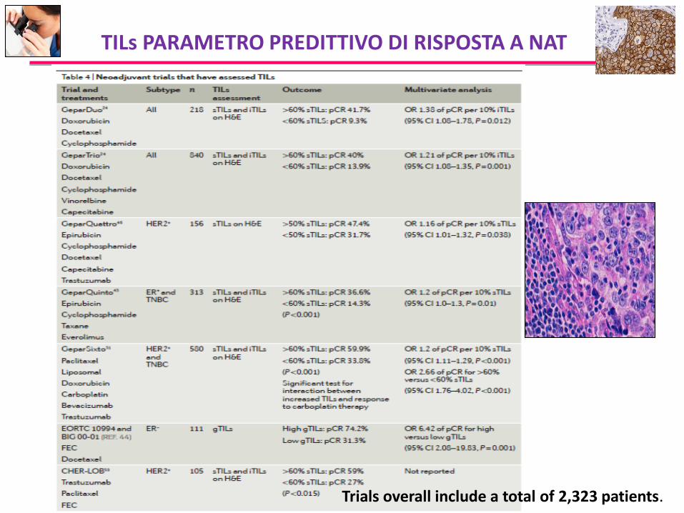
TILs PARAMETRO PREDITTIVO DI RISPOSTA A NAT
Trials overall include a total of 2,323 patients.
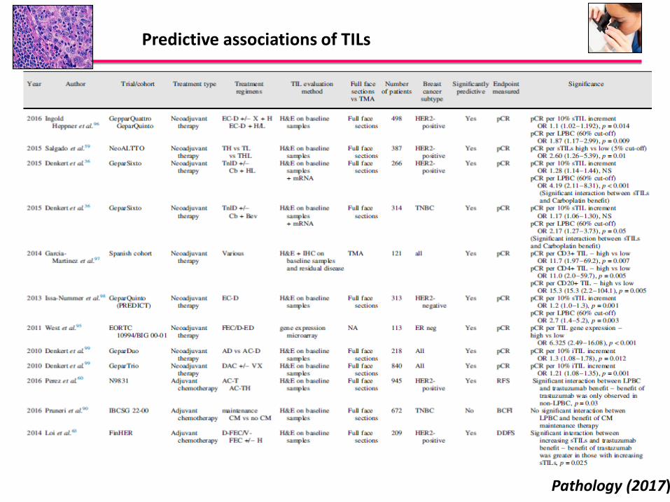
Predictive associations of TILs
Pathology (2017)
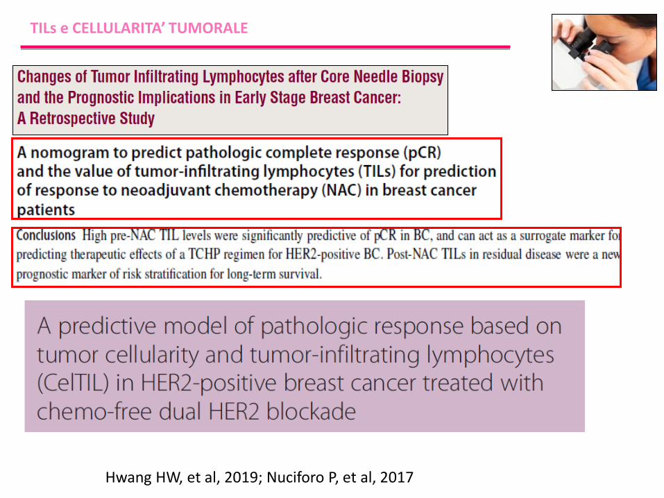
Hwang HW, et al, 2019; Nuciforo P, et al, 2017
TILs e CELLULARITA’ TUMORALE
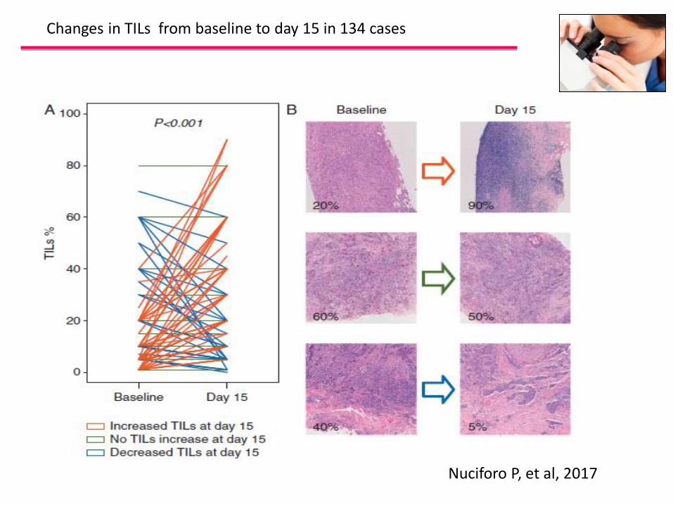
Nuciforo P, et al, 2017
Changes in TILs from baseline to day 15 in 134 cases
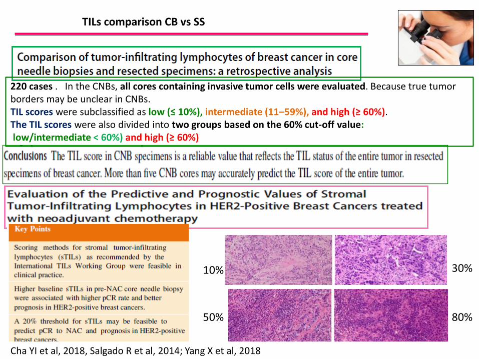
TILs comparison CB vs SS
Cha YI et al, 2018, Salgado R et al, 2014; Yang X et al, 2018
220 cases . In the CNBs, all cores containing invasive tumor cells were evaluated. Because true tumor borders may be unclear in CNBs.TIL scores were subclassified as low (≤ 10%), intermediate (11–59%), and high (≥ 60%).The TIL scores were also divided into two groups based on the 60% cut-off value:low/intermediate < 60%) and high (≥ 60%)
10%
50%
30%
80%
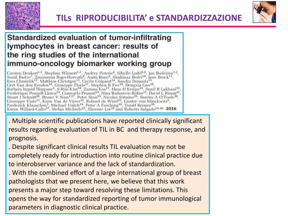
TILs RIPRODUCIBILITA’ e STANDARDIZZAZIONE
. Multiple scientific publications have reported clinically significant results regarding evaluation of TIL in BC and therapy response, andprognosis.. Despite significant clinical results TIL evaluation may not becompletely ready for introduction into routine clinical practice due to interobserver variance and the lack of standardization.. With the combined effort of a large international group of breast pathologists that we present here, we believe that this work presents a major step toward resolving these limitations. This opens the way for standardized reporting of tumor immunologicalparameters in diagnostic clinical practice.
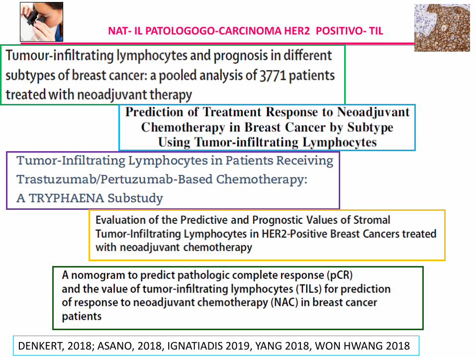
NAT- IL PATOLOGOGO-CARCINOMA HER2 POSITIVO- TIL
DENKERT, 2018; ASANO, 2018, IGNATIADIS 2019, YANG 2018, WON HWANG 2018
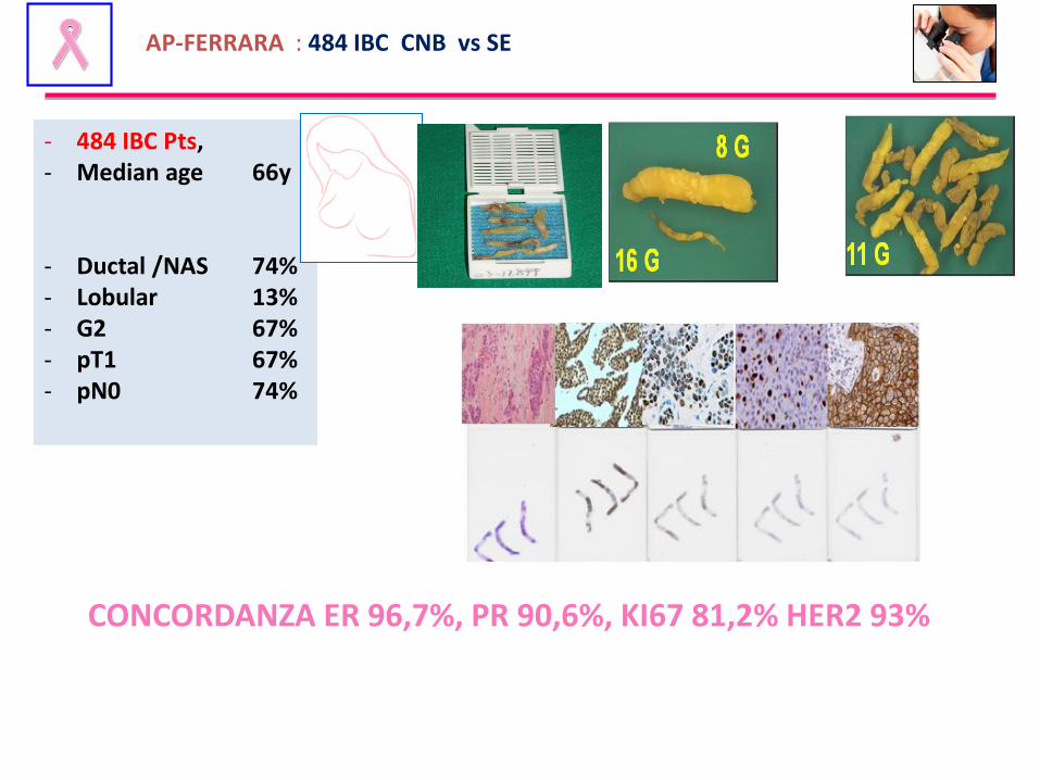
- 484 IBC Pts,- Median age 66y
- Ductal /NAS 74%- Lobular 13%- G2 67%- pT1 67%- pN0 74%
AP-FERRARA : 484 IBC CNB vs SE
CONCORDANZA ER 96,7%, PR 90,6%, KI67 81,2% HER2 93%
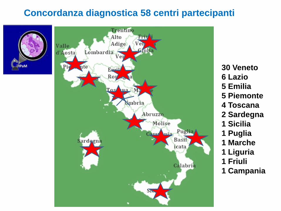
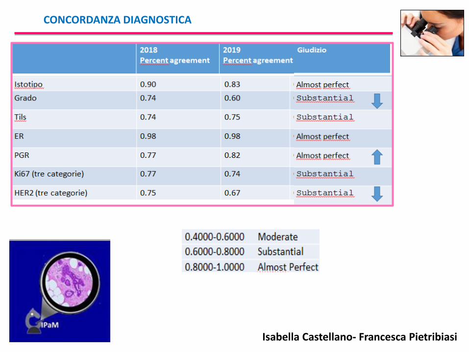
Concordanza diagnostica 58 centri partecipanti
30 Veneto
6 Lazio
5 Emilia
5 Piemonte
4 Toscana
2 Sardegna
1 Sicilia
1 Puglia
1 Marche
1 Liguria
1 Friuli
1 Campania
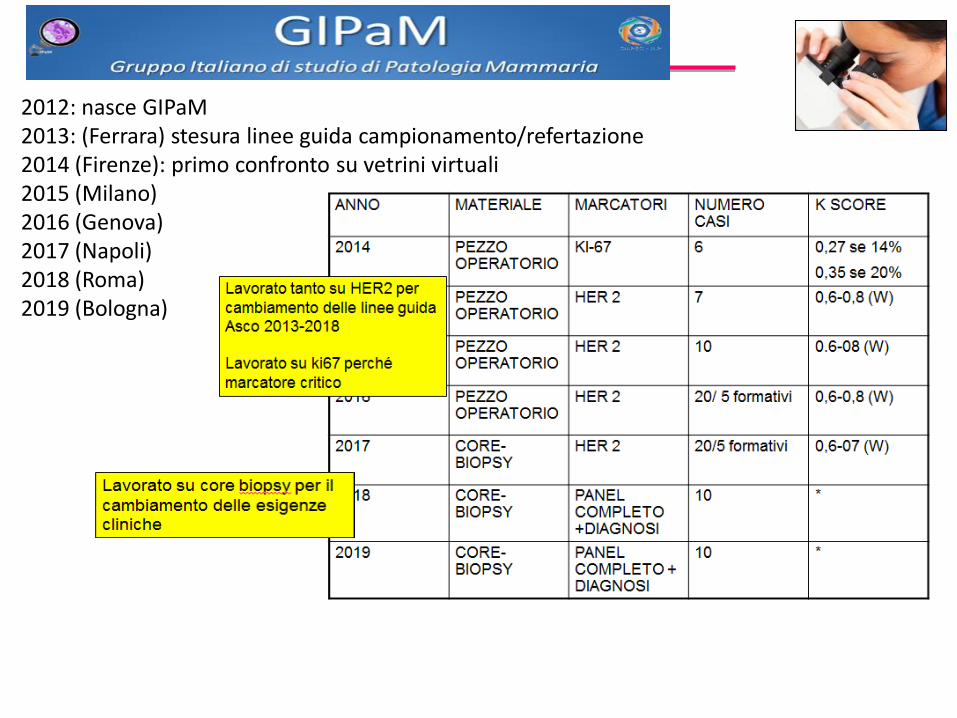
2012: nasce GIPaM2013: (Ferrara) stesura linee guida campionamento/refertazione2014 (Firenze): primo confronto su vetrini virtuali 2015 (Milano)2016 (Genova)2017 (Napoli)2018 (Roma)2019 (Bologna)
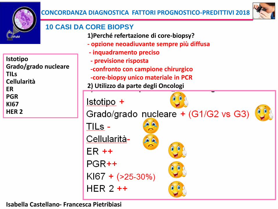
10 CASI DA CORE BIOPSY
CONCORDANZA DIAGNOSTICA FATTORI PROGNOSTICO-PREDITTIVI 2018
Isabella Castellano- Francesca Pietribiasi
IstotipoGrado/grado nucleareTILsCellularitàERPGRKI67HER 2
1)Perché refertazione di core-biopsy?- opzione neoadiuvante sempre più diffusa- inquadramento preciso- previsione risposta -confronto con campione chirurgico-core-biopsy unico materiale in PCR
2) Utilizzo da parte degli Oncologi
CONCORDANZA DIAGNOSTICA
Isabella Castellano- Francesca Pietribiasi
Related Documents


