Prepattern genes and signaling molecules regulate stripe expression to specify Drosophila flight muscle attachment sites Arjumand Ghazi 1 * , Litty Paul 2 , K. VijayRaghavan National Centre for Biological Sciences, Tata Institute of Fundamental Research, GKVK PO, Bangalore 560 065, India Received 18 November 2002; received in revised form 10 February 2003; accepted 11 March 2003 Abstract In Drosophila, muscles attach to epidermal tendon cells specified by the gene stripe (sr). Flight muscle attachment sites are prefigured on the wing imaginal disc by sr expression in discrete domains. We describe the mechanisms underlying the specification of these domains of sr expression. We show that the concerted activities of the wingless (wg), decapentaplegic (dpp) and Notch (N) signaling pathways, and the prepattern genes pannier ( pnr) and u-shaped (ush) establish domains of sr expression. N is required for initiation of sr expression. pnr is a positive regulator of sr, and is inhibited by ush in this function. The Wg signal differentially influences the formation of different sr domains. These results identify the multiple regulatory elements involved in the positioning of Drosophila flight muscle attachment sites. q 2003 Elsevier Science Ireland Ltd. All rights reserved. Keywords: sr; wg; pnr; ush; dpp; N; Tendon cells; Muscle attachment; Flight muscles 1. Introduction In vertebrates, muscles attach to bones, or cartilage, with the help of tendons. Studies from the chick hind limb indicate that reciprocal muscle – tendon interactions are important for generation of the final muscle pattern (Kardon, 1998). However, the genetic, cellular and mol- ecular mechanisms involved in this pathway remain unknown. Insect equivalents of vertebrate tendons are specialized epidermal tendon cells. Studies in Tenebrio (William and Caveney, 1980a,b) and Drosophila (Volk and VijayRaghavan, 1994) have suggested that muscle attach- ment sites not only supply insertion points but also provide navigational information to migrating myotubes (Frommer et al., 1996; Becker et al., 1997). In the Drosophila embryo, muscle attachment sites are character- ized by expression of stripe (sr), a gene encoding a Zn þþ finger protein, and a member of the vertebrate early growth response (egr) family of transcription factors. sr is required for specification and differentiation of tendon cells (Lee et al., 1995; Frommer et al., 1996; Fernandes et al., 1996; Nabel-Rosen et al., 1999). The major muscles of the adult dorsal thorax are shown in Fig. 1. These muscles develop during pupation, but their attachment sites are prefigured earlier on the wing imaginal disc, in the late third instar larva, by sr expression at discrete positions in the presumptive notum (Fig. 1A)(Fernandes et al., 1996). sr expression at this stage suggests the possibility of an earlier role for the gene, in addition to its late role in tendon cell differentiation – a view strengthened by observations that flight muscles develop closely juxtaposed to sr-expressing attachment sites from earliest stages of adult myogenesis. Significantly, adult epidermal sr expression is crucial for establishing the early expression pattern of muscle founders (Dutta et al., submitted for publication). These observations, and the role of tendon cells in muscle patterning, make it important to understand the mechanisms underlying tendon cell positioning – a process dependent on the precise spatio-temporal regulation of sr expression. This study deciphers the mechanisms controlling sr expression on the wing imaginal disc. Embryonic sr expression at the segment borders arises as a consequence of antagonistic interactions of wingless (wg) 0925-4773/03/$ - see front matter q 2003 Elsevier Science Ireland Ltd. All rights reserved. doi:10.1016/S0925-4773(03)00042-X Mechanisms of Development 120 (2003) 519–528 www.elsevier.com/locate/modo 1 Present address: Department of Biochemistry and Biophysics, University of California, San Francisco (UCSF), 600 16th Street, San Francisco, CA 94143-2200, USA. 2 Present address: 484, West 12th Avenue, The Ohio State University, Columbus, OH 43210, USA. * Corresponding author. Tel.: þ 1-415-476-9864; fax: þ1-415-514-4145. E-mail address: [email protected] (A. Ghazi).

Welcome message from author
This document is posted to help you gain knowledge. Please leave a comment to let me know what you think about it! Share it to your friends and learn new things together.
Transcript
Prepattern genes and signaling molecules regulate stripe expression
to specify Drosophila flight muscle attachment sites
Arjumand Ghazi1*, Litty Paul2, K. VijayRaghavan
National Centre for Biological Sciences, Tata Institute of Fundamental Research, GKVK PO, Bangalore 560 065, India
Received 18 November 2002; received in revised form 10 February 2003; accepted 11 March 2003
Abstract
In Drosophila, muscles attach to epidermal tendon cells specified by the gene stripe (sr). Flight muscle attachment sites are prefigured on
the wing imaginal disc by sr expression in discrete domains. We describe the mechanisms underlying the specification of these domains of sr
expression. We show that the concerted activities of the wingless (wg), decapentaplegic (dpp) and Notch (N) signaling pathways, and the
prepattern genes pannier ( pnr) and u-shaped (ush) establish domains of sr expression. N is required for initiation of sr expression. pnr is a
positive regulator of sr, and is inhibited by ush in this function. The Wg signal differentially influences the formation of different sr domains.
These results identify the multiple regulatory elements involved in the positioning of Drosophila flight muscle attachment sites.
q 2003 Elsevier Science Ireland Ltd. All rights reserved.
Keywords: sr; wg; pnr; ush; dpp; N; Tendon cells; Muscle attachment; Flight muscles
1. Introduction
In vertebrates, muscles attach to bones, or cartilage, with
the help of tendons. Studies from the chick hind limb
indicate that reciprocal muscle–tendon interactions are
important for generation of the final muscle pattern
(Kardon, 1998). However, the genetic, cellular and mol-
ecular mechanisms involved in this pathway remain
unknown. Insect equivalents of vertebrate tendons are
specialized epidermal tendon cells. Studies in Tenebrio
(William and Caveney, 1980a,b) and Drosophila (Volk and
VijayRaghavan, 1994) have suggested that muscle attach-
ment sites not only supply insertion points but also
provide navigational information to migrating myotubes
(Frommer et al., 1996; Becker et al., 1997). In the
Drosophila embryo, muscle attachment sites are character-
ized by expression of stripe (sr), a gene encoding a Znþþ
finger protein, and a member of the vertebrate early growth
response (egr) family of transcription factors. sr is required
for specification and differentiation of tendon cells (Lee
et al., 1995; Frommer et al., 1996; Fernandes et al., 1996;
Nabel-Rosen et al., 1999).
The major muscles of the adult dorsal thorax are shown
in Fig. 1. These muscles develop during pupation, but their
attachment sites are prefigured earlier on the wing imaginal
disc, in the late third instar larva, by sr expression at discrete
positions in the presumptive notum (Fig. 1A) (Fernandes
et al., 1996). sr expression at this stage suggests the
possibility of an earlier role for the gene, in addition to its
late role in tendon cell differentiation – a view strengthened
by observations that flight muscles develop closely
juxtaposed to sr-expressing attachment sites from earliest
stages of adult myogenesis. Significantly, adult epidermal sr
expression is crucial for establishing the early expression
pattern of muscle founders (Dutta et al., submitted for
publication). These observations, and the role of tendon
cells in muscle patterning, make it important to understand
the mechanisms underlying tendon cell positioning – a
process dependent on the precise spatio-temporal regulation
of sr expression. This study deciphers the mechanisms
controlling sr expression on the wing imaginal disc.
Embryonic sr expression at the segment borders arises as
a consequence of antagonistic interactions of wingless (wg)
0925-4773/03/$ - see front matter q 2003 Elsevier Science Ireland Ltd. All rights reserved.
doi:10.1016/S0925-4773(03)00042-X
Mechanisms of Development 120 (2003) 519–528
www.elsevier.com/locate/modo
1 Present address: Department of Biochemistry and Biophysics,
University of California, San Francisco (UCSF), 600 16th Street, San
Francisco, CA 94143-2200, USA.2 Present address: 484, West 12th Avenue, The Ohio State University,
Columbus, OH 43210, USA.
* Corresponding author. Tel.: þ1-415-476-9864; fax: þ1-415-514-4145.
E-mail address: [email protected] (A. Ghazi).
and hedgehog (hh) signals (Piepenburg et al., 2000) and is
induced by the ligands Hh, Wg and Spitz (Spi), in territories
adjacent to their zones of expression (Hatini and DiNardo,
2001). However, mechanisms regulating its expression in
other embryonic regions, and in the adult, are unknown. In
the wing disc, sr expression is organized into distinct
domains in the anterior and posterior compartments of
the presumptive notum (Fig. 2K). This suggests that genes
mediating patterning of the thoracic epidermis could act
to regulate sr. Thoracic patterning is brought about by
the concerted activities of a hierarchy of prepattern and
pattern forming genes (Stern, 1954; Ghysen and Dambly-
Chaudiere, 1988). The medial notum, for instance, is
organized as a result of activities of the prepattern gene
pannier ( pnr) (Ramain et al., 1993; Heitzler et al., 1996;
Calleja et al., 1996, Garcia-Garcia et al., 1999), its negative
regulator u-shaped (ush) (Cubadda et al., 1997; Haenlin
et al., 1997), and signaling molecules Wingless (Wg)
(Phillips and Whittle, 1993) and Decapentaplegic (Dpp)
(Tomoyasu et al., 1998; Tomoyasu et al., 2000; Sato and
Saigo, 2000). We have studied the expression of these
prepattern and pattern forming genes during adult develop-
ment, to examine the extent of their overlap with different sr
domains, and thus assign specific notal identities to the
different sr domains. The flight muscles of hypomorphic and
gain-of-function mutants of these genes have been analyzed
to test for their function in thoracic myogenesis. We
examine the effect of altering the expressions of these genes
on the expression of sr. We have also examined the role of
Notch (N), and its ligand Serrate (Ser) as potential
regulators of sr. Our results indicate that sr activation
depends on N, and is inhibited by Ser. pnr is crucial for
initiation of sr expression, and its function is inhibited by
ush. wg, antagonized by dpp, maintains the distinct
identities of different sr domains. These results allow us to
identify and describe the multiple regulatory elements
involved in positioning an important class of imaginal disc
derivatives – the muscle attachment sites.
2. Results
2.1. wg, pnr, ush and dpp expression domains suggest roles
in sr regulation
Much of the presumptive notum is in the anterior
compartment in which there are four domains of sr
expression. One of these is in the medial region (a in
Fig. 1A) and gives rise to the anterior tendon cells, to which
DLMs attach (Fig. 1B). The remaining three are in the
lateral region (b, c and d in Fig. 1A) and provide dorsal
attachments for DVMs (Fig. 1B). In the posterior compart-
ment, sr is expressed in a narrow band that eventually forms
the posterior insertion site for DLMs (Fig. 1A, arrow). We
examined the positioning of different sr domains on the
wing imaginal disc, with respect to prepattern and pattern
forming genes expressed in this region.
wg is expressed in a narrow region in the presumptive
notum (Fig. 2B). Using a wg lacZ reporter, we find that wg
expression is present between the large, medial sr domain
and the three lateral ones (Fig. 2A–C). wg, at its lateral
margin, borders the lateral sr domains and covers them
partially (Fig. 2C). pnr expression covers the medial sr
domain completely, whereas the lateral domains are
positioned at the border of pnr expression (Fig. 2D–F)
and the two show some overlap at the margins (Fig. 2F,
white arrow). The posterior sr domain is also partially
covered by pnr. The antagonist of pnr, ush, is expressed in a
domain similar to pnr but its levels are highest at the
proximal end of the disc, and gradually decrease distally.
ush expression covers the medial sr domain partially
(Fig. 2G-I) but does not extend to either the lateral domains
or the posterior one. sr expression commences in regions
with lowest ush levels, in both the proximo-distal and
antero-posterior axes. This is clearest for the posterior sr
domain, which begins at a position where ush expression
ceases (Fig. 2I, white arrowhead). dpp expression is
observed at the antero-posterior border of the presumptive
notum in two domains, and borders sr expression in both the
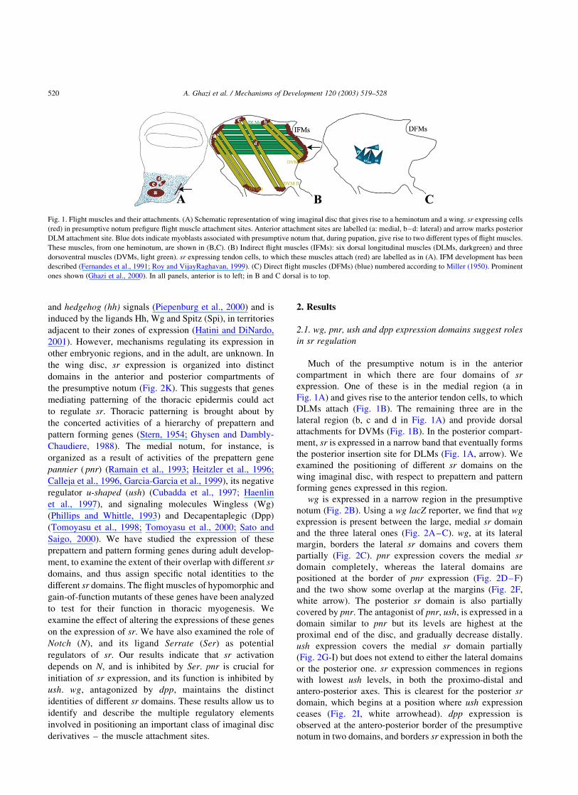
Fig. 1. Flight muscles and their attachments. (A) Schematic representation of wing imaginal disc that gives rise to a heminotum and a wing. sr expressing cells
(red) in presumptive notum prefigure flight muscle attachment sites. Anterior attachment sites are labelled (a: medial, b–d: lateral) and arrow marks posterior
DLM attachment site. Blue dots indicate myoblasts associated with presumptive notum that, during pupation, give rise to two different types of flight muscles.
These muscles, from one heminotum, are shown in (B,C). (B) Indirect flight muscles (IFMs): six dorsal longitudinal muscles (DLMs, darkgreen) and three
dorsoventral muscles (DVMs, light green). sr expressing tendon cells, to which these muscles attach (red) are labelled as in (A). IFM development has been
described (Fernandes et al., 1991; Roy and VijayRaghavan, 1999). (C) Direct flight muscles (DFMs) (blue) numbered according to Miller (1950). Prominent
ones shown (Ghazi et al., 2000). In all panels, anterior is to left; in B and C dorsal is to top.
A. Ghazi et al. / Mechanisms of Development 120 (2003) 519–528520
anterior and posterior compartments. A large domain
borders the medial sr domain (Fig. 2J, blue arrowhead
next to a) and a smaller one borders the posterior sr
expression (Fig. 2J, blue and black arrows, respectively).
The expression patterns of these genes, with respect to that
of sr, are depicted schematically in Fig. 2K, L. This profile
suggests the possibility of regulatory interactions that
determine sr expression and these are examined below.
2.2. The Wg gradient differentially influences sr domains
To assess the extent to which different sr domains detect
Wg, we expressed a truncated, non-functional, GPI-linked
form of the Wingless receptor, DFrizzled2 (GPI-Dfz2)
(Cadigan et al., 1998) in the presumptive notum. This
receptor binds Wg but prevents signal transduction. Wg
protein gets stabilized at the membrane and can be detected
using a Wg-specific antibody. This construct has been used
earlier to determine the range of wg signaling (Cadigan et al.,
1998; Sudarsan et al., 2001). We used two different Gal4
drivers to express the GPI-Dfz2 construct: (i) srGal4 to
express it in all the sr domains and examine Wg distribution
within each of them and, (ii) pnrGal4 to express in the
medial notum which includes the cells transcribing wg. We
find that not all domains of sr receive similar levels of Wg as
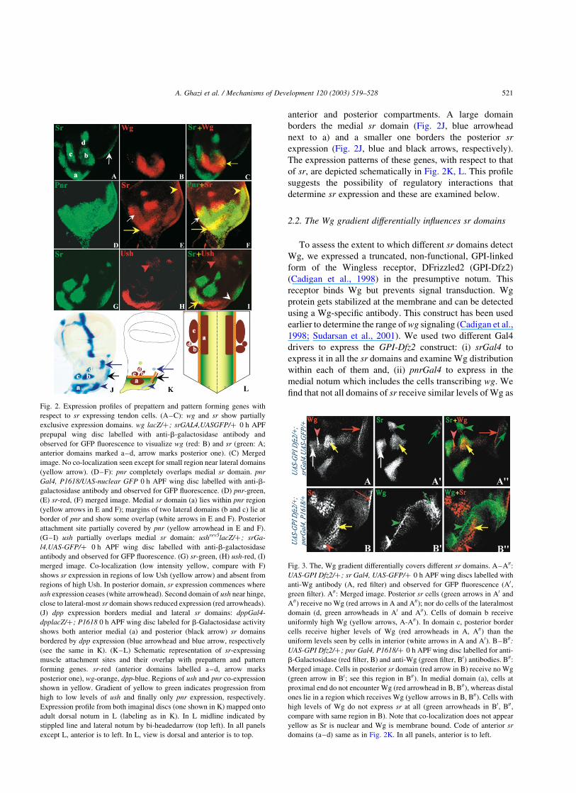
Fig. 2. Expression profiles of prepattern and pattern forming genes with
respect to sr expressing tendon cells. (A–C): wg and sr show partially
exclusive expression domains. wg lacZ/þ; srGAL4,UASGFP/þ 0 h APF
prepupal wing disc labelled with anti-b-galactosidase antibody and
observed for GFP fluorescence to visualize wg (red: B) and sr (green: A;
anterior domains marked a–d, arrow marks posterior one). (C) Merged
image. No co-localization seen except for small region near lateral domains
(yellow arrow). (D–F): pnr completely overlaps medial sr domain. pnr
Gal4, P1618/UAS-nuclear GFP 0 h APF wing disc labelled with anti-b-
galactosidase antibody and observed for GFP fluorescence. (D) pnr-green,
(E) sr-red, (F) merged image. Medial sr domain (a) lies within pnr region
(yellow arrows in E and F); margins of two lateral domains (b and c) lie at
border of pnr and show some overlap (white arrows in E and F). Posterior
attachment site partially covered by pnr (yellow arrowhead in E and F).
(G–I) ush partially overlaps medial sr domain: ushrev5lacZ/þ ; srGa-
l4,UAS-GFP/þ 0 h APF wing disc labelled with anti-b-galactosidase
antibody and observed for GFP fluorescence. (G) sr-green, (H) ush-red, (I)
merged image. Co-localization (low intensity yellow, compare with F)
shows sr expression in regions of low Ush (yellow arrow) and absent from
regions of high Ush. In posterior domain, sr expression commences where
ush expression ceases (white arrowhead). Second domain of ush near hinge,
close to lateral-most sr domain shows reduced expression (red arrowheads).
(J) dpp expression borders medial and lateral sr domains: dppGal4-
dpplacZ/þ ; P1618 0 h APF wing disc labeled for b-Galactosidase activity
shows both anterior medial (a) and posterior (black arrow) sr domains
bordered by dpp expression (blue arrowhead and blue arrow, respectively
(see the same in K). (K–L) Schematic representation of sr-expressing
muscle attachment sites and their overlap with prepattern and pattern
forming genes. sr-red (anterior domains labelled a–d, arrow marks
posterior one), wg-orange, dpp-blue. Regions of ush and pnr co-expression
shown in yellow. Gradient of yellow to green indicates progression from
high to low levels of ush and finally only pnr expression, respectively.
Expression profile from both imaginal discs (one shown in K) mapped onto
adult dorsal notum in L (labeling as in K). In L midline indicated by
stippled line and lateral notum by bi-headedarrow (top left). In all panels
except L, anterior is to left. In L, view is dorsal and anterior is to top.
Fig. 3. The, Wg gradient differentially covers different sr domains. A–A00:
UAS-GPI Dfz2/þ; sr Gal4, UAS-GFP/þ 0 h APF wing discs labelled with
anti-Wg antibody (A, red filter) and observed for GFP fluorescence (A0,
green filter). A00: Merged image. Posterior sr cells (green arrows in A0 and
A00) receive no Wg (red arrows in A and A00); nor do cells of the lateralmost
domain (d, green arrowheads in A0 and A00). Cells of domain b receive
uniformly high Wg (yellow arrows, A-A00). In domain c, posterior border
cells receive higher levels of Wg (red arrowheads in A, A00) than the
uniform levels seen by cells in interior (white arrows in A and A0). B–B00:
UAS-GPI Dfz2/þ ; pnr Gal4, P1618/þ 0 h APF wing disc labelled for anti-
b-Galactosidase (red filter, B) and anti-Wg (green filter, B0) antibodies. B00:
Merged image. Cells in posterior sr domain (red arrow in B) receive no Wg
(green arrow in B0; see this region in B00). In medial domain (a), cells at
proximal end do not encounter Wg (red arrowhead in B, B00), whereas distal
ones lie in a region which receives Wg (yellow arrows in B, B00). Cells with
high levels of Wg do not express sr at all (green arrowheads in B0, B00,
compare with same region in B). Note that co-localization does not appear
yellow as Sr is nuclear and Wg is membrane bound. Code of anterior sr
domains (a–d) same as in Fig. 2K. In all panels, anterior is to left.
A. Ghazi et al. / Mechanisms of Development 120 (2003) 519–528 521
seen in this assay. Cells of the posterior and most lateral
(d; see Fig. 2K and IA) domains do not receive Wg at all
(Fig. 3A,B00, red and green arrows). The anterior medial
domain (a; see Fig. 2K and IA) lies adjacent to cells that
transcribe wg and detects Wg protein. Cells on the lateral
border of this domain receive moderate levels of Wg
(Fig. 3B,B00, yellow arrows), whereas cells at the proximal
end do not receive any Wg at all (Fig. 3B,B00, red arrow-
heads). Of the two anterior-lateral domains that lie adjacent
to wg expressing cells (b and c; see Fig. 2K and IA), all the
cells in b receive Wg, apparently uniformly (Fig. 3AA00,
yellow arrows). However, cells at the posterior border of c
receive high Wg signal as compared to cells in the interior of
the domain (Fig. 3A,A00, red arrowheads). Thus, different
domains of sr appear to receive different levels of Wg. We
tested if different sr expressing cells responded differen-
tially to changes in wg expression.
In animals homozygous for the Sternopleural (Sp) allele
of wg no wg mRNA is detected in the presumptive notum,
while wing-pouch expression remains normal (Neumann
and Cohen, 1996). In Sp homozygotes, we find sr expression
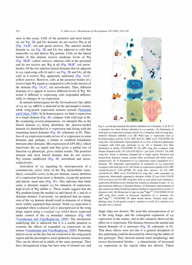
in a single domain (Fig. 4E; compare with wild type in B).
By examining several preparations, we interpret this as the
lateral domain (c) being abolished, the anterior-medial
domain (a) diminished in sr expression and fusing with the
remaining lateral domains (Fig. 4E, schematic in D). Thus,
loss of wg expression results in loss of sr expression in some
domains and, perhaps, a failure to establish boundaries
between other domains. Mis-expression of GPI-Dfz2, which
inactivates the wg signal and thus gives a partial loss of
function wg phenotype, gives similar results. The posterior
domain and most lateral domain that do not receive
Wg remain unaffected (Fig. 4F, arrowhead and arrow,
respectively).
Activation of wg signaling by misexpression of a
constitutively active form of the Wg intracellular trans-
ducer, armadillo (arm), in the pnr domain, causes abolition
of sr expression from most sr domains, except the posterior
and lateral- most ones (Fig. 4C). This indicates that while
some sr domains require wg for initiation of expression,
high levels of Wg inhibit sr. These results suggest that the
Wg gradient keeps the medial (a) and lateral (b, c and d) sr
domains distinct. Conversely, we predicted that an expan-
sion of the wg domain should result in domains of sr being
more widely separated than normal. Notal wg expression is
expanded when scalloped (sd) is misexpressed in the wing
hinge region using a vestigial (vg) GAL4 driver expressed
under control of the vg boundary enhancer (Fig. 4H)
(Varadarajan and VijayRaghavan, 1999). The mechanism
underlying this is unknown but it is a useful situation to
examine the effects of expanded wg expression on the
notum (Varadarajan and VijayRaghavan, 1999). Patterning
defects occur on the disc but are restricted to the wing pouch
region and the presumptive notal epidermis remains normal.
This can be observed in adults of the same genotype. They
have disorganized wings but have nota of normal size and
shape (data not shown). We used this misexpression of sd
in the wing hinge, and the consequent expansion of wg
expression in the notum, and in this situation observed the
effect on sr expression. The distance between the medial and
lateral domains of sr increases (Fig. 4I, schematic in G).
That these effects were not due to a general disruption of
disc patterning could be discerned by observing the adults of
this genotype. They showed normal notal morphology and
excess dorsocentral bristles – a characteristic of increased
wg expression in the region (data not shown). Taken
Fig. 4. wg and dpp maintain the distinct identities of sr domains. A, B, D–F:
sr domains lose their distinct identities in wg mutants. (A) Schematic of
wild type wg expression (orange) relative to sr domains (red) on wing disc.
Anterior domains labelled a–d. (B) Wild type sr expression seen as
b-Galactosidase activity in 0 h APF Sp/CyO, MHC lacZ; P1618/TM3 Sb
wing disc. (D) Schematic representation of wg loss of function phenotype
(compare with wild type schematic in A). (E) sr domains lose their
distinction in Sp/Sp; P1618/TM6 Tb 0 h APF wing disc (compare with
distinct domains in B). (F) UAS-GPI Dfz2/þ ; pnr Gal4, P1618/þ 0 h APF
wing disc. Some sr domains clumped into a single region. Posterior and
lateral-most domains remain normal (blue arrowhead and black arrow,
respectively). (G–I) Expansion of wg expression causes separation of sr
domains. (G) Schematic representation of expansion of wg expression
(compare with wild type in A). (H) Notal wg expression expands in 0 h APF
vgGAL4/wglacZ; UAS-sd/TM6 Tb wing disc (arrow). (I) 0 h APF
vgGAL4/CyO, MHC lacZ; P1618/UAS-sd wing disc, with expanded wg
expression. Abnormally separated sr domains visible. (C) pnr Gal4, P1618/
UAS-activated arm 0 h APF wing disc with wg activation in pnr domain-sr
expression abolished in pnr domain but remains in domains d and c. (J–L)
dpp maintains different sr domains distinct. (J) Schematic representation of
dpp expression (blue) bordering anterior medial (a) and posterior (arrow) sr
domains (red). (K) Reduction of dpp (in dppd6/dppd12; P1618/TM6 Tb 0 h
APF wing disc) results in increased separation of sr domains. (L)
dppd6/dppd12; P1618/TM6 Tb adult dorsal thorax. Normal notal mor-
phology clear. In all panels except L, anterior is to left. In L, anterior is to
top and view is dorsal.
A. Ghazi et al. / Mechanisms of Development 120 (2003) 519–528522
together, all these observations indicate a complex mech-
anism of sr regulation by wg. Moderate levels of wg
signaling appear to be required for initiation of sr expression
in some domains (c) but excessive wg signaling inhibits sr
transcription, thus allowing the Wg gradient to keep the
medial sr domain distinct from the lateral ones.
2.3. dpp is required to keep sr domains distinct
dpp expression along the antero-posterior border in
the presumptive notum, bordering the medial sr domain
(Fig. 4K, see Fig. 2J), is important for restricting wg
expression in this region (Tomoyasu et al., 1998, 2000; Sato
and Saigo, 2000). We investigated if dpp also functions in
regulating sr expression. In dppd6/dppd12; P1618/TM6 Tb
wing discs, sr domains develop much further away from
each other than normal, and distance between the medial
and lateral domains increases (Fig. 4K), similar to the discs
with expanded wg expression (compare Fig. 4K with I). The
normal appearance of the nota of these mutants confirmed
that this effect was not due to a general disruption of notal
morphology (Fig. 4L). The implications of this result –
whether dpp acts directly on sr, or by regulating wg
expression or both – are discussed later.
2.4. pnr, whose expression overlaps medial sr domain,
mediates initiation of sr expression
Following the observation that pnr expression on the
wing disc overlaps the medial sr domain completely, and the
posterior domain partially (Fig. 5A; see Fig. 2D–F), we
investigated the relationship between pnr and sr expression
by examining sr expression in pnr mutants. Two classes of
pnr allelic combinations have been described. Some, such as
pnrmd237/pnrD1, result in excess dorsocentral bristles on the
notum and are categorized as ‘gain of function’ mutants,
whereas others such as pnrVX1/pnrV1 cause a loss of
dorsocentral bristles and are categorized as ‘loss of
function’ allelic combinations. sr expression in both
categories was examined. A recombinant of srGal4 with
the pnr allele VX1 was generated and used to follow sr
expression. sr expression is reduced in its anterior domains
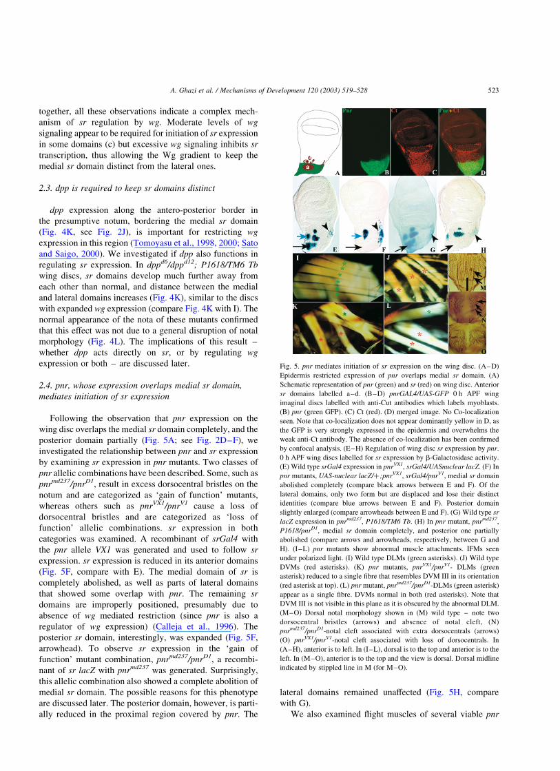
(Fig. 5F, compare with E). The medial domain of sr is
completely abolished, as well as parts of lateral domains
that showed some overlap with pnr. The remaining sr
domains are improperly positioned, presumably due to
absence of wg mediated restriction (since pnr is also a
regulator of wg expression) (Calleja et al., 1996). The
posterior sr domain, interestingly, was expanded (Fig. 5F,
arrowhead). To observe sr expression in the ‘gain of
function’ mutant combination, pnrmd237/pnrD1, a recombi-
nant of sr lacZ with pnrmd237 was generated. Surprisingly,
this allelic combination also showed a complete abolition of
medial sr domain. The possible reasons for this phenotype
are discussed later. The posterior domain, however, is parti-
ally reduced in the proximal region covered by pnr. The
lateral domains remained unaffected (Fig. 5H, compare
with G).
We also examined flight muscles of several viable pnr
Fig. 5. pnr mediates initiation of sr expression on the wing disc. (A–D)
Epidermis restricted expression of pnr overlaps medial sr domain. (A)
Schematic representation of pnr (green) and sr (red) on wing disc. Anterior
sr domains labelled a–d. (B–D) pnrGAL4/UAS-GFP 0 h APF wing
imaginal discs labelled with anti-Cut antibodies which labels myoblasts.
(B) pnr (green GFP). (C) Ct (red). (D) merged image. No Co-localization
seen. Note that co-localization does not appear dominantly yellow in D, as
the GFP is very strongly expressed in the epidermis and overwhelms the
weak anti-Ct antibody. The absence of co-localization has been confirmed
by confocal analysis. (E–H) Regulation of wing disc sr expression by pnr.
0 h APF wing discs labelled for sr expression by b-Galactosidase activity.
(E) Wild type srGal4 expression in pnrVX1, srGal4/UASnuclear lacZ. (F) In
pnr mutants, UAS-nuclear lacZ/þ ;pnrVX1, srGal4/pnrV1, medial sr domain
abolished completely (compare black arrows between E and F). Of the
lateral domains, only two form but are displaced and lose their distinct
identities (compare blue arrows between E and F). Posterior domain
slightly enlarged (compare arrowheads between E and F). (G) Wild type sr
lacZ expression in pnrmd237, P1618/TM6 Tb. (H) In pnr mutant, pnrmd237,
P1618/pnrD1, medial sr domain completely, and posterior one partially
abolished (compare arrows and arrowheads, respectively, between G and
H). (I–L) pnr mutants show abnormal muscle attachments. IFMs seen
under polarized light. (I) Wild type DLMs (green asterisks). (J) Wild type
DVMs (red asterisks). (K) pnr mutants, pnrVX1/pnrV1- DLMs (green
asterisk) reduced to a single fibre that resembles DVM III in its orientation
(red asterisk at top). (L) pnr mutant, pnrmd237/pnrD1-DLMs (green asterisk)
appear as a single fibre. DVMs normal in both (red asterisks). Note that
DVM III is not visible in this plane as it is obscured by the abnormal DLM.
(M–O) Dorsal notal morphology shown in (M) wild type – note two
dorsocentral bristles (arrows) and absence of notal cleft, (N)
pnrmd237/pnrD1-notal cleft associated with extra dorsocentrals (arrows)
(O) pnrVX1/pnrV1-notal cleft associated with loss of dorsocentrals. In
(A–H), anterior is to left. In (I–L), dorsal is to the top and anterior is to the
left. In (M–O), anterior is to the top and the view is dorsal. Dorsal midline
indicated by stippled line in M (for M–O).
A. Ghazi et al. / Mechanisms of Development 120 (2003) 519–528 523
mutants and found DLM abnormalities (Fig. 5K,L). DLMs,
which normally attach antero-posteriorly, attach abnormally
and appear dorsoventral in their orientation, and resemble
DVM III (Fig. 5K, compare with I and J). This suggests that
attachment sites were affected in these animals due to loss of
sr function. DVMs appear normal in all allelic combinations
examined. Viable alleles of pnr display a mid-thoracic cleft
due to failure of the two hemithoraces from fusing properly.
Mutant alleles have been placed in a series depending on the
severity of the cleft (Heitzler et al., 1996). To discount the
possibility that muscle defects in pnr mutants are a conse-
quence of this abnormality, we looked at flight muscles of
mutants with different degrees of clefts. Muscle defects
occur even in pnr allelic combinations that show no mid-
thoracic cleft (data not shown). These defects must be due to
pnr requirements on the epidermis, as the gene is not
expressed in the mesoderm. No mesodermal pnr expression
is seen at any stage. pnrGAL4/UAS-GFP wing discs labeled
with Cut (Ct)-specific antibody – which marks adult myo-
blasts (Blochlinger et al., 1993) – show no colocalization of
GFP with Ct-expressing myoblasts (Fig. 5B–D). The
regulation of sr by pnr, the mutant phenotypes and the
absence of pnr expression in the mesoderm suggest that the
muscle defects seen are a consequence of sr regulation
being affected.
2.5. ush negatively regulates sr expression
ush is an antagonist of pnr function (Cubadda et al.,
1997; Haenlin et al., 1997). This information, and the
observation that sr expression commences in regions of low
ush expression (see Fig. 2G–I) suggested that it may be
negatively regulating sr expression. We examined sr
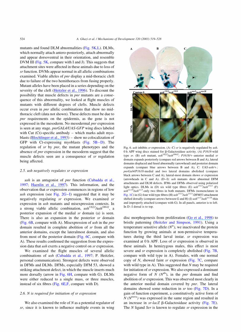
expression in ush mutants and misexpression contexts. In
a strong viable allelic combination, ushVX22/ushSW42, a
posterior expansion of the medial sr domain (a) is seen.
There is also an expansion in the posterior sr domain
(Fig. 6B, compare with A). Misexpression of ush in the pnr
domain resulted in complete abolition of sr from all the
anterior domains, except the lateralmost domain, and also
from most of the posterior domain (Fig. 6C, compare with
A). These results confirmed the suggestion from the expres-
sion data that ush exerts a negative control on sr expression.
We examined the flight muscles of several allelic
combinations of ush (Cubadda et al., 1997; P. Heitzler,
personal communication). Strongest defects were observed
in DFMs and DLMs. DFMs, especially DFM 53, showed a
striking attachment defect, in which the muscle inserts much
more dorsally (arrow in Fig. 6H, compare with G). DLMs
were either reduced to a single mass, or three muscles,
instead of six fibres (Fig. 6E,F, compare with D).
2.6. N is required for initiation of sr expression
We also examined the role of N as a potential regulator of
sr, since it is known to influence multiple events in wing
disc morphogenesis from proliferation (Go et al., 1998) to
bristle patterning (Heitzler and Simpson, 1991). Using a
temperature sensitive allele (Nts), we inactivated the protein
function by growing animals at non-permissive tempera-
tures during the third larval instar. sr expression was
examined at 0 h APF. Loss of sr expression is observed in
these animals. In hemizygous males, this effect is most
severe and sr expression is completely abolished (Fig. 7B,
compare with wild type in A). Females, with one normal
copy of N, showed faint sr expression (Fig. 7C, compare
with wild type in A). This suggested that N may be required
for initiation of sr expression. We also expressed a dominant
negative form of N (Ndn), in the pnr domain and find
abolition of sr expression. This was observed most clearly in
the anterior medial domain covered by pnr. The lateral
domains showed some reduction in sr too (Fig. 7D). In a
gain of function experiment, a constitutively active form of
N (Nintra) was expressed in the same region and resulted in
an increase in sr-lacZ b-Galactosidase activity (Fig. 7E).
The N ligand Ser is known to regulate sr expression in the
Fig. 6. ush inhibits sr expression. (A–C) sr is negatively regulated by ush.
0 h APF wing discs stained for b-Galactosidase activity. (A) P1618-wild
type sr. (B) ush mutant, ushVX22/ushSW42; P1618/þ-anterior medial sr
domain expands posteriorly (compare red arrows between B and A), lateral
domains displaced and fused abnormally (arrowhead) and posterior domain
expands (compare blue arrows between B and A), C: UAS-ush/þ ;
pnrGal4/P1618-medial and two lateral domains abolished (compare
black arrows between C and A), lateral-most domain shows sr expression
(arrowheads in C and A). (D–I): ush mutants show abnormal DFM
attachments and DLM defects. IFMs and DFMs observed using polarized
light optics. DLMs in (D) six wild type fibres (E) ush1513/ush1513 (F)
ushrev18/ushrev5-only two fibres in both mutants. DFMs (nomenclature in
Fig. 1C) in (G) four wild type fibres (H) ush1513/ush1513-DFM53 attachment
shifted dorsally (compare arrows between G and H) (I) ushrev5/ushrev18-thin
and improperly attached (compare with G). In all panels, anterior is to left.
In D–I dorsal is to top.
A. Ghazi et al. / Mechanisms of Development 120 (2003) 519–528524
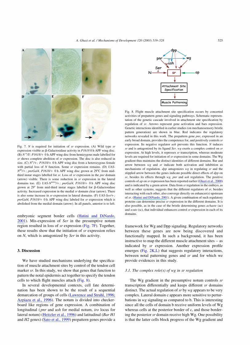
embryonic segment border cells (Hatini and DiNardo,
2001). Mis-expression of Ser in the presumptive notum
region resulted in loss of sr expression (Fig. 7F). Together,
these results show that the initiation of sr expression relies
on N, which is antagonized by Ser in this activity.
3. Discussion
We have studied mechanisms underlying the specifica-
tion of muscle attachment sites by control of the tendon cell
marker sr. In this study, we show that genes that function to
pattern the notal epidermis act together to specify the tendon
cells to which flight muscles attach (Fig. 8).
In several developmental contexts, cell fate determi-
nation has been shown to be the result of a sequential
demarcation of groups of cells (Lawrence and Struhl, 1996;
Azpiazu et al., 1996). The notum is divided into checker-
board like regions of gene expression. A combination of
longitudinal ( pnr and ush for medial notum, iro locus for
lateral notum) (Heitzler et al., 1996) and latitudinal (Bar H1
and H2 genes) (Sato et al., 1999) prepattern genes provide a
framework for Wg and Dpp signaling. Regulatory networks
between these genes are now being discovered and
functionally mapped. In such a background, it has been
instructive to map the different muscle attachment sites – as
indicated by sr expression. Another expression profile
emerges (Fig. 2K,L) that suggests regulatory interactions,
between notal patterning genes and sr and for which we
provide evidences in this study.
3.1. The complex role(s) of wg in sr regulation
The Wg gradient in the presumptive notum controls sr
transcription differentially and keeps different sr domains
distinct. The actual regulation of sr by wg appears to be very
complex. Lateral domain c appears more sensitive to pertur-
bations in wg signaling as compared to b. This is interesting
since all the cells of domain b receive uniform levels of Wg
whereas cells at the posterior border of c, and those border-
ing the posterior sr domain receive high Wg. One possibility
is that the latter cells block progress of the Wg gradient and
Fig. 7. N is required for initiation of sr expression. (A) Wild type sr
expression visible as b-Galactosidase activity in P1618 0 h APF wing disc.
(B) N ts/Y; P1618/þ 0 h APF wing disc from hemizygous male labelled for
sr shows complete abolition of sr expression. The disc is also reduced in
size. (C) Nts/þ ; P1618/þ 0 h APF wing disc from a heterozygous female
with partial loss of N function. Some sr expression remains. (D) UAS-
Ndn/þ ; pnrGal4, P1618/þ 0 h APF wing disc grown at 298C from mid-
third instar stages labelled for sr. Loss of sr expression in the pnr domain
(arrow) visible. There is some reduction in sr expression in the lateral
domains too. (E) UAS-Nintra/þ ; pnrGal4, P1618/þ 0 h APF wing disc
grown at 298 from mid-third instar stages labelled for b-Galactosidase
activity. Increased expression in the medial sr domain clear (arrow). There
is also some increase in sr expression in lateral domains. (F) UAS-Ser/þ ;
pnrGal4, P1618/þ 0 h APF wing disc labeled for sr expression which is
abolished from the medial domain (arrow). In all panels, anterior is to left.
Fig. 8. Flight muscle attachment site specification occurs by concerted
activities of prepattern genes and signaling pathways. Schematic represen-
tation of the genetic cascade involved in attachment site specification by
regulation of sr. Arrows represent gene activation and bars repression.
Genetic interactions identified in earlier studies (on mechanosensory bristle
pattern generation) are shown in blue. Red indicates the regulatory
networks revealed in this work. The prepattern gene pnr, expressed in an
early broad domain, provides the competence for, and positively controls sr
expression. Its negative regulator ush prevents this function. N induces
sr and is antagonized by its ligand Ser. wg exerts a complex control on sr
expression. At high levels, it represses sr transcription, whereas moderate
levels are required for initiation of sr expression in some domains. The Wg
gradient thus maintains the distinct identities of different domains. Bar and
arrow between wg and sr indicate both activation and inhibition as
mechanisms of regulation. dpp antagonizes wg in regulating sr and the
stippled arrow between the genes indicate possible direct effects of dpp on
sr, besides its effects through wg, pnr and ush regulation. The positive
controls of ap on sr expression has been reported earlier (Ghazi et al., 2000)
and is indicated by a green arrow. Data from sr regulation in the embryo, as
well as other systems, suggests that the different regulators of sr, besides
interacting with each other, also converge directly on enhancer(s) upstream
of sr. (Hatini and DiNardo, 2001). A given combination of such regulatory
proteins can determine precise sr expression in the different domains. It is
also possible, as in the case of the bristle determining genes achaete (ac)
and scute (sc), that individual enhancers control sr expression in each of its
domains.
A. Ghazi et al. / Mechanisms of Development 120 (2003) 519–528 525
thus determine responses of cells further away. This may be
brought about by targeting Wg to lysosomes and degrading
it, as in the embryo (Dubois et al., 2001). Another possi-
bility, not exclusive of the first, could be that the domain and
levels of wg transcription determine the range and gradient
of Wg. The precise definition of the domain of wg tran-
scription could be by mechanisms similar to that used in the
wing margin (Rulifson et al., 1996). While our data suggests
that the posterior and lateral-most domain do not receive
Wg and may lie outside its purview, the formal possibility
still exists that wg effects these domains in some other
unknown way.
3.2. The dpp and hh pathways in sr regulation
Control of sr by wg, in segment border cells of the
Drosophila embryo, has been demonstrated (Piepenburg
et al., 2000). Wg signaling restricts sr activation to a single
row of cells. In the presumptive notum on the wing disc, hh
expression is restricted to a very narrow region, which forms
the posterior compartment. Its effects in the disc are medi-
ated by dpp, which serves multiple functions. Dpp is
required for induction of wg expression, as it positively
regulates pnr, which in turn activates wg (Tomoyasu et al.,
1998; Tomoyasu et al., 2000; Sato and Saigo, 2000).
However, once wg is induced, Dpp tightly restricts its
domain. This antagonism is required for correct positioning
of the DC bristles. We find that it also defines domains of sr.
It is unclear if dpp directly regulates sr, or its effect is by
control of other genes. The similarity between sr pheno-
types observed on expansion of wg expression, and in dpp
mutants, is suggestive of its effects being mediated by wg
only, but it is also possible that it influences sr expression
directly.
3.3. pnr, antagonized by ush, activates sr expression
Pnr, a GATA-binding protein normally functions as a
transcriptional activator and is antagonized by Ush in its
function. Loss of function pnr mutants show no sr
expression in the domain covered by pnr. This, along with
sr expansion in mutants of ush, would suggest that pnr
activates sr in the notum, and is inhibited by ush. However,
there is also loss of sr expression in pnr ‘gain of function’
mutants. The reason for this is not completely clear. One
possibility is that since the mutation causes an increase in
wg activity in the region (Calleja et al., 1996) this may cause
a down-regulation of sr. This is supported by a similar effect
seen on misexpression of activated armadillo in the pnr
domain (Fig. 4C). We have taken into account results with
both pnr and ush to suggest that pnr positively regulates sr
and is antagonized by ush.
Most sr expression commences in regions of low ush.
Phenotypes of ush mutants, and ush misexpression experi-
ments, also indicate that the gene inhibits sr, in keeping with
the simplistic scenario that ush antagonizes pnr-mediated
activation of sr. However, the medial sr domain is partially
covered by ush proximally. Further, pnr is known to be
required for positive induction of ush in the embryonic
epidermis (Herranz and Morata, 2001) and in some loss of
function allelic combinations of pnr, such as pnrVX6/pnrVX1,
there is reduced ush expression on the disc (Sato and Saigo,
2000). So how is sr initiated in the region where Pnr and Ush
are co-expressed? The answer to this is not known but
probably lies in levels of Ush and Pnr at that position. In loss
of function ush mutants ectopic dorsocentral bristles form
but post vertical (PV) bristles are missing (Ramain et al.,
1993; Cubadda et al., 1997), suggesting that the Pnr–Ush
complex acts as a repressor of the DC enhancer, but as
activator of the enhancer of PV bristles. Such observations
have indicated complex, context dependent interactions
between Pnr and Ush in determining cell fate and could
explain the regulation of sr expression in the medial notal
region.
3.4. Domain specific regulation of sr expression
Our results indicate that each sr domain is regulated by a
combination of prepattern genes and signaling molecules.
But, a precise description of the ’combinatorial code’ for
regulation of each sr domain is beyond the scope of this
work and can be achieved by generation of domain specific
markers of sr. Based on our expression pattern data, and
existing literature, we suggest that high levels of Pnr, low
(or absence of) Ush and moderate levels of Wg determine
the initial induction of domain a. The distinction between
medial (a) and lateral (b–d) domains is established by
presence of very high levels of Wg (the cells where the Wg
gradient originates). Lateral expression domains are prob-
ably induced in domains controlled by the lateral prepattern
gene iro. The differences between different lateral domains
arise as a result of expression of different genes in the
region. For instance, the lateral-most domain d appears to be
regulated by ush and does not encounter Wg at all. Whereas,
all cells of b receive uniformly moderate levels of Wg, only
cells at the borders of c receive high Wg levels, and these
differences result in the distinct identities of the two
domains. Dpp, either through its effects on these regulatory
genes and/or through direct effects on sr influences the
process.
Invertebrate muscles attach to tendon cells that are
entirely epidermal, unlike mesenchymal tendons of verte-
brates. However, closer scrutiny of mechanisms underlying
patterning of musculoskeletal system of tetrapods with
those mediating insect muscle patterning suggests simi-
larities. Some molecules involved in the two systems are
conserved though the number of vertebrate players known
is fewer than in Drosophila (Schweitzer et al., 2001).
Vertebrate Tenascin (Ten), is expressed in tendons at high
levels, while muscles show faint expression (Kardon, 1998).
Drosophila Ten shows a very early and transient meso-
dermal expression that is replaced by distinct tendon cell
A. Ghazi et al. / Mechanisms of Development 120 (2003) 519–528526
expression (Baumgartner and Chiquet-Ehrismann, 1993).
In both cases, attachment tissue is marked by high Ten
expression while muscles show low levels or absence.
Cellular and molecular mechanisms underlying generation
of vertebrate tendons are not known and in the Drosophila
embryo the processes are only now beginning to be eluci-
dated. Identifying genes and mechanisms that control
tendon cell specification can lead to better understanding
of morphogenesis and function of muscle in both vertebrates
and invertebrates.
4. Materials and methods
4.1. Strains and reagents
Canton-S was used as wildtype. pnr alleles pnrD1, pnrV1
and pnrVX1, UAS-pnr, ush mutants, UAS-ush, ushGal4 and
srGal4, UAS-GFP are from Pascal Heitzler and Pat Simpson
(Strasbourg, France). The following are from the Blooming-
ton Stock Centre (Indiana, USA): pnrmd237, a P-Gal4 inser-
tion allele, wg alleles-en40 wg lacZ/CyO, dpp alleles-dpp
Gal4, dpp lacZ/CyO-TM6 Tb, dppd6/CyO, dppd12/CyO.
srlacZ (P1618) is from Talila Volk (Weizmann Inst. Israel).
Sp/CyO, MHC lacZ; P1618/TM3 Sb was made in this study.
UAS-GPI Dfz2 is described in Cadigan et al. (1998), and
UAS-sd in Varadarajan and VijayRaghavan (1999).
4.2. Immunohistochemistry
b-Galactosidase (Promega) and myosin heavy chain
(MHC) (Dan Kiehart, USA) specific antibodies (raised in
rabbit) were used at 1:1000 and 1:500 dilutions, respect-
ively. b-Galactosidase, Wg and Cut (Ct) specific mono-
clonal antibodies were used at 1:50 dilution. For fluorescent
detection, Alexa568 (red) and Alexa 488 (green) secondary
antibodies were used. Confocal microscopy was performed
on Bio-Rad Model 1024.
4.3. Dissections
sr expression was examined in wing discs at the white
prepupal stage [0 hours (h) after puparium formation
(APF)]. Larvae and pupae were dissected in phosphate
buffered saline (PBS), fixed in 4% paraformaldehyde and
histochemically stained with X-gal (Fernandes et al., 1991)
or labeled with relevant antibodies. All preparations except
fluorescent samples were mounted in 70% glycerol. Fluor-
escent preparations were mounted in Vectashield mounting
medium (Vector Chemicals). Adult hemithoraces were cut
sagitally, dehydrated through 70%, 90% and 100% ethanol,
cleared in methyl salicylate, mounted in Canada Balsam and
observed under polarized light.
4.4. Temperature shift experiments
To examine sr expression in Nts animals, Nts virgins were
crossed to P1618 (sr lacZ) males and progeny were grown at
the permissive temperature (228) till early late second instar
to early third instar stages when they were shifted to the
non-permissive temperature (318), and grown till the 0 h
APF stage for dissection.
Misexpression experiments were performed using the
Gal4-UAS system described in Brand and Perrimon (1993).
Acknowledgements
We are grateful to Pat Simpson, Gines Morata, Pascal
Heitzler, Helen Skaer, Dan Kiehart and Talila Volk for
generous gifts of fly stocks and antibodies. The assistance of
Smita Raman and M.S. Sunanda for some of the
experiments is gratefully acknowledged. Our gratitude to
Pat Simpson, Michael Bate, Helen Skaer and Veronica
Rodrigues for stimulating discussions and many useful
suggestions. Suggestions made by the anonymous reviewers
helped greatly in improving the manuscript and we are
thankful for this. A.G acknowledges the contribution of the
late Carolyn Ann-D’Souza in generating the CyO, MHC
lacZ balancer stock used in several experiments. This work
is supported by Department of Biotechnology, India and an
Indo-Israeli grant to K.V.
References
Azpiazu, N., Lawrence, P.A., Vincent, J.P., Frasch, M., 1996. Segmentation
and specification of the Drosophila mesoderm. Genes Dev. 10,
3183–3194.
Baumgartner, S., Chiquet-Ehrismann, R., 1993. Tena, a Drosophila gene
related to Tenascin, shows selective transcript localization. Mech. Dev.
40, 165–176.
Becker, S., Pasca, G., Strumpf, D., Min, L., Volk, T., 1997. Reciprocal
signaling between Drosophila epidermal muscle attachment cells and
their corresponding muscles. Development 124, 2615–2622.
Blochlinger, K., Jan, L.Y., Jan, Y.N., 1993. Postembryonic patterns of
expression of cut, a locus regulating sensory organ identity in
Drosophila. Development 117, 441–450.
Brand, A.H., Perrimon, N., 1993. Targeted gene expression as a means of
altering cell fates and generating dominant phenotypes. Development
118, 401–415.
Cadigan, K.M., Fish, M.P., Rulifson, E.J., Nusse, R., 1998. Wingless
repression of Drosophila frizzled2 expression shapes the Wingless
morphogen gradient in the wing. Cell 93 (5), 767–777.
Calleja, M., Moreno, E., Pelaz, S., Morata, G., 1996. Visualization of gene
expression in living adult Drosophila. Science 274, 252–255.
Cubadda, Y., Heitzler, P., Ray, R.P., Bourouis, M., Ramain, P., Gelbart, W.,
Simpson, P., Haenlin, M., 1997. u-shaped encodes a zinc finger protein
that regulates the proneural genes achaete and scute during the
formation of bristles in Drosophila. Genes Dev. 11, 3083–3095.
Dubois, L., Lecourtois, M., Alexandre, C., Hirst, E., Vincent, J.P., 2001.
Regulated endocytic routing modulates wingless signaling in Droso-
phila embryos. Cell 105 (5), 613–624.
A. Ghazi et al. / Mechanisms of Development 120 (2003) 519–528 527
Fernandes, J., Bate, M., VijayRaghavan, K., 1991. Development of the
indirect flight muscles of Drosophila. Development 113, 67–77.
Fernandes, J.J., Celniker, S.E., VijayRaghavan, K., 1996. Development of
the indirect flight muscle attachment sites inDrosophila: role of the PS
integrins and the stripe gene. Dev. Biol. 176, 166–184.
Frommer, G., Vorbruggen, G., Pasca, G., Jackle, H., Volk, T., 1996.
Epidermal egr-like zinc finger protein of Drosophila participates in
myotube guidance. EMBO J. 15, 1642–1649.
Garcia-Garcia, M.J., Ramain, P., Simpson, P., Modolell, J., 1999. Different
contributions of pannier and wingless to the patterning of the dorsal
mesothorax of Drosophila. Development 126, 3523–3532.
Ghazi, A., Anant, S., VijayRaghavan, K., 2000. apterous mediates
development of direct flight muscles autonomously and indirect flight
muscles through epidermal cues. Development 127, 5309–5318.
Ghysen, A., Dambly-Chaudiere, C., 1988. From DNA to form: the achaete-
scute complex. Genes Dev. 2, 495–501.
Go, M.J., Eastman, D.S., Artavanis-Tsakonas, S., 1998. Cell proliferation
control by Notch signaling in Drosophila development. Development
125 (11), 2031–3040.
Haenlin, M., Cubadda, Y., Blondeau, F., Heitzler, P., Lutz, Y., Simpson, P.,
Ramain, P., 1997. Transcriptional activity of pannier is regulated
negatively by heterodimerization of the GATA DNA-binding domain
with a cofactor encoded by the u-shaped gene of Drosophila. Genes
Dev. 11, 3096–3108.
Hatini, V., DiNardo, S., 2001. Distinct signals generate repeating striped
pattern in the embryonic parasegment. Mol. Cell 7 (1), 151–160.
Heitzler, P., Simpson, P., 1991. The choice of cell fate in the epidermis of
Drosophila. Cell 64 (6), 1083–1092.
Heitzler, P., Haenlin, M., Ramain, P., Calleja, M., Simpson, P., 1996. A
genetic analysis of pannier, a gene necessary for viability of dorsal
tissues and bristle positioning in Drosophila. Genetics 143, 1271–1286.
Herranz, H., Morata, G., 2001. The functions of pannier during Drosophila
embryogenesis. Development 128 (23), 4837–4846.
Kardon, G., 1998. Muscle and tendon morphogenesis in the avian hind
limb. Development 125, 4019–4032.
Lawrence, P.A., Struhl, G., 1996. Morphogens, compartments, and pattern:
lessons from Drosophila? Cell 85, 951–961.
Lee, J.C., VijayRaghavan, K., Celniker, S.E., Tanouye, M.A., 1995.
Identification of a Drosophila muscle development gene with structural
homology to mammalian early growth response transcription factors.
Proc. Natl Acad. Sci. USA 92, 10344–10348.
Miller, A., 1950. The internal anatomy and histology of the imago of
Drosophila melanogaster. In: Demerec, M., (Ed.), The Biology of
Drosophila, pp. 420–534.
Nabel-Rosen, H., Dorevitch, N., Reuveny, A., Volk, T., 1999. The balance
between two isoforms of the Drosophila RNA-binding protein how
controls tendon cell differentiation. Mol. Cell 4, 573–584.
Neumann, C.J., Cohen, S.M., 1996. Sternopleural is a regulatory mutation
of wingless with both dominant and recessive effects on larval
development of Drosophila melanogaster. Genetics 142, 1147–1155.
Phillips, R.G., Whittle, J.R., 1993. wingless expression mediates deter-
mination of peripheral nervous system elements in late stages of
Drosophila wing disc development. Development 118, 427–438.
Piepenburg, O., Vorbruggen, G., Jackle, H., 2000. Drosophila segment
borders result from unilateral repression of hedgehog activity by
wingless signalling. Mol. Cell 6, 203–209.
Ramain, P., Heitzler, P., Haenlin, M., Simpson, P., 1993. pannier, a
negative regulator of achaete and scute in Drosophila, encodes a zinc
finger protein with homology to the vertebrate transcription factor
GATA-1. Development 119, 1277–1291.
Roy, S., VijayRaghavan, K., 1999. Muscle pattern diversification in
Drosophila: the story of imaginal myogenesis. Bioessays 21, 486–498.
Rulifson, E.J., Micchelli, C.A., Axelrod, J.D., Perrimon, N., Blair, S.S.,
1996. wingless refines its own expression domain on the Drosophila
wing margin. Nature 384 (6604), 72–74.
Sato, M., Kojima, T., Michiue, T., Saigo, K., 1999. Bar homeobox genes
are latitudinal prepattern genes in the developing Drosophila notum
whose expression is regulated by the concerted functions of
decapentaplegic and wingless. Development 126, 1457–1466.
Sato, M., Saigo, K., 2000. Involvement of pannier and u-shaped in
regulation of decapentaplegic dependent wingless expression in
developing Drosophila notum. Mech. Dev. 93, 127–138.
Schweitzer, R., Chyung, J.H., Murtaugh, L.C., Brent, A.E., Rosen, V.,
Olson, E.N., Lassar, A., Tabin, C.J., 2001. Analysis of the tendon cell
fate using Scleraxis, a specific marker for tendons and ligaments.
Development 128 (19), 3855–3866.
Stern, C., 1954. Two or three bristles. Am. Sci. 42, 213–247.
Sudarsan, V., Anant, S., Guptan, P., VijayRaghavan, K., Skaer, H., 2001.
Myoblast diversification and ectodermal signaling in Drosophila. Dev.
Cell 1 (6), 829–839.
Tomoyasu, Y., Nakamura, M., Ueno, N., 1998. Role of dpp signalling in
prepattern formation of the dorsocentral mechanosensory organ in
Drosophila melanogaster. Development 125, 4215–4224.
Tomoyasu, Y., Ueno, N., Nakamura, M., 2000. The Decapentaplegic
morphogen gradient regulates the notal wingless expression through
induction of pannier and u-shaped in Drosophila. Mech. Dev. 96,
37–49.
Varadarajan, S., VijayRaghavan, K., 1999. scalloped functions in a
regulatory loop with vestigial and wingless to pattern the Drosophila
wing. Dev. Genes Evol. 209, 10–17.
Volk, T., VijayRaghavan, K., 1994. A central role for epidermal segment
border cells in the induction of muscle patterning in the Drosophila
embryo. Development 120, 59–70.
Williams, G.J., Caveney, S., 1980a. A gradient of morphogenetic
information involved in muscle patterning. J. Embryol. Exp. Morphol.
58, 35–61.
Williams, G.J., Caveney, S., 1980b. Changing muscle patterns in a
segmental epidermal field. J. Embryol. Exp. Morphol. 58, 13–33.
A. Ghazi et al. / Mechanisms of Development 120 (2003) 519–528528
Related Documents