energies Review Preparation, Functionalization, Modification, and Applications of Nanostructured Gold: A Critical Review Muhammad Yaseen 1 , Muhammad Humayun 2 , Abbas Khan 1, * , Muhammad Usman 3 , Habib Ullah 4 , Asif Ali Tahir 5 and Habib Ullah 5, * Citation: Yaseen, M.; Humayun, M.; Khan, A.; Usman, M.; Ullah, H.; Tahir, A.A.; Ullah, H. Preparation, Functionalization, Modification, and Applications of Nanostructured Gold: A Critical Review. Energies 2021, 14, 1278. https://doi.org/10.3390/ en14051278 Academic Editor: Ellen Stechel Received: 18 January 2021 Accepted: 19 February 2021 Published: 25 February 2021 Publisher’s Note: MDPI stays neutral with regard to jurisdictional claims in published maps and institutional affil- iations. Copyright: © 2021 by the authors. Licensee MDPI, Basel, Switzerland. This article is an open access article distributed under the terms and conditions of the Creative Commons Attribution (CC BY) license (https:// creativecommons.org/licenses/by/ 4.0/). 1 Department of Chemistry, Abdul Wali Khan University, Mardan 23200, KP, Pakistan; [email protected] 2 Wuhan National Laboratory for Optoelectronics, Huazhong University of Science and Technology, Wuhan 430074, China; [email protected] 3 Center of Research Excellence in Nanotechnology (CENT), KFUPM, Dhahran 31261, Saudi Arabia; [email protected] 4 State Key Laboratory of Advanced Technology for Materials Synthesis and Processing, Wuhan University of Technology, Wuhan 430074, China; [email protected] 5 Environment and Sustainability Institute, University of Exeter, Penryn, Cornwall TR10 9FE, UK; [email protected] * Correspondence: [email protected] (A.K.); [email protected] (H.U.) Abstract: Gold nanoparticles (Au NPs) play a significant role in science and technology because of their unique size, shape, properties and broad range of potential applications. This review focuses on the various approaches employed for the synthesis, modification and functionalization of nanostructured Au. The potential catalytic applications and their enhancement upon modification of Au nanostructures have also been discussed in detail. The present analysis also offers brief summaries of the major Au nanomaterials synthetic procedures, such as hydrothermal, solvothermal, sol-gel, direct oxidation, chemical vapor deposition, sonochemical deposition, electrochemical deposition, microwave and laser pyrolysis. Among the various strategies used for improving the catalytic performance of nanostructured Au, the modification and functionalization of nanostructured Au produced better results. Therefore, various synthesis, modification and functionalization methods employed for better catalytic outcomes of nanostructured Au have been summarized in this review. Keywords: nanomaterials; photocatalysis; pollutants degradation; solar fuel 1. Introduction Nano is a Greek word which means small; particles with at least one dimension of less than 100 nm are called nanoparticles. Because of the large volume surface area, increased chemical reactivity or stability, enhanced mechanical strength, etc., nanoparticles have gained great popularity in the field of nanotechnology [1] and wide spreads applications in the field of electrochemistry, photochemical and biomedicine [2]. In general, nanoparticles have been classified into organic-, inorganic- and carbon-based. Inorganic metal nanoparti- cles are widely used in the preparation of nanoparticles, such as aluminium (Al), cadmium (Cd), cobalt (Co), copper (Cu), gold (Au), iron (Fe), plum (Pb), silver (Ag) and zinc (Zn). The Au nanoparticles were first prepared by Michael Faraday in 1856 [3,4]. Nanoparticles range in size from 1 to 8 μm and have various shapes, including spherical ones, sub oc- tahedron ones, octahedron, decahedron ones, icosahedral few twin ones, multiple twin crystal ones, tetrahedron, nano-triangles, hexagonals and nano-rods. Nanoparticles are different in size. The Au nanoparticles have attracted significant interest because of their high-volume ratio surface, low toxicity, excellent biocompatibility, optical, electronic and chemical properties [5–7]. Au nanoparticles are widely used in catalysis, optical molecular sensing, cancer treatment and as building blocks in nanotechnology [8]. Au colloid is used Energies 2021, 14, 1278. https://doi.org/10.3390/en14051278 https://www.mdpi.com/journal/energies

Welcome message from author
This document is posted to help you gain knowledge. Please leave a comment to let me know what you think about it! Share it to your friends and learn new things together.
Transcript
energies
Review
Preparation, Functionalization, Modification, and Applicationsof Nanostructured Gold: A Critical Review
Muhammad Yaseen 1, Muhammad Humayun 2, Abbas Khan 1,* , Muhammad Usman 3 , Habib Ullah 4 ,Asif Ali Tahir 5 and Habib Ullah 5,*
�����������������
Citation: Yaseen, M.; Humayun, M.;
Khan, A.; Usman, M.; Ullah, H.; Tahir,
A.A.; Ullah, H. Preparation,
Functionalization, Modification, and
Applications of Nanostructured Gold:
A Critical Review. Energies 2021, 14,
1278. https://doi.org/10.3390/
en14051278
Academic Editor: Ellen Stechel
Received: 18 January 2021
Accepted: 19 February 2021
Published: 25 February 2021
Publisher’s Note: MDPI stays neutral
with regard to jurisdictional claims in
published maps and institutional affil-
iations.
Copyright: © 2021 by the authors.
Licensee MDPI, Basel, Switzerland.
This article is an open access article
distributed under the terms and
conditions of the Creative Commons
Attribution (CC BY) license (https://
creativecommons.org/licenses/by/
4.0/).
1 Department of Chemistry, Abdul Wali Khan University, Mardan 23200, KP, Pakistan;[email protected]
2 Wuhan National Laboratory for Optoelectronics, Huazhong University of Science and Technology,Wuhan 430074, China; [email protected]
3 Center of Research Excellence in Nanotechnology (CENT), KFUPM, Dhahran 31261, Saudi Arabia;[email protected]
4 State Key Laboratory of Advanced Technology for Materials Synthesis and Processing,Wuhan University of Technology, Wuhan 430074, China; [email protected]
5 Environment and Sustainability Institute, University of Exeter, Penryn, Cornwall TR10 9FE, UK;[email protected]
* Correspondence: [email protected] (A.K.); [email protected] (H.U.)
Abstract: Gold nanoparticles (Au NPs) play a significant role in science and technology becauseof their unique size, shape, properties and broad range of potential applications. This reviewfocuses on the various approaches employed for the synthesis, modification and functionalization ofnanostructured Au. The potential catalytic applications and their enhancement upon modification ofAu nanostructures have also been discussed in detail. The present analysis also offers brief summariesof the major Au nanomaterials synthetic procedures, such as hydrothermal, solvothermal, sol-gel,direct oxidation, chemical vapor deposition, sonochemical deposition, electrochemical deposition,microwave and laser pyrolysis. Among the various strategies used for improving the catalyticperformance of nanostructured Au, the modification and functionalization of nanostructured Auproduced better results. Therefore, various synthesis, modification and functionalization methodsemployed for better catalytic outcomes of nanostructured Au have been summarized in this review.
Keywords: nanomaterials; photocatalysis; pollutants degradation; solar fuel
1. Introduction
Nano is a Greek word which means small; particles with at least one dimension of lessthan 100 nm are called nanoparticles. Because of the large volume surface area, increasedchemical reactivity or stability, enhanced mechanical strength, etc., nanoparticles havegained great popularity in the field of nanotechnology [1] and wide spreads applications inthe field of electrochemistry, photochemical and biomedicine [2]. In general, nanoparticleshave been classified into organic-, inorganic- and carbon-based. Inorganic metal nanoparti-cles are widely used in the preparation of nanoparticles, such as aluminium (Al), cadmium(Cd), cobalt (Co), copper (Cu), gold (Au), iron (Fe), plum (Pb), silver (Ag) and zinc (Zn).The Au nanoparticles were first prepared by Michael Faraday in 1856 [3,4]. Nanoparticlesrange in size from 1 to 8 µm and have various shapes, including spherical ones, sub oc-tahedron ones, octahedron, decahedron ones, icosahedral few twin ones, multiple twincrystal ones, tetrahedron, nano-triangles, hexagonals and nano-rods. Nanoparticles aredifferent in size. The Au nanoparticles have attracted significant interest because of theirhigh-volume ratio surface, low toxicity, excellent biocompatibility, optical, electronic andchemical properties [5–7]. Au nanoparticles are widely used in catalysis, optical molecularsensing, cancer treatment and as building blocks in nanotechnology [8]. Au colloid is used
Energies 2021, 14, 1278. https://doi.org/10.3390/en14051278 https://www.mdpi.com/journal/energies
Energies 2021, 14, 1278 2 of 83
for surface modification of ideal electrodes due to its excellent stability and unique char-acteristics (including high biocompatibility maintaining the normal structure of attachedproteins or enzymes and their enzyme function). The individual physical, chemical andAu nanoparticles optical properties can be innovative ways to control the transport phar-maceutical compounds and control [9]. The Au nanoparticles possess essential propertiesby functionalizing the surface with a change of ligands to improve the properties or bringabout modifications in them that make the functionalized Au nanoparticles proper for newapplications. Since the surface plasmon resonance (SPR) effect is caused by the reasonabledriving on the surface of the nanoparticles, due to the interaction with the electromagneticradiation of the appropriate wavelength, the strong absorption band and high luminouscharacteristics of Au can be improved due to surface plasmon resonance (SPR) impact [10].The ultimate size and shape of the nanoparticles leads to its SPR optical absorption andscattering characteristics responsive to surrounding media and nanoparticles’ aggregationcondition. Rapid nanoparticle heating will cause formaldehyde oxidation in the air atenvironmental temperature [11,12].
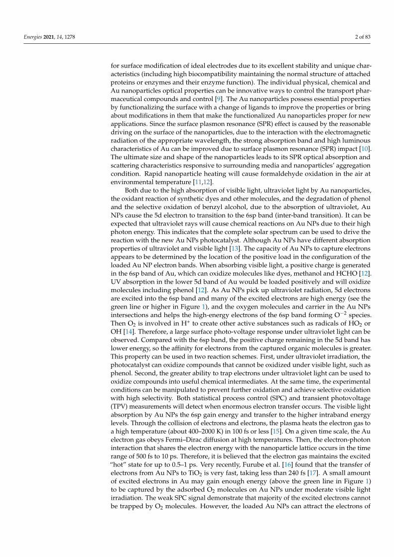
Both due to the high absorption of visible light, ultraviolet light by Au nanoparticles,the oxidant reaction of synthetic dyes and other molecules, and the degradation of phenoland the selective oxidation of benzyl alcohol, due to the absorption of ultraviolet, AuNPs cause the 5d electron to transition to the 6sp band (inter-band transition). It can beexpected that ultraviolet rays will cause chemical reactions on Au NPs due to their highphoton energy. This indicates that the complete solar spectrum can be used to drive thereaction with the new Au NPs photocatalyst. Although Au NPs have different absorptionproperties of ultraviolet and visible light [13]. The capacity of Au NPs to capture electronsappears to be determined by the location of the positive load in the configuration of theloaded Au NP electron bands. When absorbing visible light, a positive charge is generatedin the 6sp band of Au, which can oxidize molecules like dyes, methanol and HCHO [12].UV absorption in the lower 5d band of Au would be loaded positively and will oxidizemolecules including phenol [12]. As Au NPs pick up ultraviolet radiation, 5d electronsare excited into the 6sp band and many of the excited electrons are high energy (see thegreen line or higher in Figure 1), and the oxygen molecules and carrier in the Au NPsintersections and helps the high-energy electrons of the 6sp band forming O−2 species.Then O2 is involved in H+ to create other active substances such as radicals of HO2 orOH [14]. Therefore, a large surface photo-voltage response under ultraviolet light can beobserved. Compared with the 6sp band, the positive charge remaining in the 5d band haslower energy, so the affinity for electrons from the captured organic molecules is greater.This property can be used in two reaction schemes. First, under ultraviolet irradiation, thephotocatalyst can oxidize compounds that cannot be oxidized under visible light, such asphenol. Second, the greater ability to trap electrons under ultraviolet light can be used tooxidize compounds into useful chemical intermediates. At the same time, the experimentalconditions can be manipulated to prevent further oxidation and achieve selective oxidationwith high selectivity. Both statistical process control (SPC) and transient photovoltage(TPV) measurements will detect when enormous electron transfer occurs. The visible lightabsorption by Au NPs the 6sp gain energy and transfer to the higher intraband energylevels. Through the collision of electrons and electrons, the plasma heats the electron gas toa high temperature (about 400–2000 K) in 100 fs or less [15]. On a given time scale, the Auelectron gas obeys Fermi–Dirac diffusion at high temperatures. Then, the electron-photoninteraction that shares the electron energy with the nanoparticle lattice occurs in the timerange of 500 fs to 10 ps. Therefore, it is believed that the electron gas maintains the excited“hot” state for up to 0.5–1 ps. Very recently, Furube et al. [16] found that the transfer ofelectrons from Au NPs to TiO2 is very fast, taking less than 240 fs [17]. A small amountof excited electrons in Au may gain enough energy (above the green line in Figure 1)to be captured by the adsorbed O2 molecules on Au NPs under moderate visible lightirradiation. The weak SPC signal demonstrate that majority of the excited electrons cannotbe trapped by O2 molecules. However, the loaded Au NPs can attract the electrons of
Energies 2021, 14, 1278 3 of 83
organic molecules on the NPs [18]. Due to light irradiation, the Sulforhodamine B (SRB)dye molecules get excited and then these excited molecules introduce their electrons tothe substrate. The formation of O−2 species is affected by the additional dye sensitizationof Au NPs by excited SRB molecules. In combination with SPR, the SRB effect on the AuNPs leads to high rate of dye degradation. By increasing the visible light intensity, positivecharge increases and by gaining energy; a lot of electrons are seized by the O2 molecules.Molecular O2 is considered to be an oxidant that catalyzes the reaction. The selectiveoxidation of benzyl alcohol to benzaldehyde is an example of this scheme, which can onlybe observed under ultraviolet light. The main experimental observations include the bandstructure of Au NPs. Figure 1 shows the preliminary mechanism of photocatalysis usingsupported Au NPs. Considering that the 6sp band overlaps the 5d band in terms of energyscale, the proposed mechanism also provides the potential to turn on or turn off specificreactions by adjusting the wavelength of the irradiated light. This discovery reveals a newtype of photocatalyst and a possible way through which sunlight can be used to drivevarious chemical reactions on the photocatalyst at ambient temperature for environmentalpurification and solar fuels production.
Figure 1. The energy band structure diagram of the supported Au NPs and the suggested mechanismof using the supported Au NPs for photocatalysis. Reproduce with permission from reference [14],RSC, 2010.
In this review, we attempted to summarize various methods for the preparation ofnanostructured Au and its possible modification procedures for obtaining Au-based nano-materials with different surface morphologies under different environment and examinedtheir applications in several typical reactions in catalysis. This review also attempted tofurther highlight the basic understanding on the preparation, modification, functionaliza-tion and applications of nanostructured Au in catalysis. Furthermore, the basic mechanismof light absorption and simultaneous production of electron-hole, its trapping techniquesand utilization have been described briefly.
2. Synthetic Methods of Au NPs
Au nanocrystals can be synthesized by a number of methods depending on its ap-plications in various fields. Each method has its own advantages and disadvantages.Therefore, selection of an appropriate method is crucial because the growth of nanostruc-tures, as well as their properties significantly depends on the methods of preparation.Such well-known techniques are hydrothermal, solvothermal, sol gel, direct oxidation,chemical vapor deposition, electrochemical deposition, sonochemistry, laser pyrolysis and
Energies 2021, 14, 1278 4 of 83
microwave. Khumaeni Ali et al. [19] synthesized the Au NPs via pulse laser ablation tech-nique with a simple wavelength laser, using the Pulse Laser (Nd: YAG Laser, 1064 nm, 7 ns,30 mJ), the low-power Neodymium yttrium aluminum grill is guided on a highly pure Ausheet (99.95 percent). They obtained dark-red color spherical shaped colloid Au NPs thatwere placed in deionized water. These results confirmed that using a low-power Nd:YAGlaser, Au nanoparticles with high purity and identical size can b obtained. In anotherstudy, Eskandari-Nojedehi Maryam et al. [20] prepared Au nanoparticles via hydrothermalmethod in which Edible mushroom (Agaricus bisporus) extract functioned both as a re-ducing and stabilizing agent and HAuCL4.3H2O solution was mixed. The results showedpolyols and carbonyl groups in mushroom extract had effects on the formation of stable AuNPs. Further, they proposed an environmentally friendly and low-cost method relative toothers chemical and/or physical NPs synthesis methods. The fabricated Au NPs showedgreat antifungal activity against Aspergillus flavus in comparison to the Aspergillus terreus.Errez-wing, Guti et al. [21] synthesized Au NPs via the microwave assisted method using1-dodecanethiol. The results show that n-alkanethiol molecules not only act as passivationcompounds, but also prevent crystal growth, and also interact to form a cubic ordered arrayof nanoparticles. The spontaneous formation of the superstructure of homogeneous AuNPs was also confirmed, and the new nano-engineering technology field of synthesizingnano-structured materials and high-productivity in a short time was expanded by thismethod. Sakai Toshio et al. [22] synthesized Au NPs through the sonochemical reductionmethod, with the help of hydrogen (H2), the tetrachloride Au(III) ion is reduced in anaqueous solution of Au(III) tetrachloride tetrahydrate (HAuCl4.4H2O) ([AuCl4]−). Therewas no additional capping agent in the gas. They obtained the spherical Au NPs. JameelAbdulghani and Rasha K. Hussain et al. [23] have synthesized the Au NPs through chemi-cal reduction method by reducing (III) (AuCl4−) isatine anions (1H-indole-2,3-dione) inthe absence of all aqueous-solution reduction and dispersant agents. It was found that thesynthesis of spherical Au NPs at room temperature increased by increasing concentrationratio of Is/Au (III) in the range 3.4–9.52. Babak Sadeghi et al. [24] synthesized the Au NPsvia the green synthesis method by mixing the leaves extract of stevia rebadiauna (SR),which reduced the Au ions to Au NPs. The result confirmed the spherical and uniform dis-tribution of the stable Au NPs with size ranges from 5 to 20 nm. Lili Zhu et al. [25] preparedthe Au NPs via the Brust−Schiffrin process in which tetraalkylammonium complexes of Au([TOA][AuX2]) and Au thiolate ([TOA][AuSRX] and [TOA][Au(SR)2]) soluble complexeswere taken. The results confirmed that the complex [TOA][AuX2] in the precursor andsurplus thiol is reduced in to small Au NPs. If the concentration of the thiolate speciesin solution is greater then small and uniform nanoparticles will not form in this method.Hoo Xiao-Fen et al. [26] prepared the Au NPs by the seeding growth process. The Auparticles with numerous sizes were synthesized by changing the synthetic parameters. Thesynthesized Au NPs are used to manufacture glucose sensors by using cyclic Voltammetryto test the electrocatalytic activity of Au NP/ITO electrodes. The results showed the highestelectrocatalytic activity for glucose sensor with 30 nm Au NPs size compared to others.A. Ruivo et al. [27] reported the use of a sol-gel method to synthesize Au NP where theprecursor of silica Sol-gel includes, under standard atmospheric conditions, HAuCl4.3H2Oand [bmim] [BF4]. The results confirmed that, due to ionic liquid degradation, the Au NPswere produced in the sol-gel matrix at temperature in the range of 350 ◦C and 425 ◦C.
Masanori et al. [28] have mentioned in their review the synthesis of gold nanoparticlesby the photochemical synthesis methods, i.e., direct photoreduction and photosensitization.They have shown and described there that such methods are more efficient relative to oth-ers for the nanoparticles and especially for gold nanoparticles. The direct photoreductionhas taken advantage as it is without reducing agent and got applications in numerousmediums comprising polymer films, glasses, cells, etc. In addition the photosensitizationhas benefits over the photoreduction due to the fast and proficient production of metalNPs and flexibility of the excitation wave length depends on the sensitizer and not on themetal source. The various mechanisms regarding the synthesis of Au-nanoparticles via
Energies 2021, 14, 1278 5 of 83
direct photoreduction and photosensitization have also been discussed and cited there. Leeet al. [29] synthesized the Au NP microstructures using two photons lithography from Auprecursor containing poly(vinylpyrrolidone) (PVP) and ethylene glycol (EG), where EG in-dorses greater reduction rate of Au3+ via polyol reduction through two photon laser directmetal writing with characteristics for example NP size, particle density, surface roughnessand consequently, plasmonic characteristics via modulating the PVP concentration in theprecursor solution. They have also studied the gold nanoparticles within the microfluidicchannel for SERS sensing of gaseous analytes. Izquierdo-Lorenzo et al. [30] prepared thegold nanoparticles by the radical mediated photoreduction and the fabrication in threedimensional microstructures comprising gold was done by two photon lithography. Thesynthesized structure showed plasmonic activity and outstanding properties as substratefor surface enhanced Raman scattering. These substrates can be used again for multiplemeasurements and capable it for practical uses such as integration into a microfluidicssystem for online analysis. Synthesis of Au nanoparticles while using multiphoton pho-toreduction approach was also carried by Ritacco and coworkers et al. [31]. They havealso studied the physical phenomenon involved in the multi-photon direct laser writing(MP-DLW) of the gold nanoclusters through multi photon absorption in aqueous solutionof metallic precursor, emphasizing the role in the main switch factors and the boundaries ofthis method simultaneously. They have also studied the effects of the ions and water diffu-sion on the structures of the gold nanoparticles, i.e., size, dispersion and density and theirbasic use in plasmonic phenomenon. It was found that the control on the Au NPs growthand clustering can be improved when the energy dose is delivered in multiple exposures.
The chemical method can generate Au NPs at low cost and provide repeatable resultsusing the various chemical and biological methods described above (in terms of size andshape). However, the major disadvantage of the chemical synthesis method is that, toxicbyproducts are produced which have environmental effects during large scale manufactur-ing. These toxic solvents and the hazardous chemical derivatives production in the abovemethod are proven to be problematic for downstream biological uses of Au NPs [32–34].In order to solve the problem of chemical method, biological based preparation method(using carbohydrates, lipids, nucleic acids or proteins, plants extracts, microorganism, etc.)was put forwarded which has developed a significant direction of present nano technologybased research. The essence of colloidal Au NPs, including herbal components and deriva-tives, bacterias, fungi, algae, yeasts and viruses are effectively improved in the manufactureof colloidal Au NPs [35].
3. Functionalization of Au NPs3.1. Functionalization via Inorganic Moieties from p-Block
Because of the covalent nature of the p-block component bonds in the periodic table,organic molecules, but also with elements from columns V to VII are prepared almostentirely from such components. Inorganic p-block moieties are seldom used to work AuNP. Three common molecular clusters, namely fullerenes (C60), carboranes and polye-dral oligomeric silsesquioxanes have been widely studied in the literature (POSS). Theirassociation with au NPs, mainly based on fullerene clusters, is defined in the article.
3.1.1. Clusters of Fullerene (C60)
Fullerene was found in 1985 and is widely regarded as a new allotropic type of carbonelement (C60) [36]. C60 is one of the prevalent fullerenes commonly utilized for the designof composite materials because of its mechanical, spectral, structural and manageablefunctional properties [37]. Mathias Brust had the first comment on the C60′s connectionto Au NPNPs, to the best of our knowledge, in 1998 [38]. In order to promote, C60 wasused to help accrue free Au NPs in toluene. In the past, the functionality of C60 hasbeen improved and only one covalent feature for C60 with fullerene for Au NP has beenpresented. In 2001, the initial thiolated fullerene functional Au NPs were described byFujihara et al. [39,40]. In this, fullerene thiol and octanethiol resulted in stabilization
Energies 2021, 14, 1278 6 of 83
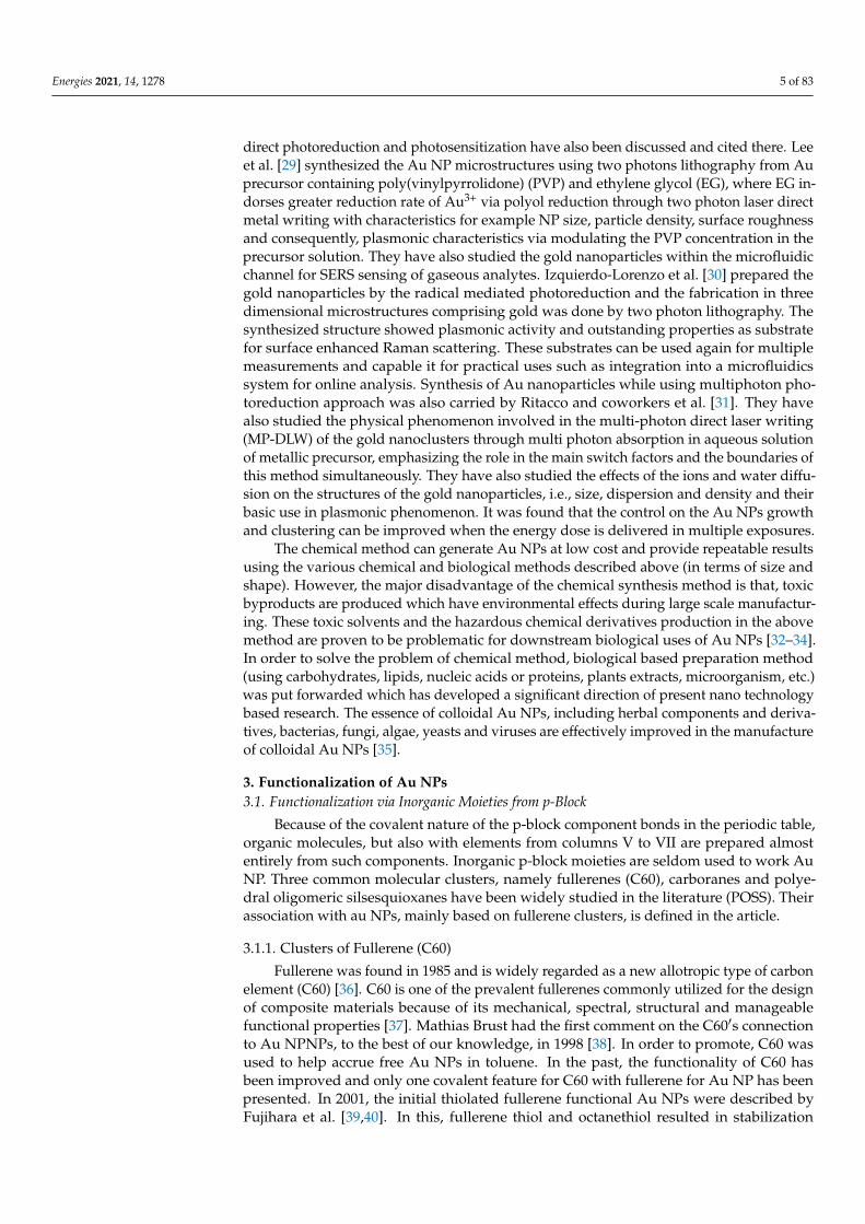
of Au NPs (see Figure 2). K.G Thomas et al. [41] developed an analogous approach byusing an alkyl chain between C60 and Au NPs in 2002 in comparison with Shon et al.,who used an aromatic amino mercaptophenol (Figure 2) [42]. However, for Fujiwara,fullerene thiols surrounded particles with other alkane thiols are used as co-stabilizingmeans (C8H17SH or C12H25SH). The addition of the fullerene-thiol moiety was carriedout by the ligand exchange method except for to Shon, who tried a direct process using amixture of C60-Ph-SH and C8H17SH. For electrochemical or photoelectrical purposes, allthese nanocomposites were prepared (Au-S-R-C60).
Figure 2. Scheme of a fullerene thiol-functional Au NP example. Reproduced with permission fromACS, 2002 [41].
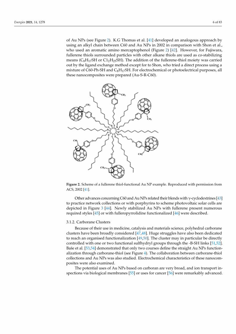
Other advances concerning C60 and Au NPs related their blends withγ-cyclodextrines [43]to practice network collections or with porphyrins to scheme photovoltaic solar cells aredepicted in Figure 3 [44]. Newly stabilized Au NPs with fullerene present numerousrequired styles [45] or with fulleropyrrolidine functionalized [46] were described.
3.1.2. Carborane Clusters
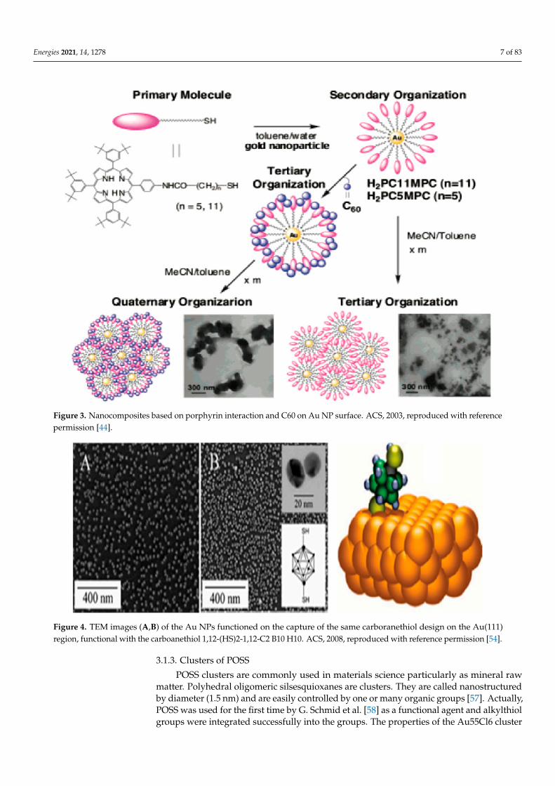
Because of their use in medicine, catalysis and materials science, polyhedral carboraneclusters have been broadly considered [47,48]. Huge struggles have also been dedicatedto reach an organised functionalization [49,50]. The cluster may in particular be directlycontrolled with one or two functional sulfhydryl groups through the -B-SH links [51,52].Baše et al. [53,54] demonstrated that only two courses define the straight Au NPs function-alization through carborane-thiol (see Figure 4). The collaboration between carborane-thiolcollections and Au NPs was also studied. Electrochemical characteristics of these nanocom-posites were also examined.
The potential uses of Au NPs based on carboran are very broad, and ion transport in-spections via biological membranes [55] or uses for cancer [56] were remarkably advanced.
Energies 2021, 14, 1278 7 of 83
Figure 3. Nanocomposites based on porphyrin interaction and C60 on Au NP surface. ACS, 2003, reproduced with referencepermission [44].
Figure 4. TEM images (A,B) of the Au NPs functioned on the capture of the same carboranethiol design on the Au(111)region, functional with the carboanethiol 1,12-(HS)2-1,12-C2 B10 H10. ACS, 2008, reproduced with reference permission [54].
3.1.3. Clusters of POSS
POSS clusters are commonly used in materials science particularly as mineral rawmatter. Polyhedral oligomeric silsesquioxanes are clusters. They are called nanostructuredby diameter (1.5 nm) and are easily controlled by one or many organic groups [57]. Actually,POSS was used for the first time by G. Schmid et al. [58] as a functional agent and alkylthiolgroups were integrated successfully into the groups. The properties of the Au55Cl6 cluster
Energies 2021, 14, 1278 8 of 83
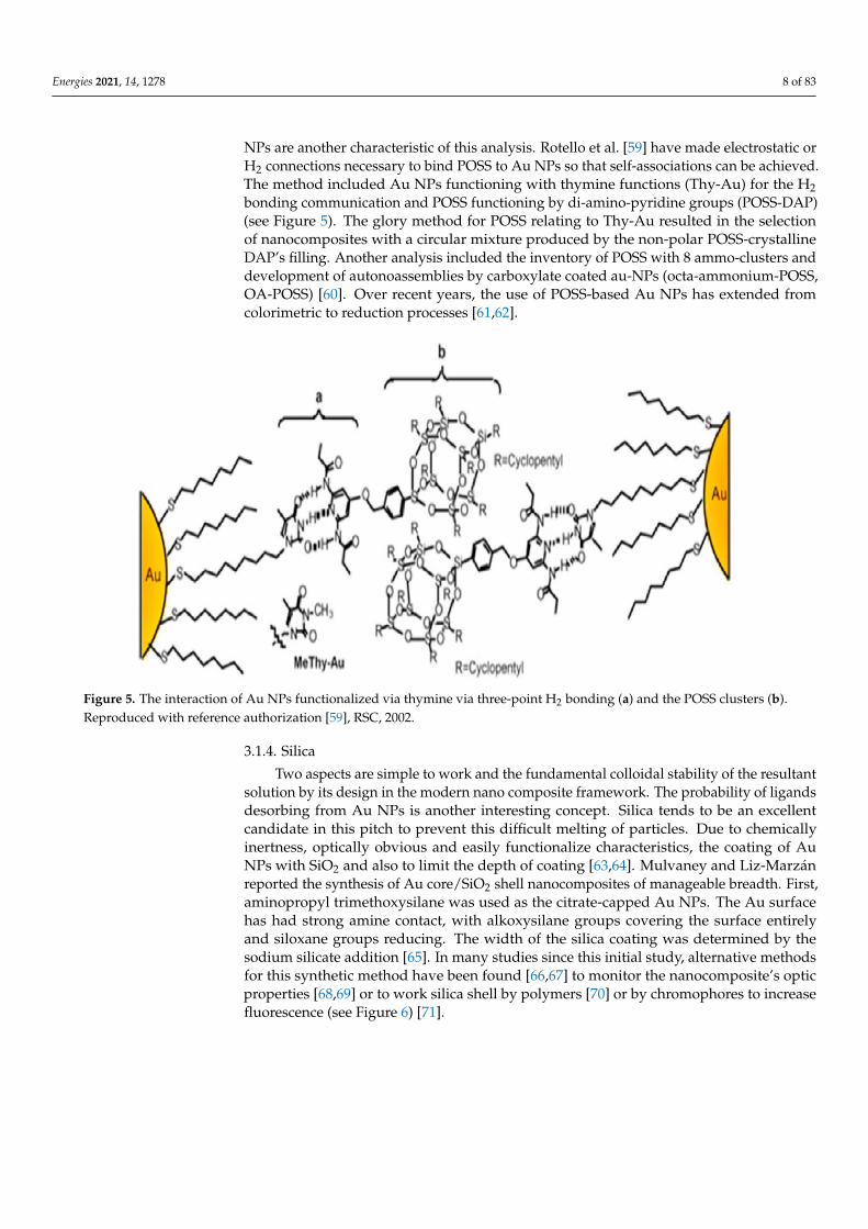
NPs are another characteristic of this analysis. Rotello et al. [59] have made electrostatic orH2 connections necessary to bind POSS to Au NPs so that self-associations can be achieved.The method included Au NPs functioning with thymine functions (Thy-Au) for the H2bonding communication and POSS functioning by di-amino-pyridine groups (POSS-DAP)(see Figure 5). The glory method for POSS relating to Thy-Au resulted in the selectionof nanocomposites with a circular mixture produced by the non-polar POSS-crystallineDAP’s filling. Another analysis included the inventory of POSS with 8 ammo-clusters anddevelopment of autonoassemblies by carboxylate coated au-NPs (octa-ammonium-POSS,OA-POSS) [60]. Over recent years, the use of POSS-based Au NPs has extended fromcolorimetric to reduction processes [61,62].
Figure 5. The interaction of Au NPs functionalized via thymine via three-point H2 bonding (a) and the POSS clusters (b).Reproduced with reference authorization [59], RSC, 2002.
3.1.4. Silica
Two aspects are simple to work and the fundamental colloidal stability of the resultantsolution by its design in the modern nano composite framework. The probability of ligandsdesorbing from Au NPs is another interesting concept. Silica tends to be an excellentcandidate in this pitch to prevent this difficult melting of particles. Due to chemicallyinertness, optically obvious and easily functionalize characteristics, the coating of AuNPs with SiO2 and also to limit the depth of coating [63,64]. Mulvaney and Liz-Marzánreported the synthesis of Au core/SiO2 shell nanocomposites of manageable breadth. First,aminopropyl trimethoxysilane was used as the citrate-capped Au NPs. The Au surfacehas had strong amine contact, with alkoxysilane groups covering the surface entirelyand siloxane groups reducing. The width of the silica coating was determined by thesodium silicate addition [65]. In many studies since this initial study, alternative methodsfor this synthetic method have been found [66,67] to monitor the nanocomposite’s opticproperties [68,69] or to work silica shell by polymers [70] or by chromophores to increasefluorescence (see Figure 6) [71].
Energies 2021, 14, 1278 9 of 83
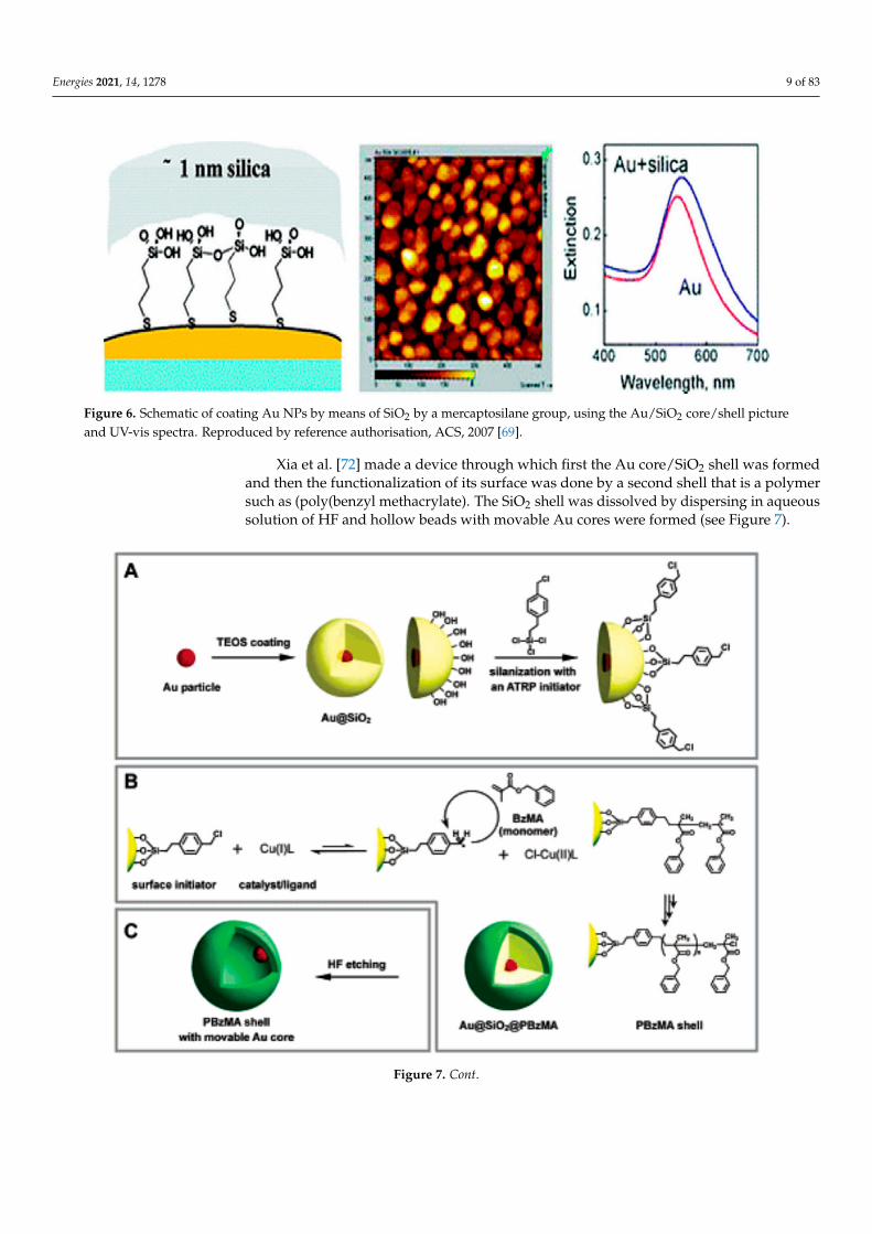
Figure 6. Schematic of coating Au NPs by means of SiO2 by a mercaptosilane group, using the Au/SiO2 core/shell pictureand UV-vis spectra. Reproduced by reference authorisation, ACS, 2007 [69].
Xia et al. [72] made a device through which first the Au core/SiO2 shell was formedand then the functionalization of its surface was done by a second shell that is a polymersuch as (poly(benzyl methacrylate). The SiO2 shell was dissolved by dispersing in aqueoussolution of HF and hollow beads with movable Au cores were formed (see Figure 7).
Figure 7. Cont.
Energies 2021, 14, 1278 10 of 83
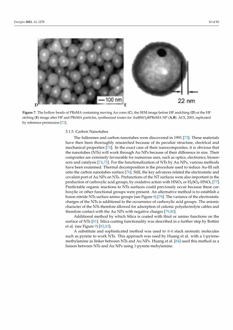
Figure 7. The hollow beads of PBzMA containing moving Au cores (C), the SEM image before HF andching (D) et the HFetching (E) image after HF and PBzMA particles, synthesized routes for Au@SiO2@PBzMA NP (A,B). ACS, 2003, replicatedby reference permission [72].
3.1.5. Carbon Nanotubes
The fullerenes and carbon nanotubes were discovered in 1991 [73]. These materialshave then been thoroughly researched because of its peculiar structure, electrical andmechanical properties [74]. In the exact case of their nanocomposites, it is obvious thatthe nanotubes (NTs) will work through Au NPs because of their difference in size. Theircomposites are extremely favourable for numerous uses, such as optics, electronics, biosen-sors and catalysis [74,75]. For the functionalization of NTs by Au NPs, various methodshave been examined. Thermal decomposition is the procedure used to reduce Au-III saltonto the carbon nanotubes surface [76]. Still, the key advances related the electrostatic andcovalent port of Au NPs on NTs. Prefunctions of the NT surfaces were also important in theproduction of carboxylic acid groups, by oxidative action with HNO3 or H2SO4-HNO3 [77].Predictable organic reactions to NTs surfaces could previously occur because these car-boxylic or other functional groups were present. An alternative method is to establish aboron nitride NTs surface amino groups (see Figure 8) [78]. The variance of the electrostaticcharges of the NTs is additional to the occurrence of carboxylic acid groups. The anioniccharacter of the NTs therefore allowed for adsorption of cationic polyelectrolyte cables andtherefore contact with the Au NPs with negative charges [79,80].
Additional method by which Silica is coated with thiol or amino functions on thesurface of NTs [81]. Silica coating functionality was described in a further step by Bottiniet al. (see Figure 9) [82,83].
A substitute and sophisticated method was used to π-π stack aromatic moleculessuch as pyrene to work NTs. This approach was used by Huang et al. with a 1-pyrene-methylamine as linker between NTs and Au NPs. Huang et al. [84] used this method as aliaison between NTs and Au NPs using 1-pyrene-mehylamine.
Energies 2021, 14, 1278 11 of 83
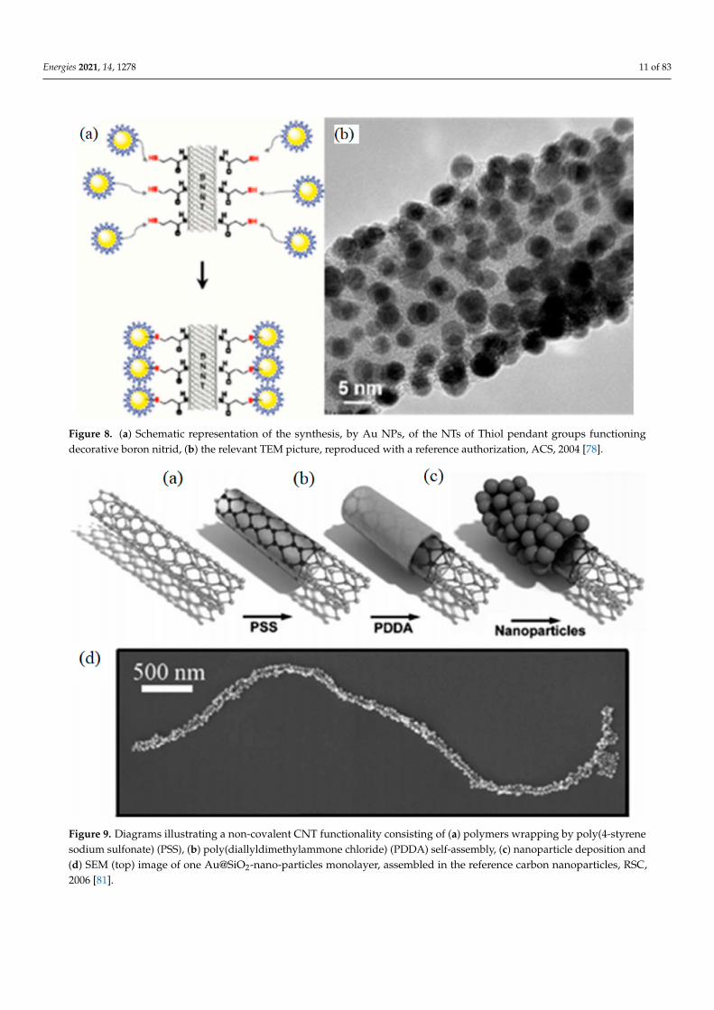
Figure 8. (a) Schematic representation of the synthesis, by Au NPs, of the NTs of Thiol pendant groups functioningdecorative boron nitrid, (b) the relevant TEM picture, reproduced with a reference authorization, ACS, 2004 [78].
Figure 9. Diagrams illustrating a non-covalent CNT functionality consisting of (a) polymers wrapping by poly(4-styrenesodium sulfonate) (PSS), (b) poly(diallyldimethylammone chloride) (PDDA) self-assembly, (c) nanoparticle deposition and(d) SEM (top) image of one Au@SiO2-nano-particles monolayer, assembled in the reference carbon nanoparticles, RSC,2006 [81].
Energies 2021, 14, 1278 12 of 83
3.1.6. Polyoxometalate Compounds (POM)
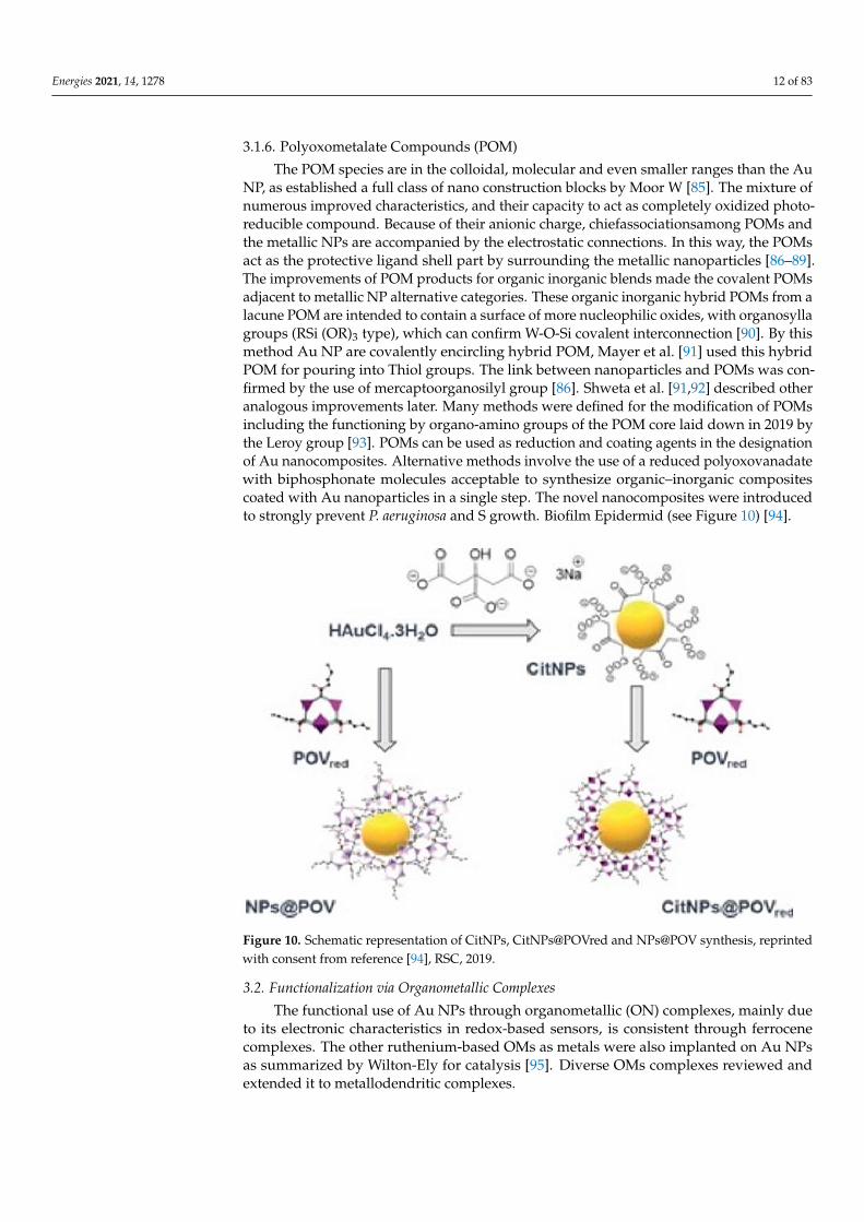
The POM species are in the colloidal, molecular and even smaller ranges than the AuNP, as established a full class of nano construction blocks by Moor W [85]. The mixture ofnumerous improved characteristics, and their capacity to act as completely oxidized photo-reducible compound. Because of their anionic charge, chiefassociationsamong POMs andthe metallic NPs are accompanied by the electrostatic connections. In this way, the POMsact as the protective ligand shell part by surrounding the metallic nanoparticles [86–89].The improvements of POM products for organic inorganic blends made the covalent POMsadjacent to metallic NP alternative categories. These organic inorganic hybrid POMs from alacune POM are intended to contain a surface of more nucleophilic oxides, with organosyllagroups (RSi (OR)3 type), which can confirm W-O-Si covalent interconnection [90]. By thismethod Au NP are covalently encircling hybrid POM, Mayer et al. [91] used this hybridPOM for pouring into Thiol groups. The link between nanoparticles and POMs was con-firmed by the use of mercaptoorganosilyl group [86]. Shweta et al. [91,92] described otheranalogous improvements later. Many methods were defined for the modification of POMsincluding the functioning by organo-amino groups of the POM core laid down in 2019 bythe Leroy group [93]. POMs can be used as reduction and coating agents in the designationof Au nanocomposites. Alternative methods involve the use of a reduced polyoxovanadatewith biphosphonate molecules acceptable to synthesize organic–inorganic compositescoated with Au nanoparticles in a single step. The novel nanocomposites were introducedto strongly prevent P. aeruginosa and S growth. Biofilm Epidermid (see Figure 10) [94].
Figure 10. Schematic representation of CitNPs, CitNPs@POVred and NPs@POV synthesis, reprintedwith consent from reference [94], RSC, 2019.
3.2. Functionalization via Organometallic Complexes
The functional use of Au NPs through organometallic (ON) complexes, mainly dueto its electronic characteristics in redox-based sensors, is consistent through ferrocenecomplexes. The other ruthenium-based OMs as metals were also implanted on Au NPsas summarized by Wilton-Ely for catalysis [95]. Diverse OMs complexes reviewed andextended it to metallodendritic complexes.
Energies 2021, 14, 1278 13 of 83
3.2.1. Ferroncenyl Complexes
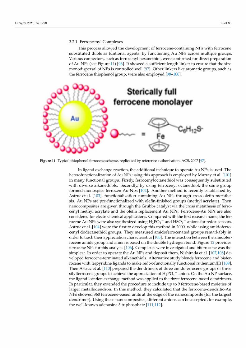
This process allowed the development of ferrocene-containing NPs with ferrocenesubstituted thiols as funtional agents, by functioning Au NPs across multiple groups.Various connectors, such as ferrocenyl hexanethiol, were confirmed for direct preparationof Au NPs (see Figure 11) [96]. It showed a sufficient length linker to ensure that the sizemonodispersal of NPs is controlled well [97]. Other linkers like aromatic groups, such asthe ferrocene thiophenol group, were also employed [98–100].
Figure 11. Typical thiophenol ferrocene scheme, replicated by reference authorisation, ACS, 2007 [97].
In ligand exchange reaction, the additional technique to operate Au NPs is used. Theheterofunctionalization of Au NPs using this approach is employed by Murray et al. [101]in many functional groups. Firstly, ferrocenyloctanethiol was consequently substitutedwith diverse alkanethiols. Secondly, by using ferrocenyl octanethiol, the same groupformed monospice ferrozen Au-Nps [102]. Another method is recently established byAstruc et al. [103], functionalization containing Au NPs through cross-olefin metathe-sis. Au NPs are pre-functionalized with olefin-finished groups (methyl acrylate). Thennanocomposites are given through the Grubbs catalyst via the cross metathesis of ferro-cenyl methyl acrylate and the olefin replacement Au NPs. Ferrocene-Au NPs are alsoconsidered for electrochemical applications. Compared with the first research name, the fer-rocene Au NPs were also synthesized using H2PO4
− and HSO4− anions for redox sensors.
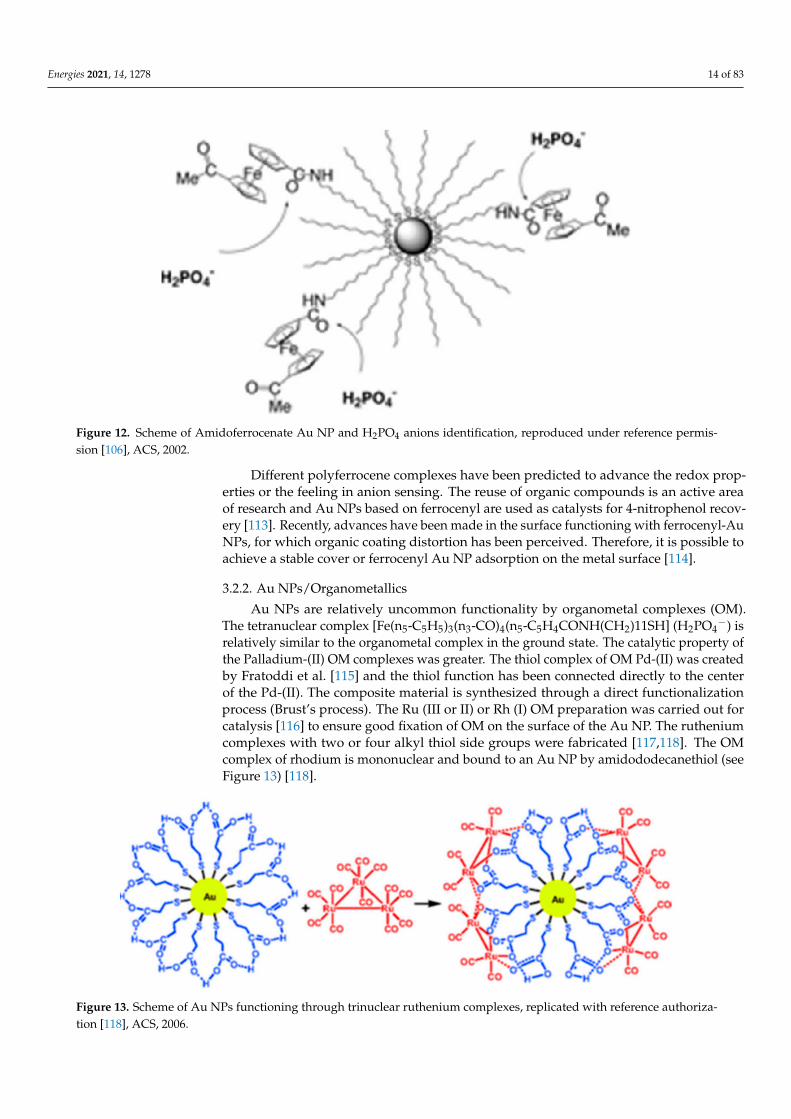
Astruc et al. [104] were the first to develop this method in 2000, while using amidoferro-cenyl dodecanethiol groups. They measured amidoferrocenated groups remarkably inorder to track their appreciation characteristics [105]. The interaction between the amidofer-rocene amide group and anion is based on the double hydrogen bond. Figure 12 providesferrocene NPs for this analysis [106]. Complexes were investigated and biferrocene was thesimplest. In order to operate the Au NPs and deposit them, Nishirada et al. [107,108] de-veloped ferrocene-terminated alkanethiols. Alternative study blends ferrocene and bisfer-rocene with terpyridine ligands to make redox-functionally functional ruthenium(II) [109].Then Astruc et al. [110] prepared the dendrimers of three amidoferrocene groups or threesilylferrocene groups to achieve the appreciation of H2PO4
− anion. On the Au NP surface,the ligand location exchange method was applied to the three ferrocene-based dendrimers.In particular, they extended the procedure to include up to 9 ferrocene-based moieties oflarger metallodendron. In this method, they calculated that the ferrocene-dendritic-AuNPs showed 360 ferrocene-based units at the edge of the nanocomposite (for the largestdendrimer). Using these nanocomposites, different anions can be accepted, for example,the well-known adenosine 5 triphosphate [111,112].
Energies 2021, 14, 1278 14 of 83
Figure 12. Scheme of Amidoferrocenate Au NP and H2PO4 anions identification, reproduced under reference permis-sion [106], ACS, 2002.
Different polyferrocene complexes have been predicted to advance the redox prop-erties or the feeling in anion sensing. The reuse of organic compounds is an active areaof research and Au NPs based on ferrocenyl are used as catalysts for 4-nitrophenol recov-ery [113]. Recently, advances have been made in the surface functioning with ferrocenyl-AuNPs, for which organic coating distortion has been perceived. Therefore, it is possible toachieve a stable cover or ferrocenyl Au NP adsorption on the metal surface [114].
3.2.2. Au NPs/Organometallics
Au NPs are relatively uncommon functionality by organometal complexes (OM).The tetranuclear complex [Fe(n5-C5H5)3(n3-CO)4(n5-C5H4CONH(CH2)11SH] (H2PO4
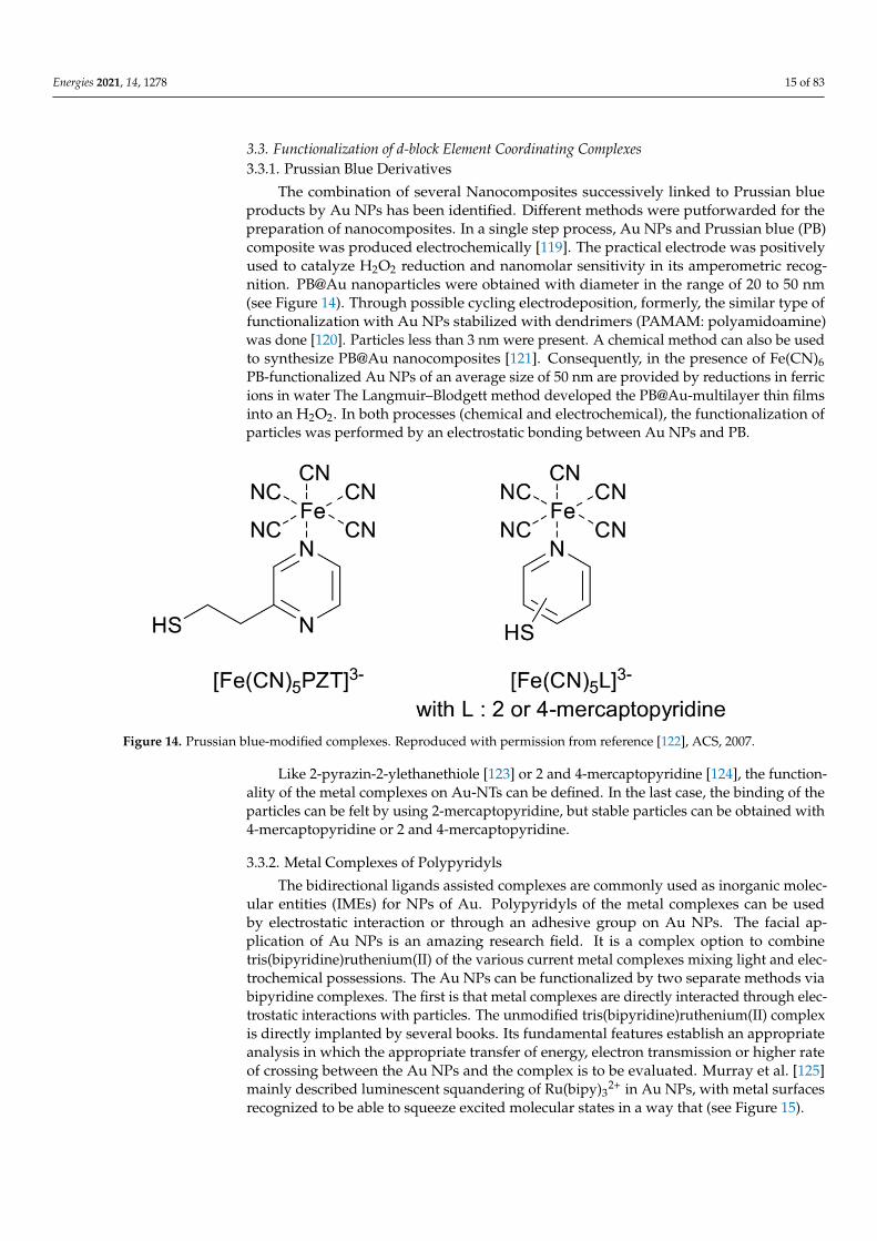
−) isrelatively similar to the organometal complex in the ground state. The catalytic property ofthe Palladium-(II) OM complexes was greater. The thiol complex of OM Pd-(II) was createdby Fratoddi et al. [115] and the thiol function has been connected directly to the centerof the Pd-(II). The composite material is synthesized through a direct functionalizationprocess (Brust’s process). The Ru (III or II) or Rh (I) OM preparation was carried out forcatalysis [116] to ensure good fixation of OM on the surface of the Au NP. The rutheniumcomplexes with two or four alkyl thiol side groups were fabricated [117,118]. The OMcomplex of rhodium is mononuclear and bound to an Au NP by amidododecanethiol (seeFigure 13) [118].
Figure 13. Scheme of Au NPs functioning through trinuclear ruthenium complexes, replicated with reference authoriza-tion [118], ACS, 2006.
Energies 2021, 14, 1278 15 of 83
3.3. Functionalization of d-block Element Coordinating Complexes3.3.1. Prussian Blue Derivatives
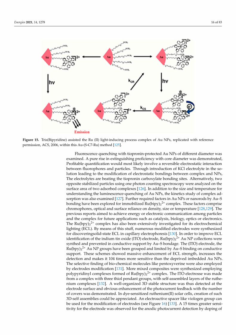
The combination of several Nanocomposites successively linked to Prussian blueproducts by Au NPs has been identified. Different methods were putforwarded for thepreparation of nanocomposites. In a single step process, Au NPs and Prussian blue (PB)composite was produced electrochemically [119]. The practical electrode was positivelyused to catalyze H2O2 reduction and nanomolar sensitivity in its amperometric recog-nition. PB@Au nanoparticles were obtained with diameter in the range of 20 to 50 nm(see Figure 14). Through possible cycling electrodeposition, formerly, the similar type offunctionalization with Au NPs stabilized with dendrimers (PAMAM: polyamidoamine)was done [120]. Particles less than 3 nm were present. A chemical method can also be usedto synthesize PB@Au nanocomposites [121]. Consequently, in the presence of Fe(CN)6PB-functionalized Au NPs of an average size of 50 nm are provided by reductions in ferricions in water The Langmuir–Blodgett method developed the PB@Au-multilayer thin filmsinto an H2O2. In both processes (chemical and electrochemical), the functionalization ofparticles was performed by an electrostatic bonding between Au NPs and PB.
Figure 14. Prussian blue-modified complexes. Reproduced with permission from reference [122], ACS, 2007.
Like 2-pyrazin-2-ylethanethiole [123] or 2 and 4-mercaptopyridine [124], the function-ality of the metal complexes on Au-NTs can be defined. In the last case, the binding of theparticles can be felt by using 2-mercaptopyridine, but stable particles can be obtained with4-mercaptopyridine or 2 and 4-mercaptopyridine.
3.3.2. Metal Complexes of Polypyridyls
The bidirectional ligands assisted complexes are commonly used as inorganic molec-ular entities (IMEs) for NPs of Au. Polypyridyls of the metal complexes can be usedby electrostatic interaction or through an adhesive group on Au NPs. The facial ap-plication of Au NPs is an amazing research field. It is a complex option to combinetris(bipyridine)ruthenium(II) of the various current metal complexes mixing light and elec-trochemical possessions. The Au NPs can be functionalized by two separate methods viabipyridine complexes. The first is that metal complexes are directly interacted through elec-trostatic interactions with particles. The unmodified tris(bipyridine)ruthenium(II) complexis directly implanted by several books. Its fundamental features establish an appropriateanalysis in which the appropriate transfer of energy, electron transmission or higher rateof crossing between the Au NPs and the complex is to be evaluated. Murray et al. [125]mainly described luminescent squandering of Ru(bipy)3
2+ in Au NPs, with metal surfacesrecognized to be able to squeeze excited molecular states in a way that (see Figure 15).
Energies 2021, 14, 1278 16 of 83
Figure 15. Tris(Bipyridine) assisted the Ru (II) light-inducing process complex of Au NPs, replicated with referencepermission, ACS, 2006, within this Au-(S-C7-Ru) method [125].
Fluorescence quenching with tiopronin-protected Au NPs of different diameter wasexamined. A pure rise in extinguishing proficiency with core diameter was demonstrated,Profitable quantification would most likely involve a reversible electrostatic interactionbetween fluorophores and particles. Through introduction of KCl electrolyte in the so-lution leading to the modification of electrostatic bondings between complex and NPs,The electrolytes are beating the tiopronin carboxylate bonding sites. Alternatively, twoopposite stabilized particles using one photon counting spectroscopy were analyzed on thesurface area of two adsorbed complexes [126]. In addition to the size and temperature forunderstanding the luminescence-quenching of Au NPs, the kinetics study of complex ad-sorption was also examined [127]. Further required factors in Au NPs or nanorods by Au–Sbonding have been explored for immobilized Ru(bipy)3
2+ complex. These factors comprisechromophores, optical and surface reliance on density, size or temperature [128,129]. Theprevious reports aimed to achieve energy or electronic communication among particlesand the complex for future applications such as catalysis, biology, optics or electronics.The Ru(bpy)3
2+ complex has also been extensively investigated for its electrochemicallighting (ECL). By means of this stuff, numerous modified electrodes were synthesizedfor discoveringsolid-state ECL in capillary electrophoresis [130]. In order to improve ECLidentification of the indium tin oxide (ITO) electrode, Ru(bpy)3
2+ Au NP collections weresynthed and prevented in conductive support by Au–S bondage. The (ITO) electrode, theRu(bpy)3
2+ Au NP groups have been grouped and limited by Au–S binding on conductivesupport. These schemes showed massive enhancement of ECL strength, increases thedetection and makes it 104 times more sensitive than the deprived imbedded Au NPs.The selective finding of bio-chemical molecules like pentoxyverine were also employedby electrodes modification [131]. More mixed composites were synthesized employingpolypyridinyl complexes formed of Ru(bpy)3
2+ complex. The ITO electrosse was madefrom a complex with three thiol pendant groups, with self-assembled layers of the ruthe-nium complexes [132]. A well-organized 3D stable structure was thus detected at theelectrode surface and obvious enhancement of the photocurrent feedback with the numberof covers was demonstrated. In dye-sensitized ruthenium(II) solar cells, creation of such3D-self assemblies could be appreciated. An electroactive spacer like viologen group canbe used for the modification of electrodes (see Figure 16) [133]. A 15 times greater sensi-tivity for the electrode was observed for the anodic photocurrent detection by doping of

Energies 2021, 14, 1278 17 of 83
functionalized Au NPs onto Au electrode. A contribution of the viologen entity throughone-electron reduction approach showed extension of that early work comprised to knowthe effect of the size of nanoparticles on the photocurrent reaction.
Figure 16. Viologen linked thiol-Ru complex imbeded onto Au NPs, Reprinted with permission from reference [133],Elsevier, 2003.
The perfect photocurrent competences of nanostructured particles have diametersof 50–100 nm [108]. On the basis of this, enhancement of the ionic strength is identifiedto clumping of Au NPs. In a novel approach viologen-doped ruthenium complex wasused for the decoration for these nanostructures [134]. In the said work, the impact of theelectrolyte nature, anionic agent in particular was studied because it can cause alterationin the morphology of these schemes and as a result change the photocurrent reactions. Inaddition to these violin-connected systems connected to Au NPs by a thiol movement,many electrostatic violin systems between Ru(bipy)3
2+ and nanoparticles capsized withcitrate were inspected [135,136].
No photo-electrochemical cells were obtained and therefore the Au NPs were trans-ferred to the violin sensitizer with energy. The association of Au NPs can also be simplyacquired by employing the metalation with pyridine entities [137]. By employing thisscheme, the thin films were produced and they showed diode like reactions. For the func-tionality and stability of Au NPs, some additional anchoring groups have been employedaround the redox centre with a polypyridinic environment. Recent work has clarified theeffect by the amine end group assisted by Ru(bipy)3
2+ complex on the rates of radiation andthe non-radiative rates of a phosphorescent compound [138]. A recognition teachniq hasbeen used for implanting phosphorescent molecules on the particles. In case of Streptavidinwith a surplus bovine serum albumin, nanoparticles were first synthesized. Phosphorescentmolecules were thus functionalized with biotin due to selective sensing of streptavidinfor biotin. Hence, anchoring of these NPs- molecules was recognised, and grounded onthe biotin-streptavidin sensing aprocah. Biotin-streptavidin appreciation, was anotherexamples which was innovative for the gathering of proteins and Au NPs on templates ofDNA [139]. Other characteristics, i.e., of a cobalt bistable complex supramolecular controlof valence-tautomeric symmetrywas also studied [140]. The anchoring effect was seen onthermodynamic factors; the binding of the valence tautomer influenced the surface contain-ment. Up to now, only nanocomposites have been identified which were obtained throughelectrostatic interations or by using aliphatic chains with thiol end groups. No electroniccontacts via the non-conjugated spacer were possible in these arrangements. In recent times,Mayer et al. [141,142] reported the preparation of numerous poly-pyridinic complexesof rutheniumof bidentate ligands phenanthroline ligands with completely delocalizedinsertions, allowing a straight message between the complex and particles (see Figure 17).Ruthenium complexes’ redox potential has been changed to display an electronic message.
Energies 2021, 14, 1278 18 of 83
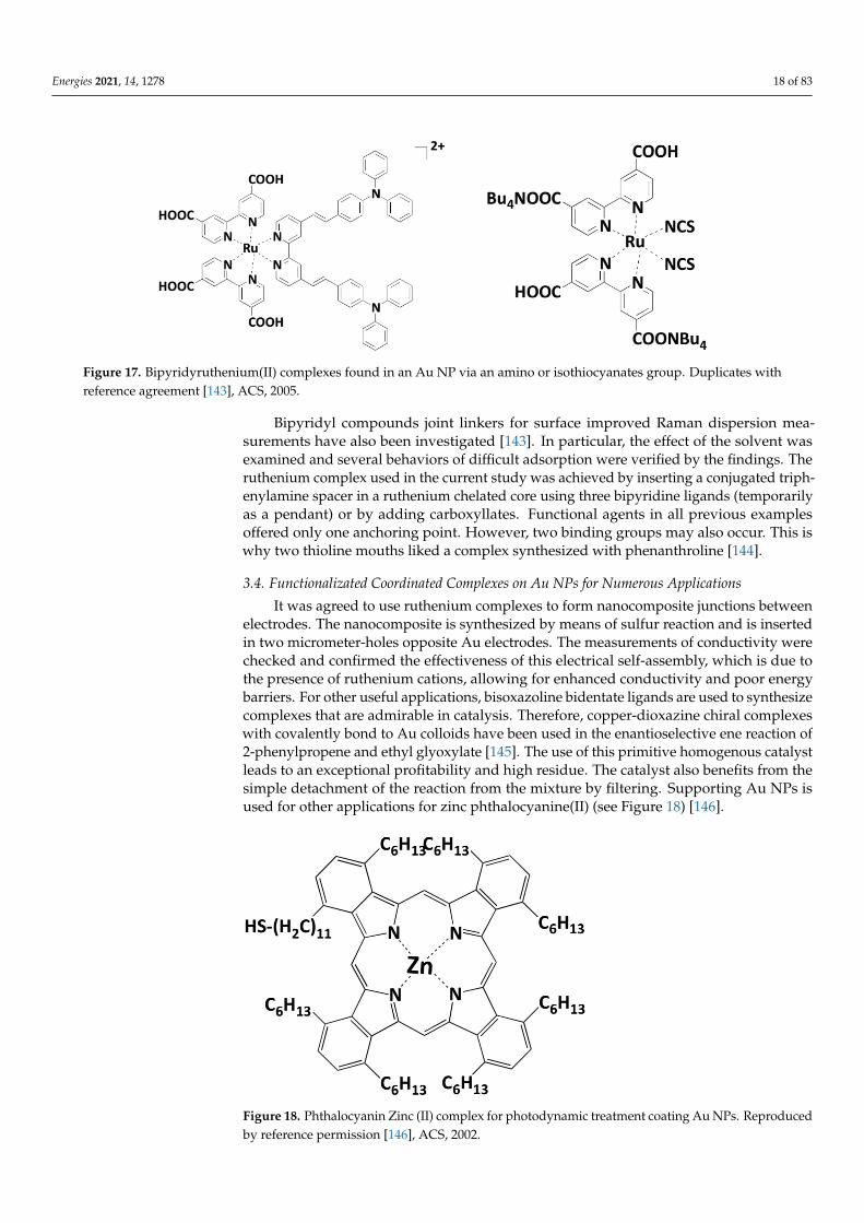
Figure 17. Bipyridyruthenium(II) complexes found in an Au NP via an amino or isothiocyanates group. Duplicates withreference agreement [143], ACS, 2005.
Bipyridyl compounds joint linkers for surface improved Raman dispersion mea-surements have also been investigated [143]. In particular, the effect of the solvent wasexamined and several behaviors of difficult adsorption were verified by the findings. Theruthenium complex used in the current study was achieved by inserting a conjugated triph-enylamine spacer in a ruthenium chelated core using three bipyridine ligands (temporarilyas a pendant) or by adding carboxyllates. Functional agents in all previous examplesoffered only one anchoring point. However, two binding groups may also occur. This iswhy two thioline mouths liked a complex synthesized with phenanthroline [144].
3.4. Functionalizated Coordinated Complexes on Au NPs for Numerous Applications
It was agreed to use ruthenium complexes to form nanocomposite junctions betweenelectrodes. The nanocomposite is synthesized by means of sulfur reaction and is insertedin two micrometer-holes opposite Au electrodes. The measurements of conductivity werechecked and confirmed the effectiveness of this electrical self-assembly, which is due tothe presence of ruthenium cations, allowing for enhanced conductivity and poor energybarriers. For other useful applications, bisoxazoline bidentate ligands are used to synthesizecomplexes that are admirable in catalysis. Therefore, copper-dioxazine chiral complexeswith covalently bond to Au colloids have been used in the enantioselective ene reaction of2-phenylpropene and ethyl glyoxylate [145]. The use of this primitive homogenous catalystleads to an exceptional profitability and high residue. The catalyst also benefits from thesimple detachment of the reaction from the mixture by filtering. Supporting Au NPs isused for other applications for zinc phthalocyanine(II) (see Figure 18) [146].
Figure 18. Phthalocyanin Zinc (II) complex for photodynamic treatment coating Au NPs. Reproducedby reference permission [146], ACS, 2002.
Energies 2021, 14, 1278 19 of 83
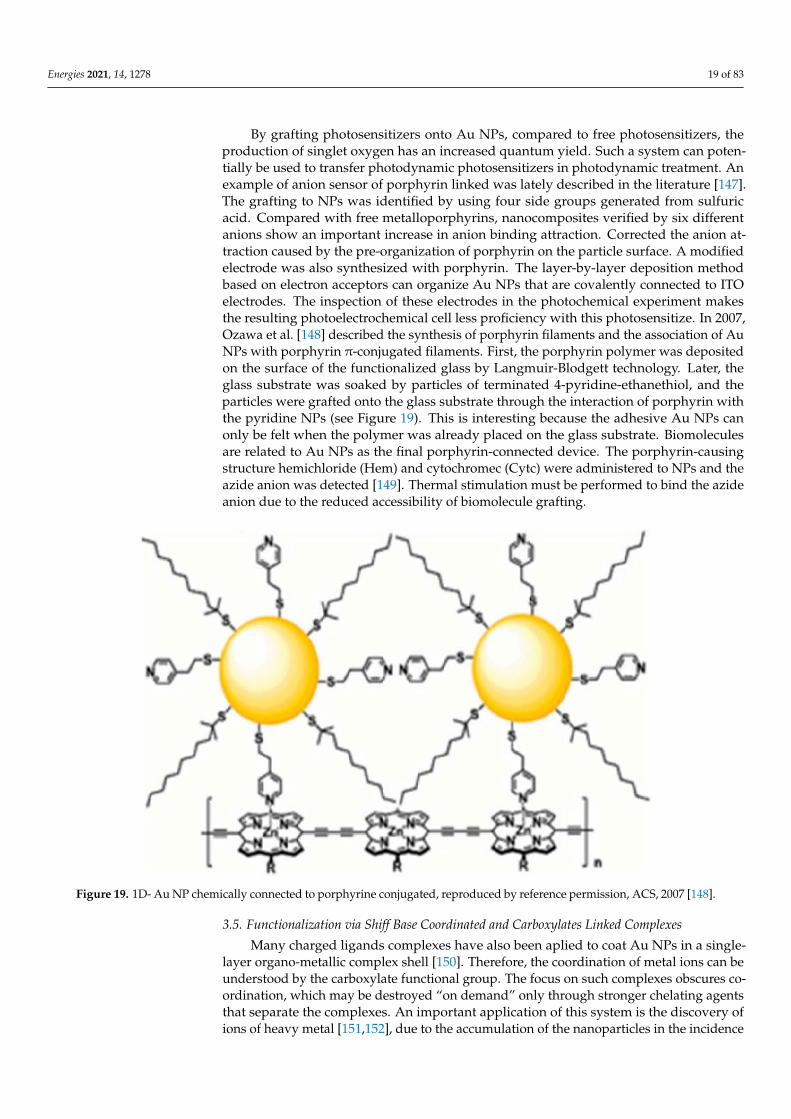
By grafting photosensitizers onto Au NPs, compared to free photosensitizers, theproduction of singlet oxygen has an increased quantum yield. Such a system can poten-tially be used to transfer photodynamic photosensitizers in photodynamic treatment. Anexample of anion sensor of porphyrin linked was lately described in the literature [147].The grafting to NPs was identified by using four side groups generated from sulfuricacid. Compared with free metalloporphyrins, nanocomposites verified by six differentanions show an important increase in anion binding attraction. Corrected the anion at-traction caused by the pre-organization of porphyrin on the particle surface. A modifiedelectrode was also synthesized with porphyrin. The layer-by-layer deposition methodbased on electron acceptors can organize Au NPs that are covalently connected to ITOelectrodes. The inspection of these electrodes in the photochemical experiment makesthe resulting photoelectrochemical cell less proficiency with this photosensitize. In 2007,Ozawa et al. [148] described the synthesis of porphyrin filaments and the association of AuNPs with porphyrin π-conjugated filaments. First, the porphyrin polymer was depositedon the surface of the functionalized glass by Langmuir-Blodgett technology. Later, theglass substrate was soaked by particles of terminated 4-pyridine-ethanethiol, and theparticles were grafted onto the glass substrate through the interaction of porphyrin withthe pyridine NPs (see Figure 19). This is interesting because the adhesive Au NPs canonly be felt when the polymer was already placed on the glass substrate. Biomoleculesare related to Au NPs as the final porphyrin-connected device. The porphyrin-causingstructure hemichloride (Hem) and cytochromec (Cytc) were administered to NPs and theazide anion was detected [149]. Thermal stimulation must be performed to bind the azideanion due to the reduced accessibility of biomolecule grafting.
Figure 19. 1D- Au NP chemically connected to porphyrine conjugated, reproduced by reference permission, ACS, 2007 [148].
3.5. Functionalization via Shiff Base Coordinated and Carboxylates Linked Complexes
Many charged ligands complexes have also been aplied to coat Au NPs in a single-layer organo-metallic complex shell [150]. Therefore, the coordination of metal ions can beunderstood by the carboxylate functional group. The focus on such complexes obscures co-ordination, which may be destroyed “on demand” only through stronger chelating agentsthat separate the complexes. An important application of this system is the discovery ofions of heavy metal [151,152], due to the accumulation of the nanoparticles in the incidence
Energies 2021, 14, 1278 20 of 83
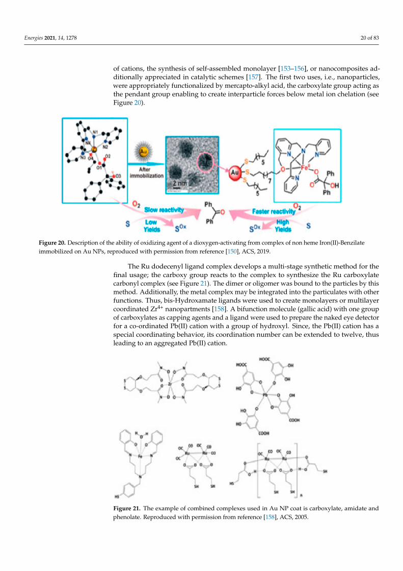
of cations, the synthesis of self-assembled monolayer [153–156], or nanocomposites ad-ditionally appreciated in catalytic schemes [157]. The first two uses, i.e., nanoparticles,were appropriately functionalized by mercapto-alkyl acid, the carboxylate group acting asthe pendant group enabling to create interparticle forces below metal ion chelation (seeFigure 20).
Figure 20. Description of the ability of oxidizing agent of a dioxygen-activating from complex of non heme Iron(II)-Benzilateimmobilized on Au NPs, reproduced with permission from reference [150], ACS, 2019.
The Ru dodecenyl ligand complex develops a multi-stage synthetic method for thefinal usage; the carboxy group reacts to the complex to synthesize the Ru carboxylatecarbonyl complex (see Figure 21). The dimer or oligomer was bound to the particles by thismethod. Additionally, the metal complex may be integrated into the particulates with otherfunctions. Thus, bis-Hydroxamate ligands were used to create monolayers or multilayercoordinated Zr4+ nanopartments [158]. A bifunction molecule (gallic acid) with one groupof carboxylates as capping agents and a ligand were used to prepare the naked eye detectorfor a co-ordinated Pb(II) cation with a group of hydroxyl. Since, the Pb(II) cation has aspecial coordinating behavior, its coordination number can be extended to twelve, thusleading to an aggregated Pb(II) cation.
Figure 21. The example of combined complexes used in Au NP coat is carboxylate, amidate andphenolate. Reproduced with permission from reference [158], ACS, 2005.
Energies 2021, 14, 1278 21 of 83
The introduction of different metal cations leaves NPs remote, since they co-ordinateto overcrowded electrostatic disintegrations between particles with less ligands and theability of cation Pb (II). For the coordination of iron (III) cations, the Schiff base ligands wereused. For the stabilization of Au-Nps, two opposite plans were used in that work. Firstly,using the neutral complexes for the stabilization of the NPs through steric (alkyl chain)repulsion and secondly the stabilization is gained through electrostatic repulsion [159].
3.6. Functionalization via Bio-Inorganic Complexes
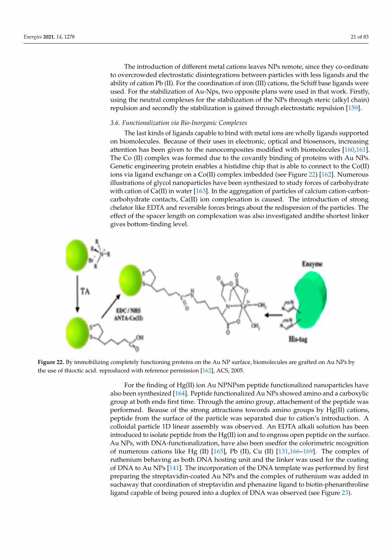
The last kinds of ligands capable to bind with metal ions are wholly ligands supportedon biomolecules. Because of their uses in electronic, optical and biosensors, increasingattention has been given to the nanocomposites modified with biomolecules [160,161].The Co (II) complex was formed due to the covantly binding of proteins with Au NPs.Genetic engineering protein enables a histidine chip that is able to connect to the Co(II)ions via ligand exchange on a Co(II) complex imbedded (see Figure 22) [162]. Numerousillustrations of glycol nanoparticles have been synthesized to study forces of carbohydratewith cation of Ca(II) in water [163]. In the aggregation of particles of calcium cation-carbon-carbohydrate contacts, Ca(II) ion complexation is caused. The introduction of strongchelator like EDTA and reversible forces brings about the redispersion of the particles. Theeffect of the spacer length on complexation was also investigated andthe shortest linkergives bottom-finding level.
Figure 22. By immobilizing completely functioning proteins on the Au NP surface, biomolecules are grafted on Au NPs bythe use of thioctic acid. reproduced with reference permission [162], ACS, 2005.
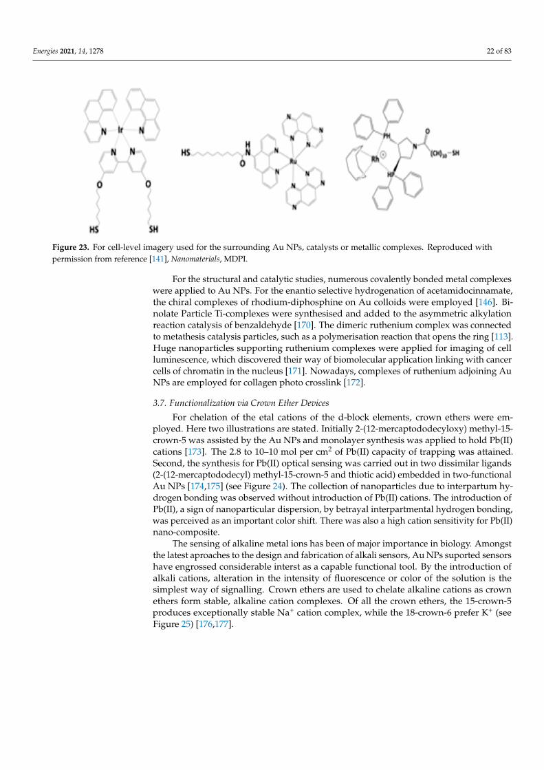
For the finding of Hg(II) ion Au NPNPsm peptide functionalized nanoparticles havealso been synthesized [164]. Peptide functionalized Au NPs showed amino and a carboxylicgroup at both ends first time. Through the amino group, attachement of the peptide wasperformed. Beause of the strong attractions towords amino groups by Hg(II) cations,peptide from the surface of the particle was separated due to cation’s introduction. Acolloidal particle 1D linear assembly was observed. An EDTA alkali solution has beenintroduced to isolate peptide from the Hg(II) ion and to engross open peptide on the surface.Au NPs, with DNA-functionalization, have also been usedfor the colorimetric recognitionof numerous cations like Hg (II) [165], Pb (II), Cu (II) [131,166–169]. The complex ofruthenium behaving as both DNA hosting unit and the linker was used for the coatingof DNA to Au NPs [141]. The incorporation of the DNA template was performed by firstpreparing the streptavidin-coated Au NPs and the complex of ruthenium was added insuchaway that coordination of streptavidin and phenazine ligand to biotin-phenanthrolineligand capable of being poured into a duplex of DNA was observed (see Figure 23).
Energies 2021, 14, 1278 22 of 83
Figure 23. For cell-level imagery used for the surrounding Au NPs, catalysts or metallic complexes. Reproduced withpermission from reference [141], Nanomaterials, MDPI.
For the structural and catalytic studies, numerous covalently bonded metal complexeswere applied to Au NPs. For the enantio selective hydrogenation of acetamidocinnamate,the chiral complexes of rhodium-diphosphine on Au colloids were employed [146]. Bi-nolate Particle Ti-complexes were synthesised and added to the asymmetric alkylationreaction catalysis of benzaldehyde [170]. The dimeric ruthenium complex was connectedto metathesis catalysis particles, such as a polymerisation reaction that opens the ring [113].Huge nanoparticles supporting ruthenium complexes were applied for imaging of cellluminescence, which discovered their way of biomolecular application linking with cancercells of chromatin in the nucleus [171]. Nowadays, complexes of ruthenium adjoining AuNPs are employed for collagen photo crosslink [172].
3.7. Functionalization via Crown Ether Devices
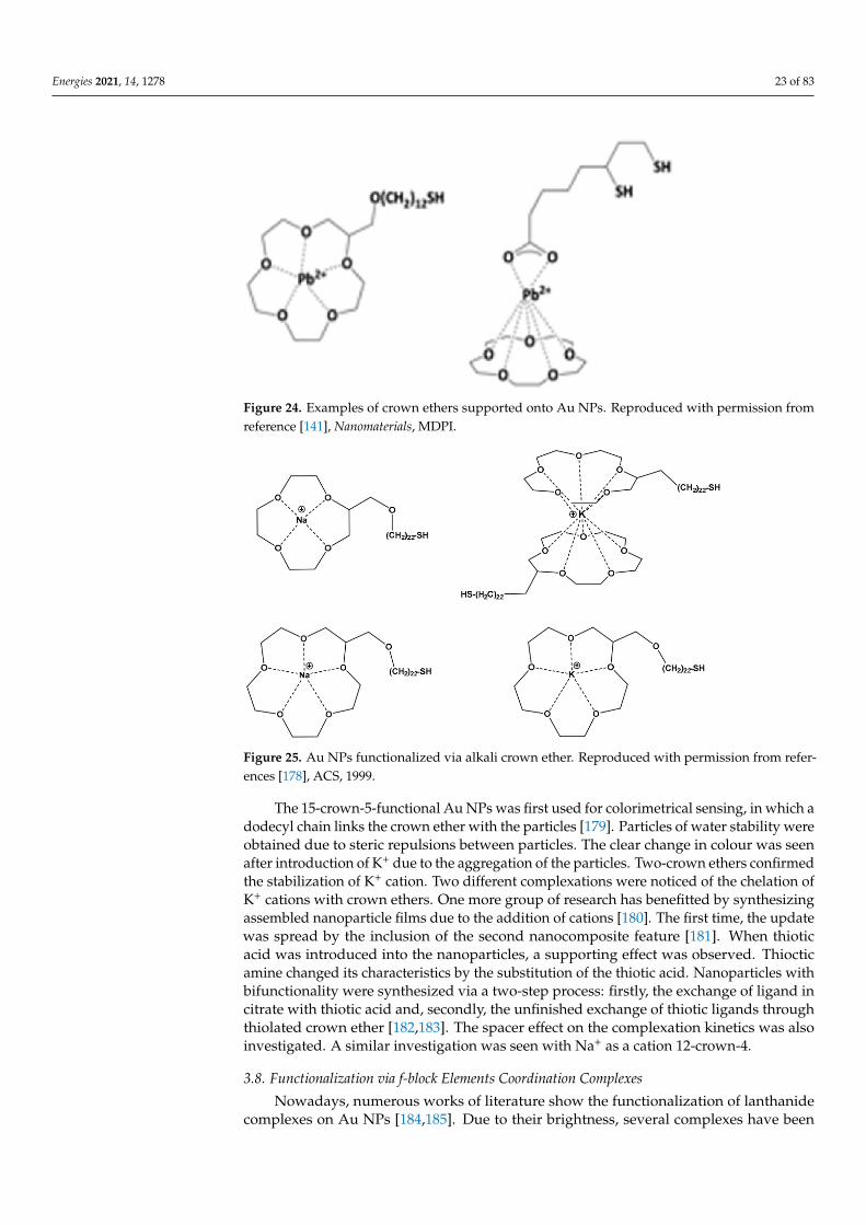
For chelation of the etal cations of the d-block elements, crown ethers were em-ployed. Here two illustrations are stated. Initially 2-(12-mercaptododecyloxy) methyl-15-crown-5 was assisted by the Au NPs and monolayer synthesis was applied to hold Pb(II)cations [173]. The 2.8 to 10–10 mol per cm2 of Pb(II) capacity of trapping was attained.Second, the synthesis for Pb(II) optical sensing was carried out in two dissimilar ligands(2-(12-mercaptododecyl) methyl-15-crown-5 and thiotic acid) embedded in two-functionalAu NPs [174,175] (see Figure 24). The collection of nanoparticles due to interpartum hy-drogen bonding was observed without introduction of Pb(II) cations. The introduction ofPb(II), a sign of nanoparticular dispersion, by betrayal interpartmental hydrogen bonding,was perceived as an important color shift. There was also a high cation sensitivity for Pb(II)nano-composite.
The sensing of alkaline metal ions has been of major importance in biology. Amongstthe latest aproaches to the design and fabrication of alkali sensors, Au NPs suported sensorshave engrossed considerable interst as a capable functional tool. By the introduction ofalkali cations, alteration in the intensity of fluorescence or color of the solution is thesimplest way of signalling. Crown ethers are used to chelate alkaline cations as crownethers form stable, alkaline cation complexes. Of all the crown ethers, the 15-crown-5produces exceptionally stable Na+ cation complex, while the 18-crown-6 prefer K+ (seeFigure 25) [176,177].
Energies 2021, 14, 1278 23 of 83
Figure 24. Examples of crown ethers supported onto Au NPs. Reproduced with permission fromreference [141], Nanomaterials, MDPI.
Figure 25. Au NPs functionalized via alkali crown ether. Reproduced with permission from refer-ences [178], ACS, 1999.
The 15-crown-5-functional Au NPs was first used for colorimetrical sensing, in which adodecyl chain links the crown ether with the particles [179]. Particles of water stability wereobtained due to steric repulsions between particles. The clear change in colour was seenafter introduction of K+ due to the aggregation of the particles. Two-crown ethers confirmedthe stabilization of K+ cation. Two different complexations were noticed of the chelation ofK+ cations with crown ethers. One more group of research has benefitted by synthesizingassembled nanoparticle films due to the addition of cations [180]. The first time, the updatewas spread by the inclusion of the second nanocomposite feature [181]. When thioticacid was introduced into the nanoparticles, a supporting effect was observed. Thiocticamine changed its characteristics by the substitution of the thiotic acid. Nanoparticles withbifunctionality were synthesized via a two-step process: firstly, the exchange of ligand incitrate with thiotic acid and, secondly, the unfinished exchange of thiotic ligands throughthiolated crown ether [182,183]. The spacer effect on the complexation kinetics was alsoinvestigated. A similar investigation was seen with Na+ as a cation 12-crown-4.
3.8. Functionalization via f-block Elements Coordination Complexes
Nowadays, numerous works of literature show the functionalization of lanthanidecomplexes on Au NPs [184,185]. Due to their brightness, several complexes have been
Energies 2021, 14, 1278 24 of 83
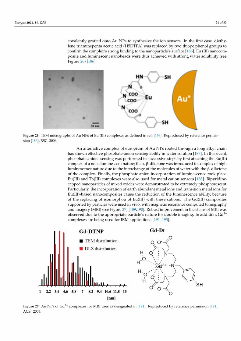
covalently grafted onto Au NPs to synthesize the ion sensors. In the first case, diethy-lene triaminepenta acetic acid (H5DTPA) was replaced by two thiope phenol groups toconfirm the complex’s strong binding to the nanoparticle’s surface [186]. Eu (III) nanocom-posite and luminescent nanobeads were thus achieved with strong water solubility (seeFigure 26) [186].
Figure 26. TEM micrographs of Au NPs of Eu (III) complexes as defined in ref. [166]. Reproduced by reference permis-sion [186], RSC, 2006.
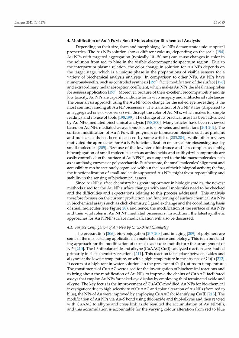
An alternative complex of europium of Au NPs rooted through a long alkyl chainhas shown effective phosphate-anion sensing ability in water solution [187]. In this event,phosphate anions sensing was performed in successive steps by first attaching the Eu(III)complex of a non eluminescent nature; then, β-diketone was introduced to complex of highluminescence nature due to the interchange of the molecules of water with the β-diketoneof the complex. Finally, the phosphate anion incorporation of luminescence took place.Eu(III) and Tb(III) complexes were also used for metal cation sensors [188]. Bipyridine-capped nanoparticles of mixed oxides were demonstrated to be extremely phosphorescent.Particularly, the incorporation of earth abundant metal ions and transition metal ions forEu(III)-based nanocomposites cause the reduction of the luminescence ability, becauseof the replacing of isomorphou of Eu(III) with these cations. The Gd(III) compositessupported by particles were used in vivo, with magnetic resonance computed tomographyand imagery (MRI) (see Figure 27) [189,190]. Robust improvement in the stems of MRI wasobserved due to the appropriate particle’s nature for double imaging. In addition, Gd3+
complexes are being used for IRM applications [191–193].
Figure 27. Au NPs of Gd3+ complexes for MRI uses as designated in [191]. Reproduced by reference permission [191],ACS, 2006.
Energies 2021, 14, 1278 25 of 83
4. Modification of Au NPs via Small Molecules for Biochemical Analysis
Depending on their size, form and morphology, Au NPs demonstrate unique opticalproperties. The Au NPs solution shows different colours, depending on the scale [194].Au NPs with targeted aggregation (typically 10−50 nm) can cause changes in color inthe solution from red to blue in the visible electromagnetic spectrum region. Due tothe interpartum plasma relation, the color change in solution for Au NPs depends onthe target stage, which is a unique phase in the preparations of visible sensors for avariety of biochemical analysis analysts. In comparison to other NPs, Au NPs havenumerousbenefits, such as controlled synthesis [195], facile modification of the surface [196]and extraordinary molar absorption coefficient, which makes Au NPs the ideal nanoprobesfor sensers application [197]. Moreover, because of their excellent biocompatibility and itslow toxicity, Au NPs are capable candidate for in vivo imagery and antibacterial substances.The bioanalysis approach using the Au NP color change for the naked eye re-reading is themost common among all Au NP biosensors. The transition of Au NP states (dispersed toan aggregated one or vice versa) will disrupt the color of Au NPs, which makes for simplereadings and no use of tools [198,199]. The change of its practical uses has been advancedby Au NPs-mediated biochemical analysis [198,200]. Many articles have been reviewedbased on Au NPs mediated assays tonucleic acids, proteins and metal ions [201,202]. Thesurface modification of Au NPs with polymers or biomacromolecules such as proteinsand nuclear acids has been discussed by some articles [203,204], while other reviewsmotivated the approaches for Au NPs functionalization of surface for biosensing uses bysmall molecules [205]. Because of the low steric hindrance and less complex assembly,bioconjugation of small molecules such as amino acids and sulfhydryl compounds areeasily controlled on the surface of Au NPNPs, as compared to the bio-macromolecules suchas as antibody, enzyme or polysaccharide. Furthermore, the small molecules’ alignment andaccessibility can be accurately organised without the loss of their biological activity; thefore,the functionalization of small-molecule supported Au NPs might favor repeatability andstability in the sensing of biochemical assays.
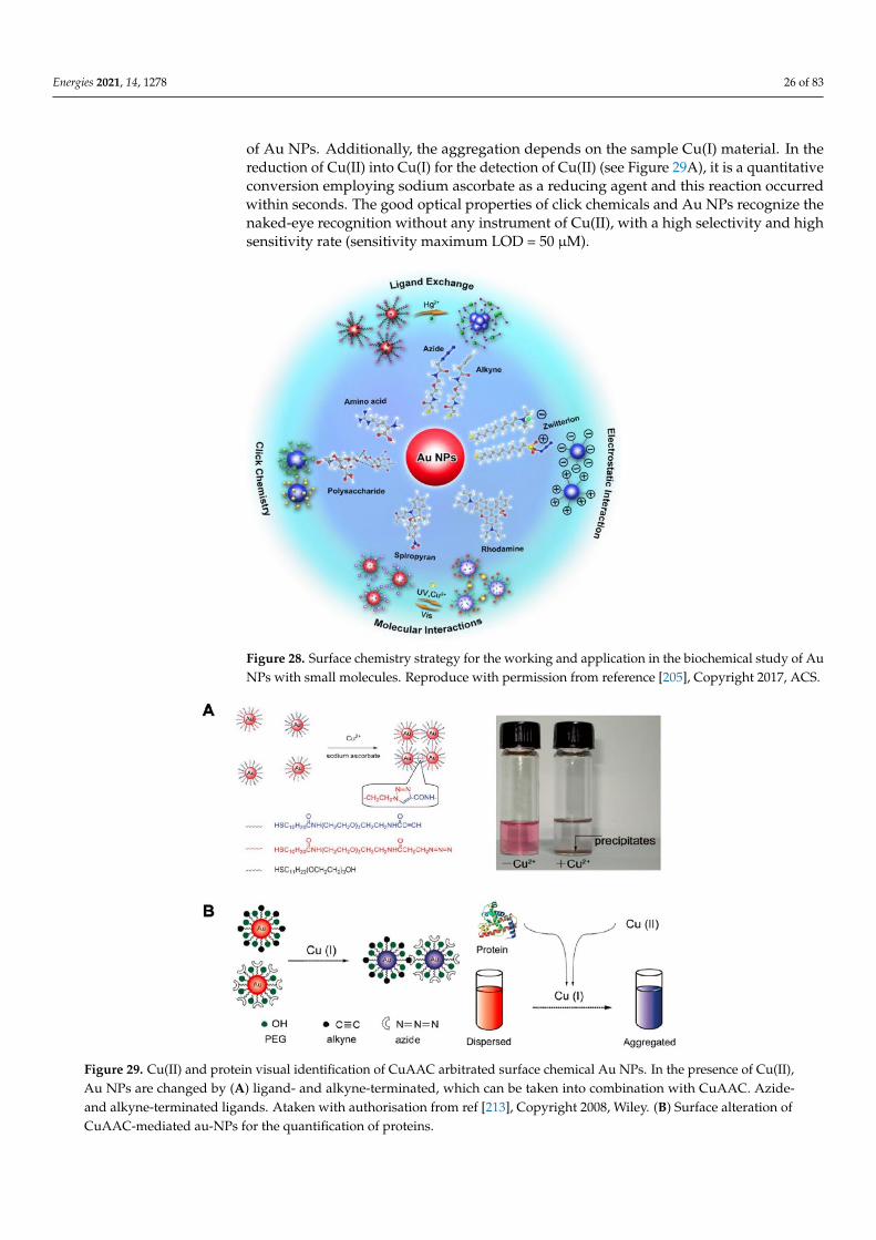
Since Au NP surface chemistry has great importance in biologic studies, the newestmethods used for the Au NP surface changes with small molecules need to be checkedand the difficulties and expectations relating to this process addressed. This analysistherefore focuses on the current production and functioning of surface chemical Au NPsin biochemical assays such as click chemistry, ligand exchange and the coordinating basisof small molecules (see Figure 28), and hence, the modification of the surface of Au NPsand their vital roles in Au NPNP mediated biosensors. In addition, the latest syntheticapproaches for Au NPNP surface modicafication will also be discussed.
4.1. Surface Conjugation of Au NPs by Click-Based Chemistry
The preparation [206], bio-conjugation [207,208] and imaging [209] of polymers aresome of the most exciting applications in materials science and biology. This is an outstand-ing approach for the modification of surfaces as it does not disturb the arrangement ofNPs [210]. The 1.3-dipolar azide and alkyne (CuAAC) Cu(I)-catalyzed reactions are studiedprimarily in click chemistry reactions [211]. This reaction takes place between azides andalkynes at the lowest temperature, or with a high temperature in the absence of Cu(I) [212].It occurs at a high rate in water solutions in the presence of Cu(I), at room temperature.The constituents of CuAAC were used for the investigation of biochemical reactions andto bring about the modification of Au NPs to improve the chains of CuAAC-facilitatedassays that employ Au NPs for naked-eye display by employing thiol terminated azide andalkyne. The key focus is the improvement of CuACC-modified Au NPs for bio-chemicalinvestigation; due to high selectivity of CuAAC and color alteration of Au NPs (from red toblue), the NPs of Au were improved by employing CuAAC for identifying Cu(II) [213]. Themodification of Au NPs via Au–S bond using thiol-azide and thiol-alkyne and then reactedwith CuAAC to alkyne and cross link azide resulted the accumulation of Au NPNPs,and this accumulation is accountable for the varying colour alteration from red to blue
Energies 2021, 14, 1278 26 of 83
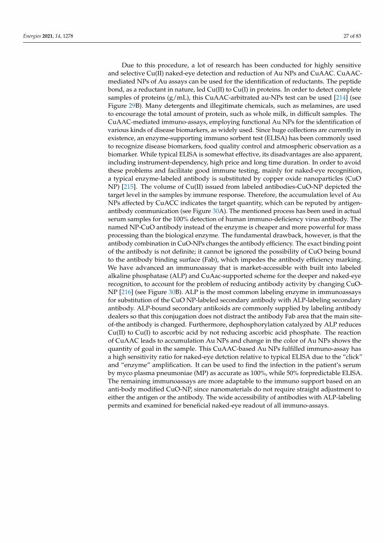
of Au NPs. Additionally, the aggregation depends on the sample Cu(I) material. In thereduction of Cu(II) into Cu(I) for the detection of Cu(II) (see Figure 29A), it is a quantitativeconversion employing sodium ascorbate as a reducing agent and this reaction occurredwithin seconds. The good optical properties of click chemicals and Au NPs recognize thenaked-eye recognition without any instrument of Cu(II), with a high selectivity and highsensitivity rate (sensitivity maximum LOD = 50 µM).
Figure 28. Surface chemistry strategy for the working and application in the biochemical study of AuNPs with small molecules. Reproduce with permission from reference [205], Copyright 2017, ACS.
Figure 29. Cu(II) and protein visual identification of CuAAC arbitrated surface chemical Au NPs. In the presence of Cu(II),Au NPs are changed by (A) ligand- and alkyne-terminated, which can be taken into combination with CuAAC. Azide-and alkyne-terminated ligands. Ataken with authorisation from ref [213], Copyright 2008, Wiley. (B) Surface alteration ofCuAAC-mediated au-NPs for the quantification of proteins.
Energies 2021, 14, 1278 27 of 83
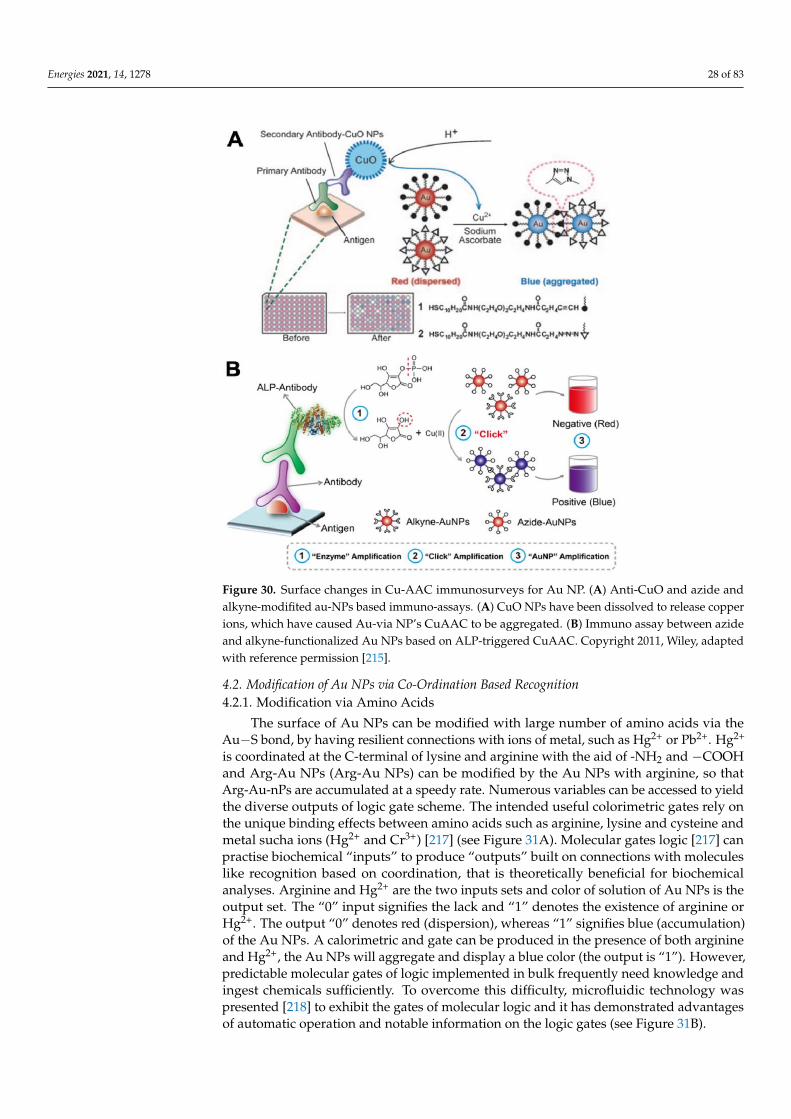
Due to this procedure, a lot of research has been conducted for highly sensitiveand selective Cu(II) naked-eye detection and reduction of Au NPs and CuAAC. CuAAC-mediated NPs of Au assays can be used for the identification of reductants. The peptidebond, as a reductant in nature, led Cu(II) to Cu(I) in proteins. In order to detect completesamples of proteins (g/mL), this CuAAC-arbitrated au-NPs test can be used [214] (seeFigure 29B). Many detergents and illegitimate chemicals, such as melamines, are usedto encourage the total amount of protein, such as whole milk, in difficult samples. TheCuAAC-mediated immuno-assays, employing functional Au NPs for the identification ofvarious kinds of disease biomarkers, as widely used. Since huge collections are currently inexistence, an enzyme-supporting immuno sorbent test (ELISA) has been commonly usedto recognize disease biomarkers, food quality control and atmospheric observation as abiomarker. While typical ELISA is somewhat effective, its disadvantages are also apparent,including instrument-dependency, high price and long time duration. In order to avoidthese problems and facilitate good immune testing, mainly for naked-eye recognition,a typical enzyme-labeled antibody is substituted by copper oxide nanoparticles (CuONP) [215]. The volume of Cu(II) issued from labeled antibodies-CuO-NP depicted thetarget level in the samples by immune response. Therefore, the accumulation level of AuNPs affected by CuACC indicates the target quantity, which can be reputed by antigen-antibody communication (see Figure 30A). The mentioned process has been used in actualserum samples for the 100% detection of human immuno-deficiency virus antibody. Thenamed NP-CuO antibody instead of the enzyme is cheaper and more powerful for massprocessing than the biological enzyme. The fundamental drawback, however, is that theantibody combination in CuO-NPs changes the antibody efficiency. The exact binding pointof the antibody is not definite; it cannot be ignored the possibility of CuO being boundto the antibody binding surface (Fab), which impedes the antibody efficiency marking.We have advanced an immunoassay that is market-accessible with built into labeledalkaline phosphatase (ALP) and CuAac-supported scheme for the deeper and naked-eyerecognition, to account for the problem of reducing antibody activity by changing CuO-NP [216] (see Figure 30B). ALP is the most common labeling enzyme in immunoassaysfor substitution of the CuO NP-labeled secondary antibody with ALP-labeling secondaryantibody. ALP-bound secondary antikoids are commonly supplied by labeling antibodydealers so that this conjugation does not distract the antibody Fab area that the main site-of-the antibody is changed. Furthermore, dephosphorylation catalyzed by ALP reducesCu(II) to Cu(I) to ascorbic acid by not reducing ascorbic acid phosphate. The reactionof CuAAC leads to accumulation Au NPs and change in the color of Au NPs shows thequantity of goal in the sample. This CuAAC-based Au NPs fulfilled immuno-assay hasa high sensitivity ratio for naked-eye detction relative to typical ELISA due to the “click”and “enzyme” amplification. It can be used to find the infection in the patient’s serumby myco plasma pneumoniae (MP) as accurate as 100%, while 50% forpredictable ELISA.The remaining immunoassays are more adaptable to the immuno support based on ananti-body modified CuO-NP, since nanomaterials do not require straight adjustment toeither the antigen or the antibody. The wide accessibility of antibodies with ALP-labelingpermits and examined for beneficial naked-eye readout of all immuno-assays.
Energies 2021, 14, 1278 28 of 83
Figure 30. Surface changes in Cu-AAC immunosurveys for Au NP. (A) Anti-CuO and azide andalkyne-modifited au-NPs based immuno-assays. (A) CuO NPs have been dissolved to release copperions, which have caused Au-via NP’s CuAAC to be aggregated. (B) Immuno assay between azideand alkyne-functionalized Au NPs based on ALP-triggered CuAAC. Copyright 2011, Wiley, adaptedwith reference permission [215].
4.2. Modification of Au NPs via Co-Ordination Based Recognition4.2.1. Modification via Amino Acids
The surface of Au NPs can be modified with large number of amino acids via theAu−S bond, by having resilient connections with ions of metal, such as Hg2+ or Pb2+. Hg2+
is coordinated at the C-terminal of lysine and arginine with the aid of -NH2 and −COOHand Arg-Au NPs (Arg-Au NPs) can be modified by the Au NPs with arginine, so thatArg-Au-nPs are accumulated at a speedy rate. Numerous variables can be accessed to yieldthe diverse outputs of logic gate scheme. The intended useful colorimetric gates rely onthe unique binding effects between amino acids such as arginine, lysine and cysteine andmetal sucha ions (Hg2+ and Cr3+) [217] (see Figure 31A). Molecular gates logic [217] canpractise biochemical “inputs” to produce “outputs” built on connections with moleculeslike recognition based on coordination, that is theoretically beneficial for biochemicalanalyses. Arginine and Hg2+ are the two inputs sets and color of solution of Au NPs is theoutput set. The “0” input signifies the lack and “1” denotes the existence of arginine orHg2+. The output “0” denotes red (dispersion), whereas “1” signifies blue (accumulation)of the Au NPs. A calorimetric and gate can be produced in the presence of both arginineand Hg2+, the Au NPs will aggregate and display a blue color (the output is “1”). However,predictable molecular gates of logic implemented in bulk frequently need knowledge andingest chemicals sufficiently. To overcome this difficulty, microfluidic technology waspresented [218] to exhibit the gates of molecular logic and it has demonstrated advantagesof automatic operation and notable information on the logic gates (see Figure 31B).
Energies 2021, 14, 1278 29 of 83
Figure 31. Modification of Au NPs for biochemical analysis by amino acid mediated surface. (A) A colorimetric (AND+OR)logic scheme that uses Au NPs with arginine/lysine. (B) Microfluidic systems for metal ion detection combined withmolecular logic gates. Schematic diagram and microfluidic chip photographs are presented in panels (1) and (2). TEMphotos and the required observation window color of the Au NPs are in panels (3) and (4). Copyright 2014, Wiley, adaptedby reference permission [217], Copyright 2014, Wiley.
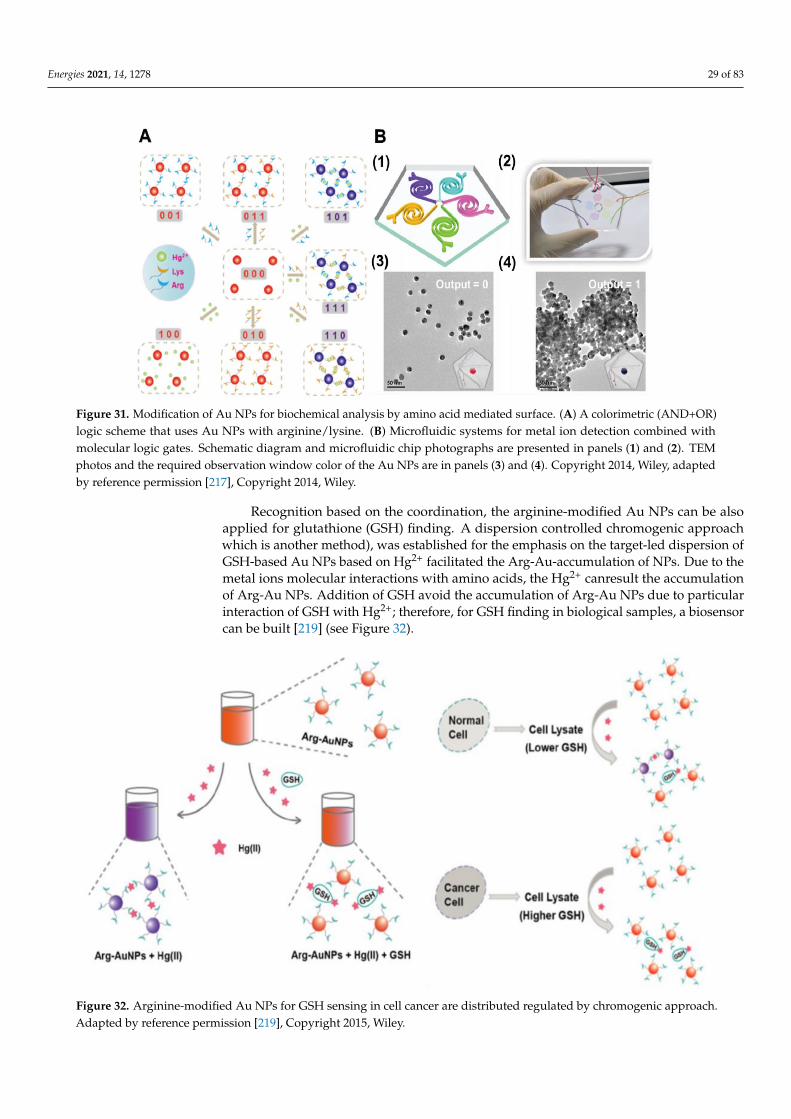
Recognition based on the coordination, the arginine-modified Au NPs can be alsoapplied for glutathione (GSH) finding. A dispersion controlled chromogenic approachwhich is another method), was established for the emphasis on the target-led dispersion ofGSH-based Au NPs based on Hg2+ facilitated the Arg-Au-accumulation of NPs. Due to themetal ions molecular interactions with amino acids, the Hg2+ canresult the accumulationof Arg-Au NPs. Addition of GSH avoid the accumulation of Arg-Au NPs due to particularinteraction of GSH with Hg2+; therefore, for GSH finding in biological samples, a biosensorcan be built [219] (see Figure 32).
Figure 32. Arginine-modified Au NPs for GSH sensing in cell cancer are distributed regulated by chromogenic approach.Adapted by reference permission [219], Copyright 2015, Wiley.
Energies 2021, 14, 1278 30 of 83
Due to the detecting LOD of GSH 10.9 × 10⁻⁹ M, this process can be utilized forestimatation and measurement of GSH levels in cell lysates are more notable than normalcells in cellular cancer cells. In spite of the fact that, restricted thinks about account,the in vivo finding utilize Au NPs for naked-eye readout, taking into consideration thatthe cell level infinitesimal pictures grounded on Au NPs might not be existing since oftheir nanometric molecule estimate and it needs plenty of Au NPs to driven within theobvious color alter. Another method colorimetric measure based on cysteine intercededaccumulation of Au NPs has been presented [215] (see Figure 33A). The amino acid cysteineencompasses a strange structure, having thiol Moity (-SH) at one side and COO− and NH2+
functional groups on other side. By means of the Au–S bond, cysteine can be attachedto the Au NPs and the Au NPs can be bridged by the electrostatic contact between thepositive and negative charged groups that aggregates the Au NPs. The cysteine oxidationby iodide to produce disulfide cysteine prevents Au NP aggregation. An HRP-mediatediodide catalyzed cascade method was introduced to control the dispersion and aggregationof Au NPs, which endorsed the naked-eye identification of details, in conjunction with thiscatalytic method with HRP catalyzed reactions. This immuno assay was utilized to examinethe hepatitis-C virus antibodies in real blood sample via a naked-eye finding precision of100%, while predictable ELISA is 20%, suggesting that this Au NP-based immuno assayenables high sensitivity and naked-eye knowledge for biomedical diagnostics. The Cd2+
and GSH-Au NPs organization authorizes the recognition of cadmium ions (Cd2+) [220](see Figure 33B). In concentrated NaCl solution, the unmodified Au NPs easily aggregate,but the presence of GSH prevents the aggregation of salt induced Au NPs. Once the Cd2+
is applied to a firm blend of Au NPs, GSH and the NaCl, one of the Cd2+ will bind with4× GSH, reducing the amount of free GSH on the Au NPs in order to weaken the integrityand aggregation of the Au NPs. In water and assimilated rice samples, this colorimetricmethod can effectively perceive Cd2+ with high precision and suitable procedures.
4.2.2. Modification via Sulfhydryl Compounds
The chelation contact among spiropyran (SP) and the Cu2+ could be changed viaUV-light and the SP could be isomerized to planar and open shape of merocyanine afterUV-light irradiations (MC). Rather than retaining in the nonplanar and sealed SP type, it ispossible to prepare and use spiropyran-modified Au NPs (spiropyran-Au NPs) to render are-settable and multi-reading logic device proficient in numerous kinds of logic proceduresbuilt on the accumulation of Au NPs in water as solvent [221] (see Figure 34A). To appearred in color, Spiropyran-Au NPs were monodispersed in solutions. Spiropyran transformsinto open merocyanine through exposure to UV, in such a manner that the distributedmerocyanine-modified Au NPs (MC-Au NPs) are aggregated by change in color from redto purple due to merocyanine and Cu2+ chelation. Significantly, by visible light, the MC-AuNPs could simply return to the SP, indicating that the Au NPs-based logic system canbe reset for different periods of discovery (see Figure 34B). In environment monitoring,identification of Cr3+ and Cr2O7
2− is very significant. For the identification of both Cr3+
and Cr2O72− [222] meso-2,3-dimercaptosuccinic acid (DMSA) was used to adjust Au NPs
(see Figure 35A). In order to prepare the Au NPs, DMSA with two -SH functional groupsand two -COOH groups was used for stabilization and reduction. To detect Cr3+ in theDMSA, the -COOH groups can be used. In the meantime, one Cr2O7
2− intermingles withthe two DMSA-Au NPs via the ending carboxyl-groups of DMSA molecules through closeOH····· O hydrogen bonds. The density-functional-theory may measure the change inGibbs free-energy (∆G) of the contacts between DMSA-Au NPs and the various metal ions.Among all of the metal ions, the lowest G for Cr3+ and Cr2O7
2− shows that DMSA-Au NPshave optimum affinity for both of the hydrated Cr3+ and Cr2O7
2−. The naked-eye LODis 10 nM, slightly smaller than the requirements of the Environmental Protection Agency(EPA) (EPA-3060A). This approach has been effectively utilized for finding Cr3+ from thechromium slag dumpsite in chromium-polluted soil.

Energies 2021, 14, 1278 31 of 83
Figure 33. For naked-eye study, amino acids mediated surface-modification of the Au NPs. (A) HRP mediated immuno-assay modulation of Au NPs. Iodide catalyzed cysteine oxidation will alter Au NPs’ surface chemistry and influence AuNPs’ aggregation and dispersion. (B) The complex of Cd2+ and GSH can alter surface morphology of Au NPs and increasestability of the citrate-Au NPs at change in concentration of NaCl. Adapted with permission from reference [220]. Copyright2014, ACS.
Rapid and straight forward detection of the tri-valent metal ions (M3+) is vital andinteresting, additionally to detecting individual ions. The researchers designed Au NPs-modified zwitterionic molecule (Zw-Au NPs). The aggregation of Zw-Au NPs with colorshift could be caused by the M3+, like Al3+, Fe3+ and Cr3+ [223] (see Figure 35B). In addition,due to various forms of the trivalent ions at diverse pH values, the Zw-Au NPs can beretrieved. The M3+ could coordinate with the Zw-Au NPs and cause their aggregation inacidic or neutral environments. The free M3+ turns into M(OH)3 in the simple solution andavoids the contact among Zw-Au NPs and the M3+, following the re-dispersion of Zw-AuNPs. The instrumental-free and low-cost features made this examination suitable for use inresource-limited components, compared to earlier accounts on M3+ findings.
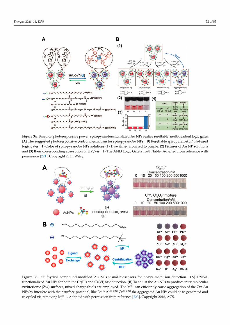
Energies 2021, 14, 1278 32 of 83
Figure 34. Based on photoresponsive power, spiropyran-functionalized Au NPs realize resettable, multi-readout logic gates.(A) The suggested photoresponsive control mechanism for spiropyran-Au NPs. (B) Resettable spiropyran-Au NPs-basedlogic gates. (1) Color of spiropyran-Au NPs solutions (1/1) switched from red to purple. (2) Pictures of Au NP solutionsand (3) their corresponding absorption of UV/vis. (4) The AND Logic Gate’s Truth Table. Adapted from reference withpermission [221], Copyright 2011, Wiley.
Figure 35. Sulfhydryl compound-modified Au NPs visual biosensors for heavy metal ion detection. (A) DMSA-functionalized Au NPs for both the Cr(III) and Cr(VI) fast detection. (B) To adjust the Au NPs to produce inter-molecularzwitterionic (Zw) surfaces, mixed charge thiols are employed. The M3+ can efficiently cause aggregation of the Zw-AuNPs by interfere with their surface-potential, like Fe3+, Al3+ and Cr3+ and the aggregated Au NPs could be re-generated andre-cycled via removing M3+ +. Adapted with permission from reference [223], Copyright 2016, ACS.
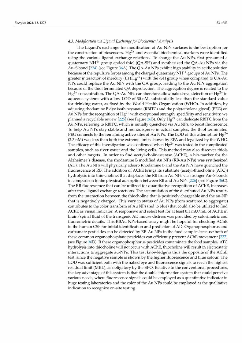
Energies 2021, 14, 1278 33 of 83
4.3. Modification via Ligand Exchange for Biochemical Analysis
The Ligand’s exchange for modification of Au NPs surfaces is the best option forthe construction of biosensors. Hg2+ and essential biochemical markers were identifiedusing the various ligand exchange reactions. To change the Au NPs, first presumed aquaternary NH4+ group ended thiol (QA-SH) and synthesized the QA-Au NPs via theAu–S bond [224] (see Figure 36A). The QA-Au NPs exhibit high stability in acidic solutionbecause of the repulsive forces among the charged quaternary NH4+ groups of Au NPs. Thegreater interaction of mercury (II) (Hg2+) with the -SH group when compared to QA-AuNPs could replace the Au NPs with the QA group, leading to the Au NPs aggregationbecause of the thiol terminated QA deprotection. The aggregation degree is related to theHg2+ concentration. The QA-Au NPs can therefore allow naked-eye detection of Hg2+ inaqueous systems with a low LOD of 30 nM, substantially less than the standard valuefor drinking water, as fixed by the World Health Organization (WHO). In addition, byadjusting rhodamine B dye isothiocyanate (RBITC) and the poly(ethylene glycol) (PEG) onAu NPs for the recognition of Hg2+ with exceptional strength, specificity and sensitivity, weplanned a recyclable review [225] (see Figure 36B). Only Hg2+ can dislocate RBITC from theAu NPs, referring to RBITC, which is initially quenched via Au NPs, to boost fluorescence.To help Au NPs stay stable and monodisperse in actual samples, the thiol terminatedPEG connects to the remaining active sites of Au NPs. The LOD of this attempt for Hg2+
(2.3 nM) was less than both the extreme limits shown by EPA and legalized by the WHO.The efficacy of this investigation was confirmed when Hg2+ was tested in the complicatedsamples, such as river water and the living cells. This method may also discover thiolsand other targets. In order to find acetyl-cholinesterase (AChE), a bio-marker for theAlzheimer’s disease, the rhodamine B modified Au NPs (RB-Au NPs) was synthesized(AD). The Au NPs will physically adsorb Rhodamine B and the Au NPs have quenched thefluorescence of RB. The addition of AChE brings its substrate (acetyl-thiocholine (ATC))hydrolysis into thio-choline, that displaces the RB from Au NPs via stronger Au–S bondsin comparison to the physical adsorption between RB and Au NPs [226] (see Figure 36C).The RB fluorescence that can be utilized for quantitative recognition of AChE, increasesafter these ligand-exchange reactions. The accumulation of the distributed Au NPs resultsfrom the interaction between the thiocholine that is positively chargeable and the au-NPthat is negatively charged. This vary in status of Au NPs (from scattered to aggregate)contributes to the color transform of Au NPs (red to blue) that could also be utilized to findAChE as visual indicator. A responsive and select test for at least 0.1 mU/mL of AChE inbrain/spinal fluid of the transgenic AD mouse distress was provided by colorimetric andfluorometric details. This RBAu NPs-based assay might be hopeful for checking AChEin the human CSF for initial identification and prediction of AD. Organophosphorus andcarbamate pesticides can be detected by RB-Au NPs in the food samples because both ofthese common organophosphate pesticides can efficiently prevent AChE movement [227](see Figure 36D). If these organophosphorus pesticides contaminate the food samples, ATChydrolysis into thiocholine will not occur with AChE, thiocholine will result in electrostaticinteractions to aggregate au-NPs. This test knowledge is thus the opposite of the AChEtest, since the negative sample is shown by the higher fluorescence and blue colour. TheLOD was sufficient both with the naked eye and fluorescence signals to reach the highestresidual limit (MRL), as obligatory by the EPO. Relative to the conventional procedures,the key advantage of this system is that the double information system that could perceivevarious needs, where fluorescence signals could be employed as a quantitative indicator inhuge testing laboratories and the color of the Au NPs could be employed as the qualitativeindication to recognize on-site testing.
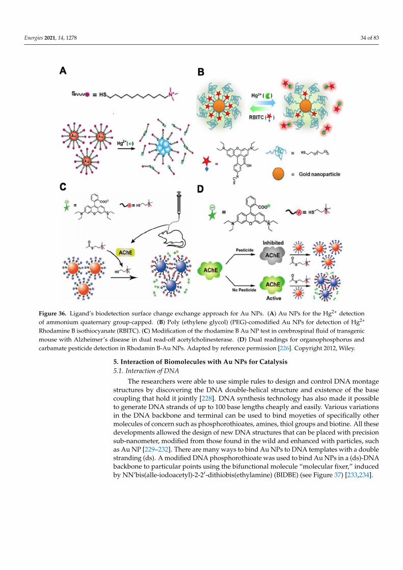
Energies 2021, 14, 1278 34 of 83
Figure 36. Ligand’s biodetection surface change exchange approach for Au NPs. (A) Au NPs for the Hg2+ detectionof ammonium quaternary group-capped. (B) Poly (ethylene glycol) (PEG)-comodified Au NPs for detection of Hg2+
Rhodamine B isothiocyanate (RBITC). (C) Modification of the rhodamine B Au NP test in cerebrospinal fluid of transgenicmouse with Alzheimer’s disease in dual read-off acetylcholinesterase. (D) Dual readings for organophosphorus andcarbamate pesticide detection in Rhodamin B-Au NPs. Adapted by reference permission [226]. Copyright 2012, Wiley.
5. Interaction of Biomolecules with Au NPs for Catalysis5.1. Interaction of DNA
The researchers were able to use simple rules to design and control DNA montagestructures by discovering the DNA double-helical structure and existence of the basecoupling that hold it jointly [228]. DNA synthesis technology has also made it possibleto generate DNA strands of up to 100 base lengths cheaply and easily. Various variationsin the DNA backbone and terminal can be used to bind moyeties of specifically othermolecules of concern such as phosphorothioates, amines, thiol groups and biotine. All thesedevelopments allowed the design of new DNA structures that can be placed with precisionsub-nanometer, modified from those found in the wild and enhanced with particles, suchas Au NP [229–232]. There are many ways to bind Au NPs to DNA templates with a doublestranding (ds). A modified DNA phosphorothioate was used to bind Au NPs in a (ds)-DNAbackbone to particular points using the bifunctional molecule “molecular fixer,” inducedby NN’bis(alle-iodoacetyl)-2-2′-dithiobis(ethylamine) (BIDBE) (see Figure 37) [233,234].
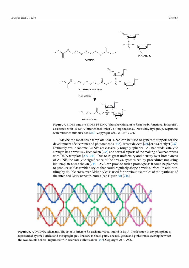
Energies 2021, 14, 1278 35 of 83
Figure 37. BIDBE binds to BIDBE-PS-DNA (phosphorothioate) to form the bi-functional linker (BF),associated with PS-DNA (bifunctional linker). BF supplies an au-NP sulfhydryl group. Reprintedwith reference authorisation [233], Copyright 2007, WILEY-VCH.
Maybe the most basic template (ds)- DNA can be used to generate support for thedevelopment of electronic and photonic rods [235], sensor devices [236] or as a catalyst [237].Definitely, while canonic Au NPs are classically roughly spherical, Au nanorods’ catalyticstrength has previously been taken [238] and several reports of the making of au nanowireswith DNA template [239–244]. Due to its great uniformity and density over broad areasof Au NP, the catalytic significance of the arrays, synthesized by procedures not usingbio-templates, was shown [245]. DNA can provide such a prototype as it could be plannedto produce self-assembled styles that could regularly shape a wide surface. In addition,tiling by double cross over DNA styles is used for previous examples of the synthesis ofthe intended DNA nanostructures (see Figure 38) [246].
Figure 38. A DX DNA schematic. The color is different for each individual strand of DNA. The location of any phosphate isrepresented by small circles and the upright grey lines are the base-pairs. The red, green and pink strands overlap betweenthe two double helices. Reprinted with reference authorisation [247], Copyright 2004, ACS.

Energies 2021, 14, 1278 36 of 83
The DX-motives consist of two double DNA strands linked to the nearby doublebeach by crossing single beams from one double beam. DX DNA provides the essentialhardness of DNA tiles if a wide surface area of reduced distortion is periodically dissolved.DX DNA was considered for a thin, rigid andtri-angular configuration with “sticky ends”and can frequently can cover large 2D surfaces, as shown in the example presented inFigure 39 [247].
Figure 39. (a) The Au NPs can be shaped in a standard array of Au-nPs in a dx DNA template as shown by DX DNAtriangles with sticky extremities, to allow the tiling of triangles; (b) Non-all ends of DNA are needed for tile purpose andthe free ends could be attached to Au NP’s (yellow) 5 or 10 nm diameter; (c) The AFM imagery on a dx-array without theadding Au demonstrate its ordinary structure. This picture only displays au-NPs. (a–d) with reference authorization, ACS,Copyright 2006 [231].
At the 5” end of one of the DNA strands, Thiol groups can be inserted into eachtriangle and provide for a decorated au-nP array with a 5 nm Au NP connection [231]Subsequently, the DNA strands modified were used in 2D DNA collections to make theAu NP “wire” lines. [248]. Four DNA strands [249] can be used to create DNA pyramid.Nanotechnology may also speak of this: the form of small, three-dimensional structuresconsisting of cubes [250], a bipyramid [251] and octahedrals from different strands ofDNA [252] have been created. The vertices of pyramidal DNA nanostructures have beenconnected to Au NP (5 nm in diameter) (see Figure 40) [250].
The discovery of DNA origami was also revolutionary [253], which enabled reasonablysimple fabrication of custom 2D and 3D structures, such as boxes [254,255]. The preparationof a DNA origami-rod and thiol-groups at specific positions on surface in order to achievean Au NP at the points on the spinal cord of a helix on rods surface is one of the mostinteresting consequences of the ability of DNA to organize Au NPs into structures ofdemanding physical and chemical characteristics. The chiral plasmonic structures weredescribed previously (see Figure 41) [256].
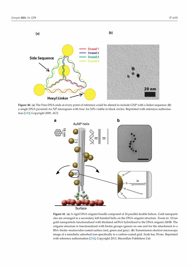
Energies 2021, 14, 1278 37 of 83
Figure 40. (a) The Free-DNA ends at every point of reference could be altered to include GNP with a linker sequence; (b)a single DNA pyramid Au NP microgram with four Au NPs visible in black circles: Reprinted with reference authorisa-tion [249], Copyright 2009, ACS.
Figure 41. (a) A rigid DNA origami bundle composed of 24 parallel double helices. Gold nanoparti-cles are arranged in a secondary left-handed helix on the DNA origami structure. Zoom in: 10 nmgold nanoparticle functionalized with thiolated ssDNA hybridized to the DNA origami 24HB. Theorigami structure is functionalized with biotin groups (green) on one end for the attachment to aBSA–biotin–neutravidin-coated surface (red, green and grey). (b) Transmission electron microscopyimage of a nanohelix adsorbed non-specifically to a carbon-coated grid. Scale bar, 50 nm. Reprintedwith reference authorisation [256], Copyright 2013, Macmillan Publishers Ltd.
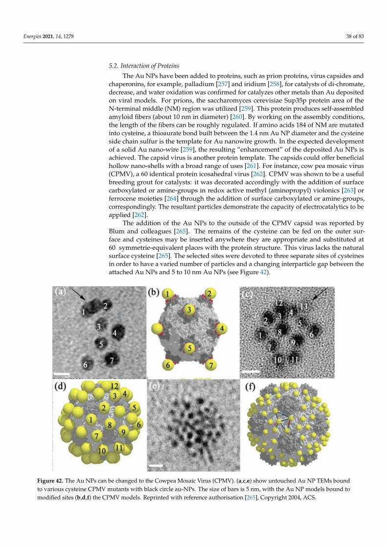
Energies 2021, 14, 1278 38 of 83
5.2. Interaction of Proteins
The Au NPs have been added to proteins, such as prion proteins, virus capsides andchaperonins, for example, palladium [257] and iridium [258], for catalysts of di-chromate,decrease, and water oxidation was confirmed for catalyzes other metals than Au depositedon viral models. For prions, the saccharomyces cerevisiae Sup35p protein area of theN-terminal middle (NM) region was utilized [259]. This protein produces self-assembledamyloid fibers (about 10 nm in diameter) [260]. By working on the assembly conditions,the length of the fibers can be roughly regulated. If amino acids 184 of NM are mutatedinto cysteine, a thioaurate bond built between the 1.4 nm Au NP diameter and the cysteineside chain sulfur is the template for Au nanowire growth. In the expected developmentof a solid Au nano-wire [259], the resulting “enhancement” of the deposited Au NPs isachieved. The capsid virus is another protein template. The capsids could offer beneficialhollow nano-shells with a broad range of uses [261]. For instance, cow pea mosaic virus(CPMV), a 60 identical protein icosahedral virus [262]. CPMV was shown to be a usefulbreeding grout for catalysts: it was decorated accordingly with the addition of surfacecarboxylated or amine-groups in redox active methyl (aminopropyl) violonics [263] orferrocene moieties [264] through the addition of surface carboxylated or amine-groups,correspondingly. The resultant particles demonstrate the capacity of electrocatalytics to beapplied [262].
The addition of the Au NPs to the outside of the CPMV capsid was reported byBlum and colleagues [265]. The remains of the cysteine can be fed on the outer sur-face and cysteines may be inserted anywhere they are appropriate and substituted at60 symmetrie-equivalent places with the protein structure. This virus lacks the naturalsurface cysteine [265]. The selected sites were devoted to three separate sites of cysteinesin order to have a varied number of particles and a changing interparticle gap between theattached Au NPs and 5 to 10 nm Au NPs (see Figure 42).
Figure 42. The Au NPs can be changed to the Cowpea Mosaic Virus (CPMV). (a,c,e) show untouched Au NP TEMs boundto various cysteine CPMV mutants with black circle au-NPs. The size of bars is 5 nm, with the Au NP models bound tomodified sites (b,d,f) the CPMV models. Reprinted with reference authorisation [265], Copyright 2004, ACS.
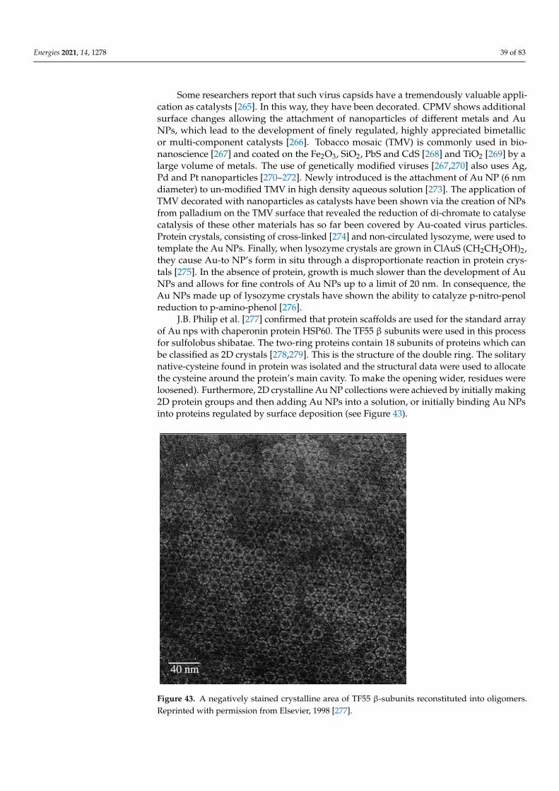
Energies 2021, 14, 1278 39 of 83
Some researchers report that such virus capsids have a tremendously valuable appli-cation as catalysts [265]. In this way, they have been decorated. CPMV shows additionalsurface changes allowing the attachment of nanoparticles of different metals and AuNPs, which lead to the development of finely regulated, highly appreciated bimetallicor multi-component catalysts [266]. Tobacco mosaic (TMV) is commonly used in bio-nanoscience [267] and coated on the Fe2O3, SiO2, PbS and CdS [268] and TiO2 [269] by alarge volume of metals. The use of genetically modified viruses [267,270] also uses Ag,Pd and Pt nanoparticles [270–272]. Newly introduced is the attachment of Au NP (6 nmdiameter) to un-modified TMV in high density aqueous solution [273]. The application ofTMV decorated with nanoparticles as catalysts have been shown via the creation of NPsfrom palladium on the TMV surface that revealed the reduction of di-chromate to catalysecatalysis of these other materials has so far been covered by Au-coated virus particles.Protein crystals, consisting of cross-linked [274] and non-circulated lysozyme, were used totemplate the Au NPs. Finally, when lysozyme crystals are grown in ClAuS (CH2CH2OH)2,they cause Au-to NP’s form in situ through a disproportionate reaction in protein crys-tals [275]. In the absence of protein, growth is much slower than the development of AuNPs and allows for fine controls of Au NPs up to a limit of 20 nm. In consequence, theAu NPs made up of lysozyme crystals have shown the ability to catalyze p-nitro-penolreduction to p-amino-phenol [276].
J.B. Philip et al. [277] confirmed that protein scaffolds are used for the standard arrayof Au nps with chaperonin protein HSP60. The TF55 β subunits were used in this processfor sulfolobus shibatae. The two-ring proteins contain 18 subunits of proteins which canbe classified as 2D crystals [278,279]. This is the structure of the double ring. The solitarynative-cysteine found in protein was isolated and the structural data were used to allocatethe cysteine around the protein’s main cavity. To make the opening wider, residues wereloosened). Furthermore, 2D crystalline Au NP collections were achieved by initially making2D protein groups and then adding Au NPs into a solution, or initially binding Au NPsinto proteins regulated by surface deposition (see Figure 43).
Figure 43. A negatively stained crystalline area of TF55 β-subunits reconstituted into oligomers.Reprinted with permission from Elsevier, 1998 [277].
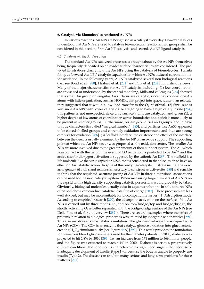
Energies 2021, 14, 1278 40 of 83
6. Catalysis via Biomolecules Anchored Au NPs
In various reactions, Au NPs are being used as a catalyst every day. However, it is lessunderstood that Au NPs are used to catalyze bio-molecular reactions. Two groups shall beconsidered in this section: first, Au NP catalysis, and second, Au NP ligand catalysis.
6.1. Catalysis via the Au NPs Itself
The standard Au NPs catalyzed processes is brought about by the Au NPs themselvesbeing frequently deposited on an oxide; surface characteristics are considered. The pro-vided illustrations clarify how the Au NPs bring the catalysis of biomolecules. Harutafirst put forward Au NPs’ catalytic capacities, in which Au NPs induced carbon monox-ide oxidation. In the following years, Au NPs catalyzed several non-biological reactions(i.e., see Bond et al. [280], Hashmi et al. [281] and Pina et al. [282], for critical reviews).Many of the major characteristics for Au NP catalysis, including: (1) low coordination,are envisaged or understood; by theoretical modeling, Mills and colleagues [283] showedthat a small Au group or irregular Au surfaces are catalytic, since they confine lone Auatoms with little organization, such as HOMOs, that project into space, rather than relocate;they suggested that it would allow load transfer to the O2 π* orbital. (2) Size: size iskey, since Au NPs with lower catalytic size are going to have a high catalytic rate [284];this pattern is not unexpected, since only surface atoms are catalyzed, and given (1), ahigher degree of low atoms of coordination across boundaries and deficit is more likely tobe present in smaller groups. Furthermore, certain geometries and groups tend to haveunique characteristics called “magical number” [285], and particles like Au55 appearedto be closed shelled groups and extremely oxidation impermeable and thus are strongcatalysts for oxidation [286]. (3) Scaffold interface: the existence and effect of the interfacebetween the deux is usually examined by the Au NP on an oxide support. The supportingpoint at which the Au NPs occur was proposed as the oxidation centre. The smaller AuNPs are more involved due to the greater amount of their support system. The Au whichis in contact with the help in the event of CO oxidation is predicted to be Au3+ and theactive site for dioxygen activation is suggested by the cationic Au [287]. The scaffold is alife molecule like the virus capsid or DNA that is considered in that discussion to have aneffect on Au catalytic action. In spite of this, enzyme-catalysts illustrate us that the exactarrangement of atoms and remains is necessary to construct an active-site [288] and sensibleto think that the regulated, accurate posing of Au NPs in three dimensional associationscan be used for the next catalytic system. When measuring large numbers of Au NPs onthe capsid with a high density, supporting catalytic possessions would probably be taken.Obviously, biological molecules usually exist in aqueous solution. In solution, Au NPsoften somehow can conduct catalytic tests free of charge [289]. These processes are lesswell studied, but may be more suitable for biocompatibility issues. (4) Adsorption mode:According to empirical research [290], the adsorption activation on the surface of the AuNPs is carried out by three modes, i.e., end-on, top/bridge/top and bridge/bridge, thestrictly activating O2 is better separated with the bridge-bridge surface of the Au NPs (seeDella Pina et al. for an overview [282]). There are several examples where the effect ofproteins in relation to biological properties was imitated by inorganic nanoparticles [291].This also involves enzyme catalysis imitation. The glucose oxidase act was copied withAu NPs (GOx). The GOx is an enzyme that catalyze glucose oxidation into glucolactone,creating H2O2 simultaneously (see Figure 44A) [292]. This result provides the foundationfor numerous blood glucose-meters used by the diabetes patients. In 2000, diabetes wasprojected to hit 2.8% by 2030 [293], i.e., an increase from 171 million to 366 million people,and the figure was expected to reach 4.4% in 2000. Diabetes is serious, progressivelydifficult condition. The condition is characterized as high blood sugar either because ofinadequate development of insulin (type 1) or because the body is unable to properly useinsulin (Type 2). The disease can result in many serious and long-term problems for thoseit affects [291].
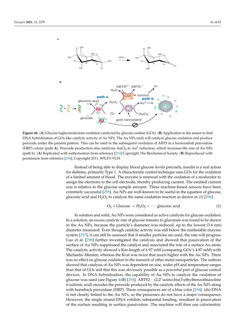
Energies 2021, 14, 1278 41 of 83
Figure 44. (A) Glucose togluconolactone oxidation catalyzed by glucose oxidase (GOx). (B) Application to the sensor to findDNA hybridization of GOx-like catalytic activity of Au NPs. The Au NPs (red) will catalyze glucose oxidation and produceperoxide under the present pattern. This can be used in the subsequent oxidation of ABTS in a horseradish peroxidose(HRP) colour (path A). Peroxide production also catalyzes AuCl4 to Au0 reduction, which increases the size of Au NPs(path b). (A) Replicated with authorisation from reference [293] Copyright The Biochemical Society. (B) Reproduced withpermission from reference [294], Copyright 2011, WILEY-VCH.
Instead of being able to display blood glucose levels precisely, insulin is a real actionfor diabetes, primarily Type 1. A characteristic control technique uses GOx for the oxidationof a limited amount of blood. The enzyme is renewed with the oxidation of a moderator toassign the electrons to the cell electrode, thereby producing current. The emitted currentsize is relative to the glucose sample amount. These machine-based sensors have beenextremely successful [295]. Au NPs are well-known to be useful in the equation of glucose,gluconic acid and H2O2 to catalyze the same oxidation reaction as shown in (1) [296].
O2 + Glucose→ H2O2 + · · · gluconic acid (1)
In solution and solid, Au NPs were considered as active catalysts for glucose oxidation.In a solution, an excess catalytic rate of glucose transfer to gluconate was found to be shownin the Au NPs, because the particle’s diameter was reduced, up to the lowest (3.6 nm)diameter measured. Even though catalytic activity was still below the marketable enzymesystem [297], it can still be assessed that if smaller particles are used, the rate will progress.Luo et al. [298] further investigated the catalysis and showed that passivation of thesurface of Au NPs suppressed the catalyst and associated the role of a surface Au atom.The catalytic activity showed a Km-length of 6.97 mM (comparing GOx’s 4.87 mM) withMichaelis–Menten, whereas the Kcat was twice that much higher with the Au NPs. Therewas no effect on glucose oxidation in the research of other metal nanoparticles. The authorsshowed that catalysis of Au NPs was dependent on size, wider pH and temperature rangesthan that of GOx and that this was obviously possible as a powerful part of glucose controldevices. In DNA hybridization, the capability of Au NPs to catalyze the oxidation ofglucose was used (see Figure 44B) [294]. ABTS2− (2,2′-azino-bis(3-ethylbenzothiazoline6-sulfonic acid) otoxides the peroxide produced by the catalytic effects of the Au NPs alongwith horseback peroxidase (HRP). These consequences are of a blue color [294]. (ds)-DNAis not closely linked to the Au NPs, so the processes do not have a major consequence.However, the single strand DNA exhibits substantial binding, resultant in passivationof the surface resulting in surface passivation. The machine will then use colorimétric
Energies 2021, 14, 1278 42 of 83
exams or direct remarks on the size of Au-and/or NP’s the plasmonic effects for free DNAor RNA strands (i.e., disease-connected miRNAs) that arose because of the fact that theperoxide prooduced could be employed to lessen AuCl4 to Au0, Au NPs producing enhanceparticle size, a procedure repressed by attached ssDNA. The last case is the catalyticAu NPs of biological molecule structural variations. Au NPs tend to catalyze proteinreshaping in this situation. The fact that Au NPs can mix with proteins and bring structuralvariations is well known. Such discrepancies are usually the result of structural andfunctional loss due to protein denaturation or development of the corona protein acquiredaround the Au NPs [299–301]. The development of higher-order structures induced byAu NPs is reduced. In Bacillus species, TRAP is a toroidal protein [302]; the protein-TRAP (trp-RNA binding shrinking protein) was employed. In Bacillus species, TRAP is atoroidal protein. In vivo, TRAP participates in the reaction regulation of the preparation oftryptophan. The crystal structures of both the B TRAPs, stearothermophilus [303], and theB. subtilis [304] were recognized and showed that TRAP has a diameter of about 8 nmsand consist of eleven equal monomers each with approximately 8.4 kDa. The protein hasother features beneficial to bionanosciences because of the exciting and well-defined form.These include high thermostability [305] and the ability to withstand multiple mutations ofthe surface without affecting overall structure. Up to now, TRAP has been used as part ofthe floating nanodot gate transistor to generate symmetry-changed structures and to createan automatically assembled nanotube protein [306] as a constituent of a floating nano-dotgate-transistor [307]. The mutation of the lysins at position 35 (37 in B. stearothermophoilusTRAP) to cystein results in the interaction of TRAPs with Au NPs (see Figure 45). It hasan ability to tie strongly to Au, because of the cysteine sulfhydryl group. However, thewild protein does not contain cysteines. The TRAP’s residue lysine 35 is accessible to thesurface and lies in the outer rim of the ring unprotected and a motivating conformativetransformation is perceived when mutated into a cysteine and blended to 1.4 nm Au NPs.
Owing to the unavailability of Au NP, the transmission-electron-microscopy (TEM)perceived TRAP protein appear to be negligible from the non-modified protein, i.e., itlooked about 8 nm in diameter as a small donut-shaped ring (see Figure 45). No importantconsequence was observed in 1.4 nm Au NPs applied to the wild type protein. However,the incorporation in the mutant protein of the same Au NPs affected a drastic shift. Themutant-protein sample had no proof of the unique ring-shaped protein after several hoursof production at 4 ◦C and was replaced with large circular proteins of almost 20 nmin diameter. Study with cryo-electron tomography of these particles revealed that theproduced structures were essentially spherical hollow proteins. The spheres looked liketwo distinct dimensions (diameter about 17 nm and 21 nm), both depending on the Auconcentration (with the superior proteins prevailing at low Au NPs concentration). Thesimilarity between these hollow-formed particles and the governing viral capsids is knownas “sphere like capsid” [308]. In addition, although there is sometimes a small clusterof one or more Au NPs inside a protein shell in one location, in other situations, no Auparticles can be presumed to be linked with the shells (though it should be well-knownthat its difficult to observe 1.4 nm Au NPs under the TEM). All of a sudden, this impliedAu could behave catalytically. It is definitely important to see how the Au can functionthrough simple templating or gagging effects because it is the divergence between thesize of the Au NPs and the following protein shell. The theory of catalysis is maintainedby the fact that the same protein remodeling is possible even at low concentrations ofAu NPs. Although the catalytic hypothesis is intriguing, now it is unconfirmed that itis difficult to fully align the small size of Au NPs in the shaped CLSs. Moreover, eventhough significant numbers of Au NPs tend to lack the CLSs, Au atoms have not finallybeen lined up. There were intriguing clues from the reaction path [308]. If the reaction isdiscontinued and seen under TEM, a minute after the Au NPs initiation, numerous stableTRAP rings were extinct before but few CLS are produced and several thread structuresappear to be unfolding or disassembling protein in place a large number of thread-likestructures. The Au NPs can constitute an infringement of the stable TRAP ring separately
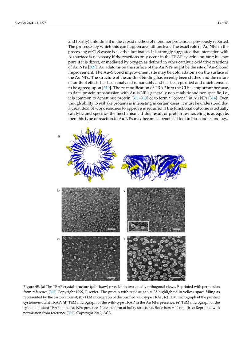
Energies 2021, 14, 1278 43 of 83
and (partly) unfoldment in the capsid method of monomer proteins, as previously reported.The processes by which this can happen are still unclear. The exact role of Au NPs in theprocessing of CLS waste is clearly illuminated. It is strongly suggested that interaction withAu surface is necessary if the reactions only occur in the TRAP cysteine mutant; it is notpure if it is direct, or mediated by oxygen as defined in other catalytic oxidative reactionsof Au NPs [309]. Au adatoms on the surface of the Au NPs might be the site of Au–S bondimprovement. The Au–S bond improvement site may be gold adatoms on the surface ofthe Au NPs. The structure of the au-thiol binding has recently been studied and the natureof au-thiol effects has been analyzed remarkably and has been purified and much remainsto be agreed upon [310]. The re-modification of TRAP into the CLS is important because,to date, protein transmission with Au-is NP’s generally non catalytic and non specific, i.e.,it is common to denaturate protein [311–313] or to form a “corona” in Au NPs [314]. Eventhough ability to reshake proteins is interesting in certain cases, it must be understood thata great deal of work residues to approve is required if the functional outcome is actuallycatalytic and specifics the mechanism. If this result of protein re-modeling is adequate,then this type of reaction to Au NPs may become a beneficial tool in bio-nanotechnology.
Figure 45. (a) The TRAP crystal structure (pdb 1qaw) revealed in two equally orthogonal views. Reprinted with permissionfrom reference [303] Copyright 1999, Elsevier. The protein with residue at site 35 highlighted in yellow space filling asrepresented by the cartoon format; (b) TEM micrograph of the purified wild-type TRAP; (c) TEM micrograph of the purifiedcysteine-mutant TRAP; (d) TEM micrograph of the wild-type TRAP in the Au NPs presence; (e) TEM micrograph of thecysteine-mutant TRAP in the Au NPs presence. Note the form of bulky structures. Scale bars = 40 nm. (b–e) Reprinted withpermission from reference [307], Copyright 2012, ACS.
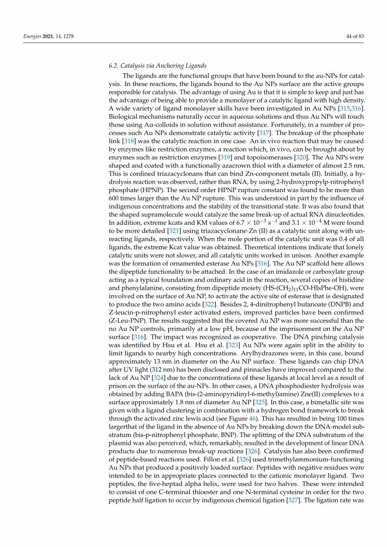
Energies 2021, 14, 1278 44 of 83
6.2. Catalysis via Anchoring Ligands
The ligands are the functional groups that have been bound to the au-NPs for catal-ysis. In these reactions, the ligands bound to the Au NPs surface are the active groupsresponsible for catalysis. The advantage of using Au is that it is simple to keep and just hasthe advantage of being able to provide a monolayer of a catalytic ligand with high density.A wide variety of ligand monolayer skills have been investigated in Au NPs [315,316].Biological mechanisms naturally occur in aqueous solutions and thus Au NPs will touchthose using Au-colloids in solution without assistance. Fortunately, in a number of pro-cesses such Au NPs demonstrate catalytic activity [317]. The breakup of the phosphatelink [318] was the catalytic reaction in one case. An in vivo reaction that may be causedby enzymes like restriction enzymes, a reaction which, in vivo, can be brought about byenzymes such as restriction enzymes [319] and topoisomerases [320]. The Au NPs wereshaped and coated with a functionally azacrown thiol with a diameter of almost 2.5 nm.This is confined triazacyclonans that can bind Zn-component metals (II). Initially, a hy-drolysis reaction was observed, rather than RNA, by using 2-hydroxypropylp-nitrophenylphosphate (HPNP). The second order HPNP rupture constant was found to be more than600 times larger than the Au NP rupture. This was understood in part by the influence ofindigenous concentrations and the stability of the transitional state. It was also found thatthe shaped supramolecule would catalyze the same break-up of actual RNA dinucleotides.In addition, extreme kcats and KM values of 6.7 × 10⁻³ s−1 and 3.1 × 10⁻⁴ M were foundto be more detailed [321] using triazacyclonane·Zn (II) as a catalytic unit along with un-reacting ligands, respectively. When the mole portion of the catalytic unit was 0.4 of allligands, the extreme Kcat value was obtained. Theoretical intentions indicate that lonelycatalytic units were not slower, and all catalytic units worked in unison. Another examplewas the formation of ornamented esterase Au NPs [316]. The Au NP scaffold here allowsthe dipeptide functionality to be attached. In the case of an imidazole or carboxylate groupacting as a typical foundation and ordinary acid in the reaction, several copies of histidineand phenylalanine, consisting from dipeptide moiety (HS-(CH2)11CO-HIsPhe-OH), wereinvolved on the surface of Au NP, to activate the active site of esterase that is designatedto produce the two amino acids [322]. Besides 2, 4-dinitrophenyl butanoate (DNPB) andZ-leucin-p-nitrophenyl ester activated esters, improved particles have been confirmed(Z-Leu-PNP). The results suggested that the covered Au NP was more successful than theno Au NP controls, primarily at a low pH, because of the imprisonment on the Au NPsurface [316]. The impact was recognized as cooperative. The DNA pinching catalysiswas identified by Hsu et al. Hsu et al. [323] Au NPs were again split in the ability tolimit ligands to nearby high concentrations. Arylhydrazones were, in this case, boundapproximately 13 nm in diameter on the Au NP surface. These ligands can chip DNAafter UV light (312 nm) has been disclosed and pinnacles have improved compared to thelack of Au NP [324] due to the concentrations of these ligands at local level as a result ofprison on the surface of the au-NPs. In other cases, a DNA phosphodiester hydrolysis wasobtained by adding BAPA (bis-(2-aminopyridinyl-6-methyl)amine) Zne(II) complexes to asurface approximately 1.8 nm of diameter Au NP [325]. In this case, a bimetallic site wasgiven with a ligand clustering in combination with a hydrogen bond framework to breakthrough the activated zinc lewis acid (see Figure 46). This has resulted in being 100 timeslargerthat of the ligand in the absence of Au NPs by breaking down the DNA-model sub-stratum (bis-p-nitrophenyl phosphate, BNP). The splitting of the DNA substratum of theplasmid was also perceived, which, remarkably, resulted in the development of linear DNAproducts due to numerous break-up reactions [326]. Catalysis has also been confirmedof peptide-based reactions used. Fillon et al. [326] used trimethylammonium-functioningAu NPs that produced a positively loaded surface. Peptides with negative residues wereintended to be in appropriate places connected to the cationic monolayer ligand. Twopeptides, the five-heptad alpha helix, were used for two halves. These were intendedto consist of one C-terminal thioester and one N-terminal cysteine in order for the twopeptide half ligation to occur by indigenous chemical ligation [327]. The ligation rate was
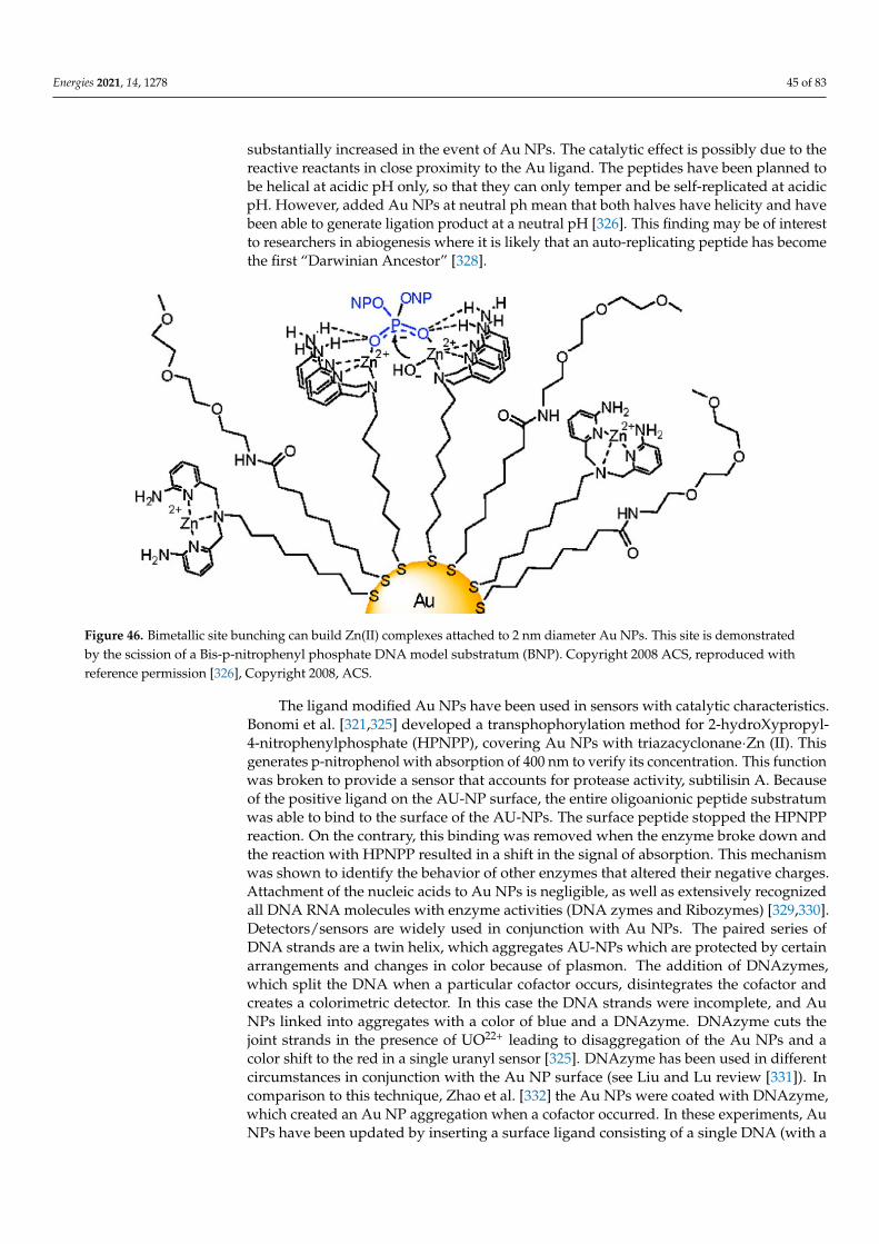
Energies 2021, 14, 1278 45 of 83
substantially increased in the event of Au NPs. The catalytic effect is possibly due to thereactive reactants in close proximity to the Au ligand. The peptides have been planned tobe helical at acidic pH only, so that they can only temper and be self-replicated at acidicpH. However, added Au NPs at neutral ph mean that both halves have helicity and havebeen able to generate ligation product at a neutral pH [326]. This finding may be of interestto researchers in abiogenesis where it is likely that an auto-replicating peptide has becomethe first “Darwinian Ancestor” [328].
Figure 46. Bimetallic site bunching can build Zn(II) complexes attached to 2 nm diameter Au NPs. This site is demonstratedby the scission of a Bis-p-nitrophenyl phosphate DNA model substratum (BNP). Copyright 2008 ACS, reproduced withreference permission [326], Copyright 2008, ACS.
The ligand modified Au NPs have been used in sensors with catalytic characteristics.Bonomi et al. [321,325] developed a transphophorylation method for 2-hydroXypropyl-4-nitrophenylphosphate (HPNPP), covering Au NPs with triazacyclonane·Zn (II). Thisgenerates p-nitrophenol with absorption of 400 nm to verify its concentration. This functionwas broken to provide a sensor that accounts for protease activity, subtilisin A. Becauseof the positive ligand on the AU-NP surface, the entire oligoanionic peptide substratumwas able to bind to the surface of the AU-NPs. The surface peptide stopped the HPNPPreaction. On the contrary, this binding was removed when the enzyme broke down andthe reaction with HPNPP resulted in a shift in the signal of absorption. This mechanismwas shown to identify the behavior of other enzymes that altered their negative charges.Attachment of the nucleic acids to Au NPs is negligible, as well as extensively recognizedall DNA RNA molecules with enzyme activities (DNA zymes and Ribozymes) [329,330].Detectors/sensors are widely used in conjunction with Au NPs. The paired series ofDNA strands are a twin helix, which aggregates AU-NPs which are protected by certainarrangements and changes in color because of plasmon. The addition of DNAzymes,which split the DNA when a particular cofactor occurs, disintegrates the cofactor andcreates a colorimetric detector. In this case the DNA strands were incomplete, and AuNPs linked into aggregates with a color of blue and a DNAzyme. DNAzyme cuts thejoint strands in the presence of UO22+ leading to disaggregation of the Au NPs and acolor shift to the red in a single uranyl sensor [325]. DNAzyme has been used in differentcircumstances in conjunction with the Au NP surface (see Liu and Lu review [331]). Incomparison to this technique, Zhao et al. [332] the Au NPs were coated with DNAzyme,which created an Au NP aggregation when a cofactor occurred. In these experiments, AuNPs have been updated by inserting a surface ligand consisting of a single DNA (with a
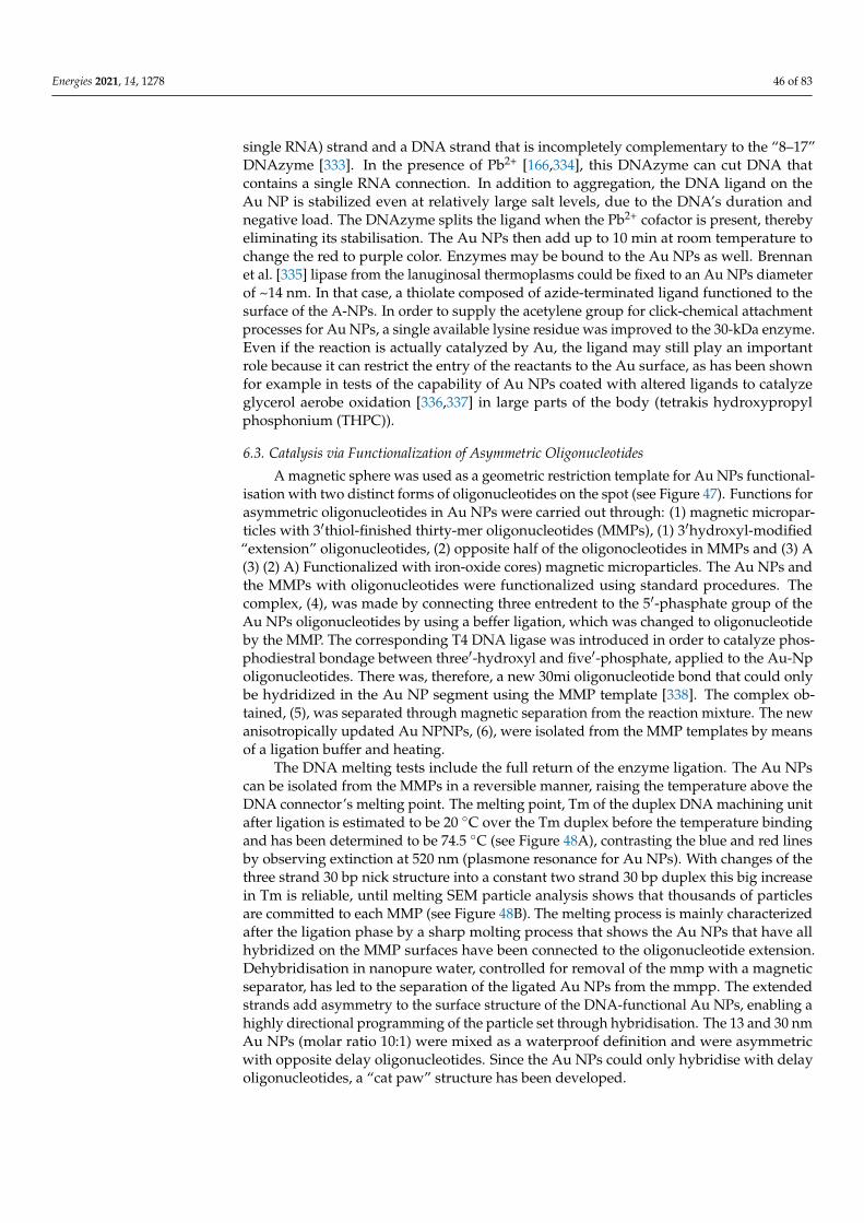
Energies 2021, 14, 1278 46 of 83
single RNA) strand and a DNA strand that is incompletely complementary to the “8–17”DNAzyme [333]. In the presence of Pb2+ [166,334], this DNAzyme can cut DNA thatcontains a single RNA connection. In addition to aggregation, the DNA ligand on theAu NP is stabilized even at relatively large salt levels, due to the DNA’s duration andnegative load. The DNAzyme splits the ligand when the Pb2+ cofactor is present, therebyeliminating its stabilisation. The Au NPs then add up to 10 min at room temperature tochange the red to purple color. Enzymes may be bound to the Au NPs as well. Brennanet al. [335] lipase from the lanuginosal thermoplasms could be fixed to an Au NPs diameterof ~14 nm. In that case, a thiolate composed of azide-terminated ligand functioned to thesurface of the A-NPs. In order to supply the acetylene group for click-chemical attachmentprocesses for Au NPs, a single available lysine residue was improved to the 30-kDa enzyme.Even if the reaction is actually catalyzed by Au, the ligand may still play an importantrole because it can restrict the entry of the reactants to the Au surface, as has been shownfor example in tests of the capability of Au NPs coated with altered ligands to catalyzeglycerol aerobe oxidation [336,337] in large parts of the body (tetrakis hydroxypropylphosphonium (THPC)).
6.3. Catalysis via Functionalization of Asymmetric Oligonucleotides
A magnetic sphere was used as a geometric restriction template for Au NPs functional-isation with two distinct forms of oligonucleotides on the spot (see Figure 47). Functions forasymmetric oligonucleotides in Au NPs were carried out through: (1) magnetic micropar-ticles with 3′thiol-finished thirty-mer oligonucleotides (MMPs), (1) 3′hydroxyl-modified“extension” oligonucleotides, (2) opposite half of the oligonocleotides in MMPs and (3) A(3) (2) A) Functionalized with iron-oxide cores) magnetic microparticles. The Au NPs andthe MMPs with oligonucleotides were functionalized using standard procedures. Thecomplex, (4), was made by connecting three entredent to the 5′-phasphate group of theAu NPs oligonucleotides by using a beffer ligation, which was changed to oligonucleotideby the MMP. The corresponding T4 DNA ligase was introduced in order to catalyze phos-phodiestral bondage between three′-hydroxyl and five′-phosphate, applied to the Au-Npoligonucleotides. There was, therefore, a new 30mi oligonucleotide bond that could onlybe hydridized in the Au NP segment using the MMP template [338]. The complex ob-tained, (5), was separated through magnetic separation from the reaction mixture. The newanisotropically updated Au NPNPs, (6), were isolated from the MMP templates by meansof a ligation buffer and heating.
The DNA melting tests include the full return of the enzyme ligation. The Au NPscan be isolated from the MMPs in a reversible manner, raising the temperature above theDNA connector’s melting point. The melting point, Tm of the duplex DNA machining unitafter ligation is estimated to be 20 ◦C over the Tm duplex before the temperature bindingand has been determined to be 74.5 ◦C (see Figure 48A), contrasting the blue and red linesby observing extinction at 520 nm (plasmone resonance for Au NPs). With changes of thethree strand 30 bp nick structure into a constant two strand 30 bp duplex this big increasein Tm is reliable, until melting SEM particle analysis shows that thousands of particlesare committed to each MMP (see Figure 48B). The melting process is mainly characterizedafter the ligation phase by a sharp molting process that shows the Au NPs that have allhybridized on the MMP surfaces have been connected to the oligonucleotide extension.Dehybridisation in nanopure water, controlled for removal of the mmp with a magneticseparator, has led to the separation of the ligated Au NPs from the mmpp. The extendedstrands add asymmetry to the surface structure of the DNA-functional Au NPs, enabling ahighly directional programming of the particle set through hybridisation. The 13 and 30 nmAu NPs (molar ratio 10:1) were mixed as a waterproof definition and were asymmetricwith opposite delay oligonucleotides. Since the Au NPs could only hybridise with delayoligonucleotides, a “cat paw” structure has been developed.
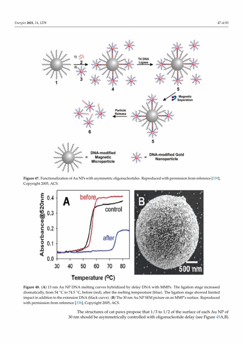
Energies 2021, 14, 1278 47 of 83
Figure 47. Functionalization of Au NPs with asymmetric oligonucleotides. Reproduced with permission from reference [339],Copyright 2005, ACS.
Figure 48. (A) 13 nm Au NP DNA melting curves hybridized by delay DNA with MMPs. The ligation stage increaseddramatically, from 54 ◦C to 74.5 ◦C, before (red), after the melting temperature (blue). The ligation stage showed limitedimpact in addition to the extension DNA (black curve). (B) The 30 nm Au NP SEM picture on an MMP’s surface. Reproducedwith permission from reference [338], Copyright 2005, ACS.
The structures of cat paws propose that 1/3 to 1/2 of the surface of each Au NP of30 nm should be asymmetrically controlled with oligonucleotide delay (see Figure 49A,B).
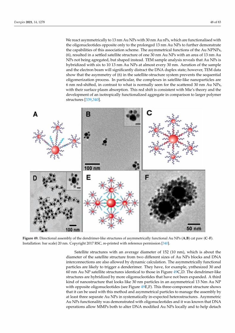
Energies 2021, 14, 1278 48 of 83
We react asymmetrically to 13 nm Au NPs with 30 nm Au nPs, which are functionalised withthe oligonucleotides opposite only to the prolonged 13 nm Au NPs to further demonstratethe capabilities of this association scheme. The asymmetrical functions of the Au NPNPs,(6), resulted in a settled satellite structure of one 30 nm Au NPs with an area of 13 nm AuNPs not being agregated, but shaped instead. TEM sample analysis reveals that Au NPs ishybridized with six to 10 13 nm Au NPs at almost every 30 nm. Aeration of the sampleand the electron beam will significantly distract the DNA duplex state; however, TEM datashow that the asymmetry of (6) in the satellite-structure system prevents the sequentialoligomerization process. In particular, the complexes in satellite-like nanoparticles are6 nm red-shifted, in contrast to what is normally seen for the scattered 30 nm Au NPs,with their surface plasm absorption. This red shift is consistent with Mie’s theory and thedevelopment of an isotropically functionalized aggregate in comparison to larger polymerstructures [339,340].
Figure 49. Directional assembly of the dendrimer-like structures of asymmetrically functional Au NPs (A,B) cat paw (C–F).Installation: bar scale) 20 nm. Copyright 2017 RSC, re-printed with reference permission [340].
Satellite structures with an average diameter of 152 (10 nm), which is about thediameter of the satellite structure from two different sizes of Au NPs blocks and DNAinterconnections are also allowed by dynamic calculation. The asymmetrically functionalparticles are likely to trigger a denderimer. They have, for example, ynthesized 30 and60 nm Au NP satellite structures identical to those in Figure 49C,D. The dendrimer-likestructures are hybridized by more oligonucleotides that have not been expanded. A thirdkind of nanostructure that looks like 30 nm particles in an asymmetrical 13 Nm Au NPwith opposite oligonucleotides (see Figure 49E,F). This three-component structure showsthat it can be used with this method and asymmetrical particles to manage the assembly byat least three separate Au NPs in systematically in-expected heterostructures. AsymmetricAu NPs functionality was demonstrated with oligonucleotides and it was known that DNAoperations allow MMPs both to alter DNA modified Au NPs locally and to help detach
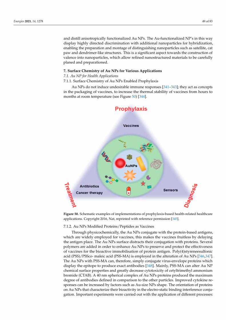
Energies 2021, 14, 1278 49 of 83
and distill anisotropically functionalized Au NPs. The Au-functionalized NP’s in this waydisplay highly directed discrimination with additional nanoparticles for hybridization,enabling the preparation and montage of distinguishing nanoparticles such as satellite, catpaw and dendrimer-like structures. This is a significant aspect towards the construction ofvalence into nanoparticles, which allow refined nanostructured materials to be carefullyplaned and preparationed.
7. Surface Chemistry of Au NPs for Various Applications7.1. Au NP for Health Applications7.1.1. Surface Chemistry of Au NPs Enabled Prophylaxis
Au NPs do not induce undesirable immune responses [341–343]; they act as conceptsin the packaging of vaccines, to increase the thermal stability of vaccines from hours tomonths at room temperature (see Figure 50) [344].
Figure 50. Schematic examples of implementations of prophylaxis-based health-related healthcareapplications. Copyright 2016, Nat, reprinted with reference permission [345].
7.1.2. Au NPs Modified Proteins/Peptides as Vaccines
Through physicochemically, the Au NPs conjugate with the protein-based antigens,which are widely employed for vaccines, this makes the vaccines fruitless by delayingthe antigen place. The Au NPs surface distracts their conjugation with proteins. Severalpolymers are added in order to enhance Au NPs to preserve and protect the effectivenessof vaccines for the bioactive immobilisation of protein antigen. Poly(4)styreneesulfonicacid (PSS)/PSSco- maleic acid (PSS-MA) is employed in the alteration of Au NPs [346,347].The Au NPs with PSS-MA can, therefore, simply conjugate virus-envelope proteins whichdisplay the epitope to produce exact antibodies [348]. Mainly, PSS-MA can alter Au NPchemical surface properties and greatly decrease cytotoxicity of cetyltrimethyl ammoniumbromide (CTAB). A 40 nm spherical complex of Au NPs proteins produced the maximumdegree of antibodies defined in comparison to the other particles. Improved cytokine re-sponses can be increased by factors such as Au-size NPs shape. The orientation of proteinson Au NPs that characterize their bioactivity in the electro-static binding interference conju-gation. Important experiments were carried out with the application of different processes:
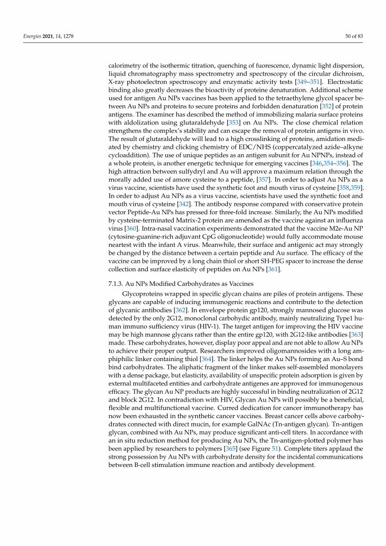
Energies 2021, 14, 1278 50 of 83
calorimetry of the isothermic titration, quenching of fuorescence, dynamic light dispersion,liquid chromatography mass spectrometry and spectroscopy of the circular dichroism,X-ray photoelectron spectroscopy and enzymatic activity tests [349–351]. Electrostaticbinding also greatly decreases the bioactivity of proteine denaturation. Additional schemeused for antigen Au NPs vaccines has been applied to the tetraethylene glycol spacer be-tween Au NPs and proteins to secure proteins and forbidden denaturation [352] of proteinantigens. The examiner has described the method of immobilizing malaria surface proteinswith aldolization using glutaraldehyde [353] on Au NPs. The close chemical relationstrengthens the complex’s stability and can escape the removal of protein antigens in vivo.The result of glutaraldehyde will lead to a high crosslinking of proteins, amidation medi-ated by chemistry and clicking chemistry of EDC/NHS (coppercatalyzed azide–alkynecycloaddition). The use of unique peptides as an antigen subunit for Au NPNPs, instead ofa whole protein, is another energetic technique for emerging vaccines [346,354–356]. Thehigh attraction between sulfydryl and Au will approve a maximum relation through themorally added use of amore cysteine to a peptide, [357]. In order to adjust Au NPs as avirus vaccine, scientists have used the synthetic foot and mouth virus of cysteine [358,359].In order to adjust Au NPs as a virus vaccine, scientists have used the synthetic foot andmouth virus of cysteine [342]. The antibody response compared with conservative proteinvector Peptide-Au NPs has pressed for three-fold increase. Similarly, the Au NPs modifiedby cysteine-terminated Matrix-2 protein are amended as the vaccine against an influenzavirus [360]. Intra-nasal vaccination experiments demonstrated that the vaccine M2e-Au NP(cytosine-guanine-rich adjuvant CpG oligonucleotide) would fully accommodate mouseneartest with the infant A virus. Meanwhile, their surface and antigenic act may stronglybe changed by the distance between a certain peptide and Au surface. The efficacy of thevaccine can be improved by a long chain thiol or short SH-PEG spacer to increase the densecollection and surface elasticity of peptides on Au NPs [361].
7.1.3. Au NPs Modified Carbohydrates as Vaccines
Glycoproteins wrapped in specific glycan chains are piles of protein antigens. Theseglycans are capable of inducing immunogenic reactions and contribute to the detectionof glycanic antibodies [362]. In envelope protein gp120, strongly mannosed glucose wasdetected by the only 2G12, monoclonal carbohydic antibody, mainly neutralizing Type1 hu-man immuno sufficiency virus (HIV-1). The target antigen for improving the HIV vaccinemay be high mannose glycans rather than the entire gp120, with 2G12-like antibodies [363]made. These carbohydrates, however, display poor appeal and are not able to allow Au NPsto achieve their proper output. Researchers improved oligomannosides with a long am-phiphilic linker containing thiol [364]. The linker helps the Au NPs forming an Au–S bondbind carbohydrates. The aliphatic fragment of the linker makes self-assembled monolayerswith a dense package, but elasticity, availability of unspecific protein adsorption is given byexternal multifaceted entities and carbohydrate antigenes are approved for immunogenousefficacy. The glycan Au NP products are highly successful in binding neutralization of 2G12and block 2G12. In contradiction with HIV, Glycan Au NPs will possibly be a beneficial,flexible and multifunctional vaccine. Curred dedication for cancer immunotherapy hasnow been exhausted in the synthetic cancer vaccines. Breast cancer cells above carbohy-drates connected with direct mucin, for example GalNAc (Tn-antigen glycan). Tn-antigenglycan, combined with Au NPs, may produce significant anti-cell titers. In accordance withan in situ reduction method for producing Au NPs, the Tn-antigen-plotted polymer hasbeen applied by researchers to polymers [365] (see Figure 51). Complete titers applaud thestrong possession by Au NPs with carbohydrate density for the incidental communicationsbetween B-cell stimulation immune reaction and antibody development.
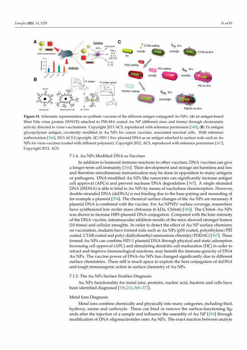
Energies 2021, 14, 1278 51 of 83
Figure 51. Schematic representation as synthetic vaccines of the different antigen conjugated Au NPs. (A) an antigen-basedWest Nile virus protein (WNVE) attached to PSS-MA coated Au NP (different sizes and forms) through electrostaticactivity directed to virus vaccinations. Copyright 2013 ACS, reproduced with reference permission [348], (B) Tn antigenglycopolymer antigen, covalently modified in Au NPs for cancer vaccines, associated mucinal cells. With referenceauthorization [366], 2013 ACS Copyright. (C) HIV-1 Env plasmid DNA as an antigen attached to surface rods such as AuNPs for virus vaccines (coated with different polymers). Copyright 2012, ACS, reproduced with reference permission [367],Copyright 2012, ACS.
7.1.4. Au NPs Modified DNA as Vaccines
In addition to humoral immune reactions to other vaccines, DNA vaccines can givea longer-term cell immunity [366]. Their development and storage are harmless and lowand therefore simultaneous immunization may be done in opposition to many antigensor pathogens. DNA-modified Au NPs like nanocytes can significantly increase antigencell approval (APCs) and prevent nuclease DNA degradation [367]. A single strandedDNA (SSDNA) is able to bind to Au NPs by means of nucleobase chemisorption. However,double-stranded DNA (dsDNA) is not binding due to the base pairing and mounding offor example a plasmid [294]. The chemical surface changes of the Au NPs are necessary ifplasmid DNA is combined with the vaccine. For Au NPNPs’ surface coverage, researchershave synthesized low molar mass chitosans (6 kDa, Chito6) [368]. The Chito6–Au NPswas shown to increase HBV-plasmid DNA conjugation. Compared with the bare intensityof the DNA vaccine, intramuscular addition results of the mice showed stronger humor(10 times) and cellular ansughts. In order to detect the effect of Au NP surface chemistryon vaccination, studants have formed rods such as Au NPs (pSS coated, polyethylene/PEIcoated, CTAB coated and poly(-diallydimethyl ammonium chloride)/PDDAC) [367]. Theseformed Au NPs can combine HIV-1 plasmid DNA through physical and static adsorption.Increasing cell approval (APC) and stimulating dendritic-cell maduration (DC) in order toretract and improve immunological reactions, may benefit the immune-genicity of DNAAu NPs. The vaccine power of DNA-Au NPs has changed significantly due to differentsurface chemistries. There still is much space to explore the best conjugation of dsDNAand tough immunogenic action in surface chemistry of Au NPs.
7.1.5. The Au NPs Surface Enables Diagnosis
Au NPs functionality for metal ions, proteins, nucleic acid, bacteria and cells havebeen identified diagnosed [196,216,369–372].
Metal Ions Diagnosis
Metal ions combine chemically and physically into many categories, including thiol,hydroxy, amine and carboxyle. These can bind or remove the surface-functioning lig-ands after the injection of a sample and influence the assembly of Au NP [294] throughmodification of DNA oligonucleotides onto Au NPs. The exact reaction between analyte
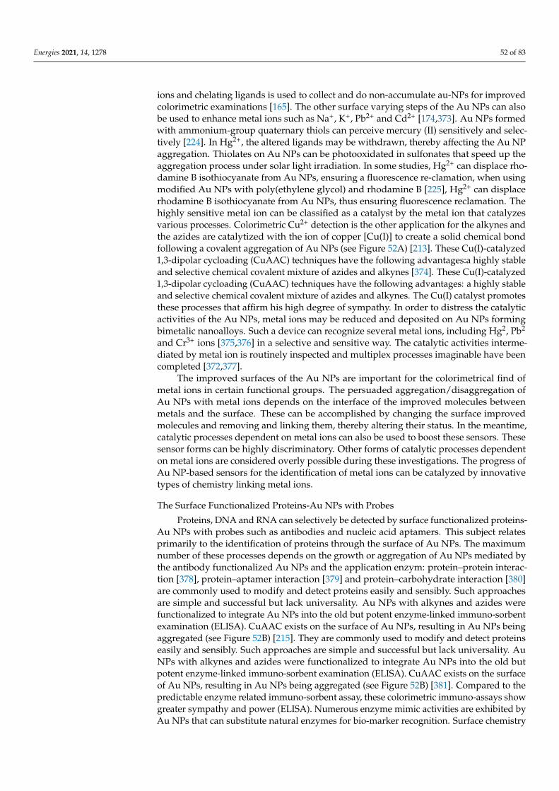
Energies 2021, 14, 1278 52 of 83
ions and chelating ligands is used to collect and do non-accumulate au-NPs for improvedcolorimetric examinations [165]. The other surface varying steps of the Au NPs can alsobe used to enhance metal ions such as Na+, K+, Pb2+ and Cd2+ [174,373]. Au NPs formedwith ammonium-group quaternary thiols can perceive mercury (II) sensitively and selec-tively [224]. In Hg2+, the altered ligands may be withdrawn, thereby affecting the Au NPaggregation. Thiolates on Au NPs can be photooxidated in sulfonates that speed up theaggregation process under solar light irradiation. In some studies, Hg2+ can displace rho-damine B isothiocyanate from Au NPs, ensuring a fluorescence re-clamation, when usingmodified Au NPs with poly(ethylene glycol) and rhodamine B [225], Hg2+ can displacerhodamine B isothiocyanate from Au NPs, thus ensuring fluorescence reclamation. Thehighly sensitive metal ion can be classified as a catalyst by the metal ion that catalyzesvarious processes. Colorimetric Cu2+ detection is the other application for the alkynes andthe azides are catalytized with the ion of copper [Cu(I)] to create a solid chemical bondfollowing a covalent aggregation of Au NPs (see Figure 52A) [213]. These Cu(I)-catalyzed1,3-dipolar cycloading (CuAAC) techniques have the following advantages:a highly stableand selective chemical covalent mixture of azides and alkynes [374]. These Cu(I)-catalyzed1,3-dipolar cycloading (CuAAC) techniques have the following advantages: a highly stableand selective chemical covalent mixture of azides and alkynes. The Cu(I) catalyst promotesthese processes that affirm his high degree of sympathy. In order to distress the catalyticactivities of the Au NPs, metal ions may be reduced and deposited on Au NPs formingbimetalic nanoalloys. Such a device can recognize several metal ions, including Hg2, Pb2
and Cr3+ ions [375,376] in a selective and sensitive way. The catalytic activities interme-diated by metal ion is routinely inspected and multiplex processes imaginable have beencompleted [372,377].
The improved surfaces of the Au NPs are important for the colorimetrical find ofmetal ions in certain functional groups. The persuaded aggregation/disaggregation ofAu NPs with metal ions depends on the interface of the improved molecules betweenmetals and the surface. These can be accomplished by changing the surface improvedmolecules and removing and linking them, thereby altering their status. In the meantime,catalytic processes dependent on metal ions can also be used to boost these sensors. Thesesensor forms can be highly discriminatory. Other forms of catalytic processes dependenton metal ions are considered overly possible during these investigations. The progress ofAu NP-based sensors for the identification of metal ions can be catalyzed by innovativetypes of chemistry linking metal ions.
The Surface Functionalized Proteins-Au NPs with Probes
Proteins, DNA and RNA can selectively be detected by surface functionalized proteins-Au NPs with probes such as antibodies and nucleic acid aptamers. This subject relatesprimarily to the identification of proteins through the surface of Au NPs. The maximumnumber of these processes depends on the growth or aggregation of Au NPs mediated bythe antibody functionalized Au NPs and the application enzym: protein–protein interac-tion [378], protein–aptamer interaction [379] and protein–carbohydrate interaction [380]are commonly used to modify and detect proteins easily and sensibly. Such approachesare simple and successful but lack universality. Au NPs with alkynes and azides werefunctionalized to integrate Au NPs into the old but potent enzyme-linked immuno-sorbentexamination (ELISA). CuAAC exists on the surface of Au NPs, resulting in Au NPs beingaggregated (see Figure 52B) [215]. They are commonly used to modify and detect proteinseasily and sensibly. Such approaches are simple and successful but lack universality. AuNPs with alkynes and azides were functionalized to integrate Au NPs into the old butpotent enzyme-linked immuno-sorbent examination (ELISA). CuAAC exists on the surfaceof Au NPs, resulting in Au NPs being aggregated (see Figure 52B) [381]. Compared to thepredictable enzyme related immuno-sorbent assay, these colorimetric immuno-assays showgreater sympathy and power (ELISA). Numerous enzyme mimic activities are exhibited byAu NPs that can substitute natural enzymes for bio-marker recognition. Surface chemistry
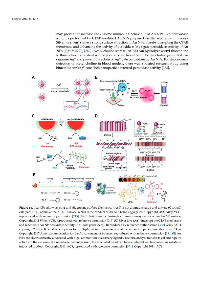
Energies 2021, 14, 1278 53 of 83
may prevent or increase the enzyme-mimicking behaviour of Au NPs. No peroxidaseaction is performed by CTAB modified Au NPs prepared via the seed growth process.Silver ions (Ag+) have a strong surface attraction of Au NPs, thereby disrupting the CTABmembrane and enhancing the activity of peroxidase (Ag+ gate peroxidase activity of AuNPs (Figure 52C)) [382]. Acetylcholine sterase (AChE) can hydrolyze acetyl-thiocholineto thiocholine as a critical neurological disease biomarker. The thiocholine generated canorganise Ag+ and prevent the action of Ag+ gate peroxidase by Au NPs. For fluorescencedetection of acetyl-choline in blood models, there was a related research study usingbimetallic Au@Ag0 core-shell nanoparticle-referred peroxidase activity [383].
Figure 52. Au NPs allow sensing and diagnostic surface chemistry. (A) The 1,3-diogravic azide and alkyne (CuAAC)catalyzed Cu(I) occurs at the Au NP surface, which is the product of Au NPs being aggregated. Copyright 2008 Wiley-VCH,reproduced with reference permission [213] (B) CuAAC-based colorimetric immunoassay occurs on an Au NP surface.Copyright 2011 Wiley-VCH, reproduced with reference permission [215] (C) Silver ions (Ag+) interrupt the CTAB membraneand regenerate Au NP peroxidase activity (Ag+ gate peroxidase). Reproduced by reference authorisation [382] Wiley-VCHcopyright 2018. (D) Set sheets of paper for multiplexed immuno-assays shall be shirked in paper barcode chips (PBCs).Copyright 2017 American Association for the Advancement of Sciences, reproduced with reference permission [384] (E) AuNPs are electrostatically associated with b-gal ammonium quaternary ligands. Bacteria analyte transfer b-gal and repairsactivity of the enzymes. If a naked-eye reading is used, the evacuated b-Gal can turn a pale-yellow chromogenous substrateinto a red product. Copyright 2011, ACS, reproduced with reference permission [371], Copyright 2011, ACS.
Energies 2021, 14, 1278 54 of 83
Based on its distinctive visual characteristics, the most fruitful applications of Au NPsare sensing uses. The high elimination coefficients of Au NPs are used by these types ofcolorimetric sensors and can give naked-eye readouts with a low detection limit. The actof recognition is interrupted by surface features of Au NPs for most of these applications.LFAs can transform from qualitative inspection to quantitative recognition with improvedsensitivity by functionalizing Au NPs with signal assessments such as enzymes and flu-orescent molecules [385]. For appropriate processes, components can be lyophilized onthe LFA strip [386]. A paper-based barcode assay system based on barcode scanning willprovide a more objective and reliable reading separately from the illustrative, fluorescentand chemi-luminescent reading of LFAs [384]. For multiplexed immuno-assays, the fixedsheets of paper are shirked into paper based barcode chips (PBCs) (see Figure 52D) [387].This explores the platform that enables various biomarkers to be discovered and providesmultiplexed implications within several minutes. For point-of-care finding of proteins inresource restricted sets, colorimetric sensors are suitable. The optical and surface char-acteristics of Au NPs provide a perfect stage for these sensors to advance. Although AuNP-based assays include a moral recognition act, the following fragments will advancethese types of examinations: full automation, improved presentation of results and pro-longed stability. Surface chemistry gives Au NPs the opportunity to identify targets whilealso increasing old sensor analytical recitals (such as sensitivity and selectivity). Whencombined with other innovations, such as microfluidics, Au NPs-based inspections mayhave provided an automated operation recital. In this area, we believe there is still muchprogress to be made, particularly in the surface chemistry of Au NPs.
Recognition of Bacteria, Viruses and Cells via Au NPs
Au NPs have been widely used for recognizing bacteria and cells [388]. The chemistryof the Au surface gives the opportunity to locate bacteria or cells. Extreme processesdepend on antibodies and on Au NP evaluations built to detain bacteria and cells. Forthese applications, Au NPs enhanced with modified small molecules may also be used. Theinteraction between the viral hemagglutinin (HA) protein and the host glycan receptors canbe tested by glycan functionalized Au NPs (Au NPs). To identifyavian infuenza visually,this technique is used [389]. Au NPs may be reserved for trimeric HAs or viruses, resultingin gAu NP aggregation. This naked-eye readout takes care of the quick and in-depthdetection of harmful viruses. Quaternary ammonium ligands are functionalized withAu NPs, which can electrostatically stabilise b-galactosidase (b-Gal, see Figure 52E) [371].Quaternary ammonium ligands can simultaneously stabilise Au NPs and increase theirsolubility and biocompatibility. The functionalized Au NPs of the quaternary ammoniumligand will resist the action of b-Gal. When the anionic-surface analyte bacteria wereapplied to the Au NPs, b-Gal transferred to the Au NPs surface and mended the enzymeeffort. For the naked-eye readouts, the moved b-Gal will alter a pale yellow chromogenicsubstrate into a red substance. Compared to the Au–S bond, physical adsorption betweenamino groups and Au is weak; however, the likelihood of colorimetric bacterial finding isgiven by this method. In a one-pot reaction, modified Au NPs are prepared from D-Alanyl-Dalanine (DADA) [390]. In extremely acid or alkaline conditions, the resulting Au NPsremain stable. DADA will recognise peptidoglycan on the bacterial surface in the bacteriasociety, influencing the aggregation of Au NPs due to the lack of DADA shield. Thiscolorimetric assay will distinguish between Staphylococcus aureus and Staphylococcusaureus immune to methicillin from ascite models in patients. In fact, colorimetric sensorsbased on physical adsorption and chemical crosslinking are largely detected based onAu NPs. For colorimetric sensors, molecular forces and chemical processes betweentarget analytes and Au NPs are vital. We can precisely control and scheme the surfacecharacteristics of Au NPs through surface chemistry and increase their utility. Otherbiochemical processes can also be used for surface modification of Au NPs, separatelyfrom antibodies and molecules we have designed beyond. The integration of finding andbehaviour into one combined platform; meanwhile, this demonstrates disproportionate
Energies 2021, 14, 1278 55 of 83
value in the provision of bacterial sicknesses. The possibility of these uses is discussed bythe surface chemistry of Au NPs.
7.1.6. The Surface Chemistry of Au NPs Enables TreatmentFunctionalized Au NPs as Antibiotics
Although bare Au NPs have no antibacterial properties, surface functionalized AuNPs are used as antibiotics with enhanced antibacterial movement compared to currentantibiotics or medicines with medications, vaccines and antibiotics [391,392]. Nanoparticlescan deliver antibiotics or drugs to bacteria competently and increase the concentrationof drugs in targeted bacteria, thus refining the antibacterial properties of old antibiotics.Maximal experiments on antibiotic-dependent nanoparticles are based on antibiotic ordrug functionalization. Recent research shows that Au’s surface chemistry offers strongantibacterial features for Au NPs. Surface modification by the Au–S bond of certain non-antibacterial molecules on Au NPs will convey antibacterial features to these Au NPs. In2010, amino-replaced pyrimidine, such as 4,6-diamino-2-pyrimidinethiol (DAPT), func-tionalized Au NPs (Au-DAPT) have been investigated as antibacterial agents targetingmulti-drug resistant Gram-negative bacteria, but none of the components have antibacterialpotential on their own. The positively charged DAPT groups will increase the permeabilityof Au NPs to the bacteria’s outer membrane, thereby refining the beneficial efficacy. Bymodifying the membrane strength and avoiding the subunit of ribosomes, the Au-DAPTexerts its antibacterial activity [393]. Numerous other accounts also record strong antibacte-rial activity of cationic and hydrophobic functionalized Au NPs [394–396]. The synergisticproperties of non-antibiotic drugs and DAPT on Au NPs in conflict with superbugs areadditional stimulating findings [397]. The Au-improved NPs’ antibacterial activity is asso-ciated with both GP-positive and Gram-negative bacteria, consisting of superbug-drivenmedication. This is after co-functionality with non-antibiotic drugs and DAPT.
Co-functioning of Au NPs with antimicrobials and other functional componentsis a common procedure that offers more dominant biomedical antibiotics based on AuNPs [398]. Further reading reveals that N-heterocyclic molecules also have the same effecton [399]. The Au NPs show broad spectrum antibacterial actions in contradiction to evensuperbugs that repel most antibiotics by surface functionalization with an N-heterocyclicmolecule. Physical adsorption is also used as a surface modification technique for Au NPsseparately from using the Au–S bond. Via physical adsorption between amino groups andAu NPs, pharmaceutical intermediates such as 7-amino-cephalosporanic acid (7-ACA),6-amino-penicillanic acid (6-APA) and 7-amino-desacetoxycephalosporanic acid (7-ADCA)will adsorb Au NPs on the surface [400]. Via cell membrane interruption and lysis ofbacterial cells, these pharmaceutical intermediate functionalized Au NPs will defendagainst Gram-negative bacteria consisting of both laboratory antibiotic-sensitive strainsand clinical multidrug-resistant separates. Numerous amino-saccharides have similarstructures to the bacterial peptide-glycan. The functionalized Au NPs (Au-GluN) of D-glucosamine(GluN) will disrupt bacterial peptidoglycan, leading to the loss of bacteria [401].Further research shows that we can find a small range of antibiotics by functionalizingAu NPs with amino-saccharides [402]. The Au-GluN will move through the cell wall andchange the cell membrane’s structure to convince the cell to lose bacteria.
Some Au NP antibacterial coverings and coatings are advanced to thoroughly dis-cover the use of Au NPs-based antibiotics. Helical healing issues arise in bacterial poisons.Strong bacterial poisons can also contribute to sepsis approximately. Electrospinning al-lows nano-sized polymer fibers to be synthesized according to human requirements. InPoly(3-caprolactone)/gelatin solution under electrospinning, we doped intermediate phar-maceutical Au NPs to [401]. Relative to physical adsorption techniques, the electrospunnanofibrous scaffolds provide a homogeneous coating of nanomaterials on fibres. With thehumiliation of poly(3-caprolactone)/gelatin nanofibrous scaffolds, anti-bacterial Au NPscan be consistently free to provide constant antimicrobial capacity. This mechanism canalso be recognised by co-electrospinning Au NPs and poly-poly-capped indole derivative
Energies 2021, 14, 1278 56 of 83
co-electrospinning (-lactic-co-glycolicacid) [403]. Indole does not show any antibacterialactivity as a major derivative in the food and drug industries. The resulting AI-IDs demon-strate strong antibacterial activity in contradiction to multidrug resistant superbugsafterphysical adsorption of indole derivatives onto Au NPs (AI-IDs). Bacterial cellulose (BC), apopular biomaterial treatment for tissue engineering, shows advantages of high biocom-patibility: the capacity to absorb water and mechanical strength. When a BC membrane issaturated in an Au-DAPT solution, through physical adsorption, Au-DAPT will adsorb onthe BC membrane, which has implications for wound healing in an antibacterial dressing(BC-Au-DAPT nanocomposites) [404]. The BC-Au-DAPT freeze-dried nanocomposites arehighly stable and, after one year of storage, maintain their antibacterial action. Anotheruse of antibiotics based on Au NPs is their use as antibacterial coatings for medical strate-gies. The surface of the medical devices is negatively emotional later in the plasma action.Positively charged surface Au-DAPT will adsorb stably on the surface of medical devicesby electrostatic self-assembly [405]. Thus, the immobilised Au-DAPT will destroy antimi-crobial constituents if nosocomial medical device contamination is a secure but proficientantibacterial method. Au NPs smaller than 2 nm, often referred to as Au nanoclusters(Au-NCs), have confirmed antibacterial activity when quaternary ammonium (QA) saltsare functionalized [406]. For bonding Au-NCs, one end of the QA has a sulfydryl groupand the other end has positively charged ligands that can increase membrane permeabilityand decrease bacteria drug efflux. In comparison to multidrug-resistant Gram-positivebacteria, these types of antibiotics can battle in vivo and have no detectable cells. The timeof circulation is equivalent to that of vancomycin (a widely used antibiotic), which confirmsits efficacy as an antibacterial agent. Additional work using antimicrobial peptide self-assembly on Au nanodots demonstrates superior antimicrobial activity by decomposingthe bacterial membrane [407]. Meanwhile, on the basis of Au–S bond-based conjugationof Au NPs and the unique thiol molecules, the outstanding antibacterial capacity is built.Millimola quantities of glutathione can competitively bind to Au NP surfaces via theAu–S bond in the blood stream and cells. The disinterest caused by the movement of theparticular thiol molecules will significantly reduce the Au NPs complex’s antibacterialcapability. The stronger chemical bond to conjugate Au NPs and particular molecules islikely to avoid invalidation caused by glutathione movement. By developing an Au–Sebond instead of an Au–S bond, researchers used selenol modified peptides with dye tosynthesise fuorescent Au NPs [408]. The more stable Au–Se bonds are able to speechlessthiol compound intervention. The carbene–Au bond can also be employed for bridgingfunctional N-heterocyclic carbene molecules and Au NPs [409]. In comparison to thermalinjury, the N-heterocyclic carbene-improved Au NPs/Au-NCs display high fluorescencequantum output and high stability [410–412]. Based on the same value, both the Au–Sebond and the stunned thiol interference should be capable of the more stable carbene-Aubond. Au’s surface chemistry confers strong antibacterial properties on Au NPs. Theseforms of antibiotics can kill superbugs and reduce the resistance of bacteria to drugs. Webelieve that surface functionalization of Au NPs with other types of molecules, apart frompyrimidine, such as N-heterocyclic molecules, aminosaccharides, indoles, pharmaceuticalintermediates and QA salts, will allow the improvement of new antimicrobial tools. In themeantime, more studies on the properties of dissimilar sizes, shapes and surface goods ofAu NPs on antimicrobial properties are still needed.
Functionalized Au NPs for Cancer Therapy
One of the greatest deadly human illnesses is cancer. Because of the range of sucha chronic illness, distinction of personal appearance and lack of effective miracle drugs,cancer therapy is still a persistent scientific test. The most real mode of cancer action iscurrently surgery in combination with radiotherapy and chemotherapy. However, it isvital to advance new treatment plans and extremely specific medicines for the simplyinvasive and metastatic products of cancer cells and the increasing drug confrontation.Nanomedicine tackles unique mechanisms and has emerged as an encouraging plan to
Energies 2021, 14, 1278 57 of 83
improve old therapeutic efficacy. It may advance the differential improvement for image-guided therapy and cancer-specific delivery of chemotherapeutic agents for joint therapyby promoting the target selection of cancer. Among various nanomaterials studied forcancer therapeutic uses, Au NPs were mostly studied taking advantage of their distinctivechemical, electrical and optical characteristics and outstanding biocompatible characteris-tics, as well as the ease of synthetic activity and precise rheostat over their physicochemicalcharacteristics [7,413,414]. The high attractiveness of Au NPs to bind thiols, amines andpolymers offers an effective way of presenting reactive functional groups that can be usedfor targeting and conjugating therapeutic agents (e.g., antibodies, peptides, aptamers andcarbohydrates, drugs, radionuclides, photosensitizers, siRNA and genes). The high X-rayand near-infrared (NIR) light absorption coefficient will openly award Au NPs for cancertherapy with the effect of radio-sensitization and photothermal control. Multifunctionalnanomedicine can achieve distinctive characteristics that can not be accomplished withAu NPs alone by combining Au NPs into other nanoplatforms, such as liposomes. In thedevelopment of useful nanomedicines that are specialised in multi-modal therapeutic usesin cancer care, Au NPs are talented. In vivo, Au NPs demonstrate the effect of radiationdose improvement and have created great interest in the field of radio-sensitization basedon Au NPs for oncology. The survival of mice with subcutaneous EMT-6 mammary carci-noma can be substantially improved by X-ray irradiation and activity with Au NPs [415].In addition, researchers have discovered novel methods of radio-sensitization for chem-ical improvement (DNA damage and radical production) and biological improvementin physical improvement (ROS-induced oxidative stress, inhibition of DNA healing andcell cycle disruption) [416]. The effectiveness of cellular approval can greatly enhance thetherapeutic effect. For Au NP dependent nanomedicines, the size and surface charges aresignificantly affected to monitor the efficacy of their cellular uptake [417]. The selectiveaccumulation at tumour sites was aided by circulating Au NPs with enhanced porousdesign and retention (EPR) effect assistance. The cellular absorption of highly negativelycharged surface Au NPs is very difficult. To increase cellular uptake, neutral or positivelycharged surfaces are helpful [418,419]. Researchers have researched the size-dependentradio-sensitization of Au NPs using an U14 tumour bearing mouse model by surfacefunctionalization with PEG-SH coatings [420]. Compared to smaller or larger PEG-AuNPs, PEG-Au NPs (10–30 nm) demonstrated higher radio-sensitivity for radiotherapy. Bymodifying the surface chemistry of Au NPs with coating molecules, with improved AuNP build-up in tumours, researchers may produce separate mutual radio-sensitization.Metabolizable ultra-small Au NPs with a biocompatible covering ligand (glutathione, GSH)(core size 1.5 nm) strongly enhance cancer radio-therapy [421]. Through the enhancedEPR effect, the GSH-Au NPs can be collected in growths differently. GSH-Au NPs can beprofessionally cleared by the kidneys following treatment, which eliminates any potentialside effects. Photo-thermal therapy uses NIR light/laser as the energy source to producehigh temperatures to destroy cancer cells instead of using harmful high-energy rays andpollutants (radiotherapy). Plasmonic Au nanoshells have been identified in previous stud-ies to demonstrate photo-thermal properties and are thus used for near-infrared thermaltreatment of tumours [422,423]. Rod-like Au NPs with a tunable LSPR band associatedwith the nanoshells compared to the nanoshells, rod-like Au NPs with a tunable LSPRband between 650 and 950 nm (falling in the NIR region) are considered to have great po-tential for beneficial nanomedicine [424–426]. In addition, multi-functional nanomedicinescan achieve a combined scheme to advance specific cancer treatment by combining thephoto-thermal effect with pre-loading anti-cancer drugs or photosensitizers. The surfacechemistry of Au NPs at this stage reveals a crucial role in regulating the loading efficacyof these therapeutic molecules [427]. Investigators have used mPEG-SH and a positivelycharged peptide (RRLAC) to alter rod-like Au NPs to modify the surface chemistry of AuNPs. When conjugated with RRLAC, the PEG on Au NP surfaces can help to stabilise andelude aggregation. Supplementary conjugate photosensitizers (aluminium phthalocyaninetetrasulfonate, AlPcS4) can be applied to the positively charged RRLAC by charge-charge
Energies 2021, 14, 1278 58 of 83
forces. The disclosure of 810 nm laser irradiation will quickly produce a photothermaleffect, as well as an indication of the reactive AlPcS4 discharge. The second 670 nm laserirradiation exposure generates the output for photodynamic therapy of a singlet oxygengenerator (SOG). Joining the photo-thermal therapy and photo-dynamics therapy canexpressively increase anti-cancer therapeutic properties. To improve the loading profi-ciency of therapeutic molecules, mesoporous silica (SiO2) was used to increase the surfacechemistry of Au NPs [428] to boost the loading skills of therapeutic molecules. As a newcancer theranosis agent [429] investigators have developed SiO2-coated rod-like Au NPs(Au@SiO2). The SiO2 large surface area and large cavity will greatly advance the loadingof chemotherapy medications.
7.2. Other Applications7.2.1. Gold Nanoparticles as a Catalyst
The gold nanoparticles are a good catalyst and have brings about the catalysis ofmany dyes degradation wether the gold nanoparticles synthesized by the chemical methodor physical method. The gold nanoparticle is a precious metal for coinage, jewelry andother arts since antiquity. Chemically, gold is a transition metal gorgeous in coordinationnumbers. However, relative to other metals Bulk Au is a renowned chemically inertmaterial and has been reflected as an unwell active catalyst in numerous reactions. ThoughAu NPs exhibit high catalytic characteristics for several reactions [430,431]. The finding ofthe outstanding catalytic properties of Au NPs was described for the first time by Harutaet al. in 1989, who showed that Au NPs can be very active in the elevation of the oxidationof carbon monoxide (CO) at little temperature [432]. Succeeding this discovery, a sample ofaerobic oxidation reactions catalyzed by Au NPs have been described. For example, theoxidation of alcohols to aldehydes [433], carboxylic acids [430] or esters [434], the oxidationof aldehydes to esters [30] or acids [435], epoxidations of olefins and the oxidation ofamines to amides [436] have been examined. Newly, the catalytic use of Au NPs in carefulhydrogenation has been extensively testified as well [437]. Specifically, it was revealedthat with the incidence of a Au NP catalyst, replaced anilines and connected productscan be formed proficiently at low temperature from their nitrogenated compounds, whichare normally toxic organic by-products formed unenviably throughout the industrialdeveloped method of chemicals, such as agrochemicals, dyes and pharmaceuticals [438].For instance, 4-nitrophenol (4-NP), a toxic nitro acromatic chemical, has a postponedinteraction with blood and then forms methaemoglobin tempting methemoglobinemia,potentially producing cyanosis, confusion and unconsciousness to human body. Whenswallowed, it causes abdominal pain and vomiting. Instead, 4-aminophenol (4-AP), ahydrogenation yield of 4-NP, is a very beneficial material which can be employed asan significant intermediate in pharmaceutical preparation of analgesic and antipyreticdrugs [439] as antioxidant in plastics fabrication [438]. More significantly, this reaction ishighly helpful for the moving environmental pollutants into beneficial chemicals. With AuNPs, unsaturated chemicals can also be selectively hydrogenated. For example, acetyleneand 1, 3-butadiene can be moderately hydrogenated into ethylene [440] and butenescorrespondingly. The chief benefit of Au NP catalysts is their capability to selectivelycatalyze the hydrogenation of the C=O group of α,β-unsaturated aldehydes manufacturingallyl-type unsaturated alcohols [441]. The reduction of 4-NP to 4-AP on Au NPs withsodium borohydride (BH4
-) in aqueous solution has also been thoroughly employed as astyle reaction to inspect the catalytic presentation of noble metal NPs, specially Au NPssynthesized from the reduction of auric acid via wet chemical methods [442]. For instance,Pal and coworkers found that a reduction in Au NP size primes to the increase of decreaserate [443]. The alteration of Au NP surfaces with diverse functional groups testified thatthe surface structure also mainly effects the catalytic characteristics of Au NPs [444].
The catalytic mechanism for reduction of 4-NP by Au NPs@vesicles catalyst to form4-AP in the presence of NaBH4 is shown in Figure 53. Here in the mechanism, whenAu NPs@vesicles was used for catalytic reduction, BH4
- and 4-NP are first spread from
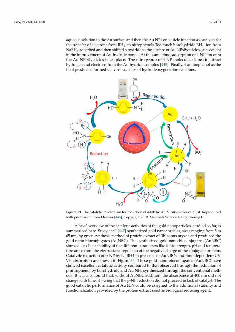
Energies 2021, 14, 1278 59 of 83
aqueous solution to the Au surface and then the Au NPs on vesicle function as catalysts forthe transfer of electrons from BH4
- to nitrophenols.Too much borohydride BH4- ion from
NaBH4 adsorbed and then shifted a hydride to the surface of Au NPs@vesicles, subsequentin the improvement of Au-hydride bonds. At the same time, adsorption of 4-NP ion ontothe Au NPs@vesicles takes place. The nitro group of 4-NP molecules slopes to attracthydrogen and electrons from the Au-hydride complex [445]. Finally, 4-aminophenol as thefinal product is formed via various steps of hydrodeoxygenation reactions.
Figure 53. The catalytic mechanism for reduction of 4-NP by Au NPs@vesicles catalyst. Reproducedwith permission from Elsevier [446], Copyright 2019, Materials Science & Engineering C.
A brief overview of the catalytic activities of the gold nanoparticles, studied so far, issummarized here. Sujoy et al. [447] synthesized gold nanoaprticles, sizes ranging from 5 to65 nm, by green synthesis method of protein extract of Rhizopus oryzae and produced thegold nano-bioconjugates (AuNBC). The synthesized gold nano-bioconjugates (AuNBC)showed excellent stability of the different parameters like ionic strength, pH and tempera-ture arose from the electrostatic repulsion of the negative charge of the conjugate proteins.Catalytic reduction of p-NP by NaBH4 in presence of AuNBCs and time-dependent UV-Vis absorption are shown in Figure 54. These gold nano-bioconjugates (AuNBC) haveshowed excellent catalytic activity compared to that observed through the reduction ofp-nitrophenol by borohydride and Au NPs synthesized through the conventional meth-ods. It was also found that, without AuNBC addition, the absorbance at 400 nm did notchange with time, showing that the p-NP reduction did not proceed in lack of catalyst. Thegood catalytic performance of Au NPs could be assigned to the additional stability andfunctionalization provided by the protein extract used as biological reducing agent.
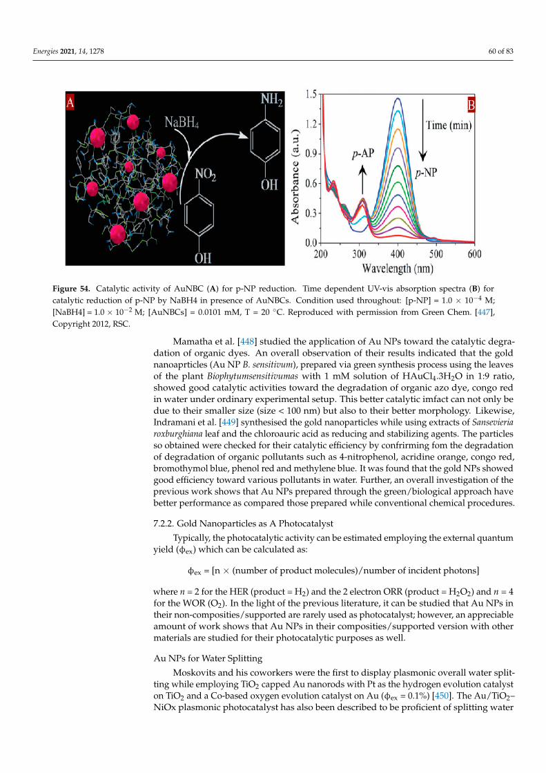
Energies 2021, 14, 1278 60 of 83
Figure 54. Catalytic activity of AuNBC (A) for p-NP reduction. Time dependent UV-vis absorption spectra (B) forcatalytic reduction of p-NP by NaBH4 in presence of AuNBCs. Condition used throughout: [p-NP] = 1.0 × 10⁻⁴ M;[NaBH4] = 1.0 × 10⁻² M; [AuNBCs] = 0.0101 mM, T = 20 ◦C. Reproduced with permission from Green Chem. [447],Copyright 2012, RSC.
Mamatha et al. [448] studied the application of Au NPs toward the catalytic degra-dation of organic dyes. An overall observation of their results indicated that the goldnanoaprticles (Au NP B. sensitivum), prepared via green synthesis process using the leavesof the plant Biophytumsensitivumas with 1 mM solution of HAuCl4.3H2O in 1:9 ratio,showed good catalytic activities toward the degradation of organic azo dye, congo redin water under ordinary experimental setup. This better catalytic imfact can not only bedue to their smaller size (size < 100 nm) but also to their better morphology. Likewise,Indramani et al. [449] synthesised the gold nanoparticles while using extracts of Sansevieriaroxburghiana leaf and the chloroauric acid as reducing and stabilizing agents. The particlesso obtained were checked for their catalytic efficiency by confrirming fom the degradationof degradation of organic pollutants such as 4-nitrophenol, acridine orange, congo red,bromothymol blue, phenol red and methylene blue. It was found that the gold NPs showedgood efficiency toward various pollutants in water. Further, an overall investigation of theprevious work shows that Au NPs prepared through the green/biological approach havebetter performance as compared those prepared while conventional chemical procedures.
7.2.2. Gold Nanoparticles as A Photocatalyst
Typically, the photocatalytic activity can be estimated employing the external quantumyield (φex) which can be calculated as:
φex = [n × (number of product molecules)/number of incident photons]
where n = 2 for the HER (product = H2) and the 2 electron ORR (product = H2O2) and n = 4for the WOR (O2). In the light of the previous literature, it can be studied that Au NPs intheir non-composities/supported are rarely used as photocatalyst; however, an appreciableamount of work shows that Au NPs in their composities/supported version with othermaterials are studied for their photocatalytic purposes as well.
Au NPs for Water Splitting
Moskovits and his coworkers were the first to display plasmonic overall water split-ting while employing TiO2 capped Au nanorods with Pt as the hydrogen evolution catalyston TiO2 and a Co-based oxygen evolution catalyst on Au (φex = 0.1%) [450]. The Au/TiO2–NiOx plasmonic photocatalyst has also been described to be proficient of splitting water
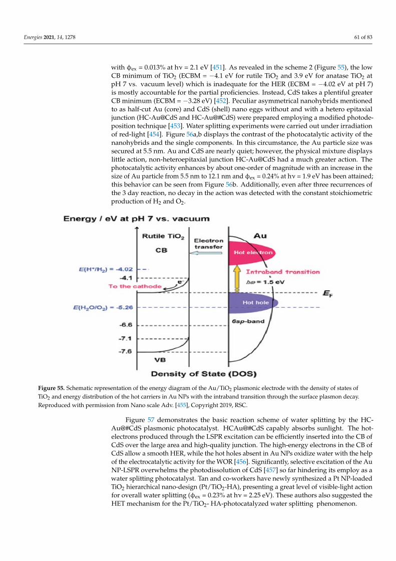
Energies 2021, 14, 1278 61 of 83
with φex = 0.013% at hν = 2.1 eV [451]. As revealed in the scheme 2 (Figure 55), the lowCB minimum of TiO2 (ECBM = −4.1 eV for rutile TiO2 and 3.9 eV for anatase TiO2 atpH 7 vs. vacuum level) which is inadequate for the HER (ECBM = −4.02 eV at pH 7)is mostly accountable for the partial proficiencies. Instead, CdS takes a plentiful greaterCB minimum (ECBM = −3.28 eV) [452]. Peculiar asymmetrical nanohybrids mentionedto as half-cut Au (core) and CdS (shell) nano eggs without and with a hetero epitaxialjunction (HC-Au@CdS and HC-Au@#CdS) were prepared employing a modified photode-position technique [453]. Water splitting experiments were carried out under irradiationof red-light [454]. Figure 56a,b displays the contrast of the photocatalytic activity of thenanohybrids and the single components. In this circumstance, the Au particle size wassecured at 5.5 nm. Au and CdS are nearly quiet; however, the physical mixture displayslittle action, non-heteroepitaxial junction HC-Au@CdS had a much greater action. Thephotocatalytic activity enhances by about one-order of magnitude with an increase in thesize of Au particle from 5.5 nm to 12.1 nm and φex = 0.24% at hν = 1.9 eV has been attained;this behavior can be seen from Figure 56b. Additionally, even after three recurrences ofthe 3 day reaction, no decay in the action was detected with the constant stoichiometricproduction of H2 and O2.
Figure 55. Schematic representation of the energy diagram of the Au/TiO2 plasmonic electrode with the density of states ofTiO2 and energy distribution of the hot carriers in Au NPs with the intraband transition through the surface plasmon decay.Reproduced with permission from Nano scale Adv. [455], Copyright 2019, RSC.
Figure 57 demonstrates the basic reaction scheme of water splitting by the HC-Au@#CdS plasmonic photocatalyst. HCAu@#CdS capably absorbs sunlight. The hot-electrons produced through the LSPR excitation can be efficiently inserted into the CB ofCdS over the large area and high-quality junction. The high-energy electrons in the CB ofCdS allow a smooth HER, while the hot holes absent in Au NPs oxidize water with the helpof the electrocatalytic activity for the WOR [456]. Significantly, selective excitation of the AuNP-LSPR overwhelms the photodissolution of CdS [457] so far hindering its employ as awater splitting photocatalyst. Tan and co-workers have newly synthesized a Pt NP-loadedTiO2 hierarchical nano-design (Pt/TiO2-HA), presenting a great level of visible-light actionfor overall water splitting (φex = 0.23% at hν = 2.25 eV). These authors also suggested theHET mechanism for the Pt/TiO2- HA-photocatalyzed water splitting phenomenon.
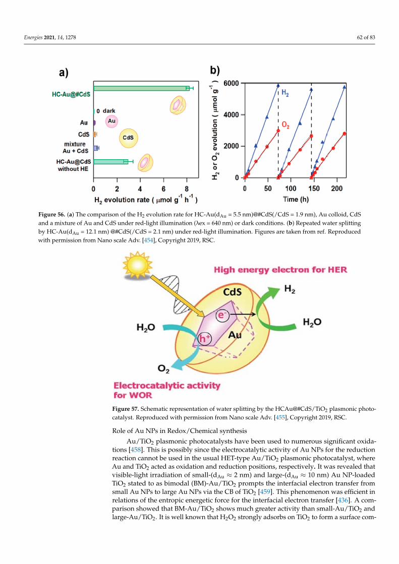
Energies 2021, 14, 1278 62 of 83
Figure 56. (a) The comparison of the H2 evolution rate for HC-Au(dAu = 5.5 nm)@#CdS(/CdS = 1.9 nm), Au colloid, CdSand a mixture of Au and CdS under red-light illumination (λex = 640 nm) or dark conditions. (b) Repeated water splittingby HC-Au(dAu = 12.1 nm) @#CdS(/CdS = 2.1 nm) under red-light illumination. Figures are taken from ref. Reproducedwith permission from Nano scale Adv. [454], Copyright 2019, RSC.
Figure 57. Schematic representation of water splitting by the HCAu@#CdS/TiO2 plasmonic photo-catalyst. Reproduced with permission from Nano scale Adv. [455], Copyright 2019, RSC.
Role of Au NPs in Redox/Chemical synthesis
Au/TiO2 plasmonic photocatalysts have been used to numerous significant oxida-tions [458]. This is possibly since the electrocatalytic activity of Au NPs for the reductionreaction cannot be used in the usual HET-type Au/TiO2 plasmonic photocatalyst, whereAu and TiO2 acted as oxidation and reduction positions, respectively. It was revealed thatvisible-light irradiation of small-(dAu ≈ 2 nm) and large-(dAu ≈ 10 nm) Au NP-loadedTiO2 stated to as bimodal (BM)-Au/TiO2 prompts the interfacial electron transfer fromsmall Au NPs to large Au NPs via the CB of TiO2 [459]. This phenomenon was efficient inrelations of the entropic energetic force for the interfacial electron transfer [436]. A com-parison showed that BM-Au/TiO2 shows much greater activity than small-Au/TiO2 andlarge-Au/TiO2. It is well known that H2O2 strongly adsorbs on TiO2 to form a surface com-
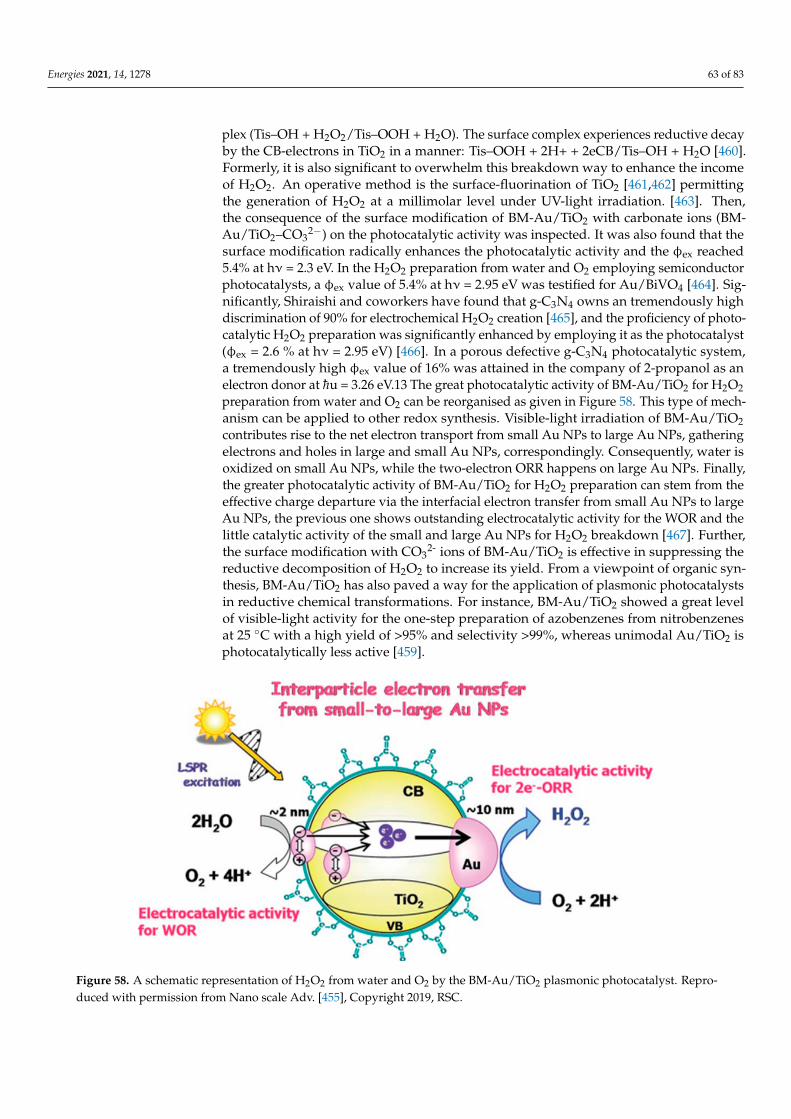
Energies 2021, 14, 1278 63 of 83
plex (Tis–OH + H2O2/Tis–OOH + H2O). The surface complex experiences reductive decayby the CB-electrons in TiO2 in a manner: Tis–OOH + 2H+ + 2eCB/Tis–OH + H2O [460].Formerly, it is also significant to overwhelm this breakdown way to enhance the incomeof H2O2. An operative method is the surface-fluorination of TiO2 [461,462] permittingthe generation of H2O2 at a millimolar level under UV-light irradiation. [463]. Then,the consequence of the surface modification of BM-Au/TiO2 with carbonate ions (BM-Au/TiO2–CO3
2−) on the photocatalytic activity was inspected. It was also found that thesurface modification radically enhances the photocatalytic activity and the φex reached5.4% at hν = 2.3 eV. In the H2O2 preparation from water and O2 employing semiconductorphotocatalysts, a φex value of 5.4% at hν = 2.95 eV was testified for Au/BiVO4 [464]. Sig-nificantly, Shiraishi and coworkers have found that g-C3N4 owns an tremendously highdiscrimination of 90% for electrochemical H2O2 creation [465], and the proficiency of photo-catalytic H2O2 preparation was significantly enhanced by employing it as the photocatalyst(φex = 2.6 % at hν = 2.95 eV) [466]. In a porous defective g-C3N4 photocatalytic system,a tremendously high φex value of 16% was attained in the company of 2-propanol as anelectron donor at hu = 3.26 eV.13 The great photocatalytic activity of BM-Au/TiO2 for H2O2preparation from water and O2 can be reorganised as given in Figure 58. This type of mech-anism can be applied to other redox synthesis. Visible-light irradiation of BM-Au/TiO2contributes rise to the net electron transport from small Au NPs to large Au NPs, gatheringelectrons and holes in large and small Au NPs, correspondingly. Consequently, water isoxidized on small Au NPs, while the two-electron ORR happens on large Au NPs. Finally,the greater photocatalytic activity of BM-Au/TiO2 for H2O2 preparation can stem from theeffective charge departure via the interfacial electron transfer from small Au NPs to largeAu NPs, the previous one shows outstanding electrocatalytic activity for the WOR and thelittle catalytic activity of the small and large Au NPs for H2O2 breakdown [467]. Further,the surface modification with CO3
2- ions of BM-Au/TiO2 is effective in suppressing thereductive decomposition of H2O2 to increase its yield. From a viewpoint of organic syn-thesis, BM-Au/TiO2 has also paved a way for the application of plasmonic photocatalystsin reductive chemical transformations. For instance, BM-Au/TiO2 showed a great levelof visible-light activity for the one-step preparation of azobenzenes from nitrobenzenesat 25 ◦C with a high yield of >95% and selectivity >99%, whereas unimodal Au/TiO2 isphotocatalytically less active [459].
Figure 58. A schematic representation of H2O2 from water and O2 by the BM-Au/TiO2 plasmonic photocatalyst. Repro-duced with permission from Nano scale Adv. [455], Copyright 2019, RSC.

Energies 2021, 14, 1278 64 of 83
7.2.3. Use of Au NPs in Sensing
The communication of light at the surface of the noble metal film stimulates surfaceelectromagnetic waves and sets them to resonate with incident light wave, subsequent inthe absorption of the light. This phenomenon is recognized as surface plasmon resonance(SPR) [468] and rest on the refractive index of the interfacial region. Metal nanoparticles,such as gold [469] and silver, display localized surface plasmon resonance (LSPR) at specificinstance wavelengths, producing strong light scattering and the arrival of intense surfaceplasmon absorption bands. The intensity and frequency of the absorption band is typicalof the specific metal nanoparticles and extremely reliant on their size and shape, as well asthe surrounding environment [470] Using this phenomenon, many LSPR-based chemicaland biological sensors have been advanced [471]. However, many biosensors have beenadvanced by means of silver nanoparticles [472]. Here, we summarize some of sensingproperties of the Au NPs reported elsewhere [473].
Sensors Based on Change in LSPR Absorption of Au NPs
The overall value behind LSPR-based sensors is the wavelength shift in the LSPRspectrum rising from local dielectric variations produced by analyte adsorption. Somestudies based on LSPR have been conducted both in solution phase [474,475] and onsurfaces coated with nanoparticle monolayers [476,477]. Solutions phase study shows thatabsorption maxima of LSPR was red shifted when Au NPs functionalized with monoclonalantibodies interrelated with analytes. Furthermore, the wavelength shift was made to beproportional to the quantity of ligands [478]. Likewise, most of Au NP-based SPR sensorswere reported by arresting nanoparticles onto surfaceof solids [479]. The addition of AuNPs onto the sensing surface delivers an actual method to enhance the sensitivity of SPRsensors is because of the high dielectric constants of Au NPs and the electromagneticcoupling between Au NPs and the metal film on the surface. For instant, a gold film-coatedchip was employed to identify dopamine in nano molar concentration by immobilizing anMIP gel with embedded Au NPs [480]. Numerous substrates, such as quartz, optical fibers,ITO glass and sol-gel matrix, have been employed for supports for Au NPs, permitting thefinding of many analytes, such as human serum albumin, BSA, human IgG, streptavidin,interleukin-1β and propanethiol [481]. Currently, Au NPs encapsulated by hydroxyl/thiol-functionalized fourth production PAMAM dendrimer were immobilized onto maleimideterminated SAMs to notice insulin. The subsequent Au NP-modified dendrimer surfacehas high stability and increased sensitivity with a recognition limit of 0.5 pM. Thesesensors showed good activity by analyzing human serum samples from normal anddiabetic patients with decent association to standard approaches [482]. The aggregationof Au NPs brings an alteration to surface absorption band that give rise to a visiblecolor change. Exploiting this value, Mirkin et al. advanced a colorimetric sensor forDNA hybridization examination employing oligonucletide functionalized Au NPs both indispersions and on surface. Other SPR grounded sensors employing aggregation of AuNPs have been described drawing attention to proteins (via antigen-antibody or biotin-streptavidin interaction) and lectin. Au NPs of mediated SPR signal amplifcation have beenemployed to enhance the spreading SPR spectroscopic signals and hence improved sensorsensitivity [483,484]. The signal amplification was described by the electronic couplingcontact of the spreading surface plasmons with localized surface plasmons of Au NPsand be dependent on many factors such as size, shape and the distance from the metalfabricating [485].
Sensing of Proteins
Protein sensing via antigen-antibody interaction can be noticed using Au NP-amplifiedSPR phenomena. For example, Natan et al. have described a Au NP-enhanced SPR immunesensing system by means of either antigen or secondary antibody functionalized Au NPsas signal enhancers [486]. In an instance of this sandwich approach, a gold film coatedwith Fc specific monoclonal goat antihuman IgG (α-h-IgG(Fc)) produces a small plasmon
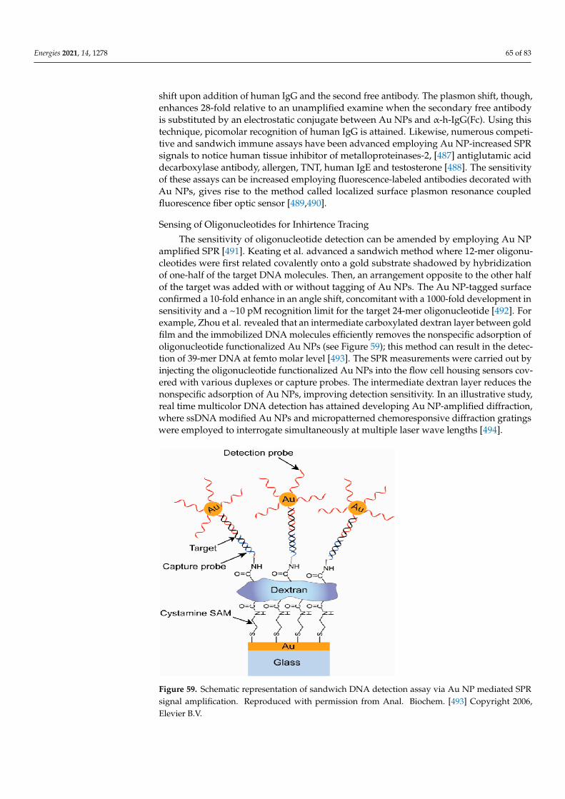
Energies 2021, 14, 1278 65 of 83
shift upon addition of human IgG and the second free antibody. The plasmon shift, though,enhances 28-fold relative to an unamplified examine when the secondary free antibodyis substituted by an electrostatic conjugate between Au NPs and α-h-IgG(Fc). Using thistechnique, picomolar recognition of human IgG is attained. Likewise, numerous competi-tive and sandwich immune assays have been advanced employing Au NP-increased SPRsignals to notice human tissue inhibitor of metalloproteinases-2, [487] antiglutamic aciddecarboxylase antibody, allergen, TNT, human IgE and testosterone [488]. The sensitivityof these assays can be increased employing fluorescence-labeled antibodies decorated withAu NPs, gives rise to the method called localized surface plasmon resonance coupledfluorescence fiber optic sensor [489,490].
Sensing of Oligonucleotides for Inhirtence Tracing
The sensitivity of oligonucleotide detection can be amended by employing Au NPamplified SPR [491]. Keating et al. advanced a sandwich method where 12-mer oligonu-cleotides were first related covalently onto a gold substrate shadowed by hybridizationof one-half of the target DNA molecules. Then, an arrangement opposite to the other halfof the target was added with or without tagging of Au NPs. The Au NP-tagged surfaceconfirmed a 10-fold enhance in an angle shift, concomitant with a 1000-fold development insensitivity and a ~10 pM recognition limit for the target 24-mer oligonucleotide [492]. Forexample, Zhou et al. revealed that an intermediate carboxylated dextran layer between goldfilm and the immobilized DNA molecules efficiently removes the nonspecific adsorption ofoligonucleotide functionalized Au NPs (see Figure 59); this method can result in the detec-tion of 39-mer DNA at femto molar level [493]. The SPR measurements were carried out byinjecting the oligonucleotide functionalized Au NPs into the flow cell housing sensors cov-ered with various duplexes or capture probes. The intermediate dextran layer reduces thenonspecific adsorption of Au NPs, improving detection sensitivity. In an illustrative study,real time multicolor DNA detection has attained developing Au NP-amplified diffraction,where ssDNA modified Au NPs and micropatterned chemoresponsive diffraction gratingswere employed to interrogate simultaneously at multiple laser wave lengths [494].
Figure 59. Schematic representation of sandwich DNA detection assay via Au NP mediated SPRsignal amplification. Reproduced with permission from Anal. Biochem. [493] Copyright 2006,Elevier B.V.
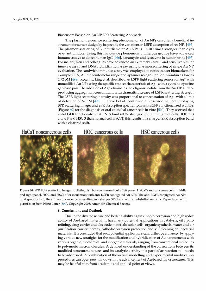
Energies 2021, 14, 1278 66 of 83
Biosensors Bassed on Au NP SPR Scattering Approach
The plasmon resonance scattering phenomenon of Au NPs can offer a beneficial in-strument for sensor design by inspecting the variations in LSPR absorption of Au NPs [495].The plasmon scattering of 36 nm diameter Au NPs is 10–100 times stronger than dyesor quantum dots. Using this nano-scale phenomena, numerous groups have advancedimmune assays to detect human IgG [496], kanamycin and lysozyme in human urine [497].For instant, Ren and colleagues have advanced an extremely careful and sensitive similarimmune assay and DNA hybridization assay using plasmon scattering of single Au NPevaluation. The sandwich immuneo assay was employed to notice cancer biomarkers forexample CEA, AFP in femtomolar range and aptamer recognition for thrombin as low as2.72 pM [498]. Recently, Ling et al. described an LSPR light scattering sensor for Ag+ withunmodified Au NPs using the specific respect characteristic of Ag+ with a cytosine-cytosinegap base pair. The addition of Ag+ eliminates the oligonucleotide from the Au NP surfaceproducing aggregation concomitant with dramatic increase of LSPR scattering strength.The LSPR light scattering intensity was proportional to concentration of Ag+ with a limitof detection of 62 nM [499]. El Sayed et al. confirmed a biosensor method employingSPR scattering images and SPR absorption spectra from anti-EGFR functionalized Au NPs(Figure 60) for the diagnosis of oral epithelial cancer cells in vitro [500]. They oserved thatanti-EGFR functionalized Au NPs bind 600% stronger to oral malignant cells HOC 313clone 8 and HSC 3 than normal cell HaCaT; this results in a sharper SPR absorption bandwith a clear red shift.
Figure 60. SPR light scattering images to distinguish between normal cells (left panel, HaCaT) and cancerous cells (middleand right panel, HOC and HSC) after incubation with anti-EGFR conjugated Au NPs. The anti-EGFR conjugated Au NPsbind specifically to the surface of cancer cells resulting in a sharper SPR band with a red-shifted maxima. Reproduced withpermission from Nano Letter [500]. Copyright 2005, American Chemical Society.
8. Conclusions and Outlook
Due to the diverse nature and better stability against photo-corrosion and high redoxability of Au-based material, it has many potential applications in catalysis, oil hydrorefining, drug carrier and electrode materials, solar cells, organic synthesis, water and airpurification, cancer therapy, cathodic corrosion protection and self-cleaning antibacterialmaterials. It is concluded that such potential applications can further be enhanced by apply-ing various new stratigies for the modification and hybridization of Au nanostructes withvarious organic, biochemical and inorganic materials, ranging from conventional moleculesto polymeric macromolecules. A detailed understanding of the correlations between itsmodified structures/natures and its catalytic activity in a particular reaction still needsto be addressed. A combination of theoretical modelling and experimental modificationprocedures can open new windows in the advancement of Au-based nanostructures. Thismay be helpful both from academic and applied point of views.
Energies 2021, 14, 1278 67 of 83
Author Contributions: Conceptualization, M.Y., M.H., A.K. and H.U. (Habib Ullah 5); validation,M.H., A.K., A.A.T., H.U. (Habib Ullah 5), M.U. and H.U. (Habib Ullah 4); writing—original draftpreparation, M.Y.; writing—review and editing, M.H., A.K. and H.U. (Habib Ullah 5); visualization,M.H., A.K., A.A.T., H.U. (Habib Ullah 4), M.U. and H.U. (Habib Ullah 5); supervision, M.H., A.K. andH.U. (Habib Ullah 5); All authors have read and agreed to the published version of the manuscript.
Funding: This research received no external funding.
Acknowledgments: We are thankful to the Engineering and Physical Science Research Council,UK (EPSRC under research grant no. EP/V049046/1 and EP/T025875/, and Saudi Aramco ChairProgramme (ORCP2390) for financial support.
Conflicts of Interest: The authors declare no conflict of interest.
References1. Wiesner, M.; Bottero, J.-Y. Environmental Nanotechnology; McGraw-Hill Professional Publishing New York: New York, NY, USA, 2007.2. Di Guglielmo, C.; López, D.R.; De Lapuente, J.; Mallafre, J.M.L.; Suàrez, M.B. Embryotoxicity of cobalt ferrite and gold
nanoparticles: A first in vitro approach. Reprod. Toxicol. 2010, 30, 271–276.3. Eck, D.; Helm, C.A.; Wagner, N.J.; Vaynberg, K.A. Plasmon resonance measurements of the adsorption and adsorption kinetics of
a biopolymer onto gold nanocolloids. Langmuir 2001, 17, 957–960.4. Edwards, P.P.; Thomas, J.M. Gold in a metallic divided state—from faraday to present-day nanoscience. Angew. Chem. Int. Ed.
2007, 46, 5480–5486.5. Kang, B.; Mackey, M.A.; El-Sayed, M.A. Nuclear targeting of gold nanoparticles in cancer cells induces DNA damage, causing
cytokinesis arrest and apoptosis. J. Am. Chem. Soc. 2010, 132, 1517–1519.6. Shang, C.; Liu, Z.-P. Origin and activity of gold nanoparticles as aerobic oxidation catalysts in aqueous solution. J. Am. Chem. Soc.
2011, 133, 9938–9947.7. Abadeer, N.S.; Murphy, C.J. Recent progress in cancer thermal therapy using gold nanoparticles. J. Phys. Chem. C 2016, 120,
4691–4716.8. Saleh, T.A. Spectroscopy: Between Modeling, Simulation and Practical Investigation. Spectr. Anal. Rev. 2014, 2, 1–2.9. Saleh, T.A. Sensing of chlorpheniramine in pharmaceutical applications by sequential injector coupled with potentiometer.
J. Pharm. Anal. 2011, 1, 246–250.10. Kelly, K.L.; Coronado, E.; Zhao, L.L.; Schatz, G.C. The optical properties of metal nanoparticles: The influence of size, shape, and
dielectric environment. J. Phys. Chem. B 2003, 107, 668–677.11. Takami, A.; Kurita, H.; Koda, S. Laser-induced size reduction of noble metal particles. J. Phys. Chem. B 1999, 103, 1226–1232.12. Chen, D. Design, synthesis and properties of highly functional nanostructured photocatalysts. Recent Pat. Nanotechnol. 2008, 2,
183–189.13. Balamurugan, B.; Maruyama, T. Evidence of an enhanced interband absorption in Au nanoparticles: Size-dependent electronic
structure and optical properties. Appl. Phys. Lett. 2005, 87, 143105.14. Chen, X.; Zheng, Z.; Ke, X.; Jaatinen, E.; Xie, T.; Wang, D.; Guo, C.; Zhao, J.; Zhu, H. Supported silver nanoparticles as
photocatalysts under ultraviolet and visible light irradiation. Green Chem. 2010, 12, 414–419.15. Litter, M.I. Heterogeneous photocatalysis: Transition metal ions in photocatalytic systems. Appl. Catal. B Environ. 1999, 23, 89–114.16. Link, S.; El-Sayed, M. Steady state and time resolved optical properties of metallic nanoparticles the surface plasmon absorption
as an analytical tool to inverstigate particle properties. Int. Rev. Phys. Chem. 2000, 19, 409.17. Furube, A.; Du, L.; Hara, K.; Katoh, R.; Tachiya, M. Ultrafast plasmon-induced electron transfer from gold nanodots into TiO2
nanoparticles. J. Am. Chem. Soc. 2007, 129, 14852–14853.18. Grirrane, A.; Corma, A.; García, H. Gold-catalyzed synthesis of aromatic azo compounds from anilines and nitroaromatics.
Science 2008, 322, 1661–1664.19. Khumaeni, A.; Budi, W.S.; Sutanto, H. Synthesis and characterization of high-purity gold nanoparticles by laser ablation method
using low-energy Nd: YAG laser 1064 nm. J. Phys. Conf. Ser. 2017, 909, 012037.20. Eskandari-Nojedehi, M.; Jafarizadeh-Malmiri, H.; Rahbar-Shahrouzi, J. Hydrothermal green synthesis of gold nanoparticles using
mushroom (Agaricus bisporus) extract: Physico-chemical characteristics and antifungal activity studies. Green Process. Synth.2018, 7, 38–47.
21. Gutierrez-Wing, C.; Esparza, R.; Vargas-Hernandez, C.; Garcia, M.F.; Jose-Yacaman, M. Microwave-assisted synthesis of goldnanoparticles self-assembled into self-supported superstructures. Nanoscale 2012, 4, 2281–2287.
22. Sakai, T.; Enomoto, H.; Sakai, H.; Abe, M. Hydrogen-assisted fabrication of spherical gold nanoparticles through sonochemicalreduction of tetrachloride gold (III) ions in water. Ultrason. Sonochem. 2014, 21, 946–950.
23. Hussain, R.K.; Abdulghani, A.J. Synthesis of gold nanoparticles via chemical reduction of Au (III) ions by isatin in aqueoussolutions: Ligand concentrations and pH effects. Baghdad Sci. J. 2014, 11, 1201–1216.
24. Sadeghi, B.; Mohammadzadeh, M.; Babakhani, B. Green synthesis of gold nanoparticles using Stevia rebaudiana leaf extracts:Characterization and their stability. J. Photochem. Photobiol. B Biol. 2015, 148, 101–106.
Energies 2021, 14, 1278 68 of 83
25. Zhu, L.; Zhang, C.; Guo, C.; Wang, X.; Sun, P.; Zhou, D.; Chen, W.; Xue, G. New insight into intermediate precursors ofBrust–Schiffrin gold nanoparticles synthesis. J. Phys. Chem. C 2013, 117, 11399–11404.
26. Hoo, X.-F.; Razak, K.A.; Ridhuan, N.S.; Nor, N.M.; Zakaria, N.D. Synthesis of tunable size gold nanoparticles using seedinggrowth method and its application in glucose sensor. AIP Conf. Proc. 2017, 1877, 030001.
27. Ruivo, A.; Ventura, M.; da Silva, M.G.; Laia, C. Synthesis of gold nanoparticles in sol–gel glass porogens containing [bmim][BF 4]ionic liquid. J. Sol-Gel Sci. Technol. 2013, 68, 234–244.
28. Masanori Sakamoto, M.F.; Majima, T. Light as a construction tool of metal nanoparticles: Synthesis and mechanism. J. Photochem.Photobiol. C 2009, 10, 33–56.
29. Mian Rong Lee, H.K.L.; Yijie Yang, C.S.L.; Koh, C.L.L.; Lee, Y.H.; Phang, I.Y.; Ling, X.Y. Direct Metal Writing and PrecisePositioning of Gold Nanoparticles within Microfluidic Channels for SERS Sensing of Gaseous Analytes. Acs Appl. Mater. Interfaces2017, 9, 45.
30. Izquierdo-Lorenzo, I.; Jradi, S.; Adam, P.-M. Direct laser writing of random Au nanoparticle three-dimensional structures forhighly reproducible micro-SERS measurements. R. Soc. Chem. 2014, 4, 4128–4133.
31. Ritacco, T.; Pagliusi, P.; Giocondo, M. Insight into diffusive and convective processes affecting gold nanoparticles microclusteringby multiphoton photoreduction. Colloids Surf. A Phys. Eng. Asp. 2021, 610, 125927.
32. Chen, Y.-S.; Hung, Y.-C.; Liau, I.; Huang, G.S. Assessment of the in vivo toxicity of gold nanoparticles. Nanoscale Res. Lett. 2009, 4, 858.33. Colvin, V.L. The potential environmental impact of engineered nanomaterials. Nat. Biotechnol. 2003, 21, 1166–1170.34. Shedbalkar, U.; Singh, R.; Wadhwani, S.; Gaidhani, S.; Chopade, B. Microbial synthesis of gold nanoparticles: Current status and
future prospects. Adv. Colloid Interface Sci. 2014, 209, 40–48.35. Thakkar, K.N.; Mhatre, S.S.; Parikh, R.Y. Biological synthesis of metallic nanoparticles. Nanomed. Nanotechnol. Biol. Med. 2010, 6,
257–262.36. Dass, A.; Guo, R.; Tracy, J.B.; Balasubramanian, R.; Douglas, A.D.; Murray, R.W. Gold nanoparticles with perfluorothiolate ligands.
Langmuir 2008, 24, 310–315.37. Kroto, H.; Heath, J.O.; Brien, S.C.; Curl, R.F.; Smalley, R.E. C60 Buckminsterfullerene. Nature 1985, 318, 162–163.38. Nakamura, E.; Isobe, H. Functionalized fullerenes in water. The first 10 years of their chemistry, biology, and nanoscience. Acc.
Chem. Res. 2003, 36, 807–815.39. Brust, M.; Kiely, C.J.; Bethell, D.; Schiffrin, D.J. C60 mediated aggregation of gold nanoparticles. J. Am. Chem. Soc. 1998, 120,
12367–12368.40. Fujihara, H.; Nakai, H. Fullerenethiolate-functionalized gold nanoparticles: A new class of surface-confined metal-c60 nanocom-
posites. Langmuir 2001, 17, 6393–6395.41. Sudeep, P.; Ipe, B.I.; Thomas, K.G.; George, M.; Barazzouk, S.; Hotchandani, S.; Kamat, P.V. Fullerene-functionalized gold
nanoparticles. A self-assembled photoactive antenna-metal nanocore assembly. Nano Lett. 2002, 2, 29–35.42. Shon, Y.-S.; Choo, H. [60]Fullerene-linked gold nanoparticles: Synthesis and layer-by-layer growth on a solid surface. Chem.
Commun. 2002, 2560–2561. [CrossRef]43. Liu, J.; Alvarez, J.; Ong, W.; Kaifer, A.E. Network aggregates formed by C60 and gold nanoparticles capped with γ-cyclodextrin
hosts. Nano Lett. 2001, 1, 57–60.44. Hasobe, T.; Imahori, H.; Kamat, P.V.; Fukuzumi, S. Quaternary self-organization of porphyrin and fullerene units by clusterization
with gold nanoparticles on SnO2 electrodes for organic solar cells. J. Am. Chem. Soc. 2003, 125, 14962–14963.45. Islam, M.T.; Molugu, S.K.; Cooke, P.H.; Noveron, J.C. Fullerene stabilized gold nanoparticles. New J. Chem. 2015, 39, 5923–5926.46. Frare, M.; Pilot, R.; De Filippo, C.; Weber, V.; Signorini, R.; Maggini, M.; Bozio, R. Fullerene functionalized gold nanoparticles for
optical limiting of continuous wave lasers. Appl. Phys. B 2019, 125, 1–12.47. Plesek, J. Potential applications of the boron cluster compounds. Chem. Rev. 1992, 92, 269–278.48. Hawthorne, M.F.; Maderna, A. Applications of Radiolabeled Boron Clusters to the Diagnosis and Treatment of Cancer. Chem. Rev.
1999, 99, 3421–3434.49. Barbera, G.; Vaca, A.; Teixidor, F.; Sillanpää, R.; Kivekäs, R.; Vinas, C. Designed synthesis of new ortho-carborane derivatives:
From mono-to polysubstituted frameworks. Inorg. Chem. 2008, 47, 7309–7316.50. Dash, B.P.; Satapathy, R.; Maguire, J.A.; Hosmane, N.S. Synthesis of a new class of carborane-containing star-shaped molecules
via silicon tetrachloride promoted cyclotrimerization reactions. Org. Lett. 2008, 10, 2247–2250.51. Smith, H., Jr.; Obenland, C.; Papetti, S. A New Series of Organoboranes. IX. The Preparation and Some Reactions of Sulfur-
Carborane Derivatives. Inorg. Chem. 1966, 5, 1013–1015.52. Plešek, J.; Hermánek, S. Syntheses and properties of substituted icosahedral carborane thiols. Collect. Czechoslov. Chem. Commun.
1981, 46, 687–692.53. Baše, T.; Bastl, Z.; Plzák, Z.; Grygar, T.; Plešek, J.; Carr, M.J.; Malina, V.; Šubrt, J.; Bohácek, J.; Vecerníková, E. Carboranethiol-
modified gold surfaces. A study and comparison of modified cluster and flat surfaces. Langmuir 2005, 21, 7776–7785.54. Baše, T.S.; Bastl, Z.; Šlouf, M.; Klementová, M.; Šubrt, J.; Vetushka, A.; Ledinský, M.; Fejfar, A.; Machácek, J.; Carr, M.J. Gold
micrometer crystals modified with carboranethiol derivatives. J. Phys. Chem. C 2008, 112, 14446–14455.55. Grzelczak, M.P.; Danks, S.P.; Klipp, R.C.; Belic, D.; Zaulet, A.; Kunstmann-Olsen, C.; Bradley, D.F.; Tsukuda, T.; Viñas, C.; Teixidor,
F. Ion transport across biological membranes by carborane-capped gold nanoparticles. Acs Nano 2017, 11, 12492–12499.
Energies 2021, 14, 1278 69 of 83
56. Wang, J.; Chen, L.; Ye, J.; Li, Z.; Jiang, H.; Yan, H.; Stogniy, M.Y.; Sivaev, I.B.; Bregadze, V.I.; Wang, X. Carborane derivativeconjugated with gold nanoclusters for targeted cancer cell imaging. Biomacromolecules 2017, 18, 1466–1472.
57. Bogireddy, N.; Pal, U.; Gomez, L.M.; Agarwal, V. Size controlled green synthesis of gold nanoparticles using Coffea arabica seedextract and their catalytic performance in 4-nitrophenol reduction. Rsc Adv. 2018, 8, 24819–24826.
58. Schmid, G.; Pugin, R.; Malm, J.O.; Bovin, J.O. Silsesquioxanes as ligands for gold clusters. Eur. J. Inorg. Chem. 1998, 1998, 813–817.59. Carroll, J.B.; Frankamp, B.L.; Rotello, V.M. Self-assembly of gold nanoparticles through tandem hydrogen bonding and poly-
oligosilsequioxane (POSS)–POSS recognition processes. Chem. Commun. 2002, 1892–1893.60. Carroll, J.B.; Frankamp, B.L.; Srivastava, S.; Rotello, V.M. Electrostatic self-assembly of structured gold nanoparticle/polyhedral
oligomeric silsesquioxane (POSS) nanocomposites. J. Mater. Chem. 2004, 14, 690–694.61. Zhang, Y.; Hao, J.; Xu, X.; Chen, X.; Wang, J. Protein Corona-Triggered Catalytic Inhibition of Insufficient POSS Polymer-Caged
Gold Nanoparticles for Sensitive Colorimetric Detection of Metallothioneins. Anal. Chem. 2019, 92, 2080–2087.62. Xia, S.; Yang, Y.; Zhu, W.; Lü, C. Quaternized polyhedral oligomeric silsesquioxanes stabilized Pd nanoparticles as efficient
nanocatalysts for reduction reaction. Colloids Surf. A Physicochem. Eng. Asp. 2020, 585, 124110.63. Hanske, C.; Sanz-Ortiz, M.N.; Liz-Marzán, L.M. Silica-Coated Plasmonic Metal Nanoparticles in Action. Adv. Mater. 2018,
30, 1707003.64. Chapman, B.S.; Wu, W.-C.; Li, Q.; Holten-Andersen, N.; Tracy, J.B. Heteroaggregation approach for depositing magnetite
nanoparticles onto Silica-overcoated gold nanorods. Chem. Mater. 2017, 29, 10362–10368.65. Liz-Marzán, L.M.; Giersig, M.; Mulvaney, P. Synthesis of nanosized gold-silica core-shell particles. Langmuir 1996, 12, 4329–4335.66. Han, Y.; Jiang, J.; Lee, S.S.; Ying, J.Y. Reverse microemulsion-mediated synthesis of silica-coated gold and silver nanoparticles.
Langmuir 2008, 24, 5842–5848.67. Casavola, M.; Buonsanti, R.; Caputo, G.; Cozzoli, P.D. Colloidal strategies for preparing oxide-based hybrid nanocrystals. Eur. J.
Inorg. Chem. 2008, 2008, 837–854.68. Rodríguez-Fernández, J.; Pastoriza-Santos, I.; Perez-Juste, J.; García de Abajo, F.J.; Liz-Marzan, L.M. The effect of silica coating on
the optical response of sub-micrometer gold spheres. J. Phys. Chem. C 2007, 111, 13361–13366.69. Ruach-Nir, I.; Bendikov, T.A.; Doron-Mor, I.; Barkay, Z.; Vaskevich, A.; Rubinstein, I. Silica-stabilized gold island films for
transmission localized surface plasmon sensing. J. Am. Chem. Soc. 2007, 129, 84–92.70. Liu, G.; Ji, H.; Yang, X.; Wang, Y. Synthesis of a Au/silica/polymer trilayer composite and the corresponding hollow polymer
microsphere with a movable Au core. Langmuir 2008, 24, 1019–1025.71. Tovmachenko, O.G.; Graf, C.; van den Heuvel, D.J.; van Blaaderen, A.; Gerritsen, H.C. Fluorescence enhancement by metal-
core/silica-shell nanoparticles. Adv. Mater. 2006, 18, 91–95.72. Kamata, K.; Lu, Y.; Xia, Y. Synthesis and characterization of monodispersed core-shell spherical colloids with movable cores. J.
Am. Chem. Soc. 2003, 125, 2384–2385.73. Iijima, S. Synthesis of carbon nanotubes. Nature 1991, 354, 56–58.74. Andrews, R.; Jacques, D.; Qian, D.; Rantell, T. Multiwall carbon nanotubes: Synthesis and application. Acc. Chem. Res. 2002, 35,
1008–1017.75. Koo, W.-T.; Kim, Y.; Savagatrup, S.; Yoon, B.; Jeon, I.; Choi, S.-J.; Kim, I.-D.; Swager, T.M. Porous Ion Exchange Polymer Matrix for
Ultrasmall Au Nanoparticle-Decorated Carbon Nanotube Chemiresistors. Chem. Mater. 2019, 31, 5413–5420.76. Deshmukh, S.P.; Dhodamani, A.G.; Patil, S.M.; Mullani, S.B.; More, K.V.; Delekar, S.D. Interfacially Interactive Ternary Silver-
Supported Polyaniline/Multiwalled Carbon Nanotube Nanocomposites for Catalytic and Antibacterial Activity. Acs Omega 2019,5, 219–227.
77. LeeáTan, K. Growth of Pd, Pt, Ag and Au nanoparticles on carbon nanotubes. J. Mater. Chem. 2001, 11, 2378–2381.78. Han, L.; Wu, W.; Kirk, F.L.; Luo, J.; Maye, M.M.; Kariuki, N.N.; Lin, Y.; Wang, C.; Zhong, C.-J. A direct route toward assembly of
nanoparticle-carbon nanotube composite materials. Langmuir 2004, 20, 6019–6025.79. Sainsbury, T.; Ikuno, T.; Okawa, D.; Pacile, D.; Frechet, J.M.; Zettl, A. Self-assembly of gold nanoparticles at the surface of
amine-and thiol-functionalized boron nitride nanotubes. J. Phys. Chem. C 2007, 111, 12992–12999.80. Jiang, K.; Eitan, A.; Schadler, L.S.; Ajayan, P.M.; Siegel, R.W.; Grobert, N.; Mayne, M.; Reyes-Reyes, M.; Terrones, H.; Terrones, M.
Selective attachment of gold nanoparticles to nitrogen-doped carbon nanotubes. Nano Lett. 2003, 3, 275–277.81. Correa-Duarte, M.A.; Liz-Marzán, L.M. Carbon nanotubes as templates for one-dimensional nanoparticle assemblies. J. Mater.
Chem. 2006, 16, 22–25.82. Fu, Q.; Lu, C.; Liu, J. Selective coating of single wall carbon nanotubes with thin SiO2 layer. Nano Lett. 2002, 2, 329–332.83. Bottini, M.; Magrini, A.; Rosato, N.; Bergamaschi, A.; Mustelin, T. Dispersion of pristine single-walled carbon nanotubes in water
by a thiolated organosilane: Application in supramolecular nanoassemblies. J. Phys. Chem. B 2006, 110, 13685–13688.84. Ou, Y.-Y.; Huang, M.H. High-density assembly of gold nanoparticles on multiwalled carbon nanotubes using 1-pyrenemethylamine
as interlinker. J. Phys. Chem. B 2006, 110, 2031–2036.85. Hill, C.L. Introduction: Polyoxometalates multicomponent molecular vehicles to probe fundamental issues and practical problems.
Chem. Rev. 1998, 98, 1–2.86. Wang, Y.; Neyman, A.; Arkhangelsky, E.; Gitis, V.; Meshi, L.; Weinstock, I.A. Self-assembly and structure of directly imaged
inorganic-anion monolayers on a gold nanoparticle. J. Am. Chem. Soc. 2009, 131, 17412–17422.
Energies 2021, 14, 1278 70 of 83
87. Wang, Y.; Zeiri, O.; Sharet, S.; Weinstock, I.A. Role of the alkali-metal cation size in the self-assembly of polyoxometalate-monolayer shells on gold nanoparticles. Inorg. Chem. 2012, 51, 7436–7438.
88. Sutter, S.; Trepka, B.; Siroky, S.; Hagedorn, K.; Theiß, S.; Baum, P.; Polarz, S. Light-Triggered Boost of Activity of CatalyticBola-Type Surfactants by a Plasmonic Metal–Support Interaction Effect. Acs Appl. Mater. Interfaces 2019, 11, 15936–15944.
89. Solarska, R.; Bienkowski, K.; Zoladek, S.; Majcher, A.; Stefaniuk, T.; Kulesza, P.J.; Augustynski, J. Enhanced water splitting atthin film tungsten trioxide photoanodes bearing plasmonic gold–polyoxometalate particles. Angew. Chem. Int. Ed. 2014, 53,14196–14200.
90. Dolbecq, A.; Dumas, E.; Mayer, C.R.; Mialane, P. Hybrid organic-inorganic polyoxometalate compounds: From structural diversityto applications. Chem. Rev. 2010, 110, 6009–6048.
91. Mayer, C.R.; Neveu, S.; Cabuil, V. A nanoscale hybrid system based on gold nanoparticles and heteropolyanions. Angew. Chem.Int. Ed. 2002, 41, 501–503.
92. Hegde, S.; Joshi, S.; Mukherjee, T.; Kapoor, S. Formation of gold nanoparticles via a thiol functionalized polyoxometalate. Mater.Sci. Eng. C 2013, 33, 2332–2337.
93. Martín, S.; Takashima, Y.; Lin, C.-G.; Song, Y.-F.; Miras, H.N.; Cronin, L. Integrated synthesis of gold nanoparticles coated withpolyoxometalate clusters. Inorg. Chem. 2019, 58, 4110–4116.
94. Tomane, S.; López-Maya, E.; Boujday, S.; Humblot, V.; Marrot, J.; Rabasso, N.; Castells-Gil, J.; Sicard, C.; Dolbecq, A.; Mialane, P.Correction: One-pot synthesis of a new generation of hybrid bisphosphonate polyoxometalate gold nanoparticles as antibiofilmagents. Nanoscale Adv. 2019, 1, 4173.
95. Wilton-Ely, J.D. The surface functionalisation of gold nanoparticles with metal complexes. Dalton Trans. 2008, 25–29. [CrossRef]96. Stiles, R.L.; Balasubramanian, R.; Feldberg, S.W.; Murray, R.W. Anion-induced adsorption of ferrocenated nanoparticles. J. Am.
Chem. Soc. 2008, 130, 1856–1865.97. Wolfe, R.L.; Balasubramanian, R.; Tracy, J.B.; Murray, R.W. Fully ferrocenated hexanethiolate monolayer-protected gold clusters.
Langmuir 2007, 23, 2247–2254.98. Chen, S.; Murray, R.W. Arenethiolate monolayer-protected gold clusters. Langmuir 1999, 15, 682–689.99. Li, D.; Zhang, Y.; Jiang, J.; Li, J. Electroactive gold nanoparticles protected by 4-ferrocene thiophenol monolayer. J. Colloid Interface
Sci. 2003, 264, 109–113.100. Li, D.; Zhang, Y.; Li, J. Electrochemical study of 4-ferrocene thiophenol monolayers assembled on gold nanoparticles. Microelectron.
Eng. 2003, 66, 91–94.101. Ingram, R.S.; Hostetler, M.J.; Murray, R.W. Poly-hetero-ω-functionalized alkanethiolate-stabilized gold cluster compounds. J. Am.
Chem. Soc. 1997, 119, 9175–9178.102. Green, S.J.; Stokes, J.J.; Hostetler, M.J.; Pietron, J.; Murray, R.W. Three-dimensional monolayers: Nanometer-sized electrodes of
alkanethiolate-stabilized gold cluster molecules. J. Phys. Chem. B 1997, 101, 2663–2668.103. Ornelas, C.; Méry, D.; Cloutet, E.; Aranzaes, J.R.; Astruc, D. Cross olefin metathesis for the selective functionalization, ferro-
cenylation, and solubilization in water of olefin-terminated dendrimers, polymers, and gold nanoparticles and for a divergentdendrimer construction. J. Am. Chem. Soc. 2008, 130, 1495–1506.
104. Labande, A.; Astruc, D. Colloids as redox sensors: Recognition of H2PO4− and HSO4− by amidoferrocenylalkylthiol–goldnanoparticles. Chem. Commun. 2000, 124, 1007–1008.
105. Wang, Y.; Salmon, L.; Ruiz, J.; Astruc, D. Metallodendrimers in three oxidation states with electronically interacting metals andstabilization of size-selected gold nanoparticles. Nat. Commun. 2014, 5, 1–12.
106. Labande, A.; Ruiz, J.; Astruc, D. Supramolecular gold nanoparticles for the redox recognition of oxoanions: Syntheses, titrations,stereoelectronic effects, and selectivity. J. Am. Chem. Soc. 2002, 124, 1782–1789.
107. Horikoshi, T.; Itoh, M.; Kurihara, M.; Kubo, K.; Nishihara, H. Synthesis, redox behavior and electrodeposition of biferrocene-modified gold clusters. J. Electroanal. Chem. 1999, 473, 113–116.
108. Yamada, M.; Tadera, T.; Kubo, K.; Nishihara, H. Electrochemical deposition of biferrocene derivative-attached gold nanoparticlesand the morphology of the formed film. J. Phys. Chem. B 2003, 107, 3703–3711.
109. Dong, T.-Y.; Shih, H.-W.; Chang, L.-S. Synthesis and redox behavior of biferrocenyl-functionalized ruthenium (II) terpyridine goldclusters. Langmuir 2004, 20, 9340–9347.
110. Daniel, M.-C.; Ruiz, J.; Nlate, S.; Palumbo, J.; Blais, J.-C.; Astruc, D. Gold nanoparticles containing redox-active supramoleculardendrons that recognize H2PO4−. Chem. Commun. 2001, 2000–2001.
111. Daniel, M.-C.; Ruiz, J.; Nlate, S.; Blais, J.-C.; Astruc, D. Nanoscopic assemblies between supramolecular redox active metallo-dendrons and gold nanoparticles: Synthesis, characterization, and selective recognition of H2PO4−, HSO4−, and adenosine-5′-triphosphate (ATP2-) anions. J. Am. Chem. Soc. 2003, 125, 2617–2628.
112. Astruc, D.; Daniel, M.-C.; Ruiz, J. Dendrimers and gold nanoparticles as exo-receptors sensing biologically important anions.Chem. Commun. 2004, 2637–2649.
113. Liu, F.; Liu, X.; Astruc, D.; Gu, H. Dendronized triazolyl-containing ferrocenyl polymers as stabilizers of gold nanoparticles forrecyclable two-phase reduction of 4-nitrophenol. J. Colloid Interface Sci. 2019, 533, 161–170.
114. Opuchlik, L.J.; Pawłowska, J.; Sek, S.; Bilewicz, R. Ferrocenylated gold nanoparticles self-assemble at carbon surfaces to formstable films. J. Electroanal. Chem. 2018, 825, 22–29.
Energies 2021, 14, 1278 71 of 83
115. Vitale, F.; Vitaliano, R.; Battocchio, C.; Fratoddi, I.; Piscopiello, E.; Tapfer, L.; Russo, M.V. Synthesis and characterization of goldnanoparticles stabilized by palladium (II) phosphine thiol. J. Organomet. Chem. 2008, 693, 1043–1048.
116. Bartz, M.; Küther, J.; Seshadri, R.; Tremel, W. Colloid-Bound Catalysts for Ring-Opening Metathesis Polymerization: A Combina-tion of Homogenous and Heterogeneous Properties. Angew. Chem. Int. Ed. 1998, 37, 2466–2468.
117. Wang, S.; Sim, W.-S. Au nanoparticles encapsulated in Ru carbonyl carboxylate shells. Langmuir 2006, 22, 7861–7866.118. Belser, T.; Stöhr, M.; Pfaltz, A. Immobilization of rhodium complexes at thiolate monolayers on gold surfaces: Catalytic and
structural studies. J. Am. Chem. Soc. 2005, 127, 8720–8731.119. Kumar, S.S.; Joseph, J.; Phani, K.L. Novel method for deposition of Gold-Prussian blue nanocomposite films induced by
electrochemically formed gold nanoparticles: Characterization and application to electrocatalysis. Chem. Mater. 2007, 19,4722–4730.
120. Crespilho, F.N.; Zucolotto, V.; Brett, C.M.; Oliveira, O.N.; Nart, F.C. Enhanced charge transport and incorporation of redoxmediators in layer-by-layer films containing PAMAM-encapsulated gold nanoparticles. J. Phys. Chem. B 2006, 110, 17478–17483.
121. Qiu, J.-D.; Peng, H.-Z.; Liang, R.-P.; Li, J.; Xia, X.-H. Synthesis, characterization, and immobilization of Prussian blue-modified Aunanoparticles: Application to electrocatalytic reduction of H2O2. Langmuir 2007, 23, 2133–2137.
122. Dumur, F.; Dumas, E.; Mayer, C.R. Functionalization of Gold Nanoparticles by Inorganic Entities. Nanomaterials 2020, 10, 548.123. Toma, S.H.; Bonacin, J.A.; Araki, K.; Toma, H.E. Controlled Stabilization and Flocculation of Gold Nanoparticles by Means of
2-Pyrazin-2-ylethanethiol and Pentacyanidoferrate (II) Complexes. Eur. J. Inorg. Chem. 2007, 2007, 3356–3364.124. Nunes, F.S.; Bonifácio, L.D.S.; Araki, K.; Toma, H.E. Interaction of 2-and 4-mercaptopyridine with pentacyanoferrates and gold
nanoparticles. Inorg. Chem. 2006, 45, 94–101.125. Cheng, P.P.H.; Silvester, D.; Wang, G.; Kalyuzhny, G.; Douglas, A.; Murray, R.W. Dynamic and static quenching of fluorescence by
1–4 nm diameter gold monolayer-protected clusters. J. Phys. Chem. B 2006, 110, 4637–4644.126. Brewer, S.H.; Glomm, W.R.; Johnson, M.C.; Knag, M.K.; Franzen, S. Probing BSA binding to citrate-coated gold nanoparticles and
surfaces. Langmuir 2005, 21, 9303–9307.127. Xu, X.-H.N.; Huang, S.; Brownlow, W.; Salaita, K.; Jeffers, R.B. Size and temperature dependence of surface plasmon absorption
of gold nanoparticles induced by tris (2, 2′-bipyridine) ruthenium (II). J. Phys. Chem. B 2004, 108, 15543–15551.128. Pramod, P.; Sudeep, P.; Thomas, K.G.; Kamat, P.V. Photochemistry of ruthenium trisbipyridine functionalized on gold nanoparti-
cles. J. Phys. Chem. B 2006, 110, 20737–20741.129. Jebb, M.; Sudeep, P.; Pramod, P.; Thomas, K.G.; Kamat, P.V. Ruthenium (II) trisbipyridine functionalized gold nanorods.
Morphological changes and excited-state interactions. J. Phys. Chem. B 2007, 111, 6839–6844.130. Sun, X.; Du, Y.; Dong, S.; Wang, E. Method for effective immobilization of Ru (bpy) 32+ on an electrode surface for solid-state
electrochemiluminescene detection. Anal. Chem. 2005, 77, 8166–8169.131. Liu, J.; Lu, Y. Stimuli-responsive disassembly of nanoparticle aggregates for light-up colorimetric sensing. J. Am. Chem. Soc. 2005,
127, 12677–12683.132. Kuwahara, Y.; Akiyama, T.; Yamada, S. Construction of gold nanoparticle-ruthenium (II) tris (2, 2′-bipyridine) self-assembled
multistructures and their photocurrent responses. Thin Solid Film. 2001, 393, 273–277.133. Akiyama, T.; Inoue, K.; Kuwahara, Y.; Terasaki, N.; Niidome, Y.; Yamada, S. Particle-size effects on the photocurrent efficiency of
nanostructured assemblies consisting of gold nanoparticles and a ruthenium complex–viologen linked thiol. J. Electroanal. Chem.2003, 550, 303–307.
134. Akiyama, T.; Inoue, K.; Kuwahara, Y.; Niidome, Y.; Terasaki, N.; Nitahara, S.; Yamada, S. Facile fabrication of morphology-controlled gold nanoparticle architectures by electrolyte-induced agglomeration and their photoelectrochemical applications.Langmuir 2005, 21, 793–796.
135. Lahav, M.; Heleg-Shabtai, V.; Wasserman, J.; Katz, E.; Willner, I.; Dürr, H.; Hu, Y.-Z.; Bossmann, S.H. Photoelectrochemistry withIntegrated Photosensitizer-Electron Acceptor and Au-Nanoparticle Arrays. J. Am. Chem. Soc. 2000, 122, 11480–11487.
136. Shipway, A.N.; Lahav, M.; Willner, I. Nanostructured gold colloid electrodes. Adv. Mater. 2000, 12, 993–998.137. Chen, S.; Pei, R.; Zhao, T.; Dyer, D.J. Gold Nanoparticle Assemblies by Metal Ion-Pyridine Complexation and Their Rectified
Quantized Charging in Aqueous Solutions. J. Phys. Chem. B 2002, 106, 1903–1908.138. Soller, T.; Ringler, M.; Wunderlich, M.; Klar, T.; Feldmann, J.; Josel, H.-P.; Markert, Y.; Nichtl, A.; Kürzinger, K. Radiative and
nonradiative rates of phosphors attached to gold nanoparticles. Nano Lett. 2007, 7, 1941–1946.139. Slim, M.; Durisic, N.; Grutter, P.; Sleiman, H.F. DNA–Protein Noncovalent Cross-Linking: Ruthenium Dipyridophenazine Biotin
Complex for the Assembly of Proteins and Gold Nanoparticles on DNA Templates. ChemBioChem 2007, 8, 804–812.140. Shultz, D.A.; Kumar, R.K.; Bin-Salamon, S.; Kirk, M.L. Valence tautomerization and exchange coupling in a cobalt–
nitronylnitroxide–semiquinone complex. Polyhedron 2005, 24, 2876–2879.141. Barmin, R.A.; Rudakovskaya, P.G.; Gusliakova, O.I.; Sindeeva, O.A.; Prikhozhdenko, E.S.; Maksimova, E.A.; Obukhova, E.N.;
Chernyshev, V.S.; Khlebtsov, B.N.; Solovev, A.A.; et al. Air-Filled Bubbles Stabilized by Gold Nanoparticle/Photodynamic DyeHybrid Structures for Theranostics. Nanomaterials 2021, 11, 415.
142. Mayer, C.R.; Dumas, E.; Miomandre, F.; Méallet-Renault, R.; Warmont, F.; Vigneron, J.; Pansu, R.; Etcheberry, A.; Sécheresse,F. Polypyridyl ruthenium complexes as coating agent for the formation of gold and silver nanocomposites in different media.Preliminary luminescence and electrochemical studies. New J. Chem. 2006, 30, 1628–1637.
Energies 2021, 14, 1278 72 of 83
143. Pérez León, C.; Kador, L.; Peng, B.; Thelakkat, M. Influence of the solvent on the surface-enhanced Raman spectra of ruthenium(II) bipyridyl complexes. J. Phys. Chem. B 2005, 109, 5783–5789.
144. Huang, W.; Tanaka, H.; Ogawa, T. Effects of Metal-Ion Complexation for the Self-Assembled Nanocomposite Films Composed ofGold Nanoparticles and 3, 8-Bis (terthiophenyl) phenanthroline-Based Dithiols Bridging 1 µm Gap Gold Electrodes: Morphology,Temperature Dependent Electronic Conduction, and Photoresponse. J. Phys. Chem. C 2008, 112, 11513–11526.
145. Ono, F.; Kanemasa, S.; Tanaka, J. Reusable nano-sized chiral bisoxazoline catalysts. Tetrahedron Lett. 2005, 46, 7623–7626.146. Hone, D.C.; Walker, P.I.; Evans-Gowing, R.; FitzGerald, S.; Beeby, A.; Chambrier, I.; Cook, M.J.; Russell, D.A. Generation of
cytotoxic singlet oxygen via phthalocyanine-stabilized gold nanoparticles: A potential delivery vehicle for photodynamic therapy.Langmuir 2002, 18, 2985–2987.
147. Beer, P.D.; Cormode, D.P.; Davis, J.J. Zinc metalloporphyrin-functionalised nanoparticle anion sensors. Chem. Commun. 2004,414–415.
148. Ozawa, H.; Kawao, M.; Tanaka, H.; Ogawa, T. Synthesis of dendron-protected porphyrin wires and preparation of a one-dimensional assembly of gold nanoparticles chemically linked to the π-conjugated wires. Langmuir 2007, 23, 6365–6371.
149. Aubin-Tam, M.-E.; Hamad-Schifferli, K. Gold nanoparticle-cytochrome C complexes: The effect of nanoparticle ligand charge onprotein structure. Langmuir 2005, 21, 12080–12084.
150. Sheet, D.; Bera, A.; Jana, R.D.; Paine, T.K. Oxidizing Ability of a Dioxygen-Activating Nonheme Iron (II)-Benzilate ComplexImmobilized on Gold Nanoparticles. Inorg. Chem. 2019, 58, 4828–4841.
151. Huang, C.C.; Yang, Z.; Lee, K.H.; Chang, H.T. Synthesis of highly fluorescent gold nanoparticles for sensing mercury (II). Angew.Chem. 2007, 119, 6948–6952.
152. Huang, C.-C.; Chang, H.-T. Parameters for selective colorimetric sensing of mercury (II) in aqueous solutions using mercaptopro-pionic acid-modified gold nanoparticles. Chem. Commun. 2007, 1215–1217.
153. Zamborini, F.P.; Hicks, J.F.; Murray, R.W. Quantized double layer charging of nanoparticle films assembled usingcarboxylate/(Cu2+ or Zn2+)/carboxylate bridges. J. Am. Chem. Soc. 2000, 122, 4514–4515.
154. Zamborini, F.P.; Leopold, M.C.; Hicks, J.F.; Kulesza, P.J.; Malik, M.A.; Murray, R.W. Electron hopping conductivity and vaporsensing properties of flexible network polymer films of metal nanoparticles. J. Am. Chem. Soc. 2002, 124, 8958–8964.
155. Hicks, J.F.; Zamborini, F.P.; Murray, R.W. Dynamics of electron transfers between electrodes and monolayers of nanoparticles.J. Phys. Chem. B 2002, 106, 7751–7757.
156. Sheibley, D.; Tognarelli, D.; Szymanik, R.; Leopold, M.C. Ultra-fast formation and characterization of stable nanoparticle filmassemblies. J. Mater. Chem. 2005, 15, 491–498.
157. Xu, R.; Wang, D.; Zhang, J.; Li, Y. Shape-dependent catalytic activity of silver nanoparticles for the oxidation of styrene. Chem.Asian J. 2006, 1, 888–893.
158. Wanunu, M.; Popovitz-Biro, R.; Cohen, H.; Vaskevich, A.; Rubinstein, I. Coordination-based gold nanoparticle layers. J. Am.Chem. Soc. 2005, 127, 9207–9215.
159. Mayer, C.R.; Cucchiaro, G.; Jullien, J.; Dumur, F.; Marrot, J.; Dumas, E.; Sécheresse, F. Functionalization of gold nanoparticles byiron (III) complexes derived from Schiff base ligands. Eur. J. Inorg. Chem. 2008, 2008, 3614–3623.
160. Guo, C.; Boullanger, P.; Jiang, L.; Liu, T. Highly sensitive gold nanoparticles biosensor chips modified with a self-assembledbilayer for detection of Con A. Biosens. Bioelectron. 2007, 22, 1830–1834.
161. Türkekul, K.; Üzer, A.; Can, Z.; Erçag, E.; Apak, R. Colorimetric Sensing of the Insensitive Energetic Material 3-Nitro-1, 2,4-triazol-5-one (NTO) Using l-Cysteine Stabilized Gold Nanoparticles and Copper (II). Anal. Lett. 2019, 52, 2809–2821.
162. Abad, J.M.; Mertens, S.F.; Pita, M.; Fernández, V.M.; Schiffrin, D.J. Functionalization of thioctic acid-capped gold nanoparticles forspecific immobilization of histidine-tagged proteins. J. Am. Chem. Soc. 2005, 127, 5689–5694.
163. Reynolds, A.J.; Haines, A.H.; Russell, D.A. Gold glyconanoparticles for mimics and measurement of metal ion-mediatedcarbohydrate-carbohydrate interactions. Langmuir 2006, 22, 1156–1163.
164. Si, S.; Kotal, A.; Mandal, T.K. One-dimensional assembly of peptide-functionalized gold nanoparticles: An approach towardmercury ion sensing. J. Phys. Chem. C 2007, 111, 1248–1255.
165. Lee, J.S.; Han, M.S.; Mirkin, C.A. Colorimetric detection of mercuric ion (Hg2+) in aqueous media using DNA-functionalized goldnanoparticles. Angew. Chem. Int. Ed. 2007, 46, 4093–4096.
166. Liu, J.; Lu, Y. A colorimetric lead biosensor using DNAzyme-directed assembly of gold nanoparticles. J. Am. Chem. Soc. 2003, 125,6642–6643.
167. Liu, J.; Lu, Y. Accelerated color change of gold nanoparticles assembled by DNAzymes for simple and fast colorimetric Pb2+
detection. J. Am. Chem. Soc. 2004, 126, 12298–12305.168. Liu, J.; Lu, Y. Optimization of a Pb2+-directed gold nanoparticle/DNAzyme assembly and its application as a colorimetric
biosensor for Pb2+. Chem. Mater. 2004, 16, 3231–3238.169. Liu, J.; Lu, Y. Colorimetric Cu2+ detection with a ligation DNAzyme and nanoparticles. Chem. Commun. 2007, 4872–4874.
[CrossRef]170. Marubayashi, K.; Takizawa, S.; Kawakusu, T.; Arai, T.; Sasai, H. Monolayer-Protected Au Cluster (MPC)-Supported Ti− BINOLate
Complex. Org. Lett. 2003, 5, 4409–4412.171. Rogers, N.J.; Claire, S.; Harris, R.M.; Farabi, S.; Zikeli, G.; Styles, I.B.; Hodges, N.J.; Pikramenou, Z. High coating of Ru (II)
complexes on gold nanoparticles for single particle luminescence imaging in cells. Chem. Commun. 2014, 50, 617–619.
Energies 2021, 14, 1278 73 of 83
172. Thangavel, N.; Jayakumar, I.; Ravichandran, M.; Ganesan, V.V.; Nair, B.U. Photocrosslinking of collagen using Ru (II)-polypyridylcomplex functionalized gold nanoparticles. Spectrochim. Acta Part A Mol. Biomol. Spectrosc. 2019, 215, 196–202.
173. Cox, J.A.; Kittredge, K.W.; Ca, D.V. Measurement platforms fabricated by layer-by-layer assembly of crown ether functionalizedgold nanoclusters. J. Solid State Electrochem. 2004, 8, 722–726.
174. Lin, S.Y.; Wu, S.H.; Chen, C.h. A simple strategy for prompt visual sensing by gold nanoparticles: General applications ofinterparticle hydrogen bonds. Angew. Chem. 2006, 118, 5070–5073.
175. Wang, C.; Yu, C. Detection of chemical pollutants in water using gold nanoparticles as sensors: A review. Rev. Anal. Chem. 2013,32, 1–14.
176. Yamauchi, A.; Hayashita, T.; Nishizawa, S.; Watanabe, M.; Teramae, N. Benzo-15-crown-5 fluoroionophore/γ-cyclodextrincomplex with remarkably high potassium ion sensitivity and selectivity in water. J. Am. Chem. Soc. 1999, 121, 2319–2320.
177. Xia, W.-S.; Schmehl, R.H.; Li, C.-J. A highly selective fluorescent chemosensor for K+ from a bis-15-crown-5 derivative. J. Am.Chem. Soc. 1999, 121, 5599–5600.
178. Siddique, S.; Chow, J.C.L. Gold Nanoparticles for Drug Delivery and Cancer Therapy. Appl. Sci. 2020, 10, 3824.179. Lin, S.-Y.; Liu, S.-W.; Lin, C.-M.; Chen, C.-h. Recognition of potassium ion in water by 15-crown-5 functionalized gold nanoparti-
cles. Anal. Chem. 2002, 74, 330–335.180. Pompano, R.R.; Wortley, P.G.; Moatz, L.M.; Tognarelli, D.; Kittredge, K.W.; Leopold, M.C. Crown ether-metal “sandwiches” as
linking mechanisms in assembled nanoparticle films. Thin Solid Film. 2006, 510, 311–319.181. Lin, S.-Y.; Chen, C.-h.; Lin, M.-C.; Hsu, H.-F. A cooperative effect of bifunctionalized nanoparticles on recognition: Sensing alkali
ions by crown and carboxylate moieties in aqueous media. Anal. Chem. 2005, 77, 4821–4828.182. Gao, J.; Fu, J.; Lin, C.; Lin, J.; Han, Y.; Yu, X.; Pan, C. Formation and photoluminescence of silver nanoparticles stabilized by a
two-armed polymer with a crown ether core. Langmuir 2004, 20, 9775–9779.183. Kuang, H.; Chen, W.; Yan, W.; Xu, L.; Zhu, Y.; Liu, L.; Chu, H.; Peng, C.; Wang, L.; Kotov, N.A. Crown ether assembly of gold
nanoparticles: Melamine sensor. Biosens. Bioelectron. 2011, 26, 2032–2037.184. Sung, S.; Holmes, H.; Wainwright, L.; Toscani, A.; Stasiuk, G.J.; White, A.J.; Bell, J.D.; Wilton-Ely, J.D. Multimetallic complexes
and functionalized gold nanoparticles based on a combination of d-and f-elements. Inorg. Chem. 2014, 53, 1989–2005.185. Truman, L.K.; Bradberry, S.J.; Comby, S.; Kotova, O.; Gunnlaugsson, T. Surface-Modified Gold Nanoparticles Possessing
Two-Channel Responsive EuIII/TbIII Cyclen Complexes as Luminescent Logic Gate Mimics. ChemPhysChem 2017, 18, 1746–1751.186. Lewis, D.J.; Day, T.M.; MacPherson, J.V.; Pikramenou, Z. Luminescent nanobeads: Attachment of surface reactive Eu (III)
complexes to gold nanoparticles. Chem. Commun. 2006, 1433–1435. [CrossRef]187. Massue, J.; Quinn, S.J.; Gunnlaugsson, T. Lanthanide luminescent displacement assays: The sensing of phosphate anions using
Eu (III)-Cyclen-conjugated gold nanoparticles in aqueous solution. J. Am. Chem. Soc. 2008, 130, 6900–6901.188. Ipe, B.I.; Yoosaf, K.; Thomas, K.G. Functionalized gold nanoparticles as phosphorescent nanomaterials and sensors. J. Am. Chem.
Soc. 2006, 128, 1907–1913.189. Alric, C.; Taleb, J.; Le Duc, G.; Mandon, C.; Billotey, C.; Le Meur-Herland, A.; Brochard, T.; Vocanson, F.; Janier, M.; Perriat, P.
Gadolinium chelate coated gold nanoparticles as contrast agents for both X-ray computed tomography and magnetic resonanceimaging. J. Am. Chem. Soc. 2008, 130, 5908–5915.
190. Debouttière, P.J.; Roux, S.; Vocanson, F.; Billotey, C.; Beuf, O.; Favre-Réguillon, A.; Lin, Y.; Pellet-Rostaing, S.; Lamartine, R.;Perriat, P. Design of gold nanoparticles for magnetic resonance imaging. Adv. Funct. Mater. 2006, 16, 2330–2339.
191. Moriggi, L.; Cannizzo, C.; Dumas, E.; Mayer, C.R.; Ulianov, A.; Helm, L. Gold nanoparticles functionalized with gadoliniumchelates as high-relaxivity MRI contrast agents. J. Am. Chem. Soc. 2009, 131, 10828–10829.
192. Liu, J.; Xiong, Z.; Zhang, J.; Peng, C.; Klajnert-Maculewicz, B.; Shen, M.; Shi, X. Zwitterionic gadolinium (III)-complexeddendrimer-entrapped gold nanoparticles for enhanced computed tomography/magnetic resonance imaging of lung cancermetastasis. Acs Appl. Mater. Interfaces 2019, 11, 15212–15221.
193. Martins, J.; Ferreira, P.M.; Geraldes, C.F.; Helm, L.; Prata, M.I.; Rodrigues, S.P.; Rodrigues, T.B.; Calle, D.; López-Larrubia, P.;Cerdan, S. Gold nanoparticles functionalised with fast water exchanging Gd3+ chelates: Linker effects on the relaxivity. DaltonTrans. 2015, 44, 4016–4031.
194. Haiss, W.; Thanh, N.T.; Aveyard, J.; Fernig, D.G. Determination of size and concentration of gold nanoparticles from UV-Visspectra. Anal. Chem. 2007, 79, 4215–4221.
195. Perrault, S.D.; Chan, W.C. Synthesis and surface modification of highly monodispersed, spherical gold nanoparticles of 50–200nm. J. Am. Chem. Soc. 2009, 131, 17042–17043.
196. Saha, K.; Agasti, S.S.; Kim, C.; Li, X.; Rotello, V.M. Gold nanoparticles in chemical and biological sensing. Chem. Rev. 2012, 112,2739–2779.
197. Li, K.; Wang, K.; Qin, W.; Deng, S.; Li, D.; Shi, J.; Huang, Q.; Fan, C. DNA-directed assembly of gold nanohalo for quantitativeplasmonic imaging of single-particle catalysis. J. Am. Chem. Soc. 2015, 137, 4292–4295.
198. Young, K.L.; Ross, M.B.; Blaber, M.G.; Rycenga, M.; Jones, M.R.; Zhang, C.; Senesi, A.J.; Lee, B.; Schatz, G.C.; Mirkin, C.A. UsingDNA to design plasmonic metamaterials with tunable optical properties. Adv. Mater. 2014, 26, 653–659.
199. Xu, X.; Han, M.S.; Mirkin, C.A. A gold-nanoparticle-based real-time colorimetric screening method for endonuclease activity andinhibition. Angew. Chem. Int. Ed. 2007, 46, 3468–3470.
Energies 2021, 14, 1278 74 of 83
200. Zhao, Q.; Huang, H.; Zhang, L.; Wang, L.; Zeng, Y.; Xia, X.; Liu, F.; Chen, Y. Strategy to fabricate naked-eye readout ultrasensitiveplasmonic nanosensor based on enzyme mimetic gold nanoclusters. Anal. Chem. 2016, 88, 1412–1418.
201. Cui, H.-F.; Xu, T.-B.; Sun, Y.-L.; Zhou, A.-W.; Cui, Y.-H.; Liu, W.; Luong, J.H. Hairpin DNA as a biobarcode modified on goldnanoparticles for electrochemical DNA detection. Anal. Chem. 2015, 87, 1358–1365.
202. Colombo, M.; Mazzucchelli, S.; Collico, V.; Avvakumova, S.; Pandolfi, L.; Corsi, F.; Porta, F.; Prosperi, D. Protein-Assisted One-PotSynthesis and Biofunctionalization of Spherical Gold Nanoparticles for Selective Targeting of Cancer Cells. Angew. Chem. Int. Ed.2012, 51, 9272–9275.
203. Sun, J.; Xianyu, Y.; Jiang, X. Point-of-care biochemical assays using gold nanoparticle-implemented microfluidics. Chem. Soc. Rev.2014, 43, 6239–6253.
204. Chen, Y.; Xianyu, Y.; Jiang, X. Surface Modification of Gold Nanoparticles with Small Molecules for Biochemical Analysis. Acc.Chem. Res. 2017, 50, 310–319.
205. Liu, D.; Wang, Z.; Jiang, X. Gold nanoparticles for the colorimetric and fluorescent detection of ions and small organic molecules.Nanoscale 2011, 3, 1421–1433.
206. Li, N.; Binder, W.H. Click-chemistry for nanoparticle-modification. J. Mater. Chem. 2011, 21, 16717–16734.207. Xie, R.; Hong, S.; Feng, L.; Rong, J.; Chen, X. Cell-selective metabolic glycan labeling based on ligand-targeted liposomes. J. Am.
Chem. Soc. 2012, 134, 9914–9917.208. Colombo, M.; Sommaruga, S.; Mazzucchelli, S.; Polito, L.; Verderio, P.; Galeffi, P.; Corsi, F.; Tortora, P.; Prosperi, D. Site-specific
conjugation of ScFvs antibodies to nanoparticles by bioorthogonal strain-promoted alkyne–nitrone cycloaddition. Angew. Chem.2012, 124, 511–514.
209. Lin, L.; Tian, X.; Hong, S.; Dai, P.; You, Q.; Wang, R.; Feng, L.; Xie, C.; Tian, Z.Q.; Chen, X. A bioorthogonal Raman reporterstrategy for SERS detection of glycans on live cells. Angew. Chem. Int. Ed. 2013, 52, 7266–7271.
210. Ramil, C.P.; Lin, Q. Bioorthogonal chemistry: Strategies and recent developments. Chem. Commun. 2013, 49, 11007–11022.211. Alonso, F.; Moglie, Y.; Radivoy, G. Copper nanoparticles in click chemistry. Acc. Chem. Res. 2015, 48, 2516–2528.212. Wang, Q.; Chan, T.R.; Hilgraf, R.; Fokin, V.V.; Sharpless, K.B.; Finn, M. Bioconjugation by copper (I)-catalyzed azide-alkyne [3+ 2]
cycloaddition. J. Am. Chem. Soc. 2003, 125, 3192–3193.213. Zhou, Y.; Wang, S.; Zhang, K.; Jiang, X. Visual detection of copper (II) by azide-and alkyne-functionalized gold nanoparticles
using click chemistry. Angew. Chem. Int. Ed. 2008, 47, 7454–7456.214. Zhu, K.; Zhang, Y.; He, S.; Chen, W.; Shen, J.; Wang, Z.; Jiang, X. Quantification of proteins by functionalized gold nanoparticles
using click chemistry. Anal. Chem. 2012, 84, 4267–4270.215. Qu, W.; Liu, Y.; Liu, D.; Wang, Z.; Jiang, X. Copper-Mediated Amplification Allows Readout of Immunoassays by the Naked Eye.
Angew. Chem. Int. Ed. 2011, 50, 3442–3445.216. Xianyu, Y.; Wang, Z.; Jiang, X. A plasmonic nanosensor for immunoassay via enzyme-triggered click chemistry. Acs Nano 2014, 8,
12741–12747.217. Xianyu, Y.; Wang, Z.; Sun, J.; Wang, X.; Jiang, X. Colorimetric logic gates through molecular recognition and plasmonic
nanoparticles. Small 2014, 10, 4833–4838.218. Zhang, L.; Feng, Q.; Wang, J.; Sun, J.; Shi, X.; Jiang, X. Microfluidic synthesis of rigid nanovesicles for hydrophilic reagents
delivery. Angew. Chem. 2015, 127, 4024–4028.219. Xianyu, Y.; Xie, Y.; Wang, N.; Wang, Z.; Jiang, X. A dispersion-dominated chromogenic strategy for colorimetric sensing of
glutathione at the nanomolar level using gold nanoparticles. Small 2015, 11, 5510–5514.220. Guo, Y.; Zhang, Y.; Shao, H.; Wang, Z.; Wang, X.; Jiang, X. Label-free colorimetric detection of cadmium ions in rice samples using
gold nanoparticles. Anal. Chem. 2014, 86, 8530–8534.221. Liu, D.; Chen, W.; Sun, K.; Deng, K.; Zhang, W.; Wang, Z.; Jiang, X. Resettable, multi-readout logic gates based on controllably
reversible aggregation of gold nanoparticles. Angew. Chem. 2011, 123, 4189–4193.222. Chen, W.; Cao, F.; Zheng, W.; Tian, Y.; Xianyu, Y.; Xu, P.; Zhang, W.; Wang, Z.; Deng, K.; Jiang, X. Detection of the nanomolar level
of total Cr [(III) and (VI)] by functionalized gold nanoparticles and a smartphone with the assistance of theoretical calculationmodels. Nanoscale 2015, 7, 2042–2049.
223. Zheng, W.; Li, H.; Chen, W.; Ji, J.; Jiang, X. Recyclable colorimetric detection of trivalent cations in aqueous media usingzwitterionic gold nanoparticles. Anal. Chem. 2016, 88, 4140–4146.
224. Liu, D.; Qu, W.; Chen, W.; Zhang, W.; Wang, Z.; Jiang, X. Highly sensitive, colorimetric detection of mercury (II) in aqueous mediaby quaternary ammonium group-capped gold nanoparticles at room temperature. Anal. Chem. 2010, 82, 9606–9610.
225. Liu, D.; Wang, S.; Swierczewska, M.; Huang, X.; Bhirde, A.A.; Sun, J.; Wang, Z.; Yang, M.; Jiang, X.; Chen, X. Highly robust,recyclable displacement assay for mercuric ions in aqueous solutions and living cells. Acs Nano 2012, 6, 10999–11008.
226. Liu, D.; Chen, W.; Tian, Y.; He, S.; Zheng, W.; Sun, J.; Wang, Z.; Jiang, X. A highly sensitive gold-nanoparticle-based assay foracetylcholinesterase in cerebrospinal fluid of transgenic mice with Alzheimer’s disease. Adv. Healthc. Mater. 2012, 1, 90–95.
227. Liu, D.; Chen, W.; Wei, J.; Li, X.; Wang, Z.; Jiang, X. A highly sensitive, dual-readout assay based on gold nanoparticles fororganophosphorus and carbamate pesticides. Anal. Chem. 2012, 84, 4185–4191.
228. Watson, J.D.; Crick, F.H. Molecular structure of nucleic acids: A structure for deoxyribose nucleic acid. Nature 1953, 171, 737–738.229. Alivisatos, A.P.; Johnsson, K.P.; Peng, X.; Wilson, T.E.; Loweth, C.J.; Bruchez, M.P.; Schultz, P.G. Organization of’nanocrystal
molecules’ using DNA. Nature 1996, 382, 609–611.

Energies 2021, 14, 1278 75 of 83
230. Pinto, Y.Y.; Le, J.D.; Seeman, N.C.; Musier-Forsyth, K.; Taton, T.A.; Kiehl, R.A. Sequence-encoded self-assembly of multiple-nanocomponent arrays by 2D DNA scaffolding. Nano Lett. 2005, 5, 2399–2402.
231. Zheng, J.; Constantinou, P.E.; Micheel, C.; Alivisatos, A.P.; Kiehl, R.A.; Seeman, N.C. Two-dimensional nanoparticle arrays showthe organizational power of robust DNA motifs. Nano Lett. 2006, 6, 1502–1504.
232. Sharma, J.; Chhabra, R.; Liu, Y.; Ke, Y.; Yan, H. DNA-templated self-assembly of two-dimensional and periodical gold nanoparticlearrays. Angew. Chem. 2006, 118, 744–749.
233. Lee, J.H.; Wernette, D.P.; Yigit, M.V.; Liu, J.; Wang, Z.; Lu, Y. Site-specific control of distances between gold nanoparticles usingphosphorothioate anchors on DNA and a short bifunctional molecular fastener. Angew. Chem. 2007, 119, 9164–9168.
234. Ludueña, R.F.; Roach, M.C.; Trcka, P.P.; Weintraub, S. N, N-Bis (α-iodoacetyl)-2, 2′-dithiobis (ethylamine), a reversible crosslinkingreagent for protein sulfhydryl groups. Anal. Biochem. 1981, 117, 76–80.
235. Ozbay, E. Plasmonics: Merging photonics and electronics at nanoscale dimensions. Science 2006, 311, 189–193.236. Liu, Z.; Searson, P.C. Single nanoporous gold nanowire sensors. J. Phys. Chem. B 2006, 110, 4318–4322.237. Chirea, M.; Freitas, A.; Vasile, B.S.; Ghitulica, C.; Pereira, C.M.; Silva, F. Gold nanowire networks: Synthesis, characterization, and
catalytic activity. Langmuir 2011, 27, 3906–3913.238. Bai, X.; Gao, Y.; Liu, H.-G.; Zheng, L. Synthesis of amphiphilic ionic liquids terminated gold nanorods and their superior catalytic
activity for the reduction of nitro compounds. J. Phys. Chem. C 2009, 113, 17730–17736.239. Richter, J. Metallization of DNA. Phys. E Low Dimens. Syst. Nanostruct. 2003, 16, 157–173.240. Ongaro, A.; Griffin, F.; Beecher, P.; Nagle, L.; Iacopino, D.; Quinn, A.; Redmond, G.; Fitzmaurice, D. DNA-templated assembly of
conducting gold nanowires between gold electrodes on a silicon oxide substrate. Chem. Mater. 2005, 17, 1959–1964.241. Kim, H.J.; Roh, Y.; Hong, B. Selective formation of a latticed nanostructure with the precise alignment of DNA-templated gold
nanowires. Langmuir 2010, 26, 18315–18319.242. Harnack, O.; Ford, W.E.; Yasuda, A.; Wessels, J.M. Tris (hydroxymethyl) phosphine-capped gold particles templated by DNA as
nanowire precursors. Nano Lett. 2002, 2, 919–923.243. Yonezawa, T.; Onoue, S.-Y.; Kimizuka, N. Metal coating of DNA molecules by cationic, metastable gold nanoparticles. Chem. Lett.
2002, 31, 1172–1173.244. Patolsky, F.; Weizmann, Y.; Lioubashevski, O.; Willner, I. Au-nanoparticle nanowires based on DNA and polylysine templates.
Angew. Chem. Int. Ed. 2002, 41, 2323–2327.245. Haruta, M. Catalysis of gold nanoparticles deposited on metal oxides. Cattech 2002, 6, 102–115.246. Fu, T.J.; Seeman, N.C. DNA double-crossover molecules. Biochemistry 1993, 32, 3211–3220.247. Ding, B.; Sha, R.; Seeman, N.C. Pseudohexagonal 2D DNA crystals from double crossover cohesion. J. Am. Chem. Soc. 2004, 126,
10230–10231.248. Le, J.D.; Pinto, Y.; Seeman, N.C.; Musier-Forsyth, K.; Taton, T.A.; Kiehl, R.A. DNA-templated self-assembly of metallic nanocom-
ponent arrays on a surface. Nano Lett. 2004, 4, 2343–2347.249. Mastroianni, A.J.; Claridge, S.A.; Alivisatos, A.P. Pyramidal and chiral groupings of gold nanocrystals assembled using DNA
scaffolds. J. Am. Chem. Soc. 2009, 131, 8455–8459.250. Chen, J.; Seeman, N.C. Synthesis from DNA of a molecule with the connectivity of a cube. Nature 1991, 350, 631–633.251. Erben, C.M.; Goodman, R.P.; Turberfield, A.J. A self-assembled DNA bipyramid. J. Am. Chem. Soc. 2007, 129, 6992–6993.252. Zhang, Y.; Seeman, N.C. Construction of a DNA-truncated octahedron. J. Am. Chem. Soc. 1994, 116, 1661–1669.253. Riordan, T.H. A Brief Overview of DNA Origami and Its Applications; Duke University: Durham, NC, USA.254. Andersen, E.S.; Dong, M.; Nielsen, M.M.; Jahn, K.; Subramani, R.; Mamdouh, W.; Golas, M.M.; Sander, B.; Stark, H.; Oliveira, C.L.
Self-assembly of a nanoscale DNA box with a controllable lid. Nature 2009, 459, 73–76.255. Kuzuya, A.; Komiyama, M. Design and construction of a box-shaped 3D-DNA origami. Chem. Commun. 2009, 4182–4184.256. Kuzyk, A.; Schreiber, R.; Fan, Z.; Pardatscher, G.; Roller, E.-M.; Högele, A.; Simmel, F.C.; Govorov, A.O.; Liedl, T. DNA-based
self-assembly of chiral plasmonic nanostructures with tailored optical response. Nature 2012, 483, 311–314.257. Yang, C.; Manocchi, A.K.; Lee, B.; Yi, H. Viral templated palladium nanocatalysts for dichromate reduction. Appl. Catal. B Environ.
2010, 93, 282–291.258. Nam, Y.S.; Magyar, A.P.; Lee, D.; Kim, J.-W.; Yun, D.S.; Park, H.; Pollom, T.S.; Weitz, D.A.; Belcher, A.M. Biologically templated
photocatalytic nanostructures for sustained light-driven water oxidation. Nat. Nanotechnol. 2010, 5, 340–344.259. Scheibel, T.; Parthasarathy, R.; Sawicki, G.; Lin, X.-M.; Jaeger, H.; Lindquist, S.L. Conducting nanowires built by controlled
self-assembly of amyloid fibers and selective metal deposition. Proc. Natl. Acad. Sci. USA 2003, 100, 4527–4532.260. Glover, J.R.; Kowal, A.S.; Schirmer, E.C.; Patino, M.M.; Liu, J.-J.; Lindquist, S. Self-seeded fibers formed by Sup35, the protein
determinant of [PSI+], a heritable prion-like factor of S. cerevisiae. Cell 1997, 89, 811–819.261. Steinmetz, N.F.; Evans, D.J. Utilisation of plant viruses in bionanotechnology. Org. Biomol. Chem. 2007, 5, 2891–2902.262. Lin, T.; Chen, Z.; Usha, R.; Stauffacher, C.V.; Dai, J.-B.; Schmidt, T.; Johnson, J.E. The refined crystal structure of cowpea mosaic
virus at 2.8 Å resolution. Virology 1999, 265, 20–34.263. Steinmetz, N.F.; Lomonossoff, G.P.; Evans, D.J. Cowpea mosaic virus for material fabrication: Addressable carboxylate groups on
a programmable nanoscaffold. Langmuir 2006, 22, 3488–3490.264. Steinmetz, N.F.; Lomonossoff, G.P.; Evans, D.J. Decoration of cowpea mosaic virus with multiple, redox-active, organometallic
complexes. Small 2006, 2, 530–533.
Energies 2021, 14, 1278 76 of 83
265. Blum, A.S.; Soto, C.M.; Wilson, C.D.; Cole, J.D.; Kim, M.; Gnade, B.; Chatterji, A.; Ochoa, W.F.; Lin, T.; Johnson, J.E. Cowpeamosaic virus as a scaffold for 3-D patterning of gold nanoparticles. Nano Lett. 2004, 4, 867–870.
266. Zafeiratos, S.; Piccinin, S.; Teschner, D. Alloys in catalysis: Phase separation and surface segregation phenomena in response tothe reactive environment. Catal. Sci. Technol. 2012, 2, 1787–1801.
267. Evans, D.J. The bionanoscience of plant viruses: Templates and synthons for new materials. J. Mater. Chem. 2008, 18, 3746–3754.268. Shenton, W.; Douglas, T.; Young, M.; Stubbs, G.; Mann, S. Inorganic–organic nanotube composites from template mineralization
of tobacco mosaic virus. Adv. Mater. 1999, 11, 253–256.269. Knez, M.; Kadri, A.; Wege, C.; Gösele, U.; Jeske, H.; Nielsch, K. Atomic layer deposition on biological macromolecules: Metal
oxide coating of tobacco mosaic virus and ferritin. Nano Lett. 2006, 6, 1172–1177.270. Dujardin, E.; Peet, C.; Stubbs, G.; Culver, J.N.; Mann, S. Organization of metallic nanoparticles using tobacco mosaic virus
templates. Nano Lett. 2003, 3, 413–417.271. Lee, S.-Y.; Royston, E.; Culver, J.N.; Harris, M.T. Improved metal cluster deposition on a genetically engineered tobacco mosaic
virus template. Nanotechnology 2005, 16, S435.272. Lim, J.-S.; Kim, S.-M.; Lee, S.-Y.; Stach, E.A.; Culver, J.N.; Harris, M.T. Formation of Au/Pd alloy nanoparticles on TMV. J.
Nanomater. 2010, 2010, 620505.273. Khan, A.A.; Fox, E.K.; Górzny, M.Ł.; Nikulina, E.; Brougham, D.F.; Wege, C.; Bittner, A.M. pH control of the electrostatic binding
of gold and iron oxide nanoparticles to tobacco mosaic virus. Langmuir 2013, 29, 2094–2098.274. Guli, M.; Lambert, E.M.; Li, M.; Mann, S. Template-Directed Synthesis of Nanoplasmonic Arrays by Intracrystalline Metalization
of Cross-Linked Lysozyme Crystals. Angew. Chem. 2010, 122, 530–533.275. Wei, H.; Wang, Z.; Zhang, J.; House, S.; Gao, Y.-G.; Yang, L.; Robinson, H.; Tan, L.H.; Xing, H.; Hou, C. Time-dependent,
protein-directed growth of gold nanoparticles within a single crystal of lysozyme. Nat. Nanotechnol. 2011, 6, 93.276. Wei, H.; Lu, Y. Catalysis of gold nanoparticles within lysozyme single crystals. Chem. Asian J. 2012, 7, 680–683.277. McMillan, R.A.; Paavola, C.D.; Howard, J.; Chan, S.L.; Zaluzec, N.J.; Trent, J.D. Ordered nanoparticle arrays formed on engineered
chaperonin protein templates. Nat. Mater. 2002, 1, 247–252.278. Trent, J.D.; Kagawa, H.K.; Yaoi, T.; Olle, E.; Zaluzec, N.J. Chaperonin filaments: The archaeal cytoskeleton? Proc. Natl. Acad. Sci.
USA 1997, 94, 5383–5388.279. Ellis, M.J.; Knapp, S.; Koeck, P.J.; Fakoor-Biniaz, Z.; Ladenstein, R.; Hebert, H. Two-Dimensional Crystallization of the Chaperonin
TF55 from the Hyperthermophilic ArchaeonSulfolobus solfataricus. J. Struct. Biol. 1998, 123, 30–36.280. Bond, G.; Louis, C.; Thompson, D. Catalysis by Gold Imperial; College Press: Joplin, MO, USA, 2006.281. Hashmi, A.S.K.; Hutchings, G.J. Gold catalysis. Angew. Chem. Int. Ed. 2006, 45, 7896–7936.282. Della Pina, C.; Falletta, E.; Rossi, M. Update on selective oxidation using gold. Chem. Soc. Rev. 2012, 41, 350–369.283. Mills, G.; Gordon, M.S.; Metiu, H. Oxygen adsorption on Au clusters and a rough Au (111) surface: The role of surface flatness,
electron confinement, excess electrons, and band gap. J. Chem. Phys. 2003, 118, 4198–4205.284. Bond, G.C.; Thompson, D.T. Catalysis by gold. Catal. Rev. 1999, 41, 319–388.285. Wales, D.J. Structure, dynamics, and thermodynamics of clusters: Tales from topographic potential surfaces. Science 1996, 271,
925–929.286. Boyen, H.-G.; Kästle, G.; Weigl, F.; Koslowski, B.; Dietrich, C.; Ziemann, P.; Spatz, J.P.; Riethmüller, S.; Hartmann, C.; Möller, M.
Oxidation-resistant gold-55 clusters. Science 2002, 297, 1533–1536.287. Bond, G.C.; Thompson, D.T. Gold-catalysed oxidation of carbon monoxide. Gold Bull. 2000, 33, 41–50.288. Benkovic, S.J.; Hammes-Schiffer, S. A perspective on enzyme catalysis. Science 2003, 301, 1196–1202.289. Comotti, M.; Della Pina, C.; Matarrese, R.; Rossi, M. The catalytic activity of “naked” gold particles. Angew. Chem. Int. Ed. 2004,
43, 5812–5815.290. Boronat, M.; Corma, A. Oxygen activation on gold nanoparticles: Separating the influence of particle size, particle shape and
support interaction. Dalton Trans. 2010, 39, 8538–8546.291. Kotov, N.A. Inorganic nanoparticles as protein mimics. Science 2010, 330, 188–189.292. Witt, S.; Wohlfahrt, G.; Schomburg, D.; Hecht, H.-J.; Kalisz, H.M. Conserved arginine-516 of Penicillium amagasakiense glucose
oxidase is essential for the efficient binding of β-D-glucose. Biochem. J. 2000, 347, 553–559.293. Wild, S.; Roglic, G.; Green, A.; Sicree, R.; King, H. Global prevalence of diabetes: Estimates for the year 2000 and projections for
2030. Diabetes Care 2004, 27, 1047–1053.294. Zheng, X.; Liu, Q.; Jing, C.; Li, Y.; Li, D.; Luo, W.; Wen, Y.; He, Y.; Huang, Q.; Long, Y.T. Catalytic gold nanoparticles for
nanoplasmonic detection of DNA hybridization. Angew. Chem. 2011, 123, 12200–12204.295. Newman, J.D.; Turner, A.P. Home blood glucose biosensors: A commercial perspective. Biosens. Bioelectron. 2005, 20, 2435–2453.296. Beltrame, P.; Comotti, M.; Della Pina, C.; Rossi, M. Aerobic oxidation of glucose: II. Catalysis by colloidal gold. Appl. Catal. A Gen.
2006, 297, 1–7.297. Biella, S.; Prati, L.; Rossi, M. Selective oxidation of D-glucose on gold catalyst. J. Catal. 2002, 206, 242–247.298. Luo, W.; Zhu, C.; Su, S.; Li, D.; He, Y.; Huang, Q.; Fan, C. Self-catalyzed, self-limiting growth of glucose oxidase-mimicking gold
nanoparticles. Acs Nano 2010, 4, 7451–7458.
Energies 2021, 14, 1278 77 of 83
299. Cedervall, T.; Lynch, I.; Lindman, S.; Berggård, T.; Thulin, E.; Nilsson, H.; Dawson, K.A.; Linse, S. Understanding the nanoparticle–protein corona using methods to quantify exchange rates and affinities of proteins for nanoparticles. Proc. Natl. Acad. Sci. USA2007, 104, 2050–2055.
300. Lacerda, S.H.D.P.; Park, J.J.; Meuse, C.; Pristinski, D.; Becker, M.L.; Karim, A.; Douglas, J.F. Interaction of gold nanoparticles withcommon human blood proteins. Acs Nano 2010, 4, 365–379.
301. Röcker, C.; Pötzl, M.; Zhang, F.; Parak, W.J.; Nienhaus, G.U. A quantitative fluorescence study of protein monolayer formation oncolloidal nanoparticles. Nat. Nanotechnol. 2009, 4, 577–580.
302. Babitzke, P.; Gollnick, P. Posttranscription Initiation Control of Tryptophan Metabolism in Bacillus subtilis by thetrp RNA-BindingAttenuation Protein (TRAP), anti-TRAP, and RNA Structure. J. Bacteriol. 2001, 183, 5795–5802.
303. Chen, X.-p.; Antson, A.A.; Yang, M.; Li, P.; Baumann, C.; Dodson, E.J.; Dodson, G.G.; Gollnick, P. Regulatory features of the trpoperon and the crystal structure of the trp RNA-binding attenuation protein from Bacillus stearothermophilus. J. Mol. Biol. 1999,289, 1003–1016.
304. Antson, A.A.; Otridge, J.; Brzozowski, A.M.; Dodson, E.J.; Dodson, G.G.; Wilson, K.S.; Smith, T.M.; Yang, M.; Kurecki, T.; Gollnick,P. The structure of trp RNA-binding attenuation protein. Nature 1995, 374, 693–700.
305. Heddle, J.G.; Okajima, T.; Scott, D.J.; Akashi, S.; Park, S.-Y.; Tame, J.R. Dynamic allostery in the ring protein TRAP. J. Mol. Biol.2007, 371, 154–167.
306. Heddle, J.G.; Yokoyama, T.; Yamashita, I.; Park, S.-Y.; Tame, J.R. Rounding up: Engineering 12-membered rings from the cyclic11-mer TRAP. Structure 2006, 14, 925–933.
307. Heddle, J.G.; Fujiwara, I.; Yamadaki, H.; Yoshii, S.; Nishio, K.; Addy, C.; Yamashita, I.; Tame, J.R. Using the Ring-Shaped ProteinTRAP to Capture and Confine Gold Nanodots on a Surface. Small 2007, 3, 1950–1956.
308. Malay, A.D.; Heddle, J.G.; Tomita, S.; Iwasaki, K.; Miyazaki, N.; Sumitomo, K.; Yanagi, H.; Yamashita, I.; Uraoka, Y. Goldnanoparticle-induced formation of artificial protein capsids. Nano Lett. 2012, 12, 2056–2059.
309. Wittstock, A.; Zielasek, V.; Biener, J.; Friend, C.; Bäumer, M. Nanoporous gold catalysts for selective gas-phase oxidative couplingof methanol at low temperature. Science 2010, 327, 319–322.
310. Häkkinen, H. The gold–sulfur interface at the nanoscale. Nat. Chem. 2012, 4, 443.311. Zhang, D.; Neumann, O.; Wang, H.; Yuwono, V.M.; Barhoumi, A.; Perham, M.; Hartgerink, J.D.; Wittung-Stafshede, P.; Halas, N.J.
Gold nanoparticles can induce the formation of protein-based aggregates at physiological pH. Nano Lett. 2009, 9, 666–671.312. Aubin-Tam, M.-E.; Hamad-Schifferli, K. Structure and function of nanoparticle–protein conjugates. Biomed. Mater. 2008, 3, 034001.313. Treuel, L.; Malissek, M.; Gebauer, J.S.; Zellner, R. The influence of surface composition of nanoparticles on their interactions with
serum albumin. ChemPhysChem 2010, 11, 3093–3099.314. Lynch, I.; Dawson, K.A. Protein-nanoparticle interactions. Nano Today 2008, 3, 40–47.315. Mancin, F.; Prins, L.J.; Scrimin, P. Catalysis on gold-nanoparticle-passivating monolayers. Curr. Opin. Colloid Interface Sci. 2013,
18, 61–69.316. Pengo, P.; Polizzi, S.; Pasquato, L.; Scrimin, P. Carboxylate-imidazole cooperativity in dipeptide-functionalized gold nanoparticles
with esterase-like activity. J. Am. Chem. Soc. 2005, 127, 1616–1617.317. Mikami, Y.; Dhakshinamoorthy, A.; Alvaro, M.; Garcia, H. Catalytic activity of unsupported gold nanoparticles. Catal. Sci. Technol.
2013, 3, 58–69.318. Manea, F.; Houillon, F.B.; Pasquato, L.; Scrimin, P. Nanozymes: Gold-nanoparticle-based transphosphorylation catalysts. Angew.
Chem. 2004, 116, 6291–6295.319. Pingoud, A.; Fuxreiter, M.; Pingoud, V.; Wende, W. Type II restriction endonucleases: Structure and mechanism. Cell. Mol. Life Sci.
2005, 62, 685.320. Pommier, Y.; Leo, E.; Zhang, H.; Marchand, C. DNA topoisomerases and their poisoning by anticancer and antibacterial drugs.
Chem. Biol. 2010, 17, 421–433.321. Zaupa, G.; Mora, C.; Bonomi, R.; Prins, L.J.; Scrimin, P. Catalytic self-assembled monolayers on Au nanoparticles: The source of
catalysis of a transphosphorylation reaction. Chem. Eur. J. 2011, 17, 4879–4889.322. Northrop, D.B. Follow the protons: A low-barrier hydrogen bond unifies the mechanisms of the aspartic proteases. Acc. Chem.
Res. 2001, 34, 790–797.323. Hsu, M.-H.; Josephrajan, T.; Yeh, C.-S.; Shieh, D.-B.; Su, W.-C.; Hwu, J.R. Novel arylhydrazone-conjugated gold nanoparticles
with DNA-cleaving ability: The first DNA-nicking nanomaterial. Bioconjugate Chem. 2007, 18, 1709–1712.324. Hwu, J.R.; Lin, C.C.; Chuang, S.H.; King, K.Y.; Su, T.-R.; Tsay, S.-C. Aminyl and iminyl radicals from arylhydrazones in the
photo-induced DNA cleavage. Bioorganic Med. Chem. 2004, 12, 2509–2515.325. Bonomi, R.; Selvestrel, F.; Lombardo, V.; Sissi, C.; Polizzi, S.; Mancin, F.; Tonellato, U.; Scrimin, P. Phosphate diester and DNA
hydrolysis by a multivalent, nanoparticle-based catalyst. J. Am. Chem. Soc. 2008, 130, 15744–15745.326. Fillon, Y.; Verma, A.; Ghosh, P.; Ernenwein, D.; Rotello, V.M.; Chmielewski, J. Peptide ligation catalyzed by functionalized gold
nanoparticles. J. Am. Chem. Soc. 2007, 129, 6676–6677.327. Dawson, P.E.; Muir, T.W.; Clark-Lewis, I.; Kent, S. Synthesis of proteins by native chemical ligation. Science 1994, 266, 776–779.328. Yarus, M. Getting past the RNA world: The initial Darwinian ancestor. Cold Spring Harb. Perspect. Biol. 2011, 3, a003590.329. Silverman, S.K. Deoxyribozymes: DNA catalysts for bioorganic chemistry. Org. Biomol. Chem. 2004, 2, 2701–2706.330. Fiammengo, R.; Jäschke, A. Nucleic acid enzymes. Curr. Opin. Biotechnol. 2005, 16, 614–621.
Energies 2021, 14, 1278 78 of 83
331. Lu, Y.; Liu, J. Functional DNA nanotechnology: Emerging applications of DNAzymes and aptamers. Curr. Opin. Biotechnol. 2006,17, 580–588.
332. Zhao, W.; Lam, J.C.; Chiuman, W.; Brook, M.A.; Li, Y. Enzymatic cleavage of nucleic acids on gold nanoparticles: A genericplatform for facile colorimetric biosensors. Small 2008, 4, 810–816.
333. Santoro, S.W.; Joyce, G.F. A general purpose RNA-cleaving DNA enzyme. Proc. Natl. Acad. Sci. USA 1997, 94, 4262–4266.334. Li, J.; Lu, Y. A highly sensitive and selective catalytic DNA biosensor for lead ions. J. Am. Chem. Soc. 2000, 122, 10466–10467.335. Brennan, J.L.; Hatzakis, N.S.; Tshikhudo, T.R.; Dirvianskyte, N.; Razumas, V.; Patkar, S.; Vind, J.; Svendsen, A.; Nolte, R.J.; Rowan,
A.E. Bionanoconjugation via click chemistry: The creation of functional hybrids of lipases and gold nanoparticles. BioconjugateChem. 2006, 17, 1373–1375.
336. Prati, L.; Spontoni, P.; Gaiassi, A. From renewable to fine chemicals through selective oxidation: The case of glycerol. Top. Catal.2009, 52, 288.
337. Villa, A.; Wang, D.; Su, D.S.; Prati, L. Gold sols as catalysts for glycerol oxidation: The role of stabilizer. ChemCatChem 2009, 1,510–514.
338. Wang, Z.; Lévy, R.; Fernig, D.G.; Brust, M. The peptide route to multifunctional gold nanoparticles. Bioconjugate Chem. 2005, 16,497–500.
339. Xu, X.; Wang, Y.; Huo, F.; Mirkin, C.A. Asymmetric Functionalization of Gold Nanoparticles with Oligonucleotides. J. Am. Chem.Soc. 2006, 128, 9286–9287.
340. Lazarides, A.A.; Schatz, G.C. DNA-linked metal nanosphere materials: Structural basis for the optical properties. J. Phys. Chem. B2000, 104, 460–467.
341. Dykman, L.; Khlebtsov, N. Immunological properties of gold nanoparticles. Chem Sci 2017, 8, 1719–1735.342. Chen, Y.-S.; Hung, Y.-C.; Lin, W.-H.; Huang, G.S. Assessment of gold nanoparticles as a size-dependent vaccine carrier for
enhancing the antibody response against synthetic foot-and-mouth disease virus peptide. Nanotechnology 2010, 21, 195101.343. Connor, E.E.; Mwamuka, J.; Gole, A.; Murphy, C.J.; Wyatt, M.D. Gold nanoparticles are taken up by human cells but do not cause
acute cytotoxicity. Small 2005, 1, 325–327.344. Pelliccia, M.; Andreozzi, P.; Paulose, J.; D’Alicarnasso, M.; Cagno, V.; Donalisio, M.; Civra, A.; Broeckel, R.M.; Haese, N.; Silva, P.J.
Additives for vaccine storage to improve thermal stability of adenoviruses from hours to months. Nat. Commun. 2016, 7, 1–7.345. Zhang, J.; Mou, L.; Jiang, X. Surface chemistry of gold nanoparticles for healthrelated applications. R. Soc. Chem. 2020, 11,
923–936.346. Gole, A.; Murphy, C.J. Azide-derivatized gold nanorods: Functional materials for “click” chemistry. Langmuir 2008, 24, 266–272.347. Wilson, C.G.; Sisco, P.N.; Gadala-Maria, F.A.; Murphy, C.J.; Goldsmith, E.C. Polyelectrolyte-coated gold nanorods and their
interactions with type I collagen. Biomaterials 2009, 30, 5639–5648.348. Niikura, K.; Matsunaga, T.; Suzuki, T.; Kobayashi, S.; Yamaguchi, H.; Orba, Y.; Kawaguchi, A.; Hasegawa, H.; Kajino, K.;
Ninomiya, T. Gold nanoparticles as a vaccine platform: Influence of size and shape on immunological responses in vitro andin vivo. Acs Nano 2013, 7, 3926–3938.
349. Yang, J.A.; Johnson, B.J.; Wu, S.; Woods, W.S.; George, J.M.; Murphy, C.J. Study of wild-type α-synuclein binding and orientationon gold nanoparticles. Langmuir 2013, 29, 4603–4615.
350. Torelli, M.D.; Putans, R.A.; Tan, Y.; Lohse, S.E.; Murphy, C.J.; Hamers, R.J. Quantitative determination of ligand densities onnanomaterials by X-ray photoelectron spectroscopy. Acs Appl. Mater. Interfaces 2015, 7, 1720–1725.
351. Huang, R.; Carney, R.P.; Ikuma, K.; Stellacci, F.; Lau, B.L. Effects of surface compositional and structural heterogeneity onnanoparticle–protein interactions: Different protein configurations. Acs Nano 2014, 8, 5402–5412.
352. Hong, R.; Fischer, N.O.; Verma, A.; Goodman, C.M.; Emrick, T.; Rotello, V.M. Control of protein structure and function throughsurface recognition by tailored nanoparticle scaffolds. J. Am. Chem. Soc. 2004, 126, 739–743.
353. Kumar, R.; Ray, P.C.; Datta, D.; Bansal, G.P.; Angov, E.; Kumar, N. Nanovaccines for malaria using Plasmodium falciparumantigen Pfs25 attached gold nanoparticles. Vaccine 2015, 33, 5064–5071.
354. Chen, H.; Zhang, J.; Gao, Y.; Liu, S.; Koh, K.; Zhu, X.; Yin, Y. Sensitive cell apoptosis assay based on caspase-3 activity detectionwith graphene oxide-assisted electrochemical signal amplification. Biosens. Bioelectron. 2015, 68, 777–782.
355. Fischer, M. Surface plasmon resonance: A general introduction. Methods Mol. Biol. 2010, 627, 1–14.356. Stone, J.W.; Thornburg, N.J.; Blum, D.L.; Kuhn, S.J.; Wright, D.W.; Crowe, J.E., Jr. Gold nanorod vaccine for respiratory syncytial
virus. Nanotechnology 2013, 24, 295102.357. Almeida, J.P.M.; Lin, A.Y.; Figueroa, E.R.; Foster, A.E.; Drezek, R.A. In vivo gold nanoparticle delivery of peptide vaccine induces
anti-tumor immune response in prophylactic and therapeutic tumor models. Small 2015, 11, 1453–1459.358. Chen, H.; Zhang, J.; Liu, X.; Gao, Y.; Ye, Z.; Li, G. Colorimetric copper (II) ion sensor based on the conformational change of
peptide immobilized onto the surface of gold nanoparticles. Anal. Methods 2014, 6, 2580–2585.359. Gao, Y.; Zou, F.; Wu, B.; Wang, X.; Zhang, J.; Koh, K.; Chen, H. CB [7]-mediated signal amplification approach for sensitive surface
plasmon resonance spectroscopy. Biosens. Bioelectron. 2016, 81, 207–213.360. Tao, W.; Ziemer, K.S.; Gill, H.S. Gold nanoparticle–M2e conjugate coformulated with CpG induces protective immunity against
influenza A virus. Nanomedicine 2014, 9, 237–251.
Energies 2021, 14, 1278 79 of 83
361. Brinas, R.P.; Sundgren, A.; Sahoo, P.; Morey, S.; Rittenhouse-Olson, K.; Wilding, G.E.; Deng, W.; Barchi, J.J., Jr. Design andsynthesis of multifunctional gold nanoparticles bearing tumor-associated glycopeptide antigens as potential cancer vaccines.Bioconjugate Chem. 2012, 23, 1513–1523.
362. Yu, X.; Feizpour, A.; Ramirez, N.-G.P.; Wu, L.; Akiyama, H.; Xu, F.; Gummuluru, S.; Reinhard, B.M. Glycosphingolipid-functionalized nanoparticles recapitulate CD169-dependent HIV-1 uptake and trafficking in dendritic cells. Nat. Commun. 2014,5, 1–12.
363. Doores, K.J.; Bonomelli, C.; Harvey, D.J.; Vasiljevic, S.; Dwek, R.A.; Burton, D.R.; Crispin, M.; Scanlan, C.N. Envelope glycans ofimmunodeficiency virions are almost entirely oligomannose antigens. Proc. Natl. Acad. Sci. USA 2010, 107, 13800–13805.
364. Marradi, M.; Di Gianvincenzo, P.; Enríquez-Navas, P.M.; Martínez-Ávila, O.M.; Chiodo, F.; Yuste, E.; Angulo, J.; Penadés, S. Goldnanoparticles coated with oligomannosides of HIV-1 glycoprotein gp120 mimic the carbohydrate epitope of antibody 2G12. J.Mol. Biol. 2011, 410, 798–810.
365. Parry, A.L.; Clemson, N.A.; Ellis, J.; Bernhard, S.S.; Davis, B.G.; Cameron, N.R. ‘Multicopy multivalent’glycopolymer-stabilizedgold nanoparticles as potential synthetic cancer vaccines. J. Am. Chem. Soc. 2013, 135, 9362–9365.
366. Tian, Y.; Wang, H.; Liu, Y.; Mao, L.; Chen, W.; Zhu, Z.; Liu, W.; Zheng, W.; Zhao, Y.; Kong, D. A peptide-based nanofibroushydrogel as a promising DNA nanovector for optimizing the efficacy of HIV vaccine. Nano Lett. 2014, 14, 1439–1445.
367. Xu, L.; Liu, Y.; Chen, Z.; Li, W.; Liu, Y.; Wang, L.; Liu, Y.; Wu, X.; Ji, Y.; Zhao, Y. Surface-engineered gold nanorods: PromisingDNA vaccine adjuvant for HIV-1 treatment. Nano Lett. 2012, 12, 2003–2012.
368. Zhou, X.; Zhang, X.; Yu, X.; Zha, X.; Fu, Q.; Liu, B.; Wang, X.; Chen, Y.; Chen, Y.; Shan, Y. The effect of conjugation to goldnanoparticles on the ability of low molecular weight chitosan to transfer DNA vaccine. Biomaterials 2008, 29, 111–117.
369. Zhou, W.; Gao, X.; Liu, D.; Chen, X. Gold nanoparticles for in vitro diagnostics. Chem. Rev. 2015, 115, 10575–10636.370. Guo, Y.; Wang, Z.; Qu, W.; Shao, H.; Jiang, X. Colorimetric detection of mercury, lead and copper ions simultaneously using
protein-functionalized gold nanoparticles. Biosens. Bioelectron. 2011, 26, 4064–4069.371. Miranda, O.R.; Li, X.; Garcia-Gonzalez, L.; Zhu, Z.-J.; Yan, B.; Bunz, U.H.; Rotello, V.M. Colorimetric bacteria sensing using a
supramolecular enzyme–nanoparticle biosensor. J. Am. Chem. Soc. 2011, 133, 9650–9653.372. Lien, C.-W.; Tseng, Y.-T.; Huang, C.-C.; Chang, H.-T. Logic control of enzyme-like gold nanoparticles for selective detection of
lead and mercury ions. Anal. Chem. 2014, 86, 2065–2072.373. Kim, Y.; Johnson, R.C.; Hupp, J.T. Gold nanoparticle-based sensing of “spectroscopically silent” heavy metal ions. Nano Lett. 2001,
1, 165–167.374. Xu, X.; Daniel, W.L.; Wei, W.; Mirkin, C.A. Colorimetric Cu2+ detection using DNA-modified gold-nanoparticle aggregates as
probes and click chemistry. Small 2010, 6, 623–626.375. Wang, C.-I.; Huang, C.-C.; Lin, Y.-W.; Chen, W.-T.; Chang, H.-T. Catalytic gold nanoparticles for fluorescent detection of mercury
(II) and lead (II) ions. Anal. Chim. Acta 2012, 745, 124–130.376. Lin, Y.-W.; Huang, C.-C.; Chang, H.-T. Gold nanoparticle probes for the detection of mercury, lead and copper ions. Analyst 2011,
136, 863–871.377. Lien, C.-W.; Chen, Y.-C.; Chang, H.-T.; Huang, C.-C. Logical regulation of the enzyme-like activity of gold nanoparticles by using
heavy metal ions. Nanoscale 2013, 5, 8227–8234.378. Tsai, C.-S.; Yu, T.-B.; Chen, C.-T. Gold nanoparticle-based competitive colorimetric assay for detection of protein–protein
interactions. Chem. Commun. 2005, 4273–4275.379. Pavlov, V.; Xiao, Y.; Shlyahovsky, B.; Willner, I. Aptamer-functionalized Au nanoparticles for the amplified optical detection of
thrombin. J. Am. Chem. Soc. 2004, 126, 11768–11769.380. Wang, X.; Ramstrom, O.; Yan, M. Quantitative analysis of multivalent ligand presentation on gold glyconanoparticles and the
impact on lectin binding. Anal. Chem. 2010, 82, 9082–9089.381. Xianyu, Y.; Chen, Y.; Jiang, X. Horseradish peroxidase-mediated, iodide-catalyzed cascade reaction for plasmonic immunoassays.
Anal. Chem. 2015, 87, 10688–10692.382. Zhang, J.; Zheng, W.; Jiang, X. Ag+-Gated Surface Chemistry of Gold Nanoparticles and Colorimetric Detection of Acetyl-
cholinesterase. Small 2018, 14, 1801680.383. Wang, C.-I.; Chen, W.-T.; Chang, H.-T. Enzyme mimics of Au/Ag nanoparticles for fluorescent detection of acetylcholine. Anal.
Chem. 2012, 84, 9706–9712.384. Yang, M.; Zhang, W.; Zheng, W.; Cao, F.; Jiang, X. Inkjet-printed barcodes for a rapid and multiplexed paper-based assay
compatible with mobile devices. Lab Chip 2017, 17, 3874–3882.385. Chen, Y.; Sun, J.; Xianyu, Y.; Yin, B.; Niu, Y.; Wang, S.; Cao, F.; Zhang, X.; Wang, Y.; Jiang, X. A dual-readout chemiluminescent-gold
lateral flow test for multiplex and ultrasensitive detection of disease biomarkers in real samples. Nanoscale 2016, 8, 15205–15212.386. Deng, J.; Yang, M.; Wu, J.; Zhang, W.; Jiang, X. A self-contained chemiluminescent lateral flow assay for point-of-care testing.
Anal. Chem. 2018, 90, 9132–9137.387. Yang, M.; Zhang, W.; Yang, J.; Hu, B.; Cao, F.; Zheng, W.; Chen, Y.; Jiang, X. Skiving stacked sheets of paper into test paper for
rapid and multiplexed assay. Sci. Adv. 2017, 3, eaao4862.388. Chen, J.; Jackson, A.A.; Rotello, V.M.; Nugen, S.R. Colorimetric Detection of Escherichia coli Based on the Enzyme-Induced
Metallization of Gold Nanorods. Small 2016, 12, 2469–2475.
Energies 2021, 14, 1278 80 of 83
389. Wei, J.; Zheng, L.; Lv, X.; Bi, Y.; Chen, W.; Zhang, W.; Shi, Y.; Zhao, L.; Sun, X.; Wang, F. Analysis of influenza virus receptorspecificity using glycan-functionalized gold nanoparticles. Acs Nano 2014, 8, 4600–4607.
390. Yang, X.; Dang, Y.; Lou, J.; Shao, H.; Jiang, X. D-alanyl-D-alanine-modified gold nanoparticles form a broad-spectrum sensor forbacteria. Theranostics 2018, 8, 1449.
391. Miller, K.P.; Wang, L.; Benicewicz, B.C.; Decho, A.W. Inorganic nanoparticles engineered to attack bacteria. Chem. Soc. Rev. 2015,44, 7787–7807.
392. Gupta, A.; Mumtaz, S.; Li, C.-H.; Hussain, I.; Rotello, V.M. Combatting antibiotic-resistant bacteria using nanomaterials. Chem.Soc. Rev. 2019, 48, 415–427.
393. Cui, Y.; Zhao, Y.; Tian, Y.; Zhang, W.; Lü, X.; Jiang, X. The molecular mechanism of action of bactericidal gold nanoparticles onEscherichia coli. Biomaterials 2012, 33, 2327–2333.
394. Li, X.; Robinson, S.M.; Gupta, A.; Saha, K.; Jiang, Z.; Moyano, D.F.; Sahar, A.; Riley, M.A.; Rotello, V.M. Functional goldnanoparticles as potent antimicrobial agents against multi-drug-resistant bacteria. Acs Nano 2014, 8, 10682–10686.
395. Huo, S.; Jiang, Y.; Gupta, A.; Jiang, Z.; Landis, R.F.; Hou, S.; Liang, X.-J.; Rotello, V.M. Fully zwitterionic nanoparticle antimicrobialagents through tuning of core size and ligand structure. Acs Nano 2016, 10, 8732–8737.
396. Gupta, A.; Saleh, N.M.; Das, R.; Landis, R.F.; Bigdeli, A.; Motamedchaboki, K.; Campos, A.R.; Pomeroy, K.; Mahmoudi, M.;Rotello, V.M. Synergistic antimicrobial therapy using nanoparticles and antibiotics for the treatment of multidrug-resistantbacterial infection. Nano Futures 2017, 1, 015004.
397. Zhao, Y.; Chen, Z.; Chen, Y.; Xu, J.; Li, J.; Jiang, X. Synergy of non-antibiotic drugs and pyrimidinethiol on gold nanoparticlesagainst superbugs. J. Am. Chem. Soc. 2013, 135, 12940–12943.
398. Wei, S.-C.; Chang, L.; Huang, C.-C.; Chang, H.-T. Dual-functional gold nanoparticles with antimicrobial and proangiogenicactivities improve the healing of multidrug-resistant bacteria-infected wounds in diabetic mice. Biomater. Sci. 2019, 7, 4482–4490.
399. Feng, Y.; Chen, W.; Jia, Y.; Tian, Y.; Zhao, Y.; Long, F.; Rui, Y.; Jiang, X. N-Heterocyclic molecule-capped gold nanoparticles aseffective antibiotics against multi-drug resistant bacteria. Nanoscale 2016, 8, 13223–13227.
400. Yang, X.; Yang, J.; Wang, L.; Ran, B.; Jia, Y.; Zhang, L.; Yang, G.; Shao, H.; Jiang, X. Pharmaceutical intermediate-modified goldnanoparticles: Against multidrug-resistant bacteria and wound-healing application via an electrospun scaffold. Acs Nano 2017,11, 5737–5745.
401. Ju, P.; Hu, J.; Li, F.; Cao, Y.; Li, L.; Shi, D.; Hao, Y.; Zhang, M.; He, J.; Ni, P. A biodegradable polyphosphoester-functionalized poly(disulfide) nanocarrier for reduction-triggered intracellular drug delivery. J. Mater. Chem. B 2018, 6, 7263–7273.
402. Yang, X.; Wei, Q.; Shao, H.; Jiang, X. Multivalent Aminosaccharide-Based Gold Nanoparticles as Narrow-Spectrum Antibiotics inVivo. Acs Appl. Mater. Interfaces 2019, 11, 7725–7730.
403. Zhao, X.; Jia, Y.; Li, J.; Dong, R.; Zhang, J.; Ma, C.; Wang, H.; Rui, Y.; Jiang, X. Indole derivative-capped gold nanoparticles as aneffective bactericide in vivo. Acs Appl. Mater. Interfaces 2018, 10, 29398–29406.
404. Li, Y.; Tian, Y.; Zheng, W.; Feng, Y.; Huang, R.; Shao, J.; Tang, R.; Wang, P.; Jia, Y.; Zhang, J. Composites of bacterial cellulose andsmall molecule-decorated gold nanoparticles for treating Gram-Negative bacteria-infected wounds. Small 2017, 13, 1700130.
405. Zheng, W.; Jia, Y.; Chen, W.; Wang, G.; Guo, X.; Jiang, X. Universal coating from electrostatic self-assembly to prevent multidrug-resistant bacterial colonization on medical devices and solid surfaces. Acs Appl. Mater. Interfaces 2017, 9, 21181–21189.
406. Xie, Y.; Liu, Y.; Yang, J.; Liu, Y.; Hu, F.; Zhu, K.; Jiang, X. Gold Nanoclusters for Targeting Methicillin-Resistant Staphylococcusaureus In Vivo. Angew. Chem. Int. Ed. 2018, 57, 3958–3962.
407. Chen, W.Y.; Chang, H.Y.; Lu, J.K.; Huang, Y.C.; Harroun, S.G.; Tseng, Y.T.; Li, Y.J.; Huang, C.C.; Chang, H.T. Self-assembly ofantimicrobial peptides on gold nanodots: Against multidrug-resistant bacteria and wound-healing application. Adv. Funct. Mater.2015, 25, 7189–7199.
408. Hu, B.; Kong, F.; Gao, X.; Jiang, L.; Li, X.; Gao, W.; Xu, K.; Tang, B. Avoiding Thiol Compound Interference: A NanoplatformBased on High-Fidelity Au–Se Bonds for Biological Applications. Angew. Chem. 2018, 130, 5404–5407.
409. Smith, C.A.; Narouz, M.R.; Lummis, P.A.; Singh, I.; Nazemi, A.; Li, C.-H.; Crudden, C.M. N-Heterocyclic carbenes in materialschemistry. Chem. Rev. 2019, 119, 4986–5056.
410. Narouz, M.R.; Osten, K.M.; Unsworth, P.J.; Man, R.W.; Salorinne, K.; Takano, S.; Tomihara, R.; Kaappa, S.; Malola, S.; Dinh, C.-T.N-heterocyclic carbene-functionalized magic-number gold nanoclusters. Nat. Chem. 2019, 11, 419–425.
411. Narouz, M.R.; Takano, S.; Lummis, P.A.; Levchenko, T.I.; Nazemi, A.; Kaappa, S.; Malola, S.; Yousefalizadeh, G.; Calhoun, L.A.;Stamplecoskie, K.G. Robust, highly luminescent Au13 superatoms protected by N-heterocyclic carbenes. J. Am. Chem. Soc. 2019,141, 14997–15002.
412. Salorinne, K.; Man, R.W.; Li, C.H.; Taki, M.; Nambo, M.; Crudden, C.M. Water-Soluble N-Heterocyclic Carbene-Protected GoldNanoparticles: Size-Controlled Synthesis, Stability, and Optical Properties. Angew. Chem. Int. Ed. 2017, 56, 6198–6202.
413. Dreaden, E.C.; Alkilany, A.M.; Huang, X.; Murphy, C.J.; El-Sayed, M.A. The golden age: Gold nanoparticles for biomedicine.Chem. Soc. Rev. 2012, 41, 2740–2779.
414. Giljohann, D.A.; Seferos, D.S.; Daniel, W.L.; Massich, M.D.; Patel, P.C.; Mirkin, C.A. Gold nanoparticles for biology and medicine.Angew. Chem. Int. Ed. 2010, 49, 3280–3294.
415. Hainfeld, J.F.; Slatkin, D.N.; Smilowitz, H.M. The use of gold nanoparticles to enhance radiotherapy in mice. Phys. Med. Biol.2004, 49, N309.
Energies 2021, 14, 1278 81 of 83
416. Her, S.; Jaffray, D.A.; Allen, C. Gold nanoparticles for applications in cancer radiotherapy: Mechanisms and recent advancements.Adv. Drug Deliv. Rev. 2017, 109, 84–101.
417. Dreaden, E.C.; Austin, L.A.; Mackey, M.A.; El-Sayed, M.A. Size matters: Gold nanoparticles in targeted cancer drug delivery.Ther. Deliv. 2012, 3, 457–478.
418. Mosquera, J.S.; Henriksen-Lacey, M.; García, I.; Martínez-Calvo, M.; Rodríguez, J.S.; Mascareñas, J.L.; Liz-Marzán, L.M. Cellularuptake of gold nanoparticles triggered by host–guest interactions. J. Am. Chem. Soc. 2018, 140, 4469–4472.
419. Mizuhara, T.; Saha, K.; Moyano, D.F.; Kim, C.S.; Yan, B.; Kim, Y.K.; Rotello, V.M. Acylsulfonamide-functionalized Zwitterionicgold nanoparticles for enhanced cellular uptake at tumor pH. Angew. Chem. 2015, 127, 6667–6670.
420. Zhang, X.-D.; Wu, D.; Shen, X.; Chen, J.; Sun, Y.-M.; Liu, P.-X.; Liang, X.-J. Size-dependent radiosensitization of PEG-coated goldnanoparticles for cancer radiation therapy. Biomaterials 2012, 33, 6408–6419.
421. Zhang, X.D.; Chen, J.; Luo, Z.; Wu, D.; Shen, X.; Song, S.S.; Sun, Y.M.; Liu, P.X.; Zhao, J.; Huo, S. Enhanced Tumor Accumulationof Sub-2 nm Gold Nanoclusters for Cancer Radiation Therapy. Adv. Healthc. Mater. 2014, 3, 133–141.
422. Hirsch, L.R.; Stafford, R.J.; Bankson, J.A.; Sershen, S.R.; Rivera, B.; Price, R.; Hazle, J.D.; Halas, N.J.; West, J.L. Nanoshell-mediatednear-infrared thermal therapy of tumors under magnetic resonance guidance. Proc. Natl. Acad. Sci. USA 2003, 100, 13549–13554.
423. Loo, C.; Lowery, A.; Halas, N.; West, J.; Drezek, R. Immunotargeted nanoshells for integrated cancer imaging and therapy. NanoLett. 2005, 5, 709–711.
424. Alkilany, A.M.; Thompson, L.B.; Boulos, S.P.; Sisco, P.N.; Murphy, C.J. Gold nanorods: Their potential for photothermaltherapeutics and drug delivery, tempered by the complexity of their biological interactions. Adv. Drug Deliv. Rev. 2012, 64,190–199.
425. Huang, X.; El-Sayed, I.H.; Qian, W.; El-Sayed, M.A. Cancer cell imaging and photothermal therapy in the near-infrared region byusing gold nanorods. J. Am. Chem. Soc. 2006, 128, 2115–2120.
426. Aioub, M.; Panikkanvalappil, S.R.; El-Sayed, M.A. Platinum-coated gold nanorods: Efficient reactive oxygen scavengers thatprevent oxidative damage toward healthy, untreated cells during plasmonic photothermal therapy. Acs Nano 2017, 11, 579–586.
427. Volsi, A.L.; de Aberasturi, D.J.; Henriksen-Lacey, M.; Giammona, G.; Licciardi, M.; Liz-Marzán, L.M. Inulin coated plasmonicgold nanoparticles as a tumor-selective tool for cancer therapy. J. Mater. Chem. B 2016, 4, 1150–1155.
428. Shen, S.; Tang, H.; Zhang, X.; Ren, J.; Pang, Z.; Wang, D.; Gao, H.; Qian, Y.; Jiang, X.; Yang, W. Targeting mesoporous silica-encapsulated gold nanorods for chemo-photothermal therapy with near-infrared radiation. Biomaterials 2013, 34, 3150–3158.
429. Zhang, Z.; Wang, L.; Wang, J.; Jiang, X.; Li, X.; Hu, Z.; Ji, Y.; Wu, X.; Chen, C. Mesoporous silica-coated gold nanorods as alight-mediated multifunctional theranostic platform for cancer treatment. Adv. Mater. 2012, 24, 1418–1423.
430. Mallat, T.; Baiker, A. Oxidation of alcohols with molecular oxygen on solid catalysts. Chem. Rev. 2004, 104, 3037–3058.431. Corma, A.; Garcia, H. Supported gold nanoparticles as catalysts for organic reactions. Chem. Soc. Rev. 2008, 37, 2096–2126.432. Haruta, M.; Yamada, N.; Kobayashi, T.; Iijima, S. Gold catalysts prepared by coprecipitation for low-temperature oxidation of
hydrogen and of carbon monoxide. J. Catal. 1989, 115, 301–309.433. Enache, D.I.; Edwards, J.K.; Landon, P.; Solsona-Espriu, B.; Carley, A.F.; Herzing, A.A.; Watanabe, M.; Kiely, C.J.; Knight, D.W.;
Hutchings, G.J. Solvent-free oxidation of primary alcohols to aldehydes using Au-Pd/TiO2 catalysts. Science 2006, 311, 362–365.434. Hayashi, T.; Inagaki, T.; Itayama, N.; Baba, H. Selective oxidation of alcohol over supported gold catalysts: Methyl glycolate
formation from ethylene glycol and methanol. Catal. Today 2006, 117, 210–213.435. Corma, A.; Domine, M.E. Gold supported on a mesoporous CeO2 matrix as an efficient catalyst in the selective aerobic oxidation
of aldehydes in the liquid phase. Chem. Commun. 2005, 4042–4044. [CrossRef]436. Klitgaard, S.K.; Egeblad, K.; Mentzel, U.V.; Popov, A.G.; Jensen, T.; Taarning, E.; Nielsen, I.S.; Christensen, C.H. Oxidations of
amines with molecular oxygen using bifunctional gold–titania catalysts. Green Chem. 2008, 10, 419–423.437. Haruta, M. When gold is not noble: Catalysis by nanoparticles. Chem. Rec. 2003, 3, 75–87.438. Boronat, M.; Corma, A. Origin of the different activity and selectivity toward hydrogenation of single metal Au and Pt on TiO2
and bimetallic Au− Pt/TiO2 catalysts. Langmuir 2010, 26, 16607–16614.439. Zhang, Z.; Shao, C.; Zou, P.; Zhang, P.; Zhang, M.; Mu, J.; Guo, Z.; Li, X.; Wang, C.; Liu, Y. In situ assembly of well-dispersed gold
nanoparticles on electrospun silica nanotubes for catalytic reduction of 4-nitrophenol. Chem. Commun. 2011, 47, 3906–3908.440. Milone, C.; Ingoglia, R.; Tropeano, M.; Neri, G.; Galvagno, S. First example of selective hydrogenation of unconstrained α,
β-unsaturated ketone to α, β-unsaturated alcohol by molecular hydrogen. Chem. Commun. 2003, 868–869.441. Ide, M.S.; Hao, B.; Neurock, M.; Davis, R.J. Mechanistic insights on the hydrogenation of α, β-unsaturated ketones and aldehydes
to unsaturated alcohols over metal catalysts. Acs Catal. 2012, 2, 671–683.442. Rashid, M.H.; Mandal, T.K. Templateless synthesis of polygonal gold nanoparticles: An unsupported and reusable catalyst with
superior activity. Adv. Funct. Mater. 2008, 18, 2261–2271.443. Panigrahi, S.; Basu, S.; Praharaj, S.; Pande, S.; Jana, S.; Pal, A.; Ghosh, S.K.; Pal, T. Synthesis and size-selective catalysis by
supported gold nanoparticles: Study on heterogeneous and homogeneous catalytic process. J. Phys. Chem. C 2007, 111, 4596–4605.444. Kuroda, K.; Ishida, T.; Haruta, M. Reduction of 4-nitrophenol to 4-aminophenol over Au nanoparticles deposited on PMMA. J.
Mol. Catal. A Chem. 2009, 298, 7–11.445. Abay, A.K.; Chen, X.; Kuo, D.-H. Highly efficient noble metal free copper nickel oxysulfide nanoparticles for catalytic reduction
of 4-nitrophenol, methyl blue, and rhodamine-B organic pollutants. New J. Chem. 2017, 41, 5628–5638.
Energies 2021, 14, 1278 82 of 83
446. Wu, G.; Liu, X.; Zhou, P.; Wang, L.; Hegazy, M.; Huang, X.; Huang, Y. A facile approach for the reduction of 4-nitrophenoland degradation of congo red using gold nanoparticles or laccase decorated hybrid inorganic nanoparticles/polymer-biomacromolecules vesicles. Mater. Sci. Eng. C 2019, 94, 524–553.
447. Das, S.K.; Dickinson, C.; Lafir, F.; Brougham, D.F.; Marsili, E. Synthesis, characterization and catalytic activity of gold nanoparticlesbiosynthesized with Rhizopus oryzae protein extract. R. Soc. Chem. 2012, 14, 1322–1334.
448. Punnoose, M.S.; Bijimol, D.; Abraham, T.; Plathanam, N.J.; Mathew, B. Synthesis, Characterization and Catalytic Activity of GoldNanoparticles synthesized using a Green Route. AIP Conf. Proc. 2019, 2269, 030003.
449. Kumar, I.; Mondal, M.; Meyappan, V.; Sakthivel, N. Green one-pot synthesis of gold nanoparticles using Sansevieria roxburghianaleaf extract for the catalytic degradation of toxic organic pollutants. Mater. Res. Bull. 2019, 117, 18–27.
450. Kobayashi, H.; Teranishi, M.; Negishi, R.; Naya, S.-I.; Tada, H. Reaction mechanism of the multiple-electron oxygen reductionreaction on the surfaces of gold and platinum nanoparticles loaded on titanium (IV) oxide. J. Phys. Chem. Lett. 2016, 7, 5002–5007.
451. Tanaka, A.; Teramura, K.; Hosokawa, S.; Kominami, H.; Tanaka, T. Visible light-induced water splitting in an aqueous suspensionof a plasmonic Au/TiO 2 photocatalyst with metal co-catalysts. Chem. Sci. 2017, 8, 2574–2580.
452. Buehler, N.; Meier, K.; Reber, J.F. Photochemical hydrogen production with cadmium sulfide suspensions. J. Phys. Chem. 1984, 88,3261–3268.
453. Tada, H.; Mitsui, T.; Kiyonaga, T.; Akita, T.; Tanaka, K. All-solid-state Z-scheme in CdS–Au–TiO 2 three-component nanojunctionsystem. Nat. Mater. 2006, 5, 782–786.
454. Naya, S.-i.; Kume, T.; Akashi, R.; Fujishima, M.; Tada, H. Red-light-driven water splitting by Au (core)–CdS (shell) half-cutnanoegg with heteroepitaxial junction. J. Am. Chem. Soc. 2018, 140, 1251–1254.
455. Tada, H. Overall water splitting and hydrogen peroxide synthesis by gold nanoparticle-based plasmonic photocatalysts. R. Soc.Chem. 2019, 1, 4238–4245.
456. Reineck, P.; Lee, G.P.; Brick, D.; Karg, M.; Mulvaney, P.; Bach, U. A solid-state plasmonic solar cell via metal nanoparticleself-assembly. Adv. Mater 2012, 24, 4750–4755.
457. Meissner, D.; Memming, R.; Bahnemann, K.D. Fundamental problems of water splitting at cadmium sulfide. Phys. Lett 1986, 127, 419.458. Lang, X.; Chen, X.; Zhao, J. Heterogeneous visible light photocatalysis for selective organic transformations. Chem. Soc. Rev. 2014,
43, 473–486.459. Naya, S.I.; Niwa, T.; Kume, T.; Tada, H. Visible-Light-Induced Electron Transport from Small to Large Nanoparticles in Bimodal
Gold Nanoparticle-Loaded Titanium (IV) Oxide. Angew. Chem. 2014, 126, 7433–7437.460. Li, X.; Chen, C.; Zhao, J. Mechanism of photodecomposition of H2O2 on TiO2 surfaces under visible light irradiation. Langmuir
2001, 17, 4118–4122.461. Minero, C.; Mariella, G.; Maurino, V.; Pelizzetti, E. Photocatalytic transformation of organic compounds in the presence of
inorganic anions. 1. Hydroxyl-mediated and direct electron-transfer reactions of phenol on a titanium dioxide-fluoride system.Langmuir 2000, 16, 2632–2641.
462. Park, H.; Choi, W. Effects of TiO2 surface fluorination on photocatalytic reactions and photoelectrochemical behaviors. J. Phys.Chem. B 2004, 108, 4086–4093.
463. Maurino, V.; Minero, C.; Mariella, G.; Pelizzetti, E. Sustained production of H2O2 on irradiated TiO2–fluoride systems. Chem.Commun. 2005, 2627–2629.
464. Hirakawa, H.; Shiota, S.; Shiraishi, Y.; Sakamoto, H.; Ichikawa, S.; Hirai, T. Au nanoparticles supported on BiVO4: Effectiveinorganic photocatalysts for H2O2 production from water and O2 under visible light. Acs Catal. 2016, 6, 4976–4982.
465. Shiraishi, Y.; Kanazawa, S.; Sugano, Y.; Tsukamoto, D.; Sakamoto, H.; Ichikawa, S.; Hirai, T. Highly selective production ofhydrogen peroxide on graphitic carbon nitride (g-C3N4) photocatalyst activated by visible light. Acs Catal. 2014, 4, 774–780.
466. Shiraishi, Y.; Kanazawa, S.; Kofuji, Y.; Sakamoto, H.; Ichikawa, S.; Tanaka, S.; Hirai, T. Sunlight-driven hydrogen peroxideproduction from water and molecular oxygen by metal-free photocatalysts. Angew. Chem. Int. Ed. 2014, 53, 13454–13459.
467. Teranishi, M.; Naya, S.-I.; Tada, H. In situ liquid phase synthesis of hydrogen peroxide from molecular oxygen using goldnanoparticle-loaded titanium (IV) dioxide photocatalyst. J. Am. Chem. Soc. 2010, 132, 7850–7851.
468. Schasfoort, R.; Tudos, A. Handbook of Surface Plasmon Resonance; RSC Publishing: Cambridge, UK, 2008.469. Link, S.; El-Sayed, M.A. Shape and size dependence of radiative, non-radiative and photothermal properties of gold nanocrystals.
Int. Rev. Phys. Chem. 2000, 19, 409–453.470. Underwood, S.; Mulvaney, P. Effect of the solution refractive index on the color of gold colloids. Langmuir 1994, 10, 3427–3430.471. Anker, J.N.; Hall, W.P.; Lyandres, O.; Shah, N.C.; Zhao, J.; Van Duyne, R.P. Biosensing with plasmonic nanosensors. Nanosci.
Technol. A Collect. Rev. Nat. J. 2010, 308–319.472. Fan, M.; Thompson, M.; Andrade, M.L.; Brolo, A.G. Silver nanoparticles on a plastic platform for localized surface plasmon
resonance biosensing. Anal. Chem. 2010, 82, 6350–6352.473. Kealley, C.S.; Arnold, M.D.; Porkovich, A.; Cortie, M.B. Sensors based on monochromatic interrogation of a localised surface
plasmon resonance. Sens. Actuators B Chem. 2010, 148, 34–40.474. Watanabe, S.; Sonobe, M.; Arai, M.; Tazume, Y.; Matsuo, T.; Nakamura, T.; Yoshida, K. Enhanced optical sensing of anions with
amide-functionalized gold nanoparticles. Chem. Commun. 2002, 2866–2867. [CrossRef]
Energies 2021, 14, 1278 83 of 83
475. Fahnestock, K.J.; Manesse, M.; McIlwee, H.A.; Schauer, C.L.; Boukherroub, R.; Szunerits, S. Selective detection of hexachromiumions by localized surface plasmon resonance measurements using gold nanoparticles/chitosan composite interfaces. Analyst2009, 134, 881–886.
476. Kalyuzhny, G.; Vaskevich, A.; Schneeweiss, M.A.; Rubinstein, I. Transmission surface-plasmon resonance (T-SPR) measurementsfor monitoring adsorption on ultrathin gold island films. Chem. A Eur. J. 2002, 8, 3849–3857.
477. Tokareva, I.; Minko, S.; Fendler, J.H.; Hutter, E. Nanosensors based on responsive polymer brushes and gold nanoparticleenhanced transmission surface plasmon resonance spectroscopy. J. Am. Chem. Soc. 2004, 126, 15950–15951.
478. Englebienne, P.; Van Hoonacker, A.; Verhas, M. High-throughput screening using the surface plasmon resonance effect of colloidalgold nanoparticles. Analyst 2001, 126, 1645–1651.
479. Prabhakar, A.; Mukherji, S. A novel C-shaped, gold nanoparticle coated, embedded polymer waveguide for localized surfaceplasmon resonance based detection. Lab Chip 2010, 10, 3422–3425.
480. Matsui, J.; Akamatsu, K.; Hara, N.; Miyoshi, D.; Nawafune, H.; Tamaki, K.; Sugimoto, N. SPR sensor chip for detection of smallmolecules using molecularly imprinted polymer with embedded gold nanoparticles. Anal. Chem. 2005, 77, 4282–4285.
481. Briglin, S.M.; Gao, T.; Lewis, N.S. Detection of organic mercaptan vapors using thin films of alkylamine-passivated goldnanocrystals. Langmuir 2004, 20, 299–305.
482. Frasconi, M.; Tortolini, C.; Botre, F.; Mazzei, F. Multifunctional Au nanoparticle dendrimer-based surface plasmon resonancebiosensor and its application for improved insulin detection. Anal. Chem. 2010, 82, 7335–7342.
483. Fu, E.; Ramsey, S.A.; Yager, P. Dependence of the signal amplification potential of colloidal gold nanoparticles on resonancewavelength in surface plasmon resonance-based detection. Anal. Chim. Acta 2007, 599, 118–123.
484. Hutter, E.; Pileni, M.-P. Detection of DNA hybridization by gold nanoparticle enhanced transmission surface plasmon resonancespectroscopy. J. Phys. Chem. B 2003, 107, 6497–6499.
485. Hutter, E.; Fendler, J.; Roy, D. Surface plasmon resonance studies of gold and silver nanoparticles linked to gold and silversubstrates by 2-aminoethanethiol and 1, 6-hexanedithiol. J. Phys. Chem. B 2001, 105, 11159–11168.
486. Lyon, L.A.; Musick, M.D.; Natan, M.J. Colloidal Au-enhanced surface plasmon resonance immunosensing. Anal. Chem. 1998, 70,5177–5183.
487. Pieper-Fürst, U.; Stöcklein, W.F.; Warsinke, A. Gold nanoparticle-enhanced surface plasmon resonance measurement with ahighly sensitive quantification for human tissue inhibitor of metalloproteinases-2. Anal. Chim. Acta 2005, 550, 69–76.
488. Mitchell, J.S.; Lowe, T.E. Ultrasensitive detection of testosterone using conjugate linker technology in a nanoparticle-enhancedsurface plasmon resonance biosensor. Biosens. Bioelectron. 2009, 24, 2177–2183.
489. Chang, Y.-F.; Chen, R.-C.; Lee, Y.-J.; Chao, S.-C.; Su, L.-C.; Li, Y.-C.; Chou, C. Localized surface plasmon coupled fluorescencefiber-optic biosensor for alpha-fetoprotein detection in human serum. Biosens. Bioelectron. 2009, 24, 1610–1614.
490. Chang, Y.-F.; Wang, S.-F.; Huang, J.C.; Su, L.-C.; Yao, L.; Li, Y.-C.; Wu, S.-C.; Chen, Y.-M.A.; Hsieh, J.-P.; Chou, C. Detection ofswine-origin influenza A (H1N1) viruses using a localized surface plasmon coupled fluorescence fiber-optic biosensor. Biosens.Bioelectron. 2010, 26, 1068–1073.
491. Fang, S.; Lee, H.J.; Wark, A.W.; Corn, R.M. Attomole microarray detection of microRNAs by nanoparticle-amplified SPR imagingmeasurements of surface polyadenylation reactions. J. Am. Chem. Soc. 2006, 128, 14044–14046.
492. He, L.; Musick, M.D.; Nicewarner, S.R.; Salinas, F.G.; Benkovic, S.J.; Natan, M.J.; Keating, C.D. Colloidal Au-enhanced surfaceplasmon resonance for ultrasensitive detection of DNA hybridization. J. Am. Chem. Soc. 2000, 122, 9071–9077.
493. Yao, X.; Li, X.; Toledo, F.; Zurita-Lopez, C.; Gutova, M.; Momand, J.; Zhou, F. Subattomole oligonucleotide and p53 cDNAdeterminations via a high-resolution SPR combined with oligonucleotide-capped gold nanoparticle signal amplification. Anal.Biochem. 2006, 354, 220–228.
494. Bailey, R.C.; Nam, J.-M.; Mirkin, C.A.; Hupp, J.T. Real-time multicolor DNA detection with chemoresponsive diffraction gratingsand nanoparticle probes. J. Am. Chem. Soc. 2003, 125, 13541–13547.
495. Du, B.A.; Li, Z.P.; Liu, C.H. One-step homogeneous detection of DNA hybridization with gold nanoparticle probes by using alinear light-scattering technique. Angew. Chem. Int. Ed. 2006, 45, 8022–8025.
496. Jans, H.; Liu, X.; Austin, L.; Maes, G.; Huo, Q. Dynamic light scattering as a powerful tool for gold nanoparticle bioconjugationand biomolecular binding studies. Anal. Chem. 2009, 81, 9425–9432.
497. Wang, X.; Xu, Y.; Xu, X.; Hu, K.; Xiang, M.; Li, L.; Liu, F.; Li, N. Direct determination of urinary lysozyme using surface plasmonresonance light-scattering of gold nanoparticles. Talanta 2010, 82, 693–697.
498. Xie, C.; Xu, F.; Huang, X.; Dong, C.; Ren, J. Single gold nanoparticles counter: An ultrasensitive detection platform for one-stephomogeneous immunoassays and DNA hybridization assays. J. Am. Chem. Soc. 2009, 131, 12763–12770.
499. Wu, C.; Xiong, C.; Wang, L.; Lan, C.; Ling, L. Sensitive and selective localized surface plasmon resonance light-scattering sensorfor Ag+ with unmodified gold nanoparticles. Analyst 2010, 135, 2682–2687.
500. El-Sayed, I.H.; Huang, X.; El-Sayed, M.A. Surface plasmon resonance scattering and absorption of anti-EGFR antibody conjugatedgold nanoparticles in cancer diagnostics: Applications in oral cancer. Nano Lett. 2005, 5, 829–834.
Related Documents