AAPS PharmSci 2004; 6 (1) Article 5 (http://www.aapspharmsci.org). Preparation, Characterization, and Biodistribution Study of Technetium-99m -Labeled Leuprolide Acetate-Loaded Liposomes in Ehrlich Ascites Tumor- Bearing Mice Submitted: August 20, 2003; Accepted: December 12, 2003; Published: February 6, 2004 N. Arulsudar, 1 N. Subramanian, 1 P. Mishra, 2 K. Chuttani, 2 R.K. Sharma, 2 and R.S.R. Murthy 1 1 New Drug Delivery Systems Laboratory, Pharmacy Department, Donor’s Plaza, M.S.University of Baroda, Vadodara, India 2 Division of Biocybernetics and Radiopharmaceuticals, Institute of Nuclear Medicine and Allied Sciences, Brig. S.K. Mazumdar Marg, Delhi, India ABSTRACT by gamma scintigraphic studies. The findings demon- strate the distribution of these liposomes within solid tu- mor and prove that the sterically stabilized liposomes ex- perience increased tumor uptake and prolonged circula- tion half life. Hence these findings will be relevant for the optimal design of long circulating liposomes for the pep- tide drugs and for targeting of liposomes toward tumor. The purpose of this study was to prepare conventional and sterically stabilized liposomes containing leuprolide acetate in an attempt to prolong the biological half life of the drug, to reduce the uptake by reticuloendothelial sys- tem (RES), and to reduce the injection frequency of in- travenously administered peptide drugs. The conven- tional and sterically stabilized liposomes containing leu- prolide acetate were prepared by reverse phase evapora- tion method and characterized for entrapment efficiency and particle size. Radiolabeling of leuprolide acetate and its liposomes was performed by direct labeling with re- duced technetium-99m. Its biodistribution and imaging characteristics were studied in ehrlich ascites tumor (EAT)-bearing mice after labeling with technetium-99m. The systemic pharmacokinetic studies were performed in rabbits. A high uptake by tumor was observed by sterically stabilized liposome containing leuprolide ace- tate compared with free drug and conventional lipo- somes. The liver/tumor uptake ratio of free drug, con- ventional (LL), and sterically stabilized liposomes (SLL5000 and SLL2000) was found to be 20, 7.99, 1.63, and 1.23, respectively, which showed the increased ac- cumulation of sterically stabilized liposomes in tumor compared with the free drug and conventional liposomes at 24 hours postinjection. Liver uptake of sterically stabi- lized liposomes was still 7-fold less than the conven- tional liposomes. The marked accumulation of lipo- somes in the tumor-bearing mice was also documented KEYWORDS: sterically stabilized liposomes, leu- prolide acetate, technetium-99m, biodistribution, gamma imaging INTRODUCTION Leuprolide acetate, a nonapeptide is one of the GnRH superagonists most widely used in the treatment of sex hormone dependent tumors such as prostate carcinoma in men, and breast cancer and ovarian cancer in women. 1-7 GnRH agonists induce cell cycle arrest in the Go/G1 phase, but the detailed molecular mechanism is unknown. One of the proposed mechanisms of the anti- tumor effect of GnRH agonists is the downregulation of GnRH receptor (GnRH-R) activity in the pituitary gland that results in inhibition of sex steroid secretion in pa- tients with premenopausal breast cancer or prostate can- cer. The other probable mechanism of action is the direct action of GnRH agonists via the GnRH-R, which is ex- pressed in malignant tumors and differs from the path- way mediated via the pituitary. This direct action pro- motes the antiproliferative effect of these agents on the tumors. 8 Corresponding Author: R.S.R. Murthy, Professor i n Pharmaceutics, Pharmacy Department, Donor’s Plaza, Opp. to University Main Office, M.S.University of Baroda, Fatehgunj, Vadodara-390 002, Gujarat, India. Tel: +91-265-2794051. Fax: +91-265-2423898. Email: [email protected]. GnRH-based system is expressed in prostate cancer and might participate in the local regulation of tumor growth. This hypothesis has been confirmed by showing that the activation of locally expressed GnRH-R by means of potent GnRH superagonists significantly reduces the proliferation of prostate cancer cells, both in vitro and in 1

Welcome message from author
This document is posted to help you gain knowledge. Please leave a comment to let me know what you think about it! Share it to your friends and learn new things together.
Transcript
AAPS PharmSci 2004; 6 (1) Article 5 (http://www.aapspharmsci.org).
Preparation, Characterization, and Biodistribution Study of Technetium-99m -Labeled Leuprolide Acetate-Loaded Liposomes in Ehrlich Ascites Tumor-Bearing Mice Submitted: August 20, 2003; Accepted: December 12, 2003; Published: February 6, 2004
N. Arulsudar,1 N. Subramanian,1 P. Mishra,2 K. Chuttani,2 R.K. Sharma,2 and R.S.R. Murthy1 1New Drug Delivery Systems Laboratory, Pharmacy Department, Donor’s Plaza, M.S.University of Baroda, Vadodara, India 2Division of Biocybernetics and Radiopharmaceuticals, Institute of Nuclear Medicine and Allied Sciences, Brig. S.K. Mazumdar Marg, Delhi, India
ABSTRACT by gamma scintigraphic studies. The findings demon-strate the distribution of these liposomes within solid tu-mor and prove that the sterically stabilized liposomes ex-perience increased tumor uptake and prolonged circula-tion half life. Hence these findings will be relevant for the optimal design of long circulating liposomes for the pep-tide drugs and for targeting of liposomes toward tumor.
The purpose of this study was to prepare conventional and sterically stabilized liposomes containing leuprolide acetate in an attempt to prolong the biological half life of the drug, to reduce the uptake by reticuloendothelial sys-tem (RES), and to reduce the injection frequency of in-travenously administered peptide drugs. The conven-tional and sterically stabilized liposomes containing leu-prolide acetate were prepared by reverse phase evapora-tion method and characterized for entrapment efficiency and particle size. Radiolabeling of leuprolide acetate and its liposomes was performed by direct labeling with re-duced technetium-99m. Its biodistribution and imaging characteristics were studied in ehrlich ascites tumor (EAT)-bearing mice after labeling with technetium-99m. The systemic pharmacokinetic studies were performed in rabbits. A high uptake by tumor was observed by sterically stabilized liposome containing leuprolide ace-tate compared with free drug and conventional lipo-somes. The liver/tumor uptake ratio of free drug, con-ventional (LL), and sterically stabilized liposomes (SLL5000 and SLL2000) was found to be 20, 7.99, 1.63, and 1.23, respectively, which showed the increased ac-cumulation of sterically stabilized liposomes in tumor compared with the free drug and conventional liposomes at 24 hours postinjection. Liver uptake of sterically stabi-lized liposomes was still 7-fold less than the conven-tional liposomes. The marked accumulation of lipo-somes in the tumor-bearing mice was also documented
KEYWORDS: sterically stabilized liposomes, leu-prolide acetate, technetium-99m, biodistribution, gamma imaging
INTRODUCTION Leuprolide acetate, a nonapeptide is one of the GnRH superagonists most widely used in the treatment of sex hormone dependent tumors such as prostate carcinoma in men, and breast cancer and ovarian cancer in women.1-7 GnRH agonists induce cell cycle arrest in the Go/G1 phase, but the detailed molecular mechanism is unknown. One of the proposed mechanisms of the anti-tumor effect of GnRH agonists is the downregulation of GnRH receptor (GnRH-R) activity in the pituitary gland that results in inhibition of sex steroid secretion in pa-tients with premenopausal breast cancer or prostate can-cer. The other probable mechanism of action is the direct action of GnRH agonists via the GnRH-R, which is ex-pressed in malignant tumors and differs from the path-way mediated via the pituitary. This direct action pro-motes the antiproliferative effect of these agents on the tumors.8
Corresponding Author: R.S.R. Murthy, Professor inPharmaceutics, Pharmacy Department, Donor’s Plaza,Opp. to University Main Office, M.S.University ofBaroda, Fatehgunj, Vadodara-390 002, Gujarat, India.Tel: +91-265-2794051. Fax: +91-265-2423898. Email:[email protected].
GnRH-based system is expressed in prostate cancer and might participate in the local regulation of tumor growth. This hypothesis has been confirmed by showing that the activation of locally expressed GnRH-R by means of potent GnRH superagonists significantly reduces the proliferation of prostate cancer cells, both in vitro and in
1
AAPS PharmSci 2004; 6 (1) Article 5 (http://www.aapspharmsci.org).
vivo.9-11 Reports revealed that the nucleotide sequence of GnRH receptors in human breast and ovarian tumors is identical to that found in pituitary.12-15 Because of the low oral bioavailability, the peptide drugs are usually administered by the parenteral route. How-ever, parenteral administration of peptide drugs has the limitation of short biological half life and results in an inconveniently high dosing frequency. Therapeutic pro-teins and peptides administered intravenously or subcu-taneously are often cleared rapidly from the circulation and therefore need to be injected frequently in order to maintain therapeutic levels in the blood. To reduce the injection frequency of the intravenously administered peptide drugs and to reduce the toxicity, it becomes es-sential to develop a safe and sustained injectable peptide delivery system. Presently, leuprolide acetate is available as microsphere formulation (Lupron Depot, Tap Pharmaceutical Prod-ucts Inc, Lake Forest, IL) and as an osmotic implant (Viadur, Bayer Corp, West Haven, CT), which has been approved by the FDA for human use. The above formu-lations were meant for longer duration of action and were supposed to release the drug slowly, the slow re-lease of drug acts on pituitary but not targeted toward the cancer site. Our aim was to prepare a targeted delivery system containing leuprolide acetate that would deliver the drug to the tumor site along with enhanced blood circulation, thereby providing a direct action to the ma-lignant cells. Various types of liposomal formulations have been used as drug delivery vehicles for sustained release of pro-teins and peptides and some have been evaluated for clinical applications.16,17 These formulations have been used as carriers of cytotoxic drugs with the strategy based on reduction of toxicity and passive delivery to tumors.18,19 The propensity of the reticuloendothelial system (RES) uptake to liposomes from the circulation has thus far limited the prospect of targeting liposomes to tissues other than liver, spleen, and lung.20 Sterically stabilized liposomes, in particular, are considered prom-ising carriers for therapeutic agents because they can facilitate controlled release and targeted delivery of drugs, thereby reducing drug-related toxicity.21,22 Sur-face coating of liposomes with hydrophilic polymers such as polyethylene glycol (PEG) resulted in decreased recognition and subsequent phagocytosis by cells of the mononuclear phagocytic system (MPS).23 The develop-ment of these PEG liposomes appeared to be a major step forward toward clinical application. An understand-ing of the liposome targeting to tumor, its blood circula-tion, and distribution to various organs could be
achieved by coupling with technetium-99m (99mTc), a radionuclide of choice because of its unique properties.24 In this study, we report the preparation and characteriza-tion of conventional and sterically stabilized liposomes containing leuprolide acetate. The systemic pharma-cokinetics, tissue distribution, and imaging studies in animal models for 99mTc-leuprolide acetate and 99mTc-leuprolide acetate liposome (99mTc-leuprolide ace-tate/liposomes) formulations were also performed. The sterically stabilized liposomes prepared using PEG showed a pronounced increase in the blood residence time with a significant decrease in uptake by RES when compared with conventional liposomes. We also report the accumulation of the liposome-loaded leuprolide ace-tate in the solid Ehrlich ascites tumor (EAT)-bearing mice.
MATERIALS AND METHODS
Chemicals Leuprolide acetate was received as a gift sample from Takeda Chemical Industries (Osaka, Japan). Hydrogen-ated soya phosphatidyl choline (HSPC) and distearoyl phosphatidyl glycerol (DSPG) were purchased from Lipoid (Carlsbad, CA). Cholesterol (Chol), diethylene triamine penta acetic acid (DTPA), and stannous chlo-ride (SnCl2.2H2O) were purchased from Sigma Chemi-cal Co (St Louis, MO). Ficoll-400 was purchased from Bangalore Genei, Bangalore, India. Methoxy PEG 5000-phosphatidylethanolamine (mPEG5000-PE) and meth-oxy PEG 2000-phosphatidylethanolamine (mPEG2000-PE) were synthesized and characterized in house. So-dium pertechnetate, separated from molybdenum-99 by solvent extraction method, was procured from the Re-gional Center for Radiopharmaceutical Division (North-ern Region), Board of Radiation and Isotope Technol-ogy, Delhi, India. All other chemicals and solvents were of analytical reagent grade and were used without fur-ther purification.
Preparation of Liposomes Conventional (LL) and sterically stabilized (methoxy PEG of molecular weight 5000 and 2000 [SLL5000 and SLL2000, respectively]) leuprolide acetate liposomes were prepared by the reverse phase evaporation tech-nique.25 The lipid mixtures (as shown in Table 1) in chloroform solution were taken in glass boiling tube (Quick fit neck B-24, Durga Scientific Works, Gujarat, India). For sterically stabilized liposomes, required con-centration of mPEG5000-PE or mPEG2000-PE was added along with the other lipids (as shown in Table 1)
2
AAPS PharmSci 2004; 6 (1) Article 5 (http://www.aapspharmsci.org).
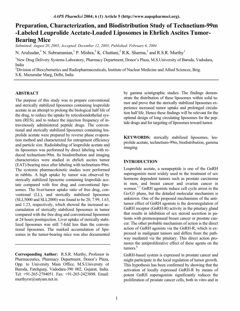
Table 1. Encapsulation Efficiency and Particle Size of Freshly Prepared Liposomal Suspension*
Type of Liposome Lipid Composition
HSPC:Cholesterol:DSPG :mPEG-PE
% Drug Entrapment +
SEM
Average Particle Size (nm)
Conventional Liposomes (LL) 1: 0.13: 0.2:0 47.12 ± 2.64 188 Sterically Stabilized Liposomes Coated With mPEG-PE 5000
(SLL5000) 1: 1:0.15:0.15 37.8 ± 3.46 175
Sterically Stabilized Liposomes Coated With mPEG-PE 2000
(SLL2000) 1: 1:0.15:0.15 33.03 ± 4.02 179
* HSPC indicates hydrogenated soya phosphatidyl choline; DSPG, distearoyl phosphatidyl glycerol; and mPEG-PE, methoxy poly-ethylene glycol 2000-phosphatidylethanolamine. Each value is the mean of triplicate results.
in organic layer. Aqueous solution of leuprolide acetate was injected rapidly into the lipid mixture through a 23-gauge hypodermic needle. The tube was closed with a glass stopper and sonicated for 10 minutes in a bath sonicator (model V33, frequency 22 KHz, 120 W, Vi-bronics Pvt Ltd, Mumbai, India.) It was then attached directly to the rotary evaporator at 60°C; the organic solvent was removed under vacuum in 2 stages: (i) evaporated at 400 mmHg until a gel was formed. Vac-uum was released and the tube was subjected to vigor-ous mechanical agitation by vortex mixer for 5 minutes, followed by (ii) evaporation of the traces of the organic solvent at 600 mmHg until a homogeneous suspension was obtained. The preparation was diluted with the con-tinuous phase (phosphate buffer saline [PBS], pH 7.4), and the vesicles formed were sequentially extruded through 2 stacked 0.2 µm nucleopore polycarbonate track-etch membrane filters (Whatman Inc, Clifton, NJ) at 60°C. The vesicles were centrifuged at 15 000 rpm for 30 minutes; the supernatant was removed and the pellet was resuspended in PBS.
Determination of Entrapment Efficiency The unentrapped drug was removed from the liposomal suspension by ficoll-gradient centrifugation.26 0.5 mL of liposomal suspension was mixed with 1 mL of 30% fi-coll solution (in saline). It was transferred to an ultracen-trifuge tube; 3 mL of 10% ficoll solution was layered gently on top of the above mixture. The upper ficoll layer was covered with a layer of buffered saline and centrifuged (Remi, C-24, Mumbai, India) at 15 000 rpm for 30 minutes. The unentrapped drug present in the lowest ficoll layer was diluted appropriately using 0.1N sodium hydroxide and the absorbance was measured at 282 nm on Shimadzu 1601 UV-Visible Spectropho-tometer, Kyoko, Japan.27 The liposomes were collected at the interface between the saline and 10% ficoll layers.
The drug present in the liposomes was estimated by Modified Bligh-Dyer extraction method.28 A mixture containing 0.5 mL of liposome suspension, 2mL of chlo-roform, 1mL of methanol, and 1mL of saturated sodium chloride solution was placed in a centrifuge tube and spun at 1000 rpm for ~10 minutes. The aqueous layer was removed and the drug content was estimated by UV-Visible Spectrophotometer at 282 nm after dilution with 0.1N sodium hydroxide. Entrapment efficiency was calculated from the difference between the initial amount of leuprolide acetate added and that present in the unentrapped form and was expressed as a percentage of the total amount of leuprolide acetate added.
Particle Size Measurements The particle size distribution of the liposomal formula-tions was estimated by laser light scattering on a Mal-vern particle size analyzer (Malvern Master Sizer 2000 SM, Malvern Instruments, Malvern, UK). Diluted lipo-some suspension was added to the sample dispersion unit containing stirrer and stirred in order to reduce the interparticle aggregation, and laser obscuration range was maintained between 15% and 20%. The average volume-mean particle size was measured after perform-ing the experiment in triplicate. The average particle size of the prepared liposome formulations is given in Table 1.
In Vitro Plasma Release Studies The liposomal suspensions were diluted with 80% hu-man plasma. A 0.5 mL sample of the diluted suspension in screw-cap eppendorf tube was used for each time point; samples were incubated under gentle rotating conditions at 37°C. Samples, to which normal saline was added, were taken for analysis according to the planned schedule, and liposomal pellets were then obtained by
3
AAPS PharmSci 2004; 6 (1) Article 5 (http://www.aapspharmsci.org).
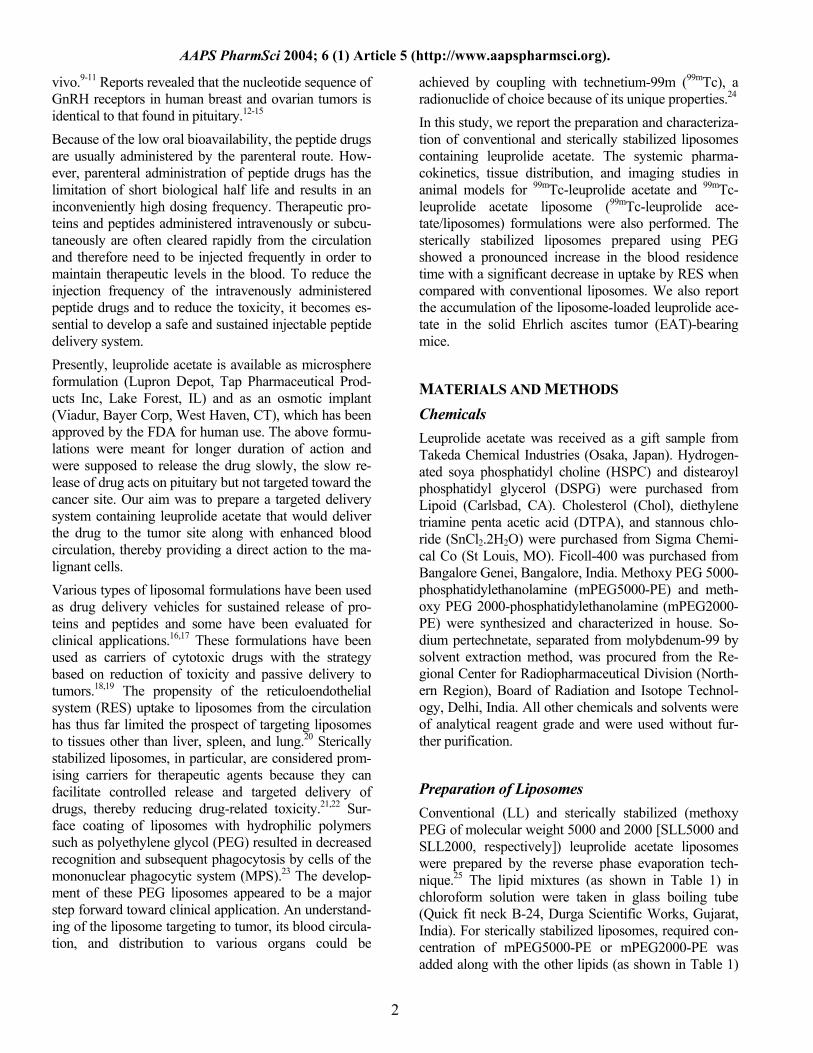
Table 2. Effect of Incubation Time on the Labeling Efficiency of Leuprolide Acetate and its Liposomes With 99mTc*
% Radiolabeled Incubation Time (min) Leuprolide
Acetate LL SLL5000 SLL2000
0 90.3 ± 1.3 90.2 ± 2.2 90.6 ± 2.1 91.7 ± 1.5 5 96.9 ± 1.6 98.9 ± 1.2 98.9 ± 1.6 98.3 ± 1.3
15 96.2 ± 2.0 98.6 ± 1.8 98.5 ± 1.5 98.7 ± 1.6 20 96.9 ± 1.3 99.1 ± 1.7 98.2 ± 1.6 98.2 ± 1.2 30 97.0 ± 1.5 99.1 ± 1.0 99.1 ± 1.8 98.6 ± 2.0 40 96.9 ± 1.9 98.2 ± 2.1 99.1 ± 2.0 98.5 ± 1.8
* Abbreviations are explained in Table 1. Results are the mean of 3 separate experiments.
centrifugation in a centrifuge at 16 000 rpm for 4 min-utes. The supernatant was aspirated and the pellet frac-tion was resuspended in 50% isopropyl alcohol, vor-texed, incubated at 37°C for 10 minutes, and centri-fuged. The amount of drug was estimated by diluting with phosphate buffer saline, and the absorbance was measured at 280 nm by spectrophotometry.
Radiolabeling of Leuprolide Acetate and Its Liposomes The leuprolide acetate and its liposomes were labeled with 99mTc by direct labeling method as described by Richardson et al.29 Briefly, 1 mL of 99mTc (2 mCi/mL) was mixed with 0.1 mL of stannous chloride solution (1mg/mL) and the pH was adjusted to 7 using 0.5M so-dium bicarbonate solution. To this mixture either 1 mL of leuprolide acetate solution (500 µg/mL) or 1 mL of liposome suspension (lipid concentration, 5-8 mg/mL) was added and incubated for 5 to 10 minutes for 99mTc-leuprolide acetate/liposomes, respectively, at room tem-perature. The quality control (percentage labeling effi-ciency and stability of the labeled complexes) was per-formed as per the method described earlier.30 The label-ing efficiency of 99mTc-leuprolide acetate/liposomes was estimated by ascending instant thin layer chromatogra-phy (ITLC) using silica gel (SG)-coated fiber glass sheets (Gelman Sciences Inc, Ann Arbor, MI). The ITLC was performed using acetone or 0.9% saline as the mobile phase. Approximately 2 to 3 µL of the radio-labeled complex was applied at a point 1 cm from one end of an ITLC-SG strip. The strip was developed in acetone or 0.9% saline and the solvent front was allowed to reach 8 cm from the point of application. The strip was cut horizontally into 2 halves, and the radioactivity in each segment was determined in a well-type gamma ray counter (Gamma ray spectrometer, type GRS23C, Electronics Corporation of India Ltd, Mumbai, India). The free 99mTc-pertechnetate that moved with the solvent (Rf = 0.9) was determined. The reduced/hydrolyzed
(R/H) technetium along with the labeled complex re-mained at the point of application. The amount of R/H 99mTc was determined using pyridine:acetic acid:water (3:5:1.5 vol/vol) as mobile phase. The R/H 99mTc re-mained at the point of application, while both the free pertechnetate and the labeled complex moved away with the solvent front. The activity moved with the solvent front using either acetone or saline was subtracted from that using pyridine:acetic acid:water as a mixture, the net amount of 99mTc-leuprolide acetate, 99mTc-LL, 99mTc-SLL5000, and 99mTc-SLL2000 was calculated and the results are shown in Table 2.
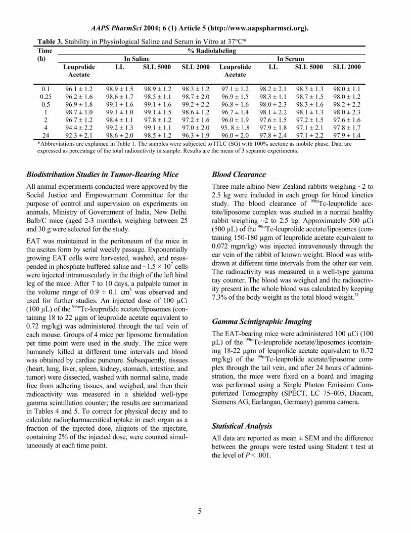
Stability Study of 99mTc-Leuprolide/Liposome Complex The stability study of 99mTc-leuprolide acetate/liposomes was determined in vitro using 0.9% sodium chloride and rabbit serum by ascending thin layer chromatography. The complex (0.1 mL) was mixed with 1.9 mL of nor-mal saline or rabbit serum and incubated at 37°C. ITLC was performed at different time intervals up to 24 hours to assess the stability of the complex and the results are given in Table 3.
Transchelation with Diethylene Triamine Penta Acetic Acid In order to check the stability and strength of the bond-ing of 99mTc with the leuprolide acetate/liposomes, 0.5 mL of the labeled preparation was challenged against various concentrations (25, 50, 75, and 100 mM) of di-ethylene triamine penta acetic acid (DTPA) and incu-bated for 2 hours at 37°C. The effect of DTPA on label-ing efficiency was measured on ITLS-SG using normal saline as the mobile phase, which allowed the separation of free pertechnetate and DTPA-complex (Rf = 0.8-1.0) from the 99mTc-leuprolide acetate/liposome complex, which remained at the point of application. (Rf = 0).
4
AAPS PharmSci 2004; 6 (1) Article 5 (http://www.aapspharmsci.org).
Table 3. Stability in Physiological Saline and Serum in Vitro at 37°C* % Radiolabeling
In Saline In Serum Time (h)
Leuprolide Acetate
LL SLL 5000 SLL 2000 Leuprolide Acetate
LL SLL 5000 SLL 2000
0.1 96.1 ± 1.2 98.9 ± 1.5 98.9 ± 1.2 98.3 ± 1.2 97.1 ± 1.2 98.2 ± 2.1 98.3 ± 1.3 98.0 ± 1.1 0.25 96.2 ± 1.6 98.6 ± 1.7 98.5 ± 1.1 98.7 ± 2.0 96.9 ± 1.5 98.3 ± 1.1 98.7 ± 1.5 98.0 ± 1.2 0.5 96.9 ± 1.8 99.1 ± 1.6 99.1 ± 1.6 99.2 ± 2.2 96.8 ± 1.6 98.0 ± 2.3 98.3 ± 1.6 98.2 ± 2.2 1 98.7 ± 1.0 99.1 ± 1.0 99.1 ± 1.5 98.6 ± 1.2 96.7 ± 1.4 98.1 ± 2.2 98.1 ± 1.3 98.0 ± 2.3 2 96.7 ± 1.2 98.4 ± 1.1 97.8 ± 1.2 97.2 ± 1.6 96.0 ± 1.9 97.6 ± 1.5 97.2 ± 1.5 97.6 ± 1.6 4 94.4 ± 2.2 99.2 ± 1.3 99.1 ± 1.1 97.0 ± 2.0 95. 8 ± 1.8 97.9 ± 1.8 97.1 ± 2.1 97.8 ± 1.7
24 92.3 ± 2.1 98.6 ± 2.0 98.5 ± 1.2 96.3 ± 1.9 96.0 ± 2.0 97.8 ± 2.4 97.1 ± 2.2 97.9 ± 1.4 *Abbreviations are explained in Table 1. The samples were subjected to ITLC (SG) with 100% acetone as mobile phase. Data are expressed as percentage of the total radioactivity in sample. Results are the mean of 3 separate experiments.
Biodistribution Studies in Tumor-Bearing Mice All animal experiments conducted were approved by the Social Justice and Empowerment Committee for the purpose of control and supervision on experiments on animals, Ministry of Government of India, New Delhi. Balb/C mice (aged 2-3 months), weighing between 25 and 30 g were selected for the study. EAT was maintained in the peritoneum of the mice in the ascites form by serial weekly passage. Exponentially growing EAT cells were harvested, washed, and resus-pended in phosphate buffered saline and ~1.5 × 10⁷ cells were injected intramuscularly in the thigh of the left hind leg of the mice. After 7 to 10 days, a palpable tumor in the volume range of 0.9 ± 0.1 cm3 was observed and used for further studies. An injected dose of 100 µCi (100 µL) of the 99mTc-leuprolide acetate/liposomes (con-taining 18 to 22 µgm of leuprolide acetate equivalent to 0.72 mg/kg) was administered through the tail vein of each mouse. Groups of 4 mice per liposome formulation per time point were used in the study. The mice were humanely killed at different time intervals and blood was obtained by cardiac puncture. Subsequently, tissues (heart, lung, liver, spleen, kidney, stomach, intestine, and tumor) were dissected, washed with normal saline, made free from adhering tissues, and weighed, and then their radioactivity was measured in a shielded well-type gamma scintillation counter; the results are summarized in Tables 4 and 5. To correct for physical decay and to calculate radiopharmaceutical uptake in each organ as a fraction of the injected dose, aliquots of the injectate, containing 2% of the injected dose, were counted simul-taneously at each time point.
Blood Clearance Three male albino New Zealand rabbits weighing ~2 to 2.5 kg were included in each group for blood kinetics study. The blood clearance of 99mTc-leuprolide ace-tate/liposome complex was studied in a normal healthy rabbit weighing ~2 to 2.5 kg. Approximately 500 µCi (500 µL) of the 99mTc-leuprolide acetate/liposomes (con-taining 150-180 µgm of leuprolide acetate equivalent to 0.072 mgm/kg) was injected intravenously through the ear vein of the rabbit of known weight. Blood was with-drawn at different time intervals from the other ear vein. The radioactivity was measured in a well-type gamma ray counter. The blood was weighed and the radioactiv-ity present in the whole blood was calculated by keeping 7.3% of the body weight as the total blood weight.31
Gamma Scintigraphic Imaging The EAT-bearing mice were administered 100 µCi (100 µL) of the 99mTc-leuprolide acetate/liposomes (contain-ing 18-22 µgm of leuprolide acetate equivalent to 0.72 mg/kg) of the 99mTc-leuprolide acetate/liposome com-plex through the tail vein, and after 24 hours of admini-stration, the mice were fixed on a board and imaging was performed using a Single Photon Emission Com-puterized Tomography (SPECT, LC 75–005, Diacam, Siemens AG, Earlangan, Germany) gamma camera.
Statistical Analysis All data are reported as mean ± SEM and the difference between the groups were tested using Student t test at the level of P < .001.
5
AAPS PharmSci 2004; 6 (1) Article 5 (http://www.aapspharmsci.org).
6
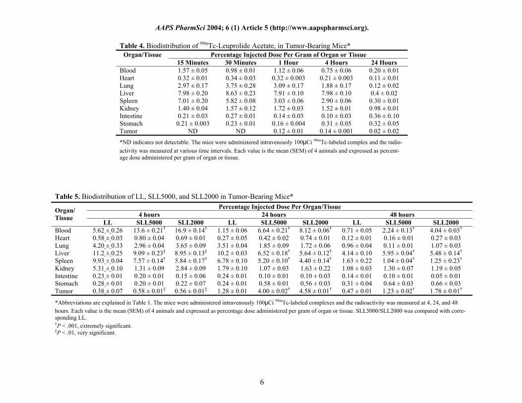
Table 4. Biodistribution of 99mTc-Leuprolide Acetate, in Tumor-Bearing Mice*
Percentage Injected Dose Per Gram of Organ or Tissue Organ/Tissue 15 Minutes 30 Minutes 1 Hour 4 Hours 24 Hours
Blood 1.57 ± 0.05 0.98 ± 0.01 1.12 ± 0.06 0.75 ± 0.06 0.20 ± 0.01 Heart 0.32 ± 0.01 0.34 ± 0.03 0.32 ± 0.003 0.21 ± 0.003 0.11 ± 0.01 Lung 2.97 ± 0.17 3.75 ± 0.28 3.09 ± 0.17 1.88 ± 0.17 0.12 ± 0.02 Liver 7.98 ± 0.20 8.63 ± 0.23 7.91 ± 0.10 7.98 ± 0.10 0.4 ± 0.02 Spleen 7.01 ± 0.20 5.82 ± 0.08 3.03 ± 0.06 2.90 ± 0.06 0.30 ± 0.01 Kidney 1.40 ± 0.04 1.57 ± 0.12 1.72 ± 0.03 1.52 ± 0.01 0.98 ± 0.01 Intestine 0.21 ± 0.03 0.27 ± 0.01 0.14 ± 0.03 0.10 ± 0.03 0.36 ± 0.10 Stomach 0.21 ± 0.003 0.23 ± 0.01 0.16 ± 0.004 0.31 ± 0.05 0.32 ± 0.05 Tumor ND ND 0.12 ± 0.01 0.14 ± 0.001 0.02 ± 0.02
*ND indicates not detectable. The mice were administered intravenously 100μCi 99mTc-labeled complex and the radio-activity was measured at various time intervals. Each value is the mean (SEM) of 4 animals and expressed as percent-age dose administered per gram of organ or tissue.
Table 5. Biodistribution of LL, SLL5000, and SLL2000 in Tumor-Bearing Mice* Percentage Injected Dose Per Organ/Tissue
4 hours 24 hours 48 hours Organ/ Tissue
LL SLL5000 SLL2000 LL SLL5000 SLL2000 LL SLL5000 SLL2000 Blood 5.62 ± 0.26 13.6 ± 0.21† 16.9 ± 0.14† 1.15 ± 0.06 6.64 ± 0.21† 8.12 ± 0.06† 0.71 ± 0.05 2.24 ± 0.13† 4.04 ± 0.03† Heart 0.58 ± 0.03 0.80 ± 0.04 0.69 ± 0.01 0.27 ± 0.05 0.42 ± 0.02 0.74 ± 0.01 0.12 ± 0.01 0.16 ± 0.01 0.27 ± 0.03 Lung 4.20 ± 0.33 2.96 ± 0.04 3.65 ± 0.09 3.51 ± 0.04 1.85 ± 0.09 1.72 ± 0.06 0.96 ± 0.04 0.11 ± 0.01 1.07 ± 0.03 Liver 11.2 ± 0.25 9.09 ± 0.23‡ 8.95 ± 0.13‡ 10.2 ± 0.03 6.52 ± 0.18† 5.64 ± 0.12† 4.14 ± 0.10 5.95 ± 0.04† 5.48 ± 0.14† Spleen 9.93 ± 0.04 7.57 ± 0.14† 5.84 ± 0.17† 6.78 ± 0.10 5.20 ± 0.10† 4.40 ± 0.14† 1.63 ± 0.22 1.04 ± 0.04† 1.25 ± 0.23† Kidney 5.31 ± 0.10 1.31 ± 0.09 2.84 ± 0.09 1.79 ± 0.10 1.07 ± 0.03 1.63 ± 0.22 1.08 ± 0.03 1.30 ± 0.07 1.19 ± 0.05 Intestine 0.23 ± 0.01 0.20 ± 0.01 0.15 ± 0.06 0.24 ± 0.01 0.10 ± 0.01 0.10 ± 0.03 0.14 ± 0.01 0.10 ± 0.01 0.05 ± 0.01 Stomach 0.28 ± 0.01 0.20 ± 0.01 0.22 ± 0.07 0.24 ± 0.01 0.58 ± 0.01 0.56 ± 0.03 0.31 ± 0.04 0.64 ± 0.03 0.66 ± 0.03 Tumor 0.38 ± 0.07 0.58 ± 0.01‡ 0.56 ± 0.01‡ 1.28 ± 0.01 4.00 ± 0.02† 4.58 ± 0.01† 0.47 ± 0.01 1.23 ± 0.02† 1.78 ± 0.01†
*Abbreviations are explained in Table 1. The mice were administered intravenously 100μCi 99mTc-labeled complexes and the radioactivity was measured at 4, 24, and 48 hours. Each value is the mean (SEM) of 4 animals and expressed as percentage dose administered per gram of organ or tissue. SLL5000/SLL2000 was compared with corre-sponding LL. †P < .001, extremely significant. ‡P < .01, very significant.
AAPS PharmSci 2004; 6 (1) Article 5 (http://www.aapspharmsci.org).
RESULTS
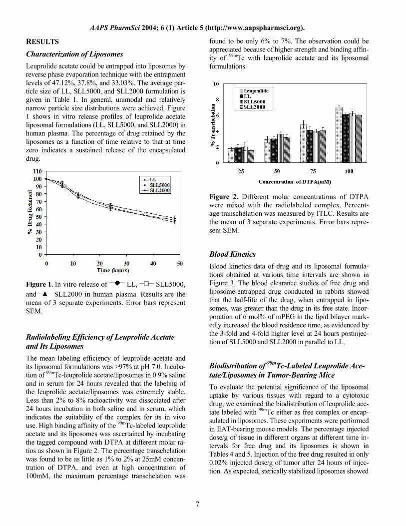
Characterization of Liposomes Leuprolide acetate could be entrapped into liposomes by reverse phase evaporation technique with the entrapment levels of 47.12%, 37.8%, and 33.03%. The average par-ticle size of LL, SLL5000, and SLL2000 formulation is given in Table 1. In general, unimodal and relatively narrow particle size distributions were achieved. Figure 1 shows in vitro release profiles of leuprolide acetate liposomal formulations (LL, SLL5000, and SLL2000) in human plasma. The percentage of drug retained by the liposomes as a function of time relative to that at time zero indicates a sustained release of the encapsulated drug.
Figure 1. In vitro release of LL, SLL5000, and SLL2000 in human plasma. Results are the mean of 3 separate experiments. Error bars represent SEM.
Radiolabeling Efficiency of Leuprolide Acetate and Its Liposomes The mean labeling efficiency of leuprolide acetate and its liposomal formulations was >97% at pH 7.0. Incuba-tion of 99mTc-leuprolide acetate/liposomes in 0.9% saline and in serum for 24 hours revealed that the labeling of the leuprolide acetate/liposomes was extremely stable. Less than 2% to 8% radioactivity was dissociated after 24 hours incubation in both saline and in serum, which indicates the suitability of the complex for its in vivo use. High binding affinity of the 99mTc-labeled leuprolide acetate and its liposomes was ascertained by incubating the tagged compound with DTPA at different molar ra-tios as shown in Figure 2. The percentage transchelation was found to be as little as 1% to 2% at 25mM concen-tration of DTPA, and even at high concentration of 100mM, the maximum percentage transchelation was
found to be only 6% to 7%. The observation could be appreciated because of higher strength and binding affin-ity of 99mTc with leuprolide acetate and its liposomal formulations.
Figure 2. Different molar concentrations of DTPA were mixed with the radiolabeled complex. Percent-age transchelation was measured by ITLC. Results are the mean of 3 separate experiments. Error bars repre-sent SEM.
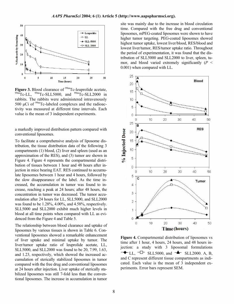
Blood Kinetics Blood kinetics data of drug and its liposomal formula-tions obtained at various time intervals are shown in Figure 3. The blood clearance studies of free drug and liposome-entrapped drug conducted in rabbits showed that the half-life of the drug, when entrapped in lipo-somes, was greater than the drug in its free state. Incor-poration of 6 mol% of mPEG in the lipid bilayer mark-edly increased the blood residence time, as evidenced by the 3-fold and 4-fold higher level at 24 hours postinjec-tion of SLL5000 and SLL2000 in parallel to LL.
Biodistribution of 99mTc-Labeled Leuprolide Ace-tate/Liposomes in Tumor-Bearing Mice To evaluate the potential significance of the liposomal uptake by various tissues with regard to a cytotoxic drug, we examined the biodistribution of leuprolide ace-tate labeled with 99mTc either as free complex or encap-sulated in liposomes. These experiments were performed in EAT-bearing mouse models. The percentage injected dose/g of tissue in different organs at different time in-tervals for free drug and its liposomes is shown in Tables 4 and 5. Injection of the free drug resulted in only 0.02% injected dose/g of tumor after 24 hours of injec-tion. As expected, sterically stabilized liposomes showed
7
AAPS PharmSci 2004; 6 (1) Article 5 (http://www.aapspharmsci.org).
Figure 3. Blood clearance of 99mTc-leuprolide acetate, 99mTc-LL, 99mTc-SLL5000, and 99mTc-SLL2000 in rabbits. The rabbits were administered intravenously 500 µCi of 99mTc-labeled complexes and the radioac-tivity was measured at different time intervals. Each value is the mean of 3 independent experiments.
a markedly improved distribution pattern compared with conventional liposomes. To facilitate a comprehensive analysis of liposome dis-tribution, the tissue distribution data of the following 3 compartments (1) blood, (2) liver and spleen (used as an approximation of the RES), and (3) tumor are shown in Figure 4. Figure 4 represents the compartmental distri-bution of tissues between 1 hour and 48 hours after in-jection in mice bearing EAT. RES continued to accumu-late liposomes between 1 hour and 4 hours, followed by the slow disappearance of the label. As the time in-creased, the accumulation in tumor was found to in-crease, reaching a peak at 24 hours; after 48 hours, the concentration in tumor was decreased. The tumor accu-mulation after 24 hours for LL, SLL5000, and SLL2000 was found to be 1.28%, 4.00%, and 4.58%, respectively. SLL5000 and SLL2000 exhibit much higher levels in blood at all time points when compared with LL as evi-denced from the Figure 4 and Table 5. The relationship between blood clearance and uptake of liposomes by various tissues is shown in Table 6. Con-ventional liposomes showed a remarkable enhancement of liver uptake and minimal uptake by tumor. The liver/tumor uptake ratio of leuprolide acetate, LL, SLL5000, and SLL2000 was found to be 20, 7.99, 1.63, and 1.23, respectively, which showed the increased ac-cumulation of sterically stabilized liposomes in tumor compared with the free drug and conventional liposomes at 24 hours after injection. Liver uptake of sterically sta-bilized liposomes was still 7-fold less than the conven-tional liposomes. The increase in accumulation in tumor
site was mainly due to the increase in blood circulation time. Compared with the free drug and conventional liposomes, mPEG-coated liposomes were shown to have higher tumor targeting. PEG-coated liposomes showed highest tumor uptake, lowest liver/blood, RES/blood and lowest liver/tumor, RES/tumor uptake ratio. Throughout the period of experimentation, it was found that the dis-tribution of SLL5000 and SLL2000 to liver, spleen, tu-mor, and blood varied extremely significantly (P < 0.001) when compared with LL.
Figure 4. Compartmental distribution of liposomes vs time after 1 hour, 4 hours, 24 hours, and 48 hours in-jection: a study with 3 liposomal formulations
LL, SLL5000, and SLL2000. A, B, and C represent different tissue compartments as indi-cated. Each value is the mean of 3 independent ex-periments. Error bars represent SEM.
8
AAPS PharmSci 2004; 6 (1) Article 5 (http://www.aapspharmsci.org).
Table 6. Tissue Uptake Ratio of Liposomes in EAT-Bearing Mice* Liposomes Liver/Blood Liver/Tumor RES/Blood RES/Tumor Leuprolide Acetate 20 ± 0.01 20 ± 0.02 35 ± 0.02 35 ± 0.03 LL 8.9 ± 0.01 7.99 ± 0.02 14.8 ± 0.01 13.2 ± 0.02 SLL5000 0.99 ± 0.03 1.63 ± 0.1 1.76 ± 0.02 2.93 ± 0.01 SLL2000 0.70 ± 0.01 1.23 ± 0.02 1.23 ± 0.01 2.19 ± 0.02 *RES indicates reticuloendothelial system. All other abbreviations are explained in Table 1.
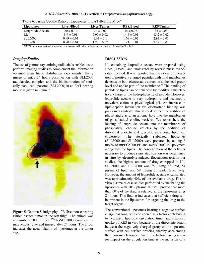
Imaging Studies The use of gamma ray emitting radiolabels enabled us to perform imaging studies to complement the information obtained from tissue distribution experiments. The γ image of mice 24 hours postinjection with SLL2000 radiolabeled complex and the biodistribution of steri-cally stabilized liposome (SLL2000) in an EAT-bearing mouse is given in Figure 5.
Figure 5. Gamma Scintigraphy of Balb/c mouse bearing Ehrich ascites tumor in the left thigh. The animal was administered 0.1 mL of 99mTc-SLL2000 complex by intravenous route and imaged after 24 hours. The arrow indicates the accumulation of liposomes at the tumor site.
DISCUSSION
LL containing leuprolide acetate were prepared using HSPC, DSPG, and cholesterol by reverse phase evapo-ration method. It was reported that the extent of interac-tion of positively charged peptides with lipid membranes depends on both electrostatic attraction at the head group level and apolar part of the membrane.32 The binding of peptide to lipids can be enhanced by modifying the elec-trical charge or the hydrophobicity of peptide. However, leuprolide acetate is very hydrophilic and becomes a univalent cation at physiological pH. An increase in lipid-peptide interaction via electrostatic binding was previously studied33; this study described the addition of phosphatidic acid, an anionic lipid into the membranes of phosphatidyl choline vesicles. We report here the loading of leuprolide acetate into the membranes of phosphatidyl choline vesicles by the addition of distearoyl phosphatidyl glycerol, an anionic lipid and cholesterol. The sterically stabilized liposomes (SLL5000 and SLL2000) were prepared by adding 6 mol% of mPEG5000-PE and mPEG2000-PE polymers along with the lipids. The concentration of the polymer necessary to produce steric stabilization was determined in vitro by electrolyte-induced flocculation test. In our studies, the highest amount of drug entrapped in LL, SLL5000, and SLL2000 was 78 µg/mg of lipid, 54 µg/mg of lipid, and 59 µg/mg of lipid, respectively. However, the amount of leuprolide acetate encapsulated was approximately 40% of the available drug. The in vitro plasma release studies performed by incubating the liposomes with 80% plasma at 37°C proved that more than 60% of the drug is retained in the liposomes after 24 hours. This finding indicates that sufficient drug will be present in the liposomes for targeting the drug to the target organs. The conventional liposomes bearing a negative surface charge has long been considered as a factor contributing to decreased liposome circulation times and enhanced uptake by RES in vivo because of the direct interaction between the negatively charged group on the liposome surface with cell surface proteins, thereby accelerating the liposome clearance. One of the factors having a ma-jor impact on the circulation time is the inclusion of a
9
AAPS PharmSci 2004; 6 (1) Article 5 (http://www.aapspharmsci.org).
10
small fraction of PEG-derivatized phospholipids. The resulting coating of the liposome surface with PEG in-creases the surface hydrophilicity, decreases opsoniza-tion and RES uptake, and prolongs liposome circulation time.34 It has been postulated that the decreased uptake of sterically stabilized liposomes by MPS is possibly due to the presence of steric barrier, which decreases the ad-sorption of plasma proteins (opsonins) on the surface of the liposomes. The lipid composition, the addition of PEG, and the particle size of the liposomes played a ma-jor role in tumor accumulation. The particle size distri-bution of the conventional and sterically stabilized leu-prolide acetate liposomes estimated by laser light scattering showed unimodal size distribution with the mean average diameter of 175 to 188 nm after extrusion through 0.2-µm nucleopore filters. The particle size was maintained in a narrow range so as to have longer blood circulation. Reports suggest that the high level of lipo-somes in the blood was only observed for small lipo-somes (d ≤ 200 nm); larger liposomes (d > 200 nm) showed high level of accumulation in spleen and low concentration in blood.35 Thus, the effect of PEG-PE to prolong liposome circulation time is limited to smaller liposomes. The small liposomes (d < 100 nm) might be expected to penetrate through the fenestrations and gain access to hepatocytes because the endothelial lining of the liver sinusoids includes fenestrae with an average diameter of 100 nm.36 The use of radiolabels has been proved to be quite useful in following the fate of liposomes in vivo and as a diag-nostic tool in nuclear medicine. The methods employed to label liposomes include entrapment of the radiolabel in the aqueous compartment, attachment of the label to the lipid components prior to liposome formulation, and the addition of label after their manufacture. The biodis-tribution of liposomes administered in vascular or ex-travascular spaces might be studied by the administra-tion of radiolabeled liposomes and followed by scinti-graphic imaging. Radiolabeled liposomes have been successfully used to monitor pharmacodynamic changes of liposomes and image tumors, abscesses, ischemic and infracted regions.37 The optimum conditions required for maximum labeling efficiency were established. The leuprolide acetate and its liposomal formulations were radiolabeled with 99mTc with an efficiency of more than 97% as shown in Table 2. Stability of the labeled complex with time was studied in saline and in serum (rabbit) at 37°C as shown in Table 3. The experimental data revealed that there was hardly any detachment of the radioisotope from the complex. Even after a period of 24 hours incubation, the presence of 92% to 97% labeled compound and only 2% to 8%
decrease of the labeled product signifies not only the high stability of the radiolabeled product but also its suitability for in vivo use. DTPA challenging test demonstrated the high binding affinity of the 99mTc with leuprolide acetate and its lipo-somes. The percentage transchelation was found to be as little as 1.02% at 25mM concentration of DTPA for 99mTc-SLL2000 and even at high concentration of 100mM the maximum percentage transchelation was found to be only 7% in case of 99mTc-LL, which proved the high strength and stability of the 99mTc-leuprolide acetate/liposome complex. Biodistribution studies in EAT-bearing mice were per-formed for the free drug (leuprolide acetate) and also its liposomes (LL, SLL5000, and SLL2000). The rapid elimination of the drug from the blood circulation is evi-dent from the values in Table 3. Only 0.75% of the in-jected dose/g was remaining in the blood 4 hours postin-jection. The rapid accumulation of leuprolide acetate in liver and spleen within 1 hour postinjection was found to be 7.91% and 3.03%, respectively. The negligible amount of labeled complex present in the stomach proved the in vivo stability of the complexes. As the drug got eliminated quickly, the amount of leuprolide acetate reaching the tumor site was much less 4 hours postinjection (0.14%). Results of the biodistribution study of LL, SLL5000, and SLL2000 are shown in Table 5. Various organs/tissues such as heart, lung, liver, spleen, kidney, stomach, intes-tine, and tumor were removed and analyzed for the la-beling content, and the percentage radioactivity was given per gram of tissue. As reported previously,38,39 there was an inverse relationship between liposome clearance by the RES and prolonged circulation time of liposomes. In turn, there appears to be a direct correla-tion between prolonged circulation time and liposome localization in tumors. The blood concentration of stealth liposomes (6.64% and 8.12% for SLL5000 and SLL2000) were significantly high even after 24 hours when compared with conventional liposomes (0.71%). The increase in circulation time may be due to the smaller particle size (<200 nm) of the liposomes or to the surface modification made by PEG. The hydropho-bic liposomal surface was converted to relatively hydro-philic surface by the incorporation of mPEG-PE, which leads to reduction in recognition by the opsonins and thereby decreases in the MPS uptake of the liposomes. The SLL2000 showed prolonged blood circulation com-pared with SLL5000, which showed that the PEG of low molecular weight provides equally, or little more resis-tance to opsonization.
AAPS PharmSci 2004; 6 (1) Article 5 (http://www.aapspharmsci.org).
The permeability of tumor vasculature is generally in-creased as compared with normal tissues, although this varies among tumors. The extravasation of macromole-cules occurs predominantly by convective transport. Liposomes could extravasate through leaky endothelium by passive convective transport. Higher concentration and longer blood residence time of liposomes obviously would result in greater efficiency of extravasations per unit volume of convective transport.40 It is, however, possible that accumulation in tumors is due to endocytic uptake or binding of liposomes by capillary endothelial cells. The increased blood circulation leads to increased tumor accumulation. The SLL5000 and SLL2000 showed ex-cellent accumulation in tumor at 24 hours postinjection, 4.00% and 4.58%, respectively, compared with 1.28% for LL and 0.02% for leuprolide acetate. There was an approximately 65-fold increase in tumor accumulation of drug from LL compared with leuprolide acetate and around 200- to 225-fold increase in tumor drug concen-tration from SLL5000/SLL2000 compared with leu-prolide acetate. The radioactivity recorded in the highly perfused organs such as liver (10.2%, 6.52%, and 5.64% for LL, SLL5000, and SLL2000, respectively) and spleen (6.78%, 5.20%, and 4.40% for LL, SLL5000, and SLL2000, respectively) could be accounted for as the combined activity of the circulating blood passing through the organs as well as that due to particle uptake by cells of RES. The tumor uptake was still below the uptake by liver and spleen, organs with fenestrated capil-laries and rich in cells of RES.39 A very low liver to tu-mor ratio was obtained in the range of 1 to 1.6 for PEG-coated liposomes in comparison with 7.9 for conven-tional liposomes. This substantial improvement in the pattern of sterically stabilized liposome biodistribution was the combined result of an absolute decrease of liver uptake and increase in tumor uptake. Although the liver uptake was still 1.6-fold higher than that of tumor, this ratio may be therapeutically acceptable, since the liver is the usual metabolizing organ for most of the drugs. RES uptake of 99mTc-SLL2000 was still 7-fold less than that of LL. The presence of a very low amount of radioactivity in the stomach after 24 hours proved the in vivo stability of the 99mTc-leuprolide acetate/liposome complexes. The gamma images were taken twenty-four hours postinjec-tion of 99mTc-SLL2000, and the images demonstrates the accumulation of liposomes in liver, spleen, and tumor present in the left hind leg of the mouse. The enhanced drug accumulation in exponentially induced EAT is ap-parently related to liposome longevity in circulation.
Thus, the long circulating liposomes containing leu-prolide acetate act as a GnRH agonist, reduce the uptake of liposomes to RES and thereby reduce the possibility of the risk of toxicity to RES generally seen with con-ventional liposomes, to prolong the biological half-life of the drug and to enhance the antiproliferative effect due to high tumor uptake.
CONCLUSION
In summary, the study demonstrated the preparation of conventional and sterically stabilized liposomes with better entrapment efficiency. The average particle size of the formulated liposomal formulations was less than 200 nm. The leuprolide acetate and its liposome formulations were then successfully radiolabeled using reduced 99mTc with the labeling efficiency of above 97%. The radio-labeled complexes were proved for their stability in both saline and serum up to 24 hours. The blood kinetic data clearly indicate the activity of mPEG-PE to prolong the circulation time of the sterically stabilized liposomes and to decrease the uptake of liposomes by RES. The pro-longed circulation time and increased tumor accumula-tion of sterically stabilized leuprolide acetate liposomes were found significantly different from that of conven-tional liposomes. The tumor accumulation of the steri-cally stabilized leuprolide acetate liposomes was also proved by gamma scintigraphy.
ACKNOWLEDGEMENTS
The research work was funded by the University Grants Commission, New Delhi, India (F-37/SA-II/2000). The authors are thankful to Takeda Chemical Industries, Osaka, Japan, for providing the gift sample of leuprolide acetate and to Major General T. Ravindranath, Director, Institute of Nuclear Medicine and Allied Sciences, Delhi, India, for providing the necessary facilities to carry out the radiolabeling experiments.
REFERENCES 1. Chrisp P, Sorkin EM. Leuprorelin. A review of its pharmacology and therapeutic use in prostatic disorders. Drugs Aging. 1991;1:487-509. 2. Oesterling JE. LHRH agonists. A nonsurgical treatment for benign prostatic hyperplasia. J Androl. 1991;12:381-388. 3. Tunn UW, Bargelloni U, Cosciani S, Fiacavento G. Guazzieris, Pagano F. Comparison of LHRH analogue 1 month depot and 3-month depot by their hormonal levels and pharmacokinetic profile in patients with advance prostate cancer. Urol Int. 1998;60(suppl 1):9-16.
11
AAPS PharmSci 2004; 6 (1) Article 5 (http://www.aapspharmsci.org). 4. Plosker GL, Brodgen RN. Leuprorelin. A review of its pharmacol-ogy and therapeutic use in prostate cancer, endometriosis and other sex hormone related disorders. Drugs. 1994;48(6):930-967. 5. Garnick MB. Leuprolide versus diethylstilbestrol for metastatic prostate cancer. N Engl J Med. 1984;311:1281-1286. 6. Okada H, Sakura Y, Kawaji T, Yashiki T, Mima H. Regression of rat mammary tumors by a potent leutinizing hormone releasing hor-mone administered vaginally. Cancer Res. 1983;43:1869-1874. 7. Redding TW, Schally AV. Inhibition of prostate tumor growth in two rat models by chronic administration of D-Trp6 analogue of LHRH. Proc Natl Acad Sci U S A. 1981;78:6509-6512. 8. Nagai N, Oshita T, Mukai K, Shiroyama Y, Shigemasa K, Ohama K. GnRH agonist inhibits human telomerase reverse transcriptase mRNA expression in endometrial cancer cells. Int J Mol Med. 2002;10:593-597. 9. Dondi D, Limnota P, Moretti RM. Marelli MM, Garattini E, Mota M. Antiproliferative effects of luteinizing hormone-releasing hormone (LHRH) agonists on human androgen independent prostate cancer cell line DU145: evidence for an autocrine-inhibitory loop. Cancer Res. 1994;54:4091-4095. 10. Limonta P, Dondi D, Moretti RM, Maggi R, Motta M. Antiprolif-erative effects of luteinizing hormone-releasing hormone agonists on the human prostatic cancer cell line LNCaP. J Clin Endocrinol Metab. 1992;75:207-212. 11. Dondi D, Moretti RM, Marelli MM, et al. Growth inhibitory ef-fects of luteinizing hormone-releasing hormone (LHRH) agonists on xenografts of the DU 145 human androgen-independent prostate cancer cell line in nude mice. Int J Cancer. 1998;76:506-511. 12. Schally AV. Hypothalamic hormones from neuroendocrinology to cancer therapy. Anticancer Drugs. 1994;5:115-130. 13. Loop SM, Gorder CA, Lewis SM, Saiers JH, Drivdahl RH, Os-tenson RC. Growth inhibition of human prostatic cancer cells by an agonist of gonadotropin-releasing hormone. Prostate. 1995;26:179-188. 14. Qayum A, Gullick W, Clayton RC, Sikora K, Waxman J. The effects of gonadotropin-releasing hormone analogues in prostate can-cer are mediated through specific tumor receptors. Br J Cancer. 1990;62:96-99. 15. Kakar SS, Grizzle WE, Neill JD. The nucleotide sequences of human GnRH receptors in breast and ovarian tumors are identical with that found in pituitary. Mol Cell Endocrinol. 1994;106:145-149. 16. Anderson PM, Hanson DC, Hasz DE, Halet MR, Blazar BR, Ochoa AC. Cytokines in liposomes: Preliminary studies with IL-1, IL-2, IL-6, GM-CSF and Interferon Gamma. Cytokine. 1994;6:92-101. 17. Meyer J, Whitcomb L, Collins D. Efficient encapsulation of pro-teins within liposomes for slow release in vivo. Biochem Biophys Res Commun. 1994;199:433-438. 18. Perez-Soler R. Liposomes as carriers of antitumor agents; towards a clinical reality. Cancer Treat Rev. 1989;16:67-82. 19. Gabizon A. Liposomes as a drug delivery system in cancer che-motherapy. In: Roerdink F, Kroon A, eds. Drug Carrier Systems, Horizons in Biochemistry and Biophysics. New York, NY: John Wiley & Sons. Vol 9. 1989:185-211. 20. Gabizon AA, Shiota R, Papahadjopoulos D. Pharmacokinetics and tissue distribution of doxorubicin encapsulated in stable lipo-somes with long circulation times. J Natl Cancer Inst. 1989;81:1484-1488. 21. Huang SK, Mayhew E, Gilani S, Lasic DD, Martin FJ, Papahad-jopoulos D. Pharmacokinetics and therapeutics of sterically stabilized
liposomes in mice bearing C-26 colon carcinoma. Cancer Res. 1992;52:6774-6781. 22. Allen TM, Hansen C, Martin F, Redemann C, Yau-Young A. Liposomes containing synthetic lipid derivatives of poly(ethylene glycol) show prolonged circulation half-lives in-vivo. Biochim Bio-phys Acta. 1991;1066:29-36. 23. Allen TM, Mehra T, Hansen C, Chin YC. Stealth liposomes: and improved sustained release system for 1-beta-D arabinofuranosylcy-tosine. Cancer Res. 1992;52:2431-2439. 24. Allen TA, Hansen C. Pharmacokinetics of stealth vs. conventional liposomes: Effect of dose. Biochim Biophys Acta. 1991;1068:133-141. 25. Szoka F, Papahadjopoulos D. Procedure for preparation of lipo-somes with large internal aqueous space and high capture by reverse evaporation. Proc Natl Acad Sci U S A. 1978;75:4194-4198. 26. New RRC. Liposomes: a practical approach. In: New RRC, ed. Preparation of Liposomes. New York, NY: Oxford University Press; 1990:95-96. 27. Adjei AL, Hsu L. Leuprolide and other LHRH analogues. In: John YW, Pearlman R, eds. Stability and Characterization of Protein and Peptide Drugs. New York, NY: Plenum Press; 1993:154-180 28. Bligh EG, Dyer WJ. A rapid method of total lipid extraction and purification. Can J Biochem Physiol. 1959;37:911-917. 29. Richardson VJ, Jeyasingh K, Jewkes RF. Properties of [99mTc] technetium-labeled liposomes in normal and tumour-bearing rats: Biochem Soc Trans. 1977;5(1):290-229. 30. Theobald AE. Theory and practice. In: Sampson CB, ed. Text-book of Radiopharmacy. New York, NY: Gorden and Breach; 1990:127-128. 31. Wu MS, Robbins JC, Ponpipom MM, Shen TV. Modified in vivo behaviour of liposomes containing synthetic glycolipids. Biochim Biophys Acta. 1981;674:19-29. 32. Lo Y, Rahman Y. Protein location in liposomes, a drug carrier: a prediction by differential scanning calorimetry. J Pharm Sci. 1995;84:805-813. 33. Schafer H, Schmidt W, Lachmann U. Preparation and properties of GnRh-loaded multilamellar liposomes. Pharmazie. 1987;42:674-677. 34. Huang L. Covalently attached polymers and glycans to alter the biodistribution of liposomes. J Liposome Res. 1992;2:289-291. 35. Litzinger CD, Buiting MJA, Rooijen VN, Huang L. Effect of liposome size on the circulation time and intraorgan distribution of amphipathic poly(ethylene glycol)-containing liposomes. Biochim Biophys Acta. 1994;1190:99-107. 36. Klibanov AL, Maruyama K, Torchilin VP, Huang L. Amphipa-thic polyethylene glycols effectively prolong the circulation time of liposomes. FEBS Lett. 1990;268:235-237. 37. Saha GB. Methods of radiolabeling. In: Saha GB, ed. Physics and Radiobiology of Nuclear Medicine. New York, NY: Springer-Verlag; 1993:100-106. 38. Gabizon G, Papahadjopoulos D. Liposome formulation with pro-longed circulation time in blood and enhanced uptake by tumors. Proc Natl Acad Sci U S A. 1988;85:6949-6953. 39. Gabizon G, Catane R, Uziely B, Kaufman B, Safra T, Cohen R, Martin F. Prolonged circulation time and enhanced accumulation in malignant exudates of doxorubicin encapsulated in polyethylene-glycol coated liposomes. Cancer Res. 1994;54:987-992. 40. Jain RK. Vascular and interstitial barriers to delivery of therapeu-tic agents in tumors. Cancer Metastasis Rev. 1990;9:253-256.
12
Related Documents


