1 PREPARATION AND CHARACTERIZATION OF PVA AND CARRAGEENAN BASED HYDROGELS FOR BIOMEDICAL PURPOSES Thesis submitted to National Institute of Technology, Rourkela For the partial fulfillment Of the Master degree in Life Science BY: ARPITA NANDA ROLL NO. 413LS2041 Under the guidance of Dr. BISMITA NAYAK DEPARTMENT OF LIFE SCIENCE NATIONAL INSTITUTE OF TECHNOLOGY, ROURKELA -769008

Welcome message from author
This document is posted to help you gain knowledge. Please leave a comment to let me know what you think about it! Share it to your friends and learn new things together.
Transcript

1
PREPARATION AND CHARACTERIZATION OF PVA
AND CARRAGEENAN BASED HYDROGELS FOR
BIOMEDICAL PURPOSES
Thesis submitted to
National Institute of Technology, Rourkela For the partial fulfillment
Of the Master degree in Life Science
BY: ARPITA NANDA
ROLL NO. 413LS2041
Under the guidance of
Dr. BISMITA NAYAK
DEPARTMENT OF LIFE SCIENCE
NATIONAL INSTITUTE OF TECHNOLOGY,
ROURKELA -769008

2

3
DECLARATION
I hereby declare the thesis entitled ”Preparation and characterization
of PVA and carrageenan based Hydrogels for biomedical
applications”, submitted to the Department of Life Science, National
Institute of Technology, Rourkela for the partial fulfilment of the Master
Degree in Life Science is a faithful record of bonafide research work
carried out by me under the guidance and supervision of Dr. Bismita
Nayak, Assistant Professor, Department of Life Science, National
Institute of Technology, Rourkela. No part of this thesis has been
submitted by any other research persons or any students. DATE: 11.05.15 PLACE: ROURKELA ARPITA NANDA

4
ACKNOWLEDGEMENT
I wish to express my sincere thanks and gratitude to my guide and coordinator Dr. Bismita
Nayak, Assistant Professor, Dept. of Life Science, National Institute of Technology, Rourkela,
for her constant inspiration, encouragement and guidance throughout my project. I consider
myself fortunate enough that she has given a decisive turn and boost to my career.
I take this opportunity to express my indebtedness to my Professors for their enthusiastic help,
valuable suggestions and constant encouragement throughout my work. I would also like to
express my whole hearted gratitude to the Head of the department of Life Science Dr. Sujit
Kumar Bhutia and other faculty members, Dr. Bibekanand Mallick, Dr. Surajit Das, Dr. Suman
Jha, and Dr. Rasu Jayabalan, Dr. Samir Kumar Patra, National Institute of Technology Rourkela,
Orissa for their good wishes, inspiration and unstinted support throughout my project.
I take this opportunity to express my indebtedness to my Professors for their enthusiastic help,
valuable suggestions and constant encouragement throughout my work. I would also like to
express my whole hearted gratitude to Dr. Kunal Pal Department of Biotechnology and medical
engineering ,for his technical inputs and to the phd students K.Uvanesh, Gauri Shankar
Shaw,Divya , Haladhara ,National Institute of Technology Rourkela, Orissa for their good
wishes, inspiration and unstinted support throughout my project.
I deeply acknowledge the constant support, encouragement, and invaluable guidance at every step of
my project by Mr. Debasis Nayak PhD scholar, Dept. of life science. I am obliged and thankful to
him for providing me the opportunity to gain knowledge and understanding of working skills of the
aspects of my work from him. Also I am thankful to Mr. Pradipta Ranjan Rauta, Sarbani Ashe PhD
scholar, Dept. of Life Science for their cooperation and supportive nature.
I take this opportunity to thank my friends Pankaj, Ankita, Jyotsna di and Aliva for their throughout co-operation.
Last but not the least I take this opportunity to thank my father Mr. Asit Kumar Nanda and my mother Mrs. Madhusmita Nanda for weathering my minor crises of confidence, for never doubting.
Thank you for everything Maa and Papa. I love you both Place: Rourkela Arpita Nanda
Date:

5
CONTENTS
SL NO LIST OF CONTENTS PAGE NO
I ABSTRACT 7
2 INTRODUCTION 8
3 REVIEW OF
LITERATURE
12
4 MATERIALS AND
METHOD
23
5 RESULTS AND
DISCUSSION
36
6 CONCLUSION 46
7 REFERANCE 47

6
LIST OF FIGURES
SL NO
LIST OF
FIGURES
PAGE NO
1 PICTURES OF
BRIGHT FIELD
MICROSCOPE
26
2 PHOTOGRAPH
OF SEM
27
3 PHOTOGRAPH
OF XRD
28
4 PHOTOGRAPH
OF FTIR
29
5 PHOTOGRAPH
OF ELECTRICAL
30
6 PHOTOGRAPH
OF
MECHANICAL
31
7 PHOTOGRAPH
OF HEMOLYSIS
32
8 PHOTOGRAPH
OF
ANTIMICROBIAL
34
9
PHOTOGRAPH
OF DRUG
RELEASE
33
10
PHOTOS OF
MICROSCOPY
38
11 PHOTOS OF SEM
39

7
12 PHOTOS OF XRD 40
13
PHOTOS OF FTIR 41
14 PHOTOS OF
ELECTRICAL
42
15 PHOTOS OF
MECHANICAL
43
16 PHOTOS OF
ANTIMICROBIAL
44
17 PHOTOS OF
HEMOLYSIS
45
18 PHOTOS OF
DRUG RELEASE
46
19 PHOTOS OF
SWELLING
46
20 PHOTOS OF MTT
ASSAY
47

8
ABSTRACT
Hydrogels are three-dimensional, cross- linked networks of water-soluble polymers. They can
be made from nearly any water-soluble polymer, covering a wide range of chemical
compositions and bulk physical properties. These are highly porous . For our preparation of
hydrogels we have used 10% PVA with 2% Carrageenan . cross linker was added for the
preparation of gel . Characterizations were done Physiochemical characterization and other
characterization . Under physiochemical characterization we have don SEM , XRD ,
MICROSCOPY, ELECTRICAL AND MECHANICAL . And under other characterization we
have done Hemocompatibility , Drug release , Swelling, Antimicrobial and MTT Assay. The
above characterisation of the hydrogels showed that the hydrogels can be used for a control drug
delivery system. It can be used for biomedical applications.We found that CP 6 shows the
better result as comparable to the rest of the concentrations as it shows the high
hemocompatibility and antimicrobial activity. It also shows good swelling ability and uniform
drug release profile

9
INTRODUCTION
Hydrogels are three-dimensional, cross- linked networks of water-soluble polymers. They can be
made from nearly any water-soluble polymer, covering a wide range of chemical compositions
and bulk physical properties. Hydrogels are commonly used in clinical practice and experimental
medicine for a wide range of applications, including tissue engineering and regenerative
medicine, diagnostics, cellular immobilization, separation of bio-molecules or cells, and barrier
materials to regulate biological adhesions. The unique physical properties of hydrogels have
provoked a particular interest in their use in drug delivery applications. Their highly porous
structure can easily be tuned by controlling the density of cross- links in the gel matrix and the
affinity of the hydrogels for the aqueous environment in which they are swollen. If water is
removed from these swollen biomaterials, they are called xerogels, which are the dried
hydrogels. These gels may be charged or non-charged depending on the nature of functional
groups present in their structure. The charged hydrogels usually exhibit changes in swelling upon
variations in pH, and it is known that they can undergo changes in shape when exposed to an
electric field [3]. The enormous attention is the parameters control by which the degradation
characteristics can be adapted. As the gels are used, this is of extreme importance that the
hydrogels have excellent biocompatibility and degradation products produced have a low toxic
potential. Hydrogels hold excellent biocompatibility. Its water loving surface has fewer
propensities for cells and proteins to stick to these surfaces. Furthermore, the elastic and soft
nature of gels minimizes irritability to the neighboring tissues (Park and Park, 1996, Smetana,
1993, Anderson and Langone, 1999 and Anderson, 1994). The properties of degradation
products produced may be modified by the proper and rational selection of the starting materials
of hydrogel. Depending on the type of cross-linking, hydrogels can be divided into two classes:
(i) chemically cross-linked hydrogels, and (ii) physically cross-linked hydrogels. Chemical
hydrogels are formed by covalent networks and do not dissolve in water without breakage of
covalent bonds [12], [13], [14] and [15]. Physical hydrogels are, however, formed by dynamic
cross-linking of synthetic or natural building blocks based on non-covalent interactions such as
hydrophobic and electrostatic interaction or hydrogen-bridges [16], [17] and [18]. The
hydrophilic and mostly inert nature of hydrogels often leads to minimized non-specific
interaction with proteins and cells which makes hydrogels ideal candidates for numerous bio-

10
related applications [19] and [20]. For most medical application , the novel engineering of
hydrogels for drug delivery require dividing them to biodegradable hydrogels which are favoured
over non- degradable hydrogels since they degrade in clinically relevant time scale [8] , smart
hydrogel or stimuli-sensitive hydrogel that respond to environmental changes, such as
temperature, pH, light, and specific molecules such as glucose [9] and finally biomimetic
hydrogels which are relatively inert polymer chains can be tailored with the selected biological
moieties to yield bioactive hydrogels [10] .
PVA is a water-soluble synthetic polymer. It has the idealized formula [CH2CH(OH)]n. It is
used in papermaking, textiles, and a variety of coatings. It is white (colourless) and odorless. It is
sometimes supplied as beads or as solutions in water.[1] Polyvinyl Alcohol (PVA) is an
environmental friendly and water soluble synthetic polymer with excellent film forming
property, and emulsifying properties and outstanding resistance to oil, grease, and solvents. It has
been extensively used in adhesive, in textile warp sizing and finishing, in paper size and coating,
in the manufacturing of PVAc emulsion, in the suspension polymerization of PVC, and as binder
for ceramics, foundry cores and several of pigment. PVA is manufactured by polymerization of
vinyl acetate monomer, followed by hydrolysis of the polymerization of vinyl acetate monomer,
followed by hydrolysis of the polyvinyl acetate, Since the properties on which technical
application of PVA depend are primarily its molecular eight and degree of hydrolysis. The
amount of hydroxylation determines the physical characteristics, chemical properties, and
mechanical properties of the PVA.1 The resulting PVA polymer is highly soluble in water but
resistant to most organic solvents. The higher the degree of hydroxylation and polymerization of
the PVA, the lower the solubility in water and the more difficult it is to crystallize.2 Due to its
water solubility, PVA needs to be crosslinked to form hydrogels for use in several applications.
The crosslinks, either physical or chemical, provide the structural stability the hydrogel needs
after it swells in the presence of water or biological fluids.3 The degree of crosslinking dictates
the amount of fluid uptake, and thus the physical, chemical, and diffusional properties of the
polymer, and ultimately its biological properties. PVA's resistance against organic solvents and
aqueous solubility makes it adaptable for many applications.1, 2 PVA is commonly used in the
textile industries, for paper products manufacturing, in the food packaging industry, and as
medical devices. PVA is used as an industrial and commercial product due to its low
environmental impact, which includes its high chemical resistance, aqueous solubility, and

11
biodegradability. FDA has approved PVA to be in close contact with food products; in fact, PVA
films exhibit excellent barrier properties for food packaging systems. In medical devices, PVA is
used as a biomaterial due to its biocompatible, nontoxic, noncarcinogenic, swelling properties,
and bioadhesive characteristics.
Carrageenan has the ability to form strong gels with certain salts or other gums and its ability to
interact with certain dairy proteins. Carrageenan is mainly used in the food industry with some
applications in the toiletries industry. Industrial applications of carrageenan are rare.
Carrageenan has three basic forms:-
Lambda Carrageenan
Iota Carrageenan
Kappa Carrageenan
Carrageenan is a generic name for a family of gel-forming and viscosifying polysaccharides,
which are obtained by extraction from certain species of red seaweeds (Table 1). Carrageenan is
derived from a number of seaweeds of the class Rhodophyceae. Carrageenans are composed of a
linear galactose backbone with a varying degree of sulfatation (between 15% and 40%).
Different carrageenan types differ in composition and conformation resulting in a wide range of
rheological and functional properties. Carrageenans are used in a wide variety of commercial
applications as gelling, thickening, and stabilizing agent in especially food products, such as
frozen desserts, chocolate-milk, cottage cheese, whipped cream, instant products, yogurt, jellies,
pet foods, sauces, and so forth.
Ciprofloxacin (INN) is an antibiotic that can treat a number of bacterial infections. It is a second-
generation fluoroquinolone.[2][3] Its spectrum of activity includes most strains of bacterial
pathogens responsible for respiratory, urinary tract, gastrointestinal, and abdominal infections,
including Gram-negative (Escherichia coli, Haemophilus influenzae, Klebsiella pneumoniae,
Legionella pneumophila, Moraxella catarrhalis, Proteus mirabilis, and Pseudomonas aeruginosa),
and Gram-positive (methicillin-sensitive, but not methicillin-resistant Staphylococcus aureus,
Streptococcus pneumoniae, Staphylococcus epidermidis, Enterococcus faecalis, and
Streptococcus pyogenes) bacterial pathogens. Ciprofloxacin and other fluoroquinolones are
valued for this broad spectrum of activity, excellent tissue penetration, and for their availability

12
in both oral and intravenous formulations.[4][page needed] Ciprofloxacin is used alone or in
combination with other antibacterial drugs in the empiric treatment of infections for which the
bacterial pathogen has not been identified, including urinary tract infections[5][6] and abdominal
infections[7] among others. It can also treat infections caused by specific pathogens known to be
sensitive. Ciprofloxacin is the most widely used of the second-generation quinolone antibiotics
that came into clinical use in the late 1980s and early 1990s.[8][9] In 2010, over 20 million
outpatient prescriptions were written for ciprofloxacin, making it the 35th-most commonly
prescribed drug, and the 5th-most commonly prescribed antibacterial, in the US.[10]
Ciprofloxacin was discovered and developed by Bayer A.G. and subsequently approved by the
US Food and Drug Administration (FDA) in 1987. Ciprofloxacin has 12 FDA-approved human
uses and other veterinary uses, but it is often used for unapproved uses.

13
REVIEW OF LITERATURE
Hydrogels are polymer networks extensively swollen with water. These are three dimensional
crosslinked architectures with proven multifaceted applications. The hydrogels are known to
have good water interaction and swell when exposed to aqueous solution [1].When swelled
they become soft and rubbery properties which could be tailored to apply them into different
field of pharmacy, pharmaceutics and biomedical engineering [2]. Natural hydrogels were
gradually replaced by synthetic hydrogels which has long service life, high capacity of water
absorption, and high gel strength. Fortunately, synthetic polymers usually have well-defined
structures that can be modified to yield tailor able degradability and functionality. Hydrogels can
be synthesized from purely synthetic components. Also, it is stable in the conditions of sharp and
strong fluctuations of temperatures [3]. Hydrogels have been defined as two- or multi-component
systems consisting of a three-dimensional network of polymer chains and water that fills the
space between macromolecules. Depending on the properties of the polymer (polymers) used, as
well as on the nature and density of the network joints, such structures in an equilibrium can
contain various amounts of water; typically in the swollen state, the mass fraction of water in a
hydrogel is much higher than the mass fraction of polymer. Hydrogels may exhibit drastic
volume changes in response to specific external stimuli, such as the temperature, solvent quality,
pH, electric field, etc. [5]. Depending on the design of the hydrogel matrices, this volume change
may occur continuously over a range of stimulus level, or, discontinuously at a critical stimulus
level. The volume transition behaviours of hydrogels received considerable interest in the last
three decades and large parts of the work have been collected in different reviews [7]. Gelation
refers to the linking of macromolecular chains together which initially leads to progressively
larger branched yet soluble polymers depending on the structure and conformation of the starting
material. The mixture of such polydisperse soluble branched polymer is called ‘sol’. Hydrogels
can take place either by physical linking (physical gelation) or by chemical linking (chemical
gelation). Physical gels can be sub categorised as strong physical gels and weak gels. Strong
physical gel has strong physical bonds between polymer chains and is effectively permanent at a
given set of experimental conditions [4]. Different types of gelation mechanism are summarised
in Figure 1. Gelation can take place either by physical linking (physical gelation) or by chemical
linking (chemical gelation). Physical gels can be sub categorised as strong physical gels and
weak gels. Strong physical gel has strong physical bonds between polymer chains and is
effectively permanent. Hence, strong physical gels are analogous to chemical gels. Examples of
strong physical bonds are lamellar microcrystals, glassy nodules or double and triple helices.
Weak physical gels have reversible links formed from temporary associations between chains
[5]. Weak physical bonds are hydrogen bond, block copolymer micelles, and ionic associations.
On the other hand, chemical gelation involves formation of covalent bonds and always results in
a strong gel. Hydrogels based on natural or synthetic polymers have been of great interest

14
regarding cell encapsulation [6], For the past decade, such hydrogels have become especially
attractive as matrices for regenerating and repairing a wide variety of tissues and organs
CLASSIFICATION OF HYDROGELS:-
Depending on their method of preparation, ionic charge, or physical structure features,
hydrogels maybe classified in several categories. Based on the method of preparation,
they may be (i) homopolymer hydrogels, (ii) copolymer hydrogels, (iii) multipolymer
hydrogels, or (iv) interpenetrating polymeric hydrogels. They are broadly classified into
Permanent / chemical gel: they are called ‘permanent' or ‘chemical’ gels when they are
covalently cross-linked (replacing hydrogen bond bya stronger and stable covalent bonds)
networks (Hennink & Nostrum, 2002). They attain an equilibrium swelling state which
depends on the polymer-water interaction parameter and the crosslink density (Rosiak &
Yoshii, 1999). Reversible / physical gel: they are called ‘reversible’ or ‘physical’ gels
when the networks are held together by molecular entanglements, and / or secondary
forces including ionic, hydrogen bonding or hydrophobic interactions. In physically
cross-linked gels, dissolution is prevented by physical interactions, which exist between
different polymer chains (Hennink & Nostrum, 2002). All of these interactions are
reversible, and can be disrupted by changes in physical conditions or application of stress
[6].
Homopolymeric hydrogels are referred to polymer network derived from a single species
of monomer, which is a basic structural unit comprising of any polymer network [8].
HYDROGELS
PHYSICAL
STRONG WEAK
CHEMICAL
CROSSLINKING

15
Homopolymers may have cross-linked skeletal structure depending on the nature of the
monomer and polymerization technique.
Copolymeric hydrogels are comprised of two or more different monomer species with at
least one hydrophilic component, arranged in a random, block or alternating
configuration along the chain of the polymer network [9].
Multipolymer Interpenetrating polymeric hydrogel (IPN), an important class of
hydrogels, is made of two independent cross-linked synthetic and/or natural polymer
component, contained in a network form. In semi-IPN hydrogel, one component is a
cross-linked polymer and other component is a non-cross-linked polymer [10] and [11].
CLASSIFICATION BASED ON CONFIGURATION:-
The classification of hydrogels depends on their physical structure and chemical composition can
be classified as follows:
(a) Amorphous (non-crystalline).
(b) Semicrystalline: A complex mixture of amorphous and crystalline phases.
(c) Crystalline.
CLASSIFICATION BASED ON TYPE OF CROSSLINKING:-
Hydrogels appears as matrix, films or microsphere depends on the technique of polymerization
involved in the preparation process.
CLASSIFICATION ACCORDING TO NETWORK ELECTRICAL CHARGE :-
Hydrogels may be categorized into four groups on the basis of presence or absence of electrical
charge located on the cross-linked chains:
(a)Nonionic (neutral).
(b)Ionic (including anionic or cationic).
(c)Amphoteric electrolyte (ampholytic) containing both acidic and basic groups.
(d) Zwitterionic (polybetaines) containing both anionic and cationic groups in each structural
repeating unit.
Hydrogel-forming natural polymers include proteins such as collagen and gelatine and
polysaccharides such as starch, alginate, and agarose. Synthetic polymers that form hydrogels are
traditionally prepared using chemical polymerization methods.

16
CHARACTERISTICS OF HYDROGELS :-
The water holding capacity and permeability are the most important characteristic features of a
hydrogel. The polar hydrophilic groups are the first to be hydrated upon contact with water
which leads to the formation of primary bound water. As a result the network swells and exposes
the hydrophobic groups which are also capable of interacting with the water molecules. This
leads to the formation of hydrophobically-bound water, also called ‘secondary bound water’.
Primary and secondary bound water are often combined and called ‘total bound water’. The
network will absorb additional water, due to the osmotic driving force of the network chains
towards infinite dilution [11] This additional swelling is opposed by the covalent or physical
cross-links, leading to an elastic network retraction force. Thus, the hydrogel will reach an
equilibrium swelling level. The additional absorbed water is called ‘free water’ or ‘bulk water’,
and assumed to fill the space between the network chains, and/or the centre of larger pores,
macropores, or voids. Depending on the nature and composition of the hydrogel the next step is
the disintegration and/or dissolution if the network chain or cross-links are degradable.
Biodegradable hydrogels, containing labile bonds, are therefore advantageous in applications
such as tissue engineering, wound healing and drug delivery. These bonds present can be either
polymer backbone or in the cross-links used to prepare the hydrogel. [10]. Biocompatibility is
the third most important characteristic property required by the hydrogel. Biocompatibility calls
for compatibility with the immune system of the hydrogel and its degradation products formed,
which also should not be toxic. Ideally they should be metabolised into harmless products or can
be excreted by the renal filtration process. Generally, hydrogels possess a good biocompatibility
since their hydrophilic surface has a low interfacial free energy when in contact with body
fluids, which results in a low tendency for proteins and cells to adhere to these surfaces.
Moreover, the soft and rubbery nature of hydrogels minimises irritation to surrounding tissue
(Anderson & Langone, 1999; Smetana, 1993). The cross-links between the different polymer
chains results in viscoelastic and sometimes pure elastic behaviour and give a gel its structure
(hardness), elasticity and contribute to stickiness. Hydrogels, due to their significant water
content possess a degree of flexibility similar to natural tissue. It is possible to change the
chemistry of the hydrogel by controlling their polarity, surface properties, mechanical properties,
and swelling behaviour.
USES OF HYDROGELS:-
Common uses for hydrogels include:
Scaffolds in tissue engineering. When used as scaffolds, hydrogels may contain human
cells to repair tissue. They mimic 3D microenvironment of cells.[10]
Hydrogel coated wells were used for cell culures.

17
Environmentally sensitive hydrogels (also known as 'Smart Gels' or 'Intelligent Gels').
These hydrogels have the ability to sense changes of pH, temperature, or the concentration
of metabolite and release their load as result of such a change.
Sustained- released drug delivery system.
It provides absorption , desloughing and debriding of necrotic fibrotic tissue.
Used in disposable diapers where they absorb urine or in sanitary napkins.
Used for contact lenses .
Rectal drug delivery and diagnosis.
For breast implants
Glue
Granules for holding soil moisture in arid areas.
Dressings for healing of burn.
Reservoirs in topical drug delivery ; particularly ionic drugs , delivered by iontophoresi.
Natural hydrogel materials are being invested for tissue engineering, these materials include
agarose ,methylcellulose ,hyaluronan , and other naturally derived polymers.
There are various other types of gels which include :-
ORGANOGELS:-
An organogel is a non-crystalline, non-glassy thermoreversible (thermoplastic) solid material
composed of a liquid organic phase entrapped in a three-dimensionally cross-linked network.
The liquid can be, for example, an organic solvent, mineral oil, or vegetable oil. The solubility
and particle dimensions of the structurant are important characteristics for the elastic properties
and firmness of the organogel. Often, these systems are based on self-assembly of the structurant
molecules.[13][14]Organogels have potential for use in a number of applications, such as in
pharmaceuticals,[15] cosmetics, art conservation,[16] and food.[17] An example of formation of
an undesired thermoreversible network is the occurrence of wax crystallization in petroleum.[18]
18
XEROGELS:-
A xerogel is a solid formed from a gel by drying with unhindered shrinkage. Xerogels usually
retain high porosity (15–50%) and enormous surface area (150–900 m2/g), along with very small
pore size (1–10 nm). When solvent removal occurs under supercritical conditions, the network
does not shrink and a highly porous, low-density material known as an aerogel is produced. Heat
treatment of a xerogel at elevated temperature produces viscous sintering (shrinkage of the
xerogel due to a small amount of viscous flow) and effectively transforms the porous gel into a
dense glass.
NANOCOMPOSITE HYDROGELS:-
Nanocomposite hydrogels are also known as hybrid hydrogels, can be defined as highly hydrated
polymeric networks, either physically or covalently crosslinked with each other and/or with
nanoparticles or nanostructures. Nanocomposite hydrogels can mimic native tissue properties,
structure and microenvironment due to their hydrated and interconnected porous structure. A
wide range of nanoparticles, such as carbon-based, polymeric, ceramic, and metallic
nanomaterials can be incorporated within the hydrogel structure to obtain nanocomposites with
tailored functionality. Nanocomposite hydrogels can be engineered to possess superior physical,
chemical, electrical, and biological properties.[19]
PROPERTIES OF HYDROGELS:--
Many gels display thixotropy – they become fluid when agitated, but resolidify when resting. In
general, gels are apparently solid, jelly-like materials. By replacing the liquid with gas it is
possible to prepare aerogels, materials with exceptional properties including very low density,
high specific surface areas, and excellent thermal insulation properties.
APPLICATIONS:-
Biomedical Applications:-
Their biocompatibility allows them to be considered for medical applications, whereas their
hydrophilicity can impart desirable release characteristics to controlled and sustained release
formulations. Hydrogels exhibit properties that make them desirable candidates for
biocompatible and blood-compatible biomaterials (Merrill et al., 1987).
One of the most earliest use of hydrogels was that they are used in contact lenses because of their
good mechanical stability and high oxygen permeability.
Other potential applications include:-
Artificial tendon materials
Wound healing bioadhesives

19
Artificial kidney membranes
Artificial skin
Maxillofacial
Vocal cord replacement materials
Pharmaceutical Applications:-
Pharmaceutical applications have become very much popular , this systems include equilibrilum
swollen hydrogels. The category of solvent-activated, matrix-type, controlled- release devices
comprises two important types of systems: swellable and swelling-controlled devices. In general,
a sys- tem prepared by incorporating a drug into a hydrophilic, glassy polymer can be swollen
when brought in contact with water or simulant of biological fluids. This swelling process may
or may not be the controlling mechanism for diffusional release, depending on the relative rat In
swelling-controlled release systems, the bioactive agent is dispersed into the polymer to form
nonporous films, disks, or spheres. Upon contact with an aqueous dissolution medium, a distinct
front (interface) is observed that corresponds to the water penetration front into the polymer and
separates the glassy from the rubbery (gel-like) state of the material. Under these conditions, the
macromolecular relaxations of the polymer influence the diffusion mechanism of the drug
through the rubbery state.es of the macromolecular relaxation of the polymer and drug diffusion
from the gel. This water uptake can lead to considerable swelling of the polymer with a thickness
that depends on time. The swelling process proceeds toward equilibrium at a rate determined by
the water activity in the system and the structure of the polymer. If the polymer is cross-linked or
if it is of sufficiently high molecular weight (so that chain entanglements can maintain structural
integrity), the equilibrium state is a water-swollen gel. The equilibrium water content of such
hydrogels can vary from 30% to 90%. If the dry hydrogel contains a water-soluble drug, the drug
is essentially immobile in the glassy matrix, but begins to diffuse out as the polymer swells with
water. Drug release thus depends on two simultaneous rate processes: water migration into the
device and drug diffusion outward through the swollen gel. Since some water uptake must occur
before the drug can be released, the initial burst effect frequently observed in matrix devices is
moderated, although it may still be present. The continued swelling of the matrix causes the drug
to diffuse increasingly easily, ameliorating the slow tailing off of the release curve.
ADVANTAGES OF HYDROGELS:-
Hydrogels posses a degree of flexibility very similar to natural tissue due to their
significant water content.
Entrapment of microbial cells with in hydrogels beads has the advantages of low
toxicity.
Hydrogels have good transport properties.
20
Hydrogels are biocompatible.
Hydrogels can be injected.
These are very easy to modify.
For our experiment we have used PVA and Carrageenan for hydrogel preparation.
PVA (Poly – Vinyl Alcohol):-
Poly(vinyl alcohol) (PVOH, PVA, or PVAl) is a water-soluble synthetic polymer. It has the
idealized formula [CH2CH(OH)]n. It is used in papermaking, textiles, and a variety of coatings. It
is white (colourless) and odorless. It is sometimes supplied as beads or as solutions in water.[12].
Another hydrophilic polymer that has received attention is poly(vinyl alcohol) (PVA). This
material holds tremendous promise as a biological drug delivery device because it is nontoxic, is
hydrophilic, and exhibits good mucoadhesive properties. Polyvinyl acetals: Polyvinyl acetals are
prepared by reacting aldehydes with polyvinyl alcohol. Polyvinyl butyral (PVB) and polyvinyl
formal (PVF) are examples of this family of polymers. They are prepared from polyvinyl alcohol
by reaction with butyraldehyde and formaldehyde, respectively. Preparation of polyvinyl butyral
is the largest use for polyvinyl alcohol in the U.S. and Western Europe. Polyvinyl alcohol is used
as an emulsion polymerization aid, as protective colloid, to make polyvinyl acetate dispersions.
This is the largest market application in China. In Japan its major use is vinylon fiber production.
USES OF POLY-VINYL ALCOHOL:-
Paper adhesive with boric acid in spiral tube winding and solid board production.
Thickener, modifier, in polyvinyl acetate glues.
Textile sizing agent
Paper coatings, release liner
As a water-soluble film useful for packaging. An example is the envelope containing
laundry detergent in "liqui-tabs".
Feminine hygiene and adult incontinence products as a biodegradable plastic backing
sheet.
Carbon dioxide barrier in polyethylene terephthalate (PET) bottles.
As a film used in the water transfer printing process.
As a form release because materials such as epoxy do not stick to it.
Movie practical effect and children's play putty or slime when combined with borax
Used in eye drops (such as artificial tears to treat dry eyes) and hard contact lens solution
as a lubricant
PVA fiber, as reinforcement in concrete.
Raw material to polyvinyl nitrate (PVN) an ester of nitric acid and polyvinyl alcohol.
As a surfactant for the formation of polymer encapsulated nanobeads.
Used in protective chemical-resistant gloves.

21
Used as a fixative for specimen collection, especially stool samples.
When doped with iodine, PVA can be used to polarize light
As an embolization agent in medical procedures
Carotid phantoms for use as synthetic vessels in Doppler flow testing
Used in 3D printing as support structure that can then be dissolved away.
STRUCTURE AND PROPERTIES OF PVA:-
PVA is an atactic material that exhibits crystallinity. In terms of microstructure, it is composed
mainly of 1,3-diol linkages [-CH2-CH(OH)-CH2-CH(OH)-] but a few percent of 1,2-diols [-
CH2-CH(OH)-CH(OH)-CH2-] occur, depending on the conditions for the polymerization of the
vinyl ester precursor.[1]Polyvinyl alcohol has excellent film forming, emulsifying and adhesive
properties. It is also resistant to oil, grease and solvents. It has high tensile strength and
flexibility, as well as high oxygen and aroma barrier properties. However these properties are
dependent on humidity, in other words, with higher humidity more water is absorbed. The water,
which acts as a plasticiser, will then reduce its tensile strength, but increase its elongation and
tear strength. PVA has a melting point of 230 °C and 180–190 °C (356-374 degrees Fahrenheit)
for the fully hydrolysed and partially hydrolysed grades, respectively. It decomposes rapidly
above 200 °C as it can undergo pyrolysis at high temperatures. PVA is close to incompressible.
CARRAGEENAN:-
Carrageenans or carrageenins from Irish carraigín, "little rock") are a family of linear sulphated
polysaccharides that are extracted from red edible seaweeds. They are widely used in the food
industry, for their gelling, thickening, and stabilizing properties. Their main application is in
dairy and meat products, due to their strong binding to food proteins. There are three main
varieties of carrageenan, which differ in their degree of sulphation. Kappa-carrageenan has one
sulphate group per disaccharide. Iota-carrageenan has two sulphates per disaccharide. Lambda
carrageenan has three sulphates per disaccharide.Gelatinous extracts of the Chondrus crispus
(Irish Moss) seaweed have been used as food additives since approximately the 1400s.[1]
Carrageenan is a vegetarian and vegan alternative to gelatin in some applications, in some
instances it is used to replace gelatin in confectionery. Carrageenan has undergone many long-
term dietary studies under defined regulatory conditions en route to its current global regulatory
status. While some indicate that carrageenan safely passes through rat GI tracts without adverse
effect when it is a dietary ingredient,[2] other animal dietary studies have observed colitis-like
disease and tumour promotion.[3] In the late 2000s, some scientists raised concerns about
whether the amount of "degraded carrageenan" (poligeenan) in food-grade carrageenan may lead
to health problems, leading to a debate in the research literature.[4] It has yet to be determined
whether such observations are pertinent to dietary safety considerations.The use of carrageenan
22
in infant formula, organic or otherwise, is prohibited in the EU for precautionary reasons, but is
permitted in other foodstuffs.[5] In the US, it is permitted in organic and non-organic foods,
including juices, chocolate milk, and organic infant formula.
PROPERTIES:-
Carrageenans are large, highly flexible molecules that curl forming helical structures. This gives
them the ability to form a variety of different gels at room temperature. They are widely used in
the food and other industries as thickening and stabilizing agents. All carrageenans are high-
molecular-weight polysaccharides made up of repeating galactose units and 3,6
anhydrogalactose (3,6-AG), both sulfated and nonsulfated. The units are joined by alternating α-
1,3 and β-1,4 glycosidic linkages. There are three main commercial classes of carrageenan:
Kappa forms strong, rigid gels in the presence of potassium ions; it reacts with dairy proteins. It
is sourced mainly from Kappaphycus alvarezii.[6] Iota forms soft gels in the presence of calcium
ions. It is produced mainly from Eucheuma denticulatum.[6] Lambda does not gel, and is used to
thicken dairy products.The primary differences that influence the properties of kappa, iota, and
lambda carrageenan are the number and position of the ester sulfate groups on the repeating
galactose units. Higher levels of ester sulfate lower the solubility temperature of the carrageenan
and produce lower strength gels, or contribute to gel inhibition (lambda carrageenan).Many red
algal species produce different types of carrageenans during their developmental history. For
instance, the genus Gigartina produces mainly kappa carrageenans during its gametophytic stage,
and lambda carrageenans during its sporophytic stage.All are soluble in hot water, but in cold
water, only the lambda form (and the sodium salts of the other two) are soluble.When used in
food products, carrageenan has the EU additive E-number E407 or E407a when present as
"processed eucheuma seaweed".[7] Technically carrageenan is considered a dietary fibre.[8][9]
In parts of Scotland and Ireland, where it is known by a variety of local and native names,
Chondrus crispus is boiled in milk and strained, before sugar and other flavourings such as
vanilla, cinnamon, brandy, or whisky are added. The end-product is a kind of jelly similar to
pannacotta, tapioca, or blancmange.
USES OF CARRAGEENAN:-
Desserts, ice cream, cream, milkshakes, salad dressings, sweetened condensed milks, and sauces:
gel to increase viscosity Beer: clarifier to remove haze-causing proteins Pâtés and processed
meats (ham, e.g.): substitute for fat, increase water retention, and increase volume, or improve
sliceability Toothpaste: stabilizer to prevent constituents separating Fruit Gushers: ingredient in
the encapsulated gel Fire fighting foam: thickener to cause foam to become sticky shampoo and
cosmetic creams: thickener, air freshener gels Marbling: the ancient art of paper and fabric
marbling uses a carrageenan mixture on which to float paints or inks; the paper or fabric is then
laid on it, absorbing the colours Shoe polish: gel to increase viscosity Biotechnology: gel to
immobilize cells/enzymes Pharmaceuticals: used as an inactive excipient in pills/tablets Soy milk
and other plant milks: used to thicken, in an attempt to emulate the consistency of whole milk

23
Diet sodas: to enhance texture and suspend flavours Pet food Personal lubricants Vegetarian hot
dogs.
24
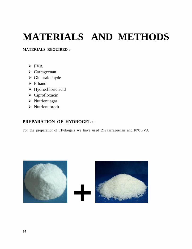
MATERIALS AND METHODS MATERIALS REQUIRED :-
PVA
Carrageenan
Glutaraldehyde
Ethanol
Hydrochloric acid
Ciprofloxacin
Nutrient agar
Nutrient broth
PREPARATION OF HYDROGEL :-
For the preparation of Hydrogels we have used 2% carrageenan and 10% PVA
+

25
Carrageenan (2%) + PVA(10%)
Aqueous solution of 10% PVA and 2% Carrageenan was prepared.
Different formulations was prepared by taking PVA and carrageenan in different
proportions.
Blending of PVA and carrageenan was done for 10 mins at 120 rpm.
After blending of both the solutions these were poured into the moulds for the preparation
of Hydrogels.
PREPARATION OF CROSS LINKER:-

26
Cross linkers are the polymers that links one polymer chain to the another. This polymers can
be covalent or it can be ionic bond. Polymers can be of several types whether it can be a
synthetic polymer or it can be natural polymer.
For the preparation of cross linker we have used Glutaraldehyde (50%) 10 ml , HCL
(Hydrochloric acid) 1ml i.e 0.1 N HCL , and then we have added Ethanol 10ml to it like this
the cross linker was prepare for the formation of the gel.
CHARACTERISATION:-
PHYSIOCHEMICAL CHARACTERISATION:-
Microscopy
SEM
XRD
FTIR
Electrical
Mechanical
OTHER CHARACTERISATION:-
Hemocompatibility
Swelling
Drug release
Antimicrobial
MTT Assay
PHYSIOCHEMICAL CHARACTERIZATION:-
MICROSCOPY OR BRIGHT FIELD MICROSCOPY:-
Microscopy is also known as bright field microscopy. It is most simple of all optical
microscopy . Sample illumination is transmitted and white light and the contrast of the
sample is caused by the absorbance of some transmitted light in the dense areas of the
sample . The light path of the bright field microscopy is very simple and there are no
additional components which are required for the normal light setup.Microscope stand
has a halogen lamp which transilluminate light source. It contains a condenser lens which
focuses the light from the light source onto the sample. And lastly for viewing the image
it has oculars.

27
SEM ( Scanning Electron Microscopy ):-
SEM analysis is also known as SEM microscopy. It is used effectively in microanalysis
failure analysis of the solid samples. SEM is performed at high magnifications, and it
generates high-resolution images and precisely measures very small features and objects . The
SEM equipment includes a variable pressure system capable of holding wet and/or non-
conductive samples with minimal preparation. The large sample chamber allows for the
examination of samples up to 200 mm (7.87 in.) in diameter and 80 mm (3.14 in.) in height.
High-resolution images are produced during SEM analysis at magnifications from 5x to
300,000x.
Fig 1 . picture of bright field microscopy

28
XRD ( X-Ray Diffraction):-
X-ray powder diffraction (XRD) is a rapid analytical technique primarily used for phase
identification of a crystalline material and can provide information on unit cell dimensions. The
analyzed material is finely ground, homogenized, and average bulk composition is determined.
X-ray diffractometers consist of three basic elements: an X-ray tube, a sample holder, and an X-
ray detector. X-rays are generated in a cathode ray tube by heating a filament to produce
electrons, accelerating the electrons toward a target by applying a voltage, and bombarding the
target material with electrons. When electrons have sufficient energy to dislodge inner shell
electrons of the target material, characteristic X-ray spectra are produced. These spectra consist
of several components, the most common being Kα and Kβ. Kα consists, in part, of Kα1 and
Kα2. Kα1 has a slightly shorter wavelength and twice the intensity as Kα2. The specific
wavelengths are characteristic of the target material (Cu, Fe, Mo, Cr). Filtering, by foils or
Fig 2 photograph of SEM
29
crystal monochrometers, is required to produce monochromatic X-rays needed for diffraction.
Kα1and Kα2 are sufficiently close in wavelength such that a weighted average of the two is
used. Copper is the most common target material for single-crystal diffraction, with CuKα
radiation = 1.5418Å. These X-rays are collimated and directed onto the sample. As the sample
and detector are rotated, the intensity of the reflected X-rays is recorded. When the geometry of
the incident X-rays impinging the sample satisfies the Bragg Equation, constructive interference
occurs and a peak in intensity occurs. A detector records and processes this X-ray signal and
converts the signal to a count rate which is then output to a device such as a printer or computer
monitor. When the geometry of the incident X-rays impinging the sample satisfies the Bragg
Equation, constructive interference occurs and a peak in intensity occurs. A detector records and
processes this X-ray signal and converts the signal to a count rate which is then output to a
device such as a printer or computer monitor. The geometry of an X-ray diffractometer is such
that the sample rotates in the path of the collimated X-ray beam at an angle θ while the X-ray
detector is mounted on an arm to collect the diffracted X-rays and rotates at an angle of 2θ. The
instrument used to maintain the angle and rotate the sample is termed a goniometer. For typical
powder patterns, data is collected at 2θ from ~5° to 70°, angles that are preset in the X-ray scan.
Fig 3 photo of XRD machine

30
FTIR ( FOURIER TRANSFORM INFRARED SPECTROSCOPY):-
Fourier transform infrared spectroscopy (FTIR)[1] is a technique which is used to produce an
infrared spectrum of absorption, emission, photoconductivity or Raman scattering of a solid,
liquid or gas. An FTIR spectrometer simultaneously collects high spectral resolution data over a
wide spectral range. This confers a significant advantage over a dispersive spectrometer which
measures intensity over a narrow range of wavelengths at a time.The term Fourier transform
infrared spectroscopy originates from the fact that a Fourier transform is required to convert the
raw data into the actual spectrum.
Fig 4 showing the working principle of FTIR

31
Fig 5 picture showing the photograph of FTIR
ELECTRICAL ANALYSIS:-
FIG SHOWING ELECTRICAL ANALYSIS SET UP

32
MECHANICAL ANALYSIS:-
Mechanical testing provides information about the suitability of a material for its intended
application to help companies design reliable products that will perform as expected. Mechanical
testing services measure materials under various temperature, tension, compression and load
conditions to determine:
Strength
Hardness
Ductility
Impact resistance
Fracture toughness
Elongation
Stress
Mechanical Test Ranges
Elevated temperature tensile testingTensile testing of metal parts and specimens in all sizes, from
the smallest fasteners to huge tubing and bolts on our 600,000 lb. capacity machines. A 10,000
lb. capacity tensile machine provides plastics testing Elevated temperature tensile testing using a
furnace carousel to process up to three samples at once. Samples can be heated to 1800°F Stress
rupture and creep testing comply with ASTM standards and can be performed at temperatures up
to 2000°F Fracture toughness and fatigue testing with equipment that can generate up to 55,000
lbs. of tensile or compressive force and controls the test temperature between -250°F and
+400°FRockwell, Brinell & Superficial hardness testing is available for metals, while a Shore
Durometer hardness tester provides plastics testing Charpy impact testing is performed from -
452°F to 500°F.
OTHER CHARACTERISTICS:-
HEMOCOMPATIBILITY TEST:-
Hemocompatibility test means to find out whether the components are hemocompatible or
not. For hemolysis study Fresh goat blood, collected in a beaker,containing Sodium Citrate
(3.8gm%, 10:1) was diluted with normal saline solution (8ml blood+10ml normal saline). For
checking hemolysis, 0.2ml of the diluted blood was adde to 10ml of 0.1% Sodium Carbonate
solution and optical density measured at 545nm in a UV-spectrophotometer. Take a 5mmX5mm
sample without sharp edges in a standard tube containing 10ml of normal saline kept in an
incubator at 37 o C for 30min providing temperature equilibration. Add 0.2ml of the diluted
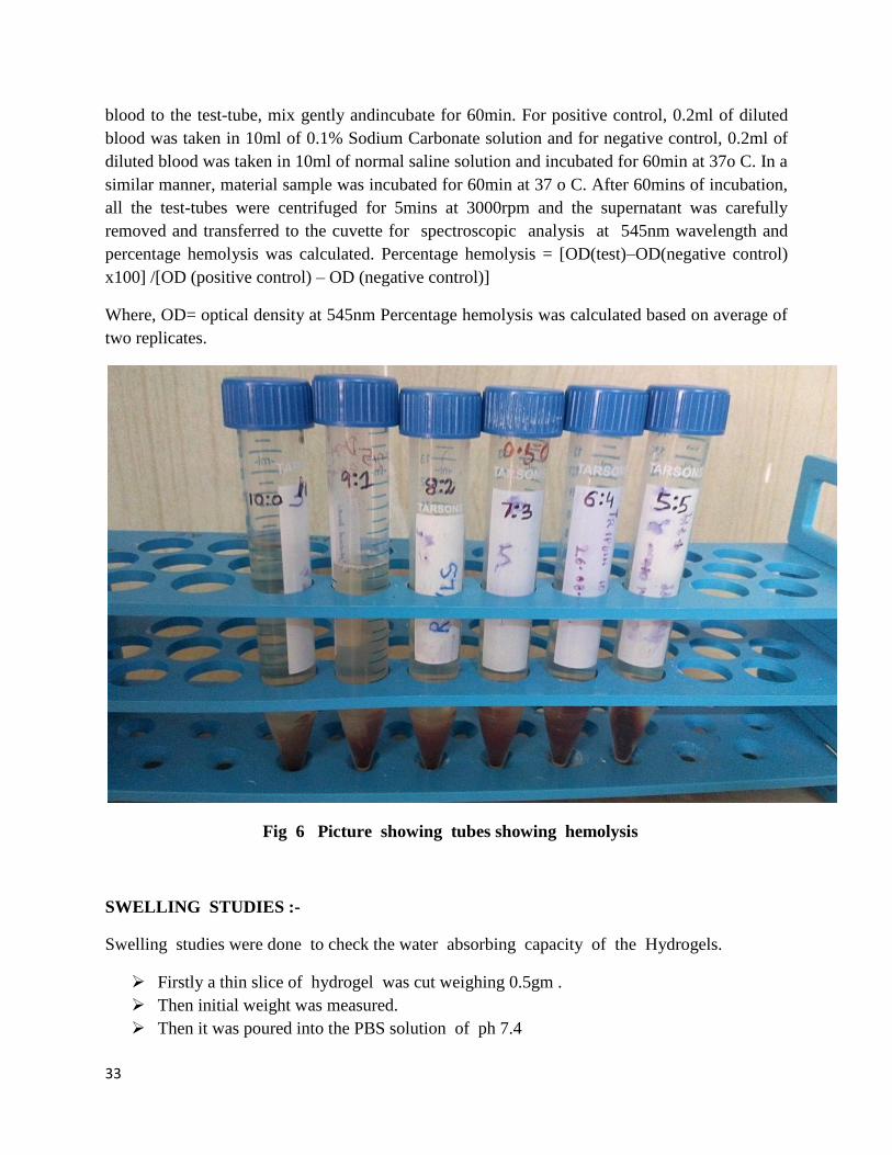
33
blood to the test-tube, mix gently andincubate for 60min. For positive control, 0.2ml of diluted
blood was taken in 10ml of 0.1% Sodium Carbonate solution and for negative control, 0.2ml of
diluted blood was taken in 10ml of normal saline solution and incubated for 60min at 37o C. In a
similar manner, material sample was incubated for 60min at 37 o C. After 60mins of incubation,
all the test-tubes were centrifuged for 5mins at 3000rpm and the supernatant was carefully
removed and transferred to the cuvette for spectroscopic analysis at 545nm wavelength and
percentage hemolysis was calculated. Percentage hemolysis = [OD(test)–OD(negative control)
x100] /[OD (positive control) – OD (negative control)]
Where, OD= optical density at 545nm Percentage hemolysis was calculated based on average of
two replicates.
Fig 6 Picture showing tubes showing hemolysis
SWELLING STUDIES :-
Swelling studies were done to check the water absorbing capacity of the Hydrogels.
Firstly a thin slice of hydrogel was cut weighing 0.5gm .
Then initial weight was measured.
Then it was poured into the PBS solution of ph 7.4

34
First with in 15 mins the weight of the samples were checked .
Then within 5 hrs at regular interval thw weight was measured till the weight remain
constant.
DRUG RELEASE STUDIES:-
Drug Release studies were done to see the control drug delivery of the system .This
study was carried out for 48 hours . 6 liters of PBS were prepared .
COMPOSITION OF PBS:-
Nacl -8gm
Kcl -0.2gm
Na2 HPO4-1.44gm
KH2PO4 – 0.24gm
A dialysis membrane was attached to the testube and a piece of drig loaded sample was
inserted inside the testtube and under that beaker was kept containg the PBS solution .
First intial time was 15 mns for 1hr then for 30 mins then for 1hr gradually the
experiment was carried out.
Fig set up for drug release studies

35
ANTIMICROBIAL ACTIVITY :-
An antimicrobial is an agent that kills microorganisms or inhibits their growth.[1] Antimicrobial
medicines can be grouped according to the microorganisms they act primarily against. For
example, antibacterials are used against bacteria and antifungals are used against fungi. They can
also be classified according to their function. Agents that kill microbes are called microbicidal,
while those that merely inhibit their growth are called microbiostatic. The use of antimicrobial
medicines to treat infection is known as antimicrobial chemotherapy, while the use of
antimicrobial medicines to prevent infection is known as antimicrobial prophylaxis.
Fig 7 showing the Antimicrobial activity
MTT ASSAY :-
MTT Assay was done to to check the cell viability of hydrogel compounds.

36
The MTT assay is a colorimetric assay for assessing cell viability. NAD(P)H-dependent
cellular oxidoreductase enzymes may, under defined conditions, reflect the number of
viable cells present. These enzymes are capable of reducing the tetrazolium dye MTT 3-
(4,5-dimethylthiazol-2-yl)-2,5-diphenyltetrazolium bromide to its insoluble formazan,
which has a purple color. Other closely related tetrazolium dyes including XTT, MTS
and the WSTs, are used in conjunction with the intermediate electron acceptor, 1-
methoxy phenazine methosulfate (PMS). With WST-1, which is cell-impermeable,
reduction occurs outside the cell via plasma membrane electron transport.[1] Tetrazolium
dye assays can also be used to measure cytotoxicity (loss of viable cells) or cytostatic
activity (shift from proliferation to quiescence) of potential medicinal agents and toxic
materials. MTT assays are usually done in the dark since the MTT reagent is sensitive to
light.
In this process :
Leachets were prepared by putting the Hydrogels samples inside the PBS for 24 hrs
Then after 24 hrs the leachets were collected.
First day media was added
2nd
day media was removed fresh media was added and to it leachets
Next day leachets were removed and mtt awas added and incubated for 4hrs and after dat
DMSO was added and reading was taken at 562 nm.

37
RESULTS AND DISCUSSION

38
PHOTOGRAPHS OF PREPARED GELS :-
Fig 9 pictures of prepared hydrogels
COMPOSITION OF HYDROGELS
SL.NO SAMPLE CODE CARRAGEENAN PVA
1 CP1 10 0
2 CP2 9 1
3 CP3 8 2
4 CP4 7 3
5 CP5 6 4
6 CP6 5 5
PHOTOGRAPHS OF MICROSCOPY:-

39
Fig 10 microscopy pictures
This microscopy of PVA and Carrageenan were the phase separated system.
Where PVA shows the dispersion phase and carrageenan dispersve phase.
PHOTOGRAPHS OF SEM:-
RESULTS TO BE PUBLISHED
40
Fig 11 photos of SEM
XRD ( X-Ray diffraction )
Fig 12 XRD GRAPHS
RESULTS TO BE PUBLISHED
RESULTS TO BE PUBLISHED

41
FTIR GRAPHS :-
Fig 13 showing the FTIR Graph
RESULTS TO BE PUBLISHED

42
ELECTRICAL ANALYSIS :-
Fig 14 showing the electrical analysis of Impedance and frequency.
RESULTS TO BE PUBLISHED

43
MECHANICAL ANALYSIS :-
Fig 15 showing the mechanical analysis
ANTIMICROBIAL GRAPHS :-
RESULTS TO BE PUBLISHED

44
Fig 16 showing the antimicrobial activity.
RESULTS TO BE PUBLISHED
45
DRUG RELEASE STUDIES :-
SWELLING STUDIES :-
RESULTS TO BE PUBLISHED
RESULTS TO BE PUBLISHED

46
HEMOCOMPATIBILITY TEST:-
MTT ASSAY :-
RESULTS TO BE PUBLISHED
RESULTS TO BE PUBLISHED

47
CONCLUSION
The above characterisation of the hydrogels showed that the hydrogels can be used for a control
drug delivery system. It can be used for biomedical applications.We found that CP 6 shows
the better result as comparable to the rest of the concentrations as it shows the high
hemocompatibility and antimicrobial activity. It also shows good swelling ability and uniform
drug release profile
48
REFERENCES
1. Couldwell WT, Chen TC, Weiss MH, et al: Cranioplasty with the Medpor porous
polyethylene Flexblockimplant. Technical note. J Neurosurg 81:483–486, 1994
2. Larry M. Wolford, and Rogerio Z. Freitas. Porous block hydroxyapatite as a bone graft
substitute in the correction of jaw and craniofacial deformities. BUMC Proceedings
1999;12:243-246.
3. Cottrell DA, Wolford LM. Long-term evaluation of the use of coralline hydroxyapatite in
orthognathic surgery. J Oral Maxillofac Surg 1998;56:935.
4. Gonzalez-Rodriguez ML, Perez-Martinez JI, Merino S, Fini A, Rabasco AM. Drug Dev.
IndPharm. 2001 May;27(5):439-46.Channeling agent and drug release from a central core matrix
tablet. Departamentode Farmacia y Tecnologia Farmaceutica, Universidad de Sevilla, Spain.
5. Dembczynski R., Jankowski T. Determination of pore diameter and molecular weight cut-off
of hydrogel-membrane liquid-core capsules for immunoisolation. Journal of Biomaterials
Science, Polymer Edition, 1October 2001, vol. 12, no., pp. 1051-1058(8)
6. http://landau1.phys.virginia.edu/classes/241L/poise/poise.htm
7. Stein, H.L., Ticona, P.E., .Ultra High Molecular Weight Polyethylene (UHMWPE), Guide to
Engineering Plastic Families: Thermoplastic Resins, Vol. 2. Engineered Materials Handbook,
ASM International, Materials Park, OH, pp. 167-171, 1999.
8. Callister Jr., W. D. 2003. Materials Science and Engineering—An Introduction , 6th ed. New
York: Wiley, Sections 6.1–6.12, 8.1–8.4.
9. Radhakrishnan S. and Khedkar S. P., Application of dip-coating process for depositing
conducting polypyrrole films. Thin Solid Films, Volume 303, Issues 1-2, 15 July 1997, Pages
167-172.
10. Yoshikazu Miyake, Yasuhiro Sekiguchi and Shinzo Kohjiya. Formation of percolated
structure during solvent casting of polymer blend-solvent systems. Journal of Chemical
Engineering of Japan, vol 26 no 5 pp 543-550(1993).
11. Bennet J.M. & Mattson Lars “Introduction to Surface Roughness and Scattering”, Optical
Society of America, Washington D.C. 1989.
12. Ravaglioli and Krajewski, Bioceramics, Published by Chapman & Hall, 3 rd edition; page:
100-101, 1991..

49
13. Peppas, A. N. and Merrill, W. E. J. Biomed. Mater. Res. 1977, 11, 423
14. Watase, M., Nishinary, K. and Nambu, M. Polym. Commun. 1983, 24, 52
15. Hyon, S. H., Cha, W. I. and Ikada, Y. Polym. Bull. 1989, 22,119 9 Ofstead, R. F. and
Poser, C. I. Polym. in Aqueous Media 1989, 223, 61
16. Peppas, N. A. 'Hydrogels Medicine and Pharmacy' (Ed.N. A. Peppas), CRC Press, Boca
Raton, 1987, Vol. 2, p. 1
17. Watase, M. and Nishinary, K. Makromol. Chem. 1988,189, 87112 Nagura, M., Hamano,
T. and Ishikawa, H. Polymer 1989, 30, 762
18 Yamaura, K., Itoh, M., Tanigami, T. and Matsuzawa, S. J. Appl.Polym. Sci. 1989, 37, 2709
19. Urushizaki, F., Yamaguchi, H., Nakamura, K., Numajiri, S.,Sugibayashi, K. and Morimoto,
Y. Int. J. Pharm. 1990, 58, 135
20. Lozinski, V. I., Vainerman, E. S., Domotenko, L. V., Mamtsis, A. M., Titova, E. F.,
Belavtseva, E. M. and Rogozhin, S. V. Colloid Polym. Sci. 1986, 264, 19
21. Andrade, J., King, R. and Gregonis, D. in 'Hydrogels for Medical and Related Applications'
(Ed. J. Andrade), ACS Symp. Ser.,American Chemical Society, Washington, DC, 1976, p. 206
22. Miller, D. and Peppas, N. Biomaterials 1986, 7, 329
23. Blank, Z. and Reimschuessel, A. J. Mater. Sci 1974, 9, 1815
24. Trieu, H. and Qutubuddin, S. Colloid Polym. Sci. 1994, 272, 301
25. Box, E. P. B. and Draper, R. N. in "Empirical Model-Building and Response Surfaces', John
Wiley & Sons, New York, 1987
26. "Standard Test Method for Rubber Property – Durometer Hardness', ASTM D2240-86, in
'Annual Book of ASTM Standards' Vol. 9.01, American Society for Testing and Materials,
Philadelphia, PA
27. 'Standard Test Method for Rubber Properties in Tension',ASTM D412-87, in 'Annual Book
of ASTM Standards' Vol. 9.01, American Society for Testing and Materials, Philadelphia, PA
28 'Standard Test Method for Rubber Property - Tear Resistance', ASTM D624-86, in 'Annual
Book of ASTM Standards' Vol.9.01, American Society for Testing and Materials, Philadelphia,
PA
29 Komatsu, M., Inoue, T. and Miyasaka, K. J. Polym. Sci., Po(vm. Phys. Edn 1986, 24, 303
30 Tong, H. M., Noda, 1. and Gryte, C. C. Colloid Polym. Sci. 1984, 262, 589
50
31 Yokoyama, F., Masada, I., Shimamura, K., Ikawa, T. and Monobe, K. Colloid Polym. Sci.
1986, 264, 595
32 Szmant, H. H. in 'Dimethyl Sulfoxide' (Eds S. W. Jacob, E. E. Rosenbaum and D. C. Wood),
Marcel Dekker, New York, 1971, Vol. 1
33 Rasmussen, D. H. and MacKenzie, A. P. Nature 1968, 220, 1315
34 Fox, M. F. and Whittingham, K. P. J. Chem. Sac., Faraday Trans. I 1975, 71, 1407
35 Bowen, D. E., Priesand, M. A. and Eastman, M. P. J. Phys. Chem. 1974, 78, 261131
Callister, S., Keller, A. and Hikmet, R. M. Makromol. Chem., Macromol. Syrup. 1990, 39,
1932 Ohkura, M., Kanaya, T. and Kaji, K. Polymer 19
Related Documents


