RESEARCH ARTICLE Preparation and characterization of niosomal gel for iontophoresis mediated transdermal delivery of isosorbide dinitrate Sanyog Jain & Bankim H. Chaudhari & Nitin K. Swarnakar Published online: 21 June 2011 # Controlled Release Society 2011 Abstract The purpose of the present study was to improve the bioavailability of isosorbide dinitrate (ISDN) through transdermal route by using cationic niosomal gel as a carrier with anodic iontophoresis. ISDN-loaded cationic niosomes prepared by thin film hydration technique had an average diameter of 262±6.92 nm, polydispersity index of 0.217± 0.02, zeta potential of +25.4±0.12, and entrapment efficiency of 68.16±1.14%. The prepared niosomes were incorporated in minimum quantity of carbopol gel which exhibited thixotropic behavior suitable for transdermal application. While free drug was found to degrade upon application of current, interestingly, it was a found that niosomes offered protection to ISDN from degradation during the iontophore- sis. The in vitro permeation studies with different current densities showed increase in transdermal flux and decrease in lag time by 11.15- and 2.42-fold (0.5 mA/cm 2 ), 12.66- and 2.58-fold (1.0 mA/cm 2 ), and 14.46- and 3.75-fold (1.5 mA/ cm 2 ), respectively, as compared to passive diffusion of free drug. The study confirms the synergistic effect of niosomes and iontophoresis in improving the transdermal permeation profile of ISDN. The enhanced permeation by iontophoresis was investigated by scanning electron microscopy and it was observed that “latent shunt” around the hair follicles became activated and their pore size also increased upon increasing the current densities. Finally, in vivo skin permeation studies demonstrated 2.47 times increased in transdermal bioavail- ability of ISDN using niosomes in comparison to free drug. The study confirmed that both niosomes and iontophoresis enhance transdermal permeation by two different mecha- nisms and combination of both has synergistic effect that resulted in higher transdermal flux of ISDN. Keywords Isosorbide dinitrate . Niosomes . Transdermal delivery . Iontophoresis . Niosomal gel Introduction Transdermal drug delivery offers benefits over other routes of administration, e.g., avoidance of hepatic first-pass metabolism, fewer side effects, and improved patient compliance [1, 2]. However, stratum corneum plays the major role in regulating the barrier function of the skin because of its unique nature, which creates an interstitial lipoidal environment. Several techniques have been devel- oped to overcome this barrier including chemical (penetra- tion enhancers) and physical techniques (iontophoresis, electrophoresis, and sonophoresis) or combination of both [1, 3–5]. The iontophoresis is a non-invasive technique which provides simplified therapeutic regimen (drug input kinetics can be modulated by the current profile) and leads to improved patient compliance [6]. It is a promising delivery technique for the charged and uncharged molecules having high and low molecular weights. A variety of drugs has been delivered across the skin by this technique [2, 7]. However, there are several factors, which affect the iontophoresis like physiochemical properties of both the drug and drug formulation and other biological variations. Moreover, iontophoresis results in subtherapeutic plasma levels of some drugs due to their degradation by the S. Jain (*) : B. H. Chaudhari : N. K. Swarnakar Centre for Pharmaceutical Nanotechnology, Department of Pharmaceutics, National Institute of Pharmaceutical Education and Research (NIPER), Sector 67, SAS Nagar, Mohali, Punjab 160062, India e-mail: [email protected] S. Jain e-mail: [email protected] Drug Deliv. and Transl. Res. (2011) 1:309–321 DOI 10.1007/s13346-011-0035-1

Welcome message from author
This document is posted to help you gain knowledge. Please leave a comment to let me know what you think about it! Share it to your friends and learn new things together.
Transcript
RESEARCH ARTICLE
Preparation and characterization of niosomal gelfor iontophoresis mediated transdermal deliveryof isosorbide dinitrate
Sanyog Jain & Bankim H. Chaudhari &Nitin K. Swarnakar
Published online: 21 June 2011# Controlled Release Society 2011
Abstract The purpose of the present study was to improvethe bioavailability of isosorbide dinitrate (ISDN) throughtransdermal route by using cationic niosomal gel as a carrierwith anodic iontophoresis. ISDN-loaded cationic niosomesprepared by thin film hydration technique had an averagediameter of 262±6.92 nm, polydispersity index of 0.217±0.02, zeta potential of +25.4±0.12, and entrapment efficiencyof 68.16±1.14%. The prepared niosomes were incorporatedin minimum quantity of carbopol gel which exhibitedthixotropic behavior suitable for transdermal application.While free drug was found to degrade upon application ofcurrent, interestingly, it was a found that niosomes offeredprotection to ISDN from degradation during the iontophore-sis. The in vitro permeation studies with different currentdensities showed increase in transdermal flux and decrease inlag time by 11.15- and 2.42-fold (0.5 mA/cm2), 12.66- and2.58-fold (1.0 mA/cm2), and 14.46- and 3.75-fold (1.5 mA/cm2), respectively, as compared to passive diffusion of freedrug. The study confirms the synergistic effect of niosomesand iontophoresis in improving the transdermal permeationprofile of ISDN. The enhanced permeation by iontophoresiswas investigated by scanning electron microscopy and it wasobserved that “latent shunt” around the hair follicles becameactivated and their pore size also increased upon increasingthe current densities. Finally, in vivo skin permeation studies
demonstrated 2.47 times increased in transdermal bioavail-ability of ISDN using niosomes in comparison to free drug.The study confirmed that both niosomes and iontophoresisenhance transdermal permeation by two different mecha-nisms and combination of both has synergistic effect thatresulted in higher transdermal flux of ISDN.
Keywords Isosorbide dinitrate . Niosomes . Transdermaldelivery. Iontophoresis . Niosomal gel
Introduction
Transdermal drug delivery offers benefits over other routesof administration, e.g., avoidance of hepatic first-passmetabolism, fewer side effects, and improved patientcompliance [1, 2]. However, stratum corneum plays themajor role in regulating the barrier function of the skinbecause of its unique nature, which creates an interstitiallipoidal environment. Several techniques have been devel-oped to overcome this barrier including chemical (penetra-tion enhancers) and physical techniques (iontophoresis,electrophoresis, and sonophoresis) or combination of both[1, 3–5]. The iontophoresis is a non-invasive techniquewhich provides simplified therapeutic regimen (drug inputkinetics can be modulated by the current profile) and leadsto improved patient compliance [6]. It is a promisingdelivery technique for the charged and uncharged moleculeshaving high and low molecular weights. A variety of drugshas been delivered across the skin by this technique [2, 7].However, there are several factors, which affect theiontophoresis like physiochemical properties of both thedrug and drug formulation and other biological variations.Moreover, iontophoresis results in subtherapeutic plasmalevels of some drugs due to their degradation by the
S. Jain (*) :B. H. Chaudhari :N. K. SwarnakarCentre for Pharmaceutical Nanotechnology, Department ofPharmaceutics, National Institute of Pharmaceutical Educationand Research (NIPER),Sector 67, SAS Nagar,Mohali, Punjab 160062, Indiae-mail: [email protected]
S. Jaine-mail: [email protected]
Drug Deliv. and Transl. Res. (2011) 1:309–321DOI 10.1007/s13346-011-0035-1
application of current [8]. Hence, an advanced iontopho-retic technique is needed which is not affected by chemicalproperties of the drug molecules and also avoid theirdegradation under current.
In this context, the use of the different novel drug carriersystems like liposomes, transferosomes [9], niosomes [10],microemulsions [11], polymeric and lipid nanoparticles[12], etc. have shown promising results in transdermaldrug delivery. These nanocarriers may not only modify thephysicochemical properties of the drugs but also ensureclose contact with the stratum corneum due to their smallersize and ultimately lead to increased permeation profile ofthe encapsulated drugs.
Additionally, the combination of novel carrier andiontophoresis could be utilized to synergize the deliveryof drugs [13] vis-à-vis to decrease the iontophoreticdegradation of the free drug. In similar type of approaches,Kulkarni et al. [14] showed that encapsulation of neutralcolchicine into positively charged liposomes significantlyenhanced the iontophoretic flux by two- to threefolds.However, liposomes as a drug carrier suffer from certaindisadvantages like chemical instability (phospholipid hy-drolysis and fatty acid oxidation), variable purity ofphospholipids and relatively higher cost [15]. Niosomes,the nonionic surfactant-based vesicles that are essentiallysimilar in properties to liposomes have been proposed as analternative to liposomes. Niosomes are more stable and freefrom other shortcoming of liposomes [10, 16]. Recently, thetransdermal delivery of certain drugs using niosomes hasbeen envisaged and niosomes have proved to be superiortransdermal nanocarriers [17, 18].
Isosorbide dinitrate (ISDN) is a small lipophilic mole-cule (molecular weight 236.4 Da) with half-life of 1 h andlow protein binding. Its average oral bioavailability isapproximately 25% and reported to be highly variable. Italso undergoes extensive first-pass metabolism in the liver[19]. Because of these reasons, long-term treatment isgenerally carried out with oral sustained release formula-tions but clinical studies showed low oral bioavailabilityand excessive intra- and intersubject variability. Theseproperties make ISDN an ideal candidate to be deliveredby transdermal route. Previously, transdermal delivery ofISDN was enhanced by different penetration enhancers [20,21] and membrane or matrix controlling transdermalpatches. However, these delivery approaches give variabledrug permeation profile due to variation in the anatomy andphysiology of different sites of application and it is reportedthat ISDN is also prone to cutaneous metabolism duringtransdermal delivery [22, 23]. Additionally, theseapproaches showed the delayed onset of action, which isnot desired for ISDN. Hence, a suitable carrier system isrequired for systemic delivery of ISDN without affecting itstherapeutic potential.
In the present study, an attempt has been made toenhance transdermal delivery of ISDN by formulating itinto niosomal gel which was further facilitated by applica-tion of iontophoresis. Both niosomes and iontophoresisenhance transdermal permeation by two different mecha-nisms and combination of both was thought to havesynergistic effect that resulted in higher transdermal fluxof ISDN. Encapsulation in niosomes cannot only protectthe drug from skin metabolism but also from degradationdue to application of current during iontophoresis.
Materials and methods
Materials
ISDN was purchased from Fluka analytical (Switzerland).Propylene glycol and sorbitan monooleate (Span 80) werepurchased from Loba chemie Ltd. (Mumbai, India).Cholesterol was obtained from Himedia (Mumbai, India).Carbopol 974P was purchased from The BF Goodrich Co.(Cleveland, USA) while Sephadex G 50 was procured fromFluka (Switzerland). Ultra pure water (SG Water Purifica-tion Systems, Barsbuttel, Germany) was used throughoutall experiments. All solvents were of high-performanceliquid chromatography (HPLC) grade. All other chemicalsand reagents were of analytical reagent grade and pur-chased from local suppliers unless mentioned.
Methods
Preparation of ISDN-loaded niosomes
Thin film hydration method reported [24] for encapsulationof lipophilic drugs was used to prepare ISDN-loadedniosomes with slights modification as per our laboratorysetup. Briefly, different molar ratio (9:1, 8:2, 7:3, 6:4, and5:5 w/w) of Span 80 and cholesterol (with constant amountof 7.5% w/w stearylamine and 20% w/w drug with respectto total weight of surfactant and cholesterol) were dissolvedin 5 ml chloroform. The organic solvent was evaporatedusing a rotary evaporator at room temperature to form a thinlipid film around the round bottom flask which was furtherpurged with nitrogen gas. The dried film was rehydratedwith phosphate buffer saline (pH 7.4) followed by probesonication (Misonix, USA) for 6 min at 60% amplitude.
Optimization of probe sonication time
Sonication for 6 min at 60 amplitude was employed duringoptimization of surfactant and cholesterol ratio. However, itwas further optimized to get niosmes with maximum
310 Drug Deliv. and Transl. Res. (2011) 1:309–321
entrapment efficiency, vesicle size, and polydispersity index(PDI). Niosomes were prepared using optimized surfactant/cholesterol ratio following the method as described above.The dispersion was sonicated for 2, 4, 6, and 8 min at 60amplitude and its effect on vesicle size, PDI, andentrapment efficiency was evaluated.
Characterization of ISDN-loaded niosomes
Vesicle size and zeta potential
The vesicle size and PDI of cationic niosomes wasdetermined using zeta sizer by dynamic light scattering(Nano ZS, Malvern Instruments, UK) and zeta potentialwas estimated on the basis of electrophoretic mobilityunder an electric field.
Entrapment efficiency
Entrapment efficiency was determined by Sephadex G-50minicolumn centrifugation technique. The niosome suspen-sion was passed through Sephadex G-50 mini-column bycentrifugation at 3,000 rpm for 3 min to remove unen-trapped drug. The vesicles were lysed with methanol (1%w/w) and entrapped drug was estimated using a developedHPLC method.
Analytical method
The ISDN was analyzed using Shimadzu HPLC systemusing Kromasil C18 end capped 5 μm column (Flexit JourLaboratories Pvt. Ltd., India). Methanol/acetate bufferpH 4.7 (80:20% v/v) was used as the mobile phase with aflow rate of 0.7 ml/min. The injection volume was 20 μland retention time of isosorbide dinitrate was 4.65±0.12 min. The detection wavelength (λmax) for isosorbidedinitrate was 214 nm. Isosorbide dinitrate was dissolved inmethanol and diluted in appropriate concentrations asstandard solutions. The developed HPLC method wasvalidated in terms of specificity, recovery, linearity, accura-cy, and precision (data not shown), as per the guidelines ofInternational Conference on Harmonisation of TechnicalRequirements for Registration of Pharmaceuticals forHuman Use [25].
Shape and morphology
The shape and morphology of the optimized drug-loadedcationic niosomes was analyzed using atomic force micro-scope (Veeco Bioscope II, USA). The niosomes suspensionwas placed on the silicon wafer with the help of a pipetteand allowed to dry in air. The measurements were made
using commercial pyramidal Si3N4 tips (Veeco’s CA,USA). The cantilever used for scanning was having length325 μm and width 26 μm with a nominal force constant0.1 N/m. Images were obtained by displaying the amplitudesignal and height signal in trace direction and retracedirection respectively. Both signals were simultaneouslyrecorded.
In vitro drug release study
The in vitro release of ISDN from the niosomes wasdetermined using dialysis method. Activation of dialysismembrane (Sigma-Aldrich Chemical Co., MO, USA) wascarried out prior to using it. Membrane was washed underthe tubing in running water for 6 h to remove glycerol.Removal of sulfur was carried out by treating with 0.3%sodium sulfide for 1 min, followed by acidification with0.2% v/v of sulfuric acid. Membrane was washed againthoroughly with water to remove excess acid. Niosomesdispersion equivalent to 1 mg drug entrapped was filled indialysis bags with a molecular mass cutoff of 12,000 Da.The bags were suspended in 20 ml of phosphate buffer(pH 7.4) at 37°C in shaking water bath at 100 rpm. Atpredetermined intervals, aliquots of 200 μl of sample werewithdrawn and estimated by HPLC method for determina-tion of amount of drug released.
Preparation of niosomal gel
A weighed amount of Carbopol 974 NF (1% w/w) wasdispersed in deionized water and allowed to swell for 12 h.After the swelling, propylene glycol (5% w/v) was addedand the mixture was neutralized up to pH 6.0 by dropwiseaddition of 10% w/v NaOH. The obtained gel was dilutedwith suitable amount of ISDN niosomal dispersion to formdifferent ISDN niosomes:gel (2:1, 1:2, and 1:1 w/w ratio).
Rheological behavior
Rotating cylinder viscometer (Bohlin Instruments, UK) wasused to study the flow curve of blank carbopol gel matrixand niosomes-loaded carbopol gel matrix with alteration ofshear rate. Viscosity of both formulations was measuredunder the temperature of 25°C at shear rate ranging from0.01 to 100 at a controlled rate.
In vitro skin permeation studies
Sprague–Dawley (SD) rats in the age group of 8–10 weeks(250–300 g), obtained from the Central Animal Facility atNational Institute of Pharmaceutical Education and Re-
Drug Deliv. and Transl. Res. (2011) 1:309–321 311
search (NIPER), were humanely killed using high dose ofanesthesia. Hairs from the dorsal surface were removedusing an animal hair clipper (Sterling 2, Wahl, UK)following which full thickness skin was harvested. Thesubcutaneous tissue was removed surgically and dermisside was wiped with isopropyl alcohol to remove adheredfat. The skin was washed with phosphate-buffered saline(pH 7.4), wrapped in aluminum foils and stored at −20°Cand used within 2 weeks [26, 27]. All experimentsinvolving the use of animals were conducted as per theprotocols approved by Institutional Animal Ethics Com-mittee, NIPER. Transdermal epidermal water loss (TEWL)measurements were performed using Tewameter (Courageand Khazaka, Germany). Measurements were taken for5 min by keeping the probe over dorsal area of rat skin.TEWL measurements were taken before and after thepreparation of skin (n=6) to check skin integrity. Allmeasurements were performed in a single ventilated roomhaving controlled temperature between 28 and 30°C, andrelative humidity of 38–40%. The obtained data suggestedthe insignificant difference in TEWL values (p>0.05)which confirmed that skin was not damaged and itsintegrity and hence permeability did not change during itsexcision and preparation [28].
Permeation experiments
Stored skin pieces were thawed before mounting onunjacketed Franz diffusion cells. Full thickness skin wasmounted between the two compartments of Franz diffusioncells with the stratum corneum side up and with effectivearea of (0.64 cm2). The two halves then were assembledtogether and placed on dry block of Reacti-Therm (Pierce,USA). The receptor compartment was then filled withphosphate-buffered saline (pH 7.4) and maintained at 37±1°C with constant stirring speed at 800 rpm using magneticstirrer. The dermal side of the skin which was placed incontact with the receptor compartment fluid was equilibrat-ed for 30 min [26]. Then mixture of all excipients in water,free drug, niosomes dispersion, and ISDN-loaded niosomalgel all in a dose equivalent to 2.0 mg/cm2 of ISDN wereevenly spread on SC side of the skin in donor compartmentand covered with parafilm (American Can, USA). Silver–silver chloride (0.5 mm×2 cm) reversible electrodes wereused for the study because they do not cause waterelectrolysis and the shift of pH. During iontophoresis,electrodes were immersed in the solutions (anode in thedonor and cathode in the receptor compartment). Theelectrodes were connected to a custom-made constantcurrent source having automatic on and off electric currentin every 2 min (Ultra Pure Scientifics, India) and a currentof 0.0, 0.5, 1.0, and 1.5 mA/cm2 was applied for 4 h [29,30]. During the course of skin permeation studies, aliquots
(200 μl) were withdrawn from the receptor compartment atspecified intervals with immediate replacement using freshmedium. The withdrawn samples were diluted up to 1 mlwith methanol and were analyzed by HPLC method asdescribed earlier.
Data analysis
The cumulative amount of ISDN permeated from variousformulations per unit time skin area was plotted againsttime and the slope of the linear portion of the plot wasestimated as the steady-state flux. All the results wereexpressed as mean±SD. Tlag is lag time in h, which wascalculated by the back extrapolation of steady-state portionof the graph. In order to compare the effect of iontophoresison drugs flux, as compared to control (without treatment)enhanced ratio (ER) was estimated by the followingformula:
ER ¼ flux after iontophoresis treatment=flux of passivetransport of drug dispersion
Effect of iontophoresis on skin
The morphology of the skin was studied using scanningelectron microscopy (SEM). Skin samples treated withdifferent current densities (0.5, 1.0, and 1.5 mAm/cm2)during the permeation studies were dried on carbon tapeattached to metallic stub with dermis facing the tape andthen visualized under SEM (S-3400 N, Hitachi) to observemorphological changes occurred on the skin surface due toapplication of current during iontophoresis experiments[31].
In vivo studies
Male SD rats weighing approximately 220–230 g weresupplied by the central animal facility, NIPER, India. Theanimals were acclimatized at temperature of 25±2°C andrelative humidity of 50–60% under natural light/darkconditions for 1 week before experiments. The animalswere randomly distributed into two groups each containingfour animals. First group and second group of animalsreceived ISDN suspension and ISDN-loaded niosomes gel,respectively, at 1.5 mA/cm2 current density. Twenty-fourhours before iontophoresis, rats were anesthetized byintraperitoneal administration of 80 mg/Kg ketamineinjection (Ketajet® sterfil laboratories Pvt. Ltd, India) andsurgical procedure and experiments were performed as perthe previously reported protocol [32, 33]. Briefly, two glasschambers (area 1.22 cm2) filled with an equal volume ofnormal saline were used as cathode and anode compart-
312 Drug Deliv. and Transl. Res. (2011) 1:309–321
ments and these were affixed 2 cm apart on the hair freeskin using adhesive. Free drug and ISDN-loaded niosomalgel both in a dose equivalent to 10 mg/kg of ISDN wasapplied onto the specific skin area in donor compartment(pH 6.6), which was separated from the cathode compart-ment by means of a salt bridge. Silver/silver chloride wereimmersed in the respective solutions and connected to acustom-made constant current source as mention previous-ly. Blood samples (200 μl) were withdrawn into heparinzedsyringes from tail vein at predetermined time intervals up to24 h. Plasma was separated by centrifuging the bloodsamples at 5,000 rpm for 5 min at 4°C. To 100 μl ofplasma, 200 μl of methanol was added to precipitateproteins and also to extract the drug. The samples werevortexed and centrifuged at 12,000 rpm for 10 min. Thesupernatants were separated and analyzed for drug contentby validated as mention earlier HPLC method. Tenmicroliter of 2,4-dinitrochlorobenzene was used as ainternal standard.
Pharmacokinetic data analysis
The pharmacokinetic analysis of plasma concentration–timedata was analyzed by one-compartmental model, usingKinetica software 4.4.1 (Thermo scientific). Requiredpharmacokinetics parameters like total area under the curve(AUC)0–∞, terminal-phase half life (t1/2), peak plasmaconcentration (Cmax), and time to reach the maximumplasma concentration (Tmax) were calculated.
Statistical analysis
All the results were expressed as mean±standard deviation.Statistical analysis was performed with SigmaStat (Version2.03) using one-way analysis of variance followed byTukey–Kramer multiple comparison test; p<0.05 wasconsidered as statistically significant difference.
Results and discussion
Preparation of niosomes
Thin film hydration method was used in present study forencapsulation of lipophilic ISDN in niosomes. Thestearylamine was used to impart the cationic charge tothe niosomes which, not only stabilized the system againstthe formation of aggregates because of electrostaticrepulsion, but also helped in the mobilization of theniosomes towards the negatively charged skin becausepenetration of cationic niosomes into the skin wasfacilitated by dipping the anode in the donor compartment(also called as anodic iontophoresis).
Optimization of Span 80 and cholesterol ratio
Table 1 shows the vesicle size, PDI, entrapment efficiency,and zeta potential obtained using different molar ratios ofSpan 80 and cholesterol. It is clear from Table 1 thatentrapment efficiency of niosomes increased as the molarratio of Span 80/cholesterol decreased. This could be due tolower transition temperature (Tc=−12°C) and presence ofunsaturation in the oleate side chain of Span 80 [34]. Abovethe phase transition temperature, a more ordered membraneis formed by cholesterol which hampers the transition ofvesicle form gel to liquid phase and prevents the drugleakage from niosomal vesicle [35, 36]. Also, at lowercholesterol content (below 40%) unstable niosomes withhigher initial PDI were formed which showed separation oflarge surfactant globules after 48 h of storage. Theentrapment efficiency and vesicle size were found to be68.16±1.14% and 262±6.92 nm, respectively, at 40%cholesterol content. Interestingly, further increase in thecholesterol concentration led to decrease in entrapmentefficiency (59.55±2.31%) and increase in particle size(306±16.06 nm). The possible reasons could be: (1)higher amounts of cholesterol may compete with thedrug for packing space within the bilayer, henceexcluding the drug as the amphiphiles assemble intovesicles and (2) increasing cholesterol beyond a certainconcentration can also disrupt the regular linear structureof vesicular membranes [37]. Hence, 6:4 (Span 80/cholesterol) molar ratio was found optimum for thepreparation of ISDN-loaded niosomes. As shown inTable 1, the zeta potential of niosomes was found almostsimilar (p>0.05) in all cases because constant amount ofstearylamine was used during the formation of niosomes.
Optimization of sonication time
As it is evident from Table 2 that vesicle size of niosomesdecreased upon increase in sonication time (2–6 min). Butlonger sonication time (8 min) led to significant increase in
Table 1 Effect of Span 80/cholesterol ratios on vesicle size, PDI,entrapment efficiency, and zeta potential
Span 80/cholesterol(mole ratio)
Vesiclesize (nm)
PDI Entrapmentefficiency (%)
Zetapotential (mV)
9:1 292±47.58a 0.330±0.04 43.97±6.52 +25.6±0.29
8:2 281±15.57a 0.295±0.05 55.26±5.78 +24.8±0.21
7:3 269±11.25a 0.258±0.03 57.45±2.97 +25.6±0.19
6:4 262±6.92 0.217±0.02 68.16±1.14 +25.4±0.12
5:5 306±16.06 0.287±0.14 59.55±2.31 +24.9±0.15
All value are expressed as mean±SD (n=6)a Separation of surfactant large globules after 48 h of preparation
Drug Deliv. and Transl. Res. (2011) 1:309–321 313
PDI and significant reduction in entrapment efficiency (p<0.05) because of excessive input of energy and shear.Hence, sonication for 6 min at 60 amplitude, which led toformation of niosome with satisfactory entrapment efficien-cy (68.16±2.23%) and vesicle size of 262±6.92 nm with0.217±0.02 PDI was selected.
Microscopy of ISDN-loaded niosomes
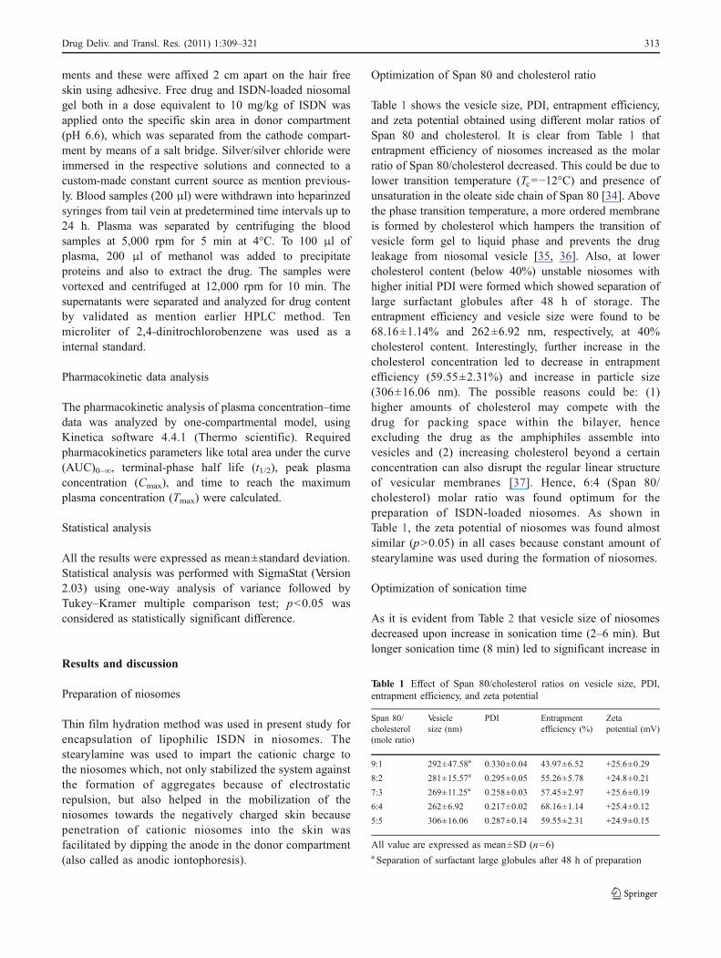
The shape and morphology of niosomes was studied usingatomic force microscopy (AFM). The advantage of AFM isthe simple sample preparation during operation and thesample does not need to be conductive. Additionally, mostwater molecules of sample are not completely removedduring the drying process thus prevents shrinkage of vesiclesystem and enables their visualization in hydrated state. A3-D surface images obtained by AFM (Fig. 1) showedsmooth and slightly flattened spherical shaped of niosomes.A good correlation was obtained in the vesicle size asobserved by both zeta sizer and AFM because these havemeasured the sample in hydrated state [33].
In vitro drug release study
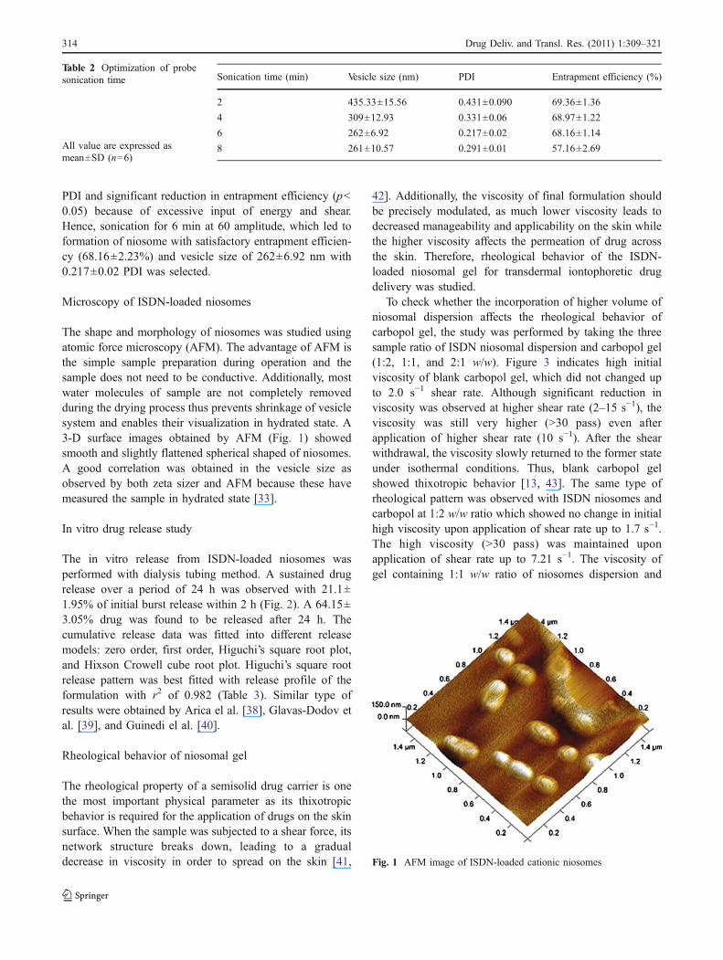
The in vitro release from ISDN-loaded niosomes wasperformed with dialysis tubing method. A sustained drugrelease over a period of 24 h was observed with 21.1±1.95% of initial burst release within 2 h (Fig. 2). A 64.15±3.05% drug was found to be released after 24 h. Thecumulative release data was fitted into different releasemodels: zero order, first order, Higuchi’s square root plot,and Hixson Crowell cube root plot. Higuchi’s square rootrelease pattern was best fitted with release profile of theformulation with r2 of 0.982 (Table 3). Similar type ofresults were obtained by Arica el al. [38], Glavas-Dodov etal. [39], and Guinedi el al. [40].
Rheological behavior of niosomal gel
The rheological property of a semisolid drug carrier is onethe most important physical parameter as its thixotropicbehavior is required for the application of drugs on the skinsurface. When the sample was subjected to a shear force, itsnetwork structure breaks down, leading to a gradualdecrease in viscosity in order to spread on the skin [41,
42]. Additionally, the viscosity of final formulation shouldbe precisely modulated, as much lower viscosity leads todecreased manageability and applicability on the skin whilethe higher viscosity affects the permeation of drug acrossthe skin. Therefore, rheological behavior of the ISDN-loaded niosomal gel for transdermal iontophoretic drugdelivery was studied.
To check whether the incorporation of higher volume ofniosomal dispersion affects the rheological behavior ofcarbopol gel, the study was performed by taking the threesample ratio of ISDN niosomal dispersion and carbopol gel(1:2, 1:1, and 2:1 w/w). Figure 3 indicates high initialviscosity of blank carbopol gel, which did not changed upto 2.0 s−1 shear rate. Although significant reduction inviscosity was observed at higher shear rate (2–15 s−1), theviscosity was still very higher (>30 pass) even afterapplication of higher shear rate (10 s−1). After the shearwithdrawal, the viscosity slowly returned to the former stateunder isothermal conditions. Thus, blank carbopol gelshowed thixotropic behavior [13, 43]. The same type ofrheological pattern was observed with ISDN niosomes andcarbopol at 1:2 w/w ratio which showed no change in initialhigh viscosity upon application of shear rate up to 1.7 s−1.The high viscosity (>30 pass) was maintained uponapplication of shear rate up to 7.21 s−1. The viscosity ofgel containing 1:1 w/w ratio of niosomes dispersion and
Sonication time (min) Vesicle size (nm) PDI Entrapment efficiency (%)
2 435.33±15.56 0.431±0.090 69.36±1.36
4 309±12.93 0.331±0.06 68.97±1.22
6 262±6.92 0.217±0.02 68.16±1.14
8 261±10.57 0.291±0.01 57.16±2.69
Table 2 Optimization of probesonication time
All value are expressed asmean±SD (n=6)
Fig. 1 AFM image of ISDN-loaded cationic niosomes
314 Drug Deliv. and Transl. Res. (2011) 1:309–321
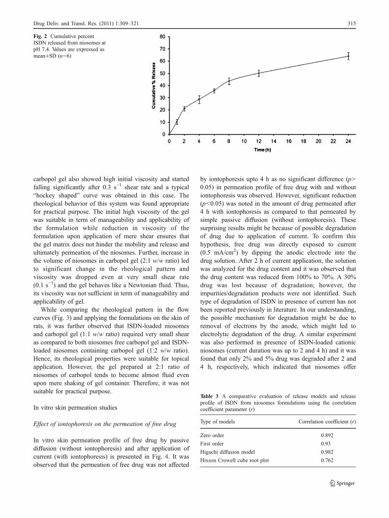
carbopol gel also showed high initial viscosity and startedfalling significantly after 0.3 s−1 shear rate and a typical“hockey shaped” curve was obtained in this case. Therheological behavior of this system was found appropriatefor practical purpose. The initial high viscosity of the gelwas suitable in term of manageability and applicability ofthe formulation while reduction in viscosity of theformulation upon application of mere shear ensures thatthe gel matrix does not hinder the mobility and release andultimately permeation of the niosomes. Further, increase inthe volume of niosomes in carbopol gel (2:1 w/w ratio) ledto significant change in the rheological pattern andviscosity was dropped even at very small shear rate(0.1 s−1) and the gel behaves like a Newtonian fluid. Thus,its viscosity was not sufficient in term of manageability andapplicability of gel.
While comparing the rheological pattern in the flowcurves (Fig. 3) and applying the formulations on the skin ofrats, it was further observed that ISDN-loaded niosomesand carbopol gel (1:1 w/w ratio) required very small shearas compared to both niosomes free carbopol gel and ISDN-loaded niosomes containing carbopol gel (1:2 w/w ratio).Hence, its rheological properties were suitable for topicalapplication. However, the gel prepared at 2:1 ratio ofniosomes of carbopol tends to become almost fluid evenupon mere shaking of gel container. Therefore, it was notsuitable for practical purpose.
In vitro skin permeation studies
Effect of iontophoresis on the permeation of free drug
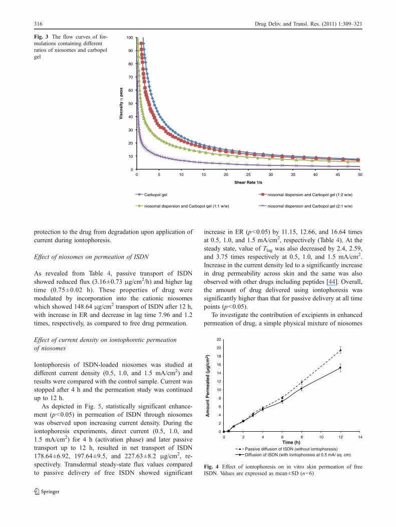
In vitro skin permeation profile of free drug by passivediffusion (without iontophoresis) and after application ofcurrent (with iontophoresis) is presented in Fig. 4. It wasobserved that the permeation of free drug was not affected
by iontophoresis upto 4 h as no significant difference (p>0.05) in permeation profile of free drug with and withoutiontophoresis was observed. However, significant reduction(p<0.05) was noted in the amount of drug permeated after4 h with iontophoresis as compared to that permeated bysimple passive diffusion (without iontophoresis). Thesesurprising results might be because of possible degradationof drug due to application of current. To confirm thishypothesis, free drug was directly exposed to current(0.5 mA/cm2) by dipping the anodic electrode into thedrug solution. After 2 h of current application, the solutionwas analyzed for the drug content and it was observed thatthe drug content was reduced from 100% to 70%. A 30%drug was lost because of degradation; however, theimpurities/degradation products were not identified. Suchtype of degradation of ISDN in presence of current has notbeen reported previously in literature. In our understanding,the possible mechanism for degradation might be due toremoval of electrons by the anode, which might led toelectrolytic degradation of the drug. A similar experimentwas also performed in presence of ISDN-loaded cationicniosomes (current duration was up to 2 and 4 h) and it wasfound that only 2% and 5% drug was degraded after 2 and4 h, respectively, which indicated that niosomes offer
Table 3 A comparative evaluation of release models and releaseprofile of ISDN from niosomes formulations using the correlationcoefficient parameter (r)
Type of models Correlation coefficient (r)
Zero order 0.892
First order 0.93
Higuchi diffusion model 0.982
Hixson Crowell cube root plot 0.762
Fig. 2 Cumulative percentISDN released from niosomes atpH 7.4. Values are expressed asmean±SD (n=6)
Drug Deliv. and Transl. Res. (2011) 1:309–321 315
protection to the drug from degradation upon application ofcurrent during iontophoresis.
Effect of niosomes on permeation of ISDN
As revealed from Table 4, passive transport of ISDNshowed reduced flux (3.16±0.73 μg/cm2/h) and higher lagtime (0.75±0.02 h). These properties of drug weremodulated by incorporation into the cationic niosomeswhich showed 148.64 μg/cm2 transport of ISDN after 12 h,with increase in ER and decrease in lag time 7.96 and 1.2times, respectively, as compared to free drug permeation.
Effect of current density on iontophoretic permeationof niosomes
Iontophoresis of ISDN-loaded niosomes was studied atdifferent current density (0.5, 1.0, and 1.5 mA/cm2) andresults were compared with the control sample. Current wasstopped after 4 h and the permeation study was continuedup to 12 h.
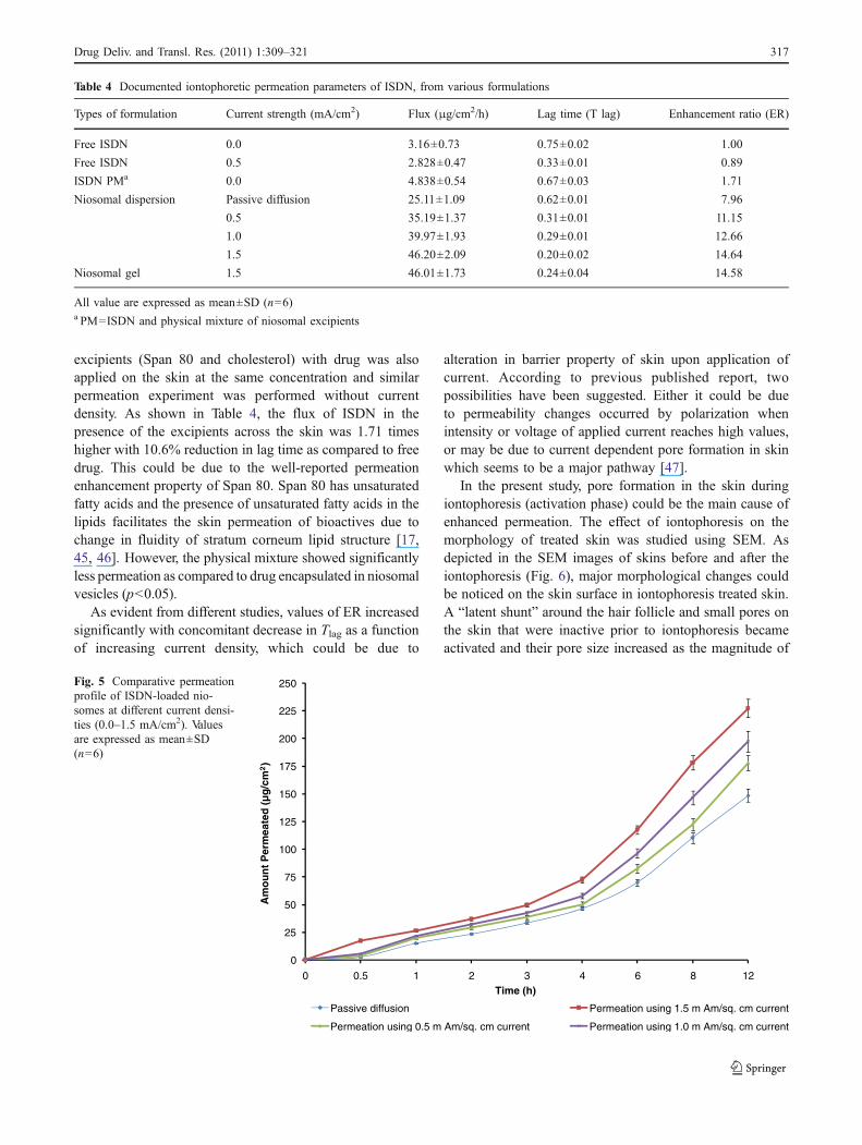
As depicted in Fig. 5, statistically significant enhance-ment (p<0.05) in permeation of ISDN through niosomeswas observed upon increasing current density. During theiontophoresis experiments, direct current (0.5, 1.0, and1.5 mA/cm2) for 4 h (activation phase) and later passivetransport up to 12 h, resulted in net transport of ISDN178.64±6.92, 197.64±9.5, and 227.63±8.2 μg/cm2, re-spectively. Transdermal steady-state flux values comparedto passive delivery of free ISDN showed significant
increase in ER (p<0.05) by 11.15, 12.66, and 16.64 timesat 0.5, 1.0, and 1.5 mA/cm2, respectively (Table 4). At thesteady state, value of Tlag was also decreased by 2.4, 2.59,and 3.75 times respectively at 0.5, 1.0, and 1.5 mA/cm2.Increase in the current density led to a significantly increasein drug permeability across skin and the same was alsoobserved with other drugs including peptides [44]. Overall,the amount of drug delivered using iontophoresis wassignificantly higher than that for passive delivery at all timepoints (p<0.05).
To investigate the contribution of excipients in enhancedpermeation of drug, a simple physical mixture of niosomes
0
2
4
6
8
10
12
14
16
18
20
22
0 2 4 6 8 10 12 14
Am
ou
nt
Per
mea
ted
(µ
g/c
m2 )
Time (h)Passive diffusion of ISDN (without iontophoresis)Diffusion of ISDN (with Iontophoresis at 0.5 mA/ sq. cm)
Fig. 4 Effect of iontophoresis on in vitro skin permeation of freeISDN. Values are expressed as mean±SD (n=6)
0
10
20
30
40
50
60
70
80
90
100
0 5 10 15 20 25 30 35 40 45 50
Vis
cosi
typ
ass
Shear Rate 1/s
Carbopol gel niosomal dispersion and Carbopol gel (1:2 w/w)
niosomal dispersion and Carbopol gel (1:1 w/w) niosomal dispersion and Carbopol gel (2:1 w/w)
Fig. 3 The flow curves of for-mulations containing differentratios of niosomes and carbopolgel
316 Drug Deliv. and Transl. Res. (2011) 1:309–321
excipients (Span 80 and cholesterol) with drug was alsoapplied on the skin at the same concentration and similarpermeation experiment was performed without currentdensity. As shown in Table 4, the flux of ISDN in thepresence of the excipients across the skin was 1.71 timeshigher with 10.6% reduction in lag time as compared to freedrug. This could be due to the well-reported permeationenhancement property of Span 80. Span 80 has unsaturatedfatty acids and the presence of unsaturated fatty acids in thelipids facilitates the skin permeation of bioactives due tochange in fluidity of stratum corneum lipid structure [17,45, 46]. However, the physical mixture showed significantlyless permeation as compared to drug encapsulated in niosomalvesicles (p<0.05).
As evident from different studies, values of ER increasedsignificantly with concomitant decrease in Tlag as a functionof increasing current density, which could be due to
alteration in barrier property of skin upon application ofcurrent. According to previous published report, twopossibilities have been suggested. Either it could be dueto permeability changes occurred by polarization whenintensity or voltage of applied current reaches high values,or may be due to current dependent pore formation in skinwhich seems to be a major pathway [47].
In the present study, pore formation in the skin duringiontophoresis (activation phase) could be the main cause ofenhanced permeation. The effect of iontophoresis on themorphology of treated skin was studied using SEM. Asdepicted in the SEM images of skins before and after theiontophoresis (Fig. 6), major morphological changes couldbe noticed on the skin surface in iontophoresis treated skin.A “latent shunt” around the hair follicle and small pores onthe skin that were inactive prior to iontophoresis becameactivated and their pore size increased as the magnitude of
Table 4 Documented iontophoretic permeation parameters of ISDN, from various formulations
Types of formulation Current strength (mA/cm2) Flux (μg/cm2/h) Lag time (T lag) Enhancement ratio (ER)
Free ISDN 0.0 3.16±0.73 0.75±0.02 1.00
Free ISDN 0.5 2.828±0.47 0.33±0.01 0.89
ISDN PMa 0.0 4.838±0.54 0.67±0.03 1.71
Niosomal dispersion Passive diffusion 25.11±1.09 0.62±0.01 7.96
0.5 35.19±1.37 0.31±0.01 11.15
1.0 39.97±1.93 0.29±0.01 12.66
1.5 46.20±2.09 0.20±0.02 14.64
Niosomal gel 1.5 46.01±1.73 0.24±0.04 14.58
All value are expressed as mean±SD (n=6)a PM=ISDN and physical mixture of niosomal excipients
0
25
50
75
100
125
150
175
200
225
250
0 0.5 1 2 3 4 6 8 12
Am
ou
nt
Per
mea
ted
(µ
g/c
m2)
Time (h)
Passive diffusion Permeation using 1.5 m Am/sq. cm current
Permeation using 0.5 m Am/sq. cm current Permeation using 1.0 m Am/sq. cm current
Fig. 5 Comparative permeationprofile of ISDN-loaded nio-somes at different current densi-ties (0.0–1.5 mA/cm2). Valuesare expressed as mean±SD(n=6)
Drug Deliv. and Transl. Res. (2011) 1:309–321 317
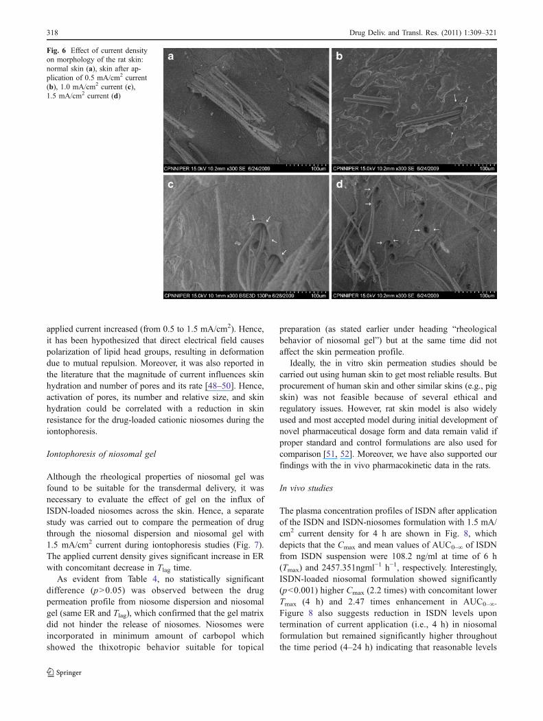
applied current increased (from 0.5 to 1.5 mA/cm2). Hence,it has been hypothesized that direct electrical field causespolarization of lipid head groups, resulting in deformationdue to mutual repulsion. Moreover, it was also reported inthe literature that the magnitude of current influences skinhydration and number of pores and its rate [48–50]. Hence,activation of pores, its number and relative size, and skinhydration could be correlated with a reduction in skinresistance for the drug-loaded cationic niosomes during theiontophoresis.
Iontophoresis of niosomal gel
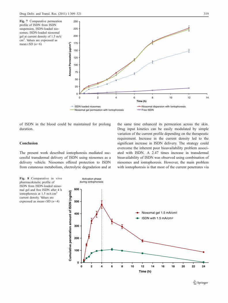
Although the rheological properties of niosomal gel wasfound to be suitable for the transdermal delivery, it wasnecessary to evaluate the effect of gel on the influx ofISDN-loaded niosomes across the skin. Hence, a separatestudy was carried out to compare the permeation of drugthrough the niosomal dispersion and niosomal gel with1.5 mA/cm2 current during iontophoresis studies (Fig. 7).The applied current density gives significant increase in ERwith concomitant decrease in Tlag time.
As evident from Table 4, no statistically significantdifference (p>0.05) was observed between the drugpermeation profile from niosome dispersion and niosomalgel (same ER and Tlag), which confirmed that the gel matrixdid not hinder the release of niosomes. Niosomes wereincorporated in minimum amount of carbopol whichshowed the thixotropic behavior suitable for topical
preparation (as stated earlier under heading “rheologicalbehavior of niosomal gel”) but at the same time did notaffect the skin permeation profile.
Ideally, the in vitro skin permeation studies should becarried out using human skin to get most reliable results. Butprocurement of human skin and other similar skins (e.g., pigskin) was not feasible because of several ethical andregulatory issues. However, rat skin model is also widelyused and most accepted model during initial development ofnovel pharmaceutical dosage form and data remain valid ifproper standard and control formulations are also used forcomparison [51, 52]. Moreover, we have also supported ourfindings with the in vivo pharmacokinetic data in the rats.
In vivo studies
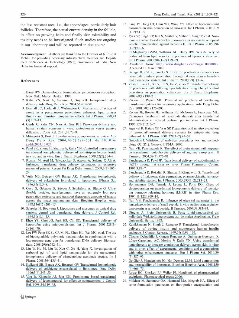
The plasma concentration profiles of ISDN after applicationof the ISDN and ISDN-niosomes formulation with 1.5 mA/cm2 current density for 4 h are shown in Fig. 8, whichdepicts that the Cmax and mean values of AUC0–∞ of ISDNfrom ISDN suspension were 108.2 ng/ml at time of 6 h(Tmax) and 2457.351ngml−1 h−1, respectively. Interestingly,ISDN-loaded niosomal formulation showed significantly(p<0.001) higher Cmax (2.2 times) with concomitant lowerTmax (4 h) and 2.47 times enhancement in AUC0–∞.Figure 8 also suggests reduction in ISDN levels upontermination of current application (i.e., 4 h) in niosomalformulation but remained significantly higher throughoutthe time period (4–24 h) indicating that reasonable levels
Fig. 6 Effect of current densityon morphology of the rat skin:normal skin (a), skin after ap-plication of 0.5 mA/cm2 current(b), 1.0 mA/cm2 current (c),1.5 mA/cm2 current (d)
318 Drug Deliv. and Transl. Res. (2011) 1:309–321
of ISDN in the blood could be maintained for prolongduration.
Conclusion
The present work described iontophoresis mediated suc-cessful transdermal delivery of ISDN using niosomes as adelivery vehicle. Niosomes offered protection to ISDNfrom cutaneous metabolism, electrolytic degradation and at
the same time enhanced its permeation across the skin.Drug input kinetics can be easily modulated by simplevariation of the current profile depending on the therapeuticrequirement. Increase in the current density led to thesignificant increase in ISDN delivery. The strategy couldovercome the inherent poor bioavailability problem associ-ated with ISDN. A 2.47 times increase in transdermalbioavailability of ISDN was observed using combination ofniosomes and iontophoresis. However, the main problemwith iontophoresis is that most of the current penetrates via
0 2 4 6 8 10 12 14 16 18 20 22 240
100
200
300
400
500
600
Niosomal gel 1.5 mA/cm2
Activation phaseduring iontophoresis
ISDN with 1.5 mA/cm2
Time (h)
Cu
mu
lati
ve p
enet
rati
on
am
ou
nt
of
ISD
N (
ng
/ml)
Fig. 8 Comparative in vivopharmacokinetic profile ofISDN from ISDN-loaded nioso-mal gel and free ISDN after 4 hiontophoresis at 1.5 mA/cm2
current density. Values areexpressed as mean±SD (n=4)
0
25
50
75
100
125
150
175
200
225
250
0 2 4 6 8 10 12 14
Am
ou
nt
Per
mea
ted
(µ
g/c
m2)
Time (h)
ISDN loaded niosomes Niosomal dispersion with IontophoresisNiosomal gel permeation with Iontophoresis Free ISDN
Fig. 7 Comparative permeationprofile of ISDN from ISDNsuspension, ISDN-loaded nio-somes; ISDN-loaded niosomalgel at current density of 1.5 mA/cm2. Values are expressed asmean±SD (n=6)
Drug Deliv. and Transl. Res. (2011) 1:309–321 319
the less resistant area, i.e., the appendages, particularly hairfollicles. Therefore, the actual current density in the follicle,its effect on growing hairs and finally skin tolerability andtoxicity needs to be investigated. Such studies are ongoingin our laboratory and will be reported in due course.
Acknowledgment Authors are thankful to the Director of NIPER atMohali for providing necessary infrastructural facilities and Depart-ment of Science & Technology (DST), Government of India, NewDelhi for financial support.
References
1. Barry BW. Dermatological formulations: percutaneous absorption.New York: Marcel Dekker; 1983.
2. Kalia YN, Naik A, Garrison J, Guy RH. Iontophoretic drugdelivery. Adv Drug Deliv Rev. 2004;56:619–58.
3. Beastall JC, Hadgraft J, Washington C. Mechanism of action ofazone as a percutaneous penetration enhancer: lipid bilayerfluidity and transition temperature effects. Int J Pharm. 1988;43(3):207–13.
4. Curdy C, kalia YN, Naik A, Guy RH. Piroxicam delivery intohuman stratum corneum in vivo: iontophoresis versus passivediffusion. J Contr Rel. 2001;76:73–9.
5. Mitragotri S, Kost J. Low-frequency sonophoresis: a review. AdvDrug Deliv Rev. 2004;56(5):589–601. doi :10.1016/j.addr.2003.10.024.
6. Patel SR, Zhong H, Sharma A, Kalia YN. Controlled non-invasivetransdermal iontophoretic delivery of zolmitriptan hydrochloridein vitro and in vivo. Eur J Pharm Biopharm. 2009;72(2):304–9.
7. Rizwan M, Aqil M, Talegaonkar S, Azeem A, Sultana Y, Ali A.Enhanced transdermal drug delivery techniques: an extensivereview of patents. Recent Pat Drug Deliv Formul. 2009;3(2):105–24.
8. Vutla NB, Betageri GV, Banga AK. Transdermal iontophoreticdelivery of enkephalin formulated in liposomes. JPharm Sci.1996;85(1):5–8.
9. Cevc G, Gebauer D, Stieber J, Schätzlein A, Blume G. Ultra-flexible vesicles, transfersomes, have an extremely low porepenetration resistance and transport therapeutic amounts of insulinacross the intact mammalian skin. Biochim Biophys Acta.1998;1368(2):201–15.
10. Schreier H, Bouwstra J. Liposomes and niosomes as topical drugcarriers: dermal and transdermal drug delivery. J Control Rel.1994;30(1):1–15.
11. Rhee YS, Choi JG, Park ES, Chi SC. Transdermal delivery ofketoprofen using microemulsions. Int J Pharm. 2001;228(1–2):161–70.
12. Lee PW, Peng SF, Su CJ, Mi FL, Chen HL, Wei MC, et al. The useof biodegradable polymeric nanoparticles in combination with alow-pressure gene gun for transdermal DNA delivery. Biomate-rials. 2008;29(6):742–51.
13. Liu W, Hu M, Liu W, Xue C, Xu H, Yang X. Investigation ofcarbopol gel of solid lipid nanoparticle for the transdermaliontophoretic delivery of triamcinolone acetoinde acetate. Int JPharm. 2008;364:135–41.
14. Kulkarni SB, Banga AK, Betageri GV. Transdermal lontophoreticdelivery of colchicine encapsulated in liposomes. Drug Deliv.1996;3(4):245–50.
15. Vora B, Khopade AJ, Jain NK. Proniosome based transdermaldelivery of levonorgestrel for effective contraception. J ControlRel. 1998;54:149–65.
16. Fang JY, Hong CT, Chiu WT, Wang YY. Effect of liposomes andniosomes on skin permeation of enoxacin. Int J Pharm. 2001;219(1–2):61–72.
17. Vyas SP, Singh RP, Jain S, Mishra V, Mahor S, Singh P, et al. Non-ionic surfactant based vesicles (niosomes) for non-invasive topicalgenetic immunization against hepatitis B. Int J Pharm. 2005;296(1–2):80–6.
18. El Maghraby GMM, Williams AC, Barry BW. Skin delivery ofoestradiol from lipid vesicles: importance of liposome structure.Int J Pharm. 2000;204(1–2):159–69.
19. Available from: http://www.drugbank.ca/drugs/DB00883.Accessed 18 March 2010.
20. Gabiga H, Cal K, Janicki S. Effect of penetration enhancers onisosorbide dinitrate penetration through rat skin from a transder-mal therapeutic system. Int J Pharm. 2000;199(1):1–6.
21. Zhao L, Fang L, Xu Y, Liu S, He Z, Zhao Y. Transdermal deliveryof penetrants with differing lipophilicities using O-acylmentholderivatives as penetration enhancers. Eur J Pharm Biopharm.2008;69(1):199–213.
22. Riviere JE, Papich MG. Potential and problems of developingtransdermal patches for veterinary applications. Adv Drug DelivRev. 2001;50(3):175–203.
23. Riviere JE, Brooks JD, Williams PL, McGown E, Francoeur ML.Cutaneous metabolism of isosorbide dinitrate after transdermaladministration in isolated perfused porcine skin. Int J Pharm.1996;127(2):213–7.
24. Agarwal R, Katare OP, Vyas SP. Preparation and in vitro evaluationof liposomal/niosomal delivery systems for antipsoriatic drugdithranol. Int J Pharm. 2001;228(1–2):43–52.
25. Guideline I. Validation of analytical procedures: text and method-ology Q2 (R1). Geneva: IFPMA; 2005.
26. Nair VB, Panchagnula R. The effect of pretreatment with terpeneson transdermal iontophoretic delivery of arginine vasopressin. IlFarmaco. 2004;59(7):575–81.
27. Panchagnula R, Patel JR. Transdermal delivery of azidothymidine(AZT) through rat skin ex vivo. Pharm Pharmacol Comm.1997;3:83–7.
28. Panchagnula R, Bokalial R, Sharma P, Khandavilli S. Transdermaldelivery of naloxone: skin permeation, pharmacokinetic, irritancyand stability studies. Int J Pharm. 2005;293(1–2):213–23.
29. Bommannan DB, Tamada J, Leung L, Potts RO. Effect ofelectroporation on transdermal lontophoretic delivery of luteiniz-ing hormone releasing hormone (LHRH) in vitro. Pharm Res.1994;11(12):1809–14.
30. Nair VB, Panchagnula R. Influence of electrical parameter in theiontophoretic delivery of small peptide: in vitro studies using arginine-vasopressin as a model peptide. Il Farmaco. 2004;59:583–93.
31. Dingler A. Freie Universität B. Feste Lipid-nanopartikel alskolloidale Wirkstoffträgersysteme zur dermalen Applikation. FreieUniversität Berlin; 1998.
32. Kanikkannan N, Singh J, Ramarao P. Transdermal iontophoreticdelivery of bovine insulin and monomeric human insulinanalogue. J Control Release. 1999;59(1):99–105.
33. Cázares-Delgadillo J, Ganem-Rondero A, Quintanar-Guerrero D,López-Castellano AC, Merino V, Kalia YN. Using transdermaliontophoresis to increase granisetron delivery across skin in vitroand in vivo: effect of experimental conditions and a comparisonwith other enhancement strategies. Eur J Pharm Sci. 2010;39(5):387–93.
34. De Gier J, Mandersloot JG, Van Deenen LLM. Lipid compositionand permeability of liposomes. Biochim Biophys Acta. 1968;150(4):666–75.
35. Rowe RC, Sheskey PJ, Weller PJ. Handbook of pharmaceuticalexcipients. Pharmaceutical press; 2006.
36. Mokhtar M, Sammour OA, Hammad MA, Megrab NA. Effect ofsome formulation parameters on flurbiprofen encapsulation and
320 Drug Deliv. and Transl. Res. (2011) 1:309–321
release rates of niosomes prepared from proniosomes. Int J Pharm.2008;361(1–2):104–11.
37. El-Samaligy MS, Afifi NN, Mahmoud EA. Increasing bioavail-ability of silymarin using a buccal liposomal delivery system:preparation and experimental design investigation. Int J Pharm.2006;308(1–2):140–8.
38. Arica B, Ozer AY, Ercan MT, Hincal AA. Characterization, invitro and in vivo studies on primaquine diphosphate lip-osomes. J Microencapsul. 1995;12(5):469–85. doi:10.3109/02652049509006778.
39. Glavas-Dodov M, Goracinova K, Mladenovska K, Fredro-Kumbaradzi E. Release profile of lidocaine HCl from topicalliposomal gel formulation. Int J Pharm. 2002;242(1–2):381–4.
40. Guinedi AS, Mortada ND, Mansour S, Hathout RM. Preparationand evaluation of reverse-phase evaporation and multilamellarniosomes as ophthalmic carriers of acetazolamide. Int J Pharm.2005;306(1–2):71–82. doi:10.1016/j.ijpharm.2005.09.023.
41. Ruel-Gariepy E, Chenite A, Chaput C, Guirguis S, Leroux JC.Characterization of thermosensitive chitosan gels for the sustaineddelivery of drugs. Int J Pharm. 2000;203(1–2):89–98.
42. Lippacher A, Müller RH, Mäder K. Liquid and semisolid SLN(TM) dispersions for topical application: rheological characteriza-tion. Eur J Pharm Biopharm. 2004;58(3):561–7.
43. Islam MT, Rodriguez-Hornedo N, Ciotti S, Ackermann C.Rheological characterization of topical carbomer gels neutralizedto different pH. Pharm Res. 2004;21(7):1192–9.
44. Lelawongs P, Liu JC, Chien YW. Transdermal iontophoreticdelivery of arginine-vasopressien (II). Evaluation of electricaland operational factors. Int J Pharm. 1990;61:179–88.
45. Valjakka-Koskela R, Kirjavainen M, Mönkkönen J, Urtti A,Kiesvaara J. Enhancement of percutaneous absorption of nap-roxen by phospholipids. Int J Pharm. 1998;175(2):225–30.
46. Jain S, Vyas SP. Mannosylated niosomes as carrier adjuvantsystem for topical immunization. JPharm Pharmacol. 2005;57(9):1177–84.
47. Sims SM, Higuchi WI, Srinivasan V. Skin alteration in convectivesolvent flow effects during iontophoresis. I. Neutral solutestransport across human skin. Int J Pharm. 1991;69:109–21.
48. Kasting GB, Bowman LA. DC electrical properties of frozen,excised human skin. Pharm Res. 1990;7(2):134–43.
49. Oh SY, Leung L, Bommannan D, Guy RH, Potts RO. Effect ofcurrent, ionic strength and temperature on the electrical propertiesof skin. J Control Rel. 1993;27(2):115–25.
50. Prausnitz MR, Bose VG, Langer R, Weaver JC. Electroporation ofmammalian skin: a mechanism to enhance transdermal drugdelivery. Proc Natl Acad Sci. 1993;90(22):10504.
51. Godin B, Touitou E. Transdermal skin delivery: predictions forhumans from in vivo, ex vivo and animal models. Adv DrugDelivery Rev. 2007;59(11):1152–61.
52. Escobar-Chavez JJ, Merino-Sanjuán V, López-Cervantes M,Urban-Morlan Z, Piñón-Segundo E, Quintanar-Guerrero D, et al.The tape-stripping technique as a method for drug quantificationin skin. J Pharm Pharma Sci. 2008;11(1):104.
Drug Deliv. and Transl. Res. (2011) 1:309–321 321
Related Documents