RESEARCH Open Access Prenatal selective serotonin reuptake inhibitor (SSRI) exposure induces working memory and social recognition deficits by disrupting inhibitory synaptic networks in male mice Weonjin Yu 1,2† , Yi-Chun Yen 1† , Young-Hwan Lee 1 , Shawn Tan 1 , Yixin Xiao 1,3 , Hidayat Lokman 1 , Audrey Khoo Tze Ting 1 , Hasini Ganegala 1 , Taejoon Kwon 4 , Won-Kyung Ho 2* and H. Shawn Je 1,3* Abstract Selective serotonin reuptake inhibitors (SSRIs) are commonly prescribed antidepressant drugs in pregnant women. Infants born following prenatal exposure to SSRIs have a higher risk for behavioral abnormalities, however, the underlying mechanisms remains unknown. Therefore, we examined the effects of prenatal fluoxetine, the most commonly prescribed SSRI, in mice. Intriguingly, chronic in utero fluoxetine treatment impaired working memory and social novelty recognition in adult males. In the medial prefrontal cortex (mPFC), a key region regulating these behaviors, we found augmented spontaneous inhibitory synaptic transmission onto the layer 5 pyramidal neurons. Fast-spiking interneurons in mPFC exhibited enhanced intrinsic excitability and serotonin-induced excitability due to upregulated serotonin (5-HT) 2A receptor (5-HT 2A R) signaling. More importantly, the behavioral deficits in prenatal fluoxetine treated mice were reversed by the application of a 5-HT 2A R antagonist. Taken together, our findings suggest that alterations in inhibitory neuronal modulation are responsible for the behavioral alterations following prenatal exposure to SSRIs. Keywords: Prenatal, Serotonin (5-HT), Selective serotonin reuptake inhibitor (SSRI), Fluoxetine, Working memory, Social recognition, Serotonin 2A receptor (5-HT 2A R) Introduction Anti-depressants are commonly prescribed to treat major depression and post-traumatic stress disorder. Currently, 17% of pregnant women experience major depression, and approximately 10% of these women use anti-depressants [1–3]. The most commonly prescribed anti-depressants, selective serotonin reuptake inhibitors (SSRIs), are believed to increase the ambient level of 5-hydrotryptamine (5-HT, serotonin) in synaptic clefts by preventing its reabsorption [4–6]. However, the exact mechanism by which SSRIs mitigates depression remains unknown. A recent systematic review showed that the potential adverse effects of SSRIs might outweigh their beneficial effects on depression [7–9]. In addition, fluox- etine (FLX), one of the most widely used SSRIs with a moderately long half-life (t ½ = 48 h), can cross the pla- cental and blood-brain barriers and is also detected in breast milk, suggesting potential accumulation of FLX as well as 5-HT in the fetal brain [10]. However, little is known about the safety of FLX use during pregnancy. Moreover, the long-term consequences of prenatal FLX exposure for adverse behavioral outcomes in offspring are uncertain and sometimes conflicting; these conflict- ing findings are likely due to the independent association © The Author(s). 2019 Open Access This article is distributed under the terms of the Creative Commons Attribution 4.0 International License (http://creativecommons.org/licenses/by/4.0/), which permits unrestricted use, distribution, and reproduction in any medium, provided you give appropriate credit to the original author(s) and the source, provide a link to the Creative Commons license, and indicate if changes were made. The Creative Commons Public Domain Dedication waiver (http://creativecommons.org/publicdomain/zero/1.0/) applies to the data made available in this article, unless otherwise stated. * Correspondence: [email protected]; [email protected] † Weonjin Yu and Yi-Chun Yen are contributed equally to this work. 2 Department of Physiology, Seoul National University College of Medicine, Seoul 03080, Republic of Korea 1 Molecular Neurophysiology Laboratory, Signature Program in Neuroscience and Behavioral Disorders, Duke-National University of Singapore (NUS) Medical School, 8 College Road, Singapore 169857, Singapore Full list of author information is available at the end of the article Yu et al. Molecular Brain (2019) 12:29 https://doi.org/10.1186/s13041-019-0452-5

Welcome message from author
This document is posted to help you gain knowledge. Please leave a comment to let me know what you think about it! Share it to your friends and learn new things together.
Transcript

RESEARCH Open Access
Prenatal selective serotonin reuptakeinhibitor (SSRI) exposure induces workingmemory and social recognition deficits bydisrupting inhibitory synaptic networks inmale miceWeonjin Yu1,2†, Yi-Chun Yen1†, Young-Hwan Lee1, Shawn Tan1, Yixin Xiao1,3, Hidayat Lokman1,Audrey Khoo Tze Ting1, Hasini Ganegala1, Taejoon Kwon4, Won-Kyung Ho2* and H. Shawn Je1,3*
Abstract
Selective serotonin reuptake inhibitors (SSRIs) are commonly prescribed antidepressant drugs in pregnant women.Infants born following prenatal exposure to SSRIs have a higher risk for behavioral abnormalities, however, theunderlying mechanisms remains unknown. Therefore, we examined the effects of prenatal fluoxetine, the mostcommonly prescribed SSRI, in mice. Intriguingly, chronic in utero fluoxetine treatment impaired working memoryand social novelty recognition in adult males. In the medial prefrontal cortex (mPFC), a key region regulating thesebehaviors, we found augmented spontaneous inhibitory synaptic transmission onto the layer 5 pyramidal neurons.Fast-spiking interneurons in mPFC exhibited enhanced intrinsic excitability and serotonin-induced excitability due toupregulated serotonin (5-HT) 2A receptor (5-HT2AR) signaling. More importantly, the behavioral deficits in prenatalfluoxetine treated mice were reversed by the application of a 5-HT2AR antagonist. Taken together, our findingssuggest that alterations in inhibitory neuronal modulation are responsible for the behavioral alterations followingprenatal exposure to SSRIs.
Keywords: Prenatal, Serotonin (5-HT), Selective serotonin reuptake inhibitor (SSRI), Fluoxetine, Working memory,Social recognition, Serotonin 2A receptor (5-HT2AR)
IntroductionAnti-depressants are commonly prescribed to treatmajor depression and post-traumatic stress disorder.Currently, 17% of pregnant women experience majordepression, and approximately 10% of these women useanti-depressants [1–3]. The most commonly prescribedanti-depressants, selective serotonin reuptake inhibitors(SSRIs), are believed to increase the ambient level of5-hydrotryptamine (5-HT, serotonin) in synaptic clefts
by preventing its reabsorption [4–6]. However, the exactmechanism by which SSRIs mitigates depression remainsunknown. A recent systematic review showed that thepotential adverse effects of SSRIs might outweigh theirbeneficial effects on depression [7–9]. In addition, fluox-etine (FLX), one of the most widely used SSRIs with amoderately long half-life (t ½ = 48 h), can cross the pla-cental and blood-brain barriers and is also detected inbreast milk, suggesting potential accumulation of FLX aswell as 5-HT in the fetal brain [10]. However, little isknown about the safety of FLX use during pregnancy.Moreover, the long-term consequences of prenatal FLXexposure for adverse behavioral outcomes in offspringare uncertain and sometimes conflicting; these conflict-ing findings are likely due to the independent association
© The Author(s). 2019 Open Access This article is distributed under the terms of the Creative Commons Attribution 4.0International License (http://creativecommons.org/licenses/by/4.0/), which permits unrestricted use, distribution, andreproduction in any medium, provided you give appropriate credit to the original author(s) and the source, provide a link tothe Creative Commons license, and indicate if changes were made. The Creative Commons Public Domain Dedication waiver(http://creativecommons.org/publicdomain/zero/1.0/) applies to the data made available in this article, unless otherwise stated.
* Correspondence: [email protected]; [email protected]†Weonjin Yu and Yi-Chun Yen are contributed equally to this work.2Department of Physiology, Seoul National University College of Medicine,Seoul 03080, Republic of Korea1Molecular Neurophysiology Laboratory, Signature Program in Neuroscienceand Behavioral Disorders, Duke-National University of Singapore (NUS)Medical School, 8 College Road, Singapore 169857, SingaporeFull list of author information is available at the end of the article
Yu et al. Molecular Brain (2019) 12:29 https://doi.org/10.1186/s13041-019-0452-5

between maternal depression and negative pregnancyoutcomes in human [11, 12].Ansorge et al. firstly observed that postnatal FLX
exposure produced anxiety behaviors and disruptedlearning in rodent offspring [13, 14]. In subsequentstudies, manipulations of brain 5-HT levels during earlydevelopment produced abnormal neuronal circuitformation in the cortex and promoted aggressive oranxiety-related behaviors [15–19]. However, a thoroughassessment of animal behaviors induced by prenatalSSRI exposure has not been performed. Furthermore,the underlying molecular and circuit mechanisms ofthese behavioral changes have not been investigated and,for this reason, no rescue experiment has been per-formed in offspring exposed to SSRIs during prenatalperiod.Using a combination of behavioral analyses and
electrophysiological investigations of affected neuronalcircuits, we examined how chronic prenatal exposureto exogenous FLX influences animal behaviors andneuronal circuitry and function. We observedimpaired social recognition and working memory inmale mice chronically exposed to FLX. Furthermore,we observed reduced frequencies in spontaneous exci-tatory postsynaptic currents recorded from layer (L) 5pyramidal neurons in the prelimbic cortex of affectedmice. Intriguingly, these reduced excitatory neuronalactivities were caused by enhanced serotonergicmodulation of fast-spiking (FS) interneurons in L5due to enhanced 5-HT2A receptors (5-HT2ARs).Moreover, the acute treatment with the 5-HT2AR an-tagonist MDL100907 (MDL) normalized the impairedsocial recognition and working memory impairmentin these animals [20].
ResultsPrenatal fluoxetine treatment induced deficits in workingmemory and social recognitionWe subjected pregnant mice to daily intraperitoneal(i.p.) injections of 0.6 mg/kg FLX or saline (SAL) fromembryonic day (ED) 4 to ED19 to examine behavioralchanges in mice exposed to SSRIs during the prenatalperiod (Fig. 1a) [21]. The mean number of pups bornper litter, percentages of male pups per litter, and aver-age body weights of mice at postnatal day 21 (P21) andP60 were not significantly different between FLX-treatedlitters and SAL-treated, control litters (Table 1). TheFLX-treated mice exhibited normal spontaneousexploratory behavior in terms of the total distancetraveled (Additional file 1: Figure S1A), but spent lesstime in the center zone (t(26) =2.12, p < 0.001; Additionalfile 1: Figure S1B), indicating a potential sign ofanxiety-like behaviors. We tested the mice using theelevated zero maze and light-dark box test to examine
anxiety-like behaviors (Additional file 1: Figures S1C, D).However, FLX-treated mice did not exhibit differencesin the time spent in the open arms of the elevatedzero maze (Additional file 1: Figure S1C) or totaltransitions in the light-dark box (Additional file 1:Figure S1D), suggesting normal anxiety levels inFLX-treated mice.To further examine the role of prenatal FLX in
working memory and cognitive function, we subjectedFLX-treated mice to the Y-maze spontaneous alternationtask [22]. Briefly, both control and FLX treated micewere allowed to freely access three arms of the Y-mazefor 10 min. Mice prefer to explore a previouslyuncharted arm of the maze rather than returning to apreviously visited arm [23]. Intriguingly, the alternationrate of prenatally FLX-treated mice was lower than thatof SAL-treated mice (t [24] =3.05, p < 0.01; Fig. 1b)without changes in general activity, as measured bythe total arm entries (p > 0.05; Fig. 1c). To testwhether this reduced alternation rate was due toeither behavioral perseverance or recognition of anew environment, we performed the novel objectrecognition test. Interestingly, both prenatally SAL-and FLX-treated mice were able to distinguish thenovel and familiar objects, as assessed by the timespent exploring the novel object (Additional file 1:Figure S1E). Additionally, there was no significantdifference in grooming behaviors between the twogroups, indicating that the behavioral perseverance ofFLX-treated mice was not associated with repetitiveor obsessive-compulsive behaviors (Additional file 1:Figure S1F).Next, we subjected the two groups of mice to the social
interaction test by using a 3-chamber apparatus [25, 26].During the 10-min habituation phase, neither groupsshowed any side preference in the 3-chamber appar-atus as reflected by the lack of differences in the timespent sniffing two empty wire pencil holders in theleft and right chambers (p > 0.05; Additional file 1:Figures S1G, H). Over the next 10 min, both groupsspent significantly more time sniffing the juvenilemale mouse (social stimulus) than the dummy object(t [9] =7.72, p < 0.001 for SAL; t [9] =10.39, p < 0.001for FLX; Fig. 1d-f ). During the last 10 min, prenatallySAL-treated mice spent significantly more time sniff-ing the novel juvenile male mouse than the familiarjuvenile mouse (t [9] =3.28, p < 0.01), confirming apreference for social novelty in SAL mice. In contrast,prenatally FLX-treated mice failed to show this preference,as indicated by the similar time spent in investigating thenovel and familiar juvenile mice (t [9] = 0.25, p =0.81; Fig. 1g-i). Taken together, these data indicatethat prenatally FLX-treated mice exhibited deficits inworking memory and social novelty recognition.
Yu et al. Molecular Brain (2019) 12:29 Page 2 of 13

Increased frequency of spontaneous and miniatureinhibitory synaptic currents in layer 5 pyramidal neuronsin the prefrontal cortex of FLX-treated miceAlthough the circuit mechanism underlying socialbehavior phenotypes observed in prenatally FLX-treatedmice is not obvious [27, 28], the deficits in workingmemory observed in the Y-maze spontaneous alternationtask prompted us to characterize neuronal activities inthe prefrontal cortex (PFC), which is functionally analo-gous to the dorsolateral PFC, a critical area known forworking memory in humans [29]. Within the PFC, infor-mation is transmitted top-down via pathways from L2/3pyramidal neurons to pyramidal neurons in L5 [30]. We
A B C
D E F
G H I
Fig. 1 Prenatal exposure to fluoxetine induces deficits in executive functions in male offspring. (a) Schematic diagram of the experimental design.From embryonic day 4 (ED4) to ED19, pregnant females received daily injections of either fluoxetine (FLX, 0.6 mg/kg/day) or saline (SAL). Maleoffspring were subjected to behavioral testing at 8–12 weeks of age. (b) Bar plot of spontaneous alternation rate on the Y-maze. (c) Bar plot ofthe total number of entries into all arms of the Y-maze. (d) Representative heat map images of results of the three-chamber social interaction testwith a novel mouse and object. The occupancy rate was normalized to the region with peak occupancy in the arena. (e) Bar plot of the timespent sniffing the novel mouse and object in the three-chamber social interaction task. (f) Bar plot of the social preference index (time spentsniffing mouse/total time spent sniffing the mouse and the object). The dotted line indicates an equal preference (50%) (g) Representative heatmap images of the results of the three-chamber social interaction tests with a familiar mouse and novel mouse. (h) Bar plot of the times spentsniffing the familiar mouse and novel mouse in the three-chamber social interaction task. (i) Bar plot of the social novelty preference index (timespent sniffing the novel mouse/total time spent sniffing both mice). Data are presented as means ± SEM. (e) and (h) Two-way repeated measuresANOVA. (b), (c), (f) and (i) Unpaired t-test. * p < 0.05, ** p < 0.01, *** p < 0.001
Table 1 Effects of prenatal SAL and FLX treatment on number,gender composition and weight of offspring
SAL FLX
Total number of litters 22 23
Total number of offspring 104 96
Total number of male mice 58 49
Average number of pups per litter 4.73 ± 0.44 4.17 ± 0.37
Average number of male pups per litter 2.64 ± 0.33 2.13 ± 0.21
Percentage of male pups in each litter 55.88 ± 4.98% 54.26 ± 4.19%
Weight of male mice at P28 16.44 ± 0.41 g,n = 20
15.89 ± 0.33 g,n = 23
Yu et al. Molecular Brain (2019) 12:29 Page 3 of 13

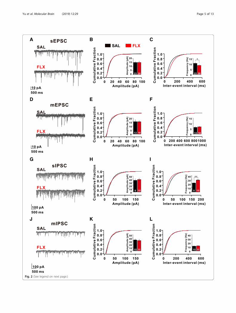
first characterized the morphology of L5 pyramidal neu-rons, which send the major corticofugal outputs fromthe PFC network. We did not observe any differences inlayer formation, the number of neurons, dendritic com-plexity, and the number of dendritic spines in L5 pyram-idal neurons between FLX-treated and SAL-treated mice(Additional file 1: Figure S2). Next, using whole-cellpatch-clamp recordings, we characterized the intrinsicproperties of L5 pyramidal neurons within the prelimbicarea (PrL), which is homologous to the dorsolateral PFC inprimates [30]. We first measured spontaneous excitatorypostsynaptic currents (sEPSCs) in L5 pyramidal neurons(Fig. 2a-c). The frequency of sEPSCs recorded fromFLX-treated mice was significantly decreased by 18% com-pared with those recorded from SAL-treated mice, whereasthe amplitude of sEPSCs was unaltered (SAL: 10.46 ± 0.50Hz, 15.23 ± 0.92 pA; FLX: 8.56 ± 0.51Hz, 15.32 ± 1.45 pA)(Fig. 2a-c). This indicates that spontaneous excitatorysynaptic transmission in the PFC was decreased inFLX-treated mice. To further explore the mechanism ofchanges in sEPSCs in FLX-treated mice, we recordedminiature excitatory postsynaptic currents (mEPSCs) andthe neuronal excitability of L5 pyramidal neurons. Intri-guingly, the frequency and amplitude of mEPSCs recordedfrom FLX-treated mice were not significantly differentfrom mEPSCs recorded from SAL-treated mice (SAL:6.34 ± 0.60 Hz, 15.61 ± 2.01 pA; FLX: 6.80 ± 0.34Hz,15.53 ± 1.11 pA) (Fig. 2d-f). More strikingly, most parame-ters of intrinsic neuronal properties (input resistance,resting membrane potentials, afterhyperpolarizationamplitude, and the threshold of action potentials (APs)) ofthe L5 neurons of FLX-treated mice were not significantlyaltered (Additional file 1: Figure S3). These data indicatethat the decreased spontaneous excitatory networkactivities did not arise from intrinsic changes in excitatoryneurons in the prefrontal cortex of FLX-treated mice.To test whether inhibitory synaptic transmission in the
PFC was affected in FLX-treated mice, we recorded bothspontaneous and miniature inhibitory postsynaptic cur-rents (sIPSCs and mIPSCs, respectively) in L5 pyramidalneurons within the PrL [30]. Surprisingly, we observed asignificant increase in the frequency of sIPSCs (SAL:22.46 ± 0.72 Hz; FLX: 29.99 ± 0.96 Hz, p < 0.01), but notthe amplitude (SAL: 36.88 ± 4.57 pA; FLX: 39.29 ± 3.36pA) (Fig. 2g-i). In contrast, neither the frequency nor theamplitude of mIPSCs were affected in L5 neurons fromFLX-treated mice (Fig. 2j-l). These data indicate thatprenatal FLX exposure increased spontaneous inhibitorynetwork activity in L5 pyramidal neurons within the PrL.
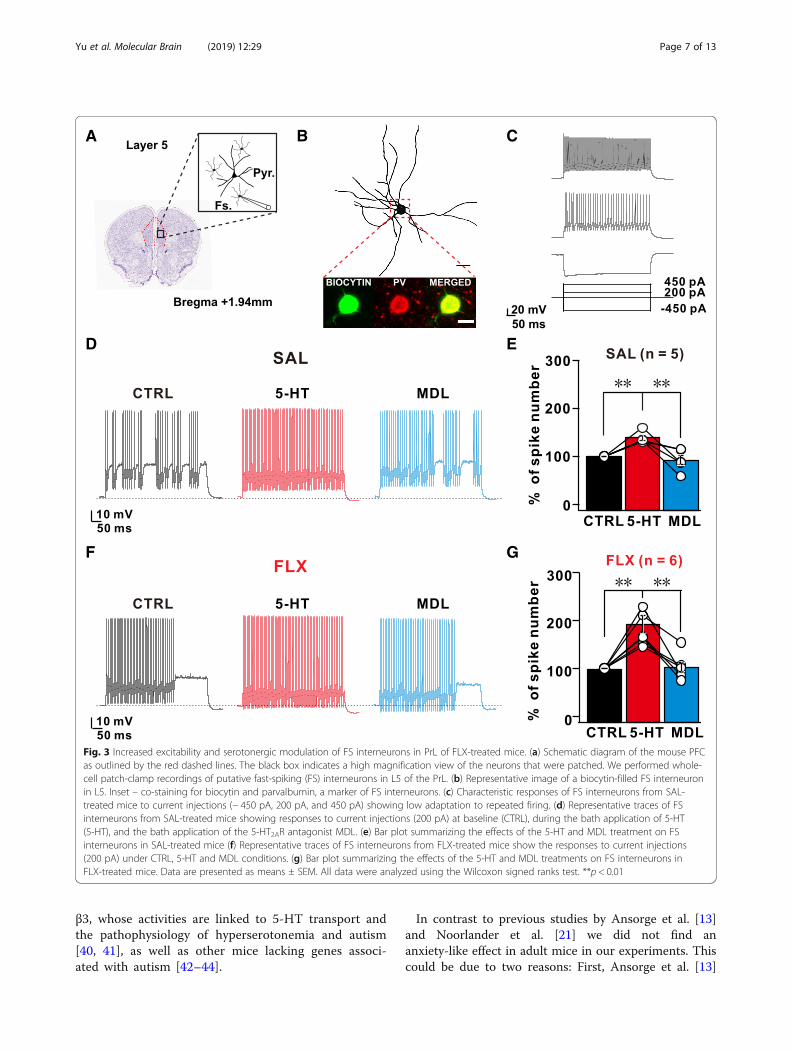
Increased excitability and serotonergic modulation of L5fast-spiking interneurons in PrL of FLX-treated micThe mPFC receives dense serotonergic innervation fromthe raphe nuclei, and both pyramidal and interneurons
within the mPFC express several 5-HT receptorsubtypes, with a particularly high density of 5-HT1A and5-HT2ARs [31–33]. Chronic increases in ambientsynaptic 5-HT due to SSRI-mediated blockade of theserotonin transporter (5-HTT) could potentially resultin desensitization, internalization, or compensatory levelchanges via transcription and translations of certain5-HT receptors [24, 34]. To test this hypothesis,qRT-PCR was performed on the PrL tissues from eitherSAL-treated or FLX-treated dams. The qRT-PCR resultsshowed a significant increase in the levels of the5-HT2AR mRNAs (SAL vs FLX fold change in 5-HT2AR:1.49 ± 0.14; p = 0.044, unpaired t-test; SAL, n = 5; FLX, n= 5). Although not statistically significant, there was anupward trend in the level of the 5-HT1AR mRNAs (SALvs FLX fold change in 5-HT1AR: 1.48 ± 0.17; p = 0.054,unpaired t-test; SAL, n = 5; FLX, n = 5)(Additional file 1:Figures S4A, B). In contrast, we did not observe any sig-nificant changes in the mRNA expression levels of other5-HT receptors and transporters in both qRT-PCR andmicroarray analyses (Additional file 1: Figures S4C, D).We wondered whether the upregulation of 5-HT 2ARs
resulted in increased spontaneous inhibitory synaptictransmission in L5 excitatory pyramidal neurons. To ad-dress this question, we first investigated functionalchanges in sIPSCs recorded from L5 pyramidal neuronsupon acute exogenous 5-HT treatment. Consistent withresults from previous studies [35, 36], treatment withexogenous 5-HT (30 μM) significantly enhanced boththe frequency and amplitude of sIPSCs in L5 pyramidalneurons in SAL-treated mice by 32.74 ± 6.65% and29.22 ± 11.09%, respectively (n = 5, p < 0.05, Additionalfile 1: Figures S4E, F). Intriguingly, L5 pyramidal neu-rons in FLX-treated mice exhibited substantial increasesin the frequency and amplitude of sIPSCs (53.25% ± 7.85and 36.93 ± 6.22%, n = 5, p < 0.05, Additional file 1,Figures S4E, F).Next, we recorded the intrinsic excitability and firing
properties of FS inhibitory neurons before and after5-HT application to further investigate the effects of5-HT on L5 FS inhibitory neurons that are critical forshaping cortical circuit activity by projecting theirinhibitory outputs onto the L5 pyramidal neurons withinthe PFC [37–39](Fig. 3a). As shown by immunohisto-chemical staining, the recorded FS interneurons werepositive for parvalbumin (PV) (Fig. 3b) and exhibited acharacteristic 224-Hz firing at a 450pA current injection(Fig. 3c). Next, we applied a series of incremental squaredepolarizing pulses to L5 FS neurons from SAL or FLXmice before and during 5-HT application (Fig. 3c-g).5-HT application significantly increased the spikefrequency of L5 FS neurons at each injected current stepin both SAL and FLX mice, and this increase in spikefrequency was normalized after washout. Intriguingly,
Yu et al. Molecular Brain (2019) 12:29 Page 4 of 13
A B C
D E F
G H I
J K L
Fig. 2 (See legend on next page.)
Yu et al. Molecular Brain (2019) 12:29 Page 5 of 13

compared with L5 FS neurons from the SAL group, L5FS neurons from the FLX group showed a largerincrease in spike frequency in response to the 5-HTtreatment (SAL: 38.57 ± 5.4% vs FLX: 92.05 ± 17.99%)(Fig. 3d-g). This increased frequency observed in L5 FSneurons was abolished by a subsequent treatment withMDL, a specific antagonist of 5-HT2ARs (1 μM), indi-cating that the increased responsiveness of 5-HT2AR inL5 FS neurons resulted in a 5-HT-dependent increasein AP frequency (Fig. 3d-g). In contrast, co-treatmentwith 5-HT1AR antagonists (WAY-100135, 10 μM) and5-HT did not affect 5-HT-mediated changes in thespike frequency of L5 FS interneurons (Additional file 1:Figures S5A-D). Thus, 5-HT-mediated changes in acutespike frequency were modulated by 5-HT2ARs in the L5FS interneurons of FLX-treated mice and subsequentlyincreased sIPSCs in L5 pyramidal neurons.We further tested the effect of 5-HT treatment on L5
pyramidal neurons. Compared with FS interneurons, L5pyramidal neurons showed a significantly reduced spikefrequency in response to 5-HT application (Additionalfile 1: Figures S6A-C). Furthermore, no significantdifference in the 5-HT-mediated reduction in the sEPSCfrequency in L5 pyramidal neurons was observedbetween SAL- and FLX-treated mice (Additional file 1:Figures S6D, G).
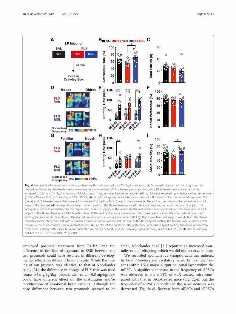
Behavioral deficits of FLX-treated mice were amelioratedby acute treatment with 5-HT2AR antagonistsUpregulation of 5-HT2AR signaling in PV neurons sup-pressed spontaneous network firing in L5 PFC microcir-cuits, resulting in the poor performance of FLX-treatedmice on working memory and social recognition tests.Therefore, we examined whether the selective suppres-sion of 5-HT2AR signaling would enhance the perform-ance of FLX-treated mice in the spontaneous alternationtest and social novelty recognition tests (Fig. 4a). I.P.injection of a 5-HT2AR antagonist (MDL) did not influ-ence the general behavior of wild-type animals, whenthey tested in the open-field test and elevated zero mazeat a given dosage (data not shown), but the applicationof the same dosage of MDL sufficiently reversed5-HT-mediated changes in the excitability of L5 FS in-terneurons from FLX-treated mice (Fig. 3g). Intriguingly,
acute administration of MDL effectively reversed thepoor performance of FLX-treated mice on the Y-mazespontaneous alternation task (Fig. 4b, c). Furthermore,the acute MDL treatment did not alter the socialpreference index in FLX-treated mice (Fig. 4d-f ), butrescued deficits in the novel recognition task inFLX-treated mice (Fig. 4g-h). Taken together, our datarevealed that the acute suppression of augmented5-HT2AR signaling in FLX-treated mice could rescuetheir behavioral deficits in working memory andsocial recognition memory.
DiscussionWe showed that prenatally FLX-treated mice exhibiteddeficits in a working memory task and social noveltyrecognition paradigm via enhanced inhibitory synapticactivities in the L5 neurons of the mPFC resulting fromenhanced 5-HT2AR signaling in FS PV neurons. Moreimportantly, the acute inhibition of 5-HT2AR signalingin FLX-treated mice successfully reversed the observedbehavioral deficits. Although 5-HT generally plays acritical role in mammalian neuronal development andbehavior, the causal relationship between alterations in5-HT homeostasis during pregnancy and adverse behav-ioral consequences in adulthood is poorly understood.Previously, several studies have attempted to address thisquestion using both genetic deletion of SERT and SSRIadministration in rodents. However, these studies suf-fered from inconsistent behavioral phenotypes, whichwere partially due to the use of different rodent strainsand the type, dosage, and timing of administered SSRIs(see Additional file 1: Table S1). In the present study, weadopted a treatment scheme similar to that of Noorlanderet al. This treatment mimicked SSRI exposure before the3rd trimester in humans, in which doctors recom-mend that pregnant women abstain from (or reducethe dose of ) SSRIs during late pregnancy [21]. In thisparadigm, we consistently observed behavioral deficitsin Y-maze spontaneous alternation tasks in prenatallyFLX-treated mice without anxiety-related behaviors.More importantly, SSRI-treated mice exhibited normalsociability but impaired preference for social noveltyin the three-chamber test (Fig. 1g-i), which is strik-ingly similar to the behaviors of mice lacking integrin
(See figure on previous page.)Fig. 2 Fluoxetine induces an increase in the number of inhibitory inputs to excitatory neurons and reduced excitatory basal transmission in theL5 pyramidal neurons of the prelimbic cortex of FLX-treated mice. (a) Representative current traces depicting spontaneous excitatory post-synaptic currents (sEPSCs) obtained from L5 pyramidal neurons in the prelimbic cortex (PrL) pyramidal neurons of SAL- and FLX-treated (red)mice. (b and c) Plots of the cumulative distributions of sEPSC (b) amplitudes and (c) inter-event intervals obtained from SAL- (black) and FLX-treated (red) mice. (d) Representative current traces depicting mEPSCs obtained from SAL- and FLX-treated mice. (e and f) Plots of the cumulativedistribution plots of mEPSC (e) amplitudes and (f) inter-event intervals. (g) Representative current traces depicting sIPSCs obtained from SAL- andFLX-treated mice. (h and i) Plots of the cumulative distribution plots of sIPSC (h) amplitudes and (i) inter-event intervals. (j) Representative currenttraces depicting mIPSCs obtained from SAL- and FLX-treated mice. (k and l) Plots of the cumulative distribution of mIPSC (k) amplitudes and(l) inter-event intervals. Data are presented as mean ± SEM. All data were analyzed using the Mann-Whitney U test. *p < 0.05, **p < 0.01
Yu et al. Molecular Brain (2019) 12:29 Page 6 of 13
β3, whose activities are linked to 5-HT transport andthe pathophysiology of hyperserotonemia and autism[40, 41], as well as other mice lacking genes associ-ated with autism [42–44].
In contrast to previous studies by Ansorge et al. [13]and Noorlander et al. [21] we did not find ananxiety-like effect in adult mice in our experiments. Thiscould be due to two reasons: First, Ansorge et al. [13]
A
D E
F G
B C
Fig. 3 Increased excitability and serotonergic modulation of FS interneurons in PrL of FLX-treated mice. (a) Schematic diagram of the mouse PFCas outlined by the red dashed lines. The black box indicates a high magnification view of the neurons that were patched. We performed whole-cell patch-clamp recordings of putative fast-spiking (FS) interneurons in L5 of the PrL. (b) Representative image of a biocytin-filled FS interneuronin L5. Inset – co-staining for biocytin and parvalbumin, a marker of FS interneurons. (c) Characteristic responses of FS interneurons from SAL-treated mice to current injections (− 450 pA, 200 pA, and 450 pA) showing low adaptation to repeated firing. (d) Representative traces of FSinterneurons from SAL-treated mice showing responses to current injections (200 pA) at baseline (CTRL), during the bath application of 5-HT(5-HT), and the bath application of the 5-HT2AR antagonist MDL. (e) Bar plot summarizing the effects of the 5-HT and MDL treatment on FSinterneurons in SAL-treated mice (f) Representative traces of FS interneurons from FLX-treated mice show the responses to current injections(200 pA) under CTRL, 5-HT and MDL conditions. (g) Bar plot summarizing the effects of the 5-HT and MDL treatments on FS interneurons inFLX-treated mice. Data are presented as means ± SEM. All data were analyzed using the Wilcoxon signed ranks test. **p < 0.01
Yu et al. Molecular Brain (2019) 12:29 Page 7 of 13
employed postnatal treatment from P4-P21 and thedifference in timeline of exposure to SSRI between thetwo protocols could have resulted in different develop-mental effects on different brain circuits. While the tim-ing of our protocol was identical to that of Noorlanderet al., [21], the difference in dosage of FLX that was used(ours: 0.6 mg/kg/day; Noorlander et al.: 0.8 mg/kg/day)could have different effect on the maturation and/ormodification of emotional brain circuits. Although thedose difference between two protocols seemed to be
small, Noorlander et al. [21] reported an increased mor-tality rate of offspring, which we did not observe in ours.We recorded spontaneous synaptic activities induced
by local inhibitory and excitatory networks in single neu-rons within L5, a major output neuronal layer within themPFC. A significant increase in the frequency of sIPSCswas observed in the mPFC of FLX-treated mice com-pared with that in SAL-treated mice (Fig. 2g-i), but thefrequency of sEPSCs recorded in the same neurons wasdecreased (Fig. 2a-c). Because both sIPSCs and sEPSCs
A B C
D E F
G H I
Fig. 4 Prenatal FLX-induced deficits in executive function are rescued by a 5-HT2AR antagonist. (a) Schematic diagram of the drug treatmentprocedure. Prenatally SAL-treated mice were injected with vehicle (VEH), whereas prenatally fluoxetine (FLX)-treated mice were randomlyassigned to VEH and 5-HT2AR antagonist (MDL) groups. Thirty minutes before behavioral testing, FLX mice received i.p. injections of either vehicle(0.5% DMSO) or MDL (0.01 mg/kg in 0.5% DMSO). (b) Bar plot of spontaneous alternation rates of SAL-treated mice that were administered VEH(black) and FLX-treated mice that were administered VEH (red) or MDL (blue) in the Y-maze. (c) Bar plot of the total number of entries into allarms of the Y-maze. (d) Representative heat map of results of the three-chamber social interaction test with a novel mouse and object. Theoccupancy rate was normalized to the region with peak occupancy in the arena. (e) Bar plot of the times spent sniffing the novel mouse andobject in the three-chamber social interaction task. (f) Bar plot of the social preference index (time spent sniffing the mouse/total time spentsniffing the mouse and the object). The dotted line indicates an equal preference (50%) (g) Representative heat map of results from the three-chamber social interaction test with a familiar mouse and novel mouse. (H) Bar plot of the times spent sniffing the familiar mouse and a novelmouse in the three-chamber social interaction task. (i) Bar plot of the social novelty preference index (time spent sniffing the novel mouse/totaltime spent sniffing both mice). Data are presented as mean ± SEM. (e) and (h) Two-way repeated measures ANOVA. (b), (c), (f) and (i) One-wayANOVA. * p < 0.05, ** p < 0.01, *** p < 0.001
Yu et al. Molecular Brain (2019) 12:29 Page 8 of 13
were recorded in the same L5 pyramidal neurons, thesedata indicate that the ratio of excitatory to inhibitorydrive onto those neurons is abnormally low inFLX-treated mice. As increase in sIPSC frequency iscaused by activity-dependent changes such as excitabil-ity, we next blocked APs in a slice with TTX and re-corded mIPSCs in L5 pyramidal neurons. Intriguingly,significant differences in both frequency and amplitudeof mIPSCs were not observed between FLX- andSAL-treated mice, indicating that the increased fre-quency of inhibitory currents observed in L5 mPFC neu-rons was due to activity-dependent GABAergic release.Because these excitatory neurons received inhibitorysynaptic inputs from PV-positive FS interneurons, wemeasured the intrinsic excitability of L5 FS interneuronsand observed a significant increase in intrinsic excitabil-ity without marked changes in channel properties orinput resistance (Additional file 1: Figure S3). We rea-soned that the compensatory augmentation of specific5-HT receptors could arise from prolonged exposure to5-HT due to SSRI treatment and observed a concurrentincrease in two 5-HT receptors, 5-HT1AR and 2AR, usingqPCR analysis. Because of the lack of suitable antibodiesagainst 5-HT receptors for immunohistochemical ana-lyses, we performed electrophysiological recordings andpharmacology to test the contribution of the increasedabundance of specific 5-HT receptors in the PFC ofFLX-treated mice. Surprisingly, increases in activity- and5-HT-dependent changes in the excitability of FS inter-neurons were mediated by 5-HT2ARs, but not 5-HT1ARs(Fig. 3d-g and Additional file 1: Figure S5). Although wedo not clearly understand why 5-HT2AR signaling or ex-pression was enhanced specifically in FS interneurons,Athilingam et al. recently showed that 5-HT treatmentin FS interneurons resulted in the suppression of aninward-rectifying potassium conductance, which eventu-ally lead to increased excitability of these interneuronsvia 5-HT2ARs [45], which might explain the excitabilitychange in FS interneurons upon chronic SSRI treatment.Changes in sIPSC frequency or altered inhibitory drive
relative to excitatory drive have been observed in otheranimal models of neurodevelopmental disorders inwhich working memory deficits have also been reported[29, 46–48]. Our findings support the hypothesis thatenvironmental changes induced by a single drug duringpregnancy elicits an imbalance in the inhibitory/excita-tory drive onto major output neurons in the L5 micro-circuits within the PFC and subsequently alters animalbehavior through non-genetic, compensatory upregula-tion of unique classes of 5-HT receptors in specificneuronal types. However, the potential mechanism ofthis compensatory 5-HT-receptor upregulation in FS in-terneurons needs to be addressed. Furthermore, studiesto determine whether L2/3 neurons in the mPFC or
hippocampal neurons exhibit comparable changes in theintrinsic excitability of neurons and excitatory/inhibitory(E/I) imbalance in the network will be interesting. Ourdata also support the findings from recent optogeneticstudies showing that acute modulation of the excitabilityof FS PV-positive interneurons within the mPFC elicitschanges in network oscillation and the performance ofcognitive flexibility behavior in mice [49, 50]. Therefore,measurements of network oscillations in FLX-treatedmice during working memory or social novelty taskusing in vivo multi-electrode recordings will be exciting.The increased 5-HT2AR-mediated changes in the
excitability of FS interneurons in FLX-treated miceprompted us to test whether the behavioral deficits ofthese mice were modulated by the application of a5-HT2AR antagonist, such as MDL. The impairments ofworking memory and social novelty recognition wererescued by an in vivo treatment with MDL (Fig. 4 b-i). Itis intriguingly, because a MDL treatment rescues atten-tional performance deficits in phencyclidine-treated [51]and NMDAR antagonist-treated [52] rats. Because pa-tients with schizophrenia, who usually exhibit workingmemory deficits, have very high 5-HT2AR occupancy inthe frontal cortex [53], the enhanced 5-HT2AR activityobserved in our study may represent a general patho-genic mechanism of behavioral deficits in some mentaldisorders, and systemic administration of MDL mightpotentially restore synaptic and behavioral deficits in pa-tients with disorders with similar etiologies.Taken together, our data revealed that altered prenatal
5-HT homeostasis results in mPFC-dependent behav-ioral deficits, and systemic modulation of the augmented5-HT2AR sufficiently rescues these behavioral deficits.Furthermore, our findings may potentially provide newopportunities for the pharmacological treatment of pa-tients who have been prenatally exposed to psychotropicmedications during the prenatal period.
Materials and methodsAnimalsAnimals were housed in a specific pathogen-freefacility maintained below 22 °C and 55% humidityunder a 12-h light-dark cycle (lights on at 0700 h)with food and water provided ad libitum [54]. Timedpregnancy was achieved by breeding wild-typeC57BL6J (The Jackson Laboratory) male mice withfemale mice.
ImmunohistochemistryMice were perfused with PBS, followed by 4% (w/v)paraformaldehyde in PBS (pH 7.4). Brains wereharvested and fixed with 4% paraformaldehyde overnight,transferred to 30% (w/v) sucrose in PBS, and then cryosec-tioned into 40 μm thick slices. For immunohistochemistry,
Yu et al. Molecular Brain (2019) 12:29 Page 9 of 13
brain slices were permeabilized with 0.2% Triton X-100 inPBS for 10min and transferred to blocking buffer (5%donkey serum, 2% BSA and 0.2% Triton X-100 in PBS) for1 h at room temperature. Next, the sections were incu-bated with primary antibody for parvalbumin (1:1000 di-luted in blocking buffer, PV-235, Swant) overnight at 4 °C.The sections were subsequently incubated with appropri-ate secondary antibodies (1:500 diluted in blocking buffer,anti-streptavidin Alexa Fluor 488, anti-mouse Alexa Fluor555; Invitrogen) for 2 h at room temperature. All sectionswere then stained with DAPI (1:5000 diluted with 0.2%Triton X-100 in PBS, Sigma-Aldrich) and mounted withFluorsafe (Merck Millipore). Images were captured usingan LSM 710 confocal microscope (Zeiss).
ElectrophysiologyL5 neurons were studied in acute coronal slices of themedial prefrontal cortex (mPFC) from male postnatalday 90–150 mice. After the mice were anesthetized byinhalation of 5% isoflurane, they were decapitated, andtheir brains were quickly removed and chilled inice-cold, high-magnesium cutting solution containingthe following components (in mM): 110 ChCl, 26NaHCO3, 3.2 KCl, 0.5 CaCl2, 7 MgCl2, 1.25 NaH2PO4,10 glucose, 2 sodium pyruvate, and 3 ascorbate [55].The pH was adjusted to 7.4 by saturation with carbogen(95% O2 and 5% CO2), and the osmolality was approxi-mately 300 mOsmol/L. The isolated brain was gluedonto the stage of a vibrating blade Compresstome(VF-200, Precisionary), and 300 μm-thick slices were cut.The slices were incubated at 34 °C for 30 min in thesame solution and thereafter maintained at roomtemperature. For experiments, we transferred a slice thatrecovered for at least one hour to a recording chambersuperfused with artificial cerebrospinal fluid (aCSF) con-taining the following components (in mM): 124 NaCl,26 NaHCO3, 3.2 KCl, 2.5 CaCl2, 1.3 MgCl2, 1.25NaH2PO4, and 10 glucose. The aCSF was bubbled with95% O2 and 5% CO2. Whole-cell voltage- orcurrent-clamp recordings (one cell per slice) were per-formed at 32 ± 1 °C, and the rate of aCSF perfusion wasmaintained at 1–1.5 ml min− 1. Recordings were per-formed in somata with a Multiclamp 700B amplifier.Patch pipettes for current-clamp mode were filled withinternal solutions containing the following components(in mM): 115 K-gluconate, 20 KCl, 10 Na2-phosphocrea-tine, 10 HEPES, 2Mg-ATP, 0.3 NaGTP, and 0.1% biocy-tin. For voltage-clamp recordings, we used internalsolutions containing the following components (in mM):120 Cs-methane sulfonate, 10 CsCl, 10 TEA-Cl, 1MgCl2, 10 HEPES, 0.1 EGTA, 0.4 Tris-GTP, 3Mg-ATP,and 5 Na2-phosphocreatine. We recorded series resist-ance throughout the experiments and excluded neuronswith series resistance > 20MΩ from data analysis.
Membrane potential values were presented as recordedwithout correcting for liquid junction potentials.MDL10090, CNQX, APV, picrotoxin, and TTX werepurchased from Tocris Bioscience. All other drugs werepurchased from Sigma-Aldrich. Stock solutions of drugswere made by dissolving in deionized water or DMSOaccording to manufacturer’s specifications and werestored at − 20 °C. On the day of the experiment, one ali-quot was thawed and used. The concentration of DMSOin solutions was maintained at 0.1%.
Behavioral assays and analysesAnimals and fluoxetine treatmentMale and female mice breeders were co-housed untilpregnancy. The date when a plug was first noted wasclassified as ED0. From ED4 to ED19, pregnant femaleswere housed individually and received daily i.p.injections of either FLX (0.6 mg/kg/day in a volume of10 ml/kg, Sigma) or equal volumes of SAL [21]. Femalesused for injection were used only once to minimize thepotential cross-generation effects of FLX administration.At postnatal day 21–23, offspring were weaned andhoused with their same-sex littermates.
Behavioral assaysFor behavioral testing, we used adult males treatedprenatally with either FLX or SAL. All animals were 12weeks old at the time of testing. All tests except for theopen-field test were conducted during the light phase.All behavioral apparatuses were cleaned with 70%ethanol and dried with tissue between each animal.
Open-field testThe open-field test was conducted to measure generalexploratory behavior. Mice were placed into the centerof a Plexiglas cage (40.5 cm × 40.5 cm × 16 cm) for a60-min test. Horizontal locomotion (i.e., distancetraveled) was automatically recorded and analyzed byusing Versamax software (AccuScan Instruments Inc).
Elevated zero mazeAnxiety-related behavior was measured using the ele-vated zero maze (CSI-MZ-ZR, Cleversys), which consistsof a circular runway subdivided into two closed and twoopen sections elevated 60 cm above the floor. Mice wereinitially introduced into one of the closed runways andwere allowed to freely explore the apparatus for 10 min.During the 10-min test, the percentage of time spent onthe open arms and the number of transitions betweentwo closed arms were scored using Topscan software(Cleversys).
Yu et al. Molecular Brain (2019) 12:29 Page 10 of 13

Y-maze testTo allow the mice to discriminate between the threearms of the Y-maze apparatus (San Diego Instruments),the sides of one arm of was lined with a pattern of blackvertical bars on a white background with a black squareat the end. The sides of another arm were lined with apattern of solid circles with black triangles on a whitebackground and a triangle marking the end. Theremaining arm was not marked. The Y-maze test wasconducted for 10 min. The first minute was not codedand treated as the habituation period. Subsequently, theentries into each arm were recoded. The spontaneousalternation index was calculated as the number ofnon-repeating triplets (for example, if each arm waslabeled “A”, “B” or “C”, “ABCAC” = 2) divided by thetotal number of possible non-repeating triplets (totalnumber of entries into each arm - 2).
Novel object recognition testThe novel object recognition test was conducted in theopen-field apparatus, which was subdivided equally intotwo arenas with a red plastic divider. For training, twoidentical objects constructed out of Lego blocks wereplaced at either end of the arena. Mice were allowed toexplore and familiarize themselves with the two objectsfor 10 mins. They were returned to their cages for 20min while the arena and the objects were cleaned to re-move any odor cues. Afterwards, a test of short-termmemory was conducted by placing the familiar objectand a novel object that differed in shape, color, and sizeat either end of the arena. The duration of sniffing andbouts of sniffing were manually recorded using Topscansoftware (CleverSys).
Social cognition testsSocial preference and social recognition were tested usinga three-chamber apparatus (CSI-SO-PP, CleverSys). Theentire test consists of three consecutive 10min blocks. Forthe first block, mice were habituated to the entire appar-atus and two empty wire pencil holders placed at oppositecorners. For the second block, one juvenile male mouse(social stimulus) was placed in one holder, and a dummyobject (non-social stimulus) was placed in the otherholder to test for social preference. For the last block, thejuvenile mouse remained in one holder (familiar), but thedummy object was replaced by a novel juvenile malemouse (novel) to test for social discrimination.
Golgi staining and tracingMice were perfused with PBS and placed in impregna-tion solution (FD Rapid Golgi-Stain Kit, FD Neurotech)for one week. 100 μm sections were cut on a cryostat,processed using the kit’s standard staining procedureand images were taken with the confocal microscope.
Z-stack images were traced and analyzed with theSimple Neurite Tracer plugin for ImageJ.
Gene expression studyTotal RNA was extracted from punched samples of themPFC using an RNeasy kit (Qiagen) followed by cDNAsynthesis using a Quantitect Reverse Transcription kit(Qiagen). Real-time polymerase chain reaction (PCR)was performed on three independent sets of templatesusing iQ SYBR Green Supermix (Bio-Rad). The amplifi-cation mixture consisted of 1 μM primers, 10 μl of iQSYBR Green Supermix (Bio-Rad), and 100 ng of templateDNA in a total volume of 20 μl. Cycling parameters were95 °C for 15 s, 57 °C for 1 min and 72 °C for 40 cyclesusing a CFX96 real-time PCR detection system(Bio-Rad). For each assay, PCR was performed aftermelting curve analysis. To reduce variability, we raneach sample in duplicate and included control qPCRreactions without the template in each run.For microarray analyses, we normalized all micro-
array data using RMA method provided by the affypackage [56] and analyzed differentially expressedgenes using the limma package [57]. For gene-probemapping information and GO term annotation, weused the EnsEMBL database (version 90).
Statistical analysesData were analyzed and plotted using GraphPad Prism(GraphPad Software) and are presented as the means ±SEM with symbols representing individual subjects. Datafrom the open-field test data were analyzed usingtwo-way repeated-measures ANOVA, followed byTukey’s post hoc test. The rest of the data were analyzedusing the Mann-Whitney test, Wilcoxon Signed ranktest, and unpaired t-test. P < 0.05 was considered statisti-cally significant. In addition, extreme values were sub-jected to Grubb’s test for outliers (http://graphpad.com/quickcalcs/Grubbs1.cfm) and excluded from the analysis.
Additional file
Additional file 1: Figure S1. Prenatal FLX treatment does not induceother behavioral deficits. Figure S2. The prenatal FLX treatment does notchange the morphology or spine density of L5 PrL neurons. Figure S3.Passive membrane properties of FS interneurons in the PrL of SAL- andFLX- treated mice. Figure S4. Expression of the mRNAs encodingserotonergic receptors and transporters and effects of the 5-HT treatmenton IPSC frequency and amplitude. Figure S5. Effect of 5-HT1AR antagonistson FS interneurons in the PrL of SAL- and FLX-treated mice. Figure S6. Ef-fects of the 5-HT treatment on pyramidal neurons in the PrL of SAL- andFLX-treated mice. Table S1. Summary of studies investigating the effects ofperinatal serotonin reuptake inhibitors (SSRI) on adult male mice. Table S2.Intrinsic properties of fast-spiking interneurons of SAL and FLX treated micebefore and after 5HT-treatment. Table S3. Statistical analysis conducted foreach behavioral test. (PDF 1403 kb)
Yu et al. Molecular Brain (2019) 12:29 Page 11 of 13

Abbreviations5-HT (serotonin): 5-hydrotryptamine; 5-HT2AR: 5-HT 2A receptor; APV: (2R)-amino-5-phosphonopentanoic acid; FLX: Fluoxetine; FS: Fast-spiking;mEPSCs: Miniature excitatory postsynaptic currents; mIPSCs: Miniatureinhibitory postsynaptic currents; mPFC: Medial prefrontal cortex; NMDA: N-methyl-D-aspartate; PrL: prelimbic area; PV: Parvalbumin; SAL: Saline;sEPSCs: Spontaneous excitatory postsynaptic currents; SERT: Serotonintransporter; sIPSCs: Spontaneous inhibitory postsynaptic currents;SSRI: Selective serotonin reuptake inhibitor; TTX: Tetrodotoxin
AcknowledgmentsWe would like to thank Dr. Ann-Marie Chacko and her team for the Duke-NUSLaboratory for Translational and Molecular Imaging (LTMI) for their helpfuldiscussions and generously sharing reagents. We thank Drs. Justin Lee,Chul-Hoon Kim, and Eddy Leman for their critical comments on the manuscript.
FundingThis work was supported by Singapore Ministry of Education (MOE)Academic Research Fund (MOE2012-T2–1-021, MOE2014-T2–2-071), NationalMedical Research Council Individual Research Grant (NMRC/CBRG/0075/2014and NMRC/OFIRG/0050/2017) and Translational Clinical Research Programmein Parkinson’s Disease (NMRC/TCR/013-NNI/2014), A*Star TranslationalCollaborative Research Partnership Grant (TCRP, 13/1/96/688), NationalResearch Foundation (NRF-CRP17–2017-04), Duke-NUS Signature ResearchProgram Block Grant (all to H.S.J.), and a Khoo Postdoctoral Fellowship Award(Duke-NUS-KPFA/2017/0016, to S. T.). T.K. was supported by Basic ScienceResearch Program via the National Research Foundation of Korea(NRF-2016R1C1B2009302) and the UNIST block grant (1.170009.01).
Availability of data and materialsPlease contact author for data requests.
Authors’ contributionsWY designed and performed most of electrophysiological recordings; YW,YHL, and ST performed behavioral assays and immunohistochemistry; YXperformed electrophysiological recordings and immunohistochemistry; ATand HG performed behavioral assays and analyzed data; HL and TKperformed qPCR and genechip analyses; HSJ supervised the project. HSJcontributed to ideas, designed, and wrote the manuscript.
Ethics approvalAll animal procedures were approved by the Institutional Animal Care andUse Committee of Duke-NUS Medical School and SingHealth (IACUC #2010/SHS/590 and #2014/SHS/999).
Consent for publicationAll authors agreed to its submission to the Molecular Brain and, if accepted,to its publication in this journal.
Competing interestsThe authors declare that they have no competing interests.
Publisher’s NoteSpringer Nature remains neutral with regard to jurisdictional claims inpublished maps and institutional affiliations.
Author details1Molecular Neurophysiology Laboratory, Signature Program in Neuroscienceand Behavioral Disorders, Duke-National University of Singapore (NUS)Medical School, 8 College Road, Singapore 169857, Singapore. 2Departmentof Physiology, Seoul National University College of Medicine, Seoul 03080,Republic of Korea. 3Department of Physiology, Yong Loo Lin School ofMedicine, National University of Singapore, Singapore 117597, Singapore.4Department of Biomedical Engineering, School of Life Science, UlsanNational Institute of Science and Technology (UNIST), UNIST-gil 50, Ulsan44919, Republic of Korea.
Received: 13 December 2018 Accepted: 18 March 2019
References1. Oberlander TF, Warburton W, Misri S, Aghajanian J, Hertzman C. Neonatal
outcomes after prenatal exposure to selective serotonin reuptake inhibitorantidepressants and maternal depression using population-based linkedhealth data. Arch Gen Psychiatry. 2006;63(8):898–906.
2. Bennett HA, Einarson A, Taddio A, Koren G, Einarson TR. Prevalence ofdepression during pregnancy: systematic review. Obstet Gynecol.2004;103(4):698–709.
3. Morales DR, Slattery J, Evans S, Kurz X. Antidepressant use during pregnancyand risk of autism spectrum disorder and attention deficit hyperactivitydisorder: systematic review of observational studies and methodologicalconsiderations. BMC Med. 2018;16(1):6.
4. Stahl SM. Mechanism of action of serotonin selective reuptake inhibitors.Serotonin receptors and pathways mediate therapeutic effects and sideeffects. J Affect Disord. 1998;51(3):215–35.
5. Krishnan V, Nestler EJ. The molecular neurobiology of depression. Nature.2008;455(7215):894–902.
6. Willner P. Antidepressants and serotonergic neurotransmission: anintegrative review. Psychopharmacology. 1985;85(4):387–404.
7. Kirsch I, Deacon BJ, Huedo-Medina TB, Scoboria A, Moore TJ, Johnson BT.Initial severity and antidepressant benefits: a meta-analysis of datasubmitted to the Food and Drug Administration. PLoS Med. 2008;5(2):e45.
8. Turner EH, Matthews AM, Linardatos E, Tell RA, Rosenthal R. Selectivepublication of antidepressant trials and its influence on apparent efficacy.N Engl J Med. 2008;358(3):252–60.
9. Kaplan YC, Keskin-Arslan E, Acar S, Sozmen K. Prenatal selective serotoninreuptake inhibitor use and the risk of autism spectrum disorder in children:a systematic review and meta-analysis. Reprod Toxicol. 2016;66:31–43.
10. Hiemke C, Hartter S. Pharmacokinetics of selective serotonin reuptakeinhibitors. Pharmacol Ther. 2000;85(1):11–28.
11. Clements CC, Castro VM, Blumenthal SR, Rosenfield HR, Murphy SN, Fava M,et al. Prenatal antidepressant exposure is associated with risk for attention-deficit hyperactivity disorder but not autism spectrum disorder in a largehealth system. Mol Psychiatry. 2015;20(6):727–34.
12. Huybrechts KF, Sanghani RS, Avorn J, Urato AC. Preterm birth andantidepressant medication use during pregnancy: a systematic review andmeta-analysis. PLoS One. 2014;9(3):e92778.
13. Ansorge MS, Zhou M, Lira A, Hen R, Gingrich JA. Early-life blockade of the5-HT transporter alters emotional behavior in adult mice. Science.2004;306(5697):879–81.
14. Gross C, Zhuang X, Stark K, Ramboz S, Oosting R, Kirby L, et al. Serotonin1Areceptor acts during development to establish normal anxiety-likebehaviour in the adult. Nature. 2002;416(6879):396–400.
15. Homberg JR. A mouse model to address unresolved antidepressant issues.Proc Natl Acad Sci U S A. 2011;108(9):3463–4.
16. Cases O, Seif I, Grimsby J, Gaspar P, Chen K, Pournin S, et al. Aggressivebehavior and altered amounts of brain serotonin and norepinephrine inmice lacking MAOA. Science. 1995;268(5218):1763–6.
17. Holmes A, Yang RJ, Lesch KP, Crawley JN, Murphy DL. Mice lacking theserotonin transporter exhibit 5-HT (1A) receptor-mediated abnormalities intests for anxiety-like behavior. Neuropsychopharmacology. 2003;28(12):2077–88.
18. Jennings KA, Loder MK, Sheward WJ, Pei Q, Deacon RM, Benson MA, et al.Increased expression of the 5-HT transporter confers a low-anxietyphenotype linked to decreased 5-HT transmission. J Neurosci.2006;26(35):8955–64.
19. Simpson KL, Weaver KJ, de Villers-Sidani E, Lu JY, Cai Z, Pang Y, et al.Perinatal antidepressant exposure alters cortical network function inrodents. Proc Natl Acad Sci U S A. 2011;108(45):18465–70.
20. Kehne JH, Baron BM, Carr AA, Chaney SF, Elands J, Feldman DJ, et al.Preclinical characterization of the potential of the putative atypicalantipsychotic MDL 100,907 as a potent 5-HT2A antagonist with a favorableCNS safety profile. J Pharmacol Exp Ther. 1996;277(2):968–81.
21. Noorlander CW, Ververs FF, Nikkels PG, van Echteld CJ, Visser GH, Smidt MP.Modulation of serotonin transporter function during fetal developmentcauses dilated heart cardiomyopathy and lifelong behavioral abnormalities.PLoS One. 2008;3(7):e2782.
Yu et al. Molecular Brain (2019) 12:29 Page 12 of 13
22. Chadman KK, Yang M, Crawley JN. Criteria for validating mouse models ofpsychiatric diseases. Am J Med Genet B Neuropsychiatr Genet.2009;150B(1):1–11.
23. Belforte JE, Zsiros V, Sklar ER, Jiang Z, Yu G, Li Y, et al. Postnatal NMDAreceptor ablation in corticolimbic interneurons confers schizophrenia-likephenotypes. Nat Neurosci. 2010;13(1):76–83.
24. Albert PR. Transcriptional regulation of the 5-HT1A receptor: implications formental illness. Philos Trans R Soc Lond Ser B Biol Sci. 2012;367(1601):2402–15.
25. Silverman JL, Yang M, Lord C, Crawley JN. Behavioural phenotyping assaysfor mouse models of autism. Nat Rev Neurosci. 2010;11(7):490–502.
26. Yen YC, Anderzhanova E, Bunck M, Schuller J, Landgraf R, Wotjak CT. Co-segregation of hyperactivity, active coping styles, and cognitive dysfunctionin mice selectively bred for low levels of anxiety. Front Behav Neurosci.2013;7:103.
27. Ko J. Neuroanatomical substrates of rodent social behavior: the medialprefrontal cortex and its projection patterns. Front Neural Circuits.2017;11:41.
28. Anderson DJ. Circuit modules linking internal states and social behaviour inflies and mice. Nat Rev Neurosci. 2016;17(11):692–704.
29. Krueger DD, Osterweil EK, Chen SP, Tye LD, Bear MF. Cognitive dysfunctionand prefrontal synaptic abnormalities in a mouse model of fragile Xsyndrome. Proc Natl Acad Sci U S A. 2011;108(6):2587–92.
30. Yuan Q, Yang F, Xiao Y, Tan S, Husain N, Ren M, et al. Regulation of brain-derived neurotrophic factor exocytosis and gamma-aminobutyric Acidergicinterneuron synapse by the schizophrenia susceptibility gene Dysbindin-1.Biol Psychiatry. 2016;80(4):312–22.
31. Jakab RL, Goldman-Rakic PS. 5-Hydroxytryptamine2A serotonin receptors inthe primate cerebral cortex: possible site of action of hallucinogenic andantipsychotic drugs in pyramidal cell apical dendrites. Proc Natl AcadSci U S A. 1998;95(2):735–40.
32. Jakab RL, Goldman-Rakic PS. Segregation of serotonin 5-HT2A and 5-HT3receptors in inhibitory circuits of the primate cerebral cortex. J CompNeurol. 2000;417(3):337–48.
33. Smiley JF, Goldman-Rakic PS. Serotonergic axons in monkey prefrontalcerebral cortex synapse predominantly on interneurons as demonstrated byserial section electron microscopy. J Comp Neurol. 1996;367(3):431–43.
34. Nautiyal KM, Hen R. Serotonin receptors in depression: from A to B.F1000Res. 2017;6:123.
35. Zhou FM, Hablitz JJ. Activation of serotonin receptors modulates synaptictransmission in rat cerebral cortex. J Neurophysiol. 1999;82(6):2989–99.
36. Weber ET, Andrade R. Htr2a gene and 5-HT (2A) receptor expression in thecerebral cortex studied using genetically modified mice. Front Neurosci.2010;4:36.
37. McBain CJ, Fisahn A. Interneurons unbound. Nat Rev Neurosci. 2001;2(1):11–23.38. Tamas G, Buhl EH, Lorincz A, Somogyi P. Proximally targeted GABAergic
synapses and gap junctions synchronize cortical interneurons. Nat Neurosci.2000;3(4):366–71.
39. Zhong P, Yan Z. Differential regulation of the excitability of prefrontalcortical fast-spiking interneurons and pyramidal neurons by serotonin andfluoxetine. PLoS One. 2011;6(2):e16970.
40. Carter MD, Shah CR, Muller CL, Crawley JN, Carneiro AM, Veenstra-VanderWeele J. Absence of preference for social novelty and increasedgrooming in integrin beta3 knockout mice: initial studies and futuredirections. Autism Res. 2011;4(1):57–67.
41. Carneiro AM, Cook EH, Murphy DL, Blakely RD. Interactions betweenintegrin alphaIIbbeta3 and the serotonin transporter regulate serotonintransport and platelet aggregation in mice and humans. J Clin Invest.2008;118(4):1544–52.
42. Molina J, Carmona-Mora P, Chrast J, Krall PM, Canales CP, Lupski JR, etal. Abnormal social behaviors and altered gene expression rates in amouse model for Potocki-Lupski syndrome. Hum Mol Genet.2008;17(16):2486–95.
43. Jin D, Liu HX, Hirai H, Torashima T, Nagai T, Lopatina O, et al. CD38 is criticalfor social behaviour by regulating oxytocin secretion. Nature.2007;446(7131):41–5.
44. Ferguson JN, Young LJ, Hearn EF, Matzuk MM, Insel TR, Winslow JT. Socialamnesia in mice lacking the oxytocin gene. Nat Genet. 2000;25(3):284–8.
45. Athilingam JC, Ben-Shalom R, Keeshen CM, Sohal VS, Bender KJ. Serotoninenhances excitability and gamma frequency temporal integration in mouseprefrontal fast-spiking interneurons. Elife. 2017;6:e31991.
46. Holtzman DM, Santucci D, Kilbridge J, Chua-Couzens J, Fontana DJ, DanielsSE, et al. Developmental abnormalities and age-related neurodegenerationin a mouse model of Down syndrome. Proc Natl Acad Sci U S A. 1996;93(23):13333–8.
47. Haberecht MF, Menon V, Warsofsky IS, White CD, Dyer-Friedman J, GloverGH, et al. Functional neuroanatomy of visuo-spatial working memory inTurner syndrome. Hum Brain Mapp. 2001;14(2):96–107.
48. Tabuchi K, Blundell J, Etherton MR, Hammer RE, Liu X, Powell CM, et al. Aneuroligin-3 mutation implicated in autism increases inhibitory synaptictransmission in mice. Science. 2007;318(5847):71–6.
49. Sohal VS, Zhang F, Yizhar O, Deisseroth K. Parvalbumin neurons andgamma rhythms enhance cortical circuit performance. Nature.2009;459(7247):698–702.
50. Cardin JA, Carlen M, Meletis K, Knoblich U, Zhang F, Deisseroth K, et al.Driving fast-spiking cells induces gamma rhythm and controls sensoryresponses. Nature. 2009;459(7247):663–7.
51. Poyurovsky M, Koren D, Gonopolsky I, Schneidman M, Fuchs C, Weizman A,et al. Effect of the 5-HT2 antagonist mianserin on cognitive dysfunction inchronic schizophrenia patients: an add-on, double-blind placebo-controlledstudy. Eur Neuropsychopharmacol. 2003;13(2):123–8.
52. Mirjana C, Baviera M, Invernizzi RW, Balducci C. The serotonin 5-HT2Areceptors antagonist M100907 prevents impairment in attentionalperformance by NMDA receptor blockade in the rat prefrontal cortex.Neuropsychopharmacology. 2004;29(9):1637–47.
53. Talvik-Lotfi M, Nyberg S, Nordstrom AL, Ito H, Halldin C, Brunner F, et al.High 5HT2A receptor occupancy in M100907-treated schizophrenic patients.Psychopharmacology. 2000;148(4):400–3.
54. Tan S, Ho HS, Song AY, Low J, Je HS. Maternal separation does not producea significant behavioral change in mice. Exp Neurobiol. 2017;26(6):390–8.
55. Lee D, Lee KH, Ho WK, Lee SH. Target cell-specific involvement ofpresynaptic mitochondria in post-tetanic potentiation at hippocampalmossy fiber synapses. J Neurosci. 2007;27(50):13603–13.
56. Gautier L, Cope L, Bolstad BM, Irizarry RA. Affy--analysis of AffymetrixGeneChip data at the probe level. Bioinformatics. 2004;20(3):307–15.
57. Ritchie ME, Phipson B, Wu D, Hu Y, Law CW, Shi W, et al. limma powersdifferential expression analyses for RNA-sequencing and microarray studies.Nucleic Acids Res. 2015;43(7):e47.
Yu et al. Molecular Brain (2019) 12:29 Page 13 of 13
Related Documents


![Selective serotonin reuptake inhibitors [SSRIs] and ... SSRIs SNRIs prevention... · Selective serotonin reuptake inhibitors (SSRIs) and serotonin-norepinephrine ... and tension-type](https://static.cupdf.com/doc/110x72/5ce01be988c99399558de41a/selective-serotonin-reuptake-inhibitors-ssris-and-ssris-snris-prevention.jpg)