Top Banner
Premalignant and Malignant Epithelial Neoplasms Lynne J. Goldberg, MD Jag Bhawan Professor of Dermatology and Pathology & Laboratory Medicine Boston University School of Medicine AAD 2018 Essential Dermatopathology February 18, 2018
40

Premalignant and Malignant Epithelial Neoplasms and Malignant Epithelial Neoplasms Lynne J. Goldberg, MD Jag Bhawan Professor of Dermatology and Pathology & Laboratory Medicine Boston

Apr 01, 2018

Download

Documents

haxuyen
Welcome message from author
This document is posted to help you gain knowledge. Please leave a comment to let me know what you think about it! Share it to your friends and learn new things together.
Transcript
Page 1: Premalignant and Malignant Epithelial Neoplasms and Malignant Epithelial Neoplasms Lynne J. Goldberg, MD Jag Bhawan Professor of Dermatology and Pathology & Laboratory Medicine Boston

Premalignant and Malignant

Epithelial Neoplasms

Lynne J. Goldberg, MD Jag Bhawan Professor of Dermatology and Pathology & Laboratory Medicine Boston University School of Medicine

AAD 2018

Essential Dermatopathology

February 18, 2018

Page 2: Premalignant and Malignant Epithelial Neoplasms and Malignant Epithelial Neoplasms Lynne J. Goldberg, MD Jag Bhawan Professor of Dermatology and Pathology & Laboratory Medicine Boston

DISCLOSURES

None

Lynne J. Goldberg, MD

Essential Dermatopathology

Premalignant and Malignant Epithelial Neoplasms

Page 3: Premalignant and Malignant Epithelial Neoplasms and Malignant Epithelial Neoplasms Lynne J. Goldberg, MD Jag Bhawan Professor of Dermatology and Pathology & Laboratory Medicine Boston

library.med.utah.edu

Actinic keratosis

• Usually multiple

• Covered with adherent scale

• No induration

• Histologic variants

• Hypertrophic

• Atrophic

• Bowenoid

• Acantholytic

• Pigmented

Page 4: Premalignant and Malignant Epithelial Neoplasms and Malignant Epithelial Neoplasms Lynne J. Goldberg, MD Jag Bhawan Professor of Dermatology and Pathology & Laboratory Medicine Boston
Page 5: Premalignant and Malignant Epithelial Neoplasms and Malignant Epithelial Neoplasms Lynne J. Goldberg, MD Jag Bhawan Professor of Dermatology and Pathology & Laboratory Medicine Boston
Page 6: Premalignant and Malignant Epithelial Neoplasms and Malignant Epithelial Neoplasms Lynne J. Goldberg, MD Jag Bhawan Professor of Dermatology and Pathology & Laboratory Medicine Boston
Page 7: Premalignant and Malignant Epithelial Neoplasms and Malignant Epithelial Neoplasms Lynne J. Goldberg, MD Jag Bhawan Professor of Dermatology and Pathology & Laboratory Medicine Boston
Page 8: Premalignant and Malignant Epithelial Neoplasms and Malignant Epithelial Neoplasms Lynne J. Goldberg, MD Jag Bhawan Professor of Dermatology and Pathology & Laboratory Medicine Boston

Bowen’s disease

• Usually solitary, larger than

AK

• Can occur on sun exposed or

sun protected skin

• Can occur on the penis

(erythroplasia of Queyrat)

• Progression to invasive SCC

3-5% to 11%

Page 9: Premalignant and Malignant Epithelial Neoplasms and Malignant Epithelial Neoplasms Lynne J. Goldberg, MD Jag Bhawan Professor of Dermatology and Pathology & Laboratory Medicine Boston
Page 10: Premalignant and Malignant Epithelial Neoplasms and Malignant Epithelial Neoplasms Lynne J. Goldberg, MD Jag Bhawan Professor of Dermatology and Pathology & Laboratory Medicine Boston
Page 12: Premalignant and Malignant Epithelial Neoplasms and Malignant Epithelial Neoplasms Lynne J. Goldberg, MD Jag Bhawan Professor of Dermatology and Pathology & Laboratory Medicine Boston
Page 13: Premalignant and Malignant Epithelial Neoplasms and Malignant Epithelial Neoplasms Lynne J. Goldberg, MD Jag Bhawan Professor of Dermatology and Pathology & Laboratory Medicine Boston

Squamous Cell Carcinoma • Sun damaged skin

• Secondarily in

• Scars (burns, Marjolin’s ulcer)

• Radiation sites

• Inflammatory dermatoses

• Lichen planus

• Lichen sclerosus

• Higher incidence in immunosuppressed patients

• Variants – classic, adenoid, mucin producing, spindle cell,

verrucous carcinoma, clear cell, basaloid, etc.

Page 14: Premalignant and Malignant Epithelial Neoplasms and Malignant Epithelial Neoplasms Lynne J. Goldberg, MD Jag Bhawan Professor of Dermatology and Pathology & Laboratory Medicine Boston

Risk stratification of cutaneous SCC

• 180,000 to 520,000 tumors/yr in the US

• 2-5% metastasis rate, typically preceded by local

recurrence and regional spread

• Brigham and Women’s Hospital high risk factors

• Depth into subcutis

• Perineural invasion of nerves > 0.1 mm

• Poor differentiation

• Other factors

Baum, et al. JAAD 2018

Page 15: Premalignant and Malignant Epithelial Neoplasms and Malignant Epithelial Neoplasms Lynne J. Goldberg, MD Jag Bhawan Professor of Dermatology and Pathology & Laboratory Medicine Boston
Page 16: Premalignant and Malignant Epithelial Neoplasms and Malignant Epithelial Neoplasms Lynne J. Goldberg, MD Jag Bhawan Professor of Dermatology and Pathology & Laboratory Medicine Boston
Page 17: Premalignant and Malignant Epithelial Neoplasms and Malignant Epithelial Neoplasms Lynne J. Goldberg, MD Jag Bhawan Professor of Dermatology and Pathology & Laboratory Medicine Boston
Page 18: Premalignant and Malignant Epithelial Neoplasms and Malignant Epithelial Neoplasms Lynne J. Goldberg, MD Jag Bhawan Professor of Dermatology and Pathology & Laboratory Medicine Boston
Page 19: Premalignant and Malignant Epithelial Neoplasms and Malignant Epithelial Neoplasms Lynne J. Goldberg, MD Jag Bhawan Professor of Dermatology and Pathology & Laboratory Medicine Boston
Page 20: Premalignant and Malignant Epithelial Neoplasms and Malignant Epithelial Neoplasms Lynne J. Goldberg, MD Jag Bhawan Professor of Dermatology and Pathology & Laboratory Medicine Boston
Page 21: Premalignant and Malignant Epithelial Neoplasms and Malignant Epithelial Neoplasms Lynne J. Goldberg, MD Jag Bhawan Professor of Dermatology and Pathology & Laboratory Medicine Boston

Verrucous Carcinoma

• Low grade squamous cell carcinoma

• First described in the oral cavity

• Highly differentiated, ultimately can invade deeply

• Regional metastases late, if at all

Page 22: Premalignant and Malignant Epithelial Neoplasms and Malignant Epithelial Neoplasms Lynne J. Goldberg, MD Jag Bhawan Professor of Dermatology and Pathology & Laboratory Medicine Boston
Page 23: Premalignant and Malignant Epithelial Neoplasms and Malignant Epithelial Neoplasms Lynne J. Goldberg, MD Jag Bhawan Professor of Dermatology and Pathology & Laboratory Medicine Boston

Keratoacanthoma (KA) • Solitary

• Separated from SCC in 1950

• Most on sun exposed areas

• Period of rapid growth, then involution

• Increased in immunosuppressed, Muir-Torre syndrome

• Variants – Giant KA, KA centrifugum marginatum, subungual KA

• Multiple

• Ferguson Smith (multiple self healing)

• Childhood or adolescence

• Grzybowski (eruptive)

• Adults, can involve mucosa

Page 24: Premalignant and Malignant Epithelial Neoplasms and Malignant Epithelial Neoplasms Lynne J. Goldberg, MD Jag Bhawan Professor of Dermatology and Pathology & Laboratory Medicine Boston
Page 25: Premalignant and Malignant Epithelial Neoplasms and Malignant Epithelial Neoplasms Lynne J. Goldberg, MD Jag Bhawan Professor of Dermatology and Pathology & Laboratory Medicine Boston
Page 26: Premalignant and Malignant Epithelial Neoplasms and Malignant Epithelial Neoplasms Lynne J. Goldberg, MD Jag Bhawan Professor of Dermatology and Pathology & Laboratory Medicine Boston

Differential diagnosis of SCC

• Pseudocarcinomatous hyperplasia

• Deep fungal infections

• Bromoderma

• Pyoderma vegetans

• Edges of ulcers of various causes

• Granular cell tumor

• Gout

Page 27: Premalignant and Malignant Epithelial Neoplasms and Malignant Epithelial Neoplasms Lynne J. Goldberg, MD Jag Bhawan Professor of Dermatology and Pathology & Laboratory Medicine Boston

www.the-dermatologist.com

Page 28: Premalignant and Malignant Epithelial Neoplasms and Malignant Epithelial Neoplasms Lynne J. Goldberg, MD Jag Bhawan Professor of Dermatology and Pathology & Laboratory Medicine Boston

Basal Cell Carcinoma

• 5 major variants

• Superficial

• Nodular

• Micronodular

• Infiltrating

• Fibroepithelioma of Pinkus

• Others – pigmented, keratinizing, cystic, morpheaform,

metatypical, basosquamous, etc.

Page 29: Premalignant and Malignant Epithelial Neoplasms and Malignant Epithelial Neoplasms Lynne J. Goldberg, MD Jag Bhawan Professor of Dermatology and Pathology & Laboratory Medicine Boston

Superficial BCC

Tandon and Brodell Derm Online J 2012;18(9):1

Page 30: Premalignant and Malignant Epithelial Neoplasms and Malignant Epithelial Neoplasms Lynne J. Goldberg, MD Jag Bhawan Professor of Dermatology and Pathology & Laboratory Medicine Boston

A proposal for a thickness-based definition

of superficial BCC

• Based on response to imiquimod

• 127 superficial BCCs treated 5x/wk for 6 weeks

• Patients followed for recurrence, both clinically and histologically

(mean 34 months)

• Medial tumor thickness

• Non-recurrent cases 0.26 mm

• Recurrent cases 0.57 mm

• No tumor </= 0.4 mm recurred

• 58% of tumors >0.4 mm recurred

McKay KM, et al. Br J Dermatol 2013

Page 31: Premalignant and Malignant Epithelial Neoplasms and Malignant Epithelial Neoplasms Lynne J. Goldberg, MD Jag Bhawan Professor of Dermatology and Pathology & Laboratory Medicine Boston

Nodular BCC

en.wikipedia.org

Page 32: Premalignant and Malignant Epithelial Neoplasms and Malignant Epithelial Neoplasms Lynne J. Goldberg, MD Jag Bhawan Professor of Dermatology and Pathology & Laboratory Medicine Boston

Micronodular BCC

Page 33: Premalignant and Malignant Epithelial Neoplasms and Malignant Epithelial Neoplasms Lynne J. Goldberg, MD Jag Bhawan Professor of Dermatology and Pathology & Laboratory Medicine Boston

Infiltrating BCC

Page 35: Premalignant and Malignant Epithelial Neoplasms and Malignant Epithelial Neoplasms Lynne J. Goldberg, MD Jag Bhawan Professor of Dermatology and Pathology & Laboratory Medicine Boston

Differential diagnosis of BCC

• Adnexal tumors

• Trichoepithelioma

• Pilomatrixoma

• Eccrine acrospiroma

• Sebaceous carcinoma

• Merkel cell carcinoma

• Basaloid carcinomas of other organ systems

Page 36: Premalignant and Malignant Epithelial Neoplasms and Malignant Epithelial Neoplasms Lynne J. Goldberg, MD Jag Bhawan Professor of Dermatology and Pathology & Laboratory Medicine Boston
Page 37: Premalignant and Malignant Epithelial Neoplasms and Malignant Epithelial Neoplasms Lynne J. Goldberg, MD Jag Bhawan Professor of Dermatology and Pathology & Laboratory Medicine Boston

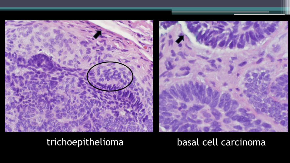
trichoepithelioma basal cell carcinoma

Page 38: Premalignant and Malignant Epithelial Neoplasms and Malignant Epithelial Neoplasms Lynne J. Goldberg, MD Jag Bhawan Professor of Dermatology and Pathology & Laboratory Medicine Boston

desmoplastic

trichoepithelioma infiltrative

basal cell carcinoma

Page 39: Premalignant and Malignant Epithelial Neoplasms and Malignant Epithelial Neoplasms Lynne J. Goldberg, MD Jag Bhawan Professor of Dermatology and Pathology & Laboratory Medicine Boston
Page 40: Premalignant and Malignant Epithelial Neoplasms and Malignant Epithelial Neoplasms Lynne J. Goldberg, MD Jag Bhawan Professor of Dermatology and Pathology & Laboratory Medicine Boston

Thank you for your attention!

Thank You! www.washingtonpost.com

Photo by Cynthia Dial