Prediction equations for human thoracic and lumbar vertebral morphometry Maria E. Kunkel, Hendrik Schmidt and Hans-Joachim Wilke Institute of Orthopaedic Research and Biomechanics, University of Ulm, Ulm, Germany Abstract Statistical correlations between anatomical dimensions of human vertebral structures have indicated a potential for the prediction of vertebral morphometry, which could be applied to the creation of simplified geometrical models of the spine excluding the need for preliminary processing of medical images. The aim of this study was to perform linear and nonlinear regressions with published anatomical data to generate prediction equations for 20 vertebral parameters of the human thoracic and lumbar spine as a function of only one given parameter that was measured by X-ray. Each parameter was considered individually as a potential predictor variable in terms of its correlation with all of the other parameters, together with the readiness with which lateral X-rays could be obtained. Based on this, the parameter vertebral body height posterior was chosen and the statistical analyses described here are related to this parameter. Our linear, exponential and logarithmic regressions pro- vided significant predictions of anterior vertebral structures. However, third-order polynomial prediction equa- tions allowed an improvement on these predictions (P-values < 0.001), e.g. endplates and spinal canal (R 2 , 0.970–0.995) as well as pedicle heights and the spinous process (R 2 , 0.811–0.882), in addition to a reasonable prediction of the posterior vertebral structures, which have shown a low or no correlation in previous studies, e.g. pedicle inclination and transverse process (R 2 , 0.514–0.693) (ANOVA). Comparisons of the theoretical predic- tions with two other sets of experimental data indicated that the predictions generally agree well with the experimental data. A time-efficient approach for obtaining anatomical data for the description of human tho- racic and lumbar geometry was provided by this method, which requires the measurement of only one para- meter per vertebra (vertebral body height posterior) from a lateral X-ray and the set of developed prediction equations. Vertebral models based on this type of parameterized geometry could be used in biomechanical studies that require geometry variation, such as in spinal deformations, including scoliosis. Key words polynomial regression; prediction; vertebral morphometry; vertebral parameters. Introduction During recent decades, finite element analyses have been performed to provide a better understanding of the biome- chanics of the human spine. Several finite element models have been developed and are summarized in Gilbertson et al. (1995) and Fagan et al. (2002). As geometrical factors exert a noticeable influence on the behavior of the spine (Robin et al. 1994), reliable simulations of human spine behavior require complex 3D modeling of the main ana- tomical structures, e.g. vertebrae, intervertebral discs and ligaments. Human vertebral geometry has typically been obtained, in vivo, through the 3D reconstruction of medical images, such as computed tomography or magnetic resonance imaging. This technique provides accurate vertebral assess- ment but requires a long processing time and considerable computational power is required for the manual or semi- automatic segmentation of the images. Moreover, the patient has to be submitted to relatively high doses of ionizing radiation. Alternative procedures have included stereo-radiographic approaches using X-rays (Aubin et al. 1997; Dumas et al. 2004). However, these require a long and tedious process of identification of numerous anatom- ical landmarks. Some semi-automatic methods have shown fast vertebral reconstruction (Pomero et al. 2004) but they require specific software and hardware. In-vitro measurements with cadaveric vertebrae have been taken directly from bony specimens or have been obtained from medical images (Krag et al. 1988). These studies have focused on only one specific anatomic struc- Correspondence Hans-Joachim Wilke, Institute of Orthopaedic Research and Biomechanics, Helmholtzstrasse 14, D-89081 Ulm, Germany. T: 0049 731 500 55320; fax: 0049 731 500 55302; E: hans-joachim. [email protected] Accepted for publication 3 November 2009 Article published online 21 December 2009 ª 2009 The Authors Journal compilation ª 2009 Anatomical Society of Great Britain and Ireland J. Anat. (2010) 216, pp320–328 doi: 10.1111/j.1469-7580.2009.01187.x Journal of Anatomy

Prediction equations for human thoracic and lumbarvertebral morphometry
Dec 24, 2015
Welcome message from author
This document is posted to help you gain knowledge. Please leave a comment to let me know what you think about it! Share it to your friends and learn new things together.
Transcript

Prediction equations for human thoracic and lumbarvertebral morphometryMaria E. Kunkel, Hendrik Schmidt and Hans-Joachim Wilke
Institute of Orthopaedic Research and Biomechanics, University of Ulm, Ulm, Germany
Abstract
Statistical correlations between anatomical dimensions of human vertebral structures have indicated a potential
for the prediction of vertebral morphometry, which could be applied to the creation of simplified geometrical
models of the spine excluding the need for preliminary processing of medical images. The aim of this study was
to perform linear and nonlinear regressions with published anatomical data to generate prediction equations
for 20 vertebral parameters of the human thoracic and lumbar spine as a function of only one given parameter
that was measured by X-ray. Each parameter was considered individually as a potential predictor variable in
terms of its correlation with all of the other parameters, together with the readiness with which lateral X-rays
could be obtained. Based on this, the parameter vertebral body height posterior was chosen and the statistical
analyses described here are related to this parameter. Our linear, exponential and logarithmic regressions pro-
vided significant predictions of anterior vertebral structures. However, third-order polynomial prediction equa-
tions allowed an improvement on these predictions (P-values < 0.001), e.g. endplates and spinal canal (R2,
0.970–0.995) as well as pedicle heights and the spinous process (R2, 0.811–0.882), in addition to a reasonable
prediction of the posterior vertebral structures, which have shown a low or no correlation in previous studies,
e.g. pedicle inclination and transverse process (R2, 0.514–0.693) (ANOVA). Comparisons of the theoretical predic-
tions with two other sets of experimental data indicated that the predictions generally agree well with the
experimental data. A time-efficient approach for obtaining anatomical data for the description of human tho-
racic and lumbar geometry was provided by this method, which requires the measurement of only one para-
meter per vertebra (vertebral body height posterior) from a lateral X-ray and the set of developed prediction
equations. Vertebral models based on this type of parameterized geometry could be used in biomechanical
studies that require geometry variation, such as in spinal deformations, including scoliosis.
Key words polynomial regression; prediction; vertebral morphometry; vertebral parameters.
Introduction
During recent decades, finite element analyses have been
performed to provide a better understanding of the biome-
chanics of the human spine. Several finite element models
have been developed and are summarized in Gilbertson
et al. (1995) and Fagan et al. (2002). As geometrical factors
exert a noticeable influence on the behavior of the spine
(Robin et al. 1994), reliable simulations of human spine
behavior require complex 3D modeling of the main ana-
tomical structures, e.g. vertebrae, intervertebral discs and
ligaments.
Human vertebral geometry has typically been obtained,
in vivo, through the 3D reconstruction of medical images,
such as computed tomography or magnetic resonance
imaging. This technique provides accurate vertebral assess-
ment but requires a long processing time and considerable
computational power is required for the manual or semi-
automatic segmentation of the images. Moreover, the
patient has to be submitted to relatively high doses of
ionizing radiation. Alternative procedures have included
stereo-radiographic approaches using X-rays (Aubin et al.
1997; Dumas et al. 2004). However, these require a long
and tedious process of identification of numerous anatom-
ical landmarks. Some semi-automatic methods have shown
fast vertebral reconstruction (Pomero et al. 2004) but they
require specific software and hardware.
In-vitro measurements with cadaveric vertebrae have
been taken directly from bony specimens or have been
obtained from medical images (Krag et al. 1988). These
studies have focused on only one specific anatomic struc-
Correspondence
Hans-Joachim Wilke, Institute of Orthopaedic Research and
Biomechanics, Helmholtzstrasse 14, D-89081 Ulm, Germany.
T: 0049 731 500 55320; fax: 0049 731 500 55302; E: hans-joachim.
Accepted for publication 3 November 2009
Article published online 21 December 2009
ªª 2009 The AuthorsJournal compilation ªª 2009 Anatomical Society of Great Britain and Ireland
J. Anat. (2010) 216, pp320–328 doi: 10.1111/j.1469-7580.2009.01187.x
Journal of Anatomy

ture, such as the dimensions of the vertebral body (Hall
et al. 1998), spinal canal, pedicles (Zindrick et al. 1987; Mar-
chesi et al. 1988; Moran et al. 1989) and facet joints (Masha-
rawi et al. 2004); a limited set of structures (Berry et al.
1987); or a limited set of vertebrae such as thoracic (Cotterill
et al. 1986; Scoles et al. 1988; Aharinejad et al. 1990) or
lumbar vertebrae (Semaan et al. 2001). The most complete
collection of quantitative 3D-surface anatomy of the main
vertebral parameters for the thoracic and lumbar human
spine has been provided in Panjabi et al. (1991, 1992). As
this dataset has been used in the current study, a detailed
description of the measured parameters is provided in the
Materials and methods.
Investigations of correlations between anatomical
dimensions of the human vertebral structures have indi-
cated that vertebral relationships could be used to predict
vertebral morphometry without the preliminary processing
of medical images. Statistical analyses that were per-
formed using simple linear regression analyses between
the main vertebral parameters and the vertebral body
height (e.g. Scoles et al. 1988; Breglia, 2006) have found
low or no correlations for some important parameters,
such as pedicle dimensions. Scoles et al. (1988) described
the posterior structures as being highly variable and lar-
gely unpredictable.
X-rays are frequently used in clinical diagnosis for
patients as well as in biomechanical experiments with
human vertebral samples. Some studies have used multiple-
linear regression analyses (e.g. Lavaste et al. 1992; Laporte
et al. 2000) to provide methods for the reconstruction of
the human vertebrae from two X-rays (anterior–posterior
and lateral). However, to explain 100% of the variability for
each parameter, the measurement of six to 15 initial param-
eters per vertebra on X-rays was needed. Moreover, none
of these previous studies have performed an evaluation of
the predictability of the generated equations with another
set of experimental measurements.
The aim of this study was to perform linear and nonlinear
regression analyses with published anatomical data to gen-
erate prediction equations for 20 vertebral parameters of
the human thoracic and lumbar spine as a function of only
one given parameter measured by X-ray.
Fig. 1 Schematic representation of the vertebral anatomical parameters that were considered for linear and nonlinear regression analyses.
ªª 2009 The AuthorsJournal compilation ªª 2009 Anatomical Society of Great Britain and Ireland
Prediction equations for human thoracic and lumbar vertebral morphometry, M. E. Kunkel et al. 321
Materials and methods
Study population
Vertebral anatomical data were collected from the studies of
Panjabi et al. (1991, 1992) and included in this study. This data-
set was considered as being an approximate average for non-
pathological human spines. It provided linear and angular
dimensions of the main parameters from human cadaveric tho-
racic and lumbar vertebrae. The mean age of the subjects
(n = 12) was 46.3 years (range: 19–59 years), weight was 67.8 kg
(range: 54–85 kg), height was 167.8 cm (range: 157–178 cm) and
the male : female ratio was 8 : 4. In order to carry out statistical
analyses, 15 linear and six angular vertebral parameters were
selected from this dataset to describe the size and shape of the
human thoracic (T1–12) and lumbar (L1–4) vertebrae (Fig. 1).
The values of the vertebral parameters related to the vertebral
level L5 were not included in the analysis.
Statistical analysis
The initial assumption for this study was that 20 vertebral
parameters on each level of the thoracic (T1–12) and lumbar
(L1–4) spine could be considered as a variable that can be
predicted (Fig. 1). All vertebral parameters that were selected
for this study were tested as a possible predictor variable. Each
vertebral parameter was individually regressed against the pos-
sible predictor variable by a least-squares estimation process.
Based on the level of correlation with the other parameters
and ease of measurement on lateral X-rays, the parameter
vertebral body height posterior (VBHP) was chosen and the
statistical analyses described in this study are related to this
parameter. Linear and nonlinear regression analyses were
employed to find the best functions to fit each parameter in a
prediction equation.
During the statistical analyses, several hypotheses were tested
for each parameter: (i) a function could not fit the data signifi-
cantly better than a horizontal line (no relationship between
the two selected variables); (ii) a second-order equation could
not fit the data significantly better than a linear equation; (iii) a
third-order equation could not fit the data significantly better
than a second-order equation, and so on. The statistical proce-
dure performed on each parameter corresponds to a four-step
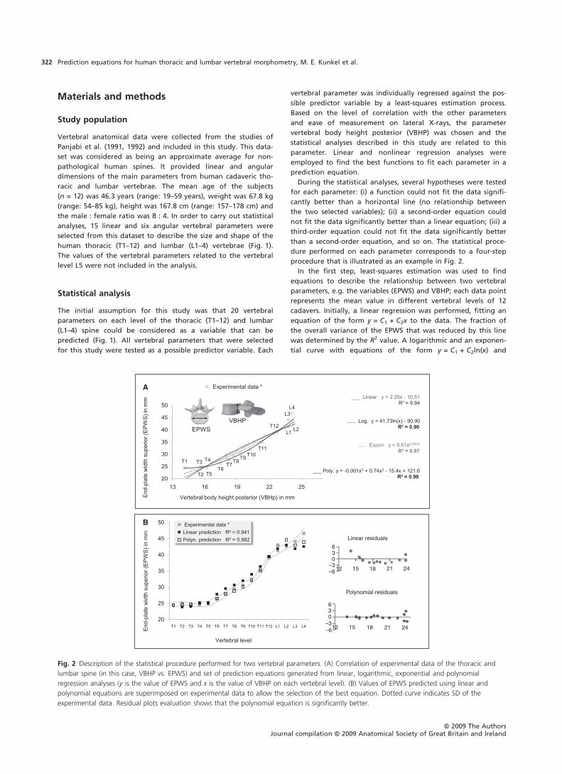
procedure that is illustrated as an example in Fig. 2.
In the first step, least-squares estimation was used to find
equations to describe the relationship between two vertebral
parameters, e.g. the variables (EPWS) and VBHP; each data point
represents the mean value in different vertebral levels of 12
cadavers. Initially, a linear regression was performed, fitting an
equation of the form y = C1 + C2x to the data. The fraction of
the overall variance of the EPWS that was reduced by this line
was determined by the R2 value. A logarithmic and an exponen-
tial curve with equations of the form y = C1 + C2ln(x) and
630
–3–6 12 15
Linear residuals
Polynomial residuals
242118
630
–3–612 15 242118
A
B
Fig. 2 Description of the statistical procedure performed for two vertebral parameters. (A) Correlation of experimental data of the thoracic and
lumbar spine (in this case, VBHP vs. EPWS) and set of prediction equations generated from linear, logarithmic, exponential and polynomial
regression analyses (y is the value of EPWS and x is the value of VBHP on each vertebral level). (B) Values of EPWS predicted using linear and
polynomial equations are superimposed on experimental data to allow the selection of the best equation. Dotted curve indicates SD of the
experimental data. Residual plots evaluation shows that the polynomial equation is significantly better.
ªª 2009 The AuthorsJournal compilation ªª 2009 Anatomical Society of Great Britain and Ireland
Prediction equations for human thoracic and lumbar vertebral morphometry, M. E. Kunkel et al.322
y = C1eC2x, respectively, were then used to test the increase in
R2. Next, polynomial equations including more coefficients (C1,
C2, C3, C4, etc.) were used to find the best fit of the data points.
This was continued until adding another higher-order term did
not significantly increase R2 (Fig. 2A).
The second step was to perform an ANOVA to select an equa-
tion from the generated set, which could predict the EPWS val-
ues significantly better. It was based not only on quality of fit
but also on the physical meaning of the prediction equations
(Motulsky & Christopoulos, 2004). High values of R2 associated
with a P-value < 0.01 indicated the third-order polynomial as
the best-fitting equation that could provide the best approxima-
tion to the experimental values of EPWS.
In the third step it was evaluated how the selected best-fit-
ting, in this case the polynomial equation fits the EPWS data
significantly better than a linear equation. Linear and third-
order polynomial predictions of the EPWS were superimposed
on experimental values together with their respective SD
(Fig. 2B). The quality of these regressions was assessed by exam-
ining the respective residual plots. The linear equation was
inappropriate for the description of the EPWS data because
residuals clustering indicated that the data differed systemati-
cally (not just randomly) from the prediction curves. Positive
residuals tended to cluster together at the first thoracic and the
last lumbar vertebrae, whereas negative residuals clustered
together in the transition zone from the thoracic to the lumbar
region. In contrast, polynomial residual plots had a random
arrangement of residuals, which was more appropriate to pre-
dict EPWS (Fig 2B).
The fourth step corresponds to the evaluation of the predict-
ability of the best-fitting equation of the set of equations devel-
oped in the third step using experimental data from two
further datasets. The dataset of Berry et al. (1987) includes 12
vertebral parameters of three thoracic vertebrae (T2, T7 and
T12) and four lumbar vertebrae (L1–4). The dataset of Scoles
et al. (1988) includes 10 vertebral parameters of five thoracic
vertebrae (T1, T3, T6, T9 and T12) and two lumbar vertebrae (L1
and L3) of male and female data.
Results
In general, there were no large differences for the correla-
tions of each of the individual 20 vertebral parameters with
VBHP when comparing linear, exponential and logarithmic
regressions with each other. For this reason, only the linear
predictions are provided from these results (Figs 3, 4 and 5).
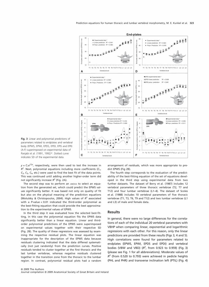
High correlations were found for parameters related to
endplates (EPWS, EPWI, EPDS and EPDI) and vertebral
bodies (VBW and VBD) (R2, from 0.923 to 0.959) (Fig. 3)
(please see Fig. 1 for all abbreviations). Moderate values of
R2 (from 0.520 to 0.793) were achieved in pedicle heights
(PHL and PHR) and transverse inclination left (PTIL) (Fig. 4)
A
B
C
D
E
F
Fig. 3 Linear and polynomial predictions of
parameters related to endplates and vertebral
body (EPWS, EPWI, EPDS, EPDI, EPIS and EPII)
(A-F) superimposed on experimental data of
Panjabi et al. (1991, 1992)*. Dotted curve
indicates SD of the experimental data.
ªª 2009 The AuthorsJournal compilation ªª 2009 Anatomical Society of Great Britain and Ireland
Prediction equations for human thoracic and lumbar vertebral morphometry, M. E. Kunkel et al. 323

as well as in the spinal canal (SCW and SCD) (Fig. 5). How-
ever, about 50% of the investigated parameters showed
low or no correlation with VBHP. These were the dimen-
sions of endplate inclinations (EPIS and EPII) (Fig. 3), pedi-
cles (PWL, PWR, PTIR, PSIL and PSIR) (Fig. 4) and other
posterior structures (SPL and TPW) (Fig. 5).
In contrast to the above regressions, third-order polyno-
mial regressions provided the best results with significant
correlations between all selected parameters and VBHP
(Table 1). As the dataset of Panjabi et al. (1991, 1992) does
not include VBW and VBD, an alternative method for the
prediction of these parameters was used. The inclusion of
more than four coefficients increased the R2 values but
ANOVAs indicated that the obtained correlations did not
significantly improve parameter predictions. For instance,
fourth- and fifth-order polynomial regressions between the
PWL and VBHP resulted in P-values > 0.05.
The parameters EPWS, EPWI, EPDS, EPDI, VBW and VBD
that showed high correlations by linear, logarithmic and
exponential regressions exhibited, after third-order polyno-
mial regressions, an increase of R2 (from 0.970 to 0.982,
P-values < 0.01) (Fig. 3). Similarly, the correlations with PHL,
PHR, PTIL, SCW and SCD were improved and R2 values rang-
ing from 0.693 to 0.964 were achieved (Figs 4 and 5).
Furthermore, for those parameters that displayed low or no
correlation with anterior procedures (EPIS, EPII, PWL, PWR,
PTIR, PSIL, PSIR, SPL and TPW), polynomial regressions
achieved reasonable correlations (R2, from 0.514 to 0.693,
P-values < 0.05) (Figs 3, 4 and 5). An exception was PTIL, for
which the best results were obtained after exponential
regression (R2 = 0.757) (Fig. 4C).
The prediction of the vertebral parameters related to
anterior vertebral structures using linear, exponential,
logarithmic and polynomial prediction equations did not
demonstrate significant differences (Fig. 6). Moreover, poly-
nomial prediction equations were required to predict the
parameters related to posterior vertebral structures. The
polynomial predictions are generally within or close to
B
C
D
A
F
G
H
E
Fig. 4 Linear and polynomial predictions of
parameters related to pedicles (PWL, PWR,
PHL, PHR, PTIL, PTIR, PSIL and PSIR) (A-H)
superimposed on experimental data of
Panjabi et al. (1991, 1992)*. Dotted curve
indicates SD of the experimental data.
ªª 2009 The AuthorsJournal compilation ªª 2009 Anatomical Society of Great Britain and Ireland
Prediction equations for human thoracic and lumbar vertebral morphometry, M. E. Kunkel et al.324

the regions of the 95% confidence intervals of the experi-
mental data of Panjabi et al. (1991, 1992).
Using the dataset of Berry et al. (1987), a comparison of
predicted EPWS and EPDI showed mean percent errors of
)14.93 and 31.86%, respectively for T1; all other levels were
very close to experimental data with mean percent errors of
)0.32 to 9.68% (Fig. 7A). Predictions of PHL showed better
results for thoracic levels with the smallest error being
)0.05 mm ()0.8%) for PHL (T2) and a mean percent error of
approximately 22.5% for thoracic and 24.6% for lumbar
A C
B D
Fig. 5 Linear and polynomial predictions of
other vertebral posterior structures (SCW,
SCD, SPL and TPW) (A-D) superimposed on
experimental data of Panjabi et al. (1991,
1992)*. Dotted curve indicates SD of the
experimental data.
Table 1 Polynomial coefficients (C1, C2, C3 and C4) for prediction equations of 20 parameters per vertebral level of the human thoracic and
lumbar spine.
Vertebral parameter Abbreviation C1 C2 C3 C4 SD R2 P-value
Endplate Width EPWS 121.650 )15.403 0.742 )0.010 1.195 0.982 1.07E)10
EPWI 300.140 )43.509 2.206 )0.035 1.454 0.976 5.19E)10
Depth EPDS )60.076 8.983 )0.293 0.004 0.852 0.981 1.31E)10
EPDI )63.590 9.473 )0.300 0.003 0.769 0.981 1.26E)10
Inclination EPIS )66.833 12.035 )0.691 0.013 0.699 0.606 0.008981
EPII 66.233 )9.095 0.418 )0.006 0.606 0.514 0.030367
Vertebral body Width VBW* 4.1409 0.748 – – 1.287 0.969 5.69E)05
Depth VBD** )80.223 10.313 )0.350 0.004 0.523 0.995 0.000666
Pedicle Width PWL 230.261 )34.915 1.777 )0.029 1.682 0.590 0.011194
PWR 157.740 )23.284 1.168 )0.019 1.446 0.537 0.022469
Height PHL 168.200 )27.194 1.522 )0.027 0.954 0.853 2.80E)05
PHR 105.820 )17.256 0.999 )0.018 0.872 0.879 8.65E)06
Transverse inclination PTIL )10.658 2.9889 )0.099 )0.001 2.741 0.693 0.002089
PTIR )202.510 31.347 )1.496 0.023 2.877 0.524 0.026147
Sagittal inclination PSIL 305.290 )39.194 1.734 )0.025 4.064 0.524 0.026141
PSIR )275.130 53.937 )3.119 0.057 3.403 0.669 0.003290
Spinal canal Width SCW 206.750 )26.838 1.218 )0.017 0.634 0.964 6.76E)09
Depth SCD )2.449 3.8232 )0.254 0.006 0.573 0.811 0.000123
Spinous process Length SPL )947.110 168.10 )9.310 0.170 3.472 0.882 7.43E)06
Transverse process Width TPW )343.670 80.885 )5.090 0.102 7.259 0.616 0.007667
SD in mm (for linear dimensions) or in degree (for angular dimensions).
The basic form of the prediction equations is y = C1 + C2x+C3x2 + C4x3 where y is the value of the parameter to be predicted and x is
the value of the VBHP on each vertebral level.
S, superior; I, inferior; L, left; R, right.
*For VBW, x is the value of EPWS ⁄ EPWI on each vertebral level.
**For VBD, x is the value of EPDs ⁄ EPDI on each vertebral level.
ªª 2009 The AuthorsJournal compilation ªª 2009 Anatomical Society of Great Britain and Ireland
Prediction equations for human thoracic and lumbar vertebral morphometry, M. E. Kunkel et al. 325

Fig. 6 Geometric models of the human
thoracic (T1–12) and lumbar (L1–4) vertebrae
constructed with parameters related to
endplates and vertebral bodies (EPWS, EPWI,
EPDS, EPDI, EPIS, EPII, VBW and VBD). The
first model corresponds to the data of Panjabi
et al. (1991, 1992) and was created using
eight parameters per vertebral level (a total of
128 parameters). The other models were
generated using only the values of the VBHP
of each vertebral level and predicted
parameters from linear, exponential,
logarithmic and polynomial equations.
Fig. 7 Comparison of some predicted
vertebral parameters (EPWS, PHL, PSIL and
SCW) with corresponding experimental data
from Berry et al. (1987) (left column, A–D)
and Scoles et al. (1988) (right column, E–H) in
selected vertebral levels. The means and 95%
confidence intervals (dotted lines) of the
experimental and predicted values are shown.
ªª 2009 The AuthorsJournal compilation ªª 2009 Anatomical Society of Great Britain and Ireland
Prediction equations for human thoracic and lumbar vertebral morphometry, M. E. Kunkel et al.326

levels (Fig. 7B). Polynomial pedicle predictions showed a
high error for PSIL (T12) (Fig. 7C). Predictions related to the
SCW and SCD also displayed better results for thoracic levels
with the largest error being 1.19 mm (7.93%). Lumbar levels
showed an approximate mean percent error of 23% (SCW)
and 31% (SCD) (Fig. 7D).
With the dataset of Scoles et al. (1988), predictions of
EPWS and VBD showed a range of mean percent errors of
)2.68 to 23.48%, with the largest errors occurring in EPWS
(L1) (Fig. 7E). The shortest error for PHL was )0.69 mm
()4.66%) for T12 and a mean percent error of approxi-
mately 18.5% for thoracic and 45.6% for lumbar levels was
found (Fig. 7F). Polynomial pedicle predictions showed a
high error for PSIL (T1) (Fig. 7G). Predictions of SCW and
SCD showed similar results to the prediction with the data-
set of Berry et al. (1987), with the largest error being
4.25 mm (22.1%) for thoracic levels (Fig. 7H).
Discussion
Linear and nonlinear regression analyses were performed
with the anatomical data of Panjabi et al. (1991, 1992) to
generate prediction equations for 20 vertebral parameters
per vertebral level of the human thoracic (T1–12) and lum-
bar (L1–4) vertebrae as a function of the VBHP. The parame-
ters corresponding to the vertebra L5 were not included in
the analyses because L5 shows remarkable morphological
differences for some parameters when compared with the
other lumbar vertebrae (Berry et al. 1987; Zindrick et al.
1987; Scoles et al. 1988). This is probably due to the position
of L5 being localized in the final transition zone, from lum-
bar to sacral region (Panjabi et al. 1989, 1992).
In this study two assumptions were necessary. First,
despite the high anatomical variability of the human verte-
brae, the dataset of Panjabi et al. (1991, 1992) was assumed
to be representative of the adult population without spinal
pathology. Second, it was assumed that the dimensions of
the vertebral structures described in this dataset were
obtained precisely. As the three datasets used in this study
were provided from in-vitro measurements, further investi-
gations are necessary to evaluate the predictability of the
regression equations with a dataset from patients.
Third-order polynomial equations represented the best
regression approximation as indicated after analysis of
covariance (Table 1). SEs indicated that, with few excep-
tions, such as for pedicle inclinations, the best fit values for
the prediction equations were accomplished with reason-
able certainty. Pedicle inclinations showed a wide variation
that can be observed in the wide confidence interval of the
sagittal plane angle for the mid-thoracic vertebrae
(Fig. 4C,D,G,H).
Our results were compared, when possible, with existing
published data. All correlation coefficients generated using
polynomial regressions were considerably better than the
values obtained by Breglia (2006) using simple linear regres-
sions on the data of Panjabi et al. (1991, 1992). The parame-
ters related to posterior structures that could not be
predicted with the regressions of Breglia (2006) have shown
a moderate correlation after polynomial regressions. Linear
regression procedures are straightforward and the results
appear to be readily evaluated statistically. However, the
relationships between the vertebral variables follow a
curved line, not a straight line. Although the methods used
for fitting a nonlinear equation such as polynomial regres-
sion are extensions of linear regression, the results are bet-
ter because polynomial equations can be used to create a
generic curve through the data points; more coefficients
create a more flexible curve, which could better fit the
data.
Comparisons of the theoretical predictions with two
other sets of experimental data (Berry et al. 1987; Scoles
et al. 1988) indicated that the predictions generally agree
well with the experimental data. Although the differences
in the predictions of pedicle inclination (Fig. 7C,G) have
been relatively great, a reasonable correlation between the
main posterior elements and VBHP was found. This is not in
accordance with Scoles et al. (1988) who declared that it
was not possible to establish useful predictors for pedicle
dimensions based on the size of the vertebral body. Differ-
ences in predicted values may also be attributed to techni-
cal factors related to obtaining these anatomical data, such
as different protocols of preparation and measurement.
Furthermore, there are individual variations and aging that
can induce substantial changes in each individual’s verte-
brae (Bernick & Cailliet, 1982; Diacinti et al. 1995).
Lavaste et al. (1992) developed a method to reconstruct
lumbar vertebral geometry from two X-rays (anterior–pos-
terior and lateral) using multiple-linear regression analysis.
However, to predict the vertebral geometry, six given
parameters per vertebra were required. A digitalization
process to define these parameters showed a relative error
of approximately 15%. Moreover, the orientation and
width of the pedicles were not taken into consideration.
Laporte et al. (2000) performed a similar study in thoracic
vertebrae, which required the measurement of 15 para-
meters per vertebra by X-ray in order to explain 100% of
the variability for each parameter.
The advantage of using the generated set of prediction
equations (Table 1) is the capability to model vertebral
geometry in each level of the thoracic and lumbar spine,
with the exception of L5, using only one parameter per ver-
tebrae (VBHP), which can be easily measured on conven-
tional lateral X-rays.
Conclusion
The present study shows that nonlinear regression analyses
provide a time-efficient approach for modeling of the
human vertebrae, allowing a better understanding of statis-
tical correlations between vertebral dimensions. The geom-
ªª 2009 The AuthorsJournal compilation ªª 2009 Anatomical Society of Great Britain and Ireland
Prediction equations for human thoracic and lumbar vertebral morphometry, M. E. Kunkel et al. 327

etry that was reconstructed using the predicted vertebral
parameters may be applied for the construction of finite
element models of the spine without the need for expen-
sive, invasive and time-consuming data collection, such as
medical images. Another advantage is that this approach
allows the values of the vertebral parameters to be chan-
ged, producing different vertebral morphologies. This could
be used for the development of parameterized models of
the spine to perform studies based on geometry variation,
such as in spinal deformations, including scoliosis.
Acknowledgements
This study was financially supported by the German Research
Foundation (Wi-1352 ⁄ 12-1).
Conflict of interest statement
Each author of this study did not and will not receive benefits
in any form from a commercial party related directly or indi-
rectly to the content of this study.
References
Aharinejad S, Bertagnoli R, Wicke K, et al. (1990) Morphometric
analysis of vertebrae and intervertebral discs as a basis of disc
replacement. Am J Anat 189, 69–76.
Aubin C-E, Dansereau J, Parent F, et al. (1997) Morphometric
evaluation of personalised 3D reconstructions and geometrical
models of the human spine. Med Biol Eng Comput 35, 611–618.
Bernick S, Cailliet R (1982) Vertebral endplate changes with
aging of human vertebrae. Spine 7, 92–97.
Berry JL, Moran JM, Berg WS, et al. (1987) A morphometric
study of human lumbar and selected thoracic vertebrae. Spine
12, 362–367.
Breglia DP (2006) Generation of a 3-D Parametric Solid Model of
the Human Spine Using Anthropomorphic Parameters. Master
dissertation. Ohio: Ohio University.
Cotterill PC, Kostuik JP, D’Angelo GD, et al. (1986) An
anatomical comparison of the human and bovine
thoracolumbar spine. J Orthop Res 4, 298–303.
Diacinti D, Acca M, D’Erasmo E, et al. (1995) Aging changes in
vertebral morphometry. Calcif Tissue Int 57, 426–429.
Dumas R, Le Bras A, Champain N, et al. (2004) Validation of the
relative 3D orientation of vertebrae reconstructed by
bi-planar radiography. Med Eng Phys 26, 415–422.
Fagan MR, Julian S, Mohsen AM (2002) Finite element analysis
in spine research. Proc Inst Mech Eng H 216, 281–298.
Gilbertson LG, Goel VK, Kong WZ (1995) Finite element
methods in spine biomechanics research. Crit Rev Biomed Eng
23, 411–473.
Hall LT, Esses SI, Noble PC (1998) Morphology of the lumbar
vertebral endplates. Spine 23, 1517–1522.
Krag MH, Weaver DL, Beynnon BD, et al. (1988) Morphometry
of the thoracic and lumbar spine related to transpedicular
screw placement for surgical spinal fixation. Spine 13, 27–32.
Laporte S, Mitton D, Ismael B, et al. (2000) Quantitative
morphometric study of thoracic spine. A preliminary
parameters statistical analysis. Eur J Orthop Surg Traumatol
10, 85–91.
Lavaste F, Skalli W, Robin S, et al. (1992) Three-dimensional
geometrical and mechanical modelling of the lumbar spine.
J Biomech 25, 1153–1164.
Marchesi D, Schneider E, Glauser P, et al. (1988) Morphometric
analysis of the thoracolumbar and lumbar pedicles, anatomo-
radiologic study. Surg Radiol Anat 10, 317–322.
Masharawi Y, Rothschild B, Dar G, et al. (2004) Facet
orientation in the thoracolumbar spine. Spine 29, 1755–1763.
Moran JM, Berg WS, Berry JL, et al. (1989) Transpedicular screw
fixation. J Orthop Res 7, 107–114.
Motulsky H, Christopoulos A (2004) Fitting Models to
Biological Data Using Linear and Nonlinear Regression: A
Practical Guide to Curve Fitting, pp. 32–37. New York:
Oxford University Press.
Panjabi MM, Yamamoto I, Oxland T, et al. (1989) How does
posture affect coupling in the lumbar spine? Spine 14, 1002–
1011.
Panjabi MM, Takata K, Goel V, et al. (1991) Thoracic human
vertebrae – Quantitative three-dimensional anatomy. Spine
16, 889–901.
Panjabi MM, Goel V, Oxland T, et al. (1992) Human lumbar
vertebrae – Quantitative three-dimensional anatomy. Spine
17, 299–306.
Pomero V, Mitton D, Laporte S, et al. (2004) Fast accurate
stereographic 3D-reconstruction of the spine using a
combined geometric and statistic model. Clin Biomech 19,
240–247.
Robin S, Skalli W, Lavaste F (1994) Influence of geometrical
factors on the behavior of lumbar spine segments: a finite
element analysis. Eur Spine J 3, 84–90.
Scoles PV, Linton AE, Buce L, et al. (1988) Vertebral body and
posterior element morphology: the normal spine in middle
life. Spine 13, 1082–1086.
Semaan I, Skalli W, Veron S, et al. (2001) Quantitative 3D
anatomy of the lumbar spine. Rev Chir Orthop Reparatrice
Appar Mot 87, 340–353.
Zindrick MR, Wiltse LL, Doornik A, et al. (1987) Analysis of the
morphometric characteristics of the thoracic and lumbar
pedicles. Spine 12, 162–166.
ªª 2009 The AuthorsJournal compilation ªª 2009 Anatomical Society of Great Britain and Ireland
Prediction equations for human thoracic and lumbar vertebral morphometry, M. E. Kunkel et al.328
Related Documents