PRE NATAL AND POST NATAL DEVELOPMENT OF MANDIBLE

Welcome message from author
This document is posted to help you gain knowledge. Please leave a comment to let me know what you think about it! Share it to your friends and learn new things together.
Transcript
PRE NATAL AND POST NATAL
DEVELOPMENT OF MANDIBLE
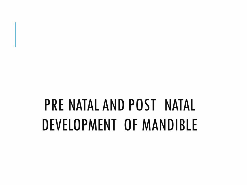
CONTENTS
2
• Introduction
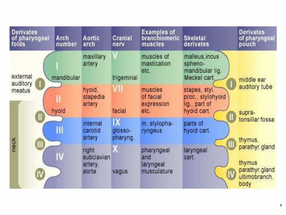
• Pharyngeal arches• Contents and derivatives of arch
• Meckel’s cartilage
• Prenatal growth• Fate of meckel’s cartilage
• Ossification
• Secondary cartilage
• Postnatal growth• Growth of mandible
• Anomalies of growth
• Conclusion
• Reference
INTRODUCTION
3
• Growth and development of an individual can be divided into pre-natal and post-natal period.
• Prenatal period is a dynamic phase where, gowth occurs at a higher rate when compared to post natal growth.
• Among facial bones, mandible undergoes largestamount of growth post natally and exhibits largevariability in morphology
PERIODS OF GROWTH
4
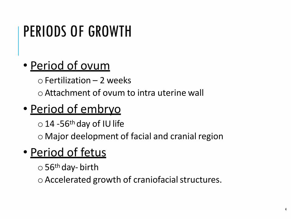
• Period of ovumo Fertilization – 2 weeks
oAttachment of ovum to intra uterine wall
• Period of embryoo14 -56th day of IU life
oMajor deelopment of facial and cranial region
• Period of fetuso56th day- birth
oAccelerated growth of craniofacial structures.

PRE-NATAL GROWTH
5

PRE NATAL GROWTH
6
• Embryonic neural crest cells
• Cells migrate ventrally to form mandibular prominence.
• Form mandibular division of trigeminal nerve.
• Ectomesenchymal condensation forming first pharyngial arch
PHARYNGEAL ARCHES
7
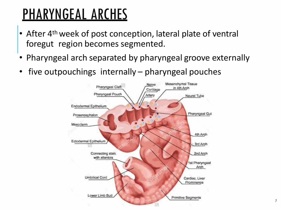
• After 4th week of post conception, lateral plate of ventral foregut region becomes segmented.
• Pharyngeal arch separated by pharyngeal groove externally
• five outpouchings internally – pharyngeal pouches
CONTENTS OF EACH ARCH
8
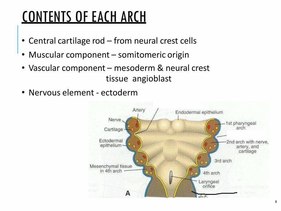
• Central cartilage rod – from neural crest cells
• Muscular component – somitomeric origin
• Vascular component – mesoderm & neural cresttissue angioblast
• Nervous element - ectoderm
9

FIRST PHARYNGEAL ARCH
10
• Precursor of both maxilla & mandible.
• It forms the lateral wall of stomatodeum.
• Maxilla derived from cranio ventral extension of mandibular arch at 28th day – 4th week.

mandibular• Arch grows ventro-medially process
• Grows towards each other &fuses in midline lower margin of stomatodeum.
• Gives rise to lower lip & lower jaw.
• Maxillary & mandibular partly fuses to form cheek.
11
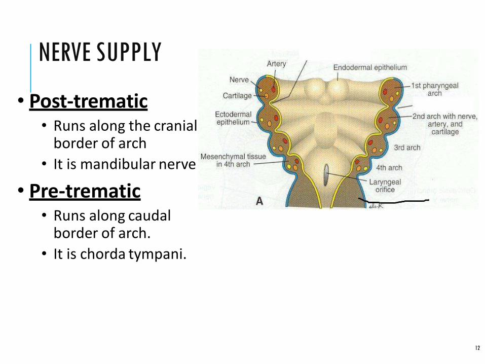
NERVE SUPPLY
12
• Post-trematic• Runs along the cranial
border of arch
• It is mandibular nerve
• Pre-trematic• Runs along caudal
border of arch.
• It is chorda tympani.

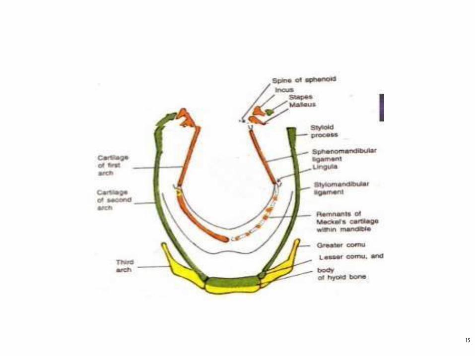
MECKEL’S CARTILAGE
13
• Present at 41st – 45th day of post conception.
• Most cartilage substance disappears in mandible.
• Extent cartilaginous otic capsule-symphysis
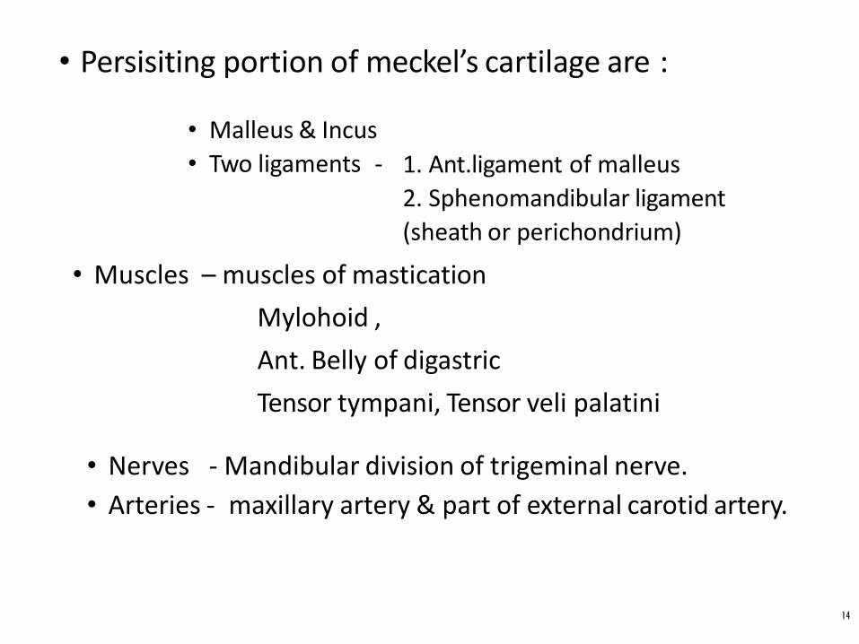
• Persisiting portion of meckel’s cartilage are :
14
• Malleus & Incus
• Two ligaments - 1. Ant.ligament of malleus
2. Sphenomandibular ligament
(sheath or perichondrium)
• Muscles – muscles of mastication
Mylohoid ,
Ant. Belly of digastric
Tensor tympani, Tensor veli palatini
• Nerves - Mandibular division of trigeminal nerve.
• Arteries - maxillary artery & part of external carotid artery.
15
FATE OF MECKEL’S CARTILAGE
16
• Meckels cartilage meets its fellow opposite side ventrally.
• diverge dorsally and ends in tympanic cavity-malleus and incus
• Remnants of ventral end are seen in fibrous tissue of symphysis- CHONDRIOLA SYMPHYSEA .

17
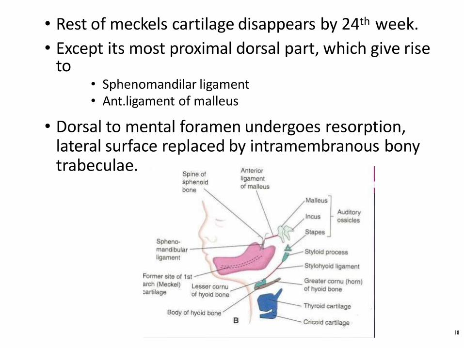
• Rest of meckels cartilage disappears by 24th week.
• Except its most proximal dorsal part, which give rise to
• Sphenomandilar ligament• Ant.ligament of malleus
• Dorsal to mental foramen undergoes resorption, lateral surface replaced by intramembranous bony trabeculae.
18

OSSIFICATION OF MANDIBLE
19
• 2 types of ossification.• Intramembranous ossification
• Endochondral ossification.
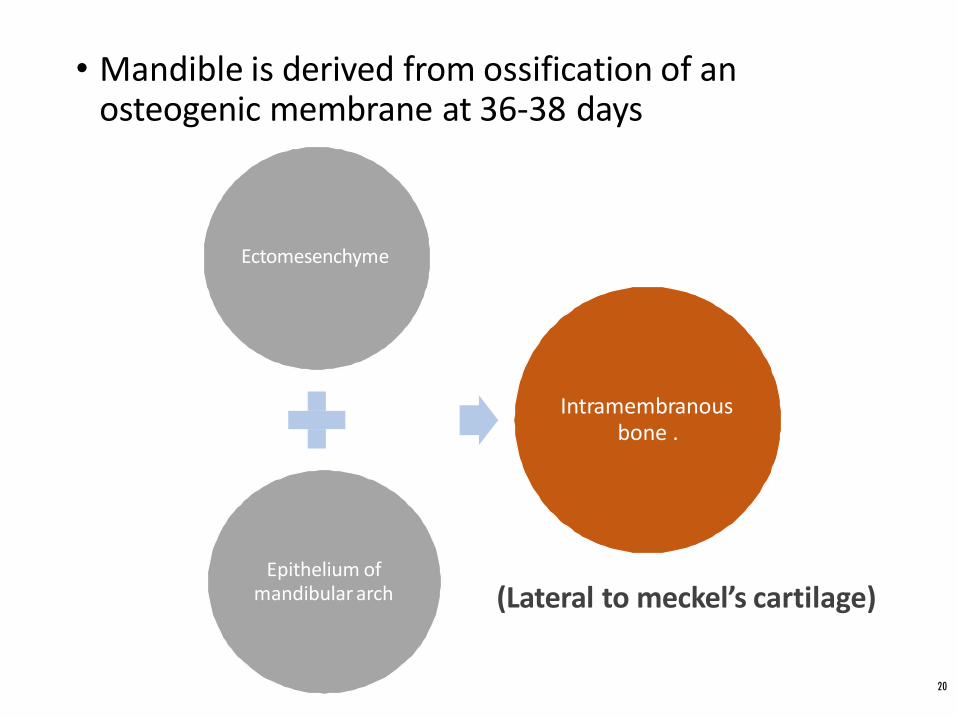
• Mandible is derived from ossification of an osteogenic membrane at 36-38 days
Ectomesenchyme
Epithelium of mandibular arch
Intramembranous bone .
20
(Lateral to meckel’s cartilage)

PROCESS OF OSSIFICATION OF
MANDIBLE
21
• Intra membranous ossification• Body of mandible except anterior
part
• Ramus of mandible till mandibular foramen
• Endochondral ossification• Symphysis of mandible
• Ramus above mandibular foramen
• Coronoid process
• Condylar process

• Single ossification centre for each half of mandibleat 6th week.
• Occurs in the region of bifurcation of Inferioralvolar nerve & artery into mental & incisivebranches.
• Ossification stops dorsally forming – Lingula
• Prior presence of nerovascular bundles gives--Mandibular foramen& canal
-Mental foramen
22
• Ossification dorsally and ventrally gives rise to-body and ramus of mandible
• Primary ossification centre spreads upwards to form a strong base for teeth.
• Meckels catilage will be invaded by bone.
• Mandible & clavicle. First bone to begin osssify.
23
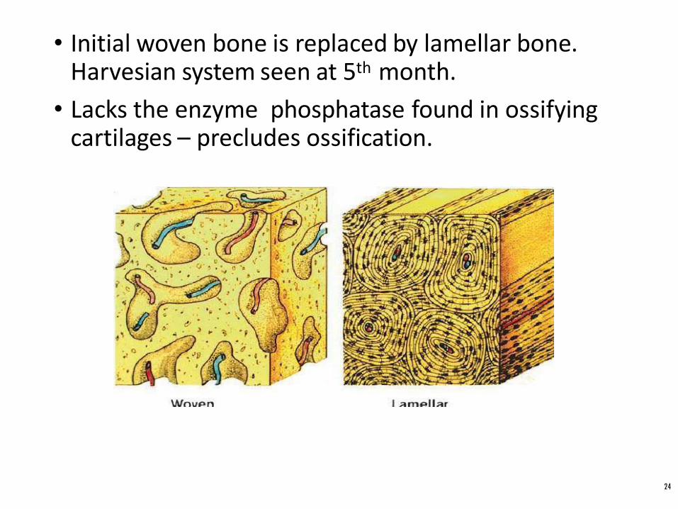
• Initial woven bone is replaced by lamellar bone. Harvesian system seen at 5th month.
• Lacks the enzyme phosphatase found in ossifying cartilages – precludes ossification.
24

SECONDARY CARTILAGES
25
• Appears between 10th – 14th week
• Forms head of condyle, part of coronoid process, mental protuberance
• Coronoid process cartilage develops within temporalis muscle.
• Later it is incorporated into the expanding intramembranous bone of ramus and disappear before birth.

CONDYLAR CARTILAGE
26
• Appears at 10th week .
• Seen as a cone shaped structure in ramal region.
• Primordium for future condyle.
• Condylar head increases by interstitial and appositional growth.
• Important centre of growth for ramus and body.

27

• Most of cartilage is replaced by bone, except its upper end which persist in adulthood.
• Act as both growth and articular cartilage.
• Growth peaks between 12.5 – 14 yrs of age, and ceases at 20yrs of age.
28

CORONOID PROCESS
29
•Secondary cartilage appears in coronoid process around
10 -14th week.
•Cartilage grow as a response of developing temporalis
muscle
•Coronoid cartilage become incorporated into expanding
intramembranous bone of ramus and disappear before
birth
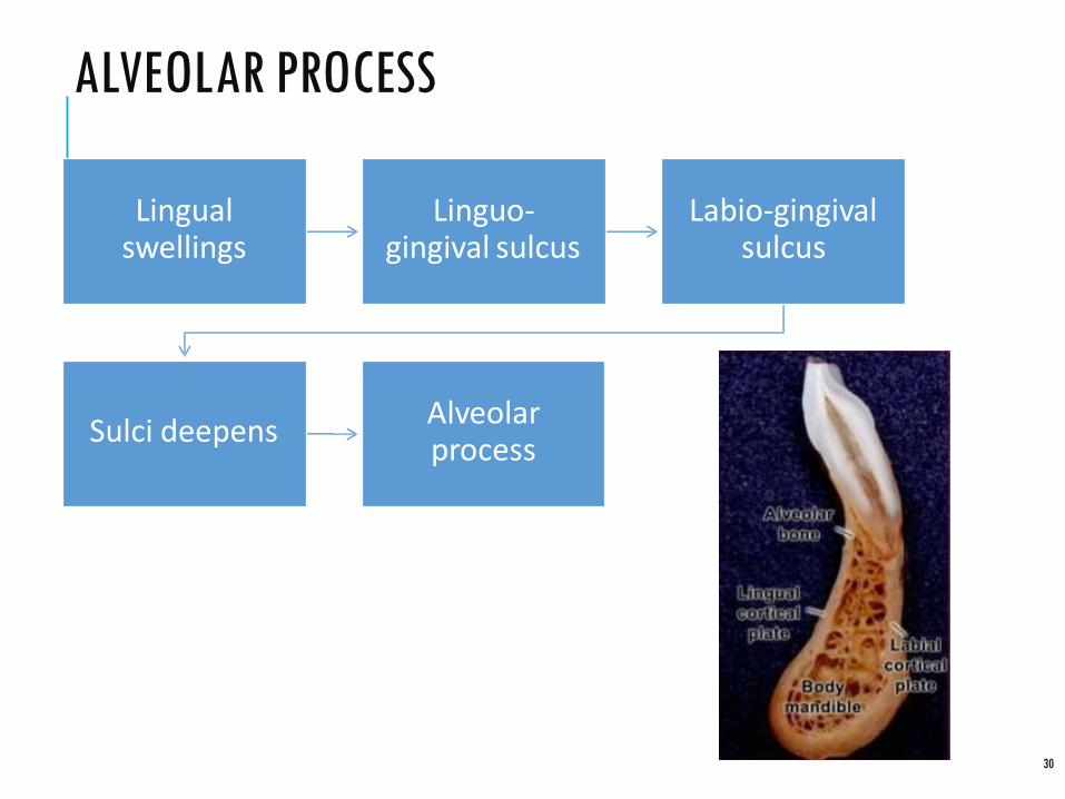
ALVEOLAR PROCESS
30
Lingual swellings
Linguo-gingival sulcus
Labio-gingival sulcus
Sulci deepensAlveolar process

MENTAL REGION
31
Cartilage ossify at 7th
month
Mental ossicles
Fused to intramembranous bone & ossifies at 1 yr.

POST NATAL GROWTH
32
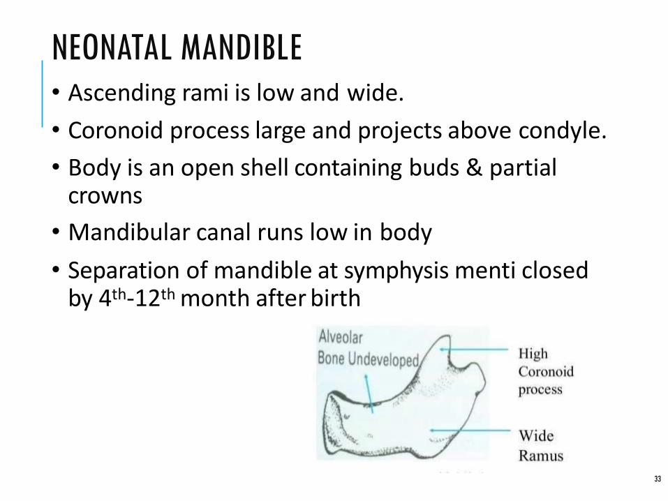
NEONATAL MANDIBLE
33
• Ascending rami is low and wide.
• Coronoid process large and projects above condyle.
• Body is an open shell containing buds & partial crowns
• Mandibular canal runs low in body
• Separation of mandible at symphysis menti closed by 4th-12th month after birth
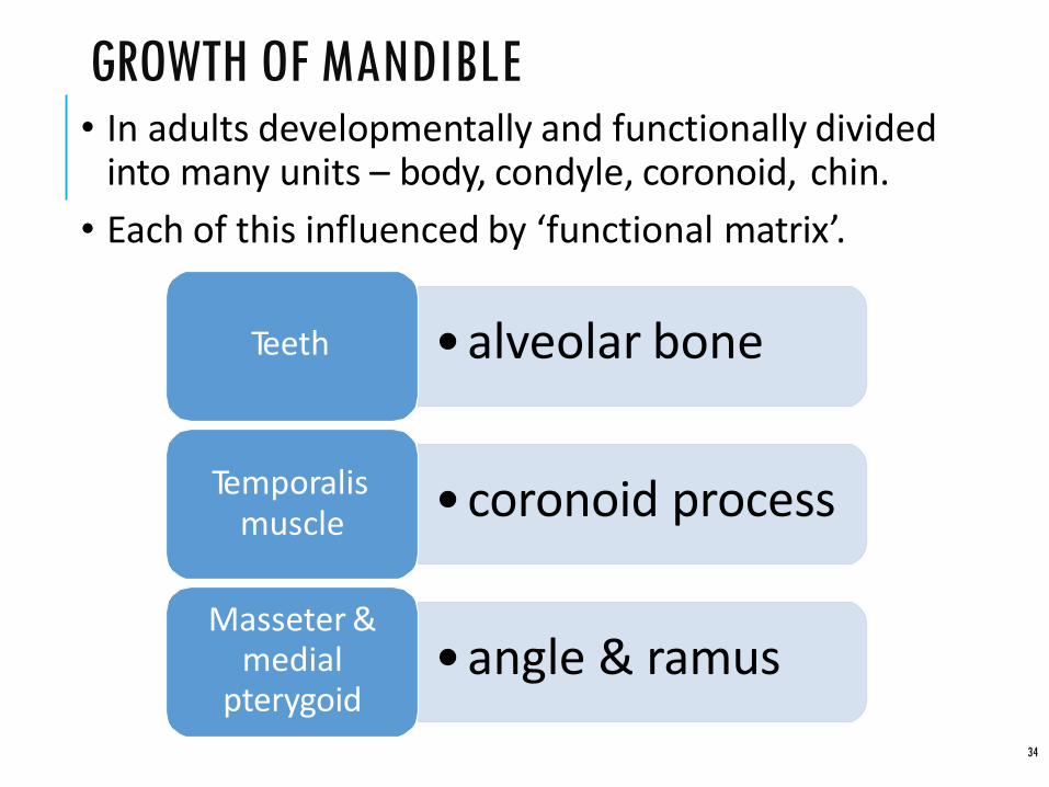
GROWTH OF MANDIBLE
34
• In adults developmentally and functionally divided into many units – body, condyle, coronoid, chin.
• Each of this influenced by ‘functional matrix’.
•alveolar boneTeeth
•coronoid processTemporalis muscle
•angle & ramusMasseter &
medial pterygoid
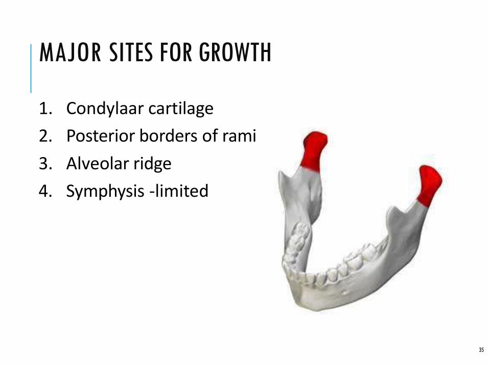
MAJOR SITES FOR GROWTH
35
1. Condylaar cartilage
2. Posterior borders of rami
3. Alveolar ridge
4. Symphysis -limited

CONDYLE
36
• Both articular cartilage in TMJ & growth cartilage.
In medullary core
appositional proliferation of
cartilage in condyle head
Provides basisfor the growth
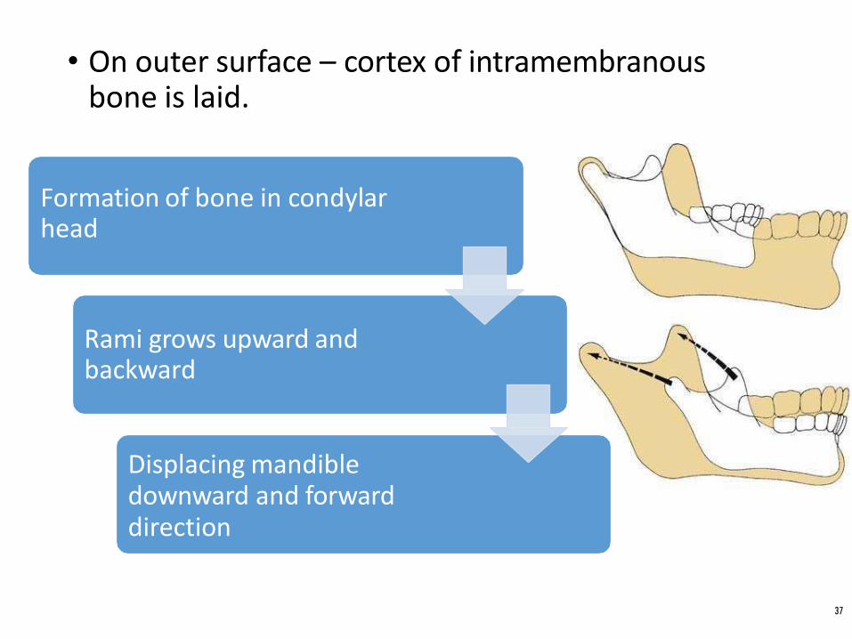
• On outer surface – cortex of intramembranous bone is laid.
Formation of bone in condylar head
Rami grows upward and backward
Displacing mandible downward and forward direction
37
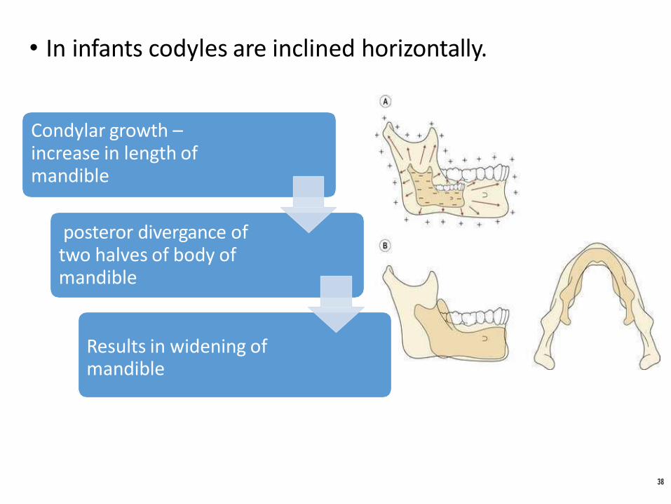
• In infants codyles are inclined horizontally.
Condylar growth –increase in length of mandible
posteror divergance of two halves of body of mandible
Results in widening of mandible
38

SYMPHYSIS MENTI
39
• No interstitial widening after it is fused at 1 year.
• Widening only happens by surface apposition
RAMUS OF MANDIBLE
40
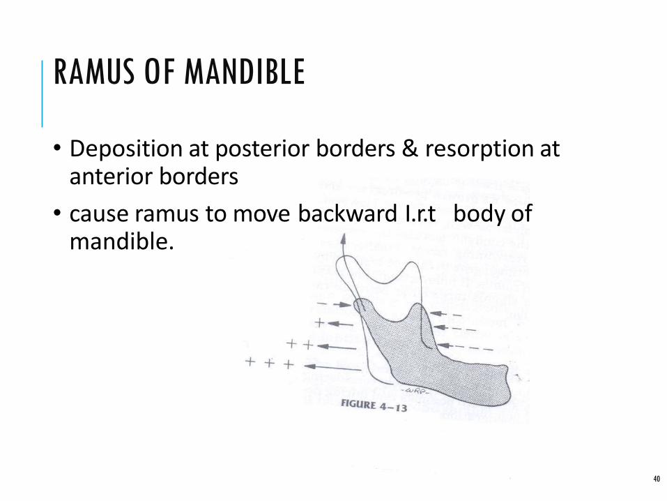
• Deposition at posterior borders & resorption at anterior borders
• cause ramus to move backward I.r.t body of mandible.

• Repositions the mandibular foramen posteriorly.
• Accomadate place for the eruption of molars
41
BODY OF MANDIBLE
42
• Post. Displacement of ramus converts - former ramal bone into post.part of body of mandible.
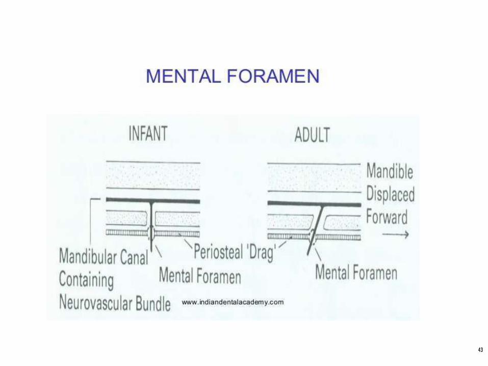
• Changes in the direction of mental foramen. Infancy – neurovascular bundles emerge at right angles
Adulthood – directed backwards
• Clinical implication – administrating L.A in mental nerve.
43

ALVEOLAR PROCESS
44
• If teeth are absent – alveolar process fails to develop & resorbtion will occur
• Orthodontic movement takes place in labile alveolar bone. Does not involve the basal bone.
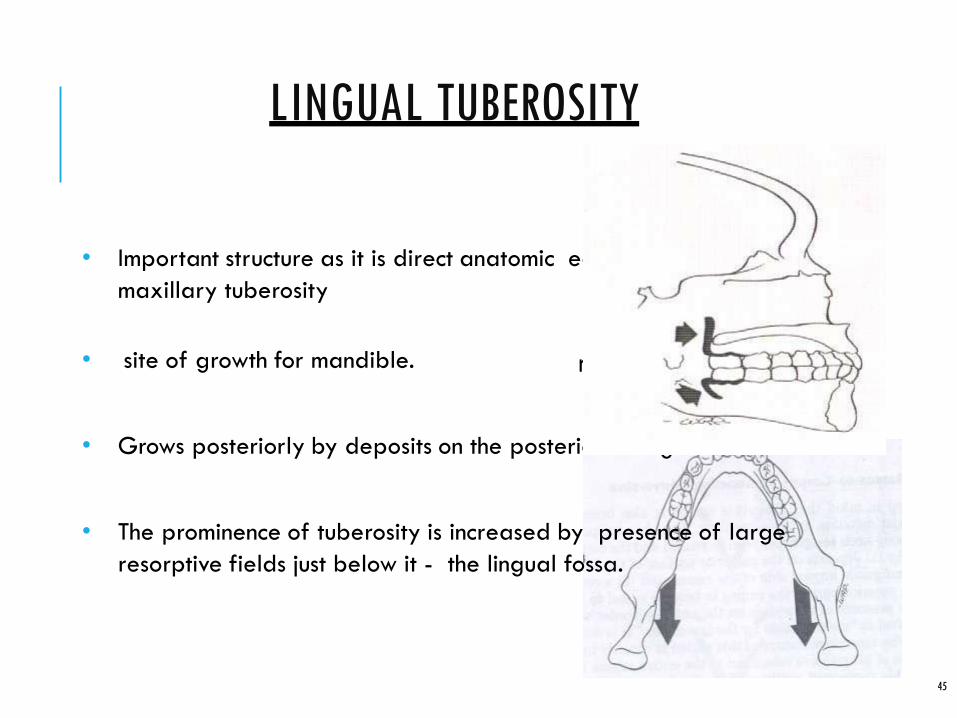
LINGUAL TUBEROSITY
• Important structure as it is direct anatomic equivalent of the
maxillary tuberosity
• site of growth for mandible.
• Grows posteriorly by deposits on the posterio facing surface.
• The prominence of tuberosity is increased by presence of large
resorptive fields just below it - the lingual fossa.
45
r

LINGUAL TUBEROSITY
46
• Remodels in posterior direction with slight lateral shift
• Increases the length of body of mandible

CHIN
47
• Accesory cartilage End of meckels cartilage.
• Unique human trait, lacks in other primates.
• Mental protuberance formed byOsseous depostion at mental region
Bone resorption at alveolar bone – supramental concavity
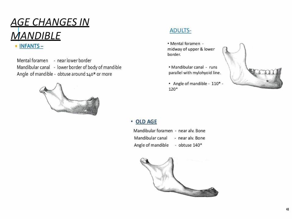
AGE CHANGES INMANDIBLE
48
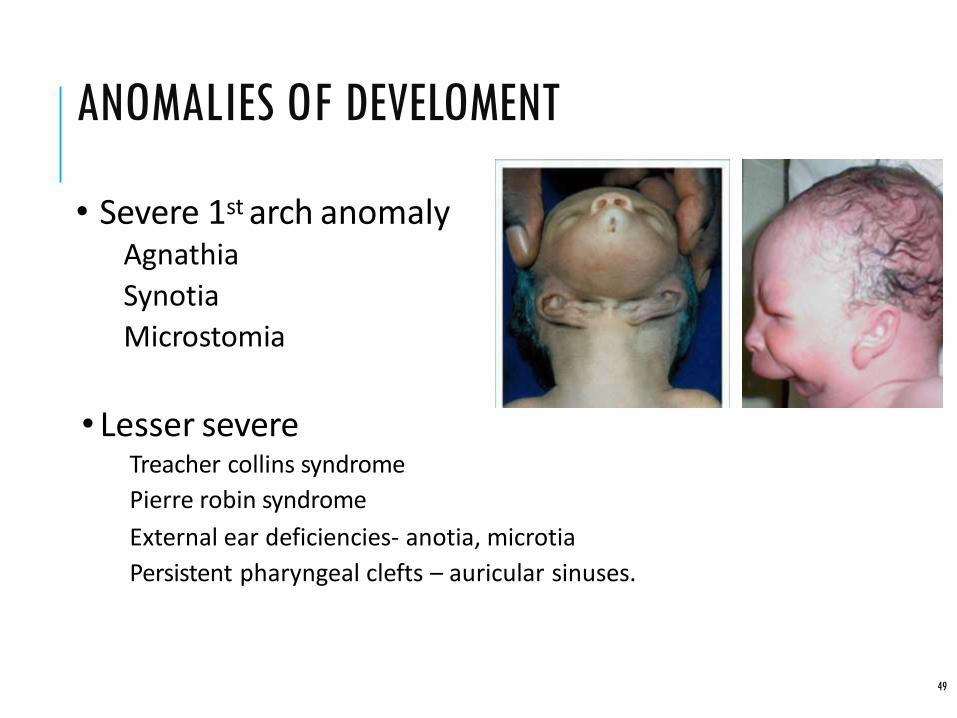
ANOMALIES OF DEVELOMENT
49
• Severe 1st arch anomalyAgnathia
Synotia
Microstomia
• Lesser severeTreacher collins syndrome
Pierre robin syndrome
External ear deficiencies- anotia, microtia
Persistent pharyngeal clefts – auricular sinuses.

CONGENITAL
50
• Agnathia -The mandible may be grossly deficient or absent in condition of agnathia, which is probably due to a neural crest deficiency in the lower face.
• Micrognathia-small jaw• Macrognathia –large jaw
• Facial hemihypertrophy – one side of face is larger than other side
• Facial hemiatropy- degeneration of oneside of face
ANOMALIES OFMANDIBLE
51
SOME OF THE SYNDROMES ASSOCIATED WITH MANDIBULAR ABNORMALITY
• MARFAN SYNDROME- genetic disorder of connective tissue. There isspeech disorder due to small jaws
• PIERRE-ROBIN SYNDROME – micrognathia, cleft palate, glossoptosis
• TREACHER- COLLINS SYNDROME (mandibulo facial dysostosis) - craniofacial deformity having micrognathia , hypoplasia of mandible, bird like face

52
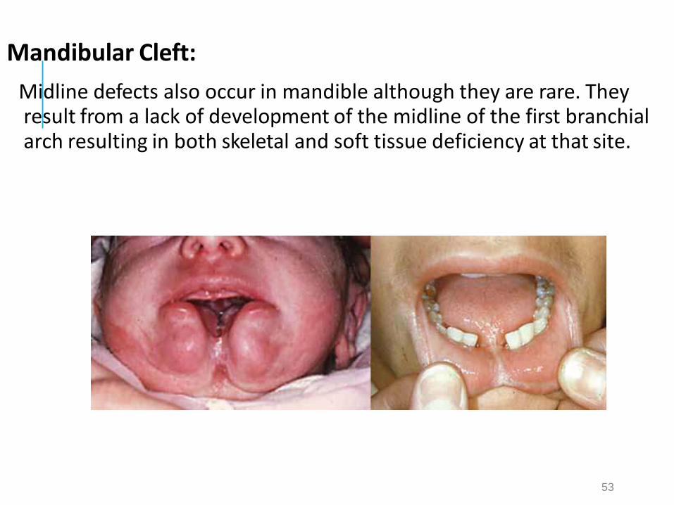
Mandibular Cleft:
Midline defects also occur in mandible although they are rare. They result from a lack of development of the midline of the first branchial arch resulting in both skeletal and soft tissue deficiency at that site.
53
CONCLUSION
54
• Variations in mandible morphology and size more significant than maxillary variability as related to malocclusions
e.g.; Class II, Class III
• Many dental abnormalities have underlying skeletal problems . In order to correct the underlying skeletal discrepancy knowledge of growth and development of the mandible is imperative
• Mandible is clinically controllable to a certain extent. Orthopedic appliances during growth period are used by orthodontists, with which mandibular position can be controlled redirected or altered

REFERENCE
55
• Proffit, William R., Henry W. Fields Jr, and David M.Sarver. Contemporary orthodontics. Elsevier HealthSciences, 2014.
• Sperber, Geoffrey H., Geoffrey D. Guttmann, and StevenM. Sperber.Craniofacial Development.
• Fundamentals of Craniofacial Growth. Andrew D. Dixon, David A.N. Hoyte, Olli Ronning
• Singh, Inderbir. Human embryology. JP Medical Ltd, 2014.

Thank you..
56
Related Documents