The Rockefeller University Press, 0021-9525/2002/06/1175/12 $5.00 The Journal of Cell Biology, Volume 157, Number 7, June 24, 2002 1175–1186 http://www.jcb.org/cgi/doi/10.1083/jcb.200111052 JCB Article 1175 PRC1 is a microtubule binding and bundling protein essential to maintain the mitotic spindle midzone Cristiana Mollinari, 1 Jean-Philippe Kleman, 1 Wei Jiang, 2 Guy Schoehn, 3 Tony Hunter, 4 and Robert L. Margolis 1 1 Institut de Biologie Structurale J-P Ebel (CEA-CNRS), 38027 Grenoble cedex 1, France 2 Department of Biochemistry, New York University Medical Center, New York, NY 10016 3 EMBL Grenoble Outstation, 38042 Grenoble cedex, France 4 The Salk Institute, La Jolla, CA 92037 idzone microtubules of mammalian cells play an essential role in the induction of cell cleavage, serving as a platform for a number of proteins that play a part in cytokinesis. We demonstrate that PRC1, a mitotic spindle-associated Cdk substrate that is essential to cell cleavage, is a microtubule binding and bundling protein both in vivo and in vitro. Overexpression of PRC1 extensively bundles interphase microtubules, but does not affect early mitotic spindle organization. PRC1 contains two Cdk phos- phorylation motifs, and phosphorylation is possibly important to mitotic suppression of bundling, as a Cdk phosphorylation- null mutant causes extensive bundling of the prometaphase M spindle. Complete suppression of PRC1 by siRNA causes failure of microtubule interdigitation between half spindles and the absence of a spindle midzone. Truncation mutants demonstrate that the NH 2 -terminal region of PRC1, rich in -helical sequence, is important for localization to the cleavage furrow and to the center of the midbody, whereas the central region, with the highest sequence homology between species, is required for microtubule binding and bundling activity. We conclude that PRC1 is a microtubule- associated protein required to maintain the spindle midzone, and that distinct functions are associated with modular elements of the primary sequence. Introduction Mitosis is a highly regulated process characterized by dramatic and coordinated morphological changes to ensure the fidelity of chromosome segregation. Cytokinesis occurs at the final stage of mitosis and is accomplished by the contraction of an acto-myosin ring that leads to daughter cell separation at the midbody (Cao and Wang, 1990). A number of proteins accumulate at the midzone of the mammalian mitotic spindle during late mitosis and have been shown to play a role in cell cleavage by antibody suppression, overexpression, or mutagenesis. Among these proteins are passenger proteins such as INCENP (Mackay et al., 1998) and survivin (Skoufias et al., 2000; Uren et al., 2000); protein kinases such as polo (Lee et al., 1995) and aurora B (Terada et al., 1998); small G-proteins such as Rho (Takada et al., 1996; Drechsel et al., 1997; O’Connell et al., 1999); and microtubule motor proteins such as CENP-E (Yen et al., 1992; Martineau- Thuillier et al., 1998), Rab6-KIFL (Hill et al., 2000), and MKLP1 (Sellitto and Kuriyama, 1988; Nislow et al., 1992). Some interactions among these proteins have been established, but specifically defined functional roles in the cleavage process are still largely unknown. Another protein that accumulates in the spindle midzone and that has a demonstrated role in sustaining cell cleavage, PRC1, has been recently described (Jiang et al., 1998). As is true of most of the other midzone proteins, the precise role of PRC1 in the cleavage process is unknown. In late anaphase, a central mitotic spindle forms between the two separating sets of chromatids. It consists of a dense network of overlapping antiparallel microtubules (MTs)*. (Mastronarde et al., 1993). The central spindle, the site of accu- mulation of numerous proteins required for cell cleavage (Glotzer, 1997; Robinson and Spudich, 2000), has been demonstrated to be critical to the completion of cytokinesis (Cao and Wang, 1996; Wheatley and Wang, 1996). This is the only time in the cell cycle that a typical mammalian culture cell generates stable and bundled MTs. The molecular basis for maintenance of the midzone MT bundle is unresolved. Address correspondence to Robert L. Margolis, Institut de Biologie Structurale J-P Ebel (CEA-CNRS), 41 rue Jules Horowitz, 38027 Grenoble cedex 1, France. Tel.: 33-43-878-9616. Fax: 33-43-878-5494. E-mail: [email protected] Key words: mitosis; Cdk; cytokinesis; microtubule-associated protein; cytoskeleton *Abbreviations used in this paper: EGFP, enhanced green fluorescent protein; MT, microtubule; NLS, nuclear localization signal; siRNA, short interfering RNA; UTR, untranslated region.

Welcome message from author
This document is posted to help you gain knowledge. Please leave a comment to let me know what you think about it! Share it to your friends and learn new things together.
Transcript

The Rockefeller University Press, 0021-9525/2002/06/1175/12 $5.00The Journal of Cell Biology, Volume 157, Number 7, June 24, 2002 1175–1186http://www.jcb.org/cgi/doi/10.1083/jcb.200111052
JCB
Article
1175
PRC1 is a microtubule binding and bundling protein essential to maintain the mitotic spindle midzone
Cristiana Mollinari,
1
Jean-Philippe Kleman,
1
Wei Jiang,
2
Guy Schoehn,
3
Tony Hunter,
4
and Robert L. Margolis
1
1
Institut de Biologie Structurale J-P Ebel (CEA-CNRS), 38027 Grenoble cedex 1, France
2
Department of Biochemistry, New York University Medical Center, New York, NY 10016
3
EMBL Grenoble Outstation, 38042 Grenoble cedex, France
4
The Salk Institute, La Jolla, CA 92037
idzone microtubules of mammalian cells play anessential role in the induction of cell cleavage,serving as a platform for a number of proteins that
play a part in cytokinesis. We demonstrate that PRC1, amitotic spindle-associated Cdk substrate that is essential tocell cleavage, is a microtubule binding and bundling proteinboth in vivo and in vitro. Overexpression of PRC1 extensivelybundles interphase microtubules, but does not affect earlymitotic spindle organization. PRC1 contains two Cdk phos-phorylation motifs, and phosphorylation is possibly importantto mitotic suppression of bundling, as a Cdk phosphorylation-null mutant causes extensive bundling of the prometaphase
M
spindle. Complete suppression of PRC1 by siRNA causesfailure of microtubule interdigitation between half spindlesand the absence of a spindle midzone. Truncation mutants
demonstrate that the NH
2
-terminal region of PRC1, rich in
�
-helical sequence, is important for localization to thecleavage furrow and to the center of the midbody, whereasthe central region, with the highest sequence homologybetween species, is required for microtubule binding andbundling activity. We conclude that PRC1 is a microtubule-associated protein required to maintain the spindle midzone,
and that distinct functions are associated with modularelements of the primary sequence.
Introduction
Mitosis is a highly regulated process characterized by dramaticand coordinated morphological changes to ensure the fidelityof chromosome segregation. Cytokinesis occurs at the finalstage of mitosis and is accomplished by the contraction ofan acto-myosin ring that leads to daughter cell separationat the midbody (Cao and Wang, 1990). A number of proteinsaccumulate at the midzone of the mammalian mitoticspindle during late mitosis and have been shown to play arole in cell cleavage by antibody suppression, overexpression,or mutagenesis. Among these proteins are passenger proteinssuch as INCENP (Mackay et al., 1998) and survivin (Skoufiaset al., 2000; Uren et al., 2000); protein kinases such aspolo (Lee et al., 1995) and aurora B (Terada et al., 1998);small G-proteins such as Rho (Takada et al., 1996; Drechselet al., 1997; O’Connell et al., 1999); and microtubule motorproteins such as CENP-E (Yen et al., 1992; Martineau-Thuillier et al., 1998), Rab6-KIFL (Hill et al., 2000), and
MKLP1 (Sellitto and Kuriyama, 1988; Nislow et al.,1992). Some interactions among these proteins have beenestablished, but specifically defined functional roles in thecleavage process are still largely unknown. Another proteinthat accumulates in the spindle midzone and that has ademonstrated role in sustaining cell cleavage, PRC1, hasbeen recently described (Jiang et al., 1998). As is true ofmost of the other midzone proteins, the precise role ofPRC1 in the cleavage process is unknown.
In late anaphase, a central mitotic spindle forms betweenthe two separating sets of chromatids. It consists of a densenetwork of overlapping antiparallel microtubules (MTs)*.(Mastronarde et al., 1993). The central spindle, the site of accu-mulation of numerous proteins required for cell cleavage(Glotzer, 1997; Robinson and Spudich, 2000), has beendemonstrated to be critical to the completion of cytokinesis(Cao and Wang, 1996; Wheatley and Wang, 1996). This isthe only time in the cell cycle that a typical mammalian culturecell generates stable and bundled MTs. The molecular basisfor maintenance of the midzone MT bundle is unresolved.
Address correspondence to Robert L. Margolis, Institut de BiologieStructurale J-P Ebel (CEA-CNRS), 41 rue Jules Horowitz, 38027Grenoble cedex 1, France. Tel.: 33-43-878-9616. Fax: 33-43-878-5494.E-mail: [email protected]
Key words: mitosis; Cdk; cytokinesis; microtubule-associated protein;cytoskeleton
*Abbreviations used in this paper: EGFP, enhanced green fluorescentprotein;
MT, microtubule; NLS, nuclear localization signal; siRNA,short interfering RNA; UTR, untranslated region.
1176 The Journal of Cell Biology
|
Volume 157, Number 7, 2002
Here we show that PRC1 is required to maintain a stablemidzone MT bundle. It is an MT binding and bundlingprotein in vitro, and it forms extensive MT interphase bun-dles when overexpressed in mammalian cells. In keepingwith these results, introduction of short interfering RNAs(siRNAs) targeting PRC1 profoundly disrupts the formationof the midzone bundle and blocks cytokinesis. The effect ofsiRNA occurs uniquely during cell cleavage, showing PRC1is required to maintain interdigitation between the two halfspindles during anaphase.
PRC1 has an evident domain structure. The NH
2
-terminalregion of the protein is largely
�
-helical with multiple pre-dicted coiled-coil motifs, whereas the COOH-terminal onequarter is predicted to be largely composed of
�
sheets andturns. Truncation mutants show that PRC1 appears to bemodular. The NH
2
-terminal half of the protein is required forits association with the cleavage furrow and midbody, whereassequence within residues 273–486 is required for MT bind-ing. Microinjection of antibody that recognizes the COOHterminus of PRC1 disrupts the function of midzone MT bun-dles and blocks cleavage, showing the conformation of theCOOH terminus may be important to the protein’s function.On the basis of our results, we conclude that the function ofPRC1 is necessary for spindle integrity during late mitosis,particularly to maintain the midzone MT bundles that are es-sential for the completion of cell cleavage.
Results
Overexpression of PRC1 modifies the MT array in HeLa cells
PRC1 is a mitotic spindle-associated protein that is requiredfor cytokinesis in mammalian cells (Jiang et al., 1998). Se-quence analysis shows that the NH
2
-terminal three quartersof the protein is largely
�
-helical (see Fig. 5 A) with pre-dicted coiled-coil motifs (Fig. 1 B), whereas the COOH-ter-minal region is predicted to be largely composed of
�
-sheetsand turns. At the junction between these two distinct re-gions there is a cluster of two Cdk phosphorylation sites,two nuclear localization motifs, and two D boxes and a Kenbox (see Fig. 5 A).
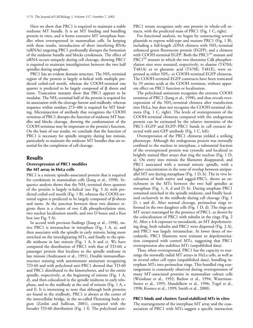
In accord with previous findings (Jiang et al., 1998), na-tive PRC1 is intranuclear in interphase (Fig. 1 A, a), andthen associates with the spindle in early mitosis, being moreenriched on the interdigitating MTs, and finally to the spin-dle midzone in late mitosis (Fig. 1 A, b and c). We havecompared the distribution of PRC1 with that of TD-60, apassenger protein that localizes to the spindle midzone inlate mitosis (Andreassen et al., 1991). Double immunofluo-rescence staining with autoimmune antiserum recognizingTD-60 and with polyclonal anti-PRC1 showed that TD-60and PRC1 distributed to the kinetochores, and to the entirespindle, respectively, at the beginning of mitosis (Fig. 1 A,d), and then colocalized in the spindle midzone in early telo-phase, and to the midbody at the end of mitosis (Fig. 1 A, eand f). It is interesting to note that although both proteinsare found in the midbody, PRC1 is always at the center ofthe intercellular bridge, in the so-called Flemming body re-gion (Zeitlin and Sullivan, 2001), compared with thebroader TD-60 distribution (Fig. 1 f). The polyclonal anti-
PRC1 serum recognizes only one protein in whole-cell ex-tracts, with the predicted mass of PRC1 (Fig. 1 C, right).
For functional analysis, we began by constructing severalplasmids to express wild-type and mutant PRC1 (Fig. 1 B),including a full-length cDNA chimera with NH
2
-terminalenhanced green fluorescent protein (EGFP), and a chimerawith COOH-terminal EGFP. Both the PRC1
AA
mutant andPRC1
EE
mutant in which the two threonine Cdk phosphor-ylation sites were mutated, respectively, to alanine (T470A;T481A) or to glutamic acid (T470E; T481E), were ex-pressed as either NH
2
- or COOH-terminal EGFP chimeras.The COOH-terminal EGFP constructs have been truncatedby 35 amino acids at the COOH terminus, without appar-ent effect on PRC1 function or localization.
The polyclonal antiserum recognizes the extreme COOHterminus of PRC1 (Jiang et al., 1998), and thus reveals over-expression of the NH
2
-terminal chimera after transfectioninto HeLa, but does not recognize the COOH-terminal chi-mera (Fig. 1 C, right). The levels of overexpression of theCOOH-terminal chimeras compared with the endogenousprotein can be estimated by the relative intensities of thePRC1–EGFP and EGFP–PRC1 bands in cell extracts de-tected with anti-GFP antibody (Fig. 1 C, left).
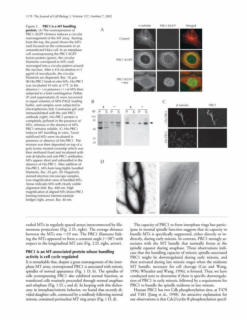
Overexpression of the PRC1 chimeras yielded a strikingphenotype. Although the endogenous protein was normallyconfined to the nucleus in interphase, a substantial fractionof the overexpressed protein was cytosolic and localized tobrightly stained fiber arrays that ring the nucleus (Fig. 1 D,a). On entry into mitosis the filaments disappeared, andPRC1 associated with a normal mitotic spindle, with ahigher concentration in the zone of overlap between antipar-allel MT sets during metaphase (Fig. 1 D, b). The in vivo lo-calization of both native and tagged-PRC1, shows an en-richment in the MTs between the two half spindles atmetaphase (Fig. 1, A, d and D, b). During anaphase PRC1remained enriched in the spindle midzone, and finally local-ized exclusively in the midbody during cell cleavage (Fig. 1D, c and d). After normal cleavage, perinuclear rings re-formed in the two daughter cells (Fig. 1 D, d). The rings areMT arrays rearranged by the presence of PRC1, as shown bythe colocalization of PRC1 with tubulin in the rings (Fig. 2A). After a 4-h exposure to nocodazole, an MT depolymeriz-ing drug, both tubulin and PRC1 were dispersed (Fig. 2 A),and PRC1 was largely intranuclear. At lower doses of no-codazole, PRC1 filaments were resistant to depolymeriza-tion compared with control MTs, suggesting that PRC1overexpression also stabilizes MTs (unpublished data).
Thus, when overexpressed, PRC1 has the capacity to rear-range the normally radial MT arrays in HeLa cells, as well asin several other cell types (unpublished data), bundling in-terphase MTs into perinuclear rings. This bundled ring rear-rangement is commonly observed during overexpression ofmany MT-associated proteins in mammalian culture cells(Weisshaar et al., 1992; Barlow et al., 1994; Waterman-Storer et al., 1995; Mandelkow et al., 1996; Togel et al.,1998; Koonce et al., 1999; Smith et al., 2000).
PRC1 binds and clusters Taxol-stabilized MTs in vitro
The rearrangement of the interphase MT array and the coas-sociation of PRC1 with MTs suggest a specific interaction

PRC1 bundles mitotic midzone microtubules |
Mollinari et al. 1177
between PRC1 and MTs. Thus, we probed for PRC1 bind-ing to MTs in an in vitro assay, and found that His-tagged,bacterially expressed PRC1 was entirely pelleted by Taxol-stabilized pure tubulin MTs (Fig. 2 B). In contrast, in theabsence of MTs, PRC1 remained in the supernatant (Fig. 2B). Direct microscopic observation of MTs after PRC1binding shows complete colocalization of the two antigens,
and reveals that the MTs have become extensively bundledin the presence of PRC1 (Fig. 2 C).
We have also analyzed the Taxol-stabilized MTs in thepresence of His-PRC1 by electron microscopy. The bundleswere extremely dense, but in favorable regions could be seento contain clusters of aligned MTs (Fig. 2 D, left, arrow).Examination of these regions at higher magnification re-
Figure 1. The distribution of the endogenous PRC1 and its chimeras in HeLa cells. (A) The localization of the endogenous PRC1 and TD-60. (a–c) HeLa cells were labeled with the affinity-purified antibody against PRC1 (green) and with propidium iodide for DNA (red). PRC1 is present in nuclei in interphase cells (a), and then becomes localized to the spindle upon entry into mitosis (b), and concentrates in the spindle midzone during late anaphase (c). (d–f) Double labeling of PRC1 (green) and TD-60 (red) during mitosis. (d) In metaphase, TD-60 is bound to the kinetochores at the metaphase plate, whereas PRC1 is associated with the spindle, enriched on interdigitating MTs. In late mitosis, the two antigens largely colocalize at the cleavage furrow (e) and midbody (f). PRC1 appears restricted to the Flemming body. Bars, 10 �m. (B) Schematic representation of the PRC1 chimeras. PRC1 was expressed in bacteria or HeLa cells as a tagged protein. For the first two constructs, the complete coding region of PRC1 (1–620 aa) was fused downstream to a 6�-His tag (His-PRC1) or an EGFP tag (EGFP–PRC1). The last construct allowed the expression of a COOH terminus truncation of PRC1 fused upstream to the EGFP (PRC1–EGFP). Both types of EGFP chimeras were used to overexpress the PRC1 mutant proteins. The PRC1AA protein is a null phosphory-lation mutant, with both Thr 470 and Thr 481 (red vertical bars) mutated into Ala. The PRC1EE protein, with both the Thr 470 and Thr 481 mutated into Glu, mimics phosphorylation. The blue boxes indicate the regions of PRC1 with a high probability of multicoil formation (residues 35–85, 89–135, 210–251, 384–411, and 437–464). (C) Expression of the endogenous PRC1 and chimeras in HeLa cells. Extracts from HeLa, either untreated or transfected with one of the PRC1 chimeras (EGFP–PRC1, PRC1–EGFP, and PRC1–EGFPAA), or with a control plasmid expressing only EGFP, were subjected to electrophoresis on 8%
polyacrylamide gels and immunoblotted with the anti-EGFP antibody (left) or anti-PRC1 antibody (right). (Right) The anti-PRC1 antibody recognizes the endogenous PRC1 (arrowhead) in all extracts along with a band at the expected molecular mass of the EGFP–PRC1 fusion protein (arrow). PRC1–EGFP and the mutant PRC1–EGFPAA fusions were not recognized because they lack the COOH- terminal residues recognized by this antibody. In contrast, the anti-EGFP antibody recognizes the unfused EGFP as well as all the three PRC1 fusion proteins. (D) The distribution of the PRC1-EGFP fusion protein in HeLa cells. During interphase, the overexpressed PRC1–EGFP protein (green) induces the formation of circular filaments around nuclei (DNA, red). In common with endogenous PRC1, overexpressed PRC1 is also in nuclei. During mitosis, the PRC1–EGFP protein is associated to a normal spindle (b), and then becomes associated to the midzone (c) and midbody at late telophase (d), similar to endogenous PRC1. The perinuclear ring filaments are evident in interphase (a) and at the end of telophase (d) but are dispersed during mitosis. Bar, 10 �m.
1178 The Journal of Cell Biology
|
Volume 157, Number 7, 2002
vealed MTs in regularly spaced arrays interconnected by fila-mentous projections (Fig. 2 D, right). The average distancebetween the MTs was
�
19 nm. The PRC1 filaments link-ing the MTs appeared to form a constant angle (
�
38
�
) withrespect to the longitudinal MT axis (Fig. 2 D, right, arrow).
PRC1 is an MT-associated protein whose bundling activity is cell cycle regulated
It is remarkable that, despite a gross rearrangement of the inter-phase MT array, overexpressed PRC1 is associated with mitoticspindles of normal appearance (Fig. 1 D, b). The spindles ofcells overexpressing PRC1 also exhibited normal function, astransfected cells routinely proceeded through normal anaphaseand telophase (Fig. 1 D, c and d). In keeping with this dichot-omy in interphase/mitotic behavior, we found that recently di-vided daughter cells, connected by a midbody following normalmitosis, contained perinuclear MT ring arrays (Fig. 1 D, d).
The capacity of PRC1 to form interphase rings but partic-ipate in normal spindle function suggests that its capacity tobundle MTs is specifically suppressed, either directly or in-directly, during early mitosis. In contrast, PRC1 strongly as-sociates with the MT bundle that normally forms at thespindle equator during anaphase. These observations indi-cate that the bundling capacity of mitotic spindle-associatedPRC1 might be downregulated during early mitosis, andthen activated during late mitotic stages when the midzoneMT bundle, necessary for cell cleavage (Cao and Wang,1996; Wheatley and Wang, 1996), is formed. Thus, we haveconducted tests to determine if there is specific downregula-tion of PRC1 in early mitosis, followed by a requirement forPRC1 to bundle the spindle midzone in late mitosis.
Human PRC1 has two Cdk phosphorylation sites, at T470and T481 (Jiang et al., 1998). An attractive explanation forour observations is that Cdc2/cyclin B phosphorylation specif-
Figure 2. PRC1 is a MT bundling protein. (A) The overexpression of PRC1–EGFP chimera induces a circular rearrangement of the MT array. Starting from the top, the panel shows the MTs (red) focused on the centrosome in an untransfected HeLa cell. In an interphase cell overexpressing the PRC1-EGFP fusion protein (green), the circular filaments correspond to MTs (red) rearranged into a circular pattern around the nucleus. After a 4-h incubation in 1 �g/ml of nocodazole, the circular filaments are dispersed. Bar, 10 �m. (B) His-PRC1 binds in vitro MTs. His-PRC1 was incubated 10 min at 37�C in the absence (�) or presence (�) of MTs then subjected to a brief centrifugation. Pellets (P) and supernatants (S) were recovered in equal volumes of SDS-PAGE loading buffer, and samples were subjected to electrophoresis (left, Coomassie gel) and immunoblotted with the anti-PRC1 antibody (right). His-PRC1 protein is completely pelleted in the presence of MTs, whereas in the absence of MTs PRC1 remains soluble. (C) His-PRC1 induces MT bundling in vitro. Taxol-stabilized MTs were incubated in presence or absence of His-PRC1. The mixture was then deposited on top of a poly-lysine–treated coverslip which was then methanol fixed and incubated with anti–�-tubulin and anti-PRC1 antibodies. MTs appear short and unbundled in the absence of His-PRC1. After addition of His-PRC1, MTs form long highly bundled filaments. Bar, 20 �m. (D) Negatively stained electron microscopy samples. Low magnification view of bundled MTs. Arrow indicates MTs with clearly visible alignment (left). Bar, 400 nm. High magnification of aligned MTs shows PRC1 forming extensive intermicrotubule bridges (right, arrow). Bar, 40 nm.

PRC1 bundles mitotic midzone microtubules |
Mollinari et al. 1179
ically downregulates PRC1 bundling capacity in early mitosis.We mutated the two Cdk-phosphorylated threonine residuesto alanine, and transfected HeLa with plasmids expressing thenonphosphorylatable PRC1–EGFP
AA
mutant. The result wasstriking. In early mitosis, mutant PRC1 caused extensive bun-dling of the MTs of the mitotic spindle (Fig. 3, bottom) com-pared with the morphology of a control spindle (Fig. 3, top).Despite extensive bundling, the MT arrays largely preserve abipolar orientation. Approximately 37% of the prometaphasecells overexpressing the mutant showed such bundled spindlesassociated with highly condensed DNA, and no mitotic cellswith such bundles were observed in anaphase, indicating themutant blocks mitotic progression.
We also constructed a phosphorylation mimic PRC1–EGFP
EE
mutant to determine if it had a distinct effect. Ex-pression of this construct gave results identical to those ob-tained with the PRC1–EGFP
AA
mutant (unpublished data).The EE mutant also had no effect on MT binding (see Fig. 5C) or bundling (unpublished data) of PRC1 in vitro. We in-terpret these results as indicating that glutamate substitutionin this case is not a good phosphorylation mimic, but insteadinterferes with a critical function at the phosphorylation site.
The absence of PRC1 affects midzone formation during anaphase
PRC1 is clearly required for cell cleavage, as microinjection ofanti-PRC1 antibody causes cleavage failure (Jiang et al., 1998).To determine the specific role that PRC1 plays in cell cleavage,we transfected cells with siRNA to block PRC1 translation, andfollowed cells in which PRC1 was suppressed. Immunofluores-cence analysis showed that PRC1 was substantially diminishedin 30–35% of transfected cells after 24 h. Many cells could befound in which PRC1 was apparently completely absent asshown by paired PRC1-negative and -positive metaphase andearly (Fig. 4 A, a) and late anaphase (Fig. 4 A, b) cells. In bothcases, one cell of the pair has no detectable PRC1 (Fig. 4 A a, ar-row and b, arrowhead). In accord with these observations,Western blots showed that PRC1 was strongly suppressed inthe entire cell population after transfection (Fig. 4 B).
In the absence of PRC1, cells were able to progress nor-mally in mitosis to metaphase (Fig. 4 A, a, arrow), and
underwent normal chromatid segregation in anaphase (Fig.4 A, b, arrowhead). However, cells lacking PRC1 alwaysshowed aberrant anaphase spindle morphology (Fig. 4 C).Interpolar MTs were radially dispersed, and interdigitatingMTs were generally absent between the two half spindles(Fig. 4 C, a). Interestingly, even in the absence of a midzoneMT bundles, cells were able to separate their sets of sisterchromatids and showed partial furrowing (Fig. 4 C, b and c).
Differing degrees of severity of the phenotype were alwaysassociated with either complete or reduced levels of PRC1expression. In some cases, midzone MTs were present, but indisarray. More often, MTs displayed no interdigitation at thespindle equator (Fig. 4 C, b), and as a result the half spindlesand their chromosomes frequently rotated away from theirnormal alignment orthogonal to the spindle equator. How-ever, even in the absence of midzone MTs, cells seemed toinitiate furrowing then regress before completing cleavage(Fig. 4 C, d). As a result of furrow regression, cells lackingPRC1 became increasingly binucleate with time (Fig. 4 D),reaching values consistent with the PRC1 suppressed popu-lation. We conclude that the absence of PRC1 disturbs nei-ther MT assembly nor chromosome segregation, but severelyalters interzonal bundling in anaphase.
Domain structure of PRC1
The primary sequence of PRC1 has several striking features(Fig. 5 A, wt). The NH
2
-terminal three quarters of the pro-tein is predicted to be largely composed of
�
-helical se-quence (within which are predicted coiled-coil motifs, Fig.1), whereas the COOH-terminal region mostly contains
�
-sheets and turns. At the junction between these two re-gions there are two Cdk consensus phosphorylation sitesclustered with two nuclear localization signals (NLSs). A cen-tral region of the protein, spanning residues 240–440, ishighly conserved among eukaryotic species, and can be as-sumed to be important to the function of PRC1. PRC1 alsocontains putative consensus sequences for ubiquitination-dependent proteolysis, including two D boxes and a KEN box(Fig. 5 A) (Glotzer et al., 1991; Pfleger and Kirschner, 2000).
To determine the relative roles of these distinct regions ofthe primary structure in PRC1 function, we constructed sev-
Figure 3. Overexpression of a double PRC1 mutant lacking the two Cdk phospho-threonines creates multiple MT bundles in mitotic spindles. A control untreated cell (top) and a cell (bottom) overexpressing PRC1–EGFPAA (green), double labeled with anti–�-tubulin antibody (red), are shown. Although PRC1 distribution shows the MTs are highly bundled, the mitotic spindle preserves a bipolar shape. Bar, 10 �m.

1180 The Journal of Cell Biology
|
Volume 157, Number 7, 2002
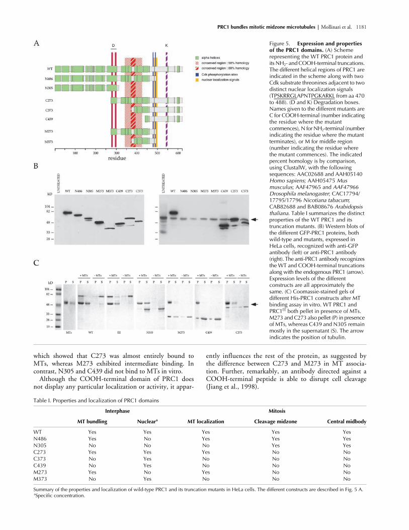
eral truncation mutants (Fig. 5 A) and expressed them asGFP-tagged chimeras to assay their properties in mamma-lian cells. Western blots of PRC1 chimeras, expressed aftertransfection, showed that truncated proteins of the correctsize were expressed, and that all constructs were being ex-pressed at approximately the same level (Fig. 5 B).
Results with the different constructs are summarized inTable I. In brief, we found that MT binding during mitosiscorrelated with the presence of the conserved central region.Further, NH
2
-terminal sequence upstream of the conservedregion was required for association with the midzone of thecleavage furrow and for localization to the center of the mid-body. Nuclear localization during interphase correlated withthe presence of the NLS consensus sequence, as expected.
Immunofluorescence images of the different chimeras dem-onstrate their distinct capacities for localization (Fig. 6). Theshortest COOH-terminal construct, C439, neither localizes tothe mitotic spindle nor to the cleavage midzone. The construct
M273, containing the central conserved region, localizes to thewhole mitotic spindle but lacks sequence required to address itto the center of the midbody at the end of mitosis.
Like M273, the longer COOH-terminal construct C273also localizes to the mitotic spindle but not to the center ofthe midbody in telophase. It is noteworthy that inclusion ofdownstream sequence causes C273 to induce stronger MTassociation in the mitotic spindle than M273 (Fig. 6 A). Incontrast to the COOH-terminal constructs, the NH
2
-termi-nal constructs N305 and N486 both specifically localize tothe center of the midbody at the end of mitosis (Fig. 6 B, in-sets), whereas the longer construct, N486, localizes to theentire mitotic spindle at metaphase, the shorter N305 doesnot (unpublished data), reinforcing the importance of thecentral region in MT binding. The result with N305 dem-onstrates that MT binding capacity is not required for local-ization to the center of the midbody. These in vivo resultswere reinforced by in vitro MT binding assays (Fig. 5 C),
Figure 4. Suppression of PRC1 function by siRNA disrupts midzone MTs in HeLa cells. (A) Panels a (arrow) and b (arrowhead) show two examples of mitotic cells negative for PRC1 (green) side by side with cells expressing endogenous PRC1. (B) Western blot showing a specific decrease of PRC1 protein after siRNA transfection compared with untreated cells. The same blot was then reprobed with an anti-tubulin antibody to show equal loading of the samples. (C) HeLa cells negative for PRC1 have an altered spindle midzone. Interdigitating MTs are absent (a) or drastically reduced and disorganized (b and c) and as a consequence the two half spindles are rotated relative to the spindle axis. Cells with altered spindle midzone initiate furrowing (b and c) but do not complete cleavage (d). (D) A binucleated cell negative for PRC1. Bar, 5 �m. A histogram showing the increase in binucleated cells after siRNA transfection. A peak is reached 48 h after transfection.
PRC1 bundles mitotic midzone microtubules |
Mollinari et al. 1181
which showed that C273 was almost entirely bound toMTs, whereas M273 exhibited intermediate binding. Incontrast, N305 and C439 did not bind to MTs in vitro.
Although the COOH-terminal domain of PRC1 doesnot display any particular localization or activity, it appar-
ently influences the rest of the protein, as suggested bythe difference between C273 and M273 in MT associa-tion. Further, remarkably, an antibody directed against aCOOH-terminal peptide is able to disrupt cell cleavage(Jiang et al., 1998).
Figure 5. Expression and properties of the PRC1 domains. (A) Scheme representing the WT PRC1 protein and its NH2- and COOH-terminal truncations. The different helical regions of PRC1 are indicated in the scheme along with two Cdk substrate threonines adjacent to two distinct nuclear localization signals (TPSKRRGLAPNTPGKARKL from aa 470 to 488). (D and K) Degradation boxes. Names given to the different mutants are C for COOH-terminal (number indicating the residue where the mutant commences), N for NH2-terminal (number indicating the residue where the mutant terminates), or M for middle region (number indicating the residue where the mutant commences). The indicated percent homology is by comparison, using ClustalW, with the following sequences: AAC02688 and AAH05140 Homo sapiens; AAH05475 Mus musculus; AAF47965 and AAF47966 Drosophila melanogaster; CAC17794/17795/17796 Nicotiana tabacum; CAB82688 and BAB08676 Arabidopsis thaliana. Table I summarizes the distinct properties of the WT PRC1 and its truncation mutants. (B) Western blots of the different GFP-PRC1 proteins, both wild-type and mutants, expressed in HeLa cells, recognized with anti-GFP antibody (left) or anti-PRC1 antibody (right). The anti-PRC1 antibody recognizes the WT and COOH-terminal truncations along with the endogenous PRC1 (arrow). Expression levels of the different constructs are all approximately the same. (C) Coomassie-stained gels of different His-PRC1 constructs after MT binding assay in vitro. WT PRC1 and PRC1EE both pellet in presence of MTs. M273 and C273 also pellet (P) in presence of MTs, whereas C439 and N305 remain mostly in the supernatant (S). The arrow indicates the position of tubulin.
Table I. Properties and localization of PRC1 domains
Interphase Mitosis
MT bundling Nuclear
a
MT localization Cleavage midzone Central midbody
WT Yes Yes Yes Yes YesN486 Yes No Yes Yes YesN305 No No No Yes YesC273 Yes Yes Yes No NoC373 No Yes No No NoC439 No Yes No No NoM273 Yes No Yes No NoM373 No Yes No No No
Summary of the properties and localization of wild-type PRC1 and its truncation mutants in HeLa cells. The different constructs are described in Fig. 5 A.
a
Specific concentration.

1182 The Journal of Cell Biology
|
Volume 157, Number 7, 2002
Upon microinjection of affinity-purified anti-PRC1 anti-bodies, we found that spindle morphology and functionwere normal in early mitosis (Fig. 7, a and b), and all stagesup to telophase were unchanged. However, at telophase, theusual broad MT bundle was greatly diminished by compari-son to normal mitotic controls, and did not appear to extendto the cell cortex (Fig. 7 c). Thus, the effect of a COOH-ter-minal directed antibody is quite similar to that of PRC1suppression by siRNA, except that the phenotype is more se-vere with siRNA, which affects the earlier step of midzoneformation during anaphase with consequent suppression ofcytokinesis. We find that
�
50% of the microinjected cellsfail in cleavage, as previously noted (Jiang et al., 1998).
Discussion
We have shown that human PRC1 is a MT-associated bun-dling protein. Both in vivo and in vitro evidence supportthis conclusion. The requirement for PRC1 in cell cleavage,and the bundling that is associated with PRC1 presence in
the midzone of the late mitotic spindle, lead us to concludethat PRC1 functions to stabilize the midzone MT bundle,permitting completion of cell cleavage.
Many proteins concentrate at the equatorial region of the latemitotic central spindle for the purpose of participating in thecleavage event. Although many of these proteins, like PRC1, areknown to be required for the cleavage process, their specificroles in cell cleavage have not as yet been defined. Some of theseproteins have defined functions, acting as MT motors (Ohkuraet al., 1997; Adams et al., 1998; Williams et al., 1995; Raich etal., 1998), small GTP-binding proteins and their regulators(Kishi et al., 1993; Drechsel et al., 1997; Prokopenko et al.,1999; Tatsumoto et al., 1999; Hill et al., 2000; Jantsch-Plungeret al., 2000), or protein kinases (Lee et al., 1995; Carmena etal., 1998; Madaule et al., 1998; Terada et al., 1998; Yasui et al.,1998). Their overall functions must be either to maintain thecentral spindle, recruit required proteins to this site, create thephysical means by which the central spindle communicates tothe cell cortex for the controlled deposition of myosin II and ac-tin, or to generate the signal for the cleavage event.
Figure 6. PRC1 contains distinct domains with different functional capacities as determined by immunofluorescence. To the left of the panels is shown a scheme of the different constructs studied. For each construct, anaphase (A) and telophase (B) cells are shown, stained for �-tubulin (red) and PRC1 (green). Removal of the COOH-terminus of PRC1 does not affect its ability to bind MTs (M273) nor its enrichment at the Flemming body during telophase (N486-N305) (B, arrowheads). In fact, the COOH terminus alone does not seem to be directly involved in any PRC1 activity assayed. Constructs C273 and M273 contain the region responsible for binding and bundling MTs, but they lack the signal to focus on the center of the midzone in anaphase (A), or the Flemming body during telophase (B). In (B) a closeup of the midbody (inset) is shown for each telophase cell. Bars, 5 �m.

PRC1 bundles mitotic midzone microtubules |
Mollinari et al. 1183
Considering that interdigitated midzone MTs are re-quired for successful cytokinesis, and that passenger proteinswith roles in cell cleavage collect at the center of the centralspindle in late anaphase, PRC1 may be essential for theseproteins to correctly localize to the cleavage furrow. Indeed,our preliminary data indicate that PRC1 has a key role intargeting the passenger proteins involved in cleavage (un-published data). The capacity of PRC1 to specifically local-ize to the Flemming body in telophase, independent of itsassociation with MTs, suggests there may be a second andimportant function for PRC1 during cleavage.
PRC1 phosphorylation sites and their potential function
A notable aspect of PRC1 behavior is that, despite bundlingMTs when overexpressed in interphase cells, it permits nor-mal spindle function. This result suggests that PRC1 MTbundling function is strongly downregulated in early mito-sis, and then reactivated in late mitosis for the purpose ofstabilizing the midzone MTs. Inactivation in early mitosis isin accord with siRNA experiments that show suppression ofPRC1 does not interfere with any aspect of mitosis until cellcleavage. Among the proteins that bundle interphase MTswhen overexpressed, PRC1 is, to our knowledge, uniquewith respect to its suppression during early mitosis.
PRC1 has two Cdk phosphorylation sites, and a nullphosphorylation mutant yields an early mitotic phenotypeconsistent with the interpretation that phosphorylation sup-presses PRC1 bundling activity, as the mutant generates MTbundles throughout the mitotic spindle. PRC1 has beenshown to be a good substrate for several Cdks in vivo and invitro (Jiang et al., 1998). It is reasonable to speculate thatthe mitosis-specific regulation is mediated by Cdc2 activity.
Although several observations indicate a physiological rolefor phosphorylation of PRC1, we were unable to clearlydemonstrate this because overexpression of a phosphoryla-tion mimic EE mutant of PRC1 generates the same bundledmitotic spindle phenotype seen with the phosphorylation-null mutant, suggesting that the EE mutant is not an ade-quate mimic of phosphorylation status.
Domain structure of PRC1
PRC1 contains multiple
�
-helical regions with the potentialfor formation of multicoils that may figure in interproteinlinkages (Fig. 1 B). It shares 57% homology with the bud-ding yeast protein Ase1 that localizes to the anaphase spindlemidzone and is required for many aspects of mitosis (Juang etal., 1997). Together with another nonmotor MT-associatedprotein, Ase1 is required for anaphase B, the elongation ofthe spindle and separation of spindle poles (Pellman et al.,1995). Loss of Ase1 protein function destabilizes the spindleduring telophase (Juang et al., 1997). Significant sequencehomology (56%) is also shared with a
Nicotiana tabacum
MT-associated protein, Map65 (GenBank/EMBL/DDBJ ac-cession no. CAC17794, CAC17795, and CAC17796). Thehomologue of Map65 in carrots has been demonstrated toform regular inter-MT linkages, thus generating MT bundles(Chan et al., 1999). Similarly, our data indicate that PRC1forms bundles of aligned MTs where inter-MT linkage ismade through filamentous projections at a constant anglewith respect to the longitudinal MT axis. MT bundling mayrequire dimers or higher oligomers of PRC1, a possibility weare currently exploring. In preliminary experiments, we havefound that PRC1 runs as a single included peak on sizing col-umns, with a mass of
�
300 kD (unpublished data), suggest-ing that it forms small oligomers in vitro. The primary se-
Figure 7. The microinjection of the anti-PRC1 antibody specifically affects the formation of the midbody during telophase. HeLa cells microinjected with the anti-PRC1 antibody, labeled with the anti–�-tubulin antibody (red) along with the anti–rabbit IgG antibody (green), are shown in the panel. Antibody microinjection does not affect the early steps of spindle formation. PRC1 is associated with the entire spindle at metaphase (a), and moves to the spindle midzone during anaphase (b), as in controls. However, during telophase (c), the midzone spindle appears as a narrow and unstructured bundle of MTs that has detached from the cell cortex. Note anti-PRC1 antiserum crossreacts with centrosomes, as previously noted (Jiang et al., 1998). Bars, 5 �m.

1184 The Journal of Cell Biology
|
Volume 157, Number 7, 2002
quence of PRC1 gives a clear indication that the protein hasdistinct domains. Truncation mutants confirm this impres-sion, and demonstrate that distinct regions of PRC1 havedistinct roles. The central region is clearly implicated in MTbinding. In contrast, the NH
2
-terminal region does not bindMTs, but is required for association of PRC1 with the Flem-ming body at the center of the midbody in late cleavage.These results show that association of PRC1 with the Flem-ming body does not require MT association. In fact, one ofthe truncation mutants of PRC1, N305, neither associateswith MTs in vitro nor with the spindle MTs in vivo, butclearly associates with the Flemming body during cleavage.
Thus, it is possible that PRC1 has two distinct functionsin cleavage, one for MT bundling and another relating to as-sociation with the Flemming body at the last stages of cleav-age. Cleavage has two distinct stages, the first involving theinitial cortical contraction, and the second, resolution andfinal cell separation (Zeitlin and Sullivan, 2001). PRC1 mayplay a role in each of these distinct events. siRNA experi-ments show that cleavage can fail at an early stage in the ab-sence of PRC1, but this result does not exclude a further rolefor PRC1 in the final stage of cleavage.
Potential association with other cleavage proteins
The spindle-associated motor protein MKLP1 has beenspeculated to play a role in bundling the late mitotic centralspindle based on its in vitro capacity to bundle MTs (Nislowet al., 1992). MKLP1 homologues in lower eukaryotes playa role in cytokinesis, as mutants of these homologues, pava-rotti (Adams et al., 1998) and ZEN-4 (Raich et al., 1998;Severson et al., 2000), exhibit derangements in cleavage.Our results suggest that MKLP1 alone is not sufficient tomaintain the midzone MT bundle. In fact, the role ofMKLP1 is complex, as an alternatively spliced form, CHO1,must bind actin to complete the terminal step in cleavage(Kuriyama et al., 2002).
Cyk-4, a Rho GAP, interacts specifically with ZEN-4 in
Caenorhabditis elegans
, and both proteins appear to be re-quired for formation of the midzone spindle in late mito-sis (Mishima et al., 2002). A human Cyk-4 orthologue,HsCYK-4, has recently been shown to form a heterotet-ramer with MKLP-1 and to bundle microtubules in vitro(Mishima et al., 2002).
Additionally, the PRC1 truncation mutant N305 does notbind directly to MTs, but does associate with the spindle mid-zone. Thus, it is possible that PRC1, in addition to binding di-rectly to MTs, also binds as part of a protein complex at the latemitotic spindle midzone.
Our evidence supports a role for in the formation of midzoneMT bundles during anaphase. Further work will establish whatproteins PRC1 associates with and, specifically, if there is inter-action and cooperation between PRC1, Cyk-4, and MKLP1 inmaintaining the spindle midzone, as well as in the terminalstage of cleavage.
Materials and methods
Cloning and mutagenesis
Wild-type PRC1 cDNA in pCL (Jiang et al., 1998) was used to generate dif-ferent constructs. Using a BamHI internal site, PRC1 cDNA, including the 5
untranslated region (UTR) up to nucleotide 1836, was subcloned into theEcoRI-BamHI sites of the pEGFP-N1 vector (CLONTECH Laboratories, Inc.).This construct encoded the PRC1 protein, lacking the last 35 amino acids atthe COOH terminus, fused upstream to the EGFP protein. To generate awild-type Histidine-PRC1 (His-PRC1), the entire coding sequence of PRC1was amplified by PCR and subcloned into the EcoRI-NotI of pHAT2(Peränen et al., 1996). To generate an EGFP–PRC1 fusion protein, the frag-ment EcoRI-NotI in pHAT2 was cut with NotI, filled with Klenow (Biolabs)and then ligated into the EcoRI-SmaI sites of pEGFP-C2. The PRC1
AA
andPRC1
EE
mutants in which Thr 470 and Thr 481 were respectively substitutedby Ala or Glu, were generated by PCR. For PRC1
AA
, two independent PCRreactions were performed using oligonucleotides 5
-CCGGAATTCATGAG-GAGAAGTGAGGTGCTG-3
, 5-TGCTCGAGGAGCGCTGCCATACAG-3and 5-CCTAGCAAGCGGCGAGGACTGGCTCCCAATGCCCC-3, 5-TTT-ATAGCGGCCGCTCAGGACTGGATGTTGGTTG-3.
These oligonucleotides were designed to substitute both Thr 470 andThr 481 by Ala along with two silent mutations generating SmaI andXhoI restriction sites. An identical PCR strategy was used to generate thePRC1EE. To generate the EGFP–PRC1AA and EGFP–PRC1EE fusions, thefragments EcoRI-NotI in pHAT2 were cut with NotI, filled with Klenowand then ligated into the EcoRI-SmaI sites of pEGFP-C2. To create thetruncations PRC1–EGFPAA and PRC1–EGFPEE, the fragments EcoRI-BamHI from the pEGFP-C2 constructs were subcloned into the EcoRI-BamHI sites of pEGFP-N1. The different truncations were generatedeither by PCR, introducing premature stop codons, or by restriction en-zymes to cut within the PRC1 coding region. C273 was generated bycutting the wt EGFP-PRC1 construct with BspEI and XhoI followed bymung bean nuclease treatment and ligation. M273 was similarly ob-tained, starting from the N486 construct. C373 was obtained by cuttingthe wt EGFP–PRC1 construct with HindIII followed by mung bean nu-clease and ligation. M373 was obtained as described for C373 startingfrom the N486 construct. N305, M273, C373, and C439 were clonedinto pHAT2 for protein expression. All the constructs were verified byDNA sequencing (Eurogentec).
Cell culture and transfectionHeLa cells were grown as a monolayer in DME (GIBCO BRL) supple-mented with 10% fetal bovine serum (Hyclone), and maintained in ahumid incubator at 37�C in a 5% CO2 environment. HeLa cells (3 �106), attached to coverslips in 10-mm dishes, were transfected by Exgen(Euromedex) or Lipofectamine 2000 (GIBCO BRL), with 4 �g of one ofthe PRC1 fusion constructs, or with a control (pEGFP-N1) plasmid, ac-cording to manufacturer’s instructions. For the preparation of the cellextracts, in order to obtain a better efficiency of transfection (�70%),cells were transfected with a mixture of 20 �g of each of the plasmidsand the Lipofectamine 2000. HeLa cells transfected with the PRC1–EGFP plasmid were selected in 1.5 mg/ml geneticin G418 (GIBCOBRL). Nocodazole was dissolved in DMSO and used at 0.04 �g/ml or 1�g/ml. For nocodazole treatment, HeLa cells transiently transfectedwith the PRC1–EGFP plasmid were exposed to 0.04 or 1 �g/ml nocoda-zole for 4 h before fixation.
siRNA oligonucleotides and transfectionTo generate single-stranded, gene-specific annealed RNA oligomers (Dhar-macon Research), we used 5-AAATATGGGAGCTAATTGGGA-3 as thehuman PRC1 cDNA sequence to be targeted by the oligonucleotides.Transfection conditions were as described by Elbashir and colleagues(2001). HeLa cells were plated on coverslips in a 24-well plate and trans-fected using oligofectamine (GIBCO BRL). Cells were fixed at differenttime points after transfection and processed for immunofluorescence. ForWestern blots, cells were harvested with trypsin and washed in PBS beforeadding SDS-PAGE sample buffer.
Immunofluorescence microscopyCells grown on poly-D-lysine–coated glass coverslips for immunofluo-rescence microscopy were fixed with 2% paraformaldehyde-PBS, or al-ternatively, with cold methanol, followed by a step of rehydration inPBS. Cells were then processed with primary and secondary antibodiesand counterstained with propidium iodide. For protocol detail see Mar-tineau-Thuillier et al. (1998). Affinity-purified PRC1 COOH-terminal an-tibody (Jiang et al., 1998), anti–�-tubulin monoclonal antibody (T4026;Sigma-Aldrich), anti-tyrosinated �-tubulin rat monoclonal antibody(Lafanechere et al., 1998) (YL 1/2), and JH human autoimmune serum,used to detect TD-60 (Andreassen et al., 1991), were diluted 500, 400,500 and 300�, respectively. Secondary antibodies, including FITC-con-jugated affinity-purified goat anti–rabbit IgG, rhodamine-conjugated

PRC1 bundles mitotic midzone microtubules | Mollinari et al. 1185
anti–human IgG (Jackson Laboratories), Texas red–conjugated sheepanti–mouse IgG, and rhodamine-conjugated goat anti–rat IgG (Cappel),were used at 2.5 �g/ml. Images were collected with a MRC-600 LaserScanning Confocal Apparatus (Bio-Rad Laboratories) coupled to NikonOptiphot microscope.
Cell extracts and immunoblotting24 h after transfection, cells were trypsinized, collected by centrifugation,and washed in PBS before the addition of SDS-PAGE loading buffer. Aftera short sonication, 10 �g/lane of cell extract was resolved on 8 or 10%polyacrylamide gels using a minigel apparatus (Bio-Rad Laboratories) andtransferred to nitrocellulose. Affinity-purified rabbit antibodies againstPRC1 (Jiang et al., 1998) were diluted 1,000-fold to detect the endogenousand the overexpressed fusion proteins. Anti-EGFP polyclonal antibody(CLONTECH Laboratories, Inc.), diluted 500-fold, was used to detect ex-pression of the fusion proteins. The anti–�-tubulin monoclonal antibodywas diluted 1,000-fold. Blots were then exposed to HRP-conjugated goatanti–rabbit IgG (TAGO), diluted 2,500-fold, for 1 h, and then developed byECL (Pierce Chemical Co.).
Antibody microinjectionFor microinjection, HeLa cells were grown on glass coverslips as previ-ously described (Jiang et al., 1998). Interphase cells were injected in thecytoplasm with affinity-purified anti-PRC1 antibodies (3.8 mg/ml), using asemiautomatic microinjector (Eppendorf). After a 21-h incubation, cover-slips were fixed and stained with FITC-conjugated goat anti–rabbit IgG,along with anti–�-tubulin monoclonal antibody.
Nickel affinity chromatographyAll the constructs in pHAT2 were expressed in BL21 DE3. Bacteria were in-duced at 37�C for 4 h in the presence of 0.5 mM isopropylthio-�-D-galacto-side (IPTG). Lysis and binding to nickel-Sepharose beads (Hitrap chelating;Amersham Pharmacia Biotech) was performed in a phosphate buffer (50mM NaH2PO4, pH 7.6, 300 mM NaCl, 5 mM imidazole, 0.1 mM PMSF, and10 mg/ml aprotinin). The proteins were eluted with 250 mM imidazole, ana-lyzed by SDS-PAGE, and used for further MT binding assays.
In vitro MT binding assays and electron microscopyThe His-PRC1 constructs from nickel-Sepharose purification were dilutedto 0.2 �g/�l in 20 �M Taxol-PEM (80 mM Pipes, pH.6.8, 1 mM EGTA, 1mM MgCl2). Pure tubulin MTs (6.4 mg/ml; provided by Dr. L. Wilson [Uni-versity of California, Santa Barbara, CA]), isolated from bovine brain (Far-rell et al., 1987), were assembled at 37�C, stabilized with 5 �M Taxol, andthen mixed with His-PRC1 to a final concentration of 0.6 �g/�l in a totalvolume of 20 �l. The mixture was then incubated 10 min at 37�C. ForSDS-PAGE and Western blot analysis of MT bundles, PRC1 constructswere cosedimented with MTs in a short centrifugation step at 16,000 g(cold, 5 min). The pellets were rinsed twice in PEM, both pellets and super-natants were recovered in equal volumes of SDS-PAGE loading buffer, andsamples subjected to electrophoresis.
For immunofluorescence studies, 1 vol of the incubated MTs/PRC1(wild-type His-PRC1 or mutant His-PRC1EE) mix was diluted in 60 vol ofprewarmed PEM-Taxol containing 0.05% glutaraldehyde. The solutionwas deposited on a polylysine-coated glass coverslip, and then fixed in100% methanol and prepared as for immunofluorescence microscopy. Acontrol MT preparation without PRC1 was treated identically.
For electron microscopy, protein samples at 0.05 �g/�l, prepared as de-scribed above, were applied to the clean side of carbon on mica (carbon/mica interface) and negatively stained with 2% uranyl acetate. Micro-graphs were taken under low-dose conditions with a JEOL 1200 EX II mi-croscope at 100 kV at a nominal magnification of 40,000�.
We thank Dr. R. Wade and Dr. R.W.H. Ruigrok for assistance with elec-tron microscopy. We also thank Dr. L. Wilson for providing bovine brainPC-tubulin.
This work was supported by funding from La Ligue Nationale Contre leCancer (Laboratoire Labelliseé), and Association pour la Recherche contrele Cancer (5338; R.L. Margolis). T. Hunter is a Frank and Else SchillingAmerican Cancer Society Research Professor. C. Mollinari is supported bya Telethon fellowship (Telethon Fondazione Onlus, Rome, Italy). J.P Kle-man is a researcher of the Commissariat à l’Energie Atomique. W. Jiang isa scholar of Mallinckrodt Foundation.
Submitted: 13 November 2001Revised: 3 May 2002Accepted: 8 May 2002
ReferencesAdams, R.R., A.A. Tavares, A. Salzberg, H.J. Bellen, and D.M. Glover. 1998. Pa-
varotti encodes a kinesin-like protein required to organize the central spindleand contractile ring for cytokinesis. Genes Dev. 12:1483–1494.
Andreassen, P.R., D.K. Palmer, M.H. Wener, and R.L. Margolis. 1991. Telophasedisc: a new mammalian mitotic organelle that bisects telophase cells with apossible function in cytokinesis. J. Cell Sci. 99:523–534.
Barlow, S., M.L. Gonzalez-Garay, R.R. West, J.B. Olmsted, and F. Cabral. 1994.Stable expression of heterologous microtubule-associated proteins (MAPs) inChinese hamster ovary cells: evidence for differing roles of MAPs in micro-tubule organization. J. Cell Biol. 126:1017–1029.
Cao, L.G., and Y.L. Wang. 1990. Mechanism of the formation of contractile ringin dividing cultured animal cells. I. Recruitment of preexisting actin fila-ments into the cleavage furrow. J. Cell Biol. 110:1089–1095.
Cao, L.G., and Y.L. Wang. 1996. Signals from the spindle midzone are requiredfor the stimulation of cytokinesis in cultured epithelial cells. Mol. Biol. Cell.7:225–232.
Carmena, M., M.G. Riparbelli, G. Minestrini, A.M. Tavares, R. Adams, G. Cal-laini, and D.M. Glover. 1998. Drosophila polo kinase is required for cytoki-nesis. J. Cell Biol. 143:659–671.
Chan, J., C.G. Jensen, L.C. Jensen, M. Bush, and C.W. Lloyd. 1999. The 65-kDa car-rot microtubule-associated protein forms regularly arranged filamentous cross-bridges between microtubules. Proc. Natl. Acad. Sci. USA. 96:14931–14936.
Drechsel, D.N., A.A. Hyman, A. Hall, and M. Glotzer. 1997. A requirement forRho and Cdc42 during cytokinesis in Xenopus embryos. Curr. Biol. 7:12–23.
Elbashir, S.M., J. Harborth, W. Lendeckel, A. Yalcin, K. Weber, and T. Tuschl.2001. Duplexes of 21-nucleotide RNAs mediate RNA interference in cul-tured mammalian cells. Nature. 411:494–498.
Farrell, K.W., M.A. Jordan, H.P. Miller, and L. Wilson. 1987. Phase dynamics atmicrotubule ends: the coexistence of microtubule length changes and tread-milling. J. Cell Biol. 104:1035–1046.
Glotzer, M. 1997. The mechanism and control of cytokinesis. Curr. Opin. CellBiol. 9:815–823.
Glotzer, M., A.W. Murray, and M.W. Kirschner. 1991. Cyclin is degraded by theubiquitin pathway. Nature. 349:132–138.
Hill, E., M. Clarke, and F.A. Barr. 2000. The Rab6-binding kinesin, Rab6-KIFL,is required for cytokinesis. EMBO J. 19:5711–5719.
Jantsch-Plunger, V., P. Gonczy, A. Romano, H. Schnabel, D. Hamill, R. Schnabel,A.A. Hyman, and M. Glotzer. 2000. CYK-4: a Rho family GTPase activat-ing protein (GAP) required for central spindle formation and cytokinesis. J.Cell Biol. 149:1391–1404.
Jiang, W., G. Jimenez, N.J. Wells, T.J. Hope, G.M. Wahl, T. Hunter, and R.Fukunaga. 1998. PRC1: a human mitotic spindle-associated CDK substrateprotein required for cytokinesis. Mol. Cell. 2:877–885.
Juang, Y.L., J. Huang, J.M. Peters, M.E. McLaughlin, C.Y. Tai, and D. Pellman.1997. APC-mediated proteolysis of Ase1 and the morphogenesis of the mi-totic spindle. Science. 275:1311–1314.
Kishi, K., T. Sasaki, S. Kuroda, T. Itoh, and Y. Takai. 1993. Regulation of cyto-plasmic division of Xenopus embryo by rho p21 and its inhibitory GDP/GTP exchange protein (rho GDI). J. Cell Biol. 120:1187–1195.
Koonce, M.P., J. Kohler, R. Neujahr, J.M. Schwartz, I. Tikhonenko, and G.Gerisch. 1999. Dynein motor regulation stabilizes interphase microtubulearrays and determines centrosome position. EMBO J. 18:6786–6792.
Kuriyama, R., C. Gustus, Y. Terada, Y. Uetake, and J. Matuliene. 2002. CHO1, amammalian kinesin-like protein, interacts with F-actin and is involved in theterminal phase of cytokinesis. J. Cell Biol. 156:783–790.
Lafanechere, L., C. Courtay-Cahen, T. Kawakami, M. Jacrot, M. Rudiger, J. Weh-land, D. Job, and R.L. Margolis. 1998. Suppression of tubulin tyrosine li-gase during tumor growth. J. Cell Sci. 111:171–181.
Lee, K.S., Y.L. Yuan, R. Kuriyama, and R.L. Erikson. 1995. Plk is an M-phase-specific protein kinase and interacts with a kinesin-like protein, CHO1/MKLP-1. Mol. Cell. Biol. 15:7143–7151.
Mackay, A.M., A.M. Ainsztein, D.M. Eckley, and W.C. Earnshaw. 1998. A dominantmutant of inner centromere protein (INCENP), a chromosomal protein, dis-rupts prometaphase congression and cytokinesis. J. Cell Biol. 140:991–1002.
Madaule, P., M. Eda, N. Watanabe, K. Fujisawa, T. Matsuoka, H. Bito, T. Ish-izaki, and S. Narumiya. 1998. Role of citron kinase as a target of the smallGTPase Rho in cytokinesis. Nature. 394:491–494.
Mandelkow, E.M., O. Schweers, G. Drewes, J. Biernat, N. Gustke, B. Trinczek,and E. Mandelkow. 1996. Structure, microtubule interactions, and phos-phorylation of tau protein. Ann. NY Acad. Sci. 777:96–106.
Martineau-Thuillier, S., P.R. Andreassen, and R.L. Margolis. 1998. Colocalization

1186 The Journal of Cell Biology | Volume 157, Number 7, 2002
of TD-60 and INCENP throughout G2 and mitosis: evidence for their pos-sible interaction in signaling cytokinesis. Chromosoma. 107:461–470.
Mastronarde, D.N., K.L. McDonald, R. Ding, and J.R. McIntosh. 1993. Interpo-lar spindle microtubules in PTK cells. J. Cell Biol. 123:1475–1489.
Mishima, M., S. Kaitna, and M. Glotzer. 2002. Central spindle assembly and cy-tokinesis requires a kinesin-like protein/RhoGAP complex with microtubulebundle activity. Dev. Cell. 2:41–54.
Nislow, C., V.A. Lombillo, R. Kuriyama, and J.R. McIntosh. 1992. A plus-end-directed motor enzyme that moves antiparallel microtubules in vitro local-izes to the interzone of mitotic spindles. Nature. 359:543–547.
O’Connell, C.B., S.P. Wheatley, S. Ahmed, and Y.L. Wang. 1999. The smallGTP-binding protein rho regulates cortical activities in cultured cells duringdivision. J. Cell Biol. 144:305–313.
Ohkura, H., T. Torok, G. Tick, J. Hoheisel, I. Kiss, and D.M. Glover. 1997. Mu-tation of a gene for a Drosophila kinesin-like protein, Klp38B, leads to failureof cytokinesis. J. Cell Sci. 110:945–954.
Pellman, D., M. Bagget, Y.H. Tu, G.R. Fink, and H. Tu. 1995. Two microtubule-associated proteins required for anaphase spindle movement in Saccharomy-ces cerevisiae. J. Cell Biol. 130:1373–1385.
Peränen, J., M. Rikkonen, M. Hyvönen, and L. Kääriäinen. 1996. T7 vectors witha modified T7lac promoter for expression of proteins in Escherichia coli.Anal. Biochem. 236:371–373.
Pfleger, C.M., and M.W. Kirschner. 2000. The KEN box: an APC recognition sig-nal distinct from the D box targeted by Cdh1. Genes Dev. 14:655–665.
Prokopenko, S.N., A. Brumby, L. O’Keefe, L. Prior, Y. He, R. Saint, and H.J.Bellen. 1999. A putative exchange factor for Rho1 GTPase is required forinitiation of cytokinesis in Drosophila. Genes Dev. 13:2301–2314.
Raich, W.B., A.N. Moran, J.H. Rothman, and J. Hardin. 1998. Cytokinesis andmidzone microtubule organization in Caenorhabditis elegans require the ki-nesin-like protein ZEN-4. Mol. Biol. Cell. 9:2037–2049.
Robinson, D.N., and J.A. Spudich. 2000. Towards a molecular understanding ofcytokinesis. Trends Cell Biol. 10:228–237.
Sellitto, C., and R. Kuriyama. 1988. Distribution of a matrix component of themidbody during the cell cycle in Chinese hamster ovary cells. J. Cell Biol.106:431–439.
Severson, A.F., D.R. Hamill, J.C. Carter, J. Schumacher, and B. Bowerman. 2000.The aurora-related kinase AIR-2 recruits ZEN-4/CeMKLP1 to the mitoticspindle at metaphase and is required for cytokinesis. Curr. Biol. 10:1162–1171.
Skoufias, D.A., C. Mollinari, F.B. Lacroix, and R.L. Margolis. 2000. Human survivinis a kinetochore-associated passenger protein. J. Cell Biol. 151:1575–1582.
Smith, D.S., M. Niethammer, R. Ayala, Y. Zhou, M.J. Gambello, A. Wynshaw-
Boris, and L.H. Tsai. 2000. Regulation of cytoplasmic dynein behaviour andmicrotubule organization by mammalian Lis1. Nat. Cell Biol. 2:767–775.
Takada, M., N. Morii, S. Kumagai, and R. Ryo. 1996. The involvement of the rhogene product, a small molecular weight GTP-binding protein, in polyploidiza-tion of a human megakaryocytic cell line, CMK. Exp. Hematol. 24:524–530.
Tatsumoto, T., X. Xie, R. Blumenthal, I. Okamoto, and T. Miki. 1999. HumanECT2 is an exchange factor for Rho GTPases, phosphorylated in G2/Mphases, and involved in cytokinesis. J. Cell Biol. 147:921–928.
Terada, Y., M. Tatsuka, F. Suzuki, Y. Yasuda, S. Fujita, and M. Otsu. 1998.AIM-1: a mammalian midbody-associated protein required for cytokinesis.EMBO J. 17:667–676.
Togel, M., G. Wiche, and F. Propst. 1998. Novel features of the light chain of mi-crotubule-associated protein MAP1B: microtubule stabilization, self interac-tion, actin filament binding, and regulation by the heavy chain. J. Cell Biol.143:695–707.
Uren, A.G., L. Wong, M. Pakusch, K.J. Fowler, F.J. Burrows, D.L. Vaux, andK.H. Choo. 2000. Survivin and the inner centromere protein INCENPshow similar cell-cycle localization and gene knockout phenotype. Curr.Biol. 10:1319–1328.
Waterman-Storer, C.M., S. Karki, and E.L. Holzbaur. 1995. The p150Glued com-ponent of the dynactin complex binds to both microtubules and the actin-related protein centractin (Arp-1). Proc. Natl. Acad. Sci. USA. 92:1634–1638.
Weisshaar, B., T. Doll, and A. Matus. 1992. Reorganisation of the microtubularcytoskeleton by embryonic microtubule-associated protein 2 (MAP2c). De-velopment. 116:1151–1161.
Wheatley, S.P., and Y. Wang. 1996. Midzone microtubule bundles are continuouslyrequired for cytokinesis in cultured epithelial cells. J. Cell Biol. 135:981–989.
Williams, B.C., M.F. Riedy, E.V. Williams, M. Gatti, and M.L. Goldberg. 1995.The Drosophila kinesin-like protein KLP3A is a midbody component re-quired for central spindle assembly and initiation of cytokinesis. J. Cell Biol.129:709–723.
Yasui, Y., M. Amano, K. Nagata, N. Inagaki, H. Nakamura, H. Saya, K. Kaibuchi,and M. Inagaki. 1998. Roles of Rho-associated kinase in cytokinesis; muta-tions in Rho-associated kinase phosphorylation sites impair cytokinetic seg-regation of glial filaments. J. Cell Biol. 143:1249–1258.
Yen, T.J., G. Li, B.T. Schaar, I. Szilak, and D.W. Cleveland. 1992. CENP-E is aputative kinetochore motor that accumulates just before mitosis. Nature.359:536–539.
Zeitlin, S.G., and K.F. Sullivan. 2001. Animal cytokinesis: breaking up is hard todo. Curr. Biol. 11:R514–R516.
Related Documents


