SEETHALAKSHMI RAMASWAMI COLLEGE (AUTONOMOUS) ACCREDITED AT ‘A’ GRADE (3 rd CYCLE) BY NAAC AFFILIATED TO BHARATHIDASAN UNIVERSITY TIRUCHIRAPPALLI – 620 002 LAB MANUAL I B.Sc., Botany PRACTICAL PAPER – I THALLOPHYTA, BRYOPHYTA, PTERIDOPHYTA, GYMNOSPERMS AND PALEOBOTANY Dr. S. Kala Assistant Professor

Welcome message from author
This document is posted to help you gain knowledge. Please leave a comment to let me know what you think about it! Share it to your friends and learn new things together.
Transcript

SEETHALAKSHMI RAMASWAMI COLLEGE (AUTONOMOUS)
ACCREDITED AT ‘A’ GRADE (3 rd CYCLE) BY NAAC AFFILIATED TO BHARATHIDASAN UNIVERSITY
TIRUCHIRAPPALLI – 620 002
LAB MANUAL I B.Sc., Botany
PRACTICAL PAPER – I
THALLOPHYTA, BRYOPHYTA, PTERIDOPHYTA, GYMNOSPERMS
AND PALEOBOTANY
Dr. S. Kala
Assistant Professor
SRC Practical Manual - I
1
PREFACE
This practical manual covers the portions prescribed for I B.Sc
students of Seethalakshmi Ramaswami College, Tiruchirappalli
including Thallophyta, Bryophyta, Pteridophyta, Gymnosperm and
Paleobotany. This manual will be an elementary hand book describing
in detail the selected genera which would supplement the classroom
theoretical knowledge helping the students understand the subject
easily. Special care is exercised in giving the exact images of
organisms and parts of higher plants. Apart from the images, drawing
is also given to serve as model of the sketches to the students to draw
in the record. The hand drawn figures are given in black background
to differentiate from the actual images. The manual is planned in
seven chapters covering algae, fungi, lichen, bryophyte, pteridophyta,
gymnosperms and paleobotany. To make the students learn the
characters before understanding the genera, at every section, general
characters of divisions are listed down. It is followed by model
question paper for practical paper- I and the expected keys along with
the brief notes, the students expected to write in the practical
examination. Worksheet for each chapter has been incorporated to
train the students in the genera prescribed. I do hope and wish this
manual will be a good guide to the students.
I profusely thank the management for the constant support. I
offer my heartfelt thanks to Dr.(Mrs) Kanaka Bhashyam, the Principal
for her best wishes. I acknowledge DBT for the Financial assistance
under Star College Scheme.
Dr. S. Kala
Assistant Professor

SRC Practical Manual - I
2
SRC Practical Manual - I
3
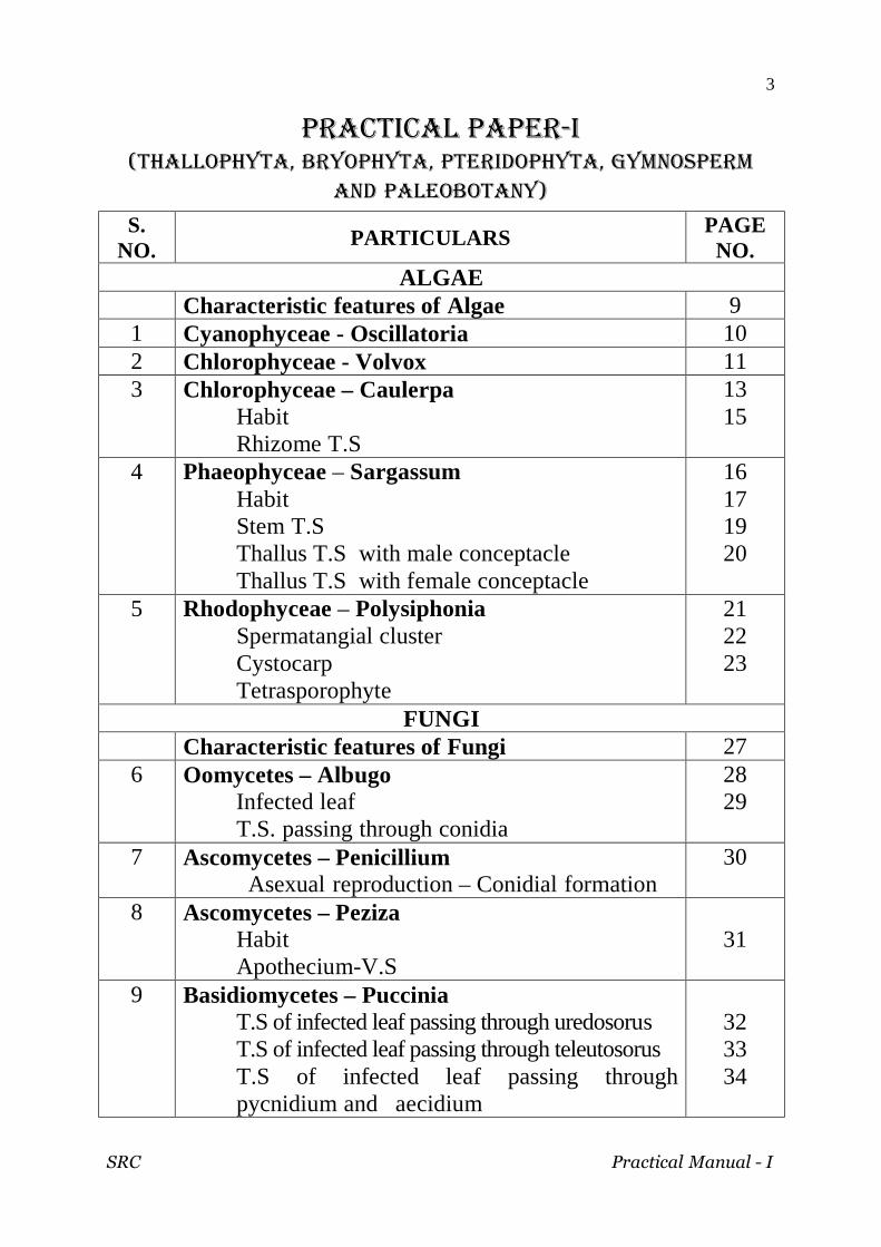
PRACTICAL PAPER-I (THALLOPHYTA, BRYOPHYTA, PTERIDOPHYTA, GYMNOSPERM
AND PALEOBOTANY)
S. NO. PARTICULARS
PAGE NO.
ALGAE Characteristic features of Algae 9 1 Cyanophyceae - Oscillatoria 10 2 Chlorophyceae - Volvox 11 3 Chlorophyceae – Caulerpa
Habit Rhizome T.S
13 15
4 Phaeophyceae – Sargassum Habit Stem T.S Thallus T.S with male conceptacle Thallus T.S with female conceptacle
16 17 19 20
5 Rhodophyceae – Polysiphonia Spermatangial cluster Cystocarp Tetrasporophyte
21 22 23
FUNGI Characteristic features of Fungi 27 6 Oomycetes – Albugo
Infected leaf T.S. passing through conidia
28 29
7 Ascomycetes – Penicillium Asexual reproduction – Conidial formation
30
8 Ascomycetes – Peziza Habit Apothecium-V.S
31
9 Basidiomycetes – Puccinia T.S of infected leaf passing through uredosorus T.S of infected leaf passing through teleutosorus T.S of infected leaf passing through pycnidium and aecidium
32 33 34

SRC Practical Manual - I
4
10 Deuteromycetes – Fusarium Macro and microconidia
36
LICHENS Characteristic features of Lichens 39
11 Habit Foliose lichen – Parmelia Fruticose lichen – Usnea Thallus T.S Thallus with Soredia
40
41 42
BRYOPHYTA Characteristic features of Bryophyta 47
12 Hepaticopsida- Marchantia Habit - Thallus Dorsal and Ventral view T.S of Thallus T.S.of thallus passing through gemma cup V.S. of Antheridiophore V.S. of Archegoniophore V.S. of Sporophyte
48 49 50 51 52 53
13 Bryopsida – Pogonatum Habit- Gametophyte with sporophyte Antheridial head Archegonial head V.S. of Capsule
54 55 56 57
PTERIDOPHYTA Characteristic features of Pteridophyta 61
14 Psilopsida – Psilotum Habit Stem.T.S Synangium T.S
62 63 64
15 Lycopsida – Lycopodium Habit- L.cernuum L.phlegmaria T.S. of stem- L.cernuum L.S of Cone
65 66 67 68

SRC Practical Manual - I
5
16 Lycopsida – Selaginella Habit T.S. of stem L.S. of cone
69 70 71
17 Sphenopsida – Equisetum Habit T.S. of Stem L.S. of Cone
72 73 75
18 Leptosporangiopsida – Adiantum Habit T.S of Petiole T.S. of Sporophyll passing through sorus
77 78 79
GYMNOSPERMS Characteristic features of Gymnosperms 83
19 Cycadopsida – Cycas T.S. of Normal root T.S. of Coralloid root T.S. of Rachis T.S. of Leaflet Male cone – Entire and L.S. Microsporophyll – Dorsal & ventral view T.S. of microsporophyll Megasporophyll L.S. of Ovule
84 85 86 87 89 90
91 92
20 Gnetopsida – Gnetum Habit T.S of stem Male cone & Female cone L.S. of Ovule
93 95 96 98
PALEOBOTANY 21 Psilopsida – Rhynia
T.S. of stem
103 22 Lycopsida – Lepidodendron
T.S. of stem Lepidocarpon
104 105
23 Spenopsida – Calamites T.S. of Stem
106
SRC Practical Manual - I
6
24 Work sheets 107-116 25 Spotters – Caulerpa racemosa, C. scalpelliformis,
Sargassum, Peziza, Foliose and fruticose lichen, Marchantia gemma cup, Pogonatum gametophyte with sporophyte, Psilotum, Lycopodium, Selaginella, Equisetum, Adiantum, Cycas- microsporphyll and megasporophyll, Gnetum- stem, Male and female cone, Rhynia, Lepidocarpon and Calamites.
118-129

SRC Practical Manual - I
7
ALGAE

SRC Practical Manual - I
8

SRC Practical Manual - I
9
CHARACTERISTIC FEATURES OF ALGAE
1. The Algae are chlorophyll bearing-autotrophic.
2. Cells contain membrane bound organelles.
3. Most algae are unicellular organisms, and there are a few
multicellular groups.
4. Excepting a few all the algae are aquatic.
5. Most algae are free-floating and drift with water currents.
6. The color of the algal thallus which varies in different classes of
algae is due to the presence of various pigments.
7. Algae reproduce asexually by cell division, or sexually producing
spores.
8. The food materials which accumulate as food reserves are in the
form of Polysaccharides, however, vary from group to group.
SRC Practical Manual - I
10
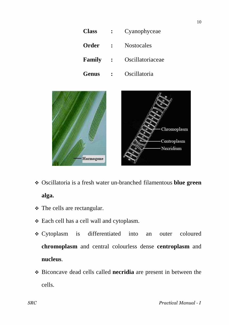
Class : Cyanophyceae
Order : Nostocales
Family : Oscillatoriaceae
Genus : Oscillatoria
Oscillatoria is a fresh water un-branched filamentous blue green
alga.
The cells are rectangular.
Each cell has a cell wall and cytoplasm.
Cytoplasm is differentiated into an outer coloured
chromoplasm and central colourless dense centroplasm and
nucleus.
Biconcave dead cells called necridia are present in between the
cells.

SRC Practical Manual - I
11
Class : Chlorophyceae
Order : Volvocales
Family : Volvocaceae
Genus : Volvox
Volvox is a small spherical motile coenobium.
The colony is made up of hundreds of cells. It is enclosed within
a common mucilage substance.
Each cell has its own mucilage envelope.
The cells are uninucleate with a large cup shaped chloroplast,
a stigma and flagella.
The cells are interconnected by protoplasmic strand.

SRC Practical Manual - I
12
SEXUAL REPRODUCTION
B: zygote, with male and female nuclei uniting, and protective shell secreted after fertilization. C: microgamete or spermatozoon. Sp. 1, 2, and 3: developing sperm-spheres.
Sexual reproduction is oogamy.
The male sex organs named antheridia develop from
reproductive cell called gonidia in the posterior part of the
colony.
Each antheridium consists of about 64 to 128 biflagellate
sperms which are arranged in the form of a hollow sphere.
Oogonia develop from gonidia in the posterior part of the
colony.
Oogonium is round with non-motile haploid uninucleate egg or
ovum.

SRC Practical Manual - I
13
Class : Chlorophyceae
Order : Siphonales
Family : Caulerpaceae
Genus : Caulerpa
ASSIMILATORS
Caulerpa racemosa
It is differentiated into a cylindrical rhizome-like creeping
portion .
The rhizome bears a number of erect branches on its upperside
and are called assimilatory shoots.
They are grape-like in appearence.
From the lower side of the rhizome arise numerous branched
thread-like colourless rhizoids.

SRC Practical Manual - I
14
Caulerpa scalpelliformis
It is differentiated into a cylindrical rhizome-like creeping
portion .
The rhizome bears a number of erect branches on its upper side
and are called as assimilatory shoots.
They possess flattened bright branches that arise pinnately
and numerous thread-like branched colourless rhizoids

SRC Practical Manual - I
15
T.S. of Rhizome
The cross section of rhizome has numerous transverse and
longitudinal cylindrical strands running across the cavity in all
parts.
These skeletal rods are made up of cellulose and pectin material
and are called trabeculae.
They are arranged perpendicular to the surface.
The cell wall gradually increases in thickness by deposition of
material in successive stratum.
SRC Practical Manual - I
16
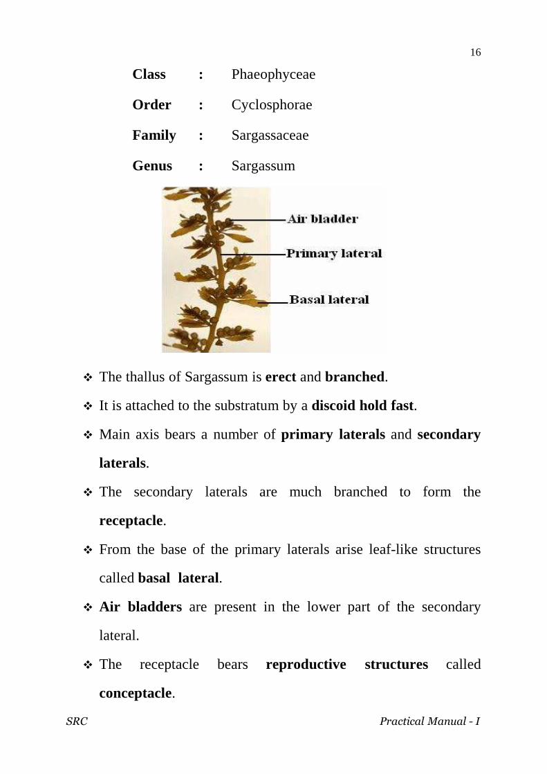
Class : Phaeophyceae
Order : Cyclosphorae
Family : Sargassaceae
Genus : Sargassum
The thallus of Sargassum is erect and branched.
It is attached to the substratum by a discoid hold fast.
Main axis bears a number of primary laterals and secondary
laterals.
The secondary laterals are much branched to form the
receptacle.
From the base of the primary laterals arise leaf-like structures
called basal lateral.
Air bladders are present in the lower part of the secondary
lateral.
The receptacle bears reproductive structures called
conceptacle.

SRC Practical Manual - I
17
Sargassum stem T.S.
The section of stem is almost circular in outline.
It is differentiated into three regions: (i) Meristoderm, (ii) Cortex
and (iii) Medulla .
Meristoderm is the outermost single layered.
It consists of many compactly arranged cells.
It is covered by mucilage and rich in chromatophores.
The cells remain meristematic.
The cortex constitutes the assimilatory region, forms most of
the axis.
The cells are narrow and elongated with intercellular spaces.
This acts as region of conduction, transporting water and
essential nutrient from one part of the thallus to the another.

SRC Practical Manual - I
18
Leaf T.S.
The cross section of leaf shows an outer epidermis, an inner
medulla and middle cortex.
The epidermis is single-layered and consists of closely arranged
thin-walled cells.
The epidermal cells contain chromatophores.
The epidermis is followed by cortex.
The cortex is made up of thin-walled polygonal cells. It is
storage in function.
Many flask shaped cavities lie embedded in the cortex. These
cavities are called cryptoblast.
The medulla occurs at the middle of the cortex. It consists of
thick-walled cells. It serves as conducting tissue.

SRC Practical Manual - I
19
Male conceptacle
Conceptacle is a depression or cavities embedded in the
cortex composed of small flat cells.
Numerous flask-shaped cavities arise from the wall of the
conceptacle.
It opens outside by small pore called ostiole.
Larger number of antheridia arise from the lower branches of
paraphysis.
The wall of the anthridia is two layered and contains 64
antherozoids.

SRC Practical Manual - I
20
Female conceptacle
Female conceptacle possess the female reproductive organs
called oogonia.
Oogonium is oval or sub-spherical and its wall is three layered.
Each oogonium is uninucleate.

SRC Practical Manual - I
21
Class : Rhodophyceae
Order : Ceramiales
Family : Rhodomelaceae
Genus : Polysiphonia
Filament with spermatangial cluster
Filaments of polysiphonia are multicellular, branched,
polysiphonous with a central siphon surrounded by peripherial
siphons.
Fertile trichoblasts produces antheridial cluster near the apex.
Spermatia are directly liberated through the surface of
spermatangium.
Spermatia are small oval or spherical, uninucleate and being
non-motile is trapped by water currents.

SRC Practical Manual - I
22
Filament with cystocarp
The pericarp forms a cystocarp after fertilization and this plant
is called as carposporophyte.
It is oval, urn-shaped structure attached lateral to the filament.
It opens to the exterior by ostiole.
The wall of the cystocarp is single layered.
At the base of the cystocarp is placenta from which arise
several gonimoblast initials.
The terminal cell of this filament acts as carposporangium
producing carpospore.
Carpospores are oval, uninucleate and diploid , develop into
tetrasporophyte.

SRC Practical Manual - I
23
Filament with tetrasporophyte
The plant bears the tetrasporangium which is diploid.
They are usually in longitudinal sori produced mostly by
pericental cell.
The tetrasporangia possess four tetrahedrally arranged
uninucleate and haploid tetraspores.
They develop into gametophyte.

SRC Practical Manual - I
24
This page intentionally left blank
SRC Practical Manual - I
25
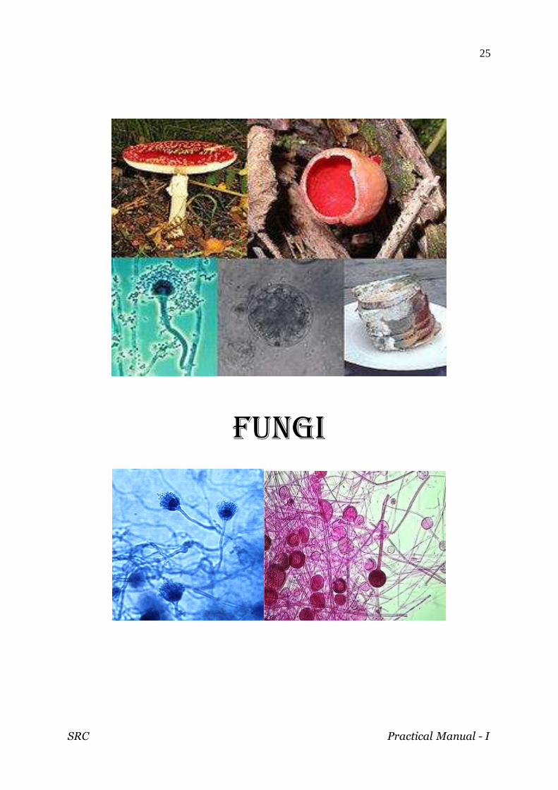
FUNGI

SRC Practical Manual - I
26

SRC Practical Manual - I
27
CHARACTERISTIC FEATURES OF FUNGI
The fungi lack chlorophyll.
They are heterotropic and live as parasites (or) saprophytes.
Thallus is mycelium, an interwoven mass of fine, tubular
structure called hyphae.
The cell wall is made up of fungal cellulose called chitin.
Reserve food material is glycogen.
Fungi grow in a wide variety of habitats.
There is gradual and progressive simplification and ultimate
elimination of the sexual apparatus from the lower to higher
forms.
Fungi reproduce by asexual and sexual methods.
They are involved in two types of symbiotic relationships. In the
mycorrhizae form, fungi derive energy from the roots of
vascular plants. The other form involves the production of
lichens with algae and cyanobacteria.

SRC Practical Manual - I
28
White Pustule
Class : Oomycetes
Order : Peronosporales
Family : Albuginaceae
Genus : Albugo Infected leaf
Albugo causes white rust or blister rust disease on the leaves
of Amaranthaceae.
The disease is in the form of shiny white smooth raised
irregular blister .
Control Measures
Rotation of crops
Removal and destruction of infected plants.
Spraying fungicides
SRC Practical Manual - I
29
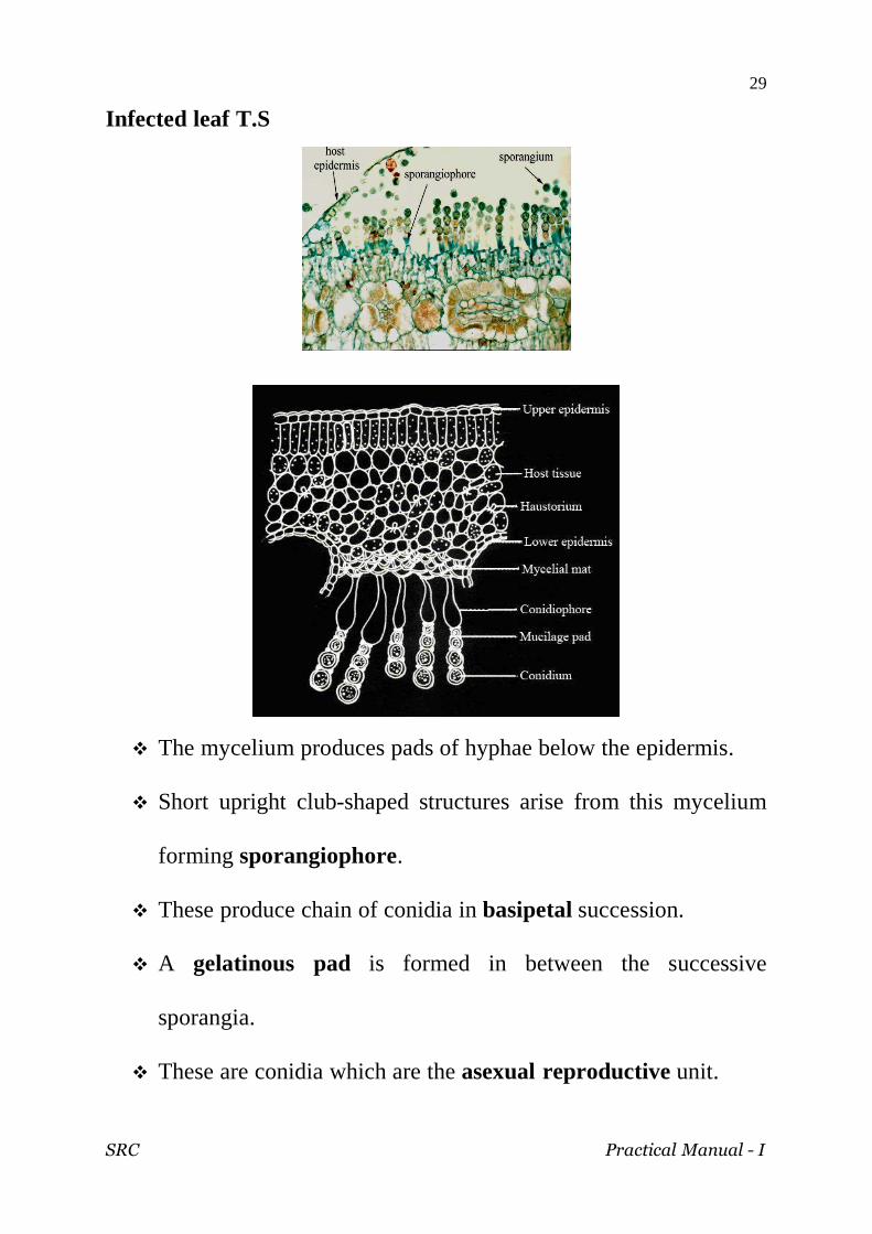
Infected leaf T.S
The mycelium produces pads of hyphae below the epidermis.
Short upright club-shaped structures arise from this mycelium
forming sporangiophore.
These produce chain of conidia in basipetal succession.
A gelatinous pad is formed in between the successive
sporangia.
These are conidia which are the asexual reproductive unit.

SRC Practical Manual - I
30
Class : Ascomycetes
Order : Aspergillales
Family : Aspergillaceae
Genus : Penicillium
Mycelium bearing conidiophore
Penicillium is a saprophytic fungus.
The mycelium is branched and septate.
From the mycelium arises short tubular structure called
conidiophore.
It divides into primary, secondary and tertiary branches.
The tip of the conidiophores bears bottle shaped structure called
sterigmata.
They cut off conidia in basipetalous succession.
Each conidium is a globular structure with single nucleus and
the branch which bears metulae is called rami.

SRC Practical Manual - I
31
Class : Ascomycetes Order : Pezizales Family : Pezizaceae Genus : Peziza
Apothecium - Entire A portion enlarged
It is a saprophytic coprophilous fungus. The fruit body is an apothecium and they are seen above
ground. It has the following structures:
(i) The base of apothecium is formed of mycelium; (ii) Hymenium consists of many fertile ascus and sterile paraphysis; (iii) sub-hymenium is made up of pseudoparenchymatous hyphae; (iv) in each ascus there are eight uninucleate ascospores.
SRC Practical Manual - I
32
Class : Basidomycetes
Order : Uredinales
Family : Pucciniaceae
Genus : Puccinia
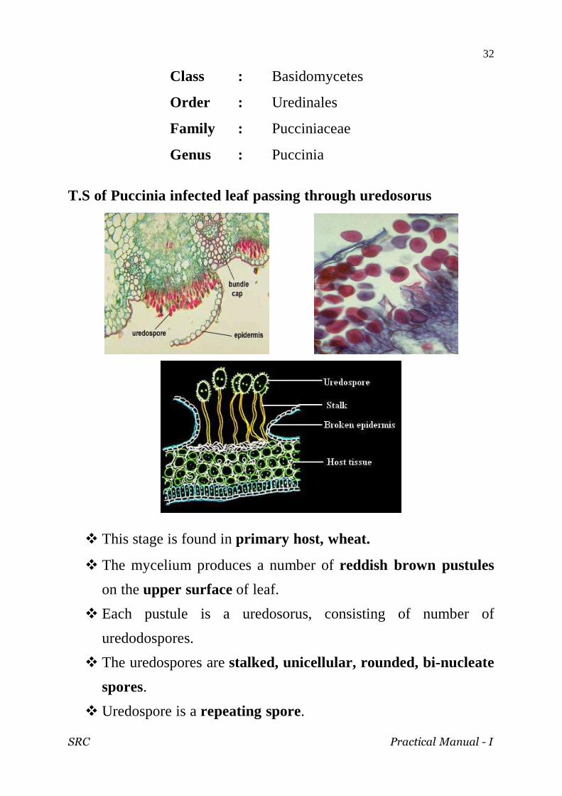
T.S of Puccinia infected leaf passing through uredosorus
This stage is found in primary host, wheat.
The mycelium produces a number of reddish brown pustules
on the upper surface of leaf.
Each pustule is a uredosorus, consisting of number of
uredodospores.
The uredospores are stalked, unicellular, rounded, bi-nucleate
spores.
Uredospore is a repeating spore.

SRC Practical Manual - I
33
T.S of Puccinia infected leaf passing through teleutosorus
This stage is found in primary host, wheat.
They are called winter spores as they are produced in the
beginning of winter.
They produce black streaks in leaf and stem.
The teleutospores are stalked, bicelled and binucleate.

SRC Practical Manual - I
34
T.S of Puccinia Infected leaf through pycnidia and aecidia
Pycnidia
This stage is found in secondary host, Berberry plant .
They produce flask-shaped structures called the pycnidium or
spermogonium.
The pycnidium opens on the surface of the leaf by a minute pore
called ostiole.
The ostiole is guarded by a tuft of sterile hairs called periphysis.
At the lower portion of the pycnidium there are uninucleate
hyphae called spermatophores (Sporophore).
The tip of the spermatophores produce pycnidiospores or
spermatia.
The pycnidiospores are of either (+) or (-) strain.

SRC Practical Manual - I
35
Aecidia
This stage is found in secondary host, Berberry plant .
The aecidial cup is surrounded by sterile hyphae called
peridium.
From the middle of the cup, the dikaryotic mycelium forms a
number of erect hyphae called sporophores.
Each sporophore produces a number of binucleate spores called
the aecidiospores in chain.
In between the aecidispores small, sterile intercalary cells are
present called disjunctor cells.
SRC Practical Manual - I
36
Class : Deutromycetes
Order : Moniliales
Family : Tuberculeriaceae
Genus : Fusarium
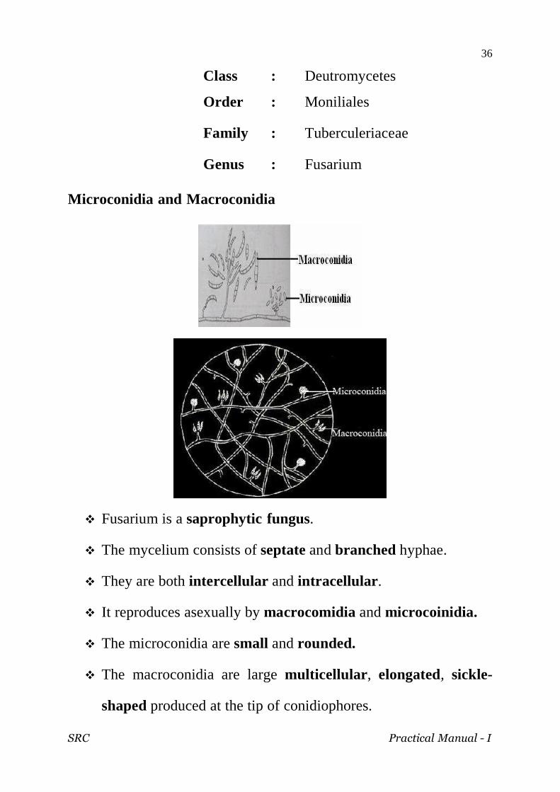
Microconidia and Macroconidia
Fusarium is a saprophytic fungus.
The mycelium consists of septate and branched hyphae.
They are both intercellular and intracellular .
It reproduces asexually by macrocomidia and microcoinidia.
The microconidia are small and rounded.
The macroconidia are large multicellular , elongated, sickle-
shaped produced at the tip of conidiophores.

SRC Practical Manual - I
37
LICHENS

SRC Practical Manual - I
38

SRC Practical Manual - I
39
Characteristic features of Lichen
Lichens are dual organisms formed by the symbiotic
association of a fungus and an algae.
Fungal partner is usually an Ascomycete (sometimes a
Basidiomycete), called as mycobiont.
Algal partner may be a green or blue-green alga, called as
phycobiont.
Fungus and alga together appear as a single plant.
Lichens vary in their growth forms and mode of attachment to
the substratum.
Based on morphological features, three types of lichens are
recognized- Crustose lichen, Foliose lichen and Fruticose
lichen.
Based on the internal structures, lichens are divided into two
types – Homoiomerous thalli and heteromerous thalli.
Reproduces by vegetative reproduction – fragmentation,
rejuvenation, isidia formation and soredia
Spore formation- pycnidia formation.
Sexual reproduction- fungal partner is responsible for
reproduction male reproductive organ is spermagonium and
female reproductive organ is carpogonium.
SRC Practical Manual - I
40
LICHEN
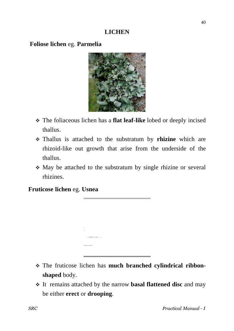
Foliose lichen eg. Parmelia
The foliaceous lichen has a flat leaf-like lobed or deeply incised
thallus.
Thallus is attached to the substratum by rhizine which are
rhizoid-like out growth that arise from the underside of the
thallus.
May be attached to the substratum by single rhizine or several
rhizines.
Fruticose lichen eg. Usnea
The fruticose lichen has much branched cylindrical ribbon-
shaped body.
It remains attached by the narrow basal flattened disc and may
be either erect or drooping.
SRC Practical Manual - I
41
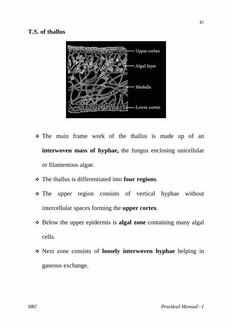
T.S. of thallus
The main frame work of the thallus is made up of an
interwoven mass of hyphae, the fungus enclosing unicellular
or filamentous algae.
The thallus is differentiated into four regions.
The upper region consists of vertical hyphae without
intercellular spaces forming the upper cortex.
Below the upper epidermis is algal zone containing many algal
cells.
Next zone consists of loosely interwoven hyphae helping in
gaseous exchange.

SRC Practical Manual - I
42
Soredium
Soredia formation is the common method of vegetative
propagation.
They develop as bud-like outgrowth either over the surface of
the thallus or in local patches called soredia.
They develop in the gonidial layer of the upper cortex.
When detached from the thallus, they are blown away by wind
and germinate falling on suitable substratum.
SRC Practical Manual - I
43
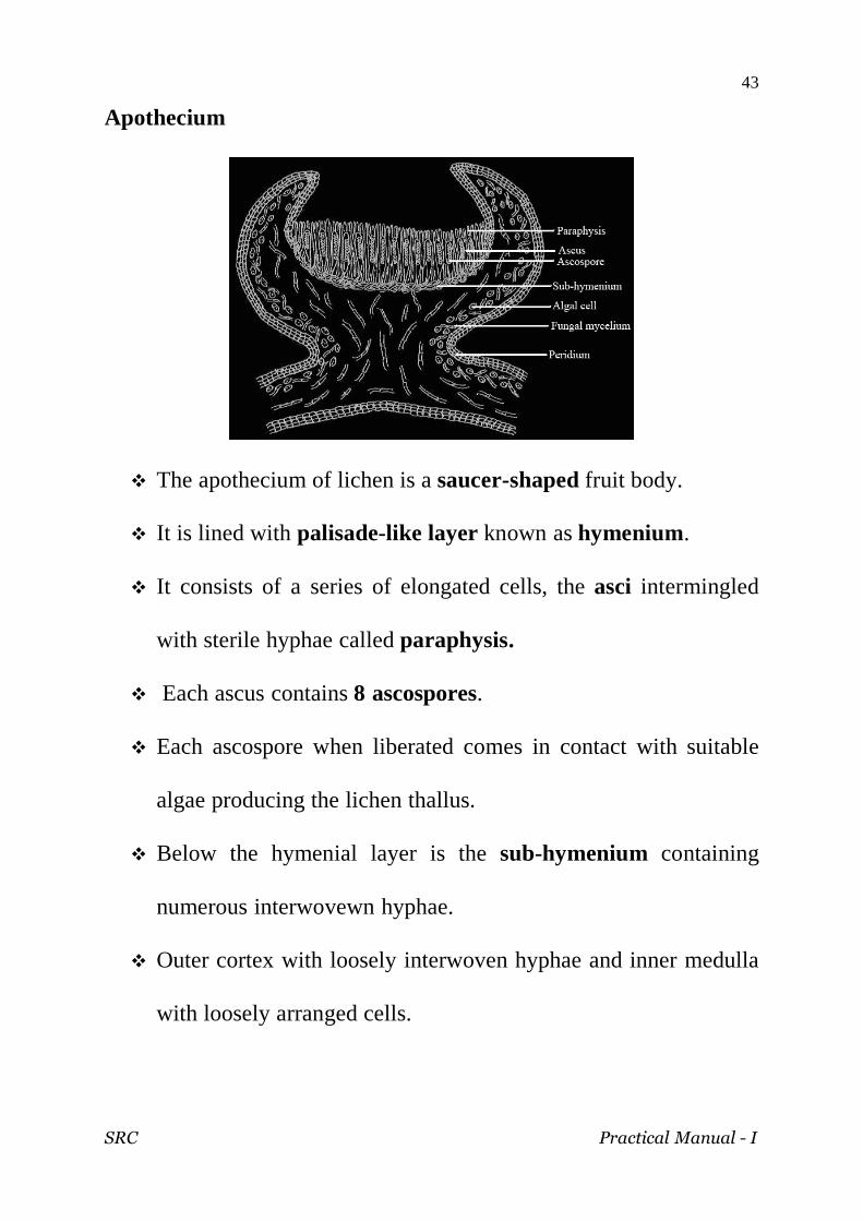
Apothecium
The apothecium of lichen is a saucer-shaped fruit body.
It is lined with palisade-like layer known as hymenium.
It consists of a series of elongated cells, the asci intermingled
with sterile hyphae called paraphysis.
Each ascus contains 8 ascospores.
Each ascospore when liberated comes in contact with suitable
algae producing the lichen thallus.
Below the hymenial layer is the sub-hymenium containing
numerous interwovewn hyphae.
Outer cortex with loosely interwoven hyphae and inner medulla
with loosely arranged cells.

SRC Practical Manual - I
44
This page intentionally left blank
SRC Practical Manual - I
45
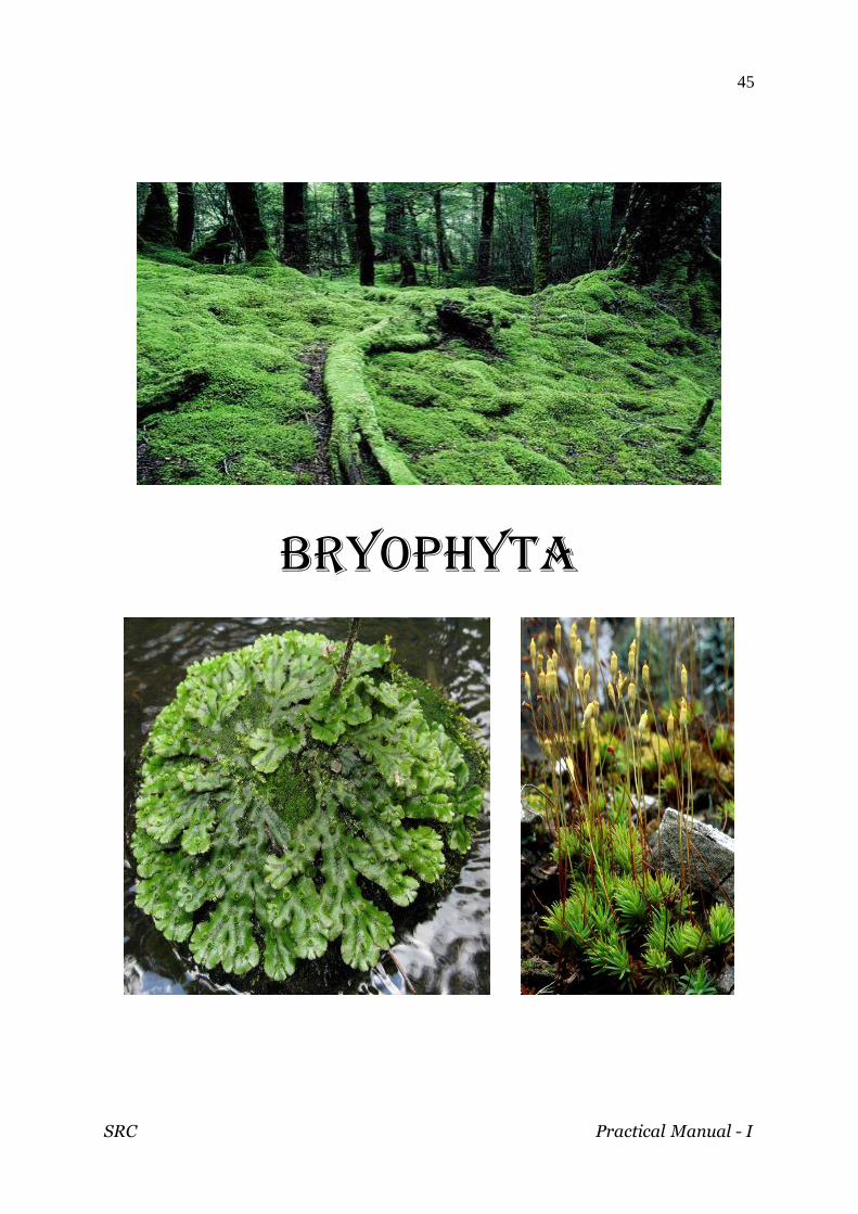
BRYOPHYTA

SRC Practical Manual - I
46
SRC Practical Manual - I
47
Characteristic features of Bryophytes
The Plant body is a gametophyte. They grow in areas which arc in between the aquatic and terrestrial habitats i.e. amphibious zone and hence known as amphibious plants.
They have thalloid or leafy multi cellular green plant body. The smallest form is microscopic (e.g. Zoopsis). The largest
genus recorded so far is an Australian Dawsonia which is about 70 cms in length.
The dominant plant body is gametophyte (n) which is independent.
The plant body lacks true roots, stem or leaves. Rhizoids - (root like structure) serve the function of roots. The plants are green and possess chloroplasts. They show autotrophic mode of nutrition. Vascular tissues are completely absent. Sexual reproduction is oogamous. Sex organs are multi cellular and jacketed. Male reproductive organ is known as antheridium . It is a club
shaped structure being borne by a narrow stalk. It produces biflagellate and motile male gametes or antherozoids.
The female sex organ is known as archegonium. It is a flask shaped structure having a swollen base and a narrow neck.
Water is essential for fertilization . The diploid zygote undergoes repeated divisions to form a multi
cellular sporophyte. Sporophyte is dependent on the gametophyte for nutrition. Sporophyte generally consists of foot, seta and capsule. It
produces haploid spores (homospores). Spores on germination give rise to gametophyte plant. Gametophyte and sporophyte differ in form which alternate with
each other, thus heterologous alternation of generation is seen in Bryophytes.
SRC Practical Manual - I
48
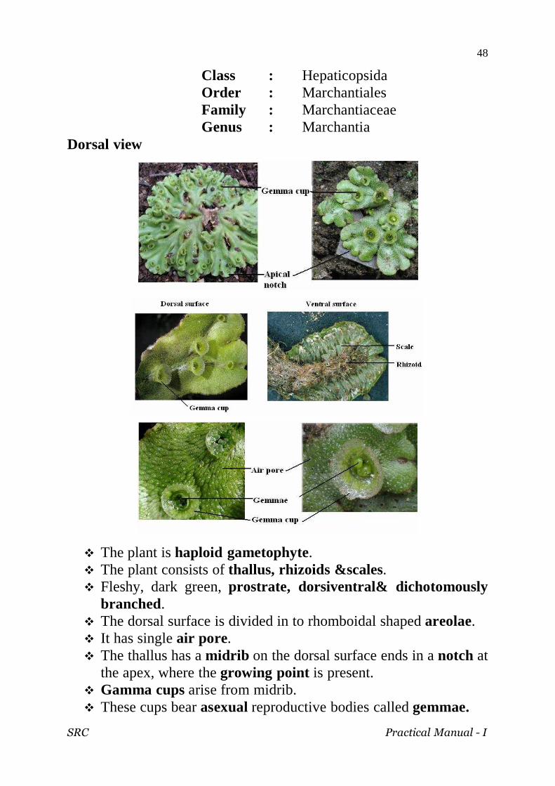
Class : Hepaticopsida Order : Marchantiales Family : Marchantiaceae Genus : Marchantia
Dorsal view
The plant is haploid gametophyte. The plant consists of thallus, rhizoids &scales. Fleshy, dark green, prostrate, dorsiventral& dichotomously
branched. The dorsal surface is divided in to rhomboidal shaped areolae. It has single air pore. The thallus has a midrib on the dorsal surface ends in a notch at
the apex, where the growing point is present. Gamma cups arise from midrib. These cups bear asexual reproductive bodies called gemmae.
SRC Practical Manual - I
49
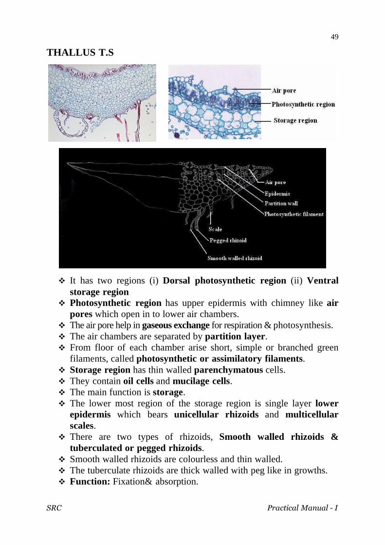
THALLUS T.S
It has two regions (i) Dorsal photosynthetic region (ii) Ventral storage region
Photosynthetic region has upper epidermis with chimney like air pores which open in to lower air chambers.
The air pore help in gaseous exchange for respiration & photosynthesis. The air chambers are separated by partition layer . From floor of each chamber arise short, simple or branched green
filaments, called photosynthetic or assimilatory filaments. Storage region has thin walled parenchymatous cells. They contain oil cells and mucilage cells. The main function is storage. The lower most region of the storage region is single layer lower
epidermis which bears unicellular rhizoids and multicellular scales.
There are two types of rhizoids, Smooth walled rhizoids & tuberculated or pegged rhizoids.
Smooth walled rhizoids are colourless and thin walled. The tuberculate rhizoids are thick walled with peg like in growths. Function: Fixation& absorption.

SRC Practical Manual - I
50
Gemma cup
Gemma Cup – A Closer View Section of a Gemma Cup
Gemmae are asexual reproductive bodies produced in cup shaped structures called gemma cups on dorsal surface of the thallus.
The margin of the gemma cup is toothed and membranous with many gemmae.
Gemmae are attached to the bottom of the cup by small, single celled stalk.
Many multicellular, glandular hairs are intermingled with gemmae.
The mature gemma is green, multicellular & lens shaped structures.
It has two deep lateral notches with growing points. The cells of gemma are chlorenchymatous with few oil cells
and rhizoidal cells. The mature gemma separate from mother plant and develop in
to new plant.

SRC Practical Manual - I
51
Thallus with antheridiophore
Each antheridiophore has stalk and receptacle. The stalk is a cylindrical structure with a layer of air chambers
on posterior side and two vertical grooves on the anterior side. The disc of the receptacle is made up of assimilatory region
and storage region. The assimilatory chambers alternate with the flask-shaped
cavities called antheridial chambers. Each antheridial chamber has single antheridium and it opens
outside through ostiole. The mature antheridium consists of a short stalk and a rounded
body called the capsule. The antheridium is surrounded by a single layered jacket or
antheridial wall. The jacket encloses androcyte mother cells.

SRC Practical Manual - I
52
Thallus with archegoniophore
Archegoniophore has stalk and disc shaped receptacle. The receptacle bears 8 lobes. Each lobe has 12-14 archegonia arranged in acropetal order. The mature archegonium is flask shaped. It has basal stalk, venter &neck. The venter has egg and a venter canal cell. The neck has 4-8 neck canal cells and 4 lid cells. In mature archegonium the neck canals and venter canal cell
disorganize forming mucilaginous fluid. This mucilage absorbs water and help in opening of lid cells.

SRC Practical Manual - I
53
Sporophyte
The sporophyte of Marchantia is produced in the female gametophyte.
The sporangium has three parts: (i) Foot, (ii) Seta and (iii) Capsule. Foot is the basal portion, anchorage and absorptive organ of
the sporophyte. Seta is the middle portion and it connects foot with the capsule. The fertile region is the capsule. The outer layer of capsule is
called capsule wall, encloses fertile sporogenous tissue. Sporogenous cells produce spore mother cells & elater mother
cells. Elater mother cells produce elaters. Elaters are hygroscopic diploid, sterile and elongated with
pointed ends. It has spiral bands of thickening on the inner surface. It helps in dehiscence of capsule.
Each spore mother cell undergoes meiotic division to form spore tetrads.
The spores are haploid & develop in to gametophyte.

SRC Practical Manual - I
54
Class : Bryopsida
Order : Polytrichales
Family : Polytrichales
Genus : Pogonatum
Gametophyte with sporophyte
The sporophyte is differentiated into underground rhizome,
erect leaf and stem.
Rhizoids are borne on the rhizome and long thick walled with
oblique septa.
The leaves are in 3 vertical rows, brown coloured.
The leaves on aerial shoots are large and spirally arranged.
Each leaf has a broad colourless membraneous one celled
sheathing leaf base.
Midrib is seen in the leaves.
During diploid phase sporophyte is developed in the
gametophyte.
SRC Practical Manual - I
55
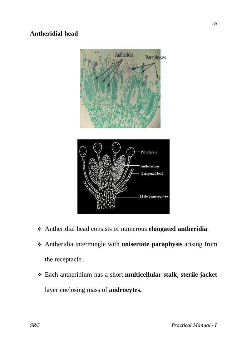
Antheridial head
Antheridial head consists of numerous elongated antheridia.
Antheridia intermingle with uniseriate paraphysis arising from
the receptacle.
Each antheridium has a short multicellular stalk , sterile jacket
layer enclosing mass of androcytes.
SRC Practical Manual - I
56
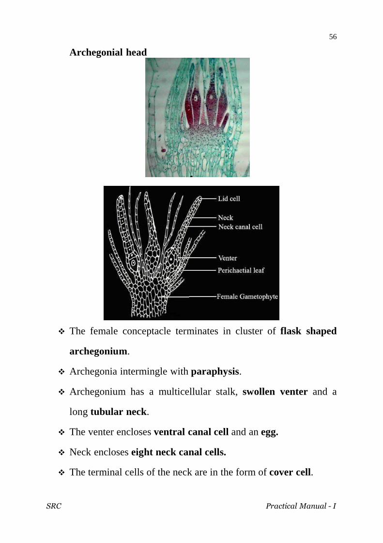
Archegonial head
The female conceptacle terminates in cluster of flask shaped
archegonium.
Archegonia intermingle with paraphysis.
Archegonium has a multicellular stalk, swollen venter and a
long tubular neck.
The venter encloses ventral canal cell and an egg.
Neck encloses eight neck canal cells.
The terminal cells of the neck are in the form of cover cell.

SRC Practical Manual - I
57
Capsule
Capsule is differentiated into apophysis, operculum and theca.
Apophysis is continuous with seta.
The epidermal cells are interrupted by stomata.
Theca is the buldged space producing part of the capsule.
Theca has central sterile tissue called columella surrounded by
inner air space, middle spore sac and outer air space.
The air spaces are traversed by trabeculae.
This spore sac contains numerous minute spores. The outer
wall of sac is singled layered and inner wall is multilayered.
Outer air space is covered by cell wall.

SRC Practical Manual - I
58
This page intentionally left blank

SRC Practical Manual - I
59
PTERIDOPHYTA

SRC Practical Manual - I
60
SRC Practical Manual - I
61
Characteristic features of Pteridophytes
The plant body is diploid sporophyte.
The sporophyte has distinct roots, stem and leaves. The leaves
are homophyllous or heterophyllous.
The vascular tissues are well-developed.
Asexual spores are haploid and produced inside the sporangia.
They may be identical (homosporous) or two different types
(heterosporous).
The sporangia are borne on sporophylls. The sporophylls are
arranged in definite cones.
The sporangial spores germinate into haploid gametophytes.
The gametophytes produce sex organs. They may be
monoecious or diecious.
The male sex organ is called antheridium . It is a globular
structure that produces motile flagellate male gametes called
spermatozoids.
The female sex organ is a flask-shaped structure called
archegonium. It produces an egg.
Fertilization takes place in the presence of water.
The diploid zygote gives rise to a young sporophyte called
embryo. The embryo grows into sporophytic plant.

SRC Practical Manual - I
62
Class : Psilopsida
Order : Psilotales
Family : Psilotaceae
Genus : Psilotum
Habit
The plant body is a sporophyte.
The aerial branch is green and bear small scale like leaves
which are devoid of any vascular system.
The scale leaves are spirally arranged.
The aerial branch is dichotomously branched and bear small
scale like and biforked outgrowth which bear the trilobed
synangium.

SRC Practical Manual - I
63
Internal structure of aerial axis
The T.S. of stem shows an outer epidermis, a cortex, an endodermis
and an inner stele.
Epidermis consists of thin-walled cells with many small pores
called stomata.
Epidermis is covered over by a layer of cuticle.
Cortex - three layered outer cortex, middle cortex and inner
cortex.
The outer cortex is made-up of thin-walled, chlorenchymatous
cells.
The middle cortex consists of elongated, densely arranged, thick –
walled cells.
The inner cortex is multi-layered and it is composed of thin-walled
cells.
Endodermis is single-layered and is composed of elongated thin-
walled cells; cells have casparian thickening.
The stele is actinostelic protostele.
Pericycle is always single-layered.
Xylem is stellate (star-shaped) and exarch.
Phloem occurs between the lobes and the surrounding regions.

SRC Practical Manual - I
64
Synangium
The synangia of psilotum are trilobed and associated with
trilobed appendage.
A section through the trilobed synangium reveals that it is
trilocular .
The synangial wall is made up of 3-4 layered wall.
The sporogenous tissue in the sporangial chamber forms the
spore mother cell.
The spore mother cell undergoes meiosis and form tetrad of
haploid spores.
Stomata
Sporangial wall
Vascular trace
Spores

SRC Practical Manual - I
65
Division : Lycophyta Class : Filicopsida Order : Lycopodiales Family : Lycopodiaceae Genus : Lycopodium
Habit Lycopodium cernnum
The plant body is terrestrial consisting of a creeping stem,
which give rise to erect aerial branches.
The branching is dichotomous but looks like monopodial.
All the foliage leaves are alike and are arranged in whorls on
the stem.
The strobili are comparatively small.
They are sessile and borne on tips of aerial branches.
Each sporophyll is provided with abaxial flange.

SRC Practical Manual - I
66
L. phlegmaria
L. phlegmaria is tropical epiphytic species.
It grows hanging from the tree trunks.
Stem covered with sharp and pointed leaves.
The stem is dichotomously branched. The two branches of a
dichotomy are almost equal.
Strobili are produced at the tips of branches.

SRC Practical Manual - I
67
Stem T.S.
The stem has got a lobed appearance.
Epidermis is cuticularised and is followed by three distinct
zones of cortex.
The outer and inner portion of cortex are parenchymatous with
the middle sclerenchymatous cortex - heterogenous.
Endodermis and pericycle are single layered.
The stele is a mixed protostele
Epidermis
Outer cortex
Middle cortex
Inner cortex Mixed protostele
SRC Practical Manual - I
68
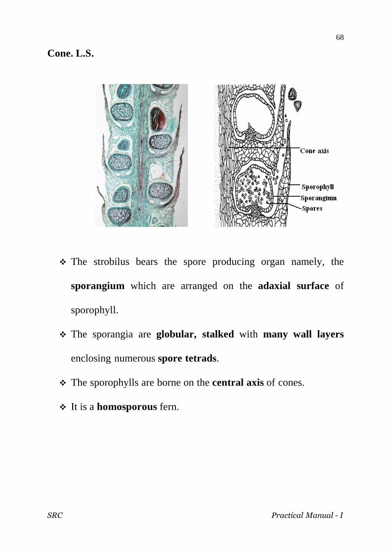
Cone. L.S.
The strobilus bears the spore producing organ namely, the
sporangium which are arranged on the adaxial surface of
sporophyll.
The sporangia are globular, stalked with many wall layers
enclosing numerous spore tetrads.
The sporophylls are borne on the central axis of cones.
It is a homosporous fern.

SRC Practical Manual - I
69
Division : Lycophyta
Class : Ligulopsida
Order : Selaginellales
Family : Selaginellaceae
Genus : Selaginella
Sporophyte
The plant body of Selaginella is differentiated into stem, root
and leaf-like parts.
The leaf is adventitious.
The stem is prostrate with erect branches bearing dimorphic
leaf with larger leaves arranged at the sides of the stem and
smaller leaves in the centre of the stem.
The leaves are sessile and ligulate.
The stem branches are cylindrical .
Leafless structures grow downwards and are known as
rhizophore, which give rise to clusters of roots at its tip.
The rhizophore is positively geotrophic in nature.
SRC Practical Manual - I
70
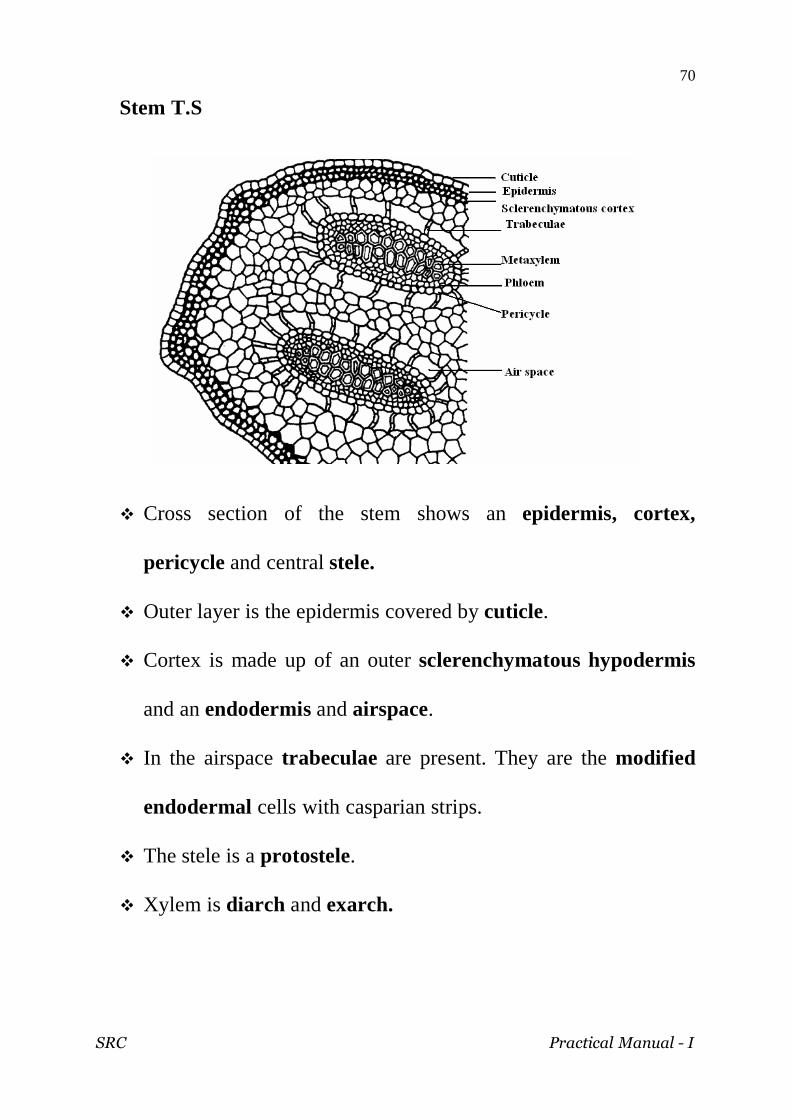
Stem T.S
Cross section of the stem shows an epidermis, cortex,
pericycle and central stele.
Outer layer is the epidermis covered by cuticle.
Cortex is made up of an outer sclerenchymatous hypodermis
and an endodermis and airspace.
In the airspace trabeculae are present. They are the modified
endodermal cells with casparian strips.
The stele is a protostele.
Xylem is diarch and exarch.

SRC Practical Manual - I
71
Cone
A PORTION ENLARGED
The strobilus of Selaginella bears many spores.
Selaginella is heterosporous producing larger megaspores and
smaller microspores in the respective sporangia namely
megasporangia and microsporangia.
The sporophyll bears the sporangia in its axis and ligulate.
The megasporangia are stalked and four lobed bearing four
megaspores.
Each megaspore has got a triradiate ridge.
The microsporangia are smaller and contains innumerable
spores.

SRC Practical Manual - I
72
Division : Sphenophyta
Class : Calamopsida
Order : Equisetales
Family : Equisetales
Genus : Equisetum
Habit
The plant body has an underground stem known as the rhizome with nodes and internodes.
Roots arise from the lower side of the rhizome. Rhizome also gives out on its upper side a number of aerial
branches which are long, slender and differentiated into nodes and internodes.
The internodes are with ridges and furrows . Leaves are very much reduced to scaly structures which fuse
with one another to form a sheath around the node. The tips of some of the branches bear cone. Each sporangiophore has stalk and a peltate disc.
SRC Practical Manual - I
73
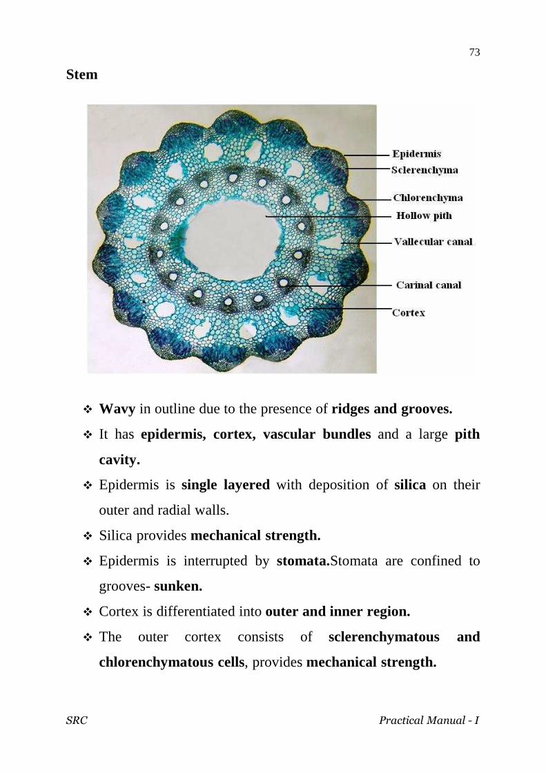
Stem
Wavy in outline due to the presence of ridges and grooves.
It has epidermis, cortex, vascular bundles and a large pith
cavity.
Epidermis is single layered with deposition of silica on their
outer and radial walls.
Silica provides mechanical strength.
Epidermis is interrupted by stomata.Stomata are confined to
grooves- sunken.
Cortex is differentiated into outer and inner region.
The outer cortex consists of sclerenchymatous and
chlorenchymatous cells, provides mechanical strength.

SRC Practical Manual - I
74
Sclerenchyma lies below the epidermis. It is followed by a band
of chlorenchyma- photosynthetic in function.
The inner cortex is composed of thin walled parenchymatous
cells. It has large schizolysigenous canals known as vallecular
canal below the furrow- aerating system.
Cortex is delimited from the stele by an endodermis.
The endodermis is followed by a single layer of
parenchymatous pericycle.
The vascular bundle is a siphonostele. Vascular bundles are
arranged in a ring around the large pith opposite to ridges
alternating the vallecular canal.
Vascular bundles are conjoint, collateral and endarch.
Xylem of a bundle is in the form of two lateral and a median
group of tracheids.
In the young vascular bundle the protoxylem is represented by
tracheids with annular or spiral thickening.
In the mature bundle the protoxylem elements disintegrate to
form a protoxylem lacuna called carinal canal.
The metaxylem tracheids have scalariform, reticulate or
pitted thickenings.
The phloem lies outside the xylem.
The carinal canals are filled with water and help in conduction
of water.
The central part of the internode of the aerial shoot has large
pith cavity.

SRC Practical Manual - I
75
Cone
Equisetum is homosporus.
Strobili are borne terminally on the vegetative shoots.
Only the peltate discs are seen in the surface view of the
strobilus.
Cone L.S

SRC Practical Manual - I
76
Strobilus has a central strobilus axis and a large number of sporangiophores.
The sporangiophore is a stalked structure with a hexagonal peltate disc at its distal end.
On the underside of the sporangiophore disc 5 – 10 sac like sporangia are borne near its periphery in a ring.
In some species a whorl of scale-like outgrowths called annulus is present at the base of the strobilus.
The development of sporangium is eusporangiate. The mature sporangia are sac-like structure attached to the
underside of the peltate disc of the sporangiophore. The wall of mature sporangium is only two layered. All spores
are alike. (homosporous) As the sporangia mature, the strobilus axis elongates,
consequently, the compactly arranged sporangiophores separate from each other and the sporangia are exposed.
As the sporangium dries the helicoid thickening bands present in the outer wall layer shrink and the sporangium ruptures.
Spores are spherical, uninucleate and green (contain numerous chloroplasts)
Spore wall is differentiated into 4 layers. The outermost perispore / epispore. The second middle layer. The third expspore. The innermost endospore.
The epispore splits into 4 strips which are separated from one another but attached to a common point on the spore. These bands are wrapped around the spherical spore but as the spore dries these bands are starched. These bands are called as elaters. They have expanded spoon-like tips.
The elaters are hygroscopic and help in the dehiscence of the sporangium.
SRC Practical Manual - I
77
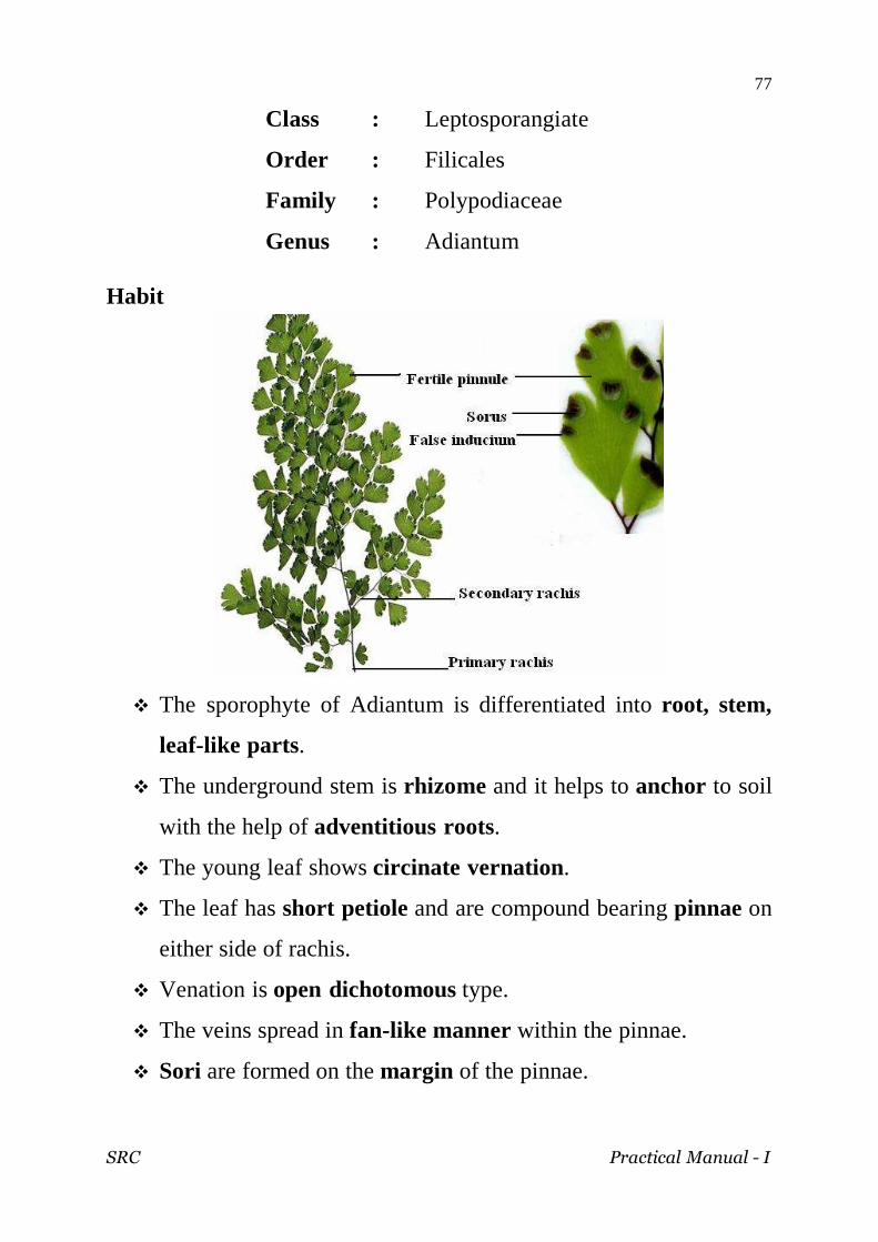
Class : Leptosporangiate
Order : Filicales
Family : Polypodiaceae
Genus : Adiantum Habit
The sporophyte of Adiantum is differentiated into root, stem,
leaf-like parts.
The underground stem is rhizome and it helps to anchor to soil
with the help of adventitious roots.
The young leaf shows circinate vernation.
The leaf has short petiole and are compound bearing pinnae on
either side of rachis.
Venation is open dichotomous type.
The veins spread in fan-like manner within the pinnae.
Sori are formed on the margin of the pinnae.

SRC Practical Manual - I
78
Petiole T.S
Outermost layer of petiole is single layered cuticularised
epidermis.
Sclerenchymatous hypodermis.
Inner cortex is parenchymatous.
Presence of well developed endodermis and pericycle.
Stele is protostele.
Two vascular bundles are present.
Xylem is diarch and exarch.

SRC Practical Manual - I
79
Sorus V.S.
The sporangia arise on the ventral surface of the fertile
pinnule and they are protected by the margins of the pinnule
called false indusium.
The sporangia develop from the receptacle or placenta of
fertile pinnule .
Each sporangium has got a multicellular stalk and spore
bearing capsule with single layered wall.
The wall of capsule has annulus with characteristic thickening
consisting the major part of wall layer and thin walled stomium
through which dehiscence takes place.

SRC Practical Manual - I
80
This page intentionally left blank

SRC Practical Manual - I
81
GYMNOSPERM

SRC Practical Manual - I
82

SRC Practical Manual - I
83
Characteristic features of Gymnosperms
The adult plants are tall, woody, evergreen, perennial trees or shrubs. They show xerophytic characters. The plant is a sporophyte.
The stem is branched but unbranched in Cycas. The vascular bundles are arranged in a ring. They are conjoint, collateral, open and endarch.
The xylem is composed of xylem parenchyma and tracheids with bordered pits. Xylem vessels are absent, except in Gnetales.
The phloem consists of sieve tubes and phloem parenchyma, but companion cells are absent.
The leaves may be dimorphic, the foliage leaves and scale leaves or of one kind only.
The reproductive parts are arranged in the form of cones or strobili. The cones are unisexual.
In male cones, many microsporophylls are arranged on the central axis. The microsporophylls bear microsporangia with microspores or pollen grains.
The microspores are haploid, and are formed from microspore mother cells after meiosis. The microspore germinates to form the male gametophyte.
In the female cones, many megasporophylls are arranged on the central axis. The megasporphylls bear megasporangia or ovules.
The ovule is orthotropous. It consists of nucellus surrounded by one or two integuments.
The ovules are naked and are not enclose by the ovary. The ovule contains the megaspore mother cell which undergoes meiosis
to produce a linear row of four megaspores. Of the four megaspores the lowest is functional. The megaspore is haploid.
The megaspore gives rise to a female gametophyte. The female gametophyte bears archegonia the upper end. The endosperm is a pre-fertilization tissue. It is haploid in
gymnosperms. Each archegonium consists of a venter and a neck. The neck canal cells
are absent. The pollination is direct, i.e. the pollen grains come in contact with the
ovule directly. Embryo development is meroblastic, i.e. develops from a small part of
zygote.

SRC Practical Manual - I
84
Class : Cycadopsida
Order : Cycadales
Family : Cycadaceae
Genus : Cycas
Normal root
The young root has got a circular outline.
It is differentiated into outer epiblema, cortex and the stele.
Epiblemma is single layered composed of thin walled cells,
some of them with root hairs.
Cortex is broad parenchymatous and multilayered.
Stele is radial, diarch, closed with endarch xylem.
SRC Practical Manual - I
85
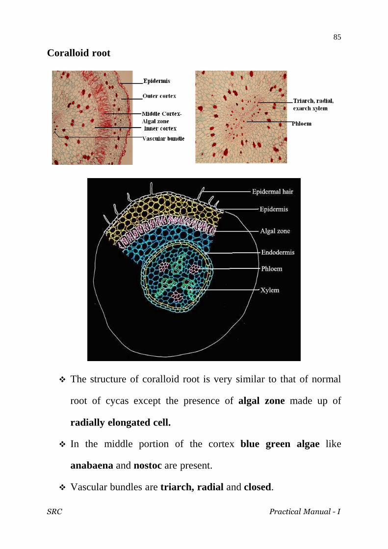
Coralloid root
The structure of coralloid root is very similar to that of normal
root of cycas except the presence of algal zone made up of
radially elongated cell.
In the middle portion of the cortex blue green algae like
anabaena and nostoc are present.
Vascular bundles are triarch, radial and closed.
SRC Practical Manual - I
86
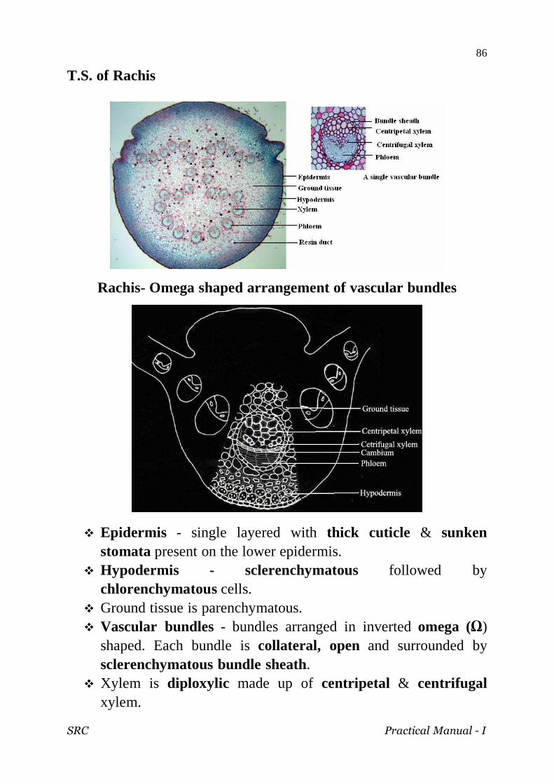
T.S. of Rachis
Rachis- Omega shaped arrangement of vascular bundles
Epidermis - single layered with thick cuticle & sunken stomata present on the lower epidermis.
Hypodermis - sclerenchymatous followed by chlorenchymatous cells.
Ground tissue is parenchymatous. Vascular bundles - bundles arranged in inverted omega (Ω)
shaped. Each bundle is collateral, open and surrounded by sclerenchymatous bundle sheath.
Xylem is diploxylic made up of centripetal & centrifugal xylem.

SRC Practical Manual - I
87
T.S. of leaflet
Leaflet has swollen midrib and narrow flat wings.
The outermost layer is cuticularised epidermis.
Sclerenchymatous hypodermis is present on both sides.
Cuticle Hypodermis
Palisade Transfusion tissue Centripetal xylem Centrifugal xylem Cambium Phloem
Lower epidermis
SRC Practical Manual - I
88
There is a centrally located vascular bundle in the midrib.
The vascular bundle is conjoint, collateral, open and
pseudomesarch.
Xylem shows a large V-shaped centripetal xylem and two
groups of centrifugal xylem.
Mesophyll is differentiated into spongy and palisade tissue.
Presence of transfusion tissue between palisade and spongy
tissue helping in lateral conduction.
Xerophytic adaptations
Presence of upper and lower epidermis covered with cuticle.
Sunken stomata restricted to the lower epidermis.
Presence of sclerenchymatous hypodermis.
Diploxylic nature of vascular bundles.
Presence of transfusion tissue.

SRC Practical Manual - I
89
CYCAS MALE CONE
Male cone is terminal, stalked, large and conical
Consists of central cone axis and numerous micro sporophylls.
Microsporophylls are spirally arranged around the cone axis.
Microsporophyll

SRC Practical Manual - I
90
Microsporophyll - leaf like, woody, wedge shaped & brown in
colour.
Lower expanded fertile region with sori & upper sterile region
called apophysis.
Sorus has 5 to 6 microsporangia with soral hairs.
Microsporangium - oval in shape with sporangial wall. It
encloses many unicellular, uninucleate, haploid microspores
or pollen grains.
Microspores has inner intine, outer exine, cytoplasm &
hapioid nucleus.
SRC Practical Manual - I
91
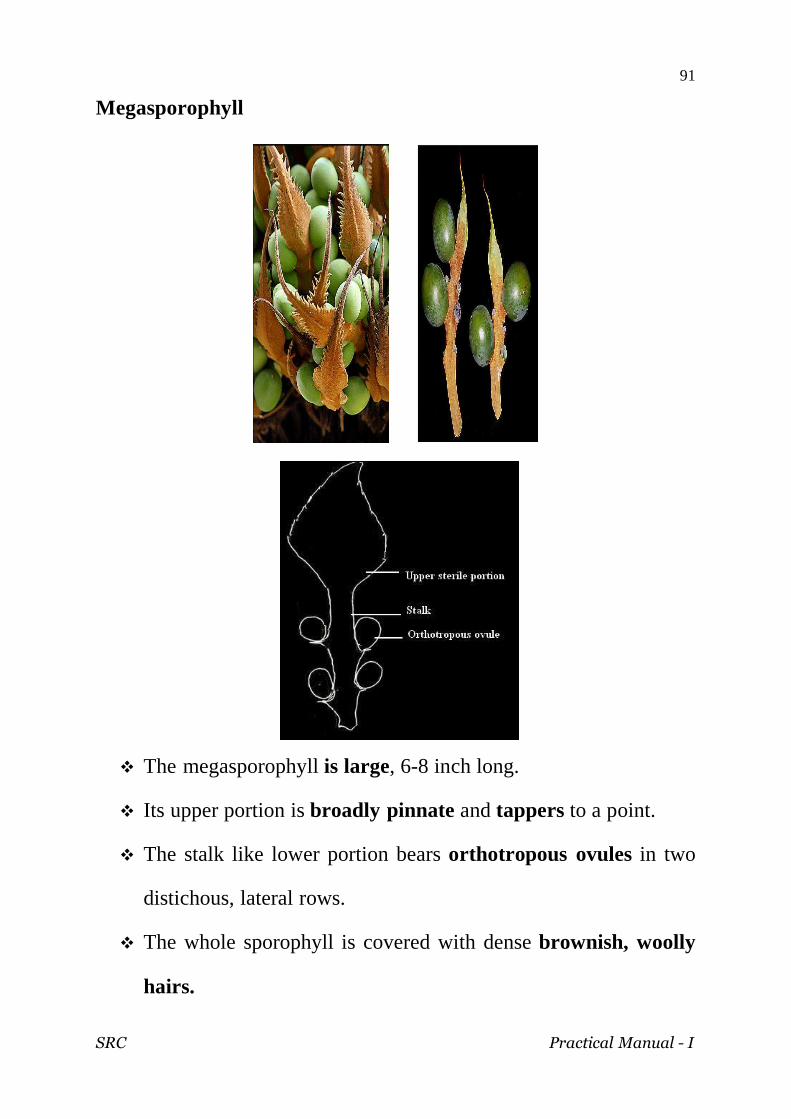
Megasporophyll
The megasporophyll is large, 6-8 inch long.
Its upper portion is broadly pinnate and tappers to a point.
The stalk like lower portion bears orthotropous ovules in two
distichous, lateral rows.
The whole sporophyll is covered with dense brownish, woolly
hairs.

SRC Practical Manual - I
92
Ovule L.S
The ovule consists of a mass of cells, the nucellus.
It is surrounded by a single massive integument.
The integument is differentiated into three layers.
The outer and inner layers are fleshy and the middle layer is
stony.
The integument fuses with nucellus all around except at the top
where it leaves a pore leading to the nucellus called micropyle.

SRC Practical Manual - I
93
Class : Gnetopsida
Order : Gnetales
Family : Gnetaceae
Genus : Gnetum
Habit
Gnetum is a sporophytic plant.
Consists of stem, leaves and tap root system.
Stem is woody and branched.
It has two types of branches- long or branches of unlimited
growth and dwarf shoot or branches of limited growth.
Long branches bear scale-like leaves at the nodes.
There are two leaves at each node, opposite decussate.
The dwarf branch arise from the axis of the scale leaves on the
long branch.
The stem is articulated with node and internode.
Leaves show reticulate venation.

SRC Practical Manual - I
94
Old stem
In some climbing species of Gnetum, accessory cambia are
formed at different levels of the cortex.
These cambia are not equally active in nature and so they
produce incomplete vascular rings.
Hence the stem represents an eccentric appearance.
The pith is also eccentric.

SRC Practical Manual - I
95
Stem T.S
The T.S of stem shows an epidermis, cortex, endodermis and a
stele.
Epideermis is the outer layer made up of rectangular cells
covered with cuticle. It has sunken stomata.
The cortex has three distinct zones. The outer cortex is
chlorenchymatous, the middle cortex is parenchymatous and
the inner cortex is sclerenchymatous.
The endodermis and pericycle are not so distinct.
The stele is an ectophtoic eustele. It consists of a ring of many
vascular bundles.
The vascular bundles are conjoint, collateral, endarch and
open.
Xylem occupies the centre. It consists of tracheids and vessels.
The phloem consists of sieve cells, phloem pacenchma and
companim cells.
Vascular bundles are separated by a broad parenclyma.
The pith is parenchymatous.

SRC Practical Manual - I
96
Entire male cone
The male flowers are arranged in many rows from 3-6 above
each collar.
Each male flower is stalked and enclosed with a sheath like
perianth.

SRC Practical Manual - I
97
Entire female cone
Female flowers are arranged in a single ring on each collar.
Each female flower consists of sessile ovule.
The ovules are naked in nature.

SRC Practical Manual - I
98
Ovule L.S.
The ovule is orthotropous. The V.S. ovule shows a spherical
nucellus and three envelops called integuments.
The three integuments are called outer integument, middle
integument and inner integument.
The outer and inner integuments are soft, but the middle one is
stony.

SRC Practical Manual - I
99
The inner integument grows beyond the others and form a tube
called micropylar tube. The opening of the tube is called
micropyle.
A pollen chamber lies below the micropylar tube.
The nucellus consists of a mass of thin-walled cells. It is a
nutritive tissue.
A female gametophyte remains embedded in the nucellus.
The female gametophyte consists of a sac-like structure
consisting of cellular tissue at the chalazal end and free nuclei
at the micropylar end.
It has one or two large nuclei which act as female nuclei.
The female nucleus fuses with the male gamete to form a diploid
zygote.

SRC Practical Manual - I
100
This page intentionally left blank

SRC Practical Manual - I
101
PALEOBOTANYPALEOBOTANYPALEOBOTANYPALEOBOTANY

SRC Practical Manual - I
102

SRC Practical Manual - I
103
Class : Psilophytopsida
Order : Psilophytales
Family : Rhyniaceae
Genus : Rhynia
Stem
Rhynia is a fossil fern.- Psilotopsida.
Stem shows epidermis,cortex and stele.
Epidermis- outermost, cuticularised with stomata.
Cortex –parenchymatous and well preserved.
Stele- protostele- xylem surrounded by phloem.
Geological period: Devonian of Paleozoic era.

SRC Practical Manual - I
104
Class : Lycopsida Order : Lepidodendrales Family : Lepidodendeaceae Genus : Lepidodendron
Stem
The old stem of Lepidodendron selaginoides has got an irregular outline due to the presence of persistant leaf bases.
There is well developed periderm followed by three layers of cortex.
The outer wall is made up of alternately arranged thick walled cells.
The cells of middle cortex have disappeared and the space is occupied by organic debris and stigmarian rootets.
The inner cortex is parenchymatous.
Secondary phloem is not preserved. Secondary xylem is exarch with tracheids intermingling with
parenchyma. Stele is protostele.
Geological period: Carboniferous period of Paleozoic era.

SRC Practical Manual - I
105
Lepidocarpon
Female fruit body of Lepidodendron- fossil fern.
It consists of single megasporangium with a megaspore.
Megasporangium enclosed by megasporophyll which forms
integument like structure.
Integument has a false micropyle.
Geological period: Carboniferous period of Paleozoic era.

SRC Practical Manual - I
106
Class : Sphenopsida Order : Calamitales Family : Calamitaceae Genus : Calamites
Stem
It is a fossil fern. - Calamopsida.
The stem shows periderm, cortex, vascular bundles and pith.
Outermost periderm.
Narrow parenchymatous cortex.
Secondary xylem wedge shaped, radiating from the centre.
Primary xylem endarch with carinal canals.
Broad medullary rays between secondary xylem.
Central hollow pith .
Geological period: Carboniferous period of Paleozoic era.

SRC Practical Manual - I
107
Spot at sight the genus, group and morphology of the following.
The first one is done for you
Part Genus Group Morphology
Pogonatum Gymnosperm Gameophyte with sporophyte
SRC Practical Manual - I
108

SRC Practical Manual - I
109
SRC Practical Manual - I
110
Identify the following fossil, write the geological period.
NAME GEOLOGICAL PERIOD

SRC Practical Manual - I
111
Observe the images and answer the questions
ALGAE
IMAGES QUESTION ANSWERS
Identify separation disc, trichome, hormogonia. Write down its systematic position.
Name the plant and the type of spores present in it. Add a note on the next generation produced by these spores.
Can you identify the plant from this cross section? Name the parts you can see in the cross section.
What dose this picture represent? Label the parts and mention the plant in which it is present.
Which alga can you see? Name the type of thallus and write down the systematic position.
Which alga you have studied possesses this structure? Mention the parts and the function.
Name the genus and species of this alga. To which division dose this alga belong? Write down the characters.
SRC Practical Manual - I
112
FUNGI
IMAGES QUESTION ANSWERS
Name the structure. Mention the fungus and the host in which it is produced?
Label the different parts with the name of the fungus that produces this. Write down the important use of this fungus.
Which fungus you have studied produces this type of fruit body? Name the different parts and the function.
Identify the causal organism, symptoms and control measure of the disease.
Name the spore and the fungus producing this type of spore. Mention the host in which it is produced.
Mention the types of spores seen. Name the fungus and the disease caused by it.

SRC Practical Manual - I
113
LICHEN
IMAGES QUESTION ANSWERS
Name the plant and describe the nature of association.
Label the different parts and mention the type of thallus.
What part of lichen is this? Mention the significance of it.
SRC Practical Manual - I
114
BRYOPHYTA
IMAGES QUESTION ANSWERS
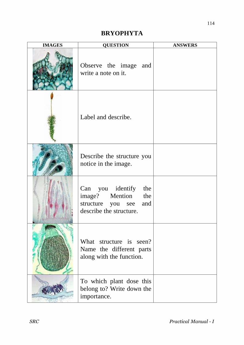
Observe the image and write a note on it.
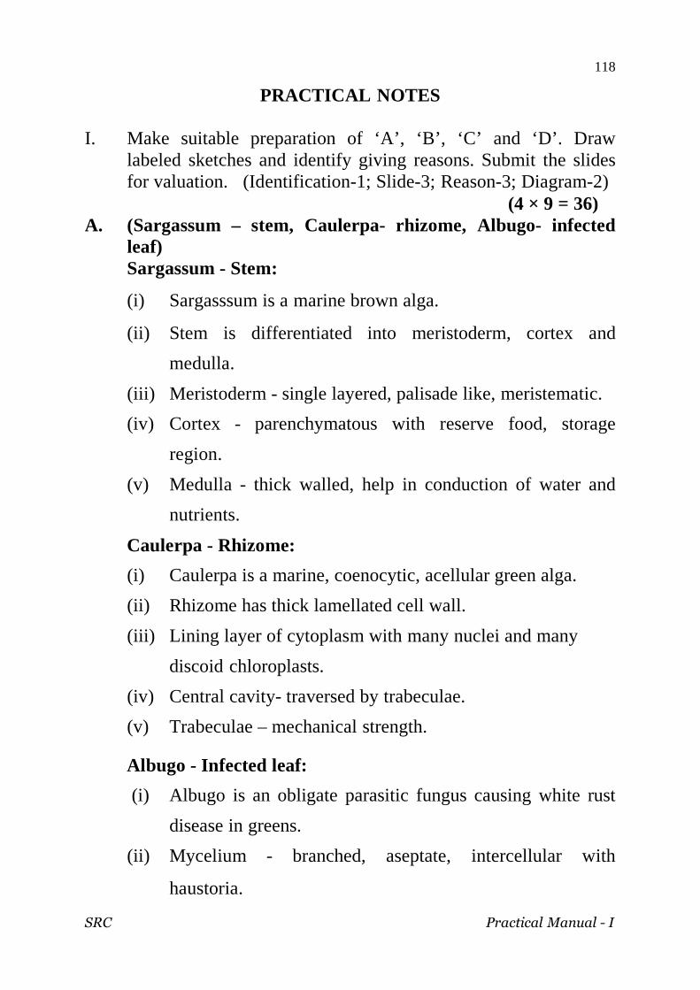
Label and describe.
Describe the structure you notice in the image.
Can you identify the image? Mention the structure you see and describe the structure.
What structure is seen? Name the different parts along with the function.
To which plant dose this belong to? Write down the importance.

SRC Practical Manual - I
115
PTERIDOPHYTA
IMAGES QUESTION ANSWERS
Is it a homosporous or heteosporous cone? Give example and reason out.
What type of stele is this? Describe with example.
Name the different parts of the structure and mention the plant which possesses it.
What is the image? Label and describe.
Name the plant and differentiate the two structures given.
Which Pteridophyte you have studied possesses this type of strobilus? Describe.

SRC Practical Manual - I
116
GYMNOSPERM
IMAGES QUESTION ANSWERS
Name the important part you can see. Mention the significance.
Name the image. Which gymnosperm possesses this?
What part of Gnetum is this? Describe the structure.
What does this image show? Name the plant and write down the systematic position.
Describe the stele. Where does this type of stele occur?

SRC Practical Manual - I
117
PRACTICAL MODEL QUESTION PAPER
I. Make suitable preparations of ‘A’, ‘B’, ‘C’ and ‘D’. Draw labeled sketches and identify giving reasons. Submit the slides for valuation. (Identification-1; Slide-3; Reason-3; Diagram-2) (4 × 9 = 36)
II. Separate the various types of algae in the given sample ‘E’.
Identify them giving reasons and draw labeled sketches. (2 × 3 = 6)
III. Identify, draw labeled sketches and write notes on ‘F’, ‘G’, ‘H’
‘I’ and ‘J’. (Identification-1; Notes-1; Sketch-1.) (5 × 6 = 30)
IV. Identify the fossil ‘K’ with reasons. Draw diagram, give the geological period. (Identification-1; Reason-2; Diagram-2; geological period-1) (1 × 6 = 6)
V. Spot at sight genus, group and morphology of L, M, N and O
(4 × 3 = 12) TOTAL = 90
RECORD = 10
SRC Practical Manual - I
118
PRACTICAL NOTES
I. Make suitable preparation of ‘A’, ‘B’, ‘C’ and ‘D’. Draw labeled sketches and identify giving reasons. Submit the slides for valuation. (Identification-1; Slide-3; Reason-3; Diagram-2)
(4 × 9 = 36) A. (Sargassum – stem, Caulerpa- rhizome, Albugo- infected
leaf) Sargassum - Stem:
(i) Sargasssum is a marine brown alga.
(ii) Stem is differentiated into meristoderm, cortex and
medulla.
(iii) Meristoderm - single layered, palisade like, meristematic.
(iv) Cortex - parenchymatous with reserve food, storage
region.
(v) Medulla - thick walled, help in conduction of water and
nutrients.
Caulerpa - Rhizome:
(i) Caulerpa is a marine, coenocytic, acellular green alga.
(ii) Rhizome has thick lamellated cell wall.
(iii) Lining layer of cytoplasm with many nuclei and many
discoid chloroplasts.
(iv) Central cavity- traversed by trabeculae.
(v) Trabeculae – mechanical strength.
Albugo - Infected leaf:
(i) Albugo is an obligate parasitic fungus causing white rust
disease in greens.
(ii) Mycelium - branched, aseptate, intercellular with
haustoria.

SRC Practical Manual - I
119
(iii) Mycelial mat forms below the lower epidermis.
(iv) Conidiophore - thick walled, unbranched, club shaped,
palisade like.
(v) Conidiophores bear chain of conidia basipetally.
(vi) Conidium - small, spherical and multinucleate and joined
by mucilage pad.
B. Marchantia – Thallus.
(i) Marchantia is bryophyte belonging to Hepaticae.
(ii) Thallus is differentiated in to epidermis, photosynthetic
region and storage tissue.
(iii) Epidermis: upper epidermis-single layered with airpores;
lower epidermis with smooth walled and tuberculate
rhizoids.
(iv) Upper photosynthetic region - air chambers with
assimilatory filaments with partition wall.
(v) Storage region - thin walled, parenchymatous. C. (Lycopodium- Stem, Adiantum- Rachis)
Lycopodium cernuum - Stem
(i) Lycopodium is a fern - Lycopsida.
(ii) Stem is wavy in outline.
(iii) It is differentiated in to epidermis, cortex and stele.
(iv) Epidermis is single layered with cuticle.
(v) Cortex - differentiated into outer and inner parenchymatous
and middle sclerenchymatous.
(vi) Stele is mixed protostele - patches of xylem in phloem.
(vii) Pith is absent.
SRC Practical Manual - I
120
Adiantum - Rachis:
(i) It is a fern - Filicopsida.
(ii) Rachis is differentiated into epidermis, cortex and stele.
(iii) Cuticularised epidermis.
(iv) Hypodermis - 3-4 layers, sclerenchymatous.
(v) Cortrex- broad, parenchymatous.
(vi) Stele - protostele with single layered endodermis and
pericycle.
(vii) Xylem – V - shaped, exarch, completely surrounded by
phloem.
D. (Cycas leaflet, Cycas rachis and Cycas microsporophyll)
Cycas - leaflet:
(i) Cycas is a gymnosperm.
(ii) The leaflet has swollen midrib and narrow wings.
(iii) Outermost cuticularised epidermis.
(iv) Lower epidermis with sunken stomata.
(v) Sclerenchymatous hypodermis on both sides.
(vi) Midrib: centrally located vascular bundle- conjoint,
collateral, open and pseudomesarch. Xylem – V shaped
centripetal xylem and two groups of centrifugal xylem-
diploxylic.
(vii) Wings: Mesophyll shows palisade and spongy.
Transfusion tissue between palisade and spongy - lateral
conduction.

SRC Practical Manual - I
121
Cycas rachis:
(i) Cycas is a gymnosperm.
(ii) Rachis is shield shaped.
(iii) It is differentiated into epidermis, hypodermis and vascular
bundles in ground tissue.
(iv) Epidermis- single layered, cuticularised.
(v) Hypodermis- multilayered, outer chlorenchyma and inner
sclerenchyma.
(vi) Ground tissue - parenchymatous.
(vii) Vascular bundle - arranged in an inverted omega shape.
(viii) Each bundle - conjoint, collateral, open and
pseudomesarch, diploxylic with centripetal and centrifugal
xylem.
Cycas microsporophyll:
(i) Cycas is a gymnosperm.
(ii) Microsporophylls are arranged spirally forming male cone.
(iii) T.S of microsporophyll is triangular in outline.
(iv) Microsporangia attached on the lower surface.
(v) Sporangia- oval, sac like with short stalk.
(vi) Sporangia enclose large number of microspores.
(vii) Soral hairs are seen among the sporangia.

SRC Practical Manual - I
122
II. Separate the various types of algae in the given sample ‘E’. Identify them giving reasons and draw labeled sketches. E- Algal mixture ( Oscillatoria and Polysiphonia). (Identification-1; Notes-1; Sketch-1) (2 × 3 = 36) Oscillatoria:
(i) Unbranched filamentous blue green alga.
(ii) Cell is rectangular - prokaryotic.
(iii) protoplasm - differentiated into outer chromoplasm and
inner centroplasm.
(iv) Presence of necridia and hormogone.
Polysiphonia:
(i) It is a marine branched filamentous red alga.
(ii) Filaments show of many siphons.
(iii) Each cell has lining layer of cytoplasm with nucleus and
many chromatophores.
(iv) Pit connection is present between the cells.
III. Identify, draw labeled sketches and write notes on ‘F’, ‘G’, ‘H’
‘I’ and ‘J’. (Identification-1; Notes-1; Sketch-1.) (5 × 6 = 30)
SPOTTERS - (F, G, H, I, J)
F - Algae or lichen. (Volvox, Lichen-apothecium)
G - Fungi. (Puccinia-uredosorus, Penicillium-conidia, Peziza-apothecium)
H - Bryophyte. (Marchantia-sporophyte, Pogonatum-antheridial head,
Pogonatum-archegonial head)
I - Pteridophyte. (Lycopodium-cone, Selaginella-cone, Equisetum-
cone)
J - Gymnosperm. (Cycas-coralloid root, Cycas-ovule, Gnetum-ovule)

SRC Practical Manual - I
123
F - Volvox:
(i) Volvox is a coenobial round planktonic green alga.
(ii) Coenobium has an outer mucilage sheath.
(iii) Cells are ovoid, biflagellate, uninucleate, interconnected by
plamodesmata.
(iv) Each cell has large cup shaped chloroplast with a pyrenoid.
(v) Reproduction- by special large cells called gonidia.
Lichen apothecium:
(i) Lichen is a composite symbiotic form with phycobiont and
mycobiont.
(ii) Apothecium is a saucer shaped fruit body.
(iii) Apothecium is differentiated into hymenium, sub-hymenium
and peridium.
(iv) Hymenium consists of asci with 8 ascospores and
paraphysis.
(v) Sub-hymenium-mycelial mat.
(vi) Peridium-fleshy wall layer differentiated into outer cortex
and medulla.
(vii) Algal cells are seen only in the outer cortex.
G - Puccinia-infected leaf with uredosorus
(i) This stage is found in primary host, wheat.
(ii) The mycelium produces a number of reddish brown
pustules on the upper surface of leaf.
SRC Practical Manual - I
124
(iii) Each pustule is a uredosorus, consisting of number of
uredodospores.
(iv) The uredospores are stalked, unicellular, rounded, bi-
nucleate spores.
(v) Uredospore is a repeating spore.
Penicillium-conida:
(i) Penicillium is saprophytic Ascomycetous fungus.
(ii) Mycelium-branched and septate.
(iii) Asexual reproduction by conidia.
(iv) Conidiophores-branched-primary, secondary and tertiary.
(v) Ultimate branch produce bottle shaped sterigmata with
chain of conidia basipetally.
(vi) Conidium-globular with single nucleus.
Peziza-apothecium:
(i) Peziza is a coprophilous Ascomycetous fungus.
(ii) Apothecium-cup shaped fruit body.
(iii) It shows hymenium, subhymenium and peridium.
(iv) Peridium-thick fleshy wall - parenchymatous.
(v) Hymenium-fertile lining layer with number of asci,
ascospores and paraphysis.
(vi) Below the hymenium is subhymenium.
SRC Practical Manual - I
125
H - Marchantia-sporophyte, Pogonatum-antheridial head,
Pogonatum-archegonial head)
Marchantia-sporophyte:
(i) Marchantia is bryophyte belonging to Hepaticae.
(ii) Sporophyte has foot, seta and capsule.
(i) Foot-bulbous help in anchorage and absorption.
(ii) Seta-short and elongates at maturity.
(iii) Capsule-oval and encloses spores and elaters.
(iv) Sporophyte is enclosed by calyptra, perigynium and
perichaetium.
Pogonatum-antheridial head:
(i) Pogonatumis a bryophyte-Bryopsida.
(ii) Antheridia arise in cluster at the tip of male branch.
(iii) Antheridium-elongated, club shaped, short-stalked with an
outer jacket enclosing a number of biflagellate spermatozoids.
(iv) Sterile uniseriate paraphyses present in between.
Pogonatum-archegonial head:
(i) Pogonatumis a bryophyte-Bryopsida.
(ii) Archegonia arise in cluster at the apex of female shoot.
(iii) Sterile paraphysis seen among archegonia.
(iv) Each archegonium is flask shaped.
(v) It has a stalk, swollen venter and long tubular neck.
(vi) Venter has venter canal cell and egg.
(vii) Neck consists of six vertical rows of cells enclosing 4-10
neck canal cells.

SRC Practical Manual - I
126
I (Lycopodium-cone, Selaginella-cone, Equisetum-cone)
Lycopodium-cone:
(i) Lycopodium is a fern-Lycopsida.
(ii) Cone-strobili - reproductive part of sporophyte.
(iii) Sporophylls arranged spirally on the cone axis.
(iv) Stalked sporangia on the adaxial side of the sporophyll.
(v) Sporangia are homosporous with spores in tetrads.
Selaginella-cone:
(i) Selaginella is a heterosporous fern-Lycopsida.
(ii) Cone-strobili-reproductive part of the sporophyte.
(iii) Ligulate sporophylls are arranged spirally-two types.
(iv) Microsporophyll - bear microspoprangia with many small
microspores- male.
(v) Megasporophyll - bear lobed megasporangia with four
large megaspores - female.
Equisetum-cone:
(i) Equisetum is a homosporous fern-Calamopsida.
(ii) Cone-strobili - reproductive part of the sporophyte - borne
at the tips of branches.
(iii) Each cone has a basal sterile sheath-annulus.
(iv) Sporangiophores with a stalk, peltate disc with pendent
sporangia.
(v) Peltate disc bears sporangia on the underside.
(vi) Sporangia-sac-like with spores and pseudoelater

SRC Practical Manual - I
127
J (Cycas-coralloid root, Cycas-ovule, Gnetum-ovule)
Cycas-coralloid root:
(i) Cycas is a gymnosperm.
(ii) Coralloid roots-apogeotropic, coral like roots with symbiotic
blue green alga.
(iii) It has epidermis, cotex and stele.
(iv) Epidermis- single layered.
(v) Cortex - outer cortex, middle algal zone and inner cortex.
(vi) Algal zone - radially elongated, loosely packed with blue
green algae.
(vii) Endodermis followed by pericycle.
(viii) Vascular bundle - triarch, radial,closed with exarch xylem.
Cycas-ovule:
(i) Cycas is a gymnosperm.
(ii) Ovule-megasporangium - large, ovoid, borne on
megasporophylls.
(iii) Ovule has nucellus covered by an integument.
(iv) Integument - differentiated in to outer and inner fleshy and
middle stony layers with narrow tubular micropyle at the
apex.
(v) Nucellus encloses female gametophyte.
Gnetum-ovule:
(i) Gnetum is a gymnosperm.
(ii) Ovule - megasporangium borne in whorls on female cone.
(iii) It has integument and nucellus.
SRC Practical Manual - I
128
(iv) Integument is three layered- outer and inner short, middle
long tubular (style) forming micropyle.
(v) Nucellus encloses female gametophyte.
(vi) Fan shaped pavement tissue present below the prothallus-
nutritive.
IV. Identify the fossil ‘K’ with reasons. Draw diagram, give the
geological period. (Identification-1; Reason-2; Diagram-2; geological period-1) (1 × 6 = 6) K- (Rhynia-stem, Lepidocarpon, Calamites-stem.)
Rhynia-stem:
(i) Rhynia is a fossil fern-Psilotopsida.
(ii) Stem shows epidermis, cortex and stele.
(iii) Epidermis-outermost, cuticularised with stomata.
(iv) Cortex-parenchymatous and well preserved.
(v) Stele-protostele-xylem surrounded by phloem.
Geological period: Devonian of Paleozoic era.
Lepidocarpon:
(i) Female fruit body of Lepidodendron-fossil fern.
(ii) It consists of single megasporangium with a megaspore.
(iii) Megasporangium is enclosed by megasporophyll which
forms integument like structure.
(iv) Integument has a false micropyle.
Geological period: Carboniferous period of Paleozoic era.

SRC Practical Manual - I
129
Calamites-stem:
(i) It is a fossil fern-Calamopsida.
(ii) The stem shows periderm, cortex, vascular bundles and
pith.
(iii) Outermost periderm.
(iv) Narrow parenchymatous cortex.
(v) Secondary xylem wedge shaped, radiating from the centre.
(vi) Primary xylem endarch with carinal canals.
(vii) Broad medullary rays between secondary xylem.
(viii) Central hollow pith.
Geological period: Carboniferous period of Paleozoic era.
V. Spot at sight genus, group and morphology of L, M, N and O (4 × 3 = 12)
GENUS GROUP MORPHOLOGY
Caulerpa Algae Thallus Sargassum Algae Thallus Peziza Fungi Apothecium Usnea Fruticose lichen Thallus with apothecium Psilotum Pteridophyte Sporophyte Lycopodium Pteridophyte Sporophyte Equisetum Pteridophyte Sporophyte Adiantum Pteridophyte Sporophyte Selaginella Pteridophyte Sporophyte Cycas Gymnosperm Old stem Cycas Gymnosperm Microsporophyll Cycas Gymnosperm Megasporophyll Gnetum Gymnosperm Male cone Gnetum Gymnosperm Female cone Gnetum Gymnosperm Wood
TOTAL = 90 RECORD = 10
Related Documents