МИНИСТЕРСТВО ЗДРАВООХРАНЕНИЯ РЕСПУБЛИКИ БЕЛАРУСЬ БЕЛОРУССКИЙ ГОСУДАРСТВЕННЫЙ МЕДИЦИНСКИЙ УНИВЕРСИТЕТ КАФЕДРА БИОЛОГИЧЕСКОЙ ХИМИИ А.Д. Таганович, И.Л. Котович, А.В. Колб, Н.Н. Ковганко БИОХИМИЯ: ПРАКТИКУМ ДЛЯ ИНОСТРАННЫХ УЧАЩИХСЯ СТОМАТОЛОГИЧЕСКОГО ФАКУЛЬТЕТА A.D. Tahanovich, I.L. Kotovich, A.V. Kolb, N.N. Kauhanka BIOCHEMISTRY: MANUAL FOR INTERNATIONAL STUDENTS OF DENTAL FACULTY Практикум Минск БГМУ 2013

Welcome message from author
This document is posted to help you gain knowledge. Please leave a comment to let me know what you think about it! Share it to your friends and learn new things together.
Transcript
МИНИСТЕРСТВО ЗДРАВООХРАНЕНИЯ РЕСПУБЛИКИ БЕЛАРУСЬ
БЕЛОРУССКИЙ ГОСУДАРСТВЕННЫЙ МЕДИЦИНСКИЙ УНИВЕРСИТЕТ
КАФЕДРА БИОЛОГИЧЕСКОЙ ХИМИИ
А.Д. Таганович, И.Л. Котович, А.В. Колб, Н.Н. Ковганко
БИОХИМИЯ: ПРАКТИКУМ ДЛЯ ИНОСТРАННЫХ УЧАЩИХСЯ
СТОМАТОЛОГИЧЕСКОГО ФАКУЛЬТЕТА
A.D. Tahanovich, I.L. Kotovich, A.V. Kolb, N.N. Kauhanka
BIOCHEMISTRY: MANUAL FOR INTERNATIONAL STUDENTS OF
DENTAL FACULTY
Практикум
Минск БГМУ 2013
2
УДК 577.1(811.111)(076.5) ББК 28.707.2я73 Т13
Рекомендовано Научно-методическим советом университета в качестве практикума 29.05.2013 г., протокол №8
А в т о р ы : проф. А.Д. Таганович, доц. И.Л. Котович, доц. А.В. Колб, доц. Н.Н. Ковганко
Р е ц е н з е н т ы : проф. Е.В. Барковский, доц. О.Н. Ринейская
Биохимия: практикум для студентов стоматологического факультета= BIOCHEMISTRY: MANUAL FOR INTERNATIONAL STUDENTS OF DENTAL
FACULTY: практикум / А.Д. Таганович, И.Л. Котович, А.В. Колб, Н.Н. Ковганко. – Минск : БГМУ, 2013. – 54 с.
ISBN
Издание содержит рекомендации по подготовке к лабораторно-практическим занятиям по биологической химии. По каждой теме даны: цель занятия, вопросы для обсуждения, литература для подготовки, описание лабораторных работ и их клинико-диагностическое значение. Включены вопросы для подготовки к коллоквиумам, примерный перечень экзаменационных вопросов.
Предназначено для иностранных студентов стоматологического факультета, обучающихся на английском языке.
УДК 577.1(811.111)(076.5) ББК 28.707.2я73
ISBN
3
CONTENTS
1. INTRODUCTION TO BIOCHEMISTRY. ENZYMES. CLASSIFICATION, STRUCTURE, PROPERTIES …..................................................................................................5
2. REGULATION OF ENZYME ACTIVITY. DETERMINATION OF ENZYME ACTIVITY…………………………………………………………………………….……..7
3. INTRODUCTION TO METABOLISM. CENTRAL METABOLIC PATHWAY - THE KREBS CITRIC ACID CYCLE ……………………………………………………………….….8
4. BIOLOGICAL OXIDATION. PATHWAYS OF OXYGEN UTILIZATION BY CELLS. OXIDATIVE PHOSPHORYLATION ………………………………………………………….10
5. COLLOQUIUM: “ENZYMES, INTRODUCTION TO METABOLISM. CENTRAL METABOLIC PATHWAY, BIOLOGICAL OXIDATION. OXIDATIVE PHOSPHORYLATION”………………………………………………………………..….……...11
6. DIGESTION OF CARBOHYDRATES. GLYCOGENESIS AND GLYCOGENOLYSIS. GLYCOLYSIS ……………………………………………….……………………………………12
7. METABOLIC PATHWAYS OF PYRUVATE. GLUCONEOGENESIS. AEROBIC OXIDATION OF GLUCOSE TO FINAL PRODUCTS (CO2 AND H2O)………..………....13
8. SECONDARY PATHWAYS OF GLUCOSE METABOLISM. EFFECT OF HORMONES ON THE BLOOD GLUCOSE LEVEL. FEATURES CARBOHYDRATE UTILIZATION OF ORAL MICROFLORA ……...................................................................................................14
9. COLLOQUIUM: “CARBOHYDRATE METABOLISM”……………….…………….…….15
10. LIPID METABOLISM. DIGESTION AND RE-SYNTHESIS. EVALUATION OF LIPASE ACTIVITY …………………………………………………………….….………………...……...16
11. INTRACELLULAR METABOLISM OF FATTY ACIDS. DETERMINATION OF PLASMA β-LIPOPROTEINS ……………………………………………………….…….……..17
12. CHOLESTEROL AND KETONE BODIES METABOLISM. DETERMINATION OF CHOLESTEROL IN SERUM ………………………………………………………….……......18
13. TRANSPORT OF EXOGENOUS AND ENDOGENOUS LIPIDS. REVERSE TRANSPORT OF CHOLESTEROL. REGULATION AND PATHOLOGY OF LIPID METABOLISM ……………………………………………………………………….…….….….20
14. COLLOQUIUM: “LIPID METABOLISM”………………………….………………………....20
15. PHYSICAL AND CHEMICAL PROPERTIES OF THE BLOOD. HEMOGLOBINOSES………………………………………………………………………….….22
16. BLOOD PLASMA PROTEINS. BLOOD CLOTTING SYSTEM …………………………..23
17. CONTROL OVER PRACTICAL SKILLS OF BIOCHEMICAL ANALYSIS…………..…24
4
18. DIGESTION AND ABSORPTION OF PROTEINS. ANALYSIS OF GASTRIC JUICE………………………………………………………………………………………………..26
19. INTRACELLULAR AMINO ACID METABOLISM. DETERMINATION OF AMINO TRANSFERASE ACTIVITY IN SERUM ……………………………………………….……..27
20. DETOXIFICATION OF AMMONIA. DETERMINATION OF NONPROTEIN NITROGEN IN BLOOD AND UREA IN URINE ………………………………………….....29
21. COLLOQUIUM: “METABOLISM OF SIMPLE PROTEINS”, “BLOOD BIOCHEMISTRY” ………………………………………………………………………………..31
22. NUCLEOPROTEINS CHEMISTRY AND METABOLISM. DETERMINATION OF URIC ACID IN URINE …………………………………………………………………………………..31
23. MATRIX BIOSYNTHESES (SYNTHESIS OF DNA, RNA, PROTEINS)………….……...32
24. CONNECTIVE TISSUE PROTEINS ……………......................................................................33
25. COLLOQUIUM: “NUCLEOPROTEINS METABOLISM”, “SYNTHESIS OF DNA, RNA AND PROTEINS”, “CONNECTIVE TISSUE PROTEINS” ………………………….……...33
26. HORMONES, GENERAL CHARACTERISTIC AND PECULIARITIES OF BIOLOGICAL ACTION. QUALITATIVE REACTIONS FOR HORMONES ……………34
27. BIOCHEMISTRY OF HORMONES. GLUCOSE TOLERANCE TEST …………………...37
28. BIOCHEMISTRY OF NUTRITION. ROLE OF PROTEINS, FATS, CARBOHYDRATES, VITAMINS. VITAMIN-LIKE SUBSTANCES ……….……………………………………….39
29. BIOCHEMISTRY OF NUTRITION. MINERAL SUBSTANCES. REGULATION OF WATER-ELECTROLYTE BALANCE ………………….………………….…..……………...42
30. BIOCHEMISTRY OF TEETH AND ORAL CAVITY FLUIDS...…………………………....42
31. COLLOQUIUM: “HORMONES”, “BIOCHEMISTRY OF NUTRITION”, “BIOCHEMISTRY OF TEETH AND ORAL CAVITY FLUIDS”...………………………...45
32. LIVER BIOCHEMISTRY …………………………………..........................................................46
Appendix. The list of examination questions in Biological Chemistry…………………………………...…..48
5
1. Topic: INTRODUCTION TO BIOCHEMISTRY. ENZYMES. CLASSIFICATION, STRUCTURE, PROPERTIES
Objective To learn how to use the knowledge of enzyme properties and enzyme composition of organs
in further study of metabolism as well as to solve problems of diagnosis, prophylaxis and treatment of diseases associated with functional impairment of enzymes.
Problems for discussion
1. Peculiarities of enzymes as protein catalysts. 2. Modern classification of enzymes and terminology of enzymes (systematic and working
names). Enzyme code. General characteristics of classes. 3. The structure of enzymes. Coenzymes, their classification and role in catalysis. Block-
structures of the NAD+, NADP+, FAD and FMN. 4. The influence of conformational changes on enzyme activity. 5. The mechanism of enzyme action. Enzyme kinetics. The effect of substrate concentration,
pH, temperature on enzyme reaction velocity (molecular mechanism, graphical relationship). Michaelis’s constant (Km), usage of Km for predicting the course of biochemical reactions.
6. Specificity of enzyme action. Types of specificity.
Recommended literature
1. Konevalova N.Yu., Buyanova S.V. Biochemistry lecture course. // Vitebsk, 2005.- P.18-21, 22-26, 69-76.
2. Lecture material.
Practical part Work 1. Studying the effect of various factors on the rate of enzyme-catalyzed reactions 1. Evaluation of saliva amylase activity and its thermolability One of characteristic properties of enzymes is their thermolability, i.e. sensitivity of the
enzyme to temperature at which a reaction takes place. For the majority of enzymes the temperature optimum is observed at 38-40oC. Enzymes heated over 70 oC, as a rule, lose their properties of biological catalysts.
Hydrolysis of starch under the action of saliva α-amilase occurs untill the stage of dextrines formation. Starch together with iodine gives blue staining. Dextrines, depending on their size, together with iodine give various staining: amilodextrines – violet, erythrodextrines – red-brown, maltose – yellow. End products of starch hydrolysis – maltose and glucose – have got free aldehyde groups and can be revealed by Trommer’s reaction. The enzyme effect is judged by the decrease of the substrate amount or by the appearance of reaction products.
Procedure. Pre-dilute the saliva with water 1:10. Apply a small quantity of diluted saliva (2-3 ml) into a clean test-tube and boil it for 5 minutes, then cool it. Take 1% starch solution and apply into 3 test tubes per 10 drops into each. Add 10 drops of native saliva diluted 1:10 into the 1st test-tube; add 10 drops of boiled saliva into the 2nd tube and 10 drops of water as a control to the 3rd tube. All test-tubes are placed into the thermostat at 38oC for 10 minutes. Then the content of the test-tubes is exposed to qualitative reactions for starch and products of its disintegration.
6
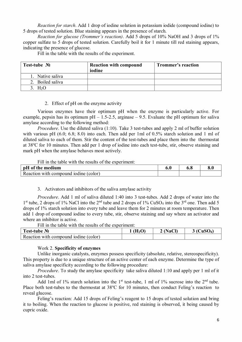
Reaction for starch. Add 1 drop of iodine solution in potassium iodide (compound iodine) to 5 drops of tested solution. Blue staining appears in the presence of starch.
Reaction for glucose (Trommer’s reaction). Add 5 drops of 10% NaOH and 3 drops of 1% copper sulfate to 5 drops of tested solution. Carefully boil it for 1 minute till red staining appears, indicating the presence of glucose.
Fill in the table with the results of the experiment.
Test-tube № Reaction with compound iodine
Trommer’s reaction
1. Native saliva 2. Boiled saliva 3. H2O
2. Effect of pH on the enzyme activity Various enzymes have their optimum pH when the enzyme is particularly active. For
example, pepsin has its optimum pH – 1.5-2.5, arginase – 9.5. Evaluate the pH optimum for saliva amylase according to the following method:
Procedure. Use the diluted saliva (1:10). Take 3 test-tubes and apply 2 ml of buffer solution with various pH (6.0; 6.8; 8.0) into each. Then add per 1ml of 0.5% starch solution and 1 ml of diluted saliva to each of them. Stir the content of the test-tubes and place them into the thermostat at 38oC for 10 minutes. Then add per 1 drop of iodine into each test-tube, stir, observe staining and mark pH when the amylase behaves most actively.
Fill in the table with the results of the experiment:
pH of the medium 6.0 6.8 8.0 Reaction with compound iodine (color)
3. Activators and inhibitors of the saliva amylase activity Procedure. Add 1 ml of saliva diluted 1:40 into 3 test-tubes. Add 2 drops of water into the
1st tube, 2 drops of 1% NaCl into the 2nd tube and 2 drops of 1% CuSO4 into the 3rd one. Then add 5 drops of 1% starch solution into every tube and leave them for 2 minutes at room temperature. Then add 1 drop of compound iodine to every tube, stir, observe staining and say where an activator and where an inhibitor is active.
Fill in the table with the results of the experiment: Test-tube № 1 (H2O) 2 (NaCl) 3 (CuSO4) Reaction with compound iodine (color)
Work 2. Specificity of enzymes Unlike inorganic catalysts, enzymes possess specificity (absolute, relative, stereospecificity).
This property is due to a unique structure of an active center of each enzyme. Determine the type of saliva amylase specificity according to the following procedure:
Procedure. To study the amylase specificity take saliva diluted 1:10 and apply per 1 ml of it into 2 test-tubes.
Add 1ml of 1% starch solution into the 1st test-tube, 1 ml of 1% sucrose into the 2nd tube. Place both test-tubes to the thermostat at 38oC for 10 minutes, then conduct Feling’s reaction to reveal glucose.
Feling’s reaction: Add 15 drops of Feling’s reagent to 15 drops of tested solution and bring it to boiling. When the reaction to glucose is positive, red staining is observed, it being caused by cupric oxide.
7
Fill in the table with the results of the experiment:
Test tube № Enzyme Substrate Feling’s reaction 1 2
Conclusions:
2. Topic: REGULATION OF ENZYME ACTIVITY. DETERMINATION OF ENZYME
ACTIVITY
Objective To learn how enzyme activity can be regulated by specific and nonspecific factors to
understand action of medicines which regulate enzyme activity, to get acquainted with the role of enzymes in diseases diagnosis and treatment monitoring.
Problems for discussion
1. The mechanism of enzymatic catalysis. The theory of the intermediate enzyme-substrate complexes, the types of relationships.
2. An active site of the enzyme, its organization. The theory explaining the work of the active site.
3. Structure peculiarities of allosteric enzymes, allosteric center. The concept of a “key enzyme”.
4. Regulation mechanisms of the enzyme-catalyzed processes rate: regulation of the enzymes amount (synthesis, break-down), enzyme activity, modification of the substrate amount, the presence of isoenzymes, joining enzymes into multienzyme complexes, compartmentation of processes.
5. Key enzymes. 6. Regulation of enzyme activity: covalent modification, activators and inhibitors
(examples). Types of inhibition (irreversible and reversible, isosteric and allosteric), characteristic, examples.
7. Multiple forms of enzymes (isoenzymes and true multiple forms), examples, their biological role.
8. Medical aspects of enzymology.
Recommended literature
1. Konevalova N.Yu., Buyanova S.V. Biochemistry lecture course. // Vitebsk, 2005.- P.27-37, 21-22.
2. Lecture material.
Practical part Determination of saliva α-amilase activity The method is based on evaluation of the least amount of amylase (at maximum saliva
dilution) that completely digests the whole added starch. Amilase activity of the saliva is expressed by the amount of 0.1% of starch solution (in ml) that is digested by 1 ml of undiluted saliva at 38oC for 30 minutes. Normal saliva amylase activity is 160-320. This method is widely used for evaluation of amylase activity of the blood and urine.
Procedure. Apply per 1 ml of water into 10 test-tubes and add 1ml of diluted saliva into the first one. Stir the content of this tube by drawing it in and out from the pipette several times. Take
8
into the pipette 1 ml of the mixture and put it into the 2nd test-tube. Stir the content of this tube and put 1 ml of it into the 3rd tube and so on to the 10th test tube. Take 1 ml of mixture from the 10th test-tube and dispose it. Add per 1 ml of water and 2 ml of 0.1% of starch solution, stir it shaking the test-tubes and place them into the thermostat at 38oC for 30 minutes. Cool the test-tubes after incubation under running water, add 1 drop of 0.1% iodine solution into each tube and stir. The fluid in the tubes is stained in yellow, rose and violet color. Mark the last tube with yellow staining where the hydrolysis has been completed and make calculations. Put down the results into the table:
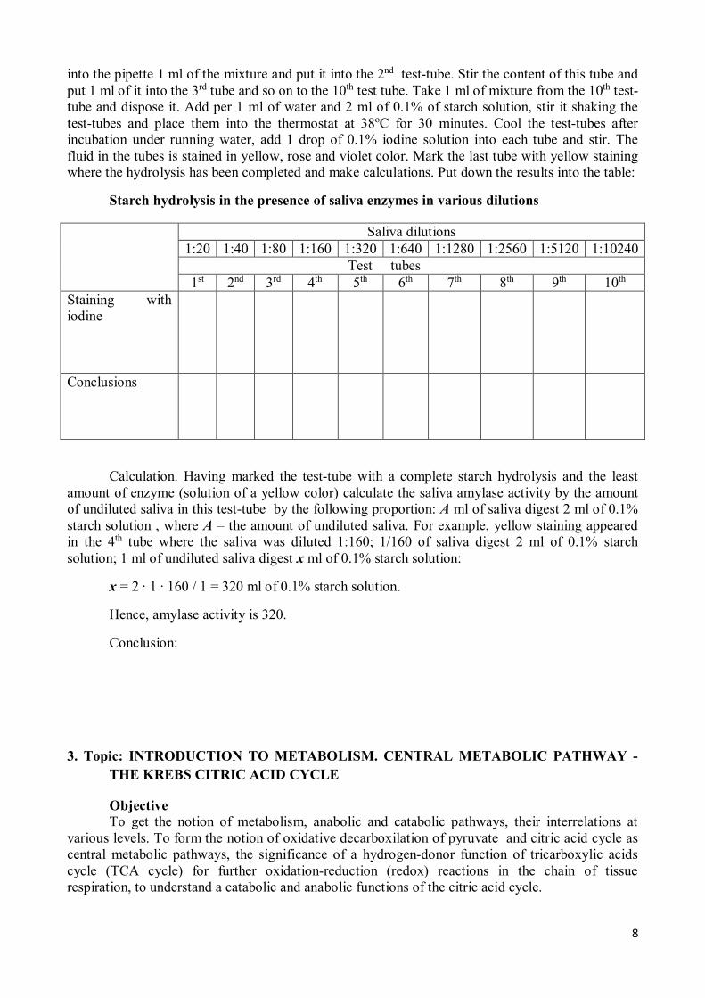
Starch hydrolysis in the presence of saliva enzymes in various dilutions
Saliva dilutions 1:20 1:40 1:80 1:160 1:320 1:640 1:1280 1:2560 1:5120 1:10240 Test tubes 1st 2nd 3rd 4th 5th 6th 7th 8th 9th 10th Staining with iodine
Conclusions
Calculation. Having marked the test-tube with a complete starch hydrolysis and the least amount of enzyme (solution of a yellow color) calculate the saliva amylase activity by the amount of undiluted saliva in this test-tube by the following proportion: A ml of saliva digest 2 ml of 0.1% starch solution , where A – the amount of undiluted saliva. For example, yellow staining appeared in the 4th tube where the saliva was diluted 1:160; 1/160 of saliva digest 2 ml of 0.1% starch solution; 1 ml of undiluted saliva digest x ml of 0.1% starch solution:
x = 2 ∙ 1 ∙ 160 / 1 = 320 ml of 0.1% starch solution.
Hence, amylase activity is 320.
Conclusion:
3. Topic: INTRODUCTION TO METABOLISM. CENTRAL METABOLIC PATHWAY - THE KREBS CITRIC ACID CYCLE
Objective To get the notion of metabolism, anabolic and catabolic pathways, their interrelations at
various levels. To form the notion of oxidative decarboxilation of pyruvate and citric acid cycle as central metabolic pathways, the significance of a hydrogen-donor function of tricarboxylic acids cycle (TCA cycle) for further oxidation-reduction (redox) reactions in the chain of tissue respiration, to understand a catabolic and anabolic functions of the citric acid cycle.
9
Problems for discussion 1. Metabolism, linear and cyclic metabolic pathways, regulatory (key) enzymes. 2. Catabolism and anabolism, their distinctions and interrelations. 3. Reactions of dehydrogenation as a basic way of oxidizing substances in the organism.
Pyridine-dependent and flavin-dependent dehydrogenases. The role of vitamins PP and B2 in redox reactions. Block-structures of coenzymes NAD+, NADP+, FAD, FMN.
4. Adenilate system of the cell, its participation in energy exchange. The central role of ATP (adenosine triphosphate) in processes coupled with energy consumption. Ways of ATP synthesis: substrate-level, oxidative and photosynthetic phosphorylation. The concept of high-energy compounds.
5. Tricarboxylic acid cycle as a central metabolic pathway. Cellular localization, reactions, enzymes, co-enzymes.
6. Dehydrogenase reactions of TCA cycle as a source of hydrogen for the system of tissue respiration. Decarboxylation in the citric acid cycle as a cellular CO2 formation mechanism that is an end product of carbonic compounds catabolism.
7. The functions of TCA cycle: integrative, catabolic, anabolic, energetic, hydrogen-donor. Regulation. Anaplerotic reactions.
Recommended literature
1. Konevalova N.Yu., Buyanova S.V. Biochemistry lecture course. // Vitebsk, 2005.- P.49-68, 86-92.
2. Lecture material.
Practical part
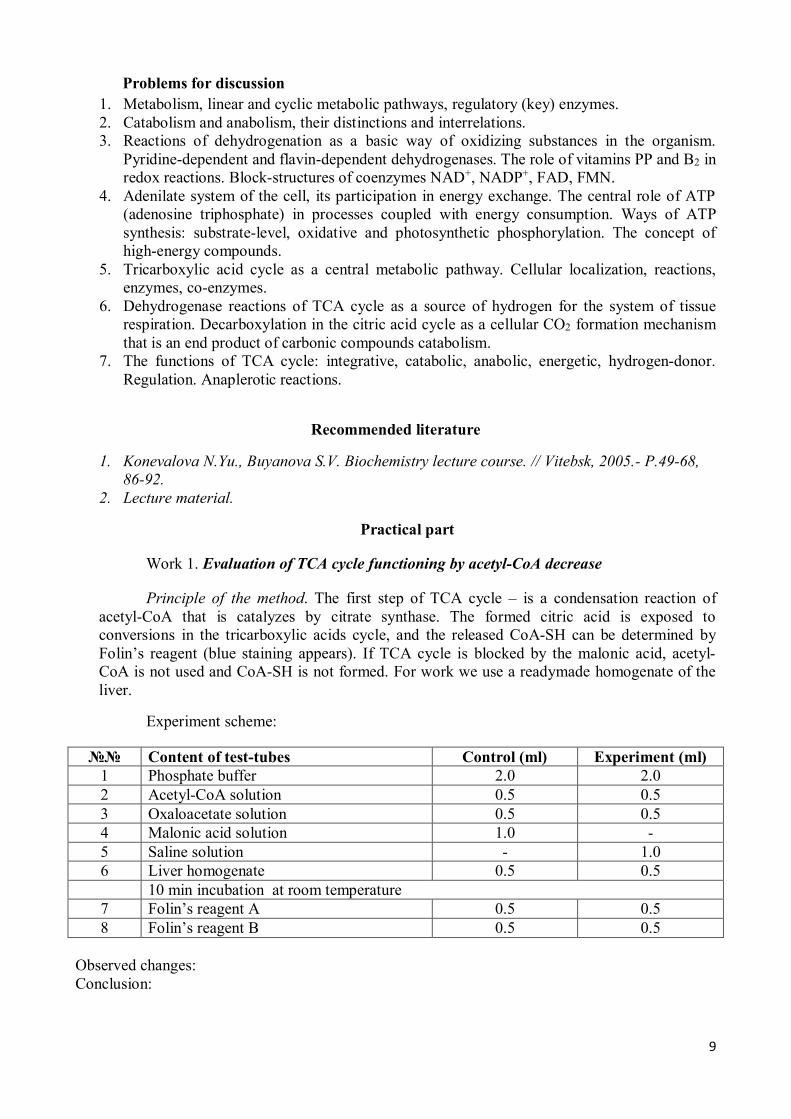
Work 1. Evaluation of TCA cycle functioning by acetyl-CoA decrease
Principle of the method. The first step of TCA cycle – is a condensation reaction of acetyl-CoA that is catalyzes by citrate synthase. The formed citric acid is exposed to conversions in the tricarboxylic acids cycle, and the released CoA-SH can be determined by Folin’s reagent (blue staining appears). If TCA cycle is blocked by the malonic acid, acetyl-CoA is not used and CoA-SH is not formed. For work we use a readymade homogenate of the liver.
Experiment scheme:
№№ Content of test-tubes Control (ml) Experiment (ml) 1 Phosphate buffer 2.0 2.0 2 Acetyl-CoA solution 0.5 0.5 3 Oxaloacetate solution 0.5 0.5 4 Malonic acid solution 1.0 - 5 Saline solution - 1.0 6 Liver homogenate 0.5 0.5 10 min incubation at room temperature 7 Folin’s reagent A 0.5 0.5 8 Folin’s reagent B 0.5 0.5
Observed changes: Conclusion:
10
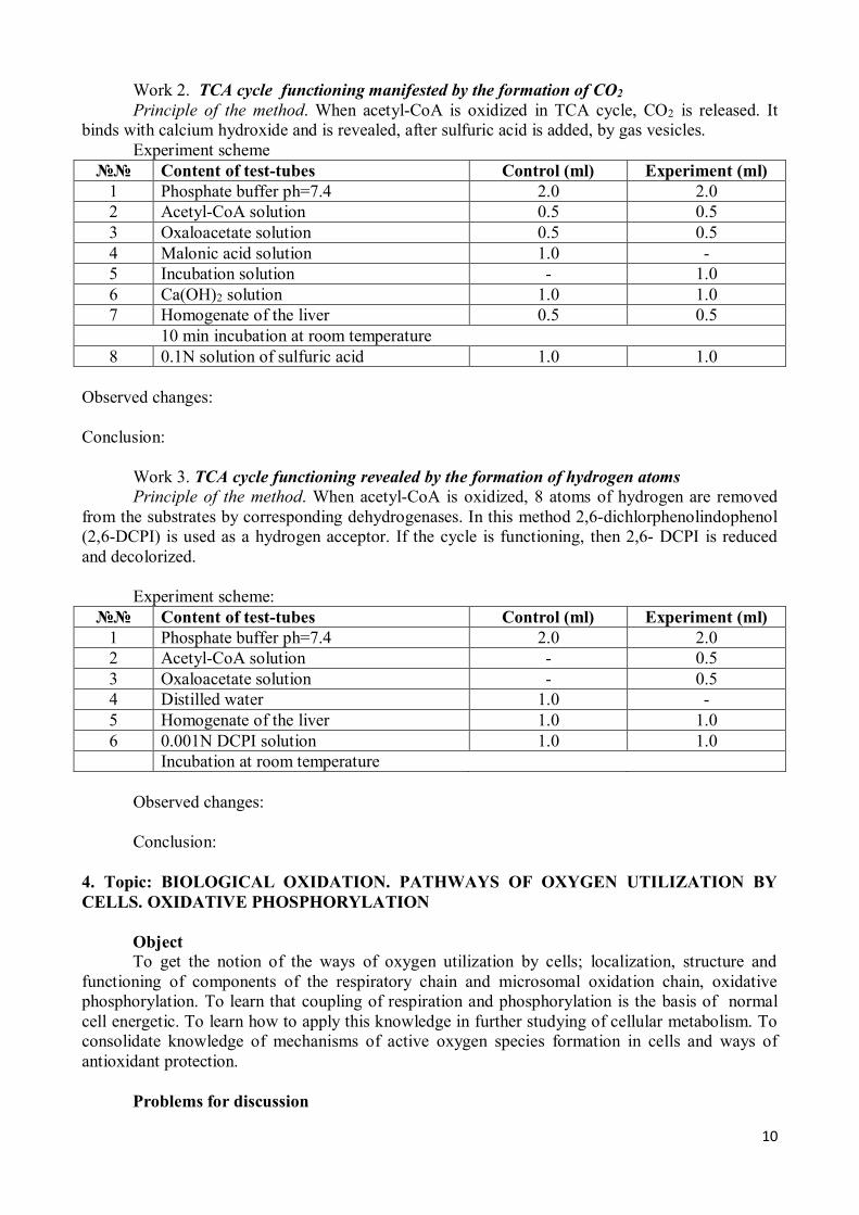
Work 2. TCA cycle functioning manifested by the formation of CO2 Principle of the method. When acetyl-CoA is oxidized in TCA cycle, CO2 is released. It
binds with calcium hydroxide and is revealed, after sulfuric acid is added, by gas vesicles. Experiment scheme
№№ Content of test-tubes Control (ml) Experiment (ml) 1 Phosphate buffer ph=7.4 2.0 2.0 2 Acetyl-CoA solution 0.5 0.5 3 Oxaloacetate solution 0.5 0.5 4 Malonic acid solution 1.0 - 5 Incubation solution - 1.0 6 Ca(OH)2 solution 1.0 1.0 7 Homogenate of the liver 0.5 0.5 10 min incubation at room temperature 8 0.1N solution of sulfuric acid 1.0 1.0
Observed changes: Conclusion:
Work 3. TCA cycle functioning revealed by the formation of hydrogen atoms Principle of the method. When acetyl-CoA is oxidized, 8 atoms of hydrogen are removed
from the substrates by corresponding dehydrogenases. In this method 2,6-dichlorphenolindophenol (2,6-DCPI) is used as a hydrogen acceptor. If the cycle is functioning, then 2,6- DCPI is reduced and decolorized.
Experiment scheme:
№№ Content of test-tubes Control (ml) Experiment (ml) 1 Phosphate buffer ph=7.4 2.0 2.0 2 Acetyl-CoA solution - 0.5 3 Oxaloacetate solution - 0.5 4 Distilled water 1.0 - 5 Homogenate of the liver 1.0 1.0 6 0.001N DCPI solution 1.0 1.0 Incubation at room temperature
Observed changes: Conclusion:
4. Topic: BIOLOGICAL OXIDATION. PATHWAYS OF OXYGEN UTILIZATION BY CELLS. OXIDATIVE PHOSPHORYLATION
Object To get the notion of the ways of oxygen utilization by cells; localization, structure and
functioning of components of the respiratory chain and microsomal oxidation chain, oxidative phosphorylation. To learn that coupling of respiration and phosphorylation is the basis of normal cell energetic. To learn how to apply this knowledge in further studying of cellular metabolism. To consolidate knowledge of mechanisms of active oxygen species formation in cells and ways of antioxidant protection.
Problems for discussion
11
1. Tissue respiration as the process of substrates’ hydrogen oxidation in the respiratory chain with formation of endogenous water in cells. Distinctions of water formation in the process of tissue respiration from a similar process in vitro.
2. The structure of the respiratory chain components, enzyme complexes, co-enzymes, functioning mechanism.
3. The diagram of the respiratory chain, phosphorylation points, the mechanism of an electro-chemical gradient formation.
4. Mechanisms of mitochondrial synthesis of ATP. H+-ATP-synthase. Coupling of respiration and phosphorylation. The chemiosmotic theory of Mitchell. Phosphorylation ratio (P/O) for various substrates supplying hydrogen to the respiratory chain.
5. Regulation of the respiratory chain and H+-ATP-synthase. 6. Causes for the hypoenergetic states development. Uncoupling of oxidative
phosphorylation (mechanism, uncoupling agents). Inhibitors of electron transport and oxidative phosphorylation.
7. Microsomal oxidation, its role for the cell.
Recommended literature
1. Konevalova N.Yu., Buyanova S.V. Biochemistry lecture course. // Vitebsk, 2005.- P.69-85. 2. Lecture material.
5. COLLOQUIUM: “ENZYMES, INTRODUCTION TO METABOLISM. CENTRAL METABOLIC PATHWAY, BIOLOGICAL OXIDATION. OXIDATIVE PHOSPHORYLATION”
Questions for preparation: 1. Enzymes, classification. 2. Properties of enzymes. Thermolability, specificity, the effect of pH and concentration of
the substrate on the activity of the enzyme. The concept of the kinetics of enzymatic reactions and Michaelis constants.
3. Enzyme active site and its structure. 4. Coenzymes, classification. 5. Mechanisms of regulation of enzyme activity, reversible and irreversible regulation,
isosteric and allosteric regulation, covalent modification of the structure of the enzyme. 6. Mechanisms of regulation of the enzyme in the cell (Jacob-Monod and Georgiev
schemes). 7. Multiple forms of enzymes (isoenzymes and actually multiple forms), examples of the
biological role. 8. Examples of enzymes and their use in medical practice regulators (including dental). 9. Metabolism. The concept of catabolism (examples) and of anabolism (examples), the
differences and levels of relationship between them. Types of metabolic pathways. The concept of linear and cyclic metabolic pathways (examples), the key (regulatory) enzymes. The central metabolic pathways.
10. Adenyl system, its components have a role in the cell. Methods for the synthesis of ATP in the cell and methods of its hydrolysis. List the reactions and processes, coupled with the hydrolysis of ATP. Their role in cells and organisms.
11. The citric acid cycle. TCA function. Write a TCA scheme, called vitamins involved in this process. What are the reactions of the Krebs cycle are associated with complexes of the respiratory chain? How many moles of ATP can still get? Show it on the scheme enzymes of tissue respiration. Calculate the energy balance of the oxidation of acetyl-CoA. Catabolic function of the Krebs cycle.
12
12. Types of biological oxidation. Vitamins B2, PP, and as participants in redox reactions. Draw a block diagram of NAD +, NADP +, FMN and FAD. Oxygenase pathway of utilization of oxygen in the cells. Scheme of the microsomal chain. Compare oxidase and oxygenase ways to utilize oxygen. The role of these processes in a cell.
13. Tissue respiration. The scheme enzymes of tissue respiration. Complexes of the respiratory chain. Show on the diagram areas with sufficient energy for the production of ATP. On the basis of what the indicator can be measured to determine the amount of energy released in the reaction of electron transfer? Regulation of the activity of respiratory chain enzymes, the role of ADP, ATP. To be able to portray the scheme of tissue respiration enzymes for NAD +-dependent and FAD-dependent substrates. To be able to include substrates of the Krebs cycle (isocitrate, α-ketoglutarate, malate, succinate) in the respiratory chain. To know what is the coefficient of phosphorylation ( P/O) for each of these substrates.
14. Oxidative phosphorylation (definition, the subcellular localization). Basics of the theory P. Mitchell, explaining the mechanism of oxidative phosphorylation. How does the process of oxidative phosphorylation by deficiencies of oxygen in the cells (to explain the mechanism)? Compare the mechanisms of oxidative and substrate phosphorylation. Which one prevails in the mitochondria?
15. Causes for the hypoenergetic states development. Electron transport inhibitors respiratory chain.
6. Topic: DIGESTION OF CARBOHYDRATES. GLYCOGENESIS AND GLYCOGENOLYSIS. GLYCOLYSIS
Objective To consolidate knowledge of the carbohydrates structure of animal tissues and dietary
vegetable carbohydrates. To form the notion of carbohydrate digestion, glucose transport to cells, molecular mechanisms of glycogen storage and mobilization, physiological significance and regulation of these pathways. To learn anaerobic pathways of glucose oxidation and their significance.
Problems for discussion
1. Carbohydrates digestion, final products. The role of cellulose and pectin in the human diet.
2. Absorption of carbohydrates digestion products, molecular mechanisms. The fate of absorbed monosaccharides. Glucose transport to cells.
3. Glycogen synthesis, purpose, sequence of reactions, expenditure of energy and regulation.
4. Degradation of glycogen in the liver and muscles, sequence of reactions, regulation. 5. Glycolysis, its biological role, subcellular localization, phases (preparatory or
unoxidative, oxidative), reactions, energy yield and mechanism of ATP formation. Glycolysis regulation, key enzymes.
Recommended literature
1. Konevalova N.Yu., Buyanova S.V. Biochemistry lecture course. // Vitebsk, 2005.- P.93-98, 112-119.
2. Lecture material.
13
Practical part
Determination of pyruvate in the urine Pyruvate is one of intermediate products of carbohydrate metabolism. Under anaerobic
conditions (hypoxia) pyruvate is reduced into lactate. Under aerobic conditions pyruvate under the influence of a pyruvate dehydrogenase complex (coenzymes: TPP, lipoamide, CoA-SH, NAD+, FAD) as a result of oxidative decarboxylation is converted into acetyl-CoA that in the citric acid cycle is oxidized to CO2 and H2O.
During 24 hours 113.7-283.9 µM/24 h (10-25 mg) of pyruvate are excreted with urine. Principle of the method. Pyruvate interacting with 2,4-dinitrophenylhydrasine in alkaline
medium forms 2,4-dinitrophenylhydrasone derivatives of yellow-orange color, the staining intensity of which is proportional to concentration of pyruvate.
Procedure. Take 2 test-tubes: apply 1 ml of H2O into a control one and 1 ml of urine into a test tube. Then add into both test-tubes 0.5 ml of 2,4-dinitrophenylhydrosine solution and leave them for 20 minutes at room temperature. Then add 5 ml of 0.4N NaOH into each tube and in 10 minutes determine the optical density of the test sample versus the control sample using 10mm cuvettes with a green light filter.
Calculation is performed according to a ready calibration graph. Recount the found value by daily diuresis (1500 ml for men and 1200 ml for women) and get the content of pyruvate in daily urine.
Clinical and diagnostic value. In avitaminosis
and hypovitaminosis of B1 in the blood and other tissues, especially in the brain, a great amount of pyruvate is accumulated and its excretion with urine increases. The content of this acid in the blood increases in diabetes mellitus, cardiac insufficiency, hyperfunction of the hypophysis-adrenal system. The amount of pyruvate increases after injection of some medicines – camphor, strychnine, epinephrine. In anesthesia the content of this acid in the blood decreases.
Conclusion:
7. Topic: METABOLIC PATHWAYS OF PYRUVATE. GLUCONEOGENESIS. AEROBIC
OXIDATION OF GLUCOSE TO FINAL PRODUCTS (CO2 AND H2O)
Objective To consolidate knowledge of pyruvate fate in cells depending on the energetic status and
peculiarities of cellular metabolism, gluconeogenesis as an important process of the blood glucose level maintaining. To form the notion of interconnection between central metabolic pathways and aerobic glycolysis. To master the enzymatic method of glucose measurement in blood.
Problems for discussion
1. Pyruvate as a central metabolite. Pathways of pyruvate conversion depending on the energetic status and peculiarities of oxidative cellular metabolism.
2. Recovery of pyruvate to lactate (reaction, LDH isoenzymes, the appointment of reactions), Cori cycle. Disposal of lactate by cells.
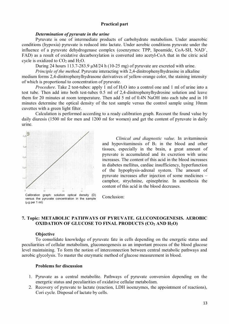
Calibration graph: solution optical density (D) versus the pyruvate concentration in the sample (mg per 1 ml)
14
3. Gluconeogenesis (purpose, substrates, key reactions and enzymes, regulation, expenditure of energy).
4. Oxidative decarboxylation of pyruvate (biological role, subcellular localization, reactions); pyruvate dehydrogenase complex (enzymes, coenzymes), regulation of pyruvate dehydrogenase activity.
5. Citric acid cycle (subcellular localization, reactions, energetic balance, enzymes, regulation, biological role).
6. Aerobic oxidation of glucose to CO2 and H2O (steps associated with oxidative phosphorylation, energy yield).
Recommended literature
1. Konevalova N.Yu., Buyanova S.V. Biochemistry lecture course. // Vitebsk, 2005.- P.98-108. 2. Lecture material.
8. Topic: SECONDARY PATHWAYS OF GLUCOSE METABOLISM. EFFECT OF
HORMONES ON THE BLOOD GLUCOSE LEVEL. FEATURES CARBOHYDRATE UTILIZATION OF ORAL MICROFLORA
Objective To form understanding of the significance of pentose phosphate and glucuronic pathways
of glucose metabolism; to learn the role of hormonal regulation in maintaining glucose concentration in the blood to know how to interpret the character of biochemical impairments in patients with pathology of carbohydrate metabolism. To understand the pathogenetic relationship of carbohydrate foods and tooth decay.
Problems for discussion
1. Pentose phosphate pathway (subcellular localization, steps, key enzymes, metabolites, biological role).
2. Glucuronic pathway (tissue and subcellular localization, biological role). 3. Regulation of blood glucose content. Mechanisms of hormonal regulation (insulin,
epinephrine, glucagon, glucocorticoids etc.). 4. Features of carbohydrate utilization oral microflora.
Recommended literature
1. Konevalova N.Yu., Buyanova S.V. Biochemistry lecture course. // Vitebsk, 2005.- P.108-111, 119-121, 122-126.
2. Lecture material.
Practical part
Effect of hormones on blood glucose content To study the effect of hormones on the blood glucose level take 3 blood samples (tested).
One of them was taken before applying hormones, the second – after injecting insulin, the third one – after injecting epinephrine.
1. Evaluate glucose content in every sample. 2. On the basis of received data make a conclusion which of the samples corresponds to the
above states.
15
To evaluate glucose concentrations in the samples use the enzymatic (glucose oxidase) method. The tested and standard samples are performed in parallel.
Procedure: see the preceding classes. A control sample can be prepared one for the whole group.
Calculate glucose concentration according to the formula: Ct (mg per 100 ml) = Et. ∙ Cs / Es. Conversion factor to SI units (mmol/l) = 0.0555.
Sample Optical density (E) Glucose concentration (mmol/l) 1 2 3
standard Conclusion:
9. COLLOQUIUM: “CARBOHYDRATE METABOLISM” Questions for preparation:
1. The digestion of carbohydrates. The biological role of dietary fiber. Mechanisms suction carbohydrates in the intestine.
2. The mechanism of glucose transport into cells. 3. The chemistry of oxidative and non-oxidative reactions of the stages of anaerobic glycolysis.
Biological role of glycolysis, the appointment of lactate dehydrogenase reaction. Regulation of glycolysis. Energy yield of glycolysis. Synthesis of ATP under anaerobic conditions.
4. Aerobic glucose oxidation. Stages and their subcellular localization, energy yield and the mechanisms of ATP synthesis. The key enzymes and their regulators. Compare the energy yield of anaerobic and aerobic (in stages) breakdown of glucose and mechanisms of ATP synthesis.
5. The fate of the end product of glycolysis - pyruvic acid and lactic acid. What is the fate of lactate formed in red blood cells? Metabolic pathways of pyruvate. The oxidative decarboxylation of pyruvic acid (biological role of sub-cellular localization, reaction). Pyruvate dehydrogenase complex (enzymes, cofactors), the regulation of pyruvate dehydrogenase activity.
6. Gluconeogenesis. The biological role, subcellular localization, substrates, key enzymes and regulation of the process. The chemistry of the key reactions.
7. The scheme glycogenesis in hepatocytes and myocytes. Hormonal regulation of glycogenesis.
8. Glycogenolysis, the biological role. What is phosphorolysis and hydrolysis of glycogen? The scheme phosphorolysis of glycogen. Know the difference of this process in the liver and muscles. Show scheme glycogenolysis under the influence of glucagon, tissue localization. The mechanisms of activation of glycogen phosphorylase adrenaline (scheme).
9. Pentose phosphate pathway breakdown of glucose, the stages, the biological role. 10. Formation of UDP-glucose. What is the role of biological processes using UDP-glucose?
Meaning glucuronic path in the liver and fibroblasts. 11. Physiological concentrations of glucose in the blood. Hormonal regulation of blood glucose
levels. 12. Features carbohydrate utilization oral bacteria. Differences in the synthesis of glycogen in
humans and bacteria. The synthesis of extracellular polysaccharides (dextrans, levans) and
16
their purpose. Cleavage of sucrose (fructose, glucose) of oral bacteria. Dependence of the Ca2+ from the enamel of the teeth on the pH of saliva. Chemical-parasitic theory of tooth decay. Role of sugar alcohols (sorbitol, xylitol) in the prevention of caries.
13. Substrate phosphorylation and oxidative phosphorylation. Chain enzymes of tissue respiration (scheme). What is the ratio of P/O in the glycolytic NADH.H+ oxidation in the respiratory chain? Name the tissue respiration substrates formed during the aerobic oxidation of glucose, and indicate the place of incorporation into the respiratory chain.
10. Topic: LIPID METABOLISM. DIGESTION AND RE-SYNTHESIS. EVALUATION OF
LIPASE ACTIVITY
Objective To consolidate knowledge of lipids chemistry. To learn molecular mechanisms of digestion
and absorption of lipids from food, re-synthesis of lipids. Problems for discussion 1. General characteristics and classification of lipids (saponifiable and unsaponifiable,
simple and complex). Characteristic of lipid groups (chemical formulas and terminology of acylglyceroles and glycerophospholipids; block-structures of waxes, sphingophospholipids, glycolipids, sulfolipids). Biological role of lipids.
2. Food lipids. Lipids digestion, phases. Emulsification (purpose, factors, stabilization of fat emulsion). Bile, bile acids (primary, conjugated, secondary). Place of formation, participation in assimilation of food lipids. Enterohepatic re-circulation of bile acids.
3. Hydrolysis of lipids (conversion patterns). Enzymes (place of formation, substrate specificity). Activation of pancreatic lipase. Absorption (mechanisms, micellar dissolution, fate of micelles).
4. Re-synthesis of triacylglycerols and glycerophospholipids in enterocytes. Transport forms of lipids in the blood. Structure and metabolism of chylomicrons.
Recommended literature
1. Konevalova N.Yu., Buyanova S.V. Biochemistry lecture course. // Vitebsk, 2005.- P.127-129, 134-135.
2. Lecture material.
Practical part
Work 1. Kinetics of pancreatic lipase Principle of the method. The lipase action rate in separate portions of milk is evaluated by
the amount of fatty acids formed in hydrolysis of milk fat for a definite interval. The amount of fatty acids is determined by alkaline titration.
Procedure. Prepare two test-tubes containing 5 ml of milk and 1 ml of 5% pancreatine (pancreas juice). Add 1 ml of water into one test-tube and 1 ml of bile – into the other. Quickly stir the fluid in the test-tubes. Take 1 ml of the mixture from every tube and apply into flasks, add 1-2 drops of 0.5% phenolphthalein solution and titrate by 0.05N solution of NaOH to a light-rose color, which doesn’t disappear for 30 seconds. Place both test-tubes with remaining mixture into the thermostat at 38oC. Every 10 minutes take out 1 ml of the mixture and titrate by 0.05N solution of NaOH in the presence of phenolphthalein to a light-rose color. Perform 5 such determinations and on the basis of received data construct 2 curves, they will reflect the process of fat hydrolysis by lipase vs time and dependence on the presence or absence of bile.
17
Conclusion: Work 2. Action of the pancreatic phospholipases Principle of the method. The pancreatic phospholipases action on glycerophospholipids of
egg yolk is manifested by the appearance of free phosphoric acid capable of forming a yellow precipitate in heating with ammonia molybdate.
Procedure. Apply per 5 drops of egg yolk suspension into 2 test-tubes. Add 2 drops of pancreatine into the first tube, and 2 drops of water into the second (control) tube. Place both test-tubes into the thermostat at 38oC for 30 minutes. After incubation add 5 drops of molybdenum reagent into both tubes, heat them over the burner and cool under running water.
Conclusion:
11. Topic: INTRACELLULAR METABOLISM OF FATTY ACIDS. DETERMINATION OF PLASMA β-LIPOPROTEINS
Objective To study the processes of oxidation and synthesis of fatty acids. To form the notion of
eicosanoids and their functions. To acquire skills of β-lipoproteins determination in serum.
Problems for discussion 1. β-Oxidation as a central pathway of fatty acids catabolism. Subcellular localization of
the process, activation of fatty acids, transport to mitochondria. Oxidation reactions, participation of vitamins. Association with oxidative phosphorylation, energetic yield. β-oxidation of fatty acids with an odd number of carbons, unsaturated fatty acids. Peculiarities of β-oxidation in peroxisomes.
2. Glycerol metabolism in cells. The energy balance of the oxidation of glycerol. 3. Acetyl-CoA as a central metabolite. 4. Biosynthesis of fatty acids. Subcellular localization, substrates, reactions, regulation.
Peculiarities of fatty acid synthase structure. The role of malic-enzyme. 5. Polyunsaturated fatty acids as essential nutritive factors: representatives, biological role. 6. Metabolism of arachidonic acid. Biosynthesis of eicosanoids (prostaglandins,
prostacyclins, leukotriens, thromboxans) and their biological role.
Recommended literature
1. Konevalova N.Yu., Buyanova S.V. Biochemistry lecture course. // Vitebsk, 2005.- P.129-133,137-142.
18
2. Lecture material.
Practical part
Determination of plasma β-lipoproteins (low density lipoproteins) The majority of lipids are not free in the blood, but compose protein-lipid complexes
(lipoproteins). Fractions of lipoproteins differ in their molecular mass, amount of protein, percentage of individual lipid components. Lipoproteins can be separated by various methods: electrophoresis, thin-layer chromatography, ultracentrifugation in density gradient. Electrophorethic separation (on chromatographic paper, acetate cellulose, agar, in polyacrylamide gel) gives fractions of chylomicrons (immobile) and lipoproteins of various density: α-lipoproteins (HDL) have mobility of α-globulins, β-lipoproteins (LDL) have mobility of β-globulins. Pre-β-lipoproteins (VLDL) are located on the electrophoregram before β-lipoproteins from the start line, that’s why they are called this way.
Evaluation of β-lipoproteins in the blood plasma is important for diagnosing atherosclerosis, acute and chronic liver diseases, xanthomatosis and other pathologies.
Principle of the method. The method is based on the ability of β-lipoproteins (VLDL) to sediment in the presence of calcium chloride and heparin; the solution turbidity being changed. Concentration of β-lipoproteins in plasma is determined by the degree of solution turbidity.
Procedure. Apply 2 ml of 0.025M solution of CaCl2 and 0.2 ml of blood plasma into a test-tube and stir. Evaluate optical density of the solution (E1) versus CaCl2 solution in cuvettes 5 mm thick under a red light filter (630 nm). Add 0.1 ml of heparin solution into the cuvette, stir and exactly in 4 minutes evaluate the solution optical density (E2) under the same conditions.
Calculation. Calculate the difference of optical densities and multiply by 10 – an empiric factor suggested by Ledvina, because the construction of a calibrating curve is associated with a number of difficulties (x (g/l) = (E2-E1)·10). Normal values for β-lipoproteins content – 3-4.5 g/l. The content of β-lipoproteins varies depending on the age and sex.
E1= E2= x (g/l) = (E2-E1)·10 =
Conclusion:
12. Topic: CHOLESTEROL AND KETONE BODIES METABOLISM. DETERMINATION
OF CHOLESTEROL IN SERUM Objective Learn to analyze the content of cholesterol in the blood and urine ketone bodies in the study
of diseases caused by disorders of lipid metabolism. Problems for discussion 1. Cholesterol, the biological role, food sources. Elimination of cholesterol from the
organism, bile acids as a major end product of cholesterol metabolism, cholelithiasis. 2. Cholesterol biosynthesis (tissue and subcellular localization, substrates, phases, reactions
of the 1st phase, regulation). 3. Ketogenesis: tissue and subcellular localization, substrates, chemistry. Synthesis of
ketone bodies. Molecular mechanisms of ketonemia in diabetes, insufficient carbohydrate diet, starvation. Utilization of ketone bodies (interconversion, activation, including in metabolism, energy oxidation).
19
4. The mobilization of lipids from adipose tissue (scheme, cAMP-dependent mechanism of activation of hormone-sensitive lipase, hormone regulation). The role of the deposition and mobilization of fat, violations of these processes in obesity.
Recommended literature
1. Konevalova N.Yu., Buyanova S.V. Biochemistry lecture course. // Vitebsk, 2005.- P.135-136, 142-146, 147-153.
2. Lecture material.
Practical part
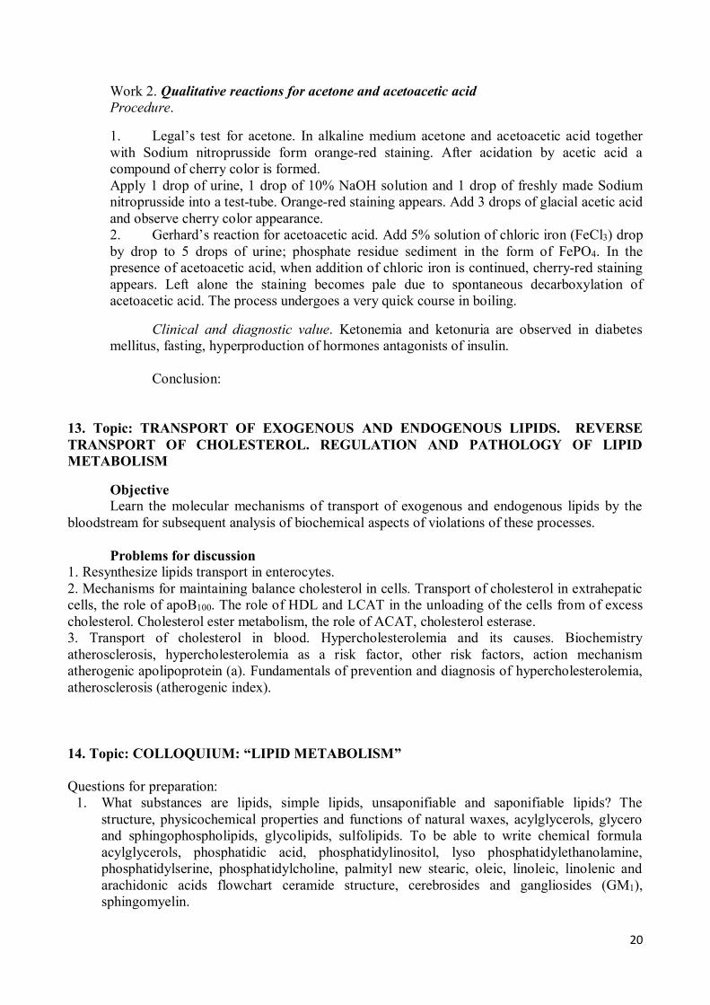
Work 1. Determination of total cholesterol concentration in serum Principle of the method. The method is based on the ability of cholesterol to give green
staining in chloroform in association with acetic anhydride and concentrated sulfuric acid. Procedure. Apply 0.1 ml of blood serum to the bottom of a dry test-tube. Measure 2.1 ml of
reagent №1 (a mixture of glacial acetic acid, acetic anhydride and concentrated sulphuric acid) with a cylinder. Work with reagent №1 very carefully, don’t take it with a pipette and don’t spill, as it is a mixture of concentrated acids! Pour reagent №1 into the test-tube with serum, stir up and place into the thermostat at 37oC for 20 minutes (or at room temperature). Green staining appears. The intensity of staining is measured photometrically in a 5 mm thick cuvette vs water under a red light filter. Use a readymade calibration graph to determine the content of cholesterol in serum sample. Multiply the found cholesterol concentration by 1000, because 0.1 ml of serum was taken during the experiment. Conversion factor to SI units (mmol/l) is 0.0258.
Calibration graph for determination of cholesterol concentration
E=
C (mg per 100 ml) =
C (mmol/l) =
The normal values of cholesterol content in serum are 3.9 - 6.2 mmol/l (150-240 mg per 100 ml).
Clinical and diagnostic value. When fat metabolism is impaired, cholesterol may
accumulate in the blood. The increased plasma cholesterol level (hypercholesterolemia) is observed in atherosclerosis, diabetes mellitus, mechanic jaundice, nephritis (especially in lipoid nephrosis), hypothyrosis. Decrease of cholesterol in the blood (hypocholesterolemia) is observed in anemias, fasting, tuberculosis, hyperthyroidism, cancerous cachexia, impairment of the central nervous system, feverish states.
Conclusion:
20
Work 2. Qualitative reactions for acetone and acetoacetic acid Procedure.
1. Legal’s test for acetone. In alkaline medium acetone and acetoacetic acid together with Sodium nitroprusside form orange-red staining. After acidation by acetic acid a compound of cherry color is formed. Apply 1 drop of urine, 1 drop of 10% NaOH solution and 1 drop of freshly made Sodium nitroprusside into a test-tube. Orange-red staining appears. Add 3 drops of glacial acetic acid and observe cherry color appearance. 2. Gerhard’s reaction for acetoacetic acid. Add 5% solution of chloric iron (FeCl3) drop by drop to 5 drops of urine; phosphate residue sediment in the form of FePO4. In the presence of acetoacetic acid, when addition of chloric iron is continued, cherry-red staining appears. Left alone the staining becomes pale due to spontaneous decarboxylation of acetoacetic acid. The process undergoes a very quick course in boiling.
Clinical and diagnostic value. Ketonemia and ketonuria are observed in diabetes mellitus, fasting, hyperproduction of hormones antagonists of insulin.
Conclusion:
13. Topic: TRANSPORT OF EXOGENOUS AND ENDOGENOUS LIPIDS. REVERSE TRANSPORT OF CHOLESTEROL. REGULATION AND PATHOLOGY OF LIPID METABOLISM
Objective Learn the molecular mechanisms of transport of exogenous and endogenous lipids by the
bloodstream for subsequent analysis of biochemical aspects of violations of these processes. Problems for discussion
1. Resynthesize lipids transport in enterocytes. 2. Mechanisms for maintaining balance cholesterol in cells. Transport of cholesterol in extrahepatic cells, the role of apoB100. The role of HDL and LCAT in the unloading of the cells from of excess cholesterol. Cholesterol ester metabolism, the role of ACAT, cholesterol esterase. 3. Transport of cholesterol in blood. Hypercholesterolemia and its causes. Biochemistry atherosclerosis, hypercholesterolemia as a risk factor, other risk factors, action mechanism atherogenic apolipoprotein (a). Fundamentals of prevention and diagnosis of hypercholesterolemia, atherosclerosis (atherogenic index). 14. Topic: COLLOQUIUM: “LIPID METABOLISM” Questions for preparation:
1. What substances are lipids, simple lipids, unsaponifiable and saponifiable lipids? The structure, physicochemical properties and functions of natural waxes, acylglycerols, glycero and sphingophospholipids, glycolipids, sulfolipids. To be able to write chemical formula acylglycerols, phosphatidic acid, phosphatidylinositol, lyso phosphatidylethanolamine, phosphatidylserine, phosphatidylcholine, palmityl new stearic, oleic, linoleic, linolenic and arachidonic acids flowchart ceramide structure, cerebrosides and gangliosides (GM1), sphingomyelin.
21
2. Mechanisms for re-synthesis of lipids in enterocytes. The formation of chylomicrons, their composition and structure.
3. Serum lipoproteins. Classification, composition, place of education, interconversion. The role of lipoprotein levels. Lecithin: cholesterol acyltransferase (LCAT) and its role.
4. Steps lipid digestion in the gastrointestinal tract. Emulsifying properties of bile acids in the bowel lumen. Role taurine and glycine.
5. β-Oxidation of fatty acids. To be able to write the reaction β-oxidation of fatty acids having an even number of carbon atoms, calculate oxidation energy yield of certain fatty acid.
6. What is fatty acid β-oxidation in the peroxisomes? The difference between β-oxidation in mitochondria and peroxisome.
7. Regulatory relationship β-oxidation of fatty acids in cells, and the aerobic oxidation of glucose. Steps conversion of carbohydrates into lipids deposited (scheme).
8. Unsaturated fatty acids as a group of essential nutritional factors. Path establishment and use of arachidonic acid in the cells.
9. Types of eicosanoids, the patterns of their action, the functions in the body. The mechanism of action of nonsteroidal antiinflammatory drugs.
10. The conditions under which the activated fatty acid biosynthesis in the cells. Intracellular localization of the process and the origin of the initial substrates. The reactions of fatty acid synthesis.
11. Schemes pathways of NADPH.H+ for biosynthesis of fatty acids. 12. The scheme enzymes of tissue respiration. Complexes of the respiratory chain. Show on the
diagram areas with sufficient energy for the production of ATP. Based on what the measured parameter can be defined amount of energy released in the electron transfer reaction? Adjusting the respiratory chain, the role of ADP, ATP.
13. The scheme of tissue respiration enzymes for NAD+-dependent and FAD-dependent substrates. To be able to include substrates β-oxidation of fatty acids (β-hydroxyacyl-CoA, acyl-CoA) in the respiratory chain. To know what is the coefficient of phosphorylation (P/O) for each of these substrates. In the scheme necessarily indicate areas of conjugation of transport of electrons and phosphorylation. Role of NADP+ into the cell.
14. What is oxidative phosphorylation (definition, subcellular localization)? Basics of the theory P. Mitchell, explaining the mechanism of oxidative phosphorylation. What is the uncoupling of oxidative phosphorylation?
15. Electron transport inhibitors respiratory chain. 16. Synthesis of β-hydroxy-β-methylglutaryl-CoA and mevalonate. Ways of using these
compounds in the cell. Regulation of the activity of HMG-CoA reductase. 17. Cholesterol synthesis in cells (scheme). The regulation of this process, aimed at protecting the
cells from cholesterol overload. Mechanism for maintaining the balance's cholesterol in cells, the role of apo A1, apo D, apo B-100, LCAT, cell receptor residues chylomicrons and HDL and IDL.
18. Factors that contribute to high levels of cholesterol in the HDL, LDL within. The likely mechanism of participation of cholesterol in the development of atherosclerosis. The origin and role of foam cells. Based on the metabolism of cholesterol in the body and its regulation, to be able to justify the approaches to the reduction of its level in the blood.
19. Possible mechanisms of action of the atherogenic lipoprotein (a). 20. Propose a mechanism of the effects as a result of decreased activity of lipoprotein lipase in the
blood, hepatic lipase, apo C-2, lack receptors for apo B-48/E. 21. Possible reasons for increasing the level of free fatty acids in the blood. The role of
apolipoproteins, hormone-sensitive lipase. 22. Location action of phospholipases A1, A2, C and D on the structure of phospholipids. Lecithin
synthesis reactions. Lipotropic factors and their role in the violation of the synthesis of glycerophospholipids in the liver.
22
23. The term "ketone body", their chemical formulas. Synthesis reaction of ketone bodies, localization in the cell.
24. Ketone bodies in the process of energy production, its organ and intracellular localization, the energy yield.
25. The concept of "ketosis", the probable mechanism of its origin in diabetes and starvation. 15 Topic: PHYSICAL AND CHEMICAL PROPERTIES OF THE BLOOD.
HEMOGLOBINOSES
Objective To study physical and chemical properties of the blood, to consolidate knowledge of the
origin of plasma components and their physiological concentrations, buffer blood systems, structure and functioning of hemoglobin, gas transport in the blood and mechanisms of hypoxia development, diagnostic significance of the most important biochemical blood components.
Problems for discussion 1. Chemical composition of plasma (physiological concentrations of the most important
plasma components and their origin). 2. The most important blood buffer systems: bicarbonate, hemoglobin, phosphate, protein
(components and their proportion, mechanism of action, capacity). The notion of acid-base disturbances (acidosis, alkalosis).
3. Proteins of erythrocytes. The structure of hemoglobin, heme, globin; varieties (normal and abnormal) and derivatives of hemoglobin.
4. Respiratory function of the blood. Erythrocytes as a main participant of gas transport by the blood (the role of hemoglobin and carbanhydrase). Reversible binding of oxygen and carbon dioxide as a means of transport (binding mechanisms of CO2 and O2 with hemoglobin, co-operative interaction of hemoglobin subunits). Hypoxia, forms, mechanisms of development.
Recommended literature
1. Lecture material.
Practical part
Work 1. Buffer properties of serum The bicarbonate, protein and phosphate buffer systems function in serum. Principle of the method. Titrate I ml of blood (the 1st test-tube) and 1 ml of water (the 2nd
test-tube) with 0.1N solution of HCl in the presence of a blue bromphenol indicator (per 1 drop into every test-tube) till yellow staining appears. Compare the results of titration.
Conclusion: Work 2. Determination of chlorides in blood according to Levinson Chlorine is present in the organism mainly in the form of ions. A chloride-ion is the main
source of anions. Chlorine anions are the most important osmotic active components of blood, lymph, cerebrospinal fluid. The content of chlorine (chloride-ions) in serum of practically healthy adult people is 95-105 mmol/l. In newborns the normal concentration of serum chloride-ions is 80-140 mmol/l.
Principle of the method. The argentometric method is based on the ability of silver ions to form insoluble salts with ions of chlorine. The amount of depositing substance (AgNO3) is equivalent to the content of chloride-ions.
23
Titration of blood chloride-ions by silver nitrate is performed in the presence of indicator K2CrO4. On reaching an equivalent titration point the excess of silver ions and the indicator form a compound of a brick-red color (Ag2CrO4).
Procedure.
1. Sedimentation of blood proteins. Prepare a mixture of solutions in two test-tubes: 5 ml of 0.45% ZnSO4 + 1 ml of 0.1N NaOH. Then apply 0.1 ml of serum into the 1st tube, 0.1 ml of H2O dist. into the 2nd tube. Heat the test-tubes over the spirit-burner for 3 minutes. Then filter the content of the test-tubes into flasks through cotton wool. Rinse the residue on the cotton wool filter twice with water (per 3 ml).
2. Sedimentation of chlorine ions in the presence of K2CrO4. Add 2 drops of 1-2% solution of K2CrO4 to the filtrate and titrate it with AgNO3 till a yellow color of the solution changes to brick-red.
Calculation. Subtract from the volume of AgNO3 spent for titration of the tested solution (Vt, ml) the volume of AgNO3 spent for titration of the control solution (Vc, ml), multiply the received difference by 0.355, if the result is expressed in mg of chlorine per 0.1 ml of blood. To get a percentage value, multiply the received value by 1000. The conversion factor to SI units (mmol/l) = 0.282.
Vt (ml) = Vc (ml) = C(mmol/l) = (Vt – Vc) ∙ 0.355 ∙ 1000 ∙ 0.282 = Conclusion:
16. Topic: BLOOD PLASMA PROTEINS. BLOOD CLOTTING SYSTEM
Objective To get acquainted with principles of blood protein composition examination, to understand a
diagnostic role of protein fractions and individual plasma proteins determination. To get the notion of hemostasis mechanisms and to study functioning of the blood clotting system.
Problems for discussion 1. Blood plasma proteins. Main protein fractions: albumins, globulins, fibrinogen (content,
functions); albumin-globulin ratio and its diagnostic value. 2. Blood plasma enzymes (secretory, indicator, excretory). Diagnostic value of plasma
enzymes activity determination. 3. Understanding of hemostasis (definition, structural-functional units and their biological
role). Vascular-thrombocytic and coagulation hemostasis. The notion of blood coagulation system functioning impairments.
4. Coagulating system (components and their origin), hemocoagulation (definition, phases and their duration, sources of phospholipid surfaces). Intrinsic and extrinsic pathways of blood coagulation.
5. Vitamin K (chemical origin, varieties, natural sources, role in coagulation). 6. Anticoagulant system, classification of physiological anticoagulants: primary and
secondary (representatives, mechanism of action). Artificial anticoagulants of direct and indirect action.
7. Fibrinolytic system, mechanisms of fibrinolysis. Plasmin system (components and their origin, mechanism of action).
24
Recommended literature
1. Konevalova N.Yu., Buyanova S.V. Clinical Biochemistry. Materials for the state examination in biochemistry // Vitebsk, 2005.- P.11-18.
2. Lecture material.
Practical part
Work 1. Determination of calcium in plasma Calcium plays an important role in realization of vital processes. It influences the
permeability of biological membranes, excitability of nerves and muscles, participates in neuromuscular conductivity, constriction and relaxation of musculature (including cardiac muscles), secretory processes, formation of bones and cartilages; produces effect on metabolism in cells, is an important factor of hemostasis and is a mediator of hormones action in the cell. Determination of plasma total calcium is of great importance for diagnosing a number of diseases and managing the treatment.
Normal total calcium concentration in blood plasma is 2.2-2.7 mmol/l (9-11 mg%). Principle of the method. The indicator, chromogen black ET-00, forms with calcium a
compound of a rose-violet color. In titration of so stained solution with EDTA (double-substituted sodium salt ethylenediaminotetraacetic acid forming tight complexes with calcium ions) staining will change to a blue-rose color in an equivalent point corresponding to binding of all calcium ions in the solution by EDTA.
Procedure. Apply 25 ml of H2O into a flask and add 1 ml of ammonia buffer solution. Then add 1 ml of studied blood plasma and 2 drops of indicator chromogen black. The solution becomes rose-violet. Then titrate the solution with 0.002M solution of EDTA to a blue-rose color. Calculate the content of calcium in blood plasma by the volume of EDTA spent for titration:
where 0.002 – molar concentration of EDTA solution; 40.8 – molecular weight of Ca; 100 –
conversion factor to mg per 100 ml; 1 – the serum volume taken for analysis; Vt – volume of EDTA spent for titration.
The conversion factor to SI units (mmol/l) – 0.245. Vt (ml) = X (mg per 100 ml) = C(mmol/l) = Conclusion:
17. Topic: CONTROL OVER PRACTICAL SKILLS OF BIOCHEMICAL ANALYSIS
Objective To consolidate acquired practical skills of qualitative and quantitative biochemical analysis.
To check the ability of students to use methods of quantitative and qualitative analysis to solve applied medical aspects while evaluating the protein content in plasma and color reactions for proteins and amino acids.
25
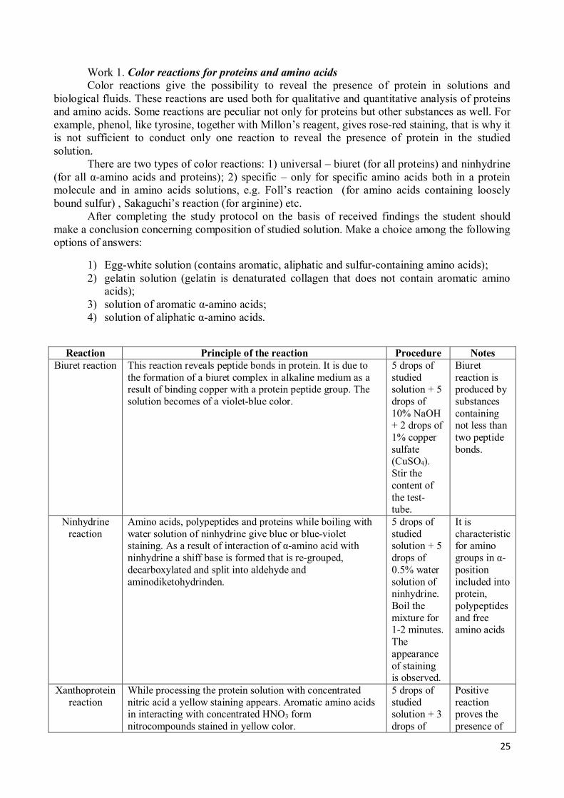
Work 1. Color reactions for proteins and amino acids Color reactions give the possibility to reveal the presence of protein in solutions and
biological fluids. These reactions are used both for qualitative and quantitative analysis of proteins and amino acids. Some reactions are peculiar not only for proteins but other substances as well. For example, phenol, like tyrosine, together with Millon’s reagent, gives rose-red staining, that is why it is not sufficient to conduct only one reaction to reveal the presence of protein in the studied solution.
There are two types of color reactions: 1) universal – biuret (for all proteins) and ninhydrine (for all α-amino acids and proteins); 2) specific – only for specific amino acids both in a protein molecule and in amino acids solutions, e.g. Foll’s reaction (for amino acids containing loosely bound sulfur) , Sakaguchi’s reaction (for arginine) etc.
After completing the study protocol on the basis of received findings the student should make a conclusion concerning composition of studied solution. Make a choice among the following options of answers:
1) Egg-white solution (contains aromatic, aliphatic and sulfur-containing amino acids); 2) gelatin solution (gelatin is denaturated collagen that does not contain aromatic amino
acids); 3) solution of aromatic α-amino acids; 4) solution of aliphatic α-amino acids.
Reaction Principle of the reaction Procedure Notes
Biuret reaction
This reaction reveals peptide bonds in protein. It is due to the formation of a biuret complex in alkaline medium as a result of binding copper with a protein peptide group. The solution becomes of a violet-blue color.
5 drops of studied solution + 5 drops of 10% NaOH + 2 drops of 1% copper sulfate (CuSO4). Stir the content of the test-tube.
Biuret reaction is produced by substances containing not less than two peptide bonds.
Ninhydrine reaction
Amino acids, polypeptides and proteins while boiling with water solution of ninhydrine give blue or blue-violet staining. As a result of interaction of α-amino acid with ninhydrine a shiff base is formed that is re-grouped, decarboxylated and split into aldehyde and aminodiketohydrinden.
5 drops of studied solution + 5 drops of 0.5% water solution of ninhydrine. Boil the mixture for 1-2 minutes. The appearance of staining is observed.
It is characteristic for amino groups in α-position included into protein, polypeptides and free amino acids
Xanthoprotein reaction
While processing the protein solution with concentrated nitric acid a yellow staining appears. Aromatic amino acids in interacting with concentrated HNO3 form nitrocompounds stained in yellow color.
5 drops of studied solution + 3 drops of
Positive reaction proves the presence of
26
concentrated nitric acid (HNO3). Carefully boil the mixture.
aromatic amino acids in the solution (trp, phe, tyr)
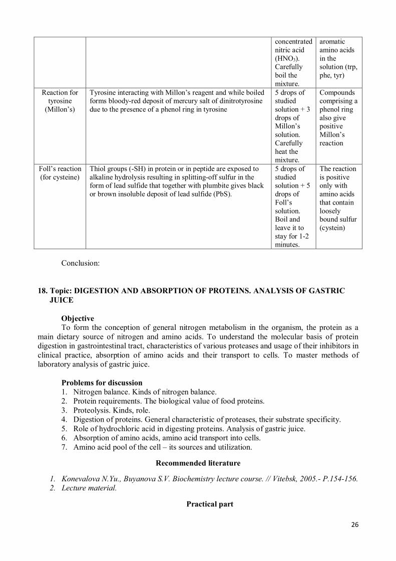
Reaction for tyrosine
(Millon’s)
Tyrosine interacting with Millon’s reagent and while boiled forms bloody-red deposit of mercury salt of dinitrotyrosine due to the presence of a phenol ring in tyrosine
5 drops of studied solution + 3 drops of Millon’s solution. Carefully heat the mixture.
Compounds comprising a phenol ring also give positive Millon’s reaction
Foll’s reaction (for cysteine)
Thiol groups (-SH) in protein or in peptide are exposed to alkaline hydrolysis resulting in splitting-off sulfur in the form of lead sulfide that together with plumbite gives black or brown insoluble deposit of lead sulfide (PbS).
5 drops of studied solution + 5 drops of Foll’s solution. Boil and leave it to stay for 1-2 minutes.
The reaction is positive only with amino acids that contain loosely bound sulfur (cystein)
Conclusion:
18. Topic: DIGESTION AND ABSORPTION OF PROTEINS. ANALYSIS OF GASTRIC
JUICE Objective
To form the conception of general nitrogen metabolism in the organism, the protein as a main dietary source of nitrogen and amino acids. To understand the molecular basis of protein digestion in gastrointestinal tract, characteristics of various proteases and usage of their inhibitors in clinical practice, absorption of amino acids and their transport to cells. To master methods of laboratory analysis of gastric juice.
Problems for discussion 1. Nitrogen balance. Kinds of nitrogen balance. 2. Protein requirements. The biological value of food proteins. 3. Proteolysis. Kinds, role. 4. Digestion of proteins. General characteristic of proteases, their substrate specificity. 5. Role of hydrochloric acid in digesting proteins. Analysis of gastric juice. 6. Absorption of amino acids, amino acid transport into cells. 7. Amino acid pool of the cell – its sources and utilization.
Recommended literature
1. Konevalova N.Yu., Buyanova S.V. Biochemistry lecture course. // Vitebsk, 2005.- P.154-156. 2. Lecture material.
Practical part
27
Determination of gastric juice acidity Principle of the method. Total acidity of gastric juice is measured in milliliters of 0.1N
solution of NaOH spent for neutralization of 1000 ml of gastric juice in the presence of a phenolphthalein indicator (pH transition zone 8.3-10.0; below 8.2 – colorless, above 10.0 – red). Normal total acidity for an adult person is 40-60 mmol/l, for a newborn – 2.8 mmol/l, for children from 1 month to 1 year – 4-20 mmol/l.
The content of free hydrochloric acid in gastric juice is measured in milliliters of 0.1N solution of NaOH spent for neutralization of 1000 ml of gastric juice in the presence of dimethylaminoazobenzole (pH transition zone is 2.9-4.0; below 2.9 – rose-red; above 4.0 – yellow). Free hydrochloric acid is almost completely neutralized at pH = 3.0; the color of dimethylaminoazobenzole changes from rose-red to orange. Normal content of free hydrochloric acid is 20-40 mmol/l (in newborns – 0.5 mmol/l).
Evaluation of total acidity, total content of hydrochloric acid and bound hydrochloric acid is done on one portion of gastric juice. Titration is performed with two indicators: dimethylaminoazobenzole and phenolphthalein.
Procedure. Add 10 ml of gastric juice by a pipette into a flask; add 1 drop of dimethylaminoazobenzole and 2 drops of phenolphthalein. When free hydrochloric acid is present in gastric juice, it is stained in red color with a rosy shade, when it is absent, orange staining appears.
Titrate free hydrochloric acid by 0.1N NaOH from a microburette till orange color staining appears and record the result (the 1st mark).Without adding alkaline into the burette continue titration till lemon-yellow color appears and record the result (the 2nd mark from 0). Continue titration till rosy staining appears (the 3rd mark from 0).
Calculation. Calculate the content of free HCl (the 1st mark), bound HCl (the 2nd mark) and total acidity (3rd mark) by the formula:
X (mmol/l) = A x 1000 x 0.1/10,
Where A – the amount of 0.1N solution of NaOH, ml; 10 – the amount of gastric juice taken
for evaluation; 0.1 – the amount of alkaline mg/eqv in 1 ml of 0.1N solution, mmol; 1000 – re-count to 1 l.
Clinical and diagnostic value. In gastric diseases the acidity can be zero, decreased and
increased. In ulcers and hyperacidic gastritis the content of free hydrochloric acid and total acidity increase (hyperchlorhydria). In hypoacidic gastritis or gastric cancer the decrease of free hydrochloric acid and total acidity occurs (hypochlorhydria). Sometimes in gastric cancer and chronic gastritis a complete absence of hydrochloric acid is observed (achlorhydria). In malignant anemia, gastric cancer a complete absence of hydrochloric acid and pepsin (achilia) are noted.
Conclusion: 19. Topic: INTRACELLULAR AMINO ACID METABOLISM. DETERMINATION OF
AMINO TRANSFERASE ACTIVITY IN SERUM
Objective To learn the common routs of amino acids metabolism. To get notion of the fate of amino
acid carbon skeletons, the role of amino acids in the formation of important biologically active compounds. To show the significance of indicator enzymes in diagnosis and prognosis of diseases by the example of determination of amino transferases activity in serum.
Problems for discussion
28
1. Transamination, aminotransferases, co-enzyme function of vitamin B6. Evaluation of amino transferases activity in serum, clinical-diagnostic value.
2. Types of amino acid deamination. Oxidative deamination of glutamic acid (reactions), the significance of a glutamate dehydrogenase reaction. Indirect deamination.
3. The fate of carbon skeletons of amino acids. Glucogenic and ketogenic amino acids. Pathways for amino acid synthesis.
4. Decarboxylation of amino acids, enzymes, co-enzymes. Biogenic amines (tryptamine, serotonin, histamine, γ-aminobutyric acid), catecholamines (dopamine, norepinephrine, epinephrine). Reactions of biosynthesis, biological role.
Recommended literature
1. Konevalova N.Yu., Buyanova S.V. Biochemistry lecture course. // Vitebsk, 2005.- P.156-161. 2. Lecture material.
Practical part
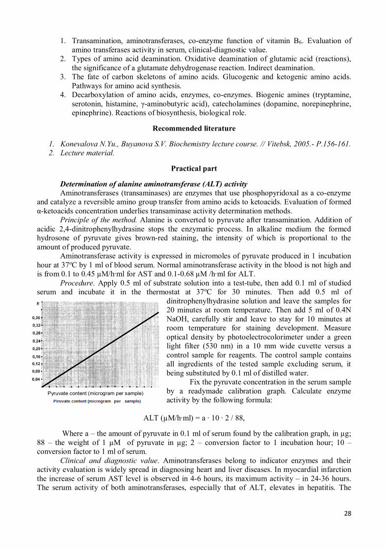
Determination of alanine aminotransferase (ALT) activity Aminotransferases (transaminases) are enzymes that use phosphopyridoxal as a co-enzyme
and catalyze a reversible amino group transfer from amino acids to ketoacids. Evaluation of formed α-ketoacids concentration underlies transaminase activity determination methods.
Principle of the method. Alanine is converted to pyruvate after transamination. Addition of acidic 2,4-dinitrophenylhydrasine stops the enzymatic process. In alkaline medium the formed hydrosone of pyruvate gives brown-red staining, the intensity of which is proportional to the amount of produced pyruvate.
Aminotransferase activity is expressed in micromoles of pyruvate produced in 1 incubation hour at 37oC by 1 ml of blood serum. Normal aminotransferase activity in the blood is not high and is from 0.1 to 0.45 µM/h∙ml for AST and 0.1-0.68 µM /h∙ml for АLT.
Procedure. Apply 0.5 ml of substrate solution into a test-tube, then add 0.1 ml of studied serum and incubate it in the thermostat at 37oC for 30 minutes. Then add 0.5 ml of
dinitrophenylhydrasine solution and leave the samples for 20 minutes at room temperature. Then add 5 ml of 0.4N NaOH, carefully stir and leave to stay for 10 minutes at room temperature for staining development. Measure optical density by photoelectrocolorimeter under a green light filter (530 nm) in a 10 mm wide cuvette versus a control sample for reagents. The control sample contains all ingredients of the tested sample excluding serum, it being substituted by 0.1 ml of distilled water.
Fix the pyruvate concentration in the serum sample by a readymade calibration graph. Calculate enzyme activity by the following formula:
ALT (µM/h∙ml) = a ∙ 10 ∙ 2 / 88,
Where a – the amount of pyruvate in 0.1 ml of serum found by the calibration graph, in µg; 88 – the weight of 1 µM of pyruvate in µg; 2 – conversion factor to 1 incubation hour; 10 – conversion factor to 1 ml of serum.
Clinical and diagnostic value. Aminotransferases belong to indicator enzymes and their activity evaluation is widely spread in diagnosing heart and liver diseases. In myocardial infarction the increase of serum AST level is observed in 4-6 hours, its maximum activity – in 24-36 hours. The serum activity of both aminotransferases, especially that of АLT, elevates in hepatitis. The
Pyruvate content (microgram per sample)
29
diagnostic value of АLT evaluation in jaundiceless form of infectious hepatitis and during the incubation period is of particular importance.
Conclusion:
20. Topic: DETOXIFICATION OF AMMONIA. DETERMINATION OF NONPROTEIN
NITROGEN IN BLOOD AND UREA IN URINE
Objective To study processes of ammonia detoxification in the organism for understanding
mechanisms of hyperammoniemia development. To acquire skills of nonprotein blood nitrogen and urine urea determination and to learn the diagnostic value of these tests.
Problems for discussion 1. Ways of ammonia binding in cells (reductive amination of α-ketoglutarate, synthesis of
glutamine and asparagine, formation of carbamoyl phosphate). Transport forms of ammonia.
2. Ammonia salts formation in kidneys (source of ammonia, the role of glutaminase and glutamate dehydrogenase, the significance of renal glutaminase activation in acidosis).
3. The role of hepatic cells in detoxification of ammonia. Ornithine cycle of urea formation (cycle pattern, substrates, enzymes, energetic supply, relation to the citric acid cycle, regulation). Fate of urea.
4. Nonprotein blood nitrogen (main components and their relative content). Principle of determination and clinical-diagnostic significance.
Recommended literature
1. Konevalova N.Yu., Buyanova S.V. Biochemistry lecture course. // Vitebsk, 2005.- P.162-169. 2. Lecture material.
Practical work
Work 1. Determination of urea in urine In a healthy person about 20-35 g or 333-583 mmol of urea are excreted with urine for 24
hours. The principle of the method. The method is based on the ability of urea containing amino
groups to form with paradimethylaminobenzaldehyde a complex compound in acid medium that is stained yellow. The staining intensity is proportional to urea concentration in the studied urine and is measured photometrically.
Procedure. Pipettes and test-tube must be dry. Apply per 0.2 ml of urine (test sample), 25 mg/l urea solution (standard sample) and water (control sample) respectively into 3 test-tubes, add per 1.2 ml of 2% solution of paradimethylaminobenzaldehyde into each of them and carefully stir. In 15 minutes perform photometry of the test and standard samples in dry 3 mm wide cuvettes under a blue light filter versus a control sample.
Calculation. Calculate the urea content in the test sample according to a standard urea solution by the formula:
Ct = Сs ∙ Еt / Еs,
30
Where Ct – urea concentration in the urine sample, mg/ml; Сs - urea concentration in the standard sample, 25 mg/ml; Еt - optical density of the sample; Еs - optical density of the standard urea solution.
Multiply the received value by diuresis (1200-1500 ml) and get the daily content of urea in the urine. Conversion factor to SI units (mmol/24 hours) is 0.0167.
Clinical and diagnostic value. The decreased urea content in urine is noted in nephritis, acidosis, parenchymatose jaundice, liver cirrhosis, uremia, while the elevated one – in fasting, malignant anemia, fever, intensive break-down of proteins in the organism, after taking salicylates, in phosphorus poisoning.
Conclusion: Work 2. Determination of nonprotein blood nitrogen Nitrogen-containing non-protein substances compose a fraction of nonprotein blood nitrogen
(intermediate or end products of protein metabolism).They are: urea, uric acid, creatine, ammonia, indican, bilirubin, polypeptides, amino acids, etc. Nitrogen of these substances is called nonprotein as it stays in filtrate after sedimentation of serum proteins.
The main part of nonprotein blood nitrogen is urea nitrogen – 50%, then nitrogen of amino acids – 25% and nitrogen of other nitrogen-containing components. Normal values for blood nitrogen are 14.3-25.0 mmol/l (20-40 mg per 100 ml); in newborns – 42.84-71.40 mmol/l (60-100 mg per 100 ml); it decreases to the level found in adults by 10th-12th day of life.
Principle of the method. Nonprotein blood nitrogen is determined in non-protein filtrate after blood proteins sedimentation by various agents (trichloracetic acid or volframate) with further mineralization of non-protein filtrate by concentrated sulfuric acid forming ammonia sulfate that interacts with Nessler’s reagent (alkaline solution of complex mercury salt K2(HgI4)) giving a compound of a yellow-orange color. The staining intensity is proportional to ammonia concentration, consequently to that of nitrogen.
Procedure. Prepare 3 usual test-tubes. Apply 1 ml of ready mineralizate and 9 ml of water into the 1st one (test sample), 1 ml of standard solution of ammonia sulfate into the second tube (standard sample) and 10 ml of water into the third one (control). Then apply per 0.5 ml of Nessler’s reagent into all tubes. Perform photometry of the tested and the standard sample versus the control under a blue light filter in 5 mm thick cuvettes.
Calculation. Calculate the nonprotein nitrogen content in the tested sample by the formula:
Ct = (Сs ∙ Еt / Еs) ∙ 100,
Where Ct –nonprotein blood nitrogen concentration in the blood, mg per 100 ml; Сs - nitrogen concentration in the standard sample (0.1 mg per 1 ml); Еt - extinction of the tested sample (mineralizate); Еs – extinction of the standard sample (ammonia sulfate).
Conversion factor to SI units (mmol/l) is 0.714. Clinical and diagnostic value. Evaluation of nonprotein nitrogen and its fractions is used for
diagnosing the impairment of renal excretory function and urea-formation function of the liver. The increase of blood nonprotein nitrogen is observed in cachexia of uncancerous origin caused by tuberculosis, diabetes and liver cirrhosis, in cardiac insufficiency, infectious diseases (scarlet fever, diphtheria). In prematurely born infants it can be associated with renal insufficiency and accelerated break-down of tissue proteins. The decrease of nonprotein blood nitrogen is observed in malnutrition and sometimes in pregnancy.
Conclusion:
31
21. COLLOQUIUM: “METABOLISM OF SIMPLE PROTEINS”, “BLOOD BIOCHEMISTRY” Questions for preparation:
1. Nitrogen balance, his condition is normal and pathological conditions. 2. The biological value of proteins. Requirement of protein in the diet. 3. Hydrolysis of proteins (proteolysis). Role limited proteolysis in the body. 4. The digestion of proteins. Gastric acidity: the principle of determination,content rate. 5. Amino acid cell pool, its completion and use. 6. Transamination. The role of vitamin B6. To be able to write transamination reaction with
alanine and aspartate transaminase. Know their diagnostic value. 7. Types of deamination. Glutamatdegidrogenase reaction: chemistry, coenzymes, value.
Indirect deamination. 8. Nitrogen-free way to becoming the balance of amino acids. Glucogenic and ketogenic
amino acids. 9. Ways neutralization of ammonia. To be able to write the synthesis reaction of asparagine,
glutamine, reductive amination of α-ketoglutarate, urea synthesis scheme. nonprotein blood nitrogen. Meaning of the urea and nonprotein blood nitrogen the clinic.
10. Decarboxylation of amino acids, biogenic amines. To be able to write the synthesis reaction of serotonin, histamine, γ-aminobutyric acid, know their rolein the body.
11. Catecholamines and their biological role. 12. Blood plasma proteins, classification and their role in the body. 13. General ideas about the regulation of acid-base status. The buffer system of blood and
their significance. The proof of the buffer properties of blood serum. 14. Mechanisms of transport of carbon dioxide and oxygen in blood. Mechanisms of hypoxic
conditions. 15. Blood clotting. Factors and clotting mechanisms. Value of calcium and vitamin K in the
blood clotting process. 16. Fibrinolysis. The role of fibrinolysis in blood coagulation. Plasmin system. 17. Anticoagulant system. Primary and secondary anticoagulants.
22. Topic: NUCLEOPROTEINS CHEMISTRY AND METABOLISM. DETERMINATION
OF URIC ACID IN URINE
Objective To get the notion of nucleoprotein catabolism in tissues and alimentary tract, mechanisms of
biosynthesis and break-down of nucleotides and regulation of these processes. To get acquainted with examples of using this knowledge in diagnosing and treatment of diseases. To perform a laboratory work on determination of uric acid in urine for consolidation of the theoretical material.
Problems for discussion 1. Mononucleotides, structure, terminology, biological role. 2. Primary, secondary and tertiary structures of nucleic acids (peculiarities of the structure,
varieties, types of stabilizing bonds). 3. Nucleoprotein metabolism. Digestion of nucleoproteins in the gastrointestinal tract
(significance, steps, enzymes). 4. Degradation of purine nucleotides (reactions, uric acid as an end-product of catabolism).
Disorders of purine metabolism (hyperuricemia and gout, urolithiasis). 5. Biosynthesis of purine nucleotides de novo (sources of nitrogen and carbon of a purine
ring, participation of folic acid, main intermediate products, key enzyme, regulation). The notion of nucleotide synthesis from free nitrogenous bases and nucleosides.
6. Degradation of pyrimidine nucleotides (end products and their fate).
32
7. Biosynthesis of pyrimidine nucleotides (substrates, process pattern, key enzyme, regulation, role of vitamins).
8. Synthesis of deoxyribonucleotides.
Recommended literature
1. Konevalova N.Yu., Buyanova S.V. Biochemistry lecture course. // Vitebsk, 2005.- P.175-180, 185.
2. Lecture material.
Practical part
Determination of uric acid in the urine Uric acid is an end-product of purine catabolism in humans. About 1.6-3.54 mmol/24 h
(270-600 mg/24 h) are usually excreted in healthy human in urine. Principle of the method. The method is based on the ability of uric acid to reduce
phosphorous-tungsten reagent into phosphorous-tungsten blue, the staining intensity of which is proportional to the content of uric acid. The amount of phosphorous-tungsten blue is determined by the red blood salt (K2[Fe(CN)6]) titration. The last one oxidizes the phosphorous-tungsten blue and blue staining disappears.
Procedure. Apply 1 ml of 20% solution of Sodium carbonate and 1 ml of phosphorous-tungsten Folin reagent to 1.5 ml of urine and titrate it by 0.01N solution of K2[Fe(CN)6] until blue staining disappears.
Calculation. Calculate the content of uric acid (in mg) in daily urine by the formula:
Uric acid, mg/24h = 0.8 · a · b / 1.5,
where 0.8 mg of uric acid corresponds to 1 ml of K2[Fe(CN)6]; a – the amount of K2[Fe(CN)6] used for titration, ml; b – diuresis, ml; 1.5 – the sample volume, ml.
Conversion factor to SI units (mmol/24h) is 0.0059. Clinical and diagnostic value. Hypouricuria (decrease of uric acid excretion with urine) is
noted in gout, nephritis, renal insufficiency; hyperuricuria (increase of uric acid excretion with urine) – in leukemia, accelerated breakdown of nucleoproteins. Children excrete relatively more uric acid than adults. Uric acid excretion depends on the purines content in food and intensity of nucleoproteins metabolism.
In gout uric acid salts (urates) precipitate in cartilages, muscles and joints. The content of uric acid in the blood can be increased while in the urine – decreased.
Conclusion:
23. Topic MATRIX BIOSYNTHESES (SYNTHESIS OF DNA, RNA, PROTEINS)
Objective To learn molecular mechanisms of replication, reparation, transcription, translation and
mechanisms of their regulation. To discuss possible impairments of genetic information realization for understanding consequences and approaches to treatment of these impairments.
Problems for discussion 1. Replication, biological role, substrates, enzymes, molecular mechanism. 2. Transcription, biological role, substrates, enzymes, RNA processing. 3. Genetic code and its properties.
33
4. Recognition and translation as steps of genetic information realization in cells. Substrate specificity of aminoacyl-tRNA synthetases. tRNA and its role in protein biosynthesis.
5. Modern understanding of protein biosynthesis. Regulation of protein biosynthesis in cells at a genetic level.
6. Modern techniques of molecular biology.
Recommended literature
1. Konevalova N.Yu., Buyanova S.V. Biochemistry lecture course. // Vitebsk, 2005.- P.180-184, 186-202.
2. Lecture material.
24. Topic CONNECTIVE TISSUE PROTEINS
Objective To form an idea of the variety of forms of connective tissues and the specific molecular composition of each form for understanding the mechanisms of occurrence and development of diseases (including dental), the development of methods of prevention and treatment.
Problems for discussion
1. Collagen. The amino acid composition, the spatial structure. Isocollagens. Features of the synthesis of collagen. Intra- and extracellular stages of the formation of the "mature" collagen.
2. Elastin. Role in the body. Features of the primary structure. The importance of extracellular stages in the mechanisms of formation of mature elastic tissue.
3. Protein-carbohydrate complexes. Principles of classification, biological role, mechanisms of synthesis and decay.
4. Fibrillar adhesive proteins of extracellular matrix and their functions. 5. Features of the protein composition of the cartilage and bone tissues. Non-collagenous
proteins, the biological role.
Recommended literature
1. Lecture material.
25. COLLOQUIUM: “NUCLEOPROTEINS METABOLISM”, “SYNTHESIS OF DNA, RNA AND PROTEINS”, “CONNECTIVE TISSUE PROTEINS”
Questions for preparation: 1. Mononucleotides, structure, terminology, biological role. 2. Primary, secondary and tertiary structures of nucleic acids (peculiarities of the structure,
varieties, types of stabilizing bonds). 3. Nucleoprotein metabolism. Digestion of nucleoproteins in the digestive tract (significance,
steps, enzymes). 4. Degradation of purine nucleotides (reactions, uric acid as an end-product of catabolism).
Disorders of purine metabolism (hyperuricemia and gout, urolithiasis). 5. Biosynthesis of purine nucleotides de novo (sources of nitrogen and carbon of a purine ring,
participation of folic acid, main intermediate products, key enzyme, regulation). The notion of nucleotide re-synthesis from free nitrogenous bases and nucleosides.
34
6. Degradation of pyrimidine nucleotides (end products and their fate). Biosynthesis of pyrimidine nucleotides (substrates, process pattern, key enzyme, regulation, role of vitamins).
7. Synthesis of deoxyribonucleotides. 8. Replication, biological role, substrates, enzymes, molecular mechanism. 9. Transcription, biological role, substrates, enzymes, RNA processing. 10. Genetic code and its properties. 11. Recognition and translation as steps of genetic information realization in the cell. Substrate
specificity of aminoacyl-tRNA synthetases. tRNA and its role in protein biosynthesis. 12. Modern understanding of protein biosynthesis. Regulation of protein biosynthesis in the cell
at a genetic level. 13. Posttranslational modification of protein molecules, kinds, biological role. 14. Modern techniques of molecular biology. 15. Collagen. The amino acid composition, the spatial structure. Isocollagens. Features of the
synthesis of collagen. Intra- and extracellular stages of the formation of the "mature" collagen.
16. Elastin. Role in the body. Features of the primary structure. The importance of extracellular stages in the mechanisms of formation of mature elastic tissue.
17. Protein-carbohydrate complexes. Principles of classification, biological role, mechanisms of synthesis and decay.
18. Fibrillar adhesive proteins of extracellular matrix and their functions. 19. Features of the protein composition of the cartilage and bone tissues. Non-collagenous
proteins, the biological role. 26. Topic: HORMONES, GENERAL CHARACTERISTIC AND PECULIARITIES OF
BIOLOGICAL ACTION. QUALITATIVE REACTIONS FOR HORMONES
Objective To learn how to apply knowledge of hormone classification, types of hormonal receptors, G-
proteins and a further cascade of intracellular transmitters for understanding specific mechanisms of hormones action. To understand mechanism of metabolic disorders development in case of insufficient or excessive hormone production.
Problems for discussion
1. Terminology and classification of hormones by the site of synthesis, chemical structure.
2. Peculiarities of hormones biological action. 3. Concept “hormone receptor “. Classification and structure of receptors: intracellular
receptors (nuclear and cytosolic), receptors of a plasma membrane (canal-forming receptors, 1-TMS and 7-TMS receptors).
4. Mechanisms of steroid, amino acid-derived, protein-peptide hormones action. The role of G-proteins, secondary messengers (cyclic nucleotides, IP3, Са2+, diacylglycerol), proteinkinases. Peculiarities of signal transduction from intracellular and 1-TMS-receptors.
Recommended literature
1. Konevalova N.Yu., Buyanova S.V. Biochemistry lecture course. // Vitebsk, 2005.- P.203-208. 2. Lecture material.
Practical part
Qualitative reactions for hormones
35
Hormones of the thyroid gland
The thyroid gland synthesizes and secretes iodine-containing thyroid hormones of high activity: thyroxine (T4) and 3,5,3'-triiodothyronine (T3), as well as noniodized hormone (polypeptide) thyreocalcitonine, the function of which is associated with regulation of calcium and phosphor levels in blood.
Work 1. Qualitative reaction for thyroxine Principle of the method. When thyroiodine is broken-down, potassium iodide is formed,
from which iodine is easily forced out by potassium iodate. Forcing iodine out of the salt of hydroiodic acid is a redox reaction where potassium iodide is a reductant and potassium iodate (residue of iodic acid) is an oxidizer. The released iodine is revealed by starch (blue staining) in acidic medium.
Procedure. Apply 24 drops of thyroiodine hydrolyzate into a test-tube, add 3 drops of 1% starch solution, 1 drop of phenolphthalein and then 4 drops of potassium iodate and about 10-15 drops of 10% solution of sulfuric acid until decolorization and appearance of blue staining.
Hormones of the pancreas
The pancreas produces insulin and glucagon. Insulin is produced by β-cells of Langerhans islets (from Latin insula – islet, island), so it got its name. Insulin is a protein consisting of two polypeptide chains connected to each other by disulfide bonds.
The primary structure of insulin is completely decoded and chemical synthesis of insulin is accomplished. Target-organs for insulin are: the liver, muscular tissue, adipose tissue. Insulin acts in a variety of ways. It plays an important role in metabolism of carbohydrates. It decreases glucose content in the blood, increases biosynthesis of glycogen in the liver and muscles, enhances lipogenesis, i.e. formation of lipids from carbohydrates, stimulates synthesis of proteins. Insulin is an antagonist of glucagon and epinephrine in regulation of metabolism.
Work 2. Color reactions for insulin Insulin gives characteristic reactions for protein: biuret, ninhydrine, Millon’s, etc. Biuret reaction Procedure. Add 5 drops of 10% solution of NaOH, 2 drops of 1% solution of copper sulfate
to 5 drops of 1% solution of insulin, stir the mixture; the content of the test-tube acquires a violet staining.
Ninhydrine reaction Procedure. Add 5 drops of 0.5% water solution of ninhydrine to 5 drops of 1% solution of
insulin and boil for 1-2 minutes. A rose-violet staining appears in the test-tube, and with time the solution becomes blue.
Xanthoprotein reaction Procedure. Apply 5 drops of 1% solution of insulin into a test-tube, then add 3 drops of
concentrated nitric acid and carefully boil. Yellow precipitate appears in the tube. Reaction for thyrosine (Millon’s) Procedure. Apply 5 drops of 1% solution of insulin into a test-tube, add 3 drops of Millon’s
reagent and carefully boil. Dark-red precipitate appears in the tube. Reaction for amino acids containing loosely bound sulfur (Foll’s reaction)
36
Procedure. Apply 5 drops of 1% solution of insulin into a test-tube and add 5 drops of Foll’s reagent, boil intensely and leave to stay for 1-2 minutes. A black or brown precipitate of lead sulfide will appear.
Hormones of the adrenal medulla
Catecholamines having a pyrocatechous nuclear and an amino group are synthesized in the adrenal medulla.
Work 3. Qualitative reactions for epinephrine Reaction with chlorous iron Principle of the method. Epinephrine has a low-alkaline reaction, is easily oxidized in the air
with formation of adrenochrome associated with staining of the solution into a red color. In interacting with nitrite yellow-orange staining is observed, with diazoreagent – a red one and with chlorous iron – a green one. The reaction with chlorous iron is characteristic of the pyrocachetinous ring included into a molecule of epinephrine and norepinephrine.
Procedure. Apply 10 drops of epinephrine solution into a test-tube and add 1 drop of chlorous iron. Green staining appears due to the presence of pyrocatechine in epinephrine molecule. Add 3 drops of 10% solution of NaOH and observe modification of staining (for cherry-red).
Diazoreaction Principle of the method. When diazoreagent interacts with epinephrine, the fluid is stained
red due to the formation of a complex compound, kind of an azostain. Procedure. Add 6 drops of 0.5% solution of Sodium nitrite (diazoreagent mixture), 10 drops
of epinephrine solution and 3 drops of 10% solution of NaOH to 6 drops of 0.5% solution of sulfonic acid. The fluid is stained red.
Work 4. Fluorescence of epinephrine oxidation products Principle of the method. Epinephrine oxidizing with air oxygen gives fluorescent products,
if alkaline is added. Procedure. Add 6 drops of 10% solution of NaOH and 6 drops of epinephrine solution to 10
drops of water. Placing the test-tube in front of a switched on fluorimeter observe green fluorescence of epinephrine oxidation products.
Hormones of sex glands
Sex hormones are synthesized in testicles, ovaries, placenta and adrenal glands. Female sex hormones –estrogens – can be considered as derivatives of estran (a hydrocarbon
with 18 atoms of carbon). The main natural estrogens are estradiol, estron and a hormone of corpus luteum –progesteron.
Male sex hormones –androgens – can be considered as derivatives of androstan (a hydrocarbon with 19 atoms of carbon). Testosteron and androsteron are male sex hormones.
Work 5. Qualitative reactions for folliculin Principle of the method. A qualitative reaction for folliculin (estron) is performed with
concentrated sulfuric acid and is due to formation of an ether compound of a straw-yellow color with green fluorescence.
Procedure. The reaction with oil solution of folliculin is conducted at room temperature. Add 30 drops of concentrated sulfuric acid to 2 drops of oil folliculin solution. Gradually a straw-yellow staining develops.
Conclusions:
37
27. Topic: BIOCHEMISTRY OF HORMONES. GLUCOSE TOLERANCE TEST
Objective To consolidate knowledge of chemical structure and mechanisms of individual hormones
action. Special attention should be paid to endocrine pathology of the pancreas. To learn how to construct and interpret various types of a sugar curves.
Problems for discussion
1. Hypothalamic hormones: chemical structure, type of receptor in target-cells and mechanism of a hormonal signal transduction, response of hypophyseal cells to the liberins and statins action.
2. Adenohypophyseal hormones: chemical structure, types of receptors in target-tissues and mechanism of a hormonal signal transduction, realization of hormonal effect at a target-tissue level. The role of excessive and insufficient secretion of hormones.
3. Neurohypophyseal hormones: chemical structure, type of receptor in target-tissue and mechanism of a hormonal signal transduction, realization of oxytocin and vasopressin effects at the level of target-tissues. The role of excessive and insufficient secretion of hormones. Diabetes insipidus.
4. Thyroxine and triiodothyronine: chemical structure, precursor, thyreoglobulin, type of receptor in target-tissue, realization of thyroidal hormones effects at a cellular level. The role of peroxidase and deiodase in hormones metabolism. Manifestations of hypo- and hyperthyroidism.
5. Hormones of the adrenal cortex: chemical structure, precursor, type of receptor in target-tissue, realization of glucocorticoids and mineralocorticoids effect at a cellular level. Cushing’s syndrome. “Bronze disease”.
6. Hormones of the adrenal medulla: chemical structure, precursor, type of receptor in target-tissue, realization of epinephrine and norepinephrine effect at a cellular level.
7. Sex hormones: chemical structure, precursor, realization of the effect of estrogens, progesterone and male sex hormones at a cellular level. Excessive and insufficient secretion of sex hormones.
8. Insulin and glucagon: chemical structure, insulin synthesis, types of receptors in target-tissues for glucagon and insulin, realization of pancreatic hormone effect at a cellular level. Diabetes mellitus. Diagnostic value of sugar curves.
Recommended literature
1. Konevalova N.Yu., Buyanova S.V. Biochemistry lecture course. // Vitebsk, 2005.- P.123-125, 209-276.
2. Lecture material.
Practical part
Studying of carbohydrate metabolism by glucose loading To diagnose diabetes mellitus and some pathologic states (insufficiency of the liver and
kidneys function, some endocrine diseases, neoplasms of the brain, pancreas and adrenal glands, B1 hypovitaminosis, some hereditary enzymopathies) it is important to realize the state of carbohydrate metabolism in patients. The blood glucose level is one of most significant parameters. Normal blood glucose concentration in adults is 3.9-6.1 mmol/l.
The peroral test for glucose tolerance (glucose loading) makes it possible to reveal pathology in those cases, when examination of blood glucose content on an empty stomach doesn’t reveal metabolic impairments.
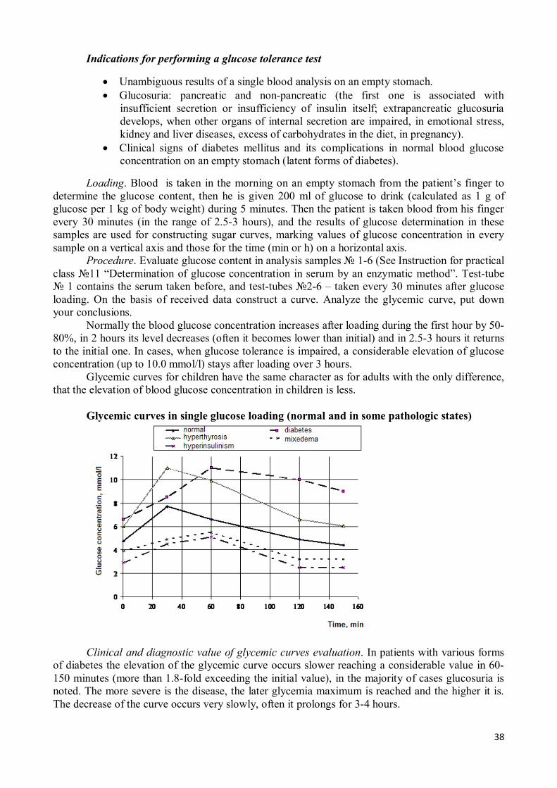
38
Indications for performing a glucose tolerance test
· Unambiguous results of a single blood analysis on an empty stomach. · Glucosuria: pancreatic and non-pancreatic (the first one is associated with
insufficient secretion or insufficiency of insulin itself; extrapancreatic glucosuria develops, when other organs of internal secretion are impaired, in emotional stress, kidney and liver diseases, excess of carbohydrates in the diet, in pregnancy).
· Clinical signs of diabetes mellitus and its complications in normal blood glucose concentration on an empty stomach (latent forms of diabetes).
Loading. Blood is taken in the morning on an empty stomach from the patient’s finger to determine the glucose content, then he is given 200 ml of glucose to drink (calculated as 1 g of glucose per 1 kg of body weight) during 5 minutes. Then the patient is taken blood from his finger every 30 minutes (in the range of 2.5-3 hours), and the results of glucose determination in these samples are used for constructing sugar curves, marking values of glucose concentration in every sample on a vertical axis and those for the time (min or h) on a horizontal axis.
Procedure. Evaluate glucose content in analysis samples № 1-6 (See Instruction for practical class №11 “Determination of glucose concentration in serum by an enzymatic method”. Test-tube № 1 contains the serum taken before, and test-tubes №2-6 – taken every 30 minutes after glucose loading. On the basis of received data construct a curve. Analyze the glycemic curve, put down your conclusions.
Normally the blood glucose concentration increases after loading during the first hour by 50-80%, in 2 hours its level decreases (often it becomes lower than initial) and in 2.5-3 hours it returns to the initial one. In cases, when glucose tolerance is impaired, a considerable elevation of glucose concentration (up to 10.0 mmol/l) stays after loading over 3 hours.
Glycemic curves for children have the same character as for adults with the only difference, that the elevation of blood glucose concentration in children is less.
Glycemic curves in single glucose loading (normal and in some pathologic states)
Clinical and diagnostic value of glycemic curves evaluation. In patients with various forms
of diabetes the elevation of the glycemic curve occurs slower reaching a considerable value in 60-150 minutes (more than 1.8-fold exceeding the initial value), in the majority of cases glucosuria is noted. The more severe is the disease, the later glycemia maximum is reached and the higher it is. The decrease of the curve occurs very slowly, often it prolongs for 3-4 hours.
39
Thyroid gland diseases associated with its hyperfunction are characterized by glycemic curves with a steeper rise, which may be caused by more intensive metabolism and excitation of a sympathetic nervous system.
Patients with adenoma of Langerhans islets, hypothyroidism (mixedema), Addison disease have a low initial level of the curve, its low peak and a high postglycemic ratio.
WHO criteria for diagnosing “diabetes mellitus” and “impaired glucose tolerance”
Diagnosis Time of taking a blood sample Venous whole blood, mmol/l Diabetes mellitus After night fasting >6.7 2 hours after glucose loading >10.0 Impaired glucose tolerance After night fasting <6.7 2 hours after glucose loading 6.7-10.0
Conclusion:
28. Topic: BIOCHEMISTRY OF NUTRITION. ROLE OF PROTEINS, FATS,
CARBOHYDRATES, VITAMINS. VITAMIN-LIKE SUBSTANCES Objective To consolidate knowledge of the chemical structure and molecular mechanisms of biological
action of co-enzyme forms of vitamins, involvement of other essential factors of nutrition into metabolism. To form the notion of biochemical mechanisms of utilizing food components for maintaining normal vital activity of the organism, pathological states of insufficient nutrition. To get acquainted with methods of vitamins detection and their determination in food products.
Problems for discussion
1. The nutritive value of proteins, fats, carbohydrates. The role of fibrous polysaccharides for functioning of the digestive tract and metabolic processes in an organism. Essential nutritive factors.
2. Syndrome of malnutrition (forms, characteristic). 3. Vitamins of B group: a) thiamine (B1); b) riboflavin (B2); c) pantothenic acid; d) niacin; e)
pyridoxine (B6); f) folic acid (B9); g) cobalamine (B12). Chemical nature, co-enzyme forms, molecular mechanisms of action.
4. Biotin (vitamin H), vitamin C. Structure, role in metabolic processes. 5. Vitamin-like substances: bioflavonoids (vit. P), choline, lipoic acid, inositol,
paraaminobenzoic acid, vit. U, etc. Biological role.
Recommended literature
1. Konevalova N.Yu., Buyanova S.V. Biochemistry lecture course. // Vitebsk, 2005.- P.285-299. 2. Lecture material.
Practical part
To reveal vitamins and evaluate their amount in various substances and biological fluids color reactions are used, which are specific for this or that group of vitamins.
Work 1. Qualitative reactions for vitamin B1 Vitamin B1 consists of pyrimidine and thiazole rings. Vitamin B1 has got a name of thiamine
as it contains sulfur and nitrogen.
40
1. Oxidation reaction
Principle of the method. In alkaline medium thiamine is oxidized by potassium ferricyanide and gives thiochrome. Thiochrome has blue fluorescence, when the solution is UV radiated on the fluoroscope.
Procedure. Add 5-10 drops of 10% solution of NaOH, 1-2 drops of 5% solution of potassium ferricyanide to 1 drop of 5% solution of thiamine and stir up the mixture. After the fluoroscope has got warmed you’ll observe blue fluorescence while radiating the solution with UV rays.
2. Diazoreaction
Principle of the method. In alkaline medium thiamine with diazoreagent forms a complex compound of an orange color.
Procedure. To diazoreagent containing 5 drops of 1% solution of sulphanic acid and 5 drops of sodium nitrate add 1-2 drops of 5% solution of thiamine and then very carefully, by the wall of the test tube add 5-7 drops of 10% solution of sodium bicarbonate. On the border of two fluids an orange ring appears.
Work 2. Qualitative reaction for vitamin B2 Principle of the method. The oxidized form of vitamin B2 represents a yellow substance
fluorescent in UV rays. The reaction for vitamin B2 is based on its ability to be reduced easily. The solution of vitamin B2 of a yellow color becomes at first rosy due to the formation of intermediate compounds and then decolorizes as a reduced form of vitamin B2 is colorless.
Procedure. Apply 10 drops of vitamin B2 into a test-tube, add 5 drops of concentrated hydrochloric acid and dip a granule of metal zinc. The release of hydrogen vesicles starts, the fluid gradually becomes rosy, then decolorizes. Compare both forms of vitamin B2 by fluorescence.
Work 3. Qualitative reaction to vitamin PP Principle of the method. Vitamin PP being heated with the solution of copper acetate forms
a poorly dissolved blue deposit of copper nicotinate. Procedure. Apply 20 drops of 3% solution of vitamin PP (before evaluation stir up the
solution) and heat it till boiling; the solution transforms from turbid into transparent. Stir up 5% solution of copper acetate, add 20 drops of it to the heated solution of vitamin PP. Bring the content of the test-tube to boiling and immediately cool it under cold running water. A blue deposit of copper nicotinate appears.
Work 4. Qualitative reaction for vitamin B6 Principle of the method. Vitamin B6 in interacting with the solution of chlorous iron forms a
complex salt kind of iron phenolat of red color. Procedure. To 5 drops of vitamin B6 add the equal amount of 1% solution of chlorous iron
and stir. Red staining develops. Work 5. Qualitative reaction to vitamin B12 B12 is the only vitamin containing a metal (cobalt) in its structure. Principle of the method. When cobalt ions interact with thiourea cobalt rodanide of a green
color is formed during heating. Procedure. Apply 2-3 drops of thiourea solution on a paper filter and dry over the burner.
Then apply 1-2 drops of mineralizate (B12) on the filter and heat again over the burner. Green staining appears, more often along the edge, it evidencing to the presence of cobalt.
Fill in the table with the results:
41
№ Experiment Observed changes Conclusions
Work 6. Determination of Vitamin C Principle of the method. The method is based on the ability of vitamin C to reduce 2,6-
dichlorphenolindophenol, which in acidic medium has red staining and decolorizes after reduction; in alkaline medium it has blue staining. To preserve vitamin C from destruction the studied solution is titrated in acidic medium by alkaline solution of 2,6-dichlorphenolindophenol till rose staining appears.
Determination of vitamin C content in urine Evaluation of vitamin C content in urine gives the notion about pools of this vitamin in the
organism, because there is correlation between blood concentration of this vitamin and its amount excreted with urine. However the content of ascorbic acid in urine is not always diminished in C hypovitaminosis. Often it is normal despite considerable insufficiency of this vitamin in tissues and organs.
In healthy people taking 100 mg of vitamin C per os quickly results in its elevation in blood and urine. In C hypovitaminosis the tissues, suffering from its insufficiency, catch the given vitamin C and its concentration in urine does not increase. The urine of a healthy person contains 20-30 mg/24 h of vitamin C or 113.55-170.33 µM/24 h. In children the level of this vitamin decreases in scurvy as well as in acute and chronic infectious diseases.
Procedure. Measure 10 ml of urine and 10 ml of distilled water into a flask, stir, acidize with 20 drops of 10% solution of hydrochloric acid and titrate with 0.001N solution of 2,6-dichlorphenolindophenol till a rose staining appears. Use the formula to calculate the content of ascorbic acid in urine:
where X – the content of ascorbic acid in mg/24 h; 0.088 – the amount of ascorbic acid (mg) corresponding to 1 ml of 2,6-dichlorphenolindophenol; A – the result of titration by 0.001N solution of 2,6-dichlorphenolindophenol, ml; B – an average diuresis (for men it is 1500 ml, for women – 1200 ml); C- the volume of urine taken for titration, ml.
Conclusions:
42
29. Topic: BIOCHEMISTRY OF NUTRITION. MINERAL SUBSTANCES. REGULATION OF WATER-ELECTROLYTE BALANCE Objective To consolidate knowledge of electrolyte composition of fluids in organism, role of micro-
and macroelements in cells and extracellular fluid for application in medical practice. Problems for discussion 1. Tocopherol. The chemical nature, involved in the metabolism, signs of vitamin
deficiencies, daily demand, the main sources of vitamin E. 2. Retinol. The chemical nature, involved in the metabolism, symptoms of hypo-and
hypervitaminosis, daily demand, the main sources of vitamin A. 3. Vitamin D. The chemical nature, absorption, biological role, the phenomenon of hypo-
and hypervitaminosis, daily demand, the main sources. 4. Vitamin K. The chemical nature, biological role, the phenomenon of hypovitaminosis,
daily demand, the main sources. Synthetic derivatives. Antivitamins. 5. Sodium, potassium, chlorine, calcium, phosphorus, magnesium, sulfur. The role in
metabolism. 6. Hormonal regulation of salt and water balance. Renin-angiotensin system, the role of
aldosterone, vasopressin, atrial natriuretic factor. 7. The role of iron in the organism (absorption, transport, intracellular metabolism). Iron
deficiency states and iron-deficient anemias. 8. The role of copper, zinc, manganese, cobalt, selenium, iodine and fluoride in tissue
metabolism. 9. The mechanism of action of hormones that regulate water and mineral metabolism
(vasopressin, sodium ureticheskogo hormone aldosterone). 10. The renin-angiotensin system and its role in the regulation of water-salt metabolism.
Recommended literature
1. Konevalova N.Yu., Buyanova S.V. Biochemistry lecture course. // Vitebsk, 2005.- P.277-284. 2. Lecture material.
30. Topic: BIOCHEMISTRY OF TEETH AND ORAL CAVITY FLUIDS
Objective To get notion of the chemical composition of bone and tooth tissues, and to consolidate
knowledge of the oral fluid composition for understanding the molecular mechanisms of dental diseases development.
Problems for discussion 1. The chemical composition of the tooth (enamel, dentin, cement, pulp) and bone tissue.
Collagen and non-collagenous proteins, protein-carbohydrate complexes. The mineral component of bone and teeth. The chemical composition. Hydroxyapatite crystals.
2. Theories of mineralization of bone tissue and hard tooth tissues. The role of organic molecules in the mechanisms of mineralization. Stages of isomorphic substitution of elements of the crystal lattice and its role in the formation of apatite crystals. Factors influencing on the processes of mineralization.
3. Ca2+-binding proteins and their role in the body.
43
4. Oral fluid. The chemical composition. Functions. Saliva from the ducts of the salivary glands. Unstimulated and stimulated saliva. Calcium and phosphate ions of the oral fluid. The role of saliva in the processes of enamel remineralization.
5. Proteins and enzymes of the oral fluid and their role. 6. "Gingival fluid" and dental liquor, the chemical composition. The value for the oral
tissues. 7. Surface formations on the enamel. Origin and chemical composition. 8. Fluorine and its role in life processes. Fluorine sources and requirements. 9. The effect of nutrition on dental health. The role of carbohydrates, proteins, vitamins
and trace elements. Caries. Causes, development mechanism, prevention.
Recommended literature
1. Lecture material.
Practical part Work 1. Determination of pH of the saliva The normal pH of saliva - 6.4-7.3. Procedure. Apply a drop of saliva on the litmus paper and determine its response: 1) blue litmus paper turns red, the red one does not change the color - the acid reaction; 2) red paper turns blue, the blue one does not change the color - alkaline reaction; 3) both papers do not change their colors - neutral reaction. Other indicator papers may be used. Work 2. Detection of mineral (inorganic) components in bone Bones and teeth are related to dense connective tissue. They have a high content of mineral substances, mostly calcium phosphate. The following research methods are used for qualitative analysis of inorganic substances of bone or tooth mineralizate: 1. Determination of sulphates. In an acidic environment, sulfates of bone (tooth) mineralizate form a white precipitate (BaSO4) with barium chloride. Procedure. Take 20 drops of mineralizate and add slowly, dropwise 5 drops of HCl (10% solution). Then add a solution of BaCl2 to form a precipitate. Filter off the precipitate. Heat the filtrate over a spirit lamp flame and boil for 2-3 minutes. The turbidity appears again due to the release of sulfuric acid. 2. Determination of phosphates. Phosphates of the bone (tooth) mineralizate form a yellow crystalline precipitate (ammonium phosphomolybdate) in the reaction with a molybdenum reagent. Procedure. Pour 20-30 drops of a molybdenum reagent solution into the test tube and heat the solution over the flame of a spirit lamp to a simmer (do not boil!). Then add a few drops of mineralizate. A yellow precipitate is formed. 3. Determination of calcium. Calcium of bone (tooth) mineralizate is precipitated by addition of ammonium oxalate. Procedure. To 20 drops of mineralizate pour 1-2 drops of 10% solution of acetic acid and 2-3 drops of 5% aqueous solution of ammonium oxalate. Crystalline precipitate of calcium oxalate appears. Work 3. Detection of proteins in bone tissue Procedure. A small piece of bone introduce into the tube containing 10 drops of 10% solution of NaOH. Bring to the boil. Proteins thus dissolve in alkali. After cooling add a drop of 1% solution of CuSO4 (biuret reaction). Appearance of purple coloration indicates the presence of protein.
44
Work 4. Quantitative determination of chlorides in the saliva by Levinson The principle of the method. Argentometric sedimentation method is based on the ability of silver ions to form insoluble salts with chloride ions. The amount of deposited material (AgNO3) is equivalent to the content of chloride ions. The titration of chloride ions with silver nitrate is carried out in the presence of an indicator K2CrO4. Upon reaching the titration equivalent point the excess of silver ions form a brick-red compound with the indicator (Ag2CrO4). Procedure. 1. Sedimentation of blood proteins. Prepare a mixture solutions in two tubes: 5 ml 0,45% ZnSO4 + 1 ml 0.1 N NaOH. Then, add 0.1 ml of saliva to the 1st test tube, and 0.1 ml of H2O dist. to the 2nd (control). Boil the tubes for 3 minutes over the flame of a spirit lamp. Then filter the content of the tubes through the cotton wool into flasks. Cotton filter should be washed twice with water (per 3 mL). 2. Precipitation of chlorine ions in the presence K2CrO4. Add 2 drops of 2% K2CrO4 solution to the filtrate and titrate with AgNO3 to change the yellow color of the solution to the brick-red. Calculation. Subtract the volume of AgNO3, gone to titrate the control solution (Vref, ml), from the volume of AgNO3, spent for titration of test solution (Vtest, ml); multiply the difference by 0.355, if the result is expressed in mg of chlorine in 0.1 ml of saliva. To express the chlorine content in mg%, value obtained must be multiplied by 1000. The conversion factor to SI units (mmol/L) - 0.282. Vref (ml) = Vtest (ml) = C (mmol/l) = (Vtest - Vref) *0,355 * 1000 * 0,282; C (mmol / l) = The normal content of chlorine (chloride ion) in saliva is 20-40 mmol/L. Work 5. Quantitative determination of calcium in saliva. The principle of the method. The indicator chromogen black ET-00 forms a purple compound with calcium. During the titration with Trilon B (disubstituted sodium salt of EDTA forming stable complexes with calcium ions) the staining of the solution will change to the blue color in the equivalent point corresponding to binding of all calcium ions in the solution by Trilon B. Procedure. 25 mL H2O and 1 mL of ammonia buffer are poured into the flask. Then, 1 ml of saliva and 2 drops of indicator chromogen black are added. The solution turns purple. The solution is then titrated with 0.002 M sodium Trilon B to a blue color. The calcium concentration in saliva is calculated from the volume of Trilon B, which was spent for titration. Calculation:
,1
1008,40002,0ml) 100per (mg TV×××=X
where 0,002 - molarity of the solution Trilon B, 40.8 - molecular weight of Ca, 100 - conversion factor to mg per 100 ml; 1 - the volume of saliva taken for analysis, VT - the amount of Trilon B consumed for the titration. Answer: VT (mL) = X (mg per 100 ml) = The conversion factor to SI units (mmol / L) - 0.245. C (mmol / l) = X*0,245 = The normal content of calcium in the saliva - 1.1-1.3 mmol/l (4-5 mg per 100 ml). Conclusions (compare with the content of chloride ions and calcium in the blood plasma):
45
31. COLLOQUIUM: “HORMONES”, “BIOCHEMISTRY OF NUTRITION”, “BIOCHEMISTRY OF THE ORAL CAVITY ORGANS AND FLUIDS”
Questions for preparation:
1. Hormones, classification by the chemical structure, site of synthesis. Peculiarities of hormonal action.
2. Hormone receptors, classification. Structure of intracellular receptors (nuclear and cytosolic), receptors of a plasma membrane (canal-forming receptors, 1-TMS and 7-TMS receptors).
3. Mechanisms of signal transduction for steroid, amino acid-derived, protein-peptide hormones. Role of G-proteins, secondary messengers (cAMP, cGMP, IP3, Са2+, DAG), proteinkinases. Signal transduction from intracellular and 1-TMS-receptors.
4. Hypothalamus hormones: chemical structure, type of receptor in target-cells and mechanism of a hormone signal transduction, response of hypophysis cells to the liberins and statins action.
5. Adenohypophysis hormones: chemical structure, types of receptors in target-tissues and mechanism of a hormone signal transduction, realization of hormone effect at a target-tissue level. The role of excessive and insufficient secretion of hormones.
6. Neurohypophysis hormones: chemical structure, type of receptor in target-tissue and mechanism of a hormone signal transduction, realization of oxytocin and vasopressin effects at the level of target-tissues. The role of excessive and insufficient secretion of hormones. Diabetes insipidus.
7. Thyroxine and triiodothyronine: chemical structure, preursor, thyreoglobulin, type of receptor in target-tissue, realization of thyroidal hormones effects at a cellular level. The role of peroxidase and deiodase in hormones metabolism. Manifestations of hypo- and hyperthyroidism.
8. Hormones of the adrenal cortex: chemical structure, precursor, type of receptor in target-tissue, realization of glucocorticoids and mineralocorticoids effect at a cellular level. Cushing’s syndrome. “Bronze disease”.
9. Hormones of the adrenal medulla: chemical structure, precursor, type of receptor in target-tissue, realization of epinephrine and norepinephrine effect at a cellular level.
10. Sex hormones: chemical structure, precursor, realization of the effect of estrogens, progesterone and male sex hormones at a cellular level. Excessive and insufficient secretion of sex hormones.
11. Insulin and glucagon: chemical structure, insulin synthesis, types of receptors in target-tissues for glucagon and insulin, realization of pancreas hormones effect at a cellular level. Diabetes mellitus. Diagnostic value of sugar curves.
12. The nutritive value of proteins, fats, carbohydrates. General notion of fuel metabolism, carbohydrates and lipids storage and utilization pathways. Role of fibrous polysaccharides. Essential nutritive factors.
13. Syndrome of malnutrition (forms, characteristic). 14. Water soluble vitamins: thiamine (B1), riboflavin (B2), pantothenic acid, niacin, pyridoxine
(B6), folic acid (B9), cobalamine (B12), biotin (vitamin H), ascorbic acid (vitamin C). Chemical nature, co-enzyme forms, molecular mechanisms of action, diet sources, signs of hypovitaminoses.
15. Fat soluble vitamins: A (retinol), E (tocopherol), D (calcipherol), K. Structure, role in metabolic processes, diet sources, signs of hypovitaminoses. Hypervitaminoses A, D.
16. Vitamin-like substances: bioflavonoids (vit. P), choline, lipoic acid, inositol, paraaminobenzoic acid, vit. U. Biological role.
17. Macroelements: sodium, potassium, chlorine, calcium, phosphorus, magnesium, sulfur. Role in metabolism.
46
18. Hormonal regulation of salt and water balance. Renin-angiotensin system, the role of aldosterone, vasopressin, atrial natriuretic factor.
19. Hormones regulating calcium and phosphorus metabolism. Chemical nature, mechanism of signal transduction in target-cells, biological action.
20. The role of iron in the organism (absorption, transport, intracellular metabolism). Iron deficiency states and iron-deficient anemias.
21. Microelements: the role of copper, zinc, manganese, cobalt, selenium, iodine and fluoride in tissue metabolism.
22. Vitamin-like substances: bioflavonoids (vit. P), choline, lipoic acid, inositol, paraaminobenzoic acid, vit. U, etc. Biological role.
23. The renin-angiotensin system and its role in the regulation of water-salt metabolism. 24. The chemical composition of the tooth (enamel, dentin, cement) and bone tissue. Collagen
and non-collagenous proteins, protein-carbohydrate complexes. The mineral component of bone and teeth. The chemical composition. Hydroxyapatite crystals.
25. Theories of mineralization of bone and hard tooth tissues. The role of organic molecules in the mechanisms of mineralization. Stages of isomorphic substitution of elements of the crystal lattice and its role in the formation of apatite crystals. Factors influencing on the processes of mineralization.
26. Ca2+-binding proteins and their role in the body. 27. Oral fluid. The chemical composition. Functions. Saliva from the ducts of the salivary
glands. Unstimulated and stimulated saliva. Calcium and phosphate ions of saliva. Role of saliva in enamel remineralization processes.
28. Proteins and enzymes of oral liquid and their role. 29. "Gingival fluid" and dental liquor, the chemical composition. The value for the oral tissues. 30. Surface formations on the enamel. Origin and chemical composition. 31. Fluorine and its role in life processes. Fluorine sources and requirements. 32. The effect of nutrition on dental health. The role of carbohydrates, proteins, vitamins and
trace elements. Caries. Causes, development mechanism, prevention. 32. Topic: LIVER BIOCHEMISTRY
Objective To be able to use knowledge of homeostatic and integrating role of the liver in carbohydrate,
lipid and amino acid metabolism for explaining mechanisms of metabolic disorders development in diseases of the liver and bile ducts. To be able to use knowledge of existing conversion patterns for xenobiotics in the liver to understand biochemical aspects of pharmacology and toxicology.
Problems for discussion 1. Basic functions and chemical composition of the liver. 2. The role of the liver in carbohydrate metabolism. 3. The role of the liver in lipid metabolism. 4. The role of the liver in protein metabolism. 5. Detoxificating function of the liver, mechanisms: (protective syntheses, acylation,
microsomal oxidation, conjugation). 6. The role of the liver in pigment exchange. Synthesis and degradation of hemoglobin
(schemes). Normal bilirubin metabolism and its disorders. 7. Biochemical methods of diagnosing liver disturbances.
Recommended literature
1. Konevalova N.Yu., Buyanova S.V. Clinical biochemistry. Materials for the state examination in biochemistry // Vitebsk, 2005.- P.19-31.
47
2. Lecture material.
Practical part
Determination of total bilirubin in serum Principle of the method. Diazoreagent forms azobilirubin stained in a rose color with soluble
bilirubin. Staining intensity of azobilirubin solution is proportional to bilirubin concentration and can be evaluated colorimetrically. Conjugated (direct) bilirubin gives a direct reaction with diazoreagent. Unconjugated (indirect) bilirubin cab be made soluble, if one adds ethyl alcohol to blood serum.
Procedure. Measure 1 ml of blood serum to a centrifuge test-tube, 2 ml of ethyl alcohol, carefully stir the content with a glass stick and centrifuge for 15 minutes at the speed of 3000 turns/min. Then pour off the supernatant into another test-tube and add 0.25 ml of diazoreagent. Red-rose staining appears. In 10 minutes determine its intensity measuring the sample optical density vs water in a 5 mm wide cuvette under a green light filter (500-560 nm). The colorimetry of azobilirubin standard solution corresponding to bilirubin concentration 0.4 mg per 100 ml (Cs), is done simultaneously.
Calculate by the formula: Сt (mg per 100 ml) = Еt ∙ Сs / Еs Normal total bilirubin concentration in plasma (serum) is 0.5-1.2 mg per 100 ml (8.55-20.52
µM/l); indirect bilirubin forms 75% of its amount. Conclusion: Clinical-diagnostic significance of studying pigment metabolism. Jaundice is one of
important signs of pigment metabolism impairment, it is usually noted at bilirubin level in the blood over 27-34 µM/l. The blood of newborns, especially prematually born, has a higher bilirubin content (physiological jaundice). The increase of bilirubin concentration observed from 2nd -3rd to 7th-10th days mainly due to indirect bilirubin is associated with functional insufficiency of the liver, in particular with low activity of enzyme UDP-glucuroniltransferase, which is necessary for the formation of direct bilirubin.
Hemolytical jaundice (suprahepatic) is the enhancement of erythrocyte hemolysis that results in enhanced formation of inconjugated bilirubin because the liver has no time for its binding.
Parenchymous jaundice (hepatic) is the function impairment of hepatic cells. It may be also caused by hereditary defects of bilirubin transport and bilirubin diglucuronide formation.
Mechanic jaundice (obturative, obstructive) – the bile flow-off troubles. It occurs in overflow of bile ducts due to obstruction, their rupture and consequent entrance of bile into the blood.
The severity of jaundice usually corresponds to the level of bilirubinemia. It is considered that jaundice has a mild form if the content of bilirubin in plasma (serum) does not exceed 85 µM/l; its level of 86-169 µM /l testifies to moderately severe, and over 170 µM /l – to a severe jaundice.
48
Appendix
EXAMINATION QUESTIONS {THE DENTAL FACULTY (2013)}
Enzymes 1. The role of enzymes in life processes. Principles of nomenclature and classification of enzymes. 2. The chemical nature of enzymes. General properties of enzymes. 3. The structure of the enzymes. The concept of the active site of the enzyme. 4. Coenzymes. Classification and role. 5. The mechanism of action of enzymes. Enzyme kinetics. 6. Multiple forms of enzymes and their classification. Isozymes. 7. General principles for the regulation of enzymatic processes. 8. Isosteric mechanism regulating the activity of enzymes. 9. The mechanism of allosteric regulation of enzyme activity. 10. Covalent modification of the enzyme structure. Types, importance. 11. Enzyme inhibitors, classification, characteristics. 12. The use of enzymes in the medical practice.
Bioenergetics
13. Metabolism and energy as an important sign of life. The general concept of metabolism. Relationship between catabolism and anabolism.
14. Redox processes in tissues. Ways to use oxygen in the cells. 15. Adenylic system and its biological significance. The mechanisms of synthesis and ways of using ATP. 16. The modern concept of tissue respiration. NAD- and FAD-dependent dehydrogenases, ubiquinone (coenzyme Q),
cytochrome system, their chemical structure and their role in oxidative processes. 17. Oxidative phosphorylation. Chemiosmotic theory of oxidative phosphorylation. 18. Causes of the cell hypoenergetic states. Inhibitors and uncouplers of oxidative phosphorylation, the mechanism of their
action. Carbohydrate metabolism
19. Carbohydrates. Classification, the biological role. 20. Nutritional value of carbohydrates. Digestion and absorption of carbohydrates. The role of dietary fiber in digestion. 21. The synthesis and breakdown of glycogen in the liver and muscles. 22. Aerobic glucose oxidation stages, the final products. Energy yield and mechanisms of ATP synthesis. 23. Glycolysis. Energy yield, the mechanism of ATP synthesis under anaerobic conditions. 24. The fate of the end products of glycolysis - pyruvic acid and lactic acid. Gluconeogenesis. Enzymes involved in
gluconeogenesis. 25. Oxidative decarboxylation of pyruvic acid and other α-keto acids, enzymes, coenzymes, biological significance. 26. Citric acid cycle. Intermediate stages, enzymes. The biological significance of the cycle. Connection with the process of
oxidative phosphorylation. 27. Pentose phosphate pathway of glucose oxidation and its biological role. 28. Uronic acid pathway of glucose metabolism. The biological role. 29. Features of carbohydrate metabolism involving oral microorganisms.
Chemistry and metabolism of lipids
30. Lipids. The biological role. Classification of lipids. Their basic properties. 31. Classification of fatty acids. Polyunsaturated fatty acids. Derivatives of arachidonic acid (prostaglandins, prostacyclins,
thromboxanes, leukotrienes) and their biological role. 32. Glycerophospholipids. The chemical structure, properties and biological role. 33. Cholesterol biosynthesis and biological role. Disorders of cholesterol metabolism. 34. Digestion of fats and phospholipids in the digestive tract: emulsification, enzymes, hydrolysis products, micellar
dissolution. Role of bile acids in the lipid digestion. 35. Re-synthesis of triacylglycerols and phospholipids in enterocytes. Formation of chylomicrons, their composition and
structure. 36. Serum lipoproteins. Classification, composition, place of formation, interconversion. The role of lipoprotein lipase,
lecithin: cholesterol acyltransferase (LCAT). 37. Synthesis and mobilization of fat in adipose tissue. Hormone-sensitive lipase of adipose tissue. 38. The central role of acetyl-CoA metabolism. 39. β-Oxidation of fatty acids. Localization in the cell. Role CoA-SH and ATP. Relationship with oxidative phosphorylation.
Energy yield of β-oxidation. 40. Ketone bodies, formation of ketone bodies. Ketosis and diabetes. Significance of determination of ketone bodies in the
urine. 41. The synthesis of fatty acids. Connection with glycolysis, the pentose phosphate pathway of glucose conversion, Krebs
cycle. The value of CO2, ATP, NADPH.H+, biotin. Multienzyme complex for fatty acid synthesis. Activators and inhibitors of fatty acid synthesis.
42. Hormonal regulation of lipid metabolism (insulin, glucagon, thyroxine, epinephrine, norepinephrine, glucocorticoids, sex hormones).
Metabolism of proteins and amino acids
49
43. Nitrogen balance. Dietary protein requirement. The biological value of proteins. 44. Characteristics of proteases. The role of selective proteolysis in the life of the cell. 45. Digesting the proteins in the gastrointestinal tract. Role of hydrochloric acid. 46. Amino acid pool in the cells. Its utilization and sources of renewal. 47. Transamination. Enzymes. Coenzymes. The role of this process for the life of the cell. The diagnostic value of the
determination of transaminase activity in serum. 48. Ways of amino acid deamination. Enzymes and coenzymes of oxidative deamination. The biological significance of
glutamate dehydrogenase reaction. 49. Use of nitrogen-free residues of amino acids. Ketogenic and glycogenic amino acids. 50. The ways of ammonia detoxification. Formation of glutamine and asparagine and their role in the transport of ammonia. 51. Formation of urea. The role of the liver in urea formation. Significance of the determination of blood urea and nonprotein
nitrogen in clinical practice. 52. Decarboxylation of amino acids. The formation of biogenic amines and their role in the body.
Chemistry and metabolism of nucleic acids
53. Modern concept of nucleic acid structure. DNA and RNA, their cell localization, functions. 54. Primary and secondary structure of DNA and RNA. Interaction of nucleic acids with proteins. The structure of the
nucleoproteins. 55. The final products of pyrimidine and purine nucleotide breakdown. The value of uric acid determination in the blood and
urine in clinical practice. 56. Biosynthesis of purine nucleotides. The initial substrates of synthesis. Regulation of synthesis. 57. Biosynthesis of pyrimidine nucleotides. The initial substrates of synthesis. Regulation of synthesis. 58. Matrix mechanism of DNA synthesis. Enzymes and substrates of synthesis. Features of synthesis in eukaryotes. 59. RNA synthesis. Enzymes and substrates of synthesis. Features of synthesis in eukaryotes. 60. The genetic code and its properties. 61. Role of t-RNA in protein synthesis. Specificity of ARSase. An adapter function of tRNA. 62. Modern concept of protein biosynthesis.
Water-soluble vitamins
63. General characteristics and classification of vitamins. The methods of the estimation of the body saturation with vitamins. 64. Biotin. Coenzyme form. The biological role. Symptoms of vitamin deficiency. Dietary sources. The daily requirement. 65. Vitamin B1. Participation in the construction of co-enzymes. Role in metabolism. Symptoms of vitamin deficiency. Dietary
sources. The daily requirement. 66. Vitamin B2. Structure, participation in the formation of flavin coenzymes. The biological role. Symptoms of vitamin
deficiency. Dietary sources. The daily requirement. 67. Vitamin B6. Participation in the formation of coenzymes. Role in metabolism. Symptoms of vitamin deficiency. Dietary
sources. The daily requirement. 68. Vitamin B12. Kobamide coenzymes. Participation in metabolism. Symptoms of vitamin deficiency. Dietary sources. The
daily requirement. 69. Vitamin C. The biological significance. Symptoms of vitamin deficiency. Dietary sources. The daily requirement. 70. Pantothenic acid. Coenzymes containing pantothenic acid. The biological role. Symptoms of vitamin deficiency. Dietary
sources. The daily requirement. 71. Vitamin PP. Structure. Participation in the formation of nicotinamide coenzymes. The biological significance. Symptoms of
vitamin deficiency. Dietary sources. The daily requirement. 72. Folic acid. The structure, participation in the formation of coenzymes. Role in metabolism. Symptoms of vitamin
deficiency. Dietary sources. The daily requirement. 73. Vitamin-like substances: bioflavonoids (vitamin P), para-aminobenzoic acid, inositol, pangamic acid, lipoic acid, choline,
vitamin U and others. Biological role.
Fat-soluble vitamins 74. Vitamin A. The biological role. The phenomena of hypo- and hypervitaminosis. Dietary sources. The daily requirement. 75. Vitamin E. The biological role. The phenomena of hypovitaminosis. Dietary sources. The daily requirement. 76. Vitamin D. The structure, biological role. The phenomena of hypo-and hypervitaminosis. Dietary sources. The daily
requirement. 77. Vitamin K. The biological role. Vitamin deficiency. Dietary sources. The daily requirement.
Hormones
78. Hormones. The chemical structure. Classification. 79. The mechanisms of hormone action. Role of G-proteins, second messengers, protein kinases. 80. Adenohypophysis. Role in regulating the function of the peripheral endocrine glands. Tropic hormones. Connection with
the hypothalamus. 81. Hormones of the posterior pituitary: oxytocin, vasopressin. Their chemical structure, the mechanism of signal transduction
in the target cells, the biological effect. 82. Thyroid hormones. Their structure, mechanism of action, effects on metabolism. Hypo- and hyperthyroidism. 83. Hormones that regulate the metabolism of calcium and phosphorus. The chemical structure, the mechanism of signal
transduction in target cells, the biological effect. 84. Insulin. Chemical structure, receptors, the mechanism of signal transduction in target cells, effects on metabolism. Diabetes
mellitus. 85. Glucagon. Chemical structure, receptors, the mechanism of signal transduction in target cells, effects on metabolism.
50
86. Glucocorticoids. Chemical structure, receptors, the mechanism of signal transduction in target cells, effects on metabolism. 87. Mineralocorticoids. Chemical structure, receptors, the mechanism of signal transduction in target cells, effects on
metabolism. 88. The hormones of the adrenal medulla. Catecholamines: dopamine, epinephrine, norepinephrine. Structure, receptors, signal
transduction mechanism in the target cells, a role in the body. 89. Male sex hormones, the chemical structure, receptors, signal transduction mechanism in the target cells. The effect on the
metabolism. 90. Female sex hormones, the chemical structure, receptors, signal transduction mechanism in the target cells. The effect on the
metabolism. Biochemistry of muscle
91. Chemical composition of the muscle tissue. The structure and role of contractile proteins. 92. Molecular mechanisms of muscle contraction and relaxation. Energy sources that provide muscle contraction.
Liver biochemistry
93. The role of the liver in the metabolic processes in the body. Antitoxic function of the liver. 94. Synthesis and breakdown of blood pigments. The role of the liver in the formation of bile pigments. The metabolism of bile
pigments.
Blood biochemistry 95. Plasma proteins and their function. 96. Blood clotting. Phases of blood coagulation. The factors and mechanisms of blood coagulation. 97. The role of calcium and vitamin K in the blood clotting process. 98. Anticoagulant system. 99. Fibrinolysis. The biological role of fibrinolysis. Plasmin system.
Water and mineral metabolism
100. Water. Importance of water. The biological role of sodium and potassium. Mechanisms of regulation of water and salt balance.
101. Trace elements. Their value. The role of iron, copper, iodine, selenium.
Biochemistry of connective tissue, bone, teeth. Biological chemistry of oral fluids 102. Collagen, features of amino acid composition and spatial structure. The role of collagen in the body. 103. Elastin. Role in the body. Features of the amino acid composition and spatial structure. 104. Features of the collagen and elastin biosynthesis. 105. The role of extracellular stages in the mechanisms of formation of extracellular matrix of connective tissue. 106. Proteoglycans. Features of the structure. Role in the body. 107. Mucoproteins. Features of the structure. Role in the body. 108. Glycoproteins. Features of the structure. Role in the body. 109. Calcium and its metabolism in the body. Role in the processes of life. 110. Phosphorus and its metabolism in the body. Role in the processes of life. 111. Fluorine and its role in life processes. Fluorine sources and requirement. 112. The mineral component of bone and teeth. The chemical composition. Hydroxyapatite crystals. 113. Chemical composition of hard tooth tissues (enamel, dentin, cement). 114. The chemical composition of bone tissue. Bone proteins and their role in mineralization processes. 115. Ca2+-binding proteins and their role in the body. 116. Theories of mineralization of bone and hard tooth tissues. Factors influencing on the processes of mineralization. 117. Stages of isomorphic substitution of elements of the crystal lattice and its role in the formation of apatite crystals. 118. The effect of nutrition on dental health. The role of carbohydrates, proteins and micronutrients. 119. Oral fluid. The chemical composition. Function. 120. The role of saliva in enamel remineralization processes. 121. The concepts of "gingival fluid" and "dental liquor", the chemical composition. The value for the oral tissues. 122. Proteins and enzymes of the oral fluid, their role in the oral cavity. 123. Surface formations on the enamel. Their origin and chemical composition. 124. Caries. Causes, mechanisms of development, prevention.
51
Учебное издание
Таганович Анатолий Дмитриевич Котович Ирина Леонидовна
Колб Александр Владимирович Ковганко Николай Николаевич
БИОХИМИЯ: ПРАКТИКУМ ДЛЯ СТУДЕНТОВ СТОМАТОЛОГИЧЕСКОГО ФАКУЛЬТЕТА
BIOCHEMISTRY: MANUAL FOR INTERNATIONAL STUDENTS OF DENTAL FACULTY
Практикум на английском языке
Ответственный за выпуск А. Д. Таганович. В авторской редакции Компьютерный набор Компьютерная верстка
Подписано в печать
Издатель и полиграфическое исполнение: Учреждение образования «Белорусский государственный медицинский университет».
Related Documents