1 Texas Society of Pathologists 2013 92 nd Annual Meeting Neuropathology: Intraoperative Frozen Section Consultation for General Surgical Pathologists Gregory N. Fuller, MD, PhD Professor and Chief Neuropathologist M D Anderson Cancer Center Practical Approach to Intraoperative Consultation (IOC) “Intraoperative Consultation (IOC) for a brain biopsy is among the most stressful situations that a surgical pathologist will encounter.”* * G Fuller, January 25, 2013 Austin

Welcome message from author
This document is posted to help you gain knowledge. Please leave a comment to let me know what you think about it! Share it to your friends and learn new things together.
Transcript

1
Texas Society of Pathologists 2013
92nd Annual Meeting
Neuropathology:
Intraoperative Frozen Section
Consultation for General
Surgical Pathologists
Gregory N. Fuller, MD, PhD
Professor and Chief Neuropathologist
M D Anderson Cancer Center
Practical Approach to
Intraoperative Consultation
(IOC)
“Intraoperative Consultation
(IOC) for a brain biopsy is
among the most stressful
situations that a surgical
pathologist will encounter.”*
* G Fuller, January 25, 2013 Austin

2
IOC
Principles
PRE-IOC Preparation
PRE-IOC Preparation
• AGE of the patient
3
PRE-IOC Preparation
• AGE of the patient
• ANATOMIC LOCATION of the lesion
PRE-IOC Preparation
• AGE of the patient
• ANATOMIC LOCATION of the lesion
• IMAGING characteristics of the lesion
NEUROIMAGING 101
4
The Importance of
Imaging Studies to the
Pathologist Cannot be
Stressed Enough!
Rubin’s Pathology
6th Edition
2011
The Importance of
Imaging Studies to the
Pathologist Cannot be
Stressed Enough!
The Importance of
Imaging Studies to the
Pathologist Cannot be
Stressed Enough!
Rubin’s Pathology
6th Edition
2011
5
The Importance of
Imaging Studies to the
Pathologist Cannot be
Stressed Enough!
Rubin’s Pathology
6th Edition
2011
The Importance of
Imaging Studies to the
Pathologist Cannot be
Stressed Enough!
Rubin’s Pathology
6th Edition
2011
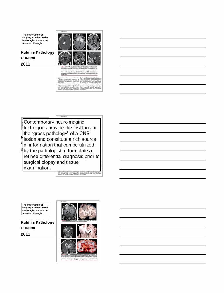
Contemporary neuroimaging
techniques provide the first look at
the “gross pathology” of a CNS
lesion and constitute a rich source
of information that can be utilized
by the pathologist to formulate a
refined differential diagnosis prior to
surgical biopsy and tissue
examination.
The Importance of
Imaging Studies to the
Pathologist Cannot be
Stressed Enough!
Rubin’s Pathology
6th Edition
2011
6
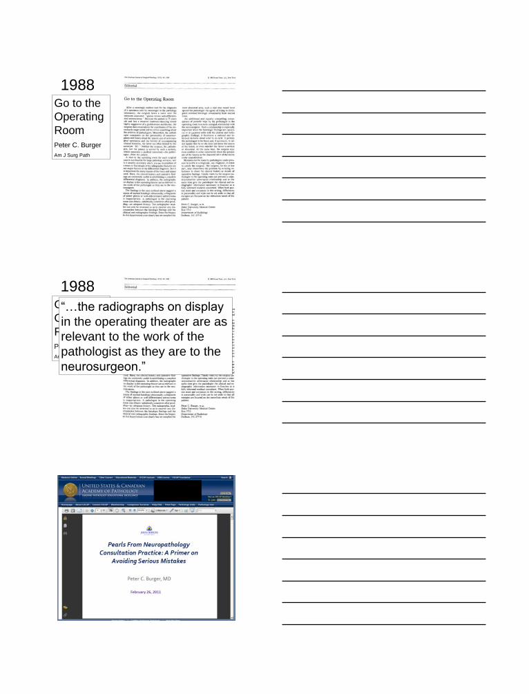
Go to the
Operating
Room Peter C. Burger
Am J Surg Path
1988
Go to the
Operating
Room Peter C. Burger
Am J Surg Path
1988
“…the radiographs on display
in the operating theater are as
relevant to the work of the
pathologist as they are to the
neurosurgeon.”
2011
7
2011
2011
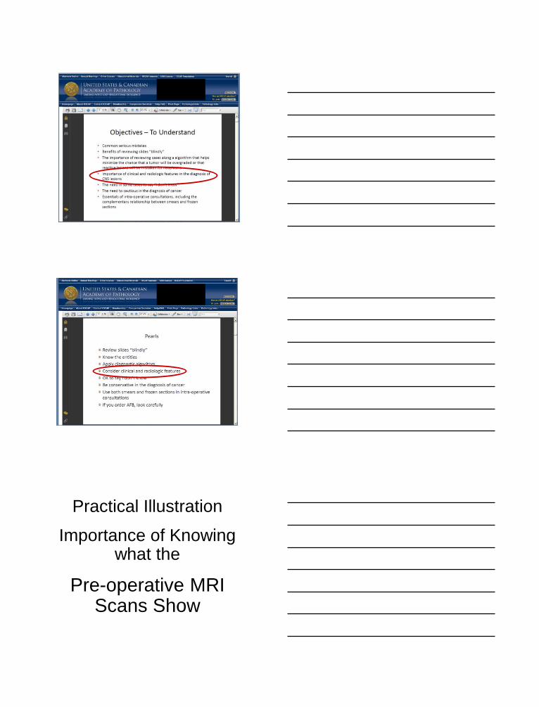
Practical Illustration
Importance of Knowing what the
Pre-operative MRI Scans Show
8
Importance of Pre-op MRI
Biopsy (H&E)
Importance of Pre-op MRI
Biopsy (H&E)
DX: Low-Grade Diffuse Glioma
Pre-operative MR Imaging
Pre-op MRI: coronal T1 post-contrast Biopsy (H&E)
Importance of Pre-op MRI
9
Pre-op MRI: coronal T1 post-contrast Biopsy (H&E)
Importance of Pre-op MRI
YIKES!
Pre-op MRI: coronal T1 post-contrast Biopsy (H&E)
Importance of Pre-op MRI
The biopsy was NOT representative!
10
Our patients are best
served by a collegial
Team Approach –
surgeon, oncologist,
radiologist, pathologist
You must know what
the imaging shows -
Talk to the
neuroradiologist!
To talk to the
neuroradiologist, you
must be able to
speak their language

11
Practical Basic
Neuroimaging
Information Gained from MRI
Information Gained from MRI
Anatomic location of the lesion(s)
12
Information Gained from MRI
Anatomic location of the lesion(s)
Nature of interface of lesion border with brain
parenchyma (sharp margin vs. diffuse infiltration)
Information Gained from MRI
Anatomic location of the lesion(s)
Nature of interface of lesion border with brain
parenchyma (sharp margin vs. diffuse infiltration)
Presence or absence of contrast enhancement
Information Gained from MRI
Anatomic location of the lesion(s)
Nature of interface of lesion border with brain
parenchyma (sharp margin vs. diffuse infiltration)
Presence or absence of contrast enhancement
If contrast-enhancing, pattern of enhancement
13
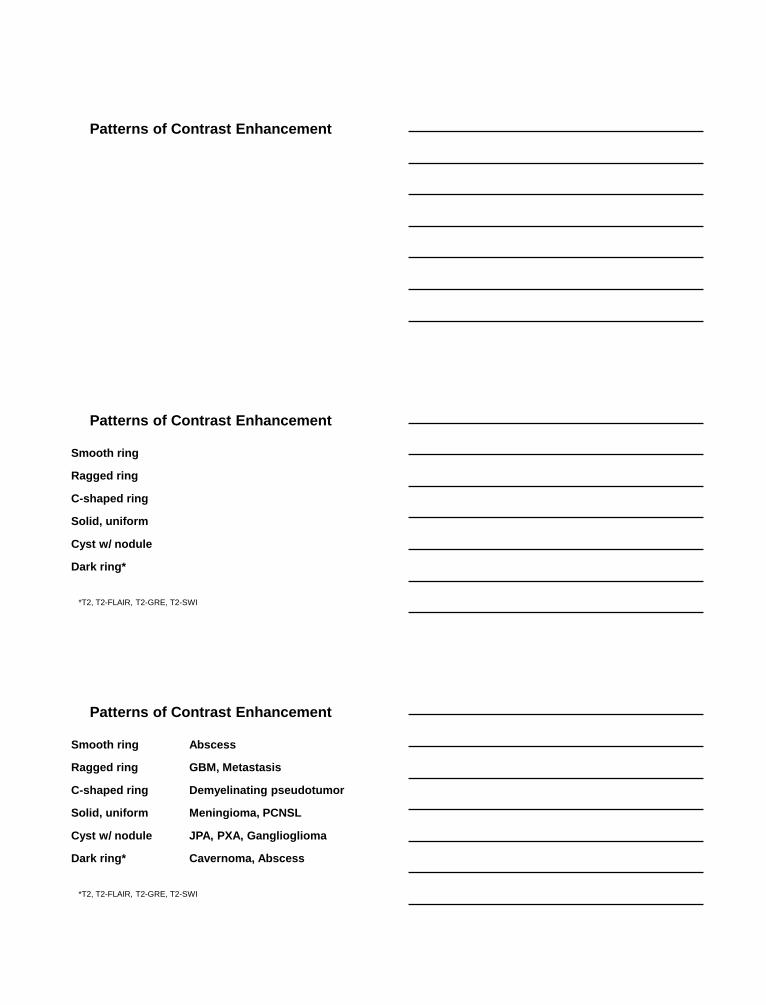
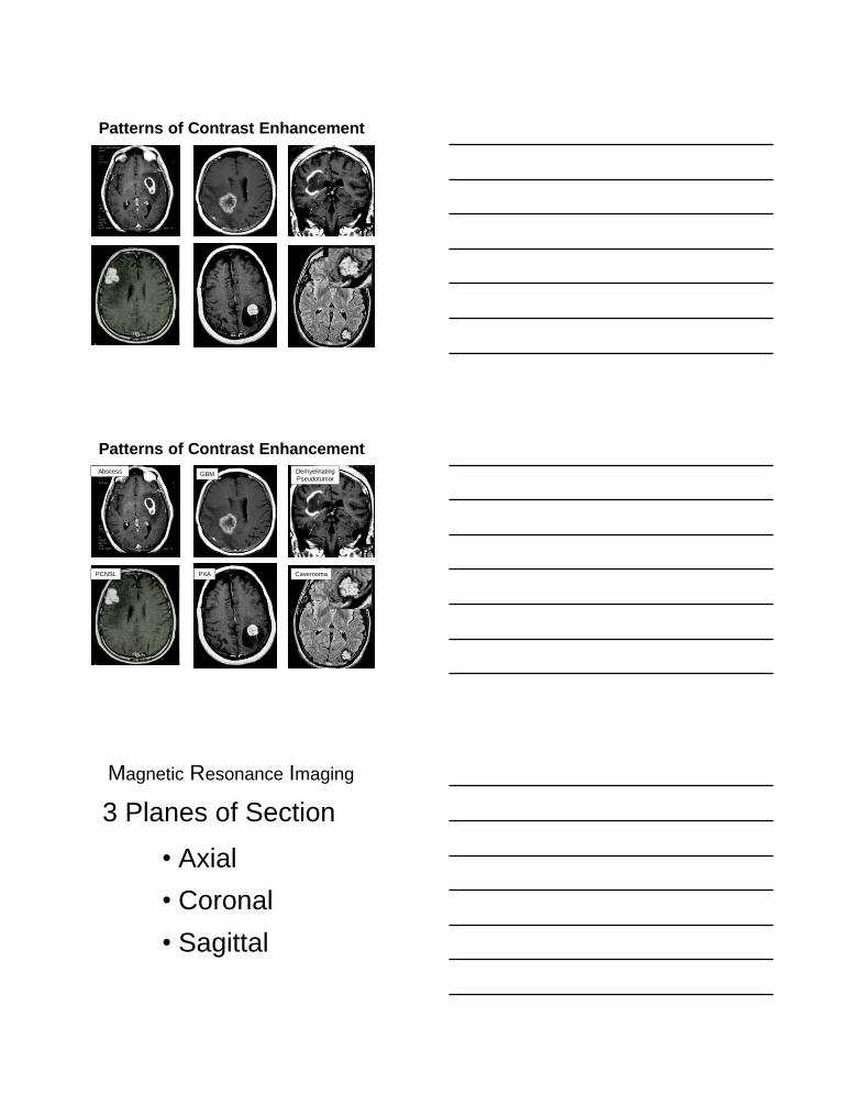
Patterns of Contrast Enhancement
Patterns of Contrast Enhancement
Smooth ring
Ragged ring
C-shaped ring
Solid, uniform
Cyst w/ nodule
Dark ring*
*T2, T2-FLAIR, T2-GRE, T2-SWI
Patterns of Contrast Enhancement
Smooth ring Abscess
Ragged ring GBM, Metastasis
C-shaped ring Demyelinating pseudotumor
Solid, uniform Meningioma, PCNSL
Cyst w/ nodule JPA, PXA, Ganglioglioma
Dark ring* Cavernoma, Abscess
*T2, T2-FLAIR, T2-GRE, T2-SWI
14
Patterns of Contrast Enhancement
Patterns of Contrast Enhancement
Abscess Demyelinating
Pseudotumor GBM
PCNSL Cavernoma PXA
Magnetic Resonance Imaging
• Axial
• Coronal
• Sagittal
3 Planes of Section
15
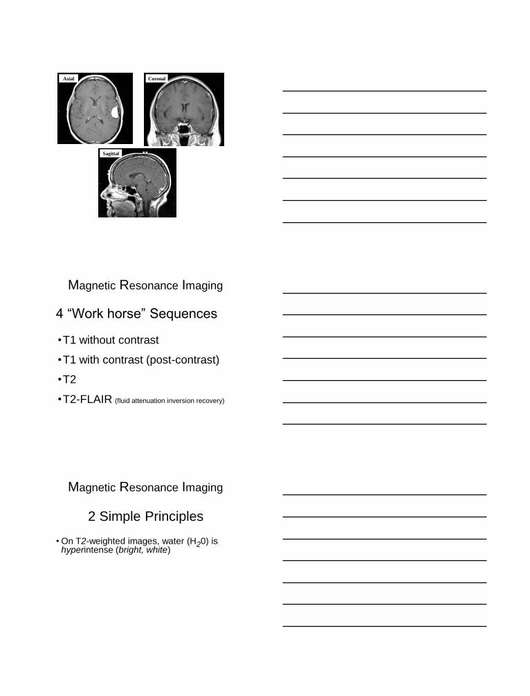
Sagittal
Coronal Axial
Magnetic Resonance Imaging
•T1 without contrast
•T1 with contrast (post-contrast)
•T2
•T2-FLAIR (fluid attenuation inversion recovery)
4 “Work horse” Sequences
Magnetic Resonance Imaging
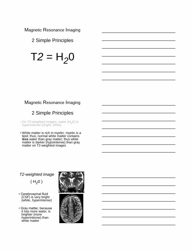
2 Simple Principles
• On T2-weighted images, water (H20) is hyperintense (bright, white)
16
Magnetic Resonance Imaging
2 Simple Principles
T2 = H20
Magnetic Resonance Imaging
2 Simple Principles
• On T2-weighted images, water (H20) is hyperintense (bright, white)
• White matter is rich in myelin; myelin is a lipid; thus, normal white matter contains less water than gray matter; thus white matter is darker (hypointense) than gray matter on T2-weighted images
T2-weighted image
( H20 )
• Cerebrospinal fluid (CSF) is very bright (white, hyperintense)
• Gray matter, because it has more water, is brighter (more hyperintense) than white matter
17
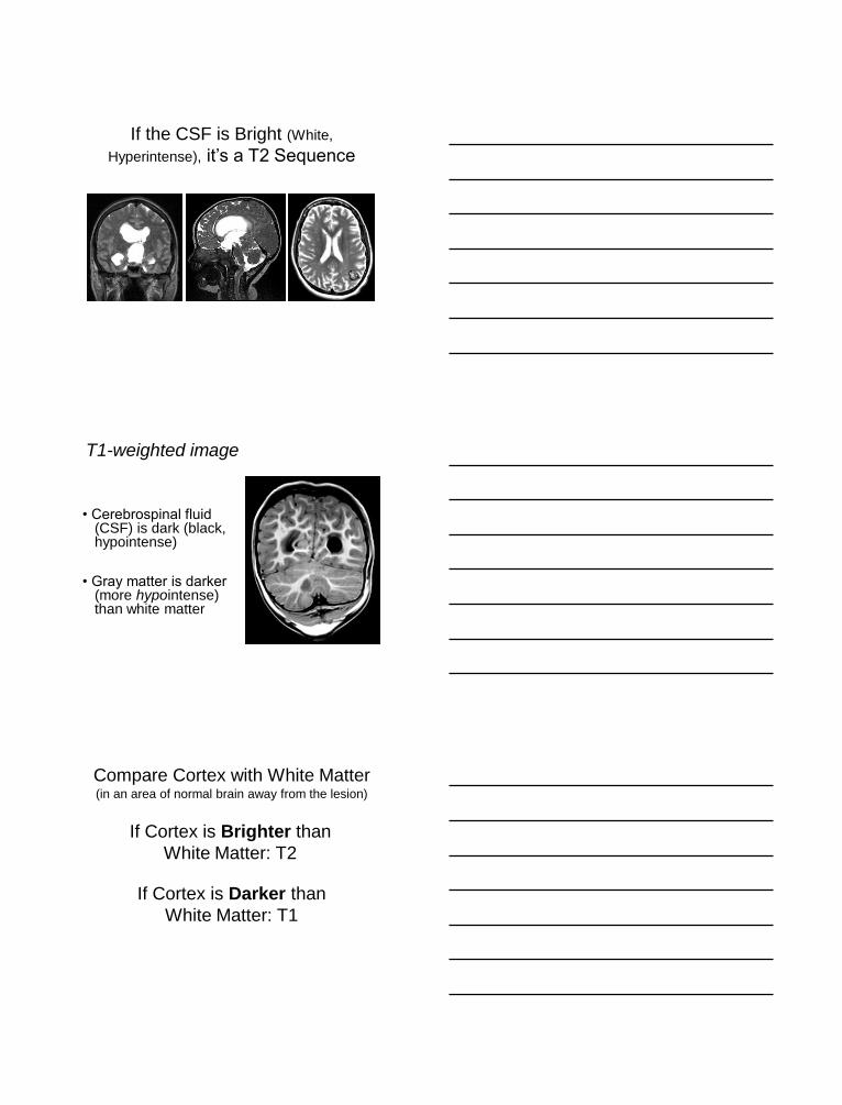
If the CSF is Bright (White,
Hyperintense), it’s a T2 Sequence
T1-weighted image
• Cerebrospinal fluid (CSF) is dark (black, hypointense)
• Gray matter is darker (more hypointense) than white matter
Compare Cortex with White Matter
If Cortex is Brighter than
White Matter: T2
If Cortex is Darker than
White Matter: T1
(in an area of normal brain away from the lesion)
18
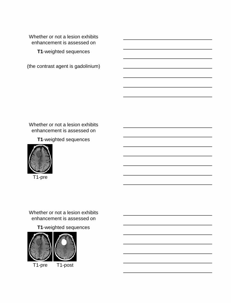
Whether or not a lesion exhibits
enhancement is assessed on
T1-weighted sequences
(the contrast agent is gadolinium)
T1-pre
Whether or not a lesion exhibits
enhancement is assessed on
T1-weighted sequences
T1-pre T1-post
Whether or not a lesion exhibits
enhancement is assessed on
T1-weighted sequences
19
T1-pre T1-post T2
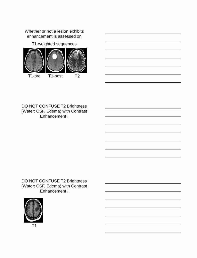
Whether or not a lesion exhibits
enhancement is assessed on
T1-weighted sequences
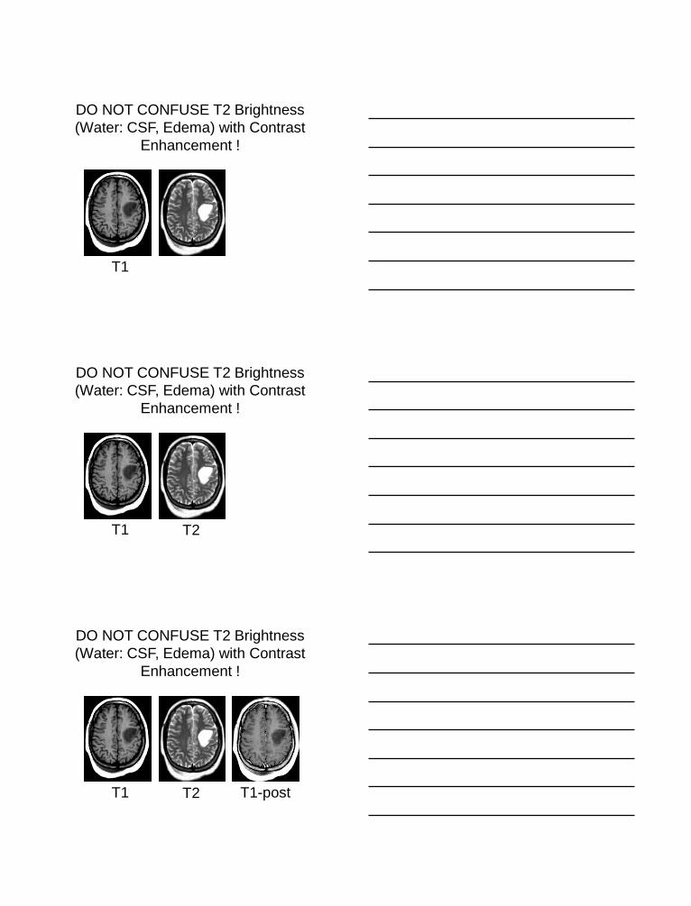
DO NOT CONFUSE T2 Brightness
(Water: CSF, Edema) with Contrast
Enhancement !
DO NOT CONFUSE T2 Brightness
(Water: CSF, Edema) with Contrast
Enhancement !
T1
20
DO NOT CONFUSE T2 Brightness
(Water: CSF, Edema) with Contrast
Enhancement !
T1
T1 T2
DO NOT CONFUSE T2 Brightness
(Water: CSF, Edema) with Contrast
Enhancement !
T1 T2 T1-post
DO NOT CONFUSE T2 Brightness
(Water: CSF, Edema) with Contrast
Enhancement !
21
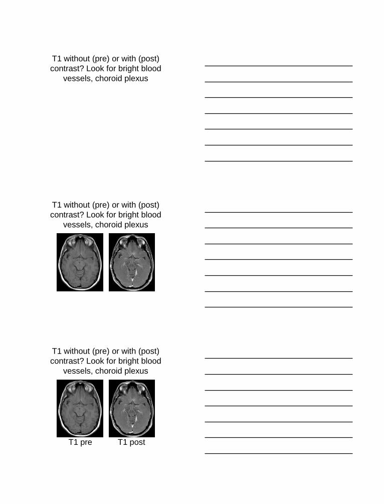
T1 without (pre) or with (post)
contrast? Look for bright blood
vessels, choroid plexus
T1 without (pre) or with (post)
contrast? Look for bright blood
vessels, choroid plexus
T1 pre T1 post
T1 without (pre) or with (post)
contrast? Look for bright blood
vessels, choroid plexus
22
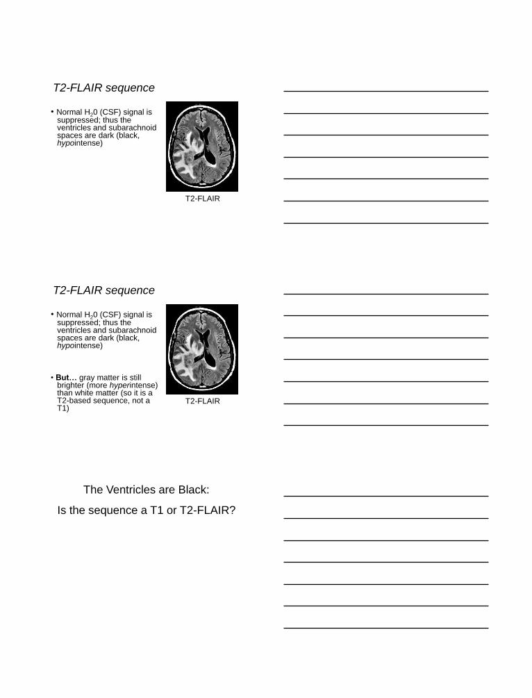
T2-FLAIR sequence
• Normal H20 (CSF) signal is suppressed; thus the ventricles and subarachnoid spaces are dark (black, hypointense)
T2-FLAIR
T2-FLAIR sequence
• Normal H20 (CSF) signal is suppressed; thus the ventricles and subarachnoid spaces are dark (black, hypointense)
• But… gray matter is still brighter (more hyperintense) than white matter (so it is a T2-based sequence, not a T1)
T2-FLAIR
The Ventricles are Black:
Is the sequence a T1 or T2-FLAIR?
23
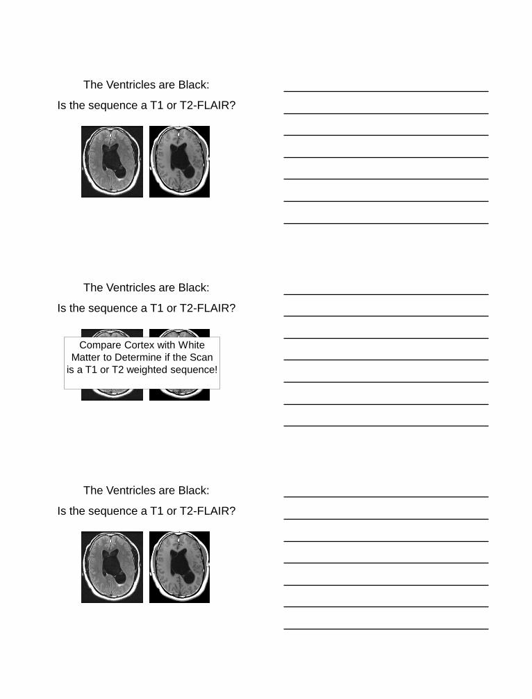
The Ventricles are Black:
Is the sequence a T1 or T2-FLAIR?
The Ventricles are Black:
Is the sequence a T1 or T2-FLAIR?
Compare Cortex with White
Matter to Determine if the Scan
is a T1 or T2 weighted sequence!
The Ventricles are Black:
Is the sequence a T1 or T2-FLAIR?
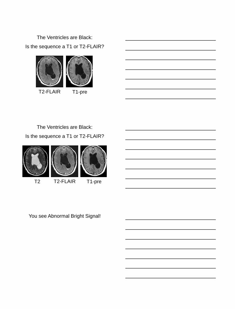
24
The Ventricles are Black:
Is the sequence a T1 or T2-FLAIR?
T2-FLAIR T1-pre
The Ventricles are Black:
Is the sequence a T1 or T2-FLAIR?
T2-FLAIR T1-pre T2
You see Abnormal Bright Signal!

25
You see Abnormal Bright Signal!
Is it contrast enhancement on a T1 post, or
is it edema on a T2 or T2-FLAIR ???
You see Abnormal Bright Signal!
Is it contrast enhancement on a T1 post, or
is it edema on a T2 or T2-FLAIR ???
You see Abnormal Bright Signal!
Is it contrast enhancement on a T1 post, or
is it edema on a T2 or T2-FLAIR ???
The cortical gray matter is
hyperintense to white matter,
thus it’s a T2 sequence
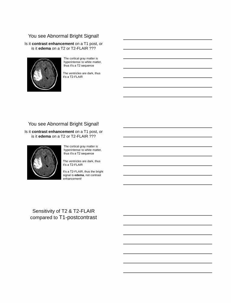
26
You see Abnormal Bright Signal!
Is it contrast enhancement on a T1 post, or
is it edema on a T2 or T2-FLAIR ???
The cortical gray matter is
hyperintense to white matter,
thus it’s a T2 sequence
The ventricles are dark, thus
it’s a T2-FLAIR
You see Abnormal Bright Signal!
Is it contrast enhancement on a T1 post, or
is it edema on a T2 or T2-FLAIR ???
The cortical gray matter is
hyperintense to white matter,
thus it’s a T2 sequence
The ventricles are dark, thus
it’s a T2-FLAIR
It’s a T2-FLAIR, thus the bright
signal is edema, not contrast
enhancement!
Sensitivity of T2 & T2-FLAIR
compared to T1-postcontrast

27
T1-post
Sensitivity of T2 & T2-FLAIR
compared to T1-postcontrast
Do you see
an obvious
lesion?
Sensitivity of T2 & T2-FLAIR
compared to T1-postcontrast
T1-post T2-FLAIR
ADVANTAGE OF T2-FLAIR over T2
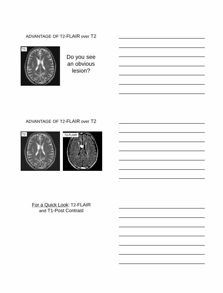
28
ADVANTAGE OF T2-FLAIR over T2
T2
Do you see
an obvious
lesion?
ADVANTAGE OF T2-FLAIR over T2
T2 T2-FLAIR
For a Quick Look: T2-FLAIR
and T1-Post Contrast
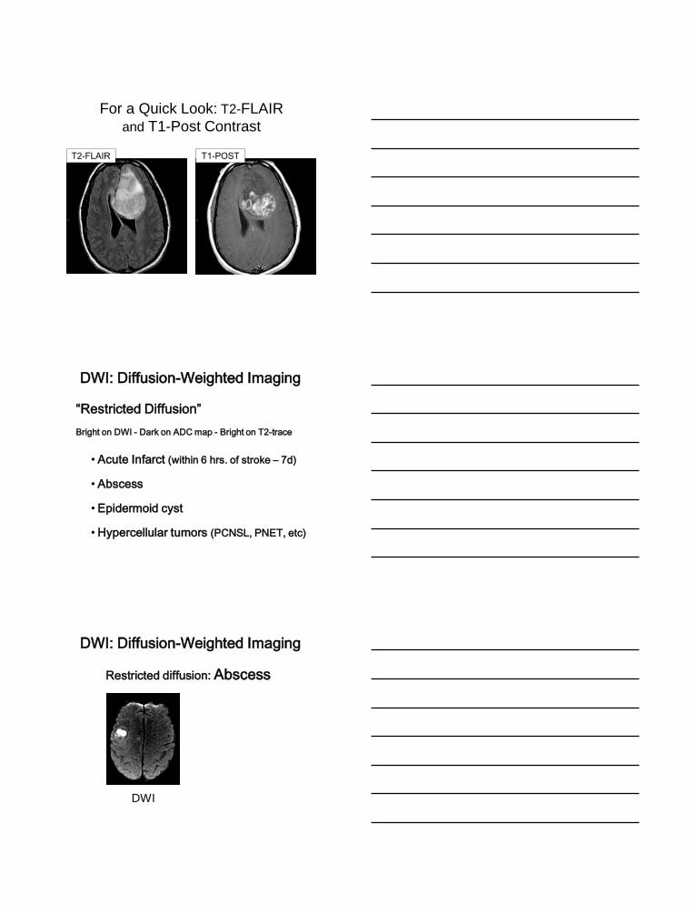
29
For a Quick Look: T2-FLAIR
and T1-Post Contrast
T2-FLAIR T1-POST
DWI: Diffusion-Weighted Imaging
“Restricted Diffusion”
Bright on DWI - Dark on ADC map - Bright on T2-trace
• Acute Infarct (within 6 hrs. of stroke – 7d)
• Abscess
• Epidermoid cyst
• Hypercellular tumors (PCNSL, PNET, etc)
Restricted diffusion: Abscess
DWI
DWI: Diffusion-Weighted Imaging
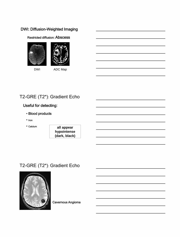
30
Restricted diffusion: Abscess
DWI ADC Map
DWI: Diffusion-Weighted Imaging
T2-GRE (T2*): Gradient Echo
Useful for detecting:
• Blood products
• Iron
• Calcium
all appear
hypointense
(dark, black)
Cavernous Angioma
T2-GRE (T2*): Gradient Echo
31
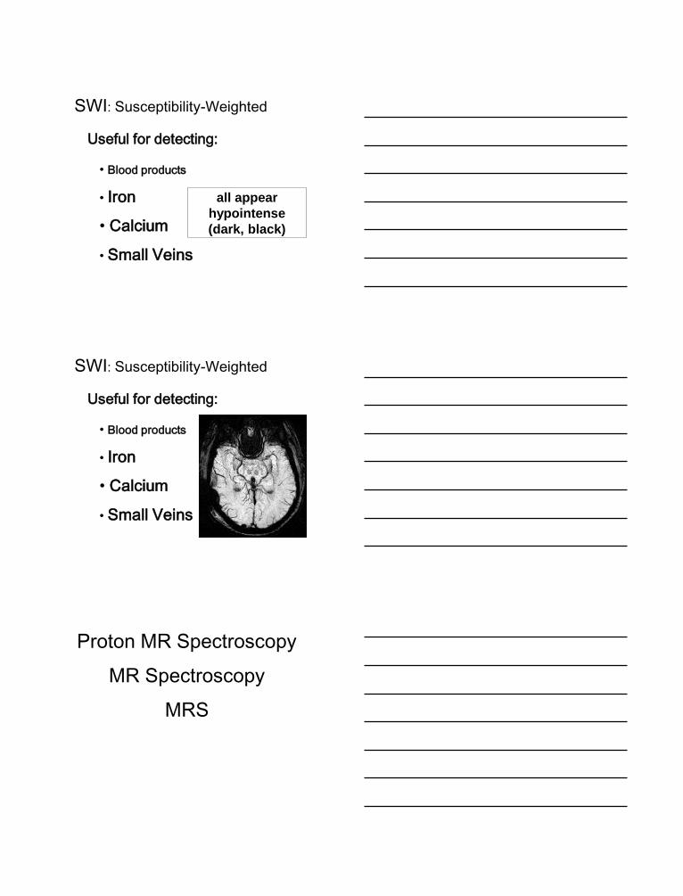
SWI: Susceptibility-Weighted
Useful for detecting:
• Blood products
• Iron
• Calcium
• Small Veins
all appear
hypointense
(dark, black)
SWI: Susceptibility-Weighted
Useful for detecting:
• Blood products
• Iron
• Calcium
• Small Veins
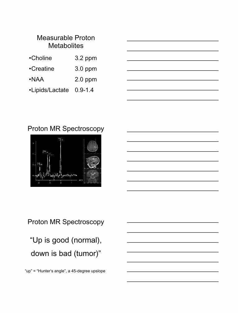
Proton MR Spectroscopy
MR Spectroscopy
MRS
32
Measurable Proton Metabolites
•Choline 3.2 ppm
•Creatine 3.0 ppm
•NAA 2.0 ppm
•Lipids/Lactate 0.9-1.4
Proton MR Spectroscopy
Cho
Cr
NAA
Lactate
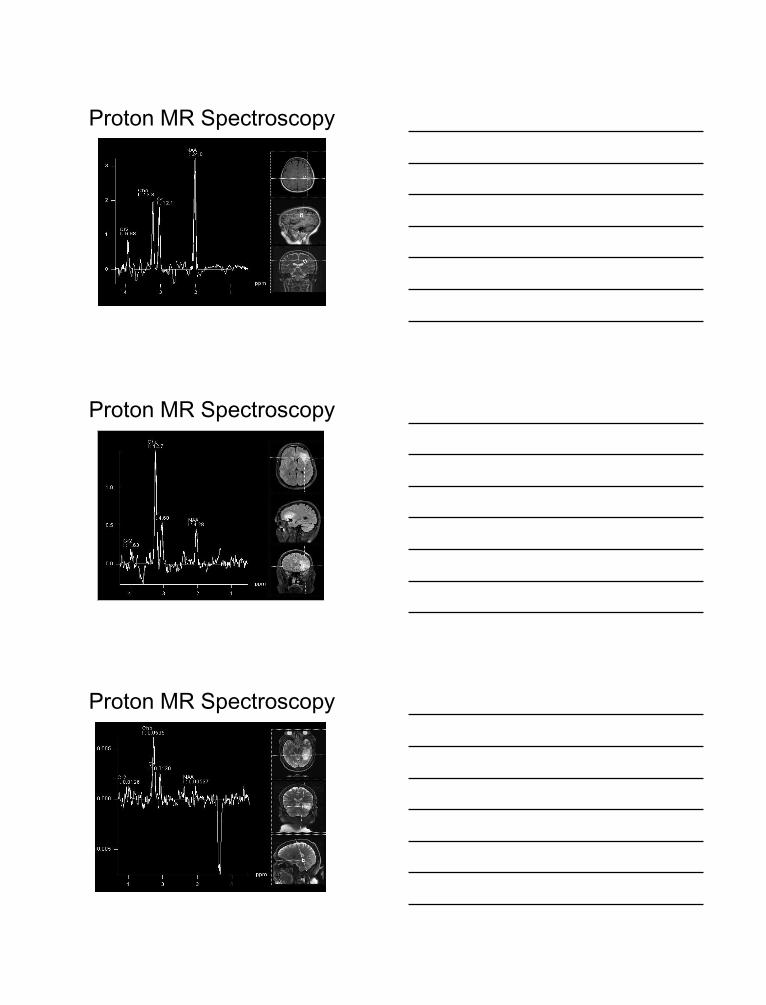
Proton MR Spectroscopy
“Up is good (normal),
down is bad (tumor)”
“up” = “Hunter’s angle”, a 45-degree upslope
33
Proton MR Spectroscopy
Cho
Cr
NAA
Lactate
Proton MR Spectroscopy
Cho
Cr
NAA
Lactate
Proton MR Spectroscopy
Cho
Cr
NAA
Lactate
34
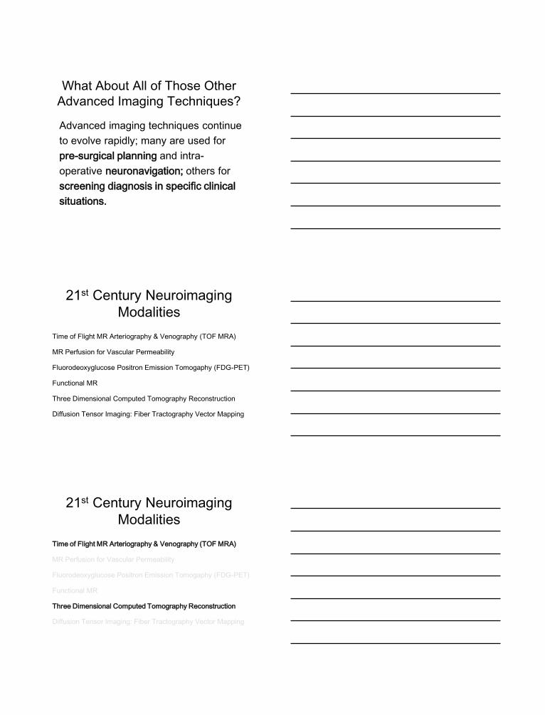
What About All of Those Other
Advanced Imaging Techniques?
Advanced imaging techniques continue
to evolve rapidly; many are used for
pre-surgical planning and intra-
operative neuronavigation; others for
screening diagnosis in specific clinical
situations.
21st Century Neuroimaging
Modalities
Time of Flight MR Arteriography & Venography (TOF MRA)
MR Perfusion for Vascular Permeability
Fluorodeoxyglucose Positron Emission Tomogaphy (FDG-PET)
Functional MR
Three Dimensional Computed Tomography Reconstruction
Diffusion Tensor Imaging: Fiber Tractography Vector Mapping
35
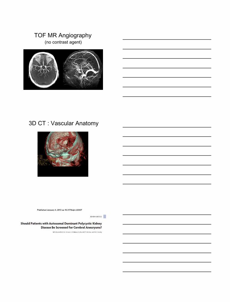
TOF MR Angiography (no contrast agent)
3D CT : Vascular Anatomy
36
21st Century Neuroimaging
Modalities
Time of Flight MR Arteriography & Venography (TOF MRA)
MR Perfusion for Vascular Permeability
Fluorodeoxyglucose Positron Emission Tomogaphy (FDG-PET)
Functional MR
Three Dimensional Computed Tomography Reconstruction
Diffusion Tensor Imaging: Fiber Tractography Vector Mapping
37
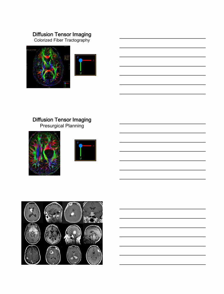
Diffusion Tensor Imaging
Colorized Fiber Tractography
Fuller GN Arch Path Lab Med 2007
Diffusion Tensor Imaging
Presurgical Planning
Fuller GN Arch Path Lab Med 2007
38
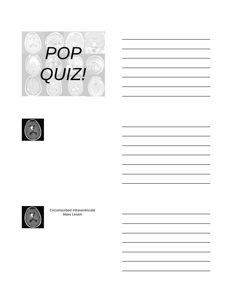
POP
QUIZ!
Circumscribed Intraventricular
Mass Lesion
39
• Choroid plexus papilloma
• Atypical choroid plexus papilloma
• Choroid plexus carcinoma
• Choroid plexus meningioma
• Choroid plexus xanthogranuloma
• Ependymoma
• Subependymoma
• Subependymal giant cell astrocytoma
• Central neurocytoma
• Solitary metastasis to the choroid plexus (especially renal cell carcinoma)
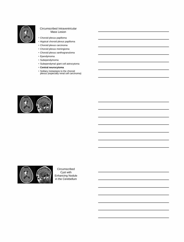
Circumscribed Intraventricular
Mass Lesion
Circumscribed
Cyst with
Enhancing Nodule
in the Cerebellum

40
• Pilocytic astrocytoma
• Hemangioblastoma
• Cystic metastasis
Circumscribed
Cyst with
Enhancing Nodule
in the Cerebellum
Circumscribed
Cyst with
Enhancing Nodule
in the Cerebellum
in a 5yo Child
Circumscribed
Cyst with
Enhancing Nodule
in the Cerebellum
in a 5yo Child
• Pilocytic astrocytoma

41
Circumscribed
Cyst with
Enhancing Nodule
in the Cerebellum
in a 56yo Man
Circumscribed
Cyst with
Enhancing Nodule
in the Cerebellum
in a 56yo Man
• Hemangioblastoma
• Cystic metastasis
42
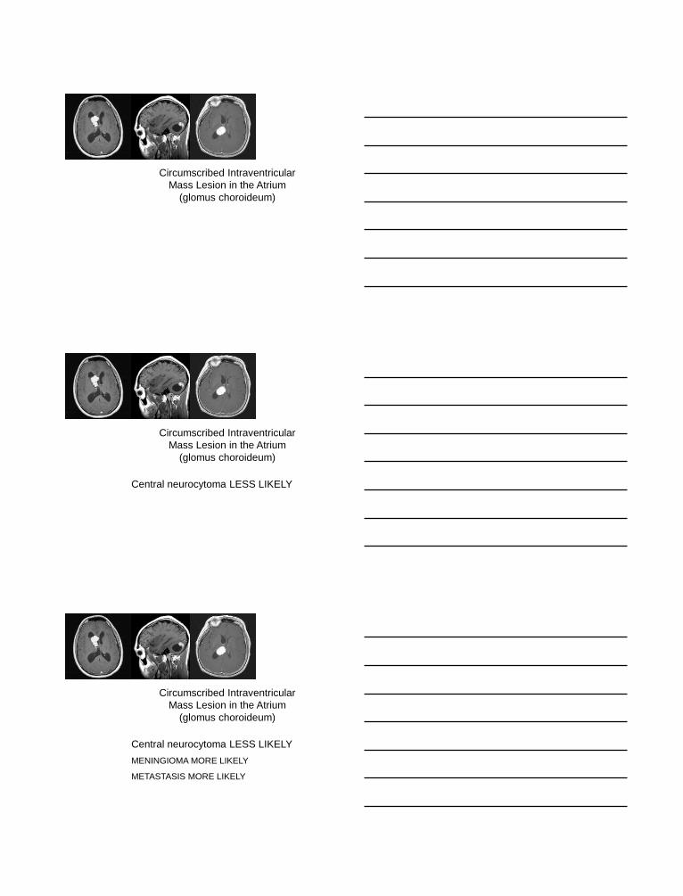
Circumscribed Intraventricular
Mass Lesion in the Atrium
(glomus choroideum)
Circumscribed Intraventricular
Mass Lesion in the Atrium
(glomus choroideum)
Central neurocytoma LESS LIKELY
Circumscribed Intraventricular
Mass Lesion in the Atrium
(glomus choroideum)
Central neurocytoma LESS LIKELY
MENINGIOMA MORE LIKELY
METASTASIS MORE LIKELY
43
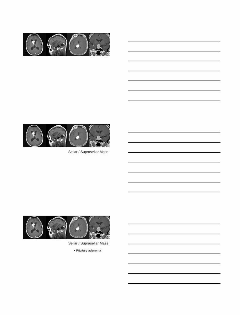
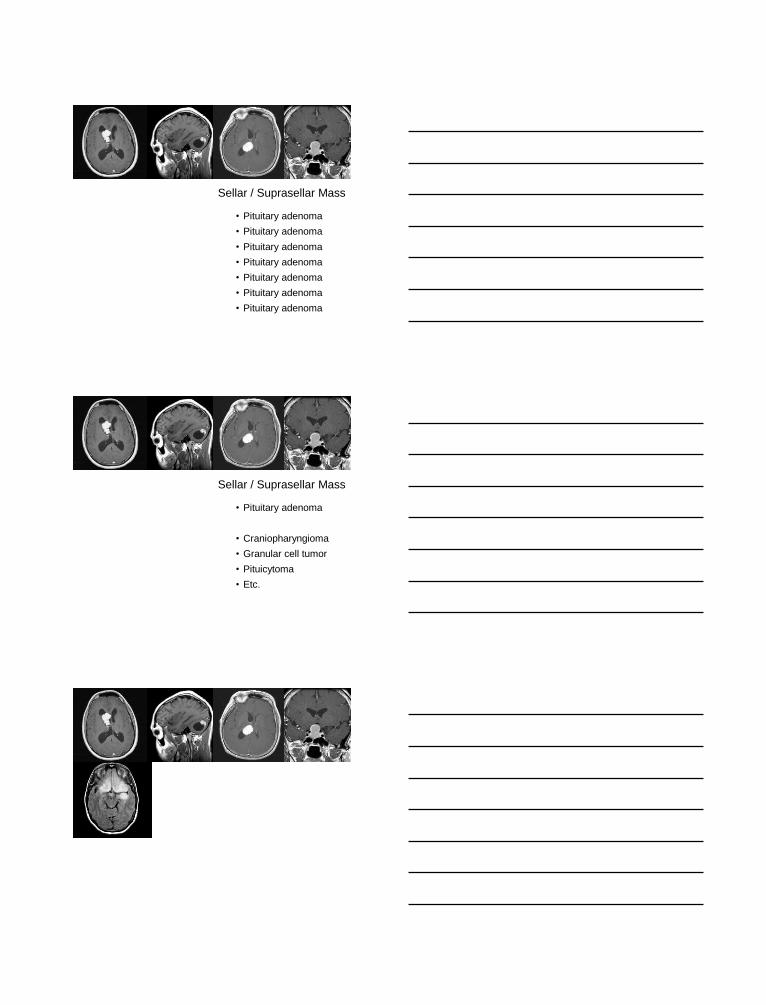
Sellar / Suprasellar Mass
Sellar / Suprasellar Mass
• Pituitary adenoma
44
Sellar / Suprasellar Mass
• Pituitary adenoma
• Pituitary adenoma
• Pituitary adenoma
• Pituitary adenoma
• Pituitary adenoma
• Pituitary adenoma
• Pituitary adenoma
• Pituitary adenoma
• Craniopharyngioma
• Granular cell tumor
• Pituicytoma
• Etc.
Sellar / Suprasellar Mass
45
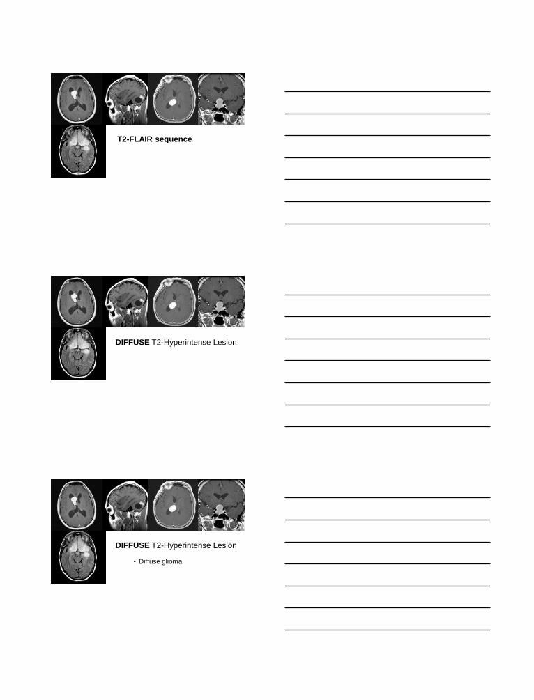
T2-FLAIR sequence
DIFFUSE T2-Hyperintense Lesion
• Diffuse glioma
DIFFUSE T2-Hyperintense Lesion
46
• Diffuse glioma
• Lymphoma
DIFFUSE T2-Hyperintense Lesion
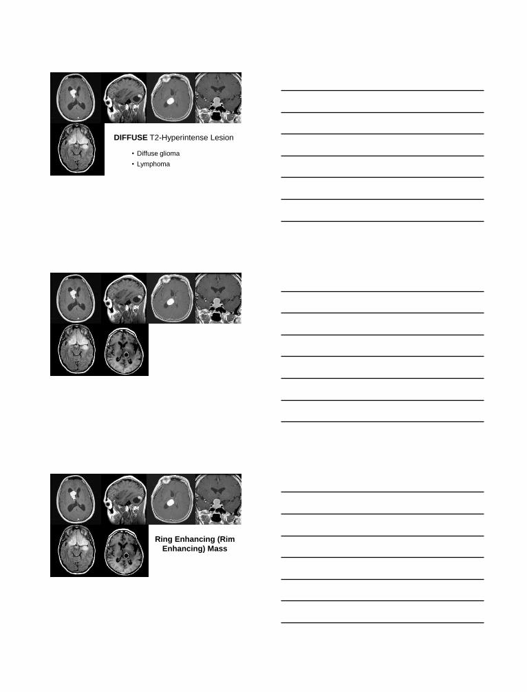
Ring Enhancing (Rim
Enhancing) Mass

47
Smooth ring
Ring Enhancing (Rim
Enhancing) Mass
• Abscess
Smooth ring
Ring Enhancing (Rim
Enhancing) Mass
• Abscess
• Check DWI sequence for restricted diffusion
Smooth ring
Ring Enhancing (Rim
Enhancing) Mass
48
• Abscess
• Glioblastoma
• Metastasis
Smooth ring
Ring Enhancing (Rim
Enhancing) Mass
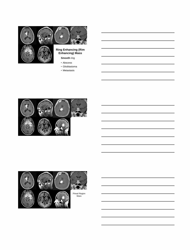
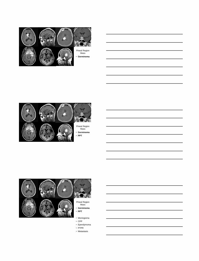
Pineal Region
Mass
49
• Germinoma
Pineal Region
Mass
• Germinoma
• PPT
Pineal Region
Mass
• Germinoma
• PPT
• Meningioma
• CPP
• Ependymoma
• PTPR
• Metastasis
Pineal Region
Mass
50
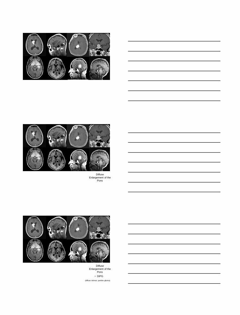
Diffuse
Enlargement of the
Pons
Diffuse
Enlargement of the
Pons
• DIPG
(diffuse intrinsic pontine glioma)

51
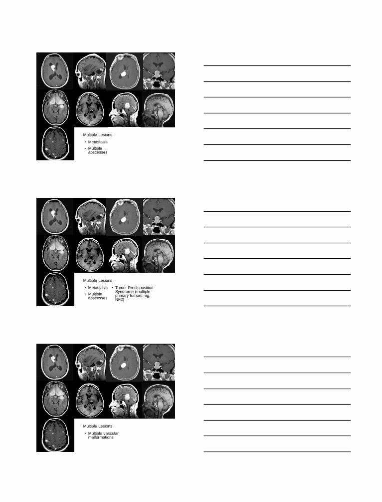
Multiple Lesions
• Metastasis
Multiple Lesions
52
• Metastasis
• Multiple abscesses
Multiple Lesions
• Metastasis
• Multiple abscesses
Multiple Lesions
• Tumor Predisposition Syndrome (multiple primary tumors; eg, NF2)
• Multiple vascular malformations
Multiple Lesions
53
• Multiple vascular malformations
• Multiple parasites (eg, cysticercosis)
Multiple Lesions
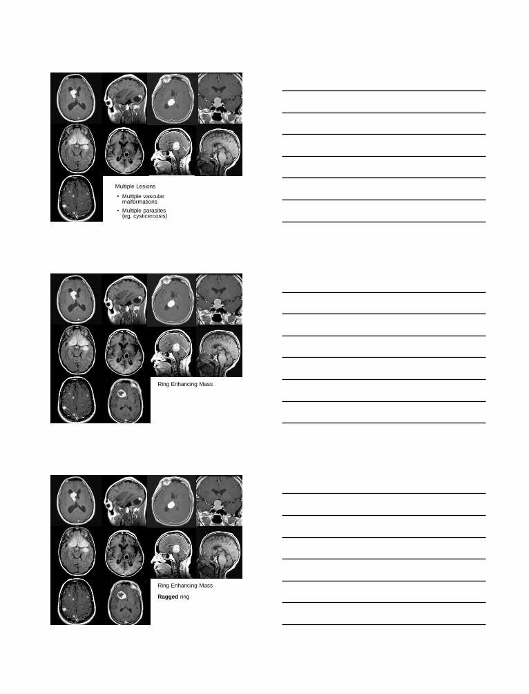
Ring Enhancing Mass
Ring Enhancing Mass
Ragged ring
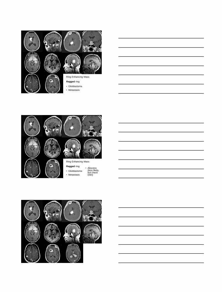
54
Ring Enhancing Mass
Ragged ring
• Glioblastoma
• Metastasis
Ring Enhancing Mass
Ragged ring
• Glioblastoma
• Metastasis
• Abscess (less likely, but check DWI)
55
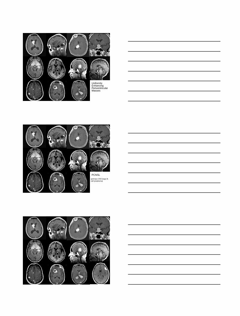
Uniformly Enhancing Periventricular Masses
PCNSL
(primary CNS large B-
cell lymphoma)

56
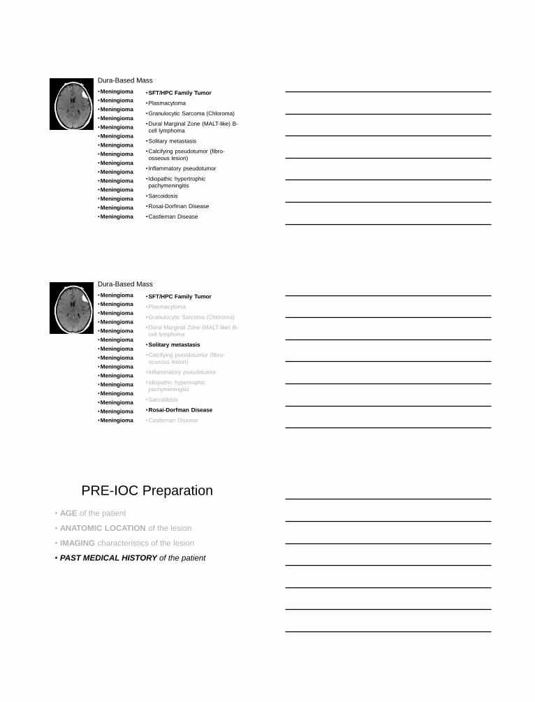
Dura-Based Mass
•Meningioma
•Meningioma
•Meningioma
•Meningioma
•Meningioma
•Meningioma
•Meningioma
•Meningioma
•Meningioma
•Meningioma
•Meningioma
•Meningioma
•Meningioma
•Meningioma
•Meningioma
Dura-Based Mass
•SFT/HPC Family Tumor •Meningioma
•Meningioma
•Meningioma
•Meningioma
•Meningioma
•Meningioma
•Meningioma
•Meningioma
•Meningioma
•Meningioma
•Meningioma
•Meningioma
•Meningioma
•Meningioma
•Meningioma
Dura-Based Mass
57
•SFT/HPC Family Tumor
•Plasmacytoma
•Granulocytic Sarcoma (Chloroma)
•Dural Marginal Zone (MALT-like) B-
cell lymphoma
•Solitary metastasis
•Calcifying pseudotumor (fibro-
osseous lesion)
• Inflammatory pseudotumor
• Idiopathic hypertrophic
pachymeningitis
•Sarcoidosis
•Rosai-Dorfman Disease
•Castleman Disease
•Meningioma
•Meningioma
•Meningioma
•Meningioma
•Meningioma
•Meningioma
•Meningioma
•Meningioma
•Meningioma
•Meningioma
•Meningioma
•Meningioma
•Meningioma
•Meningioma
•Meningioma
Dura-Based Mass
PRE-IOC Preparation
• AGE of the patient
• ANATOMIC LOCATION of the lesion
• IMAGING characteristics of the lesion
• PAST MEDICAL HISTORY of the patient

58
PRE-IOC Preparation
• AGE of the patient
• ANATOMIC LOCATION of the lesion
• IMAGING characteristics of the lesion
• PAST MEDICAL HISTORY of the patient
• TYPE and DURATION of presenting signs &
symptoms
PRE-IOC Preparation
• AGE of the patient
• ANATOMIC LOCATION of the lesion
• IMAGING characteristics of the lesion
• PAST MEDICAL HISTORY of the patient
• TYPE and DURATION of presenting signs &
symptoms
• WHAT TYPE of SURGICAL PROCEDURE?
PRE-IOC Preparation
• AGE of the patient
• ANATOMIC LOCATION of the lesion
• IMAGING characteristics of the lesion
• PAST MEDICAL HISTORY of the patient
• TYPE and DURATION of presenting signs &
symptoms
• WHAT TYPE of SURGICAL PROCEDURE?
• WHAT WILL THE SURGEON NEED TO KNOW?
59
IOC PRINCIPLES
• Cytology and Architecture are
Complementary – Use Both!
IOC PRINCIPLES
• Cytology and Architecture are
Complementary – Use Both!
• Diff-Quik and H&E stains for cytology
are Complementary – You Can Use
Both!
IOC PRINCIPLES

60
• Know what the surgeon needs to know
about the lesion intraoperatively!
IOC PRINCIPLES
• Know what the surgeon needs to know
about the lesion intraoperatively!
• The surgeon needs to know
information that is needed to
successfully complete the
operation and will determine the
subsequent course of the operation
IOC PRINCIPLES
• Know what the surgeon needs to know
about the lesion intraoperatively!
Indications for IOC
IOC PRINCIPLES
61
IOC PRINCIPLES
• Is adequate, representative tissue present?
Some Indications for IOC
Some Indications for IOC
• Is adequate, representative tissue present?
• Is the disease an infectious process?
IOC PRINCIPLES
IOC PRINCIPLES
Some Valid Indications for IOC
• Is adequate, representative tissue present?
• Is the disease an infectious process?
• If tumor, is it of a type amenable to gross total
surgical resection

62
IOC PRINCIPLES
Some Valid Indications for IOC
• Is adequate, representative tissue present?
• Is the disease an infectious process?
• If tumor, is it of a type amenable to gross total
surgical resection
• Is viable GBM present? (if so, Gliadel wafer,
Gliacyte balloon, other intraoperative
treatment can procede)
IOC PRINCIPLES
• Cytology and Architecture are
Complementary – Use Both!
• Don’t freeze all of the tissue if possible
• Know what the surgeon needs to know
about the lesion intraoperatively!
• Plan for tomorrow!
IOC PRINCIPLES
• Plan for tomorrow!

63
IOC PRINCIPLES
• Plan for tomorrow!
For very small biopsies (e.g.,
stereotactic bx), ensure adequate
specimen for IHC:
IOC PRINCIPLES
• Plan for tomorrow!
For very small biopsies (e.g.,
stereotactic bx), ensure adequate
specimen for IHC:
unstained touch preps
IOC PRINCIPLES
• Plan for tomorrow!
For very small biopsies (e.g.,
stereotactic bx), ensure adequate
specimen for IHC:
unstained touch preps
unstained sections cut from FS block
64
IOC PRINCIPLES
• Plan for tomorrow!
For very small biopsies (e.g.,
stereotactic bx), ensure adequate
specimen for IHC:
unstained touch preps
unstained sections cut from FS block
order unstained from paraffin block (“biopsy processing”)
IOC PRINCIPLES
• Know what the surgeon DOES NOT
need to know about the lesion
intraoperatively!
IOC PRINCIPLES
• Know what the surgeon DOES NOT
need to know about the lesion
intraoperatively!
Important example: the specific type of
diffuse glioma (astro, oligo, mixed
oligoastro)
65
IOC PRINCIPLES
• The specific type of diffuse glioma
WILL need to be determined with the
help of diagnostic molecular studies
for the final diagnosis, but this level
of specificity is generally not required
to complete the surgical procedure.
IOC PRINCIPLES
• Common cause of intraoperative
diagnosis / final diagnosis
discrepancy: misdiagnosing
oligodendroglioma as astrocytoma on
frozen sections.
IOC PRINCIPLES
• Freezing distorts oligo nuclei and
makes them appear more pleomorphic
and thus astrocytoma-like
• The characteristic cytoplasmic clearing
(perinuclear “halos”, “fried egg”
appearance) is only seen in FFPE
tissue, not in frozen sections
66
IOC PRINCIPLES
• One Caveat
The pathologist must ensure that
representative tumor has been obtained with
respect to grade (correlate with the
preoperative imaging studies; enhancing
diffuse gliomas are usually high-grade;
“undersampling” and hence “undergrading” of
diffuse gliomas is very common in stereotactic
biopsies)
INTRA-IOC
Perform a Cytologic
Prep!
67
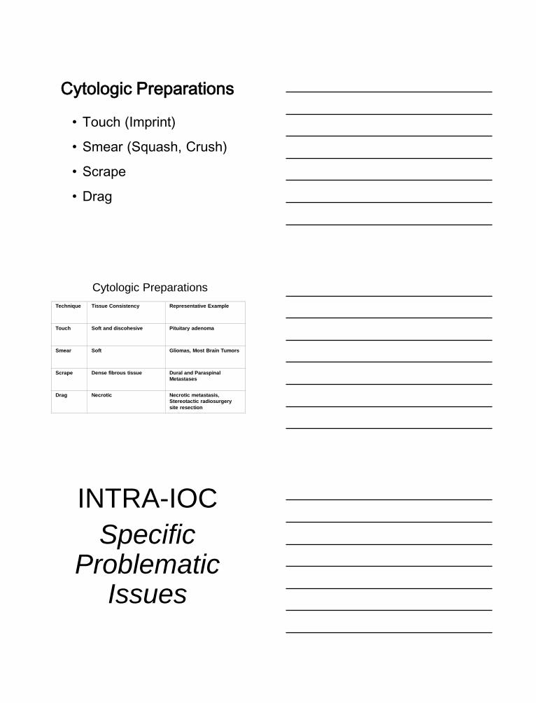
Cytologic Preparations
• Touch (Imprint)
• Smear (Squash, Crush)
• Scrape
• Drag
Technique
Tissue Consistency
Representative Example
Touch
Soft and discohesive
Pituitary adenoma
Smear
Soft
Gliomas, Most Brain Tumors
Scrape
Dense fibrous tissue Dural and Paraspinal
Metastases
Drag
Necrotic
Necrotic metastasis,
Stereotactic radiosurgery
site resection
Cytologic Preparations
INTRA-IOC
Specific Problematic
Issues
68
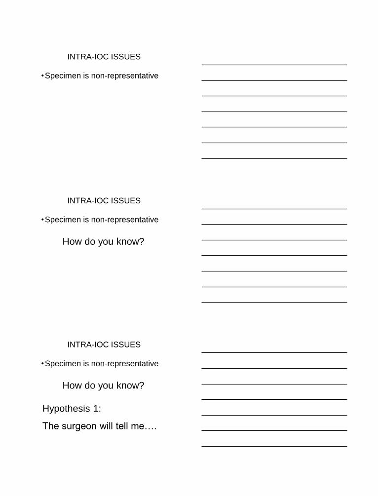
•Specimen is non-representative
INTRA-IOC ISSUES
•Specimen is non-representative
INTRA-IOC ISSUES
How do you know?
•Specimen is non-representative
INTRA-IOC ISSUES
How do you know?
Hypothesis 1:
The surgeon will tell me….

69
NOT!
•Specimen is non-representative
INTRA-IOC ISSUES
How do you know?
•Specimen is non-representative
INTRA-IOC ISSUES
How do you know? Imaging Studies!

70
•Specimen is very small
(endoscopic bx or fragment of stereotactic bx)
INTRA-IOC ISSUES
•Specimen is very small
(endoscopic bx or fragmented stereotactic bx)
INTRA-IOC ISSUES
Perform a cytologic preparation
(imprint or drag prep) before
freezing
•Specimen is seriously small!
INTRA-IOC ISSUES
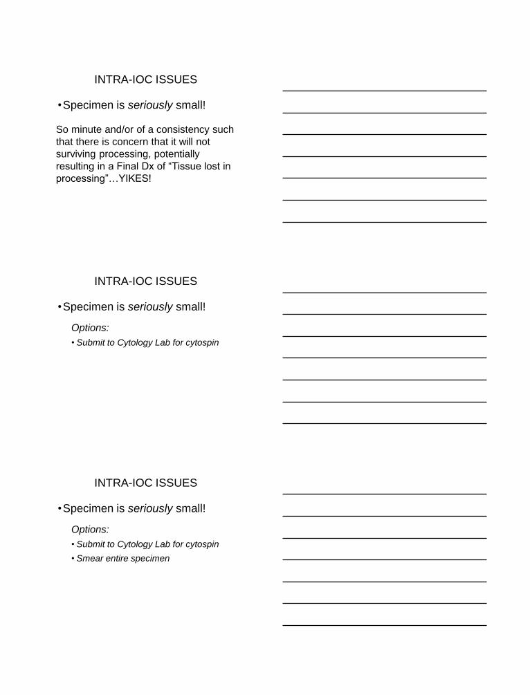
71
•Specimen is seriously small!
INTRA-IOC ISSUES
So minute and/or of a consistency such
that there is concern that it will not
surviving processing, potentially
resulting in a Final Dx of “Tissue lost in
processing”…YIKES!
•Specimen is seriously small!
INTRA-IOC ISSUES
Options:
• Submit to Cytology Lab for cytospin
•Specimen is seriously small!
INTRA-IOC ISSUES
Options:
• Submit to Cytology Lab for cytospin
• Smear entire specimen
72
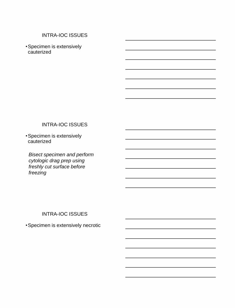
•Specimen is extensively cauterized
INTRA-IOC ISSUES
•Specimen is extensively cauterized
INTRA-IOC ISSUES
Bisect specimen and perform
cytologic drag prep using
freshly cut surface before
freezing
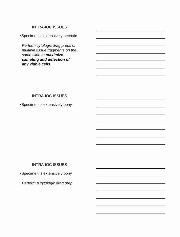
•Specimen is extensively necrotic
INTRA-IOC ISSUES
73
•Specimen is extensively necrotic
INTRA-IOC ISSUES
Perform cytologic drag preps on
multiple tissue fragments on the
same slide to maximize
sampling and detection of
any viable cells
•Specimen is extensively bony
INTRA-IOC ISSUES
•Specimen is extensively bony
INTRA-IOC ISSUES
Perform a cytologic drag prep

74
•Specimen is densely fibrous
INTRA-IOC ISSUES
•Specimen is densely fibrous
INTRA-IOC ISSUES
Perform a cytologic scrape
prep before freezing
POST-IOC ISSUE
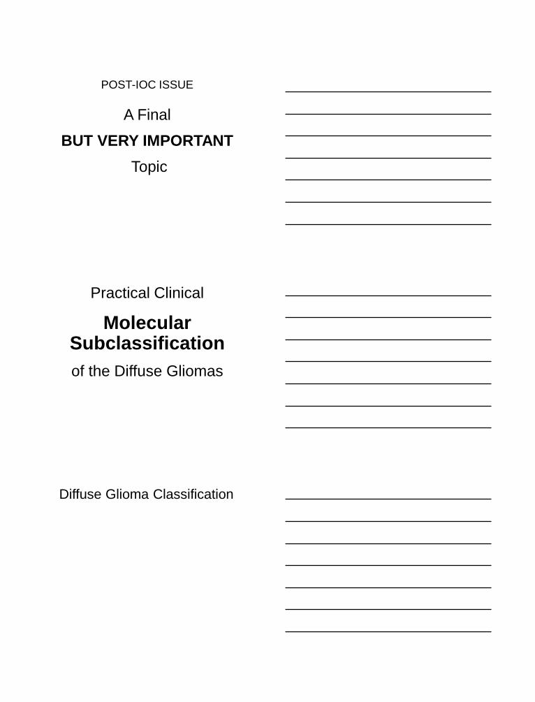
75
A Final
BUT VERY IMPORTANT
Topic
POST-IOC ISSUE
Practical Clinical
Molecular Subclassification
of the Diffuse Gliomas
Diffuse Glioma Classification
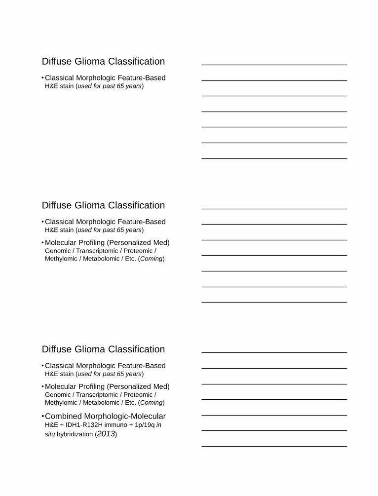
76
• Classical Morphologic Feature-Based H&E stain (used for past 65 years)
Diffuse Glioma Classification
•Classical Morphologic Feature-Based H&E stain (used for past 65 years)
• Molecular Profiling (Personalized Med) Genomic / Transcriptomic / Proteomic /
Methylomic / Metabolomic / Etc. (Coming)
Diffuse Glioma Classification
• Classical Morphologic Feature-Based H&E stain (used for past 65 years)
• Molecular Profiling (Personalized Med) Genomic / Transcriptomic / Proteomic /
Methylomic / Metabolomic / Etc. (Coming)
•Combined Morphologic-Molecular H&E + IDH1-R132H immuno + 1p/19q in
situ hybridization (2013)
Diffuse Glioma Classification
77
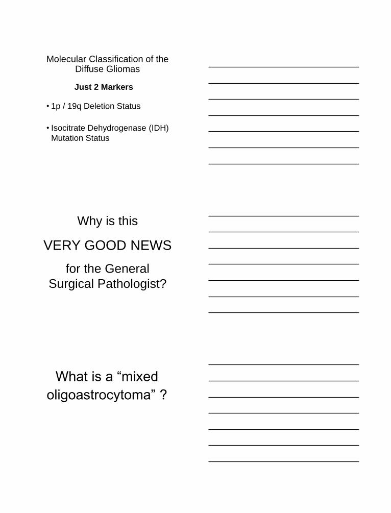
Molecular Classification of the Diffuse Gliomas
• 1p / 19q Deletion Status
• Isocitrate Dehydrogenase (IDH)
Mutation Status
Just 2 Markers
Why is this
VERY GOOD NEWS
for the General
Surgical Pathologist?
What is a “mixed
oligoastrocytoma” ?
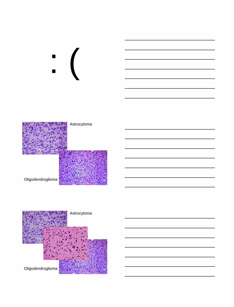
78
“Mixed Oligoastrocytoma”
: (
Astrocytoma
Oligodendroglioma
Astrocytoma
Oligodendroglioma
79
Astrocytoma
Oligodendroglioma
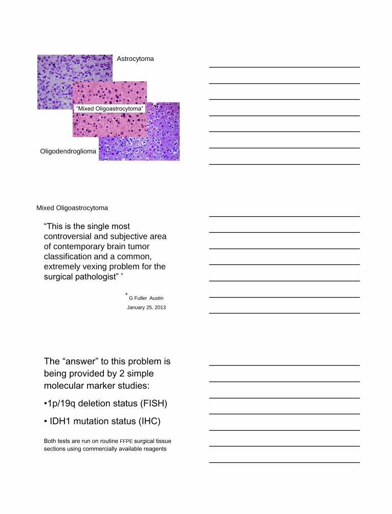
“Mixed Oligoastrocytoma”
Mixed Oligoastrocytoma
“This is the single most
controversial and subjective area
of contemporary brain tumor
classification and a common,
extremely vexing problem for the
surgical pathologist” *
* G Fuller Austin
January 25, 2013
The “answer” to this problem is
being provided by 2 simple
molecular marker studies:
•1p/19q deletion status (FISH)
• IDH1 mutation status (IHC)
Both tests are run on routine FFPE surgical tissue
sections using commercially available reagents
80
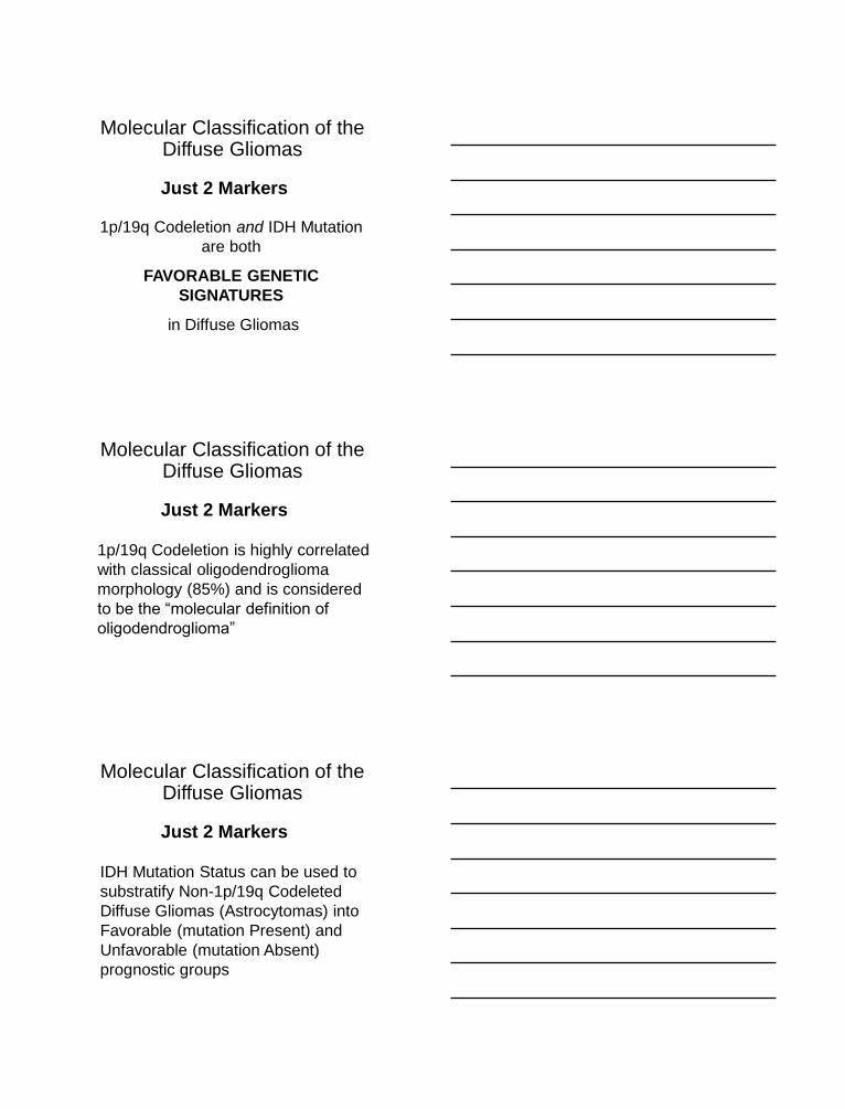
Molecular Classification of the Diffuse Gliomas
1p/19q Codeletion and IDH Mutation
are both
FAVORABLE GENETIC
SIGNATURES
in Diffuse Gliomas
Just 2 Markers
Molecular Classification of the Diffuse Gliomas
1p/19q Codeletion is highly correlated
with classical oligodendroglioma
morphology (85%) and is considered
to be the “molecular definition of
oligodendroglioma”
Just 2 Markers
Molecular Classification of the Diffuse Gliomas
IDH Mutation Status can be used to
substratify Non-1p/19q Codeleted
Diffuse Gliomas (Astrocytomas) into
Favorable (mutation Present) and
Unfavorable (mutation Absent)
prognostic groups
Just 2 Markers
81
1. If the H&E shows classic oligodendroglioma features,
there is an 85% probability of combined 1p/19q
deletion (and 100% of 1p/19q co-deleted gliomas
exhibit IDH mutation) and the diagnosis is
Oligodendroglioma
Molecular Classification of Diffuse Gliomas
1. If the H&E shows classic oligodendroglioma features,
there is an 85% probability of combined 1p/19q
deletion (and 100% of 1p/19q co-deleted gliomas also
exhibit IDH mutation) and the diagnosis is
Oligodendroglioma
2. If the morphologic features are NOT classical oligo, the
trend is to classify the tumor as an Astrocytoma
3. FISH testing for combined deletion of 1p and 19q has
become standard of care, and co-deletion constitutes
the “molecular definition” of oligodendroglioma
Molecular Classification of Diffuse Gliomas
82
1. If the H&E shows classic oligodendroglioma features,
there is an 85% probability of combined 1p/19q
deletion (and 100% of 1p/19q co-deleted gliomas also
exhibit IDH mutation) and the diagnosis is
Oligodendroglioma
2. If the morphologic features are NOT classical oligo, the
trend is to classify the tumor as an Astrocytoma
3. FISH testing for combined deletion of 1p and 19q has
become standard of care, and co-deletion constitutes
the “molecular definition” of oligodendroglioma
4. IHC for IDH1 mutation (and sometimes sequencing for
less common IDH1 and IDH2 mutations) permits
substratification of non-codeleted diffuse gliomas into
favorable and unfavorable groups.
Molecular Classification of Diffuse Gliomas
…has already reached textbook status…
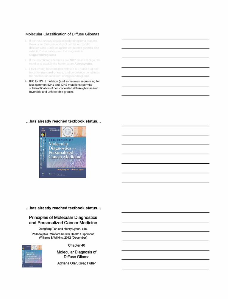
…has already reached textbook status…
Principles of Molecular Diagnostics
and Personalized Cancer Medicine
Dongfeng Tan and Henry Lynch, eds.
Philadelphia : Wolters Kluwer Health / Lippincott
Williams & Wilkins, 2012 (December)
Chapter 40
Molecular Diagnosis of
Diffuse Glioma
Adriana Olar, Greg Fuller

83
…has already reached textbook status…
Glioma Molecular Classification 2013
Olar, Fuller. In: Principles of Molecular Diagnostics and Personalized Cancer Medicine, 2012
Diffuse Glioma (H&E Diagnosis)
Olar, Fuller. In: Principles of Molecular Diagnostics and Personalized Cancer Medicine, 2012
Glioma Molecular Classification 2013
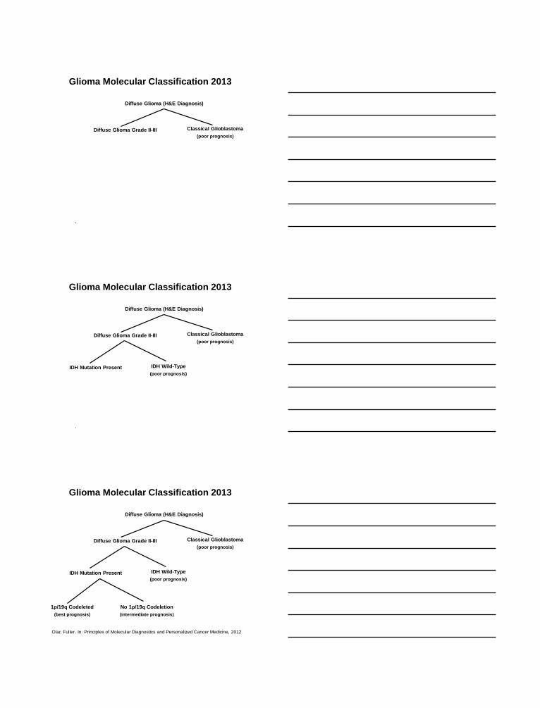
84
Diffuse Glioma (H&E Diagnosis)
Diffuse Glioma Grade II-III Classical Glioblastoma
(poor prognosis)
Olar, Fuller. In: Principles of Molecular Diagnostics and Personalized Cancer Medicine, 2012
Glioma Molecular Classification 2013
Diffuse Glioma (H&E Diagnosis)
Diffuse Glioma Grade II-III Classical Glioblastoma
(poor prognosis)
IDH Mutation Present IDH Wild-Type
(poor prognosis)
Olar, Fuller. In: Principles of Molecular Diagnostics and Personalized Cancer Medicine, 2012
Glioma Molecular Classification 2013
Diffuse Glioma (H&E Diagnosis)
Diffuse Glioma Grade II-III Classical Glioblastoma
(poor prognosis)
IDH Mutation Present IDH Wild-Type
(poor prognosis)
1p/19q Codeleted
(best prognosis)
No 1p/19q Codeletion
(intermediate prognosis)
Olar, Fuller. In: Principles of Molecular Diagnostics and Personalized Cancer Medicine, 2012
Glioma Molecular Classification 2013
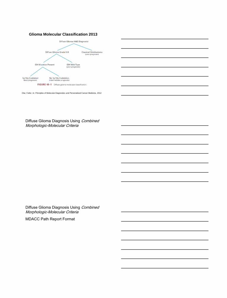
85
Olar, Fuller. In: Principles of Molecular Diagnostics and Personalized Cancer Medicine, 2012
Glioma Molecular Classification 2013
Diffuse Glioma Diagnosis Using Combined Morphologic-Molecular Criteria
Diffuse Glioma Diagnosis Using Combined Morphologic-Molecular Criteria
MDACC Path Report Format

86
DIFFUSE GLIOMA WHO Grade II Mitotic index (PHH3): 1/1000 Ki-67 index: 2% IDH1-R132H Mutation: NEGATIVE 1p/19q: INTACT (low-grade diffuse astrocytoma)
Diffuse Glioma Diagnosis Using Combined Morphologic-Molecular Criteria
MDACC Path Report Format
ANAPLASTIC DIFFUSE GLIOMA WHO Grade III Mitotic index (PHH3): 13/1000 Ki-67 index: 55% IDH1-R132H Mutation: POSITIVE 1p/19q: CO-DELETED (anaplastic oligodendroglioma)
Diffuse Glioma Diagnosis Using Combined Morphologic-Molecular Criteria
MDACC Path Report Format
ANAPLASTIC DIFFUSE GLIOMA (ANAPLASTIC OLIGODENDROGLIOMA)
WHO Grade III Mitotic index (PHH3): 13/1000 Ki-67 index: 55% IDH1-R132H Mutation: POSITIVE 1p/19q: CO-DELETED
Diffuse Glioma Diagnosis Using Combined Morphologic-Molecular Criteria
MDACC Path Report Format

87
Olar, Fuller. In: Principles of Molecular Diagnostics and Personalized Cancer Medicine, 2012
Neuropathology
References
2013
4th Edition
2011
Fuller & Burger
Chapter 11
Central Nervous System
Histology for Pathologists
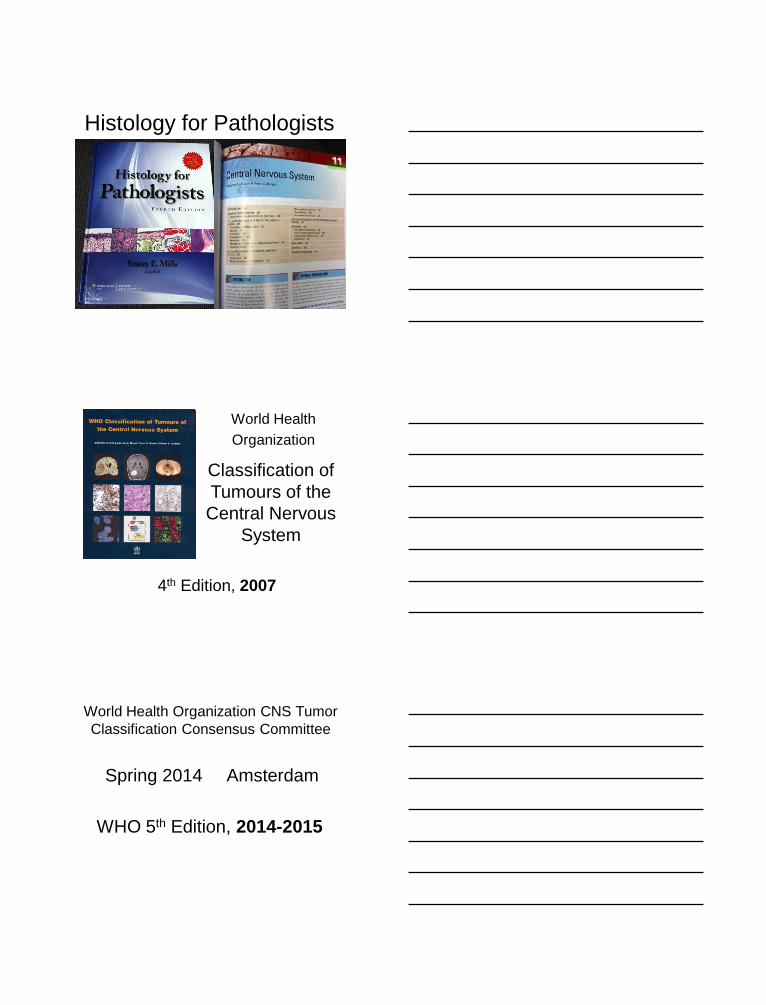
88
Histology for Pathologists
Classification of
Tumours of the
Central Nervous
System
4th Edition, 2007
World Health
Organization
Spring 2014 Amsterdam
WHO 5th Edition, 2014-2015
World Health Organization CNS Tumor
Classification Consensus Committee
89
2013 Baltimore
2014 San Diego
USCAP ANNUAL MEETING
www.uscap.org

90
Short Course #46 "Neuropathology After Dark:
Surviving Intraoperative
Frozen Section Consultation"
Chris Fuller & Greg Fuller
My Wife Tina
Related Documents


