PowerPoint ® Lecture Slides prepared by Betsy C. Brantley Valencia College C H A P T E R © 2013 Pearson Education, Inc. 8 The Peripheral and Autonomic Nervous Systems

PowerPoint ® Lecture Slides prepared by Betsy C. Brantley Valencia College C H A P T E R © 2013 Pearson Education, Inc. 8 The Peripheral and Autonomic.
Dec 13, 2015
Welcome message from author
This document is posted to help you gain knowledge. Please leave a comment to let me know what you think about it! Share it to your friends and learn new things together.
Transcript

PowerPoint® Lecture Slidesprepared byBetsy C. BrantleyValencia College
C H A P T E R
© 2013 Pearson Education, Inc.
8
The Peripheral and Autonomic Nervous Systems

© 2013 Pearson Education, Inc.
Chapter 8 Learning Outcomes
• Section 1: The Peripheral Nervous System
• 8.1 • Describe the major components of a spinal nerve, and discuss
dermatomes and shingles.• 8.2
• Name the 12 pairs of cranial nerves, give their primary functions, and identify their innervations.
• 8.3 • Define a nerve plexus and identify the four major plexuses and their
patterns of distribution.• 8.4
• Describe a sensory pathway and a motor pathway.• 8.5
• Define reflexes and describe the steps in a stretch reflex and withdrawal reflex.

© 2013 Pearson Education, Inc.
Chapter 8 Learning Outcomes
• 8.6• CLINICAL MODULE Explain the value of reflex testing and how
higher centers control reflex responses.• 8.7
• CLINICAL MODULE Describe the roles of the nervous system in referred pain, Parkinson's disease, rabies, cerebral palsy, amyotrophic lateral sclerosis, Alzheimer's disease, and multiple sclerosis.

© 2013 Pearson Education, Inc.
Chapter 8 Learning Outcomes
• Section 2: The Autonomic Nervous System
• 8.8• Describe the organization and functions of the sympathetic and
parasympathetic divisions of the autonomic nervous system.• 8.9
• Describe the innervation patterns of the sympathetic and parasympathetic divisions of the autonomic nervous system.
• 8.10• Describe the effects of sympathetic activation and
parasympathetic stimulation, and outline the anatomical and functional characteristics of each relative to neurotransmitter release.

© 2013 Pearson Education, Inc.
The Peripheral Nervous System (Section 1)
• Central Nervous System
• Processes sensory information and responds
• Peripheral Nervous System (PNS)
• Network of peripheral nerves
• Structurally divided into:
• Cranial nerves – 12 pairs; connect directly to brain
• Spinal nerves – 31 pairs; connect directly to spinal cord

© 2013 Pearson Education, Inc.
Peripheral nerves
Cranial nerves
Lumbar spinal nervesSacral spinal nerves
Coccygeal nerve
Cervical spinal nerves
Thoracic spinal nerves
Spinal nerves
Peripheral nervous system
Figure 8 Section 1

© 2013 Pearson Education, Inc.
Spinal Nerve Structure (8.1)
• Each segment of spinal cord is connected to a pair of spinal nerves
• A series of connective tissue layers surrounds each nerve
• Epineurium
• Outermost covering
• Dense network collagen fibers
• Perineurium
• Divides nerve into bundles of axons, or fascicles
• Endoneurium
• Innermost layer
• Delicate connective tissue around individual axons

© 2013 Pearson Education, Inc.
Spinal nerve structure
Blood vessels Fascicle Schwann cellMyelinatedaxon
Epineurium
Perineurium
Endoneurium
Connective TissueLayers of a Spinal Nerve
Figure 8.1 11

© 2013 Pearson Education, Inc.
Spinal Nerve Rami (8.1)
• Each spinal nerve branches into:
• Rami (singular ramus)
• Dorsal ramus – innervates muscles, joints, skin of back
• Ventral ramus – innervates structures in lateral and
anterior trunk and limbs
• In thoracic and upper lumbar segment, carry motor
output of sympathetic division of autonomic nervous
system ("fight or flight" response)

© 2013 Pearson Education, Inc. Figure 8.1 22
Dorsal rootDorsal root
ganglion Dorsal ramus
Spinalnerve
Ventralroot
Ventral ramus
SympatheticganglionAutonomic
nerve
Spinal nerve branches

© 2013 Pearson Education, Inc.
Spinal Nerve Patterns of Distribution (8.1)
• Dermatome
• Specific region of skin surface monitored by single pair of
spinal nerves
• Boundaries overlap to some degree
• C1 typically lacks sensory branch
• Face innervated by pair of cranial nerves
• Clinically important to diagnose specific location of spinal
nerve or spinal cord damage

© 2013 Pearson Education, Inc.
Dermatomes
Figure 8.1 33
Anterior Posterior
C2
C3
C2–C3
C2–C3
C3
C4
C5C4
C5
T1T2
T2
T3T4T5T6T7T8T9T10T11T12T9
T12
T11
T10
T8
T7
T6
T5
T4
T3
T2
C5L1L2
L3L4
L5
T1C7
S4S3S2
L1 S5
C8
L5S1
S2L2
L3
L4
S1
L5
L5
L3
L2
L1
C6
C8
C7T1
T2

© 2013 Pearson Education, Inc.
Shingles (8.1)
• Viral infection of dorsal root ganglia
• Caused by varicella-zoster virus
• Same herpes virus causes chickenpox
• Attacks neurons within dorsal roots of spinal
nerves and sensory ganglia of cranial nerves
• Produces painful rash and blisters on skin along
path of sensory nerve and associated dermatome

© 2013 Pearson Education, Inc. Figure 8.1 44
Shingles

© 2013 Pearson Education, Inc.
Module 8.1 Review
a. Identify the three layers of connective tissue of a
spinal nerve and identify the major peripheral
branches of a spinal nerve.
b. Describe a dermatome.
c. Explain the etiology (cause) of shingles.

© 2013 Pearson Education, Inc.
Cranial Nerves (8.2)
• 12 pairs of cranial nerves
• Classified as sensory, special sensory, motor, or
mixed
• Name relates to appearance or function
• Roman numeral corresponds to position on brain

© 2013 Pearson Education, Inc.
Cranial nerves
Figure 8.2
Trochlear Nerve (IV)Optic Nerve (II)
Motor nerve to muscles of mastication
Trigeminal Nerve (V)
Motor nerveto facialmuscles
Facial Nerve (VII)
Sensory nerveto tongue and soft palate
VestibulocochlearNerve (VIII)
Cochlear branch
Vestibular branch
Sensory nerve toposterior tongue
Motor nerve topharyngeal muscles
GlossopharyngealNerve (IX)
Trigeminalnerve (V)
Facial nerve (VII)
Vestibulocochlearnerve (VIII)
Glossopharyngealnerve (X)
Vagusnerve (X)
Vagus Nerve (X)
Olfactory Nerve (I)
Olfactory bulb
Olfactory tract
Pituitary gland
Pons
Medullaoblongata
To tonguemuscles
To sternocleidomastoidand trapezius muscles
Sensory nervesMotor nerves
KEY
Oculomotor Nerve (III)
Abducens Nerve (VI)
Hypoglossal Nerve (XII) Accessory Nerve (XI)

© 2013 Pearson Education, Inc.
Cranial Nerves I, II, III (8.2)
• Olfactory nerve (I)
• Function: special sensory
• Innervation: olfactory epithelium
• Optic nerve (II)
• Function: special sensory
• Innervation: retina of eye
• Oculomotor nerve (III)
• Function: motor
• Innervation: extrinsic and intrinsic eye muscles and eyelid muscle
(levator palpebrae superioris)

© 2013 Pearson Education, Inc. Figure 8.2 11
Olfactory bulbOlfactory tract
Pons
KEYSensory nervesMotor nerves
Olfactory Nerve (I)
Pituitarygland
Medullaoblongata
Olfactory nerve

© 2013 Pearson Education, Inc.
Cranial nerves going to and from the eye
Figure 8.2 11
Sensory nervesMotor nerves
KEY
Optic Nerve (II)
Oculomotor Nerve (III)
Trochlear Nerve (IV)
Abducens Nerve (VI)

© 2013 Pearson Education, Inc.
Cranial Nerves IV, V, VI (8.2)
• Trochlear nerve (IV)
• Function: motor
• Innervation: superior oblique muscle
• Trigeminal nerve (V)
• Function: mixed
• Innervation: sensory – parts of face, lips, palate, tongue; motor –
muscles of mastication
• Abducens nerve (VI)
• Function: motor
• Innervation: lateral rectus muscle

© 2013 Pearson Education, Inc.
Trigeminal nerve
Figure 8.2 11
Sensory nervesMotor nerves
Trigeminalnerve (V)
Motor nerve to muscles of mastication
KEY
Trigeminal Nerve (V)

© 2013 Pearson Education, Inc.
Cranial Nerves VII, VIII, IX (8.2)
• Facial nerve (VII)
• Function: mixed
• Innervation: sensory – taste receptors anterior 2/3 tongue; motor –
muscles of facial expression, lacrimal gland, salivary glands
• Vestibulocochlear nerve (VIII)
• Function: special sensory
• Innervation: cochlea (hearing), vestibule (motion and balance)
• Glossopharyngeal nerve (IX)
• Function: mixed
• Innervation: sensory – posterior 1/3 of tongue, pharynx, palate; motor –
pharyngeal muscles and parotid salivary gland

© 2013 Pearson Education, Inc. Figure 8.2 11
Sensory nervesMotor nerves
Facial nerve (VII)
Sensory nerve to tongue and
soft palate
Motor nerveto facialmuscles
KEY
Facial Nerve (VII)
Facial nerve

© 2013 Pearson Education, Inc.
Vestibulocochlear nerve
Figure 8.2 11
Sensory nervesMotor nerves
KEYCochlear
branch
Vestibular branch
Vestibulocochlear Nerve (VIII)
Vestibulocochlear nerve (VIII)

© 2013 Pearson Education, Inc. Figure 8.2 11
Glossopharyngeal nerve
Sensory nervesMotor nerves
KEY
Sensory nerve toposterior tongue
Motor nerve topharyngeal muscles
Glossopharyngealnerve (X)
Glossopharyngeal Nerve (IX)

© 2013 Pearson Education, Inc.
Cranial Nerves X, XI, XII (8.2)
• Vagus nerve (X)
• Function: mixed
• Innervation: sensory – pharynx, ear, diaphragm, visceral organs;
motor – palate and pharyngeal muscles; visceral organs
• Accessory nerve (XI)
• Function: motor
• Innervation: skeletal muscles of palate, pharynx, larynx,
sternocleidomastoid and trapezius muscles
• Hypoglossal nerve (XII)
• Function: motor
• Innervation: tongue muscles

© 2013 Pearson Education, Inc. Figure 8.2 11
Sensory nervesMotor nerves
KEY
Vagus Nerve (X)
Vagusnerve (X)
Vagus nerve

© 2013 Pearson Education, Inc.
Sensory nervesMotor nerves
KEY
To sternocleidomastoidand trapezius muscles
Accessory Nerve (XI)
Figure 8.2 11
Accessory nerve

© 2013 Pearson Education, Inc.
Sensory nervesMotor nerves
KEY
HypoglossalNerve (XII)
To tonguemuscles
Hypoglossal nerve
Figure 8.2 11

© 2013 Pearson Education, Inc.
Module 8.2 Review
a. Identify the cranial nerves by name and number.
b. Which cranial nerves have motor functions only?
c. Which cranial nerves are mixed nerves?

© 2013 Pearson Education, Inc.
Nerve Plexuses (8.3)
• Complex interwoven network of nerves
• Results from fusing small skeletal muscles (each with
separate ventral rami) into larger muscles during
development
• Four major plexuses
• Cervical
• Brachial
• Lumbar
• Sacral

© 2013 Pearson Education, Inc. Figure 8.3 11
Femoral nerveObturator nerve
Superior gluteal nerveInferior gluteal nerve
Pudendal nerveSaphenous nerveSciatic nerve
Median nerve
Ulnar nerve
Radial nerve
Thoracic nerves
Musculocutaneousnerve
Axillary nerve
Phrenic nerve
Sacral plexus
Lumbar plexus
Brachial plexus
Cervical plexusC1C2C3C4C5C6C7C8T1T2T3T4T5T6T7T8
T9
T10
T11T12
L1
L2
L3L4
L5S1
S2S3S4S5
Co1
Spinal nerve plexuses

© 2013 Pearson Education, Inc.
Cervical Plexus (8.3)
• Ventral rami of spinal nerves C1–C5
• Innervates muscles of neck and into thoracic
cavity
• Phrenic nerve
• Provides nerve supply to diaphragm

© 2013 Pearson Education, Inc.
Cervical plexus
Figure 8.3 22
Nerve Roots ofCervical Plexus
C1
C2
C3
C4
C5
Phrenic nerve

© 2013 Pearson Education, Inc.
Brachial, Lumbar, and Sacral Plexuses (8.3)
• Brachial plexus
• Ventral rami of spinal nerves C5–T1
• Innervates pectoral girdle and upper limbs
• Lumbar plexus
• Arises from lumbar segments of spinal cord
• Innervates pelvic girdle and lower limbs
• Sacral plexus
• Arises from sacral segments of spinal cord
• Innervates pelvic girdle and lower limbs

© 2013 Pearson Education, Inc.
Brachial plexus
Figure 8.3 33
Musculocutaneousnerve
Median nerveUlnar nerve
Radial nerve
Ulnar nerveMedian nerve

© 2013 Pearson Education, Inc.
Lumbar and sacral plexuses
Figure 8.3 34
Lateral femoralcutaneous nerve
Femoral nerve
Obturator nerve
Superior gluteal nerve
Inferior gluteal nerve
Posterior femoralcutaneous nerve (cut)
Sciatic nerve
Saphenous nerve
Common fibular nerve
Superficial fibular nerve
Deep fibular nerve

© 2013 Pearson Education, Inc.
Module 8.3 Review
a. Define nerve plexus and list the major plexuses.
b. Injury to which nerve plexus would interfere with
the ability to breathe?
c. List the major nerves of the brachial plexus.
© 2013 Pearson Education, Inc.
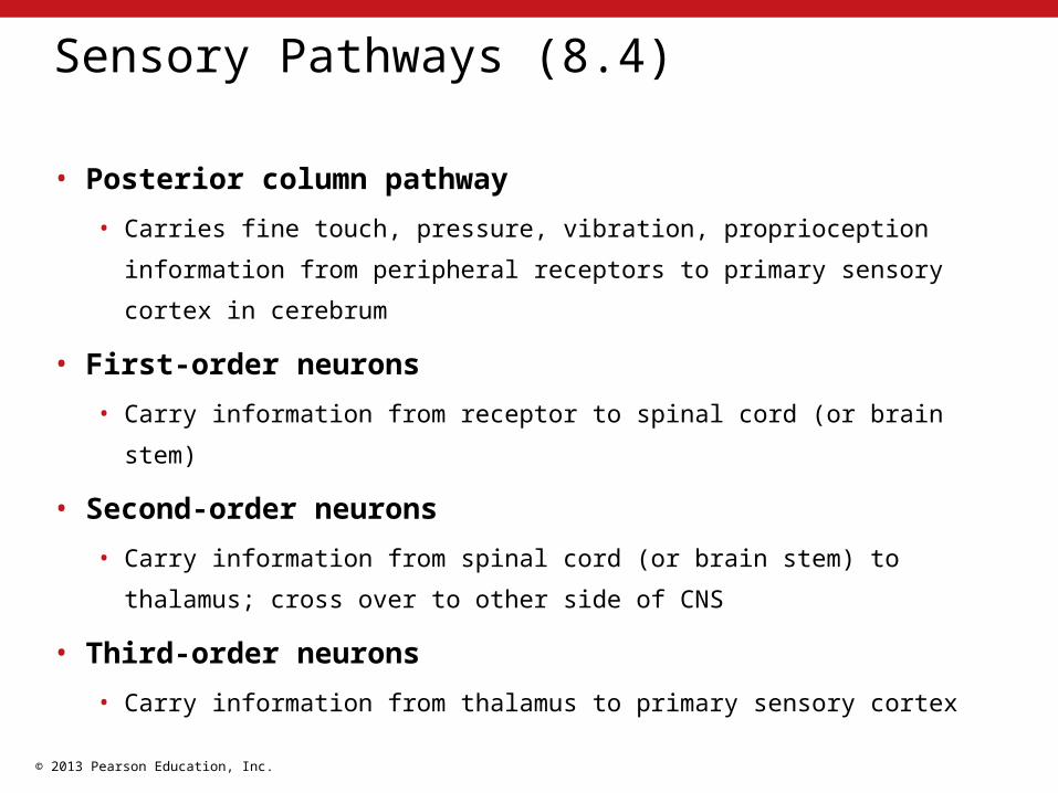
Sensory Pathways (8.4)
• Posterior column pathway
• Carries fine touch, pressure, vibration, proprioception information
from peripheral receptors to primary sensory cortex in cerebrum
• First-order neurons
• Carry information from receptor to spinal cord (or brain stem)
• Second-order neurons
• Carry information from spinal cord (or brain stem) to thalamus;
cross over to other side of CNS
• Third-order neurons
• Carry information from thalamus to primary sensory cortex

© 2013 Pearson Education, Inc.
Ventral nucleiin thalamus
Medullaoblongata
Spinalcord
Dorsal rootganglion
Third-orderneuronSecond-orderneuronFirst-orderneuronFine-touch, vibration,
pressure, andproprioception sensations
from right side of body
Midbrain
Sensory homunculus
Third order neurons
Second order neurons
First order neuronsStart
3
KEY
2
1
Sensory pathways
Figure 8.4 11

© 2013 Pearson Education, Inc.
Motor Pathways (8.4)
• Corticospinal pathway
• Provides voluntary control over skeletal muscles
• Upper motor neuron
• Cell body in CNS processing center (primary motor cortex);
communicate with lower motor neurons
• Lower motor neuron
• Cell body in nucleus of brain stem or in spinal cord; communicate
with skeletal muscles
• Axons cross to opposite side in brain stem or spinal cord

© 2013 Pearson Education, Inc.
Motor homunculus
To skeletalmuscles
Motor nucleiof cranial
nervesTo skeletal
muscles
Motor nucleiof cranial
nerves
Axons cross toopposite side
Cerebralpeduncle
Descendingaxons
Upper motorneuronLower motorneuronMedulla
oblongata
Spinal cord
Midbrain
KEY
To skeletalmuscles
Motor pathways
Figure 8.4 22

© 2013 Pearson Education, Inc.
Homunculus (8.4)
• Map created by determining area of cortex devoted to
particular part of body and function
• Sensory homunculus
• Map of primary sensory cortex
• Proportions reflect number of sensory receptors in each region
• Motor homunculus
• Map of primary motor cortex
• Proportions reflect number of motor units innervated and degree of
fine motor control available

© 2013 Pearson Education, Inc.
Module 8.4 Review
a. Define sensory homunculus.
b. Define corticospinal pathway.
c. Which side of the body does the right cerebral
hemisphere control?

© 2013 Pearson Education, Inc.
Reflexes (8.5)
• Rapid, automatic response to specific stimuli
• Preserve homeostasis by allowing rapid adjustments in
function
• Reflex arc
• Receptor
• Sensory neuron
• Information processing
• Motor neuron
• Effector

© 2013 Pearson Education, Inc.
Stretch Reflex (8.5)
• One of simplest and fastest reflex arcs
• Monosynaptic (one synapse involved)
• For example, patellar reflex
• Receptor – tap patellar tendon with hammer, receptors in
quadriceps muscle stretched
• Sensory neuron – carries stretch message directly to motor neuron
• Information processing – in patellar reflex, processing is in cell
body of motor neuron
• Motor neuron – carries message back to effector
• Effector – quadriceps muscle stimulated to contract

© 2013 Pearson Education, Inc.
STEP 2Activation of a
Sensory Neuron
STEP 1 Arrival of the Stimulus and Activation of a Receptor
STEP 3InformationProcessingin the CNS
STEP 4Activation of a Motor Neuron
STEP 5 Response of a PeripheralEffector
Motor neuron(stimulated)
Sensory neuron(stimulated)
Contraction
Response
Effector
Receptor(muscle spindle)
REFLEXARC
Spinalcord
Stretch
KEY
Steps in a stretch reflex arc
Figure 8.5 11

© 2013 Pearson Education, Inc.
Withdrawal Reflex (8.5)
• More complex than stretch reflex
• Involves multiple neurons
• Moves affected parts of body away from stimulus
• Receptor – pain receptors responding to tissue damage
• Sensory neuron – carries pain message to spinal cord
• Information processing – sensory neuron stimulates interneuron,
which activates motor neuron
• Motor neuron – carries message back to effector
• Effector – skeletal muscle contraction to pull away from stimulus

© 2013 Pearson Education, Inc.
STEP 2 The activation of a
Sensory Neuron
STEP 1 The arrival of a Stimulus and Activation ofa Receptor
STEP 3 Information Processing
STEP 5 The Response of a PeripheralEffector
STEP 4 The Activation of a Motor Neuron
Dorsal rootganglion
To highercenters
REFLEXARC
Receptor
Stimulus
Effector
Sensory neuron(stimulated)
ExcitatoryinterneuronMotor neuron(stimulated)
KEY
Steps in a withdrawal reflex arc
Figure 8.5 22

© 2013 Pearson Education, Inc.
Module 8.5 Review
a. What are the common characteristics of reflexes?
b. Define reflex and list the components of a reflex
arc.
c. In the patellar reflex, identify the response
observed and the effectors involved.

© 2013 Pearson Education, Inc.
Reflex Tests (8.6)
• Activities in brain can facilitate or inhibit reflexes
• Facilitation of reflex – reinforcement
• Stretch reflexes often tested during physical exam to
provide information about status of spinal cord segments
• Biceps reflex
• Triceps reflex
• Ankle-jerk reflex

© 2013 Pearson Education, Inc.
Clinical reflex tests
Figure 8.6 11
Bicepsreflex
Tricepsreflex
Ankle-jerkreflex

© 2013 Pearson Education, Inc.
Babinski Sign and Plantar Reflex (8.6)
• In infants, stroking foot on lateral side of sole results in
fanning of toes – called positive Babinski sign
• As descending motor pathways develop, they inhibit this
reflex
• In normal adults, stroking foot on lateral side of sole results
in curling toes – called plantar reflex (negative Babinski
sign)
• Positive Babinski sign in adult indicates damage to CNS

© 2013 Pearson Education, Inc.
Babinski sign
Figure 8.6 22
Babinski sign

© 2013 Pearson Education, Inc.
Plantar reflex
Figure 8.6 33
Plantar reflex

© 2013 Pearson Education, Inc.
Abdominal Reflex (8.6)
• Descending motor tracts can facilitate reflexes
• Abdominal reflex
• Light stroking of abdominal muscles produces reflexive
twitch moving navel toward stimulus
• Absence of reflex may indicate possible damage to
descending tracts

© 2013 Pearson Education, Inc.
Abdominal reflex
Figure 8.6 44
Abdominal reflex

© 2013 Pearson Education, Inc. Figure 8.6 55

© 2013 Pearson Education, Inc.
Module 8.6 Review
a. Define reinforcement as it pertains to spinal
reflexes.
b. What purpose does reflex testing serve?
c. After injuring her back, 22-year-old Tina exhibits
a positive Babinski reflex. What does this imply
about her injury?

© 2013 Pearson Education, Inc.
Referred Pain (8.7)
• Sensation of pain in part of body other than actual source
• For example, a heart attack with pain felt in the left arm
• Visceral pain sensations can stimulate interneurons that
are part of the spinothalamic pathway
• Stimulates primary sensory cortex
• Feel pain in specific part of body surface

© 2013 Pearson Education, Inc.
Referred pain
Figure 8.7 11
Heart

© 2013 Pearson Education, Inc.
Parkinsons Disease (8.7)
• Neurons of substantia nigra damaged or secrete
less dopamine
• Basal nuclei become more active
• Raises skeletal muscle tone
• Produces rigidity and stiffness
• Difficulty starting and stopping voluntary
movements

© 2013 Pearson Education, Inc.
Parkinson disease
Figure 8.7 22
Normal substantia nigra Diminished substantianigra in Parkinson patient

© 2013 Pearson Education, Inc.
Rabies (8.7)
• Caused by virus
• Virus enters axon terminals
• Travels from terminals along axon into CNS
• Other toxins (heavy metals), some pathogenic
bacteria, and other viruses enter CNS by the same
path

© 2013 Pearson Education, Inc.
Rabies
Figure 8.7 33
Virus travels withother materialsto the cell body.
Rabies viruses
Axon terminal

© 2013 Pearson Education, Inc.
Cerebral Palsy (8.7)
• Number of disorders that affect voluntary motor
performance
• Appear during infancy or childhood
• Persist throughout life
• Caused by:
• Trauma with premature or stressful birth
• Maternal exposure to drugs (including alcohol)
• Genetic defect

© 2013 Pearson Education, Inc.
Cerebral palsy
Figure 8.7 44

© 2013 Pearson Education, Inc.
Amyotrophic Lateral Sclerosis (8.7)
• Progressive, degenerative disorder
• Affects motor neurons in spinal cord, brain stem, and
cerebral hemispheres
• Affects both upper and lower motor neurons
• Possibly underlying defect in axonal transport
• Destruction of CNS neurons causes atrophy of associated
skeletal muscles

© 2013 Pearson Education, Inc.
Amyotrophic lateral sclerosis
Figure 8.7 55

© 2013 Pearson Education, Inc.
Alzheimer's Disease (8.7)
• Progressive disorder
• Loss of higher-order cerebral functions
• Most common cause of senile dementia or senility
• Symptoms appear age 50–60 or later; occasionally affects
younger people
• 15 percent of those over age 65 in U.S. have some form
• Causes about 100,000 deaths each year
• Results in abnormalities in brain regions associated with
memory processing

© 2013 Pearson Education, Inc.
Alzheimer’s disease
Figure 8.7 66
Abnormaldendrites,axons, and
extracellularproteins form
complexesknown as
Alzheimer'splaques.

© 2013 Pearson Education, Inc.
Multiple Sclerosis (8.7)
• Characterized by recurrent incidents of demyelination
• Affects axons in optic nerve, brain, spinal cord
• Signs/symptoms:
• Partial loss of vision
• Problems with speech, balance, general motor coordination
• Time between incidents and degree of recovery vary
• One-third of cases progressive
• Typical onset age 30–40 years
• 1.5 times higher incidence in women than men

© 2013 Pearson Education, Inc.
Multiple sclerosis
Figure 8.7 77
Demyelinatingneuron

© 2013 Pearson Education, Inc.
Module 8.7 Review
a. Define referred pain.
b. Describe how rabies is contracted.
c. Describe amyotrophic lateral sclerosis (ALS).

© 2013 Pearson Education, Inc.
The Somatic Nervous System (Section 2)
• Provides conscious and subconscious control over
skeletal muscles of body
• Lower motor neurons controlled by:
• Reflexes based in spinal cord or brain
• Upper motor neurons with cell bodies in nuclei of brain
or primary motor cortex

© 2013 Pearson Education, Inc.
Somatic nervous system motor neurons
Figure 8 Section 2 11
Upper motorneurons in
primary motorcortex
Somatic motornuclei of brain
stem
Skeletalmuscle
Lowermotor
neurons
Spinal cord
Somaticmotornuclei ofspinal cord
BRAIN
Skeletalmuscle

© 2013 Pearson Education, Inc.
The Autonomic Nervous System (Section 2)
• Controls visceral function outside our awareness
• Integrative centers in the hypothalamus
• Instead of single lower motor neuron, there are two motor
neurons in series
• Preganglionic neurons – cell bodies in brain stem and spinal
cord; part of visceral reflex arcs; communicate with:
• Ganglionic neurons – visceral motor neurons in peripheral
autonomic ganglia
• Innervate visceral effectors (cardiac muscle, smooth muscle,
glands, adipose tissue)

© 2013 Pearson Education, Inc.
Autonomic nervous system motor neurons
Figure 8 Section 2 2
Visceral motornuclei in
hypothalamus
Preganglionicneuron
Visceral Effectors
Smoothmuscle
Glands
Cardiacmuscle
Adipocytes
Autonomicganglia
Ganglionicneurons
Preganglionicneuron
Autonomicnuclei inbrain stem
Spinalcord
Autonomicnuclei inspinal cord
BRAIN

© 2013 Pearson Education, Inc.
Sympathetic Division (8.8)
• Preganglionic neurons
• From thoracic and superior lumbar segments of spinal cord
• Ganglionic neurons located:
1. Within sympathetic chain ganglia near spinal cord
2. In collateral ganglia within abdominopelvic cavity (celiac,
superior mesenteric, inferior mesenteric)
3. Within modified ganglion cells in adrenal medullae
• Affect target organs through release of hormones, epinephrine
and norepinephrine

© 2013 Pearson Education, Inc.
Sympathetic nervous system ganglia location
Figure 8.8 11
PreganglionicNeurons
Ganglionic NeuronsTarget Organs
Sympathetic chain ganglia
Collateral ganglia
Adrenal medulla
Hormones
Nervoussignals
Visceral effectorsin abdomino-pelvic cavity
Organs and systems throughoutthe body
Visceral effectorsin thoracic cavity,head, body wall,and limbs
Hormones releasedinto circulation
Postganglionic fibers
Preganglionic fibersKEY

© 2013 Pearson Education, Inc.
Sympathetic Division Axons and Neurotransmitters (8.8)
• Short preganglionic fibers (axons of
preganglionic neurons)
• Release acetylcholine (ACh)
• Long postganglionic fibers (axons of ganglionic
neurons)
• Release norepinephrine (NE)

© 2013 Pearson Education, Inc.
Sympathetic Division – Functions (8.8)
• Produces "fight or flight" response, including:
1. Heightened mental alertness
2. Increased metabolic rate
3. Reduced digestive and urinary functions
4. Activation of energy reserves
5. Increased respiratory rate and dilation of respiratory passageways
6. Elevated heart rate and blood pressure
7. Activation of sweat glands

© 2013 Pearson Education, Inc.
Parasympathetic Division (8.8)
• Preganglionic neurons
• From cranial nerves III, VII, IX, X, or sacral segments of spinal
cord
• Each typically synapses on 6 to 8 ganglionic neurons
• Ganglionic neurons found:
1. In terminal ganglia – near target organ; usually paired
2. In intramural ganglia – embedded in tissues of target organ;
usually in clusters
• Both preganglionic and postganglionic axon terminals
release acetylcholine

© 2013 Pearson Education, Inc.
Parasympathetic nervous system ganglia location
Figure 8.8 22
PreganglionicNeurons
Sacral segments of spinal cord associated nuclei
Cranial nerve associated nuclei
III
VII
IX
X
Pelvicnerves
Ganglionic Neurons
Terminal ganglia
Ganglia close to targets
Intramuralganglia
Intramuralganglia
Target Organs
Intrinsic eye muscles(pupil and lens shape)
Nasal glands, tearglands, and submandibularand sublingual salivaryglands
Parotid salivaryglands
Visceral organsof neck,thoracic cavity,and most ofabdominal cavity
Visceral organs ininferior portion ofabdominopelviccavity
KEY
Preganglionic fibers
Postganglionic fibers

© 2013 Pearson Education, Inc.
Parasympathetic Division – Functions (8.8)
• Regulates visceral function and energy conservation
• Known as the "rest and digest" system, causing:
1. Decreased metabolic rate
2. Decreased heart rate and blood pressure
3. Increased secretion by salivary and digestive glands
4. Increased motility and blood flow in digestive tract
5. Stimulation of urination and defecation

© 2013 Pearson Education, Inc.
Module 8.8 Review
a. List general responses to increased sympathetic
activity and to parasympathetic activity.
b. Describe an intramural ganglion.
c. Starting in the spinal cord, trace the path of a
nerve impulse through the sympathetic ANS to its
target organ in the abdominopelvic cavity.

© 2013 Pearson Education, Inc.
Innervation Patterns in the ANS (8.9)
• Both sympathetic and parasympathetic divisions
innervate many of same structures
• Sympathetic division
• Splanchnic nerves – bundles of preganglionic fibers on
way to collateral ganglia
• Sympathetic nerves – bundles of postganglionic fibers
innervating structures in thoracic cavity

© 2013 Pearson Education, Inc.
Superiorcervicalsympatheticganglia
Postganglionicfibers to spinalnerves
Sympatheticchain gangliaT
Coccygealganglia (Co1)
fused together
PONS
Spinalcord
Splanchnicnerves
InferiorMesentericganglion
Sympathetic nerves
Autonomicplexus
Superior mesenteric ganglion
Celiac ganglion
Ovary
UterusPenisScrotum
Eye
Salivaryglands
Heart
Lung
Liver andgallbladder
Stomach
SpleenPancreas
LargeintestineSmallintestine
AdrenalmedullaKidney
Urinary bladder
Preganglionic neuronsGanglionic neurons
KEY
T1
L2 L2
T1
Innervation in the sympathetic nervous system
Figure 8.9 11

© 2013 Pearson Education, Inc.
Innervation in the Parasympathetic Division (8.9)
• Vagus nerve
• Provides roughly 75 percent of all parasympathetic outflow
• Numerous branches intermingle with sympathetic fibers forming
plexuses
• Pelvic nerves
• Bundles of preganglionic fibers from sacral segment of spinal cord
• Innervate intramural ganglia in walls of kidneys, urinary bladder,
large intestine, and sex organs

© 2013 Pearson Education, Inc.
Vagus nerve
Spinalcord
Autonomicplexuses
Pelvic nerves
Uterus Ovary
Penis
Scrotum
Terminal gangliaLacrimal glandEye
Salivary glands
Heart
LungsLiver andgallbladderStomachSpleenPancreas Large intestineSmall intestineRectum
Kidney
Urinary bladder
S2S3S4
III
VII
IX
X
PONS
Innervation in the parasympathetic nervous system
Figure 8.9 22

© 2013 Pearson Education, Inc.
Module 8.9 Review
a. Define splanchnic nerves.
b. Which nerve carries the majority of the
parasympathetic outflow?
c. Describe sympathetic nerves.

© 2013 Pearson Education, Inc.
Functional Characteristics of Sympathetic and Parasympathetic Divisions (8.10)
• Sympathetic
• Entire division quickly activated
• Parasympathetic
• Does not undergo division-wide activation
• Does not release neurotransmitters directly into
bloodstream

© 2013 Pearson Education, Inc.
Anatomical characteristics of the sympathetic and parasympathetic divisions of the ANS
Figure 8.10 2 - 32 3–
Sympathetic
CNS
PNS
Adrenalmedulla
Bloodstream
Preganglionicneuron
Preganglionicfiber
Sympatheticganglion
Ganglionicneurons
Postganglionicfiber
NeurotransmittersKEY
AcetylcholineNorepinephrineEpinephrine
TARGET
Preganglionicneuron
Preganglionicfiber
Ganglionicneurons
Postganglionicfiber
Parasympathetic
CNS
PNS
Parasympatheticganglion
TARGET
Characteristic Sympathetic Division Characteristic Parasympathetic DivisionLocation of CNSvisceral motor neurons
Lateral gray horns of spinalsegments T1–L2
Location of CNSvisceral motor neurons
Brain stem and spinalsegments S2–S4
Location of PNS ganglia Near vertebral column (sympathetic chain ganglia)
Location of PNS ganglia Typically intramural
Preganglionic fibers Neurotransmitter
ShortACh
Preganglionic fibers Neurotransmitter
Relatively longACh
Postganglionic fibers Neurotransmitter
LongUsually norepinephrine (NE)
Postganglionic fibers Neurotransmitter
Relatively shortACh
General functions Stimulates metabolism;increases alertness; preparesfor emergency (“fight or flight”)
General functions Promotes relaxation, nutrientuptake, energy storage (“restand digest”)
or

© 2013 Pearson Education, Inc.
Module 8.10 Review
a. What neurotransmitter is released by all
parasympathetic neurons?
b. Why is the parasympathetic division called the
anabolic system?
c. What physiological changes are typical in tense
(anxious) individuals?
Related Documents