e pictures of eye injur

power point of extra material about eye trauma
Aug 13, 2015
Welcome message from author
This document is posted to help you gain knowledge. Please leave a comment to let me know what you think about it! Share it to your friends and learn new things together.
Transcript

Some pictures of eye injuries
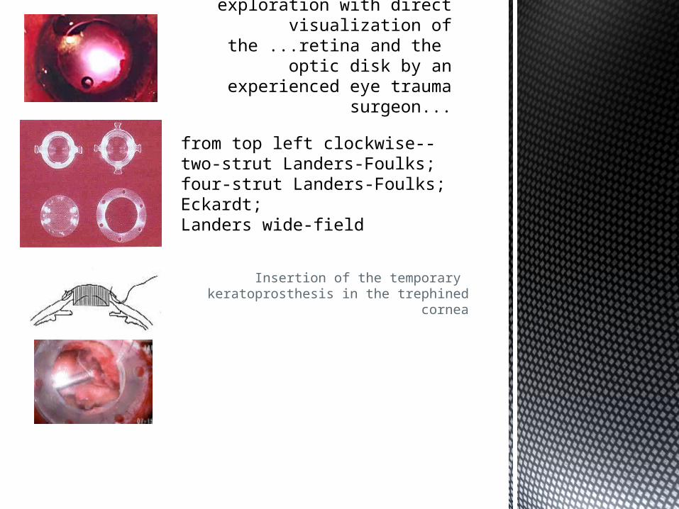
Insertion of the temporary keratoprosthesis in the trephined cornea
Severely traumatized eyes deserve surgical exploration with direct
visualization of the ...retina and the optic disk by an experienced eye trauma
surgeon...
from top left clockwise-- two-strut Landers-Foulks; four-strut Landers-Foulks; Eckardt;Landers wide-field
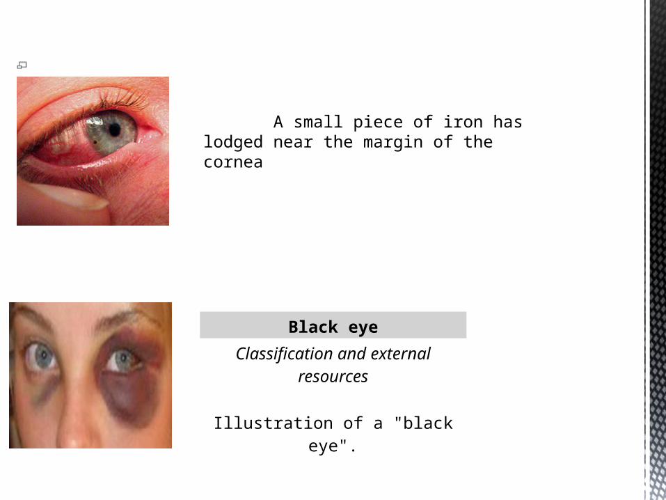
A small piece of iron has lodged near the margin of the cornea
A smallA small piece of iron has lodged near the margin of the cornea piece of iron has lodged near the margin of the cornea
Black eye
Classification and external resources
Illustration of a "black eye".
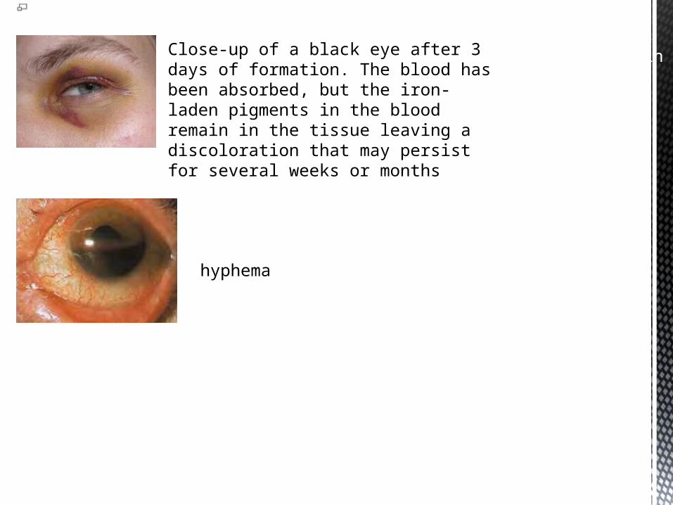
Close-up of a black eye after 3 days of formation. The blood has been absorbed, but the iron-laden pigments in the blood remain in the tissue leaving a discoloration that may persist for several weeks or months.
Close-up of a black eye after 3 days of formation. The blood has been absorbed, but the iron-laden pigments in the blood remain in the tissue leaving a discoloration that may persist for several weeks or months
hyphema
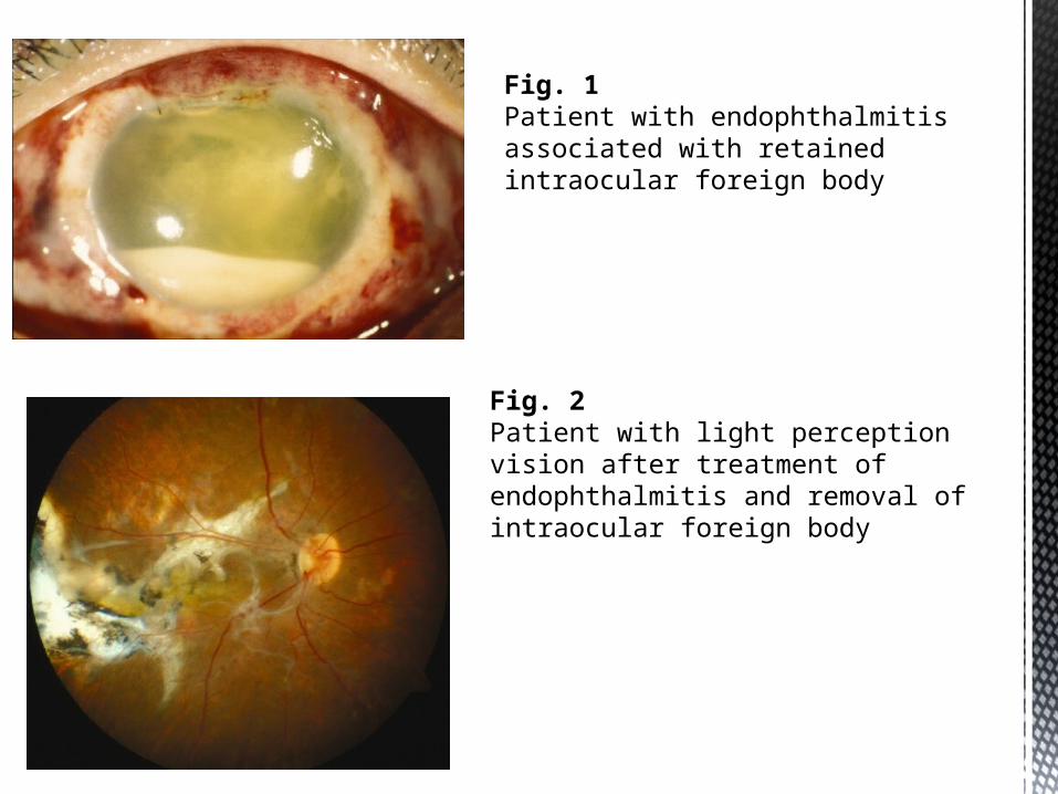
Fig. 2Patient with light perception vision after treatment of endophthalmitis and removal of intraocular foreign body
Fig. 1Patient with endophthalmitis associated with retained intraocular foreign body
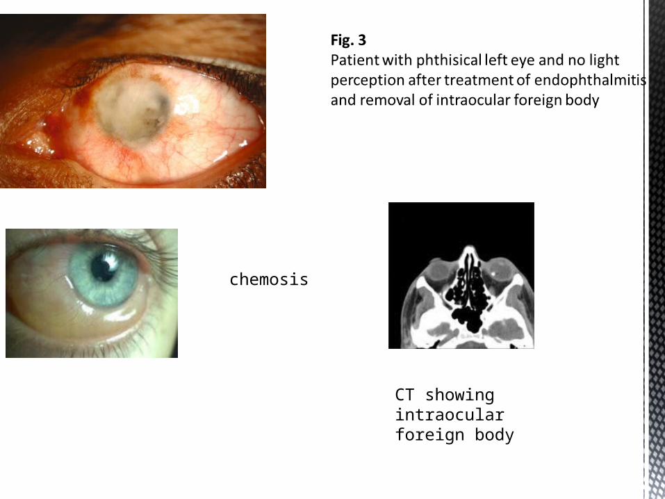
chemosis
CT showing intraocular foreign body

Figure 1: (a) Anterior segment optical coherence tomography image of the right eye showing the area of scleral discontinuity with a heterogeneous high reflectivity noted internally. The peripheral anterior chamber structures are also seen, with ruptured zonules. (b) Ultrasound biomicroscopy image of the right eye in case 1 shows the area of sclera rupture in much better detail. (c) Ultrasound biomicroscopy image over the phacocele shows a clearly demarcated structure within the subconjunctival cyst, with multiple layers of the crystalline lens
These pictures are related to the study letter (F) in the text:

Figure 3: Slit-lamp photograph of the right eye in case 2 shows a superior conjunctival mass with a diameter of 13 mm revealed by lifting the upper lid
Figure 2: External photographs of the left eye with the lid raised (a, b) in case 3 show a large, elevated subconjunctival mass with subconjunctival hemorrhage around its base. Slit-lamp photograph without elevating the lid shows aphakia (c)

Figure 4: Ultrasound biomicroscopy over the cyst shown in Fig. 4 shows a clearly demarcated structure within the cyst
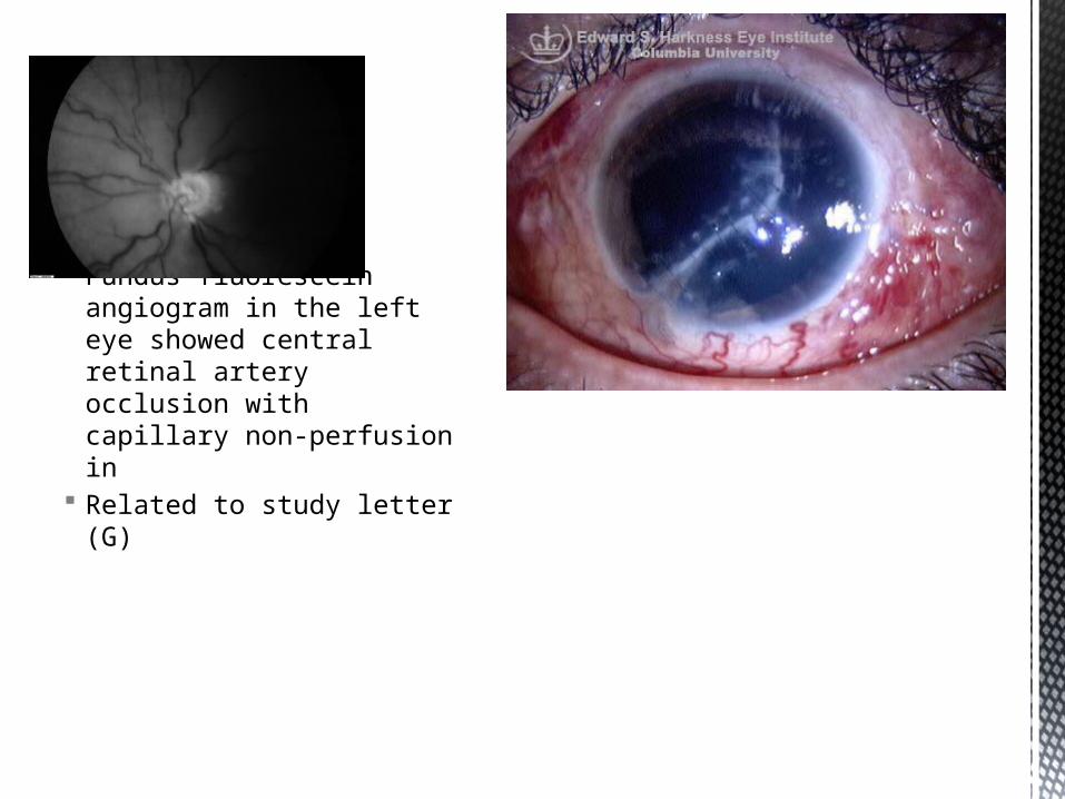
Fundus fluorescein angiogram in the left eye showed central retinal artery occlusion with capillary non-perfusion in
Related to study letter (G)
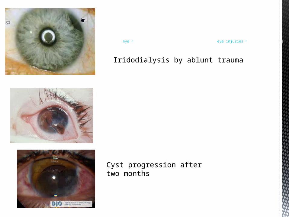
Iridodialysis caused by blunt trauma to the eyeIridodialyses are usually caused by blunt trauma to the eye,2 but may also be caused by penetrating eye injuries.3 An iridodialysis
Iridodialysis by ablunt trauma
Cyst progression after two months

Scleral rupture
Ruptured globe with erupted ocular contents
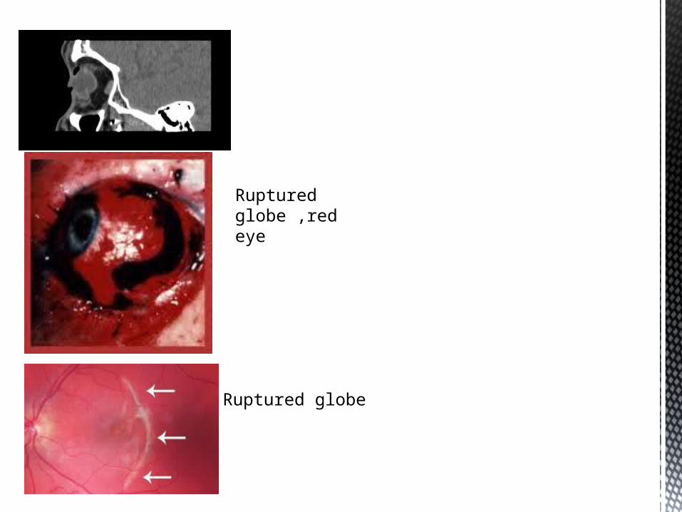
Ruptured globe ,red eye
Ruptured globe
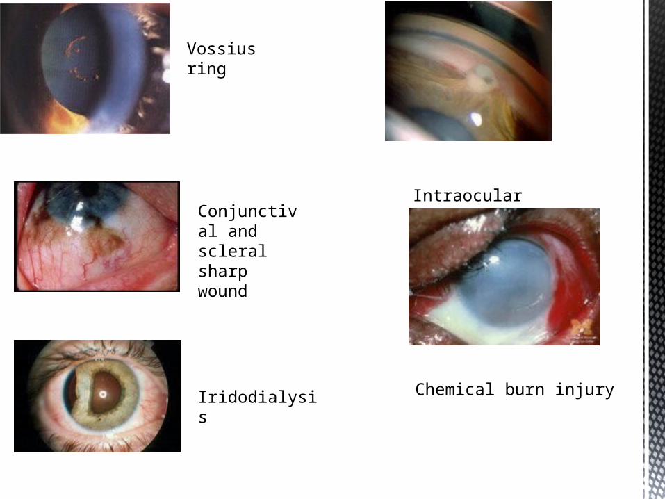
Vossius ring
Conjunctival and scleral sharp wound
Iridodialysis
Intraocular foreign body
Chemical burn injury
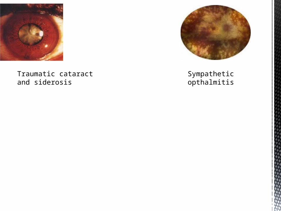
Traumatic cataract and siderosis
Sympathetic opthalmitis


Related Documents